Ascorbic Acid Infusion Blunts CD40L Upregulation in Patients Undergoing Coronary Stent
Int. J. Mol. Sci. 2013, 14, 14439-14459; doi:10.3390/ijms140714439
International Journal of
Molecular Sciences ISSN 1422-0067
www.mdpi.com/journal/ijms
Article
Upregulation of Phosphorylated HSP27, PRDX2, GRP75, GRP78 and GRP94 in Acquired Middle Ear Cholesteatoma Growth
Kuen Yao Ho 1,2, Tai Sheng Yeh 3,†, Han Hsiang Huang 4,†, Kuo Feng Hung 5,†, Chee Yin Chai 6,7,
Wan Tzu Chen 6, Shih Meng Tsai 8, Ning Chia Chang 9, Chen Yu Chien 1, Hsun Mo Wang 10
and Yu Jen Wu 4,*
1 Department of Otorhinolaryngology, Kaohsiung Medical University Hospital, Kaohsiung 80756,
Taiwan; E-Mails: [email protected] (K.Y.H.); [email protected] (C.Y.C.) 2 Department of Otorhinolaryngology, School of Medicine, College of Medicine,
Kaohsiung Medical University, Kaohsiung 80756,Taiwan 3 Department of Food Science and Nutrition, Meiho University, Pingtung 91202, Taiwan;
E-Mail: [email protected] 4 Department of Beauty Science, Meiho University, Pingtung 91202, Taiwan;
E-Mail: [email protected] 5 Graduate Institute of Applied Health and Biotechnology, Meiho University, Pingtung 91202,
Taiwan; E-Mail: [email protected] 6 Department of Pathology, Kaohsiung Medical University Hospital, Kaohsiung 80756, Taiwan;
E-Mails: [email protected] (C.Y.C.); [email protected] (W.T.C.)
7 Department of Pathology, School of Medicine, College of Medicine,
Kaohsiung Medical University, Kaohsiung 80756, Taiwan 8 Department of Public Health, School of Medicine, College of Medicine,
Kaohsiung Medical University, Kaohsiung 80756, Taiwan; E-Mail: [email protected] 9 Department of Preventive Medicine, Kaohsiung Medical University Hospital, Kaohsiung 80756,
Taiwan; E-Mail: [email protected] 10 Department of Otorhinolaryngology, Kaohsiung Municipal Ta-Tung Hospital,
Kaohsiung Medical University, Kaohsiung 80756, Taiwan; E-Mail: [email protected]
† The authors contributed equally to this work.
* Author to whom correspondence should be addressed; E-Mail: [email protected] or
[email protected]; Tel.: +886-8-7799-821 (ext. 8600); Fax: +886-8-7797-821.
Received: 7 May 2013; in revised form: 26 June 2013 / Accepted: 1 July 2013 /
Published: 11 July 2013
OPEN ACCESS
Int. J. Mol. Sci. 2013, 14 14440
Abstract: Cholesteatoma is a destructive and expanding growth of keratinizing squamous
epithelium in the middle ear or petrous apex. The molecular and cellular processes of the
pathogenesis of acquired middle ear cholesteatoma have not been fully understood. In this
study, comparative proteomic analysis was conducted to investigate the roles of specific
proteins in the pathways regarding keratinocyte proliferation in cholesteatoma. The
differential proteins were detected by comparing the two-dimension electrophoresis (2-DE)
maps of the epithelial tissues of 12 attic cholesteatomas with those of retroauricular skins.
There were 14 upregulated proteins in the epithelial tissues of cholesteatoma in comparison
with retroauricular skin. The modulation of five crucial proteins, HSP27, PRDX2,
GRP75, GRP78 and GRP94, was further determined by RT-PCR, Western blot and
immunohistochemistry. Phosphorylation of HSP27 at Ser-82 was identified by mass
spectroscopy. The results of this study suggested that phosphorylated HSP27 is the end
expression of two potential signal-transduction pathways, and together with PRDX2, they
are very likely involved in the proliferation of keratinocytes in cholesteatoma. Upregulations
of GRP75, GRP78 and GRP94 in keratinocytes may be able to counter endoplasmic
reticulum stress, to inhibit cell apoptosis, to prevent protein unfolding and to promote
cholesteatoma growth.
Keywords: cholesteatoma; HSP27; PRDX2; GRP75; GRP78; GRP94
1. Introduction
Cholesteatoma is a destructive and expanding growth of keratinizing squamous epithelium in the
middle ear cavity [1–5]. A perforation of the eardrum caused by chronic infection or direct trauma could
lead to cholesteatoma. The skin over the outer surface of the eardrum could grow through the perforation
and into the middle ear. Small remnants of skin of the eardrum (retraction pocket) are trapped into the
middle ear in most patients [6]. Local infection leads to a disturbance of self-cleaning mechanisms. Cell
debris and keratinocytes then accumulate inside the retraction pocket. Imbalance and vicious circles of
epithelial proliferation, keratinocyte differentiation and maturation, prolonged apoptosis and
disturbance of self-cleaning may occur. The inflammatory stimulus can induce an epithelial proliferation
along with expression of lytic enzymes and cytokines [1–3]. Bacteria inside the retraction pocket
produce some antigens, which are able to activate different cytokines and lytic enzymes [4,5].
Cholesteatoma keratinocytes undergo a change in behavior in vivo that is preserved after the cells are
removed from the inflammatory environment of the middle ear [7]. Cholesteatoma could cause
destruction of three ossicles located in the middle ear. It may result in hearing deterioration, deafness,
physical imbalance and vertigo. Cholesteatoma has been recognized for decades as a destructive lesion
of the skull base, which may erode and destroy important structure within the temporal bone. Its
potentials for causing central nervous system complications, bone destruction and potential recurrence
are key elements of the pathophysiology of cholesteatomas [8,9]. These features make it a potentially
dangerous disease and difficult to treat. The etiopathogenesis of middle ear cholesteatoma is still
controversial. It is possible that pathogenesis includes (1) the origin of keratinizing squamous
Int. J. Mol. Sci. 2013, 14 14441
epithelium; (2) a factor involved in the invasive and hyperproliferative behavior and (3) a first signal to
start cholesteatoma development. As a result, it is a destructive process in the middle ear, resulting in
erosion of surrounding bony structures.
The processes may involve some key proteins and pathways. It has been suggested that
cholesteatoma is associated with activation of osteoclasts and a variety of mechanisms involving cellular
functions. Identification of key proteins in the processes could provide important information for the
treatment of the disease. The molecular and cellular processes of the pathogenesis of cholesteatoma have
not been fully understood. Both autocrine and paracrine stimulations were shown to play important roles
in the pathogenesis of cholesteatoma [10,11]. Growth factors, such as transforming growth factor α
(TGF-α) and interleukin 1 (IL-1), were found to be responsible for hyperproliferation of keratinocytes in
cholesteatoma [12,13]. Keratinocyte growth factor (KGF) has been shown to play a role in epithelial
growth and differentiation in cholesteatoma [14]. Up-modulation of the KGF/KGF receptor (KGFR) has
also been found in cholesteatoma, and its signaling could be also involved [14,15]. Investigation by
Huisman et al. indicated that the keratinocytes in cholesteatoma may be protected against apoptosis [16].
Proteomic analysis is a useful tool for observing overall expression of protein mixtures, including
body fluids, cells and tissues. It has been used in our laboratory for investigation of protein-protein
interactions, development of informational database and identification of biomarkers in oral cancer and
melanoma cells [17,18]. Proteomic study using 2-DE gel and MALDI-TOF mass spectrometry for the
identification of potential biomarkers for cholesteatoma has been previously conducted. Proliferating
cell nuclear antigen (PCNA) and osteoclast stimulating factor-1 (OSF-1) were reported to be potential
biomarkers for the disease. PCNA could be correlated with cellular proliferation, and OSF-1 is possibly
associated with bone destruction [19,20].
Previous studies have shown that the phosphorylation of heat shock protein 27 (HSP27) occurs at
Ser-15, Ser-72 or Ser-82 in neoplastic tissues or cells [21,22]. Using 2-DE, LC-MS/MS analysis,
immunohistochemistry, RT-PCR and Western blot, we, for the first time, found phosphorylation of
HSP27 coupled with other regulations of cell proliferation-associated proteins in cholesteatoma. Based
on proteomic findings in the current study, some potential signaling elements relevant with keratinocyte
proliferation were also investigated and represented. Data from comparative examination of the
proteome combined with immunological analysis in this study uncovered helpful clues for
understanding the potential mechanisms of cholesteatoma growth and progression.
2. Results
2.1. 2-DE Analysis and Identification of Differential Proteins
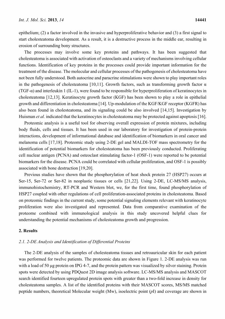
The 2-DE analysis of the samples of cholesteatoma tissues and retroauricular skin for each patient
was performed for twelve patients. The proteomic data are shown in Figure 1. 2-DE analysis was run
with a load of 50 μg protein on IPG 4-7, and the protein pattern was visualized by silver staining. Protein
spots were detected by using PDQuest 2D image analysis software. LC-MS/MS analysis and MASCOT
search identified fourteen upregulated protein spots with greater than a two-fold increase in density for
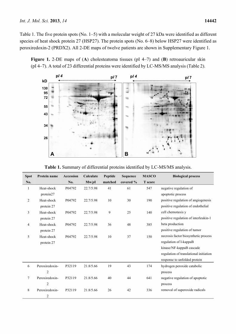
cholesteatoma samples. A list of the identified proteins with their MASCOT scores, MS/MS matched
peptide numbers, theoretical Molecular weight (Mw), isoelectric point (pI) and coverage are shown in
Int. J. Mol. Sci. 2013, 14 14442
Table 1. The five protein spots (No. 1–5) with a molecular weight of 27 kDa were identified as different
species of heat shock protein 27 (HSP27). The protein spots (No. 6–8) below HSP27 were identified as
peroxiredoxin-2 (PRDX2). All 2-DE maps of twelve patients are shown in Supplementary Figure 1.
Figure 1. 2-DE maps of (A) cholesteatoma tissues (pI 4–7) and (B) retroauricular skin
(pI 4–7). A total of 23 differential proteins were identified by LC-MS/MS analysis (Table 2).
Table 1. Summary of differential proteins identified by LC-MS/MS analysis.
Spot
No.
Protein name Accession
No.
Calculate
Mw/pI
Peptide
matched
Sequence
covered %
MASCO
T score
Biological process
1 Heat-shock
protein27
P04792 22.7/5.98 41 61 547 negative regulation of
apoptotic process
positive regulation of angiogenesis
positive regulation of endothelial
cell chemotaxis y
positive regulation of interleukin-1
beta production
positive regulation of tumor
necrosis factor biosynthetic process
regulation of I-kappaB
kinase/NF-kappaB cascade
regulation of translational initiation
response to unfolded protein
2 Heat-shock
protein 27
P04792 22.7/5.98 10 30 190
3 Heat-shock
protein 27
P04792 22.7/5.98 9 25 140
4 Heat-shock
protein 27
P04792 22.7/5.98 36 48 385
5 Heat-shock
protein 27
P04792 22.7/5.98 10 37 150
6 Peroxiredoxin-
2
P32119 21.8/5.66 19 43 174 hydrogen peroxide catabolic
process
negative regulation of apoptotic
process
removal of superoxide radicals
7 Peroxiredoxin-
2
P32119 21.8/5.66 40 44 641
8 Peroxiredoxin-
2
P32119 21.8/5.66 26 42 336
1 2 3 4 5
6
78
10
9
11
12
13
14
pI 4 pI 7
1309572
55
43
34
26
kD
A B 1516 17
pI 4 pI 7
18 20
19
2122 23
Int. J. Mol. Sci. 2013, 14 14443
Table 1. Cont.
Spot
No.
Protein
name
Accession
No.
Calculate
Mw/pI
Peptide
matched
Sequence
covered %
MASCOT
score
Biological process
9 75 kDa
glucose-
regulated
protein
P38646 73.6/5.87 13 15 157 negative regulation of apoptotic
process
protein export from nucleus
protein folding
protein targeting to mitochondrion
10 Heat shock
cognate
71 kDa protein
P11142 70.8/5.37 39 34 339 mRNA metabolic process
negative regulation of
transcription
protein folding
regulation of cell cycle
response to unfolded protein
11 Heat shock
cognate
71 kDa protein
P11142 70.8/5.37 59 46 624
12 78 kDa
glucose-
regulated
protein
P11021 72.2/5.07 15 16 123 ER overload response
ER-associated protein catabolic
process
negative regulation of apoptotic
process
positive regulation of protein
ubiquitination
regulation of protein folding in
endoplasmic reticulum
13 78 kDa
glucose-
regulated
protein
P11021 72.2/5.07 55 42 719
14 94 kDa
glucose-
regulated
protein
P14625 92.4/4.76 75 37 799 ER-associated protein catabolic
process
actin rod assembly
activation of signaling protein
activity involved in unfolded
protein response
negative regulation of apoptotic
process
protein folding
15 Uncharacterize
d protein
C7orf24
O75223 20.9/5.07 13 26 158
glutathione biosynthetic process
release of cytochrome c from
mitochondria 16 Uncharacterize
d protein
C7orf24
O75223 20.9/5.07 7 18 104
17 NEDD8-conju
gating enzyme
Ubc12
P61081 20.8/7.57 3 12 35 protein neddylation
Int. J. Mol. Sci. 2013, 14 14444
Table 1. Cont.
Spot
No.
Protein
name
Accession
No.
Calculate
Mw/pI
Peptide
matched
Sequence
covered %
MASCOT
score
Biological process
18 Serum albumin
precursor
P02768 69.3/5.92 41 25 388 lipoprotein metabolic process
maintenance of mitochondrion
location
negative regulation of apoptotic
process
response to nutrient
19 Glial fibrillary
acidic protein
P14136 49.8/5.42 11 6 141 extracellular matrix organization
intermediate filament
organization
response to wounding
20 Ig alpha-1 chain
C region
P01876 37.6/6.08 6 9 81 immune response
protein-chromophore linkage
21 Transitional
endoplasmic
reticulum
ATPase
P55072 89.2/5.14 27 21 176 ER-associated protein catabolic
process
activity involved in apoptotic
process
double-strand break repair
endoplasmic reticulum unfolded
protein response
protein ubiquitination
22 Serotransferrin
precursor
P02787 77.0/6.81 31 23 306 cellular iron ion homeostasis
transferrin transport
transmembrane transport 23 Serotransferrin
precursor
P02787 77.0/6.81 20 18 181
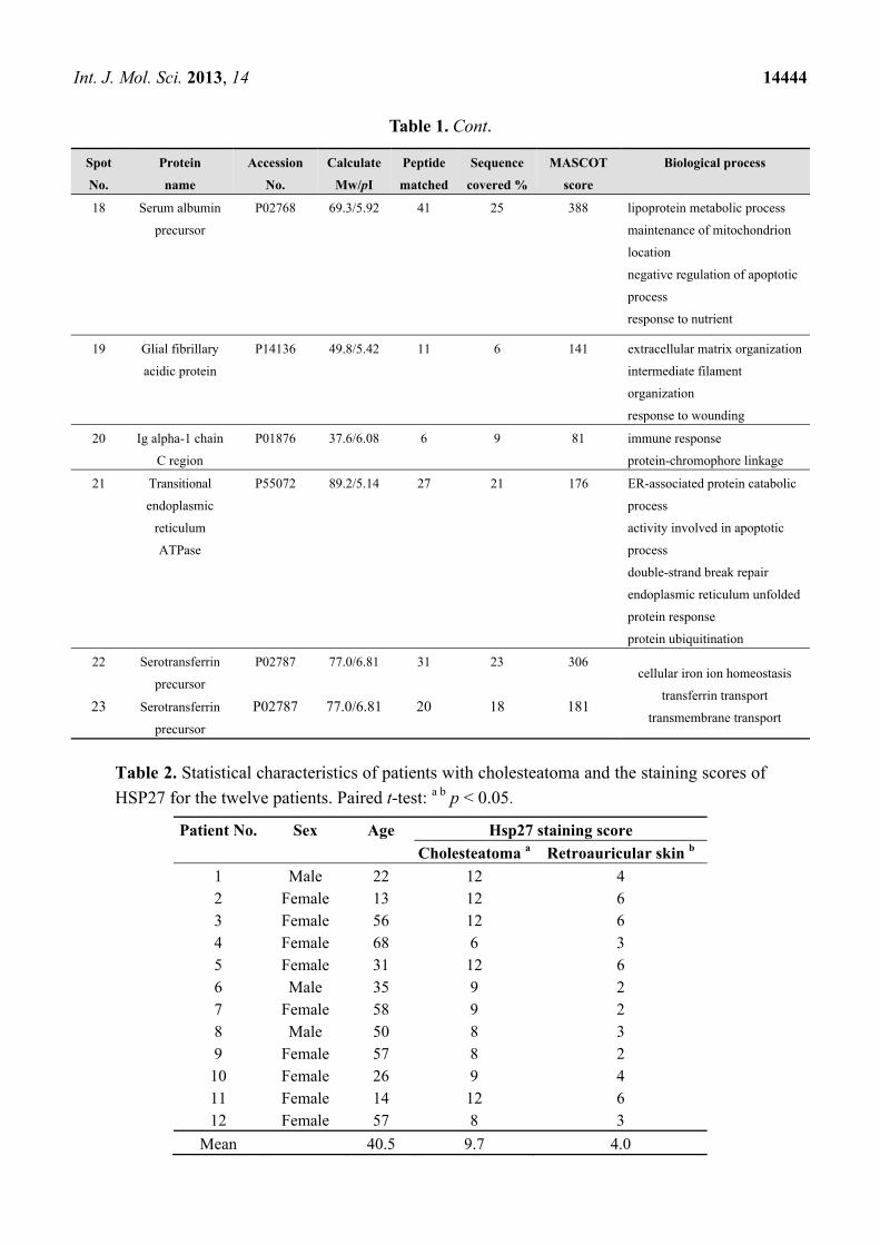
Table 2. Statistical characteristics of patients with cholesteatoma and the staining scores of
HSP27 for the twelve patients. Paired t-test: a b p < 0.05.
Patient No. Sex Age Hsp27 staining score
Cholesteatoma a Retroauricular skin b
1 Male 22 12 4 2 Female 13 12 6 3 Female 56 12 6 4 Female 68 6 3 5 Female 31 12 6 6 Male 35 9 2 7 Female 58 9 2 8 Male 50 8 3 9 Female 57 8 2 10 Female 26 9 4 11 Female 14 12 6 12 Female 57 8 3
Mean 40.5 9.7 4.0
Int. J. Mol. Sci. 2013, 14 14445
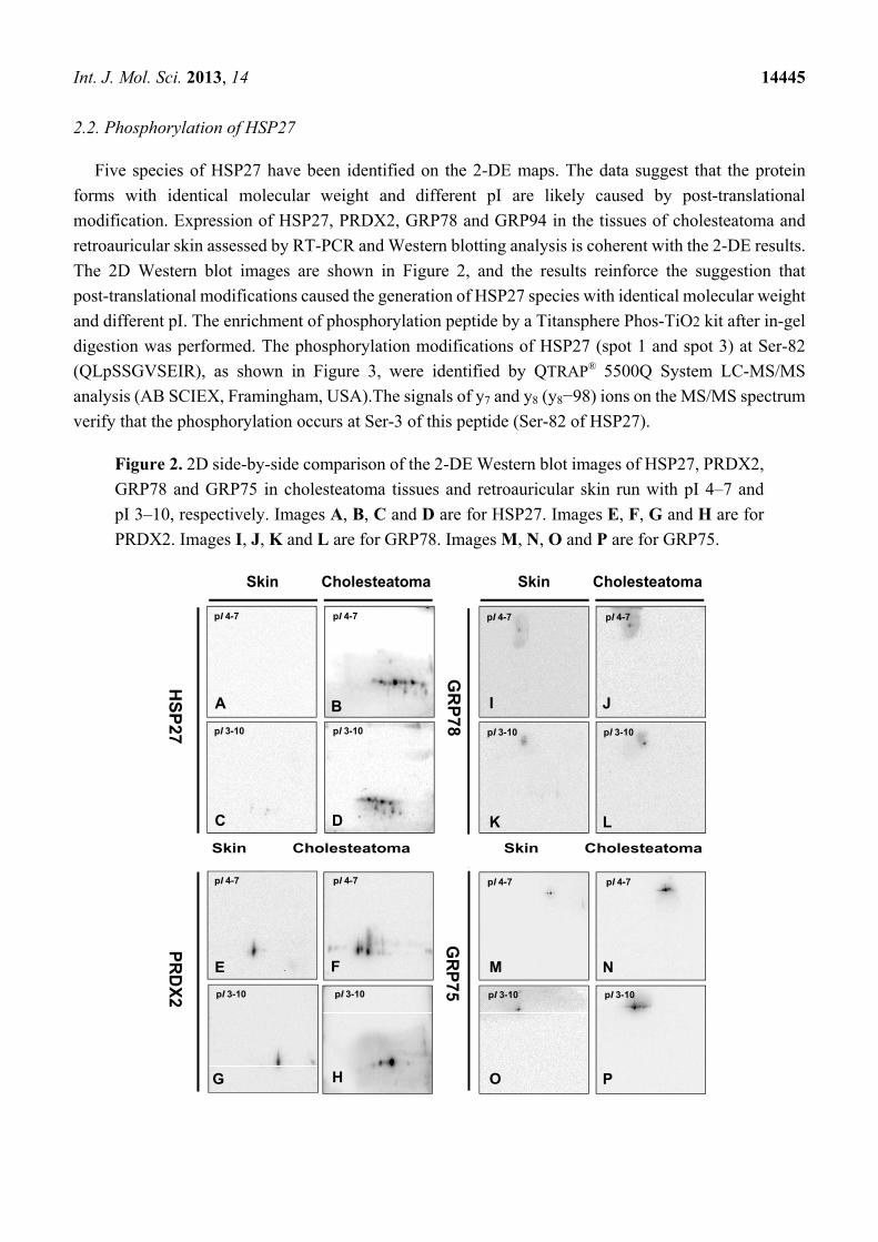
2.2. Phosphorylation of HSP27
Five species of HSP27 have been identified on the 2-DE maps. The data suggest that the protein
forms with identical molecular weight and different pI are likely caused by post-translational
modification. Expression of HSP27, PRDX2, GRP78 and GRP94 in the tissues of cholesteatoma and
retroauricular skin assessed by RT-PCR and Western blotting analysis is coherent with the 2-DE results.
The 2D Western blot images are shown in Figure 2, and the results reinforce the suggestion that
post-translational modifications caused the generation of HSP27 species with identical molecular weight
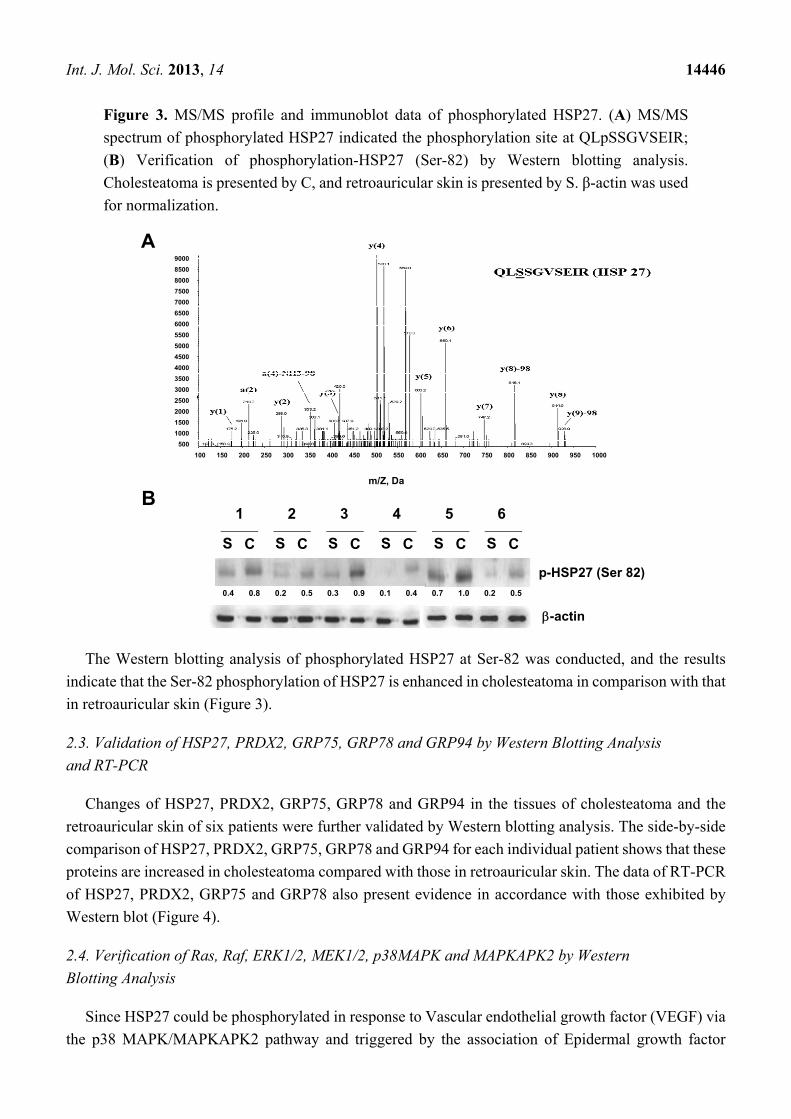
and different pI. The enrichment of phosphorylation peptide by a Titansphere Phos-TiO2 kit after in-gel
digestion was performed. The phosphorylation modifications of HSP27 (spot 1 and spot 3) at Ser-82
(QLpSSGVSEIR), as shown in Figure 3, were identified by QTRAP® 5500Q System LC-MS/MS
analysis (AB SCIEX, Framingham, USA).The signals of y7 and y8 (y8−98) ions on the MS/MS spectrum
verify that the phosphorylation occurs at Ser-3 of this peptide (Ser-82 of HSP27).
Figure 2. 2D side-by-side comparison of the 2-DE Western blot images of HSP27, PRDX2,
GRP78 and GRP75 in cholesteatoma tissues and retroauricular skin run with pI 4–7 and
pI 3–10, respectively. Images A, B, C and D are for HSP27. Images E, F, G and H are for
PRDX2. Images I, J, K and L are for GRP78. Images M, N, O and P are for GRP75.
Cholesteatoma
HS
P27
Skin
A B
C D
GR
P78
Cholesteatoma Skin
I J
K L
pI 4-7 pI 4-7
pI 3-10 pI 3-10 pI 3-10 pI 3-10
pI 4-7 pI 4-7
Cholesteatoma Skin Cholesteatoma Skin
E F
G H
PR
DX
2
GR
P75
M N
O P
pI 3-10 pI 3-10
pI 4-7 pI 4-7pI 4-7 pI 4-7
pI 3-10 pI 3-10
Int. J. Mol. Sci. 2013, 14 14446
Figure 3. MS/MS profile and immunoblot data of phosphorylated HSP27. (A) MS/MS
spectrum of phosphorylated HSP27 indicated the phosphorylation site at QLpSSGVSEIR;
(B) Verification of phosphorylation-HSP27 (Ser-82) by Western blotting analysis.
Cholesteatoma is presented by C, and retroauricular skin is presented by S. β-actin was used
for normalization.
The Western blotting analysis of phosphorylated HSP27 at Ser-82 was conducted, and the results
indicate that the Ser-82 phosphorylation of HSP27 is enhanced in cholesteatoma in comparison with that
in retroauricular skin (Figure 3).
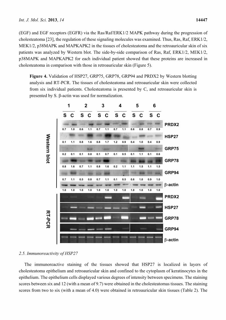
2.3. Validation of HSP27, PRDX2, GRP75, GRP78 and GRP94 by Western Blotting Analysis
and RT-PCR
Changes of HSP27, PRDX2, GRP75, GRP78 and GRP94 in the tissues of cholesteatoma and the
retroauricular skin of six patients were further validated by Western blotting analysis. The side-by-side
comparison of HSP27, PRDX2, GRP75, GRP78 and GRP94 for each individual patient shows that these
proteins are increased in cholesteatoma compared with those in retroauricular skin. The data of RT-PCR
of HSP27, PRDX2, GRP75 and GRP78 also present evidence in accordance with those exhibited by
Western blot (Figure 4).
2.4. Verification of Ras, Raf, ERK1/2, MEK1/2, p38MAPK and MAPKAPK2 by Western
Blotting Analysis
Since HSP27 could be phosphorylated in response to Vascular endothelial growth factor (VEGF) via
the p38 MAPK/MAPKAPK2 pathway and triggered by the association of Epidermal growth factor
A
m/Z, Da
100 1000950900850800750700650600550500450400350300250200150
9000
8500
8000
7500
7000
6500
6000
5500
5000
4500
4000
3500
3000
2500
2000
1500
1000
500
B
p-HSP27 (Ser 82)
-actin
S C
1
S C
2
S C
3
S C
4
S C
5
S C
6
0.4 0.8 0.2 0.5 0.3 0.9 0.1 0.4 0.7 1.0 0.2 0.5
Int. J. Mol. Sci. 2013, 14 14447
(EGF) and EGF receptors (EGFR) via the Ras/Raf/ERK1/2 MAPK pathway during the progression of
cholesteatoma [23], the regulation of these signaling molecules was examined. Thus, Ras, Raf, ERK1/2,
MEK1/2, p38MAPK and MAPKAPK2 in the tissues of cholesteatoma and the retroauricular skin of six
patients was analyzed by Western blot. The side-by-side comparison of Ras, Raf, ERK1/2, MEK1/2,
p38MAPK and MAPKAPK2 for each individual patient showed that these proteins are increased in
cholesteatoma in comparison with those in retroauricular skin (Figure 5).
Figure 4. Validation of HSP27, GRP75, GRP78, GRP94 and PRDX2 by Western blotting
analysis and RT-PCR. The tissues of cholesteatoma and retroauricular skin were collected
from six individual patients. Cholesteatoma is presented by C, and retroauricular skin is
presented by S. β-actin was used for normalization.
2.5. Immunoreactivity of HSP27
The immunoreactive staining of the tissues showed that HSP27 is localized in layers of
cholesteatoma epithelium and retroauricular skin and confined to the cytoplasm of keratinocytes in the
epithelium. The epithelium cells displayed various degrees of intensity between specimens. The staining
scores between six and 12 (with a mean of 9.7) were obtained in the cholesteatomas tissues. The staining
scores from two to six (with a mean of 4.0) were obtained in retroauricular skin tissues (Table 2). The
HSP27
-actin
PRDX2
HSP27
PRDX2
S C
1
S C
2
S C
3
S C
4
S C
5
S C
6
Western
blo
tR
T-P
CR
-actin
GRP75
GRP78
GRP94
GRP78
GRP94
1.0 1.0 1.01.0 1.0 1.0 1.0 1.0 1.0 1.0 1.0 1.0
0.7 1.1 1.00.5 0.8 0.7 1.1 0.1 0.5 0.6 1.0 0.9
0.8 1.6 1.50.7 1.1 0.8 1.6 0.2 1.1 1.1 1.5 1.1
0.2 0.1 0.60.1 0.8 0.1 0.7 0.1 0.5 0.1 1.1 0.1
0.1 1.1 0.90.8 1.6 0.8 1.7 1.2 0.9 0.4 1.0 0.4
0.7 1.0 0.6 1.1 0.7 1.1 0.7 1.1 0.6 0.8 0.7 0.9
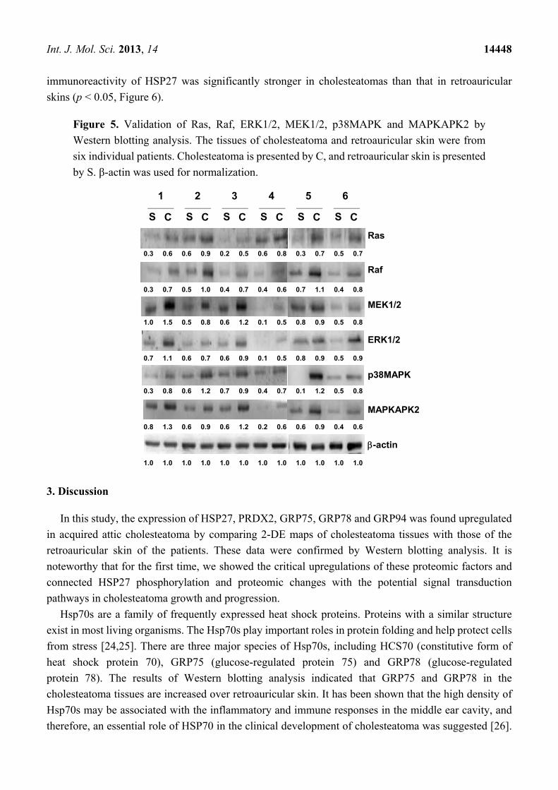
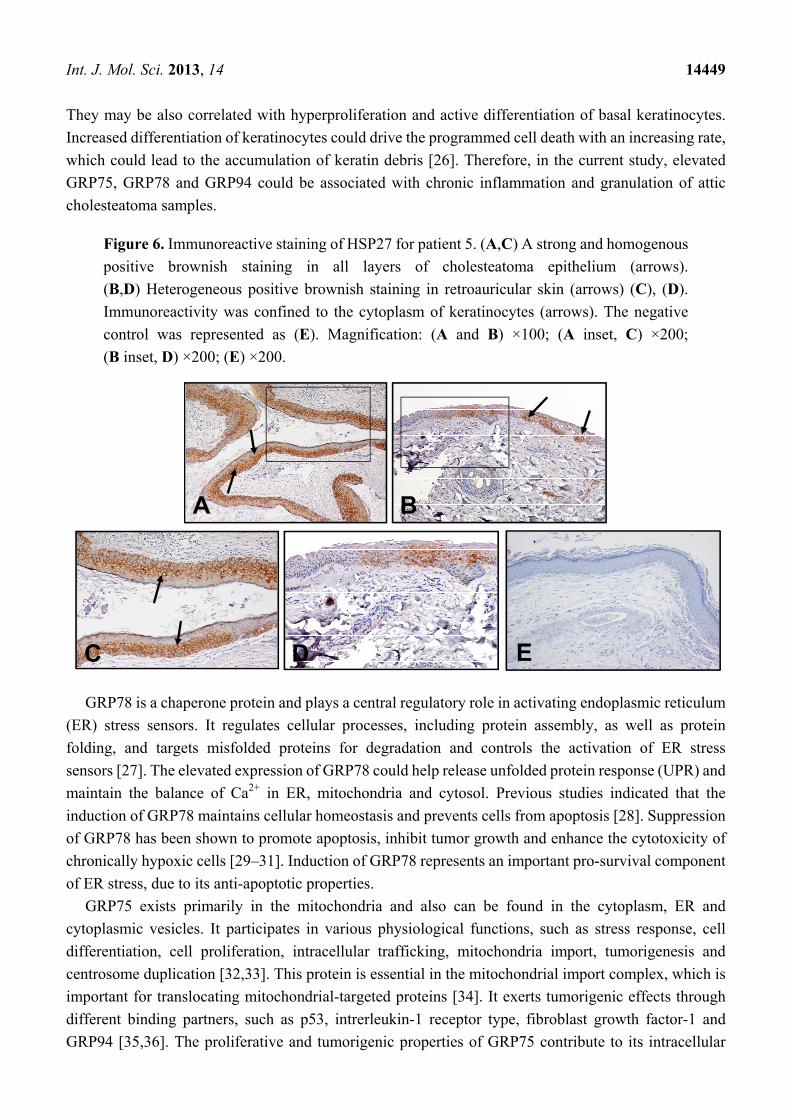
Int. J. Mol. Sci. 2013, 14 14448
immunoreactivity of HSP27 was significantly stronger in cholesteatomas than that in retroauricular
skins (p < 0.05, Figure 6).
Figure 5. Validation of Ras, Raf, ERK1/2, MEK1/2, p38MAPK and MAPKAPK2 by
Western blotting analysis. The tissues of cholesteatoma and retroauricular skin were from
six individual patients. Cholesteatoma is presented by C, and retroauricular skin is presented
by S. β-actin was used for normalization.
3. Discussion
In this study, the expression of HSP27, PRDX2, GRP75, GRP78 and GRP94 was found upregulated
in acquired attic cholesteatoma by comparing 2-DE maps of cholesteatoma tissues with those of the
retroauricular skin of the patients. These data were confirmed by Western blotting analysis. It is
noteworthy that for the first time, we showed the critical upregulations of these proteomic factors and
connected HSP27 phosphorylation and proteomic changes with the potential signal transduction
pathways in cholesteatoma growth and progression.
Hsp70s are a family of frequently expressed heat shock proteins. Proteins with a similar structure
exist in most living organisms. The Hsp70s play important roles in protein folding and help protect cells
from stress [24,25]. There are three major species of Hsp70s, including HCS70 (constitutive form of
heat shock protein 70), GRP75 (glucose-regulated protein 75) and GRP78 (glucose-regulated
protein 78). The results of Western blotting analysis indicated that GRP75 and GRP78 in the
cholesteatoma tissues are increased over retroauricular skin. It has been shown that the high density of
Hsp70s may be associated with the inflammatory and immune responses in the middle ear cavity, and
therefore, an essential role of HSP70 in the clinical development of cholesteatoma was suggested [26].
ERK1/2
p38MAPK
S C
1
S C
2
S C
3
S C
4
S C
5
S C
6
MEK1/2
Ras
MAPKAPK2
Raf
-actin
1.0 1.0 1.01.0 1.0 1.0 1.0 1.0 1.0 1.0 1.0 1.0
0.8 1.3 0.60.6 0.9 0.6 1.2 0.2 0.6 0.6 0.9 0.4
0.3 0.8 0.80.6 1.2 0.7 0.9 0.4 0.7 0.1 1.2 0.5
0.7 1.1 0.90.6 0.7 0.6 0.9 0.1 0.5 0.8 0.9 0.5
1.0 1.5 0.80.5 0.8 0.6 1.2 0.1 0.5 0.8 0.9 0.5
0.3 0.7 0.80.5 1.0 0.4 0.7 0.4 0.6 0.7 1.1 0.4
0.3 0.6 0.70.6 0.9 0.2 0.5 0.6 0.8 0.3 0.7 0.5
Int. J. Mol. Sci. 2013, 14 14449
They may be also correlated with hyperproliferation and active differentiation of basal keratinocytes.
Increased differentiation of keratinocytes could drive the programmed cell death with an increasing rate,
which could lead to the accumulation of keratin debris [26]. Therefore, in the current study, elevated
GRP75, GRP78 and GRP94 could be associated with chronic inflammation and granulation of attic
cholesteatoma samples.
Figure 6. Immunoreactive staining of HSP27 for patient 5. (A,C) A strong and homogenous
positive brownish staining in all layers of cholesteatoma epithelium (arrows).
(B,D) Heterogeneous positive brownish staining in retroauricular skin (arrows) (C), (D).
Immunoreactivity was confined to the cytoplasm of keratinocytes (arrows). The negative
control was represented as (E). Magnification: (A and B) ×100; (A inset, C) ×200;
(B inset, D) ×200; (E) ×200.
GRP78 is a chaperone protein and plays a central regulatory role in activating endoplasmic reticulum
(ER) stress sensors. It regulates cellular processes, including protein assembly, as well as protein
folding, and targets misfolded proteins for degradation and controls the activation of ER stress
sensors [27]. The elevated expression of GRP78 could help release unfolded protein response (UPR) and
maintain the balance of Ca2+ in ER, mitochondria and cytosol. Previous studies indicated that the
induction of GRP78 maintains cellular homeostasis and prevents cells from apoptosis [28]. Suppression
of GRP78 has been shown to promote apoptosis, inhibit tumor growth and enhance the cytotoxicity of
chronically hypoxic cells [29–31]. Induction of GRP78 represents an important pro-survival component
of ER stress, due to its anti-apoptotic properties.
GRP75 exists primarily in the mitochondria and also can be found in the cytoplasm, ER and
cytoplasmic vesicles. It participates in various physiological functions, such as stress response, cell
differentiation, cell proliferation, intracellular trafficking, mitochondria import, tumorigenesis and
centrosome duplication [32,33]. This protein is essential in the mitochondrial import complex, which is
important for translocating mitochondrial-targeted proteins [34]. It exerts tumorigenic effects through
different binding partners, such as p53, intrerleukin-1 receptor type, fibroblast growth factor-1 and
GRP94 [35,36]. The proliferative and tumorigenic properties of GRP75 contribute to its intracellular
A B
C D E
Int. J. Mol. Sci. 2013, 14 14450
trafficking function and the modulation of the Ras-Raf-MAPK pathway [37,38]. Overexpression of
GRP75 has been shown to lead to extended life span in nematode and normal human cells [39].
Decreased expression of GRP75 in immortalized cells causes growth arrest [40]. On the other hand,
GRP94 is a member of the heat shock protein 90 family and a chaperon of the ER. It is responsible for
the folding and maturation of nonglycosylated proteins. Increased expression of GRP 94 in malignant
tumors has been shown to have a protective effect for tumor cells [28,41–44].
The results in the current study indicated that the expression of GRP75, GRP78 and GRP94 in the
tissues of cholesteatoma is higher than that in retroauricular skin. The elevation in ER and mitochondrial
stress may cause cell damage. Therefore, the upregulation of GRP75, GRP78 and GRP94 should be
reasonably against the increased ER, mitochondrial stress, as well as apoptosis in attic cholesteatoma.
HSP27 is a chaperone protein of the small heat shock protein (sHsps) group. The functions of Hsps
are chaperone activity, regulation of cell development, thermotolerance, inhibition of apoptosis and cell
differentiation. They also participate in the signal transduction associated with apoptosis. The
interaction of HSP27 with the outer mitochondrial membranes and the interference with the activation of
the cytochrome c/Apaf-1/dATP complex causes the inhibition of procaspase-9. It was reported that
phosphorylated HSP27 inhibits Daxx apoptotic protein and, thus, prevents the association of Daxx with
Fas and Ask1 [45]. Abnormal HSP27 expression was associated with various cancers, and its
tumorigenic potential has been reported in experimental models [46,47]. The dysregulation of HSP27
has been suspected as a cause for invasion and metastasis [48]; HSP27 is recognized to play the role of
molecular chaperone. It is capable of modulating cell migration, cell survival, anti-proliferation, cell
differentiation and vascular function through phosphorylation. HSP27 could be phosphorylated by
different types of protein kinases, protein phosphatases or stimuli [49]. The study of Kindås-Mügge,
using reverse transcriptase differential display polymerase chain reaction, suggested that HSP27 is a
biomarker for differentiation in normal human keratinocytes [49]. The layers of epidermis consist of
keratinocytes at different stages of differentiation. The highly coordinated multistep process of
keratinocyte differentiation is regulated by growth factors, autocrine, paracrine, intercellular signaling
mechanisms and external stimuli. Epidermal growth factor (EGF) and other growth factors could
promote keratinocyte growth, differentiation and migration [50,51]. Previous studies have shown the
expression of EGF and increased expression of EGF receptors (EGFR) in cholesteatoma [10].
Angiogenic growth factors (VEGF) have been reported in cholesteatoma by Sudhoff and Niwa [52,53].
The connective tissue of the perimatrix in cholesteatoma requires angioneogenesis for its growth. The
wound healing process also needs angioneogenesis in response to cholesteatoma-induced tissue injury.
In this study, five protein species of HSP27 were found in cholesteatoma. This is similar to the
proteomic findings in heart diseases shown by Jungblut et al. and Schlüter et al. (2009), implicating the
possible biological roles of numerous HSP27 forms [54,55]. Our study for the first time discovered
phosphorylation of HSP27 at Ser-82 in cholesteatoma tissues. The phosphorylation of HSP27 at Ser-82
could be induced by many different factors. It has been reported that PKC/PKD is the major pathway
mediating phosphorylation of HSP27 at Ser-82 in response to VEGF [54]. It has been shown that the
elevated expression of phosphorylation of p38 is in connection with involucrin, which is an end product
of cell differentiation [14]. Niwa et al. reported that the phosphorylation of HSP27 could be induced by
TNF-α or H2O2 via the p38 MAPK pathway [52]. It is therefore proposed that HSP27 is very likely
phosphorylated in response to VEGF via the p38 MAPK/MAPKAPK2 pathway during the progression
Int. J. Mol. Sci. 2013, 14 14451
of cholesteatoma. This is partially verified by our immunoblotting data (Figure 6). The phosphorylated
HSP27 may result in cell migration and angiogenesis. Ras protein, which plays an important role in the
growth factor signal-transduction pathway, has been found in cholesteatoma specimens [56]. The
accumulation of keratin debris in cholesteatoma associated with cell proliferation and differentiation of
keratinocytes could be regulated by various growth factors. Investigation of UVB effects on human
keratinocytes showed that EGFR and p38 MAP kinase mediate HSP27 phosphorylation [57]. The
Ras/Raf/ERK1/2 MAPK signaling pathway actively involved in cholesteatoma epithelium has been
reported [23]. It has been indicated that ERK1 and ERK2 are upregulated proteins involved in the
MAPK pathway in cholesteatoma [14]. Moreover, HSP27 has been found to play a role in keratinocyte
terminal differentiation [58]. We thus proposed that the phosphorylation of HSP27 is very likely
triggered by the involvement of EGF/EGFR, the Ras/Raf/ERK1/2 pathway, as well as the MAPK
pathway. In this study, we verified the changes of these crucial signaling factors by Western blot in
cholesteatoma. These data in our study suggest that HSP27 together with the Ras/Raf/ERK1/2 and
MAPK pathways may be relevant in stimulating keratinocyte proliferation and differentiation
in cholesteatoma.
The other pathway that may induce keratinocyte proliferation and differentiation can be concluded
from previous studies and the current work as the involvement of IFN-γ induction, PLC-γ,
diacylglycerol (DAG), protein kinase C (PKC), PKD1/PKD2 and PRDX2 [59,60]. IFN-γ has been
shown to be a potent factor to induce the expression of EGFR, as well as cell differentiation in normal
neonatal skin explants or epidermal keratinocytes [61,62]. The PKC pathway and reactive oxygen stress
regulate epidermal differentiation in keratinocytes [63]. PLC can activate PKC, which is important in
cellular growth, differentiation and transformation; PLC-γ1 was overexpressed in cholesteatoma [64].
EGF is able to activate PLC, which is capable of activating PKC downstream through DAG. The overall
processes may start with an increased EGFR expression, followed by the transductions of PLC-γ, DAG,
PKC and PKD1/PKD2, as well as regulation of PRDX2. In the current study, increased expression of
Peroxiredoxin2 (PRDX2) was shown by proteomic analysis and Western blot in cholesteatoma. It is
worth noting that upregulation of PRDX2 has also been found in psoriasis, a hyperproliferative skin
disease characterized by abnormal keratinocyte proliferation [65]. These similar findings suggest that
PRDX2 may be an essential protein in the diseases or lesions correlated with keratinocyte
hyperproliferation in the epidermis.
4. Materials and Methods
4.1. Materials
The Two-D Quant Kit and IPG buffer were obtained from GE Healthcare (Buckinghamshire, UK).
SuperScript III and Taq DNA polymerase were from Invitrogen (Carlsbad, CA, USA). Rabbit
anti-human HSP27, GRP75, GRP78, GRP94 and PRDX2 antibodies were purchased from ProteinTech
Group (Chicago, IL, USA). Rabbit anti-human phosphorylation HSP27 (Ser-82), ras, raf, ERK1/2,
MEK1/2, p38MAPK and MAPKAPK2 antibodies were purchased from Cell Signaling Technology
(Danvers, MA, USA). Rabbit anti-human β-actin antibodies were obtained from Sigma (St. Louis, Mo,
USA). Goat anti-rabbit and horseradish peroxidase conjugated IgG was from Millipore (Bellerica, MA,
Int. J. Mol. Sci. 2013, 14 14452
USA). PVDF (polyvinylidene difluoride) membranes and chemiluminescent horseradish peroxidase
(HRP) substrate were from Pierce (Rockford, IL, USA).
4.2. Sample Preparation
Twelve patients (three males and nine females) participated in this study at the Affiliated Hospital at
Kaohsiung Medical University. They were aged between 13 to 68 years, with a mean age of 40.5 years.
The acquired middle ear cholesteatoma (acquired attic cholesteatoma) with slight granulation tissue
specimens were resected during surgical operations. The tissues used in proteomic analysis were the
epithelium of cholesteatoma after removal of granulation tissues. The retroauricular skin samples of the
patients were also obtained as the controls. The protocols for using human specimens in this
study were approved by the Institutional Review Board (IRB) of the hospital (approval number.
KMUH-IRB-980046). Each sample (1 mm × 1 mm × 1 mm in size) was homogenized and sonicated
with sample buffer (50 mM Tris-HCl; pH 8.0, EDTA); then, the sample was centrifuged at 12,000 rpm
for 10 min. The supernatant was collected, and the proteins were precipitated out overnight at −20 °C by
triple the volume of 10% trichloroacetic acid (TCA)/acetone solution containing 20 mM Dithiothreitol
(DTT) After centrifugation at 8,000 rpm for 30 min at 4 °C, the supernatant was discarded. The pellet
was rinsed three times in cold acetone containing 20 mM DTT and air-dried, then resuspended in a
rehydration buffer (6 M urea, 2 M thiourea, 0.5% 3-[(3-cholamidopropyl)dimethylammonio]-
1-propanesulfonate (CHAPS), 5% IPG buffer, 20 mM DTT and 0.002% bromophenol blue) at 4 °C
overnight. The protein contents were determined using a 2-D Quant Kit (GE Healthcare).
4.3. Two-Dimensional Gel Electrophoresis
The first dimension electrophoresis (isoelectric focusing) was performed on a GE Healthcare Ettan
IPGphor 3 with the protocol described previously [66]. Proteins (50 μg) extracted from whole tissue
were loaded on 11 cm Immobilized pH gradient (IPG) strips for Isoelectric focusing (IEF) and then were
separated on SDS-PAGE (12.5%).
4.4. Protein Spot Identification by LC-MS/MS
4.4.1. In-Gel Digestion
Spots of interest were excised into a piece of 1 mm × 1 mm, then placed in a microcentrifuge tube.
Briefly, 25 mM ammonium bicarbonate (pH 8.5) was added to the tube, which was shaken at 37 °C for
1 h. The gel piece was then dehydrated in acetonitrile and dried by SpeedVac to remove the remaining
acetonitrile. Zero-point-one micrograms of trypsin in 10 μL 25 mM ammonium bicarbonate (pH 8.5)
was added to the gel piece. Protein digestion was run overnight at 37 °C. Fifty microliters of 5%
trifluoroacetic acid (TFA) in 50% acetonitrile was added to quench the trypsin digestion. Peptides were
extracted with 25 mM ammonium bicarbonate, 50% acetonitrile and 0.1% trifluoroacetic acid. The
peptide solution was concentrated for the following LC-MS/MS analysis.
Int. J. Mol. Sci. 2013, 14 14453
4.4.2. LC-MS/MS Analysis and MASCOT Database Searching
After desalting with a ZIPTip®C18 (Millipore, Bellerica, MA, USA), the resulting peptide mixture
was separated using a NanoLC 1200 System (Agilent) utilizing a Zobax column (2.1 mm × 150 mm)
packed with 3μm C18 particles (Agilent, Santa Clara, CA, USA) with a linear gradient from 5% to 60%
acetonitrile containing 0.1% formic acid over 60 min. The separated peptides were analyzed online on a
QTrap 5500 mass spectrometer (AB SCIEX, Framingham, USA) equipped with a nano ESI source. The
scan range was from m/z 100 to 1,000 for MS and MS/MS. The raw data were processed into a text file
format of WIFF with Analyst 1.5.1, and the resulting text file was searched using the MASCOT search
engine v2.2 (Matrix Science, Boston, USA) with the following search parameters: (1) the protein
database was set to be Swiss-Prot; (2) the taxonomy was set as Homo sapiens (human); (3) one trypsin
missed cleavage was allowed; (4) the mass tolerance was set at 1.5 Da for the precursor and 0.8 Da for
the product ions; (5) carbamidomethyl (C) was chosen for fixed modification; (6) oxidation (M),
phospho- (ST) and phosphor- (Y) were chosen for variable modifications; and (7) proteins with scores
above the significance threshold (p < 0.05) were shown as significant hits. The hit with the highest score
that contained at least two peptides with scores beyond the identity threshold was regarded as the
identified protein from each gel spot. All MS/MS spectra of the identified peptides were further verified
by manual interpretation.
4.5. Western Blotting Analysis
After 1-DE and 2-DE PAGE analysis of the samples collected from the patients, the proteins on gel
were transferred to a PVDF membrane (Millipore, MA, USA), for 1.5 h at 400 mA using a Transphor
TE 62 (Hoeffer, Holliston, MA). The membranes were then incubated with HSP27, phosphorylation
HSP27, GRP75, GRP78, GRP94 and β-actin antibodies at 4 °C for 2 h or overnight. The membranes
were washed three times in PBST (10 mM NaH2PO4, 130 mM NaCl, 0.05% Tween 20), then probed
with the second antibodies (goat anti-rabbit and horseradish peroxidase conjugate (1:5,000) in blocking
solution) for 1 h. After washing with PBST three times, the enzyme activity on the blot was visualized
through chemiluminescence by adding ECL Western Blotting Reagents (Pierce Rockford, IL, USA).
4.6. RNA Isolation and RT-PCR
Total RNA was isolated from both cholesteatoma and normal retroauricular skin samples using
TRIzol reagent RNA Extraction Kits (Qiagen, Hilden, Germany). The RNA concentrations were
measured using a GeneQuant 1300 spectrophotometer (GE Healthcare, Buckinghamshire, UK). Reverse
transcription was carried out in the reaction containing RNA samples, dNTP, random primers, 5× first
strand buffer, DTT (0.1 M) and SuperScript III (Invitrogen, Carlsbad, CA, USA) on a PCR machine
(Bio-Rad, Hercules, CA, USA). The sequences of primers used in the PCR reactions are as below:
(1) β-actin follows:
5'-3'AGAGATGGCCACGGCTGCTT (forward);
5'-3'ATTTGCGGTGGACGATGGAG (reverse).
Int. J. Mol. Sci. 2013, 14 14454
(2) Heat shock protein 27 (HSP27) follows:
5'-3' ACGAGCATGGCTACATCTCC (forward);
5'-3' CTTTACTTGGCGGCAGTCTC (reverse).
(3) Thioredoxin peroxidase 2 (PRDX2) follows:
5'-3'GTGTCCTTCGCCAGATCACT (forward);
5'-3' ACGTTGGGCTTAATCGTGTC (reverse).
(4) Glucose-regulated protein 78 (GRP78) follows:
5'-3' TCCTATGTCGCCTTCACT (forward);
5'-3' ACAGACGGGTCATTCCAC (reverse).
(5) Glucose-regulated protein 94 (GRP94) follows:
5'-3' GGGAGGTCACCTTCAAGTCG (forward);
5'-3' GGGTGTAGACGTGGAGCTC (reverse).
For PCR, the reaction tubes containing 10× buffer, MgCl2, dNTPs, Taq DNA polymerase
(Invitrogen, Grand Island, NY, USA) and each of the forward and reverse primers were preheated at
95 °C for 3 min. The three stages of 30 cycles of PCR were accomplished as follows: denaturation at
95 °C for 30 s, annealing at 55 °C for 30 s, elongation at 72 °C for 30 s and extension was completed at
72 °C for 10 min. The PCR products were electrophoresed on 1.5% agarose gel.
4.7. Immunohistochemical Staining of Hsp27
The samples from the 12 patients were then assessed by immunohistochemistry. A cholesteatomas
specimen and a retroauricular skin specimen of each patient were resected during surgical operations.
Immunohistochemistry was performed on 4 μm thick paraffin sections. Paraffin sections of all samples
were de-paraffinized, rehydrated and autoclave-treated at 121 °C for 10 min in DAKO Target Retrieval
Solution, pH 6.0 (DAKO, Glostrup, Denmark), to induce antigen retrieval. Endogenous peroxidase in
the section was blocked by incubation in 3% hydrogen peroxide for 5 min. The sections were incubated
with HSP27 primary antibodies (1:60; Leica Novocastra, Newcastle upon Tyne, UK) at room
temperature for 1 h. Then, the DAKO REAL EnVision Detection kit (DAKO) was applied for 30 min.
Finally, sections were incubated in 3'3-diaminobenzidine for 5 min, followed by Mayer’s hematoxylin
counterstaining and mounting. Negative controls were obtained by replacing the primary antibody with
non-immune serum.
The percentage of immunoreactive staining for HSP27 in all 24 samples was evaluated by two
independent observers. They were assigned a score number according to the following rules: a score of
0 for 0% epithelium cells positive, a score of 1 for 1%–24% epithelium cells positive, a score of 2 for
25%–49% epithelium cells positive, a score of 3 for 50%–74% epithelium cells positive and score of
4 for 75%–100% epithelium cells positive. The intensity of cellular staining was also assigned a score
number: a score of 0 for zero intensity, a score of 1 for weak intensity, a score of 2 for moderate intensity
and a score of 3 for strong intensity. A staining score was obtained by multiplying the percentage score
with the intensity score, with a maximum score of 12. Statistical evaluations were performed using the
paired t-test. A difference considered statistically significance is p < 0.05.
Int. J. Mol. Sci. 2013, 14 14455
5. Conclusions
Taken together, in the current study, HSP27 and PRDX2 were enhanced, and phosphorylation of
HSP27 at Ser-82 was identified in acquired attic cholesteatoma tissue. Furthermore, phosphorylation of
HSP27 at Ser-82 can be mediated through the PKC/PKD pathway in response to VEGF or via the p38
MAPK pathway in cholesteatoma. The phosphorylated HSP27 could be associated with the activation of
cell migration, angiogenesis and proliferation in epithelial cells, resulting in subsequent growth of
cholesteatoma. Our data implicate that the phosphorylation of HSP27 is probably induced by the
pathway, including EGF/EGFR, Ras/Raf/MEK1/2/ERK1/2, as well as p38 MAPK. Upregulation of
PRDX2 could be relevant with keratinocyte hyperproliferation in the epidermis, and PRDX2 elevation is
possibly mediated via EGFR, PLC-γ, DAG, PKC and PKD1/PKD2 in the other pathway. In addition,
upregulation of GRP75, GRP78 and GRP94 may not only be associated with chronic inflammation of
attic cholesteatoma, but also counter ER and mitochondria stresses, reduce cell apoptosis, prevent
protein unfolding and could also favor keratinocyte proliferation in attic cholesteatoma. These results
shed light on the potential mechanisms of signal transduction in acquired middle ear cholesteatoma and
are helpful for understanding the pathogenesis of cholesteatoma.
Acknowledgements
This study was supported in part by a grant from the National Science Council
NSC-100-2314-B-037-009 Research Fund, Taiwan, and projects of Kaohsiung Medical University
Hospital KMUH-95-5D07 and KMUH-100-0R35 Research Fund, Kaohsiung County, Taiwan.
Conflict of Interest
The authors declare no conflict of interest.
References
1. Kuczkowski, J.; Sakowicz-Burkiewicz, M.; Iżycka-Świeszewska, E.; Mikaszewski, B.; Pawełczyk, T.
Expression of tumor necrosis factor-α, interleukin-1α, interleukin-6 and interleukin-10 in chronic
otitis media with bone osteolysis. ORL J. Otorhinolaryngol. Relat. Spec. 2011, 73, 93–99.
2. Haruyam, T.; Furukawa, M.; Kusunoki, T.; Onoda, J.; Ikeda, K. Expression of IL-17 and its role in
bone destruction in human middle ear cholesteatoma. ORL J. Otorhinolaryngol. Relat. Spec. 2010,
72, 325–331.
3. Kuczkowski, J.; Sakowicz-Burkiewicz, M.; Iżycka-Świeszewska, E. Expression of the receptor
activator for nuclear factor-κB ligand and osteoprotegerin in chronic otitis media.
Am. J. Otolaryngol. 2010, 31, 404–409.
4. Juhn, S.K.; Jung, M.K.; Hoffman, M.D.; Drew, B.R.; Preciado, D.A.; Sausen, N.J.; Jung, T.T.;
Kim, B.H.; Park, S.Y.; Lin, J.; et al. The role of inflammatory mediators in the pathogenesis of otitis
media and sequelae. Clin. Exp. Otorhinolaryngol. 2008, 1, 117–138.
5. Nason, R.; Jung, J.Y.; Chole, R.A. Lipopolysaccharide-induced osteoclastogenesis from
mononuclear precursors: A mechanism for osteolysis in chronic otitis. J. Assoc. Res. Otolaryngol.
2009, 10, 151–160.
Int. J. Mol. Sci. 2013, 14 14456
6. Hilton, C.W.; Ondrey, F.G.; Wuertz, B.R.; Levine, S.C. Interleukin-8 production in response to
tumor necrosis factor-alpha by cholesteatoma keratinocytes in cell culture. Laryngoscope 2011,
121, 372–374.
7. Helgaland, T.; Engelen, B.; Olsnes, C.; Aarstad, H.J.; Vassbotn, F.S. In vitro cholesteatoma growth
and secretion of cytokines. Acta Otolaryngol. 2010, 130, 815–819.
8. Greenberg, J.S.; Manolidis, S. High incidence of complications encountered in chronic otitis media
surgery in a U.S. metropolitan public hospital. Otolaryngol. Head Neck Surg. 2001, 125, 623–627.
9. Bagger-Sjoback, D.; Phelps, P.D. Cholesteatoma with extension to the cochlea. Am. J. Otol. 1985,
6, 338–343.
10. Chi, H.P.; Ho, K.Y.; Chai, C.Y.; Ta, C.F.; Wang, L.F.; Lee, K.W.; Kuo, W.R.; Wu, S.C.; Tsai, S.M.
Epidermal growth factor expression in middle ear cholesteatoma. Kaohsiung J. Med. Sci. 2004, 20,
6–11.
11. Raynov, A.M.; Choung, Y.H.; Park, H.Y.; Choi, S.J.; Park, K. Establishment and characterization
of an in vitro model for cholesteatoma. Clin. Exp. Otorhinolaryngol. 2008, 1, 86–91.
12. Marenda, S.A.; Aufdemorte, T.B. Localization of cytokines in cholesteatoma tissue.
Otolaryngol. Head Neck Surg. 1995, 112, 359–368.
13. Yoshikawa, M.; Kojima, H.; Wada, K.; Tsukidate, T.; Okada, N.; Saito, H.; Moriyama, H.
Identification of specific gene expression profiles in fibroblasts derived from middle ear
cholesteatoma. Arch. Otolaryngol. Head Neck Surg. 2006, 132, 734–742.
14. Raffa, S.; Leone, L.; Scrofani, C.; Monini, S.; Torrisi, M.R.; Barbara, M. Cholesteatoma-associated
fibroblasts modulate epithelial growth and differentiation through KGF/FGF7 secretion.
Histochem. Cell. Biol. 2012, 138, 251–269.
15. Yamamoto-Fukuda, T.; Aoki, D.; Hishikawa, Y.; Kobayashi, T.; Takahashi, H.; Koji, T. Possible
involvement of keratinocyte growth factor and its receptor in enhanced epithelial-cell proliferation
and acquired recurrence of middle-ear cholesteatoma. Lab. Invest. 2003, 83, 123–136.
16. Huisman, M.A.; De Heer, E.; Grote, J.J. Survival signaling and terminal differentiation in
cholesteatoma epithelium. Acta OtoLaryngol. 2007, 127, 424–429.
17. Liu, C.I.; Chen, C.C.; Chen, J.C.; Su, J.H.; Huang, H.H.; Chen, J.Y.; Wu, Y.J. Proteomic analysis of
anti-tumor effects of 11-dehydrosinulariolide on CAL-27 cells. Mar. Drugs 2011, 9, 1254–1272.
18. Su, T.R.; Lin, J.J.; Chiu, C.C.; Chen, J.Y.F.; Su, J.H.; Cheng, Z.J.; Hwang, W.I.; Huang, H.H.;
Wu, Y.J. Proteomic investigation of anti-tumor activities exerted by sinularin against A2058
melanoma cells. Electrophoresis 2012, 33, 1139–1152.
19. Kim, J.L.; Jung, H.H. Proteomic analysis of cholesteatoma. Acta OtoLaryngol. 2004, 124, 783–788.
20. Shieh, T.J.; Ho, K.Y.; Kuo, W.R.; Chai, C.Y.; Lin, C.S.; Juan, K.H. Evaluation of proliferative
activity in middle ear cholesteatoma using proliferating cell nuclear antigen. Kaohsiung J. Med. Sci
1999, 15, 468–474.
21. Yasuda, E.; Kumada, T.; Takai, S.; Ishisaki, A.; Noda, T.; Matsushima-Nishiwaki, R.; Yoshimi, N.;
Kato, K.; Toyoda, H.; Kaneoka, Y.; et al. Attenuated phosphorylation of heat shock protein
27 correlates with tumor progression in patients with hepatocellular carcinoma.
Biochem. Biophys. Res. Commun 2005, 337, 337–342.
22. Yuan, J.; Rozengurt, E. PKD, PKD2, and p38 MAPK mediate Hsp27 serine-82 phosphorylation
induced by neurotensin in pancreatic cancer PANC-1 cells. J. Cell. Biochem. 2008, 103, 648–662.

Int. J. Mol. Sci. 2013, 14 14457
23. Huisman, M.A.; De Heer, E.; Grote, J.J. Sustained extracellular signal-regulated kinase1/2
mitogen-activated protein kinase signalling is related to increased p21 expression in cholesteatoma
epithelium. Acta Otolaryngol. 2005, 125, 134–140.
24. Tavaria, M.; Gabriele, T.; Kola, I.; Anderson, R.L. A hitchhiker’s guide to the human Hsp70
family. Cell. Stress Chaperones 1996, 1, 23–28.
25. Morano, K.A. New tricks for an old dog: The evolving world of Hsp70. Ann. N. Y. Acad. Sci. 2007,
1113, 1–14.
26. Shinoda, H.; Huang, C.C. Heat shock proteins in middle ear cholesteatoma.
Otolaryngol. Head Neck Surg. 1996, 114, 77–83.
27. Lee, A.S. The ER chaperone and signaling regulator GRP78/BiP as a monitor of endoplasmic
reticulum stress. Methods 2005, 35, 373–381.
28. Lee, A.S. The glucose-regulated proteins: Stress induction and clinical applications.
Trends Biochem. Sci. 2001, 26, 504–510.
29. Hughes, C.S.; Shen, J.W.; Subjeck, J.R. Resistance to etoposide induced by three glucose-regulated
stresses in Chinese hamster ovary cells. Cancer Res. 1989, 49, 4452–4454.
30. Jamora, C.; Dennert, G.; Lee, A.S. Inhibition of tumor progression by suppression of stress protein
GRP78/BiP induction in fibrosarcoma B/C10ME. Proc. Natl. Acad. Sci. USA 1996, 93, 7690–7694.
31. Song, M.S.; Park, Y.K.; Lee, J.H.; Park, K. Induction of Glucose-regulated Protein 78 by Chronic
Hypoxia in Human Gastric Tumor Cells through a Protein Kinase C-ε/ERK/AP-1 Signaling
Cascade. Cancer Res. 2001, 61, 8322–8330.
32. Voisine, C.; Craig, E.A.; Zufall, N.; von Ahsen, O.; Pfanner, N.; Voos, W. The protein import
motor of mitochondria: Unfolding and trapping of preproteins are distinct and separable functions
of matrix Hsp70. Cell 1999, 97, 565–574.
33. Ma, Z.; Izumi, H.; Kanai, M.; Kabuyama, Y.; Ahn, N.G.; Fukasawa, K. Mortalin controls centrosome
duplication via modulating centrosomal localization of p53. Oncogene 2006, 25, 5377–5390.
34. Lim, J.H.; Martin, F.; Guiard, B.; Pfanner, N.; Voos, W. The mitochondrial Hsp70-dependent
import system actively unfolds preproteins and shortens the lag phase of translocation. EMBO J.
2001, 20, 941–950.
35. Sacht, G.; Brigelius-Flohe, R.; Kiess, M.; Sztajer, H.; Flohe, L. ATP-sensitive association of
mortalin with the IL-1 receptor type I. Biofactors 1999, 9, 49–60.
36. Takano, S.; Wadhwa, R.; Mitsui, Y.; Kaul, S.C. Identification and characterization of molecular
interactions between glucose-regulated proteins (GRPs) mortalin/GRP75/peptide-binding protein
74 (PBP74) and GRP94. Biochem. J. 2001, 357, 393–398.
37. Wadhwa, R.; Yaguchi, T.; Hasan, M.K.; Taira, K.; Kaul, S.C. Mortalin-MPD (mevalonate
pyrophosphate decarboxylase) interactions and their role in control of cellular proliferation.
Biochem. Biophys. Res. Commun. 2003, 302, 735–742.
38. Mizukoshi, E.; Suzuki, M.; Misono, T.; Loupatov, A.; Munekata, E.; Kaul, S.C.; Wadhwa, R.;
Imamura, T. Cell-cycle dependent tyrosine phosphorylation on mortalin regulates its interaction
with fibroblast growth factor-1. Biochem. Biophys. Res. Commun. 2001, 280, 1203–1209.
39. Yaguchi, T.; Aida, S.; Kaul, S.C.; Wadhwa, R. Involvement of mortalin in cellular senescence from
the perspective of its mitochondrial import, chaperone, and oxidative stress management functions.
Ann. N. Y. Acad. Sci. 2007, 1100, 306–311.
Int. J. Mol. Sci. 2013, 14 14458
40. Wadhwa, R.; Takano, S.; Taira, K.; Kaul, S.C. Reduction in mortalin level by its antisense expression
causes senescence-like growth arrest in human immortalized cells. J. Gene Med. 2004, 6, 439–444.
41. Langer, R.; Feith, M.; Siewert, J.R.; Wester, H.J.; Hoefler, H. Expression and clinical significance
of glucose regulated proteins GRP78 (BiP) and GRP94 (GP96) in human adenocarcinomas of the
esophagus. BMC Cancer 2008, 8, doi:10.1186/1471-2407-8-70.
42. Zheng, H.C.; Takahashi, H.; Li, X.H.; Hara, T.; Masuda, S.; Guan, Y.F.; Takano, Y.
Overexpression of GRP78 and GRP94 are markers for aggressive behavior and poor prognosis in
gastric carcinomas. Hum. Pathol. 2008, 39, 1042–1049.
43. Fu, Y.; Lee, A.S. Glucose regulated proteins in cancer progression, drug resistance and
immunotherapy. Cancer Biol. Ther. 2006, 5, 741–744.
44. Kubota, K.; Lee, D.H.; Tsuchiya, M.; Young, C.S.; Everett, E.T.; Martinez-Mier, E.A.; Snead,
M.L.; Nguyen, L.; Urano, F.; Bartlett, J.D. Fluoride induces endoplasmic reticulum stress in
ameloblasts responsible for dental enamel formation. J. Biol. Chem. 2005, 280, 23194–23202.
45. Garrido, C.; Brunet, M.; Didelot, C.; Zermati, Y.; Schmitt, E.; Kroemer, G. Heat shock proteins 27
and 70: Anti-apoptotic proteins with tumorigenic properties. Cell. Cycle 2006, 5, 2592–2601.
46. Bruey, J.M.; Paul, C.; Fromentin, A.; Hilpert, S.; Arrigo, A.P.; Solary, E.; Garrido, C. Differential
regulation of HSP27 oligomerization in tumor cells grown in vitro and in vivo. Oncogene 2000, 19,
4855–4863.
47. Wang, A.; Liu, X.; Sheng, S.; Ye, H.; Peng, T.; Shi, F.; Crowe, D.L.; Zhou, X. Dysregulation of
heat shock protein 27 expression in oral tongue squamous cell carcinoma. BMC Cancer 2009, 9,
doi:10.1186/1471-2407-9-167.
48. Xu, L.; Chen, S.; Bergan, R.C. MAPKAPK2 and HSP27 are downstream effectors of p38 MAP
kinase-mediated matrix metalloproteinase type 2 activation and cell invasion in human prostate
cancer. Oncogene 2006, 25, 2987–2998.
49. Kostenko, S.; Moens, U. Heat shock protein 27 phosphorylation: Kinases, phosphatases, functions
and pathology. Cell. Mol. Life Sci. 2009, 66, 3289–3307.
50. Gibbs, S.; Silva Pinto, A.N.; Murli, S.; Huber, M.; Hohl, D.; Ponec, M. Epidermal growth factor
and keratinocyte growth factor differentially regulate epidermal migration, growth, and
differentiation. Wound Repair Regen. 2000, 8, 192–203.
51. Shirakata, Y. Regulation of epidermal keratinocytes by growth factors. J. Dermatol. Sci. 2010, 59,
73–80.
52. Niwa, M.; Hotta, K.; Hara, A.; Hirade, K.; Ito, H.; Kato, K.; Kozawa, O. TNF-α decreases hsp 27 in
human blood mononuclear cells: Involvement of protein kinase c. Life Sci. 2006, 80, 181–186.
53. Sudhoff, H.; Dazert, S.; Gonzales, A.M.; Borkowski, G.; Park, S.Y.; Baird, A.; Hildmann, H.;
Ryan, A.F. Angiogenesis and angiogenic growth factors in middle ear cholesteatoma.
Otol. Neurotol. 2000, 21, 793–798.
54. Evans, I.M.; Britton, G.; Zachary, I.C. Vascular endothelial growth factor induces heat shock
protein (HSP) 27 serine 82 phosphorylation and endothelial tubulogenesis via protein kinase D and
independent of p38 kinase. Cell Signal. 2008, 20, 1375–1384.
55. Schluter, H.; Apweiler, R.; Holzhutter, H.G.; Jungblut, P.R. Finding one’s way in proteomics: A
protein species nomenclature. Chem. Cent. J. 2009, 3, doi:10.1186/1752-153X-3-11.
Int. J. Mol. Sci. 2013, 14 14459
56. Huang, C.C.; Chen, C.T.; Huang, T.S.; Shinoda, H. Mediation of signal transduction in
keratinocytes of human middle ear cholesteatoma by ras protein. Eur. Arch. Otorhinolaryngol.
1996, 253, 385–389.
57. Wong, J.W.; Shi, B.; Farboud, B.; McClaren, M.; Shibamoto, T.; Cross, C.E.; Isseroff, R.R.
Ultraviolet B-mediated phosphorylation of the small heat shock protein HSP27 in human
keratinocytes. J. Investig. Dermatol. 2000, 115, 427–434.
58. Robitaille, H.; Simard-Bisson, C.; Larouche, D.; Tanguay, R.M.; Blouin, R.; Germain, L. The small
heat-shock protein Hsp27 undergoes ERK-dependent phosphorylation and redistribution to the
cytoskeleton in response to dual leucine zipper-bearing kinase expression. J. Invest. Dermatol.
2010, 130, 74–85.
59. Mitev, V.; Miteva, L. Signal transduction in keratinocytes. Exp. Dermatol. 1999, 8, 96–108.
60. Rozengurt, E. Protein kinase D signaling: Multiple biological functions in health and disease.
Physiology (Bethesda) 2011, 26, 23–33.
61. Valyi-Nagy, I.; Jensen, P.J.; Albelda, S.M.; Rodeck, U. Cytokine-induced expression of
transforming growth factor-alpha and the epidermal growth factor receptor in neonatal skin
explants. J. Invest. Dermatol. 1992, 99, 350–356.
62. Saunders, N.A.; Jetten, A.M. Control of growth regulatory and differentiation-specific genes in
human epidermal keratinocytes by interferon gamma. Antagonism by retinoic acid and
transforming growth factor beta 1. J. Biol. Chem. 1994, 269, 2016–2022.
63. Yun, S.J.; Seo, J.J.; Chae, J.Y.; Lee, S.C. Peroxiredoxin I and II are up-regulated during
differentiation of epidermal keratinocytes. Arch. Dermatol. Res. 2005, 296, 555–559.
64. Myers, E.N.; Park, K.; Chun, Y.; Lee, D.; Hwang, S. Signal transduction pathway in human middle
ear cholesteatoma. Otolaryngol. Head Neck Surg. 1999, 120, 899–904.
65. Ryu, J.; Park, S.G.; Park, B.C.; Choe, M.; Lee, K.S.; Cho, J.W. Proteomic analysis of psoriatic skin
tissue for identification of differentially expressed proteins: Up-regulation of GSTP1, SFN and
PRDX2 in psoriatic skin. Int. J. Mol. Med. 2011, 28, 785–792.
66. Wu, Y.J.; Chen, H.M.; Wu, T.T.; Wu, J.S.; Chu, R.M.; Juang, R.H. Preparation of monoclonal
antibody bank against whole water-soluble proteins from rapid-growing bamboo shoots.
Proteomics 2006, 6, 5898–5902.
© 2013 by the authors; licensee MDPI, Basel, Switzerland. This article is an open access article
distributed under the terms and conditions of the Creative Commons Attribution license
(http://creativecommons.org/licenses/by/3.0/).
Copyright © 2022 FDOKUMEN