Transboundary Animal Diseases, an Overview of 17 ... - MDPI
58
animals Review Transboundary Animal Diseases, an Overview of 17 Diseases with Potential for Global Spread and Serious Consequences Elizabeth A. Clemmons 1, *, Kendra J. Alfson 2, * and John W. Dutton III 1 Citation: Clemmons, E.A.; Alfson, K.J.; Dutton, J.W., III Transboundary Animal Diseases, an Overview of 17 Diseases with Potential for Global Spread and Serious Consequences. Animals 2021, 11, 2039. https://doi.org/10.3390/ ani11072039 Academic Editor: Elisabetta Giudice Received: 31 May 2021 Accepted: 25 June 2021 Published: 8 July 2021 Publisher’s Note: MDPI stays neutral with regard to jurisdictional claims in published maps and institutional affil- iations. Copyright: © 2021 by the authors. Licensee MDPI, Basel, Switzerland. This article is an open access article distributed under the terms and conditions of the Creative Commons Attribution (CC BY) license (https:// creativecommons.org/licenses/by/ 4.0/). 1 Southwest National Primate Research Center, Texas Biomedical Research Institute, 8715 W. Military Drive, San Antonio, TX 78227, USA; [email protected] 2 Texas Biomedical Research Institute, 8715 W. Military Drive, San Antonio, TX 78227, USA * Correspondence: [email protected] (E.A.C.); [email protected] (K.J.A.) Simple Summary: Animals provide food and other critical resources to much of the global popula- tion. Transboundary animal diseases are highly contagious or transmissible, epidemic diseases, with the potential to spread rapidly. They have the potential to cause negative socioeconomic and public health consequences. A greater understanding of the factors contributing to disease pathogenesis and spread is needed. Further work is also needed to improve the efficacy and cost of diagnostics and pre- vention measures for these diseases. This review aims to give a broad overview of 17 transboundary diseases, providing researchers and veterinarians with a current, succinct resource of salient details regarding these significant diseases. For each disease, we provide a synopsis of the disease and its status, species and geographic areas affected, a summary of research models, and when available, information regarding prevention or treatment. Abstract: Animals provide food and other critical resources to most of the global population. As such, diseases of animals can cause dire consequences, especially disease with high rates of morbidity or mortality. Transboundary animal diseases (TADs) are highly contagious or transmissible, epidemic diseases, with the potential to spread rapidly across the globe and the potential to cause substantial socioeconomic and public health consequences. Transboundary animal diseases can threaten the global food supply, reduce the availability of non-food animal products, or cause the loss of human productivity or life. Further, TADs result in socioeconomic consequences from costs of control or preventative measures, and from trade restrictions. A greater understanding of the transmission, spread, and pathogenesis of these diseases is required. Further work is also needed to improve the efficacy and cost of both diagnostics and vaccines. This review aims to give a broad overview of 17 TADs, providing researchers and veterinarians with a current, succinct resource of salient details regarding these significant diseases. For each disease, we provide a synopsis of the disease and its status, species and geographic areas affected, a summary of in vitro or in vivo research models, and when available, information regarding prevention or treatment. Keywords: transboundary animal diseases; emerging and re-emerging infections; animal models 1. Introduction Animals provide food and other critical resources such as hides and transportation to the majority of the global population. As such, diseases of animals can cause dire consequences, especially disease with high rates of morbidity or worse, mortality. The Food and Agriculture Organization of the United Nations (FAO) and the World Organisation for Animal Health (OIE; formerly the Office International des Epizooties) maintain a list of transboundary animal diseases (TADs). These are highly contagious or transmissible, epidemic diseases, that have the potential to: spread rapidly across the globe, cause substantial socioeconomic losses, and result in negative public health outcomes [1,2]. Transboundary animal diseases are capable of threatening the global food supply through the direct loss of animal protein and products such as milk, or through production Animals 2021, 11, 2039. https://doi.org/10.3390/ani11072039 https://www.mdpi.com/journal/animals
-
Upload
khangminh22 -
Category
Documents
-
view
0 -
download
0
Transcript of Transboundary Animal Diseases, an Overview of 17 ... - MDPI
animals
Review
Transboundary Animal Diseases, an Overview of 17 Diseaseswith Potential for Global Spread and Serious Consequences
Elizabeth A. Clemmons 1,*, Kendra J. Alfson 2,* and John W. Dutton III 1
�����������������
Citation: Clemmons, E.A.;
Alfson, K.J.; Dutton, J.W., III
Transboundary Animal Diseases, an
Overview of 17 Diseases with
Potential for Global Spread and
Serious Consequences. Animals 2021,
11, 2039. https://doi.org/10.3390/
ani11072039
Academic Editor: Elisabetta Giudice
Received: 31 May 2021
Accepted: 25 June 2021
Published: 8 July 2021
Publisher’s Note: MDPI stays neutral
with regard to jurisdictional claims in
published maps and institutional affil-
iations.
Copyright: © 2021 by the authors.
Licensee MDPI, Basel, Switzerland.
This article is an open access article
distributed under the terms and
conditions of the Creative Commons
Attribution (CC BY) license (https://
creativecommons.org/licenses/by/
4.0/).
1 Southwest National Primate Research Center, Texas Biomedical Research Institute, 8715 W. Military Drive,San Antonio, TX 78227, USA; [email protected]
2 Texas Biomedical Research Institute, 8715 W. Military Drive, San Antonio, TX 78227, USA* Correspondence: [email protected] (E.A.C.); [email protected] (K.J.A.)
Simple Summary: Animals provide food and other critical resources to much of the global popula-tion. Transboundary animal diseases are highly contagious or transmissible, epidemic diseases, withthe potential to spread rapidly. They have the potential to cause negative socioeconomic and publichealth consequences. A greater understanding of the factors contributing to disease pathogenesis andspread is needed. Further work is also needed to improve the efficacy and cost of diagnostics and pre-vention measures for these diseases. This review aims to give a broad overview of 17 transboundarydiseases, providing researchers and veterinarians with a current, succinct resource of salient detailsregarding these significant diseases. For each disease, we provide a synopsis of the disease and itsstatus, species and geographic areas affected, a summary of research models, and when available,information regarding prevention or treatment.
Abstract: Animals provide food and other critical resources to most of the global population. Assuch, diseases of animals can cause dire consequences, especially disease with high rates of morbidityor mortality. Transboundary animal diseases (TADs) are highly contagious or transmissible, epidemicdiseases, with the potential to spread rapidly across the globe and the potential to cause substantialsocioeconomic and public health consequences. Transboundary animal diseases can threaten theglobal food supply, reduce the availability of non-food animal products, or cause the loss of humanproductivity or life. Further, TADs result in socioeconomic consequences from costs of control orpreventative measures, and from trade restrictions. A greater understanding of the transmission,spread, and pathogenesis of these diseases is required. Further work is also needed to improve theefficacy and cost of both diagnostics and vaccines. This review aims to give a broad overview of17 TADs, providing researchers and veterinarians with a current, succinct resource of salient detailsregarding these significant diseases. For each disease, we provide a synopsis of the disease and itsstatus, species and geographic areas affected, a summary of in vitro or in vivo research models, andwhen available, information regarding prevention or treatment.
Keywords: transboundary animal diseases; emerging and re-emerging infections; animal models
1. Introduction
Animals provide food and other critical resources such as hides and transportationto the majority of the global population. As such, diseases of animals can cause direconsequences, especially disease with high rates of morbidity or worse, mortality. The Foodand Agriculture Organization of the United Nations (FAO) and the World Organisationfor Animal Health (OIE; formerly the Office International des Epizooties) maintain a listof transboundary animal diseases (TADs). These are highly contagious or transmissible,epidemic diseases, that have the potential to: spread rapidly across the globe, causesubstantial socioeconomic losses, and result in negative public health outcomes [1,2].
Transboundary animal diseases are capable of threatening the global food supplythrough the direct loss of animal protein and products such as milk, or through production
Animals 2021, 11, 2039. https://doi.org/10.3390/ani11072039 https://www.mdpi.com/journal/animals

Animals 2021, 11, 2039 2 of 58
deficits from the loss of animal power; reducing the availability of other animal prod-ucts such as hides or fibers; or diminishing the supply of food or other animal productsthrough loss of human productivity in the case of zoonoses. There are also significantsocioeconomic consequences from the cost of control or prevention measures, and fromtrade restrictions that can result from outbreaks and countries with differing disease status.Thus, there is a high likelihood that these diseases can increase poverty and food insecurity,especially in developing nations that depend heavily on livestock. Unfortunately, TADs arepredominantly in low-income areas, thus increasing the significance of the consequencesand the difficulty in obtaining funding for control or prevention measures [3,4]. In addition,TADs have the potential for severe public health consequences when humans are alsosusceptible to the disease; in some cases, these diseases can have high morbidity andmortality in human populations. Finally, the pain and suffering of afflicted animals cannotbe discounted.
A greater understanding of transmission, spread, and pathogenesis of these diseasesis required to provide better control and mitigate negative outcomes. This will necessitatethe development of better characterized in vitro and animal models. Further work is alsoneeded to improve the efficacy and cost of both diagnostics and vaccines. The controland prevention of these diseases rely on rapid diagnostics and/or effective vaccinationstrategies [5].
This review aims to give a broad overview of transboundary diseases, providingresearchers and veterinarians with a current, succinct resource of salient details regardingthese significant diseases. For each disease, we provide a synopsis of the disease andcurrent status, species and geographic areas affected, a summary of in vitro or in vivoresearch models, and when available, information regarding prevention or treatment.Table 1 presents a brief overview of each disease, including the causative agent, speciesgenerally affected, and common symptoms. Figure 1 displays a general and broad ge-ographic distribution of each disease, including where the disease has historically beenfound versus where it is currently thought to be present. Due to the potential for thesediseases to easily cross borders, the geographic distribution is divided into broad regionsrather than being country specific. The following diseases are included, based on theconsultation of both FAO and OIE lists: African horse sickness, African swine fever, avianinfluenza, bluetongue, classical swine fever, contagious bovine pleuropneumonia, footand mouth disease, hemorrhagic septicemia, lumpy skin disease, Middle East respiratorysyndrome, Newcastle disease, peste des petits ruminants, Rift Valley fever, rinderpest,sheeppox/goatpox, swine vesicular disease, and vesicular stomatitis.
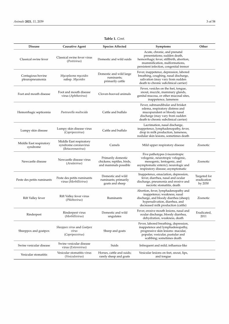
Table 1. Transboundary animal diseases overview.
Disease Causative Agent Species Affected Symptoms Other
African horse sickness African horse sicknessvirus (Orbivirus) Equids; primarily horses
Horses—acute (pulmonary) andchronic (cardiac) with high morbidity
and mortality, mules anddonkeys—mild disease,
zebras—usually asymptomatic
African swine fever African swine fever virus(Asfivirus) Domestic and wild suids
Sudden death, shock, hemorrhagicfever, pulmonary edema, depression,
anorexia, thrombocytopenia,lymphopenia
Avian influenza Avian influenza virus(Influenza A)
Domestic poultry; birdsand mammals
Highly pathogenic avian influenza (H5and H7) causes high rates of mortality,respiratory symptoms, sinus or head
swelling, depression, anorexia,cyanosis, incoordination, neurologic
symptoms, diarrhea
Zoonotic
Bluetongue Bluetongue virus(Orbivirus)
Domestic and wildruminants;
primarily sheep
Fever, swelling, vascular injury andhemorrhage, ulceration, pulmonary
edema, muscle necrosis; orasymptomatic
Animals 2021, 11, 2039 3 of 58
Table 1. Cont.
Disease Causative Agent Species Affected Symptoms Other
Classical swine fever Classical swine fever virus(Pestivirus) Domestic and wild suids
Acute, chronic, and prenatalpresentations; sudden death,
hemorrhagic fever, stillbirth, abortion,mummification, malformations,
persistent infection, congenital tremor
Contagious bovinepleuropneumonia
Mycoplasma mycoidessubsp. Mycoides
Domestic and wild largeruminants;
primarily cattle
Fever, inappetence, depression, laboredbreathing, coughing, nasal discharge,
salivation (may vary from suddendeath to chronic subclinical carrier)
Foot and mouth disease Foot and mouth diseasevirus (Aphthovirus) Cloven-hooved animals
Fever, vesicles on the feet, tongue,snout, muzzle, mammary glands,
genital mucosa, or other mucosal sites,inappetence, lameness
Hemorrhagic septicemia Pasteurella multocida Cattle and buffalo
Fever, submandibular and brisketedema, respiratory distress andmucopurulent or bloody nasal
discharge (may vary from suddendeath to chronic subclinical carrier)
Lumpy skin disease Lumpy skin disease virus(Capripoxvirus) Cattle and buffalo
Lacrimation, nasal discharge,inappetence, lymphadenopathy, fever,
drop in milk production, lameness,nodular skin lesions, sometimes death
Middle East respiratorysyndrome
Middle East respiratorysyndrome coronavirus
(Betacoronavirus)Camels Mild upper respiratory disease Zoonotic
Newcastle disease Newcastle disease virus(Avulavirus)
Primarily domesticchickens; reptiles, birds,and mammals possible
Five pathotypes (viscerotropicvelogenic, neurotropic velogenic,
mesogenic, lentogenic, andasymptomatic enteric); neurologic and
respiratory disease; asymptomatic
Zoonotic
Peste des petits ruminants Peste des petits ruminantsvirus (Morbillivirus)
Domestic and wildruminants; primarily
goats and sheep
Inappetence, emaciation, depression,fever, diarrhea, nasal and ocular
discharge, pneumonia and erosive andnecrotic stomatitis, death
Targeted foreradication
by 2030
Rift Valley fever Rift Valley fever virus(Phlebovirus) Ruminants
Abortion, fever, lymphadenopathy andinappetence; weakness, nasal
discharge, and bloody diarrhea (sheep);hypersalivation, diarrhea, and
decreased milk production (cattle)
Zoonotic
Rinderpest Rinderpest virus(Morbillivirus)
Domestic and wildungulates
Fever, erosive mouth lesions, nasal andocular discharge, bloody diarrhea,
dehydration, weakness, death
Eradicated,2011
Sheeppox and goatpoxSheeppox virus and Goatpox
virus(Capripoxvirus)
Sheep and goats
Fever, labored breathing, depression,inappetence and lymphadenopathy,
progressive skin lesions: macular,popular, vesicular, pustular and
scabbing; sometimes death
Swine vesicular disease Swine vesicular diseasevirus (Enterovirus) Suids Infrequent and mild; influenza-like
Vesicular stomatitis Vesicular stomatitis virus(Vesiculovirus)
Horses, cattle and suids;rarely sheep and goats
Vesicular lesions on feet, snout, lips,and tongue

Animals 2021, 11, 2039 4 of 58
Animals 2021, 11, x FOR PEER REVIEW 4 of 58
primarily goats and
sheep
eradication by 2030
Rift Valley fever Rift Valley fever
virus (Phlebovirus)
Ruminants
Abortion, fever, lymphadenopathy and inappetence; weakness, nasal discharge, and
bloody diarrhea (sheep); hypersalivation, diarrhea, and decreased milk production (cattle)
Zoonotic
Rinderpest Rinderpest
virus (Morbillivirus)
Domestic and wild ungulates
Fever, erosive mouth lesions, nasal and ocular discharge, bloody diarrhea, dehydration,
weakness, death
Eradicated, 2011
Sheeppox and goatpox
Sheeppox virus and Goatpox
virus (Capripoxvirus)
Sheep and goats
Fever, labored breathing, depression, inappetence and lymphadenopathy, progressive skin lesions:
macular, popular, vesicular, pustular and scabbing; sometimes death
Swine vesicular disease
Swine vesicular disease virus (Enterovirus)
Suids Infrequent and mild; influenza-like
Vesicular stomatitis
Vesicular stomatitis virus (Vesiculovirus)
Horses, cattle and
suids; rarely sheep and
goats
Vesicular lesions on feet, snout, lips, and tongue
Figure 1. Broad continental distribution of 17 transboundary diseases. This figure displays a general and broad continentaldistribution of each disease. Due to the potential for these diseases to easily cross borders, the geographic distribution isdivided into broad regions rather than being country specific. Abbreviations: African horse sickness (AHS), African swinefever (ASF), avian influenza (HPAI), bluetongue (BT), classical swine fever (CSF), contagious bovine pleuropneumonia(CBPP), foot and mouth disease (FMD), hemorrhagic septicemia (HS), lumpy skin disease (LSD), Middle East respiratorysyndrome (MERS), Newcastle disease (VND), peste des petits ruminants (PPR), Rift Valley fever (RVF), rinderpest (RP),sheeppox and goatpox (SP/GP), swine vesicular disease (SVD), vesicular stomatitis (VS).
2. Methods
Literature searches were performed using both PubMed and Google Scholar, with noinitial restriction on date range. The following search terms were used, for each disease:Disease, Disease + review, Disease + models. The results were sorted by relevance and thefirst ten results were selected from each search. Additional results that appeared potentiallyrelevant to the goals of the review were then selected. After the initial search, anothersearch was performed with a date range of 2015 to 2021 to find further, recent results.An additional search was also performed on both PubMed and Google Scholar using thesearch terms Disease + treatment. Additional references were reviewed as needed, fromthe reference list of literature found during the initial search. Searches were conductedbetween December 2020 and May 2021. Articles relevant to the goals of this review, whichinclude providing a synopsis of the disease, sharing information regarding prevention ortreatment and summarizing available research models, were selected and cited from thecompiled searches for each disease.
3. Review3.1. African Horse Sickness
African horse sickness virus (AHSV) causes African horse sickness (AHS) and is anon-enveloped, double stranded RNA arbovirus that belongs to the genus Orbivirus, in thefamily Reoviridae [6]. AHSV is divided into nine serotypes (AHSV1-9) [7,8]. The virus is
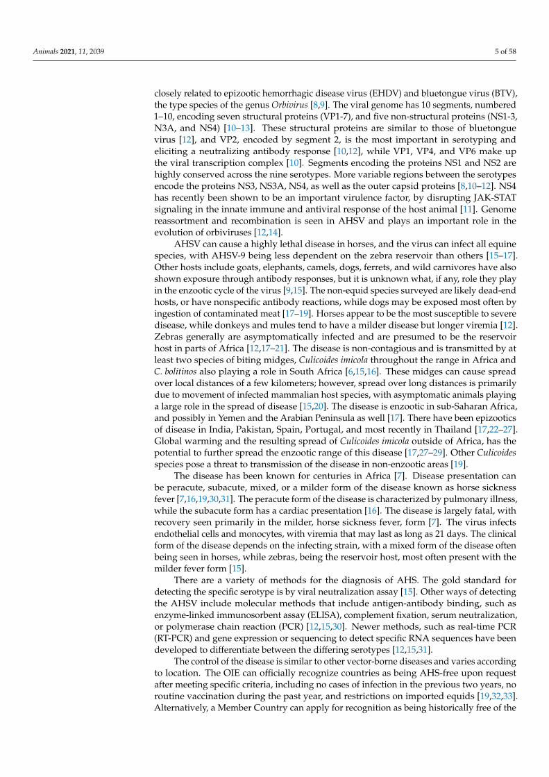
Animals 2021, 11, 2039 5 of 58
closely related to epizootic hemorrhagic disease virus (EHDV) and bluetongue virus (BTV),the type species of the genus Orbivirus [8,9]. The viral genome has 10 segments, numbered1–10, encoding seven structural proteins (VP1-7), and five non-structural proteins (NS1-3,N3A, and NS4) [10–13]. These structural proteins are similar to those of bluetonguevirus [12], and VP2, encoded by segment 2, is the most important in serotyping andeliciting a neutralizing antibody response [10,12], while VP1, VP4, and VP6 make upthe viral transcription complex [10]. Segments encoding the proteins NS1 and NS2 arehighly conserved across the nine serotypes. More variable regions between the serotypesencode the proteins NS3, NS3A, NS4, as well as the outer capsid proteins [8,10–12]. NS4has recently been shown to be an important virulence factor, by disrupting JAK-STATsignaling in the innate immune and antiviral response of the host animal [11]. Genomereassortment and recombination is seen in AHSV and plays an important role in theevolution of orbiviruses [12,14].
AHSV can cause a highly lethal disease in horses, and the virus can infect all equinespecies, with AHSV-9 being less dependent on the zebra reservoir than others [15–17].Other hosts include goats, elephants, camels, dogs, ferrets, and wild carnivores have alsoshown exposure through antibody responses, but it is unknown what, if any, role they playin the enzootic cycle of the virus [9,15]. The non-equid species surveyed are likely dead-endhosts, or have nonspecific antibody reactions, while dogs may be exposed most often byingestion of contaminated meat [17–19]. Horses appear to be the most susceptible to severedisease, while donkeys and mules tend to have a milder disease but longer viremia [12].Zebras generally are asymptomatically infected and are presumed to be the reservoirhost in parts of Africa [12,17–21]. The disease is non-contagious and is transmitted by atleast two species of biting midges, Culicoides imicola throughout the range in Africa andC. bolitinos also playing a role in South Africa [6,15,16]. These midges can cause spreadover local distances of a few kilometers; however, spread over long distances is primarilydue to movement of infected mammalian host species, with asymptomatic animals playinga large role in the spread of disease [15,20]. The disease is enzootic in sub-Saharan Africa,and possibly in Yemen and the Arabian Peninsula as well [17]. There have been epizooticsof disease in India, Pakistan, Spain, Portugal, and most recently in Thailand [17,22–27].Global warming and the resulting spread of Culicoides imicola outside of Africa, has thepotential to further spread the enzootic range of this disease [17,27–29]. Other Culicoidesspecies pose a threat to transmission of the disease in non-enzootic areas [19].
The disease has been known for centuries in Africa [7]. Disease presentation canbe peracute, subacute, mixed, or a milder form of the disease known as horse sicknessfever [7,16,19,30,31]. The peracute form of the disease is characterized by pulmonary illness,while the subacute form has a cardiac presentation [16]. The disease is largely fatal, withrecovery seen primarily in the milder, horse sickness fever, form [7]. The virus infectsendothelial cells and monocytes, with viremia that may last as long as 21 days. The clinicalform of the disease depends on the infecting strain, with a mixed form of the disease oftenbeing seen in horses, while zebras, being the reservoir host, most often present with themilder fever form [15].
There are a variety of methods for the diagnosis of AHS. The gold standard fordetecting the specific serotype is by viral neutralization assay [15]. Other ways of detectingthe AHSV include molecular methods that include antigen-antibody binding, such asenzyme-linked immunosorbent assay (ELISA), complement fixation, serum neutralization,or polymerase chain reaction (PCR) [12,15,30]. Newer methods, such as real-time PCR(RT-PCR) and gene expression or sequencing to detect specific RNA sequences have beendeveloped to differentiate between the differing serotypes [12,15,31].
The control of the disease is similar to other vector-borne diseases and varies accordingto location. The OIE can officially recognize countries as being AHS-free upon requestafter meeting specific criteria, including no cases of infection in the previous two years, noroutine vaccination during the past year, and restrictions on imported equids [19,32,33].Alternatively, a Member Country can apply for recognition as being historically free of the
Animals 2021, 11, 2039 6 of 58
virus [32]. AHS is the only equine disease for which countries can obtain this status [19,33].In order to prevent spread from areas where the disease is enzootic or epizootic, quarantineof equines moving from these areas should be practiced [6]. Additional measures thatcan be taken on the host species include vaccination in both enzootic and epizootic areasand stabling overnight in mosquito-proof stalls [6]. Many vaccines are multivalent, asthere is limited cross-protection between the nine serotypes and this will elicit a broaderimmune response [16,34]. The standard method of vaccination is based on live attenuatedvaccines (LAV). These vaccines have an inherent risk of reverting to virulence [10,35],they may not prevent infection and African horse sickness fever [36], and they do notallow for differentiating infected from vaccinated animals (DIVA) [10]. This inability todifferentiate infected from vaccinated animals results in difficulties in maintaining anAHS-free zone [37]. As a result, there has been recent progress on the development ofrecombinant and inactivated vaccines, protein, and virus-like particles [10,38–42]. Reversegenetics systems and recombinant techniques are being used to develop new vaccines totarget specific antigens of the AHSV, including several viral and nonstructural proteins ofthe virus, including the capsid proteins VP2 and VP5, as well as NS1, which may elicit aninterferon gamma host antiviral response [10–12,16,43]. These newer technologies aim tocreate a DIVA vaccine, which will greatly improve the control and detection of the virus.Finally, exclusion measures in certain AHS-free countries may include culling of positiveanimals to prevent an epizootic or establishment in the new area [19].
In addition to host-specific mitigation and prevention techniques, strategies to targetthe vector can also be implemented. These include spaying of insecticides [19], but caremust be taken when used near food producing animals [15]. Local strategies, such as theelimination of breeding habitat for the Culicoides midges, should also be practiced. Thisincludes the removal of dung and the elimination of mud or pooled water [15].
The socioeconomic impact of AHS in enzootic areas is great. This ranges from low-income communities, where working animals are affected by disease, to racing, sport,and leisure activities where either disease or impediments to movement across regions isaffected [33]. The impact on low-income areas is largely due to morbidity and mortality,and affects food security, as well as having ripple effects on poverty alleviation and genderequality. The exact impact is difficult to discern, as diagnosis and reporting is rarelydone [6,33]. The greatest potential for financial impact relates to horse racing. The total forthis industry amounts to several hundred billion dollars annually [6].
Most vaccine development studies have been done in horses, and most of whatis known about the disease is from naturally infected animals. There has been somestandardization towards a mouse model of disease. Early work was done in BALB/c mice,by several challenge routes, to compare attenuated vaccine strains of AHSV to wildtype [44].Recent work has focused on studying AHSV4 in the IFNAR −/−mouse [45–47]. Thesemice lack the type-I interferon receptor, which increases susceptibility to viral infection.Studies have focused on vaccine evaluation and a further characterization of the model.Additional work has been done to characterize a guinea pig model for the evaluation ofAHSV vaccines [39,48].
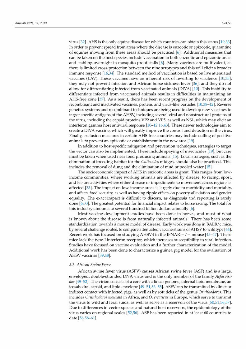
3.2. African Swine Fever
African swine fever virus (ASFV) causes African swine fever (ASF) and is a large,enveloped, double-stranded DNA virus and is the only member of the family Asfarviri-dae [49–52]. The virion consists of a core with a linear genome, internal lipid membrane, anicosahedral capsid, and lipid envelope [49–51,53–55]. ASFV can be transmitted by direct orindirect contact with infected pigs, as well as by soft ticks of the genus Ornithodoros. Thisincludes Ornithodoros moubata in Africa, and O. erraticus in Europe, which serve to transmitthe virus to wild and feral suids, as well as serve as a reservoir of the virus [50,51,56,57].Due to differences in vector species and natural host reservoirs, the epidemiology of thevirus varies on regional scales [52,56]. ASF has been reported in at least 60 countries todate [56,58–61].
Animals 2021, 11, 2039 7 of 58
African swine fever was first described in Kenya in 1921 [51,62]. It causes a hemor-rhagic fever with mortality rates nearing 100% in domestic pigs and Eurasian wild boar.Mortality can differ in domestic and wild suids according to virus strain [50,53,62,63]. Thevirus remained limited to Africa prior to the mid-twentieth century, when it spread toEurope, South America, and the Caribbean. A second expansion out of Africa spreadto the Republic of Georgia, the Russian Federation, and again into Europe, where it hadpreviously been eradicated, with the exception of Sardinia [51]. It remains enzootic in sub-Saharan Africa, but the recent expansion out of Africa has now spread to Asia, includingChina in 2018, where half of the world’s pig production occurs [53,64].
ASFV is highly contagious, and transmission can be through direct contact with in-fected pigs, ingestion of infected meat, exposure to contaminated feces, blood, or urine,or by the tick vector. There are commercially available diagnostic kits based on ELISA orPCR, but use of such tests may be regulated in certain countries [63]. Confirmation can beperformed though virus isolation in porcine leukocyte or bone marrow cells, followed byhemadsorption assays [65]. ASFV can be transmitted by fomites and is highly stable in theenvironment, especially in protein-rich matrices, such as infected wild boar and their car-casses and the meat from infected domestic pigs [64,66,67]. After exposure, the virus infectsmonocytes and macrophages, and later, endothelial cells. This leads to the classic presen-tation of death from shock secondary to disseminated vascular coagulation (DIC) [54,68].Disease presentation can vary from peracute death to persistent infection and dependson virus strain. Clinical signs include pulmonary edema, depression, fever, anorexia,petechiae, cyanosis, thrombocytopenia, lymphopenia, and hemorrhagic lesions [69]. Incontrast to domestic and feral pigs, African wild pigs (warthogs and bushpigs) tend tobe asymptomatic and likely serve as the reservoir in areas of Africa where the virus isenzootic through a sylvatic cycle with the tick vector [69–71]. However, spread of the virusis primarily due to human activities and the movement of infected pigs [64,66].
The ASFV genome encodes between 151 and 167 open reading frames (ORFs), rep-resenting more than 160 proteins [54,55,64,72,73]. The virus contains genes for enzymesrelated to DNA replication and repair, protein modification, and virus–host interactions.ASF viral transcription is independent of host RNA polymerase II, and the virus mayreplicate in the macrophage cytoplasm [69,72]. The virus is immunomodulatory for bothinnate and adaptive immune systems through numerous mechanisms [53,69,72,73]. Theviral genome encodes proteins that directly inhibit macrophage intracellular signaling aswell as intercellular signaling of immune cells. This includes the inhibition of Toll-likereceptor 3 signaling (which is the pathway that recognizes infection with dsDNA viruses),and inhibition of the Type I interferon response (also involved in innate immunity againstviral infection) [72–75]. One viral protein, EP402R or CD2v, is structurally similar to hostCD2 in that it has two immunoglobulin-like domains, Ref. [53] which inhibits lympho-cyte proliferation [72]. The viral protein is responsible for hemadsorption to the infectedmacrophage [53,72]. Viral protein EP153R resembles NK cell receptors (e.g., CD69), inhibit-ing up-regulation of MHC Class I expression, Ref. [72] which is the cellular mechanism ofpresenting foreign antigen to immune cells. ASFV also encodes several genes which inhibitcellular apoptosis through multiple signaling pathways, thus preventing the infected cellfrom undergoing apoptosis [73].
In spite of the difficulties the virus poses to the development of an effective vaccine,this is currently a very active area of research [64,69,71,76–80]. It is known that pigssurviving acute infection develop long-term resistance to infection by homologous strainsof the virus [76,80]. The development of inactivated vaccines produced by numerousmethods have all been unsuccessful, and it appears that an efficacious killed vaccine willnot be possible [77,79,80]. Attempts at developing a live attenuated vaccine (LAV) havebeen made through serial passage in bone marrow culture cells, or through using naturallyattenuated strains have led to chronic lesions and late disease [79,80]. Serial passage incell lines, such as Vero cells or COS-7 cells, has led to decreased protection, or inability toreplicate in domestic pigs [76]. Attenuated strains may also be developed by the deletion

Animals 2021, 11, 2039 8 of 58
of certain viral genes. A recent vaccine made by deletion of a virulence factor (Pret4∆9GLvirus) was found to be safe in pigs and imparted partial protection from a homologousstrain of ASFV as early as 21–28 days [81]. LAVs provide the advantage of being moresimple to develop compared to subunit vaccines [78], and they can elicit a host immuneresponse to all antigens present, as opposed to recombinant vaccines with a limited numberof antigens [79]. Also, there can be a small window of safety, with virulence at times onlydepending on the dose of the virus used [80], and attenuated strains will not likely allowfor differentiating infected from vaccinated animals [76]. Important factors in developingan acceptable vaccine include, safety, DIVA, regulated and reliable production, and cross-protection, as well as developing formulations that may be used in wildlife, such as baitvaccine formulations [78].
Subunit vaccine technology has been investigated using antigen, DNA, and virusvector based vaccines [71]. One advantage of antigen and DNA-based vaccines is a morefavorable safety profile than LAVs [71]. The development of subunit vaccines depend onidentifying neutralizing antibodies to viral antigens. Three ASFV proteins appear to bepromising in achieving this; p30, p54, and p72, however, attempts at using a recombinantvaccine targeting these antigens provided high titers but was not protective in challengestudies [82]. Other proteins may also provide immunity but attempts to produce a vaccinehave resulted in similar results [71,77,79,80]. Vaccines targeting the CD2v protein havebeen shown to reduce hemadsorption and the virus’ ability to infect monocytes, providingpartial protection [79]. Single cycle and replication deficient viruses have promise toprovide safe vaccines [77]. Virus vectored vaccines have been based on vaccinia virusAnkara, baculovirus, and alphavirus replicon particles. A combination approach usingheterologous prime-boost strategy has also been investigated. By using two vaccineplatforms, there is a hope of providing more robust innate and cellular immunity [71].
Due to the lack of an approved vaccine against ASFV, traditional methods of con-taining and eradicating an outbreak are followed, including movement restrictions andculling [64,81,83]. The early detection and eradication in ASF-free zones is vital. Thecurrent outbreak in China is believed to have spread rapidly after pigs from an affectedfarm were sold to several nearby farms. Given the widespread, decentralized nature of pigfarming in China, the long-distance transportation of pigs aided in the dispersion [64,84,85].Basic biosecurity measures include restrictions on movement and trade of live pigs andraw or treated meat products, as these can remain infective, prohibiting exposure to wildboar, and culling animals in the face of an outbreak. Additional measures include cessa-tion of swill feeding of pigs and wild boar, safe disposal of contaminated products, andrestricted zones of at least 3 km and surveillance zones of at least 10 km for pigs and porkproducts [83,86]. Effective biosecurity is important, as illustrated by outbreaks in Sardinia,Eastern Europe, and the Russian Federation, where backyard farms and small-scale pro-ducers that may lack adequate biosecurity measures led to spread of ASFV and long-termoutbreaks [67,87,88]. Outbreaks in large commercial farms can have a devastating impact.It was estimated that approximately 800,000 pigs died or were destroyed in Eastern Europeand the Russian Federation between 2014 and 2017, and exports from Poland, Lithuania,Latvia, and Estonia were reduced by US $961 million as a result of outbreaks in 2014 and2015 [67]. Spain provides a successful model for eradication of the virus. From 1985–1995;Spain instituted a program with several components, including: mobile field teams forcontrol and diagnosis, serological testing of animals, facilities improvements, includingbarriers and the safe disposal of manure, elimination of all outbreaks and a test and cullstrategy to identify carriers, and controlled movement with individual identification ofanimals. If an outbreak is detected, all pigs within the 3 km protective zone are immediatelyserologically tested immediately, movement is stopped within the 10 km surveillance zonefor 30 days, and animals within this zone are tested no sooner than 30 days after the initialcleaning and disinfection of the affected areas. Once serological data indicate the area isfree of ASFV, movement within the zones could recommence, but no movement of livepigs is allowed outside of this zone [86].
Animals 2021, 11, 2039 9 of 58
Given the complexity and cost of biocontainment studies in large animals and thegaps in understanding the immunology related to ASFV vaccinology, a small animalmodel could provide benefit in the development of new vaccines and help describe thecomplex interaction of the virus in host leading to disease. A mouse model showed thata recombinant Newcastle disease virus vaccine expressing the ASFV p72 gene was safeand effective, but previous studies showed a lack of translation when studied in pigs [71].The development of a small animal model of ASFV that recapitulates the disease andimmunology of pigs would likely shorten the time to the development of a safe andeffective ASFV vaccine to be used in the control of this important disease.
3.3. Avian Influenza
Avian influenza (AI) refers to a group of single-stranded, negative-sense, envelopedRNA viruses of the family Orthomyxoviridae in the genus Influenzavirus A [89,90]. In-fluezna viruses are classified based on two surface glycoproteins, hemagglutanin (HA) andneuraminidase (NA). There are eighteen different hemagglutinin subtypes (H1-18) andeleven different neuraminidase subtypes (N1-11). These viruses can be further classifiedas low pathogenicity (LPAI) or high pathogenicity (HPAI), by the disease they cause inthe domestic chicken (Gallus gallus domesticus) [89,91]. HPAI viruses fall into groups withH5 and H7 hemagglutinin subtypes and may result in 100% mortality. However, not allH5 and H7 viruses cause HPAI, Ref. [91] and the molecular difference between LPAI andHPAI may only be one amino acid [89].
All type A influenza viruses were derived from wild birds, mainly waterfowl (OrderAnseriformes) and shorebirds (Order Charadriiformes), with the exception of H17N11 andH18N12, which have only been isolated in bats [92,93]. Waterfowl and shorebirds are theaccepted reservoir, but typically circulate virus that is LPAI in domestic poultry. These LPAIviruses have been isolated from more than 100 species across more than 25 families [92].HPAI has evolved from LPAI in domestic poultry, first noted in Italian chickens in 1878,and from that time was known as “fowl plague” [89,91,92]. HPAI had remained a diseaseof domestic poultry until an H5N1 strain of HPAI was found in domestic geese in China(A/goose/Guangdong/1/1996 lineage H5Nx viruses) and has since caused morbidity andmortality in wild birds as well [92,94]. LPAI viruses have been identified in a wide range ofother birds and mammals, including felines, canines, suids, equines, and mustelids [95–97].The local spread and evolution of LPAI viruses can lead to continental-scale distribution [98].One LPAI currently circulating worldwide is H9N2, which poses a great risk to small scaleand family farms, where it may have great socioeconomic effects [94,99–101]. While H9N2viruses were first isolated in Wisconsin, United States, in 1966, the currently circulatingvirus has developed into many clades that are now enzootic in Asia, the Middle East, andparts of Africa. Since H9 viruses are not reportable, as H5 or H7 are, further spread will bedifficult to curtail [94].
Most HPAI outbreaks have been limited in their temporal and geographic impact.Between 1959 and 2019, 15 H5 and 27 H7 (total of 42) conversions to HPAI have oc-curred worldwide [92]. Of these, all but three were limited in scope. The exceptions in-clude A/goose/Guangdong/1/1996 (H5Nx), Mexican H7N3, and Chinese H7N9 [93,102].Deaths in wild birds, poultry, and humans have been linked to the Guangdong gooselineage (Gs/Gd). The geographic extent encompasses over 80 countries in Asia, Europe,Africa, and North America [92]. These strains have been detected in migratory birds inChina, Mongolia, South Korea, and Japan by 2011. By comparing outbreak records withthe satellite tracking of wild birds, and comparisons of whole-genome sequencing of viralsamples, it was shown that spread occurred along migratory bird routes [103]. Viruses ofthis lineage are now enzootic in wild waterfowl and have spilled over into domestic poultryand have evolved into at least 8 different genotypes [104]. At the time of this writing, thereare 410 outbreaks of H5Nx HPAI in poultry and 233 in non-poultry, reported to OIE’s earlywarning system by Member countries. This includes 215 new outbreaks in poultry in Asia,Europe, and Africa, and 79 non-poultry outbreaks in Asia and Europe [105].

Animals 2021, 11, 2039 10 of 58
The expansion of HPAI beyond a local outbreak depends largely on migratory birds.There are at least nine different types of H5 viruses circulating in wild bird populationsthat present a risk of developing into HPAI or causing human disease. These are H5N1,H5N2, H5N3, H5N4, H5N5, H5N6, H5N7, H5N8, and H5N9 [104]. This followed the 1996emergence of the Gs/Gd lineage. Prior to that time, HPAI evolved in domestic poultry andcould be eradicated through local means, including the depopulation of affected flocks,preventative culling, vaccination, and controlled marketing [93,106]. In 2009, the H5 geneof the Gs/Gd lineage was found to have integrated into at least six different H5Nx subtypes.These viruses have now been seen to spill over into domestic poultry and “spill back” intowild birds in Asia, the Middle East, Africa, Europe, and North America [106]. Surveillanceprograms for HPAI have now, more than ever, relied on the detection of potentially HPAIviruses in both domestic and wild bird species.
The surveillance of domestic and wild birds can be done through either live virusisolation or next generation sequencing (NGS) of samples or swabs. The advantage of livevirus isolation, which includes inoculation of specific pathogen free (SPF) embryonatedchicken eggs, is that it shows that live virus is present in the sample. One disadvantage isthat it selects for viruses that grow well in eggs. The advantage of NGS is that it is rapidand sensitive to all virus types, however, it does not provide information on whether thevirus is live or replication competent [102]. Sampling of live birds often includes trachealor choanal swabs along with cloacal swabs. These swabs may then be tested by real-time polymerase chain reaction (RT-PCR), along with NGS, to identify notifiable strains.Isolated virus can be identified through agar gel immunodiffusion (AGID), enzyme-linkedimmunosorbent assays (ELISA) for antigen, or other immunoassays, or by a moleculartest such as RT-PCR [95,107]. HPAI can be confirmed with the intravenous pathogenicityindex [108]. An additional method involves serological subtyping of the hemagglutininand neuraminidase subtypes using antibody inhibition [109].
The regional means of mitigation during an outbreak also includes market closure.This is especially important in China, where poultry farming is done at high densities,with >50% of duck and >80% of goose worldwide production occurs, and billions of birdsare sold annually in live bird markets (LBMs), Ref. [110] with the local density of LBMsbeing one of the greatest predictors of risk [111]. Given this scenario, these locations areimportant in the surveillance of new outbreaks of disease, which is also true for LBMsacross differing economies, such as the marketing of upland birds for private hunting inthe US [112].
In addition to surveillance and reporting of outbreaks, the best means of protectingdomestic bird farming is through prevention. Transmission occurs through direct orindirect contact with infected birds, though movement, equipment, fomites, and vehicles.Airborne transmission is fairly limited [113]. Biosecurity is the basis of protection atthe local level [95,113,114]. Influenza A viruses can be disinfected through the use ofbleach, quaternary ammonia, alcohols, aldehydes, acids, and iodine solutions, as well astemperatures greater than 56–60 ◦C (133–140 ◦F) [95]. Adequate isolation of the farm alongwith good biosecurity measures, provides good protection. An additional preventativemeasure is prophylactic vaccination of the flock. When HPAI was less wide-spread,vaccination was not considered best practice, as eradication by depopulation in theseinfrequent outbreaks was preferred [113]. Vaccination must be used in conjunction with OIEoversight, including the limited use of vaccines using OIE quality standards in situationswhere culling is not practical. Vaccination strategies must be used in combination withan exit strategy based on certain criteria to be met [115]. Vaccination is often prohibitedin countries where HPAI is not enzootic, and where vaccination strategies have beenineffective or led to antigenic drift in cases of failure [116,117].
Since vaccination using inactivated vaccines does not provide full protection and doesnot allow for DIVA, non-vaccinated sentinel animals may be used to detect outbreaks. Inaddition, a vaccine with homologous hemagglutinin to the circulating strain, but differingneuraminidase, allows identification of infection based on serology for NA [118]. Addi-
Animals 2021, 11, 2039 11 of 58
tional methods include serology for anti-NS1 (nonstructural protein-1) or anti-M2e (matrix2 ectodomain) antibodies. NS1 is only produced in active viral replication and is rarelypresent in inactivated vaccine. M2e is a viral transmembrane protein that allows for entryinto the host cell [113,118,119]. HPAI vaccines which are attenuated Newcastle diseasevirus (NDV)-vectored for H5 or H7 and are protective against both Newcastle disease andHPAI are being developed. NDV-vectored H5 vaccines are currently approved for use inChina and Mexico [120].
There are several animal models of avian influenza A viruses, as they relate to humandisease [94,121–128]. However, most animal experimentation on avian influenza, as itrelates to birds, is conducted in the host species, with most of the testing being done toevaluate pathogenicity of new strains or vaccine development [129,130]. Other work hasshown the pathogenicity of goose-origin HPAI in chickens [131]. Guidance on the perfor-mance of studies in avian species, including virus selection and preparation, host selectionand monitoring, study design, sampling, and analysis, was recently published [132].
3.4. Bluetongue
Bluetongue virus (BTV) causes bluetongue and is a nonenveloped, double strandedRNA virus that belongs to the genus Orbivirus, in the family Reoviridae [133–135]. Trans-mission is vector borne, via biting midges (Culicoides) and the disease is noncontagious.Domestic and wild ruminants are susceptible and, as such, the disease can have a largeimpact on trade and socioeconomics [133,134,136,137]. The virus has spread over timeto be present over a large geographical range, with different serotypes being present indistinct regions [138] with global spread increasing [139]. There are almost 30 distinctserotypes globally (28 officially recognized), with new serotypes identified almost on anannual basis [140–143]; at least 9 serotypes have spread across Europe in the past fewdecades [138,144].
Sheep are the primary, significant host, as clinical disease is most frequently seenin sheep [137,145,146]. Cattle are also important hosts, but they usually exhibit asymp-tomatic infections [146]. However, cattle have been shown to exhibit clinical disease inEuropean BTV-8 epidemics [147]. The role and significance of cattle involvement is com-plex and changing over time (reviewed in [138]). A wide variety of other wild ruminantsare also susceptible, including various species of deer and antelope, and camels [148].Deer belonging to the Cervinae subfamily are less susceptible to disease (red deer mayserve as reservoirs), while white tail deer (members of Capreolinae subfamily) are morehighly susceptible [149,150]. In India, seroprevalence studies have suggested that thefollowing animals are susceptible (listed in order of percent found seropositive, thoughseroprevalence was found to vary by geographic region): goats (43%), sheep (39%), cattle(38%, though prevalence was 66% when looking specifically at Bos frontalis, known asMithun), buffaloes (34%), and camels (16%), with prevalence varying based on specificregion (reviewed in [144]). Furthermore, BTV has been reported in canines [151] and avariety of African carnivores, though the overall significance of this finding requires furtherinvestigation [152].
The disease symptoms are broad and depend on many factors, including animalspecies, virus serotype, and route of infection. Disease symptoms often include lameness,painful hooves, and ulcerations or sores; animals may also develop a swollen tongue,which leads to decreased blood flow and thus a blue coloration of the tongue, giving thedisease its name (bluetongue) [145,146]. In sheep, BTV can cause serious disease withovert symptoms such as fever, hypersalivation, swelling, vascular injury and hemorrhage,ulceration, pulmonary edema, muscle necrosis, and possible death. Other animals maypresent with no symptoms at all [134,145].
Bluetongue disease is a re-emerging disease with major, global economic implica-tions [153]. Economic loss can result from losses in productivity, animal death, cost ofcontrol measures, or trade restrictions [154,155]. Surveillance and vaccination programsalso increase the financial burden [156]. Many vaccines have been developed over the years
Animals 2021, 11, 2039 12 of 58
(including modified live virus, attenuated BTV, and inactivated vaccines), as vaccinationis more feasible than vector control strategies. However, vaccines are not available for allserotypes but new vaccines are being developed (reviewed in [157]).
Animal models are crucial to study the pathogenesis and evaluate vaccination, treat-ment, and preventative measures. As sheep are the primary host impacted by clinicaldisease, they serve as a good large animal model and are commonly used to evaluate theimmune response and vaccine efficacy (reviewed in [158,159]. The difference in virulencebetween different strains has also been evaluated in sheep [158]. The characteristics ofnatural infection in sheep can be recapitulated via intravenous inoculation with infectedblood, which causes severe disease that includes symptoms seen naturally [146,160]. Inmodels utilizing subcutaneous inoculation, fever is usually the first clinical sign, followedby viral spread from the lymph nodes, tonsils, and spleen leading to viremia a few daysafter fever, followed by lesions. In these models, the virus seems to first enter the lymphnodes near where the virus is introduced, and spreads from there to the majority of tissues,via lymphatics or bloodstream. Persistence is not thought to be relevant [146,161–163]. Arelated model has been developed for cattle, using BTV-8. Intravenous or subcutaneousadministration of the virus stock results in clinical signs including fever, eye involvement,ulcers, and swelling. Symptoms were more severe and prevalent than traditionally seenwith natural infections using other serotypes [164]. The virus is first observed in peripheralblood mononuclear cells (PBMCs) with subsequent spread to the spleen and then mostother tissues; spread is temporally similar to what has been observed in experimentallyinfected sheep. Viral replication also appears to begin in the lymph nodes near the site ofinfection. Experimentally, adult cattle (not calves) have previously been shown to havea more persistent viremia [165] but this finding has been challenged and is generally nolonger accepted [146,162,163].
There are many challenges to large animal models, such as sheep and cattle. Thus,small animal models have also been an important area of research. These models havebeen especially useful for studying virus replication, characterization, virus evolution,and markers of attenuation [166,167]. As with many viral infections, suckling mice aresusceptible to BTV, generally using the common intracranial route [166,167]. Pathogenic-ity was shown along with infection of various parts of the brain, along with pathologyincluding encephalitis and lesions, similar to what is seen in sheep or cattle fetuses. Veryyoung mice were most susceptible, with animals as young as two weeks being much lesssusceptible [168]. In addition, interferon α/β receptor knockout mice (IFNAR−/−) can beused to model lethal bluetongue for some serotypes, using intravenous or subcutaneousexposure routes. This model exhibits pathogenesis similar to what is seen in natural hostsand can be used for evaluation of vaccines and the immune response [169,170]. However,murine models that lack an interferon response do not fully recapitulate what occurs innatural hosts, such as cattle [169].
As a large variety of animal species are susceptible, a number of other diverse modelsalso exist. These include: antelope (sub clinical infection, with viremia), deer (severe or fataldisease or subclinical disease, depending on deer species and virus serotype), pronghornand bighorn (clinical disease), bison (low viremia and few symptoms), and camels (lowviremia and few symptoms); reviewed in [158].
Numerous models have been developed over the years, but historical studies havebeen performed with a variety of different virus stocks, administered via different routes.As with other viruses, intramuscular injection has been commonly used but it is unclearhow well this exposure route recapitulates natural infection [171]; symptom developmentmay be artificially impacted by unnatural administration routes [172]. Results can also varybased on serotype, animal species, and differences within species (e.g., age) [158]. Theseissues can complicate results and emphasizes the need for well characterized virus stocksand well characterized models.
Animals 2021, 11, 2039 13 of 58
3.5. Classical Swine Fever
Classical swine fever virus (CSFV) causes classical swine fever (CSF) and is a smallenveloped single stranded positive-sense RNA virus that belongs to the genus Pestivirusin the family Flaviviridae [83,173–176]. It is closely related to bovine viral diarrhea virus-1and -2 (BVDV-1 and -2) of large ruminants and border disease virus (BDV) [175]. Theviral genome is a single linear strand with one open reading frame (ORF) that codes forfour structural and seven nonstructural viral proteins [83,173]. Virus replication occursin the cytoplasm, where the viral genome is enclosed in the capsid. The virions acquirea round viral envelope during budding by exocytosis. Naturally occurring strains arenon-cytopathic in cell culture [173,175,177]. Important factors in transmission and viru-lence include attachment to host cells, viral replication, immunomodulatory effects, andinhibition of cell apoptosis [178–184]. There are three distinct genotypes with three orfour sub-genotypes [83,173,175]. These genotypes are serologically related and can becross-protective [83,175].
The historic origin of CSFV is not entirely clear, but it was first reported in the UnitedStates in 1833, where the disease became known as “hog cholera.” When it was first recog-nized in Europe during the latter 1800′s, it was termed “swine fever,” and later, “Europeanswine fever,” to differentiate it from the unrelated African swine fever (ASF). ASF was firstdescribed in Kenya in 1921 but may have been misdiagnosed as classical swine fever priorto that time since the diseases may be similar [51,62,83,176,185]. Today, CSF remains animportant disease of pigs worldwide [175,176,186]. There are three recognized presenta-tions of CSF: acute, chronic, and prenatal [83,175]. The virus remains stable, even undertranspacific shipping conditions, which poses a risk of transboundary spread from enzooticcountries [187]. Diagnosis is made by clinical signs, gross pathology, indirect (serological),and direct (virus isolation, antigen, and nucleic acid) detection of the virus [188–192]. Diag-nosis should be made using methods and protocols which are validated according to OIEstandards [193], and surveillance is imperative in maintaining a CSF-free zone [188,194].Inactivation protocols have been described to prevent accidental transmission of the virusby diagnostic samples [195].
Much like BVDV, but unlike ASFV, CSFV can cross the placenta and infect the developingfetus, leading to persistent infection particularly during mid-gestation [83,175,185,196,197].Experimental data indicate that early postnatal infection with low or moderately virulentstrains may lead to persistent infection, immunosuppression, and the inability to detectinfection based on serological assays [197]. The disease presentation can be affected bymany factors, including the virus strain, route of infection, infective dose, and host immunesystem [83]. The acute form can vary from fever, lethargy, anorexia, conjunctivitis, entericlymphadenopathy, respiratory and gastrointestinal disease, and possibly neurologic signs,hemorrhagic fever, and death [83,175,198]. Hemorrhage and thrombocytopenia are seen,including hemorrhagic lymph nodes and kidneys. This may lead to characteristic pale kidneyswith multifocal hemorrhage, or “turkey egg” kidneys [199]. Piglets are more profoundlyaffected, and adult pigs may survive and develop lasting immunity [83,175,198]. The efficiencyof the virus to cross the placental barrier is dependent on the virulence of the strain, withmedium and highly virulent viruses passing more readily. Piglets become persistently infected,despite immune recognition as indicated by increased CD8+ T-cells and IFN-alpha activationin viremic animals [196]. It is important that persistently infected piglets be recognized toavoid these animals being inadvertently vaccinated, rather than identified and culled fromthe herd [200]. The chronic form is nonspecific and occurs when animals are not able tomount an effective immune response. It is initially similar to the acute disease but is causedby less virulent strains and progresses to chronic wasting, enteritis, reduced fertility, recurringfever, and invariably fatal while not being hemorrhagic. Animals that are chronically infectedwill continue to shed the virus until death [83,175,198]. The presentation of the prenatalform is dependent on the gestational age at infection and virulence of the virus strain. Thepresentation in the sow may be subclinical, but if infection occurs early in gestation, it mayresult in stillbirth, abortion, mummification, and malformations. During mid-gestation,
Animals 2021, 11, 2039 14 of 58
about 50–70 days, the piglet may be immunotolerant, persistently infected, and survive forseveral months while shedding large amounts of virus. They then develop the late onsetform of CSF, exhibiting poor growth, occasionally showing congenital tremor, and ultimatelydeath [83,175,197,198,201].
CSFV is divided into three genotypes (groups 1–3), each with three to four subgeno-types [173,175,186,202–204]. These genotypes are not serologically distinct and providecross-protection [202]. Traditionally, phylogeny was established based on short fragmentsof the 5′-nontranslated region (5′NTR) and E2 coding region [173,202]. Recent advance-ments in sequencing capabilities have led to recommendations of using the entire E2 codingregion for more detailed phylogenetic determination [173,202]. There is a geographicalpattern of genotype distribution with some overlap, mainly in Asia. Circulating genotypesin the Western Hemisphere are of group 1, group 2 strains predominate in Europe, andgroup 3 strains are apparently solely in Asia. Group 2 strains are the most prevalentgenotypes worldwide and are seen in Europe and Asia [173,205,206].
Immunity most effectively targets the structural proteins Erns and E2, which areinvolved in virus entry into the host cell [186,205]. There are LAV strains in use worldwide,and these vaccines should be produced in accordance with OIE direction [186,205,207].Attenuation may be based on mutations in the viral genomic regions encoding the E2 andErns proteins [208–210]. CSFV LAVs are often made by serial passage in either rabbits or cellculture [211]. LAVs include the Chinese C-strain, or Chinese hog cholera lapinized virus(HCLV), the Lapinized Philippines Coronel (LPC) strain, Russian LK-VNIVViM strain,the low-temperature adapted Japanese guinea pig exaltation-negative (GPE-) strain, theFrench Thiverval strain, and the Mexican PAV strain, among many others [211–213]. TheC-strain vaccine has been shown to protect against highly virulent CSFV strains withindays after vaccination [214]. However, antibodies to natural strains of CSFV in enzooticareas may interfere with this efficacy [215]. In some areas, use of LAVs is cost-prohibitivefor local farmers, leading to continuation of outbreaks [216]. Since LAVs elicit a multivalentimmune response without the ability to DIVA, there are trade restrictions on animals fromareas practicing vaccination with these strains [189,190,192,211,217]. Vaccination with theC-strain has led to selection pressure on the antigenic E2 protein, and possible escapemutants [186,205]. However, this claim is still under investigation [211]. Unintentional useof the LOM (Flc-LOM-BErns) vaccine in a combined CSF/erysipelas LAV in South Korea in2014 has led to a reemergence of CSF on Jeju Island, which had been a CSF-free zone witha non-vaccination policy for the preceding decade [211,218,219].
Recently, work has focused on developing new DIVA vaccines [212,214,217,220]. Oneapproach is the deletion of the E2 protein in the vaccine strain (C-DIVA strain), whichprovides a means of differentiation [214]. Another approach is development of recombinantvaccines, which include two licensed products: CP7_E2alf (Suvaxyn®CSF Marker, Zoetis,Louvain-la-Neuve, Belgium) in Europe, and Flc-LOM-BErns, in South Korea [212,221]. Thedeletion of glycosylation sites has shown promise for the development of attenuated vac-cine strains [181]. One further approach has been production of a fusion protein, combiningthe E2 protein to the extracellular segment of the host CD154 molecule, which is licensedas Povac® in Cuba [222,223]. Another fusion protein vaccine combines the E2 proteinwith the host Fc region of IgG. This vaccine may protect against vertical transmissionfollowing a two-dose protocol [223]. Recombinant vaccines have been made, including apseudorabies vaccine expressing CSFV E2 protein, which has shown protection againstboth pseudorabies and CSFV challenge [224], and a recombinant Newcastle disease-basedvaccine expressing E2 and Erns proteins [225].
As with African swine fever, following strict biosecurity practices is imperative. Con-trol of movement, adequate surveillance, and prophylactic and emergency vaccination arepotentially useful in successful eradication and control programs [194,226–229]. Also, as inAfrican swine fever, additional consideration must be given to the presence of infected wildboar [230–235]. Wild boars have played a role in transmission of the disease in both Europeand Asia in recent years. Exposure can be through direct contact, through fomites, or by
Animals 2021, 11, 2039 15 of 58
feeding contaminated food products [233,235]. This exposure can complicate eradicationprograms and lead to persistence and spread of the disease [231]. Biosecurity measuresinclude fencing of the facility, disinfection of people and equipment, hygiene, baiting wildboar with oral vaccines, and capturing or hunting of the wild boar [231,233–235].
Animal experimentation of CSFV is often conducted in pigs [236]. An in vitro modelusing a 3D collagen matrix was validated to study inactivation of CSFV in natural casingsfor use in sausage making, which are traded globally and may be a source of transmission.Previously, intestines from experimentally infected pigs had been used to assess inactivationprotocols [237]. Cell culture methods have been developed to eliminate the need for usingpigs to produce virus for challenge stocks [238]. BALB/c mice have been used to studyvaccines against CSFV [239]. However, a well characterized model with established viralconcentration, strain, and model species has not been developed. The development of astandard model of CSF could help better define some of the current shortcomings in CSFunderstanding, including certain aspects of pathogenesis, correlates of protection, and theinduction of immunotolerance [177].
3.6. Contagious Bovine Pleuropneumonia
Contagious bovine pleuropneumonia (CBPP), also known as “lungsickness,” is causedby Mycoplasma mycoides subsp. mycoides (Mmm), a self-replicating, pleomorphic bac-terium [240,241]. Mycoplasma mycoides subsp. mycoides is one of 5 pathogenic mycoplasmas,known as the “mycoides cluster” [240]. Previously referred to as the small colony (SC) typeof Mmm, this designation was dropped after Mmm large colony (LC) was reclassified asMycoplasma mycoides subsp. capri [240]. First described by Gallo in the 16th century, CBPPreached almost worldwide distribution in the 19th century [241,242]. Previously secondonly to rinderpest as the disease of prime concern in cattle, CBPP remains a significantconcern and threat to the livestock industry [243,244]. Following drastic stamping outefforts, CBPP was eradicated in the 20th century from most of the world but continues toplague Sub-Saharan Africa [245–252].
Contagious bovine pleuropneumonia is an acute, subacute or chronic disease thatprimarily affects cattle (Bos taurus, B. indicus) and sometimes water buffalo (Bubalusbubalis) [241,253]. Hyperacute forms, characterized by sudden death, may be seen atthe beginning of outbreaks [240,254]. Serofibrinous pleuropneumonia and severe pleuraleffusion are seen in the typical acute to subacute form of disease [240,241,253,254]. Inchronic cases, subclinical carriers may be seen [240,241,253]. Transmission occurs throughthe inhalation of aerosolized infected droplets [240]. The role of subclinical carriers intransmission is uncertain [255,256]. Mycoplasma mycoides subsp. mycoides survives a shorttime in the environment and is susceptible to most common disinfectants [240]. After an in-cubation period of generally 3–6 weeks and sometimes up to 6 months, fever, inappetence,depression and labored breathing may be seen followed by coughing, nasal discharge andsalivation [240,241,257]. Mortality can reach 75–90% in epidemic outbreak but is usuallyless than 10% in enzootic regions [240,241]. Recovering animals are weak and emaciatedand may develop pulmonary sequestra [241]. Calves may develop carpal and tarsal le-sions [240,241]. Differential diagnoses include pasteurellosis (Pasteurella multocida), eastcoast fever (Theileria parva), bovine tuberculosis (Mycobacterium bovis), Mycoplasma bovis,actinobacillosis (Actinobacillus spp.), traumatic pericarditis, and hyatid cysts (Echinococ-cus granulosus) [240,241]. Rapid, presumptive diagnosis can be made based on clinicalsigns and gross lesions seen post mortem [240]. The currently available confirmatorydiagnostics include PCR, real-time PCR, complement fixation testing and competitiveELISA [240,243,258]. Immunohistochemistry may be useful in chronic cases [240].
Use of antimicrobials is banned during official eradication programs, but they arecommonly used to treat CBPP in Sub-Saharan Africa [240]. There is concern for increasingantimicrobial resistance and potentially increasing the number of animals with pulmonarysequestra, but targeted antimicrobial treatment could play an important role in the controlalongside vaccination [240,247]. Lacking a cell wall, Mmm is naturally resistant to beta-
Animals 2021, 11, 2039 16 of 58
lactam antibiotics [240]. Antimicrobial susceptibility testing and therapeutic studies havefound efficacy of tetracyclines, macrolides, and fluoroquinolones, and shown resistanceto tylosin [240,259]. Administration of the fluoroquinolone Danofloxacin did not result inclinical improvement of diseased animals, but in contact animals showed fewer lesions andless mortality [260]. A case report described clinically effective treatment of acute CBPP ina cow using tetracycline, dexamethasone and vitamin B complex [261].
Vaccination is critical for the control of CBPP in enzootic areas [262]. The currentlyused vaccines are live attenuated vaccines developed decades ago, that show limited effi-cacy and occasional severe side effects [263,264]. As additional studies allow for a betterunderstanding of the immune responses, virulence factors and molecular characteristicsof Mmm and improved vaccine candidates can be developed [263,265–267]. Jores et al.describe research priorities for the development of improved vaccines [243]. In additionto the inadequacies of the current vaccines, vaccination campaigns have been inconsis-tent [241,268]. Before the causal agent of CBPP was identified by Nocard and Roux in1898, it had been eradicated from several countries with a variety of strategies includingstrict control of movement, slaughtering, and vaccination, showing the importance ofcontrol efforts beyond vaccination alone [269–271]. Additional research is also neededto develop simpler and faster field tests [271]. Particularly while improved vaccines arestill under development, a comprehensive control strategy will depend on clear policies,government and public commitment, adequate veterinary services, movement restrictions,robust surveillance, good vaccine manufacturing practices and maintaining high diagnosticlaboratory standards [272–278].
Mathematical models have been developed to evaluate economic impact, transmis-sion dynamics, and the potential impact of various control strategies [256,274,279–283].In vitro models utilizing bovine lung epithelial cells and a variety of assays have beendescribed [240,284,285]. Bovine respiratory explants from trachea, bronchi and lungs ofslaughtered cattle are a promising ex vivo tool for further investigation of CBPP infec-tion [286]. Rodent and rabbit models have been used for some vaccine and virulencestudies [285,287]. Mice develop mycoplasmaemia following infection, but they are not agood model of the pathology seen in CBPP [285,288]. Cattle models are costly and canpresent difficulties reproducing disease, but several challenge techniques have been devel-oped [240,241,288–291]. Contact infection studies resemble natural infection but requirean extra group of diseased animals and result in an unpredictable rate and timing oftransmission, making it difficult to compare disease outcomes [290,291]. Endobronchialinoculation of three different strains of Mmm in steers showed two strains (Ondangwaand Shawawa) may be useful for study of subacute and chronic infections, while thethird (Gladysdale) more closely mimics the peracute form of disease [289]. Nkando et al.presented nasotracheal inoculation of cattle with the aid of a bronchoscope as an alternativeto an endobronchial intubation approach where tube insertion requires sedation of theanimal [290]. Repeated aerosol nasal infection of cattle has been reported to closely mirrornatural epidemiology in which only a fraction of animals develops acute disease [291].This approach avoids “overchallenge” that could be seen with direct tracheal or bronchialinstillation [291].
3.7. Foot and Mouth Disease
Foot and mouth disease virus (FMDV) causes foot and mouth disease (FMD) andis a nonenveloped, single stranded, positive sense, RNA virus that belongs to the genusAphthovirus, in the family Picornaviridae [292,293]. FMDV was the first described viralinfectious animal disease, based on the findings of Loeffler and Frosch during the late 19thcentury [292,293]. The virus infects cloven-hooved animals, via a variety of routes, and ishighly contagious in susceptible animals. As with other RNA viruses, FMDV has a highmutation rate and exhibits high genetic diversity; there are currently seven recognizedFMDV serotypes (i.e., O, A, C, Asia-1, South African Territories (SAT) 1 through 3), eachcontaining distinct genetic lineages [292,294,295].
Animals 2021, 11, 2039 17 of 58
Prior to the 20th century, FMDV was globally distributed. Extensive eradicationefforts over the last century have resulted in a diminished distribution of FMDV andthe virus is not currently known to exist naturally in North America, Australia, NewZealand, or the majority of Europe. However, FMDV remains an enzootic problem inSouth America, the Middle East, and the majority of Africa and Asia. At the end ofthe 20th century and beginning of the 21st century, regions of Europe and East Asiaexperienced re-emergences [293,296–298]. Extensive epidemiological modeling studieshave been performed, but these studies must continue so models can be applied in theevent that outbreaks occur in regions previously free of FMDV [299].
While FMDV can infect all cloven-hooved animals, natural infections are most preva-lent and significant in domestic livestock such as cattle, pigs, sheep, and goats. Somespecies of deer (roe and muntjac deer (more severe disease), sika deer (milder disease),and fallow and red deer (subclinical disease) [300,301]) and camelids can also contributeto transmission of the virus and may be significant in instances where they are in closecontact with domestic livestock [294,296]. Generally, FMDV infections in wildlife are notsignificant but African buffaloes (Syncerus caffer) appear to be maintenance hosts, whichcomplicates eradication efforts in areas where infected buffaloes are present, as virus elim-ination and control efforts would likely need to extend beyond domestic livestock [297].A better understanding of the role of wildlife (for example, African buffalo) is neededto understand the risk of transmission posed by these possible reservoirs [299]. Otheranimals, and humans, can pose a transmission risk if they become contaminated with thevirus (e.g., from aerosolization, fomites, clothing, etc.) and then have contact with livestock.Instances of human infections are rare, often disputed, and difficult to confirm. Thus, directimpact to human health from infection does not appear to be a significant cause for concern(reviewed in [296]).
Transmission is via direct or indirect contact, through several different routes [293,296].The virus is transmitted most commonly and efficiently via airborne or aerosol spread,especially when animals are in close contact [302]. Spread via aerosol across great distancesis possible, though it is rare and dependent on the serotype or isolate involved [303].Animals can also become infected via breaks in the skin or mucosa. Skin and mucosalinfection are less efficient and likely require a higher dose of virus than respiratory infection,though information about infectious dose and route are generally from experimentallaboratory studies and natural events are difficult to fully understand [293,296,300,302].In addition, contact with fomites poses a transmission risk, and a variety of bodily fluids(including semen, urine, and feces) can harbor the virus. Furthermore, milk or other animalproducts can transmit the virus, which has severe implications for trade; internationaltrade bans can result in economic hardship for countries where the virus is enzootic [302].Pigs can also become infected from eating food contaminated with the virus, though it isunclear if infection is a direct result of ingestion or from breaks in the mucosa [293,296].
In animals that exhibit clinical illness, fever is generally one of the first symptoms. Fol-lowed by vesicle development, on the feet and tongue [292–294,296,297]. Vesicles can alsoappear around the mouth (e.g., snout, muzzle), mammary glands, genital mucosa, or othermucosal or skin sites. Lack of appetite or lameness also occur frequently [292–294,296,297].Viremia is common in animals showing clinical signs. Symptoms can vary based onserotype or strain, and are more severe in pigs and cattle, in comparison to sheep andgoats [293,296]. In situations where clinical signs are not as obvious or predominant, diag-nosis can be complicated. Furthermore, other viral diseases (such as vesicular stomatitisvirus [discussed in Section 3.17] and swine vesicular disease [discussed in Section 3.16] cancause similar vesicles. Thus, laboratory confirmation is often required to differentiate FMDfrom other possible causes of disease [296].
Mortality from FMD is low [293,296]. Rather, the significant impacts are both direct,from loss of productivity and trade restrictions, and indirect, from control and preventioncosts. These losses account for billions of dollars annually. Production losses are mostnoteworthy in developing areas and cause further issues with poverty and food insecurity.
Animals 2021, 11, 2039 18 of 58
Communities that are especially dependent on livestock are particularly vulnerable [304].Control programs are also quite costly, but the alternative can be even more detrimen-tal, as evidenced by the re-emergence that occurred after vaccination efforts stopped inEurope [304,305].
For example, an outbreak of FMD occurred in the UK in the early 21st century, resultingin the culling of millions of animals. It is believed that the epidemic originated in sheep thatwere not showing obvious clinical signs (e.g., small number of lesions, which is commonin sheep), thus delaying identification of the problem and allowing it to spread to otheranimals. FMD afflicted pigs were eventually identified at a slaughterhouse [305]. Thisepidemic and similar occurrences have further spurred research into effective vaccinesagainst FMD [292].
Currently, there are numerous different vaccines available (reviewed in [295,306]). Thefirst vaccine utilized inactivated virus and was used during the middle of the 20th century,mainly to vaccinate cattle in various parts of northern and western Europe [295,306]. Thesource of the virus used in the vaccine has changed over time (e.g., animal derived, cellculture derived from different cell types) and the inactivation procedure has been refined.Vaccination efforts using inactivated virus resulted in high success and FMDV eradicationin Europe, such that vaccination was stopped in the last 20th century [292,295].
However, if vaccinated animals are only partially protected, they may be able tosupport viral replication, thus posing a risk of infection to other animals [307]. As such,certain inactivated vaccines also rely on removal of nonstructural proteins to maintainonly antigenic portions [306]. Another challenge is related to the genetic diversity of thevirus; antigenic variation is one of the main barriers to widespread and efficient controlvia vaccination. It is generally accepted that infection or vaccination against one serotypedoes not confer protection to other serotypes, so many vaccines are now targeted at morethan one serotype. Unfortunately, even within one serotype, vaccination does not alwaysconfer protection against all strains within the serotype [306]. Further work is also neededto determine the best targets for vaccines, along with efficacy testing to determine thefeasibility of using vaccine control in enzootic countries [299]. Vaccination efforts canbe further complicated by various cultural and socioeconomic factors. Due to extensivecosts associated with vaccination and lack of confidence in efficacy, it can be difficultto gain acceptance to control programs in some areas. Understanding the cultural andsocioeconomic aspects of FMD control and maximizing local community involvement incontrol programs is essential [299].
A wide variety of cell culture models exist for studying the basic biology of FMDV aswell as more complex topics such as persistence. Primary bovine thyroid (BTY) cell cultureswere historically used for the isolation and cultivation of FMDV, but immortalized cells linesare more commonly used due to the challenges of working with primary cell lines [308].Baby hamster kidney fibroblasts (BHK-21) and pig kidney (IB-RS-2) cells are commonlyused immortalized cell lines [308], though genetic variability is, unsurprisingly, commonafter virus passage in cell culture [309]. More recently, fetal goat tongue cells (ZZ-R 127) andfetal porcine kidney cells (LFBK-αVβ6) have also been evaluated for use in isolating andcultivating FMDV, particularly porcine derived strains [310]. Numerous in vitro modelshave also been evaluated for studying FMDV persistence, including Madin-Darby bovinekidney (MDBK) [311], primary bovine pharynx tissue (PBPT) derived cells [312], theaforementioned BHK-21 or IBRS-2 cells [313], and more recently, multilayer cells from thebovine dorsal soft palate (DSP) that avoid some of the challenges associated with otherprimary cells lines [314].
Large animal models present multiple challenges in general, but especially whenworking with high containment pathogens such as FMDV. Further challenges are presentedby incomplete knowledge about large animal host immune systems and a lack of appro-priate reagents required for immunological studies. The guinea pig and suckling mousemodel have been widely used historically, and the guinea pig model remains an essentialsmall animal model for studying FMD. The guinea pig recapitulates many of the clinical
Animals 2021, 11, 2039 19 of 58
symptoms seen during natural infection, exhibits a measurable antibody response, andmortality rates are low. As such, guinea pigs are used to study basic biology of the virusand pathogenesis, along with production of antiserum, and for countermeasure efficacystudies [315]. Natural hosts such as cattle and pigs are used when feasible and have beenuseful in informing on many aspects of FMD, including transmissibility, infectious dose,pathogenesis, and immune response [296,315].
3.8. Hemorrhagic Septicemia
Pasteurella multocida (PM), a gram-negative, non-endospore forming coccobacillus isthe causative agent of hemorrhagic septicemia (HS), an acute, fatal and septicemic diseaseof cattle and buffalo [316,317]. Like other members of the family Pasteurellaceae, Pasteurellaspp. are prevalent in vertebrate animals and frequently found as commensal organismsin the oral, nasopharyngeal and upper respiratory tract [318,319]. Many are opportunisticpathogens [318,319]. Pasteurella spp. can be passed from animals to humans through bitesor nasal secretion, with PM being the most common zoonosis [316,319]. Bacteremia andlife-threatening sequelae may be seen in humans with underlying disease or immunosu-pression [316,320]. With growing concern regarding emerging or re-emerging infectionsof zoonotic origin, Pasteurella spp. have major implications for both human and animalhealth [319,321–323]. Pasteurella multocida, first shown to be the causative agent of fowlcholera by Louis Pasteur in 1881 also contributes to swine atrophic rhinitis [318,324,325].Pasteurella multocida is divided into 5 capsular serogroups A, B, D, E, and F with 16 serotypesbased on LPS antigens [325,326]. Isolates of PM that cause HS fall into 2 groups, Asian andNorth American origin (serogroup B) and African origin (serogroup E) [327].
Although seen most commonly in cattle and buffalo, other species may potentially beaffected including deer, swine, elephants, rhinoceros, and antelope [328–332]. Outbreaksmay be associated with wet, humid weather [328]. Clinical signs include fever, edematoussubmandibular and brisket swelling, respiratory distress and profuse mucopurulent orbloody nasal discharge [328]. Acute disease, characterized by sudden death within 24 hof onset may be the first indication of an outbreak [317,328]. Subacute forms of diseaseare often associated with edema and longer, chronic courses may involve rapid, painfulbreathing and nasal discharge [317]. Nervous system involvement is rare, but has beenreported2 [333]. Carrier states are also possible [317]. In enzootic areas, most deaths arein older calves and young adults [317]. Transmission occurs through inhalation of nasalsecretions or exhaled droplets from infected animals [334]. Hemorrhagic septicemia has thepotential to cause mass mortality events with up to 100% mortality [317,331,335,336]. In2015, in Kazakhstan, over 200,000 Saiga antelope, representing over 60% of the global pop-ulation of a critically endangered species, died from HS over a period of three weeks [331].Unusual high humidity and temperature in the days preceding the event illustrate thepotential contribution of environmental changes to extreme disease events [331,335,336].
The differential diagnoses for sudden death caused by HS include lightning, snakebites,blackleg and anthrax [317]. Post-mortem findings associated with HS include subcutaneousedema, fibrinous pneumonia, pericarditis, sub-serosal hemorrhage throughout the body,and blood-tinged fluid in the abdomen and thorax [317,337]. Laboratory confirmation canbe made by the identification of gram-negative, bipolar, pleomorphic bacteria in bloodsmears [317]. While generally easy to isolate pure culture from fatal cases, it can be difficultto isolate in field screening for carriers [338]. Polymerase chain reaction is a rapid andsensitive tool for species and type identification [338]. Loop-mediated isothermal amplifica-tion has been shown to be feasible for rapid DNA and RNA detection [339]. Recombinasepolymerase amplification (RPA) with lateral flow dipstick (LFD) has the potential to bean effective and practical onsite diagnostic [340]. Due to the ease of obtaining a definitivediagnosis through isolation and identification and the development of rapid molecular di-agnostics, sero-diagnosis is not usually needed [317]. Use of an indirect ELISA with higherspecificity and sensitivity than indirect hemagglutination assay has been reported [325].

Animals 2021, 11, 2039 20 of 58
Antimicrobial has limitations including cost, low efficacy once clinical signs appear,and a possibility of failure due to resistance [317,341–343]. Ceftiofur, enrofloxacin, or gen-tamicin may be effective emergency treatment options until susceptibility is known [342].In enzootic areas, vaccination is the only practical prevention method [317]. The firstprophylactic HS vaccine was killed (0.25% Lysol inactivated-broth) and offered 6 weeksof immunity [344]. Subsequent live attenuated vaccines were developed and novel acel-lular (subunit, recombinant and DNA) vaccines are under development [343]. There areseveral commercially available vaccine formulations, but broader protection and longerlasting immunity are needed [317]. The optimization of conditions for in vitro PM growthis important for maximizing vaccine production and quality [345,346]. Availability ofmultiple PM genome sequences will help better understand PM pathogenesis and hostimmunity, contributing to the development of new vaccine strategies [321–323]. In additionto the need for a highly effective, affordable vaccine, control depends on public awareness,good husbandry practices, legislation to control animal movement and responsible use ofchemotherapeutic agents [317].
Mathematical models to evaluate outbreak data and potential intervention strategieshave been described [347]. In vitro assays have been performed using macrophages andaortic endothelial cells [348–351]. Mouse models can play an important role in investigationof pathogenesis and vaccine development [317,351–363]. Rats with or without immunosup-pression have also been used to explore pathogenesis and novel vaccines [349–351,364–366].Rabbits are occasionally used for vaccine evaluation and, along with mice, were recentlyused to evaluate a novel phage lysate marker vaccine along with a DIVA ELISA [358].Goat challenge models have been described [353,354]. Experimental challenge and vaccineassessment has also been established in dairy cattle and buffalo [367–371].
3.9. Lumpy Skin Disease
Lumpy skin disease (LSD) virus is a double standed DNA virus of the genus Capripoxvirus,family Poxviridae, which causes acute or subacute disease in cattle (Bos indicus and Bos taurus)and water buffalo (Bubalus bubalis) [372]. Goats and sheep may be experimentally infected [372].Several wildlife species have been shown to be susceptible or seropositive, but the role ofwildlife in LSD epidemiology is not well understood [372]. Diagnosed for the first time inZambia in 1929, by 1944, LSD had spread to South Africa [373]. Enzootic in most Africancountries and some countries in the Middle East, LSD has also expanded into eastern Europe,Russia, and China [372,374,375]. Lumpy skin disease threatens international trade and could beused as an economic bioterrorism agent [372]. Incubation in experimentally infected animals isbetween four and seven days, but may be up to five weeks in naturally infected animals [376].Clinical signs include lacrimation, nasal discharge, inappetence, enlarged lymph nodes, fever,drop in milk production, lameness, nodular skin lesions, and sometimes death [372,376,377].Skin lesions are firm, slightly raised, circumscribed nodules usually on the neck, legs, tail,and back [372]. Skin lesions cause permanent damage to the hides [378]. Ulcerative corneallesions and subcutaneous infections may develop [376]. Common secondary complicationsinclude pneumonia, mastitis, and orchitis [372,376]. Morbidity is usually approximately 10%and morbidity between 1–3% in enzootic regions [377]. An outbreak in a large Holstein cattleherd saw 12% mortality in adult animals and clinical signs were much more severe in Holsteincattle than indigenous breeds [379,380]. Stress associated with milk production and higher thannormal ambient temperatures may have contributed to disease severity [380].
Lumpy skin disease virus may be viable for long periods in the environment [377].Transmission is primarily vector borne and is most likely mechanical [372,381]. Mostlikely vectors include blood sucking arthropods such as stable flies (Stomoxys calcitrans),mosquitoes (Aedes aegypti), horseflies (Haematopota spp.) and hard ticks (Rhipicephalusand Amblyomma spp.), but further studies are needed to better understand vector trans-mission of LSDV [381–387]. Most oubreaks occur in summer when arthropods are mostactive [372,377]. Direct transmission has been rarely reported [372,388]. Prolonged detec-tion in semen and testes raises a concern for possible spread by recovered or subclinically
Animals 2021, 11, 2039 21 of 58
infected bulls [389–391]. Introduction of new animals and communal grazing and wateringare associated with higher risk of LSD occurrence [392].
Differential diagnoses include pseudo-LSD, malignant catarrhal fever, bovine papu-lar stomatitis, pseudo-cowpox, vaccinia, cowpox, foot and mouth disease, bovine viraldiarrhea, dermatophilosis, insect or tick bites, nesnoitiosis, rinderpest, demodicosis, hy-poderma bovis infection, photosensitization, urticaria, cutaneous tuberculosis, and on-chocercosis [372,393]. Post mortem findings include nodules throughout the lungs andgastrointestinal tract and lung edema and congestion [372]. Pathognomonic histopatho-logic findings include eosinophilic intracytoplasmic inclusion bodies in keratinocytes andballooning degeneration of spinosum cells [372,377]. Vasculitis and necrosis may be seen indeeper tissue layers [377]. Diagnosis is primarily clinical with PCR confirmation. VariousPCRs have been developed for different aims such as detecting all capripoxviruses, genus-specific detection, and differentiating virulent and vaccine strains [372,394]. As describedin an overview of LSD diagnostic techniques, electron microscopy, virus isolation, virusneutralization and serological techniques have also been utilized [372].
Treatment is symptomatic and targeted at preventing secondary bacterial complica-tions [377]. Vaccination along with movement restrictions is the only effective methodto control the disease in enzootic areas [372]. Commercially available vaccines are liveattenuated [372]. Both homologous (Neethling LSDV strain) and heterologous (Sheeppoxvirus and Goatpox virus) vaccines can be used, but heterologous vaccines may not providecomplete immunity [372]. A newly developed inactivated vaccine has demonstrated safetyand efficacy in the field [395,396]. Calves from infected or immunized dams should notbe vaccinated under 6 months old to prevent maternal antibody interferences [393]. In-vestigations of reported vaccine breakdowns or failures have identified various causesincluding vaccination of animals already incubating disease, confusion with pseudo-LSD,and infrequent or improper use of the vaccine [373]. Capripoxvirus distribution seems tobe expanding, largely due to the economic effects of the Covid-19 pandemic, sanctionsin enzootic regions, increased illegal trade and global climate change [372]. Eliminationof the disease is likely to be difficult because of arthropod vectors, but controlling thespread may be achieved by accurate and timely diagnosis in enzootic areas, homologousstrain vaccination, vector control, animal movement restriction and testing of bulls usedfor breeding [372].
Several epidemiologic and mathematical models have been described to help under-stand outbreaks, transmission risks and design surveillance and control programs [397–406].In vitro growth for assessment and comparison of viral strain characteristics may beachieved on lamb kidney/testis cells and goat ovarian cells [407]. There have been severalrecent descriptions of experimental LSD infection in cattle and use of mice for immuno-genicity studies [390,407–410].
3.10. Middle East Respiratory Syndrome
Middle East respiratory syndrome coronavirus (MERS-CoV) is an enveloped, singlestranded, positive sense RNA virus that belongs to the genus Betacoronavirus (lineage C), inthe family Coronaviridae [411]. This novel coronavirus was reported in 2012 in Saudi Arabi;the initial patient presented with severe respiratory disease including pneumonia andlater developed renal failure [412]. Subsequent research shows the virus to be enzootic incamels in the Arabian Peninsula and East Africa, and it is likely bats served as the originalreservoirs [413,414]. The virus has since spread to a number of countries and exhibitshigh mortality rates [413]. Human cases arise from contact with infected camels [415,416].Human to human transmission is infrequent, but still a cause for concern leading to aneed for quarantine in instances of human infections [414]. Limiting zoonotic spread fromcamels to humans is a crucial step in control. Some have suggested camel vaccinationas the best course of action for control, and suitable vaccines are being developed [417].Experimentally, other animals such as alpacas, pigs, sheep, goats and horses can becomeinfected, though viral shedding is limited in the majority of laboratory infected animals
Animals 2021, 11, 2039 22 of 58
(reviewed in [414]). Animal models have been generally limited, and mainly focused onthe macaque [418–420].
Ten years before the identification of MERS, in 2002–2003, severe acute respiratorysyndrome coronavirus (SARS-CoV) was responsible for a large outbreak that began in Asiaand spread to North America, South America, and Europe. Research into these viruses andtheir zoonotic potential has been ongoing since their identification. However, beginning in2019, a novel coronavirus (SARS-CoV-2) sparked a global pandemic [421]. Research intoSARS-CoV-2, and the related SARS-CoV and MERS-CoV, has grown exponentially sincethe start of the pandemic. Thus, further in-depth details are better covered outside thescope of this review. Recent reviews provide more in-depth information and discussion ofMERS-CoV in the context of the SARS-CoV-2 pandemic [413,422–424].
3.11. Newcastle Disease
Newcastle disease virus (NDV) is an enveloped, negative sense, single stranded, non-segmented RNA virus with helical capsid symmetry. There are nine serotypes present in thefamily Paramyxoviridae and the genus Avulavirus. The virus is the causative agent of New-castle disease (ND) [425–430]. Nomenclature for isolates follows the convention adoptedfor Influenza A viruses, in that the isolate is named by serotype, species from which itwas isolated, geographical location, reference number or name, and year of isolation [429].Disease presentation depends on a number of factors, including the strain of virus andthe species, age, and immune status of the host [425]. NDVs have been classified into fivepathotypes related to the disease presentation that typically occurs in domestic poultry:viscerotropic velogenic, neurotropic velogenic, mesogenic, lentogenic, and asymptomaticenteric [425]. Viscerotropic velogenic viruses cause sudden death with intestinal hemor-rhage. Neurotropic velogenic viruses cause high mortality following acute respiratory andneurological disease, but usually lack intestinal pathology. Mesogenic viruses cause respi-ratory and neurologic disease, but low mortality. Lentogenic viruses cause mild respiratoryinfections, and have been used as vaccine strains [425]. Asymptomatic enteric virusescause infections, where virus replication appears to be primarily in the gastrointestinalsystem. There can be overlap in disease presentation in a single monotypic outbreak [425].The classification of pathotype is often done by either mean death time (MDT) in 9–10 dayold embryonated chicken eggs, or by the intracerebral pathogenicity index (ICPI) in 1 dayold chicks [428]. The molecular means of determining pathotype include identifying theamino acid sequence of the fusion (F) protein cleavage site [428,429,431,432]. Viral entryinto the host cell occurs through attachment by hemagglutinin-neuraminidase (HN) andvirus and cell membrane fusion by the F protein [427,430,433], and both HN and F areviral membrane proteins [430]. A virulence (V) protein has been shown to antagonizetype-1 interferon (IFN-1) response and may also play a role in host specificity [431]. Certaincomorbidities, such as coinfections or environmental factors, can lead a milder strain topresent as a more virulent disease [425]. A disease outbreak is usually accompanied bydepression, diarrhea, prostration, head and wattle edema, neurologic symptoms includingparalysis and torticollis, and respiratory symptoms [425].
Newcastle disease gets its name from Newcastle-upon-Tyne, England, where in 1926,there was an outbreak that was concurrently described on the island of Java, now in In-donesia [427]. In contrast to highly pathogenic avian influenza (HPAI), ND is enzootic insome areas of the world, where disease can have a major impact on poultry productionand small community or family flocks [426]. Assessment of the immune response is oftenevaluated by the hemagglutination inhibition (HI) test, utilizing a panel of antigens andcontrols [425,431]. While all viruses causing ND are of one serotype, avian paramyxovirustype 1 (APMV-1), there are 19 genotypes known [428]. Genotypes are based on the F genesequence [428]. Viruses are divided into class I and class II, with class I viruses represent-ing one genotype with three subtypes [428,432]. Class I viruses are largely isolated fromwild birds worldwide [428,432]. Class II viruses are comprised of the other 18 genotypes,I–XVIII [428,432]. Genotypes III, IV, V, and VI are typically considered pathogenic to chick-
Animals 2021, 11, 2039 23 of 58
ens [428]. There have been four historic panzootics of NDV, and the current panzootic isdue to genotype VII viruses [428]. Recent work in Bangledesh has identified the circulatingNDV in that country as class II genotype XIII, with a possible new variant evolving awayfrom that genotype [434].
Given the importance of family and community flocks in providing global food se-curity, the elimination of Newcastle disease could greatly increase production efficiencyin the areas in which it is enzootic [425,426]. One report from 1992 estimated that 90%of community chickens in Nepal die each year from Newcastle disease [426]. Recently,45% of 1374 chickens sampled, and 96% of 70 villages, were found to be seropositivefor NDV in rural farms across three provinces of Northern Iran [435]. Another studyfound that 57.1% of sampled flocks in Oman, with a total of 33.8% of all chickens sam-pled, were seropositive for NDV [436]. Vaccination strategies could help in resource-poorrural communities, but the vaccine should be part of a sponsored program to be worththe risk for farmers that tend to adopt new strategies only when they are low risk andthere is an obvious return for investment of time and money [437]. Vaccine programs areoften decided at the national level, in accordance with OIE guidance [431]. The success ofthese programs increases when working with farmers to deliver the vaccine in a preferredform [438]. When epizootics of ND lead to high mortality in free-range flocks, there iscurrently little incentive to put resources into the care of the chickens. When programs canshow reduced mortality and improved food security, they are more likely to be adoptedby local communities [438,439]. Contributing factors to the incidence of ND include flocksize, isolated and confined housing, multi-aged flock mixture, screening of birds, accessto ND vaccination, ND awareness, distance to service providers, and access to trainingand extension services [440,441]. There are reports of vaccine failure, with vaccinated birdsshowing disease and mortality after exposure to virulent NDV in natural settings [442,443],but this could not be reproduced experimentally following daily challenge for ten days,with no morbidity or mortality seen at 14 days post-challenge [443]. It may be that currentvaccines do not appear to be thermostable in the range of 51◦ to 61 ◦C, a considerationwhen planning vaccine strategies in rural areas [444]. Development of thermostable vac-cines is a current area of study [442,445,446]. Inactivated, or killed, vaccines have beendeveloped, and these may be given in ovo. This has been shown to provide protectionfrom morbidity and mortality in challenge studies [447]. One disadvantage of many killedvaccines is the potential for a weak cellular response, thus requiring priming with liveor other vaccine types [448]. Certain nonpathogenic lentogenic natural strains have beenconsidered for use as vaccines since there is cross protection against all pathotypes of NDV.These can be delivered by a number of routes, including directly, such as oculonasallyor by nebulization [449]. Live attenuated vaccines are traditionally produced by serialpassage in specific pathogen free (SPF) embryonated chicken eggs, which requires special-ized facilities and is resource intensive [431,450]. An alternative means of production isthrough cell culture [450]. Recent work has been done to produce newer vaccines based onmodern molecular techniques [431,448,451]. These recombinant vaccines often use eithernonpathogenic or replication deficient virus as the delivery vehicle for an antigen againstthe disease of choice [120,452]. An example of the former approach is to use NDV as avaccine platform for HPAI by insertion of the H5 or H7 gene. These vaccines have thepotential to protect against infection from both important viruses of domestic poultry [120].This approach has also been used to develop a vaccine against low pathogenicity avianinfluenza by incorporating the H9 antigen [445]. An example of a replication deficientvirus being developed as a platform for a NDV vaccine is the use of an adenovirus deliveryof NDV F protein (adeno-F) [452].
NDV infects a wide range of animals, from reptiles to mammals [425]. It is alsoknown to be zoonotic, usually causing a self-limiting conjunctivitis with no long-lastingeffects [425,429]. As many as 241 species of birds have been shown to be susceptible toinfection, but the viruses seen in wild birds are mostly of the asymptomatic lentogenicenteric pathotype when infecting chickens [425]. NDV has been isolated in caged pet and
Animals 2021, 11, 2039 24 of 58
zoo birds, as well as wild birds, including double-crested cormorants (Phalacrocorax auritus),all types of domestic poultry, racing and show pigeons, pheasants, ostriches, and captivefalcons [425,429,432,453–457]. NDV has also been isolated from domestic mink, where itcaused encephalitis, hemorrhagic pneumonia, and death [458]. Risk of introduction inareas free of virulent NDV could be associated with legal or illegal movement of birds andanimal products and feed, as well as spillover from wild species and transformation tovirulence [457,459,460]. Once in a new area, spread has been linked to movement of livebirds and poultry products, people and equipment, contaminated food and water, andcontact with other animals [429].
Much of the current literature on NDV focuses on the use of the virus as a vaccineplatform or gene delivery system for humans and other animals [461–463]. This is due toits broad range of species which the virus can infect. Many RNA viruses have been usedin this way, including retroviruses, lentiviruses, alphaviruses, flaviviruses, rhabdoviruses,measles viruses, and picornaviruses [462]. NDV is able to infect a broad range of species,but are very safe due to the limited ability to cause disease in these species, while stilleliciting a strong immune response [464]. Through various techniques, including reversegenetics and recombinant nucleic acid techniques, NDV-vectored vaccines have now beenmade for a variety of human and animal pathogens, including HPAI and LPAI, Africanswine fever virus, vesicular stomatitis virus, West Nile virus, bovine herpesvirus-1, caninedistemper virus, rabies virus, simian immunodeficiency virus (a model of HIV), enterovirus71, Rift Valley fever virus, Nipah virus, Ebolavirus, severe acute respiratory syndromecoronavirus (SARS-CoV-1), and more recently SARS-CoV-2 [120,445,461–463,465–471].
Gene therapy uses of NDV have been directed towards various types of cancer andhereditary diseases [462]. This field began in the 1990s and had early setbacks, including thedeath of a young patient being treated for a nonfatal genetic disease and the developmentof leukemia in severe combined immunodeficiency (SCID) patients [462]. In addition toproviding a platform for gene delivery, NDV has the ability to replicate in tumor cells [462]and can replicate up to 104 times faster in human cancer cells than in nonneoplastic humancells [472]. This ability to replicate in tumor cells has been coupled to tumor suppressorand immunomodulatory genes, such as tumor necrosis factor (TNF), tumor necrosis factor-related apoptosis inducing ligand (TRAIL), interferon-alpha (IFN-α), interferon-gamma(IFN-γ), and interleukin-2 and interleukin-15 (IL-2 and IL-15) [462,473]. Many of theserecombinant viruses take advantage of NDV’s natural oncolytic activity, which is basedon apoptosis, necrosis, or autophagy [472]. A proinflammatory response is triggered byNDV-HN, which drives IFN-1 simulation of TRAIL and activates natural killer (NK) cells,monocytes, macrophages, dendritic cells, and primes antigen-specific T cells and CD8+T-cell proliferation [472,474,475]. NDV-vectored therapies are in human clinical trials forprostatic carcinoma, hepatocellular carcinoma, gastric carcinoma, colorectal carcinoma,and melanoma [462,475]. Research into other cancer types has been done in cell culture orin mouse models using immortal cell lines [151,476,477].
Most of the research into NDV is done as viral challenge models in chickens orin ovo using SPF embryonated chicken eggs, but the strain of chicken and the precisedefinition of SPF are not often disclosed [444,478–483]. The most commonly reportedstrain of chicken used is the White Leghorn, along with others such as White Rock and IsaBrown layers [120,447,484–486]. It is known that differences in the innate immune responsein differing breeds of chicken can have drastic effects on the response to infection [487],making this important in describing the model. Some sources that define SPF describesubjects as being serologically or RT-PCR negative for NDV [478,480]. The virus strain andchallenge dose of NDV is usually given and is often the experimental variable, but there isnot a standard strain and dose that is used as a model of ND [478,485]. A common vaccinestrain used in studies is the LaSota strain [430,450,452,480,483,488,489]. Other host speciesare also used, including Japanese quail (Coturnix coturnix japonica) and pigeons (Columbalivia) [480,485].

Animals 2021, 11, 2039 25 of 58
3.12. Peste des Petits Ruminants
Peste des petits ruminants (PPR), is a highly contagious, devastating viral disease ofdomestic and wild ruminants, primarily affecting goats and sheep [490,491]. Peste despetits ruminants virus (PPRV) is a negative sense single stranded-RNA morbillivirus, ofthe family Paramyxoviridae, that is closely related to other members of the genus such asrinderpest, measles, and canine distemper [492]. Although only recognized as a completelydistinct disease for approximately 40 years, phylogenetic morbillivirus data suggests PPRVhas been in circulation as long as rinderpest virus [493]. In naïve populations of sheep andgoats, morbidity and mortality can be greater than 90% [494]. In enzootic areas, morbidityand mortality vary between 10–100% [495]. First identified in Cote D’Ivoire in the 1940s,PPR has since been seen in North and Central Africa, the Middle East, and parts of EastAfrica, Asia, and Europe [492]. The affected countries are home to 68% of the world’ssmall ruminant population [490]. An investigation of prevalence of PPR in sheep andgoats from 1968 to 2018, showed 40.99% prevalence in Africa and 38.43% prevalence inAsia [496]. Cattle, camelids and a wide range of wild animals and unusual hosts such aspigs, are considered susceptible with varying reports of morbidity and mortality [497].Experimental infection has shown cattle are susceptible and can display clinical signs [497].Large ruminants may have a role in transmission or, if dead-end hosts, may be of value forsurveillance [497–500].
Peste des petits ruminants virus is easily transmitted by direct contact with secre-tions and excretions from infected animals or contact with fomites [492]. The main entryroute is respiratory [501]. Peste des petits ruminants virus is a lymphotropic and epithe-liotropic virus [502]. Stages of disease include incubation, prodromal, mucosal, diarrhealand recovery if non-fatal [503]. The incubation period is generally 3–9 days [493]. Clini-cal signs include inappetence, emaciation, depression, fever, diarrhea, nasal and oculardischarge, pneumonia and erosive and necrotic stomatitis [492,504]. Fatal cases usu-ally die between 5–12 days after disease onset [493]. Pregnant animals may abort andtransient immunosuppression can make animals susceptible to an activation of latent ornew infections [493]. Goats may be more susceptible to severe disease than sheep [492].Pathognomonic histopathologic findings include multinucleated giant cells and cytoplas-mic and/or nuclear eosinophilic inclusion bodies [502]. Differential diagnoses includerinderpest, goatpox, bluetongue, contagious pustular stomatitis, contagious caprine pleu-ropneumonia, pasteurellosis, FMD, heartwater, coccidiosis, poisoning, and Nairobi sheepdisease [501,502]. Co-infections of PPR and goatpox have been reported [505,506]. Theintegration of tests and improved molecular tools may help with the rapid and accurateidentification of enzootic and outbreak PPR [507–509]. Pen-side tests and non-invasivesample techniques may improve diagnosis in remote settings and wildlife [492,507,510,511].
There is no specific treatment available, but drugs that control bacterial and parasiticcomplications may decrease mortality [512]. Experimental work has been performedwith antiviral treatment and herbal medicines are widely used in field treatment [513].Despite the availability of an efficacious and cheap live-attenuated vaccine, the virus hascontinued to spread [514]. The development of a vaccine allowing DIVA would reducethe time and cost of serological surveillance [515]. Recombinant vaccines may overcomethe thermolabile and lack of DIVA limitations of current live attenuated vaccines [516].Genetically engineered live vector vaccines are also promising candidates that can bedeveloped to be multivalent and activate both cellular and humoral immunity [517].
Peste des petits ruminants is currently targeted for global eradication by 2030 bythe PPR Global Eradication Programme [490,518]. Factors that favored the eradication ofRinderpest such as one serotype, availability of a safe vaccine that confers long immunity,simple diagnostics, short infectious period, close contact required for transmission, noknown significant wildlife reservoir or carrier state, and short virus survival in the en-vironment, also apply for PPR [490,501]. Constraints to eradication include widespreaddistribution, high population turnover in small ruminants, low value of individual animals,and clinical disease that varies by species and breed [519]. Understanding farmer’s KAP
Animals 2021, 11, 2039 26 of 58
(knowledge, attitude and practice) towards infectious diseases and consideration of genderissues are important to efforts for limiting impact and spread of disease [520,521]. Researchis needed to characterize the effective reproductive number, develop thermostable andDIVA vaccines, refine targets for molecular epidemiology, increase field diagnostics, anddetermine the role of atypical hosts [514].
Mathematical modelling has been performed to estimate economic impact, identifyrisks for transmission, and evaluate possible control techniques [522–526]. An in-silicoapproach to protein analysis may help with development of vaccines and therapeutics [527].Experimental infections of goats and sheep have been described and are suitable forcomparative studies and vaccine evaluation [528–530]. Mice, rabbits, and horses have alsoplayed a role in diagnosis, treatment, and vaccine development [531–533].
3.13. Rift Valley Fever
Rift Valley fever virus (RVFV) causes Rift Valley fever (RVF) and is an enveloped,single stranded, negative sense (with ambisense regions), RNA virus that belongs to thePhlebovirus genus, in the family Phenuiviridae [534] (formerly of the genus Phlebovirus,family Bunyaviridae [535]). There is significant RVFV diversity with at least 33 differentviruses and 15 lineages identified thus far [536]. The disease was first discovered inKenya and is enzootic in the southern and eastern parts of Africa; it has spread acrossthe African continent (e.g., Egypt and Sudan) and into the Arabian Peninsula (e.g., SaudiArabia) [535,537]. Outbreaks frequently result in the death of thousands of livestockanimals, with a substantial number of human infections and human deaths during someoutbreaks [537].
The disease primarily impact ruminants and a main route of transmission is viamosquitoes, mainly from the Aedes and Culex genera, but the virus has been found inmultiple species of mosquito from at least five additional genera, including Anopheles,Coquillettidia, Culiseta, Eretmapodites, and Mansonia [538–540]. A variety of other arthropodvectors, including ticks and flies, have been implicated in transmission (reviewed in [541]).Due to the role of arthropods in transmission, large outbreaks are frequently associated withperiods of heavy rainfall and flooding [3]. Increasing outbreaks, or emergence in new areas,is of increasing concern due to global climate change and the large global distribution ofpotential vectors; the potential loss of animal or human life, and accompanying productionlosses, could be quite detrimental if RVF continues uncontrolled [541,542].
Direct contact with fluids or tissues from infected animals, or with contaminatedfomites, pose a risk of infection for susceptible animals [541]. Sheep and cattle are theprimary livestock species of concern, leading to the majority of virus spread and themajority of clinical disease [541,543,544]. However, goats, camelids, nonhuman primates,cats, dogs, and horses are also susceptible, sometimes without signs of clinical disease.Common clinical signs of RVF include abortions in female adult animals, fever, swollenlymph nodes, and lack of appetite. Weakness, nasal discharge, and bloody diarrhea are alsoprominent features in infected sheep [541,543,544]. Cattle tend to exhibit hypersalivation,diarrhea, and decreased milk production. Consequently, RVF can result in significant lossesto production and food supply, along with high mortality, in infected livestock [3]. Younganimals exhibit different disease progression and appear to more susceptible, thoughmore research is needed to better understand the mechanisms involved in the differencesbetween young and older animals [541,544].
Additionally, human infections are a serious concern. Humans can be exposed throughcontact with mosquitoes, or fetuses aborted from infected animals; consumption of animalproducts from infected animals or contact with various fluids such as blood, milk, orsemen from infected animals; or common herdsmen activities such as slaughtering orskinning animals, or sleeping in close proximity to infected animals [545]. Humans areoften asymptomatic and long term or severe sequelae are rare. In milder cases, clinicalsigns are generally influenza like [541]. However, in large outbreaks thousands of humanscan become infected [537,541], leading to a higher number of cases of severe disease with
Animals 2021, 11, 2039 27 of 58
variable clinical presentations. These manifestations can include inflammation of the liveror retina, encephalitis and neurological disease, or hemorrhagic disease, all of which areassociated with increased mortality rates; the overall case fatality rate is less than 5% [541].
A variety of animal models for RVF have been developed, including ruminants such assheep and cattle, laboratory rodents, and non-human primates (reviewed in [543]). Viremiaand fever are classic signs during experimental infection. Evidence of liver involvementand liver damage (e.g., increased levels of liver enzymes) and decreased white bloodcells are also seen in severe experimental infections, similar to what is observed duringnatural infections. Also similar to what is seen during natural infections, experimentalinfections can present as either severe and lethal (accompanied by very high viremia),mild or asymptomatic (accompanied by no viremia or viremia that resolves quickly), ordelayed onset with more severe sequelae (accompanied by viral dissemination throughoutthe body) [541]. Inbred strains of laboratory rats show difference susceptibilities, whichmay help researchers explore the underlying causes for different susceptibilities duringnatural infections in target species [541].
Currently there are few fully licensed commercially available vaccines, or effectivetherapeutics [546,547]. In certain African countries (e.g., South Africa), a vaccine derivedfrom a plaque purified clone of an attenuated mutant is used as a vaccine known asClone 13; this vaccine is effective and safe but may cause problems when used in pregnantsheep [548,549]. One of the first vaccines generated to control RVF is the live attenuatedSmithburn vaccine, produced from repeated passage of virus isolated from a mosquito,in mouse brain; future propagation was done in BHK-21 cells [548,549]. However, thevaccine is not fully attenuated and genetic reassortment remains a concern and thus utilityis limited in certain areas. Formalin inactivated vaccines have also been produced butare either not as efficacious or requires boosters to maintain protection [548,549]. Furthervaccine strategies have been developed, including vaccines designed to protect againstother diseases as well [550], but also include drawbacks, thus necessitating continued workand continued vigilance [541]. Research is ongoing into effective antivirals, especially giventhe burden of human morbidity and mortality in large outbreaks [547].
3.14. Rinderpest
Rinderpest (RP), or “cattle plague,” is caused by a Morbillivirus, of the family Paramyx-oviridae [551]. It is generally accepted that measles emergence resulted from a spilloverof RP from cattle to humans, although the directionality has never been formally estab-lished [552]. Rinderpest belongs to a select group of infectious disease that have changedthe course of history [553]. It is thought to have originated as far back as the domesticationof cattle in Asia 10,000 years ago, probably near the Indus River [554]. Invaders fromAsia likely brought Rinderpest Virus (RPV) to Europe with their Asian Grey Steppe oxenwhich shed virus, but were resistant to RPV effects [551]. After the establishment of RPVin Europe in the 18th century, the panzootic was eventually introduced to Africa in the19th century [555]. The disease spread through warfare and cattle trade, and centuriesof epidemics deprived people of meat, milk and the ability to till land for crops, leadingto hunger and starvation [554,555]. Only one outbreak of RPV has been reported in theAmerica’s and Australia, occurring in the 1920′s in Brazil and Australia [554].
Rinderpest virus mainly affects wild and domestic ungulates including cattle (Bos spp.),Asian and African buffaloes (Bubalus bubalis and Syncerus caffer), yaks (Bos grunniens), swine(Sus spp.), and giraffes (Giraffa camelopardalis) [551,553,556]. After a 3–6 day incubation,a prodromal phase characterized by high fever is followed by an erosive mucosal phasewith severe mouth lesions and copious nasal and ocular discharges [556,557]. Eventually, adiarrheal phase is seen with severe bloody diarrhea and death from dehydration and weak-ness [556,557]. Three signs dominate the clinical picture: discharge, diarrhea, death [557].From start of fever, death occurs within a week [557]. Convalescence in nonfatal casesmay take weeks and include abortions, skin lesions and blindness [556]. Mortality couldreach 100% in susceptible populations [554]. It was often carried unnoticed by sheep, goats
Animals 2021, 11, 2039 28 of 58
and pigs and pathogenesis in wildlife was highly variable [551,557]. The Great AfricanPandemic of the 19th century, wiped out untold numbers of wild and domestic animals,leaving only around 5% of the previous cattle and herds of wild ungulates in sub-SaharanAfrica and contributing to severe famine [554,558].
Diagnosis relies on serologic methods, but antibodies do not start developing until2–10 days post infection [559]. Reverse transcription-PCR enables the detection of disease2–4 days before the appearance of clinical signs [559]. No specific treatment is known [557].Early slaughter of sick and contaminated animals was the only control option duringoutbreaks and effective vaccines were eventually developed [557]. There are reportsfrom the 18th century of vaccination against cattle “distemper” by inoculating animalswith cloth soaked in discharge from infected animals [560]. Starting in the late 19thcentury, vaccination was performed with a serum-virus active immunization techniquefollowed by an attenuated goat tissue vaccine and lapinised RP vaccine [555]. These earlyvaccine strategies could induce disease and a safer attenuated vaccine was needed [555].The Plowright tissue culture rinderpest vaccine (TCRV), developed in 1960, protectedagainst all clades, gave lifelong immunity and had no adverse reactions, but required strictcold chain handling and could be contaminated due to use of bovine kidney cells [551].The establishment of independent quality control and invention of a thermostable TCRVformulation contributed to vaccine campaign success [551]. Recombinant vaccines havebeen under development in the 21st century [555].
The consequences of RP raised attention to the studying of diseases of animals andcontributed to founding of the world’s first veterinary college in France in 1761 [554,555].By 1960, RP had been eradicated from Europe, Russia, China and the Far East, but remainedentrenched in India and Africa [553]. Multiple eradication efforts with varying degrees ofsuccess were undertaken throughout the 20th century [551]. Challenges included failureto recognize circulation in wildlife and focus on vaccination without a clearly definedobjective or exit plan [551]. In 1994, the FAO established the Global Rinderpest EradicationProgram (GREP) and one key to success was the establishment of a 2010 deadline [551]. A300-year global battle against RP led to declaration of worldwide eradication in 2011 [555].Factors that helped make RPV a candidate for eradication include uncomplicated biology,a single serotype, strong immunity in recovered animals, no carrier state and no verticalor arthropod vector transmission [561]. Keys to successful eradication included a ther-mostable vaccine and application of participatory epidemiological techniques [562]. Thecollaborative effort that led to RPV eradication may be the first example of a successful“one health” approach [551].
According to a 2011 survey, 55 labs in 35 countries still held some kind of rinderpestvirus [563]. The FAO and OIE aimed to reduce the number of sites holding live virus andvaccine stocks to a handful of officially designated labs [563]. Vaccination was prohibitedas part of the eradication plan, meaning current cattle populations are fully susceptibleand re-introduced infection would spread rapidly potentially causing “billion-dollar-scaledisruption” [564,565]. The highest risk for re-introduction is probably the accidental use oflaboratory virus stocks [566]. Other potential pathways include deliberate use in labora-tories, use of vaccines, exposure to environmental sources, and biological warfare [566].In 2016, the virus remained in 21 countries in 22 separate facilities, of which only fivewere officially inspected and approved [567]. Some labs have participated in a programcalled “sequence and destroy”, launched by the FAO and OIE in 2015 [564]. Full genomesequencing allows destruction of virus stocks while maintaining the ability to recover virusif needed [568]. Ironically, the advances in synthetic biology that made destruction morepalatable may have raised the risk of reintroduction [569]. As of August 2019, sampleswere still known to be stored in China, Ethiopia, France, Japan and the United States [569].
Initial vaccine development relied on passage in goats and rabbits but was eventuallytransitioned to bovine kidney cells [551,555]. Rabbits and mice were used to explorevarious pathogenesis and immunologic topics [570–574]. A variety of mathematical spatial,epidemiological and risk mapping models have been developed [561,575–580]. Cattle
Animals 2021, 11, 2039 29 of 58
challenge models have also been described [581]. The first approved in vivo study after themoratorium on RP research, in place from 2011–2013, investigated whether PPRV vaccinescould protect against RPV outbreaks [563,581]. Rinderpest eradication itself has been usedas a case study and inspiration to model and explore possible eradication of other diseasessuch as measles and other livestock diseases [562,582–584].
3.15. Sheeppox and Goatpox
Sheeppox virus (SPPV) and Goatpox virus (GTPV) are enveloped double-stranded DNAviruses of the genus Capripoxvirus, family Poxviridae [585]. Sheeppox (SP) and goatpox (GP)are enzootic in many African and Asian countries [586,587]. Goatpox was first reportedin Norway in 1879 [585]. Sheeppox has a documented history almost as long as that ofsmallpox, being present as early as the second century AD [585]. Sheeppox and goatpoxare generally host specific, but this may vary between isolates [585,588]. Morbidity andmortality in a susceptible flock may be 75–100% and 10–58% respectively [585]. Aftera 1–2 week incubation, clinical disease starts with fever, labored breathing, depression,inappetence and lymphadenopathy [585]. Skin lesions develop after 1–2 days and progressthrough five stages, macular, popular, vesicular, pustular and scabbing [585,589]. Ani-mals may recover in 3–4 weeks with permanent depressed scars [585]. The generalizedor malignant form of disease, characterized by prostration, high fever, depression anddischarges, is most often seen in lambs and kids aged 4–5 months [585,590]. Skin, digestive,respiratory, and urogenital mucosal lesions may be seen [590]. Older animals are morelikely to have mild or benign forms of disease [590]. Abortion and secondary pneumoniamay also occur [585,590]. There is no effective treatment aside from providing supportivecare and controlling secondary bacterial infection [585,589].
Sheeppox virus and Goatpox virus commonly enter the respiratory tract via aerosol trans-mission [585]. Transmission may also occur by contact with skin lesions or mechanicallyby insect vectors [585]. The virus is generally resistant to drying, survives freezing andthawing and remains viable for months [585]. Seasonality and environmental conditionsaffect spread with higher occurrence in warm and cold moist months [591,592]. The spreadof disease into new areas is often associated with illegal animal movement and inadequateveterinary services [593]. Diagnosis is generally based on clinical signs and gross pathologyfollowed by lab confirmation [585]. Histologic skin lesions are characterized by dermaledema and cellularity with variable numbers of “sheeppox cells,” histiocytic like cells withlarge vacuolated nuclei and poorly-defined eosinophilic cytoplasmic inclusions [594]. A va-riety of serologic and molecular diagnostics are available, with some molecular approachescapable of rapid detection and differentiation between GP and SP [588,595–602].
Despite decades of vaccination efforts in some areas, SP/GP still persist [603]. Previouskilled vaccines lacked the extracellular virion form, resulting in poor and at best, onlytemporary protection, but new killed vaccines are under development [604,605]. Immunityagainst poxviruses is both cell mediated and humoral [606]. A variety of commerciallyavailable and locally developed live attenuated capripoxvirus vaccines have been usedto provide protection against SP/GP [604,606]. Strains of capripoxviruses share a majorneutralizing site giving animals recovered from infection with one strain resistance toinfection with other strains [604]. It is possible to use a single strain to protect goats andsheep from all field strains, but some strains are host-specific and can only be used as aspecies-specific vaccination [604]. Strain variations may be associated with adaptation inthe presence or either sheep or goats alone in isolated areas [560]. Confirming the identityof vaccine seed virus and clearly indicating its origin is an important part of vaccine qualitycontrol [606,607]. There is interest in creating multivalent capripoxvirus-vectored vaccinesthat would protect against SP/GP along with multiple other viral diseases [608,609].
With several countries already heavily reliant on small ruminants and others aim-ing to increase small ruminant populations in the face of the current african swine feveroutbreak, the control of small ruminant diseases including SP/GP is economically cru-cial [603,610–613]. Early detection and notification, prompt movement restriction, and

Animals 2021, 11, 2039 30 of 58
culling affected herds based on clinical signs are effective control measures [587]. Sen-tinel animals can be used prior to re-stocking herds [587]. Characteristics that favor thepotentially successful control of SP/GP include a single serotype, no persistent infection,limited host range and available effective vaccines [593]. As with LSD, arthropod vectortransmission can cause significant challenges in the control of SP/GP [372,585].
Mathematical and spatial models have been developed to better understand outbreaks,transmission and potential impact of control methods [591,611,614–616]. Immunogenicityand vaccination studies have been performed using both in vitro cell culture and in vivomodels [585,617–621]. An intradermal inoculation of sheep was performed to help deter-mine the most suitable time for collection of diagnostic specimens and provide a descriptionof lesions [622]. Wolff et al. evaluated three different routes of SPPV infection (intravenous,intranasal and contact with infected animals) using both an Indian and Egyptian SPPVstrain [623]. Intranasal inoculation of the Indian strain turned out to be the more naturaland efficient challenge model for use in future vaccine studies [623]. With experimentalinfection showing underlying pathogenesis similar to other poxviruses, SP/GP modelsmay be convenient for evaluation of vaccines and therapeutics, but also for the study ofhost-pox virus interactions [624].
3.16. Swine Vesicular Disease
Swine vesicular disease virus (SVDV) causes swine vesicular disease (SVD) and isa non-enveloped, single stranded, positive sense RNA virus that belongs to the genusEnterovirus, in the family Picornaviridae [625,626]. While there is genetic diversity within thegenus and at least four antigenic variants, one serotype encompasses all isolates [625,627].The virus is antigenically similar to human coxsackievirus B [626]. The disease was firstreported in Italy and primarily remains a problem in Italy, but has been reported in otherEuropean and Asian countries such as Germany, Portugal, Taiwan, and China [626,628].Pigs are the natural host of SVDV and the virus is spread through contact with otherinfected pigs or their bodily fluids, or with contaminated fomites [626,627]. Outside thenatural host, one day old laboratory mice have been shown to be susceptible to SVDV andthere is a reported case of an infected laboratory worker, who developed mild influenzalike symptoms [626].
Clinical symptoms in pigs are infrequent and mild and are nearly indistinguishablefrom those observed in FMD infected pigs [628,629]. After infection, usually throughdamaged skin or mucous membranes (though ingestion is possible as well), the virusreplicates to high levels before clinical signs may appear [626]. If symptoms develop, theygenerally consist of vesicle formation on the feet, or in/around the mouth [626]. Vesicleformation on the limbs can result in lameness, which resolves quickly as the vesiclesdisappear. Mortality is negligible and outbreaks with severe clinical signs are rare [626].
A primary complication in controlling SVD is the extreme stability of SVDV [626,630].Infected pigs develop high viral titers and begin to excrete virus one to two days afterinfection, with shedding usually continuing for approximately one week, with shed virusthen remaining present in the surrounding environment, and viral shedding may occurfor months. This results in the disease being very costly to control and difficult to eradi-cate [626]. The virus is also more resistant to traditional or common methods of disinfection,such as detergents and organic solvents, though sodium hydroxide and formaldehydehave demonstrated efficacy [629]. Another challenge is related to mild or asymptomaticpresentation. Since disease is generally mild, food products from infected animals are morelikely to enter the food chain [626,630]. Infected animals are difficult to recognize, and thedisease often spreads to numerous animals, or nearby farms, before SVDV is recognized.As outbreaks of SVD are controlled by slaughter and livestock trade restrictions, SVD cancause high economic loss despite the low morbidity and mortality [626]. The similarityto FMD is largely responsible for why the control of SVD is so important, due to theimplications for trade [631]. Areas currently free of SVDV are at risk for importing the
Animals 2021, 11, 2039 31 of 58
virus in livestock or food products, further leading to a need for increased control andsurveillance [631].
Control efforts are further hampered by challenges in diagnostics. Due to the simi-larity to other vesicular diseases, laboratory-based diagnostics are crucial for control andidentification. However, commonly used serological tests can produce false negatives andresearchers are working toward developing more reliable tests [632,633]. The majority oflaboratory research is aimed at diagnostics [632] and understanding environmental persis-tence [630]. In vitro cell based assays, aimed at understanding basic virus biology such ascell entry and spread [634–636], are more common than animal modeling. As pigs are theonly natural host, experimental modeling studies focus on using pigs and are generallytargeted at understanding persistence or shedding, and vaccine testing. While modelingstudies have been underway for decades, there is little consistency in the elements of themodel (e.g., route of exposure, dose, strain, or experimental design [637–641]).
3.17. Vesicular Stomatitis
Vesicular stomatitis viruses (VSV) cause vesicular stomatitis (VS) and are enveloped,single stranded, negative sense RNA viruses belonging to the genus Vesiculovirus, in thefamily Rhabdoviridae [628,642,643]. The viruses are transmitted to a wide variety of hostsvia arthropods, such as flies, mosquitoes, and midges [628,642,643]. Infection with VSVwas likely described in the USA in the 1800s, based on symptoms in sick horses, cattle,and pigs [642], but is generally accepted as being first identified in 1916 in the USA duringan epidemic and cattle and horses [628,642,643]. The virus is enzootic in Mexico, CentralAmerica, and Northern South America—with high rates of seropositivity in humans incountries such as Panama—and widely distributed across the Americas [628,642–644].There are dozens of recognized species within the genus, but the more well characterizedand more significant New World species include VSV-New Jersey (VSV-NJ), VSV-Indiana(VSV-IN), Alagoas, Chalchaqui, Cocal, and Piry [642].
The virus can infect livestock through aerosol exposure, via arthropod, or from contactwith infected animals or contaminated fomites. Infections can be asymptomatic, especiallyin younger animals [645], but disease can also be acute. Acute disease symptoms presentin domesticated animals such as cattle, horses, and pigs. Symptoms are similar to FMDV,including fever, swelling of the mouth or nose, lameness, depression, hypersalivation,vesicular lesions in or around the mouth, on the feet, or the teats [628,642,643], thoughlesions may be less prevalent in horses. Sheep and goats can be infected experimentallybut natural infection is rare [628,642,643] and there is some evidence of infection in a widerrange of animals such as wild ruminants and carnivorous mammals, other hooved wildlife,or rodents [628,642,643]. Human infections are usually mild or symptomatic [628,642,643].However, symptoms beyond influenza like symptoms have been reported, includingconjunctivitis, nausea and vomiting, lymphadenitis, muscle pain, lesions in the mouth orskin, or rarely encephalitis (reviewed in [642]).
VSV is a prototypical virus for molecular biology and virology studies, especially inthe field of viral diversity and evolution [628,642,643]. Numerous in vitro techniques existto study the virus; VSV replicates in a large number of immortalized cell lines, such as BHK-21 cells, as well as immortalized or primary cells derived from insects, birds, mammals,reptiles, and fish [628,642,643]. Experimental infections have been performed in a large,diverse variety of animals over the past century (reviewed in [646]). In larger animalmodels, direct injection of the virus is frequently used and results in similar symptomsto natural infections. Under laboratory conditions, cattle often develop lesions near thesite of virus injection, though the lesions last only a few days; direct inoculation of thetongue has been shown to cause fever, hypersalivation, lack of appetite, and the expectedvesicular lesions [642]. Small animal models, primarily mice, are commonly used to studypathogenesis and immune responses [647–649]. Other small animal models, such as guineapigs, hamsters, ferrets, and chickens, have also been developed [642]. Unsurprisingly, the
Animals 2021, 11, 2039 32 of 58
outcome of experimental infection is largely depending on the dose administered, the routeof inoculation, and the species or strain of virus used [647].
Outbreaks in livestock can result in substantial economic losses due to loss in animalproductivity, considerable weight loss in infected animals, or lack of milk production (indairy cattle) [642,643,650]. There is also a significant economic impact from diagnosis andcontrol efforts. As the disease presents similarly to FMD, the identification of symptomaticanimals requires immediate diagnostic testing—often through RT-PCR or ELISA—andpositive test results usually trigger quarantine. Insect control measures can be challengingto implement, but are another important control measure, along with the disinfection ofcontaminated surfaces [628,642,643]. Vaccination is another control measure to be consid-ered in enzootic countries. Research in mice has demonstrated neutralizing antibodies asa result of infection [649], and cattle have been shown to develop antibodies. However,the significance and implications for protection against natural infection is unclear. Thus,research into vaccines is crucial and ongoing but much remains unknown about theirefficacy and duration of protection. Vaccines using wild type virus have been used butshould only be considered in emergent situations as the consequences can be undesirable.As such, vaccines using inactivated virus have been implemented but may be less effica-cious [642]. For other viral diseases, vector based vaccines that rely on a VSV backbone arebeing increasingly developed and evaluated (reviewed in [651]), though more informationregarding the safety and implications of such vaccines is required [642,651]. Further workis also needed to develop better vaccination strategies, and to better understand the naturallife cycle of the virus [642].
4. Discussion
Transboundary animal diseases are highly contagious or transmissible, epidemic dis-eases, with the potential to spread rapidly across the globe, cause substantial socioeconomiclosses, and result in negative public health outcomes [1,2]. These diseases can threaten theglobal food supply, by reducing the availability of animal products, causing significantsocioeconomic consequences, having severe public health consequences, and causing painor suffering in afflicted animals.
The potential economic impacts from TADs are quite severe. Defining these costs iscrucial to securing government and public commitment to disease control programs [512].During the Great African Pandemic of the 1890s, Africa lost over 2.5 million cattle toRP [556]. In addition to economic impacts, the potential for human suffering and loss oflife due to TADs is staggering. The loss of most of Ethiopia’s cattle to RP in the late 19thcentury contributed directly to famine that claimed the lives of up to one-third of the coun-try’s human population [556]. After the eradication of RP was declared in 2011, analysisof costs and benefits were performed to demonstrate the positive impact of eradicationefforts. For example, the benefit of the Pan-African Rinderpest Campaign, which hadboth regional and international support, was estimated to be up to 35,433,000 Europeancurrency units (ECU or XEU, later replaced by the Euro; approximately $43,000,000) [554].In contrast to a disease with such obviously devastating impacts, it can be challenging todemonstrate the overall importance of eradicating diseases with low mortality or that pri-marily affect animals of lower individual value or that are relied on mainly by individualsin poverty-stricken regions [519,612]. Approaches that include participatory epidemiol-ogy and studies focusing on the impact on small stakeholders could help address theseissues [3,271,652,653].
Further research is clearly needed for better diagnostics, especially in instances wherediseases resemble one another (e.g., FMD, VSV, SVD). In addition to a requirement for veryeffective diagnostics (e.g., low instance of false negative or false positives), diagnosticsmust be widely available and economically obtainable to be impactful. As with diagnostics,more work is desperately needed for vaccines to prevent TADs. While some of thesediseases have effective vaccines, most do not or the vaccines available are not as effectiveas they could be. Control efforts are expensive, on top of the economic impact caused
Animals 2021, 11, 2039 33 of 58
by the diseases themselves, and communities are understandably reluctant to invest timeor money into vaccines that do not stop spread or that cause side effects. Mistrust fromprevious vaccines that have showed low efficacy or side effects can also cause communitiesto hesitate before implementing new vaccines. Proper prevention or containment of someTADs may lessen the severity of others as well. For example, some diseases such as SVV orVSV have lower incidence and result in less severe disease, but their resemblance to FMDrequire their rapid identification and control. If FMD control measures succeed, SVV andVSV control may be easier as a result. In contrast, successful efforts to control one diseasecould inadvertently contribute to the emergence of another. For example, eradication ofRP may have facilitated spread of PPR because goats and sheep exposed to RPV wereimmune to PPRV [493]. Lessons learned from eradicated diseases like RP and smallpoxcan be applied to successful control of related disease in animals and humans [511,654].
In order to develop appropriate diagnostics and vaccines, and better understand thepathogenesis of TADs, there is a need for more and better characterized animal models.Well characterized animal models must demonstrate that disease in the model is wellunderstood and be used to adequately show efficacy and efficacy endpoints that are relatedto outcomes in the natural host (e.g., increased survival). Developing such models relieson characterizing a specific agent (e.g., species or strain), a relevant route and target dose,and appropriate disease outcomes that recapitulate natural disease [291,655,656]. In manycases for TADs, research design varies between facilities and standard models have notbeen developed.
Many of the causative agents described herein, especially the RNA viruses, have highmutation rates which result in heterogenous populations (quasispecies) (reviewed in [657]).This high level of diversity and ability to mutate quickly has many implications for TADs.For example, these viruses are more likely to be able to escape immune pressures, includingvaccine induced immune responses, and therapeutics. High genetic diversity also makesdeveloping an effective vaccine against all existing variants more difficult. These topicsare especially important for TADs, as vaccine control is crucial in many cases. As theviruses mutate, they may also exhibit modified virulence, disease symptoms, tropism, orhost range. As a result, emergence or re-emergence of these diseases can be impacted bytheir propensity for genetic diversity as mutants may spread to new geographic regions,new hosts, or via new modes of transmission. Mutation rate and quasispecies should beconsidered an important research area for TADs.
5. Conclusions
Transboundary animal diseases remain a serious threat to the global food supply andhave significant socioeconomic consequences, along with a potential for severe publichealth consequences. This will continue until we have a better understanding of thediseases and their causative agents, better control measures available, and better strategiesfor implementing control measures that rely on community buy in at the local level.
Author Contributions: Conceptualization, E.A.C., J.W.D.III and K.J.A.; methodology, E.A.C.; writing—original draft preparation, E.A.C., J.W.D.III and K.J.A.; writing—review and editing, E.A.C., J.W.D.IIIand K.J.A. All authors have read and agreed to the published version of the manuscript.
Funding: This work received support from the Southwest National Primate Research Center grantP51 OD011133 from the Office of Research Infrastructure Programs, National Institutes of Health.
Institutional Review Board Statement: Not applicable.
Data Availability Statement: Not applicable.
Acknowledgments: We thank the food, companion, and laboratory animals that support our healthand wellbeing, and we strive to treat them with compassion and respect.
Conflicts of Interest: The authors declare no conflict of interest.

Animals 2021, 11, 2039 34 of 58
References1. World Organization for Animal Health. Animal Diseases; World Organization for Animal Health: Paris, France, 2021; Available
online: www.oie.int/en/what-we-do/animal-health-and-welfare/animal-diseases/ (accessed on 16 May 2021).2. Food and Agriculture Organization of the United Nations. Transboundary Animal Diseases. 2021. Available online: www.fao.
org/emergencies/emergency-types/transboundary-animal-diseases/en/ (accessed on 16 May 2021).3. Calkins, C.M.; Scasta, J.D. Transboundary Animal Diseases (Tads) Affecting Domestic and Wild African Ungulates: African Swine
Fever, Foot and Mouth Disease, Rift Valley Fever (1996–2018). Res. Vet. Sci. 2020, 131, 69–77. [CrossRef] [PubMed]4. Rossiter, P.B.; al Hammadi, N. Living with Transboundary Animal Diseases (TADs). Trop. Anim. Health Prod. 2008, 41, 999.
[CrossRef] [PubMed]5. Torres-Velez, F.; Havas, K.A.; Spiegel, K.; Brown, C. Transboundary Animal Diseases as Re-Emerging Threats—Impact on One
Health. Semin. Diagn. Pathol. 2019, 36, 193–196. [CrossRef] [PubMed]6. Carpenter, S.; Mellor, P.S.; Fall, A.G.; Garros, C.; Venter, G.J. African Horse Sickness Virus: History, Transmission, and Current
Status. Annu. Rev. Entomol. 2017, 62, 343–358. [CrossRef]7. Mirchamsy, H.; Hazrati, A. A Review on Aetiology and Pathogeny of African Horsesickness. Arch. Razi. Inst. 1973, 25, 23–46.8. Roy, P.; Mertens, P.P.; Casal, I. African Horse Sickness Virus Structure. Comp. Immunol. Microbiol. Infect. Dis. 1994, 17, 243–273.
[CrossRef]9. Wood, H.A. Viruses with Double-Stranded RNA Genomes. J. Gen. Virol. 1973, 20, 61–85. [CrossRef]10. Dennis, S.J.; Meyers, A.E.; Hitzeroth, I.; Rybicki, E.P. African Horse Sickness: A Review of Current Understanding and Vaccine
Development. Viruses 2019, 11, 844. [CrossRef]11. Wall, G.V.; Wright, I.M.; Barnardo, C.; Erasmus, B.J.; van Staden, V.; Potgieter, A.C. African Horse Sickness Virus NS4 Protein
Is an Important Virulence Factor and Interferes with JAK-STAT Signaling during Viral Infection. Virus Res. 2021, 298, 198407.[CrossRef] [PubMed]
12. Zientara, S.; Weyer, C.T.; Lecollinet, S. African Horse Sickness. Rev. Sci. Tech. 2015, 34, 315–327. [CrossRef] [PubMed]13. Zwart, L.; Potgieter, C.A.; Clift, S.J.; van Staden, V. Characterising Non-Structural Protein NS4 of African Horse Sickness Virus.
PLoS ONE 2015, 10, e0124281. [CrossRef] [PubMed]14. Ngoveni, H.G.; van Schalkwyk, A.; Koekemoer, J.J.O. Evidence of Intragenic Recombination in African Horse Sickness Virus.
Viruses 2019, 11, 654. [CrossRef]15. Boinas, F.; Calistrib, P.; Domingo, M.; Avilés, M.M.; López, B.M.; Sánchez, B.R.; Sánchez-Vizcaíno, J.M. Scientific Report Submitted
to EFSA: Scientific Review on African Horse Sickness. EFSA Support. Publ. 2009, 6, 1–61.16. Gilkerson, J.R.; Bailey, K.E.; Diaz-Méndez, A.; Hartley, C.A. Update on Viral Diseases of the Equine Respiratory Tract. Vet. Clin. N.
Am. Equine Pract. 2015, 31, 91–104. [CrossRef]17. Mellor, P.S.; Hamblin, C. African Horse Sickness. Vet. Res. 2004, 35, 445–466. [CrossRef] [PubMed]18. Van Vuuren, M.; Penzhorn, B.L. Geographic Range of Vector-Borne Infections and Their Vectors: The Role of African Wildlife.
Rev. Sci. Tech. 2015, 34, 139–149. [CrossRef]19. Robin, M.; Page, P.; Archer, D.; Baylis, M. African Horse Sickness: The Potential for an Outbreak in Disease-Free Regions and
Current Disease Control and Elimination Techniques. Equine Vet. J. 2016, 48, 659–669. [CrossRef]20. Becker, E.; Venter, G.J.; Greyling, T.; Molini, U.; van Hamburg, H. Evidence of African Horse Sickness Virus Infection of Equus
Zebra Hartmannae in the South-Western Khomas Region, Namibia. Transbound. Emerg. Dis. 2018, 65, 278–280. [CrossRef]21. Porphyre, T.; Grewar, J.D. Assessing the Potential of Plains Zebra to Maintain African Horse Sickness in the Western Cape
Province, South Africa. PLoS ONE 2019, 14, 1–24. [CrossRef]22. Castillo-Olivares, J. African Horse Sickness in Thailand: Challenges of Controlling an Outbreak by Vaccination. Equine. Vet. J.
2021, 53, 9–14. [CrossRef]23. King, S.; Rajko-Nenow, P.; Ashby, M.; Frost, L.; Carpenter, S.; Batten, C. Outbreak of African Horse Sickness in Thailand, 2020.
Transbound. Emerg. Dis. 2020, 67, 1764–1767. [CrossRef]24. Lu, G.; Pan, J.; Ou, J.; Shao, R.; Hu, X.; Wang, C.; Li, S. African Horse Sickness: Its Emergence in Thailand and Potential Threat to
Other Asian Countries. Transbound. Emerg. Dis. 2020, 67, 1751–1753. [CrossRef]25. Mellor, P.S.; Boned, J.; Hamblin, C.; Graham, S. Isolations of African Horse Sickness Virus from Vector Insects Made during the
1988 Epizootic in Spain. Epidemiol. Infect. 1990, 105, 447–454. [CrossRef] [PubMed]26. Rodriguez, M.; Hooghuis, H.; Castaño, M. African Horse Sickness in Spain. Vet. Microbiol. 1992, 33, 129–142. [CrossRef]27. Thompson, G.M.; Jess, S.; Murchie, A.K. A Review of African Horse Sickness and Its Implications for Ireland. Ir. Vet. J. 2012, 65, 9.
[CrossRef] [PubMed]28. Guichard, S.; Guis, H.; Tran, A.; Garros, C.; Balenghien, T.; Kriticos, D.J. Worldwide niche and future potential distribution of
Culicoides imicola, a major vector of bluetongue and African horse sickness viruses. PLoS ONE 2014, 9, e0119323. [CrossRef]29. Martinez-de la Puente, J.; Navarro, J.; Ferraguti, M.; Soriguer, R.; Figuerola, J. First molecular identification of the vertebrate
hosts of Culicoides imicola in Europe and a review of its blood-feeding patterns worldwide: Implications for the transmission ofbluetongue disease and African horse sickness. Med. Vet. Entomol. 2017, 31, 333–339. [CrossRef] [PubMed]
30. Nielsen, S.S.; Alvarez, J.; Bicout, D.J.; Calistri, P.; Depner, K.; Drewe, J.A.; Garin-Bastuji, B.; Rojas, J.L.G.; Schmidt, C.G.;Herskin, M.; et al. Scientific Opinion on the Assessment of the Control Measures of the Category A Diseases of Animal HealthLaw: African Horse Sickness. EFSA J. 2021, 19, e06403.

Animals 2021, 11, 2039 35 of 58
31. Toh, X.; Wang, Y.; Rajapakse, M.P.; Lee, B.; Songkasupa, T.; Suwankitwat, N.; Kamlangdee, A.; Fernandez, C.J.; Huangfu, T. Useof Nanopore Sequencing to Characterize African Horse Sickness Virus (AHSV) from the African Horse Sickness Outbreak inThailand in 2020. Transbound. Emerg. Dis. 2021. [CrossRef] [PubMed]
32. Faber, E.; Tshilwane, S.I.; Kleef, M.V.; Pretorius, A. Virulent African Horse Sickness Virus Serotype 4 Interferes with the InnateImmune Response in Horse Peripheral Blood Mononuclear Cells In Vitro. Infect. Genet. Evol. 2021, 91, 104836. [CrossRef]
33. OIE. Application for the official recognition by the OIE of free status for African horse sickness. In Terrestrial Animal Health Code;OIE: Paris, France, 2019.
34. Redmond, E.F.; Jones, D.; Rushton, J.J.E.V.J. Economic Assessment of African Horse Sickness Vaccine Impact. Equine Vet. J. 2021.[CrossRef]
35. Weyer, C.T.; Grewar, J.D.; Burger, P.; Rossouw, E.; Lourens, C.; Joone, C.; le Grange, M.; Coetzee, P.; Venter, E.; Martin, D.P.; et al.African Horse Sickness Caused by Genome Reassortment and Reversion to Virulence of Live, Attenuated Vaccine Viruses, South.Africa, 2004–2014. Emerg. Infect. Dis. 2016, 22, 2087–2096.
36. Wernery, U.; Joseph, S.; Raghavan, R.; Dyer, B.; Spendrup, S. African Horse Sickness Fever in Vaccinated Horses: ShortCommunication. J. Equine Vet. Sci. 2020, 88, 102967. [CrossRef]
37. Grewar, J.D.; Sergeant, E.S.; Weyer, C.T.; van Helden, L.S.; Parker, B.J.; Anthony, T.; Thompson, P.N. Establishing Post-OutbreakFreedom from African Horse Sickness Virus in South Africa’s Surveillance Zone. Transbound. Emerg. Dis. 2019, 66, 2288–2296.[CrossRef]
38. Wernery, U.; Rodriguez, M.; Raghavan, R.; Syriac, G.; Miriam Thomas, S.; Elizabeth, S.K.; Federico Ronchi, G.; Muhammed, R.;Patteril, N.A.; Joseph, S. Humoral Antibody Response of 10 Horses after Vaccination against African Horse Sickness with anInactivated Vaccine Containing All 9 Serotypes in One Injection. Equine. Vet. J. 2021, 53, 826–833. [CrossRef]
39. Lelli, R.; Molini, U.; Ronchi, G.F.; Rossi, E.; Franchi, P.; Ulisse, S.; Armillotta, G.; Capista, S.; Khaiseb, S.; di Ventura, M. Inactivatedand Adjuvanted Vaccine for the Control of the African Horse Sickness Virus Serotype 9 Infection: Evaluation of Efficacy in Horsesand Guinea-Pig Model. Vet. Ital. 2013, 49, 89–98.
40. Dennis, S.J.; O’Kennedy, M.M.; Rutkowska, D.; Tsekoa, T.; Lourens, C.W.; Hitzeroth, I.; Meyers, A.E.; Rybicki, E.P. Safety andImmunogenicity of Plant-Produced African Horse Sickness Virus-Like Particles in Horses. Vet. Res. 2018, 49, 105.
41. Rodríguez, M.; Joseph, S.; Pfeffer, M.; Raghavan, R.; Wernery, U. Immune Response of Horses to Inactivated African HorseSickness Vaccines. BMC Vet. Res. 2020, 16, 322. [CrossRef]
42. Calvo-Pinilla, E.; Gubbins, S.; Mertens, P.; Ortego, J.; Castillo-Olivares, J. The Immunogenicity of Recombinant Vaccines Basedon Modified Vaccinia Ankara (MVA) Viruses Expressing African Horse Sickness Virus VP2 Antigens Depends on the Levels ofExpressed VP2 Protein Delivered to the Host. Antivir. Res. 2018, 154, 132–139. [CrossRef]
43. Calvo-Pinilla, E.; Marín-López, A.; Utrilla-Trigo, S.; Jiménez-Cabello, L.; Ortego, J. Reverse Genetics Approaches: A NovelStrategy for African Horse Sickness Virus Vaccine Design. Curr. Opin. Virol. 2020, 44, 49–56. [CrossRef]
44. O’Hara, R.; Meyer, A.; Burroughs, J.; Pullen, L.; Martin, L.-A.; Mertens, P. Development of a Mouse Model System, CodingAssignments and Identification of the Genome Segments Controlling Virulence of African Horse Sickness Virus Serotypes 3 and8. In African Horse Sickness; Springer: Vienna, Austria, 1998; pp. 259–279.
45. Aksular, M.; Calvo-Pinilla, E.; Marín-López, A.; Ortego, J.; Chambers, A.C.; King, L.A.; Castillo-Olivares, J. A Single Dose ofAfrican Horse Sickness Virus (AHSV) VP2 Based Vaccines Provides Complete Clinical Protection in a Mouse Model. Vaccine 2018,36, 7003–7010. [CrossRef]
46. Castillo-Olivares, J.; Calvo-Pinilla, E.; Casanova, I.; Bachanek-Bankowska, K.; Chiam, R.; Maan, S.; Nieto, J.M.; Ortego, J.;Mertens, P.P. A Modified Vaccinia Ankara Virus (MVA) Vaccine Expressing African Horse Sickness Virus (AHSV) VP2 Protectsagainst AHSV Challenge in an IFNAR −/−Mouse Model. PLoS ONE 2011, 6, e16503. [CrossRef]
47. Jones, L.; Hawes, P.; Salguero, J.; Castillo-Olivares, J.J.A.M. African Horse Sickness Virus: Pathogenicity in an IFNAR (−/−)Mouse Model of Infection. Access Microbiol. 2019, 1, 15. [CrossRef]
48. Ronchi, G.F.; Ulisse, S.; Rossi, E.; Franchi, P.; Armillotta, G.; Capista, S.; Peccio, A.; di Ventura, M.; Pini, A. Immunogenicity ofTwo Adjuvant Formulations of an Inactivated African Horse Sickness Vaccine in Guinea-Pigs and Target Animals. Vet. Ital. 2012,48, 67–76.
49. Salas, M.L.; Andrés, G.J.V.R. African Swine Fever Virus Morphogenesis. Virus Res. 2013, 173, 29–41. [CrossRef]50. Liu, S.; Luo, Y.; Wang, Y.; Li, S.; Zhao, Z.; Bi, Y.; Sun, J.; Peng, R.; Song, H.; Zhu, D.; et al. Cryo-EM Structure of the African Swine
Fever Virus. Cell Host Microbe 2019, 26, 836–843.e3. [CrossRef]51. Galindo, I.; Alonso, C.J.V. African Swine Fever Virus: A Review. Viruses 2017, 9, 103. [CrossRef]52. Dixon, L.K.; Stahl, K.; Jori, F.; Vial, L.; Pfeiffer, D.U. African Swine Fever Epidemiology and Control. Annu. Rev. Anim. Biosci.
2020, 8, 221–246. [CrossRef]53. Karger, A.; Pérez-Núñez, D.; Urquiza, J.; Hinojar, P.; Alonso, C.; Freitas, F.B.; Revilla, Y.; le Potier, M.-F.; Montoya, M.J.V. An
Update on African Swine Fever Virology. Viruses 2019, 11, 864. [CrossRef]54. Alejo, A.; Matamoros, T.; Guerra, M.; Andrés, G.J.J.O.V. A Proteomic Atlas of the African Swine Fever Virus Particle. J. Virol. 2018,
92, e01293-18. [CrossRef] [PubMed]55. Sánchez-Vizcaíno, J.M.; Martínez-López, B.; Martínez-Avilés, M.; Martins, C.; Boinas, F.; Vialc, L.; Michaud, V.; Jori, F.; Etter, E.;
Albina, E. Scientific Review on African Swine Fever. EFSA Support. Publ. 2009, 6, 1–141. [CrossRef]

Animals 2021, 11, 2039 36 of 58
56. Arias, M.; Jurado, C.; Gallardo, C.; Fernández-Pinero, J.; Sánchez, J.J.T.; Vizcaíno, M.S. Gaps in African Swine Fever: Analysis andPriorities. Transbound. Emerg. Dis. 2018, 65, 235–247. [CrossRef]
57. Costard, S.; Wieland, B.; de Glanville, W.; Jori, F.; Rowlands, R.; Vosloo, W.; Roger, F.; Pfeiffer, D.U.; Dixon, L.K. African SwineFever: How Can Global Spread Be Prevented? Philos. Trans. R. Soc. B 2009, 364, 2683–2696. [CrossRef] [PubMed]
58. Patil, S.S.; Suresh, K.P.; Vashist, V.; Prajapati, A.; Pattnaik, B.; Roy, P.J.V.W. African Swine Fever: A Permanent Threat to IndianPigs. Vet. World 2020, 13, 2275–2285. [CrossRef] [PubMed]
59. Gao, X.; Liu, T.; Liu, Y.; Xiao, J.; Wang, H. Transmission of African Swine Fever in China through Legal Trade of Live Pigs.Transbound. Emerg. Dis. 2021, 68, 355–360. [CrossRef]
60. Ma, J.; Chen, H.; Gao, X.; Xiao, J.; Wang, H. African Swine Fever Emerging in China: Distribution Characteristics and High-RiskAreas. Prev. Vet. Med. 2020, 175, 1–5. [CrossRef]
61. Zhou, X.; Li, N.; Luo, Y.; Liu, Y.; Miao, F.; Chen, T.; Zhang, S.; Cao, P.; Li, X.; Tian, K. Emergence of African Swine Fever in China,2018. Transbound. Emerg. Dis. 2018, 65, 1482–1484. [CrossRef]
62. Wang, N.; Zhao, D.; Wang, J.; Zhang, Y.; Wang, M.; Gao, Y.; Li, F.; Wang, J.; Bu, Z.; Rao, Z. Architecture of African Swine FeverVirus and Implications for Viral Assembly. Science 2019, 366, 640–644. [CrossRef]
63. Blome, S.; Franzke, K.; Beer, M. African Swine Fever—A Review of Current Knowledge. Virus Res. 2020, 287, 198099. [CrossRef]64. Wu, K.; Liu, J.; Wang, L.; Fan, S.; Li, Z.; Li, Y.; Yi, L.; Ding, H.; Zhao, M.; Chen, J. Current State of Global African Swine Fever
Vaccine Development under the Prevalence and Transmission of ASF in China. Vaccines. 2020, 8, 531. [CrossRef]65. OIE. African swine fever (infection with african swince fever). In Terrestrial Animal Health Manual; OIE: Paris, France, 2019.66. Penrith, M.-L.; Vosloo, W. Review of African Swine Fever: Transmission, Spread and Control. J. S. Afr. Vet. Assoc. 2009, 80, 58–62.67. Sánchez-Cordón, P.; Montoya, M.; Reis, A.; Dixon, L. African Swine Fever: A Re-Emerging Viral Disease Threatening the Global
Pig Industry. Vet. J. 2018, 233, 41–48. [CrossRef] [PubMed]68. Hervás, J.; Gómez-Villamandos, J.; Méndez, A.; Carrasco, L.; Sierra, M. The Lesional Changes amd Pathogenesis in the Kidney in
African Swine Fever. Vet. Res. Commun. 1996, 20, 285–299. [CrossRef]69. Revilla, Y.; Perez-Nunez, D.; Richt, J.A. African Swine Fever Virus Biology and Vaccine Approaches. Adv. Virus Res. 2018, 100,
41–74.70. Gaudreault, N.N.; Madden, D.W.; Wilson, W.C.; Trujillo, J.D.; Richt, J.A. African Swine Fever Virus: An. Emerging DNA Arbovirus.
Front. Vet. Sci. 2020, 7, 1–17.71. Gaudreault, N.N.; Richt, J.A. Subunit Vaccine Approaches for African Swine Fever Virus. Vaccines 2019, 7, 56. [CrossRef]72. Dixon, L.K.; DChapman, A.; Netherton, C.L.; Upton, C. African Swine Fever Virus Replication and Genomics. Virus Res. 2013,
173, 3–14. [CrossRef]73. Dixon, L.; Islam, M.; Nash, R.; Reis, A. African Swine Fever Virus Evasion of Host Defences. Virus Res. 2019, 266, 25–33. [CrossRef]74. Razzuoli, E.; Franzoni, G.; Carta, T.; Zinellu, S.; Amadori, M.; Modesto, P.; Oggiano, A. Modulation of Type I Interferon System by
African Swine Fever Virus. Pathogens 2020, 9, 361. [CrossRef]75. Zhuo, Y.; Guo, Z.; Ba, T.; Zhang, C.; He, L.; Zeng, C.; Dai, H. African Swine Fever Virus MGF360–12L Inhibits Type I Interferon
Production by Blocking the Interaction of Importin A and NF-Kb Signaling Pathway. Virol. Sin. 2021, 36, 176–186. [CrossRef]76. Sánchez, E.G.; Pérez-Núñez, D.; Revilla, Y. Development of Vaccines against African Swine Fever Virus. Virus Res. 2019, 265,
150–155. [CrossRef]77. Teklue, T.; Sun, Y.; Abid, M.; Luo, Y.; Qiu, H.J. Current Status and Evolving Approaches to African Swine Fever Vaccine
Development. Transbound. Emerg. Dis. 2020, 67, 529–542. [CrossRef]78. Bosch-Camós, L.; López, E.; Rodriguez, F. African Swine Fever Vaccines: A Promising Work Still in Progress. Porc. Health Manag.
2020, 6, 1–14. [CrossRef]79. Sang, H.; Miller, G.; Lokhandwala, S.; Sangewar, N.; Waghela, S.D.; Bishop, R.P.; Mwangi, W. Progress toward Development of
Effective and Safe African Swine Fever Virus Vaccines. Front. Vet. Sci. 2020, 7, 1–9. [CrossRef]80. Rock, D. Challenges for African Swine Fever Vaccine Development—“ . . . Perhaps the End of the Beginning”. Vet. Microbiol.
2017, 206, 52–58. [CrossRef]81. Carlson, J.; O’Donnell, V.; Alfano, M.; Salinas, L.V.; Holinka, L.G.; Krug, P.W.; Gladue, D.P.; Higgs, S.; Borca, M.V. Association
of the Host Immune Response with Protection Using a Live Attenuated African Swine Fever Virus Model. Viruses 2016, 8, 291.[CrossRef]
82. Neilan, J.G.; Zsak, L.; Lu, Z.; Burrage, T.G.; Kutish, G.F.; Rock, D.L. Neutralizing Antibodies to African Swine Fever Virus ProteinsP30, P54, and P72 Are Not Sufficient for Antibody-Mediated Protection. Virology 2004, 319, 337–342. [CrossRef]
83. Schulz, K.; Staubach, C.; Blome, S. African and Classical Swine Fever: Similarities, Differences and Epidemiological Consequences.Vet. Res. 2017, 48, 84. [CrossRef]
84. Yoo, D.; Kim, H.; Lee, J.Y.; Yoo, H.S. African Swine Fever: Etiology, Epidemiological Status in Korea, and Perspective on Control.J. Vet. Sci. 2020, 21, 1–24. [CrossRef] [PubMed]
85. Dixon, L.; Sun, H.; Roberts, H. African Swine Fever. Antivir. Res. 2019, 165, 34–41. [CrossRef]86. Arias, M.; Sánchez-Vizcaíno, J.; Morilla, A.; Yoon, K.; Zimmerman, J. Trends in Emerging Viral Infections of Swine; Iowa State Press:
Ames, IA, USA, 2002.
Animals 2021, 11, 2039 37 of 58
87. Mur, L.; Atzeni, M.; Martínez-López, B.; Feliziani, F.; Rolesu, S.; Sanchez-Vizcaino, J. Thirty-Five-Year Presence of African SwineFever in Sardinia: History, Evolution and Risk Factors for Disease Maintenance. Transbound Emerg. Dis. 2016, 63, e165–e177.[CrossRef]
88. Mur, L.; Sánchez-Vizcaíno, J.; Fernández-Carrión, E.; Jurado, C.; Rolesu, S.; Feliziani, F.; Laddomada, A.; Martínez-López, B.Understanding African Swine Fever Infection Dynamics in Sardinia Using a Spatially Explicit Transmission Model in DomesticPig Farms. Transbound Emerg. Dis. 2018, 65, 123–134. [CrossRef]
89. Swayne, D.E. Avian Influenza; John Wiley & Sons: Hoboken, NJ, USA, 2009.90. Swayne, D.E. Understanding the Complex Pathobiology of High Pathogenicity Avian Influenza Viruses in Birds. Avian. Dis. 2007,
51 (Suppl. S1), 242–249. [CrossRef] [PubMed]91. Alexander, D.J. A Review of Avian Influenza in Different Bird Species. Vet. Microbiol. 2000, 74, 3–13. [CrossRef]92. Lee, D.H.; Bertran, K.; Kwon, J.H.; Swayne, D.E. Evolution, Global Spread, and Pathogenicity of Highly Pathogenic Avian
Influenza H5Nx Clade 2.3.4.4. J. Vet. Sci. 2017, 18 (Suppl. S1), 269–280. [CrossRef] [PubMed]93. Lee, D.H.; Criado, M.F.; Swayne, D.E. Pathobiological Origins and Evolutionary History of Highly Pathogenic Avian Influenza
Viruses. Cold Spring Harb. Perspect. Med. 2021, 11, 1–22. [CrossRef]94. Peacock, T.H.P.; James, J.; Sealy, J.E.; Iqbal, M. A Global Perspective on H9N2 Avian Influenza Virus. Viruses 2019, 11, 620.
[CrossRef]95. Spickler, A.R. Highly Pathogenic Avian Influenza; 2016; pp. 1–38. Available online: http://www.cfsph.iastate.edu/DiseaseInfo/
factsheets.php (accessed on 16 May 2021).96. Shriner, S.A.; Root, J.J. A Review of Avian Influenza A Virus Associations in Synanthropic Birds. Viruses 2020, 12, 1209. [CrossRef]97. Root, J.; Shriner, S. Avian Influenza A Virus Associations in Wild, Terrestrial Mammals: A Review of Potential Synanthropic
Vectors to Poultry Facilities. Viruses 2020, 12, 1352. [CrossRef]98. Gorsich, E.E.; Webb, C.T.; Merton, A.A.; Hoeting, J.A.; Miller, R.S.; Farnsworth, M.L.; Swafford, S.R.; DeLiberto, T.J.; Pedersen, K.;
Franklin, A.B.; et al. Continental-Scale Dynamics of Avian Influenza in U.S. Waterfowl Are Driven by Demography, Migration,and Temperature. Ecol. Appl. 2021, 31, e2245.
99. Khan, M.; Chaudhry, M.; Fatima, Z.; Khan, R.U.; Ahmad, B.; Ullah, R.; Khan, A. Effect of Avian Influenza H9N2 Subtype VirusInfection on Backyard Poultry Production. Sci. Lett. 2021, 9, 19–23.
100. Gu, M.; Xu, L.; Wang, X.; Liu, X. Current Situation of H9N2 Subtype Avian Influenza in China. Vet. Res. 2017, 48, 49. [CrossRef]101. Kim, Y.; Biswas, P.K.; Giasuddin, M.; Hasan, M.; Mahmud, R.; Chang, Y.M.; Essen, S.; Samad, M.A.; Lewis, N.S.; Brown, I.H.; et al.
Prevalence of Avian Influenza A(H5) and A(H9) Viruses in Live Bird Markets, Bangladesh. Emerg. Infect. Dis. 2018, 24, 2309–2316.[CrossRef] [PubMed]
102. Bi, Y.; Chen, Q.; Wang, Q.; Chen, J.; Jin, T.; Wong, G.; Quan, C.; Liu, J.; Wu, J.; Yin, R.; et al. Genesis, Evolution and Prevalence ofH5N6 Avian Influenza Viruses in China. Cell Host Microb. 2016, 20, 810–821. [CrossRef] [PubMed]
103. Tian, H.; Zhou, S.; Dong, L.; van Boeckel, T.P.; Cui, Y.; Newman, S.H.; Takekawa, J.Y.; Prosser, D.J.; Xiao, X.; Wu, Y.; et al. AvianInfluenza H5N1 Viral and Bird Migration Networks in Asia. Proc. Natl. Acad. Sci. USA 2015, 112, 172–177. [CrossRef]
104. Nuñez, I.A.; Ross, T.M. A Review of H5Nx Avian Influenza Viruses. Adv. Vaccines Immunother. 2019, 7. [CrossRef] [PubMed]105. OIE. Highly Pathogenic Avian Influenza (HPAI) Report N◦ 21: January 15 to February 04, 2021; OIE: Paris, France, 2021.106. Verhagen, J.H.; Fouchier, R.A.M.; Lewis, N. Highly Pathogenic Avian Influenza Viruses at the Wild-Domestic Bird Interface in
Europe: Future Directions for Research and Surveillance. Viruses 2021, 13, 212. [CrossRef]107. Suarez, D.L.; Das, A.; Ellis, E. Review of Rapid Molecular Diagnostic Tools for Avian Influenza Virus. Avian Dis. 2007, 51, 201–208.
[CrossRef]108. Beerens, N.; Koch, G.; Heutink, R.; Harders, F.; Vries, D.E.; Ho, C.; Bossers, A.; Elbers, A. Novel Highly Pathogenic Avian
Influenza A (H5N6) Virus in the Netherlands, December 2017. Emerg. Infect. Dis. 2018, 24, 770–773. [CrossRef]109. OIE. Terrestrial Animal Health Manual; OIE: Paris, France, 2018.110. Liu, S.; Zhuang, Q.; Wang, S.; Jiang, W.; Jin, J.; Peng, C.; Hou, G.; Li, J.; Yu, J.; Yu, X.; et al. Control. of Avian Influenza in China:
Strategies and Lessons. Transbound. Emerg. Dis. 2020, 67, 1463–1471. [CrossRef]111. Gilbert, M.; Golding, N.; Zhou, H.; Wint, G.W.; Robinson, T.P.; Tatem, A.J.; Lai, S.; Zhou, S.; Jiang, H.; Guo, D. Predicting the Risk
of Avian Influenza a H7N9 Infection in Live-Poultry Markets across Asia. Nat. Commun. 2014, 5, 1–7. [CrossRef]112. St Charles, K.M.; Ssematimba, A.; Malladi, S.; Bonney, P.J.; Linskens, E.; Culhane, M.; Goldsmith, T.J.; Halvorson, D.A.;
Cardona, C.J. Avian Influenza in the U.S. Commercial Upland Game Bird Industry: An Analysis of Selected Practices as PotentialExposure Pathways and Surveillance System Data Reporting. Avian Dis. 2018, 62, 307–315.
113. Capua, I.; Marangon, S. Control. of Avian Influenza in Poultry. Emerg. Infect. Dis. 2006, 12, 1319–1324. [CrossRef]114. Parvin, R.; Nooruzzaman, M.; Kabiraj, C.K.; Begum, J.A.; Chowdhury, E.H.; Islam, M.R.; Harder, T. Controlling Avian Influenza
Virus in Bangladesh: Challenges and Recommendations. Viruses 2020, 12, 751. [CrossRef] [PubMed]115. OIE. Avian Influenza; OIE: Paris, France, 2021.116. Peyre, M.; Fusheng, G.; Desvaux, S.; Roger, F. Avian Influenza Vaccines: A Practical Review in Relation to Their Application in
the Field with a Focus on the Asian Experience. Epidemiol. Infect. 2009, 137, 1–21. [CrossRef]117. Capua, I.; Marangon, S. The Avian Influenza Epidemic in Italy, 1999–2000: A review. Avian Pathol. 2000, 29, 289–294. [CrossRef]118. Suarez, D.L. Overview of Avian Influenza DIVA Test Strategies. Biologicals 2005, 33, 221–226. [CrossRef]
Animals 2021, 11, 2039 38 of 58
119. Hasan, N.H.; Ignjatovic, J.; Peaston, A.; Hemmatzadeh, F. Avian Influenza Virus and DIVA Strategies. Viral. Immunol. 2016, 29,198–211. [CrossRef] [PubMed]
120. Kim, S.H.; Samal, S.K. Innovation in Newcastle Disease Virus Vectored Avian Influenza Vaccines. Viruses 2019, 11, 300. [CrossRef][PubMed]
121. Cardona, C.J.; Halvorson, D.A.; Hall, J.; Pantin-Jackwood, M.J.; Brown, J.D. Conducting Influenza Virus Pathogenesis Studies inAvian Species. Methods Mol. Biol. 2020, 2123, 195–209. [PubMed]
122. Driskell, E.A.; Jones, C.A.; Stallknecht, D.E.; Howerth, E.W.; Tompkins, S.M. Avian Influenza Virus Isolates from Wild BirdsReplicate and Cause Disease in a Mouse Model of Infection. Virology 2010, 399, 280–289. [CrossRef] [PubMed]
123. Kim, S.M.; Kim, Y.I.; Pascua, P.N.; Choi, Y.K. Avian Influenza A Viruses: Evolution and Zoonotic Infection. Semin. Respir. Crit.Care Med. 2016, 37, 501–511. [CrossRef]
124. Koo, B.S.; Kim, H.K.; Song, D.; Na, W.; Song, M.S.; Kwon, J.J.; Wong, S.S.; Noh, J.Y.; Ahn, M.J.; Kim, D.J.; et al. Virological andPathological Characterization of an Avian H1N1 Influenza A Virus. Arch. Virol. 2018, 163, 1153–1162. [CrossRef] [PubMed]
125. Li, R.; Yuan, B.; Xia, X.; Zhang, S.; Du, Q.; Yang, C.; Li, N.; Zhao, J.; Zhang, Y.; Zhang, R.; et al. Tree Shrew as a New AnimalModel to Study the Pathogenesis of Avian Influenza (H9N2) Virus Infection. Emerg. Microbes Infect. 2018, 7, 166. [CrossRef]
126. Pulit-Penaloza, J.A.; Brock, N.; Pappas, C.; Sun, X.; Belser, J.A.; Zeng, H.; Tumpey, T.M.; Maines, T.R. Characterization of HighlyPathogenic Avian Influenza H5Nx Viruses in the Ferret Model. Sci. Rep. 2020, 10, 12700. [CrossRef]
127. Veldhuis Kroeze, E.; Bauer, L.; Caliendo, V.; van Riel, D. In Vivo Models to Study the Pathogenesis of Extra-RespiratoryComplications of Influenza A Virus Infection. Viruses 2021, 13, 848. [CrossRef]
128. Zanin, M.; Koçer, Z.A.; Poulson, R.L.; Gabbard, J.D.; Howerth, E.W.; Jones, C.A.; Friedman, K.; Seiler, J.; Danner, A.; Kercher, L.;et al. Potential for Low-Pathogenic Avian H7 Influenza A Viruses to Replicate and Cause Disease in a Mammalian Model. J. Virol.2017, 91, 1–19. [CrossRef]
129. Lei, H.; Lu, X.; Li, S.; Ren, Y. High Immune Efficacy against Different Avian Influenza H5N1 Viruses Due to Oral Administrationof a Saccharomyces Cerevisiae-Based Vaccine in Chickens. Sci. Rep. 2021, 11, 8977. [CrossRef] [PubMed]
130. Germeraad, E.A.; Sanders, P.; Hagenaars, T.J.; Jong, M.C.M.; Beerens, N.; Gonzales, J.L. Virus Shedding of Avian Influenza inPoultry: A Systematic Review and Meta-Analysis. Viruses 2019, 11, 812. [CrossRef]
131. Mei, K.; Guo, Y.; Zhu, X.; Qu, N.; Huang, J.; Chen, Z.; Zhang, Y.; Zhao, B.; He, Z.; Liao, M.; et al. Different Pathogenicity andTransmissibility of Goose-Origin H5N6 Avian Influenza Viruses in Chickens. Viruses 2019, 11, 612. [CrossRef]
132. Cardona, C.J.; Halvorson, D.A.; Hall, J.; Pantin-Jackwood, M.J.; Brown, J.D. Conducting Influenza Virus Pathogenesis Studies in AvianSpecies, in Animal Influenza Virus; Springer: New York, NY, USA, 2020; pp. 195–209.
133. Roy, P.; Noad, R. Bluetongue Virus Assembly and Morphogenesis. In Reoviruses: Entry, Assembly and Morphogenesis; Roy, P., Ed.;Springer: Berlin/Heidelberg, Germany, 2006; pp. 87–116.
134. Schwartz-Cornil, I.; Mertens, P.; Contreras, V.; Hemati, B.; Pascale, F.; Bréard, E.; Mellor, P.; MacLachlan, N.; Zientara, S.Bluetongue Virus: Virology, Pathogenesis and Immunity. Vet. Res. 2008, 39, 46. [CrossRef] [PubMed]
135. Prasad, B.V.; Yamaguchi, S.; Roy, P. Three-Dimensional Structure of Single-Shelled Bluetongue Virus. J. Virol. 1992, 66, 2135–2142.[CrossRef] [PubMed]
136. Jones, A.E.; Turner, J.; Caminade, C.; Heath, A.E.; Wardeh, M.; Kluiters, G.; Diggle, P.J.; Morse, A.P.; Baylis, M. Bluetongue Riskunder Future Climates. Nat. Clim. Chang. 2019, 9, 153–157. [CrossRef]
137. Rao, P.P.; Hegde, N.R.; Singh, K.P.; Putty, K.; Hemadri, D.; Maan, N.S.; Reddy, Y.N.; Maan, S.; Mertens, P.P.C. Bluetongue: Aetiology,Epidemiology, Pathogenesis, Diagnosis and Control., in Emerging and Re-Emerging Infectious Diseases of Livestock; Bayry, J., Ed.; SpringerInternational Publishing: Cham, Switzerland, 2007; pp. 3–54.
138. Walton, T.E. The History of Bluetongue and a Current Global Overview. Vet. Ital. 2004, 40, 31–38. [PubMed]139. Maclachlan, N.J.; Guthrie, A.J. Re-Emergence of Bluetongue, African Horse Sickness, and Other Orbivirus Diseases. Vet. Res.
2010, 41, 35. [CrossRef] [PubMed]140. Ries, C.; Sharav, T.; Tseren-Ochir, E.-O.; Beer, M.; Hoffmann, B. Putative Novel Serotypes ‘33’ and ‘35’ in Clinically Healthy Small
Ruminants in Mongolia Expand the Group of Atypical BTV. Viruses 2021, 13, 42. [CrossRef]141. Bumbarov, V.; Golender, N.; Jenckel, M.; Wernike, K.; Beer, M.; Khinich, E.; Zalesky, O.; Erster, O. Characterization of Bluetongue
Virus Serotype 28. Transbound. Emerg. Dis. 2020, 67, 171–182. [CrossRef]142. Marcacci, M.; Sant, S.; Mangone, I.; Goria, M.; Dondo, A.; Zoppi, S.; van Gennip, R.G.P.; Radaelli, M.C.; Cammà, C.; van Rijn, P.A.;
et al. One after the Other: A Novel Bluetongue Virus Strain Related to Toggenburg Virus Detected in the Piedmont Region(North-Western Italy), Extends the Panel of Novel Atypical BTV Strains. Transbound. Emerg. Dis. 2018, 65, 370–374. [CrossRef][PubMed]
143. Ries, C.; Domes, U.; Janowetz, B.; Böttcher, J.; Burkhardt, K.; Miller, T.; Beer, M.; Hoffmann, B. Isolation and Cultivation of a NewIsolate of BTV-25 and Presumptive Evidence for a Potential Persistent Infection in Healthy Goats. Viruses 2020, 12, 983. [CrossRef][PubMed]
144. Rupner, R.N.; Vinodh Kumar, R.O.; Karthikeyan, R.; Sinha, D.K.; Singh, K.P.; Dubal, Z.B.; Tamta, S.; Gupta, V.K.; Singh, B.R.;Malik, Y.S.; et al. Bluetongue in India: A Systematic Review and Meta-Analysis with Emphasis on Diagnosis and Seroprevalence.Vet. Q. 2020, 40, 229–242. [CrossRef]
145. Parsonson, I.M. Pathology and Pathogenesis of Bluetongue Infections; Springer: Berlin/Heidelberg, Germany, 1990.
Animals 2021, 11, 2039 39 of 58
146. Maclachlan, N.J.; Drew, C.P.; Darpel, K.E.; Worwa, G. The Pathology and Pathogenesis of Bluetongue. J. Comp. Pathol. 2009, 141,1–16. [CrossRef]
147. Darpel, K.E.; Batten, C.A.; Veronesi, E.; Shaw, A.E.; Anthony, S.; Bachanek-Bankowska, K.; Kgosana, L.; Bin-Tarif, A.; Carpenter, S.;Müller-Doblies, U.U.; et al. Clinical Signs and Pathology Shown by British Sheep and Cattle Infected with Bluetongue VirusSerotype 8 Derived from the 2006 Outbreak in Northern Europen. Vet. Rec. 2007, 161, 253–261. [CrossRef]
148. Sperlova, A.; Zendulkova, D. Bluetongue: A Review. Vet. Med. 2011, 56, 430–452. [CrossRef]149. Linden, A.; Gregoire, F.; Nahayo, A.; Hanrez, D.; Mousset, B.; Massart, A.L.; de Leeuw, I.; Vandemeulebroucke, E.;
Vandenbussche, F.; de Clercq, K. Bluetongue Virus in Wild Deer, Belgium, 2005–2008. Emerg. Infect. Dis. 2010, 16, 833–836.[CrossRef] [PubMed]
150. Ruiz-Fons, F.; Sánchez-Matamoros, A.; Gortázar, C.; Sánchez-Vizcaíno, J.M. The Role of Wildlife in Bluetongue Virus Maintenancein Europe: Lessons Learned after the Natural Infection in Spain. Virus Res. 2014, 182, 50–58. [CrossRef]
151. Wilbur, L.A.; Evermann, J.F.; Levings, R.L.; Stoll, I.R.; Starling, D.E.; Spillers, C.A.; Gustafson, G.A.; McKeirnan, A.J. Abortion andDeath in Pregnant Bitches Associated with a Canine Vaccine Contaminated with Bluetongue Virus. J. Am. Vet. Med. Assoc. 1994,204, 1762–1765. [PubMed]
152. Alexander, K.A.; MacLachlan, N.J.; Kat, P.W.; House, C.; O’Brien, S.J.; Lerche, N.W.; Sawyer, M.; Frank, L.G.; Holekamp, K.;Smale, L.; et al. Evidence of Natural Bluetongue Virus Infection among African Carnivores. Am. J. Trop. Med. Hyg. 1994, 51,568–576. [CrossRef] [PubMed]
153. Maclachlan, N.J. Global Implications of the Recent Emergence of Bluetongue Virus in Europe. Vet. Clin. N. Am. Food Anim. Pract.2010, 26, 163–171. [CrossRef] [PubMed]
154. MacLachlan, N.J.; Osburn, B.I. Impact of Bluetongue Virus Infection on the International Movement and Trade of Ruminants.J. Am. Vet. Med Assoc. 2006, 228, 1346–1349. [CrossRef]
155. Velthuis, A.G.J.; Mourits, M.C.M.; Saatkamp, H.W.; de Koeijer, A.A.; Elbers, A.R.W. Financial Evaluation of Different VaccinationStrategies for Controlling the Bluetongue Virus Serotype 8 Epidemic in the Netherlands in 2008. PLoS ONE 2011, 6, e19612.[CrossRef]
156. Pinior, B.; Lebl, K.; Firth, C.; Rubel, F.; Fuchs, R.; Stockreiter, S.; Loitsch, A.; Köfer, J. Cost Analysis of Bluetongue Virus Serotype 8Surveillance and Vaccination Programmes in Austria from 2005 to 2013. Vet. J. 2015, 206, 154–160. [CrossRef] [PubMed]
157. Mayo, C.; Lee, J.; Kopanke, J.; MacLachlan, N.J. A Review of Potential Bluetongue Virus Vaccine Strategies. Vet. Microbiol. 2017,206, 84–90. [CrossRef] [PubMed]
158. Coetzee, P.; van Vuuren, M.; Venter, E.H.; Stokstad, M. A Review of Experimental Infections with Bluetongue Virus in theMammalian Host. Virus Res. 2014, 182, 21–34. [CrossRef] [PubMed]
159. Martinelle, L.; Pozzo, F.D.; Thiry, E.; de Clercq, K.; Saegerman, C. Reliable and Standardized Animal Models to Study thePathogenesis of Bluetongue and Schmallenberg Viruses in Ruminant Natural Host Species with Special Emphasis on PlacentalCrossing. Viruses 2019, 11, 753. [CrossRef] [PubMed]
160. Maclachlan, N.J.; Crafford, J.E.; Vernau, W.; Gardner, I.A.; Goddard, A.; Guthrie, A.J.; Venter, E.H. Experimental Reproduction ofSevere Bluetongue in Sheep. Vet. Pathol. 2008, 45, 310–315. [CrossRef] [PubMed]
161. Pini, A. Study on the Pathogenesis of Bluetongue: Replication of the Virus in the Organs of Infected Sheep. Onderstepoort J. Vet.Res. 1976, 43, 159–164.
162. Gibbs, E.P.J.; Greiner, E.C. The Epidemiology of Bluetongue. Comp. Immunol. Microbiol. Infect. Dis. 1994, 17, 207–220. [CrossRef]163. Melville, L.F.; Hunt, N.T.; Davis, S.S.; Weir, R.P. Bluetongue Virus Does Not Persist in Naturally Infected Cattle. Vet. Ital. 2004, 40,
502–507.164. Dal Pozzo, F.; Saegerman, C.; Thiry, E. Bovine Infection with Bluetongue Virus with Special Emphasis on European Serotype 8.
Vet. J. 2009, 182, 142–151. [CrossRef]165. Barratt-Boyes, S.M.; MacLachlan, N.J. Dynamics of Viral Spread in Bluetongue Virus Infected Calves. Vet. Microbiol. 1994, 40,
361–371. [CrossRef]166. Caporale, M.; Wash, R.; Pini, A.; Savini, G.; Franchi, P.; Golder, M.; Patterson-Kane, J.; Mertens, P.; di Gialleonardo, L.;
Armillotta, G.; et al. Determinants of Bluetongue Virus Virulence in Murine Models of Disease. J. Virol. 2011, 85, 11479–11489.[CrossRef]
167. Stokstad, M.; Coetzee, P.; Myrmel, M.; Mutowembwa, P.; Venter, E.H.; Larsen, S. Refined Experimental Design May Increase theValue of Murine Models for Estimation of Bluetongue Virus Virulence. Lab. Anim. 2021, 55, 53–64. [CrossRef]
168. Narayan, O.; Johnson, R.T. Effects of Viral Infection on Nervous System Development. I. Pathogenesis of Bluetongue VirusInfection in Mice. Am. J. Pathol. 1972, 68, 1–14. [PubMed]
169. Calvo-Pinilla, E.; Rodríguez-Calvo, T.; Anguita, J.; Sevilla, N.; Ortego, J. Establishment of a Bluetongue Virus Infection Model inMice that Are Deficient in the Alpha/Beta Interferon Receptor. PLoS ONE 2009, 4, e5171.
170. Ortego, J.; de la Poza, F.; Marín-López, A. Interferon α/β Receptor Knockout Mice as a Model to Study Bluetongue Virus Infection.Virus Res. 2014, 182, 35–42. [CrossRef] [PubMed]
171. Darpel, K.E.; Monaghan, P.; Simpson, J.; Anthony, S.J.; Veronesi, E.; Brooks, H.W.; Elliott, H.; Brownlie, J.; Takamatsu, H.-H.;Mellor, P.S.; et al. Involvement of the Skin During Bluetongue Virus Infection and Replication in the Ruminant Host. Vet. Res.2012, 43, 40. [CrossRef]
Animals 2021, 11, 2039 40 of 58
172. Umeshappa, C.S.; Singh, K.P.; Channappanavar, R.; Sharma, K.; Nanjundappa, R.H.; Saxena, M.; Singh, R.; Sharma, A.K. AComparison of Intradermal and Intravenous Inoculation of Bluetongue Virus Serotype 23 in Sheep for Clinico-Pathology, andViral and Immune Responses. Vet. Immunol. Immunopathol. 2011, 141, 230–238. [CrossRef] [PubMed]
173. Blome, S.; Staubach, C.; Henke, J.; Carlson, J.; Beer, M. Classical Swine Fever-An Updated Review. Viruses 2017, 9, 86. [CrossRef][PubMed]
174. Moennig, V.; Becher, P.; Beer, M. Classical Swine Fever. Dev. Biol. 2013, 135, 167–174.175. Moennig, V.; Floegel-Niesmann, G.; Greiser-Wilke, I. Clinical Signs and Epidemiology of Classical Swine Fever: A Review of New
Knowledge. Vet. J. 2003, 165, 11–20. [CrossRef]176. Penrith, M.L.; Vosloo, W.; Mather, C. Classical Swine Fever (Hog Cholera): Review of Aspects Relevant to Control. Transbound.
Emerg. Dis. 2011, 58, 187–196. [CrossRef]177. Summerfield, A.; Ruggli, N. Immune Responses Against Classical Swine Fever Virus: Between Ignorance and Lunacy. Front. Vet.
Sci. 2015, 2, 10. [CrossRef] [PubMed]178. Li, S.; Wang, J.; Yang, Q.; Anwar, M.N.; Yu, S.; Qiu, H.J. Complex Virus-Host Interactions Involved in the Regulation of Classical
Swine Fever Virus Replication: A Minireview. Viruses 2017, 9, 86.179. Zhang, L.; Qin, Y.; Chen, M. Viral Strategies for Triggering and Manipulating Mitophagy. Autophagy 2018, 14, 1665–1673.
[CrossRef] [PubMed]180. Zhu, E.; Chen, W.; Qin, Y.; Ma, S.; Fan, S.; Wu, K.; Li, W.; Fan, J.; Yi, L.; Ding, H.; et al. Classical Swine Fever Virus Infection
Induces Endoplasmic Reticulum Stress-Mediated Autophagy to Sustain Viral Replication In Vivo and In Vitro. Front. Microbiol.2019, 10, 2545. [PubMed]
181. Leifer, I.; Ruggli, N.; Blome, S. Approaches to Define the Viral Genetic Basis of Classical Swine Fever Virus Virulence. Virology2013, 438, 51–55. [CrossRef]
182. Ma, S.M.; Mao, Q.; Yi, L.; Zhao, M.Q.; Chen, J.D. Apoptosis, Autophagy, and Pyroptosis: Immune Escape Strategies for PersistentInfection and Pathogenesis of Classical Swine Fever Virus. Pathogens 2019, 8, 239. [CrossRef] [PubMed]
183. Vuono, E.A.; Ramirez-Medina, E.; Azzinaro, P.; Berggren, K.A.; Rai, A.; Pruitt, S.; Silva, E.; Velazquez-Salinas, L.; Borca, M.V.;Gladue, D.P. SERTA Domain Containing Protein 1 (SERTAD1) Interacts with Classical Swine Fever Virus Structural GlycoproteinE2, Which Is Involved in Virus Virulence in Swine. Viruses 2020, 12, 421. [CrossRef]
184. Liu, C.C.; Liu, Y.Y.; Cheng, Y.; Zhang, Y.N.; Zhang, J.; Liang, X.D.; Gao, Y.; Chen, H.; Baloch, A.S.; Yang, Q.; et al. The ESCRT-ISubunit Tsg101 Plays Novel Dual Roles in Entry and Replication of Classical Swine Fever Virus. J. Virol. 2021, 95, 1–19. [CrossRef][PubMed]
185. Dahle, J.; Liess, B. A Review on Classical Swine Fever Infections in Pigs: Epizootiology, Clinical Disease and Pathology. Comp.Immunol. Microbiol. Infect. Dis. 1992, 15, 203–211. [CrossRef]
186. Fatima, M.; Luo, Y.; Zhang, L.; Wang, P.-Y.; Song, H.; Fu, Y.; Li, Y.; Sun, Y.; Li, S.; Bao, Y.-J. Genotyping and MolecularCharacterization of Classical Swine Fever Virus Isolated in China during 2016–2018. Viruses 2021, 13, 664. [CrossRef]
187. Stoian, A.M.M.; Petrovan, V.; Constance, L.A.; Olcha, M.; Dee, S.; Diel, D.G.; Sheahan, M.A.; Rowland, R.R.R.; Patterson, G.;Niederwerder, M.C. Stability of Classical Swine Fever Virus and Pseudorabies Virus in Animal Feed Ingredients Exposed toTranspacific Shipping Conditions. Transbound. Emerg. Dis. 2020, 67, 1623–1632. [CrossRef]
188. Greiser-Wilke, I.; Blome, S.; Moennig, V. Diagnostic Methods for Detection of Classical Swine Fever Virus–Status Quo and NewDevelopments. Vaccine 2007, 25, 5524–5530. [CrossRef] [PubMed]
189. De Smit, A.J. Laboratory Diagnosis, Epizootiology, and Efficacy of Marker Vaccines in Classical Swine Fever: A Review. Vet. Q.2000, 22, 182–188. [CrossRef] [PubMed]
190. Wang, L.; Madera, R.; Li, Y.; McVey, D.S.; Drolet, B.S.; Shi, J. Recent Advances in the Diagnosis of Classical Swine Fever andFuture Perspectives. Pathogens 2020, 9, 658. [CrossRef] [PubMed]
191. Beemer, O.; Remmenga, M.; Gustafson, L.; Johnson, K.; Hsi, D.; Antognoli, M.C. Assessing the Value of PCR Assays in Oral FluidSamples for Detecting African Swine Fever, Classical Swine Fever, and Foot-and-Mouth Disease in U.S. Swine. PLoS ONE 2019,14, e0219532. [CrossRef] [PubMed]
192. Chander, V.; Nandi, S.; Ravishankar, C.; Upmanyu, V.; Verma, R. Classical Swine Fever in Pigs: Recent Developments and FuturePerspectives. Anim. Health Res. Rev. 2014, 15, 87–101. [CrossRef] [PubMed]
193. OIE. Classical swine fever (infection with classical swine fever). In Terrestrial Animal Health Manual; OIE: Paris, France, 2019.194. Pineda, P.; Santa, C.; Deluque, A.; Peña, M.; Casal, J. Evaluation of the Sensitivity of the Classical Swine Fever Surveillance System
in Two Free Zones in Colombia. Transbound. Emerg. Dis. 2021. [CrossRef]195. Meyer, D.; Petrov, A.; Becher, P. Inactivation of Classical Swine Fever Virus in Porcine Serum Samples Intended for Antibody
Detection. Pathogens 2019, 8, 286. [CrossRef]196. Bohórquez, J.A.; Muñoz-González, S.; Pérez-Simó, M.; Muñoz, I.; Rosell, R.; Coronado, L.; Domingo, M.; Ganges, L. Foetal
Immune Response Activation and High. Replication Rate during Generation of Classical Swine Fever Congenital Infection.Pathogens 2020, 9, 285.
197. Muñoz-González, S.; Ruggli, N.; Rosell, R.; Pérez, L.J.; Frías-Leuporeau, M.T.; Fraile, L.; Montoya, M.; Cordoba, L.; Domingo, M.;Ehrensperger, F.; et al. Postnatal Persistent Infection with Classical Swine Fever Virus and Its Immunological Implications. PLoSONE 2015, 10, e0125692. [CrossRef] [PubMed]

Animals 2021, 11, 2039 41 of 58
198. Kleiboeker, S.B. Swine Fever: Classical Swine Fever and African Swine Fever. Vet. Clin. N. Am. Food Anim. Pract. 2002, 18,431–451. [CrossRef]
199. Gómez-Villamandos, J.C.; Carrasco, L.; Bautista, M.J.; Sierra, M.A.; Quezada, M.; Hervas, J.; Mde, L.C.; Ruiz-Villamor, E.;Salguero, F.J.; Sónchez-Cordón, P.J.; et al. African Swine Fever and Classical Swine Fever: A Review of the Pathogenesis. Dtsch.Tierarztl. Wochenschr. 2003, 110, 165–169. [PubMed]
200. Coronado, L.; Bohórquez, J.A.; Muñoz-González, S.; Perez, L.J.; Rosell, R.; Fonseca, O.; Delgado, L.; Perera, C.L.; Frías, M.T.;Ganges, L. Investigation of Chronic and Persistent Classical Swine Fever Infections under Field Conditions and Their Impact onVaccine Efficacy. BMC Vet. Res. 2019, 15, 247. [CrossRef]
201. Barman, N.N.; Khatoon, E.; Bora, M.; Deori, L.; Gogoi, S.M.; Kalita, D. Investigation of Congenital Tremor Associated withClassical Swine Fever Virus Genotype 2.2 in an Organized Pig Farm in North-Eastern India. Virus Dis. 2021, 32, 1–10.
202. Beer, M.; Goller, K.V.; Staubach, C.; Blome, S. Genetic Variability and Distribution of Classical Swine Fever Virus. Anim. HealthRes. Rev. 2015, 16, 33–39. [CrossRef] [PubMed]
203. Paton, D.J.; McGoldrick, A.; Greiser-Wilke, I.; Parchariyanon, S.; Song, J.Y.; Liou, P.P.; Stadejek, T.; Lowings, J.P.; Björklund, H.;Belák, S. Genetic Typing of Classical Swine Fever Virus. Vet. Microbiol. 2000, 73, 137–157. [CrossRef]
204. Hao, G.; Zhang, H.; Chen, H.; Qian, P.; Li, X. Comparison of the Pathogenicity of Classical Swine Fever Virus Subgenotype 2.1cand 2.1d Strains from China. Pathogens 2020, 9, 821.
205. Postel, A.; Austermann-Busch, S.; Petrov, A.; Moennig, V.; Becher, P. Epidemiology, Diagnosis and Control of Classical SwineFever: Recent Developments and Future Challenges. Transbound. Emerg. Dis. 2018, 65 (Suppl. S1), 248–261. [CrossRef]
206. Malik, Y.S.; Bhat, S.; Kumar, O.R.V.; Yadav, A.K.; Sircar, S.; Ansari, M.I.; Sarma, D.K.; Rajkhowa, T.K.; Ghosh, S.; Dhama, K.Classical Swine Fever Virus Biology, Clinicopathology, Diagnosis, Vaccines and a Meta-Analysis of Prevalence: A Review fromthe Indian Perspective. Pathogens 2020, 9, 500. [CrossRef]
207. Li, Y.C.; Chiou, M.T.; Lin, C.N. Serodynamic Analysis of the Piglets Born from Sows Vaccinated with Modified Live Vaccine or E2Subunit Vaccine for Classical Swine Fever. Pathogens 2020, 9, 427. [CrossRef] [PubMed]
208. Cao, T.; Wang, Z.; Li, X.; Zhang, S.; Paudyal, N.; Zhang, X.; Li, X.; Fang, W. E2 and E(Rns) of Classical Swine Fever Virus C-StrainPlay Central Roles in Its Adaptation to Rabbits. Virus Genes 2019, 55, 238–242. [CrossRef]
209. Cao, T.; Zhang, S.; Li, X.; Xu, Y.; Wang, Z.; Chen, C.; Paudyal, N.; Li, X.; Sun, J.; Fang, W. Classical Swine Fever Virus C-Strain withEight Mutation Sites Shows Enhanced Cell Adaptation and Protects Pigs from Lethal Challenge. Arch. Virol. 2019, 164, 1619–1628.[CrossRef]
210. Xie, L.; Han, Y.; Ma, Y.; Yuan, M.; Li, W.; Li, L.F.; Li, M.; Sun, Y.; Luo, Y.; Li, S.; et al. P108 and T109 on E2 Glycoprotein Domain IAre Critical for the Adaptation of Classical Swine Fever Virus to Rabbits but Not. for Virulence in Pigs. J. Virol. 2020, 94, 1–14.[CrossRef] [PubMed]
211. Coronado, L.; Perera, C.L.; Rios, L.; Frías, M.T.; Pérez, L.J. A Critical Review about Different Vaccines against Classical SwineFever Virus and Their Repercussions in Endemic Regions. Vaccines 2021, 9, 154. [CrossRef]
212. Wei, Q.; Liu, Y.; Zhang, G. Research Progress and Challenges in Vaccine Development against Classical Swine Fever Virus. Viruses2021, 13, 445. [CrossRef]
213. Lamothe-Reyes, Y.; Bohórquez, J.A.; Wang, M.; Alberch, M.; Pérez-Simó, M.; Rosell, R.; Ganges, L. Early and Solid ProtectionAfforded by the Thiverval Vaccine Provides Novel Vaccination Alternatives Against Classical Swine Fever Virus. Vaccines 2021, 9,464. [CrossRef]
214. Jelsma, T.; Post, J.; Born, E.v.d.; Segers, R.; Kortekaas, J.J.V. Assessing the Protective Dose of a Candidate DIVA Vaccine againstClassical Swine Fever. Vaccines 2021, 9, 483. [CrossRef] [PubMed]
215. Villanueva-Cabezas, J.P.; Wangchuk, J. Classical Swine Fever: Challenges for the Emerging Swine Sector in Bhutan. Trop. Anim.Health Prod. 2020, 52, 2731–2735. [CrossRef] [PubMed]
216. Singh, V.K.; Rajak, K.K.; Kumar, A.; Yadav, S.K. Classical Swine Fever in India: Current Status and Future Perspective. Trop. Anim.Health Prod. 2018, 50, 1181–1191. [CrossRef]
217. Huang, Y.L.; Deng, M.C.; Wang, F.I.; Huang, C.C.; Chang, C.Y. The Challenges of Classical Swine Fever Control: Modified Liveand E2 Subunit Vaccines. Virus Res. 2014, 179, 1–11. [CrossRef] [PubMed]
218. Je, S.H.; Kwon, T.; Yoo, S.J.; Lee, D.U.; Lee, S.; Richt, J.A.; Lyoo, Y.S. Classical Swine Fever Outbreak after Modified Live LOMStrain Vaccination in Naive Pigs, South Korea. Emerg. Infect. Dis. 2018, 24, 798–800. [CrossRef] [PubMed]
219. Jang, G.; Kim, J.A.; Kang, W.M.; Yang, H.S.; Park, C.; Jeong, K.; Moon, S.U.; Park, C.K.; Lyoo, Y.S.; Lee, C. Endemic Outbreaks Dueto the Re-Emergence of Classical Swine Fever after Accidental Introduction of Modified Live LOM Vaccine on Jeju Island, SouthKorea. Transbound. Emerg. Dis. 2019, 66, 634–639. [CrossRef]
220. Blome, S.; Moß, C.; Reimann, I.; König, P.; Beer, M. Classical Swine Fever Vaccines-State-of the-Art. Vet. Microbiol. 2017, 206,10–20. [CrossRef] [PubMed]
221. Blome, S.; Wernike, K.; Reimann, I.; König, P.; Moß, C.; Beer, M. A Decade of Research into Classical Swine Fever Marker VaccineCP7_E2alf (Suvaxyn® CSF Marker): A Review of Vaccine Properties. Vet. Res. 2017, 48, 51. [CrossRef] [PubMed]
222. Sordo-Puga, Y.; Suárez-Pedroso, M.; Naranjo-Valdéz, P.; Pérez-Pérez, D.; Santana-Rodríguez, E.; Sardinas-Gonzalez, T.;Mendez-Orta, M.K.; Duarte-Cano, C.A.; Estrada-Garcia, M.P.; Rodríguez-Moltó, M.P. Porvac® Subunit Vaccine E2-CD154 InducesRemarkable Rapid Protection against Classical Swine Fever Virus. Vaccines 2021, 9, 167. [CrossRef]

Animals 2021, 11, 2039 42 of 58
223. Park, Y.; Oh, Y.; Wang, M.; Ganges, L.; Bohórquez, J.A.; Park, S.; Gu, S.; Park, J.; Lee, S.; Kim, J.; et al. A Novel E2 GlycoproteinSubunit Marker Vaccine Produced in Plant Is Able to Prevent Classical Swine Fever Virus Vertical Transmission after DoubleVaccination. Vaccines 2021, 9, 418.
224. Tong, W.; Zheng, H.; Li, G.X.; Gao, F.; Shan, T.L.; Zhou, Y.J.; Yu, H.; Jiang, Y.F.; Yu, L.X.; Li, L.W.; et al. Recombinant PseudorabiesVirus Expressing E2 of Classical Swine Fever Virus (CSFV) Protects against Both Virulent Pseudorabies Virus and CSFV. Antivir.Res. 2020, 173, 104652. [CrossRef]
225. Kumar, R.; Kumar, V.; Kekungu, P.; Barman, N.N.; Kumar, S. Evaluation of Surface Glycoproteins of Classical Swine Fever Virusas Immunogens and Reagents for Serological Diagnosis of Infections in Pigs: A Recombinant Newcastle Disease Virus Approach.Arch. Virol. 2019, 164, 3007–3017. [CrossRef]
226. Ganges, L.; Crooke, H.R.; Bohórquez, J.A.; Postel, A.; Sakoda, Y.; Becher, P.; Ruggli, N. Classical Swine Fever Virus: The Past,Present and Future. Virus Res. 2020, 289, 198151. [CrossRef]
227. Backer, J.; Brouwer, H.; van Schaik, G.; Van Roermund, H. Using Mortality Data for Early Detection of Classical Swine Fever inThe Netherlands. Prev. Vet. Med. 2011, 99, 38–47. [CrossRef] [PubMed]
228. Backer, J.A.; Hagenaars, T.J.; van Roermund, H.J.; de Jong, M.C. Modelling the Effectiveness and Risks of Vaccination Strategiesto Control Classical Swine Fever Epidemics. J. R. Soc. Interface 2009, 6, 849–861. [CrossRef]
229. Fan, J.; Liao, Y.; Zhang, M.; Liu, C.; Li, Z.; Li, Y.; Li, X.; Wu, K.; Yi, L.; Ding, H.; et al. Anti-Classical Swine Fever Virus Strategies.Microorganisms 2021, 9, 761. [CrossRef]
230. Artois, M.; Depner, K.; Guberti, V.; Hars, J.; Rossi, S.; Rutili, D. Classical Swine Fever (Hog Cholera) in Wild Boar in Europe. Rev.Sci. Tech. l’OIE 2002, 21, 287–303. [CrossRef] [PubMed]
231. Isoda, N.; Baba, K.; Ito, S.; Ito, M.; Sakoda, Y.; Makita, K. Dynamics of Classical Swine Fever Spread in Wild Boar in 2018–2019,Japan. Pathogens 2020, 9, 119. [CrossRef] [PubMed]
232. Ito, S.; Jurado, C.; Bosch, J.; Sánchez-Vizcaíno, J.M.; Isoda, N.; Sakoda, Y. Role of Wild Boar in the Spread of Classical Swine Feverin Japan. Pathogens 2019, 8, 206. [CrossRef] [PubMed]
233. Moennig, V. The Control of Classical Swine Fever in Wild Boar. Front. Microbiol. 2015, 6, 1211. [CrossRef]234. Shimizu, Y.; Hayama, Y.; Murato, Y.; Sawai, K.; Yamaguchi, E.; Yamamoto, T. Epidemiology of Classical Swine Fever in Japan—A
Descriptive Analysis of the Outbreaks in 2018–2019. Front. Vet. Sci. 2020, 7, 573480. [CrossRef]235. Shimizu, Y.; Hayama, Y.; Murato, Y.; Sawai, K.; Yamaguchi, E.; Yamamoto, T. Epidemiological Analysis of Classical Swine Fever
in Wild Boars in Japan. BMC Vet. Res. 2021, 17, 188. [CrossRef]236. Kameyama, K.I.; Nishi, T.; Yamada, M.; Masujin, K.; Morioka, K.; Kokuho, T.; Fukai, K. Experimental Infection of Pigs with a
Classical Swine Fever Virus Isolated in Japan for the First Time in 26 Years. J. Vet. Med. Sci. 2019, 81, 1277–1284. [CrossRef]237. Jelsma, T.; Wijnker, J.J.; Smid, B.; Verheij, E.; van der Poel, W.H.M.; Wisselink, H.J. Salt Inactivation of Classical Swine Fever
Virus and African Swine Fever Virus in Porcine Intestines Confirms the Existing In Vitro Casings Model. Vet. Microbiol. 2019,238, 108424. [CrossRef]
238. Badasara, S.K.; Mohan, M.; Sah, V.; Kumari, P.; Upmanyu, V.; Dhar, P.; Tiwari, A.K.; Chander, V.; Gupta, V.K. Replacement ofAnimal Model for Propagation of Classical Swine Fever Challenge Virus by Adaption in the PK-15 Cell Line. J. Anim. Res. 2017,7, 581. [CrossRef]
239. Sordo, Y.; Suárez, M.; Caraballo, R.; Sardina, T.; Brown, E.; Duarte, C.; Lugo, J.; Gil, L.; Perez, D.; Oliva, A.; et al. Humoral andCellular Immune Response in Mice Induced by the Classical Swine Fever Virus E2 Protein Fused to the Porcine CD154 Antigen.Biologicals 2018, 52, 67–71. [CrossRef]
240. Di Teodoro, G.; Marruchella, G.; Di Provvido, A.; D’Angelo, A.R.; Orsini, G.; Di Giuseppe, P.; Sacchini, F.; Scacchia, M. ContagiousBovine Pleuropneumonia: A Comprehensive Overview. Vet. Pathol. 2020, 57, 476–489. [CrossRef]
241. Amanfu, W. Contagious Bovine Pleuropneumonia, in Transboundary Animal Diseases in Sahelian Africa and Connected Regions;Kardjadj, M., Ed.; Springer Nature: Cham, Switzerland, 2019; pp. 423–437.
242. Dupuy, V.; Manso-Silván, L.; Barbe, V.; Thebault, P.; Dordet-Frisoni, E.; Citti, C.; Poumarat, F.; Blanchard, A.; Breton, M.;Sirand-Pugnet, P.; et al. Evolutionary History of Contagious Bovine Pleuropneumonia Using Next Generation Sequencing ofMycoplasma Mycoides Subsp. Mycoides “Small Colony”. PLoS ONE 2012, 7, e46821. [CrossRef]
243. Jores, J.; Baldwin, C.; Blanchard, A.; Browning, G.F.; Colston, A.; Gerdts, V.; Goovaerts, D.; Heller, M.; Juleff, N.; Labroussaa, F.;et al. Contagious Bovine and Caprine Pleuropneumonia: A Research Community’s Recommendations for the Development ofBetter Vaccines. NPJ Vaccines 2020, 5, 1–9. [CrossRef] [PubMed]
244. Amanfu, W. Contagious Bovine Pleuropneumonia (Lung Sickness) in Africa. Onderstepoort J. Vet. Res. 2009, 76, 13–17. [CrossRef][PubMed]
245. Dedieu-Engelmann, L. Contagious Bovine Pleuropneumonia: A Rationale for the Development of a Mucosal Sub-Unit Vaccine.Comp. Immunol. Microbiol. Infect. Dis. 2008, 31, 227–238. [CrossRef] [PubMed]
246. Thiaucourt, F.; Yaya, A.; Wesonga, H.; Huebschle, O.J.; Tulasne, J.J.; Provost, A. Contagious Bovine Pleuropneumonia: AReassessment of the Efficacy of Vaccines Used in Africa. Ann. N. Y. Acad. Sci. 2006, 916, 71–80. [CrossRef]
247. Olorunshola, I.D.; Peters, A.R.; Scacchia, M.; Nicholas, R.A.J. Contagious Bovine Pleuropneumonia—Never Out of Africa? CABRev. 2017, 12, 1–7. [CrossRef]
248. Mamo, Y.; Bitew, M.; Teklemariam, T.; Soma, M.; Gebre, D.; Abera, T.; Benti, T.; Deneke, Y. Contagious Bovine Pleuropneumonia:Seroprevalence and Risk Factors in Gimbo District, Southwest Ethiopia. Vet. Med. Int. 2018, 2018, 1–7. [CrossRef]

Animals 2021, 11, 2039 43 of 58
249. Zerbo, L.H.; Dahourou, L.D.; Sidi, M.; Ouoba, L.B.; Ouandaogo, S.H.; Bazimo, G.; Sie, B.N.; Traore, K.Z.A.; Tapsoba, M.;Ouedraogo, A.; et al. Seroprevalence and Determinants of Contagious Bovine Pleuropneumonia in Cattle in Burkina Faso. Trop.Anim. Health Prod. 2021, 53, 1–8. [CrossRef]
250. Nicholas, R.; Bashiruddin, J.; Ayling, R.; Miles, R. Contagious Bovine Pleuropneumonia: A Review of Recent Developments. Vet.Bull. 2000, 70, 827–838.
251. Abdela, N.; Yune, N. Seroprevalence and Distribution of Contagious Bovine Pleuropneumonia in Ethiopia: Update and CriticalAnalysis of 20 Years (1996–2016) Reports. Front. Vet. Sci. 2017, 4, 100. [CrossRef] [PubMed]
252. Yansambou, M.S.; Diallo, A.A.; Idi, M.; Gagara, H.; Haido, A.M.; Alambedji, R.B. Serological Prevalence of Contagious BovinePleuropneumonia in Niger in 2017. Front. Vet. Sci. 2018, 5, 238. [CrossRef] [PubMed]
253. Thiaucourt, F.; van der Lugt, J.J.; Provost, A. Contagious Bovine Pleuropneumonia. In Infectious Diseases of Livestock; Coetzer, J.A.,Tustin, R.C., Eds.; Oxford University Press Southern Africa: Cape Town, South Africa, 2004; pp. 2045–2057.
254. Masiga, W.; Domenech, J.; Windsor, R. Manifestation and Epidemiology of Contagious Bovine Pleuropneumonia in Africa. Rev.Sci. Tech. 1996, 15, 1283–1308. [CrossRef] [PubMed]
255. Windsor, R.; Masiga, W. Investigations into the Role of Carrier Animals in the Spread of Contagious Bovine Pleuropneumonia.Res. Vet. Sci. 1977, 23, 224–229. [CrossRef]
256. Lesnoff, M.; Bonnet, P.; Workalemahu, A. A Mathematical Model of Contagious Bovine Pleuropneumonia (CBPP) within-HerdOutbreaks for Economic Evaluation of Local Control Strategies: An Illustration from a Mixed Crop-Livestock System in EthiopianHighlands. Anim. Res. 2004, 53, 429–438. [CrossRef]
257. Windsor, R.S. The Eradication of Contagious Bovine Pleuropneumonia from South Western Africa: A Plan for Action. Ann. N. Y.Acad. Sci. 2006, 916, 326–332. [CrossRef]
258. Le Goff, C.; Thiaucourt, F. A Competitive ELISA for the Specific Diagnosis of Contagious Bovine Pleuropneumonia (CBPP). Vet.Microbiol. 1998, 60, 179–191. [CrossRef]
259. Ayling, R.D.; Bisgaard-Frantzen, S.; March, J.B.; Godinho, K.; Nicholas, R.A.J. Assessing the In Vitro Effectiveness of Antimicrobialsagainst Mycoplasma Mycoides Subsp. Mycoides Small-Colony Type to Reduce Contagious Bovine Pleuropneumonia Infection.Antimicrob. Agents Chemother. 2005, 49, 5162–5165. [CrossRef] [PubMed]
260. Huebschle, O.J.; Ayling, R.D.; Godinho, K.; Lukhele, O.; Tjipura-Zaire, G.; Rowan, T.G.; Nicholas, R.A. Danofloxacin (Advocin™)Reduces the Spread of Contagious Bovine Pleuropneumonia to Healthy In-Contact Cattle. Res. Vet. Sci. 2006, 81, 304–309.[CrossRef]
261. Danbirni, S.; Kaltungo, B.Y.; Dandu, B.B.; Mohammed, F.U.; Abubakar, U.B.; Ibrahim, S.; Usman, A.; Sackey, A.K.B. ClinicalManagement Protocol of an Acute Contagious Bovine Pleuropneumonia in a 6-Year-Old Bunaji Cow in Sakaru Village of SobaLocal Gov-Ernment Area, Kaduna State, Nigeria. Niger. J. Anim. Prod. 2020, 47, 51–56. [CrossRef]
262. Tulasne, J.J.; Litamoi, J.K.; Morein, B.; Dedieu, L.; Palya, V.J.; Yami, M.; Abusugra, I.; Sylla, D.; Bensaid, A. Contagious BovinePleuropneumonia Vaccines: The Current Situation and the Need for Improvement. Rev. Sci. Tech. 1996, 15, 1373–1396. [CrossRef][PubMed]
263. Sacchini, F.; Luciani, M.; Salini, R.; Scacchia, M.; Pini, A.; Lelli, R.; Naessens, J.; Poole, J.; Jores, J. Plasma lLevels of TNF-alpha,IFN-gamma, IL-4 and IL-10 during a Course of Experimental Contagious Bovine Pleuropneumonia. BMC Vet. Res. 2012, 8, 44.[CrossRef] [PubMed]
264. Nicholas, R.A.J.; Ayling, R.D.; Tjipura-Zaire, G.; Rowan, T. Treatment of Contagious Bovine Pleuropneumonia. Vet. Rec. 2012, 171,510–511. [CrossRef]
265. Hamsten, C.; Tjipura-Zaire, G.; McAuliffe, L.; Huebschle, O.J.B.; Scacchia, M.; Ayling, R.D.; Persson, A. Protein-Specific Analysisof Humoral Immune Responses in a Clinical Trial for Vaccines against Contagious Bovine Pleuropneumonia. Clin. VaccineImmunol. 2010, 17, 853–861. [CrossRef]
266. Jores, J.; Mariner, J.C.; Naessens, J. Development of an Improved Vaccine for Contagious Bovine Pleuropneumonia: An AfricanPerspective on Challenges and Proposed Actions. Vet. Res. 2013, 44, 122. [CrossRef]
267. Westberg, J.; Persson, A.; Holmberg, A.; Goesmann, A.; Lundeberg, J.; Johansson, K.-E.; Pettersson, B.; Uhlén, M. The GenomeSequence of Mycoplasma Mycoides Subsp. Mycoides SC Type Strain PG1T, the Causative Agent of Contagious Bovine Pleuropneu-monia (CBPP). Genome Res. 2004, 14, 221–227. [CrossRef]
268. Windsor, R.S.; Wood, A. Contagious Bovine Pleuropneumonia: The Costs of Control in Central/Southern Africa. Ann. N. Y. Acad.Sci. 1998, 849, 299–306. [CrossRef]
269. Ter Laak, E. Contagious Bovine Pleuropneumonia A Review. Vet. Q. 1992, 14, 104–110. [CrossRef]270. Nicholas, R.; Bashiruddin, J. Mycoplasma Mycoides Subspecies Mycoides (Small Colony Variant): The Agent of Contagious
Bovine Pleuropneumonia and Member of the “Mycoplasma Mycoides Cluster”. J. Comp. Pathol. 1995, 113, 1–27. [CrossRef]271. Alhaji, N.B.; Ankeli, P.I.; Ikpa, L.T.; Babalobi, O.O. Contagious Bovine Pleuropneumonia: Challenges and Prospects Regarding
Diagnosis and Control Strategies in Africa. Vet. Med. Res. Rep. 2020, 11, 71–85. [CrossRef] [PubMed]272. Marobela-Raborokgwe, C.; Modise, B.; Kgotlele, T.; Masoba, K.; Keokilwe, L.; Dipuo, K. The Experience of Contagious Bovine
Pleuropneumonia Ring Trials in Botswana. Rev. Sci. Tech. 2018, 37, 897–906. [CrossRef] [PubMed]273. Rweyemamu, M.; Litamoi, J.; Palya, V.; Sylla, D. Contagious Bovine Pleuropneumonia Vaccines: The Need for Improvements.
Rev. Sci. Tech. 1995, 14, 593–601. [CrossRef] [PubMed]
Animals 2021, 11, 2039 44 of 58
274. Ssematimba, A.; Jores, J.; Mariner, J.C. Mathematical Modelling of the Transmission Dynamics of Contagious Bovine Pleuropneu-Monia Reveals Minimal Target Profiles for Improved Vaccines and Diagnostic Assays. PLoS ONE 2015, 10, e0116730. [CrossRef]
275. Kusiluka, L.; Sudi, F. Review of Successes and Failures of Contagious Bovine Pleuropneumonia Control Strategies in Tanzania.Prev. Vet. Med. 2003, 59, 113–123. [CrossRef]
276. Marobela-Raborokgwe, C. Contagious Bovine Pleuropneumonia in Botswana: Experience with Control, Eradication, Preventionand Surveillance. Vet. Ital. 2011, 47, 397–405.
277. Muuka, G.; Songolo, N.; Kabilika, S.; Hang’ombe, B.M.; Nalubamba, K.S.; Muma, J.B. Challenges of Controlling ContagiousBovine Pleuropneumonia in Sub-Saharan Africa: A Zambian Perspective. Trop. Anim. Health Prod. 2013, 45, 9–15. [CrossRef]
278. Onono, J.; Wieland, B.; Suleiman, A.; Rushton, J. Policy Analysis for Delivery of Contagious Bovine Pleuropneumonia ControlStrategies in Sub-Saharan Africa. Rev. Sci. Tech. 2017, 36, 195–205. [CrossRef]
279. Tambi, N.E.; Maina, W.O.; Ndi, C. An Estimation of the Economic Impact of Contagious Bovine Pleuropneumonia in Africa. Rev.Sci. Tech. 2006, 25, 999–1011. [CrossRef]
280. Aligaz, A.A.; Munganga, J.M.W. Modelling the Transmission Dynamics of Contagious Bovine Pleuropneumonia in the Presenceof Antibiotic Treatment with Limited Medical Supply. Math. Model. Anal. 2021, 26, 1–20. [CrossRef]
281. Mariner, J.; McDermott, J.; Heesterbeek, J.; Thomson, G.; Martin, S. A Model of Contagious Bovine Pleuropneumonia TransmissionDynamics in East Africa. Prev. Vet. Med. 2006, 73, 55–74. [CrossRef] [PubMed]
282. Mariner, J.; McDermott, J.; Heesterbeek, J.; Thomson, G.; Roeder, P.; Martin, S. A Heterogeneous Population Model for ContagiousBovine Pleuropneumonia Transmission and Control in Pastoral Communities of East Africa. Prev. Vet. Med. 2006, 73, 75–91.[CrossRef]
283. Aligaz, A.A.; Munganga, J.M.W. Mathematical Modelling of the Transmission Dynamics of Contagious Bovine Pleuropneumoniawith Vaccination and Antibiotic Treatment. J. Appl. Math. 2019, 2019, 1–10. [CrossRef]
284. Aye, R.; Weldearegay, Y.B.; Lutta, H.O.; Chuma, F.; Pich, A.; Jores, J.; Meens, J.; Naessens, J. Identification of Targets of MonoclonalAntibodies that Inhibit Adhesion and Growth in Mycoplasma Mycoides Subspecies Mycoides. Vet. Immunol. Immunopathol. 2018,204, 11–18. [CrossRef] [PubMed]
285. Gaurivaud, P.; Lakhdar, L.; le Grand, D.; Poumarat, F.; Tardy, F. Comparison of In Vivo and In Vitro Properties of Capsulated andNoncapsulated Variants of Mycoplasma Mycoides Subsp. Mycoides Strain Afade: A Potential New Insight into the Biology ofContagious Bovine Pleuropneumonia. FEMS Microbiol. Lett. 2014, 359, 42–49. [CrossRef]
286. Di Teodoro, G.; Marruchella, G.; Di Provvido, A.; Orsini, G.; Ronchi, G.F.; D’Angelo, A.R.; D’Alterio, N.; Sacchini, F.; Scacchia, M.Respiratory Explants as a Model to Investigate Early Events of Contagious Bovine Pleuropneumonia Infection. Vet. Res. 2018, 49,5. [CrossRef]
287. Waite, E.; March, J. Capsular Polysaccharide Conjugate Vaccines against Contagious Bovine Pleuropneumonia: ImmuneResponses and Protection in Mice. J. Comp. Pathol. 2002, 126, 171–182. [CrossRef]
288. Mwirigi, M.; Nkando, I.; Olum, M.; Attah-Poku, S.; Ochanda, H.; Berberov, E.; Potter, A.; Gerdts, V.; Perez-Casal, J.; Wesonga, H.;et al. Capsular Polysaccharide from Mycoplasma Mycoides Subsp. Mycoides Shows Potential for Protection against ContagiousBovine Pleuropneumonia. Vet. Immunol. Immunopathol. 2016, 178, 64–69. [CrossRef]
289. Gull, T.; French, R.A.; Gorton, T.S.; Burrage, T.G.; Prozesky, L.; Geary, S.J.; Adams, L.G. Models of Contagious Bovine Pleuro-pneumonia: Evaluation of Two Novel Strains. Open Vet. Sci. J. 2013, 7, 23–33. [CrossRef]
290. Nkando, I.G.; Wesonga, H.O.; Kuria, J.K.; McKeever, D. Assessing the Effectiveness of Intubation as a Challenge Model inConta-Gious Bovine Pleuropneumonia Vaccine Experiments. Trop. Anim. Health Prod. 2010, 42, 1743–1747. [CrossRef] [PubMed]
291. Sacchini, F.; Liljander, A.M.; Heller, M.; Poole, E.J.; Posthaus, H.; Schieck, E.; Jores, J. Reproduction of Contagious BovinePleu-Ropneumonia via Aerosol-Based Challenge with Mycoplasma Mycoides Subsp. Mycoides. Acta Vet. Scand. 2020, 62, 62.[CrossRef] [PubMed]
292. Mahy, B.W.J. Introduction and History of Foot-and-Mouth Disease Virus; Springer: Berlin/Heidelberg, Germany, 2005; pp. 1–8.293. Grubman, M.J.; Baxt, B. Foot-and-Mouth Disease. Clin. Microbiol. Rev. 2004, 17, 465–493. [CrossRef]294. Jamal, S.M.; Belsham, G.J. Foot-and-Mouth Disease: Past, Present and Future. Vet. Res. 2013, 44, 116. [CrossRef]295. Segundo, F.D.-S.; Medina, G.N.; Stenfeldt, C.; Arzt, J.; Santos, T.D.L. Foot-and-Mouth Disease Vaccines. Vet. Microbiol. 2017, 206,
102–112. [CrossRef]296. Alexandersen, S.; Mowat, N. Foot-and-Mouth Disease: Host Range and Pathogenesis. Curr. Top. Microbiol. 2005, 288, 9–42.297. Thomson, G.; Vosloo, W.; Bastos, A. Foot and Mouth Disease in Wildlife. Virus Res. 2003, 91, 145–161. [CrossRef]298. Brito, B.P.; Rodriguez, L.L.; Hammond, J.M.; Pinto, J.; Perez, A.M. Review of the Global Distribution of Foot-and-Mouth Disease
Virus from 2007 to 2014. Transbound. Emerg. Dis. 2017, 64, 316–332. [CrossRef]299. Knight-Jones, T.J.D.; Robinson, L.; Charleston, B.; Rodriguez, L.L.; Gay, C.G.; Sumption, K.J.; Vosloo, W. Global Foot-and-Mouth
Disease Research Update and Gap Analysis: 2—Epidemiology, Wildlife and Economics. Transbound. Emerg. Dis. 2016, 63(Suppl. S1), 14–29. [CrossRef]
300. Gibbs, E.P.; A Herniman, K.; Lawman, M.J.; Sellers, R.F. Foot-and-Mouth Disease in British Deer: Transmission of Virus to Cattle,Sheep and Deer. Vet. Rec. 1975, 96, 558–563. [CrossRef]
301. Kittelberger, R.; Nfon, C.; Swekla, K.; Zhang, Z.; Hole, K.; Bittner, H.; Salo, T.; Goolia, M.; Embury-Hyatt, C.; Bueno, R.; et al.Foot-and-Mouth Disease in Red Deer—Experimental Infection and Test. Methods Perform. 2017, 64, 213–225.

Animals 2021, 11, 2039 45 of 58
302. Paton, D.J.; Gubbins, S.; King, D.P. Understanding the Transmission of Foot-and-Mouth Disease Virus at Different Scales. Curr.Opin. Virol. 2018, 28, 85–91. [CrossRef]
303. Donaldson, A.I.; Gloster, J.; Harvey, L.D.; Deans, D.H. Use of Prediction Models to Forecast and Analyse Airborne Spread duringthe Foot-and-Mouth Disease Outbreaks in Brittany, Jersey and the Isle of Wight in 1981. Vet. Rec. 1982, 110, 53–57. [CrossRef]
304. Knight-Jones, T.; Rushton, J. The Economic Impacts of Foot and Mouth Disease—What Are They, how Big Are They and whereDo They Occur? Prev. Vet. Med. 2013, 112, 161–173. [CrossRef] [PubMed]
305. Davies, G. The Foot and Mouth Disease (FMD) Epidemic in the United Kingdom 2001. Comp. Immunol. Microbiol. Infect. Dis.2002, 25, 331–343. [CrossRef]
306. Doel, T.R. Natural and Vaccine Induced Immunity to FMD. In Foot-and-Mouth Disease Virus; Mahy, B.W.J., Ed.; Springer:Berlin/Heidelberg, Germany, 2005; pp. 103–131.
307. Sutmoller, P.; Gaggero, A. Foot-and Mouth Diseases Carriers. Vet. Rec. 1965, 77, 968–969. [CrossRef] [PubMed]308. Ferris, N.; King, D.; Reid, S.; Hutchings, G.; Shaw, A.; Paton, D.; Goris, N.; Haas, B.; Hoffmann, B.; Brocchi, E.; et al. Foot-and-
Mouth Disease Virus: A First Inter-Laboratory Comparison Trial to Evaluate Virus Isolation and RT-PCR Detection Methods. Vet.Microbiol. 2006, 117, 130–140. [CrossRef] [PubMed]
309. Sobrino, F.; Dávila, M.; Ortín, J.; Domingo, E. Multiple Genetic Variants Arise in the Course of Replication of Foot-and-MouthDisease Virus in Cell Culture. Virology 1983, 128, 310–318. [CrossRef]
310. Gray, A.R.; Wood, B.A.; Henry, E.; Azhar, M.; King, D.P.; Mioulet, V. Evaluation of Cell Lines for the Isolation of Foot-and-MouthDisease Virus and Other Viruses Causing Vesicular Disease. Front. Vet. Sci. 2020, 7, 426. [CrossRef]
311. Kopliku, L.; Relmy, A.; Romey, A.; Gorna, K.; Zientara, S.; Bakkali-Kassimi, L.; Blaise-Boisseau, S. Establishment of PersistentFoot-and-Mouth Disease Virus (FMDV) Infection in MDBK Cells. Arch. Virol. 2015, 160, 2503–2516. [CrossRef]
312. O’Donnell, V.; Pacheco, J.; Larocco, M.; Gladue, D.; Pauszek, S.; Smoliga, G.; Krug, P.; Baxt, B.; Borca, M.; Rodriguez, L. Virus–HostInteractions in Persistently FMDV-Infected Cells Derived from Bovine Pharynx. Virology 2014, 468–470, 185–196. [CrossRef][PubMed]
313. De la Torre, J.C.; Dávila, M.; Sobrino, F.; Ortín, J.; Domingo, E. Establishment of Cell Lines Persistently Infected with Foot-and-Mouth Disease Virus. Virology 1985, 145, 24–35. [CrossRef]
314. Hägglund, S.; Laloy, E.; Näslund, K.; Pfaff, F.; Eschbaumer, M.; Romey, A.; Relmy, A.; Rikberg, A.; Svensson, A.; Huet, H.;et al. Model of Persistent Foot-and-Mouth Disease Virus Infection in Multilayered Cells Derived from Bovine Dorsal Soft Palate.Transbound. Emerg. Dis. 2019, 67, 133–148. [CrossRef] [PubMed]
315. Habiela, M.; Seago, J.; Perez-Martin, E.; Waters, R.; Windsor, M.; Salguero, F.J.; Wood, J.; Charleston, B.; Juleff, N. LaboratoryAnimal Models to Study Foot-and-Mouth Disease: A Review with Emphasis on Natural and Vaccine-Induced Immunity. J. Gen.Virol. 2014, 95, 2329–2345. [CrossRef] [PubMed]
316. Weber, D.J.; Wolfson, J.S.; Swartz, M.N.; Hooper, D.C. Pasteurella Multocida Infections. Report Of 34 Cases and Review of theLiterature. Medicine 1984, 63, 133–154. [CrossRef] [PubMed]
317. Shivachandra, S.B.; Viswas, K.N.; Kumar, A.A. A Review of Hemorrhagic Septicemia in Cattle and Buffalo. Anim. Health Res. Rev.2011, 12, 67–82. [CrossRef]
318. Biberstein, E.L. Our Understanding of the Pasteurellaceae. Can. J. Vet. Res. 1990, 54, S78–S82.319. Wilson, B.A.; Ho, M. Pasteurella Multocida: From Zoonosis to Cellular Microbiology. Clin. Microbiol. Rev. 2013, 26, 631–655.
[CrossRef]320. Raffi, F.; Barrier, J.; Baron, D.; Drugeon, H.B.; Nicolas, F.; Courtieu, A.L. Pasteurella Multocida Bacteremia: Report of Thirteen
Cases over Twelve Years and Review of the Literature. Scand. J. Infect. Dis. 1987, 19, 385–393. [CrossRef]321. Hunt, M.L.; Boucher, D.J.; Boyce, J.D.; Adler, B. In Vivo-Expressed Genes of Pasteurella multocida. Infect. Immun. 2001, 69,
3004–3012. [CrossRef]322. Hurtado, R.; Maturrano, L.; Azevedo, V.; Aburjaile, F. Pathogenomics Insights for Understanding Pasteurella Multocida
Adaptation. Int. J. Med. Microbiol. 2020, 310, 151417. [CrossRef] [PubMed]323. Peng, Z.; Wang, X.; Zhou, R.; Chen, H.; Wilson, B.A.; Wu, B. Pasteurella Multocida: Genotypes and Genomics. Microbiol. Mol. Biol.
Rev. 2019, 83, 83. [CrossRef]324. Harper, M.; Boyce, J.; Adler, B. Pasteurella Multocida Pathogenesis: 125 Years after Pasteur. FEMS Microbiol. Lett. 2006, 265, 1–10.
[CrossRef]325. Tankaew, P.; Srisawat, W.; Singhla, T.; Tragoolpua, K.; Kataoka, Y.; Sawada, T.; Sthitmatee, N. Comparison of Two Indirect ELISA
Coating Antigens for the Detection of Dairy Cow Antibodies Against Pasteurella Multocida. J. Microbiol. Methods 2018, 145, 20–27.[CrossRef]
326. Harper, M.; Cox, A.; Adler, B.; Boyce, J. Pasteurella Multocida Lipopolysaccharide: The Long and the Short of It. Vet. Microbiol.2011, 153, 109–115. [CrossRef] [PubMed]
327. Johnson, R.B.; Dawkins, H.J.; Spencer, T.L. Electrophoretic Profiles of Pasteurella Multocida Isolates from Animals with Hemor-Rhagic Septicemia. Am. J. Vet. Res. 1991, 52, 1644–1648.
328. De Alwis, M.C. Haemorrhagic Septicaemia—A General Review. Br. Vet. J. 1992, 148, 99–112. [CrossRef]329. Eriksen, L.; Aalbaek, B.; Leifsson, P.S.; Basse, A.; Christiansen, T.; Eriksen, E.; Rimler, R.B. Hemorrhagic Septicemia in Fallow Deer
(Dama Dama) Caused by Pasteurella Multocida Multocida. J. Zoo Wildl. Med. 1999, 30, 285–292. [PubMed]

Animals 2021, 11, 2039 46 of 58
330. Khairani, K.O.; Nydam, D.; Felippe, M.J.; McDonough, P.; Barry, J.; Mahmud, R.; Haryono, M.; Radcliffe, R.W. Surveillancefor Hemorrhagic Septicemia in Buffalo (Bubalus Bubalis) as an Aid to Range Expansion of the Javan Rhinoceros (RhinocerosSondaicus) in Ujung Kulon National Park, Indonesia. J. Wildl. Dis. 2018, 54, 14–25. [CrossRef] [PubMed]
331. Kock, R.A.; Orynbayev, M.; Robinson, S.; Zuther, S.; Singh, N.J.; Beauvais, W.; Morgan, E.R.; Kerimbayev, A.; Khomenko, S.;Martineau, H.M.; et al. Saigas on the Brink: Multidisci-Plinary Analysis of the Factors Influencing Mass Mortality Events. Sci.Adv. 2018, 4, eaao2314. [CrossRef]
332. Tankaew, P.; Singh-La, T.; Titaram, C.; Punyapornwittaya, V.; Vongchan, P.; Sawada, T.; Sthitmatee, N. Evaluation of an In-HouseIndirect ELISA for Detection of Antibody against Haemorrhagic Septicemia in Asian Elephants. J. Microbiol. Methods 2017, 134,30–34. [CrossRef]
333. Marza, A.D.; Abdullah, F.F.J.; Ahmed, I.M.; Chung, E.L.; Ibrahim, H.H.; Zamri-Saad, M.; Omar, A.R.; Bakar, M.Z.; Sa-haree, A.A.;Haron, A.; et al. Involvement of Nervous System in Cattle and Buffaloes Due to Pasteurella Multocida B:2 Infection: A Review ofClinicopathological and Pathophysiological Changes. J. Adv. Vet. Anim. Res. 2015, 2, 252–262. [CrossRef]
334. Jilo, K.; Belachew, T.; Birhanu, W.; Habte, D.; Yadeta, W.; Giro, A. Pasteurellosis Status in Ethiopia: A Comprehensive Review. J.Trop. Dis. Public Health 2020, 8, 1–5.
335. Fereidouni, S.; Freimanis, G.L.; Orynbayev, M.; Ribeca, P.; Flannery, J.; King, D.P.; Zuther, S.; Beer, M.; Hoper, D.; Kydyrmanov, A.;et al. Mass Die-Off. of Saiga Antelopes, Kazakhstan, 2015. Emerg. Infect. Dis. 2019, 25, 1169–1176. [CrossRef]
336. Robinson, S.; Milner-Gulland, E.J.; Grachev, Y.; Salemgareyev, A.; Orynbayev, M.; Lushchekina, A.; Morgan, E.; Beauvais, W.;Singh, N.; Khomenko, S.; et al. Opportunistic Bacteria and Mass Mortality in Ungulates: Lessons from an Extreme Event.Ecosphere 2019, 10, e02671. [CrossRef]
337. Khan, A.; Saleemi, M.K.; Khan, M.Z.; Gul, S.T.; Irfan, M.; Qamar, M.S. Hemorrhagic Septicemia in Buffalo (Bubalus bubalis)Calves under Sub-Tropical Conditions in Pakistan. Pak. J. Zool. 2011, 43, 295–302.
338. Townsend, K.M.; Frost, A.J.; Lee, C.W.; Papadimitriou, J.M.; Dawkins, H.J. Development of PCR Assays for Species- andType-Specific Identification of Pasteurella Multocida Isolates. J. Clin. Microbiol. 1998, 36, 1096–1100. [CrossRef] [PubMed]
339. Moustafa, A.M.; Bennett, M.D. Development of Loop-Mediated Isothermal Amplification-Based Diagnostic Assays for Detectionof Pasteurella Multocida and Hemorrhagic Septicemia-Associated P Multocida Serotype B:2. Am. J. Vet. Res. 2017, 78, 134–143.[CrossRef]
340. Zhao, G.; He, H.; Wang, H. Use of a Recombinase Polymerase Amplification Commercial Kit for Rapid Visual Detection ofPasteurella Multocida. BMC Vet. Res. 2019, 15, 154. [CrossRef]
341. Bote, Y.; Legesse, K.; Tassew, A.; Zeit, E.D. Isolation, Identification and Antimicrobial Susceptibility of Pasteurella Multocida fromCattle with Hemorrhagic Septicemia in Assosa and Bambasi Districts, Benishangul Gumuz Regional State, Ethiopia. Int. J. Anim.Res. 2017, 1, 67–75. [CrossRef]
342. Cuevas, I.; Carbonero, A.; Cano-Terriza, D.; García-Bocanegra, I.; Amaro, M.Á.; Borge, C. Antimicrobial Resistance of PasteurellaMultocida Type B Isolates Associated with Acute Septicemia in Pigs and Cattle in Spain. BMC Vet. Res. 2020, 16, 222. [CrossRef]
343. Mostaan, S.; Ghasemzadeh, A.; Sardari, S.; Shokrgozar, M.A.; Brujeni, G.N.; Abolhassani, M.; Ehsani, P.; Karam, M.R.A. PasteurellaMultocida Vaccine Candidates: A Systematic Review. Avicenna J. Med. Biotechnol. 2020, 12, 140–147.
344. Verma, R.; Jaiswal, T. Haemorrhagic Septicaemia Vaccines. Vaccine 1998, 16, 1184–1192. [CrossRef]345. Hussain Shah, A.; Kamboh, A.A.; Rajput, N.; Korejo, N.A. Optimization of Physico-chemical Conditions for the Growth of
Pasteurella multocida under In Vitro. J. Agric. Soc. Sci. 2008, 4, 176–179.346. Varshney, R.; Varshney, R.; Chaturvedi, V.K.; Rawat, M.; Saminathan, M.; Singh, V.; Singh, R.; Sahoo, M.; Gupta, P.K. De-Velopment
of Novel Iron-Regulated Pasteurella Multocida B: 2 Bacterin and Refinement of Vaccine Quality in Terms of Minimum Variationin Particle Size and Distribution Vis-a-Vis Critical Level of Iron in Media. Microb. Pathog. 2020, 147, 1–9. [CrossRef]
347. Gulia, D.; Aly, S.S. Modeling Vaccination Programs in Outbreaks of Hemorrhagic Septicemia in India. J. Anim. Res. 2020, 10,19–24.
348. Yap, S.K.; Zakaria, Z.; Othman, S.S.; Omar, A.R. In Vitro Treatment of Lipopolysaccharide Increases Invasion of PasteurellaMul-Tocida Serotype B:2 into Bovine Aortic Endothelial Cells. J. Vet. Sci. 2018, 19, 207–215. [CrossRef] [PubMed]
349. Puspitasari, Y.; Annas, S.; Adza-Rina, M.N.; Zamri-Saad, M. In-Vitro Phagocytosis and Intracellular Killing of PasteurellaMul-Tocida B:2 by Phagocytic Cells of Buffaloes. Microb. Pathog. 2019, 131, 170–174. [CrossRef] [PubMed]
350. Puspitasari, Y.; Salleh, A.; Zamri-Saad, M. Ultrastructural Changes in Endothelial Cells of Buffaloes Following In-Vitro Exposureto Pasteurella Multocida B:2. BMC Vet. Res. 2020, 16, 186. [CrossRef] [PubMed]
351. Shah, N.; Biewenga, J.; De Graaf, F.K. Vacuolating Cytotoxic Activity of Pasteurella Multocida Causing Haemorrhagic Septicaemiain Buffalo and Cattle. FEMS Microbiol. Lett. 1996, 143, 97–101. [CrossRef]
352. Sawada, T.; Rimler, R.B.; Rhoades, K.R. Hemorrhagic Septicemia: Naturally Acquired Antibodies against Pasteurella MultocidaTypes B and E in Calves in the United States. Am. J. Vet. Res. 1985, 46, 1247–1250.
353. Yasin, I.-S.M.; Yusoff, S.M.; Mohd, Z.-S.; Mohd, E.A.W. Efficacy of an Inactivated Recombinant Vaccine Encoding a FimbrialProtein of Pasteurella Multocida B:2 against Hemorrhagic Septicemia in Goats. Trop. Anim. Health Prod. 2010, 43, 179–187.[CrossRef]
354. Zamri-Saad, M.; Ernie, Z.A.; Sabri, M.Y. Protective Effect Following Intranasal Exposure of Goats to Live Pasteurella MultocidaB:2. Trop. Anim. Health Prod. 2006, 38, 541–546. [CrossRef]
Animals 2021, 11, 2039 47 of 58
355. Kharb, S.; Charan, S. Mouse Model of Haemorrhagic Septicaemia: Dissemination and Multiplication of Pasteurella Multocida B:2in Vital Organs after Intranasal and Subcutaneous Challenge in Mice. Vet. Res. Commun. 2012, 37, 59–63. [CrossRef]
356. Kumar, B.; Chaturvedi, V.K.; Somrajan, S.R.; Kumar, P.; Sreedevi, R.; Kumar, S.; Kaushik, P. Comparative Immune Responseof Purified Native Omph Protein Derived from Pasteurella Multocida P52 and Oil Adjuvant Vaccine against HemorrhagicSepticemia in Mice. Indian J. Anim. Sci. 2011, 81, 1193–1196.
357. Qu, Y.; Qiu, Z.; Cao, C.; Lu, Y.; Sun, M.; Liang, C.; Zeng, Z. Pharmacokinetics/Pharmacodynamics of Marbofloxacin in aPasteurella Multocida Serious Murine Lung Infection Model. BMC Vet. Res. 2015, 11, 194. [CrossRef] [PubMed]
358. Qureshi, S.; Saxena, H.M. Efficacy of bacteriophage Lysed Pasteurella Marker Vaccine in Laboratory Animal Models with a NovelDIVA for Haemorrhagic Septicaemia. Saudi J. Biol. Sci. 2019, 26, 141–147. [CrossRef] [PubMed]
359. Nimtrakul, P.; Atthi, R.; Limpeanchob, N.; Tiyaboonchai, W. Development of Pasteurella Multocida-Loaded Microparticles forHemorrhagic Septicemia Vaccine. Drug Dev. Ind. Pharm. 2013, 41, 423–429. [CrossRef]
360. Ramdani, H.J.S.D.; Johnson, R.B.; Spencer, T.L.; Adler, B.; Dawkins, H. Pasteurella Multocida Infections in Mice with Reference toHaemorrhagic Septicaemia in Cattle snd Buffalo. Immunol. Cell Biol. 1990, 68, 57–61. [CrossRef] [PubMed]
361. Shivachandra, S.B.; Yogisharadhya, R.; Kumar, A.; Mohanty, N.N.; Nagaleekar, V.K. Recombinant Transferrin Binding Protein A(Rtbpa) Fragments of Pasteurella Multocida Serogroup B:2 Provide Variable Protection Following Homologous Challenge inMouse Model. Res. Vet. Sci. 2015, 98, 1–6. [CrossRef] [PubMed]
362. Singh, M.; Verma, R.; Pandit, R.S.; Tarfain, N.U. Host Responses and Bacterial Colonization Following Inoculation of PasteurellaMultocida P52 Strain into Unvaccinated and Aluminum Hydroxide Gel Hemorrhagic Septicemia Vaccinated Mice. Microb. Pathog.2017, 111, 269–273. [CrossRef] [PubMed]
363. Tabatabaei, M.; Liu, Z.; Finucane, A.; Parton, R.; Coote, J. Protective Immunity Conferred by Attenuated aroA Derivatives ofPasteurella multocida B:2 Strains in a Mouse Model of Hemorrhagic Septicemia. Infect. Immun. 2002, 70, 3355–3362. [CrossRef][PubMed]
364. Chelliah, S.; Velappan, R.D.; Lim, K.T.; Swee, C.W.K.; Rashid, N.N.; Rothan, H.A.; Kabir, N.; Ismail, S. Potential DNA Vaccine forHaemorrhagic Septiceamia Disease. Mol. Biotechnol. 2020, 62, 289–296. [CrossRef] [PubMed]
365. Hussaini, J.; Nazmul, M.H.M.; Masyitah, N.; Abdullah, M.A.; Ismail, S. Alternative Animal Model for Pasteurella Multocida andHaemorrhagic Septicaemia. Biomed. Res. India 2013, 24, 263–266.
366. Kang, T.L.; Velappan, R.D.; Kabir, N.; Mohamad, J.; Rashid, N.N.; Ismail, S. The ABA392/pET30a Protein of PasteurellaMul-Tocida Provoked Mucosal Immunity against HS Disease in a Rat Model. Microb. Pathog. 2019, 128, 90–96. [CrossRef]
367. Annas, S.; Abubakar, M.S.; Zamri-Saad, M.; Jesse, F.F.A.; Zunita, Z. Pathological Changes in the Respiratory, Gastrointestinal andUrinary Tracts of Buffalo Calves Following Experimental Hemorrhagic Septicemia. Pak. Vet. J. 2015, 35, 430–435.
368. Hodgson, J.C.; Finucane, A.; Dagleish, M.P.; Ataei, S.; Parton, R.; Coote, J.G. Efficacy of Vaccination of Calves against HemorrhagicSepticemia with a Live aroA Derivative of Pasteurella multocida B:2 by Two Different Routes of Administration. Infect. Immun.2005, 73, 1475–1481. [CrossRef]
369. Muangthai, K.; Tankaew, P.; Varinrak, T.; Uthi, R.; Rojanasthien, S.; Sawada, T.; Sthitmatee, N. Intranasal Immunization with aRecombinant Outer Membrane Protein H Based Haemorrhagic Septicemia Vaccine in Dairy Calves. J. Vet. Med Sci. 2018, 80,68–76. [CrossRef]
370. Muenthaisong, A.; Nambooppha, B.; Rittipornlertrak, A.; Tankaew, P.; Varinrak, T.; Muangthai, K.; Atthikanyaphak, K.;Sawada, T.; Sthitmatee, N. An Intranasal Vaccination with a Recombinant Outer Membrane Protein H against HaemorrhagicSep-ticemia in Swamp Buffaloes. Vet. Med. Int. 2020, 2020, 3548973. [CrossRef]
371. Tabatabaei, M.; Jula, G.R.M.; Jabbari, A.R.; Esmailzadeh, M. Vaccine Efficacy in Cattle Against Hemorrhagic Septicemia with LiveAttenuated Aroa Mutant of Pasteurella Multocida B:2 Strain. J. Cell Anim. Biol. 2007, 1, 062–065.
372. Namazi, F.; Tafti, A.K. Lumpy Skin Disease, an Emerging Transboundary Viral Disease: A Review. Vet. Med. Sci. 2021, 7, 888–896.[CrossRef] [PubMed]
373. Hunter, P.; Wallace, D. Lumpy Skin Disease in Southern Africa: A Review of the Disease and Aspects of Control. J. S. Afr. Vet.Assoc. 2001, 72, 68–71. [CrossRef] [PubMed]
374. Lu, G.; Xie, J.; Luo, J.; Shao, R.; Jia, K.; Li, S. Lumpy Skin Disease Outbreaks in China, since 3 August 2019. Transbound. Emerg. Dis.2021, 68, 216–219. [CrossRef] [PubMed]
375. Beard, P.M. Lumpy Skin Disease: A Direct Threat to Europe. Vet. Rec. 2016, 178, 557–558. [CrossRef]376. Tuppurainen, E.; Alexandrov, T.; Beltran-Alcrudo, D. Lumpy Skin Disease Field Manual—A Manual for Veterinarians. FAO Animal
Production and Health Manual No. 20. Rome; Food an Agriculture Organization of the United Nations (FAO): Roma, Italy, 2017;p. 60.
377. Mulatu, E.; Feyisa, A. Review: Lumpy Skin Disease. J. Vet. Sci. Technol. 2018, 9, 1–8. [CrossRef]378. Davies, F.G. Lumpy Skin Disease, an African Capripox Virus Disease of Cattle. Br. Vet. J. 1991, 147, 489–503. [CrossRef]379. Kumar, S.M. An Outbreak of Lumpy Skin Disease in a Holstein Dairy Herd in Oman: A Clinical Report. Asian J. Anim. Vet. Adv.
2011, 6, 851–859.380. Tageldin, M.H.; Wallace, D.B.; Gerdes, G.H.; Putterill, J.F.; Greyling, R.R.; Phosiwa, M.N.; Al Busaidy, R.M.; Al Ismaaily, S.I.
Lumpy Skin Disease of Cattle: An Emerging Problem in the Sultanate of Oman. Trop. Anim. Health Prod. 2014, 46, 241–246.[CrossRef]

Animals 2021, 11, 2039 48 of 58
381. Sprygin, A.; Pestova, Y.; Wallace, D.; Tuppurainen, E.; Kononov, A. Transmission of Lumpy Skin Disease Virus: A Short Review.Virus Res. 2019, 269, 197637. [CrossRef]
382. Calistri, P.; De Clercq, K.; Gubbins, S.; Klement, E.; Stegeman, A.; Abrahantes, J.C.; Marojevic, D.; Antoniou, S.; Broglia, A. LumpySkin Disease Epidemiological Report IV: Data Collection and Analysis. EFSA J. 2020, 18, e06010. [CrossRef] [PubMed]
383. Tuppurainen, E.; Venter, E.; Coetzer, J.; Bell-Sakyi, L. Lumpy Skin Disease: Attempted Propagation in Tick Cell Lines and Presenceof Viral DNA in Field Ticks Collected from Naturally-Infected Cattle. Ticks Tick Borne Dis. 2015, 6, 134–140. [CrossRef] [PubMed]
384. Sohier, C.; Haegeman, A.; Mostin, L.; De Leeuw, I.; Van Campe, W.; De Vleeschauwer, A.; Tuppurainen, E.S.M.; Berg, T.V.D.; DeRegge, N.; De Clercq, K. Experimental Evidence of Mechanical Lumpy Skin Disease Virus Transmission by Stomoxys CalcitransBiting Flies and Haematopota spp. Horseflies. Sci. Rep. 2019, 9, 20076. [CrossRef] [PubMed]
385. Lubinga, J.C.; Tuppurainen, E.S.M.; Mahlare, R.; Coetzer, J.A.W.; Stoltsz, W.H.; Venter, E.H. Evidence of Transstadial andMechanical Transmission of Lumpy Skin Disease Virus byAmblyomma hebraeumTicks. Transbound. Emerg. Dis. 2013, 62, 174–182.[CrossRef] [PubMed]
386. Lubinga, J.C.; Tuppurainen, E.S.M.; Coetzer, J.A.W.; Stoltsz, W.H.; Venter, E.H. Transovarial Passage and Transmission of LSDVby Amblyomma Hebraeum, Rhipicephalus Appendiculatus and Rhipicephalus Decoloratus. Exp. Appl. Acarol. 2014, 62, 67–75.[CrossRef]
387. Lubinga, J.C.; Tuppurainen, E.S.M.; Coetzer, J.A.W.; Stoltsz, W.H.; Venter, E.H. Evidence of Lumpy Skin Disease Virus Over-Wintering by Transstadial Persistence in Amblyomma Hebraeum and Transovarial Persistence in Rhipicephalus DecoloratusTicks. Exp. Appl. Acarol. 2013, 62, 77–90. [CrossRef]
388. Aleksandr, K.; Olga, B.; David, W.B.; Pavel, P.; Yana, P.; Svetlana, K.; Alexander, N.; Vladimir, R.; Dmitriy, L.; Alexander, S.Non-Vector-Borne Transmission of Lumpy Skin Disease Virus. Sci. Rep. 2020, 10, 7436. [CrossRef]
389. Irons, P.; Tuppurainen, E.; Venter, E. Excretion of Lumpy Skin Disease Virus in Bull Semen. Theriogenology 2005, 63, 1290–1297.[CrossRef] [PubMed]
390. Kononov, A.; Prutnikov, P.; Shumilova, I.; Kononova, S.; Nesterov, A.; Byadovskaya, O.; Pestova, Y.; Diev, V.; Sprygin, A.Determination of Lumpy Skin Disease Virus in Bovine Meat and Offal Products Following Experimental Infection. Transbound.Emerg. Dis. 2019, 66, 1332–1340. [CrossRef]
391. Annandale, C.; Cornelius, H.; Holm, D.E.; Ebersohn, K.; Venter, E.H. Seminal Transmission of Lumpy Skin Disease Virus inHeifers. Transbound. Emerg. Dis. 2013, 61, 443–448. [CrossRef] [PubMed]
392. Gari, G.; Waret-Szkuta, A.; Grosbois, V.; Jacquiet, P.; Roger, F. Risk Factors Associated with Observed Clinical Lumpy Skin Diseasein Ethiopia. Epidemiol. Infect. 2010, 138, 1657–1666. [CrossRef]
393. Abdulqa, H.Y.; Rahman, H.S.; Dyary, H.O.; Othman, H.H. Lumpy Skin Disease. Reprod. Immunol. Open Access 2016, 1, 6.[CrossRef]
394. Haegeman, A.; De Vleeschauwer, A.; De Leeuw, I.; Vidanovic, D.; Šekler, M.; Petrovic, T.; Demarez, C.; Lefebvre, D.; De Clercq, K.Overview of Diagnostic Tools for Capripox Virus Infections. Prev. Vet. Med. 2020, 181, 104704. [CrossRef] [PubMed]
395. Hamdi, J.; Boumart, Z.; Daouam, S.; El Arkam, A.; Bamouh, Z.; Jazouli, M.; Tadlaoui, K.O.; Fihri, O.F.; Gavrilov, B.; El Harrak, M.Development and Evaluation of an Inactivated Lumpy Skin Disease Vaccine for Cattle. Vet. Microbiol. 2020, 245, 108689.[CrossRef] [PubMed]
396. Haegeman, A.; De Leeuw, I.; Mostin, L.; Campe, W.; Aerts, L.; Venter, E.; Tuppurainen, E.; Saegerman, C.; De Clercq, K.Comparative Evaluation of Lumpy Skin Disease Virus-Based Live Attenuated Vaccines. Vaccines 2021, 9, 473. [CrossRef]
397. Alkhamis, M.A.; VanderWaal, K. Spatial and Temporal Epidemiology of Lumpy Skin Disease in the Middle East, 2012–2015.Front. Vet. Sci. 2016, 3, 19. [CrossRef]
398. European Food Safety. Lumpy Skin Disease II. Data Collection and Analysis. EFSA J. 2018, 16, e05176.399. Gubbins, S.; Stegeman, A.; Klement, E.; Pite, L.; Broglia, A.; Abrahantes, J.C. Inferences about the Transmission of Lumpy Skin
Disease Virus between Herds from Outbreaks in Albania in 2016. Prev. Vet. Med. 2020, 181, 104602. [CrossRef]400. Kahana-Sutin, E.; Klement, E.; Lensky, I.; Gottlieb, Y. High Relative Abundance of the Stable Fly Stomoxys Calcitrans Is Associated
with Lumpy Skin Disease Outbreaks in Israeli Dairy Farms. Med. Vet. Entomol. 2016, 31, 150–160. [CrossRef]401. Saegerman, C.; Bertagnoli, S.; Meyer, G.; Ganière, J.-P.; Caufour, P.; De Clercq, K.; Jacquiet, P.; Fournié, G.; Hautefeuille, C.;
Etore, F.; et al. Risk of Introduction of Lumpy Skin Disease in France by the Import of Vectors in Animal Trucks. PLoS ONE 2018,13, e0198506. [CrossRef]
402. Saegerman, C.; Bertagnoli, S.; Meyer, G.; Ganière, J.-P.; Caufour, P.; De Clercq, K.; Jacquiet, P.; Hautefeuille, C.; Etore, F.; Casal, J.;et al. Risk of introduction of Lumpy Skin Disease into France through Imports of Cattle. Transbound. Emerg. Dis. 2018, 66, 957–967.[CrossRef] [PubMed]
403. Machado, G.; Korennoy, F.; Alvarez, J.; Picasso-Risso, C.; Perez, A.; VanderWaal, K. Mapping Changes in the SpatiotemporalDistribution of Lumpy Skin Disease Virus. Transbound. Emerg. Dis. 2019, 66, 2045–2057. [CrossRef] [PubMed]
404. Allepuz, A.; Casal, J.; Beltrán-Alcrudo, D. Spatial Analysis of Lumpy Skin Disease in Eurasia—Predicting Areas at Risk forFurther Spread within the Region. Transbound. Emerg. Dis. 2018, 66, 813–822. [CrossRef] [PubMed]
405. Magori-Cohen, R.; Louzoun, Y.; Herziger, Y.; Oron, E.; Arazi, A.; Tuppurainen, E.; Shpigel, N.Y.; Klement, E. MathematicalModelling and Evaluation of the Different Routes of Transmission of Lumpy Skin Disease Virus. Vet. Res. 2012, 43, 1. [CrossRef]
406. Ardestani, E.G.; Mokhtari, A. Modeling the Lumpy Skin Disease Risk Probability in Central Zagros Mountains of Iran. Prev. Vet.Med. 2020, 176, 104887. [CrossRef] [PubMed]
Animals 2021, 11, 2039 49 of 58
407. Kononova, S.; Kononov, A.; Shumilova, I.; Byadovskaya, O.; Nesterov, A.; Prutnikov, P.; Babiuk, S.; Sprygin, A. A Lumpy SkinDisease Virus which Underwent a Recombination Event Demonstrates More Aggressive Growth in Primary Cells and Cattle thanthe Classical Field Isolate. Transbound. Emerg. Dis. 2021, 68, 1377–1383. [CrossRef]
408. Wolff, J.; Krstevski, K.; Beer, M.; Hoffmann, B. Minimum Infective Dose of a Lumpy Skin Disease Virus Field Strain from NorthMacedonia. Viruses 2020, 12, 768. [CrossRef] [PubMed]
409. Sanz-Bernardo, B.; Haga, I.R.; Wijesiriwardana, N.; Hawes, P.C.; Simpson, J.; Morrison, L.R.; MacIntyre, N.; Brocchi, E.;Atkinson, J.; Haegeman, A.; et al. Lumpy Skin Disease Is Characterized by Severe Multifocal Dermatitis with NecrotizingFibrinoid Vasculitis Following Experimental Infection. Vet. Pathol. 2020, 57, 388–396. [CrossRef]
410. Munyanduki, H.; Douglass, N.; Offerman, K.; Carulei, O.; Williamson, A.-L. Influence of the Lumpy Skin Disease Virus (LSDV)Superoxide Dismutase Homologue on Host Transcriptional Activity, Apoptosis and Histopathology. J. Gen. Virol. 2020, 101,645–650. [CrossRef]
411. Van Boheemen, S.; de Graaf, M.; Lauber, C.; Bestebroer, T.M.; Raj, V.S.; Zaki, A.M.; Osterhaus, A.D.M.E.; Haagmans, B.L.;Gorbalenya, A.E.; Snijder, E.J.; et al. Genomic Characterization of a Newly Discovered Coronavirus Associated with AcuteRespiratory Distress Syndrome in Humans. mBio 2012, 3, 00473–00512. [CrossRef]
412. Zaki, A.; Van Boheemen, S.; Bestebroer, T.; Osterhaus, A.; Fouchier, R. Isolation of a Novel Coronavirus from a Man withPneumonia in Saudi Arabia. N. Engl. J. Med. 2012, 367, 1814–1820. [CrossRef] [PubMed]
413. Ramadan, N.; Shaib, H. Middle East. Respiratory Syndrome Coronavirus (MERS-CoV): A Review. Germs 2019, 9, 35–42.[CrossRef] [PubMed]
414. Widagdo, W.; Okba, N.; Raj, V.S.; Haagmans, B.L. MERS-Coronavirus: From Discovery to Intervention. One Health 2017, 3, 11–16.[CrossRef]
415. Azhar, E.I.; El-Kafrawy, S.A.; Farraj, S.A.; Hassan, A.M.; Al-Saeed, M.S.; Hashem, A.M.; Madani, T.A. Evidence for Cam-el-to-Human Transmission of MERS Coronavirus. N. Engl. J. Med. 2014, 370, 2499–2505. [CrossRef] [PubMed]
416. Dighe, A.; Jombart, T.; van Kerkhove, M.D.; Ferguson, N. A Systematic Review of MERS-Cov Seroprevalence and RNAPreva-Lence in Dromedary Camels: Implications for Animal Vaccination. Epidemics 2019, 29, 100350. [CrossRef] [PubMed]
417. Haagmans, B.L.; van den Brand, J.M.A.; Raj, V.S.; Volz, A.; Wohlsein, P.; Smits, S.L.; Schipper, D.; Bestebroer, T.M.; Okba, N.;Fux, R.; et al. An Orthopoxvirus-Based Vaccine Reduces Virus Excretion After MERS-Cov Infection in Dromedary Camels. Science2016, 351, 77. [CrossRef]
418. Munster, V.; De Wit, E.; Feldmann, H. Pneumonia from Human Coronavirus in a Macaque Model. N. Engl. J. Med. 2013, 368,1560–1562. [CrossRef]
419. Yao, Y.; Bao, L.; Deng, W.; Xu, L.; Li, F.; Lv, Q.; Yu, P.; Chen, T.; Xu, Y.; Zhu, H.; et al. An Animal Model of MERS Produced byInfection of Rhesus Macaques with MERS Coronavirus. J. Infect. Dis. 2014, 209, 236–242. [CrossRef]
420. De Wit, E.; Prescott, J.; Baseler, L.; Bushmaker, T.; Thomas, T.; Lackemeyer, M.G.; Martellaro, C.; Milne-Price, S.; Haddock, E.;Haagmans, B.L.; et al. The Middle East. Respiratory Syndrome Coronavirus (MERS-CoV) Does Not. Replicate in Syrian Hamsters.PLoS ONE 2013, 8, e69127. [CrossRef]
421. Jaiswal, N.K.; Saxena, S.K. Classical Coronaviruses. In Coronavirus Disease 2019 (COVID-19): Epidemiology, Pathogenesis, Diagnosis,and Therapeutics; Saxena, S.K., Ed.; Springer: Singapore, 2020; pp. 141–150.
422. Liya, G.; Yuguang, W.; Jian, L.; Huaiping, Y.; Xue, H.; Jianwei, H.; Jiaju, M.; Youran, L.; Chen, M.; Yiqing, J. Studies on ViralPneumonia Related to Novel Coronavirus SARS-CoV-2, SARS-CoV, and MERS-CoV: A Literature Review. APMIS 2020, 128,423–432. [CrossRef]
423. Xie, M.; Chen, Q. Insight into 2019 Novel Coronavirus—An Updated Interim Review and Lessons from SARS-CoV and MERS-CoV.Int. J. Infect. Dis. 2020, 94, 119–124. [CrossRef]
424. Yan, Y.; Chang, L.; Wang, L. Laboratory Testing of SARS-CoV, MERS-CoV, and SARS-CoV-2 (2019-nCoV): Current Status,Challenges, and Countermeasures. Rev. Med. Virol. 2020, 30, e2106. [CrossRef]
425. Alexander, D.J.; Bell, J.G.; Alders, R.G. A Technology Review: Newcastle Disease—With Special Emphasis on Its Effects on VillageChickens; Food and Agriculture Organization of the United Nations: Rome, Italy, 2004; pp. 1–63.
426. Alexander, D.J. Newcastle Disease and Other Avian Paramyxovirus. Rev. Sci. Tech. 2000, 19, 443–462. [CrossRef]427. Alexander, D.J. Gordon Memorial Lecture. Newcastle Disease. Br. Poult. Sci. 2001, 42, 5–22. [CrossRef]428. Bello, M.B.; Yusoff, K.M.; Ideris, A.; Hair-Bejo, M.; Peeters, B.P.H.; Jibril, A.H.; Tambuwal, F.M.; Omar, A.R. Genotype Diversity of
Newcastle Disease Virus in Nigeria: Disease Control Challenges and Future Outlook. Adv. Virol. 2018, 2018, 1–17. [CrossRef][PubMed]
429. Alexander, D.J. Ecology and Epidemiology of Newcastle Disease; Springer: Milan, Italy, 2009; pp. 19–26.430. Jadhav, A.; Zhao, L.; Ledda, A.; Liu, W.; Ding, C.; Nair, V.; Ferretti, L. Patterns of RNA Editing in Newcastle Disease Virus
Infections. Viruses 2020, 12, 1249. [CrossRef]431. Mayers, J.; Mansfield, K.L.; Brown, I.H. The Role of Vaccination in Risk Mitigation and Control of Newcastle Disease in Poultry.
Vaccine 2017, 35, 5974–5980. [CrossRef]432. Dimitrov, K.M.; Ramey, A.; Qiu, X.; Bahl, J.; Afonso, C.L. Temporal, Geographic, and Host Distribution of Avian Paramyxovirus 1
(Newcastle Disease Virus). Infect. Genet. Evol. 2016, 39, 22–34. [CrossRef] [PubMed]433. Cox, R.M.; Plemper, R.K. Structure and Organization of Paramyxovirus Particles. Curr. Opin. Virol. 2017, 24, 105–114. [CrossRef]
[PubMed]

Animals 2021, 11, 2039 50 of 58
434. Nooruzzaman, M.; Mumu, T.T.; Kabiraj, C.K.; Hasnat, A.; Rahman, M.M.; Chowdhury, E.H.; Dimitrov, K.M.; Islam, M.R. Geneticand Biological Characterization of Newcastle Disease Viruses Circulating in Bangladesh During 2010–2017: Further GeneticDi-Versification of Class II Genotype XIII in Southcentral Asia. J. Gen. Virol. 2021, 102, 1–11. [CrossRef] [PubMed]
435. Alamian, A.; A Pourbakhsh, S.; Shoushtari, A.; Keivanfar, H. Seroprevalence Investigation of Newcastle Disease in Rural Poultriesof the Northern Provinces (Golestan, Gilan, and Mazandaran) of Iran. Arch. Razi. Inst. 2019, 74, 365–373. [PubMed]
436. Alsahami, A.; Ideris, A.; Omar, A.; Ramanoon, S.Z.; Sadiq, M.B. Seroprevalence of Newcastle Disease Virus in Backyard Chickensand Herd-Level Risk Factors of Newcastle Disease in Poultry Farms in Oman. Int. J. Vet. Sci. Med. 2018, 6, 186–191. [CrossRef][PubMed]
437. Nwanta, J.; Abdu, P.; Ezema, W. Epidemiology, Challenges and Prospects for Control of Newcastle Disease in Village Poultry inNigeria. World Poult. Sci. J. 2008, 64, 119–127. [CrossRef]
438. Terfa, Z.; Garikipati, S.; Kassie, G.; Bettridge, J.; Christley, R. Eliciting Preferences for Attributes of Newcastle Disease VaccinationProgrammes for Village Poultry in Ethiopia. Prev. Vet. Med. 2018, 158, 146–151. [CrossRef]
439. De Bruyn, J.; Thomson, P.C.; Bagnol, B.; Maulaga, W.; Rukambile, E.; Alders, R.G. The Chicken or the Egg? Exploring Bi-Directional Associations between Newcastle Disease Vaccination and Village Chicken Flock Size in Rural Tanzania. PLoS ONE2017, 12, e0188230. [CrossRef]
440. Ipara, B.O.; Otieno, D.J.; Nyikal, R.; Makokha, N.S. The Contribution of Extensive Chicken Production Systems and Practices toNewcastle Disease Outbreaks in Kenya. Trop. Anim. Health Prod. 2021, 53, 1–13. [CrossRef]
441. Hugo, A.; Makinde, O.D.; Kumar, S.; Chibwana, F.F. Optimal Control and Cost Effectiveness Analysis for Newcastle DiseaseEco-Epidemiological Model in Tanzania. J. Biol. Dyn. 2016, 11, 190–209. [CrossRef]
442. Habibi, H.; Firuzi, S.; Nili, H.; Asasi, K.; Mosleh, N. Efficacy of Thermostable Newcastle Disease Virus Strain I-2 in BroilerChickens Challenged with Highly Virulent Newcastle Virus. Arch. Razi. Inst. 2020, 75, 31–37.
443. Taylor, T.L.; Miller, P.J.; Olivier, T.L.; Montiel, E.; Garcia, S.C.; Dimitrov, K.M.; Williams-Coplin, D.; Afonso, C.L. RepeatedChallenge with Virulent Newcastle Disease Virus Does Not Decrease the Efficacy of Vaccines. Avian Dis. 2017, 61, 245–249.[CrossRef]
444. Osman, N.; Goovaerts, D.; Sultan, S.; Salt, J.; Grund, C. Vaccine Quality Is a Key Factor to Determine Thermal Stability ofCommercial Newcastle Disease (ND)Vaccines. Vaccines 2021, 9, 363. [CrossRef]
445. Mahmood, M.S.; Sabir, R. Preparation and Evaluation of Avian Influenza (H9) and Newcastle Disease (Thermostable I-2 Strain)Bivalent Vaccine for Commercial Poultry. Agrobiol. Rec. 2021, 3, 17–23.
446. Zhao, Y.; Liu, H.; Cong, F.; Wu, W.; Zhao, R.; Kong, X. Phosphoprotein Contributes to the Thermostability of Newcastle DiseaseVirus. BioMed Res. Int. 2018, 2018, 1–6. [CrossRef]
447. Peebles, E. In ovo Applications on Poultry: A review. Poult. Sci. 2018, 97, 2322–2338. [CrossRef] [PubMed]448. Ike, A.C.; Ononugbo, C.M.; Obi, O.J.; Onu, C.J.; Olovo, C.V.; Muo, S.O.; Chukwu, O.S.; Reward, E.E.; Omeke, O.P. Towards
Improved Use of Vaccination in the Control of Infectious Bronchitis and Newcastle Disease in Poultry: Understanding theImmunological Mechanisms. Vaccines 2021, 9, 20. [CrossRef] [PubMed]
449. Cvetic, Ž.; Nedeljkovic, G.; Jergovic, M.; Bendelja, K.; Mazija, H.; Gottstein, Ž. Immunogenicity of Newcastle Disease Virus StrainZG1999HDS Applied Oculonasally or by Means of Nebulization to Day-Old Chicks. Poult. Sci. 2021, 100, 101001. [CrossRef][PubMed]
450. Shittu, I.; Zhu, Z.; Lu, Y.; Hutcheson, J.M.; Stice, S.L.; West, F.D.; Donadeu, M.; Dungu, B.; Fadly, A.M.; Zavala, G.; et al.Development, Characterization and Optimization of a New Suspension Chicken-Induced Pluripotent Cell Line for the Productionof Newcastle Disease Vaccine. Biologicals 2016, 44, 24–32. [CrossRef]
451. Shahar, E.; Haddas, R.; Goldenberg, D.; Lublin, A.; Bloch, I.; Hinenzon, N.B.; Pitcovski, J. Newcastle Disease Virus: Is an UpdatedAttenuated Vaccine Needed? Avian Pathol. 2018, 47, 467–478. [CrossRef]
452. Ferreira, H.; Miller, P.; Suarez, D. Protection against Different Genotypes of Newcastle Disease Viruses (NDV) Afforded by anAdenovirus-Vectored Fusion Protein and Live NDV Vaccines in Chickens. Vaccines 2021, 9, 182. [CrossRef]
453. Aldous, E.; Alexander, D. Newcastle Disease in Pheasants (Phasianus Colchicus): A Review. Vet. J. 2008, 175, 181–185. [CrossRef]454. Alexander, D.J.; Alexander, D.J. Newcastle Disease in Ostriches (Struthio Camelus)—A Review. Avian Pathol. 2000, 29, 95–100.
[CrossRef]455. Kuiken, T.; Frandsen, D.; Clavijo, A. Newcastle Disease in Cormorants. Can. Vet. J. 1998, 39, 299.456. Samour, J. Newcastle Disease in Captive Falcons in the Middle East: A Review of Clinical and Pathologic Findings. J. Avian Med.
Surg. 2014, 28, 1–5. [CrossRef] [PubMed]457. Brown, V.R.; Bevins, S.N. A Review of Virulent Newcastle Disease Viruses in the United States and the Role of Wild Birds in Viral
Persistence and Spread. Vet. Res. 2017, 48, 68. [CrossRef] [PubMed]458. Zhao, P.; Sun, L.; Sun, X.; Li, S.; Zhang, W.; Pulscher, L.A.; Chai, H.; Xing, M. Newcastle Disease Virus from Domestic Mink,
China, 2014. Vet. Microbiol. 2017, 198, 104–107. [CrossRef] [PubMed]459. Elmberg, J.; Berg, C.; Lerner, H.; Waldenström, J.; Hessel, R. Potential Disease Transmission from Wild Geese and Swans to
Livestock, Poultry and Humans: A Review of the Scientific Literature from a One Health Perspective. Infect. Ecol. Epidemiol.2017, 7. [CrossRef] [PubMed]
Animals 2021, 11, 2039 51 of 58
460. Rehan, M.; Aslam, A.; Khan, M.-U.-R.; Abid, M.; Hussain, S.; Umber, J.; Anjum, A.; Hussain, A. Potential Economic Impact ofNewcastle Disease Virus Isolated from Wild Birds on Commercial Poultry Industry of Pakistan: A Review. Hosts Viruses 2019, 6,1–15. [CrossRef]
461. Bello, M.B.; Yusoff, K.; Ideris, A.; Hair-Bejo, M.; Jibril, A.H.; Peeters, B.P.H.; Omar, A.R. Exploring the Prospects of EngineeredNewcastle Disease Virus in Modern Vaccinology. Viruses 2020, 12, 451. [CrossRef]
462. Lundstrom, K. RNA Viruses as Tools in Gene Therapy and Vaccine Development. Genes 2019, 10, 189. [CrossRef]463. Vijayakumar, G.; McCroskery, S.; Palese, P. Engineering Newcastle Disease Virus as an Oncolytic Vector for Intratumoral De-livery
of Immune Checkpoint Inhibitors and Immunocytokines. J. Virol. 2020, 94, 1–14. [CrossRef]464. Choi, K.-S. Newcastle Disease Virus Vectored Vaccines as Bivalent or Antigen Delivery Vaccines. Clin. Exp. Vaccine Res. 2017, 6,
72–82. [CrossRef]465. Chen, X.; Yang, J.; Ji, Y.; Okoth, E.; Liu, B.; Li, X.; Yin, H.; Zhu, Q. Recombinant Newcastle Disease Virus Expressing African Swine
Fever Virus Protein 72 Is Safe and Immunogenic in Mice. Virol. Sin. 2016, 31, 150–159. [CrossRef]466. Ch’Ng, W.-C.; Stanbridge, E.J.; Ong, K.-C.; Wong, K.-T.; Yusoff, K.; Shafee, N. Partial Protection against Enterovirus 71 (EV71)
Infection in a Mouse Model Immunized with Recombinant Newcastle Disease Virus Capsids Displaying the EV71 VP1 Fragment.J. Med. Virol. 2011, 83, 1783–1791. [CrossRef] [PubMed]
467. Debnath, A.; Pathak, D.C.; D’Silva, A.L.; Batheja, R.; Ramamurthy, N.; Vakharia, V.N.; Chellappa, M.M.; Dey, S. Newcastle DiseaseVirus Vectored Rabies Vaccine Induces Strong Humoral and Cell Mediated Immune Responses in Mice. Vet. Microbiol. 2020, 251,108890. [CrossRef] [PubMed]
468. Manoharan, V.K.; Khattar, S.K.; LaBranche, C.C.; Montefiori, D.C.; Samal, S.K. Modified Newcastle Disease Virus as an Im-ProvedVaccine Vector against Simian Immunodeficiency Virus. Sci. Rep. 2018, 8, 8952. [CrossRef] [PubMed]
469. Shirvani, E.; Samal, S.K. Newcastle Disease Virus as a Vaccine Vector for SARS-CoV-2. Pathogens 2020, 9, 619. [CrossRef] [PubMed]470. Sun, W.; McCroskery, S.; Liu, W.C.; Leist, S.R.; Liu, Y.; Albrecht, R.A.; Slamanig, S.; Oliva, J.; Amanat, F.; Schäfer, A.; et al. A
Newcastle Disease Virus (NDV) Expressing a Mem-brane-Anchored Spike as a Cost-Effective Inactivated SARS-CoV-2 Vaccine.Vaccines 2020, 8, 771. [CrossRef] [PubMed]
471. Yang, Y.; Shi, W.; Abiona, O.M.; Nazzari, A.; Olia, A.S.; Ou, L.; Phung, E.; Stephens, T.; Tsybovsky, Y.; Verardi, R.; et al. NewcastleDisease Virus-Like Particles Displaying Prefusion-Stabilized SARS-CoV-2 Spikes Elicit Potent Neutralizing Responses. Vaccines2021, 9, 73. [CrossRef]
472. Schirrmacher, V. Immunobiology of Newcastle Disease Virus and Its Use for Prophylactic Vaccination in Poultry and as Adjuvantfor Therapeutic Vaccination in Cancer Patients. Int. J. Mol. Sci. 2017, 18, 1103. [CrossRef]
473. Vijayakumar, G.; Zamarin, D. Design and Production of Newcastle Disease Virus for Intratumoral Immunomodulation. MethodsMol. Biol. 2019, 2058, 133–154. [CrossRef]
474. Burman, B.; Pesci, G.; Zamarin, D. Newcastle Disease Virus at the Forefront of Cancer Immunotherapy. Cancers 2020, 12, 3552.[CrossRef]
475. Meng, Q.; He, J.; Zhong, L.; Zhao, Y. Advances in the Study of Antitumour Immunotherapy for Newcastle Disease Virus. Int. J.Med. Sci. 2021, 18, 2294–2302. [CrossRef]
476. Yurchenko, K.S.; Zhou, P.; Kovner, A.V.; Zavjalov, E.L.; Shestopalova, L.V.; Shestopalov, A.M. Oncolytic Effect of Wild-TypeNewcastle Disease Virus Isolates in Cancer Cell Lines In Vitro and In Vivo on Xenograft Model. PLoS ONE 2018, 13, e0195425.[CrossRef]
477. Amin, Z.M.; Ani, M.A.C.; Tan, S.W.; Yeap, S.K.; Alitheen, N.B.; Najmuddin, S.U.F.S.; Kalyanasundram, J.; Chan, S.C.;Veerakumarasivam, A.; Chia, S.L.; et al. Evaluation of a Recombinant Newcastle Disease Virus Expressing Human IL12 againstHuman Breast Cancer. Sci. Rep. 2019, 9, 13999. [CrossRef]
478. Li, X.; Chai, T.; Wang, Z.; Song, C.; Cao, H.; Liu, J.; Zhang, X.; Wang, W.; Yao, M.; Miao, Z. Occurrence and Transmission ofNewcastle Disease Virus Aerosol Originating from Infected Chickens under Experimental Conditions. Vet. Microbiol. 2009, 136,226–232. [CrossRef] [PubMed]
479. Soliman, M.A.; Nour, A.A.; Erfan, A.M. Quantitative Evaluation of Viral Interference among Egyptian Isolates of HighlyPathogenic Avian Influenza Viruses (H5N1 And H5N8) with the Lentogenic and Velogenic Newcastle Disease Virus GenotypeVII in Specific Patho-Gen-Free Embryonated Chicken Eggs Model. Vet. World 2019, 12, 1833–1839. [PubMed]
480. Carrasco, A.D.O.T.; Seki, M.C.; Benevenute, J.L.; Ikeda, P.; Pinto, A.A. Experimental Infection with Brazilian Newcastle DiseaseVirus Strain in Pigeons and Chickens. Braz. J. Microbiol. 2016, 47, 231–242. [CrossRef] [PubMed]
481. Gallili, G.E.; Ben-Nathan, D. Newcastle Disease Vaccines. Biotechnol. Adv. 1998, 16, 343–366. [CrossRef]482. Palya, V.; Tatár-Kis, T.; Arafa, A.S.A.; Felföldi, B.; Mató, T.; Setta, A. Efficacy of a Turkey Herpesvirus Vectored Newcastle Disease
Vaccine against Genotype VII.1.1 Virus: Challenge Route Affects Shedding Pattern. Vaccines 2021, 9, 37. [CrossRef]483. Rehman, Z.; Ren, S.; Butt, S.; Manzoor, Z.; Iqbal, J.; Anwar, M.; Sun, Y.; Qiu, X.; Tan, L.; Liao, Y.; et al. Newcastle Disease Virus
Induced Pathologies Severely Affect the Exocrine and Endocrine Functions of the Pancreas in Chickens. Genes 2021, 12, 495.[CrossRef]
484. Lardinois, A.; Berg, T.V.D.; Lambrecht, B.; Steensels, M. A Model for the Transfer of Passive Immunity against Newcastle Diseaseand Avian Influenza in Specific Pathogen Free Chickens. Avian Pathol. 2014, 43, 118–124. [CrossRef]
485. Susta, L.; Segovia, D.; Olivier, T.L.; Dimitrov, K.M.; Shittu, I.; Marcano, V.; Miller, P.J. Newcastle Disease Virus Infection in Quail.Vet. Pathol. 2018, 55, 682–692. [CrossRef] [PubMed]
Animals 2021, 11, 2039 52 of 58
486. Dimitrov, K.M.; Ferreira, H.L.; Pantin-Jackwood, M.J.; Taylor, T.L.; Goraichuk, I.V.; Crossley, B.M.; Killian, M.L.; Bergeson, N.H.;Torchetti, M.K.; Afonso, C.L.; et al. Pathogenicity and Transmission of Virulent Newcastle Disease Virus from the 2018–2019California Outbreak and Related Viruses in Young and Adult Chickens. Virology 2019, 531, 203–218. [CrossRef]
487. Schilling, M.A.; Katani, R.; Memari, S.; Cavanaugh, M.; Buza, J.; Radzio-Basu, J.; Mpenda, F.N.; Deist, M.S.; Lamont, S.J.; Kapur, V.Transcriptional Innate Immune Response of the Developing Chicken Embryo to Newcastle Disease Virus Infection. Front. Genet.2018, 9, 61. [CrossRef]
488. Miller, P.J.; Kim, L.M.; Ip, H.; Afonso, C.L. Evolutionary Dynamics of Newcastle Disease Virus. Virology 2009, 391, 64–72.[CrossRef]
489. Abd El-Baky, N.; Amara, A.A. Newcastle Disease Virus (Lasota Strain) as a Model for Virus Ghosts Preparation Using H2O2Bio-Critical Concentration. Int. Sci. Investig. J. 2014, 3, 38–50.
490. Njeumi, F.; Bailey, D.; Soula, J.J.; Diop, B.; Tekola, B.G. Eradicating the Scourge of Peste des Petits Ruminants from the World.Viruses 2020, 12, 313. [CrossRef]
491. Albina, E.; Kwiatek, O.; Minet, C.; Lancelot, R.; de Almeida, R.S.; Libeau, G. Peste des Petits Ruminants, the Next EradicatedAnimal Disease? Vet. Microbiol. 2013, 165, 38–44. [CrossRef]
492. Idoga, E.S.; Armson, B.; Alafiatayo, R.; Ogwuche, A.; Mijten, E.; Ekiri, A.B.; Varga, G.; Cook, A.J.C. A Review of the CurrentStatus of Peste des Petits Ruminants Epidemiology in Small Ruminants in Tanzania. Front. Vet. Sci. 2020, 7. [CrossRef] [PubMed]
493. Baron, M.D.; Parida, S.; Oura, C.A.L. Peste des Petits Ruminants: A Suitable Candidate for Eradication? Vet. Rec. 2011, 169, 16–21.[CrossRef] [PubMed]
494. Kabir, A.; Kalhoro, D.H.; Abro, S.H.; Kalhoro, M.S.; Yousafzai, H.A.; Shams, S.; Khan, I.U.; Lochi, G.M.; Mazari, M.Q.; Baloch,M.W.; et al. Peste des Petits Ruminants: A Review. Pure Appl. Biol. 2019, 8, 1214–1222. [CrossRef]
495. Torsson, E.; Kgotlele, T.; Berg, M.; Mtui-Malamsha, N.; Swai, E.S.; Wensman, J.J.; Misinzo, G. History and Current Status of Pestedes Petits Ruminants Virus in Tanzania. Infect. Ecol. Epidemiol. 2016, 6, 32701. [CrossRef] [PubMed]
496. Ahaduzzaman, M. Peste des Petits Ruminants (PPR) in Africa and Asia: A Systematic Review and Meta-Analysis of the Prevalencein Sheep and Goats between 1969 and 2018. Vet. Med. Sci. 2020, 6, 813–833. [CrossRef] [PubMed]
497. Rahman, A.U.; Dhama, K.; Ali, Q.; Hussain, I.; Oneeb, M.; Chaudhary, U.; Wensman, J.J.; Shabbir, M.Z. Peste des Petits Ru-Minantsin Large Ruminants, Camels and Unusual Hosts. Vet. Q. 2020, 40, 35–42. [CrossRef] [PubMed]
498. Agga, G.E.; Raboisson, D.; Walch, L.; Alemayehu, F.; Semu, D.T.; Bahiru, G.; Woube, Y.A.; Belihu, K.; Tekola, B.G.; Bekana, M.;et al. Epidemiological Survey of Peste des Petits Ruminants in Ethiopia: Cattle as Potential Sentinel for Sur-veillance. Front. Vet.Sci. 2019, 6, 302. [CrossRef]
499. Rahman, A.U.; Wensman, J.J.; Abubakar, M.; Shabbir, M.Z.; Rossiter, P. Peste des Petits Ruminants in Wild Ungulates. Trop. Anim.Health Prod. 2018, 50, 1815–1819. [CrossRef]
500. Schulz, C.; Fast, C.; Wernery, U.; Kinne, J.; Joseph, S.; Schlottau, K.; Jenckel, M.; Höper, D.; Patteril, N.A.G.; Syriac, G.; et al.Camelids and Cattle Are Dead-End Hosts for Peste-des-Petits-Ruminants Virus. Viruses 2019, 11, 1133. [CrossRef]
501. Nour, H.S.H. Challenges and Opportunities for Global Eradication of Paste des Petits Ruminants (PPR). J. Trop. Dis. Public Health2020, 8, 1–10.
502. Zakian, A.; Nouri, M.; Faramarzian, F.; Tehrani-Sharif, M.; Rezaie, A.; Mokhber-Dezfouli, M.R. Comprehensive Review on PesteDes. Petits Ruminants [PPR] Disease in Ruminants and Camels: With Emphasis on Clinical Signs and Histopathological Finding.J. Vet. Sci. Med. Diagn. 2016, 5, 1–8. [CrossRef]
503. Balamurugan, V.; Hemadri, D.; Gajendragad, M.R.; Singh, R.K.; Rahman, H. Diagnosis and Control of Peste des Petits Ruminants:A Comprehensive Review. Virus Dis. 2013, 25, 39–56. [CrossRef]
504. Giato, C.G.; SKihu, M.H.; Bebora, L.C.; Njenga, J.M.; Wairire, G.G.; Maingi, N.; Muse, E.A.; Karimuribo, E.D.; Misinzo, G.;Mellau, L.S.B.; et al. Comparison of Peste des Petits Ruminants (PPR) Disease between Tanzania and Kenya. In Proceedings ofthe 3rd RUFORUM Biennial Conference, Entebbe, Uganda, 24–28 September 2012.
505. Akanbi, O.B.; Franzke, K.; Adedeji, A.J.; Ulrich, R.; Teifke, J.P. Peste des Petits Ruminants Virus and Goatpox Virus Co-Infectionin Goats. Vet. Pathol. 2020, 57, 550–553. [CrossRef] [PubMed]
506. Adedeji, A.J.; Dashe, Y.; Akanbi, O.B.; Woma, T.Y.; Jambol, A.R.; Adole, J.A.; Bolajoko, M.B.; Chima, N.; Asala, O.; Tekki, I.S.; et al.Co-Infection of Peste des Petits Ruminants and Goatpox in a Mixed Flock of Sheep and Goats in Kanam, North Central Nigeria.Vet. Med. Sci. 2019, 5, 412–418. [CrossRef]
507. Kinimi, E.; Odongo, S.; Muyldermans, S.; Kock, R.; Misinzo, G. Paradigm Shift in the Diagnosis of Peste des Petits Ruminants:Scoping Review. Acta Vet. Scand. 2020, 62, 7–14. [CrossRef]
508. Manzoor, S.; Abubakar, M.; Bin Zahur, A.; Yunus, A.W.; Ullah, A.; Afzal, M. Genetic Characterization of Peste des PetitsRuminants Virus (Pakistani Isolates) and Comparative Appraisal of Diagnostic Assays. Transbound. Emerg. Dis. 2020. [CrossRef]
509. Kardjadj, M.; Luka, P.D. Molecular Epidemiology of Foot and Mouth Disease, Bluetongue and Pest des Petits Ruminants inAlgeria: Historical Perspective, Diagnosis and Control. Afr. J. Biotechnol. 2016, 15, 2474–2479.
510. Mahapatra, M.; Howson, E.; Fowler, V.; Batten, C.; Flannery, J.; Selvaraj, M.; Parida, S. Rapid Detection of Peste des PetitsRuminants Virus (PPRV) Nucleic Acid Using a Novel Low-Cost Reverse Transcription Loop-Mediated Isothermal Amplification(RT-LAMP) Assay for Future Use in Nascent PPR Eradication Programme. Viruses 2019, 11, 699. [CrossRef] [PubMed]
511. Baron, M.; Diallo, A.; Lancelot, R.; Libeau, G. Peste des Petits Ruminants Virus. Adv. Clin. Chem. 2016, 95, 1–42. [CrossRef]512. Edo, T.; Deneke, Y.; Abdela, N. Peste Des Petits Ruminants and its Economic Importance. Glob. Vet. 2017, 18, 256–268.
Animals 2021, 11, 2039 53 of 58
513. Abubakar, M.; Irfan, M. An Overview of Treatment Options to Combat Peste des Petits Ruminants in Endemic Situations. Res. J.Vet. Pract. 2014, 2, 4–7. [CrossRef]
514. Baron, M.D.; Diop, B.; Njeumi, F.; Willett, B.J.; Bailey, D. Future research to underpin successful peste des petits ruminants virus(PPRV) eradication. J. Gen. Virol. 2017, 98, 2635–2644. [CrossRef] [PubMed]
515. Parida, S.; Muniraju, M.; Mahapatra, M.; Muthuchelvan, D.; Buczkowski, H.; Banyard, A. Peste des Petits Ruminants. Vet.Microbiol. 2015, 181, 90–106. [CrossRef]
516. Kumar, N.; Barua, S.; Riyesh, T.; Tripathi, B.N. Advances in Peste des Petits Ruminants Vaccines. Vet. Microbiol. 2017, 206, 91–101.[CrossRef]
517. Jia, X.-X.; Wang, H.; Liu, Y.; Meng, D.-M.; Fan, Z.-C.; Hui, W. Development of Vaccines for Prevention of Peste-des-Petits-Ruminants Virus Infection. Microb. Pathog. 2020, 142, 104045. [CrossRef]
518. Dundon, W.G.; Diallo, A.; Cattoli, G. Peste des Petits Ruminants in Africa: A Review of Currently Available Molecular Epide-Miological Data, 2020. Arch. Virol. 2020, 165, 2147–2163. [CrossRef]
519. Cameron, A.R. Strategies for the Global Eradication of Peste des Petits Ruminants: An Argument for the Use of Guerrilla RatherThan Trench Warfare. Front. Vet. Sci. 2019, 6, 331. [CrossRef] [PubMed]
520. Jafari-Gh, A.; RLaven, A.; Eila, N.; Yadi, J.; Hatami, Z.; Soleimani, P.; Jafari-Gh, S.; Lesko, M.M.; Sinafar, M.; Heidari, E.Transboundary and Infectious Diseases of Small Ruminants: Knowledge, Attitude, and Practice of Nomadic and Semi-NomadicPastor-Alists in Northern Iran. Small Rumin. Res. 2020, 183, 1–6. [CrossRef]
521. Acosta, D.; Hendrickx, S.; McKune, S. The Livestock Vaccine Supply Chain: Why It Matters and How it Can Help Eradicate Pestedes Petits Ruminants, Based on Findings in Karamoja, Uganda. Vaccine 2019, 37, 6285–6290. [CrossRef]
522. Bardhan, D.; Kumar, S.; Anandsekaran, G.; Chaudhury, J.K.; Meraj, M.; Singh, R.K.; Verma, M.R.; Kumar, D.; Kumar, P.T.N.;Lone, S.A.; et al. The Economic Impact of Peste des Petits Ruminants in India. Rev. Sci. Tech. 2017, 36, 245–263.
523. Ma, J.; Gao, X.; Liu, B.; Chen, H.; Xiao, J.; Wang, H. Peste des Petits Ruminants in China: Spatial Risk Analysis. Transbound. Emerg.Dis. 2019, 66, 1784–1788. [CrossRef] [PubMed]
524. Fournié, G.; Waret-Szkuta, A.; Camacho, A.; Yigezu, L.M.; Pfeiffer, D.; Roger, F. A Dynamic Model of Transmission and Eliminationof Peste des Petits Ruminants in Ethiopia. Proc. Natl. Acad. Sci. USA 2018, 115, 8454–8459. [CrossRef] [PubMed]
525. Assefa, A.; Tibebu, A.; Bihon, A.; Yimana, M. Global Ecological Niche Modelling of Current and Future Distribution of Peste desPetits Ruminants Virus (Pprv) with an Ensemble Modelling Algorithm. Transbound. Emerg. Dis. 2020. [CrossRef]
526. Ruget, A.-S.; Tran, A.; Waret-Szkuta, A.; Moutroifi, Y.O.; Charafouddine, O.; Cardinale, E.; Cêtre-Sossah, C.; Chevalier, V. SpatialMulticriteria Evaluation for Mapping the Risk of Occurrence of Peste des Petits Ruminants in Eastern Africa and the Union of theComoros. Front. Vet. Sci. 2019, 6, 455. [CrossRef]
527. Munduganore, D.S.; YMundaganore, D.; Ashokan, K.V. Sequence Analysis of Protein in Peste des. Petits Ruminants: An. In SilicoApproach. Int. J. Food Agric. Vet. Sci. 2012, 2, 26–36.
528. Parida, S.; Selvaraj, M.; Gubbins, S.; Pope, R.; Banyard, A.; Mahapatra, M. Quantifying Levels of Peste des Petits Ruminants (PPR)Virus in Excretions from Experimentally Infected Goats and Its Importance for Nascent PPR Eradication Programme. Viruses2019, 11, 249. [CrossRef] [PubMed]
529. Truong, T.; Boshra, H.; Embury-Hyatt, C.; Nfon, C.; Gerdts, V.; Tikoo, S.; Babiuk, L.A.; Kara, P.D.; Chetty, T.; Mather, A.; et al.Peste des Petits Ruminants Virus Tissue Tropism and Pathogenesis in Sheep and Goats following Experimental Infection. PLoSONE 2014, 9, e87145. [CrossRef] [PubMed]
530. Bamouh, Z.; Fakri, F.; Jazouli, M.; Safini, N.; Tadlaoui, K.O.; Elharrak, M. Peste des Petits Ruminants Pathogenesis on ExperimentalInfected Goats by the Moroccan 2015 Isolate. BMC Vet. Res. 2019, 15, 1–8. [CrossRef] [PubMed]
531. Chandran, N.D.J.; Kumanan, K.; Venkatesan, R.A. Differentiation of Peste des Petits Ruminants and Rinderpest Viruses byNeutralisation Indices Using Hyperimmune Rinderpest Antiserum. Trop. Anim. Health Prod. 1995, 27, 89–92. [CrossRef] [PubMed]
532. Yan, F.; Banadyga, L.; Schiffman, Z.; Huang, P.; Li, E.; Gao, Y.; Feng, N.; Xia, X.; Feng, N. Peste des Petits Ruminants Virus-LikeParticles Induce a Potent Humoral and Cellular Immune Response in Goats. Viruses 2019, 11, 918. [CrossRef] [PubMed]
533. Youssef, M.; Ibrahim, H. Preparation of Hyperimmune Serum against Peste des Petits Ruminants to Be Used in Emergency Cases.Suez Canal Vet. Med. J. SCVMJ 2016, 21, 143–152. [CrossRef]
534. Adams, M.J.; Lefkowitz, E.J.; King, A.M.Q.; Harrach, B.; Harrison, R.L.; Knowles, N.J.; Kropinski, A.M.; Krupovic, M.; Kuhn, J.H.;Mushegian, A.R.; et al. Changes to Taxonomy and the International Code of Virus Classification and Nomenclature Ratified bythe International Committee on Taxonomy of Viruses (2017). Arch. Virol. 2017, 162, 2505–2538. [CrossRef]
535. Ikegami, T. Molecular Biology and Genetic Diversity of Rift Valley Fever Virus. Antivir. Res. 2012, 95, 293–310. [CrossRef]536. Grobbelaar, A.A.; Weyer, J.; Leman, P.A.; Kemp, A.; Paweska, J.T.; Swanepoel, R. Molecular Epidemiology of Rift Valley Fever
Virus. Emerg. Infect. Dis. 2011, 17, 2270–2276. [CrossRef]537. Nanyingi, M.O.; Munyua, P.; Kiama, S.G.; Muchemi, G.M.; Thumbi, S.M.; Bitek, A.O.; Bett, B.; Muriithi, R.M.; Njenga, M.K. A
Systematic Review of Rift Valley Fever Epidemiology 1931–2014. Infect. Ecol. Epidemiol. 2015, 5, 28024. [CrossRef]538. Anyamba, A.; Linthicum, K.J.; Small, J.; Britch, S.C.; Pak, E.; de la Rocque, S.; Formenty, P.; Hightower, A.W.; Breiman, R.F.;
Chretien, J.-P.; et al. Prediction, Assessment of the Rift Valley Fever Activity in East. and Southern Africa 2006?2008 and PossibleVector Control. Strategies. J. Am. Soc. Trop. Med. Hyg. 2010, 83 (Suppl. S2), 43–51. [CrossRef]
539. Linthicum, K.J. A Rift Valley Fever Risk Surveillance System for Africa Using Remotely Sensed Data: Potential for Use on OtherContinents. Vet. Ital. 2007, 43, 663.

Animals 2021, 11, 2039 54 of 58
540. Tantely, L.M.; Boyer, S.; Fontenille, D. A Review of Mosquitoes Associated with Rift Valley Fever Virus in Madagascar. Am. J.Trop. Med. Hyg. 2015, 92, 722–729. [CrossRef] [PubMed]
541. Pepin, M.; Bouloy, M.; Bird, B.H.; Kemp, A.; Paweska, J. Rift Valley Fever Virus (Bunyaviridae: Phlebovirus): An Update onPathogenesis, Molecular Epidemiology, Vectors, Diagnostics and Prevention. Vet. Res. 2010, 41, 61. [CrossRef]
542. Clark, M.H.A.; Warimwe, G.M.; di Nardo, A.; Lyons, N.A.; Gubbins, S. Systematic Literature Review of Rift Valley Fever VirusSeroprevalence in Livestock, Wildlife and Humans in Africa from 1968 to 2016. PLoS Negl. Trop. Dis. 2018, 12, e0006627. [CrossRef][PubMed]
543. Bird, B.H.; Ksiazek, T.G.; Nichol, S.T.; Maclachlan, N.J. Rift Valley Fever Virus. J. Am. Vet. Med. Assoc. 2009, 234, 883–893.[CrossRef] [PubMed]
544. Odendaal, L.; Davis, A.S.; Venter, E.H. Insights into the Pathogenesis of Viral Haemorrhagic Fever Based on Virus Tropism andTissue Lesions of Natural Rift Valley Fever. Viruses 2021, 13, 709. [CrossRef]
545. Anyangu, A.S.; Gould, L.H.; Sharif, S.K.; Nguku, P.M.; Omolo, J.O.; Mutonga, D.; Rao, C.Y.; Lederman, E.R.; Schnabel, D.;Paweska, J.T.; et al. Risk Factors for Severe Rift Valley Fever Infection in Kenya, 2007. Am. Soc. Trop. Med. Hyg. 2010,83 (Suppl. S2), 14–21. [CrossRef]
546. Lang, Y.; Li, Y.; Jasperson, D.; Henningson, J.; Lee, J.; Ma, J.; Li, Y.; Duff, M.; Liu, H.; Bai, D.; et al. Identification and Evaluation ofAntivirals for Rift Valley Fever Virus. Vet. Microbiol. 2019, 230, 110–116. [CrossRef]
547. Atkins, C.; Freiberg, A.N. Recent Advances in the Development of Antiviral Therapeutics for Rift Valley Fever Virus Infection.Future Virol. 2017, 12, 651–665. [CrossRef]
548. Faburay, B.; LaBeaud, A.D.; McVey, D.S.; Wilson, W.C.; Richt, J.A. Current Status of Rift Valley Fever Vaccine Development.Vaccines 2017, 5, 29. [CrossRef] [PubMed]
549. Ikegami, T.; Makino, S. Rift Valley Fever Vaccines. Vaccine 2009, 27 (Suppl. S4), D69–D72. [CrossRef] [PubMed]550. Calvo-Pinilla, E.; Marín-López, A.; Moreno, S.; Lorenzo, G.; Trigo, S.U.; Jiménez-Cabello, L.; Benavides, J.; Nogales, A.; Blasco, R.;
Brun, A.; et al. A protective bivalent vaccine against Rift Valley fever and bluetongue. npj Vaccines 2020, 5, 70. [CrossRef]551. Roeder, P.; Mariner, J.; Kock, R. Rinderpest: The veterinary perspective on eradication. Philos. Trans. R. Soc. B Biol. Sci. 2013, 368,
20120139. [CrossRef] [PubMed]552. Düx, A.; Lequime, S.; Patrono, L.V.; Vrancken, B.; Boral, S.; Gogarten, J.F.; Hilbig, A.; Horst, D.; Merkel, K.; Prepoint, B.; et al.
Measles virus and rinderpest virus divergence dated to the sixth century BCE. Science 2020, 368, 1367–1370. [CrossRef] [PubMed]553. Roeder, P.L.; Taylor, W.P. Rinderpest. Vet. Clin. N. Am. Food Anim. Pract. 2002, 18, 515–547. [CrossRef]554. Tounkara, K.; Nwankpa, N. Rinderpest Experience. Rev. Sci. Tech. 2017, 36, 569–578. [CrossRef]555. Njeumi, F.; Taylor, W.; Diallo, A.; Miyagishima, K.; Pastoret, P.-P.; Vallat, B.; Traore, M. The Long Journey: A Brief Review of the
Eradication of Rinderpest. Rev. Sci. Tech. 2012, 31, 729–746. [CrossRef]556. Barrett, T.; Rossiter, P. Rinderpest: The Disease and Its Impact on Humans and Animals. Adv. Appl. Microbiol. 1999, 53, 89–110.
[CrossRef]557. Vallat, F. An Outbreak in France in the XVIIIth Century: Rinderpest. C. R. Biol. 2012, 335, 343–349. [CrossRef] [PubMed]558. Roeder, P.L. Rinderpest: The End of Cattle Plague. Prev. Vet. Med. 2011, 102, 98–106. [CrossRef]559. Carrillo, C.; Prarat, M.; Vagnozzi, A.; Calahan, J.D.; Smoliga, G.; Nelson, W.M.; Rodriguez, L.L. Specific Detection of Rinderpest
Virus by Real-Time Reverse Transcription-PCR in Preclinical and Clinical Samples from Experimentally Infected Cattle. J. Clin.Microbiol. 2010, 48, 4094–4101. [CrossRef]
560. Letter to Mr Urban. In The Gentleman’s Magazine; Henry and Cave: St. John’s Gate, London, UK, 1754; pp. 493 and 549.561. Rossiter, P.B.; James, A.D. An epidemiological Model of Rinderpest. II. Simulations of the Behaviour of Rinderpest Virus in
Popula-Tions. Trop. Anim. Health Prod. 1989, 21, 69–84. [CrossRef]562. Mariner, J.C.; House, J.A.; Mebus, C.A.; Sollod, A.E.; Chibeu, D.; Jones, B.A.; Roeder, P.L.; Admassu, B.; Klooster, G.G.V.
Rinderpest Eradication: Appropriate Technology and Social Innovations. Science 2012, 337, 1309–1312. [CrossRef] [PubMed]563. Butler, D. Rinderpest Research Restarts. Nat. Cell Biol. 2013, 499, 264–265. [CrossRef]564. Butler, D. Sequence and Destroy: The Quest to Eliminate the Last Stocks of Deadly Rinderpest Virus. Nat. Cell Biol. 2019, 572, 18.
[CrossRef] [PubMed]565. Hamilton, K.; Visser, D.; Evans, B.; Vallat, B. Identifying and Reducing Remaining Stocks of Rinderpest Virus. Emerg. Infect. Dis.
2015, 21, 2117–2121. [CrossRef]566. Fournié, G.; Jones, B.A.; Beauvais, W.; Lubroth, J.; Njeumi, F.; Cameron, A.; Pfeiffer, D.U. The Risk of Rinderpest Re-Introduction
in Post-Eradication Era. Prev. Vet. Med. 2014, 113, 175–184. [CrossRef]567. Hamilton, K.; Baron, M.; Matsuo, K.; Visser, D. Rinderpest Eradication: Challenges for Remaining Disease Free and Implications
for Future Eradication Efforts. Rev. Sci. Tech. 2017, 36, 579–588. [CrossRef]568. King, S.; Rajko-Nenow, P.; Ropiak, H.M.; Ribeca, P.; Batten, C.; Baron, M.D. Full Genome Sequencing of Archived Wild Type and
Vaccine Rinderpest Virus Isolates Prior to Their Destruction. Sci. Rep. 2020, 10, 1–14. [CrossRef] [PubMed]569. The Lancet Infectious Diseases. Rinderpest, Smallpox, and the Imperative of Destruction. Lancet Infect. Dis. 2019, 19, 789.
[CrossRef]570. Kurosawa, S.; Kobune, F.; Okuyama, K.; Sugiura, A. Effects of Antipyretics in Rinderpest Virus Infection in Rabbits. J. Infect. Dis.
1987, 155, 991–997. [CrossRef] [PubMed]
Animals 2021, 11, 2039 55 of 58
571. Yoneda, M.; Bandyopadhyay, S.K.; Shiotani, M.; Fujita, K.; Nuntaprasert, A.; Miura, R.; Baron, M.D.; Barrett, T.; Kai, C. RinderpestVirus H Protein: Role in Determining Host Range in Rabbits. J. Gen. Virol. 2002, 83, 1457–1463. [CrossRef]
572. Rahman, M.; Shaila, M.S.; Gopinathan, K.P. Baculovirus Display of Fusion Protein of Peste des Petits Ruminants Virus andHemagglutination Protein of Rinderpest Virus and Immunogenicity of the Displayed Proteins in Mouse Model. Virology 2003,317, 36–49. [CrossRef]
573. Galbraith, S.E.; McQuaid, S.; Hamill, L.; Püllen, L.; Barrett, T.; Cosby, S.L. Rinderpest and Peste des Petits Ruminants VirusesExhibit Neurovirulence in Mice. J. Neurovirol. 2002, 8, 45–52. [CrossRef] [PubMed]
574. Khandelwal, A.; Renukaradhya, G.J.; Rajasekhar, M.; Sita, G.L.; Shaila, M.S. Systemic and Oral Immunogenicity of Hemagglu-Tinin Protein of Rinderpest Virus Expressed by Transgenic Peanut Plants in a Mouse Model. Virology 2004, 323, 284–291. [CrossRef][PubMed]
575. James, A.D.; Rossiter, P.B. An Epidemiological Model of Rinderpest. I. Description of the Model. Trop. Anim. Health Prod. 1989, 21,59–68. [CrossRef] [PubMed]
576. Manore, C.; McMahon, B.; Fair, J.; Hyman, J.M.; Brown, M.; LaBute, M. Disease Properties, Geography, and Mitigation Strategiesin a Simulation Spread of Rinderpest across the United States. Vet. Res. 2011, 42, 55. [CrossRef]
577. Ortiz-Pelaez, A.; Pfeiffer, D.U.; Tempia, S.; Otieno, F.T.; Aden, H.H.; Costagli, R. Risk Mapping of Rinderpest Sero-Prevalence inCentral and Southern Somalia Based on Spatial and Network Risk Factors. BMC Vet. Res. 2010, 6, 22. [CrossRef] [PubMed]
578. Tillé, A.; Lefèvre, C.; Pastoret, P.-P.; Thiry, E. A Mathematical Model of Rinderpest Infection in Cattle Populations. Epidemiol.Infect. 1991, 107, 441–452. [CrossRef] [PubMed]
579. Mariner, J.; McDermott, J.; Heesterbeek, J.; Catley, A.; Roeder, P. A Model of Lineage-1 and Lineage-2 Rinderpest VirusTransmission in Pastoral Areas of East Africa. Prev. Vet. Med. 2005, 69, 245–263. [CrossRef]
580. Mourant, J.R.; Fenimore, P.W.; Manore, C.A.; McMahon, B.H. Decision Support for Mitigation of Livestock Disease: Rinderpest asa Case Study. Front. Vet. Sci. 2018, 5, 182. [CrossRef]
581. Holzer, B.; Hodgson, S.; Logan, N.; Willett, B.; Baron, M.D. Protection of Cattle against Rinderpest by Vaccination with Wild-Typebut Not Attenuated Strains of Peste des Petits Ruminants Virus. J. Virol. 2016, 90, 5152–5162. [CrossRef] [PubMed]
582. De Swart, R.L.; Duprex, W.P.; Osterhaus, A. Rinderpest Eradication: Lessons for Measles Eradication? Curr. Opin. Virol. 2012, 2,330–334. [CrossRef]
583. Thomson, G.R.; Penrith, M.L. Eradication of Transboundary Animal Diseases: Can the Rinderpest Success Story be Repeated?Transbound. Emerg. Dis. 2017, 64, 459–475. [CrossRef]
584. Mukhopadhyay, A.K.; Taylor, W.P.; Roeder, P.L. Rinderpest: A Case Study of Animal Health Emergency Management. Rev. Sci.Tech. 1999, 18, 164–178. [CrossRef]
585. Rao, T.V.; Bandyopadhyay, S.K. A Comprehensive Review of Goat Pox and Sheep Pox and Their Diagnosis. Anim. Health Res. Rev.2000, 1, 127–136. [CrossRef]
586. Ben Chehida, F.; Ayari-Fakhfakh, E.; Caufour, P.; Amdouni, J.; Nasr, J.; Messaoudi, L.; Ammar, H.H.; Sghaier, S.; Bernard, C.;Ghram, A.; et al. Sheep Pox in Tunisia: Current Status and Perspectives. Transbound. Emerg. Dis. 2018, 65, 50–63. [CrossRef]
587. European Food Safety. A Scientific Opinion on Sheep and Goat Pox. EFSA J. 2014, 12, 1–122.588. Bhanuprakash, V.; Venkatesan, G.; Balamurugan, V.; Hosamani, M.; Yogisharadhya, R.; Chauhan, R.S.; Pande, A.; Mondal, B.;
Singh, R.K. Pox Outbreaks in Sheep and Goats at Makhdoom (Uttar Pradesh), India: Evidence of Sheeppox Virus Infection inGoats. Transbound. Emerg. Dis. 2010, 57, 375–382. [CrossRef]
589. Yune, N.; Abdela, N. Epidemiology and Economic Importance of Sheep and Goat Pox: A Review on Past and Current Aspects. J.Vet. Sci. Technol. 2017, 8, 1–5. [CrossRef]
590. Mirzaie, K.; Barani, S.M.; Bokaie, S. A Review of Sheep Pox and Goat Pox: Perspective of Their Control and Eradication in Iran. J.Adv. Vet. Anim. Res. 2015, 2, 373–381. [CrossRef]
591. Malesios, C.; Kostoulas, P.; Dadousis, K.; Demiris, N. An Early Warning Indicator for Monitoring Infectious Animal Diseases andIts Application in the Case of a Sheep Pox Epidemic. Stoch. Environ. Res. Risk Assess. 2017, 31, 329–337. [CrossRef]
592. Aregahagn, S.; Tadesse, B.; Tegegne, B.; Worku, Y.; Mohammed, S. Spatiotemporal Distributions of Sheep and Goat Pox DiseaseOutbreaks in the Period. 2013–2019 in Eastern Amhara Region, Ethiopia. Vet. Med. Int. 2021, 2021, 6629510. [CrossRef] [PubMed]
593. Babiuk, S.; Bowden, T.R.; Boyle, D.B.; Wallace, D.B.; Kitching, R.P. Capripoxviruses: An Emerging Worldwide Threat to Sheep,Goats and Cattle. Transbound. Emerg. Dis. 2008, 55, 263–272. [CrossRef]
594. Gitao, C.G.; Mbindyo, C.; Omani, R.; Chemweno, V. Review of Sheep Pox Disease in Sheep. J. Vet. Med. Res. 2017, 4, 1–5.595. Balinsky, C.A.; Delhon, G.; Smoliga, G.; Prarat, M.; French, R.A.; Geary, S.J.; Rock, D.L.; Rodriguez, L.L. Rapid Preclinical
Detection of Sheeppox Virus by a Real-Time PCR Assay. J. Clin. Microbiol. 2008, 46, 438–442. [CrossRef]596. Bhanuprakash, V.; Indrani, B.K.; Hosamani, M.; Singh, R.K. The Current Status of Sheep Pox Disease. Comp. Immunol. Microbiol.
Infect. Dis. 2006, 29, 27–60.597. Bowden, T.R.; Coupar, B.E.; Babiuk, S.L.; White, J.R.; Boyd, V.; Duch, C.J.; Shiell, B.J.; Ueda, N.; Parkyn, G.R.; Copps, J.S.; et al.
Detection of Antibodies Specific for Sheeppox and Goatpox Viruses Using Recombinant Capripoxvirus Antigens in an IndirectEnzyme-Linked Immunosorbent Assay. J. Virol. Methods 2009, 161, 19–29. [CrossRef] [PubMed]
598. Haegeman, A.; Zro, K.; Sammin, D.; Vandenbussche, F.; Ennaji, M.M.; de Clercq, K. Investigation of a Possible Link BetweenVaccination and the 2010 Sheep Pox Epizootic in Morocco. Transbound. Emerg. Dis. 2016, 63, e278–e287. [CrossRef] [PubMed]
Animals 2021, 11, 2039 56 of 58
599. Hosamani, M.; Mondal, B.; Tembhurne, P.A.; Bandyopadhyay, S.K.; Singh, R.K.; Rasool, T.J. Differentiation of Sheep Pox andGoat Poxviruses by Sequence Analysis and PCR-RFLP of P32 Gene. Virus Genes 2004, 29, 73–80. [CrossRef] [PubMed]
600. Pestova, Y.; Byadovskaya, O.; Kononov, A.; Sprygin, A. A Real Time High-Resolution Melting PCR Assay for Detection andDifferentiation among Sheep Pox Virus, Goat Pox Virus, Field and Vaccine Strains of Lumpy Skin Disease Virus. Mol. Cell. Probes2018, 41, 57–60. [CrossRef]
601. Sumana, K.; Revanaiah, Y.; Shivachandra, S.B.; Mothay, D.; Apsana, R.; Saminathan, M.; Basavaraj, S.; Reddy, G.B.M. MolecularPhylogeny of Capripoxviruses Based on Major Immunodominant Protein (P32) Reveals Circulation of Host Specific Sheeppoxand Goatpox Viruses in Small Ruminants of India. Infect. Genet. Evol. 2020, 85, 104472. [CrossRef]
602. Tian, H.; Chen, Y.; Wu, J.; Shang, Y.; Liu, X. Serodiagnosis of Sheeppox and Goatpox Using an Indirect ELISA Based on SyntheticPeptide Targeting for the Major Antigen P32. Virol. J. 2010, 7, 245. [CrossRef]
603. Kardjadj, M. Prevalence, Distribution, and Risk Factor for Sheep Pox and Goat Pox (SPGP) in Algeria. Trop. Anim. Health Prod.2017, 49, 649–652. [CrossRef]
604. OIE. Sheep Pox and Goat Pox. In Manual of Diagnostic Tests and Vaccines for Terrestrial Animals; OIE: Paris, France, 2019;pp. 1513–1524.
605. Boumart, Z.; Daouam, S.; Belkourati, I.; Rafi, L.; Tuppurainen, E.; Tadlaoui, K.O.; el Harrak, M. Comparative Innocuity andEfficacy of Live and Inactivated Sheeppox Vaccines. BMC Vet. Res. 2016, 12, 133. [CrossRef]
606. Tuppurainen, E.S.; Lamien, C.E.; Diallo, A. Capripox (Lumpy Skin Disease, Sheep Pox, and Goat Pox). In Veterinary Vaccines;Metwally, S., El Idrissi, A., Viljoen, G., Eds.; John Wiley & Sons, Ltd.: London, UK, 2021.
607. Kali, K.; Kardjadj, M.; Touaghit, N.; Yahiaoui, F.; Ben-Mahdi, M.H. Understanding the Epidemiology of Sheep-Pox Outbreaksamong Vaccinated Algerian Sheep and Post Vaccination Evaluation of the Antibodies Kinetics of the Commercially Used Vaccine.Comp. Immunol. Microbiol. Infect. Dis. 2019, 65, 128–131. [CrossRef]
608. Boshra, H.; Truong, T.; Nfon, C.; Gerdts, V.; Tikoo, S.; Babiuk, L.A.; Kara, P.; Mather, A.; Wallace, D.; Babiuk, S. Capripoxvirus-Vectored Vaccines against Livestock Diseases in Africa. Antiviral Res. 2013, 98, 217–227. [CrossRef]
609. Fakri, F.; Ghzal, F.; Daouam, S.; Elarkam, A.; Douieb, L.; Zouheir, Y.; Tadlaoui, K.O.; Fassi-Fihri, O. Development and FieldApplication of a New Combined Vaccine against Peste des Petits Ruminants and Sheep Pox. Trials Vaccinol. 2015, 4, 33–37.[CrossRef]
610. Didarkhah, M.; Vatandoost, M.; Dirandeh, E.; Davachi, N.D. Characterization and Pattern of Culling in Goats. Arch. Razi. Inst.2019, 74, 441–446. [PubMed]
611. Garner, M.G.; Sawarkar, S.D.; Brett, E.K.; Edwards, J.R.; Kulkarni, V.B.; Boyle, D.B.; Singh, S.N. The Extent and Impact of SheepPox and Goat Pox in the State of Maharashtra, India. Trop. Anim. Health Prod. 2000, 32, 205–223. [CrossRef] [PubMed]
612. Limon, G.; Gamawa, A.A.; Ahmed, A.I.; Lyons, N.A.; Beard, P.M. Epidemiological Characteristics and Economic Impact of LumpySkin Disease, Sheeppox and Goatpox Among Subsistence Farmers in Northeast Nigeria. Front. Vet. Sci. 2020, 7, 8. [CrossRef][PubMed]
613. Pham, T.H.; Lila, M.A.M.; Rahaman, N.Y.A.; Lai, H.L.T.; Nguyen, L.T.; Do, K.V.; Noordin, M.M. Epidemiology and Clinico-Pathological Characteristics of Current Goat Pox Outbreak in North Vietnam. BMC Vet. Res. 2020, 16, 128. [CrossRef]
614. Chamchod, F. Modeling the Spread of Capripoxvirus Among Livestock and Optimal Vaccination Strategies. J. Theor. Biol. 2018,437, 179–186. [CrossRef]
615. Singh, B.; Prasad, S. Modelling of Economic Losses due to Some Important Diseases in Goats in India. Agric. Econ. Res. Rev. 2008,21, 297–302.
616. Zhuravlyova, V.A.; Lunitsin, A.V.; Kneize, A.V.; Guzalova, A.G.; Balyshev, V.M. Epizootic Situation and Modeling of PotentialNosoareals of Peste des Petits Ruminants, Sheep and Goat Pox and Rift Valley Fever Up to 2030. Agric. Biol. 2020, 55, 343–354.[CrossRef]
617. Chervyakova, O.V.; Zaitsev, V.L.; Iskakov, B.K.; Tailakova, E.T.; Strochkov, V.M.; Sultankulova, K.T.; Sandybayev, N.T.; Stanbekova,G.E.; Beisenov, D.K.; Abduraimov, Y.O.; et al. Recombinant Sheep Pox Virus Proteins Elicit Neutralizing Antibodies. Viruses 2016,8, 159. [CrossRef]
618. Hamdi, J.; Bamouh, Z.; Jazouli, M.; Boumart, Z.; Tadlaoui, K.O.; Fihri, O.F.; el Harrak, M. Experimental Evaluation of theCross-Protection between Sheeppox and Bovine Lumpy Skin Vaccines. Sci. Rep. 2020, 10, 8888. [CrossRef]
619. Kitching, R.P. Passive Protection of Sheep against Capripoxvirus. Res. Vet. Sci. 1986, 41, 247–250. [CrossRef]620. Kitching, R.P.; Taylor, W.P. Clinical and Antigenic Relationship between Isolates of Sheep and Goat Pox Viruses. Trop. Anim.
Health Prod. 1985, 17, 64–74. [CrossRef]621. Zhugunissov, K.; Bulatov, Y.; Orynbayev, M.; Kutumbetov, L.; Abduraimov, Y.; Shayakhmetov, Y.; Taranov, D.; Amanova, Z.;
Mambetaliyev, M.; Absatova, Z.; et al. Goatpox Virus (G20-LKV) Vaccine Strain Elicits a Protective Response in Cattle againstLumpy Skin Disease at Challenge with Lumpy Skin Disease Virulent Field Strain in a Comparative Study. Vet. Microbiol. 2020,245, 108695. [CrossRef]
622. Plowright, W.; MacLeod, W.G.; Ferris, R.D. The Pathogenesis of Sheep Pox in the Skin of Sheep. J. Comp. Pathol. 2012, 146, 97–105.[CrossRef]
623. Wolff, J.; el Rahman, S.A.; King, J.; El-Beskawy, M.; Pohlmann, A.; Beer, M.; Hoffmann, B. Establishment of a Challenge Model forSheeppox Virus Infection. Microorganisms 2020, 8, 2001. [CrossRef]

Animals 2021, 11, 2039 57 of 58
624. Bowden, T.R.; Babiuk, S.L.; Parkyn, G.R.; Copps, J.S.; Boyle, D.B. Capripoxvirus Tissue Tropism and Shedding: A QuantitativeStudy in Experimentally Infected Sheep and Goats. Virology 2008, 371, 380–393. [CrossRef]
625. Brocchi, E.; Zhang, G.; Knowles, N.J.; Wilsden, G.; McCauley, J.W.; Marquardt, O.; Ohlinger, V.F.; de Simone, F. MolecularEpidemiology of Recent Outbreaks of Swine Vesicular Disease: Two Genetically and Antigenically Distinct Variants in Europe,1987–1994. Epidemiol. Infect. 1997, 118, 51–61. [CrossRef] [PubMed]
626. Lin, F.; Kitching, R.P. Swine Vesicular Disease: An Overview. Vet. J. 2000, 160, 192–201. [PubMed]627. Tamba, M.; Plasmati, F.; Brocchi, E.; Ruocco, L. Eradication of Swine Vesicular Disease in Italy. Viruses 2020, 12, 1269. [CrossRef]
[PubMed]628. EFSA Panel Animal Health and Welfare (AHAW) Scientific Opinion on Swine Vesicular Disease and Vesicular Stomatitis. EFSA J.
2012, 10, 2631. [CrossRef]629. Terpstra, C. Vesicular Swine Disease in The Netherlands. Tijdschr Diergeneeskd 1992, 117, 623–626. [PubMed]630. Dekker, A.; Moonen, P.; de Boer-Luijtze, E.A.; Terpstra, C. Pathogenesis of Swine Vesicular Disease after Exposure of Pigs to an
Infected Environment. Vet. Microbiol. 1995, 45, 243–250. [CrossRef]631. Dibaba, A.B. The Risk of Introduction of Swine Vesicular Disease Virus into Kenya via Natural Sausage Casings Imported from
Italy. Prev. Vet. Med. 2019, 169, 104703. [CrossRef]632. Yang, M.; Gagliardi, K.; McIntyre, L.; Xu, W.; Goolia, M.; Ambagala, T.; Brocchi, E.; Grazioli, S.; Hooper–McGrevy, K.; Nfon, C.;
et al. Development and Evaluation of Swine Vesicular Disease Isotype-Specific Antibody Elisas Based on Recombinant Virus-LikeParticles. Transbound. Emerg. Dis. 2020, 67, 406–416. [CrossRef]
633. Brocchi, E.; Berlinzani, A.; Gamba, D.; de Simone, F. Development of Two Novel Monoclonal Antibody-Based Elisas for theDetection of Antibodies and the Identification of Swine Isotypes against Swine Vesicular Disease Virus. J. Virol. Methods 1995, 52,155–167. [CrossRef]
634. Escribano-Romero, E.; Jimenez-Clavero, M.A.; Gomes, P.; García-Ranea, J.A.; Ley, V. Heparan Sulphate Mediates Swine VesicularDisease Virus Attachment to the Host Cell. 2004, 85, 653–663. J. Gen. Virol. 2004, 85, 653–663. [CrossRef]
635. Fry, E.E.; Knowles, N.J.; Newman, J.W.I.; Wilsden, G.; Rao, Z.; King, A.M.Q.; Stuart, D.I. Crystal Structure of Swine VesicularDisease Virus and Implications for Host Adaptation. J. Virol. 2003, 77, 5475. [CrossRef]
636. Martín-Acebes, M.A.; González-Magaldi, M.; Rosas, M.F.; Borrego, B.; Brocchi, E.; Armas-Portela, R.; Sobrino, F. SubcellularDistribution of Swine Vesicular Disease Virus Proteins and Alterations Induced in Infected Cells: A Comparative Study withFoot-and-Mouth Disease Virus and Vesicular Stomatitis Virus. Virology 2008, 374, 432–443. [CrossRef] [PubMed]
637. Burrows, R.; Greig, A.; Goodridge, D. Swine Vesicular Disease. Res. Vet. Sci. 1973, 15, 141–144. [CrossRef]638. Chu, R.M.; Moore, D.M.; Conroy, J.D. Experimental Swine Vesicular Disease, Pathology and Immunofluorescence Studies. Can. J.
Comp. Med. 1979, 43, 29–38.639. Lin, F.; Mackay, D.K.J.; Knowles, N.J. The Persistence of Swine Vesicular Disease Virus Infection in Pigs. Epidemiol. Infect. 1998,
121, 459–472. [CrossRef]640. Mann, J.A.; Hutchings, G.H. Swine Vesicular Disease: Pathways of Infection. J. Hyg. 1980, 84, 355–363. [CrossRef]641. Mowat, G.N.; Prince, M.J.; Spier, R.E.; Staple, R.F. Preliminary Studies on the Development of a Swine Vesicular Disease Vaccine.
Arch. Gesamte Virusforsch 1974, 44, 350–360. [CrossRef]642. Letchworth, G.J.; Rodriguez, L.L.; del Cbarrera, J. Vesicular Stomatitis. Vet. J. 1999, 157, 239–260. [CrossRef]643. Whelan, S.P.J. Vesicular Stomatitis Virus. In Encyclopedia of Virology, 3rd ed.; Mahy, B.W.J., Van Regenmortel, M.H.V., Eds.;
Academic Press: Oxford, UK, 2008; pp. 291–299.644. Vanleeuwen, J.A.; Rodriguez, L.L.; Waltner-Toews, D. Cow, Farm, and Ecologic Risk Factors of Clinical Vesicular Stomatitis on
Costa Rican Dairy Farms. Am. J. Trop. Med. Hyg. 1995, 53, 342–350. [CrossRef]645. Webb, P.A.; Monath, T.P.; Reif, J.S.; Smith, G.C.; Kemp, G.E.; Lazuick, J.S.; Walton, T.E. Epizootic Vesicular Stomatitis in Colorado,
1982: Epidemiologic Studies along the Northern Colorado Front Range. Am. J. Trop. Med. Hyg. 1987, 36, 183–188. [CrossRef]646. Webb, P.A.; Holbrook, F.R. Vesicular Stomatitis. In The Arboviruses: Epidemiology and Ecology; Monath, T.P., Ed.; CRC Press:
Boca Raton, FL, USA, 1989.647. Fultz, P.N.; Holland, J.J. Differing Responses of Hamsters to Infection by Vesicular Stomatitis Virus Indiana and New Jersey
Serotypes. Virus Res. 1985, 3, 129–140. [CrossRef]648. Gresser, I.; Tovey, M.G.; Bourali-Maury, C. Efficacy of Exogenous Interferon Treatment Initiated after Onset of Multiplication of
Vesicular Stomatitis Virus in the Brains of Mice. J. Gen. Virol. 1975, 27, 395–398. [CrossRef] [PubMed]649. Huneycutt, B.S.; Plakhov, I.V.; Shusterman, Z.; Bartido, S.M.; Huang, A.; Reiss, C.S.; Aoki, C. Distribution of Vesicular Stomatitis
Virus Proteins in the Brains Of BALB/C Mice Following Intranasal Inoculation: An Immunohistochemical Analysis. Brain Res.1994, 635, 81–95. [CrossRef]
650. Leder, R.R.; Maas, J.; Lane, V.M.; Evermann, J.F. Epidemiologic Investigation of Vesicular Stomatitis in a Dairy and Its EconomicImpact. Bov. Pract. 1983, 1983, 45–49.
651. Clarke, D.K.; Hendry, R.M.; Singh, V.; Rose, J.K.; Seligman, S.J.; Klug, B.; Kochhar, S.; Mac, L.M.; Carbery, B.; Chen, R.T. LiveVirus Vaccines Based on a Vesicular Stomatitis Virus (VSV) Backbone: Standardized Template with Key Considerations for aRisk/Benefit Assessment. Vaccine 2016, 34, 6597–6609. [CrossRef] [PubMed]

Animals 2021, 11, 2039 58 of 58
652. Campbell, Z.; Coleman, P.; Guest, A.; Kushwaha, P.; Ramuthivheli, T.; Osebe, T.; Perry, B.; Salt, J. Prioritizing Smallholder AnimalHealth Needs in East. Africa, West. Africa, and South. Asia Using Three Approaches: Literature Review, Expert Workshops, andPractitioner Surveys. Prev. Vet. Med. 2021, 189, 105279. [CrossRef] [PubMed]
653. Hopker, A.; Pandey, N.; Saikia, D.; Goswami, J.; Hopker, S.; Saikia, R.; Sargison, N. Spread and Impact of Goat Pox (“SagolayBohonta”) in a Village Smallholder Community around Kaziranga National Park, Assam, India. Trop. Anim. Health Prod. 2019, 51,819–829. [CrossRef] [PubMed]
654. Holzmann, H.; Hengel, H.; Tenbusch, M.; Doerr, H.W. Eradication of Measles: Remaining Challenges. Med. Microbiol. Immunol.2016, 205, 201–208. [CrossRef]
655. Food and Drug Administration (FDA) New Drug and Biological Drug Products; Evidence Needed to Demonstrate Effectivenessof New Drugs when Human Efficacy Studies Are Not Ethical or Feasible. Final Rule Fed. Regist. 2002, 67, 37988–37998.
656. Gronvall, G.K.; Trent, D.; Borio, L.; Brey, R.; Nagao, L. The FDA Animal Efficacy Rule and Biodefense. Nat. Biotechnol. 2007, 25,1084–1087.
657. Domingo, E.; Perales, C. Viral Quasispecies. PLoS Genet. 2019, 15, e1008271. [CrossRef]