Tracy Neal PhD thesis
259
STUDIES ON THREONINE SYNTHASE Tracy Neal A Thesis Submitted for the Degree of PhD at the University of St Andrews 1998 Full metadata for this item is available in St Andrews Research Repository at: http://research-repository.st-andrews.ac.uk/ Please use this identifier to cite or link to this item: http://hdl.handle.net/10023/15427 This item is protected by original copyright
-
Upload
khangminh22 -
Category
Documents
-
view
0 -
download
0
Transcript of Tracy Neal PhD thesis

STUDIES ON THREONINE SYNTHASE
Tracy Neal
A Thesis Submitted for the Degree of PhD at the
University of St Andrews
1998
Full metadata for this item is available in St Andrews Research Repository
at: http://research-repository.st-andrews.ac.uk/
Please use this identifier to cite or link to this item: http://hdl.handle.net/10023/15427
This item is protected by original copyright

STUDIES ON THREONINE SYNTHASE
a thesis presented by
Tracy Neal
to the
University of St. Andrews
in application for
THE DEGREE OF DOCTOR OF PHILOSOPHY
St. Andrews October 1997

ProQuest Number: 10170943
All rights reserved
INFORMATION TO ALL USERSThe quality of this reproduction is dependent upon the quality of the copy submitted.
In the unlikely event that the author did not send a complete manuscript and there are missing pages, these will be noted. Also, if material had to be removed,
a note will indicate the deletion.
uest.ProQuest 10170943
Published by ProQuest LLO (2017). Copyright of the Dissertation is held by the Author.
All rights reserved.This work is protected against unauthorized copying under Title 17, United States Code
Microform Edition © ProQuest LLO.
ProQuest LLO.789 East Eisenhower Parkway
P.Q. Box 1346Ann Arbor, Ml 48106- 1346


DECLARATIONS
I, Tracy Neal, hereby certify that this thesis, which is approximately 55, 000 words in
length, has been written by me, that it is the record of work carried out by me and that it has not
been submitted in any previous application for a higher degree.
Date:. ........ Signature of candidate?
I was admitted as a research student in October 1993 and as a candidate for the degree of
Ph.D. in October 1997; the higher study for which this is a record was carried out in the
university of St. Andrews between 1993 and 1997.
Date:... ...... Signature of candidate..
I hereby certify that the candidate has fulfilled the conditions of the Resolution and
Regulations appropriate for the degree of Ph.D. in the University of St. Andrews and that the
candidate is qualified to submit this thesis in application for that degree.
Date:.. Signature of supervisor./:.

COPYRIGHT
In submitting this thesis to the University of St. Andrews I understand that I am giving
permission for it to be made available for the use in accordance with the regulations of the
University Library for the time being in force, subject to any copyright vested in the work not
being affected thereby. I also understand that the title and the abstract will be published, and
that a copy of the work may be made and supplied to any bona fide library or research worker.
Date:...,pZ-.O/.^.-Z.Sf.^...... Signature of candidate

Tb my mother and my grandparents

cIhere are some things wfuch cannot Be learned (piiclty and time, which is
we fave, must Be paidheavilyfor their acquiring. ‘Ihey are the very simplest
things, and Because it takes a man's hfe to know them, the little new that each
mangetsfrvm fife is very costly and the only heritage he has to (eave.
Ernest Hemingway

Acknowledgements
I wish to thank my supervisor, Professor David Gani for his support and encouragement
throughout my PhD. I also thank Dr. Martin Ryan for being such a great surrogate supervisor,
for helping me to understand and enjoy the world of molecular biology and for his hospitality.
I would like to thank Donald McNab and Graham Allan for all their help in lab 424 in
Chemistry and for providing the entertainment! I would also thank all those in lab 41 at
Biochemistry for their patience while I was learning so many new techniques, especially Sue
Monaghan and Michelle Donnelly.
Thank you to the proof-readers of this thesis; to Martin Ryan, Mahmoud Akhtar, Elaine Brown
and Saqib Furukkh. Thanks also to Melanja Smith for all the friendly NMR advice, to Colin
Millar for providing the mass specs, sometimes at short notice, to Dr. John Wilkie for advice
about sequence alignments and molecular modelling and to Alex Houston for all the
sequencing. Thanks to my great friend Jane Smith for helping me to find last-minute
references in Cardiff university library! Thanks also to Roger Pybus for all his help when I
had computer troubles!
Thanks to my friends in St Andrews: Colin French, Arwel Lewis, Iain Greig, Neil Anderson,
Sue Richardson, Karen Muirhead and Elaine for some most amusing and interesting nights
both out and in which helped me to forget I was doing a PhD! Thanks also to Martin and
Gillian Beaton, Nigel Pitt, Elaine and Fred Kroll for all the silly games of tennis!
I would especially like to thank some people without whose love and support I would never
have got through this: thanks to Andrew Danby for helping me to survive the first year, to
Panthea Hormozdiari for the free counselling service!, to Stacey for her encouragement and to
Nigel who always listened.
Finally I thank the EPSRC for funding my PhD.

Abstract
Threonine synthase (TS, EC 4.2.99.2), catalyses a ^-replacement reaction to convert (2S')-0-
phosphohomoserine (32) into (2S,3/?)-threonine (33). Although threonine as an essential amino
acid is vital to all lifeforms, the enzyme is not expressed in mammals. This makes it an ideal
target enzyme for herbicides, fungicides and bactericides, and several inhibitors of the enzyme
have been produced with this in mind. In particular, the enzyme from Escherichia coli has
previously been the subject of inhibition studies, and the reaction mechanism of this particular TS
has also been partially elucidated. However, no product inhibition studies have been carried out
on TS from E. coli. In order to carry out such studies, a [U-14C]-labelled version of the substrate
(32b) was synthesised in this work, starting from [U-14C]-(25)-aspartate (28b), via a route
previously developed for producing the unlabelled substrate. Various analogues of the substrate
have been synthesised and these were also to be tested with the enzyme, either as potential
inhibitors, or to elucidate further the reaction mechanism and active-site structure of TS.
The mutant E. coli strain K-12 TirS, which had been used previously as a source of TS, appeared
to have reverted to wild-type, no longer over-expressing the enzymes of the thr operon.
Therefore, the thrC gene from E. coli, coding for TS, was cloned into a pET-expression system.
In a host cell containing such a construct TS could amount to 50% of total cell protein. ThrC was
amplified via PCR and inserted into a cloning vector, pGEM-T, and then subcloned into pET-3a,
pET-3b and pET-16b, pET-16b constructs produce a His-tagged version of the recombinant
protein. Sequencing of the recombinant gene in two pGEM-T constructs revealed one deletion
and two mutations in the thrC sequence, which probably occurred during the PCR-amplification
of the gene. These alterations were confirmed in sequences obtained for the pET-constmcts.
V,V-diisopropyldichlorophosphamidite (78), a precursor to a reagent used originally for the
phosphorylation of oc-isopropyl-Al-trifluoroacetyl-(2iS')~homoserine (75), proved difficult to
synthesise. Instead, dipentafluorophenyl phosphorochloridate (81) was used for the
phosphorylation reaction, as it was easier to synthesise and gave a good yield of the phosphate
ester. Deprotection of dipentafluorophenyl phosphates has, however, only been achieved
previously on the solid-phase. The solid-phase synthesis of the substrate was therefore attempted
using Wang, p-hydroxymethyl polystyrene and Merrifield resins. A new linker was attached to
Merrifield, to produce the novel resin, polystyrene-4-oxymethyl-2-phenylethanol (97). Although
selective opening of the V-trifhioroacetyl-(2S)-aspairtic anhydride (73) was successfully
accomplished to attach the a-carboxyl group to these resins, subsequent reduction of the (P-acid
has not been achieved. a-p-Eenzyloxybenzylpolystyrene-V-trifluoroacetyl-^S^aspartate (87)
proved unstable towards reducing agents and bases. It is hoped that compounds attached to 97
will prove more stable towards reducing agents.
ii

List of Figures
Figure 1.1: Dermatitis caused by pellagra.
Figure 1.2: Sequence homology between various eukaryotic and prokaryotic AspATs.close to
the active-site lysine residue.
Figure 1.3: A summary of the reactions carried out by enzymes of the (-family of PLP-
dependent enzymes.
Figure 1.4: Sequence homology of (-family enzymes around the lysine motif and the glycine-
rich turn.
Figure 1.5: Complex between cobalt (III) and PLP-glycine Schiffs base.
Figure 1.6: Active-site structures of the (-subunit of wild-type and (K87T TrS.
Figure 1.7: Casal's necklace - a common symptom of pellagra.
Figure 1.8: Route for the biosynthesis of threonine from aspartic acid postulated in the 1950s.
Figure 1.9: Sequence homology of amino acid residues around the active-site (underlined) of
threonine synthase from various microorganisms.
Figure 1.10: Genetic map of the threonine operon of E. coli K-12.
Figure 1.11: Regulatory region of the thr operon.
Figure 1.12: Model for a typical repression mechanism of gene regulation.
Figure 1.13: The thr regulatory region from -71 to -27, which forms the termination structure.
Figure 1.14: Internal phosphate complex formed prior to elimination of phosphoric acid.
Figure 1.15: Inhibitors of PLP-enzymes.
Figure 1.16: Amino acid inhibitors of threonine synthase.
Figure 1.17: General structure of pET-vectors used in cloning experiments
Figure 2.1: Graphic map of the restriction sites on thrC used during cloning experiments.
Figure 2.2: DNA sequences of the primers used in the first PCR reaction.
Figure 2.3: 1% agarose gel electrophoresis of amplified thrC,
Figure 2.4: Primers complementary to the centre of thrC.
Figure 2.5: Results of overlap PCR experiments on thrC.
Figure 2.6: Ndel digest of pGEM-TTSa and pGEM-TTSg ,
Figure 2.7: SP6 and T7 primer sequences.
Figure 2.8: Orientation of thrC in pGEM-TTSg.
Figure 2.9: Left: primers used for the third PCR experiment to amplify thrC. Right: result of
PCR using primers TS5 and TS6.
Figure 2.10: Results of ligations of thirC into pGEM-T.
Figure 2.11: Sequences obtained for pGEM-TTSj and pGEM-TTSn.
Figure 2.12: Vector maps of pTN-TS constructs.
Figure 2.13: Gels of restriction digests of pTN-TS constructs.
4
9
12
13
18
20
29
37
41
45
46
46
47
51
57
58
68
80
80
81
83
84
85
86
87
89
91
93
95
96
iii

Figure 2,14: Restriction analyses of pTN-TS constructs derived from pET-16b. 97
Figure 2.15: DNA sequence of the two termini of the f^-imet^lrylaspartase gene and the primers 98
used in the PCR reaction.
Figure 2.16: Graphic map of the restriction sites on the gene for P-methylaspartase used during 99
cloning work.
Figure 2,17: Result of the first PCR experiment to amplify the gene for (-methylaspartase. 99
Figure 2.18: Primers complementary to the centre of the gene for p-methylaspartase. 100
Figure 2.19: Results of overlap PCR experiments. 100
Figure 2.20: Ndel restriction digest of pGEM-TMAj ( 1) and pGEM-TMAn (2). 101
Figure 2.21: Left: primers used in the third set of PCR reactions for ^--m^tll^^dl^^.sJ^artase, right: 102
the result of PCR using these primers.
Figure 2.22: Ndel restriction digest of several putative pGEM-TMA constructs. 102
Figure 2.23: Restriction digests on maxiprep DNA produced from samples shown in Fig. 2.22. 103
Figure 2.24: Alternative phosphorylating agents tried in the synthesis of (ZSS-O- 106
phosphohomoserine.
Figure 2.25: NMR spectra of A-trifhioroacetyl-(25')-6)-phosphohomoserine diphenyl ester (83). 109
Figure 2.26: Examples of NMR spectra comparing ratios of a- vs ^-substitution of 86 on 119
Wang.
Figure 2.27: I3C gel-phase NMR spectra of top: Wang resin, bottom: 87, both in DMSO. 120
Figure 2.28: Hindered reducing agents. 130
Figure 2.29: ‘H-NMR spectrum of material cleaved from Wang resin after an attempted 131
reduction of 87 using LiBH4
Figure 2.30: 13C gel-phase NMR spectra of top: Menifield resin, middle: 97 and bottom: 99 138
Figure 2.31: 19F NMR spectra referred to in Table 2.14 139
iv

List of Schemes
Scheme 1.1: Some types of reaction catalysed by PLP-dependent enzymes.
Scheme 1.2: The metabolism of vitamin Eg.
Scheme 1.3: Snell's "Shuttle Mechanism" for enzymic transamination.
Scheme 1.4: Conversion of bound cofactor to pyridoxamine phosphate - a process common to
many PLP-enzymes.
Scheme 1.5: Condensation between e-lysine residue and PLP at the enzyme active-site followed
by transaldimination.
Scheme 1.6: Preferential cleavage of the bond orthogonal to the plane of the pyridinium ring as
predicted by the Dunathan hypothesis.
Scheme 1.7: Formation of the a-aminoacrylate intermediate.
Scheme 1.8: The aspartate pathway.
Scheme 1.9: The reaction catalysed by threonine synthase.
Scheme 1.10: (2S)-0-phosphohomoserine deaminase reaction.
Scheme 1.11: Alternative metabolic pathways for phosphohomoserine.
Scheme 1.12: Transaldimination reaction involving (2S)-0-phosphohomoserine (32)
Scheme 1.13: Labilisation of H“ by an enzyme-bound base.
Scheme 1.14: p,y-elimination of phosphoric acid.
Scheme 1.15: Labilisation of the 3-pro-S hydrogen atom.
Scheme 1.16: Comparison of proposed mechanisms for the elimination of phosphate.
Scheme 1.17: Resaturation of vinylglycine intermediate and release of threonine.
Scheme 1.18: Protocol for the purification of His-tagged TS
Scheme 2.1: Route A: Fickel and Gilvarg's synthesis of (25)-(?-phosphohomoserine, Route E:
Schnyder and Rottenberg's method.
Scheme 2.2: The biomimetic synthesis of (25')-C--p^hosphohomoserine (32) from (2S)-aspartic
acid (28).
Scheme 2.3: Synthesis of N,N-diisopropyl(bis)benzyl phosphoramidate (78).
Scheme 2.4: Final cloning procedure adopted for thrC.
Scheme 2.5: Plan for the ligation of thrC into pET-3a derived from a pTC construct.
Scheme 2.6: Synthesis of dipentafluorophenylphosphorochloridate (81).
Scheme 2.7: Attempted methods of phosphorylating N-trifluoroacetyl-a-isopropyl-^S)-
homoserine (75).
Scheme 2.8: The proposed solid-phase synthesis of (25’)-0-phosphohomoserine (32) on Wang
resin.
Scheme 2.9: Selective reduction of a carboxylic group in the presence of N-trifluoroacetyl and
methyl ester functionalities.
V
2
3
5
6
16
17
22
36
37
38
38
49
49
51
52
53
54
68
70
72
73
78
90
107
108
113
123

Scheme 2.10: Proposed scheme for the reaction of' 87 with NMM and IBCF. 126
Scheme 2.11: Proposed mechanism for the diborane-mduced cl^ea^va^C5 of 8(5 from Wang resin. 127
Scheme 2.12: Coupling reactions with dietliylamine. 128
Scheme 2.13: The K(^]^^-^in?-:^ia^t^-i loading of 95 onto Memfield. 134
Scheme 2.14: McKillop's ether synthesis. 135
Scheme 2.15: Mechanism for the reaction earned out by Davis and Muchowski. 136
vI

List of Tables
Table 1.1: PLP-dependent enzymes belonging to the a-family.
Table 1.2: Enzymes of the y-family.
Table 1.3: Minimal requirements of humans for some of the essential amino acids (mg kg" 4
day"4).
Table 1.4: Threonine synthase from microbial sources.
Table 1.5: Kinetic properties of threonine synthase from various higher plants.
Table 1.6: inhibition of TS from different sources by various anions and salts.
Table 1.7: inhibitors of threonine synthase.
Table 2,1: The results of ligations using thrC and pGEM-T.
Table 2.2: Results of ligations of new rh/'C fragment into pGEM-T.
Table 2.3: pTN-TS constructs produced from various pET-vectors and pGEM-TTS constructs.
Table 2.4: Description of primers used in sequencing experiments.
Table 2,5: Loading of Wang resin with Y-TFA-asp under various conditions.
Table 2.6: The racemisation effects of varying amounts of DMAP.
Table 2.7: Effects of varying reaction conditions on the regioselectiviely of Wang loading.
Table 2.8: Conditions for loading p-hydroxymethylpolystyrene resin.
Table 2.9: Conditions for attempted reductions of a-p-benzyloxybenzylpolystyrene-Y-
trifluoroacetyl-(25)-aspartate (87).
Table 2.10: Experiments to find reagent(s) responsible for cleavage of 86 from the resin.
Table 2.11: Reagents tried for the reduction of the activated (-carboxyl group on 87.
Table 2.12: Conditions used in experiments to load 95 onto Merrifield resin.
Table 2.13: Optimisation of the conditions for the loading of 95 onto Merrifield using
potassium hydroxide.
Table 2.14: Comparison of the regioselectivity of substitution reactions of 73 onto 98.
Table 3.1: Oligonucleotides used for amplification of gene fragments.
Table 3.2: Oligonucleotides used in sequencing experiments.
8
14
39
40
43
59
61
86
91
94
104
115
117
118
121
124
125
130
133
137
140
175
176
YU

List of Compounds
7374
7578
7976
32
74b75b76b
32b
818687
1019 4
10297
10399
7-Trifiuoroacetyl~(25)-aspartic anhydride
a-Isopropyl-Y-tri•tluoroacetyi-(2S)iOsportote
a4sopropylV-trifiuoroacetyli(2£)ihomoserine
Y,Y-Diisopropyldichlorophosphamidile
A,V-Diisopropylbis(benzyl)phosphoramidate
ai-IsopropyliY-tritluoroacetyl-(2S)-pl•losphohomoserinebis(benzyl) ester
(25)-0-phosphohomoserine
[U-^4C]•-(XiIsopropyliY-tritluoroacetyli(25,)iaspartate
[U-14C]-iaiIsopropyl-Vitl•ifluoroacetyli(215)-homoserine
[U-14C]-a-Isopropyl-.Yitrifluoroacetyl-(215)-phosphohomoserinebis(benzyl) ester
[Ui14C]--(S))i9-phosphohomoserine
Dipentafluorophenylphosphorochloridate
V-Trifluoroacetyl-(2S)-aspartate
ni■pi-Benzyloxybetlzylpolysty^ene-V-tritiuoroocetyl-(2,S')-osportate
Oi-h?-Benzyloxybenzylpolystyrene-.7h-trit'uoroacetyl-(2>Sl)-asportoteip-methyl ester
Oi•p?-Benzyloxybenzylpolysyr^ene-Y-irifluoroacetyl“(215')iasportate-P-diethyi amide
o-p-Methylpolystyrene-Y-trifluoroacetyl-^Syaspartate
Polystyrene-4ioxymethyl-2iphenylethanol
a-[2-(4MIetiloxyphenyl)-ethyl]Y4rifluoroocetyl-(25')-ospai’tote
ai-(Polystyrene-4-oxymethyl-2-phetlylethyl)-7h-tri:tluoroocetyl-(215)-aspartate
152
153
154
155
156
157
158
159
159
159
159
160
161
162
163
164
165
166
166
167
Viii
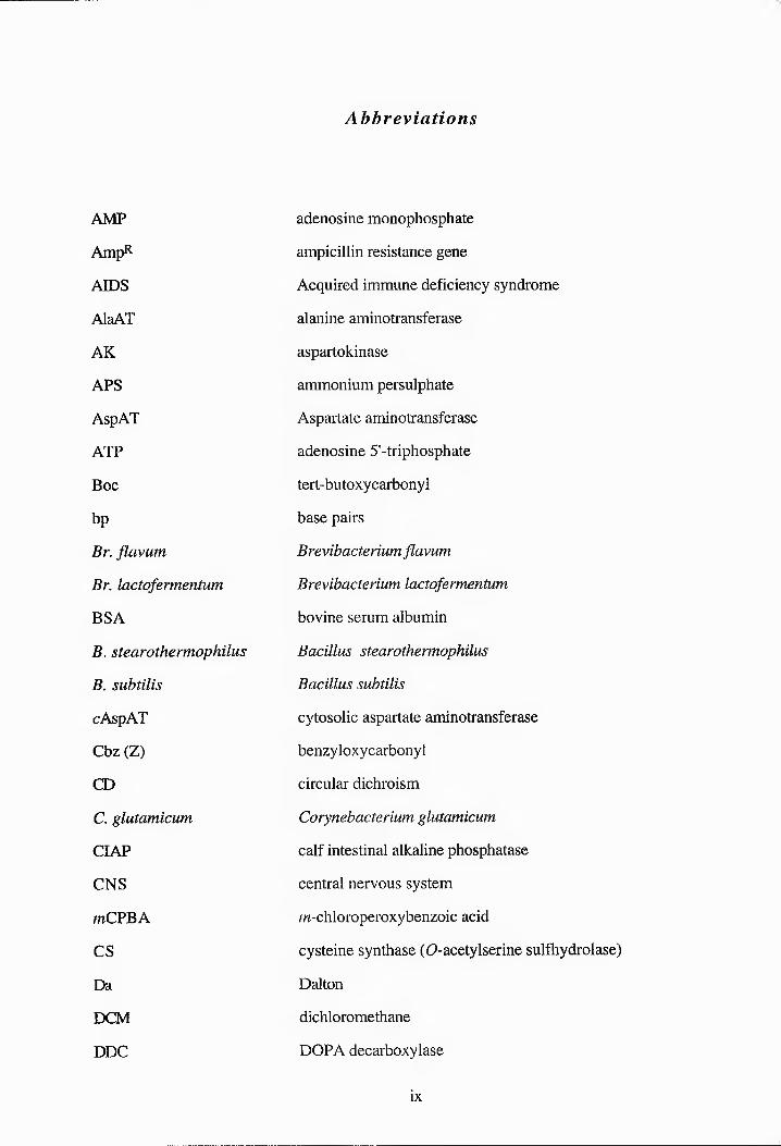
Abbreviations
AMP adenosine monophosphate
AmpR ampicillin resistance gene
AIDS Acquired immune deficiency syndrome
AlaAT alanine aminotransferase
AK aspartokinase
APS ammonium persulphate
AspAT Aspartate aminotransferase
ATP adenosine 5'-triphosphate
Boc tert-butoxycarbonyl
bp base pairs
Br. flavum Brevibacterium flavum
Br. lactofermentum, Brevibacterium lactofermentum
BSA bovine serum albumin
B. stearothermophilus Bacillus stearothermophilus
B. subtilis Bacillus subtilis
cAspAT cytosolic aspartate aminotransferase
Cbz (Z) benzyloxycarbonyl
CD circular dichroism
C. glutamicum Corynebacterium glutamicum
CIAP calf intestinal alkaline phosphatase
CNS central nervous system
mCPBA m-chloroperoxybenzoic acid
CS cysteine synthase (D-acetylserine sulfhydrolase)
Da Dalton
DCM dichloromethane
DDC DOPA decarboxylase
ix

ddNTP chain terminating dideoxyribonucleotide triphosphates
DEA diethylamine
DIPEA diisopropylethylamine
DMAP 4-dimethylamino pyridine
DME 1,2-dimethoxyethane
DMF A,A-dimethylformamide
DMSO dimethylsulfoxide
DNA deoxyribonucleic acid
dNTP deoxyribonucleotide triphosphates
DOPA 3,4-dihydroxyphenylalanine
dsDNA double-stranded DNA
DSDHT D-serine dehydratase
DTP dithiothreitol
EC Enzyme Catalogue
E. coli Escherichia coli
EDTA ethylenediaminetetraacetic acid
eq equivalents
Et3N triethylamine
Etp diethyl ether
GAEA y-aminobutyric acid
GAEA-T y-aminobutyric acid transaminase
HDC histidine decarboxylase
HEPES (/V--2-hydroxyethyl]piper£azne--V'-[2-etha.nesulfonic acid])
H. influenzae Hermophilus influenzae
HK homoserine kinase
HRMS high resolution mass spectrometry
HSD homoserine dehydrogenase
IECF isobutylchloroformate
IPTG isopropyl-P-D-thiogalactopyranoside
X

IR infrared
LMP low-melting point
kb kilobases
Kd dissocation constant
kDa kilo Dalton
K enzymic inhibition constant
Km Michaelis constant
KOH potassium hydroxide
lacZ gene for P-galactosidase
LB Luria-Bertani medium
LSDHT L-serine dehydratase
mAspAT mitochondrial aspartate aminotransferase
M. glycogenes Methylobacillus glycogenes
M. leprae Mycobacterium leprae
Mr relative atomic mass
mRNA messenger RNA
M. tuberculosis Mycobacterium tuberculosis
NAD nicotinamide adenine dinucleotide
NADP nicotinamide adenine dinucleotide phosphate
N. crassa Neurospora crassa
Ni-NTA nickel-nitrilo-tri-acetic acid
NMDA A-methyl-D-aspartate
NMM N-methylmorpholine
NMR nuclear magnetic resonance
OASS D-acetylserine sulfhydrylase (cysteine synthase)
ODC ornithine decarboxylase
ompT outer membrane protease
0/N overnight
ORF open reading frame
xi
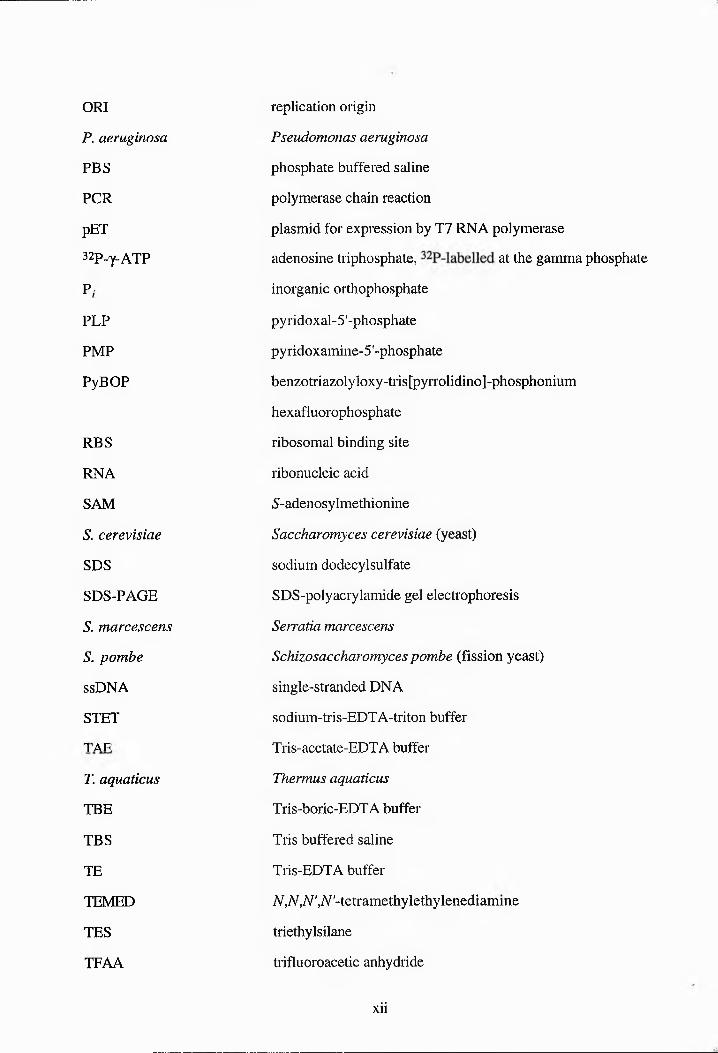
ORI replication origin
P. aeruginosa Pseudomonas aeruginosa
PBS phosphate buffered saline
PCR polymerase chain reaction
pET plasmid for expression by T7 RNA polymerase
32P-y-ATP adenosine triphosphate, at the gamma phosphate
P/ inorganic orthophosphate
PLP pyridoxal-5'-phosphate
PMP pyridoxamine-5'-phosphate
PyBOP benzotriazolyloxy-tris[pyrrolidino]-phosphonium
hexafluorophosphate
RBS ribosomal binding site
RNA ribonucleic acid
SAM 5-adenosylmethionine
S. cerevisiae Saccharomyces cerevisiae (yeast)
SDS sodium dodecyl sulfate
SDS-PAGE SDS-polyacrylamide gel electrophoresis
S. marcescens Serratia marcescens
S. pombe Schizosaccharomyces pombe (fission yeast)
ssDNA single-stranded DNA
STET sodium-tris-EDTA-triton buffer
TAE Tris-acetate-EDTA buffer
T. aquaticus Thermus aquaticus
TBE Tris-boric-EDTA buffer
TBS Tris buffered saline
TE Tris-EDTA buffer
TEMED A,A^,.A',A'-tetramethyiethyienediamme
TES triethylsilane
TFAA trifluoroacetic anhydride
xii

TFA trifluoroacetic acid
THF tetrahydrofuran
thr threonine operon
thrA aspartate kinase I- homoserine dehydrogenase I
thrB gene for homoserine kinase
thrC gene for threonine synthase
tic thin layer chromatography
T4 PNK T4 polynucleotide kinase
tRNA transfer ribonucleic acid
TrS tryptophan synthase
TS threonine synthase
UV ultraviolet
DV Vh/Vd
DV/K (VhWd)/(Kh/Kd)
X-gal 5-bromo-4-chloro-3-indolyl“P-D-galactopyranoside
Z-APPA (25,)-2-ammo-5-phosphono-3-cw-pentenoic acid
3D 3-dimensional
xill
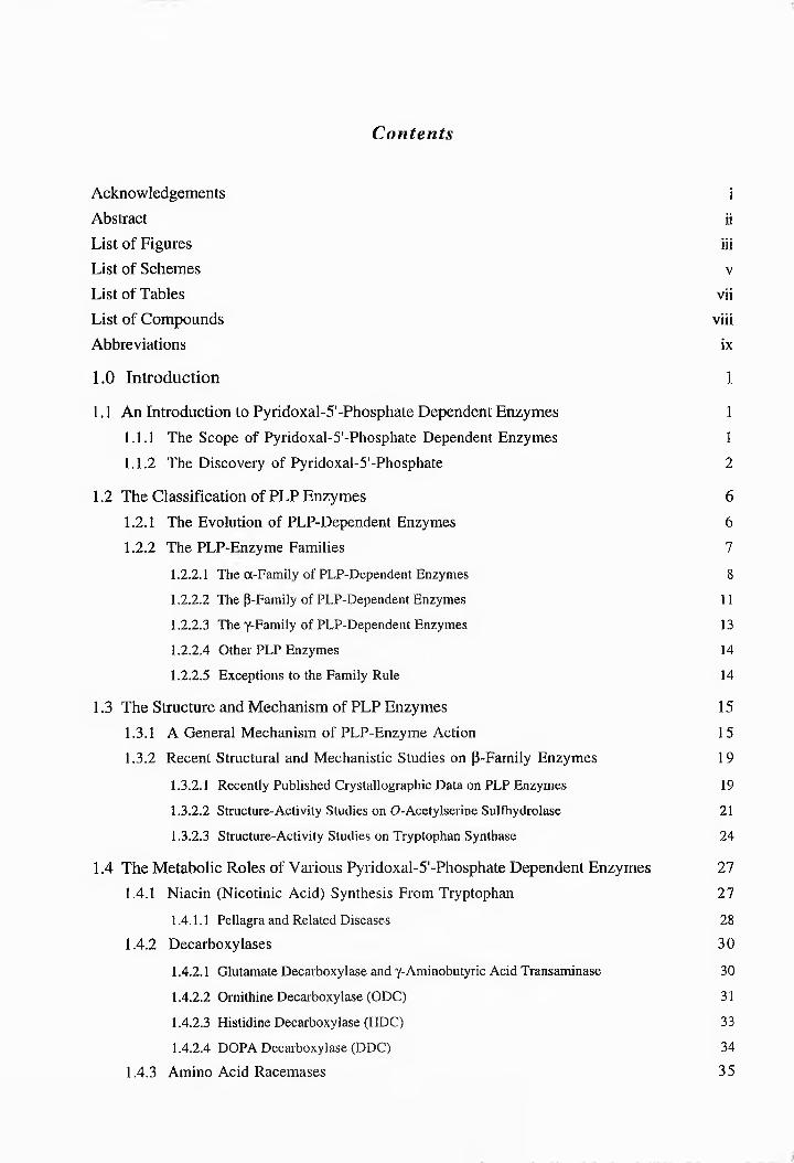
Contents
Acknowledgements i
Abstract il
List of Figures iii
List of Schemes v
List of Tables vii
List of Compounds viii
Abbreviations ix
1.0 Introduction 1
1.1 An Introduction to Pyridoxal-5'-Phosphate Dependent Enzymes 1
1.1.1 The Scope of Pyridoxal-5’-Phosphate Dependent Enzymes 1
1.1.2 The Discovery ot Py^rTi^o^^^l-l^'-l^lic^^i^li^l^ 2
1.2 The Classification 0! PLL Enzymee 6
1.2.1 The Evolutton of PL^I^-I^^t^^tlt^e^t^t Enzymes 6
1.2.2 Thh PILLlXn^^y^y^^ Famiiies 7
1.2.2.1 The a-Family of ILL-Dependent Enzymes 8
1.2.2.2 The (-Family of LLL-Deprzdrzt Enzymes 11
1.2.2.3 The y-Family of LLL-Depezdrzt Enzymes 13
1.2.2.4 Other ILL Enzymes 14
1.2.2.- Exceptions to the Family Rule 14
1.3 The Structure and Mechanism of ILL Enzymes 15
1.3.1 A General Mechanism of PLI-Enzyme Action 1 5
1.3.2 Recent Structural and Mechanistic Studies on P-Family Enzymes 19
1.3.2.1 Recently Published Crystallographic Data on LLP Enzymes 19
1.3.2.2 Structure-Activity Studies on O-Acety-serize Sulfhydrolase 21
1.3.2.3 Structure-Activity Studies on Tryptophan Synthase 24
1.4 The Metabolic Roles of Various Lrridfxal-5'-Lhfsphatr Dependent Enzymes 277
1.4.1 Niacin (Nicotinic Acid) Synthesis From Tryptophan 27
1.4.1.1 Lrllagra and Related Diseases 28
1.4.2 Decarboxylases 30
1.4.2.1 Glutamate Decarboxylase and r-Amizfbutyric Acid Transaminase 30
1.4.2.2 Ornithine Decarboxylase (ODC) 31
1.4.2.3 Histidine Decarboxylase (HDC) 33
1.4.2.4 DOLA Decarboxylase (DDC) 34
1.4.3 Amino Acid Racemases 35

1.4.4 The Aspartate Pathway
1.5 Introduction to Threonine Synthase
1.5.1 The Discovery of Threonine Synthase1.5.2 The Associated Deaminase Activity of Threonine Synthase1.5.3 The Regulation of Threonine Synthase
1.6 The Structure and Character of Threonine Synthase from Various Organisms
1.6.1 Microbial Sources of Threonine Synthase1.6.2 Threonine Synthase from Plant Sources1.6.3 Threonine Synthase Expression in Mammalian Systems1.6.4 Threonine Synthase as a Potential Target Enzyme for Herbicides
1.7 The Threonine Operon of Escherichia coli
1.7.1 The Structure of the Operon1.7.2 The Regulation of Thr Expression1.7.3 Cloning of 77zr
1.8 The Mechanism of (25,37?)-Threonine Synthesis
1.8.1 Introduction1.8.2 Reaction Mechanism1.8.3 Unanswered Questions Concerning the Threonine Synthase Reaction
1.9 Inhibitors of Threonine Synthase
1.9.1 Functional Groups Which Inhibit PLP-Enzymes1.9.2 Amino Acid Inhibitors of Threonine Synthase1.9.3 Inhibition of Threonine Synthase by Various Anions1.9.4 Phosphonic Acid Analogues of (2S)-(9-Phosphohomoserine1.9.5 Phosphate Analogues
1.10 The Aims of This Project
1.10.1 Testing New Substrate Analogues as Inhibitors of Threonine Synthase1.10.2 Product Inhibition Assays1.10.3 Further Assessment of the Allosteric Activation of TS by SAM1.10.4 Structure-Activity Studies on Threonine Synthase
1.11 pET Expression Vectors
1.11.1 Introduction1.11.2 T7 RNA Polymerase1.11.3 Expression of Target Genes1.11.4 Vectors Selected For Expressing Wild-Type and Mutant ThrC
36
37
373839
40
40424444
45
454647
48
484855
56
5657596062
63
63646565
66
66666768

2.0 Results and Discussion 69
2.1 Substrate Synthesis 69
2.1.1 A Survey of the Literature Methods for Synthesising (2S)-0-
Phosphohomoserine.
69
2.1.2 The Biomimetic Synthesis of -2S))“6-Phosphohomoserine. 71
2.1.3 Syn^ies^ of [[B14CC^(-(2)^<2^-IPlo.sihlc^lh<^Im^^i^trin^. 74
2.2 The Growth of Threonine Synthase 76
2.3 Cloning of ThrC and the Gene for p-Methylaspartase 77
2.3.1 The General Clonnng Strategy 77
2.3.2 Cloning oo ThrC 79
2.3.2.1 Amplification of ThrC 79
2.3.2.1.1 Iehiaiihn of C0rhmhehmal DNA 79
2.3.2.1.2 Polymerase Chain Reaction on 7/zrC 79
2.3.2.2 Attempted Ligation of ThrC Into Cloning Vectors 81
2.3.2.3 Overlap Polymerase Chain Reaction Experiments. 83
2.3.2.4 Ligation of ThrC into pGEM-T. 84
2.3.2.5 Attempted Ligation of ThrC Into Expression Vectors. 87
2.3.2.5.1 pET-SaandpET-^b. 87
2.3.2.5.2 pKK223-3 87
2.3.2.6 New PCR Fragment. 88
23.21 Attempted Ligation of ThrC into pET-3a. 89
2.3.2.8 Liggtioo oo New Geee Fraamoet IntepGEM-T. 90
2.3.2.8.1 Amolificctioh cd TTirC 90
2.3.2.9 Sequencing of ThrC in hEEM-TT8i and hEEM-TTSIi. 92
2.3.2.10 Ligation of ThrC Into hET-Exhrreeioe Vectors. 94
2.3.3 donmg oo -he p-Methylalpafiase Gene 98
2.3.3.1 Cloning the Gene Into hEEM-T 98
2.3.3.1.1 Gene Amplification 98
2.3.3.1.2 Overlap PCR Exhrrimreii 99
2.3.3.1.3 hEEM-T 100
2.3.3.1.4 A New Erer Fragment 101
2.3.3.1.5 New hEEM-TMA Cheetrucie 102
2.3.3.2 Further Work on the Erer for h-MrtOylaehariasr 103
2.4 Sequencing of the ThrC Gene Fragment in Recombinant Plasmids 104

2.5 Attempted Solid-Phase Synthesis of (2S)-0-Phosphohomoserine 106
2.5.1 Aitsrnstivs Pefepefryisting Agents 106
2.5.2 19F Gel-phsee NMR Spectroscopy 111
2.5.3 p-Bsnzyifxyibsnzyi Alcohol (Wang) Resin 112
2.5.3.1 The Loading of Wang Resin With i^-"^TiSfiu^l•ifs^<^t^h^li^((^.^')-^i^^<spsI•tsts 114
2.5.3.2 p-Hydroxymsthyipoiyetyrsns Resin 121
2.5.3.3 Attempted Reduction of a,-/;-Benzyloxylbsnzylpoiystyrene-N- 122
trifisorfscetyl-(25--aspsrtate
2.5.3.4 Experiments to Confirm the Activation of the Carboxylic Group on 87 128
2.5.3.5 Reduction of a-t?-Benzyloxylbenzylpolyetyrsns-W-Trifisorfscstyl-(2.5-- 129
aspartate
2.5.4 A New Solid Support 132
2.5.4.1 Attachment of 2-(p-Hydroxypesnyi--stesnoi to Merrifield Resin 132
2.5.4.2 Loading of the New Resin With Y-trifisfroscstyi-(25)-separtsts 137
2.6 Conclusions and Future Work 142
3.0 Experimental 1493.1 Synthesis 149
3.1.1 General Experimental Procedures 149
3.1.1.1 Compound Characterisation 149
3.1.1.2 Reagent and Solvent Preparation 150
3.1.1.3 Suppliers 151
3.1.2 Synthetic Procedures and Characterisations 152
3.2 Moleeutas Clonmg 169
3.2.1 Cloning Techniques 169
3.2.2 Polymerase Chain Reaction 173
3.2.3 Oligonucleotides 175
3.2.4 Expression in E. coli 176
References 179
Appendices 191

CHAPTER ONE
INTRODUCTION

Introduction to PLP Enzymes -Scope of PLP Enzymes
1.1 An Introduction to Pyridoxai-5’-Phosphate Dependent Enzymes
1.1.1 The Scope of Pyridoxal-5’-Phosphate Dependent Enzymes
A wide variety of enzymes responsible for amino acid metabolism utilise pyridoxal 5'-
phosphate (PLP, 1) as their cofactor. The enzymes dependent on PLP include the
transaminases1 (or aminotransferases), through whose action the Ca-N bonds of most amino
acids are formed, the racemases,2 many of which are involved in the biosynthesis of
peptidoglycan in bacterial cell walls, and the decarboxylases which convert amino acids to
amines (Scheme 1.1).2 The latter function is one of the most important physiological roles of
PLP.
Pyridoxal-5'-phosphate (1).
There are over 66 known transaminases altogether as well as 50 different decarboxylases. All
the aformentioned enzymes alter a substituent at the opposition on the substrate. Many other
enzymes which are PLP-dependent, in addition to first removing the a-proton of the amino
acid, also eliminate or replace groups which are in a P- or a y-position. The numerous
examples of PLP-dependent enzymes indicate that PLP is one of nature’s most versatile
coenzymes; playing a crucial role in amino acid metabolism/ connecting the carbon and
nitrogen cycles and providing entry into the ‘one-carbon pool’. PLP also participates in the
formation of biogenic amines as well as a number of other important processes/ The
versatility of this coenzyme is reflected in the fact that PLP-dependent enzymes occur
1

Introduction to PLP Enzymes Discovery of PLP
in four of the six EC classes of enzymes.'
NH2H/J
{OCX
PLP■ “ — -1'
NH2
decarboxylases
NH2H/J
HChC^X
PLPX
HCfcC^Xaminotransferases
NH2H/J
HO2C*^X
PLP NH2HA
HChC^Xracemases
nh2HO2C>VcO2H
PLP nh21
serine hydroxy methyl transferase
HOzC^
Scheme 1.1: Some types of reaction catalysed by PLP-dependent enzymes.
1.1.2 The Discovery of PLP
PLP is a metabolite of vitamin Bg (or pyridoxol (2), see Scheme 1.2), a nutritional factor which
was first identified in 1934 by Paul Gyorgy.7 In the early 1930s, when purified preparations
of thiamine (Bj) and riboflavin (B2) first became available, it became appareer that a separate
heat-stable vitamin, later found to be vitamin Bg, could arrest the development of a pellagra-like
dermatitis (similar to acrodynia) in rats (Fig. 1.1).7 In fact, as early as 1926, Goldberger and
Lille had noted that rats given a diet deficient in a so-caned 'pellagra-preventative factor'
developed this condition^ In 1932 Ohdake isolated a eitrhgenhus compound from rice
polishings, but no vitamin significance was attached to it at the time.7 It was only later that it
was shown to be identical with vitamin B7. Birch and Gyhrgy studied the properties of the
vitamin in 193677 and subsequent to this the compound was isolated independently by five
2
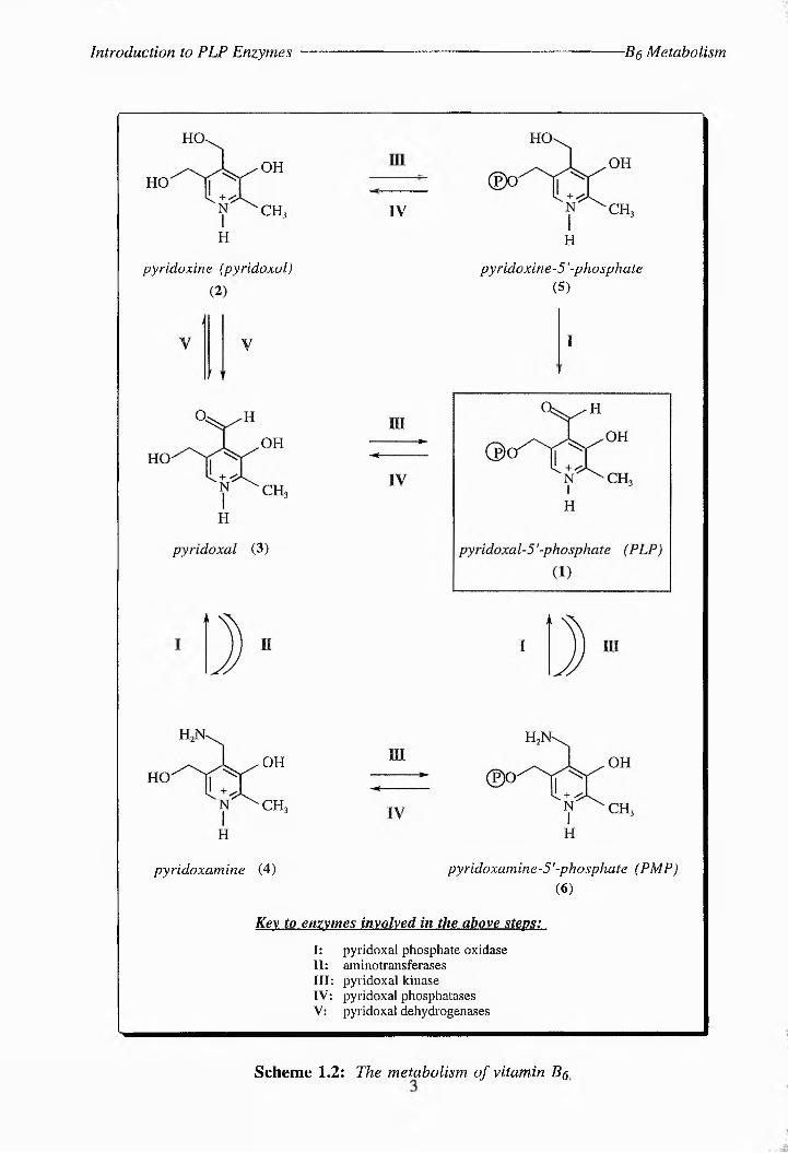
Introduction to PLP Enzymes -Bq Metabolism
OH
CH, IV
H
pyridoxine (pyridoxol) pyridoxine-5 '-phosphate (5)
OH
ch3
I
pyridoxamine (4) pyridoxamine-5 '-phosphate (PMP) (6)
Key to enzymes involved in the above steps:
I: pyridoxal phosphate oxidaseII: sminotransfsi'ssss III: pyridoxal kinase IV: pyridoxal phosphatases V: pyridoxal dehydrogsnasse
Scheme 1.2: The metabolism, of vitamin Bq

Introduction to PLP Enzymes Codecarboxylase
laboratories in 1938 and its structure and synthesis were reported in 1939 when it was named
pyridoxine.1114
■fa.
•. •-■ ■■S;>v\s-:4V,t /:■
wo
Figure 1.1: Dermatitis caused by pellagra.*
Originally the cofactor was named 'cfdecsrbfxyisee' by many research workers as a result of
its ability to reactivate the resolved inactive apoenzyme of (25--lysine decarboxylase.15 It was
not until Bellamy and Gunsalus were investigating the prfpsrtiss of tyrosine decarboxylase that
the link between the nutritional factor Bfi and 'cfdecsrbfxylsse' was made.'6 In their
experiments media containing larger amounts of pyridoxine (2- and nicotinic acid than were
necessary for growth of bacteria, were found to give high yields of the active enzyme. A
couple of years later Snell realised that vitamin B£ occurs in multiple free forms, and by
studying the non-enzymic transaminations between pyridoxal (3- and amino acids he provided
the first clues to the function of the vitamin. 16 The reaction shown in Scheme 1.3 was shown
to occur in the case of ^--glutamic acid, and the data suggested that a similar reaction could
a reproduced from wsb-eits: http://www.mc.vandei'bilt.edu/bioiib/el/pd4.htmi
4

Introduction to PLP Enzymes ■The First Known PLP Enzymes
take place between pyridoxal (3) and certain other amino acids. It is now known that most
amino acids are biosynthesised in this manner from (25)-glutamic acid and the corresponding
a-keto acid via the action of transaminases.
Scheme 1,3: Snell's "Shuttle Mechanism" for enzymic transamination.
The next decade witnessed the identification of tryptophanase, (2/?)-serine dehydratase and
cysteine desulfhydrase as PLP-enzymes.2 The availability of further enzymes for study meant
that much stereochemical, kinetic and primary structural information about PLP enzymes could
be gained, providing insights into the general mechanisms of such enzymes and the role of
PLP. 3D structures of most PLP enzymes and their enzyme-substrate complexes are,
however, still scarce, except in the case of the transaminase group of enzymes.
5

Classification of PLP Enzymes- Jensen's Hypothesis
1.2 The Classification of PLP Enzymes
1.2.1 The Evolution of PLP-Dependent Enzymes
Ail the PLP enzymes studied to date share certain similarities in the preliminary stages of their
reaction mechanisms (see also Section 1.3). In 1974, Dueathae and Voet also noted that
several PEP-dependent enzymes convert the bound cofactor to hyridhxamiee phosphate (6)
during normal or abortive rraesamiearihe reactions and that this proceeds sterehspecifically,
with the same absolute stereochemistry in seven quite different PLP-enzymes.18 The key step
in this reaction is the prhthearihe of the bound substrate-pyridoxal phosphate Schiff base (7) at
the C-4' carbon of the cofactor, from the A-si face, after loss of one of the groups at the
substrate Ca (Scheme 1.4). They inferred that this demonstrated a remarkable regularity in the
geometry of the binding of the cofactor to the apoenzyme in all PLP enzymes, and interpreted
this as evidence for the evolution of this entire family of enzymes from a common progenitor.
Jensen's hypothesis supports this theory, as it suggests that ancestral organisms produced a
relatively small number of enzymes with flexible substrate specificity, which gradually mutated
into more selective enzymes.19
Scheme 1.4; Conversion of bound cofactor to pyridoxamine phosphate- a process common
to many PLP-enzymes. Ha-elimination is illustrated here.
6

Classification of PLP Enzymes- ■Homologies
However, not only do PLP enzymes share many mechanistic similarities, they also demonstrate
definite sequence and structural homologies. Mehta et ai found that most aminotransferases
formed a group of homologous proteins, which could then be divided further into distinct
evolutionary subgroups on the basis of their sequence similarities (see Section 1.2.2.1).20
Subsequent profile analysis to detect distant relationships between amino acid sequences of
various enzymes showed that the evolutionary relationships included other Be enzymes.21-23
In 1994 Christen and co-workers carried out a comprehensive analysis of the amino acid
sequences of PLP enzymes and of their 3D structures and, as a result, proposed that most of
the enzymes could be assigned to one of three different families of homologous proteins; the
a-, p- or y-families.20 Although the sequence similarity is too low in some cases to allow
meaningful alignments by standard programmes, hydropathy plots and secondary structure
predictions do indicate evolutionary relationships between PLP enzymes.25 Their affiliation
with one ' of these structurally defined families also correlates in most cases with their
regiospecificity.24 The folding pattern of the polypeptide chain of the y-family enzyme is
similar to that of aspartate aminotransferase (AspAT), further suggesting an evolutionary
relationship between the a- and y-families.24 Christen et. al also cited various reports of the
side reactions catalysed by Be enzymes as further evidence for a common ancestral enzyme
with broad substrate specificity.22 Decarboxylases catalyse transamination, aminotransferases
cause racemisation, tryptophan synthase (TrS) produces racemisation and transamination and
glycine hydroxymethyltransferase also initiates a-decarboxylation, transamination and
racemisation.
1.2.2 The PLP-Enzyme Families
The largest family of PLP-dependent enzymes, the a-family, contains enzymes catalysing
reaction at Ca of the substrate. Similarly, the P-family contains those acting at CP and the y-
family those acting at CY. There are, however, some exceptions to this general rule, and some
PLP-dependent enzymes do not seem to belong to any of the groups.
7
Classification of PLP Enzymes- ■The a-Family
1.2.2.1 The a-Family of PLP-Dependent Enzymes24I
i The a-family is the largest of all the PLP-dependent groups and includes the aminotransferases
(ATs) and the group II decarboxylases (DCs). In spite of the low level of sequence similarity
between members of the a-family, (only two residues are conserved throughout the a-family;
the lysine and aspartate residues which bind PLP),25 many of these enzymes have 3D
structures available for comparison and the conformational similarities between different types
of enzyme can clearly be seen. John reviews these similarities in more detail, but they include
eight or nine a-helices packed around a seven-stranded (3-sheet in the large domain.25 In the
a-family the PLP-binding lysine residue occurs between residues 209 and 256 of the various
members of this family, which are listed in Table 1.1.
Table 1.1: PLP-dependent enzymes belonging to the a-family.
Most aminotransferases (excluding branched chain and D-amino acid) 20> 2^ Group II amino acid decarboxylases
Tryptophanase Tyrosine phenol-lyase^
Glycine hydroxymethyltransferase I -aminocyclopropane-1 -carboxylate synthase^
2-amino-6-caprolactam racemase
Glutamate-1 -semialdehyde 2,1 -aminomutase
Isopenicillin N-epimerase 2,2-dial/cyl decarboxylase^
4-amino-4-deoxychorismate synthase^'
Glycine-C-acetyltransferase
5-Aminolevulinate synthase
8-Amino-7-oxononanoate synthase
The gene product of cobC (cobalamin synthesis)
The gene product o/nifS (nitrogen fixation)
The gene product ofmsXY (abolishes induction of the maltose system)
The most intensely studied members of this family are the ATs. In their studies on the
evolutionary relationship between ATs, Mehta and co-workers discovered 107 invariant
8
Classification of PLP Enzymes The a-Family
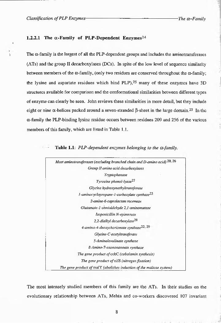
residues between the cAspAT and mAspAT sequences from, vertebrates, which have an average
length of around 400 amino acids.26 Their average pairwise identity was 41%. The active-site
sequences of AspATs from both prokaryotic and eukaryotic sources have been aligned here
showing the conservation of various motifs, including the lysine residue for binding PLP (Fig.
1.2).
I******** * * * * ** * * * * *******
A ----------- HHIGISQSYA1MMGLYGQRB OAHnPTCT^EPIlLIQkTSI^RQ^a^RDK^-i^lU^PFFDSAYQDFASDSIjDADAQPVDLFVADD---------- DILLVAQSYAKNMDLYDIDC aAHffPR3TT^DTn^l^^QQIAA^tRRR^I^I^I^I^]FDSAYQDlFASDSI^DKDAWAVRYF^^]I3----------- FILFCAQSFSKNFDLYNEDD <CAHN^T^-V)Pl^BOWB^II^C^ILMRS-f^]L^]^^FDSAYQCa^2^1^<DSLDjAD^C^fSVDC;FVA]D3----------DICLAAQSYAKNMDLYeEDI CAAHJPTCIDPTPIQWQIASSVKRR-FFLPFFDSAYQGFASDLERDAWAIRYYVSEG-----------FEFFCFQSFSQNFGLYNEDI CIFHTP^TCV>PTLDQIAIIRRILMPS-.-FJLPFFDSF.YQGFFSDSLDQDFQSVPMIVFD----------DILLMAQSY7Q3SMGLYDIP.e CIAHJPPG,TDPTPIQWAQIA£SVKQRpFLFPFFDSAYQGFASDNLIQDAWAIRYFVSEG-----------FILFCAQSFSQNFGLYNEDH CEFHNP1GLDPTSEQWAQIVVAFASK~NHIALFIT^AYIGFFTD;DLDQDAYAVRLGVEIQJSTV SPVFVCQSFAQNADMYGERI <CFHN>TGmPRQEQI^JQ:AIFSAQKp3NLGYFEMFYQFASGDIAPDAFAALPHIE$G---------- IDAGSSSSFQKMGLYGERJ CIAHPPGDOTRPEQWKEIATTAQRpIMiLAFFFMAY$3FASGID3DKDDAA\VD^FILIQ----------- INVCIJCQSYA^NMGLYGEPQ CIAH'IPPGIDPTPEQAEKIADVIQI—-SIH^IP'FFDVFYQGFASDSLDI'DFFSVDLFVF.DG---------- LEVLVF.QSYSQNLGLYFEDL CIFHIP1AVDPPIQQWIIMFTLVQQN-MLFFF'FDMAYQGFFSGEGNQDFWAFVRHIE<I3---------- I^VICLCQSYA!SMGLYGEPM CCHOTTILDPTQIQL'raKIIDTIYLL-TJYPPIVDVYQDGISSGlLuLlDAYLLRLLLVVQKYSIASNGILCQQSFAISMGLYGER
* ** * . . **. *.***N PS-NPTGVMY^EEELS]FGeICCLIHDILIVSDIIYEQLTYDGK-QH/SIFQLSDDL:------------ IQTVIlNGSSKSHSITDADO P.S-N]PSGPFYSHEELIAG7TD\IMMPHVAV1L^DDMMYIHLYGDF-RFATPVEVEPGLY----------- IDTL^MNGVSKFYFMDA^PP PS-NPTGMVYRDELLIIAF-M.,LN-INILIVSDIIYE-GLYNGA-EHFSIF.QISEEVC---------- AQTIVnflVSQKHHMTrcADQ PS-NPGMIYTFI:ELKALGIVC]GA^-G\VJIVSDEIYIQLTY(D3A-QEH/SIAELSPELQ---------- AQTVIINCVSS-SSHSMDADD <CCHNPI3IDPTLEQAQlGFQLSVK-GDAPLLDFAFY^eFF3-A-EIHSSAQISEEVK---------- AQTIVINGV-KSHSITDADS CCIHN)TCIDPTPIQWAQILFFLSKQ-GGALPLFFAFYG:IDLFG-AFFQHSSIF;LSPELK----------AQTVTNDVS-KHSSITKADT P-HNPTCTLFSPNDVKQTOISPDN-QIILLSDEIYDIFVYffiGQ---------- MRSTLEDSD ADOFLIYVNDFSKTF]M^DAJR
** * * * * *
A - Arabidopsis thaliana (mouse-ear cress), 277; B - Medicago sativa (alfalfa), 264; C - cAspAT Callus gallus (chicken), 257; D - cAspAT Daucus carota (carrot), 251; E - cAspAT Homo sapiens (human), 258; F - cAspAT Oryza sativa (rice), 253; G - cAspAT Sus scrofa (pig), 258; H - cAspAT S. cerevisiae (baker's yeast), 254; I - mAspAT G. gallus, 272; J - mAspAT H. sapiens, 279; K - mAspAT Lupinus angustifolius (narrow-leaved blue lupine), 299; L - mAspAT S. scrofa, 279; M - mAspAT S. cerevisiae, 286; N - Bacillus suhtilis, 237; O - Rhizobium meliloti, 239; P - Bacillus sp. (strain ym-2), 239; Q - B. stearothermophilus, 237; R - E. coli, 246; S - H. influenzae, 246; T - Sulfolobus solfataricus, 241.
Sequences A - M are eukaryotic, sequences N - T are from prokaryotes. Residue number of the active-site lysine is shown in blue. Asterisks denote identical residues, periods denote conservative replacements (e.g. I-V- L-M-F, D-E, R-K, S-T). For the eukaryotic sequences, identical residues are highlighted in red. For the prokaryotic sequences, identical residues are highlighted in blue. The homology of each set of sequences is also indicated- by asterisks and periods above the sequences. The overall homology between all the AspATs aligned here are indicated by the asterisks and periods underneath both sets of sequences. The lysine residue responsible for binding to PLP is indicated by an arrow. The alignment was performed using Clustalw (1.60).
Figure 1.2: Sequence homology between various eukaryotic and prokaryotic AspATs. close
to the active-site lysine residue.

Classification of PLP Enzymes- The a-Family
When the various vertebrate ATs were aligned the percentage identity ranged from as low as
5% up to 75%, whereas the percentage similarity (which accounts for conservative
replacements- ranged from 25% to as high as 87%.20 Four residues were conserved
throughout the sequences: G197 which participates in a p-turn at the domain interface, D/E222
which forms a esit-bridgs or hydrogen bond to N1 of PLP, K258 which forms a Schiff base
with PLP and R386 which forms a hydrogen bond with the a-csi'hfxylate of the substrate.20
Mehta reviews various site-directed mutagenesis experiments to replace these residues in E. coli
AspAT which resulted in significant reductions in the catalytic activity of the enzyme. 20
On the basis of sequence similarities the ATs could be divided into four subgroups. 11
residues were invariant in subgroup I, 19 in subgroup II, 82 in subgroup III and 25 in
subgroup IV.20 Jensen and Gu have subsequently reviewed the evolutionary relationship
between 47 group I ATs and proposed seven new subfamilies on the basis of sequence
alignments.20 They suggested that these originate from a single ancestral broad specificity AT.
Another study has demonstrated structural homology between serine eydroxymethylti•ansfsrase
(SHMT- and AspAT, with 66% of aligned residues around the central and C-terminal regions
predicted to share the same conformation^1 Bossa also cited a previous observation of weak
sequence similarity between the active-site regions of decarboxylases and various PLP
enzymes, including SHMT.31 In addition to this, the NifS group of proteins (responsible for
nitrogen fixation in bacteria- appears homologous to a new class of PLP-snzymes, which
includes various aminftranefsrsses and isopenicillin N epim^i's^i^s^52 although they all have very
different functions. The sequence identity between distant members within this class is below
25%, but secondary structure predictions indicate that this class of enzymes might have an
overall a-p topology. Sander and Ouzounis also reported sequence similarities of around 30%
between mammalian peroxisomal serine-pyruvate aminotransferases and the small subunit of
soluble hydrogenases.32 Most recently Babbitt and Gerlt have observed sequence similarities
between 5-ksto-4-defxyglucarate dehydratase/decarboxylase and various dehydratases and
aldflases.33 In a separate study on decarboxylases Kang and Joh have demonstrated significant
homologies between porcine and bovine L-amino acid decarboxylases and feline glutamic acid
10
Classification of PLP Enzymes- The p-Family
decarboxylase and tyrosine decarboxylase from C. roseus (31% identity beween all
sequences).34 Christen et al. have more recently compared sequences from nine different DCs
and divided them into four subgroups:.35 However, only group II demonstrated a distant
relationship with the ATs. No evidence was obtained that the other DCs were in any way
related to other PLP enzymes and profile analysis suggests that the decarboxylases have
evolved along multiple lineages, even if they have the same substrate specificity.
1.2.2.2 The P-Family of PLP-Dependent Enzymes24
This family has seven members including threonine synthase (TS), which are shown in Fig.
1.3. Although few crystal structures are available for the comparison of the tertiary stmctures
of these enzymes, their sequences do show significant homology. Parsot has demonstrated
extensive - homology between the TS enzymes of E. coli and B. subtilis, threonine dehydratase
(TD) from S. cerevisiae and D-serine dehydratase (DSDHT) from E.coli.^ There are 72
conserved amino acid residues (21%) and 40 conservative replacements between TS from B.
subtilis and TD from yeast; an overall homology of 33%. Similar results were noted between
the sequences of yeast TD and DSDHT of E. coli. As a result he postulated the evolutionary
connection of these enzymes. Bork and Rohde also aligned various p-family enzymes from a
number of microorganisms and identified three regions of significant homology in the
sequences including a lysine motif and a glycine-rich turn, which are illustrated in Fig. 1.4.56
They proposed a new alignment, which revealed a similar folding topology in long segments of
the enzymes studied. The pairwise identities ranged from 15% to 19%. They also found that
the sequences of TrS and D-acetylhomoserine thiolyase, a y-family enzyme, had an amino acid
identity of 11% and suggested that there could exist an evolutionary relationship between these
two families, although the findings of Christen et al. refute this.24 Appendix 1 shows a more
extensive alignment of p-family enzymes from B. subtilis, E. coli and yeast whose pairwise
identities ranged from as low as 4% between DSDHT and TD of B. subtilis to 100% between
TD and TS from this source. The average pairwise identity was 15.7%, which increased to
24.9% around the active-site.
11

Classification of PLP Enzymes- ■p-Family Enzymes
(25--SERINE DEHYDRATASE EC 4.2.1.13 P-elimination, deamination
NH3
Fp‘*y\^0H + H2O"CbC
(2S)-serlne
(2R--SERINE DEHYDRATASE EC 4.2.1.14 P-elimination, deamination
CO2
H3N
OAc"PccH3
pyruvate
+ NH4 + H2O
-OH + H20
(2R)-serine
Ou-O2c *pcCh3
pyruvate
+ NH4 + H2O
THREONINE DEHYDRATASE EC 4.2.1.16p-elimination, deamination
+NH3
.CH3 + H2O ["H
OH( 2S, 3R)-threonine
O2C
oA.CH3 -f NH4
a-ketobutyrate
+ H2O
TRYPTOPHAN SYNTHASE EC 4.2.1.20 P-replacement
NH3
HO2C
OH
Indole Tryptophan(2S)-serine
THREONINE SYNTHASE EC 4.2.99.2 P, T-replacement
NH3I + H2O ---------------- 3
-O2^^X^OP°32'
(2S)-0-Phosphohomoserine
cysteine y-SYNTHASED-ACETYLSERINE (THIOL--LYASE EC 4.2.99.8 p-replacement
HO2C
NH3•« X.,
C
CH3
OH
+ HOPO3"
( 2S, 3R)-threonine
Y
O
cil + H2S
NH3
H “*;j O2C
SH
(2S)-cysteine
+ CH3CO2' + H+ Acetate
+ H2O
Figure 1.3: A summary of the reactions carried out by enzymes of the P-family of PLP-
dependent enzymes
12

Classification of PLP Enzymes The P-Family
cs__ bacill 39♦
----- PG-SJVKDRI-GLAMIEAAE- ----- K 58 167 DD-^QLDGFVGGIG-TGG^ITGGG^rLK E 199cs__ e_coli 36 —ps-fvkkcrIiGAMUWDGE- -----K 55 165 dg-qvdvfiggvg-tggtltgvsryikg-tk 183cs__ yeast 81 -----PG-ISGKDRV-GLNIIKTGE- ----- E 100 219 -KGNIDGFGGCG-GGTIrGVGKFL.JKERGK 248dsdht__ bac 111 HELPISGSIKRGGirEVLKYGENLGLQ 136 266 SLETPLFVYhPCGvVCGSGPIGVGFGLKLLYG 296lsdht -yea 34 -----pg-gsfksrg-ighlirksn- ---Q 53 171 SLPRVKGLVCJSVI-GGGLFSII IKGLDR-NH 200td__ bacill 54 ----- PT,-<SFKD^^MVMGVGKGK- ----- E 73 174 -GEGPDVLAIPVG-GGGNIGYWK<GFKEYHE 203td__ e_coli 57 ----- PV-HSKLRGG-AY7MMGGLT- ----- E 76 178 —~GHLDRWFVVG-GGIGAA(GWVLKKQMP 206td__ yeast 104 ----- PV-FSKRG-AYIMIGKLD- ----- D 123 225 TGNKIGAVFVPVG-GGGLIGGIGAYLKRVAP 256trs__ bac11 85 ----- HT-GSHKINN--LGQGLL-K~ ----- K 104 223 EGTM>D]KWACVG-GGSN-MCGM'QQ-?ILJ-ED 252trs e col 81 ----- HG-GGHKTNQ-VLGQALLGK- ----- R 100 219 EGRLPDAVIA<CVG-<G3S1N-I(GMF-)DFN-ET 248ts__ bacill 54 ----- PT'-CSFKKRG^-VMGVGKGI^C- ----- E 73 174 -GE-PDVLAIPVG-GGGNI-YWKGFKEYHE 203ts e coli 102 FHGPT-UFKDFG-GIFMGQMLI?- -----H 121 236 ETRNQLWSVPSGNFGDLTGGL]LG<SILI-LP 266ts__ yeast 119 ^^PT^TG^KDVG^LQFV^BLra-
*----- Y 138 265 KDSKKVKFWP^^^F^D^^IG
*296
The lysine residue responsible for binding to PLP is indicated by an arrow. The consensus sequence is given
below the other sequences. Homologous residues are indicated by a period, identical residues by an asterisk.
Figure 1.4: Sequence homology of p-family enzymes around the lysine motif and the
— glycine-rich turn,.
Further evidence of the evolutionary relationship between the P-family enzymes is found in
their ability to catalyse similar reactions and produce similar intermediates, suggesting a similar
active-site conformation. TS from B. subtilis and E. coli still exhibit some TD activity both in
vivo and in vitro and both TD and TS produce a-aminocrotonate as an intermediata.19 TrS
can catalyse the conversion of vinylglycine to a-ketobutyrate, a reaction normally carried out
by TS.37 TS also catalyses various side-reactions (Appendix 2).
In contrast to members of the a-family, in the P-enzymes the PLP-binding lysine is positioned
in the NF^-Terminal segment of the polypeptide chain between residues 41 and 118.24 This
difference may reflect the alternative PLP arrangement needed for the enzymes to carry out
reactions at the Ca-centre rather than at the cP-atom.
1.2.2.3 The y-Family of PLP-Dependent Enzymes24
The y-family contains four members and is therefore the smallest group of PLP enzymes (Table
13

Classification of PLP Enzymes ■Other ‘ PLP-Enzymes
1.2 ). Belfaiza et al. noted an overall homology of 36% (31% identiiy) between the two
cystathionase enzymes.38
Alexander et al. found that the PLP-binding lysine residue is in the same sequence segment as
in the a-family suggesting a possible evolutionary relationship between these two families.24
As stated earlier, significant sequence similarities between TrS (of the P-family) and O-
acetylhomoserine sulfliydrolase of this family have also been shown.36
Table 1.2: Enzymes of the y-family .
O-succrnylhomostrrme (thiolylyase (cystathionine-y^-synthase)
O-acetylhomoserine (thiol)-lyase (methionine synthase)
Cystathronine-y-l,yast (y-cystathromast)
- - Cystathionine-f-lyase (f-cystathromase)
1.2.2.4 Other PLP Enzymes
A few of the PLP-dependent enzymes studied by Alexander et al. proved to be unrelated to any
of the three family groups. These are alanine racemase, selenocysieine synthase and many
amino acid decarboxylases which are not in group 1124
1.2.2.5 Exceptions to the "Family Rule"
As already mentioned a few PLP-dependent enzymes do not belong to the family that would be
suggested by their regioselectivity. Tryptophanase, tyrosine phenol lyase and 4-amino-4-
deoxychorismate synthase are all members of the a-family by virtue of their sequences, yet
they catalyse P-elimination reactions.24 1-aminocyclopropane-1-carboxylate synthase, also an
a-family enzyme, catalyses an ay-replacement reaction. Similarly, TS, which is a member of
the p-family, catalyses a p,y-replacement and cystathionine p-lyase, supposedly a y-family
enzyme, catalyses a P-elimination reaction .24
14

PLP Enzymes - Structure and Mechanism- General Mechanism
1.3 The Structure and Mechanism of PLP Enzymes
1.3.1 A General Mechanism of PLP-Enzyme Action
Much of the information concerning the general mechanism of PLP-mediated amino acid
reactions and the structure of PLP enzymes has been gleaned from studies of the PLP-
dependent transaminases. In particular the action of aspartate aminotransferase (AspAT- has
received much attention. Between 1954 and 1955 two independent workers, Braunstein and
Snell, developed a general theory for the mechanism of PLP enzymes.39- 40 The following
section gives a detailed discussion of this mechanism, and examines the experimental data that
was used to prove it.
For all known PEP-enzymes the first reaction step involves the formation of the Michaelis
complex (10- otherwise known as the holoenzyme sidimine or internal aldimine between the e
amino group of a specific enzyme-bound lysine residue and the C-4' carbonyl group of PLP
(1, Scheme 1.5).4> 5
Upon formation of the hfioenzyme the amino acid substrate can bind to give the Schiff's base
substrate aldimine (or external aldimine, 7-41 This involves the condensation of the amino
acid substrate with the internal aldimine (a process known as transaldimination4 or
transiminstiotl3) with the simultaneous release of the enzyme-bound lysine residue (Scheme
1.5-.42 This occurs via the formation of a tetrahedral transition complex. Formation of the
external aldimine was originally observed for AspAT by a change to an absorption maximum at
430 nm in the UV-visible spectrum and a decrease in the positive CD value .43-45
The structure of this external aldimine at the active-site has not been rigorously established, but
a rra«5-configuration at the imine double bond (C-4'-N) is most likely on steric groundsP as
unfavourable interactions would occur if the 4'-N substituent were in the czy-coplanar
conformation.46 This structure would also allow hydrogen bonding between the imine
15

PLP Enzymes - Structure and Mechanism- Dunathan's Postulate
nitrogen and the hydroxyl group on the pyridine ring,5 helping to hold the imine double bond
in the plane of the pyridinium ring. Braunstein and Snell first postulated the formation of the
external aldimine as a solution to the mechanistic problem of C°5bond cleavage leading to an
intermediate carbanion which is too basic to be kinetically competent.59 40 They suggested that
the developing negative charge is stabilised by the delocalisation of electrons mesomerically
with the positively charged pyridinium nitrogen atom47
R = Enz-Lys-NHi
1
400 - 430 nmEnz
H
External aldimine (7)
®o
320 »340 nm
H
13H12
Scheme 1.5; Condensation between lysine residue and PLP (1) at the enzyme active-site followed,
by transaldimination. hmax absorbance values are given for various intermediates.
Dunathan's postulate states that, in the external aldimine, rotation about the Ca-N bond is
controlled by the enzyme, so that the bond to be cleaved at Ca is held orthogonal to the plane of
the pyridinium ring, thus allowing maximum orbital overlap between the developing
16

PLP Enzymes - Structure and Mechanism- Quinoid Formation
negative charge and the conjugated electron-deficient 71-system (Scheme 1.6).4> 5 The Ca-
CO2H bond is broken by the a-decarboxylases whereas the amino acid transaminases and
racemases cleave the Ca-H bond.4 These enzymes will be discussed further in Section 1.4.
The formation of the conjugated enamine (or quinonoid intermediate, 14) in Asp ATs is
accompanied by a characteristic absorption band at 502 nm and a sharp negative CD.43'45
IB-Enz400 - 430 nm
External aldimine (7)
z
= N'C©wco2‘;,„.H U H
N CH3
H
Quinonoid intermediate (14)
H
Scheme 1.6: Preferential cleavage of the bond orthogonal to the plane of the pyridinium ring as
predicted by the Dunathan hypothesis. This example shows Ca-H cleavage but Ca-CO2 and
Ca-R bond cleavage can also occur. The diagram also shows the hydrogen bond between the
PLP hydroxyl group and the nitrogen on the substrate, which helps to hold the imine bond in the
plane of the pyridinium ring.
17

PLP Enzymes - Structure and Mechanism -Abbot's Complex
Quantum mechanical calculations have indeed implied that the strength of a a-bond between Ca
of the Schiffs base and the electrofuge is at its weakest when the bond lies perpendicular to the
pyridinium ring.88 Floss and Vederas suggest that, if an enzyme were to bind the relatively
rigid PLP-cofactor at the pyridine nitrogen and at the phosphate, attachment of a single distal
group on the substrate, which in most cases would probably be the carboxyl group, would
result in a three-point binding of the complex fixing a particular conformation of the C“-N
bond. 5 There is little actual evidence to support this suggestion however plausible it may
seem.
Abbot has provided experimental support for Dunathan's hypothesis by demonstrating that in a
complex between cobalt (III) and PLP-glycine Schiffs base the two C--hydrogens are held in
distinct conformations relative to the 7-system and apparently exchange at different rates, as
measured by -H-NMR spectroscopy (see Fig. 1.5).-9
Figure 1.5: Complex between cobalt (III) and PLP-glytine Schiffs base
Karpeisky and Ivanov proposed a model for the mechanism of enzyme-catalysed
transamination, suggesting that the transaldimination step requires reorientation of the
coenzyme by rotatory dislocation round an axis passing through its 2-methyl and 5'-phosphaie
groups (indicated on structure 10, Scheme 1.5).8f’ 5i The reversal to the original frientaiifn
of the coenzyme ring would be linked with the transition from the PMP ketimine to the free
PMP-enzyme and keto acid.
18

PLP Enzymes - Structure and Mechanism- Crystal Structures
A large number of reaction pathways are available to a PLP-amino acid, and it is the enzyme
present which determines which of the Ca bonds are broken by orienting the Ca-R bond and
which determines the reaction specificity by the positioning of specific binding sites and
catalytically active residues. Snell and Metzler postulated that the catalytic abilities of PLP-
dependent systems are, to an exaggerated degree, those of the coenzyme and that enzymic and
non-enzymic reactions proceed by similar mechanisms.40 It has also been postulated that the
reactions of PLP-enzymes all take place only on one face of the planar PLP-substrate complex
(the ‘exposed’ or ‘solvent’ face) the other being covered by the protein. It has been noted that
the ‘exposed’ face is always the si -face at C-4’ of the cofactor (below the plane of the page in
all structures shown).5
1.3.2 Recent Structural and Mechanistic Studies on p-Family Enzymes
1.3.2.1 Recently Published Crystallographic Data on PLP Enzymes
The first X-Ray stmctures of cytosolic chicken heart AspAT became available in 1975.52 The
most recent studies on AspATs include that on a R292D mutant complexed with PLP and
sulfate at 2.8A resolution in 1993, and a K259H mutant complexed with PLP at 2.1A
resolution in 1995.53,54 Arg292 is responsible for substrate charge specificity in the wild-type
enzyme and Lys259 is the residue which forms a Schiff base with PLP in the active-site. The
R292D mutant preferred arginine to aspartate as a substrate, but had a lower catalytic efficiency
than the wild-type enzyme. This was attributed to small side-chain and main chain
reorientations. The catalytic competence of the K259H mutant in the forward half-reaction was
only 0.1% that of the wild-type enzyme. The most recently obtained structure was produced in
1997 of wild-type pig cAspAT refined to 1.6A resolution.55 However, for most of the a- and
y-family enzymes there are few representative 3" stmctures available.
Of the p-family enzymes only crystal structures for TrS are available (Fig 1.6). Complexes of
wild-type TrS with PLP in the presence of Cs+ and K+ ions have been resolved to 2.3 and
19

PLP Enzymes - Structure and Mechanism- ■Tryptophan Synthase Active-Site
Wild-type TrS complexed with PLP
-A
Gln1I4/""^dy303
Lys382
Glu350Ala348
Lys8,
Thrl90
Ser:
er377
Cys230
Ser351
Alal91 Gly23 Val231Asn2
Ala237
PK87T TrS complexed with PLP and (2S)-serine
lyl 16
Ala23
Thrll O
Leu 16'
Leu304AIa302
Figure 1.6: Active-site structures of the (i-subunit of wild-type and (5K87T TrS.aa< 58> 59
20

PLP Enzymes - Structure and Mechanism- ■O-Acetylserine Sulfhydrylase
2.0A respectively.60 Substitution of these monovalent cations for Na+ induce local and long-
range changes in the 3D structure of the enzyme. A crystal structure of the wild-type
holoenzyme is also available without cations at 2.5A resolution (Fig. 1.6, top).56 A mutant
form of TrS ((3K87T), lacking the PLP-binding lysine residue in the P-subunit, has been
crystallised recently in two forms, complexed with PLP and the natural substrate, (2S)-serine
(Fig. 1.6, bottom), or with PLP and the product, (25')-tryptophan.57 These structures were
refined to 1.9A resolution.
1.3.2.2 Structure-Activity Studies on O-Acetylserine Sulfhydrylase
In 1994 McClure and Cook carried out fluorescence studies on OASS to attempt to determine
the effect of product and substrate binding on the structure of the enzyme.61 Previously the
emissions of both enzymic and synthetic PLP Schiff bases in both aqueous and non-aqueous
buffers had been studied and these results were used for comparison. An emission maximum
was observed at 497 nm with OASS, consistent with the presence of a PLP-Schiff base in an
aqueous environment. In addition, emissions attributed to the presence of tryptophan residues
in the enzyme were considerably quenched by the formation of the holoenzyme, consistent
with a model in which energy transfer occurs between at least one Trp residue and the PLP
Schiff base during transaldmination. A similar phenomenon had been observed with TrS and
this was attributed either to singlet-singlet or triplet-singlet energy transfer from an excited Trp
residue to the PLP Schiff base in the holoenzyme.62-63
Other results with OASS indicated that product binding does not affect the equilibrium between
tautomers of PLP that might be present in the enzyme. An enhanced fluorescence observed
upon the binding of (25)-cysteine to OASS was thought to indicate a conformational change
that occurs upon transaldimination to increase the efficiency of energy transfer from a Trp
residue. The presence of a Trp residue, namely Trp 163, in the active-site was therefore
postulated. More recent studies on Trp luminescence have produced further evidence that such
a conformational change takes place.64 Addition of O-acetylserine (OAS) to OASS led to
21

PLP Enzymes - Structure and Mechanism- ■O-Acetylserine Sulfhydrylase
a splitting of the 0,0 vibronic band in the phosphorescence spectrum of Trp 163 yielding poorly
resolved peaks at 406 and 408.5 nm. This indicated that the polarity of the environment of the
Trp residue had changed as the internal Schiff base was converted to a^^e^n^j^r^c^a^ctylate (Scheme
1.7). The distance between Trpl63 and PLP was estimated to be about 25A which is
comparable to the distance between Trp 177 and PLP in TrS.55
Scheme 1.7; Formation of the a-aminoacrylate intermediate. R’ = OAc for OASS or OH
for TrS.
Studies by Cook et al. confirmed that transaldimination produces a conformational change in
the enzyme, but their data indicated that this is reversed by the formation of the a-
aminoacrylate intermediate 15.55 They studied the characteristics of OASS with various
substrate analogues. With (2S)-serine and (2S)-cysteine the enzyme was trapped in the
external aldimine form, whereas with the natural substrate, OAS, the a^-^aminoacrylate 15 was
formed. UV-CD spectra recorded for OASS in the presence or absence of the three amino
acids revealed a significant increase in the ac-helical content of the enzyme in the external Schiff
base as compared to the internal aldimine. This was interpreted as the closing of the active-site
into a catalytic conformation. No difference was observed, however, between the 2“ structures
of the internal aldimine and species 15, evidence which was later refuted by investigations on
22

PLP Enzymes - Structure and Mechanism -O-Acetylserine Sulfhydrylase
the phosphorescence of Trp 163 (see earlier discussion).64 The formation of the external
aldimine from the holoenzyme aldimine was observed by a shift in the Xmax value from
412 nm, for OASS and PLP, to 418 nm. The absorption band at 412 nm indicates that the
Schiff base between the enzyme lysine and PLP is protonated. Cook et al. also suggest that the
pyridinium nitrogen is protonated in the internal aldimine66 The external aldimine formed with
(2S)-serine also produced a second absorbance band at 320 nm suggestive of the establishment
of an equilibrium between different tautomeric forms of the Schiffs base. In the presence of
OAS, absorbance at 481 nm increased, concomitant with a decrease in the absorbance at 412
nm, indicating concerted formation of 15. No quinonoid intermediate was detected for OASS,
whereas for TrS a quinonoidal species has been detected as a fleeting transient, before an
equilibrium is established between the a-aminoacrylate and the external Schiff base.67
The 31p NMR spectra for the reactions of OASS with (25)-serine and (2S)-cysteine were also
evaluated.65 The internal aldimine has a relatively high 5p value, indicative of a strong salt
bridge formed between the 5’-phosphate group in the dianionic form and an enzyme residue,
which is excluded from solvent. Similar findings for TrS and D-serine dehydratase (DSDHT)
were also cited.68 The line widths of the 31p NMR signals for the external aldimines formed
were considerably increased, indicating the presence of different tautomeric forms of the Schiff
bases. ,
Initial velocity studies on the basis of pH indicated that in the internal aldimine an enzyme
residue with a pK of 6.7 - 7 must be protonated for maximum activity.66 This residue is
thought to hydrogen bond to the O-acetyl group on the substrate to hold it in the correct
conformation for p-elimination to occur. A similar mechanism may operate for TS to hold the
y-phosphate group of O-PHse orthogonal to the PLP ring prior to elimination. Additional data
suggested that the enzyme lysine (Lys42) that forms the internal Schiff base with PLP also acts
as a general base to accept a proton from the a-carbon of the external Schiff base. A K42A
mutant was also found to be incapable of forming 15. The protonated lysine is thought to
donate a proton back to the a-carbon of 15 to form the product external Schiff base.
23

PLP Enzymes - Structure and Mechanism- Tryptophan Synthase
At the start of the second half-reaction, in which 15 undergoes nucleophilic attack by HS\ the
lysine residue is protonated and the enzyme group with a pK of 6.7 - 7 is unprotonated.66 The
latter hydrogen-bonds to the incoming nucleophile. A similar mechanism may operate in other
p-family enzymes. Nucleophilic attack at C-3 of 15 is accompanied by protonation of the a-
carbon. The unprotonated lysine residue then attacks C-4’ of the product external aldimine to
regenerate the interal aldimine.
More recent studies on OASS have involved stopped-flow and single wavelength absorbance
stopped-flow experiments and kinetic isotope studies.67-69 These implied that the first half
reaction (formation of 15) was limiting to the overall reaction. The external Schiff base formed
rapidly, but its conversion to 15 was slower and quasi-irreversible.
1.3.2.3 Structure-Activity Studies on Tryptophan Synthase
TrS exists as an a2p2 tetramer of which the a-subunits catalyse the formation of indole and the
P-subunits utilise PLP in catalysing the formation of (25)-tryptophan. Various studies on TrS
have attempted to elucidate the functions of a number of amino acid residues by using mutated
enzymes. In one such study the role of PGlu35O was investigated.70 Crystal structures had
already revealed that this residue is located near the protonated pyridinium nitrogen of PLP and
stacked parallel to the plane of the coenzyme in a geometry that might not favour strong
interaction (Fig. 1.6 and Appendix 3). Comparisons of the wild-type, PE350A and PE350Q
mutants indicated that although E350 is not essential for catalysis it may serve a role in
stabilising an optimal conformation of the active-site. Mutation of this residue did not induce
gross conformational change, however, as all the enzymes had many similarities in their
spectroscopic properties and substrate specificities. A reduced intensity of the 412 nm peak for
the internal Schiff base did suggest, that the orientation of PLP was altered in the mutants. The
mutant enzymes also appeared incapable of a-proton removal, or of catalysing P-elimination to
form 15. This may be attributed to an alteration of the positions of catalytic groups in the
active-site relative to the p-hydroxyl group of (25)-serine. Later studies on mutants PE109A,
pE306A and pD3O5A showed similar alterations in their reaction specificities, indicating that
24

PLP Enzymes - Structure and Mechanism- Tryptophan Synthase
the active-site conformation of the wild-type enzyme is readily altered and that these changes
affect the selectivity of the enzyme.70
A pK167T TrS mutant was observed to have a drastically reduced specific activity in its
p-reaction (4% of wild-type) although the a-reaction was unaffected! 1 The substrate and
reaction specificities were also altered from the wild-type enzyme. The K167T a2p2 complex,
like the wild-type P-subunit, was more active with P-chloro-^Sj-alanine than with (2S)-serine
and had similar activities in the p-replacement and p-elimination reactions. This differed from
the wild-type a%p2 complex. The spectroscopic properties of the K167T a%p2 complex also
differed from that of the wild-type tetramer. In the presence of the substrate, (2S)-serine, the
major intermediate for the mutant enzyme had a dominant absorbance at 420 nm, ascribed to
the external aldimine. The wild-type Ot2p2 complex produced a major absorbance at 340 nm
with the substrate, ascribed to 15. Interestingly, upon binding of a-subunit ligands, the
properties of the mutant enzyme became closely similar to those of the wild-type enzyme.
These results were seen to imply that the interaction of PK167 with aD56 is important for
promoting intersubunit communications in the tetramer.
Luminescence studies of TrS revealed a number of details concerning the allosteric activation of
the CK2P2 tetramer63 . The binding of a-subunit ligands caused a reduction in the thermal
quenching of the fluorescence of Trp 177 and slower triplet decay kinetics. (See earlier
discussion on Trp fluorescence in OASS). From this it was inferred that there was a
considerable tightening of the active-site region and N-domain. However, the constancy of
Trp 177 fluorescence and phosphorescence in the various complexes of the native enzyme
suggested that the structural changes observed in phosphorescence do not involve a different
distance or orientation of the PLP moiety relative to Trp 177. This would indicate that the PLP
ring does not tilt during transaldimination in TrS. Reorientation of the coenzyme has been
observed, however, during the tcansaldinination reactions of a-family enzymes 42 In addition
the internal Schiff base at K87 of the p-subunit was clearly demonstrated to exist in two
tautomeric forms, that of a ketoenamine and an enolimine fluorescing at around 490-510 and
361-380 nm respectively.
25

PLP Enzymes - Structure and Mechanism- Tryptophan Synthase
Although studies have been carried out on the roles of the PLP-binding lysine residue of
various a-family enzymes, it was not until 1993 that the altered properties of a PK87T mutant
TrS were investigated by E. W. Miles et al..11 Spectroscopic and kinetic studies revealed that
the mutant enzyme formed external aldimines much more slowly than the wild-type and only
carried out two slow partial P-elimination reactions: the conversion of P-chloro-(25)-alanine
and (2S)-serine to 15. The latter reaction was catalysed by ammonia, which partially replaced
the deleted e-amino group. These results highlighted the role of K87 in facilitating the
formation of external aldimines by transaldimination and its importance for substrate and
product release, because (25)-serine, (25)-tryptophan and aminoacrylate dissociated very
slowly from the mutant (X2P2 complex. The residue does not play an essential role in cofactor
binding, which is in agreement with results observed for other PLP enzymes.
One of the most recent studies on the p-subunit of TrS involved the construction of 4
C-terminal deletion proteins and 11 point mutant proteins.73 Multiple sequence alignments had
revealed that residues 384 - 397 are variable, whereas residues 378 - 383 are conserved (see
Appendix 3 for the positioning of these residues relative to PLP). Residues 383 - 393 form
helix 13. Deletion of the variable region (A384 - 397) resulted in 80% inactivation of the
enzyme, whereas the additional deletion of one or two conserved residues (A383 - 397 or
A382 - 397) resulted in greater inactivation. Point mutants at the conserved residues R379,
D381 or K382 exhibit significant activities (2 - 40%). The authors concluded that the C-
terminal conserved and variable regions and helix 13 play no essential functional roles, but may
serve structural roles.
26

Metabolic Role of PLP Enzymes ■Nicotinic Acid Biosynthesis
1.4 The Metabolic Roles of Pyridoxal 5'-Phosphate Dependent Enzymes
In Section 1.1 introducing PLP-enzymes some of their metabolic functions were alluded to.
This section describes the roles of various important PLP-dependent enzymes and some
reference is also made to the biochemical effects of pyridoxine deficiency to further illustrate its
physiological function.
1.4.1 Niacin (Nicotinic Acid) Synthesis From Tryptophan
The tryptophan-kynurenine-nicotinic acid metabolic pathway in most animal species is under
the control of four Bg-dependent enzymes. If a deficiency of pyridoxine (Bg) occurs then
kynurenine (16) is diverted away from its further conversion to niacin to become involved
instead in the formation of xanthurenic acid. Excretion levels of this acid are therefore used as
an indicator of the levels of Bg deficiency.74
Kynurenine (16), xanthurenic acid (17) and niacin (18).
Niacin is an essential component of the two cofactors, nicotinamide adenine dinucleotide
(NAD+) and nicotinamide adenine dinucleotide phosphate (NADP+), which are involved in a
number of processes: tissue respiration, glycolysis, the hydrogen transport series, the
formation of high energy phosphate bonds, pyruvate metabolism, pentose biosynthesis, lipid
metabolism, amino acid and protein metabolism and photosynthesis. In fact more than forty
27

Metabolic Role of PLP Enzymes Pellagra
reactions are known to be dependent on these two cofactors. Niacin is therefore required by all
living cells.
1.4.1.1 Pellagra and Related Diseases
In man, if niacin is not obtained through the diet, this can result in a condition called pellagra.
The name of this disease is derived from two italian words: "pelle" meaning skin and "agra”
meaning rough or unsightly.75 Although pyridoxine deficiency rarely leads to the disease in
adults, in infants the lack of pyridoxine is much more likely to be a factor in niacin deficiency.
In the 1940’s a liquid baby formula in which a considerable portion of vitamin Bg had been
destroyed by heat sterilisation produced convulsions in infa^ts7^ Prompt administration of the
vitamin to -affected babies produced quick recovery. Similar symptoms were observed by
Hawkins and Barsky when they attempted to produce experimental pyridoxine deficiency in
human subjects, although one child developed hyper chromic anaemia instead^7
Pellagra has often been referred to as the disease of the four 'D's: dermatitis, diarrhoea,
dementia and death.55 Early signs of the disease include lassitude, anorexia, weakness,
digestive disturbances (such as heartburn and nausea) and various psychic and emotional
changes such as anxiety, depression and irritability 78 One main indicator for the disease is an
affection of the skin resembling severe sunburn, which is usually symmetrically disposed on
those parts of the body which have been exposed to the sun (Fig. 1.7).55 (In cases of chronic
pellagra skin changes include thickening, scaling, hypdrkdratinization and pigmentation). The
mouth can also become sore, especially the tongue, and the gastrointestinal tract inflamed,
leading to diarrhoea.70 If the disease progresses to an advanced state the patient may
experience delirium, hallucinations, disorientation, confusion and stupor.85 Fortunately
treatment of the disease with the administration of a B vitamin complex is swift to reverse the
effects of the disease, with recovery sometimes produced within 24 hours.
28

Metabolic Role of PLP Enzymes Diseases of Pyridoxine Deficiency
Figure 1.7: Casal's necklace - a common symptom of pellagra.
As described in Section 1.1 rats fed a diet similar to that which caused pellagra in humans
developed a dermatitis similar to a human disease known as acrodynia.75 There was reddening
and scaliness of the snout, the paws and the tail. It was because riboflavin (vitamin B2) and
niacin (nicotinic acid) failed to cure the condition that Bg was first identified as a new vitamin,
which restored the rats to normal health? The lack of pyridoxine also causes rats to lose
hunger, thus aggravating any inherent problems of nutritional deficiency, with ensuing weight
loss and a decrease in fat storage. There was also oedema, growth retardation, anaemia,
proteinuria, nerve degeneration and abnormal reproductive performances in rats maintained on
the pyridoxine-deficient diet?0
Various other animal species have also been tested for their response to pyridoxine deficiency
in the diet. The vitamin is essential for the growth of chicks, dogs, pigs, mice and infant
humans as well as for a number of microorganisms?? In dogs, as in several other animals,
microcytic anaemia results from a lack of Bg.?? In monkeys Rinehart and Greenberg
29

Metabolic Role of PLP Enzymes Decarboxylases
demonstrated that a lack of Bg caused atherosclerosis and hypercholesterolemia.83 This was
attributed to the role of PLP in essential fatty acid synthesis. In chicks there was poor growth
and feathering, inflammation of the mouth and diarrhoea as a result of Bg deficiency, whereas
rabbits suffered from subnormal weight gain, leukopemia and terminal diarrhoea..78
Pyridoxine deficiency has also been implicated in kidney stone formation due to the
accumulation of calcium oxalate caused by a lack of transaminase activity necessary for the
conversion of oxalate to glycine. A loss of conditional reflex performance and some
impairment of the immune response in various Bg-deficient animals has also been found.84
1.4.2 Decarboxylases.
PLP-dependent decarboxylases control the formation of a number of important amines. These
include GABA (19), putrescine (20), histamine (21) and dopamine (22)
1.4.2.1 y-Aminobutyric Acid Transaminase (GABA-T)
Glycine and y-amino butyrate (GABA, 19) increase the permeability of postsynaptic
membranes towards chloride ions.74 This results in an improved chloride conductance causing
membrane hyper polarisation. It is this effect which increases the threshold for the triggering
of an action potential and thus effectively inhibits neurotransmission in the central nervous
system (CNS). In a therapeutic capacity high cerebral concentrations of GAB A have been
shown to prevent convulsions by depressing the firing of neurons throughout the CNS.
y-aminobutyrate (19) and glutamate (23).
30

Metabolic Role of PLP Enzymes Ornithine Decarboxylase
GABA is formed via the decarboxylation of glutamate (23), a reaction which is catalysed by
glutamate decarboxylase in the mammalian brain. Both the decarboxylase and y-amino butyric
acid transaminase (GABA-T) are PLP-dependent enzymes involved in the regulation of the
GABA-ergic system. The inhibition of GABA-T, the first enzyme on this catabolic pathway,
may be important in anti-convulsive therapy. Several factors indicated the role of GABA in
causing convulsions: its abundance in the brain, its ability to produce hyperpolarizing
inhibition of almost all neurons, its association with benzodiazepines and the discovery that
many convulsants inhibited its synthesis.85 One anti-epileptic drug, y-vinyl GABA (GVG),
also known as vigabatrin, is a selective irreversible suicide inhibitor of GABA-T and increases
brain and cerebrospinal fluid GABA levels, without a deleterious effect on cognitive
function.86-87 This drug has been widely tested for side-effects and tolerance development,
with encouraging results and is the only GABA-T inhibitor in clinical use.86-94 Patients
suffering from chronic epilepsy when treated with this drug maintained a 54.7% reduction of
seizure frequency compared with an 18.6% increase for those on placebo.86 GVG and
ethanolamine 6>-sulfate have been shown to reduce rat brain GABA-T activity in vitro by 65 -
79% and increase GABA content by 40 - 99% compared with control values.95 Other
GABA-T inhibitors include gabaculine, y-acetylenic GABA and aminooxyacetic acid
(AOAA).91’96 AOAA has been shown to dose-dependently increase spinal GABA content and
markedly surpress procaine-induced convulsions in rat spinal cord.97 Pyridoxine has also
been used as a treatment of epilepsy in infants.98- 99 A treatment for migraine prophylaxis,
valproic acid, is believed to act by inhibiting GABA-T and the antidepressant phenelzine also
reduced GABA-T activity in rat brains.100-101
1.4.2.2 Ornithine Decarboxylase (ODC)
Ornithine decarboxylase (ODC) is the first, rate limiting enzyme in the biosynthetic pathway to
putrescine (20) and higher polyamines.102 Both spermine and spermidine (24) are
synthesised from putrescine. These two positively-charged polyamines help to counteract the
charge repulsion effects of negatively charged phosphate groups on DNA, thus ensuring that
31

Metabolic Role of PLP Enzymes ■Ornithine Decarboxylase
chromosomal DNA is sufficiently compacted to fit inside a cell.
Putrescine (20) and spermidine (24).
The activity of ODC in vivo increases dramatically in response to cellular stimulation which
promotes regeneration and replication. Cellular levels of the enzyme are therefore high during
growth and protein synthesis and very low in growth arrested cells.103 ODC activity has been
shown to increase markedly in cells undergoing apoptosis followed by a paradoxical decrease
of the intracellular content of poly amines.104 Liver sepsis and traumatic brain injury in rats is
also accompanied by increased ODC activity.105-107
The association of high polyamine levels with rapid cellular proliferation and protein
biosynthesis led to the idea that polyamines may be required for RNA and DNAi
biosynthesis.108 For this reason the enzyme has been identified as a target for cancer
chemotherapy .4 Many tumour promoters have been shown to increase ODC activity and a
recent study confirms the potential role of ODC in causing tumor growth. 109’111 Constitutive
expression of epidermal ODC in transgenic mice led to several abnormalities, including the
development of dermal follicular cysts, excessive skin wrinkling, enhanced nail growth,
alopecia and spontaneous tumor development. 111 The tumors themselves were analysed for
ODC activity and the absolute values of ODC-specific activity were extremely high in all
tumors. ODC activity has also been reported to be high in human neoplastic lesions from
breast, stomach and colon.H2 Tamori and co-workers compared the nucleotide sequence of
ODC cDNA with that of non-tumorous tissue in the same patients and discovered three point
mutations in hepatoma ODC cDNA, which led to a change in two amino acids and the
replacement of codon 415 (CAA) with TAA, a stop coddn.112 The truncated ODC from
32

Metabolic Role of PLP Enzymes -Histidine Decarboxylase
hepatoma tissue is stabilised, thus increasing its half-life compared to that of wild-type ODC.
Studies have indicated that a high level of tumor ODC expression predicts a favourable
prognosis in humn colorectal carcinoma but not in breast cancer.1 13«114
Many highly specific and potent inhibitors of ODC are based on the lead compound
a-dhfluoromethylonnthme, an enzyme-activated irreversible inhibitor. W9- H5’ H6 p-carotene
and DL-<o-tocopherylacetote oIso effect a reduction in ODC activity in human atrophic stomach
mucosa and intestinal metaplasia, by 50% and 18% respectively.117 Unfortunately p-carotene
appears to increase the incidence of some types of canccr.Hi Some green tea polyphenols
inhibit ODC induction dose-dependently and crude extracts of lowbush blueberry, cranberry
and lingonberry are active inhibitors of ODC activity in vitro.U9< 120 Wilms' tumor surpressor
has been shown to function by binding to multiple sites within the human ODC promoter.^1
1.4.2.3 Histidine Decarboxylase (HDC)
Histidine decarboxylase (HDC) catalyses the decarboxylation of histidine (25) to give
histamine (21). In mammals histamine is important as a major receptor agonist and regulatory
factor for peripheral blood circulation. 122 The overproduction of histamine is associated with
many biological responses such as gastric secretion and various allergic and hypersensitivity
reactions. 123 The development of antihistamines; antagonists for Hj and H2 receptors, is an
important area in medicinal chemistry. An alternative, more recent approach to the problem,
has been to evaluate methods for controlling the biosynthesis of histamine in vivo through the
use of specific histidine decarboxylase inhibitors 4 In vivo and in vitro experiments using the
HDC irreversible inhibitor, ac^^'luoIO^m^r^t^hlylhistidine, produced virtually complete inactivation
of HDC activity in the rat hypothalamus, similar to results obtained with mice. 124 i8-O-p-
glycyrrhetinic acid has been shown to inhibit HDC activity in vitro in cultured mast cells. 125
Somastatin also inhibits HDC in cells isolated from rabbit gastric mucosa by a dual mechanism
involving a decrease in the affinity of the enzyme for (25)-histidine and a reduction in the
number of functional HDC molecules. 126 HDC activity in rat oxyntic mucosa has been
33
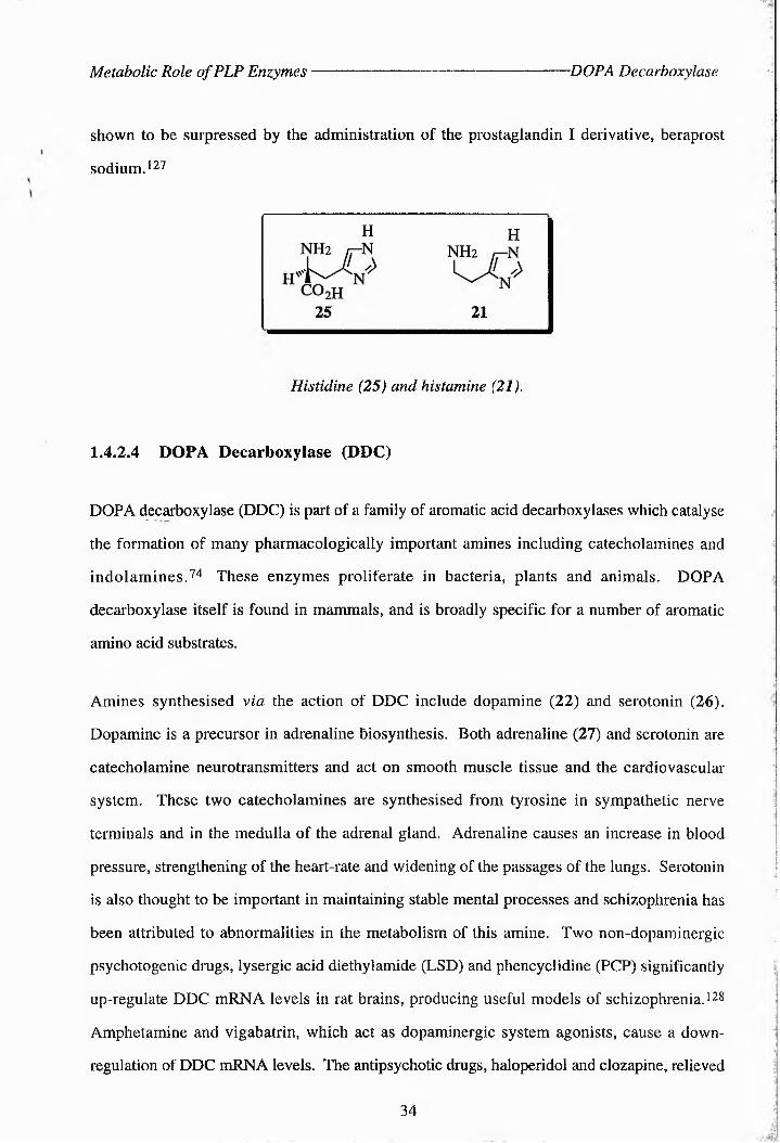
Metabolic Role of PLP Enzymes DOPA Decarboxylase
shown to be surpressed by the administration of the prostaglandin I derivative, beraprost
sodium. 127
H h
25 21
Histidine (25) and histamine (21).
1.4.2.4 DOPA Decarboxylase (DDC)
DOPA decarboxylase (DDC) is part of a family of aromatic acid decarboxylases which catalyse
the formation of many pharmacologically important amines including catecholamines and
indolamines.74 These enzymes proliferate in bacteria, plants and animals. DOPA
decarboxylase itself is found in mammals, and is broadly specific for a number of aromatic
amino acid substrates.
Amines synthesised via the action of DDC include dopamine (22) and serotonin (26).
Dopamine is a precursor in adrenaline biosynthesis. Both adrenaline (27) and serotonin are
catecholamine neurotransmitters and act on smooth muscle tissue and the cardiovascular
system. These two catecholamines are synthesised from tyrosine in sympathetic nerve
terminals and in the medulla of the adrenal gland. Adrenaline causes an increase in blood
pressure, strengthening of the heart-rate and widening of the passages of the lungs. Serotonin
is also thought to be important in maintaining stable mental processes and schizophrenia has
been attributed to abnormalities in the metabolism of this amine. Two non-dopaminergic
psychotogenic drugs, lysergic acid diethylamide (LSD) and phencyclidine (PCP) significantly
up-regulate DDC mRNA levels in rat brains, producing useful models of schizophrenia. ' 28
Amphetamine and vigabatrin, which act as dopaminergic system agonists, cause a down
regulation of DDC mRNA levels. The antipsychotic drugs, haloperidol and clozapine, relieved
34

Metabolic Role of PLP Enzymes -Racemases
the symptoms of PCP-treated animals. Evidence exists that the excess amounts of 2-
phenylethylamine (2PE) and tryptamine produced by overactive DDC are associated with
schizopprenia.128 These compounds are modulators of all the neuroreceptors known to play a
role in the disease.
The headache, chronic pain and depression associated with serotonin deficiency are often
alleviated by administering Bg supplements to the patient. DOPA and various inhibitors of the
decarboxylase are used in the treatment of Parkinson's disease, a neurological disorder
characterised by tremor, rigidity and disturbances of posture.74
3,4-dihydroxyphenylalanine (dopamine, 22), serotonin (26) and adrenaline (27).
Serotonin has recently been shown to be a mechanism-based inactivator of DDC in both a time-
and concentration-dependent manner, 729 This occurs via a decarboxylation-dependent
transamination. Other substrates such as DOPA and m-tyrosine and their a-methyl derivatives
also undergo this minor reaction and it is thought that his could be the basis of a control
mechanism. 129 Two quaternary benzophenanthridine alkaloids, sanguinarine and
chelerythrine, have oIso exhibited strong inhibition of the enzyme from rat liver in vitro J30
1.4.3 Amino Acid Racemases
The racemases, which catalyse the interconversion of (2S)- and (2/?)-amino acids, are
extremely common in prokaryotes where they play a crucial role in the biosynthesis of
peptidoglycan in bacterial cell walls. 731 These enzymes are therefore a possible target for
chemotherapy.
35

Metabolic Role of PLP Enzymes ■The Aspartate Pathway
1.4.4 The Aspartate Pathway
In both plants and bacteria, threonine synthase (TS) is the last of five enzymes involved in the
biosynthesis of (25,3/?)-threonine (37) from (25)-aspartic acid (32)54 The first two steps of
this biosynthetic pathway, known as the aspartate pathway, are common to lysine, threonine,
isoleucine and methionine biosynthesis (see Scheme 1.8).132’133 (25,3T?)-threonine is one of
the twenty common, nutritionally essential amino acids and as such is vital to all life forms41
(2S)-aspartate (28)
Mg2*’ ATP
NH2Hh^CC^h
H02C
Mg2+, ADP
P-aspartyl phosphate(29)
Aspartate Kinase (AK)
NH2Osc
NADPH + H
HO2C
Aspartate Semialdehyde Dehydrogenase (ADH)NADP+ + P/
aspartate-p-semialdehyde(30)
NH2 oH»LA
HO2C
NH
lysineNH2
NADPH+ H +
NADP+*<
(2$)-homoserine (31)
Homoserine Dehydrogenase (HSD)
NH2OH
HO2C HMg2+, ATP
Mg2*' ADPHomoserine Kinase (HK)
(2S f-Q-phosphohomoserine(32)
NH2
HO2C o©
NH
HO2C ®methionine
H20AdoMet
PLPThreonine Synthase (TS)
HPO42-
(2S,3R)-threomne (33)NH2
H3X0HHO2C >CH ,
H *
NH2H-kcCHs
HO2C \
isoleucineCH,
A
A
A
H
Scheme 1.8: The aspartate pathway.
36

Threonine Synthase- Discovery of Threonine Syn thase
1.5 Introduction to Threonine Synthase
1.5.1 The Discovery of Threonine Synthase
The existence of the enzyme threonine synthase (EC 4.2.99.2)1 was first suggested by H. J.
Teas et al. in 1947 1 34 Jn studies on mutant strains of both Neurospora crassa and Bacillus
subtilis, which had a requirement of threonine (33) for growth, he demonstrated that
homoserine (31) was a precursor to both threonine and methionine. During the early 1950s a
number of other authors reported on work with various other species of bacteria which led
them to assume that the pathway for threonine biosynthesis in all bacteria was as
follows:535'139
aspartic acid ----------- *- homoserine ------------ ► threonine
Figure 1.8: Route for the biosynthesis of threonine from aspartic acid postulated in the 1950s.
However, the enzyme was not properly identified in bacteria until 1960 when Flavin and
Slaughter reported a pyridoxal phosphate-dependent enzyme, "which will be called threonine
synthetase, [and which] catalyzed an elimination of orthophosphate coupled to isomerisation
from y- to p- hydroxy compound, to yield threonine from P-homoserine."140 The conversion
of (2.5)-0>-phosphohomoserine to threonine actually takes place stereospecifically via a p,y-
replacement, and a side-product of the reaction is inorganic phosphate (Scheme 1.9). It was
not until 1975 that the enzyme was first isolated from plants. 141
+H-3N,H"
o C
OH
H
+ PiInorganicphosphate33
Scheme 1.9: The reaction catalysed by threonine synthase.
37

Threonine Synthase- -Phosphohomoserine Deaminase
1.5.2 Associated Deaminase Activity of Threonine Synthase
TS is very selective for the substrate, and is only known to catalyse a few other reactions, often
with substrates similar to (2S)-0>-phosphohomoserine (see Appendix 2). In addition to
catalysing the dephosphorylation of (2S)-0-phosphohomoserine, TS in B, subtilis and E. coli
has also been shown to catalyse its subsequent deamination; a step usually carried out by the
enzyme threonine deaminase (Scheme 1.10)442 This reaction yields a-ketobutyrate (34).
+ NH3
H"JL nQzC
.O'O'
32
----------~ rJ^CH3 + HOPO32' + NH3
34
o
Scheme 1.10: (2S)-O-phosphohomoserine deaminase reaction..
Mutation events affecting TS activity also affected the deaminase activity suggesting that both
activities were produced by the same enzyme.142’ 143 In contrast to the reaction involving
threonine deaminase, the deaminase activity of TS has been shown not to pass through a
threonine intermediate (Scheme 1.11).142 This activity of TS was therefore 'named
phosphohomoserine deaminase. This associated phosphohomoserine deaminase activity was
shown to be a lesser activity of threonine synthase, as the two activities co-purified, were of the
same molecular weight, co-inactivated upon heating and also displayed the same value for
the substrate, phosphohomoserine. 144 This associated deaminase activity appears unique to the
TS enzymes of B. subtilis and E. coli.
phosphohomoserine■... . ■■ ' ■ ■’
phosphohomoserine / deaminase /
-NH3 -PO43- ' \ threonine synthase
isoleucine -
h-------------------
r
*---------- a-ketobutyratethreonine deaminase,
threonine ;-NH’
Scheme 1.11: Alternative metabolic pathways for phosphohomoserine
38

Th reo n i n e Syn th a s e - -Regulation of Threonine Synthase
1.5.3 Regulation of Threonine Synthase
As mentioned in Section 1.4.4 threonine synthase (TS) is the last of five enzymes involved in
the biosynthesis of (25,3/?)-threonine (33) from (2S)-aspartic acid (28, Scheme 1.8).54 Table
1.3 compares the human requirement for some of the common amino acids. Interestingly
threonine is also one of only two essential amino acids containing two chiral centres, the other
being isoleucine, itself synthesised from threonine. Threonine has also been found to play a
role in the regulation of the aspartate pathway in a number of microorganisms by feedback
inhibition of three of the enzymes involved. 145 Depending on the organism concerned it has
been shown to inhibit aspartokinase, 146-151 homoserine dehydrogenase, 152-155 or homoserine
kinase. 140, 142, 156-160 jn plants TS does not generally appear to be subject to feedback
inhibition by amino acids, but levels of TS activity are repressed by supplementation of the
growth medium with mettionine.161 In wheat seedlings threonine almost completely inhibits
its own synthesis without affecting methionine biocsntntsi6.1i1 An inadequate supply of
threonine results in a negative nitrogen balance by inhibiting protein synthesis due to stalling of
the ribosome at codons for threonine (see Section 1.7). Other amino acids then accumulate and
are transfeiTed into degradative metabolic pathways causing a net loss in nitrogen.
Table 1.3: Minimal requirements of human beings for some of the essential amino acids (mg
kg-1 daylf'2
lie Leu Lys Phe Met Cys Thr Trp Vai
Child 90.0 - 90.0 90.0 85.0 - 60.0 30.0 85.0
Man 10.4 9.9 8.8 4.3 1.3 11.6 6.5 2.9 8.8
Woman 5.2 7.1 3.3 3.1 4.7 0.5 3.5 2.1 9.2
WHO* 3.0 3.4 3.0 2.0 1.6 1.4 2.0 3.0 -
*World Health Organisation norms
39

TS Structure and Character- Microbial Enzyme
1.6 Tlte StmcCtire and Character of Threonine from V:ar.<ou;
Organisms
Until fairly recently little was known about the structure of threonine synthase (TS). Within the
last ten years, however, the gene encoding the TS protein has been isolated and sequenced
from a number of organisms, as has the enzyme itself. No 3D structures of the enzyme are
available as yet. In all cases the enzyme has proved to be dependent on pyridoxal-5'-phosphate
(PLP, 1).
1.6.1 Microbial Sources of Threornne Synthase
The gene for TS has been isolated and sequenced from B. subtilis,162 Brevibacterium
lactofermentumP^^' 163 Corynebacterium glutamicumP22 E. colli65 Pseudomonas
aeruginnsaa66 Seraatia maecsssn.s222 md Saccharomyces cerevisiae (yeast). 168> 169 The
enzyme has been isolated and characterised from these sources and also from Nsurdspora
crassa.222 Table 1.4 shows the various masses of the enzyme from different sources.
Table 1.4. Thrednins synthase from microbial sources
Species Molecular Mass(Mr) of TS (Da)
Number of Amino
Acid Residues
Literature
Reference
B. subtilis - 351 19
Br. lactofseosntso 52, 807 481 170
C. glutamicum 54, 481 481 164
56, 000 - 133
E. coli 47, 060 428 165
( " 36, 000 - 171 )
M. glycogenes 52, 167 475 172
S. cseevisiae 58, 000 514 169
40

TS Structure and Character -Microbial Enzyme
Depending on the species in question, the TS protein has a molecular weight ranging from
36 to 58 kDa. As Fig. 1.9 shows, it is an enzyme which shows significant homology
particularly in the amino acid residues in and around the active site (see also Appendix 6).173
The average pairwise identity around the active-site is 41.8%. The lysine residue (indicated by
an arrow) is thought by analogy with (27?)-serine dehydratase to be the residue responsible for
binding pyridoxal phosphate to the enzyme by formation of the internal aldimine.19 This idea
is supported by the conservation of this residue and much of the surrounding sequence, such as
the phenylalanine and aspartic acid residues to either side (underlined), in all of the microbes
studied. As mentioned in Section 1.2.3, TS also shows significant sequence homology with
and a similar active-site structure to other enzymes from the p-family of PLP-dependent
enzymes, of which it is a member (see Figs. 1.3 and 1.4 and Appendix l).24
B. subtilis{
--------- TEGVN- PTGSFKDRG-----MVMAVAKAKEEGN-------- ----------- DTIMCASTGNTSAA
B. lactofermentum --------- YEGAN-PTGSFKDRG----- MVMAVAKAKEEGS------- ----------- EAIICASTGNTSAS
C. glutamicum --IYLGHLSEGPTAAFKDMAMQLLGELFEYELRRRN------- ------- ETINILGATSGDT-GS
E. coli —VGCLELFHGPTLAFKDFGGRFMAQMLTHIAGDKP------- ----------- VTILTATSGDT-GA
H. influenzae - -I YALELFHGPTLAFKDFGGRFMAQALAAVRGDGK------- ----------- ITILTATSGDT-GA
M. glycogenes - -LYLLSLSNGPTLAFKDMAMQLLGNLFEYVLAQKG------- ------- ETINILGATSGDT-GS
M. leprae --------- VEGLN-PTGSFKDRG----- MTMAVTDALARGQ------- ----------- RAVLCASTGNTSAS
M. tuberculosis --------- VEGLN- PTGSFKDRG----- MTMAVTDALAHGQ------- ----------- RAVLCASTGNTSAS
P. aeruginosa ------- CVELFHGPTLAFKDFALQLLGRLLDHVLAKRG------- ------- ERWIMGATSGDT-GS
S. pombe —LNVLELFHGPTFAFKDVALQFLGNLFEFFLTRKNGNKPEDERDHLTWGATSGDT-GS
S. marcescens --VSCLELFHGPTLAFKDFGGRFMAQMLAEVAGEQP------- ----------- VTILTATSGDT-GA
S. cerevisiae ENLHILELFHGPTYAFKDVALQFVGNLFEYFLQRTNANLPEGEKKQITWGATSGDT-GS ** **** . * * *
Arrow indicates the lysine residue thought to be responsible for binding to PLP. Asterisks indicate conserved residues (highlighted in blue); homologous residues are shown by a period.^ Note the conserved PT--FKD motif at the active-site in all enzymes. (Appendix 6 shows the alignment of full TS sequences and gives values for the similarity between each sequence). Multiple alignment was performed using Clustal W 1.60.
Figure 1.9: Sequence homology of amino acid residues around the active-site of threonine
synthase from various microorganisms.
41

TS Structure and Character Plant Enzyme
The first reported purification of threonine synthase was a 500-fold purification from
Neurospora in 1960 through a series of acetone and ammonium sulfate fractionations followed
by ion-exchange chromotography4 37 A crude preparation was obtained from Brevibacterium
flavum by Miyajima et al. in 1968,45 and a purified preparation from C. glutamicum by Han
and co-workers in 1990464 However, there were no reports of the enzyme being purified to
homogeneity until Parsot and his co-workers described a preparation of the enzyme from E.
coli K-12 strain Tir 8 (strain Tir 8 is a constitutive mutant for the enzymes on the threonine
operon)465> 1?4 They carried out a 35-fold purification of the enzyme to 100% homogeneity in
a protocol which has been adopted in subsequent experiments involving TS from this strain.
Szczesiul and Wampler have since found TS from the same strain to have a pH optimum of 8.0
- 8.5411 and the same enzyme has an apparent Km of 0.053 mM for phosphohomoserine475
1.6.2 Threonine Synthase from Plant Sources
The study of the aspartate pathway in plants has aroused interest for two main reasons. Firstly,
as a result of man's desire to improve the nutritional qualities of various crop plants and
secondly, because an understanding of the aspartate pathway in plant tissues may aid in the
design and synthesis of effective and selective herbicides. Cereals are deficient in the
nutritionally essential amino acids lysine and methionine and legume seeds exhibit low
quantities of methionine.1”76 increasing the efficiency of aspartate metabolism in plants would
lead to improved levels of a number of amino acids in fccdstoiks.
Although there have been relatively few studies on TS in plants a number of groups have
successfully isolated and purified threonine synthase from plant sources. The enzyme was first
detected in 1975 by Schnyder et al. in the homogenates of pea seedlings and Lemna minor. 14i
The following year Madison and Thompson identified the enzyme in sugar beet and radish
leaves,177 and it has since been partially purified from Lemna pausicostata,118 barley
seedlings,!79 pea seedlings, 180 soybean plants481 Wallsgrove and ^workers have also
42

TS Structure and Character S-Adenosylmethionine
shown that the enzymes of the aspartate pathway are confined to the chloroplasts of plants.182
In all plants studied to date threonine synthase demonstrates an almost absolute requirement for
S-adenosylmethionine (SAM, or AdoMet, 35).132 SAM acts as a potent allosteric activator of
the enzyme, stimulating its activity as much as twenty-fold with a Hill coefficient of 2.0-2.5
(Table 1.5).177- 183
S-adenosylmethionine (35).
Table 1.5: Kinetic properties of threonine synthase from various higher plants.
Plant Source SAM (mM) vr maxHill
CoefficientLiteratureReference
Sugar Beet X 2.7 38.2 nmol h“l - 165
11 J 2.2 534.3 nmol h"^ - 165
Pea Seedlings X 2.2 - 2.1 168
tl y 0.67 4-fold increase - 168
Barley Seedlings y 0.05 - - 167
Lemna pausicostata y 2.2-6.9 70.8 pmol h"l 2.5 166
The Hill Coefficient is a measure of cooperativity, where the binding of a ligand (in this case SAM) to protein is at 50% saturation
Half-maximal activation of TS occurs at concentrations of between 40 and 200 (iM SAM.180
Aarnes has found TS to have a pH optimum of around 8-9 in barley seedlings.179 No
isoenzymes of TS have been detected.132
43

TS Structure and Character- Mammalian Expression
1.6.3 Threonine Synthase Expression in Mammalian Systems
«
In order to improve the diet of non-ruminants it is necessary to supplement their feed with the
essential amino acids, especially lysine and threonine (33). If the genes encoding the enzymes
used in the biosynthesis of these amino acids could be expressed in animal cells, the need for
supplementation of the diet would be eliminated. 184 Rees and Hay recently achieved the
expression of aspartokinase and threonine synthase in mouse 3T3 cells. 185 These were then
able to grow successfully on medium lacking threonine but containing (25)-homoserln6 (31),
the precursor to pHospHchomos6rin6 (32).
Threonine synthase and Homcserine kinase have never been shown to occur naturally in
mammals. It has, however, been noted that (25)-C^-^phosphohomcs6rine is an antagonist of
N-methyl-D-aspartate (NMDA) in the rat brain. 1 86.187 (NMDA is an excitatory amino acid in
the mammalian CNS). Phosphohomcserice has also been the subject of a patent for its use,
together with the drug levemopamil, in the treatment of patients suffering from damage to
neurons as a result of AIDS-related dementia, myelopathy, peripheral neuropathy and vision
loss. This is caused by a reduction of the gp 176 responsive rise in calcium ions. 188
1.6.4 Threonine Synthase as a Potential Target Enzyme for Herbicides
Because of its high substrate specificity (see Section 1.5) and the range of organisms in which
it is found, TS is an ideal target enzyme for herbicides, bactericides and fungicides. Selective
inhibitors of the enzyme should not have adverse effects on animal species, which do not
express TS. It is also known that blocking amino acid biosynthesis is a highly effective
method of inhibiting growth in plants and bacteria.41 Any structural and mechanistic
information obtained about TS can be used in the rational design of potential inhibitors of the
enzyme Many inhibitors of TS have already been synthesised and tested and provide insight
into the active-site structure of the enzyme. These are discussed fully in Section 1.9.
44

Threonine Operon Operon Structure
1.7 The Threonine Operon of Escherichia coli
1.7.1 The Structure of the Operon
The thr operon is at 0 min on the genetic map of E. coli and contains three structural genes,
thrA, thrB and f/zrC.^9-190 Transcription of the thr operon proceeds from thrA to thrC (Fig.
1.10) as established by analysis of phage Mu insertions and nonsense mutations in the thr
cluster, and by analysis of a phage X insertion into the thr regulatory region, which
pleitropically affected the expression of both thrA and r/zrB.191-193 The bi-cistronic thrA gene
encodes the threonine-sensitive bifunctional enzyme complex aspartate kinase I - homoserine
dehydrogenase I (AKI-HDHI, Scheme 1.8).194- 195 E. coli strain K-12 contains two
additional aspartate kinases (AKII and AKIII) and one additional homoserine dehydrogenase
(HDHII), controlled by methionine and lysine respectively.196- '97 The thrB gene encodes
homoserine kinase (HK), which is inhibited by threonine. 19 PhrC, a gene of 1287 bp,
encodes TS (Appendices 7 and 8).
Direction of translation
thrAA
serB C thrA .—...
thr A2 9 thr B thrC pyrA
K
AKI
1 {
HDHI HK
I
TS
Figure 1.10: Genetic map of the threonine operon of E. coli K-72. ’85. 190
The promoter for the operon has been localised approximately 230 nucleotides upstream of
thrA, between a BstE II site at position -254 and a Hha I site at -184.'99 An operator region
has been located between the promoter region and the structural genes. 190 Positions -147 to
-82 upstream of thrAj on the operon encode a putative leader peptide of 21 amino acids
(Fig. 1.11), containing eight threonine and four isoleucine residues, eleven of which occur
consecutively.
45

Threonine Operon Regulation of Expression
Regulatory Region
Structural GenesthrA
_____ A_______thrA i thr Ai ' thr li thrC
3k,
Figure 1.11: Regulatory region of the thr operon.
1.7.2 The Regulation of Thr Expression
At first the expression of the thr operon was thought to be under the control of a multivalent
repression system dependent on the concentration of free threonine and iso^crne (Fig.
1.12).200 However, Samt-Girons and Margarita were unable to detect the presence of any
repressor bound to the operator and further research has shown that factors affecting threonyl
tRNA synthase also affect thr expression, no, 172 a model has therefore been proposed for the
multivalent regulation of thr operon expression involving coupled transcription and translation
of the leader region. *1 This is a comon regulatory mechanism in phage and bacteria.
Regulatory gene Control sites ,---------A---------v-—
thrA
Structural genes
I thrB 1 thrC I
I i mRNA
I _
(121 Repressor
Repressor binds to operator (O) and prevents transcription of structural genes. If an inducer molecule is present, this forms a complex with the repressor. The repressor then cannot bind to the operator and the structural genes are expressed.
Figure 1.12: Model for a typical repression mechanism of gene regulation.™
If the intracellular levels of He and Thr are sufficient, RNA encoded by the thr regulatory region
46

Threonine Operon ■Cloning of Thr
from bases -71 to -27 folds to form a termination structure (Fig. 1.13).172> 201> 202 This
signals RNA polymerase to terminate transcription at a site (attenuator site) upstream from the
structural genes. Under conditions of threonine and isoleucine deficiency, translation of the
leader RNA sequence is stopped due to a shortage of threonyl- and isoleucinyl-tRNA
molecules. This pre-empts the termination structure, allowing transcription of the structural
genes. A region of overlapping rotational symmetry from bases -93 to -49 overlaps the
attenuator and can form of a structure which blocks the attenuator region.203 a G-C rich
region of dyad symmetry followed by a stretch of six uridine residues occurs 21 base pairs
after the stop codon of thrC, which is thought to be a rho-dependent transcription termination
she.204
G AU C
C A C-GA-UC-GG-CC-GC-GC-GG-CA-UA-UA-UA-U
5 ■ -AAACACAGA-IUUJU--
Figure 1.13: The thr regulatory region from -71 to -27, which forms the termination structure.
1.7.3 Cloning of Thr
Amplification of the thr operon has been carried out by various groups205-207 and has also been
the subject of patents.208-209 The thrC gene has been cloned from C. glutamicum by Follettie
and coworkers,si0 and from Br. lactofermentum by Malumbres et al..}1° A constitutive
mutant E. coli K-12 Tir8 has also been produced, which over-expresses the enzymes of the thr
operon.165
47

TS Mechanism- Introduction
1.8 The Mechanism of (2S,3R)-Threonine Synthesis
1.8.1 Introduction
Although there is a wide interest in PLP-dependent enzymes, relatively few studies have been
carried out on the mechanism of the TS reaction.
The first few steps in the conversion of (25)-(^-^f^h^c^s^phohomoserine (32) to (2S,3/?)-threonine
(33) are assumed to follow the same general mechanism as the reactions of other PLP-
dependent enzymes acting at C-4 of the substrate. Most of the mechanistic information for
such enzymes has been acquired from studies using cystathionine y-synthase from
Salmonella^ The reaction has been demonstrated to be essentially irreversible by Rognes. ia2
He determined that no [iacj-O-phosphohomoserine was formed when N. crassa TS was
incubated with [i4C]-threonine and inorganic phosphate (Pj), and that no enzymic exchange of
a2pf. into phosphohomoserine occurred.
1.8.2 Reaction Mechanism
The first two steps in the TS reaction are described in more detail in Section 1.3.2. They
involve the formation of a holoenzyme aldimine (10) between the e-amino group of a specific
enzyme-bound lysine residue and the C-4’ carbonyl group of PLP (Scheme 1.5)4- 5. 42 The
condensation of (2,S)-^i^ -phosphohomoserine with the internal aldimine (10) then leads to the
formation of a Schiff’s base substrate aldimine (36, Scheme 1.12), with the release of the
enzyme-bound lysine residue.4 42 Farrington et al. found that substrate analogues, which
were capable only of binding to PLP and undergoing C" proton abstraction, exhibit rapidly
reversible binding, suggesting that transaldimination is a fast step. J25
48

TS Mechanism -Ha-Cleavage
Scheme 1.12: Transaldimination reaction involving (2S)-O-phosphohomoserine (32) .
The next stage in the reaction is labilisation of the Ca-H bond to form the quinonoid
intermediate (37, Scheme 1.13). As described in Section 1.3.2, Dunathan's postulate states
that in the external aldimine the bond to be cleaved at Ca is held orthogonal to the plane of the
pyridinium ring, thus allowing maximum orbital overlap between the developing negative
charge and the conjugated electron-deficient rc-system.4-5
Scheme 1.13: Labilisation of Ha by an enzyme-bound base.
Fuganti suggests that not only the Ca-H, but also the Cp-H and CY-OPO3H a bonds in the
Schiff’s base, all of which are broken in the course of the TS reaction, must be held
perpendicular to the extended rc-system, in order to optimize orbital interactions in the bond
49

TS Mechanism- -fi-elimination
breaking and making probi6ses.612 It has been demonstrated that the known reactions of PLP-
enzymes all take place only on the ‘exposed’, i.e. the si -face at C-4’ of the cofactor (below the
plane of the page in all structures shown here).1 It is therefore assumed that TS follows this
general pattern.
The cleavage of Ha is further facilitated by an enzyme-bound base, probably the ucprotocated
lysine residue originally involved in binding the enzyme to the coenzyme, PLP.1 This has
certainly been suggested by data produced in studies on OASS (Section 1.3.3.1)11 The e
amino group of this lysine residue stabilizes the positive charge as it forms on the
electrofuge.11 The removal of Ha forms a carbarnon at C“, which is resonance stabilised as a
pyridcxal-ketimine-p-quiccnoid (37). In this intermediate the acidity of the protons at C-3 is
increased through being adjacent to a conjugated system of double bonds, and they become
more labile. OASS, another p-family enzyme, does not appear to form a quinon^d
intermediate, whereas for TrS such a species has been fleetingly detected.11 When F, Barclay
incubated (25))[2-2Hl]-<0-phcsphchomoserice with the enzyme from E. coli, isotope effects of
1.2 andd 1.0 weee obtainnd for DV and D1V/K) rr6peeiivety. Tins i ndicctte t hat the rrmooal oo
C"-H from the substrate is kindtiially significant but not ente-limtt1ng.l1 The fact that the value
of DV is small, but larger than the value of D1V/K) for the C^-demer^ted compound, strongly
suggests that the high forward commitment to the C“-H proton removal step occurs because
teansaldiminatibn in the reverse reaction dir6ctibc ncd/oe dissociation of the substrate from the
enzyme is very slow. Farrington et al. also proposed that immediately after Cl-elimicatloc, the
phenolic oxygen and C-4’ of PLP me prbtbcated (as shown in Scheme 1.14).H5 7S has been
shown to produce PMP in half-teacsamicatioc renctiocs with a number of amino acid substrates
which do not possess a leaving group in the p-position.21 1
In 1979 Fuganti first demonstrated, using yeast TS, that it was the 3-pro-S rather tenc the 3-
pro-R proton which was eliminated in the next stage of the reaction, using (3/?)- and (35)-
[3-1H; U-llCJ-(25)-O-phosphbaomoserine as substrates.111 This is unusual, as most other
PLP-depecdent enzymes acting at C-4 of the substrate catalyse the removal of the 3-pro-R
hydrogen.4 This was also observed for the E. coli enzyme. The (2S,35'--[3-2Hl]-lsotbpom6r
of the substrate displayed isotope effects of 1.74 and 1.0 for DV and D(V/K) respectively
50
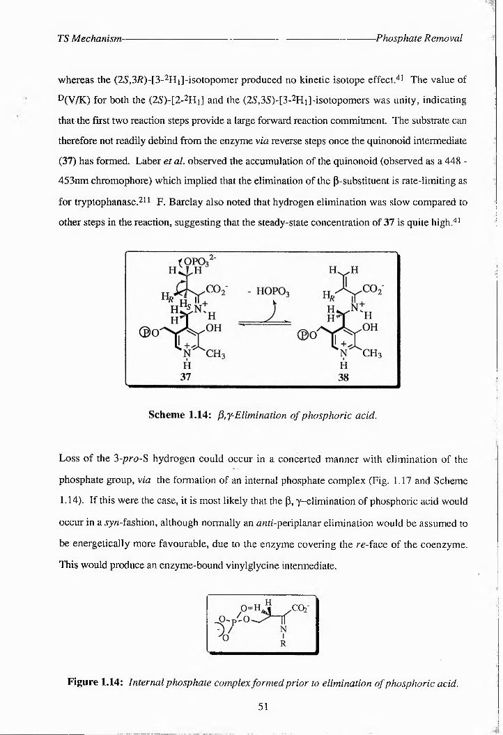
TS Mechanism- Phosphate Removal
whereas the (2S,3/1)-[3^2]Hi]-isotopomer produced no kinetic isotope effectJi The value of
D(V/K) for both the (2S)-[2-2Hi] and the (2S,3S)-[3-2Hi]-isotopomers was unity, indicating
that - the first two reaction steps provide a large forward reaction commitment. The substrate can
therefore not readily debind from the enzyme via reverse steps once the quinonoid intermediate
(37) has formed. Laber et al. observed the accumulation of the quinonoid (observed as a 448 -
453nm chromophore) which implied that the elimination of the p-substituent is rate-limiting as
for tryptophanase.21 1 F. Barclay also noted that hydrogen elimination was slow compared to
other steps in the reaction, suggesting that the steady-state concentration of 37 is quite high.aa
Scheme 1.14: py-Elimination of phosphoric acid.
Loss of the 3-pro-S hydrogen could occur in a concerted manner with elimination of the
phosphate group, via the formation of an internal phosphate complex (Fig. 1.17 and Scheme
1.14). If this were the case, it is most likely that the p, y-elimination of phosphoric acid would
occur in a jyn-fashion, although normally an ««fLperiplanar elimination would be assumed to
be energetically more favourable, due to the enzyme covering the ra-face of the coenzyme.
This would produce an enzyme-bound vinylglycine intermediate.
------- ,
CO?"
Figure 1.14: Internal phosphate complex formed prior to elimination of phosphoric acid..
51

TS Mechanism.---------------------------------------------------------------------------Vinyglycine I intermediate
Another postulate for the elimination of the phosphate group involves a sequential reaction,
whereby the 3-pro-S hydrogen is removed first by a base approaching from the 4-si -face to
form a resonance stabilised CP carbanion (39, Scheme 1.15). Subsequent elimination of the
phosphate group (the nucleofuge), also positioned on the 4'-si -face of the coenzyme, from CY
then generates the vinylglycine intermediate, as shown before (38, Scheme 1.15).
Phosphorylation of homoserine to replace a hydroxyl group with the more electronegative
phosphate group facilitates this y-elimination.
OPO32'
H
39
Scheme 1.15: Labilisation of the 5-pro-S hydrogen atom,.
Laber et al. observed that vinylglycine was converted to threonine upon incubation with TS,
confirming that the pyridoxal p-quinonoid of vinylglycine 38 is an intermediate in the
reaction.2^ The initial absence of any long-wavelength chromophore was seen to argue
against the transient accumulation of 38.
Although it is not yet known which of the aforementioned y-elimination mechanisms are
52

TS Mechanism- Vinylglycine Intermediate
preferred, it has been shown that inorganic phosphate is formed by nonhydrolytic elimination
rather than by hydrolysis of the phosphate group at C7 {i.e. the C7-O rather than the P-O bond
is broken). This was discovered by Flavin and Slaughter in studies carried out in 18O-labelled
water, where 18O was incorporated into (25,3/?)-threonine but not into phosphate.213 They
also proved, using 3H-labelling, that it was the vinylglycine that was formed, rather than the
reaction proceeding via a hydride-shift mechanism (see Scheme 1.16).
Scheme 1.16: Comparison of proposed mechanisms for the elimination of phosphate.
The terminal alkene (or vinylglycine intermediate, 38) produced by the removal of the
phosphate group is re-saturated by a hydrogen atom, which may come from the solvent, or
from a protonated base on the enzyme, again probably from the 4'-fi -face (40, Scheme 1.17).
Farrington et al. propose that, immediately prior to this step, the phenolic oxygen and C-4' of
PLP are deprotonated again. 175
53

TS Mechanism- Reverse T ransaldimination
38 40
10 42 41
Scheme 1.17: Resaturation of vinylglycine intermediate and release of threonine.
It is known from incubations conducted in deuterium oxide that threonine synthase introduces
two atoms of deuterium into the product, one at C-2 and one at C-4, leading to the assumption
that no significant internal proton transfer takes place.4
The TS reaction is then completed by a hydroxyl group or water attacking at C-3 of the
substrate4 As the absolute configuration of threonine is {2S,SR) this process must occur via a
retentive mode.4 (2S,3/?)-threonine can now be released from the coenzyme via reverse
transaldimination, which regenerates the holoenzyme aldimine.
The mechanism of this p/y—replacement requires a completely different conformation at the
active site compared with p-replacement systems. This difference in the stereochemical courses
of the two types of reaction can be explained either by the transient quinonoid intermediate
described earlier or by a two-base system4
54

TS Mechanism- Unanswered Questions
1.8.3 Unanswered Questions Cbdierdidg the THeebdide Synthase Reaction
• The removal of the C-3 proton and the elimination of phosphate from the quinonoid
intermediate (37) may be sequential or concerted (Section 1.8.2). The concerted elimination
should occur in a 1yn-fashion, if the enzyme covers the e6-faie of PLP, but it is not yet certain
from which face of the coenzyme the phosphate group leaves. It also not clear whether the
phosphate group or the e-amido group of the tradsaldimidated lysine residue would act as the
base removing the C-3 proton (Scheme 1.15).
• It is not yet know whether the eeprotonation of the enamine (38) to give the imino acid
moiety (40) occurs with retention or inversion of configuration, whether the protonation occurs
from the 4'-s‘-face as assumed and whether internal proton transfer from C-3 to C-4 occurs.
Flavin and Slaughter saw no internal proton transfer when they assessed tritium transfer from
the substrate to the prod.uct.211 However, if the proton in the conjugate acid is readily
exchangeable with solvent protons, then very few tritium atoms would be transferred back to
the substrate in this type of assay. More careful analysis of the isotope content of the threonine
methyl group from very short-time experiments is required. The reprotocatioc step may also
be reversible.
• The attack of water at C-3 of 40 is defined by the stereochemistry at C-3 of threonine (33)
{i.e. 3R) but the geometry of the attack is not known with respect to the coenzyme.
55

Inhibitors ofTS- ■Inhibitors of PLP-Enzymes
1.9 Inhibitors of Threonine Synthase
1.9.1 Functional Groups Which Inhibit PLP-Enzymes
In section 1.4 the metabolic role of various PLP-enzymes was discussed, demonstrating the
wide scope of action of such enzymes. As a result of their varied functions the inhibition of
PLP-enzymes has potential phamiacological and therapeutic utility. Generally PLP-enzymes
are inhibited by carbonyl attacking reagents such as hydroxyl amines and hydrazines, and
substrate analogues which bind at the active-si te but cannot undergo further reaction^ In the
absence of endogenous coenzyme many PLP-dependent enzymes are also inhibited by the
production of PMP in abortive transaminase side reactions. The development of mechanism-
based, suicide inhibitors of PLP-enzymes which are substrate analogues has been widely
studied, both for their inhibitory properties and as a means of elucidating the mechanism of
specific enzyme.2.4'... As the role of PLP is to stabilise carbanionic intermediates that form
during the catalytic process (Section 1.3.1), inhibitors possessing functional groups which
become activated by their close proximity to an enzyme-generated carbanion can break down to
yield a reactive species. This may react with an active-site amino acid side-chain, or with
tightly bound PLP-coenzyme. Either of these processes will lead to the inactivation of the
enzyme, in the first instance blocking the active-site with an unreactive species, or in the second
case causing the coenzyme to become unavailable to bind substrate.
Functional groups commonly used in the design of such inhibitors include acetylenic, olefinic,
p-halo substituents and other leaving groups, nitriles, aryl sulfoxides, dihydroaromatics and
phosphonamino acids.214- 217 A number of olefinic substrate analogues such as
vinylglycine.2. 6- 218 (E)-methoxy vinylglycine (43)717 and p-methylene aspartate (44)22°
have been demonstrated to inhibit AspAT. Acetylenic analogues such as (25)-propargyl
glycine (45) inhibit a range of PLP-enzymes including the transaminases AspAT and co
ornithine aminotransferase and both y-cystathionase and cystationine-y-synthetase.22h 222
56

Inhibitors ofTS-------------------------------------------------------------------------------------------- Amino Acids
Some of the most common suicide substrates for transaminases are those which possess a good
leaving-group at CP, as they can undergo a facile elimination to generate an olefinic
intermediate. (25)-3-chloroalanine (46)223* 224 inhibits AlaAT and various halogenated
alanines are suicide substrates for tryptophan synthase (Ti'5*)^^2^5 (25)-s<^ir^r^<^-^(0-sulfate (47)
also acts as an inhibitor of varous PLP-enzymes including AspAT, and glutamate
decarboxylase.226 GABA-T is inhibited by various acetylenic, olefinic and halogenated
substrate analogues227’ 228 4nd a number of carbonyl attacking reagents such as
hydroxylamine, aminooxyacetic acid, hydrazines and carbazides4
och3
44 45
NH2
CO2H
43
NH2II"A^C1
co2h
nh2H"|k^OSO3H
co2h
46 47
Figure 1.15: Inhibitors of PLP-enzymes.
1.9.2 Amino Acid Inhibitors of Threonine Synthase
Various studies have been carried out on the inhibitory properties of both biogenic and
exogenous amino acids on enzymes of the aspartate pathway, some of which are proven
inhibitors of threonine synthase. In 1967 Miyajima et al. found that TS from Br.flavum was
scarcely inhibited by amino acids of the aspartate family suggesting that the enzyme may only
be subject to weak feedback inhibition or repression in vzvo.2^29 Lysine, threonine and
isoleucine only showed approximately 15 - 20% inhibition of the enzyme, and the presence of
methionine negated the effects of lysine and isoleucine. However, cysteine (48) and
57

Inhibitors ofTS- Amino Acids
glutathione (49) showed 100% inhibition and alanine (50) showed 44% inhibition of TS. In
Lemna pausicostata cysteine has also been shown to inhibit the stimulation of TS by SAM480 «
As cysteine and phosphohomoseeine are converted to cystathionine by another enzyme it has
been suggested that the conversion of (2S)-(9-phosphohomoserine to either threonine or
cystathionine is regulated by the relative amounts of SAM and cysteine present. 180 in 1984
Giovanelli and coworkers carried out a separate study on the activity of TS in Lemna
pausicostata with mechanism-based inhibitors of PLP enzymes. Although DL-vinylglycine and
cysteine both irreversibly inhibited TS to a small extent (22 and 17% respectively) both (25)-
propargyl glycine (PAG) and gabaculine failed to act as suicide substrates for the enzyme. 118
In a later study by Rognes et al. the TS activity in pea seedlings was reduced by up to one half
in the presence of excess methionine (51)1160 Aarnes also tested a number of amino acids on
TS from barley seedlings, but only (25)-orni1hide (52) and 2,4-diamino butyrate had any
significant effect (75 and 77% of control activity respectively). The latter was also found to
inhibit the enzyme in Lathyras sylvestris. 119
Figure 1.16: Amino acid inhibitors of threonine synthase.
58
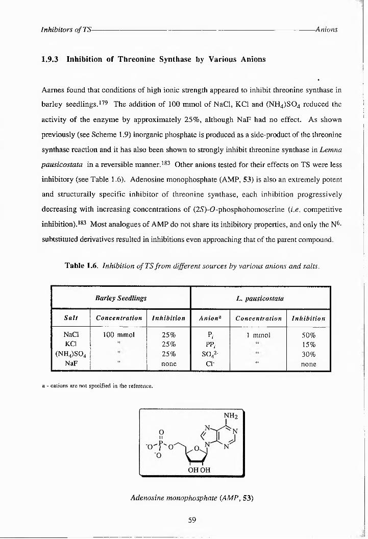
Inhibitors ofTS- -Anions
1.9.3 Inhibition of Threonine Synthase by Various Anions
«
Aarnes found that conditions of high ionic strength appeared to inhibit threonine synthase in
barley seedlings. 179 The addition of 100 mmol of NaCl, KCl and (NH.^O. reduced the
activity of the enzyme by approximately 25%, although NaF had no effect. As shown
previously (see Scheme 1.9) inorganic phosphate is produced as a side-product of the threonine
synthase reaction and it has also been shown to strongly inhibit threonine synthase in Lemna
pausicostata in a reversible manner. 183 Other anions tested for their effects on TS were less
inhibitory (see Table 1.6). Adenosine monophosphate (AMP, 53) is also an extremely potent
and structurally specific inhibitor of threonine synthase, each inhibition progressively
decreasing with increasing concentrations of (2.5)-0-phosphohomoserine {i.e. competitive
inhibition). 183 Most analogues of AMP do not share its inhibitory properties, and only the N6-
subtstituted derivatives resulted in inhibitions even approaching that of the parent compound.
Table 1.6. Inhibition of TS from different sources by various anions and salts.
Barley Seedlings L. pausicostata
Salt Concentration Inhibition Aniona Concentration Inhibition
NaCl 100 mmol 25% P. 1 mmol 50%KCl It 25% PP. 15%
(NH4)S04 25% SO42- 30%NaF ♦1 none Cl- 44 none
a - cations are not specified in the reference.
Adenosine monophosphate {AMP, 53)
59

Inhibitors ofTS Phosphonate Analogues
1.9.4 Phospho^c Acid Analogues of (2S’)-<0-Phiispabhiimoserlne
Phosphonic acid structural analogues often exhibit high affinities (and are therefore potent
inhibitors) for enzymes which catalyze phosphoryl transfer or phosphate hydrolysis reactibcs
and theeonicd synthase is no exception. This is due to the fact that these ncnlbgues do not
undergo complete catalysis due to the chemically inert nature of the C-P bond. 115 Ash et al.
first explored the suitability of various sulfide analogues as inhibitors of E. coli TS, finding teal
2-amido-3-[(phbsphbdomethyl)thio]pebpionic acid (55, see Table 1.7) was a slow-binding
potent inhibitor of the enzyme.230 The efficacy of this compound probably lies in the fact tenl
it cannot proceed past the formation of the quiconbld intermediate (37, Scheme 1.14), but is
already committed to the catalytic process. In 1993 Fareicglon et al. carried out further studies
on phosphornc acid inhibitors ofTS from E. coli K-12 Tir-8,m in particular analogues 55-58
and 61.125 The results of similar studies by Pohlenz and his group were published in 1994.
They describe the synthesis of a number of analogues of the natural substrate (55, 59, 60 and
64-68) which they later tested on TS along with analogues 61 63.23f 232
Although the sulfide analogue 55 is a good inhibitor, the p-sulfoxide and p-oxo-analogues of
the substrate (56 and 60)have a significantly reduced potency (one order of magnitude). In the
case of the sulfoxide analogue this is probably due to steric aindeacce on the active-site
preventing it from effectively binding to TS. The oxo-ncalogue 60 may be inert as an inhibitor
due to the less nucleophilic nature of oxygen relative to sulfur. This may have some
significance for the binding of substrates to TS. Taibetaers also have the ndanctng6 lanl the
sulfur atom, which is more readily polarised, will stabilise adjacent carbnnlocs. This renders
the a-protons much more acidic taac is the case for ethers. Thus, in the case for teloeleer the
quinonbid intermediate is more readily formed (Scheme 1.15). The P-amino analogue 59 on
the other ancd demonstrates a K\ value (59 jiM) comparable with the Km of the substrate
(53ji,M).175 Analogues with a cHiIc length of teee6 carbons are, generally speaking, more
potent inhibitors, with the potency decreasing as the number of carbons along the cHiIc
increases, indicating that larger compounds anve difficulty fitting imo active-site of the enzyme.
This also has implications for the positioning of the phosphate group in the nctlae-slte (see later
60

Inhibitors ofTS -Inhibitor Structures
Structure No. (mM) Type of Inhibition
H2O3PZ\ZK>C02H
nh25 4 0.031 competitive
h2o3px\s^>co2h
nh255 0.011 slow-binding
h2o3p S 1O nh2
56 0.194 competitivg
h2o3pC,C
xxsX^co2h
nh2
57 0.225 competitive
H3C^>co2h
nh2
58 0.330 competitive
H2O3P^^CO2HN y* 2
H NH259 0.059 enzyme-activated
irreversible inhibition
h203pz^o^>CO2H
nh260 none
h2o3p
CHxO^A^CO2H
nh2
61 0.05competitive
H2O3Px o^^^oo2h
NH262 0.01 competitive
h2o3p"°^Z\Z\zc°2H
nh263 none
h£>3p>
nh26 4 0.10 irreversible
^o3p-'XX=<xCO2H
NH2
65 0.40 irreversible
h203p
nh266 0.54 reversible competiive
H2O3P 4x==x^>OO2H
NH26 7 none
H2O 3P4^ 1
NH268 none
Table 1.7: Inhibitors of threonine synthase.
61

Inhibitors ofTS- Phosphate Analogues
discussion). Replacement of Ha with a methyl group also hinders the binding of the substrate
to the enzyme both sterically and because this precludes any reaction beyond the formation of
the holoenzyme aldimine. These substrates are therefore less committed to the catalytic
pathway.1.5 Olefinic and acetylenic amino acids are well known inactivators of various PLP-
dependent enzymes (Section 1.4.1). Indeed the olefinic analogues 64 and 65 do inhibit TS in
E. coli, but the olefinic analogue 67 and the acetylenic analogue 68 both fail to inactivate TS.
The most likely explanation for the lack of reactivity of these compounds is that the phosphate
group is held too rigidly in a conformation which prevents it from interacting with specific
residues on the enzyme. In a study by Giovanelli et. al. the importance of the y-phosphate
group on (25)-0-phosphohomoserine for binding was clearly demonstrated. 1?8 A variety of
0-acyl homoserine esters had a binding affinity for TS at least 2 orders of magnitude smaller
than that of the natural substrate. Also the poor binding affinity of O-phosphoserine and O-
phosphothreonine indicated that the optimal position for the phosphate group was at the y-
position. Analogue 64, [(25')-2-amrno-5-phosphono-3-cz£-pentenorc acid (Z-APPA)], has
been shown to be the active component of plumtemycins233’ 234 and rhrzocticms,277 which are
di- and tripeptides that posess bactericidal, fungicidal or acaricidal activity according to the
nature of the peptide. The acid has been shown to decrease the activity of threonine synthase by
acting as a suicide inhibitor and is also known to have herbicidal activity.
1.9.5 Phosphate Analogues
The use of naturally occuring antimetabolites as bacteriocidal agents has received much
attention. In 1984 Shames et al. carried out studies on the action of aspartate-derived
antimetabolites on the enzymes of the threonine biosynthetic pathway, and found that
(25,)-eAre<?-3-hydroxyhomoseriie phosphate (61) was a potent inhibitor of threonine
synnhasa,236 a finding confirmed by Laber et al.-32 They also investigated the properties of
cyclopropyl analogues 62 and 63 and showed that the first of these was a potent inhibitor of
TS, but the latter showed no inhibition under standard assay conditions. This is probably due
to the extended length of the compound, causing the phosphate group to be in the wrong
position for binding.
62

Aims of this Project- Testing New Substrate Analogues
1.10 The Aims of This Project
1.10.1 Testing New Substrate Analogues as Inhibitors of Threonine Synthase
Previous inhibition studies on TS indicate the following about potent inhibitors of the enzyme:
• The should be 3 i3a^T^c?c^s in length.
• A phosphate (or phosphonate) group should be n the y--fD^^lttic^n for optimum binding.
• The inhibitor should undergo both C1-H cleavage and 3-pro-S proton I^^mc^azlll,
exhibiting a high forward reaction commitment, but remain inert to further reaction.
Potential inhibitors of TS were therefore designed which should promote the removal of a
proton in the C-3 position; (2R,3S)- and (2/?,3/?)-3-fluorophosphohomoserine. P-fluoro-
amino acids have previously been shown to be good inhibitors of certain PLP-ddpendent
enzymes. It was assumed laat the small size of fluorine would allow these compounds to fit
easily into the active site, but that the electronegativity of fluoridd would cause perturbation of
the enzyme mechanism. The analogues (25,35), (2S,3R)-, (25,45)- and (25,4R)-methyl--0-
phbsphbhomoseelne were also designed to aid in the elucidation of the geometry of bound
reaction intermediates at bbla C-3 and C-4. These substrate analogues have been synthesised
by G. Allan (unpublished results). The aim of this work was to assay all laese analogues with
TS from E. coli K-12 Tir8 and determine their effectiveness as substrates or inhibitors of the
enzyme. The rate of turnover of these analogues and the substrate was to be mocilor6d using
malachite green assays, which detect even nadomolae quantities of the side-product, inorganic
phos^j>li£it<e.4h 238-241 values can be determined by measuring the initial reaction velocity
(d) for a series of different analogue codcentra1iods [S]. This data can then be represented on a
Lineweaver-Burk plot (1/3) against 1/[S]). The slope of this plot is KMAjm1X, the y intercept is
1/Vnmx and the x intercept is -l/KM.142 Alternatively the Eadie-Hbfstee plot (i) vs. 3/(8]) is
more accurate and gives a slope of -A^M, a y intercept of Vj^ and an x intercept of Vmax/ArM.
Different types of inhibition can also be determined using these types of plot. Values of i) are
determined for a constant inhibitor ibdcenteation ([I]) over a range of [S] values (in this case S
is the natural substrate). This data is determined for several values of [I] and the separate
63

Aims of this Project- Product Inhibition Assays
lines for each [I] value are drawn on the same graph. Competitive inhibition is indicated by a
family of lines with a common y intercept on both Eadie and Lineweaver plots. Uncompetitive
inhibition (where I binds to the enzyme-substrate complex only) is indicated on a Lineweaver-
Burk plot by parallel lines or on an Eadie plot by lines with a common x intercept.
Noncompetitve inhibition (where I and S bind simultaneously) is shown on an Eadie plot by
parallel lines and on a Lineweaver-Burk plot by a set of lines with a common, negative x
intercept. K[ can be calculated once Vmax and and the type of inhibition are known.
The (2i?,3(5)-3-fluoro- and (2S,3/?)-3-methyl- analogues are not expected to undergo reaction
beyond formation of the quinonoid intermediate, as there is no 3-pro-S proton available. These
substrates may therefore undergo abortive transamination reactions to produce PMP (6), which
can be monitored by UV-Vis studies. The removal of the 3-pro-S proton in the {2R,3R)-3-
fluoro- analogue may proceed more quickly than is the case for the natural substrate, due to the
electron-withdrawing effects of the fluorine atom. This analogue may therefore prove to be an
enzyme-activated irreversible inhibitor ofTS. The analogues methylated at C-4 will indicate
how constrained the y-phosphate group is normally in the TS active-site, as they cannot bind
effectively if there is too much steric hindrance.
1.10.2 Product Inhibition Assays
Inorganic phosphate (Pj) is produced as a side-product of the TS reaction (Scheme 1.9) and has
been shown to strongly inhibit the enzyme from Lemna pausicostata at 1 mmol concentration in
a reversible manner. Threonine has also been shown to inhibit the enzyme from Br. flavum
but only by 15 - 20%. It was considered of value to carry out product inhibition studies on the
enzyme from E. coli, but malachite green cannot be used to monitor substrate turnover in this
case. Therefore it was envisaged that part of this work would involve the synthesis of [U-
14C]]-22>))t9-phosphohomoserine (32b, Section 2.1.3). The substrate turnover in the presence
of various starting concentrations of P, or threonine was to be monitored by quenching the
assays after a given time, separating the (33b) produced from 32b
64

Aims of this Project- Structure-Activity Studies on TS
via ion-exchange chromatography and determining the quantity of 33b present in solution by
scintillation counting.
1.10.3 Further Assessment of the Allosteric Activation of TS by SAM
The effect of SAM on TS from E. coli K-12 Tir8 has been investigated by F. Barclay for
comparison with its effect on plant TS enzymes-H The value of Xm for TS was almost trebled
in the presence of SAM (115.1 jiM compared to a value of 43.5 pM without SAM), and the
Vmax value was increased almost two-fold (7.34 pM mim7 compared to 4.18 jtM mind without
SAM). More kinetic data is needed in order to determine the Hill coefficient, h, which at the
upper limit is equal to the number of allosteric binding sites. The value of h can be determined
from the slope of the following equation in the region of 50% saturation:
log —7------ = /ilog[S]-logKfax " 7
It was also hoped that some of the other unanswered questions concerning the TS mechanism
(see Section 1.8.3) could be addressed in this work.
1.10.4 Structure-Activity Studies on Threonine Synthase
No crystal structure of TS from any source has yet been produced. It was hoped that an X-ray
structure of the enzyme complexed with PLP could be produced during this work, either using
TS isolated from the Tir8 strain, or from thrC cloned into an expression system, such as the
pET system (Section 1.11). This would elucidate the active-site geometry and provide
information on how substrates bind to the enzyme. In Section 1.3.3 the altered properties of a
number of mutant TrS enzymes were described. It was hoped that similar site-directed
mutagenesis experiments could be carried out on TS to elucidate the exact role of key residues,
such as Lys 107 (responsible for binding PLP), which are conserved in all the known microbial
TS sequences (Appendix 6). Mutant enzymes were to be expressed in pET vectors.
65

pET Expression Vectors- 77 RNA Polymerase
1.11 pET Expression Vectors
1.11.1 Introduction243’ 244
The pET system is one of the most powerful systems to date for the cloning and expression of
recombinant proteins in E. coli. Expression of target genes in pET plasmids is under the
control of strong bacteriophage T7 1ranscrip1ibd and translation signals and is therefore only
induced by the presence of T7 RNA polymerase provided by the host cell. This polymerase is
so selective and active that it can direct almost all the cell's resources towards expression of the
target gene. Recombinant protein can amount to up to 50% of total cell protein.
1.11.2 T7 RNA Polymerase245
The RNA polymerase of bacteriophage T7 has a stringent specificity for its own promoters and
active T7 promoters have never been found in DNAs unrelated to T7 DNA.246 No T7
promoters exist on E. coli chromosomal DNA (a natural host for T7). Once an E. coli cell
becomes infected with T7, the bacteriophage can use its highly selective RNA polymerase to
direct gene tradsieiptibc away from the host's DNA to its own. Transcription by T7 RNA
polymerase is very active as well as selective, elongating RNA chains about five times faster
taan E. coli RNA polymerase.2662248 77 rna polymerase also appears not be subject to the
factors that cause termination of transcription by E. coli RNA polymerase.249 The one
terminator specifically for the polymerase in T7 DNA is also not entirely efficient.^ Studier
and co-workers therefore concluded that T7 RNA polymerase should be capable of efficiently
producing complete transcripts from almost any DNA linked to a T7 promoter. Since T7
promoters are never utilized by E. coli RNA polymerase the presence of this promoter in the
cell should have little effect on Host gene expression. However, once T7 RNA polymerase is
introduced into tie host cell this induces active and selective transcription from the T7
promoter.
66

pET Expression Vectors Expression of Target Genes
Rosenberg and co-workers first reported the construction of pET vectors (plasmid for
Expression by T7 RNA polymerase) based on the plasmid pBR322.251-253 T7 gene 1 is
contained in the expression host; the plasmid containing both the T7 promoters and terminators
either side of the cloning site.
1.11.3 Expression of Target Genes243
The target genes are initially cloned in hosts which do not contain the T7 RNA polymerase
gene. Background expression of the gene product is minimal in the absence of T7 RNA
polymerase helping to establish stable recombinants. This is particularly advantageous in cases
where the gene product would be toxic to the cell.
For protein expression the recombinant plasmid is transferred to a host containing a
chromosomal copy of T7 gene 1. These are lysogens of the bacteriophage XDE3. Of the two
lysogenic strains suitable for gene expression, BL21(DE3) and HMS174(DE3), the former
lacks the ompT outer membrane protease which can degrade some proteins during purification.1
Both strains contain the polymerase gene under the control of the inducible lacUVS promoter.
Expression of target genes can therefore be halted until the growing culture has reached a
suitable stage. Upon the addition of the inducer isopropyl-p-D-thiogalactoside (IPTG) T7
RNA polymerase is expressed and the gene contained on the plasmid is transcribed. Some pET
vectors also contain a Tllac promoter operator system. This means that they contain a lac
operator sequence just downstream of the T7 promoter, and also the natural promoter and
coding sequence for the lac repressor (lacI). The latter is oriented so that the Tllac and lad
promoters diverge. In DE3 lysogens the lac repressor acts at the lacUVS promoter to repress
background transcription of the T7 RNA polymerase gene by the hosts RNA polymerase. The
lac repressor also acts at the Tllac promoter to block transcription of the target gene by any T7
RNA polymerase that is made.
67

pET Expression Vectors' -pET-16b
1.11.4 Vectors Selected For Expressing Wild-Type and Mutant ThrC
The three vectors chosen for expressing thrC, pET-3a. pET-3b and pET-16b (Fig. 1.17), are
translation vectors containing efficient translation initiation signals. 243 All these vectors
carry the strong ribosome binding site of the T7 gene 10 protein (the major capsid protein of
phage T7). pET-16b also contains the Tllac promoter operator system and produces
recombinant protein with a His» Tag™ affinity handle at the N-temrinus.243 This permits
one-step purification of expressed proteins by metal chelation chromatography on Ni-NTA
resin (see Scheme. 1.18). 254
T7 TerminatorAmpr Jk . BamV I-site
[ ORI'f-'Nde I-site || Ribosomal Binding Site
pET-vector TT T7 TranscriptionControl Region
Figure 1.17: General structure of pET-vectors to be used in cloning experiments.
81 , •
•:WWIHis-tagged protein binds to Ni-NTA resin
1. Contaminating proteins flow through column.2. Tagged protein eluted in imidazole gradient.3. His-tag may be cleaved by Factor Xa.
Resin
Purified enzyme
Scheme 1.18: Protocol for the purification ofHis-tagged TS.
68

CHAPTER TWO
RESULTS AND DISCUSSION

Results and Discussion- Substrate Synthesis
2.1 Substrate Synthesis
A protocol for the synthesis of (SS)-0-phosphohomoserine (32) had previously been
developed by F. Barclay.41- 255 This approach was adapted for the solution-phase synthesis of
both the unlabelled and [U-^CJ-labelled substrate in these studies, with a view to continuing
the elucidation of the TS reaction mechanism. Several other approaches to the compound had
been reported in the literature, but none of these were very efficient, and many of them relied
on enzymic synthesis.
2.1.1 A Survey of the Methods for Synthesising (2S)-<0-Phosphohomoserine
Agren reported the isolation of phosphohomoserine from trichloroacetic acid extracts of
Lactobacillus casei in 1962, obtaining 55 mg of substrate from 650 g (dry weight) of cells.256
A few years later Watanabe et al. reported the production of phosphohomoserine by the
treatment of homoserine with a yeast-derived preparation of homoserine kinase.257 Skarstedt
and Greer also phosphorylated (25)-homoserine (31) enzymically, using aspartokinase
purified from a threonine-deficient strain of B. subtilis.143 Contaminating phosphate-
containing side-products were completely removed by ion-exchange chromatography.
The first chemical synthesis of the compound was in 1973 by Fickel and Gilvarg, starting from
(25)-homoserine (Scheme 2.1, Route A).258 The starting material was A-protected with
t-butoxycarbonylazide, esterified with benzyl 3-p-tolytriazine and phosphorylated with
diphenylchlorophosphate. The protecting groups were then removed using boron trifluoride
and hydrogenolysis. This synthetic route unfortunately led to 20% racemisation of the end
product. Schnyder and Rottenberg developed this route further, synthesising the substrate via
a-p-nitrobenzyl-Ar-benzyloxycarbonyl-(25)-homoserine (72) instead, (Scheme 2.1, Route
B).i42 This was again phosphorylated with diphenylchlorophosphate, and deprotected by
69

Results and Discussion- Substmite Sy nthes is
hydrogenolysis, producing the substrate (32) in 49% overall yield. F. Barclay developed the
first biomimetic synthesis of the substrate, starting from (25)-aspartic acid (32) ,41-255
Scheme 2.1: Route A: Fickel and Gilvarg’s synthesis of (2S)-Ophosphohomoserine,
Route B: Schnyder and Rottenberg's method.
70

Results and Discussion- Substrate Synthesis
2.1.2 The Biomimetic Synthesis of (254) ~O-pHosphoHomossrine
The biomimetic synthesis of the substrate developed by F. Barclay41- 255 was repeated in this
study of TS as follows:
(25)-aspartic acid (28) was N-protected using trifluoroacetic anhydride in tetrahydIofuIar,
giving the /V-trifluoroocetyl cyclic anhydride (73) in excellent yield (99%, Scheme 2.3). Other
N-protecting groups tried previously in this laboratory by F. Barclay such as 4-butoxy carbonyl
and benzyloxycarbonyl gave much poorer yields.4i The IR spectrum of 73 confirmed the
presence of anhydride carbonyl groups {1886 m (C=O, anhydride) and 1722 s (C=0,
anhydride)} and of a secondary amide carbonyl group {1556 m (C=O, 2° amide)}, as well as
various peaks corresponding to C-F bonds. The 13C-NMR spectrum confirmed the presence
of two new anhydride carbonyl groups {169.28 (“C=0) and 167.61 (PC=O)} and two quartets
corresponding to the N--rifluoroacetyl protecting group {157.43 (q, 7 p^C 39, F3CCO) and
115.34 (q,/F,c285, F3C)}.
The anhydride was regioselectively opened using isopropanol to give the a-isopropyl ester (3-
acid (74) in quantitative yield after one recrystollisation from ether and light petroleum. Other
alcohols used by F. Barclay were less selective in opening the anhydride to gain the a-ester
due to their higher nucleophilicity. The 4H-NMR spectrum of 74 produced new signals
corresponding to the a-isopropyl ester {5.11 (1 H, sept, 7 ch3,h 6.25, ' PrCH) and 1.26 and
1.23 (6 H, d, 7cH2, h 6.25, 2 x ( PrCH3)} and IR spectroscopy confirmed the presence of ester
and acid carbonyl groups {1738 s (C=O, ester), 1709 s (C=O, carboxylic)}. In the r,(^-lNMR
spectrum the signals for the carbonyl groups had shifted downfield {175.54 (PCO2H) and
168.56 (oCO^Pi)}.
Reduction of the free p-acid was performed using mixed anhydride methodology. The mixed
P-aspartic isobutylcarbonic anhydride was formed from the acid by treatment with 1.1
equivalents each of N-methyl morpholine and isobutylchloroformate ot -20 °C. The
71

Results and Discussion- Substrate Synthesis
32
ho2c
NH2 —H"K/CO2H
ho2c
NH2
32
Reagents and conditions: (1) TFAA, THF, 0 °C, 4 h; (ii) 'PrOH, 0 OC, 24 h; (iii) NMM, ‘BuOCOCl, THF, -20 ”C, 20 min; (iv) NaBH4, THF, -20 °C, 2 h then rt 2 h; (v) ‘Pr2NP(OBn)2, 1-W tetrazole, CH2Cl2, 20 OC, 4.5 h; (vi) m-CPBA, CH2Cl2, 0 °C, 45 min; (vii) Pd/C, H2, MeOH, 20 °C, 96 h; (viii) KOH, EtOH, 20 °C, 18 h.
Scheme 2.2: The biomimetic synthesis of (2S)S0-phosphohomoserine (32) from (2S)-
aspartic acid (28).
precipitated salts were then removed, and the mixed anhydride treated with sodium
borohydride in THF. This is a problematic reaction, and yields varied widely depending on the
equivalents of NMM and 1BCF and of sodium borohydride used. N-selectride was also tested
as a reducing agent for this reaction by G. Allan (unpublished results), but although this
produced a much cleaner product, the yield was significantly lower (20% crude). The results
using this reducing agent were also not easily reproducible. The best yield of
trifluoroacetyl-(2S)-homoserine (75) obtained after purification by flash silica
chromatography, eluting with 3% ethanol in DCM gave 75 as a yellow oil in a yield of 35%.
The 13C-NMR spectrum revealed the loss of the signal at 175.54 ppm corresponding to the
PCO4H group of 74, and the appearance of a new signal at 58.58 ppm, corresponding to the
7CH2OH of 75. The carboxylic C=O signal (1709) had disappeared from the 1R spectrum. F.
Barclay reported that the deprotection of this compound to give (25)-homoserine (31) showed
72

Results and Discussion------------------------------------------------------------------------ Substrate Synthesis
that no racemisation had occurred even during saponification, the [a]o being the same as that
obtained for an authentic sample of 31. .
Phosphorylation of 75 was achieved using a reagent which was first developed for the
synthesis of myo-inositol phosphates; A,ANdiisopropyl dibenzyl phosphoramidite (79,
Scheme 2.3).259 This compound is produced from Aa^^-f^i-diisoDpy^iyicidorophosp^iK^rrn^iidite
(78, Scheme 2.3). 78 was synthesised by the addition of two equivalents of
Ciisohropyiamine (77) to phosphorous trichloride in diethyl ether under a nitrogen atmosphere
(HRMS: Found: M+ 201.0235, Calc, for CgH^NPC^ 201.0241). Although this reaction
worked well in some cases (up to 81% yield was achieved) it was very moisture sensitive, and
the product had a tendency to hydrolyse whilst the contaminating hydrochloride salts were
being removed by filtration, prior to solvent removal and distillation of the product. Although
this problem was originally overcome by the use of cannula filtration under nitrogen to syphon
off the product in solution, later experiments to reproduce this method were unsuccessful, and
a new phosphorating reagent had to be selected (see Section 2.6). G. Allan has since
successfully perfomied this reaction under argon, suggesting that the nitrogen source used here
Scheme 2.3: Synthesis of 'Hf}-diisopropyl(bis)benzyl phosphoramidate (79).
Esterification of 78 via nucleophilic displacement of chloride with excess benzyl alcohol in the
presence of triethylamine yielded the p0osp0oramiCate (79), which was purified by silica
73

Results and Discussion- Substrate Synthesis
column chromatography using 25% ethyl acetate-light petroleum as the eluant, in 84% yield.
(HRMS: Found: [M + H]+346.1600, Calc, for C20H29NO2P 346.1936). The ^CCNMR
spectrum showed the presence of aromatic carbons, some of which were coupled to
phosphorous: 138.11 and 138.01 (2 C, d, J 4.89, Ar-C quaternary), 128.31, 127.41 and
126.87 (10 C, Ar-CH) and 64.41 and 64.19 (2 C, d,/CH2,OP 10-88, 2 x OCHiPh).
The alcohol, 75 was treated with 79 and the intermediate was oxidised with m-CPBA as
described by Yu and coworkers.259 Only one equivalent of phosphoramidote was necessary to
produce the phosphate triester (76) in 63% yield. The nC-NMR spectrum revealed two new
signals corresponding to the benzyl groups {127.93 ppm (10 x Ar-C) and 69.46 ppm
( -C^pty} ond 31p-NMR spectroscopy produced o single signal at -1.07 ppm. Deprotection
of this to give the substrate (32) was achieved by catalytic hydrogenolysis of the benzyl
protecting groups ond base-cotalysed hydrolysis of the A-trifluoroacetyl and A-isopropyl
groups. This proceeded in excellent yield (91%) after purification by cation-exchange
chromotography (Dowex 50-W, H+), eluting with water, mp 167-169 0C (lit., 41 170 °C);
Oh(2OO MHz; 2^0) 3.97 (H, dq, 7ox 7.6, JBX 4.8, H«), 3.93 (H, dq, Jqx 116, 7Dx 5.4,
HY) and 2.09 (2 H, m, HP); 0c(50.31 MHz; 2H2O) 175.09 (aCO2H), 64.67 («C), 54.19 and
54.03 (d, /c,op 8.14, YC) and 33.40 and 33.25 (d, /c,cop 7.63, PC)) 5p(121.42 MHz; 2H2O)
0.59; m/z (El) 200 (5%, [M + H]+), 199 (5, M+), 181 (9, M - 110]+), 119 (17, [M -
H3p0>3]+) and 104 (23, [M - PO4P). The overall yield of 32 achieved here by this method
was 20%, although F. Barclay reported a yield of 66% from (25)-aspartic acid (28),44 on
improvement on the previous best yield of 49% reported by Schnyder and Rottenberg. '42
2.1.3 Synthesis of [U-14C]-(2S)--0-PhospHoHomoserine
Previous methods for studying the kinetics of the reactions of TS from E, coli in this group
involved the use of malachite green assays to detect inorganic phosphate, which is produced as
a side-producS.238224I Lonzetto et al. first reported the detection of nanomolar quantities of
74

Results and Discussion- Substrate Synthesis
phosphate by measuring the absorption at 660 nm of a sample treated with malachite-molybdate
complex.241 Although this has proved a very accurate method for monitoring the progress of
the TS reaction, it precludes product inhibition studies.
In order to avoid using malachite green as an indicator of product concentration, an assay was
designed using [U-14C]-labelled substrate, 32b. In the case of malachite green assays, the
colorimetric dye was also used to quench the reaction. For these assays it was envisaged that
the reactions would be quenched using trichloroacetic acid as reported by Rognes. 132 The
product, [U-^CC-^W/O-threonine (33b), could then be isolated by passing the reaction
mixture down an anion-exchange column {e.g. Dowex 50-W, Cl -), which would bind
unreacted labelled phosphohomoserine. The amount of radioactivity still present in the eluted
solution could then be determined by scintillation counting to monitor turnover of the substrate.
To attempt such an assay 32b was synthesised from [U^CP^^-aspartic acid (28b), using
the protocol described for the synthesis of the unlabelled substrate, in 32% overall yield.
75

Results and Discussion- Threonine Synthase Production
2.2 The Growth of Threonine Synthase
In her work on threonine synthase, F. Barclay used an E. coli constitutive mutant for the
enzymes encoded by the thr operon.165 The use of this mutant, in which the enzymes of the
thr operon are continually expressed, was necessary because threonine synthase is normally
only expressed under conditions where threonine and isoleucine become limiting for growth,
as described in Section 1.7.
Attempts to isolate the enzyme from the E. coli strain K-12 Tir8 in this work, using the
protocol developed by F. Barclay, were unsuccessful. No TS activity could be detected after
just a few purification steps and malachite green assays on cmde cell-free extracts were not
possible due to the high background levels of phosphate. The radiolabelled substrate was
prohibitively expensive for use during these enzyme purification experiments. The reason for
the lack of enzyme activity was not fully investigated at the time, but it was thought possible
that the mutant had reverted to wild-type. It has been reported that such constitutive mutants do
revert readily to wild-type and that wild-type cells will rapidly overtake a growing culture.174
It is also possible that excessive cell lysis or lengthy purification procedures may have
inactivated the enzyme. After a few abortive attempts to purify the enzyme from Tir8 cells
grown from fresh frozen glycerol stocks, attempts were made to clone the gene for threonine
synthase (thrC) into a pET expression vector.
76

Results and Discussion- Cloning Experiments
2.3 Cloning of ThrC and the Gene for P-Methylaspartase
2.3.1 The General doning Strategy
Experiments were undertaken to attempt the cloning of both thrC and the gene for p-
methylaspartase into expression vectors. The latter had already been cloned into a different
expression system, which was no longer producing sufficient enzyme.
Amplified genes were not cloned directly into pET vectors, because previous research in the
group had indicated that the ligation of gene fragments with these vectors was problematic.
The intention was that each gene be cloned into a pGEM cloning vector so that large
quantities of DNA could be produced relatively easily, removing the need for repeated PCR
experiments. The stategy for cloning thrC and the gene for p-methylaspartase into a pET™
expression vector was therefore as follows (see Scheme 2.4):
• Amplification of thrC (from E. coii chromosomal DNA) and of the gene for (—-
methylaspartase [from plasmid pSG4 contained in E.coli TGl {K-12 A [lac-pro] supE
thi hsdR5 /F'-traD36 proA+ B+ lac] Q lacZ A M15 )],260 adding specified restriction
sites at each terminus. These were chosen such that the restricted gene fragment
could be ligated to a similarly restricted vector.
• Insertion of the gene fragmeni into a doning vector ) in this case hie pGEM™ series of
vectors was chosen.
• Maxiprep purification of the recombmani paasmid produced m the above seep.
• Ex^ston of the gene fragmeni usmg the resrriction sUes at) each Urrmn^us of the gene.
• Subdonmg of the gene fragmeni mto a pET expression vecto) cu) with) the same
enzymes.
77

Results and Discussion- Cloning Experiments
\ Primer 1
Primer 2
Chromosomal DNA
Amplification of thrC via PCR
i Direct ligation I into pGEM-T
ThrC gene fragment
Amprfl ori
T7(l)+
Nde I (82)
-VP6 Promoter T7 Promoter
wobbtf SP6(126)
— pGEM-TTS construct
Nde I - site contained in vector1. Transformation of E. coli XLl-Blue with recombinant plasmid2, Maxiprep of plasmid pGEM-TTS
▼ 3. Excision of gene fragment using Nde I onD BamH I
2 base 5'-overhang
RBS
4 base 5'- overhang
Ligation into pET-Ampr f ”
Ba^Hl (319)
ST" Nde I (330)
> tacZ16b pre-cut with ■ pET-16b
▼ Nde I and BamH I\ 5,711 bp
fl ori^"
iBomH I - siteEH3
Nde I - site T7 Promoter
pET16b- TS Construct
T7 Terminator
Scheme 2.4: Final cloning procedure adopted for thrC. A similar strategy was applied to
the gene for f-mnethylaspartase and other expression vectors were also used..
78

Results and Discussion- Cloning Experiments
2.3.2 Cloning of ThrC
2.3.2.1 Amplification of ThrC •
2.3.2.1.1 Isolation of Chromosomal DNA.
Preliminary attempts to isolate chromosomal DNA from E. coli JM109 using freeze-thaw and
sonication methods for lysing cells were unsuccessful. The freeze-thaw method appeared not
to be harsh enough to disrupt the cell walls and sonication led to fragmentation of the DNA
making it unsuitable for use in PCR experiments. However, in an experiment for the
minipreparation of pSG4 for the cloning of p-methylaspartase a small amount of
chromosomal DNA also remained, which was a suitable template for amplifying thrC.
DNA was isolated using a variation of the alkaline lysis method of miniprep plasmid
purification. 26 O 262 p minipreps were carried out in this manner. Cells were harvested from
an overnight culture in LB and ampicillin and lysed with lysozyme in STET buffer. Cells
were then incubated on ice with SDS to denature bacterial proteins and potassium acetate to
precipitate insoluble cell debris, such as genomic DNA and protein, leaving the plasmid in1
solution. The supernatant was treated with RNAse. The aqueous DNA solution was further
purified by repeated extraction with phenol/cOloroform. DNA was precipitated with cold
absolute ethanol, allowed to dry briefly and stored in water at -20 °C.
2.3.2.1.2 Polymerase Chain Reaction on ThrC
The ligation of cohesive termini is known to be more efficient than blunt-end ligation, as
indicated by the shorter incubation time and much smaller amount of ligase (50 times less to
achieve the same extent of ligation) necessary for the former. 263 264 The pET vectors chosen
for the expression of thrC each contain a unique Nde I-site (see Appendix 9 and Fig. 1.17) in
the multi-cloning region. As thrC does not contain this site (Fig. 2.1 and Appendix 8), the
first primers for PCR experiments were designed to produce Nde I-sites on either side of the
79

Results and Discussion- Cloning Experiments
termini of thrC (Fig. 2.2). The gene was amplified via 20 cycles of PCR (annealing temp.
42 °C, polymerisation step 2 min) using the AmpliTaq™ form of Taq DNA polymerase. This
thermophilic polymerase lacks a 3' - 5' exonuclease proofreading activity, and therefore has a
high error rate per nucleotide polymerized ( 110 x KF4 at lOmM MgCh and ImM dNTPs).265
This results in a base substitution rate of one in 9 000 and a frameshift rate of one in 41 (XX)
nucleotides.26-5 Taq was, however, readily available and had previously provided satisfactory
results in the group, so it was used again during these experiments. The enzyme does possess
a 5' - 3' structure specific nuclease activity, which means that it will degrade any DNA
annealed to the template strand while adding on bases to the 3' end of another annealed
primer264 In addition the presence of a 3’ - 5’ exonuclease activity can sometimes cause-
some degradation of primers from the 3' end, although this has only been observed to be a
problem with primers of a length greater than 30 basest4
228 BjsH II 109 £coRI
717 PvuIIM3 Pvu n
1159 Xmn I 1086 Bel I
I I
Threonine Synthase - (E.coli) 1287 base pairs
Figure 2.1: Graphic map of the restriction sites on thrC used during cloning experiments.
Met Lys Leu Tyr Asn Leu gji 25mrr. Forward5 • -tttt CAT aaa ctc tac aat ctg primer from N-Urmunus
Nde I
Met Lys Leu Tyr Asn Leu / Met Met Asn His Glu *5' - ATG AAA CTC TAC AAT CTG / ATG ATG AAT CAT CAG TAA - 33‘ - TAC TTT GAG ATG TTA GAG / TAC TAG TTA GTA GTC ATT - 5
TS2. 28mer. Reverse Met Met Asn His Glu * Metprimer from C-terminus tag tag tta gta gtc att GTA TACAaa-5
Nde I
Figure 2.2: DNA sequences of the primers used in the first PCR reaction. Nde I-sites are
underlined. The middle sequences show the two termini of thrC.
80

Results and Discussion Cloning Experiments
PCR produced a DNA fragment of approximately 1300 bp, consistent with an amplified gene
fragment (1287 bp) with 17 additional bases added at the termini by primers. This was
determined by agarose gel electrophoresis, staining with the fluorescent dye ethidium
bromide and using a 1 kb DNA ladder molecular weight marker for comparison (Fig. 2.3).
The amplified gene was gel-isolated by running the band of DNA into a section of low-
melting agarose, which was cut out and incubated at 70°C in phenol/acetate. The DNA was
then purified and extracted from the aqueous layer in the usual manner.
1904 bp-----1584 bp-----1375 bp-----
947 bp-----
564 bp-----
amplified thrC fragment
Figure 2.3: 1% agarose gel electrophoresis of amplified thrC (left lane contains a X DNA
ladder-).
2.3.2,2 Attempted Ligation of ThrC Into Cloning Vectors.
For all cloning work, competent E. coli cells were prepared using the CaCl] method,266’ 267
and blue-white screening for a-complementation was used to test for the presence of putative
recombinant plasmids in individual colonies.266, 268, 269 pjl maxipreps of recombinant
plasmids were performed using theQIAGEN™ maxiprep plasmid purification protocol,
using strain XLl-Blue. This slow-growing strain of E. coli is known to produce a very high
quality of DNA, which works well for sequencing. Other strains (TGI and the JM1OO series
for example) release large amounts of carbohydrate during lysis and have high endonuclease
activities.
81

Results and Discussion Cloning Experiments
The restriction endonuclease, Nde I, has a very short half-life (15 minutes) and can often be
inefficient at Digesting DNA.264 Nde I is also particularly sensitive to impurities present in
some DNA preparations; for example, DNA purified by standard miniprep procedures is
reportedly cleoveD ot lower rates,264 Some ligations corried out prior to the results reported
here, using Nde I-restricteD DNA, hove produced o high vector background, which may in
part be Attributed to this inefficiency. Remnants of undigested plasmid DNA are frequently
undetected by routine agarose gel electrophoresis and this can lead to o significant proportion
of cells transfected with the undigested vector as opposed to recombinants^63 In addition to
having a short holf-life, Nde I has been reported to require a segment of ot least 8 DNA bases
before the Nde I-site in order to function correctly .264 As the thrC PCR fragment had been
designed with just 4 bases before the restriction site at each terminus (Fig. 2.2) it was thought
that the enzyme would not, in ony cose, cleave the termini of the PCR product. Some
apparent disruption of the lacZ region of cloning vectors after Nde I Digestion has also been
noted, cousing even self-ligateD plosmiDs to produce white colonies. For these reasons the
use of Nde I was avoided in the preliminary cloning work in favour of blunt-end ligations, in
spite of their reduced efficiency.
pGEM-3Zf(+) (3199 bp, Appendix 9) contains a unique site for Sma I in its multi-cloning
region, an enzyme which produces blunt-enDeD DNA fragments. The first experiments
involving Sma I-digesteD vector ond the thrC PCR fragment (uncut) produced few white
colonies onD control experiments indicated o significant background of self-ligoted vectors
with disrupted lacZ regions. In order to Decrease the vector background in ligation
experiments, the 5'-phospHate groups on the linearised vector could have been removed using
calf intestinal alkaline phosphatase (CIAP). However, the use of CIAP in previous ligation
experiments with other vectors hod failed to produce successful recombinant plasmids. CIAP
is very difficult to remove from DNA and the continued presence of the enzyme will impede
subsequent ligotion reactions. Even if great care is taken to remove CIAP, organic
contaminonts or particles of ogarose from DNA preparations, any of these may contribute to
the failure of ligotion reactions. The use of CIAP in this case would also have necessitated
82

Results and Discussion- Cloning Experiments
use of T4 polynucleotide kinase to add 5'-phosphate groups onto the PCR product and this
was considered too inefficient. Increasing the ratio of PCR product to vector did produce
several putative recombinants, of which three were selected for maxiprep plasmid
purification. These plasmid preparations were subjected to restriction analyses using the
enzymes BamH I, EcoK I, Hpa I, Hind III, and double restrictions using BamH I and Kpn I,
EcoK I and Hind III, EcoK I and BamH I and Hind III and BamH I. Only the restriction using
BamH I produced the predicted size of DNA fragment (corresponding to the linearised
recombinant plasmid). This suggested a possible mutation of the gene insert or that the
wrong gene or segment of chromosomal DNA had been amplified during the original PCR
experiment.
2.3.2.3 Overlap Polymerase Chain Reaction Experiments
To quickly confirm that the original PCR experiments had produced the desired thrC
fragment, overlap PCR experiments were carried out using the original chromosomal DNA
preparation as a template. Two experiments were performed to produce half-gene fragments
originating from either terminus of the gene. This was achieved using the original external
primers (Fig. 2.2) and two new primers complementary to the centre of the gene (Fig. 2.4)
The two half-gene fragments were then subjected to overlap PCR in a reaction containing the
half-gene fragments as templates and the outside primers TS 1 and TS2.
5' - AAC ATC AGC COT TTC CTG - 3'TS4 I8mer Reverse primer from base 660 on thrC
TS3 18mer Forward primer from base 643 on thrC
y - TTG TAG TCG GCA AAC GAC - 5'
Figure 2.4: Primers complementary to the centre of thrC.
Both sets of experiments successfully produced DNA fragments of the correct apparent sizes
as visualised on agarose gels (Fig. 2.5). The new PCR gene fragment was purified more
stringently on this occasion, using Wizard Prep™ DNA purification columns, to rule out the
83
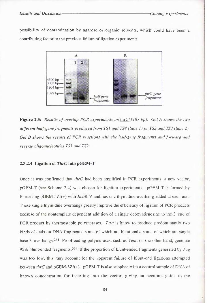
Results and Discussion- Cloning Experiments
possibility of contamination by agarose or organic solvents, which could have been a
contributing factor to the previous failure of ligation experiments.
Figure 2.5: Results of overlap PCR experiments on thrC( 1287 bp). Gel A shows the two
different half-gene fragments produced from TSl and TS4 (lane 1) or TS2 and TS3 (lane 2).
Gel B shows the results of PCR reactions with the half-gene fragments and forward and
reverse oligonucleotides TSl and TS2.
2.3.2.4 Ligation <rf ThrC into pGEM-T
Once it was confirmed that thrC had been amplified in PCR experiments, a new vector,
pGEM-T (see Scheme 2.4) was chosen for ligation experiments. pGEM-T is formed by
linearising pGEM-5Zf(+) with EcoR V and has one thymidine overhang added at each end.
These single thymidine overhangs greatly improve the efficiency of ligation of PCR products
because of the nontemplate dependent addition of a single deoxyadenosine to the 3' end of
PCR product by thermostable polymerases. Taq i^ know to produce predominantly two
kinds of ends on DNA fragments, some of which are blunt ends, some of which are single
base 3' overhangs.264 Proofreading polymerases, such as Vent, on the other hand, generate
95% blunt-ended fragments.26^ if the proportion of blunt-ended fragments generated by Taq
was too low, this may account for the apparent failure of blunt-end ligations attempted
between thrC and pGEM-3Zf-+). pGEM-T is also supplied with a control sample of DNA of
known concentration for inserting into the vector, giving an accurate guide to the
84

Results and Discussion- Cloning Experiments
overall efficiency of ligations and transformations using the pGEM-T system. Ligations were
carried out using various ratios of insert: vector and incubated at room temperature overnight,
as summarised in Table 2.1. Competent JM83 were used for transformations. Colonies taken
from experiment 4 produced a number of putative recombinants which were subjected to
restriction analyses using Apa I, BssH II, EcoR I, Sea I, Ssp I and Xmn I. Two of these
plasmids produced the predicted results with all enzymes. Maxiprep plasmid preparations
were made of these two recombinants (denoted pGEM-TTSa and pGEM-TTSs)and restricted
with Nde I (expected fragments 31, 1287 and 2973 bp). This indicated that pGEM-TTSa had
lost one of its Nde I-sites as no thrC PCR fragment was excised (Fig. 2.6).
4486 bp---- >2973 bp---- >
1287 bp---- >
----- 4500 bp----- 3003 bp----- 1904 bp
----- 1099 bp
Figure 2.6: Nde I-digest of pGEM-TTSa and pGEM-TTSb.
In previous ligation experiments the digested vectors had been purified by phenol/chloroform
extraction and ethanol precipitation, rather than gel-isolation. Although this should have
removed any enzyme and buffer components from the DNA solution, undigested vectors may
have remained, which are smaller and more likely to be taken up by competent cells. As
pGEM-T is provided in a linear form, such vector background is eliminated. Linear DNA is
taken up 50 times more slowly than supercoiled circular DNA molecules by E. coli cells2263
An attempt was made to sequence the gene from pGEM-TTSe, the plasmid which had
retained both Nde I-sites, and from which the gene could therefore be subcloned, using the
fmol™ cycle sequencing protocol. SP6 and T7 primers (Fig. 2.7) were used for this purpose
as these promoter sites are found on all pGEM™ plasmids (Appendix 9). A successful
85

Results and Discussion- Cloning Experiments
sequence was obtained for just the first 317 bases at the C-terminus of the gene (slightly
downstream of the SP6 promoter). This confirmed that the C-terminal Nde I-site was still
intact.
Table 2.1: The results of ligations using thrC and pGEM-T,
No. Ratio of Insert: Vector
Amount of
Transformed
Cells per Plate
Number
ofColonies
% Whites
A Vector + control insert - - 45%
B Vector only control lOOpl 10 10%
1 1:1 50jxl 20 40%
2 1:1 lOOpl 50 20%
3 2:1 50pl 20 0%
4 2:1 mm 50 20%
5 4:1 50pl 20 15%
6 4:1 ioopI 50 60%
7 20:1 50pl 15 80%
8 20:1 IOOpI 25 60%
10 200:1 ioopI 10 70%
9 200:1 50fPl 2 100%
Highlighted row indicates ligation. experiment from, which recombinant plasmids were isolated.
SP6 Promoter Primer: 5' - d(GATTTAGGTGACACTATAG) - 3'
T7 Promoter Primer: 5' - d(TAATACGACTCACTATAGGG) - 3'
Figure 2.7: SP6 and T7 primer sequences
86

Results and. Discussion- Cloning Experiments
BssH n (278)Mte 1(50)1
T1 promoter ( 1)i| thrC insert
Xmn 1 (3281) Ssp I (3695) Af 1(1337) 1
SF6 Promoter (1483) Ssp I (3486)J____ 1___________i______ L_l_____ __
N IApa. I (14)
EcoR I (159)
n i 1SflcI (1381) 5ca 1(3162) $spl (3671)
4PGEM-TTSb Graphic map 4291 base pairs
Figure 2,8: Orientation of thrC in pGEM-TTSg. Restriction sites used in analysis of the
construct are also shown..
2.3.2.S Attempted Ligation of ThrC Into Expression Vectors
2.3.2.5.1 pET-3a and pET-16b
The recombinant thrC gene was excised from maxipreps of pGEM-TTSg using Nde I and
purified using the phenol/acetate method of gel-isolation described earlier, or the Wizard™
Prep DNA Purification System, according to the manufacturer's instructions. The pET-
vectors named above were restricted with Nde I, gel-purified and treated with CIAP prior to
ligations to try to eliminate the self-ligation of plasmids. Although numerous ligations were
attempted with varying ratios of insertrvector, and a number of white colonies were obtained
when selecting for the lac+ phentotype, no recombinants were isolated. Unphosphalased
linear plasmids also failed to produce successful recombinants, indicating that CIAP was not
the sole causative factor in the failure of ligation experiments. Various putative recombinants
failed to produce the expected fragments on digestion with Nde I and EcoR I. As many of
these produced fragments corresponding to the linear vector, but not an excised thrC
fragment, this suggested that the plasmids were self-ligating, as had been observed earlier in
ligations experiments involving cloning vectors.
2.3.2.5.2 pKK223-3
Once again, in an attempt to avoid using Nde I where possible, in case the enzyme was
contributing to the failure of ligations, blunt-end ligation of the excised thrC fragment into an
87

Results and Discussion- Cloning Experiments
expression vector was attempted. The vector chosen for these experiments, pKK223-3,
contains a Sma /-site in the multiple-cloning region (Appendix 9).
The pKK223-3 vector contains the strong tac promoter, which in an appropriate host is
regulated by the lac repressor. Expression of gene inserts can be induced by the use of IPTG
as is the case for pET-vectors. Immediately downstream of the tac promoter is the pUC8
multiple-cloning site (MCS) and the strong rrn ribosomal terminator which stabilises this
vector-host system by inhibiting transcription initiated from the tac promoter in the parent
plasmid. The linearised vector was dephosphatased and purified in the usual manner to
remove CIAP. The thrC gene fragment was excised from pGEM-TTSg using Nde I and
incubated with T4 DNA polymerase to flush the 5'(protruding ends with dNTPs. Ligations
(at room temperature) and transformations were carried out in-gel using SeaPlaque™ GTG
agarose gel in low EDTA TAE buffer, as recommended by the manufacturer. This was to
prevent any loss of the thrC fragment during purification procedures.. After two attempts at
using this protocol without success a new strategy for cloning was proposed.
2.3.2.6 New PCR Fragme^
For some reason the ligation of gene fragments with Nde I-sites at each terminus into pET-
vectors is very problematic. This may be due to Nde I non-specifically digesting the ends of
restricted DNA, as observed by the disruption of the lacZ region of cloning vectors digested
with the enzyme. It may also be due, in part, to the inefficiency of the enzyme in restricting
DNA. In addition, the orientation of such thrC fragments in pET-vectors cannot be
controlled, further decreasing the efficency of ligations. The use of CIAP also presents a
problem. It is beneficial to hinder the self-ligation of the expression vector in order to
increase the efficiency of ligation reactions. However, because CIAP is difficult to remove,
the enzyme may also inhibit ligations between DNA fragments and dephosphatased vectors.
Some success had been achieved previously in the group ligating gene fragments with
different restriction sites at each terminus to pET-vectors. For this reason two new primers
88

Results and Discussion Cloning Experiments
were designed for PCR experiments (Fig. 2.9). The forward primer (TS5) contains an Nde I-
site directly before the gene start codon. As a minimum of 8 bases are needed before the site
for the enzyme to function efficently, a ten base sequence was also added before this
restriction site. The reverse primer (TS6) contains a BamH I-site, which is found on all the
pET-vectors that had been used previously (see Fig. 1.17).
ThrC was amplified via PCR as before, from the original chromosomal DNA preparation,
gel-isolated, restricted with Nde I and BamH I and gel-purified, using the Wizard™ protocol,
before carrying out further ligation reactions.
TS5 3l mer. Forward primerMet Lys Leu Tyr Asn Leu
5' - GTC AGT CAGT CAT ATG AAA CTC TAC AAT CTG Nde I
TS6 2Hmer. Reverse primerMet Met. Asn His Glu * Met TAC TAC TTA GTA GTC ATT CCT AGG CGTA - 5‘
BarH I
4072 bp 3054 bp 2036 bp 1636 bp 1018 bp
517 bp
_ ThrC fragment
Figure 2.9: Left: primers used for the third PCR experiment to amplify thrC. Right: result
of PCR using primers TS5 and TS6.
2.3.2.7 Attempted Ligation of ThrC into pET-3a
Attempts were made to ligate the doubly-restricted PCR product directly into pET-3a. In
order to monitor the progress of the restriction of pET-3a, the original vector was not used in
digestion experiments. Instead, constructs made previously in the group, from pET-3a and
various gene inserts, were restricted with Nde I and BamH I. These had been produced using
the same protocol now being applied to thrC, the gene insert having an Nde I-site at one end
and a BamH I-site at the other. The progress of the restrictions could be monitored by the
observation of a gene insert and linearised vector on agarose gels (Scheme 2.5).
89

Results and Discussion- Cloning Experiments
Gene insertpTC construct
linearised pET-Sa
Ligation
x Nde I x BamH I
thrC x Nde I x BamH I
Gene insert♦+
pTN-TS construct
Scheme 2.5: Plan for the ligation q/thrC into pET-Sa derived from a pTC construct.
A number of different pTC constructs were linearised using Nde I. Subsequent
BamH I-digests produced an observable gene fragment and linearised pET-3a vector (4640
bp). The linearised vector was then isolated from Nusieve™ agarose gel. Two sets of
ligations were attempted, using CIAP-treated vector only in the second case, but no
recombinants were identified.
2)3)2)8 Ligation of New Gene Into p<GEIM-T
2.3.2.81 Amplification of ThrC
It appeared that, even if the termini of the thrC gene fragment contained 2 different restriction
sites, ligation of the gene into a pET-vector would prove difficult. In order to prevent the
need to perform repeated PCR experiments the gene was cloned as before into pGEM-T. At
this stage the original chromosomal DNA preparation (also containing pSG4) from TGI
became ineffective as a template for PCR. Chromosomal DNA was therefore isolated from
XLl-Blue using the SDS-Proteinase K protocol.2™ PCR experiments were carried out using
either this chromosomal DNA preparation or pGEM-TTSe as a template. The first set of
these experiments produced fragments of DNA too small to be thrC, but the use of freshly
synthesised primers and a lower annealing temperature (40 °C) achieved the desired result.
ThrC fragments from PCR were gel-isolated and purified using the Wizard™ PCR Preps
DNA Purification System. Direct ligation experiments produced the recombinant plasmids
90

Results and Discussion Cloning Experiments
pGEM-TTSi and pGEM-TTSn (see Table 2.2). QIAGEN maxiprep plasmid purifications of
these vectors were subjected to Nde I- and BamW I-digestion, with the predicted results (Fig
2.10).
Table 2.2: Results of ligations of the new thrC fragment into pGEM-T.
No. Vector: PCR product ratio Number of Colonies % Whites
1 1:0.8 49 80%
2 1:0.8 37 81%
3 1:1 Lawn 95%
4 1:1 Lawn 95%
5 1:1.5 Lawn 95%
6 1:1.5 Lawn 95%
7 1:2 11 55%
8 1:2 8 63%
9 1:3 15 67%
10 1:3 11 55%
Figure 2.10: Results of ligations of thrC into pGEM-T. Left: plasmid miniprep DNA
preparations pGEM-TTSi and //, taken from ligation experiments 9 and 10 respectively,
showing they are putative recombinants. Right: restriction digests of these recombinants
using Nde / and Bam// I. Lanes 2 and 4 contain restricted pGEM-TTSi, 1 and 3 pGEM-
TTSu. The Nde I-restriction indicates that the gene is oriented differently in the two vectors.
91

Results and Discussion Cloning Experiments
23.2.9 Sequencing of ThrC in pGEM-TTSi and pGEM-TTSu.
cSequences were obtained of the termini of the recombinant gene in both vectors. This was
achieved using the fmol™ protocol for cycle sequencing with SP6 and T7 primers (Fig. 2.7).
Both primers were at the 5'-terminus by incubation with 32P-y-ATP and T4 PNK.
PCR experiments were performed with each reaction containing one of the primers and one
of four chain-terminating ddNTPs. The radiolabelled DNA fragments obtained in this
manner were separated by high-resolution denaturing polyacrylamide gel electrophoresis
using different lanes for each reaction. The sequences obtained are shown in Fig. 2.11,
confirming that the genes are oriented in different ways in either vector. Attempts were also
made to sequence the middle of the recombinant genes at this time, using primers TS3 and
TS4 (Fig. 2.5), but without success. This could be due to the plasmid DNA being too dilute,
the primer solutions having deteriorated during storage, or their concentration having altered
since last being calculated. There could also have been some residual contaminants in the
plasmid DNA preparation, which made the polymerase reaction too inefficient. Whilst
further attempts were made to obtain fuller sequence information on the two recombinant
genes, experiments were carried out to subclone the genes into pET vectors.
92

Results and Discussion Cloning Experiments
dGEM-TTSl x SP6 I.st 120 bases of gene from N-terminus
TT gga gct CTC CAT atg gtc cgg CTC TCT gca ggc ggc cgc ACT cag tga TTG
TCA gtc agt CATIaTG AAA CTC TAC AAT ctg AAA gat CAC AAC gag cag gtc agc
Nde ITTT gcg CAA gcc gta ACC CAG ggg TTG ggc AAA AAT CAG ggg ctg TTT TTT ccg
CAC gac ctg ccg gaa TTC agc ctg
pGEM-TTS, x T7 1st 170 bases of gene from C-terminus
CC cgg ccg CCA|-----------►
TGG cgg gat TTA TGC GGaITCC TTA ctg atg ATT CAT CAT CAA
TTT ACG CAA CGCBamH I
AGC AAA ATC GGC GGG CAC ATT ATG TGA ACT TTT GGC AGA TCC
AAC GTT TCA CCG AGA ATC GCT TCC ACG CTC TCT TTA AAT TTC GCC GGA TGC GCG
gtg ccg A
pGEM-TTSjj x SP6 1 st 80 bases of gene from C-terminus
gga gct CTC CAT ATT ggc gac ATC tgc agg cgg ccg CAC tag tga TTA tgc GGABam
Itcc
h i ATT
tta ctg atg ATT CAT CAT CAA TTT acg CAA cgc agc AAA ATC ggc ggg cag
atg tga aag cag CAC gtt ctg CCA gct CA
pGEM-TTS i 1X T7 1st 200 bases of gene from N-terminus
AT tgt ACA gtc agt cat)Nde
Iatg
I
AAA CTC TAC AAT ctg AAA gat CAC AAC gag cag
gtc agc TTT gcg CAA gcc gta ACC cag ggg TTG ggc AAA AAT cag ggg ctg TTT
TTT ccg CAC gac ctg ccg gaa TTC agc ctg ACT gaa ATT gat gag atg ctg aag
ctg gat TTT gtc ACC cgc agt gcg aag ATC CTC tcg cgc TTT ATT ggt gat gaa
AT
Figure 2.11: Sequences obtained for pGEM-TTSi and pGEM-TTSu. Primer sequences are
shown in red except for the Nde I and Bam// I-restriction sites which are shown in blue and
underlined. The first base, of the thrC gene is indicated by the arrow.
93

Results and Discussion- Cloning Experiments
2.3.2.10: Ligation of ThrC Into pET-Expression Vectors.
Once the thrC gene had been cloned into pGEM-T it was a simple procedure to excise the
gene fragment using Nde I and BamH I and gel-purify it for use in ligations. The pET-
vectors pET-3a, -3b and -16b were also restricted with these enzymes, but not treated with
CIAP as the enzyme was thought to be inhibiting ligations. Various ligation experiments
were carried out using different ratios of insert : vector and a number produced successful
recombinants. This could not be determined from the plasmid minipreps, as the DNA was
smeared when run on a gel. Therefore maxiprep plasmid DNA was produced, as previously
described, from 8 putative recombinants (see Table 2.3).
Table 2.3: pTN-TS constructs produced from various pET-vectors and pGEM-TTS
constructs.
No. pET-vector Source o»ThrC Number of
Colonies^pTN-TS construct(s) obtained
1 pET-3a pGEM-TTS! 8 -
2 ft pGEM-TTSu 8 pTN-TSl and pTN-TS2
3 pET-3b pGEM-TTSi 11 pTN-TS3 '
4 t! pGEM-TTSii 18 pTN-TS4
5 pET-16b pGEM-TTSi 30 pTN-TS5 and pTN-TS6
6 pGEM-TTSn 45 pTN~TS7 and pTN-TS8
a - only white colonies were produced.
The recombinant vectors derived from pET-3a and -3b were subjected to restriction analyses
using Nde I, BamH I, EcoR I, Pvu II and Xmn I and produced the predicted fragments in all
cases. The vectors derived from pET-16b were also restricted using these enzymes and Bel I,
producing the predicted fragment sizes in all cases. Fig. 2.12 shows the graphic maps of the
pTN-TS constructs. The gels for each restriction digest are shown in Figs. 2.13 and 2.14.
94

Results and Discussion- Cloning Experiments
Figure 2.12: Vector map ofpTN-TS constructs Restriction sites for pTN-TS3 and 4 are
similar to those in pTN-TSl and 2 but orientated in the reverse direction..
95

Results and Discussion^- Cloning Experiments
r
7126 bp^_ 6108 bp----4072 bp----3054 bp----
2036 bp----1636 bp----
Nde I-digest
■6987 bp 5888 bp
4198 bp
1690 bp
x EcoR I
.6108 bp 5090 bp 4072 bp 3054 bp
------2036 bp------1636 bp
------1018 bp
3379 bp
2485 bp
xPvu II
.6108 bp ■5090 bp 4072 bp 3054 bp
■ 2036 bp 1636 bp
------1018 bp
2916 bp —♦
1934 bp—*
1038 bp—»
xXmn I
6108 bp 5090 bp 4072 bp 3054 bp
------2036 bp------1636 bp
------1018 bp
Figure 2.13: Gels of restriction digests of'pTN-TS constructs. Lanes are marked 1 - 8
denoting the pTN-TS constructs 1 - 8. The EcoR I-digest ofpTN-TSl and 2 produced the
same fragment sizes as shown for pTN-TS3 and 4.
96

Results and Discussion Cloning Experiments
x Bcl I
6108 bp 5090 bp — 4072 bp — 3054 bp-----
2036 bp-----1636 bp-----
1018 bp-----
x Xmn I
4206 bp —►
1933 bp—*
848 bp—►
____5090 bp------4072 bp------3054 bp
------2036 bp------1636 bp
------1018 bp
------506 bp
X Pvu II
3172 bp-----*2274 bp-----*
1502 bp----- ►
7 8-----5590 Ip-----4072 bp-----3054 bp
-----2036 bp---- 1636 bp
-----1018 bp
----- 506 bp
5487 bp
X EcoR I
1500 bp----- ♦
^6108 bp-----5090 bp-----4072 bp-----3054 bp-----2036 bp-----1636 bp
-----1018 bp
-----506 bp
Figure 2.14: Restriction analyses of pTN-TS constructs derived from pET-16b.
97

Results and Discussion- Cloning Experiments
2.3.3 Cloning of the p-Methylaspartase Gene
Much of the work carried out during attempts to clone the gene for p-methylaspartase into
pET-vectors was similar to that already described for thrC, as the two sets of experiments
were performed simultaneously. Cloning experiments will therefore not be discussed in as
much detail in this section.
2.3.3.1 Cloning the Gene into pGEM-T
2.3.3.1.1 Gene Amplification
The plasmid pGG4 was isolated from E. coli. strain TGI (K-12 flac-prol supE thi hsdRS /
F'-traD36 proA + B+ lad £ lacZ M15 )26° using the alkaline lysis method (Section
2.3.2.l.l).26i. 262 The p-methylaspartase gene was originally amplified using primers which
incorporated Nde I-sites into each terminus of the gene fragment (Fig. 2.15) as for thrC.
These sites are not contained on the gene (Fig. 2.16 and Appendix 10). The PCR product
was then gel-isolated.
Met Lys lie Val Asp Val MAl. 25mer Forward 5 . -tttt cat atg aaa att gtt gac gta primer from N-terminus
Nde IMet Lys lie Val Asp Val / Val Gly Arg Arg Lys *
5' - ATG AAA ATT GTT GAC GTA / GTA GGA AGA AGA AAA TAA - 33' - TAC TTT TAA CAA CTG CAT / CAT CCT TCT TCT TTT ATT - 5
MA2. 28mer Reverse Val Gly Arg Arg Lys * Metprimer from C-terminus cat cct tct tct ttt att gta tac aaaa-5'
Nde I
Figure 2.15: DNA sequence of the two termini of the p-methylaspartase gene (centre) and
the primers (top and bottom) used in the PCR reaction. Nde I-sites on the primers are
underlined.
98

Results and Discussion- Cloning Experiments
343 EcoR I218 Seal
699 Ssp I625 Xmn I 1012 Hpal
Methyl Aspartase 1242 base pairs
Figure 2.16: Graphic map of the restriction sites on the gene for p-methyl-aspartase used
during cloning work..
1904 bp___1584 bp___1375 bp —
947 bp —
564 bp-----
1 2
S
Gene fragment "---- (1242 bp)
Figure 2.17: Result of the first PCR experiment to amplify the gene for p-methylaspartase
(1242 bp). Lane 1 contains a X DNA ladder-, lane 2 a portion of the PCR reaction mixture
after 20 cycles.
2.3.3.1.2 Overlap PCR Experiments
As was the case with thrC, preliminary attempts to ligate the amplified gene into cloning
vectors were unsuccessful and overlap PCR was performed to confirm that the correct gene
had been produced, as for thrC (Section 2.3.2.3). Two DNA fragments were produced using
the two external primers MAI and MA2, and two new primers complementary to the centre
of the gene (Fig. 2.18), which were of the correct size to be half-gene fragments. These were
used as a template in overlap PCR experiments, producing cloned DNA of approximately
1300 bp, consistent with an amplified gene fragment of 1259 bp (1242 bp for the gene plus
17 additional bases produced by the primers, Fig. 2.19).
99
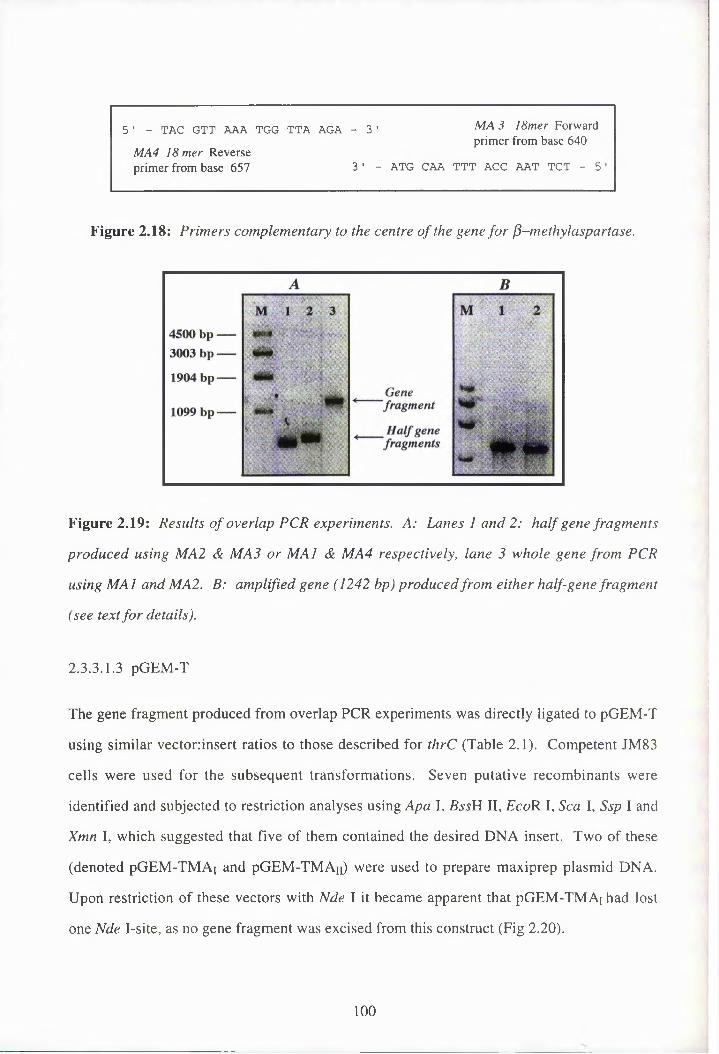
51 - TAC GTT AAA TGG TTA AGA - 3'
MA4 18 mer Reverseprimer from base 657 3 ' -
MA 3 18mer Forward primer from base 640
ATG CAA TTT ACC AAT TCT - 5
Figure 2.18: Primers complementary to the centre of the gene for fi—methylaspartase.
Figure 2.19: Results of overlap PCR experiments. A: Lanes land 2: half gene fragments
produced using MA2 & MA3 or MAI & MA4 respectively, lane 3 whole gene from PCR
using MAI and MA2. B: amplified gene (1242 bp) produced from either half-gene fragment
(see text for details).
2.3.3.1.3 pGEM-T
The gene fragment produced from overlap PCR experiments was directly ligated to pGEM-T
using similar vector:insert ratios to those described for thrC (Table 2.1). Competent JM83
cells were used for the subsequent transformations. Seven putative recombinants were
identified and subjected to restriction analyses using Apa I, BssH II, EcoR I, Sea I, Ssp I and
Xmn I, which suggested that five of them contained the desired DNA insert. Two of these
(denoted pGEM-TMAi and pGEM-TMAn) were used to prepare maxiprep plasmid DNA.
Upon restriction of these vectors with Nde I it became apparent that pGEM-TMAj had lost
one Nde I-site, as no gene fragment was excised from this construct (Fig 2.20).
100

Nde I-digest
Figure 2.20: Nde I-restriction digest of pGEM-TMAi( 1) and pGEM-TMAu(2.)
It was confirmed that the gene insert in pGEM-TMAj had only retained one Nde /-site by
sequencing of the gene insert in each recombinant plasmid, using the fmol™ protocol, with
the GP6 and T7 promoter primers (Fig. 2.7). The correct sequence for the first 120 bases of
the gene from the GP6 promoter (the C-terminus of the gene) and the first 120 bases from the
T7 promoter (the N-terminus) were obtained from pGEM-TMAn-
The p-methylaspartase gene fragment was excised from maxipreps of pGEM-TMAn using
Nde I and purified by agarose gel electrophoresis. As with thrC, attempts to ligate this into
various expression vectors were unsuccessful.
2.3.3.1.4 A New Gene Fragment
After several failed attempts at cloning the gene fragment into various expression vectors, a
new gene fragment was produced with an Nde I-site at the N-terminus and a BamW I-site at
the C-terminus (Fig. 2.21). The PCR product was gel-isolated in the usual manner and
attempts were made to clone this directly into pGEM-T.
101
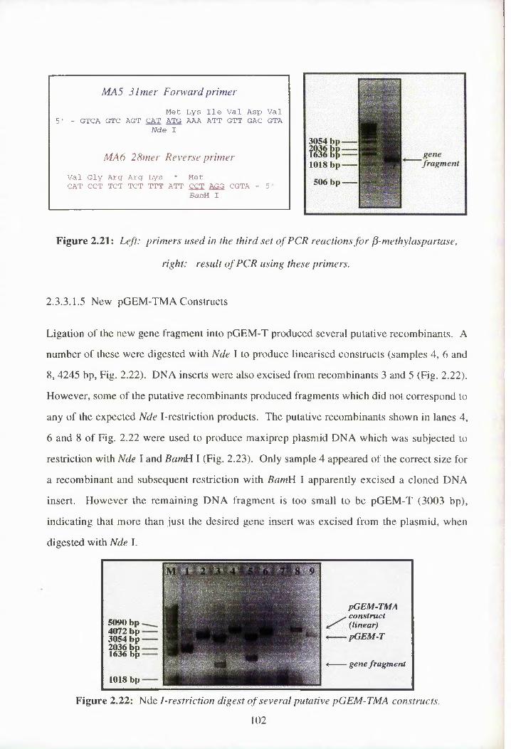
MA5 3lmer Forward primer
Met Lys lie Val Asp Val 5' - GTCA GTC AGT CAT ATG AAA ATT GTT GAC GTA
Nde I
MA6 28mer Reverse primer
Val Gly Arg Arg Lys * Met CAT CCT TCT TCT TTT ATT CCT AGG CGTA - 5
BamH I
3054 bp
1018 bp
506 bp
genefragment
Figure 2.21: Left: primers used in the third set of PCR reactions for p-methylaspartase,
right: result of PCR using these primers.
2.3.3.1.5 New pGEM-TMA Constructs
Ligation of the new gene fragment into pGEM-T produced several putative recombinants. A
number of these were digested with Nde I to produce linearised constructs (samples 4, 6 and
8, 4245 bp, Fig. 2.22). DNA inserts were also excised from recombinants 3 and 5 (Fig. 2.22).
However, some of the putative recombinants produced fragments which did not correspond to
any of the expected Nde I-restriction products. The putative recombinants shown in lanes 4,
6 and 8 of Fig. 2.22 were used to produce maxiprep plasmid DNA which was subjected to
restriction with Nde I and BanM I (Fig. 2.23). Only sample 4 appeared of the correct size for
a recombinant and subsequent restriction with BamH I apparently excised a cloned DNA
insert. However the remaining DNA fragment is too small to be pGEM-T (3003 bp),
indicating that more than just the desired gene insert was excised from the plasmid, when
digested with Nde I.
5090 bp 4072 bp 3054 bp 2036 bp 1636 bp
1018 bp
*------gene /augment
construct(linear)pGEM-T
pGEM-TMA
Figure 2.22: Nde I-restriction digest of several putative pGEM-TMA constructs.
102

Results and Discussion Cloning Experiments
x Nde Ia ■
5090 bp _4072 bp------3054 bp------2036 bp------ 1636 bp
-------1018 bp
- ------ 506 bp
x Nde I x BamH I
5090 bp 4072 bp _3054 bp-------2036 bp-------1636 bp-------
Figure 2.23: Restriction digests on maxiprep DNA produced from samples shown in
Fig. 2.22.
2.3.2.3 Further Work on the Gene for p-Methylaspartase
Further ligation experiments were attempted to produce successful pGEM-TMA constructs,
and several more putative recombinants were identified. However, at this stage efforts were
focused on the work to clone the thrC gene into pET-vectors and further work to clone the
gene for P-methylaspartase into pGEM-T and then into expression vectors was continued by
other researchers. Successful pGEM-TMA constructs were indeed obtained and the gene
fragment excised from these was inserted into pET-5a. Unfortunately, although some success
was achieved in purifying the recombinant enzyme from this source, efforts made by
S. McMahon of J. Naismith's group to produce crystals in order to obtain a 3D-structure of
the enzyme were without success. This was thought to be due to the presence of the
His • Tag, which could not be cleaved with Factor Xa as expected. The gene was therefore
excised and cloned into a new pET-vector to prevent the formation of a His#Tag. However
crystals could not be obtained of the enzyme from this source either. Sequencing
experiments later revealed that the stop codon at the end of the gene had mutated and further
amino acid residues were being added to the end of the protein. The gene then had to be
cloned directly from C. tetanomorphum.
103

Results and Discussion- Gene Sequencing
2.4 Sequencing of The ThrC Gene Fragment in Recombinant Plasmids
Sequencing of each vector was carried out by the University of St. Andrews DNA
Sequencing Unit. They produce sequence data using the ABI PRISM™ DNA Sequencer,
which uses a different fluorescent dye for each DNA extension reaction: A, C, G and T.
They also use AmpliTaq FS, a mutant form of Taq DNA polymerase, which is reported to
read easily sequences which are otherwise difficult. The fluorescent dye is incorporated on
each dideoxy terminator, although it is also possible to use dye primers which are
fluorescently labelled instead. All four extension reactions are then loaded onto a single lane
on a polyacrylamide gel. Perkin-Elmer software is used to read the sequence directly from
the gel by scanning for the four fluorescent dyes.
Table 2.4: Description of primers used in sequencing experiments.
Sequence
SP6SP6 Promoter Primer: External primer
complementary to the SP6 promoter on
pGEM vectors.
5' - d(GATTTAGGTGACACTATAG>- 3'
TS3 Internal primer producing sense strand of
thrC (starting base 643).5' - d(AAC ATC AGG CGT TTG CTG) - 3'
TS4 Internal primer producing anti-sense strand
o/thrC (starting base 660).5' - d(CAG CAA AGG GCT GAT GTT) - 3'
TS6 External primer producing sense strand of
thrC from N-terminus of gene.
5' - d(ATGG GGA TCT TTA CTG ATG
ATT GAT GAT) - 3'
T7
T7 Promoter Primer: External primer
complementary to the T7 promoter on pET
and pGEM vectors.
5' - d(TAATACCACTCACTATACCG) - 3'
104

Results and Discussion- Gene Sequencing
The constructs pGEM-TTSi and pGEM-TTSu were submitted for sequencing using the
vector promoter primers SP6 and T7 and the gene internal primers TS3 and TS4 (Table 2.4).«
Full sequences were obtained of the gene inserts (Appendix 11). The following changes from
the wild-type thrC DNA sequence were observed:
■ In pGEM-TTSi base base 320 (adenosine) of thrC had been deleted, disrupting the
reading frame of most of the gene. Base 944 was also mutated from thymidine to
cytidine. Bases 340 and 716 were also mutated (A to G in both cases).
■ In pGEM-TTSn the mutations described for pGEM-TTSi were found, except that base
340 (A) remained unaltered.
Each pTN-TS vector was also sequenced using the T7 promoter primer, TS6, which is
complementary to the sense strand at the N-terminus of the gene and the two internal primers,
TS3 and TS4. The data obtained from these constructs confirmed the findings for the
pGEM-TTS constructs (Appendix 12). It should be noted that the gene inserts in pTN-TS3,
pTN-TS5 and pTN-TS6 were subcloned from pGEM-TTSi and all others were subcloned
from pGEM-TTSn (Section 2.3.2.10). Some additional mutations were observed in
sequences of the pTN-TS constructs as follows :
■ In pTN-TS2 base 1051 was mutated (G to A).
■ In the construct pTN-TS5 base 278 was mutated (T to A).
The deletion of base 320 of thrC will produce a truncated protein, as this leads to a stop
codon (TGA) after a further sequence of 19 amino acids. In fact, the frameshift caused by
this deletion leads to a total of 18 stop codons along the DNA sequence.
105

Results and Discussion Solid-phase Synthesis
2.5 Solid-Phase Synthesis of (2£)-0-Phosphohomoserine
2.5.1 Alternative Phosphorylating Agents
Due to the difficulties encountered in the synthesis of AJ^-c^j^isopnrpyhlicl'^c^rc^j^lK ls]^lianiidite
(78), a key reagent for the phosphorylation of A-trifluoroac(^i^;yl-^^-isoproyyl-(25)-homoserine
(75) in the solution-phase protocol for substrate synthesis (Section 2.2.2), it was necessary to
find some new means of attaching the phosphate group to 75. To this end a few readily
available phosphory^ting agents were tried: diyhenylyhosyhorpchloridate (80),
diyentafluorophenylpCosyhorocClocidate (81) and A,/Addsopropyl methyl yhosyhpnamidic
chloride (82, Fig. 2.24).
Figure 2.24: Alternative phosphorylating agents tried in the synthesis of(2S)-O-
phosphohomoserine.
Both the phosphonamidic chloride (82) and diyhenylyhooyhorochloridate (80) are
commercially available. There are many reported uses of 80, with different methods for the
subsequent deprotection of the phenyl protecting groups.271-283 The main method for the
removal of the phenyl groups involves hydrogenolysis using platinum dioxide (Adams
catalyst) or platinum/ activated carbon.
106
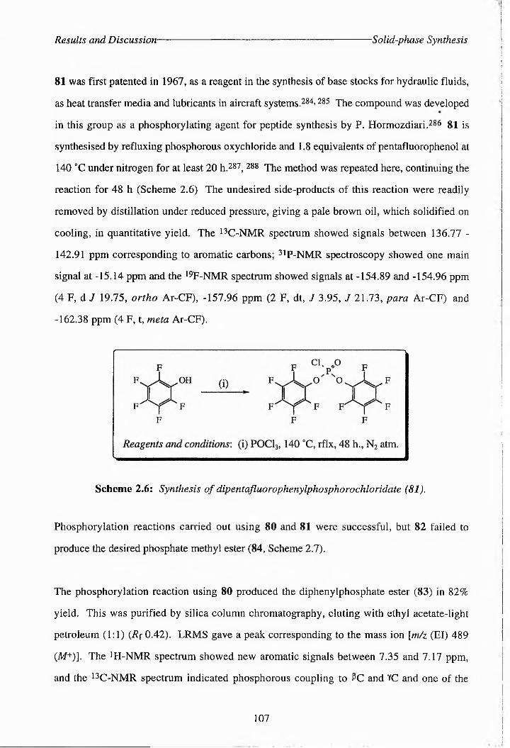
Results and Discussion- Solid-phase Synthesis
81 was first patented in 1967, as a reagent in the synthesis of base stocks for hydraulic fluids,
as heat transfer media and lubricants in aircraft systems.284-285 The compound was developed
in this group as a phosphorylating agent for peptide synthesis by P. Hormozdiari.286 gl is
synthesised by refluxing phosphorous oxychloride and 1.8 equivalents of pentafluorophenol at
140 °C under nitrogen for at least 20 h.287, 288 The method was repeated here, continuing the
reaction for 48 h (Scheme 2.6) The undesired side-products of this reaction were readily
removed by distillation under reduced pressure, giving a pale brown oil, which solidified on
cooling, in quantitative yield. The i4C-NMR spectrum showed signals between 136.77 -
142,91 ppm corresponding to aromatic carbons; 31p-NMR spectroscopy showed one main
signal at -15.14 ppm and the 194F-NMR spectrum showed signals at -154.89 and -154.96 ppm
(4 F, d 7 19.75, ortho Ar-CF), -157.96 ppm (2 F, dt, J 3.95, J 21.73, para Ar-CF) and
-162.38 ppm (4 F, t, meta Ar-CF).
Scheme 2.6: Synthesis of dipentafluorophenylphosphorochloridate (81).
Phosphorylation reactions carried out using 80 and 81 were successful, but 82 failed to
produce the desired phosphate methyl ester (84, Scheme 2.7).
The phosphorylation reaction using 80 produced the diphenylphosphate ester (83) in 82%
yield. This was purified by silica column chromatography, eluting with ethyl acetate-light
petroleum (1:1) (Rf 0.42). LRMS gave a peak corresponding to the mass ion [m/z (El) 489
(M+)]. The 4H-NMR spectrum showed new aromatic signals between 7.35 and 7.17 ppm,
and the 13C-NMR spectrum indicated phosphorous coupling to PC and YC and one of the
107

Results and Discussion- Solid-phase Synthesis
aromatic carbons (Fig. 2.25). The YC signal was also shifted downfield by 6.6 ppm:
5c(50.31 MHz; C2HC13) 169.49 (CO2*Pr), 150.56 and 150.42 (d, 7 7.04, Ar-C), 130.11 (Ar
C), 124.80 (Ar-C), 120.21 and 120.13 (d, 7 17.21, Ar-C), 70.88 (*PrCH), 65.17 and 65.06
(d, 7ptc 5.49, YC), 50.40 («C), 31.82 and 31.68 (d., 7 6.71, PC), 21.79 (fPrCH3). 31P-NMR
spectroscopy produced just one signal at -11.57 ppm.
O
Reagents and conditions: (i) diphenyl chlorophosphorate (80), DMAP, EtgN, DCM, r.t., 17 h.; (ii) dipentafluorophenyl chlorophosphorate (81), EtjN, DCM, r.t., 21 h; (iii) A,A-diisopropyl methyl phosphonamidic chloride (82), DMAP, DIPEA, THE, r.t., 18.5 h.; (iv) MeOH, r.t., 60 h.; (v) m-CPBA, DCM, r.t., 12 h.
Scheme 2.7; Attempted methods of phosphorylating N-trifluoroacetyl-a-isopropyl-(2S)-
homoserine (75).
The phosphorylation using 81 also proceeded in good yield. The 13C-NMR spectrum of 84
showed a downfield shift of the YC signal and phosphorous coupling to PC and
108
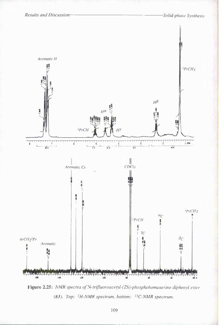
Results and Discussion- Solid-phase Synthesis
Aromatic Cs CDCI3
Figure 2.25: NMR spectra ofNotrifluoroacetyl-(2S)-phosphohomoserine diphenyl ester
(S3). Top: 1H-NMR spectrum, bottom: ,3C-NMR spectrum.
109

Results and Discussion Solid-phase Synthesis
YC. {0c(50.31 MHz; CiHClg) 169.46 (CO2*Pr), 157.82 (F3CCO), 138.46 (Ar-C), 71.19
(*PrCH), 67.44 and 67.32 (d., 5.90, YC), 50.23 («C), 32.03 and 31.88 (d., Jp^-e 7.32,
PC), 21.69 (^PrCHg)- The 4ip-NMR spectrum gave a new signal at -10.77 ppm, and 19F-
NMR spectroscopy of the crude material showed new multiplet signals at 189.99, 188.27,
187.25 and 185.45 ppm. 19F signals for 81 in this spectral window appeared at 198.10,
195.06 and 190.60 ppm.
Having established two new methods for the phosphorylation of 75, attempts were made to
deprotect the phosphate esters 83 and 84 using base-catalysed hydrolysis. If this were
successful then the removal of all protecting groups could be performed in one step to give the
substrate (32), a major advantage over the previous route (Scheme 2.3).
Although 83 was subjected to hydrolysis in IM sodium hydroxide, for several days, the
deprotection appeared to be incomplete. The 1 H-NMR spectrum still showed aromatic signals
at between 7.28 and 7.00 ppm, and although 2ip-NMR spectroscopy showed one signal at
0.67 ppm, which possibly indicated the presence of the desired product {8p( 121.42 MHz;
4HOH) 0.59 for (32)}, a number of other 31P signals were also present. P. Hormozdiari
found, whilst using this phosphorylating agent, that hydrogenolysis of diphenyl phosphates at
atmospheric pressure, using either Adams catalyst or platinum/ activated carbon, failed to
produce complete removal of the phenyl protecting groups.^6 Reports in the literature,
however, state that complete deprotection can be effected using this method within 30 min to 3
h.271-283 84
84 was subjected to overnight hydrolysis in IM sodium hydroxide. A white precipitate was
formed, and tic on cellulose [eluant ‘PrOH/H2O/NH3 (20:5:6), developing with ninhydrin]
showed a band with a similar Rf value to that of the target compound. The 1 H-NMR spectrum
was, however, inconclusive, and the 4PrCH3 signal of the ot-ester was still present, indicating
that hydrolysis was incomplete. The i2C-NMR spectrum contained many aromatic signals at
between 142.64 and 128.50 ppm, indicating that the pentafluorophenyl groups were still
present. P. Hormozdiari had attempted a number of methods of hydrolysing off the
110

Results and Discussion- Solid-phase Synthesis
pentafluorophenyl groups, using water, 5% NaHCOg, or an excess of base, but the reaction
would not go to completion, even when an ethanolic solution of KOH and an organic base,
pyridine/ H2O mixture was used.286
These two routes to the substrate appeared then to be unviable and the phosphate esters were
not characterised further. Fortunately, the acid-lability of pentafluorophenol esters has been
reported previously in the literature,288’289 and some success had been achieved in this group
prior to these studies in the deprotection of such phosphates on the solid-phase, using acidic
conditions.286 With this in mind the solution-phase protocol for the synthesis of (2S)-0-
phosphohomoserine (32) was modified in order to apply it to the solid-phase.
2.5.2 19F Gel-phase NMR Spectroscopy
One drawback to solid-phase synthesis is that monitoring of reaction progress is problematic,
unless one of the reagents in solution is a strong UV-chromophore. Often the compound must
also be cleaved from the resin to allow proper characterisation. Conventional 1H- and 13C-
NMR spectroscopy of compounds attached to solid supports (gel-phase NMR) usually requires
prolonged spectral acquisition, gives broad resonances and is dominated by signals from the
solid support.290 Various approaches to overcome these problems include magic angle
spinning and the use of 13C-enriched building blocks, but these methods are expensive.
Recently, however, it has been shown that 19F gel-phase NMR is a very sensitive technique
for measuring transformations on solid-phase supports involving fluorine-containing
compounds. 19F chemical shifts are spread over a wide frequency range and structural
changes which are even quite distant from the fluorine atom will affect the position of the
signal. Svensson and coworkers found that the correlation between the 19F chemical shifts for
the support-linked compounds and the corresponding soluble references was excellent (< 0.1
ppm difference) and that there was a significant change in the 19F chemical shift (> 1.0 ppm)
for each transformation they carried out.290 Shapiro et ai. also reported the successful use of
111

Results and Discussion Solid-phase Synthesis
this technique for monitoring the time-course of nucleophilic displacement of fluorine from a
solid-sup)p^ort2^(Ji In our experiments to perform the solid-phase synthesis of (2S)-0-
phosphohomoserine, the presence of the A-trifluroacetyl group lent itself readily to the use of
this technique, as well as FT-IR and 13C gel-phase NMR spectroscopy, for the characterisation
of compounds on the solid support.
2.5.3 p-Benzyloxybenzyl Alcohol (Wang) Resin
Wang resin had previously been widely used in the group and was therefore an obvious first
choice as a solid support. The strategy developed for the solid-phase synthesis of (2SY0-
phosphohomoserine (32, Scheme 2.8) was similar to the solution-phase procedure (see
Scheme 2.3). The only major modification was that Wang resin was to be loaded with
A-trifluoroacetyl-(25)-aspartate (86) by using the alcohol group on the resin, in the place of
propan-2-ol, for the ring opening of the symmetric anhydride (73). It was hoped that, due to
the bulky nature of the resin, the opening of the anhydride would proceed regioselectively as
with isopropanol. The P-carboxyl group was to be reduced in the usual manner by mixed
anhydride methodology and the resulting alcohol group phosphorylated using
dipentafluorophenyl chlorophosphate (81). The phosphate group would then be deprotected
using TFA/^O/TES (90:5:5) which would also cleave A-trifluoroacetyl-(2S)-<9-
phosphohomoserine (90) from the resin. This would leave only the hydrolytic removal of the
N-TFA-group using base to achieve the target compound. The solid-phase methodology also
removes the need for time-consuming purification steps and hydrogenolysis of the phosphate
benzyl protecting groups of 76, which often appeared to be inefficient.
Ou
F.C NHH'/Aco2h
ho2c
N-trifluoroacetyl-(2S)-aspartate (86)
112

Results and Discussion- Solid-phase Synthesis
Scheme 2.8: The proposed, solid-phase synthesis of (2S)-O-phosphohomoserme (32) on
Wang resin.
113

Results and Discussion Solid-phase Synthesis
2.5.3.1 The Loading of Wang Resin With 2V-TrifluoroacetyI-(2S)-aspartate
When using propan-2-ol to selectively protect the a-carboxylic group of 73 the reaction was
carried out at room temperature, after the addition of the alcohol at 4 “C (Scheme 2.3).
Experiments to load 86 onto Wang were carried out as indicated in Scheme 2.8 and using the
conditions listed in Table 2.5. A loading temperature of 4 °C was tested with Wang resin first
of all (experiments 1-3, Table 2.5), as this low temperature might be expected to enhance the
regioselectivity of the substitution. The reaction proceeded too slowly, however, to be useful
at this temperature. Higher temperatures and catalytic amounts of DMAP were necessary to
produce satisfactory loading within a reasonable time. Experiments 4-9 (Table 2.5) established
that the optimum temperature for loading was probably between room temperature (23 0C) and
45 °C. In most cases a reaction temperature of 30-35 aC was used, as the regioselectivity of
the substitution was better at this temperature than at higher temperatures. IR spectroscopy
indicated the formation of an ester linkage {omax(]KBr)/cm-1 1735 s (C=O, ester)} and both the
13C and 19F gel-phase NMR spectra confirmed the substitution of 86 onto the resin: 5C(74.76
MHz; DMSO) 171.33 (aCO2R), 162.48 (pCOoH), 130.02 (F3CCO), 117.84 (F3C), 49.40
(«C), 34.81 (PC); 5p(282.2 MHz; DMSO) -74.91 (F3C on ct-ester). A portion of A-TFA-
aspartate (86) was also cleaved from the resin using TFA/H2O/TES (90:5:5) and characterised.
The i2C-NMR spectrum showed two signals for carboxylic groups {172.54 (aCO2H) and
171.06 (pCO2H))} shifted downnield from die carbonnl ssggnll for 73 {{66.28 (0tC=O) aan
167.61 (pC=O)}. There were also two quartets which corresponded to the A-trifluorocaetyl
group (156.70 (q, 37.18, F3CCO) and 115.43 (q, f 285.26, F3C)}. The optical
rotation of the cleaved material was in good agreement with that obtained for the solution-phase
product (-25.09 (c. 0.87, EtOH) compared to -24.35 for the solution-phase product},
indicating that the use of DMAP as an acylation catalyst does not cause significant
racemisation, although P. Sieber found it did when attaching Fmoc-amino acids to Wang
resin.292
The regioselectiv^y of substitution reactions under various conditions were determined by
114

Results and Discussion- -Solid-phase Synthesis
Table 2.5: Loading of Wang resin with N-TFA-asp under various conditions
Experiment Temperature Time (hours) Equivalents of
DMAP
Equivalents of
Anhydride
Stirring % Loadinga
1 4'C 15 none 10 X minimal
2 4"C 15 0.5 " X none
3 4'C 15 1.5 X none
4 O°C / rt 15/6 none " X none
5 O’C / rt 15/6 1 " X none
6 rt 19.5 none X none
7 rt 19.5 1 " X none
8 rt / 45'C 28.5 / 16 none " X 12
9 rt / 45°C 30 / 10.5 1 " X 62
10 rt / 60°C 55.5 / 8 1 " X 65b
11 rt 65 2 " J 31
12 rt 160 2 " J 82
13 30°C 118.5 2 J 100
14 25'C 36.5 1 •• J none
15 15°C / 35°C 48 / 116 1 J 75
16 rt ./ 30°C 14/49 1 " J 58
17 30’C 142 1 " J 46
18 30°C 1 5 J 56
19 30'C " 1 2.5 J 12
20 30°C " 2 10 J 79
21 30’C " 4 " J 81
a - loading was determined by comparing the mass of the resin before and after loading experiments (after careful
washing and drying of loaded resin), values given are only approximate; b - a reaction was carried out using 1 eq.
pyridine under similar conditions, giving a loading of 60%
115

Results and Discussion- SoUd-pha.se Synthesis
methylating the free acid group of 87 with diazomethane. The methyl-ester 91 was then
cleaved from the resin using TFA/H2O/TES (90:5:5), and the ratio of a- vj 3~methyl ester
peaks on iH-NMR spectra was determined {3.70 (3 H, s, aCO2CH3, 16%), 3.63 (3 H, s,
PCO2CH3, 84%)}. 19F solution-phase NMR spectroscopy of the methyl ester 91 (6p(282.2
MHz; C2HO3) -76.66 (F3C on P-ester)} and 14F gel-phase NMR spectroscopy on 77-TFA-
asp-Wang (87) {8p(282.2 MHz; (C2H3)2SO) -74.27 (12%, F3C on P-ester), -74.91 (88%,
F3C on ac-^c^^teM')} also confirmed the findings of these NMR experiments (Fig. 2.26 and Table
2.8). This indicated that the lower temperatures produced better selectivity for a--substitution.
An experiment left for a week at room temperature (12, Table 2.5) gave a high loading (82%)
and one of the the highest regioselectivities achieved (a/p 5.2:1 as judged from the 1 H-NMR
spectrum of the methyl ester (91)}.
The P-methyl ester (91) produced by diazomethane treatment of 87.
Some experiments were necessary in order to optimise the equivalents of anhydride and DMAP
used in the reaction. It was also of interest to ascertain the effect of varying the number of
equivalents of DMAP, with respect to the racemisation of the loaded amino acid and the
regioselectivity of substitution. Experiments 17-20 (Table 2.6) show the effect of different
quantities of anhydride and DMAP on the [a]]) values of 86 cleaved from the resin. Loading
experiments 8, 9, 10, 12 and 22 - 27 (Table 2.7) show the effect of different temperatures and
equivalents of DMAP on the regioselectivity of the nucleophilic substitution reaction. Although
2.0 equivalents or more of DMAP gives a markedly faster loading of the resin compared to 1.0
equivalents, this has an adverse effect on the regioselectivity and causes greater racemisation of
86.
116

Results and Discussion -Solid-phase Synthesis
Table 2.6: The racemisation effects of varying amounts of DMAP.
Experiment0 Ratio of Equivalents of
DMAP vs. Anhydride[of
17 1:10 -25.09
18 1:5 -20.25
19 2:5 - b
20 1:5 -18.91
21 2:5 -20.89
Solution phase
product‘- -24.35
a - numbers refer to experiments listed in Table 2.5; b - insufficent 86 was loaded on the resin to obtain an
[a]o reading; c - synthesised by hydrolysis of 78.
All reactions were carried out at 30 °C. N-TFA-asp (86) for these measurements was cleaved using 90:5:5 TFA/H2O/TES and recrystallised from ether.
The use of sonication in the attachment of compounds to solid-supports has been reported in
the liteLnt^i;rr<eJ2S>3 To pursue this method a loading experiment was carried out overnight at
room temperature using 1 equivalent of DMAP and sonicating the reaction for three bursts of
15 minutes. This produced a loading of 26%, which appeared quite efficient for a relatively
short reaction time However, an attempt to repeat this method, sonicating the resin and
anhydride for longer failed to produce any loading. There was insufficient time in these studies
to optimise the amount of sonication that can be tolerated by the resin, before cleavage of the
loaded amino acid occurs. No studies were made at this time of the regioselectivity and
racemisation effects of this technique, but sonication remains a promising method for
efficiently loading amino acids onto Wang resin.
117
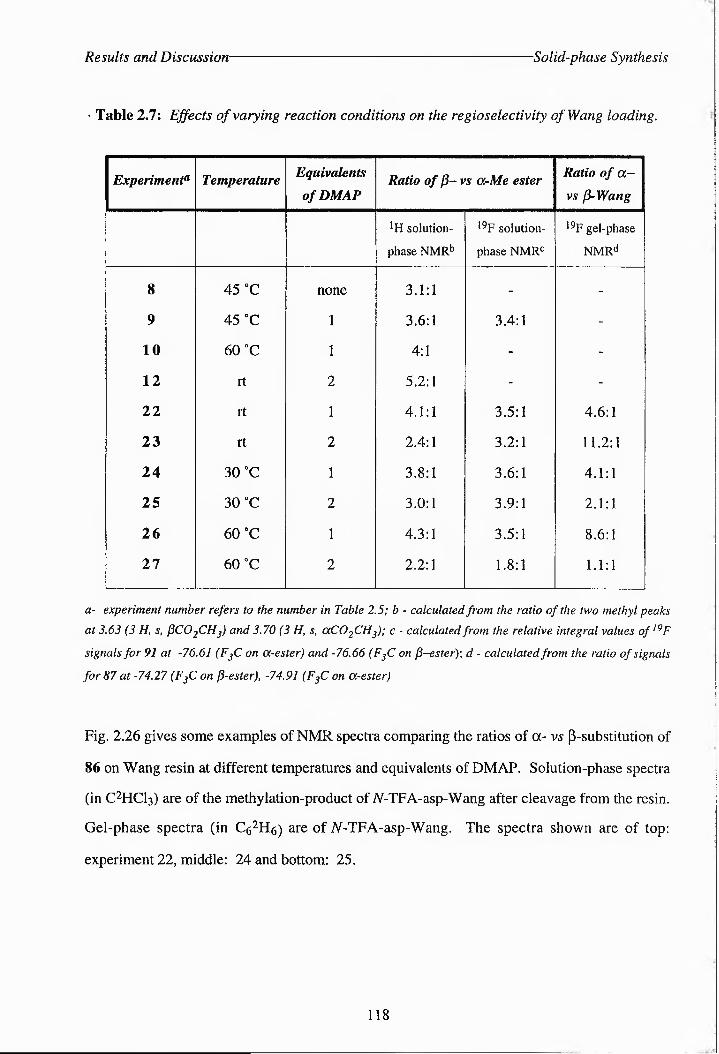
Results and Discussion- -Solid-phase Synthesis
' Table 2.7: Effects of varying reaction conditions on the regioselectivity of Wang loading.
Experiment? Temperature Equivalents
of DMAPRatio of p- vs O-Me ester Ratio of a
vs p-Wang
*H solution-
phase NMR2
l9F solution-
phase NMR2
19F gel-phase
NMRd
8 45 OC none 3.1:1 - -
9 45 °C 1 3.6:1 3.4:1 -
10 60 °C 1 4:1 - -
12 rt 2 5.2:1 - -
22 rt 1 4.1:1 3.5:1 4.6:1
23 rt 2 2.4:1 3.2:1 11.2:1
24 30 0C 1 3.8:1 3.6:1 4.1:1
25 30 °C 2 3.0:1 3.9:1 2.1:1
26 60 "C 1 4.3:1 3.5:1 8.6:1
27 60 °C 2 2.2:1 1.8:1 1.1:1
a- experiment number refers to the number in Table 2.5; b - calculated from the ratio of the two methyl peaks at 3.63 (3 H, s, fiC02CH3) and 3.70 (3 H, s, aCO2CH3); c - calculated from the relative integral values of,9F
signals for 91 at -76.61 (F3C on a-ester) and -76.66 (F?C on P-esterf, d - calculated from the ratio of signals
for 87 at -74.27 (F3C on p-ester), -74.91 (F3C on a-ester)
Fig. 2.26 gives some examples of NMR spectra comparing the ratios of a- vs P-substitution of
86 on Wang resin at different temperatures and equivalents of DMAP. Solution-phase spectra
(in C2HCl3) are of the methylation-product of N-TFA-asp-Wang after cleavage from the resin.
Gel-phase spectra (in C62H6) are of iV-TFA-asp-Wang. The spectra shown are of top:
experiment 22, middle: 24 and bottom: 25.
118

Results and Discussion -Solid-phase Synthesis
1H solution phase 19F solution-phasetHKOKO
KO
o
7.34 25.52
19F gel-phase<Tl00Mi
24,965.43
3.70 ppm 6.34 26.07
3.70 ppm7.81 29.77
76.60 ppm8.12 29.02
tnoo
t-
ct-Wong vs p-Wang4.1:1
Tn - r | t: nyt ^PTT-^ ]TT^|Trr^p-75 -78 ppm
32.77 .....8.05
MJm • MJ co
a-Mc w P-Mc ' I 1:3.0
a-Mc w p-Mc | 1:3.9 \
I a-Wong v.i P-Wong2. 1:1
J
to
76.60 ppm 7.48
28.97
iin i rjisn] rrn pn n pn i--75 -78 ppm
20.88 9.85
Figure 2.26; Examples of NMR spectra comparing ratios of a-vs ^-substitution of 86 on Wang.
119

Results and. Discussion 'S olid-phase Synthes is
Figure 2.27: 13C- gel-phase NMR spectra of top: Wang resin, bottom: 87, both in
DMSO.
120
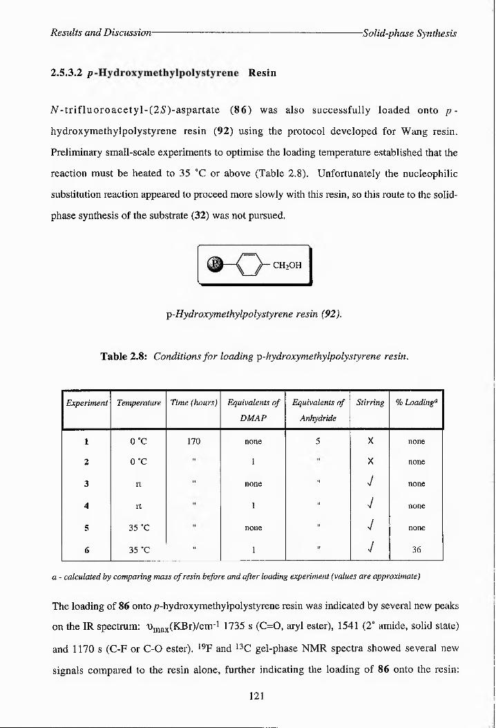
Results and Discussion -Solid-phase Synthesis
2.S.3.2 p Resin
/*--rifluoroacetyl-(2S)-aspartate (86) was also successfully loaded onto p~
hydroxymethylpolystyrene resin (92) using the protocol developed for Wang resin.
Preliminary small-scale experiments to optimise the loading temperature established that the
reaction must be heated to 35 °C or above (Table 2.8). Unfortunately the nucleophilic
substitution reaction appeared to proceed more slowly with this resin, so this route to the solid-
phase synthesis of the substrate (32) was not pursued.
CH2OH
p-Hydroxymethylpolystyrene resin (92),
Table 2.8: Conditions for loading p-hydroxymethylpolystyrene resin.
Experiment Temperature Time (hours) Equivalents of
DMAP
Equivalents of
Anhydride
Stirring % Loading0
1 0 °C 170 none 5 X none
2 0 °C 1 X none
3 rt " none tl J none
4 rt 1 II V none
5 35 °C none II J none
6 35 °C " 1 II J 36
a - calculated by comparing mass of resin before and after loading experiment (values are approximate)
The loading of 86 onto p-hydroxymethylpolystyrene resin was indicated by several new peaks
on the IR spectrum: DjpaxCKBPrV^m’1 1735 s (C=O, aryl ester), 1541 (2° amide, solid state)
and 1170 s (C-F or C-0 ester). 19F and 13C gel-phase NMR spectra showed several new
signals compared to the resin alone, further indicating the loading of 86 onto the resin:
121

Results and Discussion- Solid-phase Synthesis
(Sc(74.76 MHz; (C2H3)2SO) 171.37 (ocC02R), 162.58 (pCO2H), 157.00 (F3CCO), 117.87
(F3C), 49.45 («C), 34.79 (PC); 6F(282.2 MHz; C2HC13) -74.87 (F3C)}.
2.5.3.3 Attempted Reduction of oc-p-Benzyloxybenzylpolystyrene-A-
trif!uoroacetyl-(2S)-aspartate
Once the procedure for loading 86 onto Wang resin had been established, attempts were made
to reduce the free P-carboxylic group of 87 to an alcohol using the mixed anhydride
methodology developed previously for the solution-phase synthesis of 75. The first attempt
using this method led to cleavage of most of the A-trifluoroacetyl-(25)-aspartate (86) from the
resin. The failure of this method, and a desire to avoid the awkward step of removing excess
NMM and EBCF under nitrogenvza cannulae, led us to investigate a number of other methods
for the selective reduction of carboxylic acid groups in the presence of both amide and ester
functionalities.
The use of boranes in organic synthesis has been widely reported in the literature.294 Some
borane complexes can selectively reduce carboxyl groups to primary alcohols in the presence of
other functionalities, without the need for prior activation of the acid group. A popular reagent
is diborane in THF, which, according to the literature, reacts extremely rapidly and selectively
with acids.295-299 H. C. Brown et al. observed that aliphatic acids were reduced under mild
conditions within 15-30 minutes and aromatic acids within 24 hours.295 In fact, the relative
reactivity of this reagent with various functionalities is almost the reverse of that of alkali metal
borohydrides.294 This is a result of the Lewis acid nature of diborane, which functions best as
a reducing agent in attacking groups at positions of high electron density.295 Of particular
interest was a carboxylic acid reduction carried out by Kluender and coworkers in the presence
of A-trifluoroacetyl and methyl ester groups (Scheme 2.9).300
122
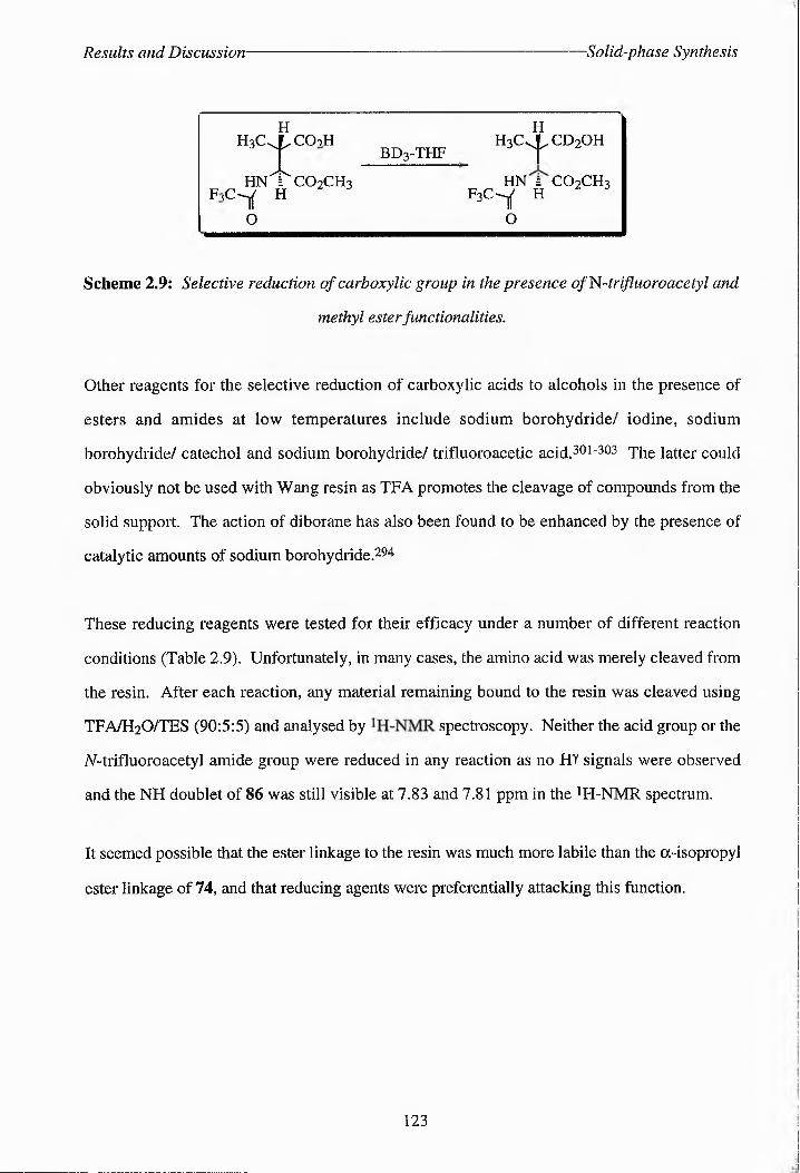
Results and Discussion- -Solid-phase Synthesis
H H--------------- -
H3C.XCOKH H3CJ cd2ohT - BD3-THF y
HN'"bcO2CH3 HN £ CO2CH3F,Cd H F3C-Y H
0 O
Scheme 2.9: Selective reduction of carboxylic group in the presence ofNtrifiuoroacetyl and
methyl ester functionalities.
Other reagents for the selective reduction of carboxylic acids to alcohols in the presence of
esters and amides at low temperatures include sodium borohydride/ iodine, sodium
borohydride/ catechol and sodium borohydride/ trifluoroacetic acid. 301-303 The latter could
obviously not be used with Wang resin as TFA promotes the cleavage of compounds from the
solid support. The action of diboranc has also bccn found to bc enhanced by thc presence of
catalytic amounts of sodium borohydride.294
These reducing reagents were tested for their efficacy under a number of different reaction
conditions (Table 2.9). Unfortunately, in many cases, the amino acid was merely cleaved from
the resin. After each reaction, any material remaining bound to the resin was cleaved using
TFA/H2O/TES (90:5:5) and analysed by spectroscopy. Neither the acid group or thc
A-rrifluoroaceryl amide group were reduced in any reaction as no H7 signals were observed
and the NH doublet of 86 was still visible at 7.83 and 7.81 ppm in the 1H-NMR spectrum.
It seemed possible that the ester linkage to the resin was much more labile than the a-isopropyl
ester linkage of 74, and that reducing agents were preferentially attacking this function.
123

Results and Discussion- -Solid-phase Synthesis
Table 2.9: Conditions for attempted reductions of a-p-benzyloxybenzylpolystyrene-N-
trlfluoroacetyl-( 2S faspartate.
Exp. Solvent Reducing Reagent Temp. Time%
cleavage
1 DMF-THF2.5 eq ea NMM and IBCF
2.5 eq NaBH4-5 °C
-5 ’C/ rt30 min
2 h/O/N 65%
2DMF/ THF
2.5 eq ea NMM and IBCF2.5 eq NaBH
O°C0 °C
2 h24 h -
3 THF1.1 eq ea NMM and IBCF
0.6 eq NaBH40 °C
0 *C/ rt30 min
3 h/ 14 h 35%
4 DMF-THF5 eq ea NmM and IBCF
2.5 eq NaBH40 °C0 °C
30 min6 h/ 1.5 h -
5 DME5 eq ea NMM and IBCF
0.6 eq NaBH40 °CO °C
0/N5 h -
6DME
5 eq ea NMM and iBCf0.55 eq NaBH4
O°C0 °C/ rt
0/N1.5 h/ 1.5 h 13%
7 DMF-THF 1.1 eq BH3-THF 0 °C O/N 72%8 DMF-THF l.l eqBH3-THF 0' °C/ rt 7h/0N -9 Thf 1 eq BHg-THF, 0.1 eq NaBH4 0 °C 5 h -10 DMF-THF 1.2 eq NaBH4, 0.5 eq I2 (dark) 0 °C 14 h 7%11 DMF 1.2 eq NaBH4, 0.6 eq I2 0 °C/ rt 5 h/O/N -12 THF/ EtOH 1.2 eq NaBH4, 0.5 eq I2 (dark) 0 °C 24 h -13
DMF2,5 eq ea NMM and IBCF
2.1 eq N-selecti'ide0 °C0 °C
30 min2 h/ 30 min 39%
DMF was used in preliminary reactions due to its resin-swelling properties. When several
reactions failed in this solvent, the solvent was changed to THF, as recommended in the
solution-phase procedures. It was thought that the reducing reagents might be interacting with
DMF, rendering them less effective. After reactions in THF also failed DME was selected as a
possible alternative. Sodium borohydride is soluble in few organic solvents excepting alcohols
and DME.294 Experiments to reduce the protected aspartate 74 using this reagent were
successful in THF probably because it was less essential to solvate the sodium borohydride as
74 was already in solution. In this case, however, the reaction was hindered by both the
reducing agent and the substrate being in the solid-phase. DME also has the advantage of
being slightly more dipolar than THF, and therefore has better resin swelling properties.
Reactions had been carried out successfully in the group previously using NMM and IBCF
with compounds on Wang resin. The results obtained here were therefore difficult to explain.
124

Results and Discussion- -Solid-phase Synthesis
In order to better understand the mechanism by which 86 was being cleaved, a number of
control experiments were carried out using just these two reagents (Table 2.10). Some of these
reactions resulted in significant proportions of 86 being cleaved from the resin.
The washings from experiments 3-6 were analysed by OH- and 19F_NMR spectroscopy and all
were found to contain at least some traces of 86. These findings, and the elimination of 86
caused by NMM alone (experiments 2, 5 and 6, Table 2.10), indicated that the cleavage of 86
from the resin could be induced by the presence of base. It was also noted that, in some early
experiments using NMM and IBCF, the HH-NMR spectrum of material cleaved from the resin
displayed multiplet signals at around 2 and 4 ppm, indicative of an isobutanol moiety derived
from IBCF. On the basis of these findings the elimination mechanism shown in Scheme 2.10
was proposed.
Table 2.10: Experiments to find the reagent(s) responsible for cleavage of 86 from the
resin.
Exp. Reagent Equivalentsa Time % cleavage
1 NMM 5 3 h none
2 NMM 5 4 84.5
3 (i) NMM
(ii) IBCF
5
5
2 h
2.5 h40
4 (i) NMM and IBCF
(ii) DIEA
5 of each
2
30 min
3.5 h54.4
5 NMM 2.5 30 min 49.7
6 NMM 2.5 12 h 32
All reactions listed in this table were carried out at 0 °C in DME. a - relative to loading of 86 on the resin.
125

Results and Discussion- Soiid-phase Synthesis
Scheme 2.10: Proposed scheme for the reaction of 87 with NMM and IBCF.
The postulated elimination mechanism shown in Scheme 2.10 was confirmed when
experiments were canied out to provide evidence for the activation of the carboxylic acid group
(Section 2.5.3.4), It was found that if the bases used for these reactions were added gradually
in dilute solution, this resulted in less compound being cleaved from the resin. IBCF or the
coupling reagent PyBOP (93) were also added before any base. This meant that as small
amounts of corboxylate ion were fonned, it reacted preferentially with the relative excess of
activating agent, leaving little carboxylate available to cause elimination/ cleavage.
It was assumed that, because the formation of a carboxylate ion in 74 did not lead to cleavage
of the ac-isopropyl group, the carboxylate ion formed by 87 would also not attack an ester
function. There ore two possible explanations for the difference in reactivity between these two
126

Results and Discussion- ■Solid-phase Synthesis
esters. Firstly it may be that the ester linkage is weaker in 87 than in 74. Another possible
explanation is based on the different phases of 87 and 74. The carboxylate ion formed by 74
is fully solvated. The carboxylate ion of 87, on the other hand, cannot interact to such an
extent with the solvent, and is therefore much more nucleophilic towards the ester linkage.
BenzotriazolyZoxy-/ri‘s/pyrro/idin0/-phosphoni'umhexafZuorophosphate (PyBOP, 93)
Although the mechanism given in Scheme 2.10 could explain the cleavage of 86 in the
presence of NMM, there was also significant cleavage of the compound in the presence of
diborane. This cleavage must therefore occur via a different mechanism. One possibility is
that, due to its Lewis acidic nature, diborane readily coordinates with the oxygen atom linking
86 to the resin, which further weakens the ester bond (Scheme 2.11) One advantage of using
p-hydroxymethylpolystyrene resin (92) instead of Wang resin, is that this elimination
mechanism would not be possible.
O?h3 o
CH2-^6 'T CO2H
HN CF3Ifo
Scheme 2.11; Proposed mechanism for the dlborane-lnduced cleavage of 86from Wang
resin.
127
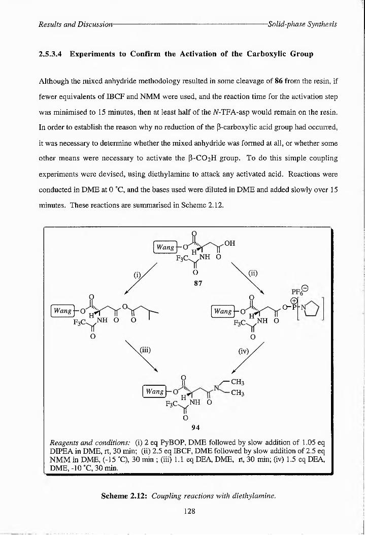
Results and Discussion -Solid-phase Synthesis
2.S.3.4 Experiments to Confirm the Activation of the Carboxylic Group
Although the mixed anhydride methodology resulted in some cleavage of 86 from the resin, if
fewer equivalents of IBCF and NMM were used, and the reaction time for the activation step
was minimised to 15 minutes, then at least half of the N-TFA-asp would remain on the resin.
In order to establish the reason why no reduction of the p-carboxylic acid group had occurred,
it was necessary to determine whether the mixed anhydride was formed at all, or whether some
other means were necessary to activate the P-CO2H group. To do this simple coupling
experiments were devised, using diethylamine to attack any activated acid. Reactions were
conducted in DME at 0 0C, and the bases used were diluted in DME and added slowly over 15
minutes. These reactions are summarised in Scheme 2.12.
o-p
Wang\- CH3H
©I
F3<C^NH o
0
O94
Reagents and conditions: (i) 2 eq PyBOP, DME followed by slow addition of 1.05 eq DIPEA in DME, rt, 30 min; (ii) 2.5 eq IBCF, DME followed by slow addition of 2.5 eq NMM in DME, (-15 °C), 30 min ; (iii) 1.1 eq DEA, DME, rt, 30 min; (iv) 1.5 eq DEA, DME, -10 °C, 30 min.
Scheme 2.12; Coupling reactions with diethylamine.
128

Results and Discussion Solid-phase Synthesis
Both methods successfully produced the amide 94, although a consistent yield could not be
obtained, and some cleavage still occurred in nearly all cases. A quantitative yield was
obtained in one case using PyBOP. The PyBOP activation also produced the cleaner product:
For characterisation the amide was cleaved from the resin using TFA/H2O/TES (90:5:5){m/z
(El) 284 (42%, M+)}. The 'H-NMR spectrum showed signals for a carboxylic group {172.29
(aCO2H)| and an amide {169.91 [pC(O)N)]} and two sets of signals corresponding to the
ethyl groups {42.52 and 40.89 (CH3CH2N, 2 conformations) and 13.50 and 12.46
(CH3CH2N, 2 conformations)}.
2.5.3.5 Reduction of a-/?-Benzyloxybeiizylpolystyrene-.A-trifluoroacetyl-(2S)-
aspartate
Having established two methods of activating the acid group and the way to minimise cleavage
of 86 from the resin, a number of different reducing agents were tested (Fig. 2.28). Some
more hindered reducing agents were tried, as these might have greater selectivity toward the
activated acid rather than the ester linkage. PyBOP was used in most cases to activate the P~
carboxyl group, as indicated in Scheme 2.12. The results of these experiments are summarised
in Table 2.11. The success or failure of the reduction reactions was monitored by cleaving any
remaining material from the resin using TFA/H2O/TES (90:5:5), and examining the product by
]1H-NMR spectroscopy. A successful reduction was indicated by an upfield shift in the signals
for HP to approximately 1.9 ppm and the appearance of new signals for HYat around 3.3 ppm
[cf. HH^NMR spectrum of 86, Fig. 2.29}. Although a number of reagents managed to
produce some level of reduction, the amount of 86 eliminated from the resin was still too high.
Any excess DIPEA and PyBOP or NMM and IBCF was removed prior to the addition of the
reducing agent to reduce the opportunity for base-catalysed cleavage/ elimination of 86. It
seemed, therefore, that there was another mechanism via which elimination could occur.
Experiments were carried out in which Wang resin, or 87 were stirred overnight with 0.6
equivalents sodium or lithium borohydride to check their stability towards these reagents.
129

Results and Discussion •Solid-phase Synthesis
Figure 2.28; Hindered reducing agents.
Table 2.11; Reagents tried for the reduction of the activated p-carhoxyl group.
Reducing
AgentEquivalents Temperature Time Reduction % ?
Cleavage
N-selectride 3 -15 °C/rt 1 h/16 h X 95
9-BBN 3 -15 °C/rt 20 min/ 25 h X 95
Super-Hydride 3 -15 °C/rt 30 min/ 11 h J -
Super-Hydride 2.5 0 "Cb rt 2.5 h/ 1 li - 100
BH3-THFa 1.1 - 10 °a rt 50 min/ 21 h X 47
NaBH4b 2.5 0 OC 12 h minor 24
NaBH4 2.5 0 °C/ rt 4 hb 10.5 - 92
NaBH4 0.6 rt/ 30 °C 11 h/ lOh 7 95
LiBH 4a 1.1 0 °C 1.5 h 7 27
L i BH 4 0.6 rt 18 h 7 13
LiBH 4 0.6 rtb 30 °C 10.5 hb 10 h 7 95
a - non-activated PCO2H, b - NMM/ IBCF activation
The results of the control experiments with sodium and lithium borohydride were not entirely
conclusive. There was some loss of mass from both the Wang resin and 87, although some of
this may be due to losses incurred during the washing procedure. At this stage attempts to
synthesise 32 using Wang resin were abandoned in favour of searching for a new solid-
support system which would prove more stable to reducing agents.
130

Results and Discussion -Solid-phase Synthesis
9.09 45.77 8.12 31.41
8 7 6 5 4 3 2 ppm
Figure 2.29: Top: 1H-NMR spectrum of material cleaved from 87 (i.e. 86), bottom: lH-
NMR spectrum of material cleaved from the resin after treatment of 87 with PyBOP/DIPEA
then LiBH4.
131
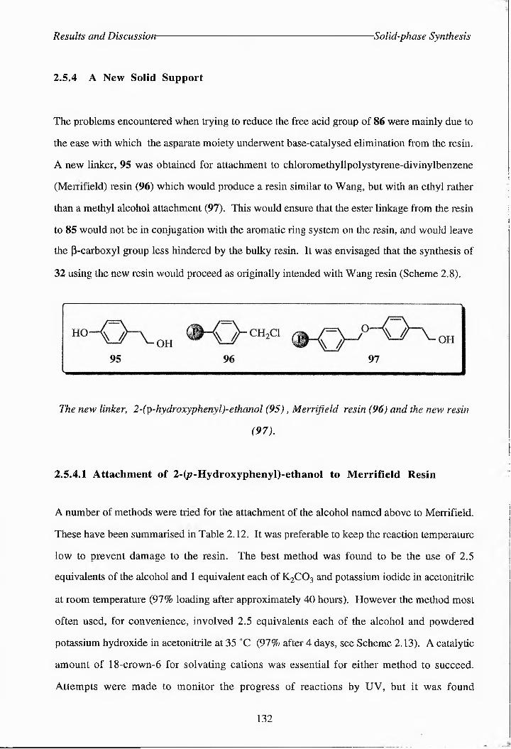
Results and Discussion Solid-phase Synthesis
2.5.4 A New Solid Support
The problems encountered when trying to reduce the free acid group of 86 were mainly due to
the ease with which the asparate moiety underwent base-catalysed elimination from the resin.
A new linker, 95 was obtained for attachment to chloromethyllpolystyrene-diviTylbenzeTe
(Memfield) resin (96) which would produce a resin similar to Wang, but with an ethyl rather
than a methyl alcohol attachment (97). This would ensure that the ester linkage from, the resin
to 85 would not be in conjugation with the aromatic ring system on the resin, and would leave
the p-carboxyl group less hindered by the bulky resin. It was envisaged that the synthesis of
32 using the new resin would proceed as originally intended with Wang resin (Scheme 2.8).
The new I'inker, 2~(^p~lvyd.r(ox-yp'heiryl)-eth-an.ol (95), Merrifield resin (96) and the new resin
(97).
2.5.4.1 Attachment of 2-(^-Hydroxyphenyl)-ethanol to Merrifield Resin
A number of methods were tried for the attachment of the alcohol named above to Merrifield.
These have been summarised m Table 2.12. It was preferable to keep the reaction temperature
low to prevent damage to the resin. The best method was found to be the use of 2.5
equivalents of the alcohol and 1 equivalent each of K2CO3 and potassium iodide in acetonitrile
at room temperature (97% loading after approximately 40 hours). However the method most
often used, for convenience, involved 2.5 equivalents each of the alcohol and powdered
potassium hydroxide in acetonitrile at 35 “C (97% after 4 days, see Scheme 2.13). A catalytic
amount of 18-crown-6 for solvating cations was essential for either method to succeed.
Attempts were made to monitor the progress of reactions by UV, but it was found
132
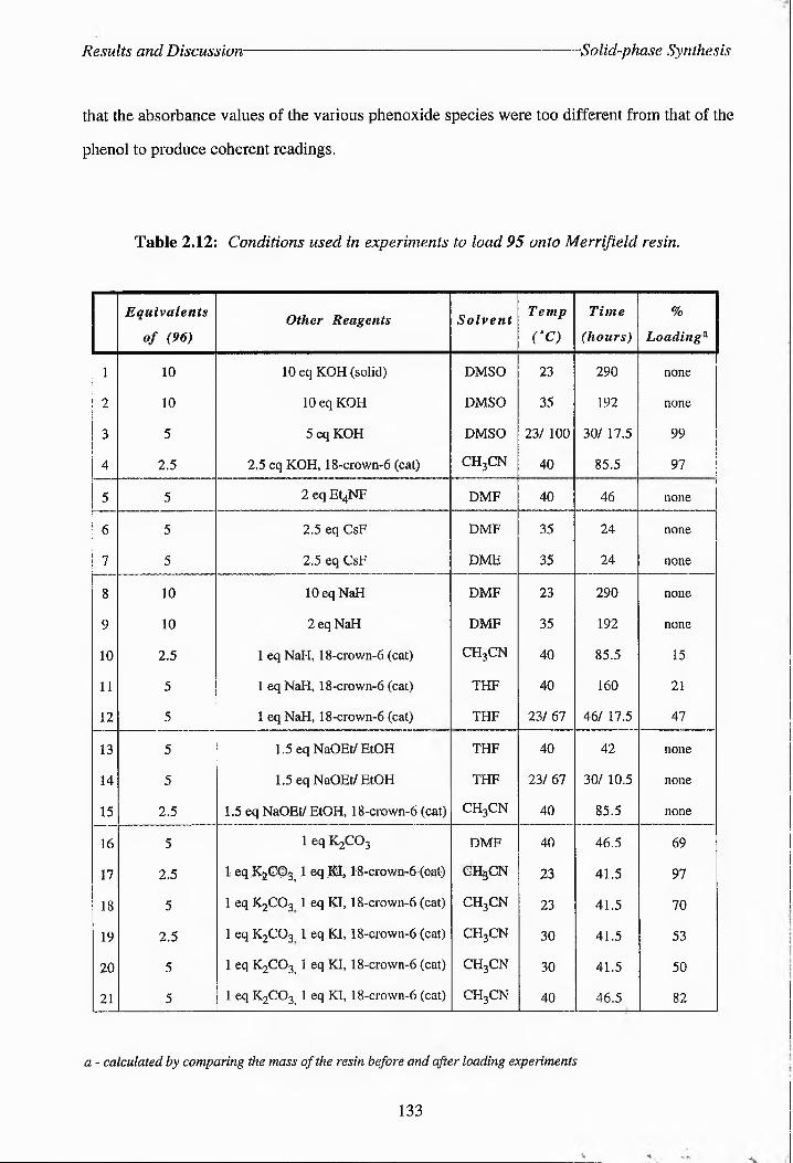
Results and Discussion Solid-phase Synthesis
that the absorbance values of the various phenoxide species were too different from that of the
phenol to produce coherent readings.
Table 2.12: Conditions used in experiments to load 95 onto Merrifield resin..
Equivalents
of (96)Other Reagents Solvent Temp
CC)
Time
(hours)
h
Loading^
1 10 10 eq KOH (solid) DMSO 23 290 none
2 10 10 eq KOH DMSO 35 192 none
3 5 5 eq KOH DMSO 23/ 100 30/ 17.5 99
4 2.5 2.5 eq KOH, 18-crown-6 (cat) CH3CN 40 85.5 97
5 5 2 eq Et4NF DMF 40 46 none
6 5 2.5 eq CsF DMF 35 24 none
7 5 2.5 eq CsF DME 35 24 none
8 10 lOeq NaH DMF 23 290 none
9 10 2 eq NaH DMF 35 192 none
10 2.5 1 eq NaH, 18-crown-6 (cat) CH3CN 40 85.5 15
11 5 1 eq NaH, 18-crown-6 (cat) THF 40 160 21
12 5 1 eq NaH, 18-crown-6 (cat) THF 23/ 67 46/ 17.5 47
13 5 1.5 eq NaOEt/EtOH THF 40 42 none
14 5 1.5 eq NaOEt/ EtOH THF 23/ 67 30/ 10.5 none
15 2.5 1.5 eq NaOEt/ EtOH, 18-crown-6 (cat) CH3CN 40 85.5 none
16 5 1 eq K2CO3 DMF 40 46.5 69
17 2.5 1 eq K2GO3,, 1 -eq KI 18^cjrown-6-- (cat) ch3cn 23 41.5 97
18 5 1 eq K2CO3 1 eq KI, 18-crown-6 (cat) CH3CN 23 41.5 70
19 2.5 1 eq K2CO3 1 eq KI, 18-crown-6 (cat) CH3CN 30 41.5 53
20 5 1 eq K2CO3 - 1 eq KI, 18-crown-6 (cat) CH3CN 30 41.5 50
21 5 1 eq K2CO3 - 1 eq KI, 18-crown-6 (cat) CH3CN 40 46.5 82
a - calculated by comparing the mass of the resin before and after loading experiments
133
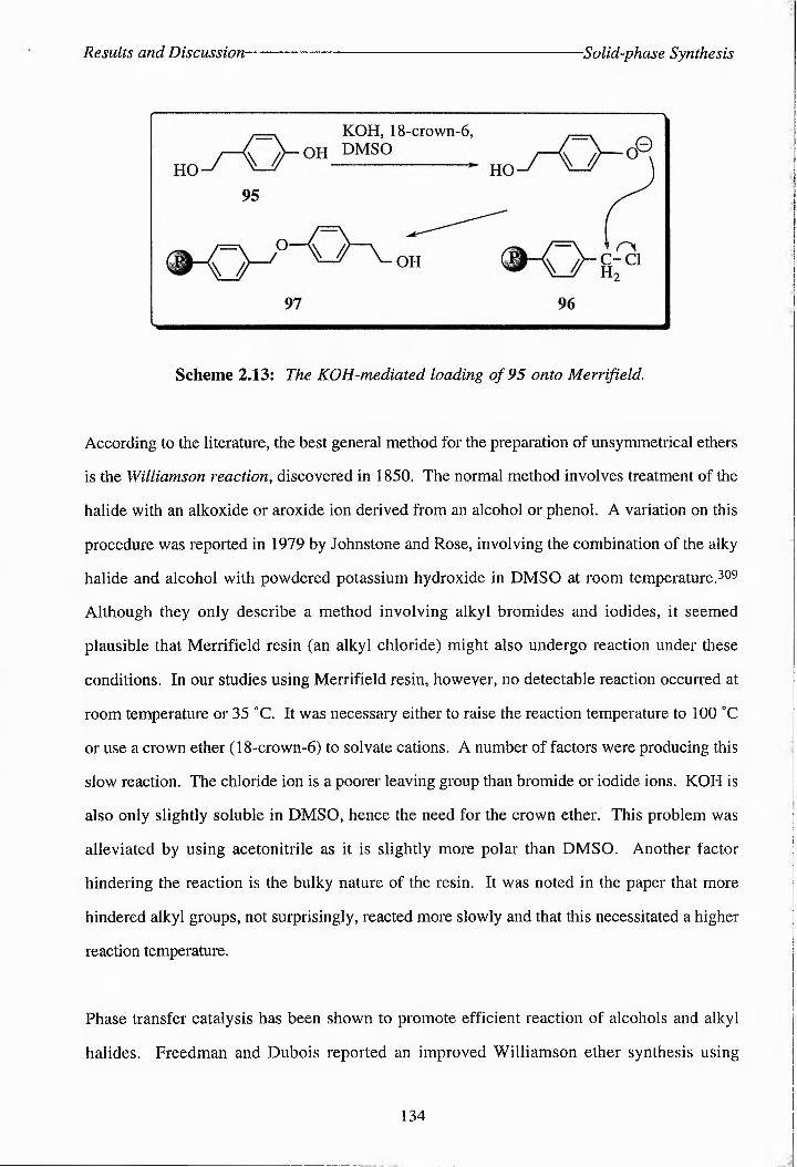
Results and Discussion Solid-phase Synthesis
Scheme 2.13: The KOH-mediated loading of 95 onto Merrifield.
According to the literature, the best general method for the preparation of unsymmetrical ethers
is the Williamson reaction, discovered in 1850. The normal method involves treatment of the
halide with an alkoxide or aroxide ion derived from an alcohol or phenol. A variation on this
procedure was reported in 1979 by Johnstone and Rose, involving the combination of the alky
halide and alcohol with powdered potassium hydroxide in DMSO at room temperature.309
Although they only describe a method involving alkyl bromides and iodides, it seemed
plausible that Merrifield resin (an alkyl chloride) might also undergo reaction under these
conditions. In our studies using Merrifield resin, however, no detectable reaction occurred at
room temperature or 35 °C. It was necessary either to raise the reaction temperature to 100 °C
or use a crown ether (18-crown-6) to solvate cations. A number of factors were producing this
slow reaction. The chloride ion is a poorer leaving group than bromide or iodide ions. KOH is
also only slightly soluble in DMSO, hence the need for the crown ether. This problem was
alleviated by using acetonitrile as it is slightly more polar than DMSO. Another factor
hindering the reaction is the bulky nature of the resin. It was noted in the paper that more
hindered alkyl groups, not surprisingly, reacted more slowly and that this necessitated a higher
reaction temperature.
Phase transfer catalysis has been shown to promote efficient reaction of alcohols and alkyl
halides. Freedman and Dubois reported an improved Williamson ether synthesis using
134

Results and Discussion- Solid-phase Synthesis
tetrabutylammonium bisulphate (TBAB)J305 McKillop et al. also used a quaternary
ammonium salt as a phase transfer catalyst in the synthesis of phenol ethers.306 of particular
interest was the reaction shown in Scheme 2.14, which is similar to what we were trying to
achieve, which produced a yield of 86% of the desired product.
Scheme 2.14: McKillop's ether synthesis.
J. M. Miller et al. also reported on the use of a quaternary ammonium halide for the promotion
of the synthesis of alkyl phenyl ethers, including thc product shown in Scheme 2.14.307 He
used tetraethylammonium fluoride in DMF as a source of fluoride ions. The fluoride ion can
act as a powerful hydrogen bond donor. In an article published in 1977 it was shown that
phenol and fluoride can form a strong H-bond together, resulting in a large shift of thc
hydroxyl stretching band in the IR spectrum (Aus(OH) ~ 1000 cm-1}.308 An attempt was
made to use this reagent for promoting the alkylation (experiment 5, Table 2.12), but to no
avail. Again steric hindrance may be the reason why this reaction failed and a higher reaction
temperature may prove necessary. In the same paper Miller reports that the condensation of
phenol with halogenoalkanes is promoted by the presence of KF.308 The reaction is
accelerated by an order of magnitude if CsF is used instead. Although, in our hands, this
reagent failed to produce a successful reaction, this may be attributed to the low solubility of the
alkali metal fluoride in either of the solvents used. Again the use of 18-crown-6 and a higher
reaction temperature would probaby have solved this problem. The use of fluoride-containing
reagents was not pursued further, however, because other, more cost-effective methods had
produced satisfactory results.
The use of sodium hydride to form an alkoxide ion which could react with benzyl bromide was
reported by D. J. Hart et al. in their work on the total synthesis of lythrancepines.309 3.6
135

Results and Discussion Solid-phase Synthesis
equivalents of NaH were added to the alcohols in DMF at room temperature. After 1 hour
benzyl bromide was added and the reaction left for 36 hours. Harsher reaction conditions were
necessary here to produce a reaction between 95 and Merrifield resin (96). The best loading
(47%) was obtained by refluxing the reagents in THF for over 60 hours in the presence of a
catalytic amount of 18-crown-6. F. Kroll and D. Stones (unpublished results) have achieved
some success with the loading of various phenols onto Merrifield resin using NaH in DMF at
60 "C.
By far the best method found for loading 95 onto Merrifield was a protocol developed
originally for the protection of phenols with a functionality that would be stable to
hydrogenolysis at atmospheric pressure (Scheme 2.15).3i° This involved the use of potassium
carbonate and potassium iodide in acetonitrile with a phenol and the alkyl chloride. The iodide
replaces the chloride group on a small proportion of the alkyl chloride. This acts as a better
leaving group when attacked by a phenoxide ion, thus accelerating the reaction. A reaction
carried out using just K2CO3 produced a good loading of 95 on the resin (69%), but was not
as efficient as the method using KI in conjunction with K2CO3.
Scheme 2.15: Mechanism for the reaction carried out by Davis and Muchowski.
The loading of 95 onto Merrifield was confirmed by the presence of several new peaks in the
IR spectrum compared that for the resin alone: t)max(KBr)/cm'i 3453 hr m (O-H, alcohol),
1517 m (aromatic C=C), 1500 (aromatic C=C), 1390 (O-H, alcohol). The 'I3C-NMR spectrum
also showed the presence of several new signals: 6C(74.76 MHz; CgiHi) 158.55
136
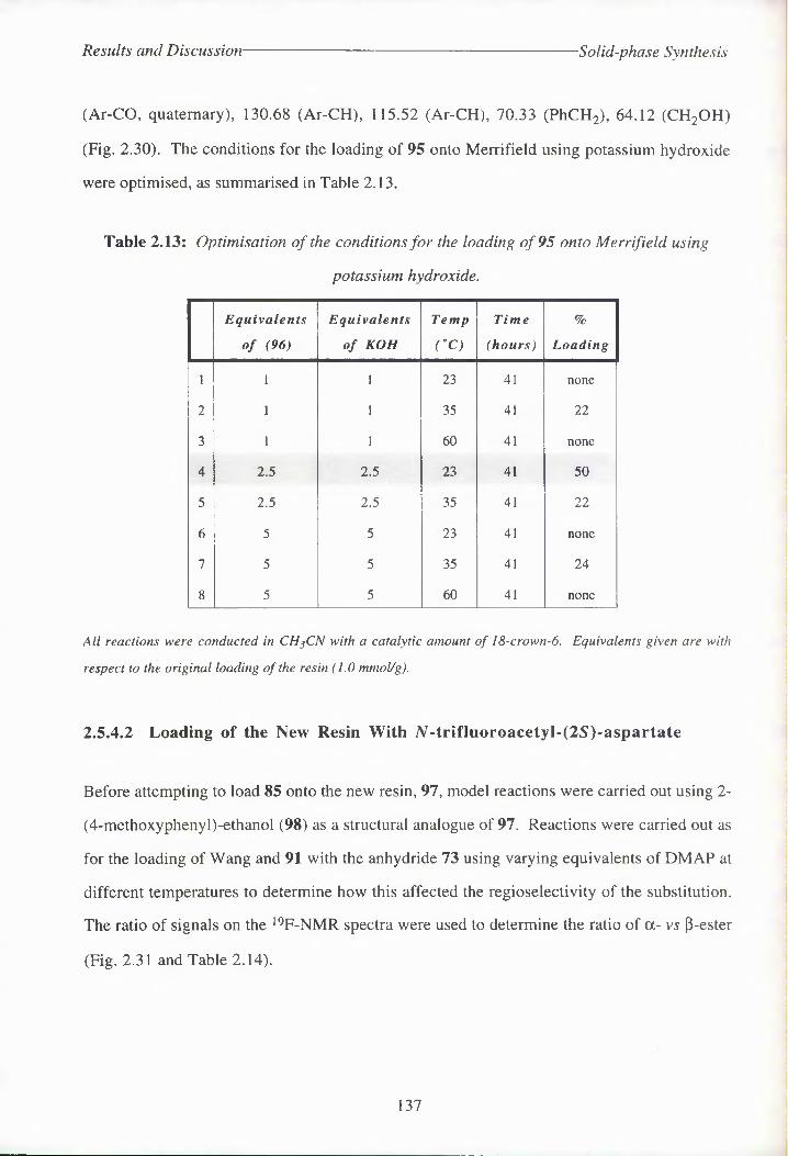
Results and Discussion- -Solid-phase Synthesis
(Ar-CO, quaternary), 130.68 (Ar-CH), 115.52 (Ar-CH), 70.33 (PhCH2), 64.12 (CH2OH)
(Fig. 2.30). The conditions for the loading of 95 onto Merrifield using potassium hydroxide
were optimised, as summarised in Table 2.13.
Table 2.13: Optimisation of the conditions for the loading of 95 onto Merrifield using
potassium hydroxide.
Equivalents
of (96)
Equivalents
of KOH
Temp
(°C)
Time
(hours)
h
Loading
1 1 1 23 41 none
2 1 1 35 41 22
3 1 1 60 41 none
4 2.5 2.5 23 41 50
5 2.5 2.5 35 41 22
6 5 5 23 41 none
7 5 5 35 41 24
8 5 5 60 41 none
All reactions were conducted in CH$CN with a catalytic amount of 18-crown-6. Equivalents given are with
respect to the original loading of the resin (1.0 mmol/g).
2.5.4.2 Loading of the New Resin With A-trifluoroacety!-(2S)-aspartate
Before attempting to load 85 onto the new resin, 97, model reactions were carried out using 2-
(4-methoxyphenyl)-ethanol (98) as a structural analogue of 97. Reactions were carried out as
for the loading of Wang and 91 with the anhydride 73 using varying equivalents of DMAP at
different temperatures to determine how this affected the regioselectivity of the substitution.
The ratio of signals on the 19F-NMR spectra were used to determine the ratio of a- vs (-ester
(Fig. 2.31 and Table 2.14).
137

Results and Discussion ■Solid-phase Synthesis
138

Results and Discussion Solid-phase Synthesis
>VO
o
VO3
VOr*a- vs p~ester 67% j
i/
VO
ipnipnipnipnipi}inij!nipni]nfiiinifoi;ii[ii]inr.
76.56 -76.68 ppm30.24
14.82
-76.56 -76.68 ppm27.11
_ 10.71 o
-76.56 -76.68 ppm39.59
16.27
4
a-vs p-ester 58%
t" z/
h
\ \
-76.56 -76.68 ppm37.85
2g.22
-76.56 -76.68 ppm44.22
23.79
-76.56 -76.68 ppm35.43
25.69
-72.5 -75.0 ppm Figure 2.31: 19F-NMR spectra referred into Table 2.14.
139

Results and Discussion- -Solid-phase Synthesis
2l(4-methoxyphenyl)-ethanol (98)
Toble 2.14: Comparison of the regioselectivity of substitution reactions of 73 onto 98.
Experiment Equivalents
of DMAPTemperature a- vs p-estera
1 0 0°C 72%
2 0.5 0°C 71%
3 1.0 ooc 67%
4 0 rt 58%
5 0.5 rt 65%
6 1.0 rt 58%
Reactions were carried out in DMF using 25 mg/ml 98 and 175 mg/ml 73.
Reactions 3, 5 and 6 (Table 2.14) appeared to be complete within 17 hours as shown by tic
[band on baseline; eluant ethyl acctate-light petroleum (60%)], but reaction 2 took a few days to
go to completion, and reactions 1 and 4 (no catalyst) were not completed even after 6 days. It
was therefore obvious that the reaction to load 73 onto the new resin (97) would have to bc
performed at room temperature or higher using a minimum of 0.5 equivalents of DMAP.
The first two loading experiments using the new resin were carried out at 0 °C and room
temperature respectively using 5 equivalents of the anhydride 73 and 0.5 equivalents of
DMAP. Both were left for three days, but no protected aspartate 85 had loaded onto the resin
as determined by comparison of thc mass of the resin before and after the loading experiments.
Both i9F gel-phase NMR and IR spectroscopy confirmed that no A^-t^'Tffl^coT^^^(^<ctyl-(25')-
aspartate was present. A later experiment using the same equivalents of DMAP and 73 but
increasing the temperature to 35 0C did not produce a significant loading after 4 days, by
140

Results and Discussion Solid-phase Synthesis
comparison of the mass of the resin at the start and end of the experiment. However, both 19F
and 13C gel-phase NMR spectroscopy indicated that some of the protected asparate 85 had
attached to the resin; 6C(74.76 MHz; C62H6) 34.09 (PC of aspartate), 48.42 (“C of
aspartate), 117.44 (F3C), 169.11 (F3CCO), 173.77 (PCO2H of aspartate); 0p(282.2 MHz;
(^^^SO) -74.81 (F3C). The IR spectrum showed the presence of an aryl ester linkage
{1727 s (C=O)} and the W-trifluoroacetyl amide group {1653 m (2° amide, solid state)}. The
most recent experiment was carried out using 1 equivalent of DMAP and 5 equivalents of 73 at
35 0C. After 6 days the loading appeared to be quantitative, and the synthesis of 99 was
confirmed by 19F and 13C gel-phase NMR and IR spectroscopy (Fig. 2.30 and 2.31). A
method for cleaving 85 from the new resin has yet to be found. A number of methods for the
selective cleavage of esters have been reported. Boron tribromide has been used for the
deprotection of a number of ester protecting groups to the corresponding acids, without
affecting amide bonds.iii Iodotrimethylsilane has also been shown to be a mild and effective
reagent for the non-saponificative hydrolysis of esters under mild conditions.31^314 This can
be synthesised in situ from chlorotrimethylsilane and sodium iodide.3 M This reagent does,
however, also cleave ethers.
141

Results and Discussion- Conclusions and Future Work
2.6 Conclusions and Future Work
2.6.1 Substrate Synthesis
2.6.1.1 Solid-Phase Synthesis
The synthesis of both the non-labelled and [U-14C]-labelled substrate using previously
established protocols has been reported here, N,N-diisoproTyl dibenzyl phosphoTamidite (79)
has always been used in the past as a phosphorylating agent in this synthesis. However,
problems can be encountered in the synthesis of this reagent, due to the instability of its
precursor iV,N-diisoproppldichlooophosphoramidite (78). Two new reagents were therefore
identified for the phosphorylation of 7/-trifluoroacety^(0-isopropyl-(25')-homoseriTe (75), one
of which was available commercially (80), the other, 81, being synthesised easily from POCI3
and pentafluorophenpl. These gave good clean yields of the phosphorylated compounds 83
and 84. However, these phosphate groups could not be subsequently deprotected.
A/-trifluoroacetyl-(2S)-aspartatc (86) has been successfully loaded onto Wang, p-
hydroxymethyl polystyrene (92) and polystyrene-4-pxymethyl-2-TheTylethaTol (97) resins
during attempts to synthesise the substrate on the solid-phasc. The latter resin is a novel resin,
which was synthesised by the base catalysed addition of 2-(T-hydroxyphenyl)-ethaTol (95) to
Merrifield resin (96) in 97% yield. This was promoted by the use of KI to exchange chloride
for the better leaving group, iodide, and by 18-ctowt-6 to solvate the ionic species. Both
K2CO3 and powdered KOH could be used as bases for this reaction. Acetonitrile was found to
be the best of the solvents tried in this reaction.
The nucleophilic addition reactions of each resin with 85 proceeded with good regioselectiv^y
to favour protection of the a-carboxylic group of 86. Wang resin was the most selective for
the ot-ester (84% a- vs. P-ester). This is due to the more hindered nature of the hydroxyl
group on Wang compared to 92 and 97. This regioselectivity compares reasonably well with
the ring opening of 85 by isopropanol, which produces a ratio of a- vs. P-ester of 95%. The
ring opening of 85 using resins does, however, proceed much more slowly, and requires
142

Results and Discussion- Conclusions and Future Work
heating of the reaction to at least 30-35 °C, which in turn lowers the regioselectivity.
The compound produced from the reaction of 85 with Wang resin, 87, was found to be
unstable to reducing agents. It was thought that the compound produced using
p-hydroxymethyl polystyrene (92) would also be unstable to reducing agents, as it resembles
the structure of 87. Resin 92 was, in any case, less selective for the a-ester than Wang resin.
The novel resin, 97, wos also less selective than Wang resin for the at-ester (72% a- vs. p
ester), but it is hoped that 98 will prove more stable to reducing agents.
2.6.1.2 Suggestions for Future Synthetic Work
The conditions for the loading of 86 onto 97 have yet to be optimised, and a mild and selective
method for the cleavage of the ester linkage must be developed. It would be preferable to find a
method which would simultaneously deprotect the dipenlafIuorodiphenylphosphate triester to
minimise the number of deprotection steps necessary in the solid-phase synthesis of 32. The
possibility of reducing the p-carboxyl group on 99 using either the mixed anhydride or the
PyBOP method of activation has yet to be explored, and it remains to be seen whether the new
linker prevents the cleavage/elimination of 86 from the resin. It may be possible to use
diborane with the new resin, as the ester linkage is no longer adjacent to a system of conjugated
double bonds. If cleavage still occurs, more sterically hindered linkers such as 100 may be
necessary, which resemble more closely the structure of the isopropyl group of 74, which is
stable to reduction. A more hindered linker may also improve the regioselectivity of the
nucleophilic addition reaction in favour of the a-ester.
If a resin is developed with enhanced stability towards reducing agents, this may have o wider
application for general use in the solid-phase synthesis of peptides and amino acid derivatives.
143

Results and Discussion- Conclusions and Future Work
2.6.2 A Viable Source of Threonine Synthase
2.6.2.1 The Tir8 Mutant
The mutant form of E. coli K-12 Tir8 is was first characterised by its resistance to
thiaisoleucine (2-amino-3-methylthiobutyric acid).174 The growth of wild-type K-12 cells is
prevented by the presence of this compound, due to the inhibition of threonine deaminase
activity. The growth inhibition is reversed by isoleucine at a concentration one-tenth that of the
inhibitor. Tir mutants have altered isoleucyl sRNA synthetases and derepressed levels of three
of the five enzymes involved in the biosynthesis of isoleucine and valine. These mutants are
also slower growing than wild-type cells.
In Section 2.2 it was reported that the Tir8 mutant might have reverted to wild-type during
experiments to isolate TS and so that it longer over-expressed the enzymes coded for by the thr
operon. One of the possible reasons for this is that wild-type cells are faster growing than Tii8
cells and can rapidly overtake a growing culture. As Tir8 exhibits enhanced resistance to
thiaisoleucine one method for detecting any remaining mutant cells is to plate out a fresh
culture, grown from frozen glycerol stocks of Tir8, onto L-agar plates containing the
compound and incubate these overnight. This would provide growth conditions selective for
the mutant colonies, which could then be isolated and grown up in LB-medium for further
attempts to isolate TS from this source. If this were still unsuccessful or if no mutants were
isolated, then it would be necessary to immediately carry out more cloning work to insert thrC
into a pET vector. Cloning the gene into such vectors does also have some advantages over the
use of Tir8. Only thrC would be over-expressed in cells transformed with these constructs,
facilitating the purification of the enzyme. A His-tagged version of the enzyme should also
make the purification of TS much quicker and easier (see Section 1.117.
144

Results and Discussion Conclusions and Future Work
2.6.2.2 Cloning Amplified77zrC Into pET Vectors
A protocol for the cloning of amplified gene fragments into pET vectors has been established
and reported here. This involves cloning the gene first into pGEM-T from which it is
subcloned into pET vectors. This first cloning step allows for the production of a large amount
of the amplified gene DNA without the need for repeated PCR experiments, during which
mutations can arise. Cloning of gene fragments with Nde I-sites at either terminus into pET
vectors via ligation of cohesive termini does not appear to be possible, but fragments with a
Nde I-site at one terminus and a BamY I site at the other can be ligated into pET vectors
restricted with these two enzymes.
It has been found that ligations using pET-vectors have a specific temperature requirement of
16 °C, whereas ligations into pGEM-T can tolerate warmer conditions (i.e. room temperature).
The DNA insert must be extremely pure for ligation into pET expression systems. Successful
results were obtained using PCR products and excised gene fragments which had been purified
on Wizard Prep™ DNA purification columns and using recombinant plasmids which had been
isolated using the QIAGEN maxiprep protocol and purified where necessary using Wizard™
DNA Clean-up Purification Resin.
Both the thrC gene and the gene coding for p-methylaspartase, containing Nde I-sites at each
teimiinus, were amplified and cloned into the pGEM-T transcription vector. The identity of the
gene inserts was identified by a number of means:
• Diagnostic PCR - whereby half as well as whole amplified gene fragments have been
obtained using internal and external primers .
• Restriction analyses - carried out on the recombinant plasmids which gave distinct
fragments of the predicted sizes.
• Subsequent di-deoxynucieotide sequencing of the recombinants which gave the sequence of
the first 127 bases of the C-terminus of the thrC gene, including the Nde I-site, and the first
145
Results and Discussion Condusions ond Future Woro
120 bases of both the C- and N-termini of the gene for p-methylaspartase. These gene
fragments could not be subcloned into pET vectors and the constructs were therefore discarded
before full sequencing could be carried out.
The two genes were amplified again to produce gene fragments with an Nde I-site at the
N-terminus and a BamH I-site at the C-tenninus. This also improved the efficiency of ligations
into pET vectors, as the orientation of gene inserts in the vector could now be controlled.
These PCR products were cloned into pGEM-T. Two recombinants for thrC (pGEM-TTSi and
pGEM-TTSii) were identified by a number of restriction analyses. Preliminary sequencing
experiments produced correct sequences for the two termini of the gene in both constructs.
Subsequent sequencing of the whole thrC gene insert in both constructs has revealed one
deletion and two mutations of the recombinant gene (Section 2.4).
The thrC fragment was excised from pGEM-TTSi and pGEM-TTSn for subcloning into the
three pET-vectors pET-3a, -3b and -16b. Recombinants were identified by restriction analysis.
Sequencing of these vectors was also carried out (Section 2.4) confirming the gene mutations
observed in the pGEM-TTS constructs.
The asymmetric P-methylaspartase PCR product was also inserted into pGEM-T, and further
cloning work has been carried out by other researchers to insert the gene fragment into pET-
vectors (Section 2.3.2.3).
2.6.2.3 Future Work on ThrC
Since it has been determined that the recombinant thrC genes do not have the same sequence as
the wild-type gene, if the Tir8 mutant is confirmed to have completely reverted to wild-type it
will be necessary to repeat the cloning procedure which has been optimised during these
studies.
146

Results and Discussion- Conclusions and Future Work
Taq DNA polymerase lacks a 3' - 5' exonuclease proofreading activity, and therefore has a
relatively high error rate. The mutation frequency using this enzyme is 110 x IO’4 at lOmM
MgCl2 and ImM dNTPs.244 A mutation is therefore highly likely to occur after relatively few
cycles of PCR using this enzyme.265 Vent™ DNA polymerase has a fidelity 5-15 fold higher
than that of Taq, having a mutation frequency of 190 x 10‘4, and is the only thermophilic
polymerase with a 3' - 5' exonuclease proofreading activity.264’ 265 Therefore, in experiments
to repeat the PCR amplification of thrC, it is obvious that Vent should be used instead of Taq.
A new amplified thrC fragment should be produced with an Nde I-site at the N-terminus and a
Bam H I-site at the C-terminus using this enzyme. As a precaution the PCR product(s7 should
then be sequenced before undertaking any further work, to determine whether the PCR reaction
has been successful. If a mutation occurs at an early stage in the PCR reaction, then that error
will be amplified in subsequent cycles of PCR. If this is the case then in subsequent ligation
experiments most of the recombinant plasmids will contain the mutant gene, and this is
probably what happened when Taq was used in PCR reactions.
Many factors can affect the fidelity of Taq and these have been detailed by Eckert and
Kunkel.2.5 A discussion of these may offer an explanation for the PCR errors encountered in
this work. These factors may also affect the fidelity of other thermophilic polymerases, which
have been less extensively studied.
• The concentration of dNTPs can have a detrimental effect on Taq fideltiy if too high. A
1000 pM DNTP concentration causes the error rate to be doubled in comparison to using 1 pM
dNTPs. In the work reported here the dNTP concentration was 125 pM, which gives a result
very similar to that obtained for 1000 pM dNTPs. If a polymerase lacks an exonuclease to
remove terminal mispairs, then unextended errors will be lost because they do not yield full-
length DNA products for further amplification. At low dNTP concentrations the level of
mispairing is reduced and the error discrimination is enhanced. The concentration of dNTPs
should therefore be lowered in future PCR reactions.
147

Results and Discussion- Conclusions and Future Work
• The fidelity of Taq can be improved by decreasing the magnesium chloride concentration
relative to the concentration of dNTPs present in the reaction. The concentration of MgCh
should be equimolar with the dNTP concentration for optimum fidelity. The reactions reported
here were carried out using 2.5mM MgCl2, as recommended by the manufacturer. In future
reactions the concentration of MgCl2 relative to the dNTP concentration could be lowered to
reduce the level of mutation.
• The error rate of Taq is reduced by decreasing the reaction pH to 5 - 6 and the frequency of
mutations continues to increase as the pH is raised through 8.2. At pH 5.1, the error rate is
reported to be 1/180 000 using 1000 pM dNTPs and 10 mM MgCh whereas at pH 8.2 the
error rate is increased to 1/3 200 (both reactions at 70 °C). However, the incubation of DNA at
high temperatures and low pH causes DNA damage, increasing the potential for mutations, so
this must also been taken into consideration if lowering the reaction pH of PCR reactions in
order to improve polymerase fidelity.
If the thrC gene is successfully cloned into pET vectors then it may prove possible to purify TS
in a more efficient way than previously possible using the Tir8 mutant. If this were the case, it
would then be useful to attempt the first X-ray crystallographic studies on the enzyme,
particularly in order to elucidate the active-site structure of the enzyme. TS from either source
could also be used, as originally intended, for product inhibition assays. The substrate
analogues, which have been synthesised by G. Allan (these are discussed in Section 1.10),
could also be tested as inhibitors of the enzyme or to further elucidate the TS reaction
mechanism, as appropriate.
Site-directed mutagenesis experiments could later be carried out to monitor the effects of
altering residues, which are conserved in all microbial TS sequences, on the structure and
catalytic activity of the enzyme from E. coli. Mutant thrC genes produced via PCR could again
be cloned into pET constructs using the established protocol.
148

CHAPTER THREE
EXPERIMENTAL

Experimental- Synthesis
3.1 Synthesis
3.1.1 General Experimental Procedures
3.1.1.1 Compound Characterisation
Elemental microanalyses were performed in the departmental microanalytical laboratory.
NMR spectra were recorded on Varian Gemini 300 (.H, 300 MHz; 13C, 74.76 MHz; 31p,
121.42 MHz; I9F, 282.2 MHz7, or Varian Gemini 200 (iH, 200 MHz; i.C, 50.31 MHz7
spectrometers. .H- and .3C-NMR spectra are described in parts per million downfield shift
from SiMe4 and are reported consecutively as position (8h or 5c7, relative integral, multiplicity
(s = singlet, d = doublet, t = triplet, q = quartet, dd = double of doublets, dt = doublet of
triplets, dq = doublet of quartets, sept = septet, m = multiplet, and br = broad7, coupling
constant (7/Hz7 and assignment (numbering according to the lUPAC nomenclature for the
compound7. .H-NMR spectra were referenced internally on 2HOH (5 4.80 ppm7, C2HC13
(8 7.27 ppm7 or (C2H372SO (8 2.50 ppm7. 13C-NMR spectra were referenced on C2HC13
(6 77.23 ppm7, C^H. (8 128.39 ppm7 or (CH^SO (6 39.51 ppm7. i.F-NMR spectra
were referenced internally on CFCI3 and described in parts per million downfield shift from
this compound. Aip-NMR spectra were referenced on external ^PO4.
IR spectra were recorded on a Perkin-Elmer 1710 FT-IR spectrometer. The samples were
prepared as Nujol mulls, thin films between sodium chloride discs or as dispersions in
potassium bromide discs. The frequencies (U as absorption maxima are given in
wavenumbers (cnr^ relative to a polystyrene standard at 1603 cm-1. Signal strengths are
reported as s = strong, m = medium, w = weak and br = broad.
Mass spectra and accurate mass measurements were recorded on VG 70-250 SE, Kratos
MS-50, VG Platform E/S . Major fragments were given as percentages of the base peak
intensity (100H7.
149

Experimental- Synthesis
UV data was obtained on Pye-Unicam SP8-500 or SP8-100 spectrophotometers.
Flash chromatography was performed according to the method of Still et al.3i5 using Sorbsil
C60 (40-60 pm mesh) silica gel. Analytical thin layer chromatography (tic) was carried out on
0.25 mm precoated silica gel plates (Macherey-Nagel SIL g/UV254) or 0.25 mm precoated
cellulose plates and compounds were visualised using UV fluorescence, potassium
permanganate solution, vanillin, or ninhydrin. Preparative tic was performed on glass-backed
cellulose plates.
Melting points were taken on an Electrothermal or Gallenkamp melting point apparatus and are
unconnected.
Optical rotations were measured at 23 °C on an Optical Activity AA-1000 polarimeter using
10 cm path length cells. Readings are given in 10_1 deg cm2 g-2 and the solution concentration
is given in g c^'3.
3.1.1.2 Reagent and Solvent Preparation
The solvents used were either distilled or of Analar quality and light petrol ether refers to that
portion boiling between 40 and 60 “C. Solvents and bases were dried according to literature
procedures, distilling under nitrogen. Ethanol and methanol were dried using magnesium
turnings and iodine and stored over freshly activated Linde 4A molecular sieves. Isopropanol
and acetonitrile were distilled over CoH^ and stored over activated Linde 4A molecular sieves.
Toluene, CH^^ and diisopropylamine were distilled over Cal-L directly before use.
Diisopropylethylamine was distilled from KOH. Triethylamine and N-methylmorpholine were
distilled from ninhydrin, then KOH. THF and E^O were distilled from the sodium ketal of
benzophenone directly before use, under nitrogen. DME was also distilled under nitrogen
from the sodium ketal of benzophenone onto 4A molecular sieves. DMF and benzyl alcohol
were distilled under reduced pressure onto activated Linde 4A molecular sieves.
Diazomethane preparation: Ntnitroso-N-methyIt4ttoIuene-suIphonamide (Diazald) (21 g,
150

Experimental- Synthesis
98 mmol) in ether (150 cm3) was added slowly to a solution of potassium hydroxide (4.8 g,
86 mmol) in water (8 cm3) and isopropanol (12 cm3). The solution was heated in a clear
jointed distillation apparatus and the ethereal diazomethane distilled into ether (10 cm3) at 0 °C.
The yellow solution obtained was used in the synthesis of methyl ester derivatives.
3.1.1.3 Suppliers
2-(p-Hydroxyphenyl)-ethanol and 2-(4-methoxyphenyl)-ethanol were purchased from Aldrich.
p-Benzyloxybenzyl alcohol (Wang), hydroxymethylpolystyrene and chloromethyllpolystyrene-
divinylbenzene (Merrifield) resins were purchased from Novabiochem.
151

Experimental- -Synthesis
3.1.2 Synthetic Procedures and Characterisations
2V-Trifluoroacetyl-(2S)-aspartic anhydride 73.
OA
F3C NH HB71O
oo
To a stirred suspension of (25)-aspartate (500 mg, 3.76 mmol) in dry THF at -10 -* 0 °C
was added dropwise trifluoroacetic anhydride (2.65 cm3, 18.8 mmol). The reaction was
stirred under nitrogen at room temperature. The progress of the reaction was monitored by tic
[7?f 0.38: ethyl acetate-light petroleum (3:2)]. Upon completion (48 h), the solvent was
removed under reduced pressure to give 73 as a white solid (785 mg, 99%); mp 110-115 °C;
'Umax(Nujol)/cm'1 3350 m (N-H stretching, 2° amide), 1886 m (C=O, anhydride), 1722 s
(C=O, anhydride), 1556 m (C=O, 2° amide), 1371 s (C-F), 1342 s (C-F), 1225 s (C-F, C-O),
1176 s (C-F, C-O) and 1083 m (C-F, C-O); 8H(200 MHz; C2HC13) 3.01 (2 H, dq, ABX,
7ab 18.5, Jax 10.1,7Bx 6.8, HP), 4.85 (1 H, dt, 7nh,h 7.0, 7ax 10.1, 7b x 6.8, Ha) and 9.57
(1 H, d, 7nh,h 7.0, amide NH); 8C(74.76 MHz; C2HC13) 34.09 (CP), 48.67 (C«), 115.34 (q,
J FiC 285.28, F3C), 157.43 (q, 7 f3c,c 38.58, F3CCO), 167.61 (pC=O) and 169.28 (0tC=O);
5f(282.2 MHz; C2HC13) -76.37 (F3C); m/z (CI) 230 (100%, [M + H + H2O]+) and 212 (14,
[M + H]+).
152

Experimental: -Synthesis
a-Isopropyl-AMrifluoroacetyl-(2S)-aspartate 74.
To a stirred suspension of (257-aspartic acid (1.15 g, 8.61 mmol7 in dry THF (30 cm37 at
-5 °C was added dropwise (8 cmA, 56.7 mmol7 of trifluoroacetic anhydride. After 4 h the
solvent was removed under reduced pressure, and the anhydride 73 was dissolved in dry
isopropanol at room temperature. The progress of the alcoholysis was monitored by tic
[Hf 0.46: ethyl acetate-light petroleum (3:27] and after 1 day the isopropanol was removed
under reduced pressure, giving a clear yellow oil, which on standing formed a white solid.
Recrystallisaton from ether gave a white crystalline solid (2.3Ig, 99H7, mp 92 "C (lit.,35
91 °C7; [a]D -49.95 (c 1.0 in MeOH7, [lit.,35 -40.7 (c 1.0 in MeOH7]; Dmax(Nujol)/cm-i 3310
s (N-H, 2° amide7, 3115s (OH, acid7, 1738 s (C=0, ester7, 1709 s (C=O, carboxylic7, 1566 s
(N-H, amide II7, 1467 s (CH2, CH3, C-H deformations7, 1390 and 1378 s (C-F7, 1313 s (C-1
F7, 1280, 1236, 1208 and 1186 s (C-O and C-F stretching7 and 1106 s (C-F7; 0.(200 MHz;
CTHCy 1.25 (6 H, d, JcH3,h 6-25, 2 x ' PrCH37, 3.08 (2 H, dq, ABX, Ab 17.83, Ax 4.42,
Jbx 4.25. HP7, 4.21 (I H, br s, pCO2H7, 4.78 (1 H, dt, 7NH 6.60, ABX, ,/AX 4.42, 7Bx
4.25, H“7, 5.11 (1 H, sep, Jch3,h 6.25, 'PrCH) And 7.38 (1 H, d, Jnh h 6.60, amide NH7;
8(2(74.76 MHz; C2HC137 21.30 (‘PrCHj7, 35.06 (CP7, 48.80 (C°7, 70.96 ('PrCH7, 115.65
(q, JF,c 285.28, F3C7, 157.19 (q, Jp,c,c 37,53, F3CCO7, 168.56 (ACO2'Pr7 and 175.54
(PCO2H7; 8p(282.2 MHz; C4HCl37 -76.77 (F3C7; m/z (EI7 226 (4%, [M - CO4I]<). 212 (7,
[M - CHaCO2H]+7, 184 (36, [M - CO.'Prn, 139 (35, [M - CO2'Pr and COaH]+7, 96 (22,
F3CCO7, 88 (8, ['PrO2CH]+7, 69 (36, F3C+7, 59 (8, CH2CO2H+7, 43 {100, (CH372CH+7 and
28 (35, CO+7.
153

Experimental- Synthesis
a:-IsopropyI’'A-^^r'inu(U'()accl5'l((2^^^^‘)-homoserine 75.
To a stirred solution of the isopropyl ester 74 (2.02g, 7.46 mmol) in dry THF (30 cm2) at
-20 °C was added N-methyl morpholine (0.82 cm2, 7.46 mmol) and isobutylchloroformate
(1.02 cm3, 7.46 mmol). After 20 min the solution was filtered into a suspension of sodium
borohydride (0.169 g, 4.48 mmol) in dry THF at -20 °C. The solution was stirred for 2 h
before being allowed to warm to room temperature and then stirred for a further 2 h. Excess
borohydride was destroyed by the addition of water (5 cm2), and stirring continued for a
further 30 min. The THF was removed under reduced pressure and the product extracted from
the aqueous layer using ether (10 x 10 cm2). The combined organic extracts were washed with
saturated NaHCOg solution (12 cm2) and saturated brine (12 cm2) and dried (MgS0>4). The
ether was removed under reduced pressure giving the crude alcohol as a clear orange oil.
Purification of the cmde oil by flash silica column chromatography [eluant DCM-EtOH (97:3)
containing a few drops of acetic acid] gave the pure protected homoserine 75 as a clear yellow
oil (0.672 g, 35%); umax(Nujol)/cm'i 3318 br m (OH and NH superimposed), 2923 m (OH,
acid/ alcohol), 1723 s (C=O ester), 1560 w (NH), 1458 w (CH^ CH., C-H deformations),
1377 m (CH3), 1184 bs (C-O, C-F) and 1106 m (C-O, C-F); 0H(200 MHz; C^HCI.) 1.29
(6 H, d, 7cH3,h 6.25, 2 x 'PrCH3), 2.10 (2 H, m, HP), 3.73 (2 H, m, HY), 4.69 (1 H, dt,
7Nh,h 4.35, AEX, 7ax 12.00, 7gX 3.20, H«), 5.09 (1 H, sept, 7ch3.h6.25, 'PrCH) and 7.82
(1 H, d, 7Nh,h 4.35, NH); 0c(50.31 MHz; C2HCl3), 21.46 ('PrCH.), 32.90 (CP), 51.32
(C«), 58.58 (CY), 70.17 ('PrCH), 115.74 (q, 7p,c 287.37, F3C), 157.09 (q, 7f3c,c 37.78,
F.CCO) and 170.47 (oCC^Pi); 0p(282.2 MHz; C^HCl.) -76.74 (F3C); m/z (El) 258 (100%,
[M + H]+), (17, [M - OH]+), 216 (33, [M - Tr + 2H]+), 198 (85, [M - 'PrO]+), 170 (80, [M
- CO2'Pr]+), 140 (86, [M + H - CH^H and CO2'Pr]+), 69 (28, F3C+ and 57 (20, F.+).
154
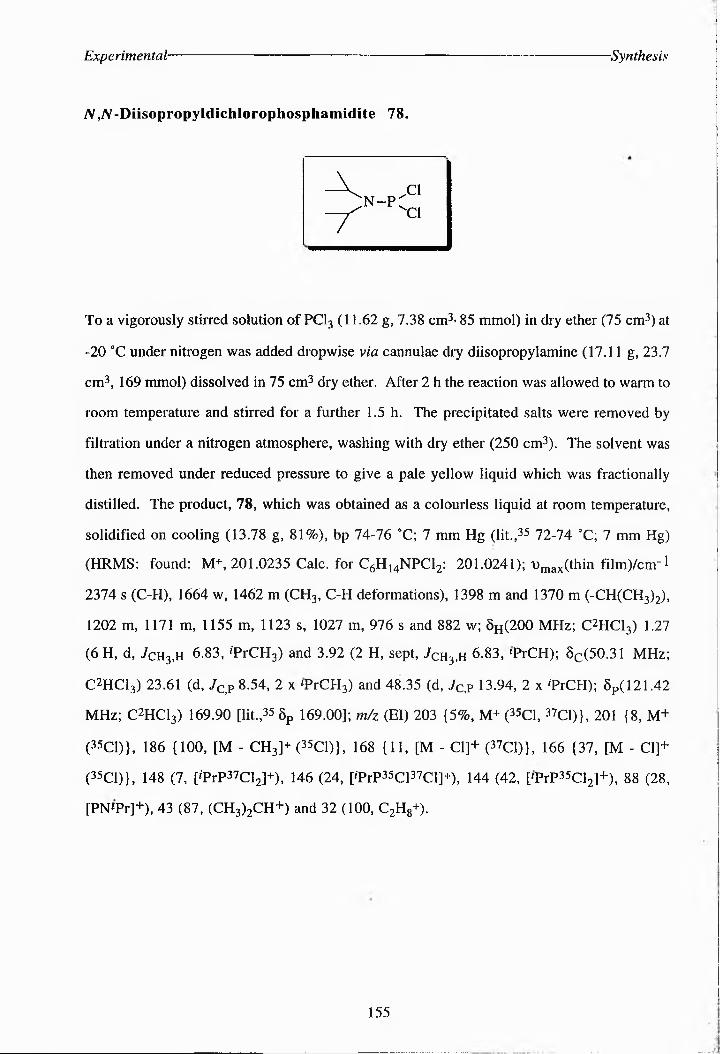
Experimental- Synthesis
A,A-Diisopropyldichlorophosphamidite 78.
AA- xCl
“P4^1V
To a vigorously stirred solution of PCI3 (11.62 g, 7.38 cm3- 85 mmol) in dry ether (75 cm3) at
-20 °C under nitrogen was added dropwise via cannulae dry diisopropylamine (17.11 g, 23.7
cm3, 169 mmol) dissolved in 75 cm3 dry ether. After 2 h the reaction was allowed to warm to
room temperature and stirred for a further 1.5 h. The precipitated salts were removed by
filtration under a nitrogen atmosphere, washing with dry ether (250 cm3). The solvent was
then removed under reduced pressure to give a pale yellow liquid which was fractionally
distilled. The product, 78, which was obtained as a colourless liquid at room temperature,
solidified on cooling (13.78 g, 81%), bp 74-76 OC; 7 mm Hg (lit,^ 72-74 0C; 7 mm Hg)
(HRMS: found: M+, 201.0235 Calc, for C6H14NPCl2: 201.0241); Tmax(thin film)/cm-l
2374 s (C-H), 1664 w, 1462 m (CH3, C-H deformations), 1398 m and 1370 m (-CH(CH3)2),
1202 m, 1171 m, 1155 m, 1123 s, 1027 m, 976 s and 882 w; 0H(200 MHz; C2HCl3) 1.27
(6 H, d, 7cH3,h 6.83, 'PrCHg) and 3.92 (2 H, sept, 7ch3,h 6.83, 'PrCH); 0c(50.31 MHz;
C2HCl3) 23.61 (d, 7c,p 8.54, 2 x 'PrCH3) and 48.35 (d, 7c,p 13.94, 2 x 'PrCH); 0p(121.42
MHz; C2HCl3) 169.90 [lit,35 Sp 169.00]; m/z (El) 203 {5%, M+ (35C1, 37Ci)}, 201 (8, M+
(35C1)}, 186 {100, [M - CH3]+ (35C1)}, 168 {11, [M - Cl]+ (37C1)}, 166 {37, [M - Cl]+
(35C1)}, 148 (7, p'PrP37Cl2]+), 146 (24, ['PrP^Cl^Cir), 144 (42, ['PrP^C^]*), 88 (28,
[PNPr]+), 43 (87, (CI^^CH*) and 32 (100, C2H8+).
155

Experimental- Synthesis
A,A-Diisopropylbis(benzyI)phosphoramidate 79.
To a stirred solution of the dichlorDFhosphamidite 78 (1.52 g, 7.51 mmol7 in dry DCM (5
cm37 At -10 °C under a nitrogen atmosphere was added via cannulxe a solution of dry benzyl
alcohol (2.13 g, 19.6 mmol7 and dry Et.N (2.8 cmA, 20.1 mmol7 in dry DCM (10 cm.7. After
30 min the reaction was allowed to warm to room temperature and stirred for a further 4 H.
The solution was then diluted with DCM (15 cm47, washed with NaHCO. solution (10 cm3 of
5g w/w) and saturated brine solution (10 cm.7, and dried (MgSO47. The solvent was then
removed under reduced pressure to yield the crude product as a pale yellow clear liquid.
Purification of the crude oil by silica column chromatography on triethylamine-basified silica
[eluant petroleum-ether/ethyl acetate 4:1] gave the phosphorA^niDate 79 as a colourless oil
(2.19 g, 84%7, (HRMS: found: [M + H]+, 346.1600, Calc, for C20H29NO2P: 346.19357;
3)max(thin film7/ cm-i 3070 m, 3060 s, 3010 s (triplet, C-H7, 2900 br s (CH2, CH3, C-H7,
1930 w, 1860 w, 1780 w, 1600 m and 1580 m (doublet, Ar ring7, 1490 s and 1450 s (CH2,
CH3, C-H deformations7, 1370 s (CH3 symmetrical deformation7, 1010 br s (P-O), 910 s xnd
870 m (Ar C-H7, and 770 br s (CH-Ar out of plane7; 0.(200 MHz; C2HCl37 1.23 (6 H, d,
7Ch3,h 6.84, 2 x 'PrCHy, 3.72 (2 H, m, 2 x 'PrCH), 4.75 (4 H, q, 7c,P 8.05, PhC/) and
7.35 (10 H, m, Ar-H7; 0c(50.31 MHz; C2HCI3) 24.56 (d, J^H 7.22, 2 x 'PiCH3), 42.95
(d, 7cH3,H 7.22, 'PrCH7, 64.30 (d, 7Ch2,op 10.88, 2 x OCH2Ph), 128.31, 127.41 and 126.87
(Ar-C7, 138.06 (d, /p,c 4.89, Ar-C quaternary/ 8p(121.42 MHz; C4HCl37 140.01 [lit.,35
6p(36.2 MHz; C^HC^ 148.10]; m/z (El7 346 (4%, [M + H]+7, 261 (3, [NH2P(OBn72]+7, 91
(34, ['PrNHPH2]+), 86 (39, 'PiNCH2]+), 44 {100, (CH372CH2+7, 43 (27, 'Pr+), 42 {17,
(CH37C+7 and 28 (16, C2H4+7.
156

Experimental- -Synthesis
a-Isopropyl-iV-tri'fluoroacetyl-(2S)-FhosFhohomoserinebis(benzyI) ester 76.
OX
F.c ^NH
CO2TPr °"PC
oII ..OBn
OBnH
To a stirred mixture of the alcohol 75 (0.192 g, 0.75 mmol) and \-H tetrazole (157 mg, 2.24
mmol) in dry DCM (12 cm2) Was added M,A^--^ii^c^l^l*<^l^^ll^i^(l^^l^:z^^)]phosphoramidite 79
(388 mg, 1.13 mmol). The mixture was stirred at room temperature for 4.5 h then cooled to
-40 °C. m-CPEA (258 mg, 1.50 mmol) in dry DCM (10 cm2) Was added dropwise to the
reaction mixture and the resulting solution was stirred at 0 °C for 45 min before being diluted
further with DCM (30 cm2), washed with NalSO3 (7 x 10 cm3 of 10% w/w ), NaHCO. (6 x
10 cm3 of 5% w/w ), water (30 cm2) and saturated brine solution (15 cm2), and then dried
(NalSO4). The solvent was removed under reduced pressure, giving the product as a clear
yellow oil (0.244 g, 63%); Dmax(thin film)/cnri 1786 w, 1725 m (C=0, ester), 1560 w (2°
amide), 1457 m (-CHl-, -CH., CH deformations), 1377 w (-CH., symmetrical
deformations), 1262 m (C-F, C-O or P=0), 1215 m (C-O, C-F), 1182 m (C-F, C-O), 1106 w
(C-O, C-F) and 1018 m (C-F); 6h(200 MHz; C2HCI3) 1.26 (6 H, m, 2 x 'PrCHg), 2.08 (2 H,
m, HP), 3.75 (2 H, m, HY), 4.71 (IH, m, H«), 5.07 (5 H, m, 2 x PHCH and 'PrCH
superimposed), 7.39 (10 H, m, Ar-H) and 7.62 (1 H, m, NH); 02(50.31 MHz; C2HCI.)
18.99 ('PrCH3), 21.60 (PC), 47.28 («C), 68.39 (d, /c.op 5.49, YC), 69.46 (CH^h), 70.31
('PrCH), 127.93 (10 x Ar-C), 135.33 (q, 7p,c 217.69, F.C) and 169.67 (pCO2'Pr);
5p(121.42 MHz; dHCI.) -1.07 [lit.,35 6p 0.55].
157

Experimental- •Synthesis
{2SyO -Phosphohomoserine 32.
To a stirred solution of phosphate triester 76 (0.201 g (0.39 mmol) in methanol (10 cm3) Was
added 5% palladium charcoal (45 mg). The mixture was purged with hydrogen for 30 min and
left to stir under a hydrogen atmosphere. After 4 days (when the reaction was judged to be
complete by tic [eluant MeOH-DCM 19:1]), the mixture was filtered through a celite pad and
concentrated under reduced pressure. The residue was dissolved in lM KOH (4 cm3) in
ethanol (5 cm3) which resulted in the solution turning yellow. The mixture was left stirring for
18 h and then concentrated under reduced pressure. The resulting pale brown solid was
subjected to ion-exchange chromatography (Dowex 50-W H+> 200-400 mesh, 8% cross
linked, 1cm x 6 cm) eluting with water. The ninhydrin positive fractions were combined and
lypholized to yield 32 as a white crystalline solid (700 mg, 91%), mp 167-169 °C (lit.,
170 °C); 'OjmxxNujolj/cm"1 1733 w (C=0, saturated acid), 1462 m (C-H deformations), 1249
w (P=O), 1020 w (C-O); 0h(200 MHz; ) 2.09 (2 H, m, HP), 3.93 (2 H, dq, 7cx 11-6,
7dx 5.4, HY) and 3.97 (1 H, dq, 7ax 7.6, 7bx 4.8, H«); 0C (50.31 MHz; CHgO) 33.40 and
33.25 (d, 7c,cop 7.63, PC), 54.19 and 54.03 (d, 7c,op 8.14, YC), 64.67 («C) and 175.09
(aCO2H); 0p(121.42 MHz; %20) 0.59 [lit,35 8p 0.245]; m/z (El) 200 (5%, [M + H]+), 199
(5, M+), 181 (9, M - H2O]+), 119 (17, [M - HgPOgP), 104 (23, [M - POJ+l, 97 (15,
[H2PO41U, 91 (73, [M - COPOgHH), 57 (23, [NH2CHCH2CH2H), 55 (37, [CgHsN]*) and
44 (100, CO2+).
158

Experimental- Synthesis
[U-14C]]a-Isopropyl-"V-trifluoroacetyl-(25)-aspartate 74b
This compound was prepared in a manner identical to the unlxbelled ester 74 starting from
[U-14C]-(2*07-xspartic acid (700 mg, 5.26 mmol7. THe reaction progress was monitored by tic
[Rf 0.46: ethyl acetate-light petroleum (3:27]. Recrystallisation from ether-light petroleum gave
the product as a white solid (1.0644g, 75%7l All spectroscopic and analytical data was
identical to that for 74.
[U-^Cba-Isopropyl-jV-trifluoroacetyl-^S^homoserine 75b
This compound was prepared in a manner identical to the alcohol 75 starting from 74 b
(1.0644 g, 3.93 mmol7. Purification by silica column cHromatograpHy [eluant DCM-EtOH
(97%7] gave the product as a yellow oil (0.518g, 51H7. All spectroscopic and analytical data
was identical to that for 75.
[U-i4<C ]-x-Isopropyl-V-trifluoroacetyl-(2S)-phosphohomoserinebis(ben2yl)
ester 76b
This was prepared in a similar manner to the phosphate triester 76, starting from 75b (518
mg, 2.02 mmol7. The reaction was monitored by tic [ethyl acetate-petroleum ether (1:47]. The
crude product was obtained as a yellow oil (1.085g, >100% due to benzyl alcohol impurities7.
All spectroscopic and analytical data was identical to that for 76.
[U-i4C]-(2S)“0 -phosphohomoserine 32b
This compound was prepared in a similar manner to 32, starting from 76b (1.085g, 2.1
mmolf Any organic impurities were then removed by extraction with ether (3 x 10 cm47, and
the product recrystallised from water-etHanol (346 mg, 83%7. All spectroscopic and analytical
data was identical to that for 32.
159

Experimental- -Synthesis
Dipentafiuorophenylphosphoro chloridate 81.
Using a modification of the procedure of EaerC27 freshly distilled POCI. (7.64 g, 50 mmol)
and pentafIuiropheNil (17.41 g, 94.6 mmol) were refluxed at 140 °C under a nitrogen
atmosphere for 48 h. The contaminants were distilled under reduced pressure to leave the
product as a pale brown oil which solidified upon cooling (28.99 g, >100%), bp 181-183 °C,
20 mm Hg; Dmax(lhm film)/cm-2 1346 m (C-F), 1318 m (C-F), 1239 m (C-O, C-F, P-O-aryl),
1156 s (C-F, C-O), 1034 (C-F, C-O) and 1001 (C-F, C-O); 0c(74.76 MHz; dHClg) 136.75
{meta Ar-CF), 139.70 {para Ar-CF), 140.13 (ortho Ar-CF), 143.01 (Ar-CO); 8p(121.42
MHz; C2HC16) main signal -15.14; 6p(282.2 MHz; C^dg) -154.89 and -154.96 (4 F, d
J 19.75, ortho Ar-CF), -157.96 (2 F, dt, 7 3.95, 7 21.73, para Ar-CF), -162.38 (4 F, t,
meta Ar-CF); m/z (EI) 449 (40%, M +), 413 (30, [M - Cl]+), 265 (14, [M - OC^M and 249
(17 [M - OoC^F5]U.
160

Experimental -Synthesis
A-TrifluoroacetyI-(2S)-aspartate 86.
AFaC NH H
CO,Hco2h
2
A solution of the anhydride 73 (0.555 g, 2.629 mmol) in distilled water (20 cm3) was stirred
overnight at rt until a yellow oil crashed out of solution. THe mixture was extracted with ether
(25 cm3), washed with water (2 x 10 cm3), 5% citric acid (10 cm3) and saturated brine
solution (10 cm3) and dried (MgSO3). The ether was removed under reduced pressure to give
a white solid, which was recrystallised from ether-hexane to give the diacid 86 (0.147 g,
25%); mp 132-134 °C; [at£> -24.35 (c 0.87 in EtOH); Dmaxxthin fUmycm! 2974 s (acid OH),
2877 s (acid OH), 2604 w (acid OH), 1717 (C=0, saturated acid), 1566 m (NH, 2° amide),
1371 m (C-F), 1303 m (C-F, C-O) and 1195 m (C-O, C-F); 0h(200 MHz; C4HC)3) 2.91 (2
H, dq, ABX, 7Ab 17.47, 7Ax 4.76,7Bx 4.16, HP), 4.71 (1 H, dt, 7nh,h 7.65, 7Ax 4.76, JBx
4.16, H“), 7.84 (1 H, d, 7Nh,h 7.65, amide NH) and 8.37 (2 H, br s, CO2H); 8C(74.76 MHz;
C2HCl3) 34.90 (PC), 48.63 (“C), 115.43 (q, 7F.c 285.26, F3C), 156.70 (q, fj^C 37.18,
F3CCO), 171.06 (pCOjH) and 172.54 ^^1); 8p(282.2 MHz; C2HCl3) -76.62 (FgChm/z
(Cl) 230 (28%, [M - H20]+), 184 (100, [M - HCC^H]*), 114 (22, [F3CC(O)NHCH2]+), 69
(35, F3C+) and 45 (32, CO2H+).
161

Experimental- Synthesis
a-p-Benzyloxybenzylpolystyrene-/V-trifluoroacetyl-(2S)-aspartate 87.
To a stirred suspension of Wang resin (200 mg, loading: 1.16 mmol/g) in dry DMF (5 cm3)
under a nitrogen atmosphere was added the anhydride 73 (0.4897 g, 2.32 mmol) and DMAP
(283 mg, 0.232 mmol). The reaction was warmed to 30-35 °C. After 1 week the resin was
filtered, washed copiously with DCM, DMF, 5% citric acid-DMF and methanol, then dried
under reduced pressure (248.9 mg, 99.9% loading, 0.931 mmol/g of A-TFA-asp);
i)max(KBr)/crn-1 1735 s (C=O, ester linkage); 8C(74.76 MHz; (C2H3)2SO) 34.81 (PC), 49.40
(«C), 117.84 (F3C), 130.02 (F3CCO), 162.48 (PCO2H) and 171.33 (aCO2R); SF(282.2
MHz; (C2H3)2SO) -74.27 (12%, F3C on p-ester) and -74.91 (88%, F3C on a-ester).
A portion of 87 was treated with TFA/H2O/TES (90:5:5) for 2 h at room temperature to
produce the free acid 86. All spectroscopic and analytical data was identical to that for 86,
{[a]D -25.09 (c 0.87 in EtOH)}.
162

Experimental- -Synthesis
a-p-BenzyIoxybenzylpolystyrene-A--rrifiuGroacetyI"(2S)-aspartate-P-methyl
ester 101.
To 87 (500 mg, loading: 0.913 mmol/g of A-TFA-asp) was added an excess of ethereal
diazomethaNe (10 cm2). The mixture was left at room temperature until all bubbling Had
ceased, and after the addition of another aliquot of diazomethane (5 cm2) the mixture was left
for a futher 30 min. Excess diazomethane was removed by bubbling nitrogen through the
solution and the resin was filtered and washed copiously with ether, DMF, 5% citric acid-DMF
and methanol. THe resin was dried under reduced pressure (524 mg, 99%). THe ester was
cleaved from the resin using TF^'H^O/TES (90:5:5) at room temperature for 2 H, filtered off
and dried under reduced pressure, giving the monoester 91 as a yellow oil; t)max((hin film)/cnr
2 3111 s and 2974 s (acid O-H), 2604 m (acid O-H), 1727 s (C=0, saturated acid), 1566 s
(C=O, 2" amide), 1454 s (saturated C-H), 1298 s (C-O, C-F), 1240 (C-O, C-F), 1186 s (C-O
ester, C-F); 0h(200 MHz; ^^2) 2.93 (2 H, dq, AEX, 7ob 17.3, Tax 5.02/Bx 4.74, HP),
3.63 (3 H, s, PCO2CH3, 84%), 3.70 (3 H, s, oC02CH., 16%), 4.68 (1 H, dt, Jnh.h 7.2,
7ax 5.02, 7bx 4.74, H«), 6.56 (1 H, br s, aCO2H) and 7.86 and 7.82 (1 H, d, 7NH)h 7.2,
amide NH); 0C(74.76 MHz; C2HCI.) 34.85 (PC), 48.54 («C), 51.77 ^CO^H., major
product), 52.71 (oCO2CH3, minor product), 115.43 (q, Jpc 285.28, F.C), 156.72
(q, 7f3c.c 36.48, F.CCO) and 170.81 (aCO^); 0p(282.2 MHz; C2HC13) -76.66 (F.C); m/z
(EI) 244 (17%, [M + H]+), 212 (27, [M - MeOH]+), 198 (100, [M - COgHl+j, 184 (55, [M -
CO2Me]+), 139 (38, [M - CO^H and CO2Me]+), 114 (25, [F3CC(O)NH3]+), 99 (43,
[F3CC(O)HJ+), 69 (47, F.C+) and 59 (41, [CO2Me]+).
163
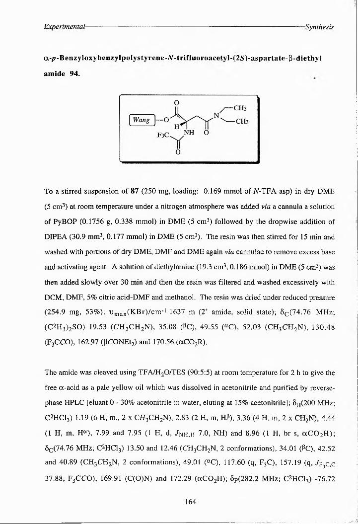
Experimental- -Synthesis
(X-p-Benzyloxyhtin.^yJ|)(‘Iys^l^ArenK’^^^^^^-rrtl3J(MrKKc.ety”i21S')-;^ispurtat.e-p-dicthyi
amide 94.
To a stirred suspension of 87 (250 mg, loading: 0.169 mmol of iV-TFA-asp) in dry DME
(5 cm3) at room temperature under a nitrogen atmosphere was added via a cannula a solution
of PyBOP (0.1756 g, 0.338 mmol) in DME (5 cm3) followed by the dropwise addition of
DIPEA (30.9 mm3, 0.177 mmol) in DME (5 cm3). The resin was then stirred for 15 min and
washed with portions of dry DME, DMF and DME again via cannulae to remove excess base
and activating agent. A solution of diethylamine (19.3 cm3, 0.186 mmol) in DME (5 cm3) WaS
then added slowly over 30 min and then the resin was filtered and washed excessively with
DCM, DMF, 5% citric acid-DMF and methanol. The resin was dried under reduced pressure
(254.9 mg, 53%); iDmax(KBr)/cm_1 1637 m (2" amide, solid state); 0^(74.76 MHz;
(C2H3)2SO) 19.53 (CH2CH2N), 35.08 (PC), 49.55 («C), 52.03 (CHaCH2N), 130.48
(F3CCO), 162.97 (PCONEt2) and 170.56 (aCO2R).
The amide was cleaved using TFA/H2O/TES (90:5:5) at room temperature for 2 h to give the
free a-acid as a pale yellow oil which was dissolved in acetonitrile and purified by reverse-
phase HPLC [eluant 0 - 30% acetonitrile in water, eluting at 15% acetonitrile]; 8H(200 MHz;
C2HCl3) 1.19 (6 H, m., 2 x C7?3CH2N), 2.83 (2 H, m, HP), 3.36 (4 H, m, 2 x CH2N), 4.44
(1 H, m, H«), 7.99 and 7.95 (1 H, d, /NH,H 7.0, NH) and 8.96 (1 H, br s, aCO2H);
8C(74.76 MHz; C2HCy 13.50 and 12.46 (CH3CH2N, 2 conformations), 34.01 (PC), 42.52
and 40.89 (CH3CH2N, 2 conformations), 49.01 (®C), 117.60 (q, F3C), 157.19 (q, 7f3c,c
37.88, F3CCO), 169.91 (C(O)N) and 172.29 (aCO2H); Sp(282.2 MHz; C2HCl3) -76.72
164

Experimental" -Synthesis
(F3C); m/z (El) 284 (42%, M+), 239 (15, [M - CO4C]+), 184 (14, [M - CON(CH2CH3)2]+),
114 (29, [CH2CCO)N(CH2CH3)2] + ), 100 (59, [CON(CH2CH3)] + ), 74 (46,
[(CH2CH3)2NH2]+), 72 (83, [(CH2CH3)2N]+), 58 (100, [CH3NCH2CH3]+) and 44 (36,
CO2+).
a"P-MethyIpolystyrene-N-trifIuoroacetyh(2S)-aspa.rtate 102.
-----------—.......... ... ......................... ,
0II
Merrifield —O ^'S-'-'^CChH
f3c nh3 ¥
0
To a stirred suspension of hydroxymethylpolystyrene resin (1.00 g, loading: 1.16 mmol/g) in
dry DMF (10 cm3) under a nitrogen atmosphere was added the anhydride 73 (2.4487 g,
11.60 mmol) and DMAP (0.1417 g, 1.16 mmol). The reaction mixture was left at 30 °C for
10 days. THe resin was then filtered, washed as copiously in DCM, DMF, 5% citric acid-
DMF and methanol and dried under reduced pressure (1.2450 g, 100% loading, 0.863 mmol/g
of N-TFA-asp); 'Umax(KBr)/cm-1 1735 s (C=0, aryl ester), 1541 (2° amide, solid state), 1170
s (C-F or C-O ester); 0c(74.76 MHz; (C^^SO) 34.79 (PC), 49.45 («C), 117.87 (F3C),
157.00 (F3C<CO), (PCO2H) and 11^7.^37 (aCO2R); MHz; C23HCC3) -74M
(F3C on a-ester).
165

Experimental- -Synthesis
PoIystyrene”4-oxymethyl-2-phenyIethanol 97.
To a stirred suspension of cHloromethylipolystyrenetdivinylBenzene (Merrifield) resin (200
mg, loading: 0.2 mmol) and 2-(pthydroxyphenyl)tethanol (0.0691 g, 0.5 mmol) in dry
acetonitrile (5 cm2) was added powdered potassium hydroxide (0.0281 g, 0.5 mmol) and
18-crownt6 (10 mg, 0.038 mmol). THe reaction was left for 3.5 days at 40 °C under a
nitrogen atmosphere. The loaded resin was filtered and washed copiously with DCM, 5%
citric acid-DMF, DMF and methanol and dried under reduced pressure (226.7 mg, 97% yield,
0.859 mmol/g loading); ^^(KBO/cirr1 3453 br m (O-H, alcohol), 1517 m (aromatic C=C),
1500 (aromatic C=C) and 1390 (O-H, alcohol); 02(74.76 MHz; Cg^g) 64.12 ((^OH),
70.33 (PhCtf2), 115.52 (Ar-CH), 130.68 (Ar-CH) and 158.55 (Ar-CO quaternary).
a-[2-(4-Methoxyphenyl)-ethyl]-W-trifIuoroacetyl-(2S)-aspartate 103.
To a stirred solution of 2-(4-metHoxypheNyl)-etHanil (0.50 g, 3.29 mmol) In dry DMF (15
cm2) under a nitrogen atmosphere was added the anhydride 73 (3.4676 g, 16.43 mmol) and
DMAP (0.2007 g, 1.643 mmol). After 2 days the reaction was judged to Be complete by tic
[/?f 0.0: ethyl acetate-light petroleum (3:2)]. THe solvent was removed under reduced pressure
166

Experimental- Synthesis
and the crude product redissolved in ether (20 cm3), washed with water (2x5 cm3), aqueous
citric acid (25% w/w 5 cm3) and saturated brine (5 cm3) and dried (MgSO4) (1.98 g, >100%
due to impurities). The crude product was purified by silica column chromatography [ethyl
acetate-petroleum ether (3:1)] (1.09 g, 91%), mp 89-90 "C; (HRMS: found: 363.0930
C15H16NO6F3 requires 363.2897); Dma/thin film^m! 1736 s (C==O, aryl ester), 1522 m
(C=O, 2“ amide), 1259 s (C-O, C-F), 1229 s (C-O, C-F) and 1181 s (C-O, C-F), 03(200
MHz; C4HCl)) 2.85 - 3.14 (4 H, m, HP and CH2O- overlapping), 3.77 (3 H, s, ArOCHg),
4.33 (2 H, m, PhCH2), 4.82 (1 H, dt, /^.h 7.78, H«), 6.85 and 6.80 (2 H, d, / 8.45, Ar-
H), 7.11 and 7.07 (2 H, d, J 8.45, Ar-H), 7.54 and 7.50 (1 H, d, JNH H 7.78, NH), 7.69 (1
H, br s, CO2H); 0c(74.76 MHz; C4HCl)) 33.66 (PC), 34.88 (0^0-), 48.50 («C), 55.09
(PhOCHg), 67.04 (PhCHz), 114.04 (Ar-CH), 115.46 (q, /p.c 287.65, F3C), 128.98 (Ar-C
quaternary), 129.69 (Ar-CH), 157.05 (q, ^^,6 38.91, F3CCO), 158.42 (ArC quaternary)
and 168.87 (CO2H); Sp(282.2 MHz; C4HCl)) -76.67 (F3C); m/z (El) 363 (11%, M+), 134
(100, [NH3CH(C02H)CH2C02H]+).
a-(PoIystyrene-4..oxymethyl-2-Fhenylethyl)-,A-trifIuoroAcety^(21S)-aspartAte
99.
To a stirred suspension of 97 (1.50 g, loading: 1.431 mmol/g) in dry DMF was added the
anhydride 73 (1.007 g, 4.77 mmol) and DMAP (0.0583 g, 0.477 mmol). THe reaction was
stirred at room temperature for 10 days. The resin was filtered, washed copiously with DCM,
5% citric acid-DMF, DMF and methanol and dried under reduced pressure (1.711 g, 97%,
167

Experimental- -Synthesis
0.541 mmol/g in tV-TFA-asp); i)max(KBr)/cnr1 1727 s (C=O, aryl ester), 1653 m (2° amide,
solid state) and 1078 w (C-O, C-F); 5C(74.76 MHz; C62H6) 34.09 (PC of aspartate),. 40.04
(CH2O-), 48.42 («C of aspartate), 114.31 (Ar-CH), 117.44 (F3C), 129.35 (Ar-CH), 145.15
(Ar-C quaternary), 157.08 (Ar-CO), 169.11 (F3CCO), 173.77 (CO2H of aspartate); 6p(282.2
MHz; (C2H3)2SO) -74.81 (F3C).
168

Experimental- Molecular Cloning
3.2 Molecular Cloning
3.2.1 Cloning Techniques
Bacterial Strains and Plasmids. Chromosomal DNA for the amplification of thrC was
prepared from E. coli. strain TGI or XL 1-Blue. Minipreparation of the plasmid pSG4
containing the gene for P-methylaspartase was from E. coli. strain TGI (K-12 A [lac-pro] supE
thi hsdR5 / F'-traD36 proA+ B+ lac] Q lacZ A M15 .26° E. con strains JM83 and XL1-Blue
were used as hosts for cloning experiments and BL21 (DE3) was used as the Host strain for
analytical expression of recombinant proteins. Transcription vectors pGEM™-3Zf(+),
pGEM™-5Zf(+) and pGEM-T™ and the expression vector pKK223-3 were purchased from
Promega. The expression vectors pET-3a, pET-3b and pET-16b were purchased from
Novagen, AMS Biotechnology. All vectors contain the ampicillin resistance gene (AmpR) and
a multi-cloning site contained in the N-terminal region of the lacZ operon.
Isolation of chromosomal DNA from E. coli. This was performed either as described
for the mimprepatation of plasmid DNA from E. coli, strain TGI (K-12 A [lac-pro] supE thi
hsdRS / F'-traD36 proA+ 5+ lac] Q lacZ A Ml5 or using the SDS-Proteinase K method of
large-scale chromosomal DNA preparation as follows:270
A starter culture of XLl-Blue cells in LB (10 cm2) was incubated at 37 °C for 3 h with
shaking. Luria broth (200 cm2) was inoculated with 200 p,l of this starter culture and incubated
overnight at 37 "C with shaking. Cells were pelleted by centrifugation (4 °C for 15 min at
6, 000 xg) and stored at -20 °C for 2 h before resuspeNsion in 40 cm2 te (10 mM Tris. HCl
(pH 8.0), 1 mM EDTA). The cell suspension was stored on ice for 10 min then centrifuged
(4 °C for 45 min at 3, 000 rpm). The pellet was then resuspended in 3.2 cm2 50 mM
Tris.HCl (pH 8.0) and 0.7 M sucrose. To this was added 0.6 cm2 lysozyme (20 mg/cm2) and
the mixture was stored on ice for 5 min. 0.6 cm2 EDTA (pH 8.0) and 0.5 cm2 10%
169

Experimental- Molecular Cloning
(w/v) SDS were added and the mixture stored on ice for a further 5 min The suspension was
adjusted to 1% (w/v) SDS and 0.5 mg/cmC Proteinase K (Promega) and incubated at 55 °C for
7.5 h. The reaction was halted by the addition of 5 cm3 phenol (pre-equilibrated with 1 M
Tris.HCl (pH 8.0)) and mixed for 3 h. The mixture was transferred to a Falcon tube and
centrifuged (25 °C, 10 min at 4, 000 rpm), discarding the lower phenol layer. 1.0 vol
phenol/ chloroform was added to the aqueous layer, mixed gently and the mixture centrifuged
at room temperature (10 min at 3, 000 rpm). The upper aqueous layer was transferred to a
fresh tube and stored on ice for 5 min before the addition of 2.5 cm3 7.5 m ammonium acetate
and 10 cm3 ethanol to precipitate the DNA. The mixture was stored at -20 °C for 30 min after
which the DNA was pelleted by centrifugation at room temperature (10 min at 3, 000 rpm),
rinsed with 70% (v/v) ethanol and air-dried for 2 min. The DNA was dissolved in H2O and
stored at -20OC before further use.
Restriction enzyme digestions. Restriction enzymes and buffers were purchased from
Promega or New England Biolabs. All plasmid DNA or gene fragment digestions with
restriction enzymes were carried out under the conditions specified by the supplier. In general
lpg of DNA was digested with 1 unit of restriction enzyme in the appropriate buffer in a total
volume of 20 jxl. DNA was purified by phenol/chloroform extraction.
CIAP-digestions. In general lpg of DNA was digested with 1 unit of calf intestinal
alkaline phosphatase (CIAP, Promega) in the appropriate buffer in a total volume of 20 pi
incubating at 37 °C for 1 h.. The plasmid DNA was purified by extraction 3 times with
phenol/chloroform, precipitation using 2.5 volumes ethanol and 1/20 volumes of 4M sodium
acetate and the mixture was stored at -20 OC for a minimum of 20 min. The DNA was pelleted
by centrifugation (13, 000 rpm for 15-20 min and redissolved in H2O to a final concentration
of 1 pg/pl.
Agarose gel electrophoresis. Flat bed 1% agarose gels were prepared using 1 x TAE
(0.04M Tris-acetate, 0.00 IM EDTA) containing 0.2 pg/cmC ethidium bromide. A 1 kb DNA
170

Experimental Molecular Cloning
ladder (Gibco) was run against DNA fragments to determine their approximate size.
«Gel purification of DNA fragments. To isolate DNA fragments from low melting point
(LMP) agarose the following methods were used:
(i) Phenol/acetate method. THe slice of LMP agarose containing the DNA fragment to
be isolated was placed in 500 pi of phenol/acetate (phenol equilibrated with 0.3 M sodium
acetate) and heated at 70 °C until the agarose was melted. After vortexing the tube was
incubated on ice for 10 min and then spun in a microfuge (10 min at 13, 000 rpm). The upper,
aqueous layer was removed to a fresh tube and subjected to a further extraction with
phenol/acetate. DNA was precipitated from the resulting aqueous fraction by ethanol
precipitation, dried in a vacuum dessicator and then redissolved in TE (10 mM Tris. HCl (pH
8.0), 1 mM EDTA) or HgO as appropriate to give an approximate concentration of 1 mg/cm3
DNA.
(ii) WIZARD™ Prep DNA Purification System. DNA was isolated from a section of
LMP agarose using the WIZARD™ Prep DNA purification system (Promega) according to the
manufacturer's instructions.
(Hi) Seaplaque™ agarose. THe desired DNA fragment was run into a section of
Seaplaque agarose (PMC Bioproducts) which was then excised, melted by incubation at 70 °C
and used directly for in-gel ligations according to the manufacturer’s instructions.
Blunt-end repair of overhangs. 5'-OverHangs were filled in to produce blunt ends using
T4 DNA polymerase. 1 jig of plasmid DNA was incubated for 5 min with 5 units of T4 DNA
polymerase (Promega), 100 |LM of each dNTP in T4 polymerase buffer (33 mM Tris.acetate
(pH 7.9), 66 mM potassium acetate, 10 mM magnesium acetate, 0.5 mM DTT, 0.1 mg/cmP
BSA). The reaction was halted by Heating to 75 °C for 10 min.
171

Experimental Molecular Cloning
Ligations. Ligation reactions were routinely carried out with 0.5 - 1 |xg of vector DNA, 1.5
jxg of insert DNA, 4 Weiss units of T4 DNA ligase (Promega) In a total of 25 pl of T4 DNA
ligase buffer [30 mM Tris.HCl (pH 7.8), 10 mM MgClg, 10 mM DTT, 0.5 mM ATP]. THe
reactions were incubated at room temperature or 16 0C overnight. Ligation reactions for the
insertion of gene fragments into pGEM-T were carried out using 50 ng pGEM-T vector
(Promega), with vector:iNsert ratios ranging from 1:1 and 1:10 using 1 unit T4 DNA ligase in
a total volume of 10 pi. THe concentration of cloned DNA and plasmid DNA solutions were
estimated from their luminescence on agarose gels. The following formula was used to
calculate the amount of insert needed for a ratio of 1:1 insert: vector:
ng of vector x kb size of insert------------------------------------------------------- x insert : vector molar ratio = ng of insert
kb size of vector
In-gel ligations were performed using Seaplaque™ agarose according to the manufacturer's
instructions.
Transformation of E. coli, E. coli was transformed with plasmid DNA after making
competent cells using the calcium chloride method (adapted from CoHen et al.2®1). 10 cm2
cultures of cells were grown to exponential phase in Luria Bertam broth [LB brotH: 1% (w/v)
bacto-tryptone, 0.5% (w/v) barto-yeast extract, 1% (w/v) NaCl, pH 7.5] cooled to 4 °C for 10
min and pelleted by centrifugation (4 °C for 10 min at 4, 000 rpm). Each pellet was
resuspended in 10 cm2 of ice-cold 0.1 M CaC^, stored on ice for 10 min and pelleted again as
before. Cells were then suspended in 200 pl 0.1 M CaC^ per pellet and stored at 4 °C for at
least 30 min to attain competency. Cells could be stored in CaC^ solution at 4 0C for 24 - 48 H
before losing competency.266 200 pi of competent cells were gently mixed with plasmid DNA
(up to 50 ng In 10 cm2 or less of solution) in sterile eppendorfs and the mixture stored on ice
for 30 min. The tubes were transferred to a circulating water batH preheated to 42 °C for
exactly 90 sec and then rapidly transferred to an ice-BatH for 1-2 min. 800 pi of LB was added
to each tube which were then incubated at 3°C for 45 min. Up to 200 pi of each culture was
172

Experimental- Molecular Cloning
then transferred to L-agar plates coated with 40 pi of X-gal (20 mg/ml in DMF) and 4 pi IPTG
(200 mg/ml in DMF) for overnight incubation at 37 OC. •
Minipreparation of plasmid DNA. 10 cm3 of LB containing 10 pg/cm3 ampicillin was
inoculated with a single colony of transformed E. coli JM83 or XL 1-Blue and incubated
overnight at 37 °C with shaking. The cells were pelleted (4 °C, 4, 000 rpm, 10 min) and the
supernatant removed. Pellets were resuspended in 700 pi of STET buffer (0.1 M NaCl,
10 mM Tris.HCl (pH 8.0), 1 mM EDTA (pH 8.0), 5% (v/v) Triton X-100), containing
1 mg/cmC lysozyme, and incubated at room temperature for 10 min. 30 pi of 10% (w/v) SDS
was added and mixed in then 75 cm3 of 4 m potassium acetate was added and mixed. After
incubation on ice for 15 min the mixture was spun in a microfuge for 15 min at 13, 000 rpm.
The supernatant was transferred to a fresh tube, 1 pi of 1 mg/cmC RNase A (Sigma) was added
and the mixture incubated at room temperature for 15 min before repeated extraction with
phenol/chloroform. The aqueous phase was removed to a fresh tube, and the DNA ethanol
precipitated twice. The DNA precipitate was washed with 70% (v/v) ethanol and resuspended
in 100 cm3 H2O.
Maxipreparation of plasmid DNA. This was performed according to the QI'AGEN
maxiprep plasmid purification protocol. LB was inoculated with a single colony of E. coli
XL 1-Blue, or with a starter culture of XL 1-Blue cells which had been transformed with
miniprep plasmid DNA isolated as above, and grown overnight at 37 °C with shaking. For
high copy number plasmids (pGEM vectors) 150 cm3 cultures were used, for low copy
number plasmids such as pET vectors 500 cm3 cultures were grown. 10 cm3 starter cultures in
LB were incubated at 37 °C for 3 or 4 h.
3.2.2 Polymerase Chain Reaction
Standard PCR Protocol. Sequences encoding thrC or the gene for p-methylaspartase were
amplified from chromosomal DNA via PCR. Reactions were carried out in TAQ DNA
173

Experimental- Molecular Cloning
polymerase buffer [50 mM KCl, 10 mM Tris.HCl (pH 9.0), 0.1% (v/v) Triton X-100, 2.5
mM MgCl2] with 1 mM DTT, 0.125 mM of each dNTP, 200 pmol of each primer and up to
1 |ig of template DNA in a total 100 |xl reaction volume. Reactions were overlaid with
100 cm3 mineral oil (Sigma), heated to 94 °C for 5 min and then Held at 85 0C whilst 2.5 units
of TAQ DNA polymerase (Boehringer Mannheim or Promega) were added to the aqueous
phase of each reaction.
PCR was carried out using the following programs:
For gene fragments with Nde l-sites at each terminus: 1: 94 °C, 2 min; 2: 42 °C, 1 min; 3:
72 °C, 2 min; 4: 94 0C, 1 min; 5: Cycle of steps 2-4, x 20; 6: 72 °C, 5 min; 7: Hold at
4 °C indefinitely.
For gene fragments with Nde l-sites at the N-terminus, BamH l-sites at the C-terminus: as
above except step 2: 40 °C, 1 min.
Products of PCR reactions were extracted against phenol/chloroform and ethanol precipitated
before further use.
Overlap PCR. THe reaction conditions were as stated above except that Half-gene fragments
were produced using either the external forward primer and the internal reverse primer, or the
external reverse primer and the internal forward primer. These were gel-purified and used as
the template for a PCR reaction using outside primers only to produce a whole gene fragment.
Cycle Sequencing. Plasmid DNA sequencing was carried out using the fmol™ kit
(Promega) according to the supplier's instructions. Sequencing gels were made using 31.5 g
urea, 15 cm3 Sequagel™ Concentrate (National Diagnostics), 15 cm3 x5 TEE [x5 composition:
54 g Tris base, 27.5 g boric acid, 0.5 M EDTA (pH 8.0) and dH2O to 11], making up to a total
volume of 75 cm3. To 68 cm3 of this was added 300 pi 10% APS solution and 100 pi
TEMED. Gels were preheated to 44 0C before applying samples for separation, run at
200 Vand dried under vacuum on a slab-gel drier for 2 H at 80 °C. Dried gels were exposed
on X-Ray photographic film overnight.
174
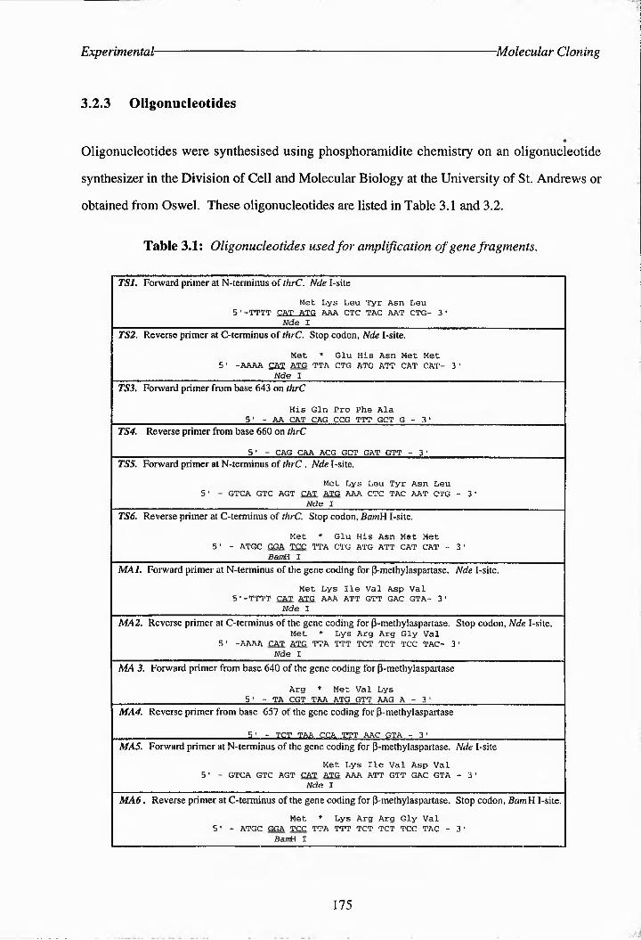
Experimental -Molecular Cloning
3.2.3 Oligonucleotides
Oligonucleotides were synthesised using phisphotamidite chemistry on an oligonucleotide
synthesizer In the Division of Cell and Molecular Biology at the University of St. Andrews or
obtained from Oswel. These oligonucleotides are listed in Table 3.1 and 3.2.
Table 3.1: Oligonucleotides used for amplification of gene fragments.
TSl. Forward primer at IN-terminus of thrC. Nde I-site
Met Lys Leu Tyr Asn Leu 5•-tTTT CAT ATG AAA CTC TAC AAT CTG- 3'
_______________________________Nde I_______________________________________________TS2. Reverse primer at C-terminus of thrC. Stop codon, Nde I-site.
Met * Glu His Asn Met Met 5' -AAAA CAT ATG TTA CTG ATG ATT CAT CAT- 31
_____ __ _____________________ Nde I_______________________________ _________________TS3. Forward primer from Base 643 on thrC
His Gin Pro Phe Ala________________________ 5' - AA CAT CAG CCG TTT GCT G - 3 1________________ ________TS4. Reverse primer from Base 660 on thrC
________________________ 5 ' - CAG CAA ACG GCT GAT GTT - 3 '__________ __ _____ ______TS5. Forward primer at ^terminus of thrC . Nde I-site.
Met Lys Leu Tyr Asn Leu5' - GTCA GTC AGT CAT ATG AAA CTC TAG AAT CTG - 3'
___________________________________ Nde I___________________________________________TS6. Reverse primer at C-terminus of thrC. Stop codon, BamH I-site.
Met * Glu His Asn Met Met 5■ - ATGC GGA TCC TTA CTG ATG ATT CAT CAT - 3'
_______ _______________________ BamH I_____________________________________ _________ __MAI. Forward primer at ^terminus of the gene coding for P-metHylaspartase. Nde I-site.
Met Lys lie Val Asp Val 5.-TTTT CAT ATG AAA ATT GTT GAG GTA- 3’
_______________________________Nde I_______________________________________________MA2. Reverse primer at C-terminus of the gene coding for PtmetHylaspartase. Stop codon, Nde I-site.
Met * Lys Arg Arg Gly Val 5‘ -AAAA CAT ATG TTA TTT TCT TCT TCC TAG- 3'
_____________________________ Nde I_________________________________________________MA 3. Forward primer from Base 640 of the gene coding for ptmetHyiaspartase
Arg * Met Val Lys________________________ 5 1 - TA CGT TAA ATG GTT AAG A - 3 '________________________MA4. Reverse primer from Base 657 of the gene coding for PtmetHylaspartase
_________________________ 5 ' - TCT TAA CCA TTT AAC GTA - 3 ' _______ _________ _____MAS. Forward primer at N-terminus of the gene coding for PtmetHylasparlase. Nde I-site
Met Lys lie Val Asp Val51 - GTCA GTC AGT CAT ATG AAA ATT GTT GAC GTA - 3'
Nde IMA6. Reverse primer at C-terminus of the gene coding for ptmetHylospartase. Stop codon, BamH I-site.
Met * Lys Arg Arg Gly Val 5' - ATGC GGA TCC TTA TTT TCT TCT TCC TAG - 3'
BamH I
175

Experimental- Molecular Cloning
Oligonucleotide primers were designed to contain restriction sites to enable PCR products to be
easily cloned. For forward coding sense primers the encoded amino acid sequence is shown - *
above the nucleic acid sequence. Stop codons are indicated on the complementary strand by
a *.
Table 3.2: Oligonucleotides used in sequencing experiments.
SP6 promoter primer.5' - d(GAT TTA GGT GAC ACT ATA G) - 3’
T7 promoter primer.
5' - d(TAA TAC GAC TCA CTA TAG GG) - 3'
Primer concentrations were calculated from their A260 values using the following formula:
Concentration (pmol pl-1) = Amo x dilution factor(nA x 1.52) + (nC x 0.75) + (nG x 1.17) + (nT x 0.92)
3.2.4 Expression in E. colt
SDS-polyacrylamide gel electrophoresis (SDS-PAGE). The discontinuous buffer
gel system based on that of Laemmli was used.317 All gels were constructed with a 4%
stacking gel and a polyacrylamide resolving gel. Gels were run at 40 mA upper current
limit and 250 V upper voltage limit. Generally proteins were run through the stacking gel at
120 V and run through the resolving gel at 225 V. Gels were stained overnight in Coomassie
blue according to the method of Sambrook et al.,266 destained in destaining buffer (5%
methanol, 10% acetic acid in aqueous solution) and dried under vacuum on a slab-gel drier at
60 OC for 2 h.
Analytical expression of proteins. 10 cm3 cultures of LB-ampicillin (LB with 100
pg/cmC ampicillin) were inoculated with single colonies ofE coli BL21(DE3) transformed
176

Experimental: Molecular Cloning
with pTN-TS expression vectors and grown overnight at 37°C with shaking. 2 x l cm3 of
each overnight culture was transferred to 2 x 9 cm3 of fresh LB-ampicillin (one culture for
induction and one as a control) and incubated at 37 °C for 3 h with shaking. To the cultures to
be induced was added 100 pi of 100 mM 1PTG and all cultures were incubated for a further 2 H
at 37 °C with shaking. The cultures were then kept at 4 °C for a short time before the cells
were pelleted (4°C, 4, 000 rpm, 10 min), resuspended in 500 pi chilled PBS buffer containing
12 mg/cm3 lysozyme and transferred to sterile eppendorfs. THe cells were then lysed using a
probe sonicator in several 10 sec bursts resting on ice in between. Insoluble material was
removed by centrifugation in a microcentrifuge (4 °C, 10, 000 rpm, 30 min) and 20-40 pi of
the lysate was subjected to SDS-PAGE analysis using molecular weight markers of 66 and 45
kDa. The total protein concentration of the lysates was measured using the BCA protein assay
(Pharmacia) as recommended by the manufacturer, using a BSA calibration curve for
comparison. Varying amounts of two of the lysates were applied to a gel first of all in order to
determine what protein concentration would produce the clearest results. Approximately 50 pg
of protein from each sample was loaded onto gels.
Analytical purification of His-tagged proteins. Ni-NTA Resin synthesised by C.
Botting at the Division of Cell and Molecular Biology was prepared for use by equilibrating
with 10 volumes of the sonication buffer desribed above for 1 H. 250 pi of the cell lysate from
cells transformed with vectors pTN-TS5 - 8 were stirred with between 100 and 140 pi of a
50% Ni3+ resin slurry at room temperature for 1 h. The slurry was pelleted by centrifugation
on a microfuge (10, 000 rpm, 10 min) and the supernatant retained for analysis. THe resin was
then washed with 150 pi TBS (20 mM Tris, 0.3 M NaCl, pH 8.0), centrifuged as before and
the supernatant retained for analysis. THe protein was then eluted from the resin by washing
with 150 cm3 of elution buffer (250 mM imidazole, 20 mM Tris, 0.3 M NaCl, pH 7.5). This
was centrifuged and the supernatant containing the His-tagged proteins removed. THe
supernatant from each of the Ni3+ treatment steps was analysed by SDS-PAGE.
Plasmid Stability Test, E. coli BL21(DE3) freshly transformed with pTN-TS vectors 5-8
177

Experimental- Molecular Cloning
were grown in 5 cmC portions of LB-medium containing lOOmg/cm3 ampicillin overnight at
37 °C with shaking. A small subculture was transferred into 500 cm3 of fresh LB medium - «
containing 100 mg/cmC ampicillin and allowed to grow at 37 °C to an ODeoo reading of 0.6.
Three aliquots of 1,000 mmC, 147 mm3 and 135 mm3 of LB were transferred to three sterile
eppendorfs. A primary dilution of bacteria was prepared by transferring 1 mmC of log phase
culture into 1 cm3 of LB. Two secondary dilutions were prepared by transferring 3 mm3 and
15 mmC of the primary dilution into tubes containing 147 mm3 (dilution A) and 135 mmC
(dilution B) of LB, respectively. Bacteria were plated onto L-agar plates containing no
additives, lOOgg/ml ampicillin only, 0.4 mM IPTG only or both lOOgg/ml ampicillin and
0.4mM IPTG., as indicated in Table 2.6. The plates were incubated overnight at 37 "C.
178

REFERENCES

-References-
1. P. P. CoHet, 'The Enzymes', ed. J. B. Summer and K. MyrbCck, Academic Press 1tc., New York, 1951, vol. 2, p. 1040.
2. W. A, Wood and 1. C. Gunsalus, J. Biol. Chem., 1951, 190, 403 - 416.
3. E. F. Gale, Adv. Enzymol, 1946, 6, 1 - 32.
4. D. Gani, 'Pyridoxal Dependent Systems', CH. 6.5, it ‘Comprehensive Medicinal Chemistry - The Rational Design, Mechanistic Study and Therapeutic Application of Chemical Compounds’, ed. C. HrtscH, P. G. Sammes, and J. B. Taylor, Pergamon Press, Oxford, 1990, vol. 2, pp- 213 - 254.
5. H, G. Floss and J. C. Vederas, 'Stereochemistry of pyridoxal phosphate-catalyzed reactions’, CH. 4, it ’Stereochemistry', ed. C. Tamm, Elsevier Biomedical Press, 1982, pp- 161 - 165 and 188 - 195.
6. 'Enzyme Nomenclature', it Recommendations of the Nomenclature Committee of the International Union of Biochemistry and Molecular Biology', Academic Press, 1992.
7. P. Gyorgy, Nature, 1934, 133, 498 - 501.
8. J. Goldberger and R. D. Lille, Public Health Reports (US), 1926, 41, 1025 - 1027.
9. S, OHdake, Bull. Chem.. Soc. Jpn., 1932, 8, 11 - 15.
10. T. W. BircH and P. Gy orgy, Biochem. J., 1936, 30, 304 - 315.
11. J. C. Keresztesy and J. R. Stevens, Proc. Soc. Exp. Biol. Med., 1938, 38, 64 - 69.
12. E. T. Stiller, J. C. Keresztesy and J. R. Stevens, J. Am. Chem. Soc., 1938, 61, 1237 - 1242.
13. S. A. IHarri and K. Folkers, Science, 1939, 89, 347 - 350.
14. R. Kuhn and G. Wendt, Ber. Deutsch. Chem. Gee, 19^1^, 71, 780 - 783.
15. E. F. Gale and H. M. R. Epps, Biochem. J., 1944, 38, 232 - 256.
16. W. D. Bellamy and 1. C. Gutsalus, J. Bacteriol., 1943, 46, 573 - 578.
17. E. F. Snell, J. Am. Chem. Soc, 1945, 61, 194 - 197.
18. H. C. DunaChan and J. G. VoeU Proc. Nath Acad. Sci. USA, 1974, 71, 3888 - 3891.
19. C. Parsot, EMBO J., 1986, 5, 3013 - 8C13.
20. P. K. Mehta, T. 1. Hale and P. Christen, Eur. J. Biochem., 1338, 214, 549 - 561.
21. M. Gribskov, R. LutHy and D. Eisenberg, Methods Enzymol, 1990, 183, 146 - 159.
22. P. K. Mehta and P. CNri sten, Eur. 7. B^uochem, cSS3, 293, 373 1- 336.
23. P. K. Mehta nnd P. ChiisteC norNe/Si Biemhys. Res. Cornmunommun. 198, 938 - 143.
24. F. W. Alexander, AN Sandmeirila. idr Me hta nnd P. Christen,riwr. 7. BiocJiem, 1993, 933, 393 - 560.
25. R. A. John, Bioihim. Bsr3hys. Ana, 19c5, 1248, 81 496.
26. P. K. Mehia, T. I. HaAe and P. Christen, Eur. J. Biochem, 1989, 186, 249 - 253.
27. A. A. Antson, T. V. Demidkinar, P. Gohnick, Z. Dauter, R. L. Von Tersh, J. Long, S. N . Berzshnoy , PL. S. Philips, E. H. HarutyuNyat and K. S. Wilson, Biochemistry, 1338, 32, 3135 - 33C6.
28. M. D. Toney, E. Hohenester, S. W. Cowan and J. N. Jansonius, 1993, 261, 1156 - 759.
29. J. M. Green, W. IK Merkel and B. P. Nichols, J. Badeevl,, 1992, 174, 5317 - 5323.
179
-References-
30. R. A. Jensen and W. Gu, J. Bacteriol., 1996, 178, 2161 - 2171.
31. S. Pascarella, V. Schirch and F. Bossa, FEBS Lett., 1993, 331, 145 - 149.
32. C. Ouzounis and C. Sander, FEBS Lett., 1993, 332, 159 - 164.
33. C. Babbitt and J. A. Gerlt, FASEB J., 1995, 9, A1266.
34. U. J. Kang and T. H. Job, Mol. Brain Res., 1990, 8, 83 - 87.
35. E. Sandmeier, T. I. Hale and P. Christen, Eur. J. Biochem., 1994, 221, 997 - 1002.
36. P. Bork and K. Rohde, Biochem. Biophys. Res. Commun., 1990, 171, 1319 - 1325.
37. C. Parsott Proc. Natl. Acad. Sci. USA, 1987, 84, 5207 - 5210.
38. J. Belfaiza, C. Parsott A. Martel, C. Bouthier de la Tour, D. G. N. Cohen and I. Saint-Girons,Proc. Natl. Acad. Sci. USA, 1986, 83, 867 - 871.
39. A. E. Braunstein and M. M. Shemyakin, Biokhymiya, 1953, 18, 393 - 411.
40. D. E. Metzler, M. Ikawa and E. E. Snell, J. Am. Chem. Soc., 1954, 76, 648 - 652.
41. F. Barclay, PhD Thesis, 1994, University of St Andrews.
42. D. M. Smith, N. R. Thomas and D. Gani, Experientia, 1991, 47, 1104 - 1118.
43. H. Lis, Biochim. Biophys. Acta, 1958, 28, 191 - 197.
44. W, T. Jenkins, D. A. Yphantts and I. W. Si^^e! J. Biol. Chem., 1159, 234, 51 - 52.
45. H. Lis, P. FaseUa, C. Turano and P. Vecchmi, Biochim. Biophys. Acta, I960, 45, 529 - 536.
46. D. Gani and J. A. Robinson, 'Stereochemical and Mechanistic Features of Pyridoxal-5'-phosphate- Dependent Enzymes', Ch. 1, in ‘Natural Product Reports', 1986, pp. 1 - 50.
47. V. C. Emery and M. Akhtar, 'Pyridoxal Phosphate-Dependent Enzymes', Ch. 18, in 'Enzyme Mechanisms', ed. M. I. Page and A. Williams, Royal Society of Chemistry, London, pp. 345 - 389.
48. V. P. Almazov, Y. V. Morozov, F. A. Savin and B. S. Sukhareva, Int. J. Quantum Chem., 1979, 16, 769 - 775.
49. J. R. Fischer and E. H. Abbott, J. Am. Chem.. Soc., 1979, 101, 2781 - 2782.
50. M. Y. Karpeisky and V. I. Ivanov, Nature, 1966, 210, 493 - 496.
51. V. I. Ivanov and M. Y. Karpeisky, Adv. Enzymol, 1969, 32, 21 - 54.
52. M. Yu Torchinsky and A. E. Braunstein, FEBS Symp., 1979, 52, 293 - 303.
53. S. C. Almo, D. L. Smith, A. T. Danishefsky and D. Ringe, Protein Eng., 1994, 7, 405 - 412.
54. V. N. Ma^hkovic^ J. Jager, M. Ziak, U. Sauder, H. Gehring, P. Christen and J. N. Jansonius, Biochemistry, 1995, 34, 405 - 414.
55. S. Rhee, M. M. Silva, C. C. Hyde, P. H. Rogers, C. M. Metzler, D. E. Metzler and A. Arnone, J. Biol. Chem., 1997, 272, 17293 - 17302.
56. C. C. Hyde and E. W. Miles, Biotech., 1990, 8, 27 - 32.
57. S. Rhee, K. D. Parris, C. C. Hyde, S. A. Ahmed, E. W. Miles and D. R. Davies, Biochemistry, 1997, 36, 7664 - 7680.
180

-References-
58. F. C. Bernstein, T. F. Koetzle, G. J. B. Williams, E. F. Meyer Jr., M. D. Brice, J. R. Rodgers, O. Kennara,T. ShiHim^t^i^c^u^i i ndM. Tasami, 7. MM. Bioi., 1917, 112, 535 5 542.
59. E. E. ABolo, F. C. Bernstein, S. H. Bryant, T. F. Koetzle and J. Weng, 'Protein Data Bank' in 'Crystallographic Databases - Information Content, Software Systems, Scientific Applications', ed. F. H. Aller, G. BergerHoff and R. Sievers, Data Commission of the InternatioNal Urior of Crystallography, Born/CamBridge/Chester, 1987, pp. 107 - 132.
60. S. Rhee, K. D. Parris, S. A. AHmed, E. W. Miles ord D. R. Davies, Biochemistry, 1976, 35, 4211 - 4221.
61. G.D. McClure ord P. F. Cook, Biochemistry, 1994, 33, 1674 - 1683.
62. G. B. Stfambinii P. Cioni, A. PeroccHi and A. Mozzarella Biochemisry, 15^^^, 31, 7527 - 7534.
63. G. B. Strambinii P. Cioni, A. PeraccHi and A. Mozzarelli, Biochemistry, 1^^2, 31, 7535 - 7542.
64. G. B. Strambiri, P. Ciom ord P. F. Cook, Biochemistry, 1996, 35, 8392 - 8400.
65. K. D . Shhnckkerz, C-H. Tai, J. W. Simmons, T. M. JocoBsor, G. S. Jogorrotho Rao ord P. F. Cook, Biochemistry, 1995, 34, 12152 - 12160.
66. C-H. Tai, S. R. NoIoBoIu, J. W Simmons, T. M. Jacobson ord P. F. Cook, Biochemistry, 1995, 34, 12311 - 12322.
67. E. U. Woehl, C-H. Tai, M. F. Durr ord P. F. Cook, Biochemistry, 1996, 35, 4776 - 4783.
68. P. F. Ccok, S. Hara. S . NalciaBkianNK. D. Schhnacerz, Bioccemistry, 1999, 31, 2229- 2203.
69. C-CcHhwij^, E. U. Woohl, D.E.Minier, M. F. Duun anNPP P-Ccoo,#iocCemistry, 1996, 33, 6335 -6365.
70. A. M. KayostHo, Y. Sowo, S. No6o1o ord E. W. Miles, J. Biol. Chem., 1991, 266, 7618 - 7625.
71. X-J. Yang and E. W. Miies, J. Biok Chem., 1193, 268, 22269 - 22272.
72. Z. Lu, S. Nagata, P. McPhie and E. W. Miies, J. Biok Chem. 1993, 268, 8727 - 8734.
73. A. Ahmed and E. W. Miies, FASEB J., 1196, 10, 1374.
74. ,Biochemiitty,' 3od Ed.. ed. Lubert Stryer, W. H. Freeman and Co., New York, 1988.
75. 'The Vitamins', CH. 8 in 'Basic Nutrition', ed. E. W. McHenry ord G. H. Beoton, Pitman Medical Publishing Co. Ltd., London, 1163, p. 116.
76. A. D. Hurt, J. Stokes, W. W. McCroBy ord H. H. Stroud, Pediatrics, 1954, 13, 140 - 142.
77. W. W. Hawkins ord J. Barsky, Science, 1948, 108, 284 - 287.
IS. G. A. Goldsmith, 'Thh B 7110011^: Thiamine, Riboflavin and Niacin', CH. 2 in 'Nutrition: A Comprehensive Treatise', c,. G. H. Beaato aoNE. W. MccHnry, AccadmicPress, Nnw YoikaarLoordo, 1994, vol. 2, pp. 109 - 206.
79. M. Pykn, 'Deficiency Diseases', CH. 7 in Man and Nutrition', World University LiBrary, 1970, pp. 117 -121.
80. B. F. Chow, 'The B Vitamins: B& By2, Folic Acid, Panthenoic Acid and Biotin', Ch. 3 ir Nutrition: A Comprehensive Treatise’, ed. G. H. Bnoton ord E. W. McHenry, Academic Press, New York ord Lordor, 1964, vol. 2, pp. 207 - 264.
81. E. E. Smll, Phys. Rev., 1953, 30, 509 - 524.
181

-References-
82. M. M. Wintrobe, R. H. Follis Jir, M. H. Miller and H. J. Stein, Bull. John Hopkins Hosp., 1943, 72, 1-9.
83. J. F. Rin^lh^ir and L. D. Greenberg, Am. J. Path.. 11>94), 25, 481 - 485.
84. S. Gerhoof, F. Faragalla, D. A. Ne^on and S. B. Andrus, Am. J. Med.. 1959, 27,1^2 - 77.
85. S. R. Snodgrass, J. Chiid Neurol 1992, 7, 77 - 86.
86. H. A. Ring, A. J. HeUer, I. N. Farr, E. H. Reynolds, J. Neurol Psych., 1990, 1051 -1055. ................
87. P. R. M. Debittencosrt, S. Mazer, T. Marcotn'aRis, M. M. Bigarella, Z. S. Ferreira and J. P. Mumford, Epilepsia, 1994, 35, 373 - 380.
88. A, Sabers and L. Gram, Pharmacol. Toxicol. 1992, 70, 237 - 243.
89. E. A. Hambbrger aad J. Mumford, Epiiepsy Res.. 1999, 16, 241 - 243.
90. W. Loscher ann D. Horrtennann, Naunyn-SchmiedeSesgs Arch. Pharmacol,. 1194, 349, 270 1 278.
91. I. E. Leprik, Epilepsia, 1994, 35, S29 - S40.
92. R. H. Mattson, O. Petroff, D. Rothman and K. Behar, Epiiepsiit, H94, 35, S29 - S32.
93. M. C. T. F. M. Dekrom, N. Vei’duin, E. Visser, M. Kleijer, F. Scholtes and J. H. M. Degroen, Ssiznrs, 1995, 4, 159 - 162.
94. R. Pacific, P. Zuccm’o, P. Ianetti, U. Raucci and C. Imperato, Epilepsia, 1995, 36, 423 - 426.
95. M. Qume and L. J. Fowler, Brit. J. Pharmacol 1997, 122, 539 - 545.
96. H. Golan and Y. Grossman, J. Neurophysiol 1994, 71, 48 - 58.
97. K. Sawaki, K. Ouchi, T. Sato and M. Kawaguchi, Jpn. J. Phaamacol 1991 , 56, 327 - 335.
98. J. Pietz, C. Bendingee, H. Schafer, D. S^tlreim!-, G. Misteemniee and D. Rating, Epilepsia, 1993, 34, 757 - 763.
99. S. B. Coker, Pediatrics, 1992, 90, 221 - 223.
100. K. Taylor and J. Goldstein, Headache, 1996, 36, 514 - 515.
101. K. F, McKenna, D. J. McManus, G. B. Baker and R, T. CouSSs, J. Neurol. Transmission - Supplementum, 1994, 115 - 122.
102. O. Heby, Adv. Enz. Rsgnl, 1985, 24, 103 - 124.
103. C. W. Taylor and H. Tabor, Anno. Rsv. Piochem., 1976, 45, 285 - 301.
104. E. Grassilli, M. Alfonsina Desiderio, E. Bellesia, P. Salomoni, F. Benatti and C. Franceschi, Biochsm. Biophys. Rss. Common., 1995, 216, 708 - 714.
105. G. Tiao, Y. Noguchi, M. A. Lieberman, J. E. Fischer and P. O. HnsseIgeen, Shock, 1995, 4, 403 - 410.
106. M. F. K. BBakaaa, A. M. Raa, M. R. Presan ami R. J, Dempsey, Naergsurggsg, H99, 38, 110 , 145.
107. C. M. Henley, C. Museynski, L. Chhrian aad C. S. Rabbresen, J. Nsnrotranma, 1996, 13, 487 - 496.
108. D. H. Rasserl, ini 'Phlynminys in Namm aan Naeoiacyie Grgwth', Raven, New York, 11973.
109. A. E. Pegg, L. M. Shams and C. S. Coteman, J. Cdl, Biocham.. 1995, 132 - 138.
182

-RsfsrsnBss■
110. G, Chen, E. M, Perchellet, X. M. Gao, S. W. Newell, R. W. Hemingway, V. Bottari and J. P. Perchellet, AntiranrsJ Res., 1995, 15, 1183 - 1189.
111. L. Megosh, S. K. Gilmour, D. Rosson, A. P. Soler, M. Blessing, J. A. Sawicki and T. G. O'Brien, Cancer Res., 1995, 55, 4205 - 4209.
112. A. Tamori, S. Nishiguchi, T. Kuroki, N. Koh, K. Kobayashi, Y. Yano and S. Otani, Cancer Res., 1995, 55, 3500 - 3503.
113. N. Matsubara, O. A. Hietala, S. K. Gilmour, K. Y. Yum, S. Litwin, P. Watts, E. J. Brennan and T. G. O'Brien, min. Cancer Res, 1995, 1, 665 - 671.
114. A. Manni, D. Mauger, P. Gimotty and B. Badger, Clin. Cancer Res., 1996, 2, 1901 - 1906.
115. F. L. Meyskens and E. W. Gerner, J. Cell. Biochem., 1995, 126 - 131.
116. S. Merali and A. B. Clarkson, Antimicrobial Agents and nSsmotSsraey, 1996, 40, 973 - 978.
117. Y. V. Bukin, V. A. Draudinkylenko, E. N. Orlov, Y. P. Kuvshinov, B. K. Poddubny, O. V. Vorobyeva and M. A. Shabanov, Cancer Epidemiology Biomarkers and Prevention, 1995, 4, 865 - 870.
118. Y. Okai, K. Higashiokai, Y. Yano and S. Otani, Cancer Lett, 1996, 99, 15 - 21.
119. M. Wadatani, K. Ohtani, K. Kageyama, Y. Yano, T. Hasuma, S. Otani, S. Sakanaka, H. Kim and I. Matsuiyuasa, Crncer JJ 191^<^, 9, 1 61 1 - 67.
120. J. Bomser, D. L. Madhavi, K. Singletary and M. A. L. Smith, Planta Medica, 1996, 62, 212 - 216.
121. J. A. Moshier, M. Skunca, W. Wu, S. M. Boppana, F. J. Rauscher and J. Dodescu, Nucleic Acids Res., 1996, 2, 1149 - 1157.
122. 'International Encyclopaedia of Pharmacology and Therapeutics', ed. M. Schlachter, Pergamon, Oxford, 1973, Section 74.
123. M. J. Jung, Bioorg. Chemi., 1986, 14, 429 - 443.
124. J. J. Skratt, L. B. Hough, J. W. Nalwalk and K. E. Barke, BiorSsm. Pharmacol., 1994, 47, 397 - 402.
125. Y. M. Lee, S. Hirota, T. Jippokanemoto, H. R. Kim, T. Y. Shin, Y. Yeom, K. K. Lee, Y. Kitamura, S. Nomura and H. M. Kim, ton Aid. Allergy Immmnol., 1996, , 10, 272 - 277.
126. F. Hollande, A. Choquet, J. P. Ball and R. Magous, Endocrinol., 1997, 138, 955 - 962.
127. T. Kaneko, Y, Nagamachi and S. Matsuzaki, Prostaglandins, 1997, 53, 37 - 47.
128. P. R. Buckland, R. Marshall, P. Watkins and P. McGuffin, Mol. Brain Res., 1997, 49, 266 - 270.
129. M. Bertoldi, P. S. Moore, B. Maras, P. Dominici and C. B. Voltattorni, J, Biol. Chem., 1996, 271, 23954 - 23959.
130. J. Drsata, J. Ulrichova and D. Walterova, J. Enz. Inhib., 1996, 10, 231 - 237.
131. J. T. Park, Symp. Sor. Gen. Microbiol, 1958, 8, 49 - 61.
132. S. E. Rognes, 'Threonine Biosynthesis', Ch. 19, in ‘Methods in Plant Biochemisty’, Academic Press Ltd., 1990, vol. 3, pp. 315 - 324.
133. A. J. Sinskey, International Patent Application, Int. App. Number: PCT/US88/02029, 1988.
134. H. J. Teas, N. H. Horowitz and M. Fling, J. Biol. Chem, 1947, 172, 651 - 658.
135. H. E. Umbarger and E. A. Adel berg, J. Biol. Chem., 1951, 192, 883 - 889.
183

-References-
136. G. Ehrensvard, L. Reio, E. Saluste and R. Stjernholm, J. Biol. Chem., 1951 , 189, 93 - 108.
137. P. H. Abelson, E. T. Bolton and E. Aldous, J Biol. Chem., 1952, 198, 173 - 173.
138. K. McQuillen and R. B. Roberts, J. Biol. Chem., 1954, 207, 81 - 95.
139. P. H. Abelson, J. Biol. Chem., 206, 3^5 - 343.
140. M. Flavin and C. Slaughter, J. Biol. Chem., 1960, 235, 1103 - 1108.
141. J. Schnyder, M. Rottenberg and K. H. Erismann, Biochem. Phys. Pflanzen, 1975, 167, 605 - 608.
142. I. Schildkraut and S. B. Girit^ir J. Bacterial., \973. 115, 777 - 785.
143. M. T. Skarstedt and S. B. Greer, J. Biol. Bhenu, B973, 248, 1032 - 3044.
144. I. Schildkraut, PhD Thesis, University of Miami, 1974.
145. R. Miyajama, S. Otsuka and I. Shiio, J. Biochem., 1968, 6, 139 - 148.
146. B, R. Stadtman, G. N. Cohen, G. LeBras and H. Robichon-Szulmajster, J. Biol. Chem., 1961, 236, 2033 - 2038.
147. H. Paulus and E. Gray, J. Biol. Chem., 1964, 239, PC4008 - 4009.
148. P. Datta and H. Gest, Proc. Natl.. Acad.. Sci. USA , 1964, 52, 1004 - 1009,
149. K. Nakayama. K. Tanaka, H. Ogino and S. Kinoshita, Agr. Biol. Chem., 1966, 30, 611.
150. J. Giovanelli, S. H. Mudd and A. H. Datko, Plant Phys., 1989, 90, 1577 - 1583.
151. M. Szczesiul and D. E. Wampler, Biochemistry, 1976, 15, 2236 - 2244.
152. J-C. Patte, G. LeBras, T. Loviny and G. N . C ohen, Biochim. Bimphys.Ayta, 19a3, 67, 16 - 30.
153. K. D. Gibson, A. Neuberger and G . HI . Tai,, Biochmm. J., 1^^2,, 84, 483 - 490.
154. T. Nara, H. Sameeima, C. Fujita, M. Iio, K. Nakakama and d . Kinoshita, Agr. Biol.Phym., 1961, '25, 532.
155. R. Miyajima and I. Shio, J. Biochem., 1970, 68, 311 - 319.
156. E. H. Wormser and A. B. Pardee, Arch. Biochem.. Biophys., 1958, 78, 416 - 432.
157. R. Miyajima and I. Shio, J. Biochem., 1972, 71, 219 - 006.
158. H. Yen and H. Gest, Arch.. MicrobioL, 1974, 303, 187 - 210.
159. B. Burr, J. Walker, P. Truffa-Bachi and G. N. Cohen, Ear. J. Biochem., 1976, 62, 519 - 526.
160. S. E. Rognes, R. M. Wallsgrove, J. S. H. Kueh and S. W. J. Bright, Plant Sci., 1986, 43, 45 - 50.
161. S. W. J. Bright, P. J. Shewry and B. J. Miflin, Planta, 1978, 139, 119 - 125.
360. J. Schnnder and M. Rottonbbrg, Helv. CCiim. Acta, 1975, 5858!8 - 523.
163. M. Malumbres, L. M. Mateos, C. Guerrero and J. F. Martin, Nucleic Acids Res., 3985, 16, 9859.
164. K. S. Han, J. A. C. Archer and A. J. ^skey, Mol. MicrobioL, 1990, 4, 3651 - 1702.
165. C. Parsoi, P. Cossari, I. Cdidi-Girods and G. N. Cohen, Nucleic Acids Res., 1983, 11, 7331 - 7345.
184

^RsfsJsnBss-
166. C. Clepet, F. Bortr, V. C. Baird, J. C. Patir and B. Crmi, Mol. Microbiol, 1992, 6,3109- 3119.
167. K. Omot, Y. Imai, S-l. Suzuki atH S. Komrtsubara, J. Bacteriol., 1993, 175, 785 - 733.
168. S. FfAas andS. E. Rognes, Nuclcie Adds Res, H90, 18, 665.
169. G, .-lannhaupt, G, Van der Linden, I . Velte. K. Maurer U. PP^ R. Planla aa d H. Feldmin n, Yeast, 133C, 6, 353 - 361.
170. M. Malumbres, L. M. Metros, M. A. Lumbrerrs, C. Guerrero and J, F. Martin, Appl. Environ. Microbiol,, Xm 60, 33C3 -2219.
171. S. P. Lytt aid J. F. GarHter 'The Threonine Operon of Escherichia coli', 'CH. 10 in 'The Amino Acids: Biosynthesis and Genetic Regulation', ed. K. Hermant and R. Somerville, Addison-Wesley Publishing Co., pp. 173 - 189.
172. H. Motoyama, K. Maki, H. Atrzawa, S. IsHito and S. TesHiba, Appl. Environ. Microbiol, 1333, 60, 111 - 119.
173. B. Cami, C. ClrFrr atH J-C. Pam, Biochimie, 1993, 75, 487 - 335.
174. A. SzentirnmC M. SsenNnnai and H. E. UmaargBt, J. Bacteriol., -938, 95, -679-l 679.
175. G. K. Fsrrlngtoe, A. Kmuimr S. L. ShHmat, T 1. EwasketWerC; D. E. Adh art F. C. Wedler, Arch.Biochem. Biophys., 1338, 307, 165 - 174.
176. P. R. Shew^SE J . MifHn, B. G. Forde ord S.W. . . Bri^hB CBi. Pcog. Oxf,lgCO 5,5 9 1057
177. J. T. Madison and J. F. Thompson, Biochem. Biophys. Res. Commun., 1976, 71, 684 - 691.
178. J. GiovanUli, K. VrlurHrmbi, G. A. Thompson, S. H. Mudd atd A. H. Darke, Plant Physiol, 1383, 76, 285 - 333.
179. H. AarNrh, Planta, 1978, 140, 185 - 192.
180. A. THerN, S. E. ReBtrh atH H. Aartes, Plant Sci. Lett., 1978, 13, 113 - 119.
181. J. M. GrrrNberB, J. F. THompson rtH J. T. Madison, Plant Cell Rep., 1988, 7, 477 - 480.
182. R. M. Wallsgrove, P. J. Lea rtd B. J. Miflin, Plant Phys, 1983, 71, 780 - 784.
183. J. GievaNelli, S. H. Mudd, A. H. Datko rtd G. A. Thompson, Plant Phys., 1986, 81, 577 - 583.
184. W. D. RoIi M. F. FuHer andH. . . Flint , Binteadi, 1090, 8, 029 - 629 .
185. W. D. ReeT snetd . M. Hiy,7 Bartenol, i 9993 233l 331 - 322.
186. J. H. CotNick, G. C. Hey weer, D. A. S. Smith cnH T. W. StoNe, NeuroscL Lett., 1986, 68, 333 - 251.
187. T. W. Stone, J. H. Cottick, G. C. Hey weeH rtd D. A. S. Smith, Neurol. NeuroMal, 1987, 24, 177 - 180.
188. S. A. Lipaon, lNtrrtrrietrl Patent Application, 1tt. App. Number; PCT WO33/C818, 1933.
189. J. THeze, D. Margarita, G. N. Colut, F. BenNr rtd J. C. PartrI J. Bacterid, 1373, 117, 133 - 138.
190. 1. SaiNt-GiroNs cnH D. Margarita, Mol. Gen. Genet., 1978, 162, 101 - 107.
191 J. THeze atH 1. Saitt-Girons, J. Bacterid, 1373, 118, —0 - 338.
192. J, F. Grerlntr O. H. Smiih, W. W. Frrericks art M. A. McKinnte, H97, 7. Mol. Biol, 99, 611 . 6331
185

Rsfsrsyosy-
193. J. F. Gardner and O. H. Smith, J. Bacteriol., 1975, 124, 161 - 166.
194. P. Truffa-Bachi, M. Veron and G. N, Cohen, CRC Crit. Rev. Biochem., 1974, 2, 379 - 415.
195. G. N. Cohen and A. Dautry-Varsat, in 'Multifunctional Proteins', ed. H. Bisswanger and E. Schminke-Ott, John Wiley, New York, 1980, pp. 49 - 121.
196. J. C. Patte, G. le Bras, T. Loviny and G. N. Cohen, Biochim. Biophys. Acta, 1963, 16 - 30.
197. E. R. Stadtman, G. N. Cohen, G. le Bras and H. de Robichon-Szulmajster, J. Biol. Chem., 1961, 236, 2033 - 2038.
198. J. Theze, L. Kleidman and I. Saint-Girons, J. Bacteriol., 1974, 118, 577 - 581.
199. S. P. Lynn, J. F. Gardner and W. S. Reznikoff, J. Bacteriol., 1982, 152, 363 - 371.
200. M. Freundlich, Biochsm. Biophys. Res. Commun., 1963, 10, 277 -282.
201. C. Yanofsky, Nature, 1981, 289, 751 - 758.
202. S. P. Lynn, W. S. Burton, J. H. Donohue, R. M. Gould, R. I. Gumport and J. F. Gardner, J. Mol,. Biol, 1987, 194, 59 - 69.
203. J. F. Gardner, Proc. Natl. Acad. Sci,. USA, 1979, 76, 1706 - 1710.
204. M. Malumbres, L. M. Mateos, C. Guerrero and J. F. Martin, Nucleic Acids Res., 1988, 16, 9859.
205. R. Katsumata, T. Mizukami, Y. KikucHt and K. Kino, 5th Int. Symp. Genst. Ind. Microorgs., 1986, 217- 226.
206. M. Ishida, E. Yoshino, R. Makihara, K. Sato, H. Enei and S. Nakamori, Agric. Biol. Chsm., 1989, 53, 2269 - 2271.
207. B. J. Eikmanns, M. Metzger, D. Reinscheid, M. Kircher and H. Salim, Appl. Microbiol. Biotechnot, 1991, 34, 617 - 622.
208. R. Katsumata, T. Mizukami, M. Yokoi, Y. Kikuchi and T. Oka, International Patent Application, Int. App. Number: PCT 86 107595, 1986.
209. A. J. Sinskey, M. T. Folleltie, W. Liebl and O. P. Peoples, International Patent Application, Int. App. Number: PCT WO88/09819, 1988.
210. M. T. Follettie, H. K. Shin and A. J. Sinskey, Mol. Microbiol, 1988, 2, 63 - 75
211. C. Harde, K. H. Neff, E. Nordhoff, K-P. Gerbling, B. Laber and H-D. Pohlenz, Bioorg. Med. Chem. Lett., 1994, 4, 273 - 278.
212. C. Fuganti, J. Chem. Soc. Chsm. Commun., 1979, 337 - 339.
213. M. Flavin and C. Slaughter, J. Biol. Chem., 1960, 235, 1112 - 1118.
214. C. T. Walsh, in 'Vitamin B$: Pyridoxal Phosphate, Chemical, Biochemical and Medical Aspects', ed. D. Dolphin, R. Poulson and O. Avramovic, Wiley, New York, 1986, p. 43.
215. C. Danzin and M. J. Jung, in ’Chemical and Biological Aspects of Vitamin B$ Catalysis', ed, A. E. Evangelopolous, Liss, New York, 1984, parts A and B, p. 377.
216. R. Rando, Biochemistry, 1974, 13, 3859 - 3863.
217. M. Johnston and C. Walsh, in 'Molecular Basis for Drug Action', ed. T. Singer and R. Ondarza, Elsevier, North Holland, 1981, pp. 167 - 184.
218. H. Gehring, R. Rando and P. Christen, Biochemistry, 1977, 16, 4832 - 4836.
186

-Hefeyencms
219. R. Rordo, N. Pnlyno ond L. Cheng, J. Biol. Chem., 1976, 251, 0306 - 0312.
H0. A. Cooper, S. Fitzpatrick, C. Kaufmon ord P. Dowd, J. Am. Chem. Soc., 1982, 104, 302.
221. J. P. Ccorrnl' D. L. Pr-mss, T. C. Dnnry' F. Weiss, T. Williams and A. Ctnmpnl' J. Antibiot., 1971, 4, 239- 334.
222. Y. Morino ad M- 0.180x0 eln£0-lem. Biophms. lios. Comrnun., fnnI^,Z018, 1761 - 1006.
223. Y. Morino, A_ M. OsinaM and M. ONdnwto, k. nloi Chem., 1974, 249, 7482 - 6662.
224. R. B. Silverman ond R. ABeles, Biochemistry, 1976, 15, 4718 - 4713'
225. B. Lippert, B. W. Antcolf' M. J. Jung ond P. Cosaro, Eur. J. Biochem., 1977, 74, 441 - 445.
H6. P. J. Cchnchtnr ord Y. Tramer, ir 'Enzyme Activated Irreversible Inhibitors', ed. N. Snilne' M. H. Jurg ard J. KrocHtWnsnr' Elsevier, Amsterdam, p. 14.
227. L. J. Fowler and R. A. John, Biochem. J., 1972, 130, 569 - 570.
228. R. Rardo ord F. W. Borgnrtnr' J. Am. Chem. Soc., 1976, 98, 4741 - 6764.
H9. R. Aiyajama' S. Otsuka ond I. CHil0' J. Biochem., 194-' 63, 109 - 148.
200. G. K. Poerirgtor' A. Kumor, J. E. Ewoskinwicr' D. E. Ash, S. L. SHomes ord F. C. Wedler, Arch. Biochem. Biophys., 1990, 307, 165 - 174.
231. C. Harde, K. H. Neff, E. NordHoff, K-P. GnrBlirg' B. LoBer ord H-D. PohlnNr' Bioorg. Med. Chem. Lett., 1994, 4, 273 - 278.
232. B. LoBer, K-P. GneBl1rg' C. Hoedn' K-H. Neff, E. NordHoff ond H-D. PoHlnrr' Biochemistry, 1994, H, 0413 - 0423.
203. W. Loeffler, W. Katzer, S. Kremer, M. Rugler, F, Peterser, G. Jurg, C. Rapp ord J. S-M. TscHen, NaturwissesschaftUche Mikycbiolcgie, 1990, 3, 156 - 160.
234. C. Ropp, G. Jurg, M. Kuglrn- ond W. Loeffler, Leibigs Ann. Chem., 1988, 655 - 661.
235. B, K. Pork, A. Hinto ond H. Sokai, Agric. Biol. Chem., 1976, 40, 1905 - 1906.
206. S. L. SHames, D. E. AsH, F. C. Wedler ond J. J. Villofrorco, J. Biol. Chem., 1984, 259, 15031 - 15039.
207. A. Fersht, 'Active-site-directed and Enzyme-activated Irreversible Inhibitors: "Affinity Labels" and "Suicide Ishbitors".', CH. 9 in 'Enzyme Structure and Mechanism', 2rd Ed., W. H. Frnnmor ord Co., New York, 1985, pp. 248 - 262.
208. K. Haya aarl M. Uk Ctin. Clum. Acta, 1196, 14, 361 - 366.
209. P. Druenkea, A. Sdilnzel aad D. Pahm Anal Biochem.. HH, 230, 117 - 117.
240. P. S. CHer, T. Y. ToriBon ond H. Woi-mm-, Anal. Chem., 1956, 28, 1756 - 1758.
241. P. A. Lanzetta, L. J. Alvarez, P. S. Remaah aad O. A. Candia, AnaL Biochem.. 1979, 100, 95 - 97.
242. A. Feaehtl ‘17^ Basse Equations of Ensymm Kinstiet., Ch. 3 in ‘Enzyme Striicctire and Mechanism', 2ndEd., W. H. Prnnmor ord Co., New York, 1785' pp. 98 - 120.
240. AMS BiotechNology (UK) Ltd., Cotoligun' 1996.
244. F. W. Studier, Meth. Eszymol., 1990, 185, 60 - 89.
245. F. W. Studier, B. A. Moffati, J. Bid. Chem., 1986, 189, 113 - 130.
me

-Rsfegsyosy■
246. M. Chamberlin, J. McGrath, L. Wnskell, Nature, 1970, 228, 227 - 231.
247. M. Chamberlin, J. Ring, J. Biol. Chem., 1973, 248, 2235 - 2200.
248. M. Golomb, M. Chamberlin, J. Biol,. Chem., 1974, 249, 2858 - 2862.
249. M. W. Holmes, T. PIsSS, M. Rosenberg, Cell, 1983, 32, 1029 - 1032.
250. A. D. Carter, C. E. Morris,. W. T. McAllister, J. Virol., 1981, 37, 636 - 642.
251. A. H. Rosenberg, B. N. Lade, D-S Chui, S-W Lin, J. J. Dunn, F. W. Ssudier, Gene, 1987, 56, 125 - 135.
252. F. Bolivar, R. L. Rodriguez, P. J. Greene, M. C. Beslnch, H. L. Heyneker, H. W. Boyer, J. H. Cross, S. Falkow, Gene, 1977, 2, 95 - 113.
253. J. G. Sutcliffe, Cold Spring Harbor Symp. Quant. Biol., 1979, 43, 77 - 90.
254. QIAGEN™, 'The QIAexpressionist' Booklet, 1992.
255. F. Barclay, D. Gani and E. ChrysSnl, J. Chem,. Soc. Chem,. Commun., 1994, 7, 815 - 816.
256. G. Agren, Acta Chem.. Scand., 1962, 16, 1607 - 1615.
257. Y. Watanabe and K. Shimurn, J. Biochem., 1965, 43, 283 - 285.
258. T. E. Fickel and C. Gilvarg, J. Org. Chem., 1973, 38, 1421 - 1423.
259. K-L Yu and B. Fraser-Reid, Tetrahedron Lett., 1988, 29, 979 - 982.
260. S. K. Goda, N. P. Minion, N. P. Bosting and D. Gani, Biochemistry, 1992, 31, 10747 - 10756.
261. H. C. Birnboim and J. Doly, Nucleic Acids Res., 1979, 7, 1513 - 1516.
262. D. Ish-Horowicz and J. F. Burke, Nucleic Acids Res., 1981, 9, 2989 - 2998.
263. F. Gannon and R. Powell, 'Construction of Recombinant DNA Molecules', Ch. 7 in 'Essential Molecular Biology - A Practical Approach, ed. T. A. Brown, IRL Press, Oxford University Press, Oxford, vol. 1, 1994, pp. 143 - 160.
264. n) New England Biolabs Catalog, 1998; b) hstp://www.deb.com/deb/frame_sych.hsml.
265. K. A. Eckers and T. A. Kunkel, 'The Fidelity of DNA Polymerases Used in the Polymerase Chain Reactions', Ch. 14 in 'PCR - A Practical Approach, ed. M. J. McPherson, P. Quirke and G. R. Taylor, Information Press Ltd., Oxford, vol. 1, 1994, pp. 225 - 244.
266. J. Sambrook, E. F. Fritsch nnd T. Mndintie, 'Molecular Cloning: A Laboratory Manual', 2nd Ed., Cold Spring Harbor Laboratory, Cold Spring Hnrbor, New York, 1989.
267. S. N. Cohen, A. C. Y. Chang and L. Hsu, Proc. Natl. Acad. Sci. USA, 1972, 69, 2110 - 2114.
268. A. Ullmann, F. jJnob and J. Monod, J. MoI. Biol, 1967, 24, 339 - 343.
269. J. P. Horowitz, J. Chua, R. J. Curby. A. J. Tomson, M. A. Darooge, B. E. Fisher, J. Maurico and I, Kundt, J. Med. Chem., 1964, 7, 574 - 578.
270. P. Towner, ’Purification of DNA', Ch. 3 in 'Essential Molecular Biology - A Practical Approach', ed. T. A. Brown, IRL Pryse, Oxford University Press, Oxford, vol. 1, 1994, pp. 52 - 53.
271. J. W. Perecll, R. M. Valerio and R. B. Johns. Tetrahedron Lett., 15986, 27, 1377 - 12380.
272. J. H. van der Neul, J. H. Uhlydbeoyk nnd P. E. Verkade, Reel. Trav. Chim. Pays-Bas, 1^553, 72, 365 - 376.
188

■RsfeJsnBSs■
273. J. H. Uhlenbroek and P. E. Verkade, Reel. Trav. Chim. Pays-Bas, 1953, 72, 395 - 410.
274. J. W. Perich end R. 13. Jo hns, o. ns/imt. Soc. Chem. Commun., 1988, <564 - 666.
275. J. W. Perich and R, B. Johns, J. Org. Chem,, 1988,, 53, 4103 - 4105.
276. A. Paquet and M. Johns, Int. J. Peptide. Protein Res., 1990, 3<^, 97 - 103.
277. M. JoneSs K. K. Rana, J. G. Ward and R. C. Young, Tettrahedron Lett., 1989, 30, 5353 - 5356.
278. J. G. Ward and R. C. Young, Tetrahedron Lett., 1988, 29, 6013 - 6016.
279. S. J. Hecker, M. L. Minich and K. Lackey, J. Org. Chem., 1990, 55, 4909 - 4911.
280. N. Mora, J. M. Lacombe and A. A. Pavia, Tetrahedron Lett., 1993, 34, 2461 - 2464.
281. J. W. Perich, P. F. Alewood and R. B. Johns, Tetrahedron Lett., 1986, 27, 1373 - 1376.
282. T. B. Johnson and J. K. Coward, J. Org. Chem., 1987, 52, 1771 - 1779.
283. T. Baasov, S. Sheffer-Dee-Noor, A. Kohen, A. Jakob and V. Belakhov, E9O. J. Biochem., 1993, 217, 991 - 999.
284. R. H. Boschan cnd J . P. Hold er, Clemi. Aim., 196, , 68, P77961 a.
285. R. H. Boschan and J. I5 . Holder, U. S. Patent Office 9l94-l790, 1967.
286. P. Hormozdiari, PhD Thesis, University of St Andrews, 1996.
287. E, Baer, Biochem. Prep., 1951, 1, 50.
288. A. V. Podolskii and M. A. Bulatov, Chem. Abs., 1976, 85, 32557h.
289. M. I. Kabachnik, L. S. Zakharov, I. Yu. Kudryavtsev, A. P. Kharchenko, G. P. Tatauro v and L. I. Korobeinikova, Tzv. Akad. Na9.k. CCCR, Cer. Khitn., 1979, 4, 896 - 900.
290. A. Svensson, T. Fex and J. Kihlberg, Tetrahedron Lett., 1996, 37, 7649 - 7652.
291. M. J. Shapiro, G. Kumaravel, R. C. Petter and R. Beveridge, Tetrahedrhn Lett., 1196, 37, 4646 - 4467.
292. P. Sitebt^ir Te^tt^r^J^e^c^t^c^n Lett:., 1987, 28, 61-47 - 6150.
293. M. V. Anuradha and B. Ravindranath, Tetrdasdetm, 1195, 51,55(571 - 5674.
294. H. C. Brown, ‘Booanes in Ornanic Chemistty’, CornhH University Press, 19er.
295. H. C. BrowC aBd B. C. SuBba Rsu, b Am. Ch em Coe., I960, 82j 681 - 681,
296. H. C. Biowc, P. Heim anH N. M. Yoon, J. Arn. Chem. Soc., XC'^teOT^, 1(537 - 1 646,
297. N. M. Yoon and C. S. PuIc J. Org. Chem,, 1 97O, 3C, 2O86 - 2798,
298. C. F. Lance, Chem. Rev., 1976, 76, 773 - 799.
299. A. Pelter, Chem. and Ind., 1976, 8138 - 896.
300. H. Kluender, F.-C. Huang, A. Fritzbrng, H. Schnoes, C. J , Sih , IP. Fawcett and E. P. Abraham, J. Am. Chem.. Coc„ 1974, 96, 4054 - 4055.
301. J. V. Bhaskar Kanth and M. arriasamyl J. Org. Chem., 1991, 56, 5964 - 5965.
302. A. S. Bhanu Prasad, J. V. Bhaskar Kanth and M. Periasamy, Tatrahedron, 1992, 48, 4623 - 4628.
189

■References-
707. Y. Suseela and M. Peridsdmy- Tetrahedron, 3990- 48, 371 - 776.
304. R. A. W. Johnstone and M. E. Rose, Tetrahedron, 1979, 35, 2169 - 0177.
305. H. H. Freedman and R. A. Dubois, Tetrahedron Lett., 1975, 38, 7051 - 7054.
306. A. McKillop, J.-C. Fiaud and R. P. Hug, Tetrahedron, 1974, 30, 1379 - 1752.
307. J. M. Miller, K. H. So and J. H. Clark, Can. J. Chem., 1979, 57, 1887 - 1559.
308. J. H. Clark and J. M. Miller, J. Am. Chem. Soc., 3977- 99, 498 - 502.
309. D. J. Hart, W-P Hong and L-Y Hsu, J. Org. Chem., 3957- 52, 4665 - 4673.
310. R. Davis and J. M. Muchowski, Synthesis, 3952- 987 - 555.
311. A. M. Felix, J. Org. Chem., 1974, 39, 1427 - 1429.
312. T. L. Ho and G. A. Olah, Angew. Chem. Int. Ed. Engl., 1976, 15, 774 - 775.
313. M. E. Jung and M. A. Lyster, J. Am. Chem. Soc., 1977, 99, 968 - 969.
314. G. A. Olah, S. C. Nai'ang, B. G. Balaram Gupta and R. Malhotra, J. Org. Chem., 1979, 44, 1247 3253.
315. W. C. Still, M. Kahn and A. Mira, J. Org. Chem., 3575- 43, 2923 - 2925.
316. M. Dageri and S. D. Ehrlich, Gene, 3575- 6, 23 - 25.
317. U. K. Laemmli, Nature, 1970, 227, 680 - 685.
190

APPENDICES

-Appendices-
Appendix 1
CLUSTAL W( 1.60) Multiple Sequence Alignment of fi-family Enzymes.
Q A representation of the ClustalW alignment of ^-family enzymes from various organisms generated using
SeqVu. Homologous residues are shaded and identical residues are boxed.
The lysine residue responsible for binding to PLP is at the following positions in each enzyme;
Cysteine synthase: B. subtilis - 44; E. coli - 41; yeast - 86
D-serine dehydratase: B. subtilis - 117
L-serine dehydratase: yeast - 39
Threonine dehydratase: B. subtilis - 60; E. coli - 62; yeast - 109
Tryptophan synthase (p-subunit): B. subtilis - 90; E. coli - 86
Threonine synthase: B. subtilis - 59; E. coli - 107; yeast - 124
CS_B. subtilis 1CS_E. coli 1CS_Yeast IDSDHT_B. subitlis 1LSDHTBYeast 1TD_B. subtilis 1TD_E. coli 1TDjYeast 1TrS_B. subtilis 1TrS_E. coli 1TS_B. subtilis 1TS_E. coli 1TS_Yeasi 1
CS_B. subtilis 1CS_E. coli 1CSjYeast 1 9DSDHTJ. subitlis 4 3LSDHT BYeast 1TD_B. subtilis 1TD_E. coli 1TDjYeast 4 5TrS_B. subtilis 2 5TrS_E. coli 2 2TS_B. subtilis 1TS_E. coli 2 9TSjYeast 4 3
________________________________________________________________________ o_________________________________________________________________________ o------------------------------------------ MSCSQNKTSVSLAWRECI 18--MSVHSEVLHALLKDPFIQKLIDAEPVFWANSGKKEGPLPRAD 42_________________________________________________________________________ 0________________________________________________________________________ 0________________________________________________________________________ 0MSATLLKQPLCT V ' V RQGKQSKVSGLNLLRLKAHLHRQHLSPSLI 44-------------------------------- M Y P Y P N E I G R Y G D F G G K F V P E T L M 24------------------------------------- TTLLNPYFGEFGGMYVPQILM 21_________________________________________________________________________ 0--------------------------M K L Y N L K D H N E Q V S F A Q A V T Q G L G K N Q G 28--MPNASQVYRSTRSSSPKTISFEEAIIQGLATDGGLFIPPTIP 42
-------------------------------- VRVAN------ S IT E L I G-------------------------------- SKIFE------ DNSLTIGSIASVLIGAYASYKYYKLFKTRDIPRPKEGVEELIGewateiaeaekrmqrfapyiaevfp----- etkga kIgJi I_________________________________________________MEMTHYE--------MWKG L I H Q YKE F L P VTDQT P------ ALTL H eJg]- --MADSQPLSGAPEG - -AEYLRAVLR----- A^IPVYEAAQ-KLHSELKLDELQTDNTPDYVRLVLR----- SSVYD N -
R - R -
T P T PT P
E^gjpT P
ES
T P T P
-IV
IL Q I S
QPLDEIQTAFKQIKDDPAFREEYYK----- LLKDYSGPELRQLEEEFVSAAKDPEFQAQFND----- LLKNYAG-------MWKGLIHQYKEFLPVTDQTP------ ALTLHEGLFFPHDLPEFSLTEIDEMLKLDFVTRSAKILSAF I [CL --““riQVDQATLFNDWSKLSFQDLEFEIMRQLYIAQEEIPDEQDLK d[l]i
A - A- T Pl-
GjD - E ijp]LPJQ E I
1 - 1 -5 - 8 1 1 2 3 - 3 5 8 26 2 5 - 3 -7 -8 -

-Appendices-
CS_B. subtilis CS_E. coli CS_Yeast DSDHTJ. stAitlis LSDHT _Yeast TD_B. subtilis TD_E. coli TDJYeast TrS_B. subtilis TrS_E. coli TS_B. subtilis TS_E. coli TSJYeast
CS_B. subtilis CS_E. coli CS_Yeast DSDHTJ . subitlis LSDHTJYeast TD_B. subtilis TD_E. coli TDJYeast TrS_B. subtilis TrS_E. coli TS_B. subtilis TS_E. coli TS_Yeast
CS_B. subtilis CS_E. coli CSJYeast DSDHTJ3. subitlis LSDHT_Yeast TD_B. subtilis TD_E. coli TDJYeast TrS_B. subtilis TrS_E. coli TS_B. subtilis TS_E. coli TS_Yeast
CS_B. subtilis CS_E. coli CS_Yeast DSDHTJ. subitlis LSDHT_Yeast TD_B. subtilis TD_E. coli TD_Yeast TrS_B. subtilis TrS_E. coli TS_B. subtilis TS_E. coli TS_Yeast
CS_B. subtilis CS_E. coli CS_Yeast DSDHTJ. subiilis LSDHTJYeast TD_B. subtilis TD_E. coli TD_Yeast TrS_B. subtilis TrS_E. coli TS_B. subtilis TS_E. coli TS Yeast
1 8 1 8 6 0 8 2 1 3 3 3 3 6 8 36 3 6 03 37 28 5
4 7 4 48 9
1204 2 6 2 6 5
1 1 29 3 8 9 6 2
1 1 0127
6 9 6 6
1 1 3 1 64
6 6 8 1 8 4
1 3 1 1 1 2 108
8 1 1 3 1 1 5 8
108 105 1 52 203 103 1 1 8 124 17 1 152 148 1 1 8 1 7 2 200
142 14 1191 237 139 14 8 1 5 2 19 9192 1 8 8 1 4 8 206 234
KLNRLADENS RLNRIGN----
EVQHMKGKLERQVFNNGKTNHLPKLSEQLGKMEKLSSRLDQGVGLSSRLNYADRVTEYLGKCQNITAGTNHLPKLSEQLG
- -ADVYL K L E YMN------------ P G - S S V K D R- - GRI LA K V E S RN------------ P S - F S V K C R- -VNIYA K L E L E N------------ P A - G S A K D R- - A A Y Q Q P F P G RWLLKCDHEL P I S G S I K A R- - SWFY V K H E I l Q------------ P G - G S F K S R- - IELH V K T E GVN------------ P T - G S F K D R-- NVILV K R E DRQ------------ P V - H s F K L R--TNVIL K R E dll------------ P V - F s F K L R- GAKIYL K R E DLN------------ H T - G s H K I N-- TTLYL K R E dll------------ H G - G A H K T N-- IELHV 0 T E GVN------------ P T - gOSJf K d|rJ-FPAPVANV E SDVGCLELFHG P T - L A F K D FLVQNVTGDK E NLHILELFHG- P T - Y A F K D V
I-GLAMIEAAE------KEGKLKAGNT---------------------------- III-GANMIWDAE------KRGVLKPGVE---------------------------- LVV-ALNIIKTAE------ELGELVRGEP-------------------------GWVFGGIYEVLKYAENLALQEGMLQETDDYRILQEERFTGFFSRYSIAG-IGHLIRKSN------QQPLSEGSGK-------------------------LAVFG-MVMAVAKAK------EEGNDTI---------------------------------MCG-AYAMMAGLT----- EEQKAHG-----------------------------G-AYNMIAKLD----- DSQRNQGN-ALGQALLAK----- KMGKTKI
V IV I I A
Q-VLGQALLAK----- RMGKTEI---------------------------------IAMVMAVAKAK----- EEGNDTI M C
G-GRFMAQMLT----- HIAGDKP---------------------------- VTILA-LQFVGNLFE----- YFLQRTNANLPEGEKK-------------QITVV
E P T S E P T S E G T S V S S T S S S G
N T N T S T N L N A
IG L AMVAAAKG-LKAILVM P I A L AYVAAARG-YKLTLTM A I S I^Jv VCNALG-YRAHIS L^jD - L SIGIIGAALG-FRVTVHMSA-
DT--MSMERR--NLLR ET--MSIERR--KLLK
--DTSLEKLALLE --DAKQWKKDLLR
LA AA S T G - N T S A A AT A S A A A C S A A E T G A A
NHAQGV NHAQGV Q hIgIv A P
E T G A£J Q h[gJV A S ASTG-NTSAAA TAT sfGlD tIGIa A V
TACRSMA-LNCSVVV AYAARAN-MKCIVII F S S A R L GiV KA LIVM FAAKHLK-IPATIVM
A K----- TTKP R--MVKKPN----GKIAFG--KLAQATA-TADIKVD-- A V R E EJV C-TPSIKYQ--NVSR
TVAAKFG-FSCTVFMGE----EDVARQSLNVFRLASALLG-LKCRIY MAGA----KDVERQSPNVFRAYAARAN-MKCIVII Jhafyglpnvkvvily
GAT S|GJD t|gJs AAIYGLRGKKDVSVFILY;|gJi
A - - Y A - - L S - - L Q - - K I Q S A A VM Y F - - L - - M K L L M S L S A VM Y
A E L V i L T P PGa EG----- M K GA N L V - L T e|gJa KG----- M K GATVNKVKPASIVDPNQYVN V E V M i E Y ETA---------Y S EAKVI--IH AEII-AID GEVL - - LH SQVV - - LY AEVVPVTS AEVIPVHS AEII-AID
TLGGNIETV-AID VPDENVQTLS - V E
D S W EN---------FDDA iN-----FDEN i E------- FD EN G E----- L K DS A E----- L K DN---------FDD
N----GKIAFG--KLAQR----GKISPLQEKLFCTGRISPIQEE- -QMTT
IKKAEELAEKHG IQKAEEIVASNP AKKACNELKKSG
AJVNE GRRQAEQDP
- Y F V P E - K Y L L L----- N G I R- F C Y F I D
- - EADEYLRHKLMAQESQ----H G S KALKIVRSICEKSP AKAKAIELSQQQG AKAECAKLAEERG ATNEA I RYWVQ H C ACNEALRDWSGSY pL KIVRSICEKSP
D---------FDACQALVKQAFDDEET---------FDNCQDIVKAIFGDKE
QQFNNPS-NPEIH----R QQQFSNPA-NPEIH----EKAVFADQFENEANWKVHYQ
T G K T G P T G P
----F--------L----E D H F Y M I E T A S Y M L
----- L K V A----- F N S K
A IV E0F G------ DD-QLDAFVAGAIWEDT--------DG-QVDVFIAGEJI A h|Ht--------- KGNIDAFIAG
DEHSRQLFLGYAVAASRLKTQ LDCM NIK P----- SLETPLFVYLPTLYVHPFDNETIWEG-HSTIVD IALVNSV-NPYRIEG-QKTAAF -TWVPPFDHPMVIAG-QGTLAL -TNIPPFDHPYVIAG-Q g|tJv A M GSVVGPHPYPQVVREFQKMIGE GTAAGPHPYPTIVREFQRMIGE IALVNSV-NPYRIEG-Q K0A A S LGLNSANSINISRLLAQ ICYY S
I I E V E E L L Q I L R A K D T K A VC E AVAHNVGAVNSINWARILAQ M0Y Y F Y H H H
ALKENDISLPRVKALVCSAL--------- GEAPDVLAIPAD----------- AHLDRVFVPAV R------ TANKIGAVFVPAL K R-- IEGTMPDKVVACAILE--REGRLPDAVIACAL--------- GEAPDVLAIPQL P-QETRNQLVVSVP
'QJA TN - - GKDSKKVKFVVP
4 6 4 6 8 6 1 1 6 4 1 6 1 6 6 1 1 1 9 I 8 6 6 1 109 126
6 8 6 5 1 1 2 1 63 6 6 8 0 8 6 130 1 1 1 107 8 0 130 1 67
107 104 1 6 1 202 102 117 123 1 7 0 1 6 1 147 1 1 7 1 6 1 1991 6 1 140 190 236 138 147 1 6 1 198 1 9 1 1 87 147 205 233
176 174 228 276 1 8 1 1 63 1 86 236 233 229 1 83 246 27 5

-Appendices^
CS_B. subtilis CS_E. coli CS_Yeast DSDHTJ. subitlis LSDHTJYeast TD_B. subtilis TD_E. cali TDJYeast TrS_d. subtilis TjS_E. coli TS_B. subtilis TS_E. cali TS_ Yeast
CS_B. subtilis CS_E. cali CS_Yeast DSDHTJJ. subtilis LSDHT_Yeast TD_B. subtilis TD_E. caei TD_Yeast TrS_B. subtilis TrS_E. cali TS_B. subtilis TS_E. cali TS_ Yeast
CS_B. subtilis CS_E. cali CSJYeast DSDHTJ. subtilis LSDHTJYeast TD_B. subtilis TD_E. cali TD_Yeast TjS_B. subtilis TjS_E. cali TS_d. subtilis TS_E. cali TS Yeast
m175229 27 7 1 8 2 184 187 237 234230 184 247 276
202 202 257 305 209 21 1 224274 264 26 1 2 1 1275 320
223 229 286 335 234 237 266 3 1 6 298 295 237 302 358
CS_B. subtilis 250CS_E. cali 256CS_Yeast 322DSDHTJJ subitlis 369LSDHTJYeast 269TD_B. subtilis 270TD_E. coli 300TDJYeast 350TjS_S. subtilis 336TrS_E . cali 33 3TS_d. subtilis 270TS_E. caei 337TSJYeast 402
- T- T- T- V- G
G G G G G G G G G G
- N "AG
T 6 T T L T T 6 T G P G L F S
AGEVLK----EAYPSIKIYAVSRYIKG-TKGKTDLISVA VAKFLKERAKIPCHVVLAD VAFGLKLLYGDDVHVFFAE IIKGLDR-NHLAEKIPVVA
NITAYWKGFKEYHE-KNGTGLPK- G G G LAAGVAVLIKQLMPQIKVIAVEAE---------DSACLKAA- G GGj L IAGIGAYLKRVAPHIKIIGVETY---------DAATLHNS- G GSNAMGMFQAFLN- ED - VELIGAEAA---------G---------------- KG- gig s n a iIg]m fadfin-etnvgligvepg---------G--------HG- N A F]n FTAYWKGFKEYHE - KNGTGLPK------------------------N f|g D L T aIGIl LAKSLG - LPVKRFIAATN------------------------N FIgJd f l a[gJy fakkmglpieklaiatnendildrflksglyer
VE----PSDSP---- V - LSGG---------- KPGPHKIQG---------------V E----PTDSP---- V - IAQALAGEEI-KPGPHKIQG---------------------PQGSGFYNR -VNYGVMYDYVEKEGTRRRHQ---------------------PTHSPCMLLGLYSGLHEKISVQDIGLDNQT-------------------VETAGCD---- V-LNKSLKKG---- SPVTLEKLT---------------MR----GFEAE--------GAAAIVRNEV- IENPETIAT--------------LDAGHPVDLPRVGL-FAEGVAVKRI -GDETFRLCQEYLDDI ITV LQRNQRTPLPVVGT-FADGTSVRMI-GEETFRVAQQVVDEVVLVIDT--PLHAA----T - ISKGTVGVIH-GSLTYLIQDEFG---- QIIIET--GEHGA----P-LKHGRVGIYF-GMKAPMMQTEDG---- Q I EMR----GFEAE--------GAAAIVRNEV-IENPETIAT--------------VN----- DTVPR - -F-LHDGQWSPKA-TQATLSNAM--------------SDKVAATLSPAMDI -LISSNFERLLWYLAREYLANGDD------- L
IGAGF - VPDILNTEVYDEIFP------------------- VKNEEAFIGAGF - IPANLDLKLVDKVIG--------------------ITNEEAI- V D tJ EGIGLNRITHNFHMGEKFID----------- ESIRVNDNQAI
AADGLAVGR-PSGFVGKLIEPLLSG--------------CYTVEDNTLY----SJVA TSLASPYIASFAFEEFNKYEC---------- KSVVLSDQDVL----AD 6 D A N T D E E P Y S E S Y E ----A
RIGNPASWDKAVKAAEESNGK-------------IDEVTDDEILCAAM-KDLFEDVRAVAEPSG-------------- ALALAGMKKYCAAV - KDIFEDTRSIVEPSG-------------- ALSVAGMKKYSAGLDYPGIGPEHAYLHKSGRV--------- TYDSITDEEAVSAGLDFPSVGPQHAYLNSTGRA--------- DYVSITDDEALRIGNPASWDKAVKAAEESNGK-------------IDEVTDDEIL
DVSQPNNWPRVEELFRRKIWQLKE LGYAAVDDETTK A E E10V NNWFQELKTNGKFQVDKSIIEGASKDFTSERVSNEETS
2 0 1 20 1 256 304 208 2 1 0 223273 263 260 2 1 0274 3 1 5
222228285334 233 236 265 3 1 5 297 294 236 30 1 357
249255321368268269299349335 332 269336 40 1
EYARRAAREEG------ILGGISSGAAIYAALQVAKKL------------G 282STARRLMEEEG------ILAGISSGAAVAAALKLQEDE------------S 288RMAKYLSVNDG------LFVGSSTAINAVAAIQVAKTL------------P 354TLLHMLAVSED------KYLEPSALAGMFGPVQLFSTEEGR-------R 404ATCLRYADDYN----FIVEPACGASLHLCYHPEILED----------- I 302HAYQLIARVEG------VFAEPGSCASIAGVLKQVKSG------------E 302IALHNIRGER------- LAHILSGANVNFHGLRYVSER------------C 33 1ISTVHPEIDHTK- NTYVPILSGANMNFDRLRFVSER---------- A 385DALKLLSEKEG------ILPAIESAHALAKAFKLAKG------------- M 367EAFKTLCLHEG------IIPALESSHALAHALKMMREN------------ P 365HAYQLIARVEG----- VFAEPGSCASIAGVLKQVKSG------------ E 302QQTMRELKELG----- YTSEPHAAVAYRALRDQLNPGEYG------- L 372ETIKKIYESSVNPKHYILDPHTAVGVCATERLIAKDNDKSIQYI 445
CS_a. subtilis 283 KGKK--------VLAIIPS---- NGCS_E. cali 289 FTNKN------ IVVILPS-----SGCSJYeast 355 HGSN--------IVIIACD---- SGDSDHTJ. subtilis 405 YAQKYKMEHAVHVVWGT - - GGLSDHT_Yeast 303 LEQKIYEDDIVIIIAC-----GGTD_d. subtilis 303 IPKGSK---- VVAVLTG----- NGTD_E. cali 332 ELGEQREALLAVTIPEE - - KG
386 VLGEGKEVFMLVTLPDV - - PG TrS_a. subtilis 368 DRGQL - - - - ILVCLSG 5 - - RGTrS_E. cali 366 DKEQL------LVVNLSG----- RGTS_B. subtilis 303 IPKGSK---- VVAVLTG----- NGTS_E. cali 373 FLGTAHPAKFKESVEAI- - l[gTS Yeast 446 SLSTAHPAKFADAVNNALSGF
ERYLSTPLYQFD----------------- 307ERYLSTALFADLFTEKELQQ---- 322SRHLSKFWKEAKEIDHDVSLEEV 390 SMVPKDEMAAYNRIGADLLKKRN 446SCMTYEDLVK^SSTLNVS------- 338LKDPNTAVDISEIKPVTLPTDED 340 SFLKFCQLLGGRSVTEFNYRFAD 373 AFKKMQKIIHPRSVTEFSYRYNE 427DKDVNTLMNVLEEEVKRHV----- 400DKDIFTVHDILKARGEI---------396LKDPNTAVDISEIKPVTLPTDED 340 ETLDLPKELAERADLPLLSHNLP 416 SNYSFEKDVLPEELKKLSTLKKK 489

-Appendices-
CSjB. subtilis 0CS_E. coli 0CS_Yeast 3 9 1DSDIH'Jl. subitlis 447LSDHTjYeast 0TD_B. subtilis 341TD_E. coli 374TD_Yeast 428TrSjB. subtilis 0TrS_E. coli 0TS_B. subtilis 34 1TS_E. coli 4 1 5TSjYeast 490
CS_B. subtilis 0CS_E. coli 0CSjYeast 0DSD1H'_B. subitlis 0LSDHT_Yeast 0TDjB. subtilis 0TD_E. coli 4 1 0TDjYeast 472TrS_B. subtilis 0TrS_E. coli 0TS_B. subtilis 0TS_E. coli 0TS_Yeast 0
CS_B. subtilis 0CS_E. coli 0CS_Yeast 0DSIDH:_B. subitlis 0LSDHTjYeast 0TD_B. subtilis 0TD_E. coli 454TDjYeast 515TrS_B. subtilis 0TrS_E. coli 0TS_B. subtilis 0TS_E. coli 0TSjYeast 0
________________________________ ______ ________________________________ 307_____________________________ ___ ____________________ ______ ______ _____ 3 2 2I N --------------------------------------------------------------------- 393G k--------------------------------------------------------------------- 448--------------- 338S5LEYVKGEERV---------------------------------------------------- 352A KN------------ ACIFVGVRLSRGLEERKEILQMLNDGGYSVVDL 409HRHESSSEVPKAYIYTSFSVVDREKEIKAVMAALNELGFEAVDI 471
40 0___________-_____- -- --------------------- ------ --------- - ---- -----3 96SILEYVKGEARV---------------------------------------------------- 352ADFAALRKLMMNH Q------------------------------------------------ 428L K F - E R A D V E L V K N A - E E E L A KM K L------------------------------ 514
------------ ---------------- ------------------- ---- ------------------ 3 07________________________________________________________________________ 322________________________________________________________________________ 393________ ________ _ --- ----- ---- ----- ----------------------------- - 4 4 8----------------- ------------------------------------------------------ 3 38----------------- ----------------------------- ---------- ------------- 352SDDEMEKLHVRYMVGGRPSHPLAERLYSFEFPESPGALLRFLNT 453 SDNELAKSHGRYLVGG-ASKVPNERIISFEFPERPGALTRFLGG 5 - 4 ------------------------------------------------------------------------ 400------------------------------------------------------------------------ 396________________________________________________________________________ 352_ ------- ------------- --------------------- ---- - --------------------- 4 28---------- --------------------- - --------- ---------------------------- 514
---------- - ------------------ ----------------------------- ----------- 30 7________________________________ _____________________ _ __________ __ - 3 2 2----- ------------------------------------ - _-------------------- -----39 3------- -------------------------- - -------------------- ------ ------ - 44 8__________ ______ - ---------------------- ---- --------- --------- ----- 3 3 8•___ ______________________________________ _ ________ -■ - - ------ - ----- 3 5 2LGTYWNISLFHYRSHGTDYGRVLEEFELGQDHEPDFETRLNELG 496 LSDSWNLTLFHYRNHGADIGKVLEGISVPPRENLTFAKFLEDLG 558
4 00______________ ___________ __________ ______________ _________ ___________ 396_______ ______ ___ ______ ________________________ ____ _______ ______ _____ 3 5 2________________________________________________________________________ 428________________________________________________________________________ 5 - 4
Abbreviations given are as follows:
es - cysteine synthase (O-acetylserine sulfhydrolase); dsdht - D-serine dehydratase; lsdht - L-serine dehydratase; td - threonine dehydratase (threonine deaminase); trs - tryptophan synthase ((---ubunit); ts - threonine synthase.

-Appendices-
Appendix 2
Table of Reactions Catalysed by Threonine Synthase.
SUBSTRATE TYPE OF REACTION PRODUCT
(2S)-homoserine half-transamination PMP (from PLP) a
(2S)-serine deamination (P-elimination) pyruvate
(2S)-0 -phosphohomoserine p, y-replacement (2S,3/?)-threonine
DL-0 -methylserine half-transamination PMP (from PLP) »
(2S,3j?)-threonine deamination (p-elimination) a-ketobutyrate
{2S)-allo -threonine deamination (p-elimination) a-ketobutyrate
DL-vinylglycine P - (25,3/R)-threonine
DL-allylglycine half-transamination PMP (from PLP) a
(2S)-alanine half-transamination PMP (from PLP) a
DL-3-chloroalanine deamination (p-elimination) pyruvate
DL-3,3,3-trifluoroalanine half-transamination PMP (from PLP) a
(2S)-norvaline half-transamination PMP (from PLP) »
DL-3-hydroxynorvaline deamination (P-elimination) a-ketovalerate
(2S)-2-aminobutanoic acid half-transamination PMP (from PLP) a
{2S)-threo - 2-amino- 3 -chlorobutanoic acid deamination (p-elimination) a-ketobutyrate
DL-2-amino-5-phosphonopentanoic acid half-transamination PMP (from PLP) »
a - for the structure of PMP (pyridoxamine-5'-phosphate) see Scheme 1.2
b - (2S)-vinylglycine is an intermediate in the synthesis of (2S,3j?))-hreonine from (2S)-0 -phosphohomoserine (see
Schemes 1.16 - 1,18)

-AppsndiBss-
Appendix 3
Wild-type tryptophan synthase stJCBtcJss, highlighting residues involved in mutation
experiments discussed in Section 1.3.

-Appendices-

-Appendices-
Appendix 4
Table of Amino Acid Codes
Amino Acid Code Side-Chain Structure
Alanine Ala, A -CH3
Arginine Arg, RNh2
JJ 'h2n n^^
H
Asparagine Asn, NH2 Ny^
0
Aspartic acid Asp, D ho2c""x
Cysteine Cys, C HSCH2-
Glutamic acid Glu, E HO2C'ZXX
Glutamine Glu, QO
Glycine Gly, G -H
Histidine His, H
Isoleucine He, I Me(Et)CH-Leucine Leu, L (Me)2 CHCH2Lysine Lys, K H3^
Methionine Met, M MeSxx"X
Phenylalanine - Phe, F PhCH2
Proline Pro, P N^CO2 HH
Serine Ser, S HOCH2 -
Threonine Thr, TMe.
CHHO'
/=\/C"2-
Tryptophan Trp, Wv/1
H
Tyrosine Tyr, Y HO 2 -
Valine Val, V (Me)2CH-

■AppsndtBss
Appendix 5
The Genetic Cade
5-OHTerminal
Base
Middle Base3'-OH
TerminalBasej
u c A G
Phe Ser Tyr Cys UU Phe Ser Tyr Cys c
Leu Ser STOP1 STOP 2 ALeu Ser STOP 3 Trp G
Leu Pro His Arg UC Leu Pro His Ai'g CLeu Pro Gin Arg A
- -- Leu Pro Gin Arg G
De Thr Asn Ser UA De Thr Asn Ser c
He Thr Lys Arg AMet 4 Thr Lys Arg G
Val Ala Asp Gly UG Val Ala Asp Gly c
Val Ala Glu Gly AVal Ala Glu Gly G
I - Ochre; 2 - Opal; 3 - Amber; 4 - chain-initiating N-formIl-msthtonine
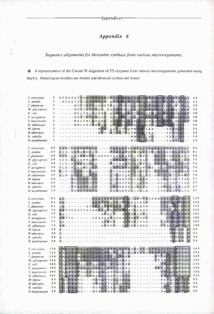
Appendices
Appendix 6V
Sequence alignments for threonine synthase from various microorganisms.
@ A representation of the Clustal W alignment of TS enzymes from various microorganisms generated using
SeqVu. Homologous residues are shaded and identical residues are boxed.
S. cerevisiae 1S. pombe 1C. glutamicum 1M. glycogenes 1 E. coli tP. aeruginosa 1S. marcescens 1H. influenzae 1M. leprae 1M. tuberculosis 1B. subtilis tBr. lactofermentum 1
MPNASQVW R STRSSS?Kt -MSSQVSVLS ARG GSS-N----------M 13 Y I S TR D A S R TP-------MK1^ISTRGQS-PA------- M K L Y N TI E H N E - Q------- M RY I S TRG Q AP - a|-------MKLYN L# DHN#-Q-------MN L Y N I R h’IeB - Q-
S E I AT Y EQY IHDL EN L
Il d l
YRDRiY K E FY ASY
AjE E A I I q|gI
FEEAVLKGIIIatdgIg Ja N D G G
FISDILL G GIJa p d g g LYF|S E I L L GIG IjIA p d gig LIYFA Q A V T QlG IJg K N Q GF|E D V L L AIG I LIYF A Q, A I K QlG IJg k q q Jgfejs Q A V R QGIJg rdqIs.
T T H Q P W PIGp|I A A - - - -q P Wi - MW. -MY
4 4 4 2 3 9 3 8 3 8 3 8 3 8 3 6 2 4 2 4 1 4 1 4
S. cerevisiae S. pombe C. glutamicum M. glycogenes E. coli P. aeruginosa S. marcescens H. influenzae M. leprae M. tuberculosis B. subtilis Br. lactofermenlum
4 5 4 3 4 0 3 9 3 9 3 9 3 9 3 7 2 5 2 5 1 5 1 5
YQATjLFNYWeKLg PSG-WJEAWKPKS DI DA - QLSKWREVL SAD-AIiSAMRGHNS L T - EIDEM L'i# D T L E - EpP S 1Wv Gj E E T i E...Bln- N -----------(g1---------- VT DQT P At.T----- VN EKT PDV
L L
----- ETDLAFAIMRLY----- FPEIAFEVMSLYIPRSANEG TAAPAREVI S-DFV - - T----- YRDLAFTTLSRLI--J----- AV T RS A KI * S A F* G-ft----- Y.HELAFRV.MRP FVAG ------ A VT R S S RIL SAFIG----- SVigRSQKISGALID-G----- %L#GGTPLIA APR---- L----- &LRGGTPLI AA N---- L----- L H E GNTpLiHL-P K---- L-----L MEGNTPLI PLlN------ I
I
VA I T II V riv 3PL K RV T L D A Mt V S E.Q:TGGTIHL SKQTGCTIHL SEQLGIEL HV S KQ -LHVQ L YG
S A DV EAD
8 4 8 1 8 0 7 5 7 6 7 6 7 6 7 4 5 7 5 7 4 7 4 7
S. cerevisiae S. pombe C. glutamicum M. glycogenes E. coli P. aeruginosa S. marcescens H. influenzae M. leprae M. tubercuuosis B. subtilis Br. laclofermentum
8 6 8 2 8 1 7 6 7 7 7 6 7 7 7 5 C 8 5 8 4 8 4 8
K R--------------5YSTFRSDEVT|DR--------------SYE TFRHI ET TAj|-------------- A YlTa|^K#))[4*^W$VJDKT YR AD V Y A YARGQEABAEDITR A--------------------AFAFR--AJE1 TYG--------- VFAHDASGAAAq A------------------- aFUfII i - aKN------------------- AFTF>--a|
K----------------------------------------------------------K----------------------------------------------K----------------------------------------------K----------------------------------------------------------
|l V-Q N V T G D K E N-L-.H-.I LJE L F HIk s lkng---------- LNVm
^ILEDN----------XY&G|T Y R L E D D---------- L Y LLS
' AN V E S D----------VG C>e|»t h %---------- --- - cv|e
HAD ---------V------- V------- T----Y
G - DN G L N - GVN - CAN -b
VAKVTDD----------VS CL SYFHLeKVEEN-------
1 1 9 1 1 2 1 1 3 1 1 5 102 108 102 100 6 4 6 4 5 4 5 4
S. cerevisiae 120S. pombe 11 3C. glutamicum 1 1 4M. glycogenes 1 1 6E. coli 103P. aeruginosa 109S. marcescens 103H. influenzae 1 0 1M. leprae 6 5M. tubercubsis 6 5B. subtilis 5 5Br. lactofermentum 5 5
MAQMLAEyAGE MEQ ALA AVRGD MTMAVTDALAR MTMJAlfT DAL AH M VMAVAKAKEE M V mEIE A K A K E H
RAVL T R ApII- C D tE|M C
|i c(Ss tUn
KH TN K TE EE R ■ DW LTrW-----------------e tsnkg----------------ettn
GDKP-------------------- VTXLTARRG-------------ERVVi
-------------VT
TS GD T S G D T S GD
AT S GD AT S G D AT SG D ATSGD A^LSGD
GN A ST GN
TGN
163 1 56 149 1 5 1 1 36 1 44 1 36 1 34 9 5 9 5 8 5 8 5
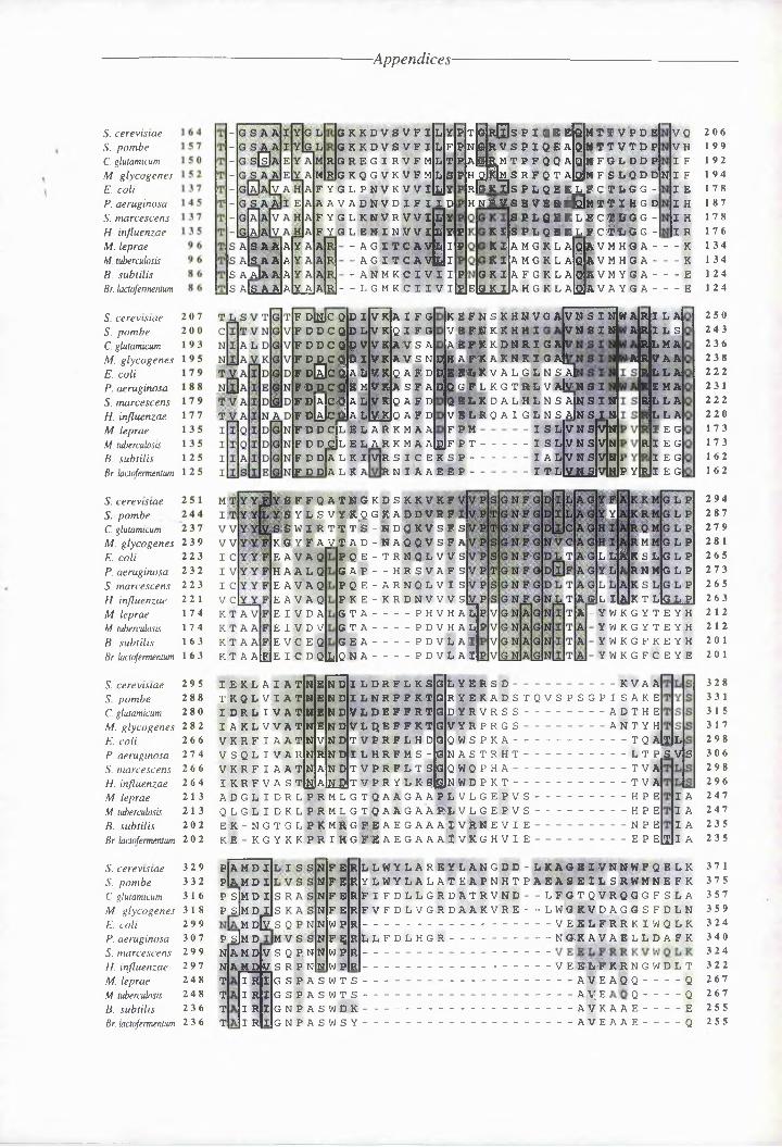
-Appendices
S. Bsrsvistas S. pombe C. glutamicum M. geIBogsnss E. cali P. aeruginosa S. maJCssBsns H. infecsnzas M. leprae M. tuberculosisB. subtilis Bj. laBtofeJmsntcm
S. csJSvtstas S. pombeC. glutamicum M. glycogenes E. coli P. aeruginosa S. maJCssBsns H. influenzae M . leprae M. tcbeJcub)stsB. subtilis Bj. lactofsrmentcm
S. BSJsvtstas S. pombeC. glutamicumM . glycogenes 2 3 9
2 2 3 2 3 2 223 22 1 174 174 163 163
E. coei P. aeruginosa S. maJcescens H. influenzae M . leprae M . ttbeJcutbsis B. subtilis Bj. bli:lofsJmentum
S A A AiS A A A
G K K D ' V . S V FI LYP GKKDVSVFILFP RGREGIRVFM LTP
MlRjG KQGVKVFM L S P FYGL PNVKVViy|Y AAVADNVDIFIID F Y G L K NV R VV I l|y F^G L ENINVVILYP
-AGITCAVLIP - AGITCAvtJl -ANMKCIVIIS A AjA A A
s aIs a AlAlY AlAlRl- -lgmkcnnvn
F DyC 3D IV K A IFG fddcqdok OICG F DDC Q p V VKAVS A F D D C QP I VK AV S N F P0C QALV KQAFD FDDCQFmVKASFA F DIaI CQ A L V K Q A F D F d|AcPJA L1V.K1O A 'F D FDDqLELARKMAA
T GR0S ■ P NGF E L MTTV P D E NG A'V F P I Q E A L MTTVTDP a|g IRMTPFQQA QMFGL DDP hqHm s r p q t aIajm F S L Q D D rIgkUs F L Q E KL F C T ..L G G - H nF-VS- eve; r-rIQIm t ti hg d
VQ VH 1 F
N I FI E N H I H I R
pl£lE
S PLQEKLJFCpfrQG - KI SR L Q SR L C. c CTG G -KIA MIK L A QAVM HG A-----KK IAMGKLA SiVMHG A----K
G K I A F GK LAQAVMYGA----- ERFKiRa H G K L AjQJA VAYGA----E
KEFNIKHNVGA VSfMkKhHIa.efkkdnriga \
DHA^CAK^-RIGAh D E El# K V A L G ■ L N . S Al DQGFLKGTRLVaFnSI
Q E L K DALHLNSANSINVELRQAIGLN S AJN SIN
VNSINWARILA VNEI N
F PM - I SL VN S V|NV NS VNV N S V NI
F D I DAL I L AR I I A I |gF P T------------ISLF D d|a L K IMR I I C E K S P-----------I AL
R N I A A E E P - -F D DlA I K A
GF FQ A TNGKD S KK V KF S Y L S V ■ Y K , Q G 16 A D D V R F " WI I I R T T I IS I R I IK V S F S|
KI Y F 11JAT AD-NAQGV SF AE AVAG H HAL Q E A VA 0 E A VA Q E I V D A E I V D V E VI E Q
K I A aIeJe I C D GRi
ARILS RLNA
FAIR VAA RL L A RIMA RL L A
DLA I E G R I E G
I E G----- IT LlV N .SiVlNiP Y|RJI E G
FQ E-T RNQLVVS GAP - - HRSVAFS PGE- A RN Q L VI S PKE-KRDNVVVSII A----- PHVHALIT A----- PDVHALPIE A----- PDVLAIQN A----- PDVLAI
L PL L AKSL G L P
RNNGLP
A G|L IlAIK T LlGJLP A - YWKIYTEYH
YWKIYTEYH YWKIFKEYH YWKIFCEYE
S. BSJsvistas 2 9 5SO pombe 2 8 8C glutamicum 2 8 0M. glycogenes 2 8 2
2 6 6 2 7 4 2 6 6 2 6 4 2 1 3 2 1 3 2 0 2 202
E . coli P. aeruginosa S. majcescens H. influenzae M. leprae M . tcbeJcclvsisB. subtilis Bj. lbcofsJmentlC3
S. csJSvtstas 3 2 9 S. pombe 3 3 2C. glutamicum 3 1 6M. glycogenes 3 1 8 E. coli 2 9 9P. aeruginosa 3 0 7 S. majcescens 2 9 9 H. influenzae 2 9 7 M. leprae 2 4 8 M. tcbsrcuh>sis 2 4 8 B. subtilis 2 3 6 Bj. lbC0)fsJmentc3l 2 3 6
IEKLAIATNE TKQLV I ATLE IDRLIVATNF I AKLVVATNE VKRFIAATLV VSQLIVAR Hl VKRI IAATNA
ND ND ND ND N D ND ND
ILDRFLKSG ILNR FFKTG VLDEFFRTG
LYERSD-------------------------- KVAARYEKADSTQVSPSIPISAKE DYR V R IS------------ A D T H E
VLQECFKCG VYRPRIS------------ANTYHTVPRFLHD L ILHRFMI I L T V I R I L I I L
QWSPKA----------------- TQ AlTJLNASTRHT--------------- LT P SV|S
I K R F V A I TCiJAliejT V P R Y L R S|gN WD P K TQ WQ I H A----------------- TVA
TVA H P E H P E N I E
ADILIDRL PRMLITQAAIAAP LVLIEPVS----------Q L I L I D K L P R M I I TQ A A I A A ' FL V I I E P V I----------E KIN G TIL PR mRgFEAEIAA AIVAN EVIE----------KE-KIYKK PRIHGF'EAEGAAAIVKIHVIE--------------E P E
I A I A I A
:U|I a
P|A MD I pk N D I P Si ND I P S|N D I
N D E p"sJn D N N T T T T
AND V S Q P N ANDVS R P N
N FE RN F B RN F B RN F E RN W P RN FF RN WF RN W P R
IRI I R I I RI I RI
G S P A S W T S GSPASWTS G N P A S WDR INPASWSY
LLWYLAREYLANIGD-LKAGEIVNNWFQELK YDWYLALATEAPNHTPAEAVEILSRWMNEFK FIFLLLIRLATRVND - - LFGTQVRQGIFSLA FVFLLVGRLAAKVRE--LWGKVLAIGSFLLN---------------------------- VEEL FRRKIWQLKLlfDLHGR---------------- N GK AVASLLDAFK-------------------------------------------------------- v eBLFkrngwdlt--------------------------------^V^E a|I Q-------- Q--------------------------------------- ^V^E^ Q---------Q--------------------------------A v KAAE------- 'E--------------------------------aVeaae---------Q
206 1 99 1 92 194 178 1 87 178 176 134 134 1 241 24
250 243 236 238 222 23 1 222 220 173 173 162 1 62
294 287 279 28 1 265 273 265 2632 1 2 2 1 2 20 1 20 1
328 33 1 3 1 5 3 1 7 298 306 298 296 247 247 235 235
37 1 375 357 359 324 340 324 322 267 267 255 255

Appendices-
S. cerevisiae 3 7 2S. pombe 3 7 6C glutamicum 3 5 8M. glycogenes 3 6 QE. coli 325P. aeruginosa 341S. marcescens 325H. influenzae 323M. leprae 268M. tuberculosis 268B. subtilis 256Br. lactofermentum 256
S. cerevisiae 416S. pombe 416C. glutamicum 393M. glycogenes 394E. coli 347P. aeruginosa 38 1S. marcescens 347H. influenzae 345M. leprae 292M. tuberculosis 292B. subtilis 280Br. lactofermentum 2 8 Q
t ' >
dd an - D G|E L A S E L D L E
RRQQA A T E RRmA A s D
SNii----------- KI E* VT DS HIGH ----------------- E I E MV sId
sk|
Ik p q v o k s.i . I E g A S ■ K D F T S E R VS NE E T S E T I.K KIYESSV NPK JtVTVR P EVI^EAARRDF VSIRVVNDET I DAI KKIYES-------A
F E K A AAE YGF ASGRSTHADR VATI A^l^^S R - gH v&G S S qDI ADH R m-'—"'
Y AAV D R W T E®
HAAVS S G ML S
E
- - L- - YR -
L----- S
LAMQ S K
E I LRRY%-L|r%R------ EE I L AAYHLVARV------ EE I LER YQ LDRRV-------EE I LMR YRL'LAKT-------E
vnt
Ikb s v IpERVL SVP M V T
egefElf A <3-1 F
GGWV dg w v| S G E I
V TT- It S ELLV I S YLCV F V
, V F V'
E TV L’RFSK E H Vr|»QV R E KV A lLI HR-
:a Y R^l|Rd,Q L N GV R-A aREC R R
li RYRALRtDQ.LQ FI r Y Q V L K-D Q L K |R SIAGlJkAI D
IaR iJg vlkqVk in ais'BAg V iKHVe
FIK e s fJk e sG S T V G S T V GSKV G E T V
KT
459 459 433 433 386 421 386 384 322 322 3 10 310
S. cerevisiae 4 6 QS. pombe — 4 6 QC glutamicum 4 3 4M. glycogenes 4 3 4E. coli 3 8 7P. aeruginosa 4 2 2S. marcescens 3 8 7H. influenzae 3 8 5M. leprae 3 2 3M. tubercubsis 3 2 3B. subtilis 311Br. bctofementum 31 1
E A I ER IC T I Tl C T VT av| a vHt
NII veA V E A E A IEKA^IjS Q AR A -
Y N
JQ E lM L 11 Q LPL ING LKD In g l'k d In GL K D
IN G LKD
OD G.$ LD EE R T CIF S G K R F RRIMDRPFKVSDLPN SLEGLESLPQRFFVMER EL A , E R A D L P LL S H N L P A
ADD F E R E E EGT V L P N AL ALRAELPLLS H TLPA TLDKHNQLPLLSLEMDE t||li-em- -psISS P mfi TA LK -DM--P SVS PVFI
\^DIS E - - I%P VTLPT ISSNQ--LDIASVSNI
IDILKQTDAVKQR R VIK Q
fi|F A A L R K E L A K VQ A SF G E L R K DJF AQLRA
A VA VVEP V i AV V EE D SIL E
[fill E Q i K D
503 503 469 469 422 458 4 22 420 355 355 344 344
S. cerevisiae 5 Q 4S. pombe 5 Q 4C glutamicum 4 7 QM. glycogenes 4 7 QE. coli 4 2 3P. aeruginosa 4 5 9S. marcescens 4 2 3H. influenzae 4 2 1M. leprae 3 5 6M. tubercubsis 3 5 6B. subtilis 3 4 5Br bctofementum 3 4 5
rieeelrvkm-kl - I I EVT.L SReWa - Y I VD Ajl dkDQ T S VKnvDDi------------L M M Ktt Q------- --- - -VD I D'H G NP gV P LD0MGLP&----------YIl&s--------------Q%G L V---------------------kSgla---------------------
YVKGAARV-----------HIKG vImS-----------
5 1 4 5 1 4 48 1 475428 47Q429 425 36Q 36Q 352 352

-Appendices-
Protein sequences for TS were obtained from the SwissProt database (web-site: http://expasy.hcuge.ch/cgi-
bin/sprot-search-de) under the following accession numbers:
No. Organism Accession Number
1 B. subtilis P04990
2 B. lactofermentum P09123
3 C. glutamicum P23669
4 E. coli P00934
5 H. influenzae P44503
6 M. glycogenes P37145
7 M. leprae P45837
8 M. tuberculosis Q10610
9 P. aeruginosa P29363
10 S. pombe Q42598
11 S. marcescens P27735
12 S. cerevisiae (yeast) P16120
Percentage identities between the various threonine synthase enzymes (organisms are numbered
as in the table above).
1 2 3 4 5 6 7 8 9 10 11
2 68
3 17 15
4 17 12 28
5 18 16 28 66
6 10 11 57 29 28
7 55 53 18 16 16 17
8 55 55 18 14 13 16 93
9 18 11 36 31 27 40 15 15
10 17 13 33 28 30 37 13 16 37
11 17 13 25 83 66 27 14 15 33 28
12 13 12 35 31 30 33 13 12 35 55 30

-Appendices-
Comparison between the threonine synthase enzymes quoted in reference 170.
P. aeruginosa B. subtilis C. glutamicum Br. lactofermentum S. cerevisiae
E. coli 34.9 24.2 30.2 21.9 37
43 47.5 48.8 40.8 56
261.8 137.7 229.5 131.4 252.9
P. aeruginosa 25.9 40.4 16.8 40.8
48.7 56.8 30.2 59.7
145.1 311.6 129.7 332.5
B. subtilis 22.5 67.8 24.5
49.5 79.8 47
139 397.4 143.3
C. glutamicum 17.4 39.4
36.3 58.4
134.3 324.1
Br, lactofermentum 16.1
33.2
143.5
i
First line, percentage of identity; second line, percentage of similarity; third line, quality scores.

-Appendices
Appendix 7,
Gene Sequence and Translation of ThrC from E. coli.
1/1ATG AAA gtg TAG AAT GTG AAA GAT GAG AAGM K L Y N L K D H N61/21 GAG GGG TTG GGG AAA AAT GAG CCC GTG TTT0 G L G K N Q G L F121/41 ACT GAA ATT GAT GAG ATG GTG AAG GTG GATT E I D E M L K L D181/61 GCG TTT ATT ggt GAT GAA ATG GGA GAG GAAA F I G D E I P Q E241/81 GGG TTG ggg GGT CCC gig CCC AAT GTT GAAA F P A P V A N V E301/101 GGG GGA AGG gtg GGA TTT AAA GAT TTG GGGG P T L A F K D F G361/121 ATT GGG GGT GAT AAG GGA GTG AGG ATT GTGI A G D K P V T I L421/141 GTG GGT GAT GGT TTG TAG GGT TTA ggg AATV A H A F Y G L p N481/161AAA ATG AGT GGA GTG GAA GAA AAA GTG TTGK I S P L Q E K L F541/181 GGG ATG GAG GGG GAT TTG GAT Grc TGT GAGA I D G D F D A G Q601/201GAA GTG AAA GTG CCC GTA Grc TTA AAG TGGE L K V A L G L N S661/221 GGG GAG ATT TGG TAG TAG TTT GAA GGT GTTA Q I G Y Y F E A V721/241 GTG GTT GTG TGG GTG GGA AGG GGA AAG TTGL V V S V P S G N F781/261 TGA dG GGT GTG GGG GIG AAA ggt TTT ATTS L G I, P V K R F I841/281 GGT TTG GTG GAG GAG GGT GAG TGG TGA GGGR F L H D G Q W S P901/301 ATG GAG GTG AGT GAG CCC AAG AAG TGG GGGM D V S Q P N N W P961/321 TGG GAA GTG AAA GAG gtg GGT TAT GGA GGGW Q L K E L G Y A A1021/341GGT GAG TTA AAA GAA GTG GGG TAG AGT TGGR E L K E L G Y T S1081/361GTG GGT GAT GAG TTG AAT GGA GGG GAA TATL R D Q L N P G E Y1141/381AAA TTT AAA GAG AGG GIG GAA GGG ATT GTGK F K E S V E .A I L
31/11GAG GAG GTG AGG TTT GGG GAA GGG GTA AGGE Q V S F A Q A V T91/31 TTT GGG GAG GAG GTG GGG GAA TTG AGG GTGF P H D L P E F S L151/51 TTT GTG AGG GGG AGT GGG AAG ATG GTG TGGF V T R S A K I L S211/71 ATG GTG GAA GAG GGG GTG GGG GGG GTC TttI L E E R V R A A F271/91 AGG GAT GTG GGT TGT GTG GAA TTG TTG GAGS D V G G L E L F H331/111 GGT GGG TTT ATG rcA GAA ATG GTG AGG GATG R F M A Q M L T H391/131 AGG GGG AGG tgg GGT GAT AGG GGA GGG GGAT A T S G D T G A A451/151 GTG AAA GIG GTT ATG GTG TAT GGA GGA GGGV K V V 1 L Y P R G511/171 TGT AGA TTG GGG GGG AAT ATG GAA AGT GTTG T L G G N I E T V571/191 GGG GTG GTG AAG GAG GGG TTT GAT GAT GAAA L V K Q A F D D E631/211 GGT AAG TTC ATT AAG ATG AGG ggt TTG GTGA N S I N I S R L L691/231 GGG GAG GTG GGG GAG GAG AGG GGG AAG GAGA Q L P Q E T R N Q751/251 GGG GAT TTG AGG GGG GGT GTG GTG GGG AAGG D L T A G L L A K811/271 GGT GGG AGG AAG GTG AAG GAT AGG GIG GGAA A T N V N D T V P871/291 AAA GGG AGT GAG GGG AGG TTA TGG AAG GGGK A T Q A T L S N A931/311 GGT GTG GAA GAG TTG TTG Grc GGG AAA ATGR V E E L F R R K I991/331 GTG GAT GAT GAA AGG AGG GAA GAG AGA ATGV D D E T T Q Q T M1051/351GAG GGG GAG GGT GGG GTA GGT TAT GGT GTCE P H A A V A Y R A1111/371GGG TTG TTG GTG GGG AGG GGG GAT GGG GGGG L F L G T A H P A1171/391GGT GAA AGG TTG GAT GTG GGA AAA GAG GTGGET L D L P K E L

-Appendices-
1201/401 1231/411GCA GAA CGT GGT GAT TTA CGC TTG CTT TGA GAT AAT GTG GGG GGG GAT TTT GGT GGG TTGA E R A L P L L S H N L P A D F A A L1261/421GGT AAA TTG ATG ATG AAT CAT GAG TAAR K L M M N H Q ★

-Appendices-
Appendix 8,
Positions of Common Restriction Endonuclease Sites on ThrC from E. coli.
Fsp I1
ATGAAACTCTACAATCTrcAAAGA^TKACAA^^GCA^GGTCAGCmraC^GCAAGCCGTAACCCAGGGGTTGGO^^AAAATCA 8 0 TACTTTGAGATGTTAGACTTTCT1A^G^T:GT?Trac:TCG^TC^CAG^TCGAAACGCGTTCG^<^.ATTGG^,r'CiC(^(^.A^^iC(^<^^TrTTTAGT
• • • • | • • • •45
EcoR II
(GGGGCTGTTTTTTCCGCACGACCTGCCGGAATTCAGCCTGACTGAAATTGATGAGATGCTGAAGCTGGATTTTGTCACCC 160 CCCCGAC^AAAAAG(G^GGK^(^'rc(^7^^CGG^Crr^^jA^^^^(^iG7^C^CT37^C^^TAJACTACTCTACGACTTCGACCT^AAAA^C:A^^TGGG
• • | • • • • • •109
BssH III
GCAGTGC^^aATCCTCTCGGCGnTDAWnKXT^JWKAyyTC^C(A^CAG^?yy«C^<T^GaAG^^GCGTCCGICGCGGCGPrT 240 C^TCAC^CTTCTAG^^J^SCGC^rAACCACTACTTT?A^GGGTGTCCm^AGGA^CCTTCTC^:^^CGC:GCGCCGCAAA
• • • • • * | • •228
_ Mcr I Dra I’ I I
GCCTTCCCGGCTCCGGTCGCCAATGTTGAAAGCGATGTCGGTTGTCTGGATTGTTCCACGGGCC.AACGCT<GGC ATTTTAA 320 CGGAAG^(^(^*G^^C^iACG^CG^TT^T^C^.^<^'rrrC^'rACAGCC.AACAGAC^TTAACAAG^TG^(CC<G^,^T^^G.^^<^^'^^^TT
• | • » • • • • | *254 316
Mcr I1
AGAT'raC^GGCGGTCGCTTT^^T^^^C^^^TrcC^T^^^C^CC^^T^AkT^T^C^^^TC^l^^GCCAGTGACCATTCTGACCGCGACCT 400 TCTAAAGCC^CAG(^iG^^^;^(^(^'Gr^(^TTTACGACTGa^TATAACGCCCACTATTCGGTCACTGGTAAGACTGGC^TCGA
i1 • •••*••330
CCGGTGATACCGGGCCGGAGTTGGCCArcGCTrTCTACGSTOTACCGAATGTGAAAGTGSTTATCCTCTATCCACGAGGC 480 GGCCACTATGGCCTCGCCGTCACCGAGTACGAAAGATGCCAAATGGCTTACACTTTCACCAATAGGAGATAGGTGCTCCG
• •••«•••Bspl407 T
IGGGGTAAGTAAGATGCGGGGGGAGATGTTCCGTAAGTraGCGGAGGTGTAGGGACCGTTGCAGTCGGAGGAGATCTCGG 560C,CTCGGCCGGGTGGCGTTACCCTCGACGGGGCGCGC,GG.ACCGAAGTCGCGCATCCX3GCGGCGGCGGCC,GCCGCCGGGGCC
• • • | • • • • •511
CGCCCGTCAGGCGATGGCGGGGAGGGAGTTTGGCGGTGAGGAACCGGGGGTGGAGACAGGTTAAAATCGGCTAGATCGA 640 GCGGACAGTGC(GCGAGCACCCCCGTCCGCCGGGCTAGTAGTTCC,TGGCCCCAACCGCGACACCGG.TCC1GAGCAGATTGGGCC
• •••*• • ©Pvu II
Fsp I JPvu III I I
CCGACACAGGCCGTCCGCTGGAGCAGACTCGCCGCTAATTTCGG(GCTGTC^GCGCAGCCGACGCGGGGGGCGCGCAGCCAG 720 AGCTGTGGTCGGCGGGCGAACGAGCCTGGACGGTGATGGGAATTCGGAGACGCGTAGGCGG CGTCATACGAGAG'TTGGTC
• • • • II* * I *690 718
694Pspl406 II
ATGGCTGCCTAGGTGACGAGCGGAGACTCAGGAGGCCTGGCGGAGGGTATGATGGAG;AGGTAAATCGGTCCGCCGGTGAA 800 GGCCGACAGAGCGAGGSTCGGCGTCTGGAGCCGGTGGGATGACGCCCAGGCGACAGGTCAGGTGGGCCAGACGGGGAACC
800

-Appendices-
SsTE iiI
ACGTTTTATTCCTCCGACCAACGTGAACGATACCGTGCCACGTTTCCTGCACGACGGTCAGTGGTCACCCAAAGCGACTCTGCAAAATAA^CGACGCTGGTTGCA^C^TTG^T^A^^GG^A^C^CGT^C^TAA^^A^CGTGCTGCCAGTCACCAGTGGGTTTCGCTGAG
• • • • • • j • •863
SsTX I PsU. II I
AG^OAOOTTATCCAACG^SATGGACGTGAGrCAO^^^CAACTGG^CaCGro'R^JAASA^TKGTPCC^C^^AAATCTCCGCTGCAATAGGTTGCGCTACCTGCACTCAGTCGGCTTGTTGACCGGCGCACACCTTCTCAACAAGGCGGCGTTTTAG
«
893 904
TGGCAACTGAAAGAGCTaGGTTATGCAGCCGTGGATGATGAAACCACGCAACAGACAATGCGTGAGTTAAAAGAACTGGGACCGTTGACTTTCTCGACCCAATACGTCGSCACCTACTACTTTrcGIGCGTTGTCTGTTACGCACTCAAT'rTTCTTGACCC
• •••••••ScI I
ICTACACTTCGGAG(^(^(G JAC(G^'TG^(^(Gr^iA(G^'rrATCGTGCG^rrG<^^rT^2^rT^^^T'T^^rrCC^G^<^(^JA^^^iATG(^^T'rGTTCCGATGTGAAG^^TCG^<G'TC(^(^^C^<GG^C^^^(^(^^^^i^^(^.^^(^(^G2^C^C^(CAC^C^^^C^^CA^^Tr^2AG(^,T^<^(G^TTATACCGAACAAGG
• • • • | • • • •I086
Dra I Xnrn I Psp4006 II I I
TCGGCACCGCGCATCCGGCG7CACT^TAAAGAGAGCGT(G3AAGCGAT^CTCGGTG7CACCGT^GGATCTGCCAAAAGAGCTGAGCCGTGGCOCGTAaXZCGCCTTAAATTTCTCTCGCACCTTCGCTAAGAGCCACTTTGCAACCTAGACGGTTTTCTCGAC
• |. « •1144 1159 1176
GCAGAACG'KGCCX3ATTrACCC'Pr<GCTTTCACATAATCTGCCCGCCGATTTTGCTGCGTTGCGTAAATTGATGATGAATCACGTCTTGCACGACTAAATGGGACCGAAAGTGTATTAGACGG<GCG<GCTAAACGAC(GCAC(GATf]TrACCTACTACTTAGT
880
960
I040
1120
I200
I280
TCAGTAA 1287AGTCATT

-Appendices-
Appendix 9.
Accession numbers for vector DNA sequences.
Full sequences for the pET-expression vectors can be obtained via e-mail on the Internet al the following address:
[email protected]. The sequences for pBR322 and pKK223-2 are available from the EMBL database under
the following accession numbers:
pBR322 VB(00O4a
pKK223-2 M77749
a - also in the following journal: Gene, 1982, 9, 287 - 305.
Full sequences for the cloning vectors pGBM-3Zf(+), pGEM-5Zf(+) and pGEM-T are available from the EMBL
database under the following accession numbers:
pGEM-3Zf(+)
pGEM-5Zf(+)
pGEM-T
X65304
X55008
X63088

-Appendices-
Appendix 10,
Positions of Common Restriction Endonuclease Sites on the Gene for fi-Methylaspartase.
Hinc II Bspl407 I
ATGAAAATTGTTCACCTACTTTGTACACCA(CATTAACCTGCA^TFCTATTTTGATGACC7AAG3AGCAATr ’CAAAAACCGAGCTACTTTTAACAACTGCATGAAACATCTGCTCCTAATTGACCTAAGATAAAACTACTGCT^TCTCCTTACTTTTT'CCCTCC
8 0
a
10 22
AGCACATCATCCATTTACATATAC^CCC:TCTACTGTAACACAAGCATTTACTCAAGTAACACAAAAAGGTCAATCAATATTCCTGTACTACCTAAATCTATATCACCGAGATCACAATGTCT^CCTrAAATGAGTTCATTCTCTT^TTCCACTTACTTATA
160
Pst I Sea I
CTGTATTATTAGTTCTTCAAWAI,CG,CAGCT]TCCTrcAC(GAGA^TG^GCTGCAGTTCACT?A<TrcJGGACCACCCCGAACACACATAATAATCAAGAACTTCTACCAGTCCAACCACTGCCTCTAACACCACGTCAACTCATCACTCCTCCTCCCCCTTCT
a
240
209 218
CATCCATTATTCTTACCTAAACATTTTATACCTGTTATAG7AAAAC/AATAGCTCCAAAATTCATT’GCAACACAAATAACCTAG^r^J^^J^r^.AA(^^zA^T^^(^C^zA]^^^r^(^r^IA^JA^rIT^rT(^<^7^C^CA^7^T^l^T^T^l^l^vTTCrTTvTATCG^(^C^GTTTz^^,^j^(^rlT^j^(^^^^"^(^'TC'TT'TATTG
320
Dra I EcoR I
TAACTTTAAACCAATGGCACAACAATTCCACAAAATCACTCTAAATCGTAATAGATTACATACTCCTATCACATATGCTAATTGAAATTTCCTTACCCTCTTCTTAACCTCTTTTACTCACATTTACCATTATCTAATCTATCACCATACTCTATACCAT
400
325 3 33
TAACTCAGCCTATACTTCATCC:ACTACCTAAAACTACAAAACTAACTATGGCTCAAGTAATCAGACATCAATACAATCCAATTCACTCCCATATGAAC^ACGTCATCGATT'TTCATCTT,TTCATTGATACCCACTTCATTACTCTCTACTTATCTTAGCT
480
Pspl406 I
GCAGGAGA?AATCAATCCAGTTCCTCTATTTGCTCACTCAGCTGATGATACATACCATAACGTTCATAACATGATAATTAACCTCCTCTTTACTTACGTCAAGCACATAAACG>CAGTCCACTACTATCTA,T3CTAT'T3C7AACTA,T^GTACTATTAATT
560
538Xmn I
AGAAGCTCATGTTTTACCACATGCTTTAAT,CAACAATGTTCAAGAAAAATTAGGACTTAAACCACAAAAACTTCTTGACTTCTTCCACTACAAAATGCTCTACCCAA\TTAG^T^GTTACAC'TTC'TTTTTAATCCTCAATTTCCTCTTTTTC;AACAACTCA
* * a a a a | a a
625Ssp I
640
ACCTTAAATGCTTAACACATACAATAATCAAATTAACACTAACACAAGACTATG<C^CCAATATTCCACATCCACGTATATTGCAATTTACCAATTCTCTATCTTATTACTTTAATTCTCATTCTCTCCGGATACGAGCTTATAAGCTCTAGCTCCATATA
720
699Hinc 11
CGAACAA,TCGGT<C3TCGCATTTCATGTTCACATCAAGGC^TATGGCTGATTACATTCACACTTTAGCTGAAGCTCCTAAGCCCCTTGTRACCCACGACCTAAACTACAACTCTACTTCCCATACCGACTAATGTAAGTCTCAAATCGACTTCCACGATTCGC
Afl II Afl 11
ATTCCACTTAMGAA^TCAyGCAC(:AA^CCACCTTGAACSATAGACAJAAACAAATGCAAGCTATCACACACTT,AAGACCTGTAAGCTCAATTCTTAACTTCCTGCTTACCTCCAACTTCTATCTCTTTTTGT ,TTACCTTCGATACTCTCTGAATTCTCCAC
807 870
a a ♦ a I
9 I a
800
745
880

-Appendices-
Acc II
TTAATCTGCCTTCTCCTCATCTGCGTCTTAATCAACGACTACTTACCACATTATGTCAACTTCTACAATTTAAGAAGTGA• | • • • •'• • .
898Hpa I Hinc III
GATAACAAAGCTGGACACATGGTTCAGATCAAAACTCCAGACCTTGGTGGAGTTTAACTAATATAGCAGA'KGCrATTATGTACTATTGTTTCGACCTGTGTACCAAGTCTAGTTTTCAGGTCTGGAACCACCTCAATTGTTATATCGTCTACGATAATACAT
• • • • • | « • •10121012
CTGTAAA^TAACG<^^rT^<GG(G^CTjATT^,T^(^2^CG^2A^C^CTrc^iA^^<^iAA^( rTA^(C^(G^TK^(^(^(^^T^JA(^rT^i^^AAC'^JAACJAGACATTTCGATTGCCTTACCCTCGAATAACACCTCCTTGAACATTGCTTTGATTGTCTAGGCGACTTCAATGTTGATTGT
• ••••«••Hinc II
ITAGGTATGGCTTGTGGAGCTAGACAGGTTCTTGCTAAA.CCAGGTATGGGTGTTGACGAAGGAATGATGATCGTTAAGAACATCCATACCGAACACCTCGATCTGTCCAAGAACGATTTGGTCCATACCCACAACTCGCTTCCTTACTACTAGCAATTCTTG
• • • • • I • • «1171
960
1040
1120
1200
GAAATGAACAGAGTTTTAGCTCTTGTAGGAAGAAGAAAATAA 1242 CTTTACTTGTCTCAAAATCGAGjaACATCCCPTCTTCTTTTATT

Appendices
Appendix 11.
The Sequence Data. Obtained far the ThrC Recombinant Gene in pGEM-TTSrand pGEM-TTSn
1. pGEM-TTSlX SP6:
Sense strand sequence from N-terminus of thrC (first codon underlined in red) to base 568. Primer sequence
is given in red at start of sequence. Mutations (underlined in the main sequence): 319-321 (deletion of A);
340 (A to G).
GTCAGTCAGT CATATGAAAC
TGGGCAAAAA TCAQGGGCTC
GATTTTGTCA CCCGCAGTGC
GCGCGCGGCG TTTGCCTTCC
CGCTG^LATT TMGATTTCG
TGACCGCGAC CTCC^TGAT
TATCCACGAN GCAAAATCAG
CGOCG^'TTTC NATGCCTGTC
TCTACAATCT GZGA^CA^^KT^C
TTTTTTCCGC ACXCGC<CCTGC
GAANATCCTC TCTCGGCmTT
CGGCTCCGGT C(CG^C^rK^rn’
GCGGTCGCTT TG'QTCGCCCCG
ACCGGAGCGG CAGrGTG3rCTC
TCCACTGCAA GA^AA^(^rCTr
CA^C^fGC^CjAC^ TTCGCCTTTC
OGATTCACG CCrCC'CGCGG
CTTCGITCC^TCC 2GGrCCAGAA
rCAGGCCCGTC ,rcCC;GTTCTA,
ATGCTCACCC ATA,CTTCCCCr
TaCrTTCTAC GCCTrT^AC<CC<
TCTCTACA'TT (C;CCCCCCGG
CCCGGCCCCA ACCC^OGG^'T
^ITTCG[GAGCG, CCTCAACC,TC
GCAAGTZC^TCr AACGCCGCGT
(GCGGTTGTr CGCCCCCCGA
rITCCGTAGGCCA GT^^CC^'TTC
A^]TC^TG7AGCGT G^TTATCCTC
A^TCCVGCCTC TTGCCATCNA
2. pGEM-TTTSx T7:
Anti-sense strand sequence from C-terminus of thrC (stop codon shown in blue) to base 899. Primer
sequence is given in red at start of sequence.
ATCCCCGTCC TTGCTCATCA NNCAATAATC ATTTACGCAA CCCGCCGNGG TC<C:CCCCCAC CGTTGTCTCG GGCCGGCCCT
GAGTCGCCAC CTTATCACGC CATCTTTTCC AGATJCAACG TTTAGCCCGC .?ACGTCr:CTT:C GCCCTCTC^TT TAGATTTACC
CCCGTCCNCC CTCCCCGCCG AGAAACCAAA TTAGGCTCGT CA(CC:CGJCCCC ACNGTAGCCT ACCCAGCCCT
CCCCCTCCCN GCTCTGCCCA AACTTCTTTA NCTCACGCAI’ TCTCTCTTCA G^TCCITTTACT CG,TACGCCCC TCCGTAGCCC

Appendices
3. pGEM-TTSi x TS3:
Sense strand sequence from base 683 to base 1191. Mutations (underlined): 716 (A to G); 944 (T to C).
AAGCTGTTGC GCAGCTGCCG GCAACCAGCT CGGTGTCTCG GTT3<^CCA^CXC3 GGAACTTTA<r CGATTTGACG
GCGG^TCTGC TCCCCAACTT AC^TCGGCTG CTTGGGGAAC GGTTr^T^rTKC: ^TCA<CTAAC G<TGACAGTA CCTCTC:CAAC
TTTAATCAAA GACG^'T^.AGT CC;<TACCCTA ACCACCTTAG CCAG.CTCTAT CCTACAr'TGT (:CGCTCGGGC CACTAGAACA
ACTCCTACTG TGTCCAAGAC ']TQCT^CA3CC GGAAAATCTTr CCAA.CATCAA <:GAGATCG^C^? A^XGACCTAG CCATCATCAA
ACAAACTAAC ACACAATCTG TGGGGTTAAA. CCACCTCCCA’ ACAAA7TTCCr (C^CTCA^CCCA’ <CTAGACCAT ATTCTGCCCT
^G'BG^^T^.AN GTTCAATTAA GGCACATATCr ^rTTCCTCTA' CAGCACCTCT CATC<ACCTGr .GGiTiT .'AA'AG NACTCTCGAA
CAGATTCTCC C,TCAAACGTT GCCTCTGrCT
4. pGEM-TTSi x TS4:
Anti-sense strand sequence from base 605 to base 25. Mutations (underlined): 340 (T to C); 319-321
(deletion of T).
AGTTCTTCAT
TGTCGTCCAA
CCTAAAACCT
TCAAATATCC
ATAACACAAA
CTCGGA,T^TC
TCACTCACGT
CATCAAACCT
TCTACAGAAC
ANAAACCATC
GTAA<CTA^TTT
TACTTTTAAA
ATTACCAATA
TCAATTCCCC
^^INTGAAACC
AGTGTT’TC'n’
AGccAT■'^c;cc
G^CTCTCAQAr
CATATX:CCA<r
AAC<ACTT<A<
CACAcrGACxr
AAKGTG^T^TTQr
CGAGGCCCAA
TCATCTCr,AT
GrCCACCAeCG
TT<CA<CTC<<
C<GTC^TXACT
CCCAAGAACAr
TAG<CTATCGA
GA,TT’T■TCACT
AATCCCA(CC^
AAATCHAAA
AACGGGAAAN
AATCTCCCTC
GCACCTGA,TT,
AATACTCCTT
TGTCCATAGA
CGACCITAAGA
TCAAACCGT^
CCCAGACTAC
ACAAAATAAA
TTTCACTANC
GATCGCAACA
CCATAACCAT
ATCCTCAATG
CCACCGTCCA
CA<CTTCTTTC
GATTAACTAT
CTTTCCCTTA
GTTTAGATAT
TTTAACATTA
CTTTATTAAC
ACAATTCCAG
AGCA'TTTCTT
CTCATCAATT
CCCCTTGCCT
AAACTTCAAA TGATTGTTCT

Appendices
5. pGEM-TTSn x SP6:
Anti-sense strand sequence from C-terminus of thrC (stop codon is shown in blue) to base 786. Primer
sequence is shown in red. Mutations (underlined in main sequence): 944 (A to G), 785 (A to G, not confirmed
in other sequencing experiments).
ATGCaGATCC
AAATCAGCAC
AACNTGAAAA
AAAACTACAN
ANCTCTTTCA
CTTQ^ATAAC
NCGCAGC^T
TTACTGATGA
ATTATCACNA
ATAAAGNACT
AGTGTANCCC
ATTGACNAAT
ATACCCTGNA
AAAACGTTTC
TTCATCATCA
ATATTTTACA
ACAAGCCATA
ACTTCTTTTA
TTTAAGAAGC
TCACTTTGGC
ACCGGCAGAC
ArTT^ACGCAA
AGATCCAACG
TTCAAATAGN
ACTCACG^T
AAC^CTCTT
TGACCACTCA
CGCAGCAAAA
TTTATACAAA
TTCAACTGAT
TGTCTGTTGC
ACNCNCAAGC
CCATCATAAA
TACAAGAGAT
TNTAAATTCC
GAGGC^^^
GTGGTTTCAT
C^CTTCTTC
ACTTNCATGC
ANTTTTCTCN
ACG^TCT^TT
ACGATAAGCT
CATC'CAlCGGC
GGCTGACTCA
CACGGTATCG
TNAANAGAAT
TAAATTTCGC
NAAAANACAT
TGCATAACCC
ACTACNTAAA
TTANCATTAC
6. pGEM-TTShx T7:
Sense strand sequence from N-terminus of thrC (first codon is underlined in red) to base 348. Primer
sequence is shown in red. Mutation (underlined in main sequence): 319-321 (deletion of A).
GTCNATATAT CATATGAAAC rT^TJ^(CArT^^r GAAAGATCAC /TTCCTGATA3 T^^^TTTGC GCAAGCCGTA ACCCACCCGT
TGGGCAAAAA TCAGGGGCTG 'TriTlTCOGC ACGACCrT3CC GG;AATTTANC C'TG^^f^TG.AAA TTCTTATCTT ACTGATAATG
CATTTTCTCA AACAANCTGA GAACGATCCrT'' 'TC<GCAGTT^A ^ITTAATA^TTG. AATCCCACAG ATTATCCTGA ATCTAACCGT
AAACCCGAAA TTTGCATTAC C(AACTCC<CAT C(AA<ANTrArT^ <GTNTGCCTT TCCG^TTGTA^ GGNTCTGTIA AAAAACCCAA
CAATACAATT TMGNTTTAA <GAGCTAA:AT TA'TAGATANC

Appendices
7. pGEM-TTSn x TS3:
Sense strand sequence from base 671 on thrC to base 1220 . Mutation (underlined): 944 (C to T). Note a
mutation at 785 was not found in this sequencing experiment {cf. pGEM-TTSn x SP6).
GCTGCTGCTT
CGCGATTTGA
TGCCGTCCCG
CTCAGCCGGA
GTGGGTGGTC
TTATAGTGCG
GNGGCGTGGG
TGGGGCTGTT
CGCCCCGTCT
CGTTTCCTGC
CAGCTGGCCG
AGACCGCGAG
CTGCGTGATC
GGC(GCTTCTC
GCNCGGCTGC
GCTCGCGGAG
GCCACCCTCG
CGTGTCGGGG
GCGGGCGATG
GGTTGAGTCC
CGTGAAACGT
CGCANGAGAC
TCACTCCGTC
GTCGTAACCC
AGTCGT^CG
CGTGGGTTGA
GGGACAATGT
TGCATCTCCC
GCGCGACCGG
TGCCCGTGAA
AAAGCGACTC
CCGCAGGATC
GAGGACTGGG
GGCTTGTTCC
GGGAGANCTC
CTOGTTGTCT
AC<CiTTTATT
ACGCGACGTT
TGGCGACTCA
CTGCGCTTCG
TCCGCGCCGC
GCGNGGCGTG
CGGTCCCAGG
GCTGCGGCCG
ATCCAACCAG
AGGAGCTGGG
GGGCCGCGCG
NCGTACGGOG
CTCmTACC
CGG^CTTC
ACGTGAACGA
GTGGGCGTCG
TTGTGAGGCC
CTCCCGTANC
GAGTTTAGAG
8. pCEM-TTSo it TS4:
Anti-sense strand sequence from base 611 on thrC to base 252. Mutation (underlined): 319-321 (deletion of
T).
ACT'rTCAGT'T
CGATATTGCC
GAGTTCGGTG
ATCACCCGCA
C^IAATCATA
GCCCAGTGTG
GGCCGTGGGA
GTATGGGTCA
7AGcccrc^rTr
CAGCG.GAGCT’
AtGC^ATG^^
GGArTTTCTCC
TTTACCAGCK
TTTTATTCCAG
7A’.ATG’c.c^CiT'c
CATJAAGGCGC
GCCTGGCGGG
TGGACTGATT
CGCrA'I'CACC
CCGCCGAAAT
CA,TCGCGGCTC
TTGCCTCGTC
(GGAGCCTCCGCC
C:EGGGTG<CC
GCCACrTCGArTC
(G^^TWG^^TT
GrTC^CGA^'TQC
ACGCGTTGGCC
GCAACAGTTT
AACAACT^
TCACTCGATT
CGTGGAACAG
TTCCGGGCAG CCGGCGTAGC rTTTCCGCCAGT CGCGACCGGG

Appendices
Appendix 12.
The Sequence Data Obtained for the ThrC Recombinant Gene in pTN-TS Constructs./
The results for pTN-TS3, pTN-TS5 and pTN-TS6> should be compared with those for pGEMd-TT'Sp all others
should be compared with those for pGEM-TTSn.
1. pTN-TSl x TS3:
Sense strand from base 712 on thrC to base 1287. Mutation (underlined): 944 (C to T).
AGCAACCAGA
GCCCCTCAAA
AACTGACTCA
TCAAAAATAT
ACAACTCCGT
CTTTCTTCAT
AAACACTTCC
GATCAATTAT
TCCTTCTTTA
CGTTTTATTC
NCTGACGTTA
CCTAACTCAA
TACAATTACG
CCCAACCCTC
AAGAACCTGT
CAGTNA
CCTGTCAAGT
CTCTGACAAA
TTCAATCTCA
ACACATCGCT
AGACCTACCT
CATCCCCACA
TCATTAAAAT
CCAAATTTTG
CCTCAACCAT
TCCAAGTGAG
TATCAAGCCG
TCTCCTAGAT
AATTTAAAGA
TCTTTTCACA
GACATTTCAT
ACCGTCTCAC
TAACTTCAAA
TCCATCATCA
TATTGTCTCT
GACACTCCAA
TAATATCACC
CGACC^TCTC
GTTTTCTCTA
AACTCGTCCT
AACAACCAAA
TGO^^l^TCA
CCGATTCTTG
GACGATTTTG
CTCGTGAACT
CGAACCTCAG
CTCTCCAAGA
TAGACAATCT
GTTGAATCCA
GTCAAAAGTT
CTCACTTCAA
AACTACCTTT
TCGTAACACA
GTCCTTAACA
GTGAGTTAAA
CGCGAATATC
CCATCTCTCA
GTAAATTGAT
2. pTN-TSl x TS4:
Anti-sense strand sequence from base 588 of thrC to base 28. Mutation (underlined): 319-321 (deletion of
T).
CTCTCCTTCA CAACCCCCTC A(A^CACC^TXX5 rW\TCCC:CG^ CGATGCTAAC AGrT^rc(C\TA ^TTGAC(CAC<Ar. ATGTATACAA
CAGCTTTIAT TGAAGTCGAC TGArnTTKTCC TCG^TCCrTAG ACCATAACCA CTTTCACATTT C<CCTTAArCCT< TAGAAAGCAT
CAGACACTCT CGATTCCCTA TCACCGGC<^G T<CCACGIXTAG AATCCTAACT CCCAT?A^TAC CC<CTAA?ATC <CCrTTAGTATT
TCTCAAATAA AGTCAACCTC G.AAATCTITTA A^G'C'AGCGT TCCACCCTCG 2A^CAAr^]7^C(Ar GAC?AACC(G\C ATAGATTTAA
AAATTCCTGA CCCGAGTCCG G7C\aCCTAAr GCCCCA<CGCA. ACTGCTAT^C CT^CCG^A^TKCA 'KC]TTCCCATT? TATTATCAAT
AAACCCCCAG ACCATTTTCC CACTWaGGr AGCTTCAGrA rTCTCArKCA\T T^AAG^.TCA<CC CTCAATTACG
CAACCTCGTC ACCAAAAAAC ACAZCCCTCAT rTT^CTCAAA CACATCCCTT AC<CG2TTCAC CCAAr<CT]TCAC ATCATCGTT

-Appendices-
3. pTN-TSl x T7:
Failed to produce a readable sequence.
4. pTN-TSl x T7T:
Anti-sense strand sequence from base 1284 of thrC to base 770. Mutation (underlined): 944 (A to G).
CTCATGC^TC ATCATCAATT TGCGCAGCGC GGCGAAATCG GCGGG^.AGAT TATGTGGGAG CAGGGGTAGG TCAGCACGTl
CTCCCAGCTC TTTTGGAAGG TCCAACG'TTT CGCCGAGGGT CGCTTCCACG CTCTCTTTGG ATTrAGACGG ATGACC2CCTTC
CCGGGGGACG GGCCGTGTTC GCCTGGATTC AACTCATCGC GCAGCGCACG GTAGGCTACG GCACCGlCGAG GCTCCGAAGT
GTGGCCAGGT TC^llTTAACT CACGCCTTCT CTGTTGCGT3 G^^TCATCAT CAACGGATGC GTAGCCCGGC IX'-ITTCA/CTT
GCCA^'TlTl' GCGGCGGAAC GGCTCT,TCGC GCGCGGCCGG TTGTTCCGCT GACTCACGTC CATCGCG'^lG GATGACGTAC
CCTCAGTCGC TTTGGGTGAC CACTCACCGT CGTCAGCGGG GCGTGGAGCG GTATCGTlCA CGTTGGTCCC AGCAATAGAA
CGT^lCACCG GCAGGCCGGG TGACTTAGNC AGCA
5. pTN-TS2 x TS3:
Sense strand from base 717 on thrC to base 1110. Mutation (underlined): 944 (T to C).
CGCGCCGGCT GGTTGTCTCG GTOGCCWAGC G^TAA^C^rTTC(GC CGATTTCACG GCCCGGVClGc IGGCGAAGTA ACTCCGTCTG3
CCGGlGAAAC GTTTTATTGC TCTCCCC<AAGC G^TGGCCGATA CC<GTGCCA<AC TTTTCCTGrAC GACCGTCAGT GCTAGACCCGG.
AGCGGCTTAG GCG.CCCTTAT CCAAC(CC(CCT (GG^C(^^TG<CT CA«CAGG«CC ACTGCCOGAC TGT^C^^G TGTTACOCCC
CCAAGATCTG GCAGCTGGGG GAGCATGCCG ATGCACACGr CG.G^rTG^^lG^ ACCACGCAAC ^GACA^T^^ (CGAGrTCAGGA
GCACTTNGGG GCCrACACACG (GGTGGAGGCT ATCGN(GC<GCT (G2G^TGGNCAG TTGGGTACGG CAGGGlTAT

Appendices
6. pTN-TS2 x TS4:
Anti-sense strand sequence from base 637 of thrC to base 30. Mutation (underlined): 319-321 (deletion of
T).
TTTAAACCTA AAACGTCITIT ANC'TTATTAT TAATAAAACA
ANTAAAANCN ATTTCAATNT TAACAAACTA TATNCNAAAC
GCTTTAACAC TTTATCTTTA ACTTTNCCAT AAATAAANTG
ATACTCNATC ACTTNTANCA CAAANTNTAC ATCAACAT^T^
ACCCACTGAA AAATTTCATA TCNTAAAAAN TCACTTTATA
AAGATCTTAA NACATTTCCT ATGAGNTTTA NTANAATNTN
AATTANAATT CTATTCNTTT TANGTANACA TAAATTAAAC
CCTQCCTTTA AGATTGAACT TAAATACCTA ATAA
7. pTN-TS2x T7:
Failed to produce a readable sequence.
8. pTN-TS2 x T7T:
Anti-sense strand sequence from base 1283 of thrC
TAATGTTTAN TANTAAT^TTT ACCACNTC<CCT GCA7GAANTN
TAAAAACTCT TTTCCAAAAT <ACTTAATrc ACCGNCNA^A
CCTAATTCNC CNGAGACTGT CNCAAATCAT AATAGACATC
NAITTTTTCTT ACTCTCATCT GNTTTTAAAT CATACATAAA
AAAAACTGCC AATAAACTNT CNAAACAAAT CCAAGTCNCA
ATAACNTTTN AAAAACCAAC NTATC2^TNN TGACNAAATT
CNTTGGAATC AACTAACGAC NAGAATAAAG CAGAAACCAC
ANCAACGNAT GAATCTTAAA NCTGCAACTA NCTAAATAAN
CNAATCATGA CATNTTTTCA AACAATATTT TTCACCANAA
to base 1044. Mutation (underlined): 1051 (C to T).
CCCCCAAATA TTGTNTGTAN TAGGGTAAAT CGTNNTTATrc
(A:A^rc(ATCCA TCTATTTNTT ^rTACAAC(GN TACCAACTCC
ACTGGNTATN AAANTTTTAC CCrTA3ATTTiA ACTGAATANG CAAATAACAA ATAGCTAC(TT CAGCGTGATC GTGAAACTTA

Appendices
9. pTN-TS3 x TS3:
Sense strand sequence from base 686 on thrC to base 1045. Mutations (underlined): 716 (A to G); 944 (T
to C).
CTG^TTCAGAA
CGTTTCATCC
CATCTAAGAA
CCCCGCCTCT
AACTAACACA
CTTCACCTAT
CGAAGTCACT
CCTCACTCGT
CCAA^tAGT£C
AAATCTGTCA
GA<A<AACTCC
CCACCTAX2<A
CACCCATACAr
'^rc(AT:cGTCA
GKAAAAGAA
ACCAGCTGGT
CGGCAACGG^
GGCATAGCCG
AAAACAGGCC
CTQCGCTACC
TCTTTCCGTC
TIAT-reCTOC
ACGTrATCC
ACTCAAAGAG
CCCAA:CTCCA
GAC<AACG^IG
ACAGTCArKGG
CTCCC^7AT3
A(ATRCCCACr
/ArCAATACCT<
CGTCCGCTAG
CAC<:CGTC<Gr.
TTTCACCCTC
TCTCACCT^T
CAGAACAACT
TCATCAAACA
10. p'P^^-'IT^3x TS4:
Anti-sense strand sequence from base 589 of thrC to base 80. Mutations (underlined): 319-321 (deletion of
T); 340 (T to C)
AAGTCTCTTT
AACAG'T'T’TTT
ATCAGTTAAT
TTTCTCCCAC
CAAAATTGGC
ATAAAACACG
CACCACTCTA
CTTCTACTCC
CTCGCTCCCC
AAACTGAACG
CACCCCACTC
AGACGATCrTT
TGACACACCC^
ACKTGnTTTT
TA'ATTCCGGC
CCGJCATCTA
GCGGC^GAG^<Ar
CCACCCrATCCTC
CCCAA^[TACAC
CCTCGTGAT
rGCTTCTCGTC
AAArTCA2AGC
AC<CTCCACCC
GCTCCTAAAk^T
GTACATCCAA
ACACGATAAC
ACAATCGTTA
GTTCCTCCGT
CACGCGCTCT
AAACTTTAAG
ACAGmTCCG
CACrTTGACA
CTXGC?ATTX^TC
(CGACAATT-
^rccCACCAT[T
CATCCTA^XAr
TATTTG2CCGXC
TTITAGC?^AAAT
AC<TCGAATA
CA(Cr(AA\CCA
CC^KGKCCGT
A,^^^<AArTTr
CAATCTACAG
CGTANAAACT
TCCGTCACAA
ACATCG^TTT
TTCATNACCA
CGCTCAATTA
TCCAACGTTG TCACGAAAAA ACACACCCTC
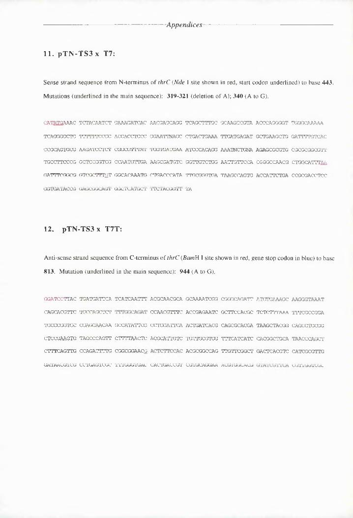
A pp en die es
11. pTN-TS3x T7:
Sense strand sequence from N-terminus of thrC {Nde I site shown in red, start codon underlined) to base 443.
Mutations (underlined in the main sequence): 319-321 (deletion of A); 340 (A to G).
CNTTTGANTA TATNAANTAT GAAAGA^AACCNTAAGCAGC TCTAATTTGA GCTAAGCCGTA AAAAAGAGAT TAAAAATTNT
TANAAAGATA TTTTTTA?AAA ACTAGTAATGA GCCNTTTJACA CTANATATAT TTGATCAGAT GCTGAAGCTG AATTTTGTAAA
CCGCAGTGCG ANCTTCATAT COAACGTAT 1100110^^ ATACCNCNGG ^^TNCTGSZr NCTCCACCTC CACAAAGAGTT
TAACTTAAAA AATCCAATCA C<AAT^TTCTTC ^<0(0^01:: GATTGTATCC ANT^G'TTC(AT CCAAACAACC A'TGAANTTTM
CTTTTAGCAC AT^?G^'^TTGT G<A?ACAAATN <ATCATAAATA TTACCCATGN TAAGCCAGTG ACANTTATGN CCCACAACTAA
ACTGTTNCAA ATCCGCONAL' GiCGATATTcL• 'J^rT^-T^i^^i.G^CT^’' TA
12. pTN-TS3x T7T:
Anti-sense strand sequence from C-terminus of thrC {BamH I site shown in red, gene stop codon in blue) to base
813. Mutation (underlined in the main sequence): 944 (A to G).
GGTTAATTNC
CAGCACGTTC
TAAGCACTAA
ATCCCANCTA
CTTTANATTA
GATAACG'ICG
TGNTATTTCA
TAACTACTAT
AANGATAATA
TNAACANC,TT
AATCT,^^iTG
CCTGAGTCGC
TTATTAATTT
GGAANTTTTA
CATTTTT^<AT:
CACAAGGATA
1'T'TGG^j.IGAC
NAGATAAGAA
CANTCCTTTA
ACTGCTTTCA
NCAAATTGTA
CACTGACCGT
GCA.NTNTAAC
AC(CCAGCTTrA
ACTAATATAC
TG^TTGAGTAG
ACGAGCAGTG
CGTGCAGGAA
CCGGCAAATT
(ACTT'CACCA
CATGACATAG
^ITTTATCATT
ttt3ttt^^^^:^c:c’
AGG CIGAA■NAc
A^TGTGNTTC:
^T2^T:ATT^ZATT
TrAGC-rACCAC
(CACGGCKGCA
GATACACGrIT
NAAAATTANT
TTTAGACCAA
CNGACTGAAC
TNTCACNAAT
CNTAAAATTA
CGTTGGTCGC

Appendices
13. pTN-TS4x TS3:
Sense strand sequence from base 687 on thrC to base 1239. Mutation (underlined): 944 (T to C).
TGTNGCGCAGGTCTGCTGGCCTICACIACIGCCGCITGTG
CGCAACAGACGATCAGTTGATCTCGGTGAA
CTGCCGCAGGGAAGTCACTCITCAITGITCiaagaitagt
AATGCITGAIATCCAIICIAACITTGIATC
AIACGCICAA
IITCTGCCIIACCCAAAICITCCICCICAA
TTAAAAIAACATATGGCTTITINCAAAAIA
CCAICTGITTTIAAACITTTACTCAIICIAAATCTCGCAA
TIIICTACACTTCCTCIGCAICTIGCAIAA
ITCTCIITGCTATTICTGCICITTATCCAACTIAAAIAIC
TTCIIAICCICCICICATCCCITGCTGATT
CAAICIIAAAACCAACITIACICIATGIACTGGGTTATCCGACICTICCIIICIAAATTTTACCCTNICT
CTTCGICIATACIATACCITITGAITCAICAGCCITIIAT
TAICTTATCIAAAIAIAICGTTCACATAAT
TTIACIICIIGCCACITTTCCIAACAACTIIATIAAACCA
TICICTICITTIIAAICIATCTG
14. pTN-TS4 x TS4:
Anti-sense strand sequence from base 657 of thrC to base 85. Mutation (underlined): 319-321 (deletion of
T).
ICCTGCTTCA CCAICGCCTI ACAIICATCI AAATCICCIT CIATIICAAC AITTTCGATA TT3CCICCCA ATGTACAIAACAITTTTTCT TICAITGIAC TIATTTTGCC TCGTGIATAI AIIATAACCA CTTTCACATT CIITAAACCI TAIAAAICATIAICCACTGC CICTCCIITA TNACCKIAII TCICIITCAI AATIITCACT GGCTTATT4AC CCICAATATG IITCAICATTTGTICCATAA AGCIACCICC IAAATC2TAA ATICCAICGT TGICCCGTGI AACAATTNCA IACAACCIAC ATCICTTTCA
ACATTGICIA CCIIAICCII IAAIICAAAC ICCGNICICA CGCGCTCTTT CAIIANTTCC TINIGIAATT CATCACCAATAAACGCCIAI AGGATCTTCI CACTGCIIIT GACAAAATNC AICTTCAICA TCTCATCAAT TTCAITCAII CTIAATTCCIICAGITCITI CGIAAAAAAC AI

Appendices
15. pTN-TS4x T7:
Sense strand sequence from bases 70 to 408 on thrC . Mutations (underlined); 88 (T to G); 106 (C to T);
282-284 (deletion of T); 395-396 (CG to TN). The sequence above (pTN-TS4 x TS4) does not confirm any
of these alterations, and shows a mutation at 319-321 not found in this sequencing experiment.
GGCAAAAATC ANGGGCTGGT TTTTCCGCAC GACCTGTOr AATTCAGCCT GACTGAAAtTTT QATGAGATGC TGAAGCTGGA
TITIGTCACC CGATGTCAGA ACATCCTCTC GGCGTTTATT GCCGAATAAA TarACAGGA AATNCTCGAA GTCACAGTCACAGACAAGTT TACATTCACG CATAACATAA CCAATJT3;^TIA ATTGCATGGC C<GyTATC1CC TT'TTCM'^ICCT CCGGACTTAG
CTCGATTTTT TCTTTTANCA CATCGCTTTT TCTGAATATTT GCA3ATAAAT ATT3C^CGCTC ATAAGCCAINT GTCCTTTATGTAAGTNTACT NCCATGTT
16. pTN-T44x T^T:
Anti-sense strand sequence from base 1283 on thrC to base 786. Mutation (underlined in the main sequence):
944 (A to G).
TGTTCTTTCTTCAATGATCT
CATGGTTATTTTGAACTGTTAATGTNTTTC
ACTATATCCA
TCA'TCAATTTTTTCGATGTT
CAATTTTTCGCTTITAACTNACGACGTTCg
TTTACATCTC
ACCGATTAGACCATTCGG^TNCCrraGGArTMAAC(AAT^lTrcNCACTCTTCraC
CACTCACCGT
(ACTTTT]TAGGA<AAGTATTlr^AC^XCTrlTNTCG,KA^TCTC'rcC5A<ACACCNT.GCG^TGTTJGCT
GAA^rcCACCA
(ATGACCTAAlOTTCAATATC,]TTA^TAA3CA,
ACCCTAGATAC
ATCTCAAAGC
TCTCTTrAAATAAGCTACAGATAGCATCCTGACTNACGTNGNATCG'TTCA
7TTGCC^/TTT^mTCAACGA
CATGACrcCAA5TTTT^A^CCTCA?
CC^rITTG^rlT^CG?
NAGCACGTTCTAACAGATCA
ATNCGAAGTCA,TT^NTGTTCGATAACGTCG
AGCAATAAAAAGTTINTAAA CATGTCCATA TCACC7TCG

Appendices
17. pTN-TS5 x TS3:
Sense strand from base 680 on thrC to base 1182. Mutation (underlined): 944 (T to C).
CTITTICICA
GITCTICTII
CCTICACIACIICCGCITIT
ACICAACAIA
TCA^TCCGGTTC
TLCNNSGLTA
ICTGCCICAI
CIAAITCACT
IITCAITGITCICCICLTCI
CAATICITIA
AATCCAIICI
CCCLTTCGAC
GALACICLCA
CGGNITICCI
CACCCAAALCTTCCLCCLCA
ITTCCCACCC
AATATLICTT
TTT
ACCAGLTGIL
GLTIAACGLT
GLCTCLCLGCACCATGGCCG
CGGLLIGTCG
GCTGCGGGLC
IGTCTCGGIT
ITAT'IGCTGC
ICGTTATCCAICTGAAACAG
ITTCGGAGGG
FCCGCGCCGG
CCAAGCGGAA
TLAGGGCCGG
AC<TCGA^CGLCCTACCCCATC
CLACGGLGCC
CGGCGCAAGC
ACTCCGGCGA
AACGACAGCG
CG1CCGTCAGCAOCCGTCGA
GTGGCCTACG
CCAAGTGCGC
ICGCACGGCG
TGCCACGACC
CCCALCCCCCTTGATGAAACC
TCGGFCTIGL
GTCGAAGCGA
18. pTN-TS5 x TS4:
Anti-sense strand sequence from base 609 of thrC to base 100 . Mutation (underlined): 319-321 (deletion of
T).
TTICCGTTCT TGATGCTCAC ACCAIGCA,’IA, GACCGACCGA TGCCAGICCT CC^7CA^rT^CG^C G'GALCTLCGC ACALTTTGGCTAT7II?(CCGCC CACTGTACAL AACTJLTi’IT'T CCTT^G^T^ AGTLC'IG'T,IG CCTTCGTLGCT AdACGATTCCC CAATTTCACATTCILTCCCC GLTALCCCLG ATAT^GACC^CC? TGCGCCTGGG TCTCACCLLC CLGTCCCZCCGT ACLCCTLTTA CTQCGTTATGACGGLGCATA 'TCGCTACLGA TTy^TCT^C^CVTP TCCACCIC^C^G NGGCCATANT AACCTACAGC GTTCCCCCGT CCCCCCCTTC
CCICCACCGI CCCTGLGTTT CAC^CA^CA^^ GGCCCICCIC CICCACIGAC ACGACCCGNIC H^jAC^G^G^'TC TTCCACICTTTCGTCNCGCC TTGGATGACC AiAC^CTC^C^CCL: CCkC^ZVCLL^C^CT TGCGACTCGI (LGTGACCCCCC TNCAGATTSN LGATCTCAT,NcAGTGAAIGC AICCTGCATT GGccGAcGTG

Appendices
19. pTN-TS5 x T7:
Sense strand sequence from N-terminus of thrC {Nde I site shown in red, start codon underlined) to base 440.
Mutations (underlined in the main sequence): 278 (T to A); 319-321 (deletion of A).
CATATGAAAC TCTACAATCT GAAAGATCAC CAAGGAGCACG TCAGCTTTGC (GCAAGCCGTA ACCCAGGGGT TGGGCAAAAA
TCAGGGGCTG N'lTiTlCCCC ACGAGC^CXA GGACCrTGACA CTGACTGAAA ^ATACAAACAAC^ ACTAAAGCTA GATnTG:^TNACCCGCAAAAC GAAGAT^JCTC rKACAA3ANTA TITAAXACIAG\ AATCCCACAG GAAATCC^AGA AANAGCACAT GCGCACGACAATAACCTTCC CAACTCCGAT CCCGCTCTIG AGAAACACA;AC CAGATATCTG GTA^’TT^'TT^C ACAAGCCAAC GCTG^CATTTAAGTNNNCNG CGGNCGCTTT ATATGGCACAA TGCTCACCCCA TATTGCGGGT GATAAGCCAG tgaccattct gaccgcnaccIVAAjLjlVaH'lH. C/ikjIUjV 1VH IVaVX’l'XVlMU
20. pTN-TS5x T7T:
Anti-sense strand sequence from base 1283 on thrC to base 1134. No mutations noted.
AAATAATACA TCATCAAATA ACGCAACGCA ACAAAAACGA CGGACAAATA ATATGAAAGC AAAGATAAAT NAACACATAC
AGNCAACACT ATTAACAAAN NCAACGTTTN ACCAAAAATN ATTANCACGC TNTNTTTAAA ATTCGCCGAA
21. pTN-TS6x TS3:
Sense strand sequence from base 716 on thrC to base 1056. Mutation (underlined): 944 (T to C)
AACCAACAAAGATGAAACATCAACTCAGACAAAAACAAAC
TTAACTCAATTTAATTACTAAACATAATCC
AACTAAAAGA
GACAA.AGGAGCCG^C^CA^C^C^AACGAGACGG
GG^A^AAATAT
GcCCTAC(AACGGAACGATACC
GCGTGAGTCA
GCAGCCG^AAA
AATTGACAGCATACCACATAGCCGAACAAC
ATGATAAAAC
GAAGATGATCTCCTAAACCATAAACGAGAG
CACCAAACAA
GCGAAGTCACCAGTCAGTAGAGAAAAAAT£
ACAATACATA
TCGGTC'TGCC
TCACCCAAAGATACCACCGC
AGTTAAAAGA
ACAAGACTAC ACTTCAGAGC CG.

Appendices
22. pTN-TSl> x TS4:
Anti-sense strand from base 583 to base 103. Mutation (underlined): 321-319 (deletion of T).
CATTCTCCTG ACACTCTCTG GCTTAGTTTT <ACAA^^K:<G^^T <GGCTTAGCT TCGATATTGC CGCCCAATGT TATATTCTGTTTTTATTCAT TTTAACTCGT (GC^TQTA^CCAt T7TTTAACATT CA(AT^rcCCCT AAACCGTAA TTCATTGTGCATCTCAAGAT CCGATTTATA AGGTCGTCGC GrrcATTCACA TArTCTCCC(CA AATA,TCACTC TCTTTTTATCAATTTTTGCA TAAGAAATTT TC;EETT.^GT CAGC'G^T!TGGC' CATr!rCGTT?A ATTCGAGACA ACCGACATC CTTTATTATT
TCGCATCAGA TCAAGGGTTG CATTTCCACG CGC^ACGGC?, CATATYCTTAJ ATTR^CTCrC (GGATTrCATC TAATTTTTTCCACATGTCGT TTTTAGATAT CACGATGTCT 2TTTTAATAT TTCTCCT^XTC ATTTT^TR.A GTATGGCTCA TTTAACCTTA
23. pTN-TS6x T7:
Sense strand sequence from N-terminus of thrC {Nde I site shown in red, start codon underlined) to base 460
Mutations (underlined in the main sequence): 319-321 (deletion of A); 340 (A to G).
ATTTTCTTTA TCTTCTTTAT GAAAGATNAC ATTAGTGATA TCATCA^TTA G<:ATTCC:AA,A ACCCANGGGT TGCGATTTTTTNTNGGGATC NTTTTTCACA ACGANCTGCC GCCTTTTATK? CAGCTAGCTT ,^rcATTATAT GCTGAAAATC CTTTTTCTCT
AAAGATATCA CTTGTTAATC TCGGCGTTTA TrrcCGXATTA AATCACACAT GCTTTTCAGG AAGAGCGCGT GCGCACCGAATSTCXCTTCC CGGATCAGGT CTCCAATGA^ GCTTTCACTT; TCCGCI^TCTT GCCT^TTT^CTTA CTCGCGAATT CCATCCATTTTAA(TN^TAG CACGNAGATT TGAGGCAC'AA A,]TA'TCGTCC ATAT]TCAGG; ^TGATAATCAA GTCTCATT^C TCTCACAGTC
V X X IV kjdN X X/H-VAxrtM XbXbnfinb X^ V

Appendices
24. pTN-TTS x T7T:
Anti-sense strand sequence from C-terminus of thrC {BamH I site shown in red, gene stop codon in blue) to
base 770. Mutation (underlined); 944 (A to G).
CLCTGCTTAG TCCTCCT^A TCACATAACT ACCCGCCGIA LCACAATCII CdCCCAGATT A'TLTyCCCGC CACCITCCAT
CALGCGGTTC TCCGCLGTCT TTTTTICAGAT GGCAACGITGA ACCCAIGCATA GCTTCCACGC 1GC,TATTGA2CC TGAGLGGCIA
TCGGGLIGGC CICLGAACAC GC<ATAGAGT CCCGLLCIGTA ACTIATAAGG CGCGCACGA TAAGCTACGG AACGLGCGLI
CACGGAALTL TAGCCCAITT C,IGGTGGJCrAG ACdACTTCT GITG,LGGGLL 'TTTCATCATC C.A<^CII^rT^<CA TCACCGAIGTCTTTCALTTC CCAICTTTGC C<GIIGGIAAS ACTT'CTCCAC AAGAGGNCAG TTGTTCGICT GACTCACGTC CATGGGLGGC
CATCAGLTGI GCTCCCTCCG TTTTTGGIAI CC^CCT^G^CCC^TT CITGCA^CLAC ACGTCCGC^CCG GTATCGTTCA CCGTCcTGGG
CICCATCACC CLTTTCACGL <2GCAICGAG GTACCTTCI'C ACCA
25. pTN-TS7 x TS3:
Sense strand sequence from base 712 on thrC to base 1287 (gene stop codon shown in blue). Mutation
(underlined): 944 (T to C)
CLCAACCALG TCLTGITCTG GGG^C^CCL G(ACACGTTA ICGATTTGAC 1CIACGCrAC^TC CTCQGCGAACCT CACTCCITCTLGCCLTCAAA GCTTTTATT3 CTAGLAICAG CGr[GIACGIC ACCCCTGCCAC GTTTCCTGCA CGACCCLTAG TCLTAACCGAAALCLACTCA CIGIACCTTA TCCAACGCCG ATIACGCTCcG TCAGCCGAAC AACTGGCCGC GTCTILAACA (^TVC^TGC^C^
GLGCAAAGGT GGCGGCTGGG ACAd^CGGTTA^[TGACIGA ITCGCTGATGA AACCACGCAA CAAICCAATIC ITICITTAAAAGAAGTGCIG TGAGATTCGG ACAI^^^^C^C^C'C^': GACCATTCGA IATCGTGCGC TGCGTGATCA GTTGAATCCA CGCIAATATCIGTTGNGCCT CCLGAGCICI CATCGCGGLA CACTTAAACI IAGCGTGGAA GCGATTCTCG GTGAAACGTT GCATCT3CAAGGGGGATGG GAIACCITCG TGACATATCC GTTCATTTAC ATAATCTCCC GCCGACNTTTC CTKCLTTICG TAAATTIATIATCAATGCTC ALTAC

-Appendices
26. pTN-TS7 x TS4:
Anti-sense strand from base 602 to base 169. Mutation (underlined): 321-319 (deletion of A).
ACAA’CAACAAACCCAAAATAAACCATAAAAATAAAGAACACCAACATNACAATATCAAA'A
CAAACACCAACAAAACAAATAGCAAAAACCACAAATATACTATNAACATACCAATAAACG
CATCACCAACA’TTCTTACAAACTACCGCTCCATAAAGCAAAACGACCGAACCAAAAGAAT
ACCAAACAAAA,GAACAAAATCAATATNACCCCACCAAAATACCAGAAAAACTT
CATCGAAATCATACCTCAAAGAAAAACGCANTTAAATGCCCAAACGCCAC
ACCAACAAAAAATAAAGAATAACAAAATAAAGCGTTAACCACGCACACGC
ACAACAAAATAACCACTTTCTCACAAACTACAAAAAACAATCTnCAGGA
CAATAATACCACAATCAATAATNACCCGCA
TTCCAGACAAATTCCAAAAG
27. pTrCTST x T7:
Sense strand sequence from N-terminus of thrC (Nde I site shown in red, start codon underlined) to base 273.
CATATGAAAC TCTACANTCT
TCAGAGGCTA AAAA'i,ACCACCCCACAANAC AAAAAACCAC
NTAACCTTCC CGACTCCAAT
GAAA.GA^A7AC AACCAGCA<A3
ACGACCTCCC (AAACATCGKCTCACGZ('dAA^/A CATAGTACCTAC(C:CCCAAAA^ GAGGGCGArCG
ACAACATTAC <ACGCCACCTA
CTAACTAAAAG^ATCCCACAG g ,AAGC’CCr^AG;
TCCGNT
ACCaC GAGGCAAAAA
CA,'rTAACGC'IG GATTTTATCAGACNCAGGGGr^ GCGCACGGCG
28. pTN-TS7x T7T:
Anti-sense strand sequence from C-terminus of thrC (gene stop codon in blue) to base 910. Mutation
(underlined): 944 (A to G)
ATACTAAAAACAATCTGCCAAATGCCAAGAAAAAATAGCC
ATTCATCATCGCTCTTTTAAAACAAGCCATCAAATCTATA
AATATACGAACAGGTC'CAACATTAGACTGGAAOTNACGCA
ACGAAAAACNGATAAJAeCGAACTACA^C^IAA.TAAAJCTCAAA
AACAACAAGCAAATCACA'TCTCACGCAGCGCAAAATTTCA
AGArA^A^AA^AACACCA?rrc^AC^CAO:ACCACC<A^^NCTCCACCAA
GCACCA7GAAAGAKGGC^^IACTAa.GACA<AG
CfIAACAAGCCX:
TAAATNAGCACCAAAAACACAACAACTCCGCAACTCAATC
AAATACCAAA ATTTGCAACA GAACGACTAT TTCACACCAG ANCAAATAAT CAACTAACT

Appendices
29. pTN-TS4 x TS3:
Sense strand sequence from base 712 on thrC to base 1184 . Mutation (underlined): 944 (T to C).
CATTAATGAT CATTCTCTAC GTGCCTTCAG ATTTCTTCCA CGTTTTCTAA GCCGATCTCA 7CCAGTTGTC TCTAGATATACCGCTGTTTA GTTTTTTTCA TGCGACCAAC ATGTTCGTTT CCCTCAATCG TTTACTGATC GTAGATATCT CATCTCAATTAGCGACTNAG CAATCGTTTT CATTAGAGTT CGTAGTCTGT CTGACATTCT TCTCGAAACG tgtggaagag Tlgttccgcc
CATTTTTATC GCTTCTCTTT GTGTTCGATT TTCATGCCGT CGTTGTTGTT TCCTCACTTC TATCTTTACC TCTGTTTTTTATTCTCGGAT TATCTTCCGT CACGNTCGAT CACGTTGCTT TTAATGCGAT GAGTGTTATC TTGTTTNATG GAATTTTTAACTTCNTACTC CGTTCCCAGA TTNCGGAGTT TT^^TTTCGAG TCAGTGGTTG AGTTTCTNGA TCTTTCCTTC GA
30. pTN-TS8x TS4:
Anti-sense strand from base 587 to base 88. Mutation (underlined): 321-319 (deletion of T)
CAATCATTCT CCTCAAACTC AGA^CA'Ar; ATTTCAGAAG A(GTTGGATTC AGTTTCGATA ^T7A?C(A?CCAT TTATTATGTTCTGTTTTTAT TCATGTCGTA TGATTTCCC TCAA3AGTAT AGGATAACCA CTTTATATTT 03317^002 TTGTTTGATTGTCACTCTCA CGATACGATT T^T\CC<ACA(TC TCAGAAGCAT AA^TCCrrcACT GGCTTATTAC CC(AATTTATC CATATCATTTTCTCAATTTT TGAAAACGAA G.AAT]TTAAT ATGCAATCAG TCcCACCG'TAC TTCTTTTNAT GACATTAAGTC TTNCATTTNTTCTTTCAAGT CCGGTGAACG G^CTTCGAT^CT GGAAAGCACA A(CTA^^TA^TKA TCGTTTTTCT G'TGG^TA'TTTrc: TTTTCATTTTTTCCACGTGT CGTTATTNGA AC,A7GAGAiG ATATTTTTCA GCTTAGCAT CTNATNAATT rrcAG^TTTGGA TGTTTTCAGACTCATCATCA CATTTTTT
31. pTN-TS4 x TT:
Failed to produce a readable sequence.

Appendices
32. pTN-TSSx T7T:
Anti-sense strand sequence from C-terminus of thrC (gene stop codon in blue) to base 694. Mutation
(underlined): 944 (A to G)
TAAC’AAATGA ATCATCAACA ATTTTCGAAA GCCAC/AGCA ^rcG«AACAA GAGAAAGCAA.. AGC(GACGGAA TGA,TCCGAAC
AATCAACCAG CTCA,1,^,1AGC AGATACCACG GTTCACCGAG ACATCGC^ArcC ACGATCTCTT TAAA\rAIAAAA CGGA^AAC(AAC
GAACCAAGAA ACAAACCATA ^^TAGGCTGAG GTCAACTCAT CACTG'3AGC<G? AC(GC^AGA:AC^ ACCCAAGAAT GCQCGCrC<CT
AAAATAACCC AAATCAA,i,i,A ACTAACGAAA GG'^CAAA^A'A Gr]CA3rAAreAT CATCCACGC T(AATCACC<X AGCTCTTrcAAA^ACCAAAT ATAACGACAA AACCACTATTNCA<NCCXCC NAAG1CAAA^A AGGAAACGCA CATAAATCAC GGrrcGT\TAACC
GTCGCCAAAA AGGCAATAGA TGACCACTAA GCGTCGAACA <CGAGCCCCKA CAGXATAAAC:3 TGGGAC^iCA^T
AAAACATTTN ACCGACAGAC CGGGATGCTA GGCCC^CCC^CGC CCCX3CCGACA AA^ATCG.JAA GATnCO:rICr TGGGACCCACC
ACAACCAGCT AAATACACAA CTTCTAAXAN AGCTC