PHD DEGREE TITLE OF THE PHD THESIS - Cagliari - IRIS
207
Università degli Studi di Cagliari PHD DEGREE Life, Environmental and Drug Sciences, Pharmaceutical Curriculum Cycle XXXIII TITLE OF THE PHD THESIS IDENTIFICATION AND DEVELOPMENT OF NEW ANTITUMOR AGENTS WITH MULTI-TARGET ACTION Scientific Disciplinary Sector CHIM/08 PhD Student: Dott. Serenella Deplano Supervisor Prof. Elias Maccioni Final exam. Academic Year 2019/2020 Thesis defence: January 2022 Session
-
Upload
khangminh22 -
Category
Documents
-
view
0 -
download
0
Transcript of PHD DEGREE TITLE OF THE PHD THESIS - Cagliari - IRIS

Università degli Studi di Cagliari
PHD DEGREE Life, Environmental and Drug Sciences, Pharmaceutical Curriculum
Cycle XXXIII
TITLE OF THE PHD THESIS
IDENTIFICATION AND DEVELOPMENT OF NEW
ANTITUMOR AGENTS WITH MULTI-TARGET ACTION
Scientific Disciplinary Sector
CHIM/08
PhD Student: Dott. Serenella Deplano
Supervisor Prof. Elias Maccioni
Final exam. Academic Year 2019/2020
Thesis defence: January 2022 Session

2
Table of the content
TUMOR .......................................................................................................................................... 5
1.1 Tumor: an overview ........................................................................................................... 7
1.2 Cellular pathways involved in tumors ............................................................................. 10
1.3 General mechanism of drugs ........................................................................................... 16
1.4 Tumors drugs .................................................................................................................... 19
1.4.1 Resistance to chemotherapy ..................................................................................... 21
1.5 Multi target inhibitors ...................................................................................................... 23
CARBONIC ANHYDRASE .............................................................................................................. 27
2.1 Carbonic Anhydrase: overview ........................................................................................ 29
3.1.1 α-Carbonic Anhydrase ............................................................................................... 29
2.2 Carbonic Anhydrase IX E XII: physiological and pathological roles ................................ 35
2.3 Inhibitors and activators of Carbonic Anhydrase ............................................................ 37
CYCLOXYGENASE ........................................................................................................................ 45
3.1 Cyclooxygenase: overview ............................................................................................... 47
3.2 Cyclooxigenase-2: physiological and pathological roles ................................................. 52
3.3 Inhibitors and activators of Cyclooxygenase-2 ................................................................ 54
KINASES ....................................................................................................................................... 57
4.1 Kinases: overview ............................................................................................................. 59
4.2 c-Src and Bcr-Abl: physiological and pathological roles .................................................. 63
4.3 Inhibitors and activators of c-Src and Bcr-Abl ................................................................. 65
PROJECTS .................................................................................................................................... 69
5.1 Selective inhibition of carbonic anhydrase IX and XII by coumarin and psoralen
derivatives .............................................................................................................................. 71
5.1.1 Background ................................................................................................................ 71
5.1.2 Biological results and discussion ............................................................................... 76
5.1.3 Conclusions ................................................................................................................ 82
5.2 New 4-((4-oxo-2-phenylthiazolidin-3-yl)amino)benzenesulfonamide synthesis and
inhibitory activity toward carbonic anhydrase I, II, IX, XII and the potential dual CA/COX-2
inhibition ................................................................................................................................. 83
5.2.1 Background ................................................................................................................ 83
5.2.2 Biological results and discussion ............................................................................... 85
5.2.3 Conclusions ................................................................................................................ 87
5.3 Evaluation of drug like properties ................................................................................... 88
5.3.1 Background ................................................................................................................ 88

5.3.1 Biological results and discussion ............................................................................... 91
5.4 Synthesis of new pyrazolo[3,4-d]pyrimidine compounds ............................................ 111
5.4.1 Background .............................................................................................................. 111
5.4.2 Biological results and discussion ............................................................................. 116
5.4.3 Conclusions .............................................................................................................. 116
EXPERIMENTAL SECTION .......................................................................................................... 117
EMAC 10155 (a-m) and EMAC 10158 (a-m) ......................................................................... 119
EMAC 10156 (a-m) and EMAC 10159 (a-m) ......................................................................... 126
EMAC 10157 (a-m) and EMAC 10160 (a-m) ......................................................................... 136
EMAC 10161 (a-m) and EMAC 10162 (a-m) ......................................................................... 149
EMAC 10190 (b-l) and EMAC 10191 (b-l) ............................................................................. 160
Pyrazolo[3,4-d]pyrimidine .................................................................................................... 176
BIBLIOGRAPHY .......................................................................................................................... 187
APPENDIX .................................................................................................................................. 203
Oral Communications in Scientific Meetings ....................................................................... 203
Poster Communications in Scientific Meetings ................................................................... 203
Manuscripts in International Journals ................................................................................. 204

4

5
TUMOR

6

7
1.1 Tumor: an overview
Cancer, according to the World Health Organization (WHO), is the second leading cause of
death in the world, with 10 million deaths in 2020 and an estimated 19.3 million new
cases.1, 2
Tumor is a disease that can affect any tissue and organ of the body, in which cells grow
uncontrollably, cross the normal border to invade other parts of the body, and
metastasizing: tumoral cells sever from adjacent cells and invade other tissue through
extracellular matrix component and lymphatics.3, 4 Metastases are the major cause of
cancer mortality.3, 5
The most common types of cancer that affect women are breast, colorectal, lung, cervical
and thyroid cancer; the most common types of cancer that affect men are lung, prostate,
colorectal, stomach and liver cancer.5
Normal cells can grow and differentiate under supervision of biochemical stimuli: the
presence of growth stimuli allows cells to divide and proliferate, in absence of this type of
stimuli, cells stop to grow and undergo programmed death, the apoptosis. In tumor cells
these normal processes are deregulated: cells have uncontrolled growth, without external
stimuli, lose their capacity to differentiate, resulting without their normal structure and
functions; they also disactivate the capacity of apoptosis being immortal.6, 7
Tumorigenesis describes the process of tumor formation: the transformation of normal
cells in tumor cells, with a multistep process that born with a precancerous lesion and
continue with a malignant tumor. This process is conditioned by interaction between
genetic factors and three external agents: biological carcinogens like infection from
bacteria or virus, physical carcinogens, like UV radiation, and chemical carcinogens, such
us arsenic and asbestos.5
Tumorigenesis is a 4-steps process showed in Figure 1:8
INITIATION
In the first phase alteration or mutation of a gene is observed, it can happen
spontaneously or following an exposition with cancerogenic agent: in normal cells
deregulation of biochemical pathways occurs, followed by cellular proliferation,
differentiation and survival. 8-10 These changes can be increased by mutation of other
factors response of DNA repair function.8-10

8
PROMOTION
Different component, like environmental, interfere to promote preneoplastic cells
proliferation and their accumulation; this phase is slow, and it can be reversible.8-10
PROGRESSION
In this stage proto-oncogene can convert themselves in oncogene, there are neoplastic
transformations, with genetic and phenotypic mutations. Tumor dimensions increase, cells
become invasive and promote the carcinogenesis.8-10
METASTASIS
With metastasis tumor cells can move in other districts of the body, using bloodstream,
lymph system, and they can generate themself new lymph system with angiogenesis.8, 9
Figure 1 Tumorigenesis: Initiation, Promotion, Progression and Metastasis8
Cancer is also defined like an evolutionary process caused by somatic mutations in the
progeny of a normal cell, that bring to uncontrolled replication and to a selective growth of
mutated cells.11 Different cellular and molecular events lead to malignant transformation
of cells, like evasion of tumor suppression, inhibition of cell death, creation of a particular
microenvironment containing blood vessels, acquisition of invasive and metastatic
potential.12
The cancer stem cells (CSCs) can drive tumor initiation and can cause relapse, in fact they
are also known as tumor-initialing cells (TICs): they are the principal controller of tumor
growth for proliferation, resistance to chemotherapy and metastasis (Figure 2).11 Both
normal and neoplastic cell population include a subpopulation of stem cells that can self-
renew and differentiate. The normal SC usually can support a genetic alteration but, if they
are not able, they can generate altered SC; which can generate, in turn, a preneoplastic
cell population.13

9
Figure 2 Carcinogenesis, tumorigenesis, and tumor resistance11

10
1.2 Cellular pathways involved in tumors
Tumor cells present altered genetic, biochemical and histological mechanism, with respect
to the normal cells; in particular they have altered energy metabolic activities, which
promote the acquisition and maintenance of malignant properties.14, 15 The cells
reprogram their activities through the metabolism, to have advantages for the
tumorigenesis and for survival, growth and proliferation under stressful conditions; in
particular they modify bioenergetics, enhanced biosynthesis and redox balance.14
Tumor cells can reprogram their metabolism: in conditions of nutrient-replete they prefer
anabolic growth, otherwise, during nutrient-limitation conditions they can use catabolism
and fortify the redox homeostatic systems to support the cell survival, and to resist to
tumor suppressor.14, 16
The most important feature of reprogrammed metabolic pathways is the aerobic
glycolysis; this form of modified cellular metabolism is also known as the Warburg effect.14,
16
Glycolysis supplies the necessary energy to the cells, in mitochondria, and its
intermediates can be used for the synthesis of amino and fatty acid: one molecule of
glucose is exploited to produce adenosine triphosphate (ATP), nicotinamide adenine
dinucleotide hydrogen (NADHs) and pyruvate.17 The process occurs in absence or presence
of oxygen: under aerobic conditions pyruvate is converted into acetyl coenzyme A (acetyl-
CoA) which is oxidized to CO2 and H2O, through tricarboxylic acid (TCA) cycle and oxidative
phosphorylation (OxPhos); in absence of oxygen, the pyruvate is reduced to lactate,
necessary for the regeneration of NAD+, that can be used by the enzymes involved in
glycolysis, in order to have an uninterrupted cycle.17, 18
Healthy tissues usually prefer using oxidative phosphorylation to produce energy and use
anaerobic glycolysis like a normal response to hypoxia. On the contrary, many tumor cells
can exploit anaerobic glycolysis to produce lactate, regardless of the presence of oxygen:
Warburg effect.14 Moreover, in cancer cells there is an increasing of glycolytic flux to
respond to the rapid growth of tumor, causing condition of hypoxia in blood vessels, with
an oxygen concentration in the range of 0-2% O2.19 According to this, in tumor cells,
anaerobic glycolysis prevails. With the Warburg effect, there is an increase of lactate
secretion, that contribute to create an acid microenvironment, toxic both for the normal

11
cells and for the weakest cancer cells, but not for the strong aggressive cancer cells;
furthermore, the extracellular environment acidosis promotes angiogenesis and increases
the invasion ability of cancer cells, creating an environmental advantage for tumor
progression.14, 20, 21
Due to Warburg effect, the respiratory chain in mitochondria is downregulated, resulting
in reduction of OxPhos and oxygen consumption: during the aerobic glycolysis a great
amount of reactive oxygen species (ROS) were produced, thhey supports tumor cells to
avoid apoptosis and increases their proliferation.21 The high rate of glycolysis in tumor cells
is suggest by the increased transcription of genes of most enzymes and transporters
involved in the pathway, that match with an increased synthesis of protein.15
In tumor cells there is a deregulation of signaling pathways such as over-regulation of
pathways that support functions like anabolism, catabolism and redox balance, necessary
for the cell survival (Figure 3).14, 22 Usually, in normal cells, growth factors stimulate and
activate phosphatidylinositol 3-kinase (PI3K) and its pathways, protein kinase B (AKT) and
mammalian target of rapamycin (mTOR), resulting in the activation of hypoxia-inducible
factor–1 (HIF-1) and sterol regulatory element–binding protein (SREBP), that induce
increase of glycolytic flux and fatty acid synthesis, respectively.14, 23 Tumor cells usually
present mutations that interfere with the pathway of PI3K-AKT-mTOR, resulting in high
levels of signaling, independent to the presence of growth factor.18 mTOR is a
serine/threonine kinase, that after his activation by PI3K/Akt system, induce cell growth
and inhibit the catabolic reactions.18 Aerobic glycolysis of tumor cells is also fundamental
for the activation of oncogenes, loss of tumor suppressor and up-regulation of the PI3K
pathway.14

12
Figure 3 Signaling pathways that regulate cancer metabolism14
Hypoxia, induced by cellular changes, results necessary for tumor proliferation, metastasis,
and resistance to therapy. Hypoxic and inflammatory stress, as well as metabolic and
oxidative stress, can upregulate HIF-1, a transcription factor constituted by the subunits
HIF-1α and HIF-1β.15, 18, 24 In normal and aerobic conditions, there is a program of HIF-1α
degradation when it is not necessary for the cells, otherwise, in anaerobiosis condition, the
transcription factor became stable. 15, 25 HIF-1α can also be induced by cytokines, growth
factors, reactive oxygen species, nitric oxide, and by energy-metabolism intermediates,
like pyruvate (Pyr), lactate and oxaloacetate.15 The role of HIF-1 is to promote the
expression of different enzymes involved in energetic metabolism, in order to stimulate
the glycolytic flux; moreover, metastatic tumor cells line generally present overexpression
of HIF-1, glycolytic enzymes and high glycolysis.15, 26 Two other targets of HIF-1 are vascular
endothelial growth factor (VEGF) and carbonic anhydrases (CAs): the first promote
angiogenesis, necessary for the reuptake of nutrient and oxygen; while CAs are necessary
for the pH homeostasis in microenvironment.24 Furthermore, HIF-1 transactivate
cytochrome c oxidase (COX), to facilitate the adaptation of mitochondria to an hypoxic
environment.18 HIF-1 work together with c-Myc, a proto-oncogene that code for
transcription factors, to stimulate aerobic glycolysis through the induction of enzymes

13
involved in the cycle, like hexokinase 2 (HK2) and pyruvate dehydrogenase kinase 1
(PDK1).27 HIF-1 with oncogenes and tumor suppressor genes are capable to
reprogramming tumor cells metabolism in different step.
The protein p53 works as a tumor suppressor by regulating the replication cycle cell; it is
involved in several stages of metabolism control and regulate proteins necessary to
cellular response to cancer. This protein is also known as the “guardian of the genome”,
because it is usually upregulated after a DNA damage to stop the cell cycle and to induce
apoptosis.21 Also, the protein play an important role during viral infection, oxidative stress,
altering cell metabolism. p53 acts inducing/inhibiting key metabolic genes involved in
glycolytic pathway, OxPhos, lipid and amino acids metabolism and cell growth (Figure 4).21
In normal cells p53 usually promote OxPhos and suppress glycolysis.21 In many tumor cells
a mutated p53 is present and overexpressed; mutated p53 have strong oncogenic activity,
in fact it has a relevant role in inducing cell transformation. It is involved in several
pathways such as modulating the mTOR pathway, repressing autophagy, and promoting
drug resistance.
Figure 4 Regulation by p53 of proteins involved in cells metabolism21
Other important molecules involved in tumor cells metabolism are ROS, like hydroxyl
radical, superoxide and hydrogen peroxide; they are by-products of metabolism of oxygen.
Usually, in normal cells, they are present in a small quantity, while in tumor cells there is

14
an over-production; this situation leads to DNA damage, and consequently mutation and
tumorigenesis: they activate apoptotic signaling and induce cell death.21 In normal cells,
DNA damages induced by ROS, activate p53, that work to lower ROS levels. In tumor cells
p53 is downregulated, and there is an enhanced of ROS, while cells with low amounts of
p53, stimulate the expression of antioxidant genes like glutathione peroxidase (GPX1). The
mutated p53 are present in tumor cells where they suppress the expression of genes
involved in ROS inhibition.21
To respond at low oxygen levels in microenvironment, tumor cells apply changes, not only
in metabolism and proliferation, but also in gene expression, genetic stability and
survival.28-30 The high production of lactate and carbon dioxide by glycolysis, which are
expelled in extracellular environment to preserve intracellular environment, concur to
reducing extracellular pH. Tumoral cells can adapt their intracellular environment thanks
to the activity of enzymes like carbonic anhydrase IX (CA IX).31 CAs are involved in the
catalysis of carbon dioxide hydration to bicarbonate ions and protons. CA IX acidify
extracellular environment, increasing carbon dioxide and protons level, and neutralize
intracellular pH by the formation of bicarbonate ions.28 HIF-1 is involved in regulation of
CA IX transcription, so in tumor cells where HIF-1 is overexpressed, CAs are overexpressed
too, especially the IX isoform.32
Cycloxygenase-2 (COX-2) is another enzyme involved in tumorigenesis, especially in
colorectal cancer. Inflammation induce COX-2 expression; it is responsible of
prostaglandins synthesis. His overexpression has been observed and investigated in
several tumors, in association with his product, prostaglandin E2 (PGE2), that promote
proliferation, evasion of apoptosis, angiogenesis and tissue invasion.3 PGE2 has pleiotropic
effects linked to the acquisition of cancer hallmarks and inflammation. Some tumor
present a deregulation of COX-2 pathway caused by epigenetic alteration, like DNA
methylation: Figure 5 shows the hyper-methylated genes, evidenced in red boxes, and in
which type of cancer they have been reported.33

15
Figure 5 Deregulation of COX-2 pathway in different tumors33

16
1.3 General mechanism of drugs
Drug effects depend by different factors: interactions between chemical component of
molecule with proteins, nucleic acids and other macromolecules, as well as their
degradation by enzymes in the organism; their transport through membrane, water
solubility and, more generally, ADMET profile (absorption, distribution, metabolism,
excretion, and toxicity).34, 35
Figure 6 ADME36
− SOLUBILITY
As a general rule, drugs must be polar enough to be water soluble: usually is necessary
that the polar groups present in molecules exceed lipophile groups to achieve water
solubility. In some cases, acid or basic groups are introduced to obtain ionizable species,
more soluble in water.

17
− MEMBRANE TRANSPORT
Biological membrane wrap the cells and they are constituted by lipids. They represent the
wall that must necessary overstepped by drugs to play their role; for this reason, drugs
need to be water soluble and fat-soluble at the same time.
− SYSTEM BIOLOGY INTERACTION
Structure-activity relationship (SAR) is defined as the relationship between chemical
structure of molecule with its biological activity; SAR helps to predict activity of a new
molecule: similar molecules are supposed to act in a similar way and functional groups of a
molecule usually contribute to its biological action and/or chemical-physic propriety.
Molecular structure, its composition and the spatial orientation of the functional groups
lead to the pharmacological effect of the molecule.34
− METABOLISM
Metabolism is in most cases necessary to increase hydrophilicity of molecules as one of
the main ways to facilitate excretion of the molecules.
− ACTIVE TRANSPORT
Active transports can bring a molecule across a physiological barrier, such as the
membrane of gastrointestinal tract and the blood brain barrier. This process requires
energy and can be divided in two different types: primary, that use ATP molecules and
secondary, that use electrochemical gradient.37
Figure 7 Example of active and passive transports36
− RECEPTORS

18
Drugs interact with a target macromolecule generally leads to its activation or inactivation.
As a general scheme, molecule con act as agonists if their binding to the receptor activate
it, or antagonist if their binding block the receptor without activating it.
More generally, molecules need to interact with their biological target to express their
activity, no matter if the target is a receptor, an enzyme, a nucleic acid, a membrane etc.
In other words, the molecule needs to be complementary with its target or to be capable
to achieve this complementarity according to conformational adaptations that involve
both the ligand and the target. Thus, complementarity is a dynamic process strictly
dependent on the molecule chemical-physic proprieties.

19
1.4 Tumors drugs
Current treatments for tumor, usually consist in chemotherapy, radiotherapy, surgery,
targeted therapy, immunotherapy and endocrine therapy that can be used individually or
in combination, depending on the stage and type of tumor and diagnosis.38-41
Early detection of cancer is necessary to have a promising clinical prognosis and a
successful treatment, but, unfortunately, most patients usually have a late-stage diagnosis,
that, in combination with delayed therapies, lower the possibility of survival.42
Following some approaches to cancer therapies.
Biomarkers
An interesting method for early tumor diagnosis can be the identification of tumor
biomarkers. Biomarker is described by U.S. National Institutes of Health (NIH) as “a
characteristic that is objectively measured and evaluated as an indication of normal
biologic processes, pathogenic processes, or pharmacologic responses to a therapeutic
intervention”.42-44 Cancer biomarkers found in tumor tissue or serum, can be molecules
like proteins, enzymes, DNA, RNA, metabolites, transcription factors.42 By identifying
cancer biomarkers, it is more easy to have an early cancer diagnosis, tumor classification
and consequently, the appropriate therapy for the patient.
Precision oncology
Precision oncology is another approach to identify targetable genomic alterations, that
characterize tumors in order to have a specific treatment for patients.45-47 The goal of
precision oncology is to improve cancer therapy, lowering toxicity, by using drugs that are
directed towards selective genes or proteins of tumor cells. From 2000 to 2014 almost the
79% of the newly approved tumor drugs are targeted therapeutic agents.47
Treatments based on precision oncology:
PRODRUGS
Prodrugs are derivatives of drug molecules which need a bioconversion by human
enzymes to perform their activity, and have the purpose to improve pharmacokinetics
profile of the drug.48, 49 Some cancer prodrugs are activated in tumoral cells by specific
enzymes, and need to have good activity at low pH values.50

20
KINASE INHIBITORS
During the last twenty years an increasing number of small molecules directed toward
tumor associated kinases, such as BCR-ABL, EFGR, VEGFR have been identified and
introduced in therapy.50-52 However, this therapeutic approach might lead to the
expression of mutated resistant proteins to drugs.51, 53
IMMUNOTHERAPY
Immunotherapy is based on the observation that immune cells are able to destroy tumor
cells during the firsts stage of tumorigenesis.54, 55 During cells transformation from normal
to cancer, cancer cells exhibit neo-antigens which can be recognize by the immune system
provoking its activation and the destruction of the growing tumor. Unfortunately, tumor
cells develop specific mechanisms to escape from immune cells: local immune evasion,
induction of tolerance, systemic disorder of T cells signaling.54, 56Among immunotherapy
there are: cancer vaccines, oncolytic virus, adoptive transfer of ex vivo activated T and
natural killer cells.54
Gene therapy
With gene therapy, vectors take in tumoral cells exogenous nucleic acid, which modify
gene expression to correct the abnormalities.57
Nanocarriers
Nanocarriers are colloidal nano-scale system that transport in tumor cells, great
concentration of antitumoral agents, like small molecules or macromolecules that result
cytotoxic for cells.
Biosimilar
Biosimilars are synthetic macromolecules that have similar pharmacokinetic and
pharmacodynamic properties to a license biological product, without differences for
safety, purity and potency.58, 59 They were approved in EU in 2005.58
Epigenetic therapy
This therapy is based on studies of genic pathways, which cause deregulation in gene
expression, without modify DNA sequence.60 Histone proteins are involved in modification
of gene expression: they may undergo different post-translational changes and
methylation that imprint on transcription.60

21
1.4.1 Resistance to chemotherapy
The major problem in cancer treatments is the resistance to chemotherapy agents and to
novel targeted drugs; it is responsible for the majority of therapy failures and patients
deaths .38 There are two type of resistance: acquired and intrinsic.38, 61, 62
INTRINSIC RESISTANCE
Drug resistance can be present before therapy and it is called intrinsic; it affects almost
50% of patients.38, 61, 62 Three important factors cause innate resistance:
− Inherent genetic mutation
− Tumor heterogeneity, in which exist different type of cancer cells with distinct
phenotypic profile, gene expression, metabolism, metastatic potential; in this case
is very high the possibility of relapse after treatment
− Intrinsic pathway for defense against external toxins.38
Therapeutic effects of tumoral drugs decrease with intrinsic resistance.
ACQUIRED RESISTANCE
Acquired resistance is induced after treatment and affects most of patient; it causes the
reduction of efficacy of cancer drug during the therapy. 38, 61, 62 Tumoral cells show defense
mechanisms against external molecules, in this way the tumor acquire resistance thanks to
different pathway:
- Activation of second proto-oncogene
- Genetic and epigenetic mutation that lead to modification in level of drug target
expression
- Modification in tumor microenvironment (TME) after therapy. 38, 61
Some factors involved in resistance: members of the adenosine triphosphate (ATP)-binding
cassette transporter gene family, glutathione-dependent enzymes, topoisomerase,
metallothionein, O6-methylguanine-DNA-methyltransferase, thymidylate synthetase,
dihydrofolate reductase, heat shock proteins, growth factors, proliferative, apoptotic and
angiogenetic factors, protooncogenes and suppressor genes.63 Cancer cells can also
acquire resistance to targeted drugs, thanks to develop of new mutation in target protein
genes; in this way there is a secondary mutation, such as BCR-ABL kinase domain.38, 64

22
Figure 8 Cancer drug resistance38

23
1.5 Multi target inhibitors
Drug design and discovery is a very dynamic process, necessary to counteract the global
morbidity and mortality caused by incurable disease like cancer. Usually, the discovery of
new therapies is based on the design of selective chemical compound that target a single
enzyme or, more generally biological target. Most commonly they target a protein called
“on-target”, the dominant or most relevant one in the pathology. By this approach the
researchers aim to identify therapeutic agents devoid of side effects.65, 66 Unfortunately,
the therapeutic agents directed towards one single target have shown low efficacy
towards complex diseases; in particular this type of therapeutic agents have been
associated with the increase of drug resistance.67
Polypharmacology is a new approach to design and use compound that simultaneously act
towards two or more targets, or multiple biochemical pathways involved in the disease; a
single compound can recognize more than one biological target.67
Multifactorial disease, such as cancer, are characterized by several intrinsic and/or
environmental factors that contemporary act on the organism; with polypharmacology,
multi-target compound show some benefit: a more predictive pharmacokinetic, better
compliance of the patient and last but not list, the reduced risk of drug interaction.68
The advantage of using selective inhibitors is the decrease chance to have side effects
during the therapy; while multi-target inhibitors can act simultaneously towards different
cell pathways or compensatory mechanisms, and this mechanism is more effective than
selective inhibitions of one enzyme, principally in tumors in which are involved more
pathway.69
The first difficult for the development of multi-target compound, is the right combination
of target for the disease: it is necessary to understand the target-disease associations, the
pathway-target-drug-disease relationship and the side effects.70 It is necessary to monitor
the additive effects if the targets belong to the same pathway, and the synergism effects
is the targets belong to complementary pathway: in both situation there is an effect with
lower doses and therefore a batter safe profile than the single-target compound.71
The design of multi-target compounds starts with combination of two distinct structures,
take from inhibitors with activity towards the selected targets, into a single chemical
entity.72, 73 Usually, multi-target compounds derive by assimilation of pharmacophores of

24
molecules like drugs or drug candidates, specific for the targets (Fig. 9). Pharmacophores
can have similar scaffolds and, in this case, can be fused; if the pharmacophores have
different structure for the target interaction, they can be combined through linkers.74 To
design multi-target compounds, in necessary to consider a fundamental requisite: every
framework must interact with its target; so, became important to considered the
structure-activity relationship between the interaction of starting molecules and their
targets.75
Figure 9 Design of multi-target compounds66
One part of the molecule interacts with the target proposed, the other parts can impede
the binding action. To solve the problem may can be interesting working on the flexibility
of the molecule, without exceeding to not interfere negatively in the binding affinity or in
bioavailability of the compound.66, 75, 76
Multi-target compounds represent a very promising strategy to cope problems of multi-
factorial disorders and particularly the drug resistance; they have more predictable
pharmacokinetics respect to the combination therapies, guarantee higher patient
compliance, and there are minus drug interactions.68 Furthermore, with multi-target

25
agents toxicity and side effects decrease, respect to the single-target therapies, because
require smaller doses.72
Research of multi-target agents can be optimized by the use of computer-aided drug
design (CADD)76 the
Therefore, with the aim of simplifying the screening of new multi-target molecules, in
silico method have been studied, such as fragment-based and combinatorial approaches.77
Combinatorial approaches execute parallel searches to determinate the hit molecules
capability to interact with more than one target.78 The use of in silico methods in drug
design revealed to be very advantageous, often assuring better results with lower
expenses and in a relatively shorter period of time. Moreover, as mentioned above can
perform a screening on different targets permitting to only validate the activity of the
most promising candidates.76

26

27
CARBONIC ANHYDRASE

28

29
2.1 Carbonic Anhydrase: overview
Carbonic anhydrases (CAs) are a superfamily of ubiquitous zinc metalloenzyme, present in
prokaryotes and eukaryotes, like animals, photosynthetic organisms and some bacteria.79,
80 They catalyze the reversible interconversion of carbon dioxide hydration to bicarbonate
and protons:79, 81
CO2 + H2O ⇆ H+ + HCO3−
This reaction is essential for different physiological process: respiration and transport of
CO2/bicarbonate, pH and CO2 homeostasis, metabolism, where they appear important for
different pathways such as gluconeogenesis and ureagenesis, cell growth, calcification,
photosynthesis, ionic, electrolyte secretion in tissue and organism.80-85
Five different and genetically unrelated gene families have been studied: α-, β-, γ-, δ- and
ξ-CA.79, 81 α-CAs genes are principally expressed by vertebrates, but they are also found in
algae, bacteria and cytoplasm of green plants, β-CAs genes are predominantly in bacteria,
as well as expressed in algae, chloroplasts and both mono- dicotyledons. γ-CAs are found
in in archaea and some bacteria; ζ- and ξ-CA are present in some marine diatoms.80, 81, 86-88
CAs active form present one metal ion bound to the active site cavity; M(II) ion bound
three amino acids residue and a water molecule/hydroxide ion, in a tetrahedral
geometry.89 α-, β- and δ-CAs use Zn(II) ion, ζ -CAs usually use Fe(II) but some study show
that they can be active also with Zn(II) and Co(II) ions; γ-CAs use Cd(II) or Zn(II) ions.90-94
Each class of enzyme is found in different oligomerization state. α-CAs are usually
monomers, and rarely they were found like dimers; β-CAs are oligomers, formed by 2-6
monomers, or octamers; γ-CAs are trimers, and δ-CAs are monomers; ζ-CAs are pseudo
trimers, because of the presence of three active site present in the same protein
backbone.88
3.1.1 α-Carbonic Anhydrase
Sixteen mammalian α-CA isozymes, also called CA-related proteins (CARP), have been
studied, each one with different subcellular localization and tissue distribution.80, 82, 83, 95 CA
I-III, CA VII and CA XIII are cytosolic forms, CA IV, CA IX, CA XII, CA XIV and CA XV are
membrane bound isozymes, CA VA and VB are found in mitochondria, and CA VI is a
secreted isozyme in milk and saliva. Twelve isoforms (CA I−IV, VA−VB, VI−VII, IX, and

30
XII−XIV) present enzymatic activity, while three isoforms (VIII, X, and XI) do not present any
catalytic activity. All human carbonic anhydrases belong to α-CAs class.
Physiological function of human CAs have been studied:96-98
- CA I, II and IV isoforms have a role in respiration and regulation of pH homeostasis,
processes that involve transport of CO2/bicarbonate between tissues and excretion
sites, such as lungs and kidneys, and facilitated CO2 secretion in capillary and lung,
and the elimination of H+ ions in renes98
- CA II is also involved in bone function and development, like differentiation of
osteoclast and supply proton for bone resorption in osteoclasts96
- CA II and VA supply bicarbonate for gluconeogenesis and fatty acids de novo
biosynthesis
- CA V have a role in molecular signaling process, most of all the insulin secretion in
pancreas β98
- CA IX, XII, CARP VIII are found in many tumors, and are involved in oncogenesis and
tumor progression98
In Table 1 the subcellular localization, organ and tissue distribution, CO2 hydrase activity
with kinetic parameters (at 20°C and pH 7.5), and affinity for sulphonamides for all sixteen
α-CA isozymes is represented.88
Table 1 Subcellular localization, Organ/Tissue distribution, CO2 hydrase activity with Kinetic Parameters and Affinity for Sulphonamides of α-CA88
Isozymes Subcellular
localization
Organ/Tissue
distribution
Catalytic
Activity
(CO2
Hydration)
kcat/Km
(M-1 s-1)
Affinity for
Sulphonamides
hCA I Cytosol Erythrocytes, GI
tract, eye Low 5.0x10⁷ Medium
hCA II Cytosol
Erythrocytes, GI
tract, eye, bone
osteoclasts,
kidney, lung,
testis, brain
High 1.5x10⁸ Very high
hCA III Cytosol Skeletal muscle,
adipocytes Very low 3.0x10⁵ Very Low
hCA IV Membrane-
bound
Kidney, lung,
pancreas, brain
capillaries, colon,
heart muscles, GI
High 5.1x10⁷ High
31
tract
hCA VA Mitochondria Liver Moderate-
high 2.9x10⁷ High
hCA VB Mitochondria
Heart and skeletal
muscle, pancreas.
Kidney spinal
cord, GI tract
High 9.8x10⁷ High
hCA VI
Secreted in
Saliva and
Milk
Salivary and
mammary glands Moderate 4.9x10⁷ Medium-low
hCA VII Cytosol CNS High 8.3x10⁷ Very high
CARP VIII Cytosol CNS Acatalytic / *
hCA IX Transmembra
ne
Tumors, GI
mucosa High 5.5x10⁷ High
CARP X Secreted CNS Acatalytic / *
CARP XI Secreted CNS Acatalytic / *
hCA XII Transmembra
ne
Kidney, intestine,
reproductive
epithelia, eye,
tumors
Low 3.5x10⁷ Very high
hCA XIII Cytosol
Kidney, brain,
lung, gut,
reproductive tract
Moderate 1.1x10⁷ High
hCA XIV Transmembra
ne
Kidney, brain,
liver, eye Low 3.9x10⁷ High
hCA XV Membrane-
bound Kidney Low 3.3x10⁷ Unknown
*The native CARP isozymes do not contain Zn(II), so that their affinity for the sulfonamide inhibitors has
not been measured. By site-directed mutagenesis it is possible to modify these proteins and transform
them in enzymes with CA-like activity which probably are inhibited by sulfonamides, but no detailed
studies on this subject are presently available
α-CAs catalyze several reactions represented in Table 2, not only the hydration of carbon
dioxide (2.A )but also, the hydration of cyanate to carbamic acid, or of cyanamide to urea
(2.B and 2.C); the aldehyde hydration to gem-diols (2.D); the hydrolysis of carboxylic, or
sulfonic (2.E, 2.F), other hydrolytic processes, such as those described by equations 2.G-
2.I.80

32
Table 2 Reaction catalyzed by α-CA98
A O=C=O + H2O ↔ HCO3- + H+
B O=C=NH + H2O ↔ H2NCOOH
C HN=C=NH + H2O ↔ H2NCONH2
D RCHO + H2O ↔ RCH(OH)2
E RCOOAr + H2O ↔ RCOOH + ArOH
F RSO3Ar + H2O ↔ RSO3H + ArOH
G ArF + H2O ↔ HF + ArOH (Ar = 2,4-dinitrophenyl)
H PhCH2OCOCl + H2O ↔ PhCH2OH + CO2 + HCl
I RSO2Cl + H2O ↔ RSO3H + HCl (R = Me; Ph)
X-ray crystallographic 3D structure data of all α-CA isozymes, except CA VB, have been
studied.99-103 Cytosolic hCAs exhibit high sequence similarity and their secondary structure
elements are preserved. Consequently, their 3D structure are very similar. Due to their
relevant role in human metabolism several X-ray crystal structures of catalytically active
isozyme of hCAs have been reported.89, 94, 99-103 By structural comparison of all crystallized
hCAs, some structural differences can be observed in the region of residues 125-130 (using
the hCA II amino acid numbering system), located in the surface, and residue 131 in the
active site of the protein.86, 89, 94
The cavity of the active site has a conical shape, with a 15 Å diameter entrance; it is
divided in two distinct environments: the deepest part of the active site is formed by
hydrophobic amino acids, in particular for hCA II, the most studied carbonic anhydrase,
present Val-121, Val-143, Leu-198, Thr-199-CH3, Val-207, and Trp-209 (Fig. 9 green
surface); on the contrary, in the other side of the cavity, the surface, hydrophilic amino
acids are prevailing (Tyr-7, Asn-62, His-64, Asn-67, Thr-199-Oγ1, and Thr-200-Oγ1) (Fig. 9
magenta stick).89

33
Figure 10 3D structure of hCAII and CA IX 5FL4.89, 104
CA I, II, IX, and XII show interesting homologies between the residues of the active site.
(Fig. 10). Very interesting the residue 131, different among the isoform: in CA II is a
phenylalanine (F): its bulky side chain causes steric hindrance in the active site; while in CA
IX there is a Valine in position 131 which permits a more favorable binding and greater
isoform selectivity toward CA IX respect to CA II isoform.105
Figure 11 Homologies between hCAI-II-IX and XII residues
The active site, approximately wide 12 Å and deep 15 Å, present the metal ion Zn(II) in a
tetrahedral configuration, coordinated by three histidine residues, His94, His96 and
His119, and a water molecule/hydroxide ion (Fig. 11); zinc-bound water binds by an

34
hydrogen bond the hydroxyl moiety of Thr199, that binds the carboxylate moiety of Glu
106.80, 82-85, 95 These interactions make the zinc-bound water molecule nucleophile, and
facilitate the nucleophilic attack to the substrate CO2 (Fig. 12).80, 82-84, 95
Figure 12 Zn2+ ion coordination80

35
2.2 Carbonic Anhydrase IX E XII: physiological and pathological roles
Carbonic Anhydrase IX is a membrane-bound isozyme, in particular it is a glycoprotein that
consists of an N-terminal proteoglycan domain, a CA domain, a single-pass
transmembrane region, and a short intracellular tail.95, 101, 106-108 This enzyme forms a
dimer, with an intermolecular disulfide bond between two Cys residues of the catalytic
domains.109 In normal conditions, the IX isoform is expressed in the epithelium of the
stomach, bile duct, gallbladder duct, pancreatic duct, and in the rapidly-proliferating
normal cells of small intestine 107, 110 This isoform is strongly upregulated in different
malignant and hypoxic tumor cells, such as breast, kidney, colon, ovarian, head-and-neck,
pancreatic and lung tumor.95, 106, 107, 111
Carbonic Anhydrase XII is also a membrane-bound isozyme, with a molecular mass of 40
kDa and a similar structure to CA IX: a N-terminal domain, a α-helical transmembrane
region, and a C-terminal intracytoplasmic tail with two potential phosphorylation site,
without the proteoglycan region domain present in CA IX.106, 108 This isoform form a dimer,
with the active sites directed toward the extracellular environment. CA XII is expressed in
different tissues in normal condition, like colon, kidney, prostate, endometrium, rectum,
esophagus brain, pancreas, ovary, testis and sweat glands of skin.106, 112, 113 CA XII isozyme is
overexpressed in several tumors like renal, breast, non-small cell lung cancer; both CA IX
and XII are overexpressed in hypoxic tumor.32, 112, 113
CA IX and XII are involved in pH homeostasis in tumor cells; they are potent biomarkers for
patient prognosis and treatment resistance for different solid tumors.114-118 Both enzymes
are essential for migration and invasion of tumor cells, and for metastases growth, but IX
isoform have been studied more in depth with respect to the XII isoform.116, 119, 120 Indeed,
several studies have shown that the pharmacological inhibition of both enzymes is related
to the decrease of migration and invasion in different tumor cells. As a confirmation of
that, the knockdown of CA IX in some breast cancer cells, with hypoxic condition, and
other type of tumor, such as human fibrosarcomas, glioblastomas or ovarian cancer cells,
reduce the migration and invasion of cells.121, 122 CA XI can generate invasive membrane
retrusions and focal adhesion, necessary for tumor cell migration, thanks to interaction
with integrins and ion exchange.119, 123 CA XII inhibits hedgehog pathway (Hh), involved in
36
tissue repair and homeostasis in healthy tissue, and in migration, survival and adaptation
in tumor tissue with hypoxic environment and low pH.124
The over-expression of CA IX and XII is connected to two different mechanism:
development of tumor phenotype that cause stress in microenvironment, and the loss of
expression of VHL (von-Hippel Lindau) tumor suppressor gene.108 Hypoxia caused by
stress, is related to the expression of both CA IX and XII: the up-regulation of CA IX is
directly connected to the transcriptional activation via HIF-1 complex; in normal condition
ubiquitin-mediated proteolysis regulate the HIF-1, that afterwards is targeted by pVHL for
destruction; CA9 and CA12 genes are induced by HIF-1.32, 125 VHL is a tumor suppressor
gene with the role to stop the uncontrolled growth and proliferation of normal cells; if it is
mutated or deleted, cells can growth uncontrolled, leading to defects in regulatory
proteins and became cancerous.108
Hypoxic tumor cells produce energy with glycolysis, but this process also generates
pyruvate and lactate, two acidic byproducts. CA IX isoform is involved in pH regulation in
tumor cells. It transfers protons form cytoplasm to extracellular environment, through the
reversible hydration of CO2, necessary for tumor cells survival.95, 107, 126, 127

37
2.3 Inhibitors and activators of Carbonic Anhydrase
In the first step of the hydration, hydrophobic region, of the active site, sequesters the
substrate, a carbon dioxide molecule, making available for the nucleophilic attack by Zn2+-
bound hydroxide ion. This leads to the formation of bicarbonate, replaced from the active
site, by a water molecule, positioned near the hydrophobic pocket, also known as “deep
water”.89, 128, 129
CO2 + EZnOH- ⇆ EZnHCO3- ⇆ EZnH2O + HCO3
-
The rate limiting step consist in regeneration of catalytic active Zn2+ bound hydroxide ion:
there is a proton transfer from Zn2+-bound water to a proton acceptor or to an active site
residue.86
EZnH2O + B ⇆ EZnOH- + BH+
For the α-CA class, in particular hCA II, the reaction occurs in presence of a solvent
molecule with a pKa =7, directly coordinated to Zn2+.79

38
Figure 13 CO2 hydration by α-CA II.80
(13A) represented the active form of the enzyme, in which Zn(II) bound hydroxide; in this
state, the enzyme attacks a CO2 molecule bounding the hydrophobic pocket (in hCAII, the
substrate binding site comprises Val121, Val143 and Leu198 residues) (13B), with the
formation of bicarbonate coordinate to Zn(II), like shows in 13C. A water molecule replace
the bicarbonate, inducing the formation of the acid and inactive form of the enzyme (Fig.
13D), in which a water molecule bound the Zn(II).80, 82-84, 95 The last reaction is a proton
transfer from the active site, usually this reaction is assisted by an active site residues like
His64 or buffers present in the environment; the regeneration of active form the enzyme
occurs according to this mechanism.88
Several compound can inhibit Carbonic Anhydrases. In particular three separate class of
CAIs (CA inhibitors) can be distinguished:
• Compound that binds directly the metal ion, characterized by the presence of a
typical zinc-binding group (ZBG);

39
• Molecules that incorporate into their strictures, an anchoring group (AG), a
hydrogen bonded to the metal ion coordinate nucleophile, maintaining the
structural features and the variable tail as the zinc binders;
• Compounds with a suicide inhibition mechanism.
- CAIs with zinc-binding group
The compound that directly binds the Zn(II) are divided in two separate classes: metal
complexing anions, that substitute the water molecule binding Zn(II), and sulfonamide
which can bind the Zn(II) ion; they can bind the metal in two different ways, addition to
the metal coordination sphere, developing trigonal-bipyramidal species (A), or by
substitution of non-protein zinc ligand (B).98, 130-132
Anionic inhibitors, like thiocyanate, interact with CA in a very simple way, making a
trigonal-bipyramidal adduct (Fig. 14A).98
Fig. 14B shows the interaction between the enzyme and a sulfonamide: there are several
hydrogen bonds that involve the NH2 and OH groups of the Thr199 and Glu106.
Furthermore, the skeleton of the inhibitor (R) interacts with hydrophilic and hydrophobic
residues of the active site, leading to the formation of a tetrahedral geometry.96, 98
Figure 14 A: Trigonal-bipyramidal adduct; B: Tetrahedral adduct98
This CAIs present a scaffold that interact with hydrophobic and/or hydrophilic region of
the cavity, and a tail that can interact with other parts of the enzyme.89
Sulfonamides are the most clinically used ZBG based inhibitors: acetazolamide,
methazolamide, ethoxzolamide, sulthiamide, dichlorophenamide, dorzolamide,

40
brinzolamide, sulpiride, zonisamide, topiramate, saccharin, celecoxib, chlorothiazide
derivatives, including hydrochlorothioazide, furosemide, bumethanide; they are used as
diuretics, antiglaucoma agents, antiepileptic drugs in clinic since several years (Fig. 16).133
Figure 15 Example of Carbonic Anhydrase Inhibitor with Zinc-binding group133

41
Figure 16 Sulphonamides: in the blue area the sulphonamides derivates used as diuretics, in detail
the green area contains sulfonamides that are used also like antiglaucoma drugs, and the orange
are anticonvulsant
- CAIs with an anchoring group
CA inhibitors with the AG, anchor to the metal coordinated water molecule/hydroxide ion,
with hydrogen bonds: one with zinc-bound water molecule/hydroxide ion, and the other
with the OH of the “gate-keeping residues”.89
This class of inhibitors contain chemical families like phenols, polyamines, esters and
thioxocoumarins.133

42
Figure 17 Example of Carbonic Anhydrase Inhibitor with anchoring-group133
- CAIs with suicide mechanism
These molecules act with a suicide like inhibition mechanism: they partially occlude the
entrance of the active site; most of CAIs that act in this way are coumarins.134, 135
Coumarins act like prodrugs, moreover the real inhibitors are their hydrolysis products:
esterase CA activity opens the lactone ring of the coumarin, creating these products, that
remain bound to the entrance of cavity with many hydrophobic and polar interaction,
occluding it (Fig. 18).135
Figure 18 Hydrolysis product by esterase activity of CA enzyme135

43
Figure 19 Example of Carbonic Anhydrase Inhibitor occluding active site entrance133
- CAIs with a sticky group
This class of inhibitors has a sticky group (SG), such as OH, amino, COOH and other (Fig.
20).
Figure 20 Example of CAI with SG133
The last CA inhibitor class was recently discovered by means of X-ray crystallography:
benzoic acid derivative can bind a hydrophobic pocket situated near and yet out the active
site of the enzymes. They act blocking His64, the proton shuttle residue, in its out
conformation.133, 136

44

45
CYCLOXYGENASE

46

47
3.1 Cyclooxygenase: overview
Cyclooxygenases (COX), also known as prostaglandin-endoperoxide synthase (PTGS), are a
family of enzymes present in two different isoforms: COX-1 and COX-2; lately COX-3, a
splice variant of COX-1 usually known as COX1b or COX-1 variant, has been identified.137-140
They are membrane-bound heme-containing bifunctional glycoproteins with two different
activities: peroxidase and prostaglandin synthase.141 They are involved in the conversion of
arachidonic acid to prostanoids: prostaglandins (PGs) and thromboxanes (Txs).142, 143 More
specifically they catalyze the double dioxygenation of arachidonic acid leading to
prostaglandin G2 (PGG2), followed by the reduction of PGG2 to prostaglandin H2 (PGH2).144
Both isoform COX-1 and COX-2 have a cyclooxygenase active site, a hydrophobic channel
with four amphipathic helices of the membrane binding domain in the entrance of the
cavity where the conversion of arachidonic acid is performed.145 These enzymes also
contain a peroxidase active site, a globular catalytic domain with two intertwining lobes
that create a fissure on the surface of the enzyme where heme is located.145
For the first reaction, two molecules of O2 are incorporated in arachidonic acid to obtain
PGG2 that move in the peroxidase active side, where it is reduced to PGH2; subsequently,
PGH2 can be converted to different prostaglandins and thromboxane A2 (TxA2) by cell
specific synthases (Fig. 21).144
Figure 21 COX functions: peroxidase and prostaglandin synthase144

48
COX-1 and COX-2 are encoded by two separate genes and have different amino acid
sequences. However, both of the enzymes are homodimers, and need dimerization for
their catalytic activity.
COX-1 isozyme is a housekeeping enzyme, usually expressed in several tissues like kidney,
stomach, intestine, colon; it is important to preserve basal prostaglandin levels in tissues.3
COX-2 is expressed in normal cells at low levels, it has functions in brain, kidney and
cardiovascular system; however it is mostly an inducible enzyme and is production is
promoted by cytokines, mitogens, endotoxin, and tumor promoters; this isozyme
produces prostaglandins during inflammatory and tumorigenesis (Fig. 22).3, 146-148
Figure 22 COX biosynthesis and function148

49
Figure 23 Role of COX-1 and COX-2149

50
Prostaglandins are located in different human tissues; they are important for inflammation
and regulate physiological response: blood clotting, ovulation, initiation of labor, bone
metabolism, nerve growth and development, wound healing, kidney function, blood vessel
tone, and immune responses.150
COX-1 isozymes are necessary to supply precursors for thromboxane synthesis for platelet
aggregation; in the vascular endothelium platelets release eicosanoids necessary for
prostacyclin (PGI2) production, molecules necessary for vasodilatation stimulation, to
contrast the vasoconstrictor effects by thromboxanes. COX-1 is also present in kidney and
stomach. After changes in blood flow, like lowered blood volume, angiotensin is released
by the kidney to stimulate vasodilating PGs synthesis by kidney. Thus, COX-1 is necessary
to PGs homeostasis and to PGs levels.150
Fig. 24 shows the differences between the two enzymatic pockets of COX1 and COX2
enzyme. Position 522 of enzymatic pocket of COX-1, is occupied by a residue of isoleucine,
in COX-2, this residue is displaced by a valine in position 523: this modify confers
selectivity to COX-2 inhibitors.151

51
Figure 24 Differences on enzymatic pocket of COX-1 and COX-2. a) Enzymatic pocket of COX-1; with the inhibitor flurbiprofen b) Enzymatic pocket of COX-2 with co-crystallized compound S58 similar
to Celecoxib; c) Superimposition of enzymatic task of both isoforms.

52
3.2 Cyclooxigenase-2: physiological and pathological roles
Cyclooxygenase-2 is overexpressed in many tumors and its expression can be induced by
somatic mutations that continually activate stimuli: it can be also induced by cytokines and
inflammatory mediators during an inflammation state; with increase of prostanoids
synthesis tyrosine kinase, protein kinase C and several transcription factors can converge
in MAPK pathway (mitogen activated protein kinase), involved in inflammatory genes
transcription.143 Its expression can be regulated during transcription, post-transcription, or
post-translation. COX-2 contribute to get worse the inflammatory conditions, causing
cancer, respiratory disorders, Alzheimer and other diseases.42, 143, 152
Cyclooxygenase-2 concurs to tumorigenesis with prostaglandins synthesis, in particular
PGE2 have an important role in inflammation and in tumor progression.153 PGE2 act at
different levels to induce tumorigenesis. It upregulates proto-oncogene Bcl-2, a suppressor
of apoptosis, resulting in a longer life for tumor cells.154 Moreover, prostaglandins E2
interact with MAPK-PI3K-AKT and NF-κB (Nuclear Factor kappa-light-chain-enhancer of
activated B cells) pathways, that control DNA transcription, cytokine production, cell
survival; they induce transcription of other factors that lead with tumorigenesis like
vascular endothelial growth factor (VEGF), a protein that stimulate new blood vessels
growth, responsible for angiogenesis.155 PGE2 is responsible of metastasis: by upregulation
of two matrix metalloproteinases MMP2 and MMP9 and IL-11 (interleukin 11); they are
also responsible of tumor growth by epidermal growth factor receptor (EGFR)
upregulation.154, 156 PGE2 can also regulate cancer cell invasion by ERK (extracellular signal-
regulated kinase) 157
COX-2 induce Bcl-2 (B-cell lymphoma 2), an important member of a regulator proteins
family that regulate cell death, inhibiting or inducing the apoptosis process, through the
prostaglandin E2; in this way, COX-2 protecting tumor cells from apoptosis.158, 159
Lifestyle factors, like high-fat diet and tobacco, in association with genetic, and epigenetic
alteration, supply to deregulation of COX pathway in chronic inflammation and
subsequently in cancer.33 The deregulation include the overexpression of receptor-
mediated activity of prostaglandin E2 respect to others PG (Fig. 25).33

53
Figure 25 Deregulation of COX pathway33

54
3.3 Inhibitors and activators of Cyclooxygenase-2
COX-2 isoform can be induced by several extracellular and intracellular factors, such as
Lipopolysaccharide, interleukin-1, tumor necrosis factor, epidermal growth factor and
transforming growth factor-α (TGF-α).160 Furthermore, overexpression of COX-2 and PG
due to grow factors is typically observed in tumors, promoting survival of cancer cells, and
to tumor progression.161 Several carcinogenic agents induce the expression of COX-2
isoform, like tobacco smoke, UV radiation, polyunsaturated fatty acids, microbial agents
and inflammatory cytokines.162-168
Overexpression of cycloxygenase-2 and prostaglandins contribute to mutagenesis,
mitogenesis, angiogenesis and metastasis, all important factors for the carcinogenesis; by
inhibiting the enzyme, and arresting the prostaglandin cascade, it may be possible to
reduce neoplastic growth and cancer development. 169-173
COX inhibitors are subdivided in two major class, based to their selectivity toward the two
isoforms: the nonsteroidal anti-inflammatory drugs (NSAIDs), and the selective COX-2
inhibitors (COXIBs).174, 175 NSAIDs are the most known inhibitors of both COX-1 and COX-2:
they act by competitive binding to both the isozymes.174 The smaller NSAIDs bind the
pocket in the activation site of both enzymes, conversely, the bulkier COX-2 inhibitors are
more specific toward the binding pocket of the COX-2 isozyme.175 Different studies have
demonstrated that the regular consumption of NSAIDs decrease the risk of cancer;
however, their use leads to the emergence of gastrointestinal problems, mainly due to the
inhibition of the constitutive COX-1 isoform.33, 176-178
Celecoxib belongs to the class of COXIBs, it was developed for the treatment of
rheumatoid arthritis and osteoarthritis, however it is currently used as an anti-
inflammatory agent for different chronic inflammation, and has demonstrated to possess
potential anticancer properties.179-181 Celecoxib is a diaryl-substituted pyrazole compound
(Fig. 26). COX-2 inhibition by celecoxib, is an efficient treatment for different type of
tumor, including the lung and colorectal cancer, skin malignancy, mammary carcinoma
breast cancer and hepatic carcinoma; several studies have demonstrated that the inhibitor
decrease the incidence of colorectal cancer.182-184

55
Figure 26 Celecoxib
Valdecoxib is also a COXIB selective COX-2 inhibitor, that reduce the production of
prostaglandin E2 and I2; it was approved by FDA (Food and Drug Administration) in
2001.185 It is a diaryl-substituted isoxazole, with a structure similar to colecoxib (Fig. 27).186
This drug was discovered with the purpose to decrease the side effects of NSAIDs and
maintaining the anti-inflammatory role; moreover it is used in the rheumatological
practice for its gastrointestinal tolerability.185 Valdecoxib acts entering in the hydrophobic
channel of the enzyme, to block the entry of fatty acids.185
Figure 27 Valdecoxib
Rofecoxib belong to COXIB class, and, as its congeners, is a selective inhibitor of COX-2,
developed for osteoarthritis and acute pain.187 Rofecoxib is a methyl sulphonyl phenyl
derivative (Fig. 28). This inhibitor can decrease the risk of colorectal cancer, but its use is
associated with cardiovascular risk.188
Figure 28 Rofecoxib

56

57
KINASES

58

59
4.1 Kinases: overview
Protein kinases are the major enzyme superfamily implicated in cell signal transduction.189
They catalyze the protein phosphorylation; more specifically the transfer of the gamma-
phosphoryl group of an ATP molecule, to a tyrosine, threonine or serine residues in the
target protein. The activated protein could be deactivated through the reverse reaction
performed by a protein phosphatases, these enzyme catalyze the protein
dephosphorylation through the hydrolysis of the phosphate group from the substrate.189
The phosphorylation/dephosphorylation process has an important role in the regulation of
several biological function.190
Protein kinase act as a template for the substrate, namely the molecule of ATP and the
tyrosine/serine/threonine protein, inducing the transfer of a phosphoryl group from the
first substrate (ATP), to the side chain hydroxyl of the protein.191 The transferring process
can be divided in two different transition states, associative and dissociative (Fig. 29). In
the associative transition state, the attacking oxygen create a bond with the reactive
phosphorus atom while the bond between ADP and the phosphoryl group steel exist; in
this state there is a tetrahedral intermediate.189 In the dissociative transition state there is
a nucleophilic attack from hydroxyl group followed by the detachment of the leaving
group in a way similar to the SN1 reaction.189, 192
Figure 29 Associative and dissociative transfer of phosphoryl group, by protein kinase189
The catalytic domain, also called kinase domain, is formed by two subdomains: the N-
terminal lobe with five β-strands and one critical α-helix, and the C-terminal lobe,

60
constituted by α-helix; between these two subdomain, there is a peptide strand with a
cleft that constitute the active site (Fig. 30).190 The cleft has two pockets: the front pocket
with the residues implicated in catalysis or ATP binding, and the back hydrophobic pocket
for regulatory functions.193, 194
Figure 30 Protein kinase: A) Phosphorylase kinase bonding an ATP and a peptide substrate; B) Phosphorylase kinase catalytic region; C) Molecular contacts between substrates.190

61
The superfamily of kinase, can be divided in two fundamental classes, based on the
substrate specificity: the serine/threonine-specific, and the tyrosine-specific.195
Protein tyrosine kinases (TKs) belong to the superfamily of protein kinases and have the
role to transfer the γ-phosphate group from ATP to a tyrosine residue of other proteins,
resulting in a conformational change, required for the protein functions. This reaction is
cellular key strategy, to communicate between cells, regulate cellular specific activities
such as the cell division and proliferation.
Receptor tyrosine kinases (RTKs) are transmembrane glycoproteins, that once activated by
specific ligands, transduce the extracellular signal to cytoplasm through phosphorylation of
tyrosine residues in receptors and on downstream signaling proteins. RTKs include
different family.196 The non-receptors tyrosine kinases (NRTKs) participate to the signaling
cascades triggered by RTKs and other cell surface like G-proteins coupled receptors; Src
and Abl belong to this big family.189
Bcr-Abl and c-Src are two cytoplasmatic TKs, in particular Src belong to a Src family of
kinases (SFKs); these enzymes show high structural homology within their catalytic
domains69
Src
Proteins belonging to SFKs family, have a N-terminal domain (SH4), a unique region, two
Src homology domains (SH2 and SH3), a polyproline type II domain (SH1), responsible for
TK activity, and a short C-terminal tail.69 The regulation of SFKs activity is located in the
SH2 and SH3 domains; SH2 have a β-sheet and two single helix packed to form two
pockets.197 The N-terminal lobe contain a glycine-rich G-loop, necessary for binding the γ-
phosphate of ATP; C-terminal lobe contains the positive regulatory activation loop (A-loop)
with Tyr-419, and The C.-terminal tail have the Tyr-530 residue, important for the negative
regulation.198 The two tyrosine residues present in the C-terminal lobe and tail, are
responsible for the Src different conformations: the closed conformation, when the
enzyme is in the inactive form, and the open conformation, when it is in the active form ;
these two residues being phosphorylated in active state, and dephosphorylated while in
the inactive state.69
Src have two different regulatory tyrosine phosphorylation sites: one in the C-terminal tail,
necessary to repress the activity. In fact, with the truncation of the C-terminal portion, the

62
enzyme is constitutively active, and the result is an uncontrolled growth of the cells. The
second tyrosine is located in the activation loop and is an autophosphorylation site. The
mutation of this tyrosine residue lead to an activation of the enzyme.199, 200
c-Abl
c-Abl protein consists of a N-terminal, SH3 and SH2 domains, SH2-kinase linker, SH1
Catalytic domain, and a long C-terminal region.69 SH1, SH2 and SH3 have high homology
with those of Src. As observed in the case of Src, Abl can exist in two different
conformations: the closed and inactive form, and the open, active form.69 The N-terminal
cap region is necessary for the formation of the inactive form of the enzyme: c-Abl
proteins lacking this region, are mutated into a oncogenic, constitutively activated
protein.201 SH3 domain is necessary for the repression of the enzyme activity, a mutation
of SH3 can constitutively activate the kinase, thus leading to cellular transformation.202

63
4.2 c-Src and Bcr-Abl: physiological and pathological roles
Bcr-Abl and c-Src are involved in the development of malignant tumor.69 More specific,
Bcr-Abl is responsible for the chronic myeloid leukemia (CML); Src is involved in the
regulation of multiple signal transduction pathways for the cellular growth, proliferation,
differentiation, migration, but also metabolism and apoptosis. Moreover, both proteins
participate in different signaling processes like mitogenesis, T- and B- cells activation, and
cytoskeleton restructuring.69, 196, 203
Src is physiologically activated during several cellular events. Conversely, in solid tumor
cells, it is overexpressed and hyper-activated. For this reason. it has an important role in
the development of metastatic cancer phenotypes; Src is also implicated in several human
carcinomas, such as breast, lung and colon cancer186, 204, 205
SFKs are also involved in the proliferation of cells expressing Bcr-Abl.206
Src can be activated by several extracellular molecules, like growth factor receptors,
integrins G-protein-coupled receptors (GPCRs), cytokine receptors, and many others. After
its activation, the enzyme phosphorylates different targets to regulate multiple signal
transduction pathways such as PI3K/Akt (Fig. 31).207
Figure 31 Src signaling pathways69

64
Abl can be activated by phosphorylation of Tyr residue by PDGF and Src or by trans-
phosphorylation.69 With its activation, Abl can phosphorylate different substrates involved
in signal transduction, like PI3K, Ras, paxillin and FAK (focal adhesion kinase).207, 208

65
4.3 Inhibitors and activators of c-Src and Bcr-Abl
Having Abl and Src high structural homology, different inhibitors developed for Src
exhibited inhibition also for Abl.69
The most common dual Src/Abl inhibitors used in therapy are type I inhibitors targeting
ATP-binding site in the active conformation mimicking the interaction of the adenine
moiety of ATP.69 The ATP-binding site is divided in subregions that are shown in Fig. 32:
hydrophobic region I and II, ribose region, and phosphate-binding region.209, 210 The
potency and selectivity of small molecules inhibitors mostly depends on the hydrophobic
region I. In the entrance of the pocket there is a specific amino acid, called the gatekeeper,
specific for Abl and for Src, Thr315 and Thr388 respectively: The mutation of the
gatekeeper T315I in Abl is responsible for enzymatic resistance to inhibitors.211-213
Dasatinib, a thiazole-carboxamide derivative, is one of this dual inhibitors Src/Abl, and it is
used for the therapy of imatinib-resistant CML.69 Bosutinib belonging to type I inhibitors is
a 7-alkoxy-3-quinolinecarbonitrile derivative; with this inhibitors the proliferation of
imatinib resistance tumor cells decreases with the exception of the T315I mutation.214
Figure 32 Details of ATP-binding site69

66
Type II inhibitors were only used to target Abl. Imatinib, a milestone in the development
history of kinase inhibitors, is a type II inhibitor. It is a Bcr-Abl ATP-competitive inhibitor
approved for CML therapy, like nilotinib.69 Both these inhibitors target the ATP-binding site
when the enzyme is in the closed conformation. The new dual inhibitors of Bcr-Abl and
Src, can bind both the ATP-binding site and the SH3 domain.69
Type III inhibitors are allosteric, they inhibit the enzyme activity by binding a site remote
from the active one. They are used in in the presence of resistance mutation to the first
generation ATP-competitive inhibitors.215
PP1 and PP2 are pyrazolo[3,4-d]pyrimidines, they are the first dual inhibition, discovered
as specific inhibitors for SFK members. Their activity towards Abl was later revealed.216
Figure 33 Abl inhibitor Imatinib, and dual Src/Abl inhibitors: Dasatinib, PP1 and PP2, and Bosutinib
Ponatinib is a specific inhibitor of Bcr-Abl T315I. More in detail it is an ATP-competitive
inhibitor, and it has been recently approved for the treatment of resistant CML form, but it
has several side effects (Fig.45).217 Atixinib is another ATP-competitive inhibitor for Bcr-Abl
T315I, developed by Pfizer and it is a promising candidate for the CML resistant form
(Fig.34).218
In the last few years have been discovered some classes of allosteric inhibitors active
towards T315I mutant (Fig.34).218, 219Among these drugs, DCC-2036 is a promising
candidate that binds the motif used by Abl to switch from the inactive to the active
conformation; Other candidates are GNF-2 and GNF-5 that are active towards T315I Bcr-

67
Abl mutant only in cooperation with I and II generation of ATP-competitive inhibitors (like
Imatinib and Dasatinib) (Fig.35).220, 221
Figure 34 ATP-competitive and allosteric Bcr-Abl T315I inhibitors

68

69
PROJECTS

70

71
5.1 Selective inhibition of carbonic anhydrase IX and XII by coumarin and
psoralen derivatives
5.1.1 Background
Carbonic anhydrases IX and XII allow the proliferation and invasion of cancer cells, they are
necessary for the tumor survival and the malignancy.
Several studies demonstrated that synthetic and natural coumarin derivatives possess high
selectivity and activity toward specific hCA isoforms, and they are also able to interact and
inhibit others tumoral targets, such as CK2, EFGR, PI3K-AKT-mTOR pathway.222
With the purpose to further explore the influences of structural modifications on the
coumarin and the psoralen core on the activity and selectivity towards tumoral CAs
isoforms, we have designed and synthesized four different series of differently substituted
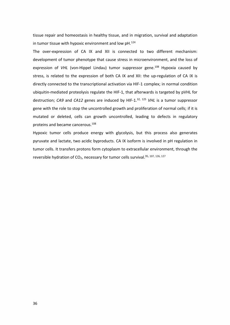
coumarins and psoralenes derivatives (Scheme 1) (Fig. 35).
The EMAC 10155 (a-m) and EMAC 10158 (a-m) series present no substituent in the
position 8 or 9 of the coumarin and psoralen nucleus respectively.
The EMAC 10156 (a-m) and EMAC 10159 (a-m) series are characterized by the presence of
a methylene carboxyl group in the position 3 of the chromene, that, as already reported,
might lead to the formation of a bidentate chelator of the Zn2+ ion in the catalytic pocket
of the enzymes.223
The EMAC 10157 (a-m) and EMAC 10160 (a-m) series, namely methyl-2-[4-methyl-2-oxo-
7–(2-oxo-2-arylethoxyi)-8-propylchromen-3-yl]acetate and 2–(5-methyl-7-oxo-3-aryl-9-
propyl-7H-furo[3,2-g]chromen-6-yl)acetic acid respectively, present a methylene carboxyl
group in position 3 of the chromene, and a propyl substituent in the position 8 of the
coumarin or in the position 9 of the psoralen nucleus.
Finally, the EMAC 10161 (b-m) and EMAC 10162 (b-m) series are characterized by the
presence of ethylene carboxyl group in position 3 of the chromene.
72
Figure 35 General structure of EMAC 10155, 10156, 10157, 10158, 10159, 10160, 10161, 10162
The synthesis of the series of EMAC 10156 (a-m), EMAC 10159 (a-m), EMAC 10157 (a-m),
EMAC 10160 (a-m), EMAC 10161 (b-m) and EMAC 10162 (b-m) is divided in three steps:
the first reaction is a Pechmann condensation performed in concentrated H2SO4 at room
temperature. In this step dimethylacetylsuccinate and either resorcinol or 2-
methylresorcinol were reacted; 96% H2SO4 was used as condensing agent and made
unnecessary the use of the solvent (Fig. 36).
Furocoumarin core Coumarin core
EMAC 10158
EMAC 10159
EMAC 10160
EMAC 10162
EMAC 10155
EMAC 10156
EMAC 10157
EMAC 10161

73
Figure 36 Example of Pechmann condensation mechanism
The second step of the synthetic pathway is a Williamson reaction between the phenolic
group in the position 7 of the chromene ring and an appropriate α-haloketone. The
reaction was performed in dry acetone and potassium carbonate as proton scavenger, to
generate the corresponding substituted oxoethers which were purified and further
reacted to give the formation of furocoumarins. More in detail, the asymmetric ethers
were refluxed in NaOH 1N water solution to form the furan ring by intramolecular
electrophilic substitution.
By heating in presence of NaOH (1M) the saponification of the ester groups in the position
3 of the chromene to the corresponding substituted 2-(5-methyl-7-oxo-7H-furo[3,2-
g]chromen6-yl)acetic acids was observed. The furan ring formation always led to linear
furocoumarins as confirmed by 1H-NMR spectra. The formation of the angular isomer was
only observed for compound 10162 d, as confirmed by 1H-NMR spectra (Fig.37).

74
Figure 37 Angular and linear furocoumarins
The synthetic pathway to EMAC 10155 (a-m) and EMAC 10158 (a-m) series consists of only
two steps as the Pechmann condensation step is no required.
All synthesized compounds were characterized by means of analytical and spectroscopic
methods.

75
Scheme 1 General synthesis of compound EMAC 10155 (a-m), 10156 (a-m), 10157(a-m), 10158 (a-m), 10159 (a-m), 10160 (a-m), 10161 (a-m), 10162 (b-m).

76
5.1.2 Biological results and discussion
Compounds EMAC10155 (a-m), EMAC10156 (a-m), EMAC10157(a-m), EMAC10158 (a-m),
EMAC10159 (a-m), EMAC10160 (a-m), EMAC10161 (a-m) and EMAC10162 (b-m) were
submitted to enzymatic evaluation towards hCA I, II, IX, and XII. The results are reported in
Table 3. None of these compounds exhibited any inhibition activity towards hCA I and II
isozymes. On the contrary, all of them are nanomolar inhibitors of the hCA tumor
associated isoforms IX and XII. This behavior agrees with our previous findings and with
the generally observed selectivity profile of coumarin derivatives.223
For what concerned hCA XII inhibition, derivates EMAC10159A, EMAC10159D,
EMAC10159L and EMAC10159M, that present a methyl group in position 4 of the
furocoumarin core, and respectively a 4-methyl phenyl, 4-fluoro phenyl, phenyl and 4-
chloro phenyl, as substituent in position 8 of the furocoumarin core, results as the most
potent inhibitor with Ki value of 9.1 nM, 7.4 nM, 9.4 nM and 9.3 nM, respectively.
Interestingly, this derivates, exhibited selectivity towards the isoform XII.
For isoform hCA IX, derivate EMAC10156M, that presents a methyl group in position 4 of
the coumarin core and a 4-chloro phenyl substituent bonded with the coumarin core, and
derivates EMAC10159A, EMAC10159D and EMAC10159L, are the most potent inhibitors,
with Ki value of 23.6 nM, 23.0 nM, 17.7 nM and 17.5 nM respectively. More specific, the
compound EMAC10156M, shows selectivity toward the tumor associate isoform IX.
The introduction of a methyl group in position 4 of the furocoumarin core, led to an
increase of the activity and selectivity toward the CA tumoral associate isoforms.
For what concern about the EMAC10157 and EMAC10160 derivatives, they are selective
toward CA IX and XII, but the introduction of a methyl group in position four and a propyl
group in position 9 of coumarin and furocoumarin core, led to a general decrease in the
activity.
EMAC10160 derivatives with 4-methyl, 4-fluoro and 4-chloro as substituent of the phenyl
ring bonded to the coumarin core, show increase of selectivity toward CA XII.
Unfortunately, COX inhibition evaluation for EMAC10155 (a-m), EMAC10156 (a-m),
EMAC10157(a-m), EMAC10158 (a-m), EMAC10159 (a-m), EMAC10160 (a-m), EMAC10161
(a-m) and EMAC10162 (b-m) is still in progress.

77
The data are extremely encouraging and push our group toward further investigation and
studies to better clarify and determine the structural determinant features for achieving
the best activity and selectivity towards the CA tumoral isoforms.

78
Table 3 Inhibition data towards hCA I, II, IX, and XII of compounds EMAC 10155(a-m), 10158 (a-m), 10156(a-m), 10159(a-m), 10157(a-m), 10160(a-m),
10161(a-m) and 10162(b-m)
KI (nM)*
Compound R R1 R2 R3 hCA I hCAII hCA IX hCA XII
EMAC10155A 4-CH3 H H H >10000 >10000 191.6 31.8
EMAC10155B 4-OCH3 H H H >10000 >10000 107.3 44.2
EMAC10155C 4-Br H H H >10000 >10000 91.4 47.2
EMAC10155D 4-F H H H >10000 >10000 130.7 46.9
EMAC10155G 4-Phenyl H H H >10000 >10000 126.8 43.4
EMAC10155H 2,4-F H H H >10000 >10000 144.3 56.2
EMAC10155L 4-H H H H >10000 >10000 97.5 80.5
EMAC10155M 4-Cl H H H >10000 >10000 97.6 77.5
EMAC10158A 4-CH3 H H H >10000 >10000 151.3 91.0
EMAC10158M 4-Cl H H H >10000 >10000 172.7 85.2

79
EMAC10156A** 4-CH3 CH3 CH2COOCH3 H >10000 >10000 122.8 56.6
EMAC10156B 4-OCH3 CH3 CH2COOCH3 H >10000 >10000 213.4 87.4
EMAC10156C 4-Br CH3 CH2COOCH3 H >10000 >10000 213.4 381.2
EMAC10156D** 4-F CH3 CH2COOCH3 H >10000 >10000 84.7 250.0
EMAC10156F 3-NO2 CH3 CH2COOCH3 H >10000 >10000 250.4 425.9
EMAC10156L** 4-H CH3 CH2COOCH3 H >10000 >10000 89.7 72.5
EMAC10156M** 4-Cl CH3 CH2COOCH3 H >10000 >10000 23.6 446.6
EMAC10159A** 4-CH3 CH3 CH2COOH H >10000 >10000 23.0 9.1
EMAC10159B 4-OCH3 CH3 CH2COOH H >10000 >10000 898.6 75.0
EMAC10159C 4-Br CH3 CH2COOH H >10000 >10000 291.2 83.9
EMAC10159D** 4-F CH3 CH2COOH H >10000 >10000 17.7 7.4
EMAC10159L** 4-H CH3 CH2COOH H >10000 >10000 17.5 9.4
EMAC10159M** 4-Cl CH3 CH2COOH H >10000 >10000 94.7 9.3
EMAC10157A*** 4-CH3 CH3 CH2COOCH3 CH2CH2CH3 >10,000 >10,000 247.7 350.3
EMAC10157B*** 4-OCH3 CH3 CH2COOCH3 CH2CH2CH3 >10,000 >10,000 352.7 324.2
EMAC10157D*** 4-F CH3 CH2COOCH3 CH2CH2CH3 >10,000 >10,000 239.5 257.1
EMAC10157F 3-NO2 CH3 CH2COOCH3 CH2CH2CH3 >10000 >10000 238.5 91.0
EMAC10157G*** 4-Phenyl CH3 CH2COOCH3 CH2CH2CH3 >10,000 >10,000 135.2 283.1

80
EMAC10157H 2,4-F CH3 CH2COOCH3 CH2CH2CH3 >10000 >10000 154.2 86.7
EMAC10157L 4-H CH3 CH2COOCH3 CH2CH2CH3 >10000 >10000 957.0 79.8
EMAC10157M 4-Cl CH3 CH2COOCH3 CH2CH2CH3 >10000 >10000 239.2 344.1
EMAC10160A*** 4-CH3 CH3 CH2COOH CH2CH2CH3 >10,000 >10,000 467.3 758.1
EMAC10160B*** 4-OCH3 CH3 CH2COOH CH2CH2CH3 >10,000 >10,000 489.3 859.4
EMAC10160D*** 4-F CH3 CH2COOH CH2CH2CH3 >10,000 >10,000 379.7 460.0
EMAC10160G*** 4-Phenyl CH3 CH2COOH CH2CH2CH3 >10,000 >10,000 397.7 550.0
EMAC10160L 4-H CH3 CH2COOH CH2CH2CH3 >10000 >10000 291.7 469.6
EMAC10160M 4-Cl CH3 CH2COOH CH2CH2CH3 >10000 >10000 310.5 481.4
EMAC10161A 4-CH3 CH3 CH2CH2COOCH3 H >10000 >10000 82.5 67.8
EMAC10161B 4-OCH3 CH3 CH2CH2COOCH3 H >10000 >10000 278.8 226.7
EMAC10161D 4-F CH3 CH2CH2COOCH3 H >10000 >10000 238.4 64.8
EMAC10161G 4-Phenyl CH3 CH2CH2COOCH3 H >10000 >10000 126.7 92.6
EMAC10161H 2,4-F CH3 CH2CH2COOCH3 H >10000 >10000 80.9 92.4
EMAC10161L 4-H CH3 CH2CH2COOCH3 H >10000 >10000 278.1 480.0
EMAC10161M 4-Cl CH3 CH2CH2COOCH3 H >10000 >10000 87.1 36.1
EMAC10162B 4-OCH3 CH3 CH2CH2COOH H >10000 >10000 232.8 79.9
EMAC10162D 4-F CH3 CH2CH2COOH H >10000 >10000 297.0 438.2

81
EMAC10162G 4-Phenyl CH3 CH2CH2COOH H >10000 >10000 892.3 82.9
EMAC10162L 4-H CH3 CH2CH2COOH H >10000 >10000 283.8 98.1
EMAC10162M 4-Cl CH3 CH2CH2COOH H >10000 >10000 261.6 295.1
AAZ 250.0 12.1 25.8 5.7
*Mean from 3 different assays, by a stopped flow technique (errors were in the range of ± 5-10 % of the reported values).
**Following previous work
***224

82
5.1.3 Conclusions
I have designed and synthesized four different series of coumarin derivates and four series
of psoralen derivates: EMAC10155 (a-m), EMAC10156 (a-m), EMAC10157(a-m),
EMAC10158 (a-m), EMAC10159 (a-m), EMAC10160 (a-m), EMAC10161 (a-m) and
EMAC10162 (b-m), to evaluate their activity and selectivity toward on hCA I, II, IX, and XII
isozymes, and to obtain information on their structure-activity relationships. All
compounds show selectivity toward the tumor associate isozymes IX and XII, in the
nanomolar range, and none of they were able to inhibit the off-target isoforms I and II,
confirming the literature reported chromene derivatives selectivity profile. According to
these results, psoralen and coumarins derivates could be considered as promising
scaffolds for design of highly selective inhibitors of the hCAs IX-XII isoforms. These results
are very encouraging and pushed us to further investigate these derivatives, in order to
identify potential candidates for the treatment of hypoxic tumors.

83
5.2 New 4-((4-oxo-2-phenylthiazolidin-3-yl)amino)benzenesulfonamide
synthesis and inhibitory activity toward carbonic anhydrase I, II, IX, XII
and the potential dual CA/COX-2 inhibition
5.2.1 Background
Tumors are a multifactorial disease, characterized by extremal condition caused by
different factors such as the deregulation of several enzymes’ activity. In this respect hCAs
IX and XII, and COX-2 were reported to be involved in the survival and progression of
tumor cells. On these bases, the idea to design and synthesize multi-target inhibitors
directed to both hCA and COX-2 is very attractive.
During my PhD, I have synthesized different series of potential multi-target agents to
investigate on the structural requisites for the selective inhibition of tumor overexpressed
hCAs and COX-2 enzymes. Identification of a common structural denominator between
hCA and COX-2 inhibitors was the starting point to design multi-target compounds. Indeed,
both inhibitors of the two enzyme families present a primary sulfonamide moiety in the
majority of the most effective agents. Celecoxib and valdecoxib (Fig. 24 e 25) already
demonstrated to have inhibitory activity towards both COX-2 and cancer related hCA
enzymes.
Therefore, a structural model has been studied with the aim to satisfy both enzyme
pharmacophoric requirements: the benzensulphonamide group turned out to be not only
efficient for the inhibition of COX-2 enzyme, but also capable to coordinate the hCAs zinc
cofactor in the catalytic site. On these bases, we have designed potential dual inhibitors
were the benzensulphonamide moiety, binds a differently substituted central heterocyclic
core through a linker. (Fig.38).

84
Figure 38 General structure of EMAC 10190 and EMAC 10191
According to this model, different series of 4-((4-oxo-2-phenylthiazolidin-3-
yl)amino)benzensulphonamide have been synthesized. All compounds are characterized by
the presence of a thiazole core, substituted in the position 2 with various aromatic groups
with different electronic and stearic features.
The synthesis of EMAC 10191 (b-l) is subdivided in three steps (Scheme 2); the second
step led to the formation of compounds, EMAC 10190 (b-l), which can be further reacted
to give derivatives EMAC 10191.
Scheme 2 Synthesis of derivates EMAC10190 (b-l) and EMAC10191 (b-l): (i.) Hydrazine, MW, 250 psi, 1h, H2O, 80°C, 1h; (ii.) benzaldehyde, acetic acid, MeOH, Δ; (iii.) mercaptoacetic acid, 60°C.
Essential fragment benzene
sulfonamide moiety
Thiazole
linker
Substituent with
different steric and
electronic characteristic
Hydrazin
e linker

85
To find the best synthetic pathway, trials were done by using different strategies. Among
these, very interestingly, a one-pot two-step synthetic procedure was proposed for the
preparation of the EMAC 10191 (b-l) derivatives.
All synthesized compounds were characterized by means of analytical and spectroscopic
methods.
5.2.2 Biological results and discussion
All the synthetized compounds EMAC10190 (b-k) and EMAC10191 (b-k) were submitted to
enzymatic assay to evaluate their activity and selectivity towards human CA isozymes I, II,
IX, XII. The results are illustrated in Table 4. Different activities towards CA isoforms were
observed, with the majority of the compounds showing Ki values between the high to the
low nanomolar range. In particular, most of the compounds exhibited interesting
inhibition Ki values towards CA IX. EMAC10190 D and EMAC10191 D, that present 4-
acethoxy-3-methoxy phenyl as substitution, are the most potent inhibitors towards the
tumoral isoform CA IX, with Ki value 5.3 nM and 7.5 nM respectively. Most of EMAC10191
derivatives (except for EMAC10191C and EMAC10191D) were selective against hCA II,
suggesting that the introduction of a thiazole spacer led to inhibition of CA II; these
derivatives can be used for a different therapeutic application.
Unfortunately, COX inhibition evaluation for EMAC10190 (b-k) and EMAC10191 (b-k) is
still in progress.

86
Table 4 Inhibition data towards hCA I, II, IX, and XII of compounds EMAC 10190 (b-l) and 10191(b-l)
KI (nM)*
Compound R hCA I hCAII hCA IX hCA XII
EMAC10190B 4-CH3 792.4 43.3 41.0 78.1
EMAC10190C 3,4,5-OCH3 5093 621.7 66.4 40.0
EMAC10190D 4-Acethoxy-3-OCH3 727.2 26.5 5.3 80.7
EMAC10190F 2-Naphtal 5400 81.9 208.7 54.7
EMAC10190G 1-Naphtal 17.5 626.2 92.1 67.7
EMAC10190H 4-OCH2CH3-3-OCH3 3365 826.0 270.8 74.7
EMAC10190J 4-OCH3 2210 287.8 69.7 268.7
EMAC10190K 4-F 441.3 73.9 41.8 82.2
EMAC10190L 4-Cl 83.4 8.5 9.6 154.2
EMAC10191B 4-CH3 42.5 6.5 7.3 325.7
EMAC10191C 3,4,5-OCH3 403.1 8.8 8.3 63.5
EMAC10191D 4-Acethoxy-3-OCH3 87.9 22.6 7.5 83.6
EMAC10191F 2-Naphtal 72.4 6.8 23.6 91.2
EMAC10191G 1-Naphtal 63.8 5.1 8.3 158.7
EMAC10191H 4-OCH2CH3-3-OCH3 703.8 3.9 8.7 65.7
EMAC10191J 4-OCH3 51.5 5.1 8.6 83.8
EMAC10191K 4-F 16.7 4.5 5.7 39.5
EMAC10191L 4-Cl 26.8 3.2 5.3 42.2
AAZ 250.0 12.1 25.8 5.7
* Mean from 3 different assays, by a stopped flow technique (errors were in the range of ± 5-10 % of the
reported values).

87
5.2.3 Conclusions
Two series of potential dual inhibitors have been synthetized, in order to simultaneously
obtain the block of a tumor associated inflammatory process and the mechanisms that the
tumor cell activates to survive in hypoxic conditions.
The substitution of the phenyl ring bonded to the hydrazine spacer or to the thiazole
spacer is determinant to the selectivity toward the CA tumoral isoform, in particular
toward the isoform IX.
Hopefully, more data will be available in the next future and will guide us to the design and
synthesis of new derivatives. For this reason, these compounds deserve further
exploration and might constitute the basis for future development.

88
5.3 Evaluation of drug like properties
5.3.1 Background
It is well recognized that potential ADMET problems should be addressed at the early
stages of drug discovery during the identification and optimization. This has been
recognized as a significant cause of high rates of attrition in drug discovery.
Nearly 40% of drugs under development fail to pass clinical trials due to poor ADME. The
ability to identify problematic candidates can dramatically reduce the amount of time and
resources wasted and streamline the overall development process.
Among the properties analyzed, those described by Lipinsky's rule are the best known.
Researcher Christopher Lipinsky in 1997 devised a simple algorithm that turns out to be
very important for the design and development of orally absorbed drugs. The rule,
properly called the “rule of 5 (Ro5)”, is based on five simple foundations:
•Molecular weight < 500
•AlogP < 5, a measure of lipophilicity
•Hydrogen-bond donors < 5 bonds
•Hydrogen-bond acceptor < 10 bonds
•Rotatable bonds < 10
This criterion is very useful in the chemical-pharmaceutical field to simplify and favor the
field of investigation, and thus drastically reduce the number of molecules among which a
candidate molecule could be explored as a potential drug.225
There has been significant effort in developing more comprehensive and quantitative rules
of drug-likeness.
To the properties described by Lipinsky, other properties have been added that define
important molecular characteristics for the different phases of absorption, distribution,
metabolism and excretion of drugs, such as: solubility,226 surface exposed to the solvent,
molecular volume, overcoming of the blood-brain barrier, permeability of Caco cells -2
(colorectal Carcinoma) and MDCK (Madin – Darby Canine Kidney), binding to albumin,
blocking the HERG K + (Human-a-go-go- Related Gene) channel,227 are some examples.4, 228,
229

89
Computational methods can help to predict, in silico, these properties and several
programs suites implemented tools that help the researcher to evaluate the drug-likeness
of compounds have been developed.
Qikprop predicts physically significant descriptors and pharmaceutically relevant
properties of organic molecules.225, 226 It runs in normal mode predicting 44 properties
(Tab. 5).
Table 5 QikProp properties and descriptors.
RANGE OR RECOMMENDED VALUES
MOLECULE NAME Molecule name taken from the title line in the input structure file. If the title line is blank, the input file name is used.
#STARS
Number of property or descriptor values that fall outside the 95% range of similar values for known drugs. Outlying descriptors and predicted properties are denoted with asterisks (*) in the .out file. A large number of stars suggests that a molecule is less drug-like than molecules with few stars. The following properties and descriptors are included in the determination of #stars: MW, dipole, IP, EA, SASA, FOSA, FISA, PISA, WPSA, PSA, volume, #rotor, donorHB, accptHB, glob, QPpolrz, QplogPC16, QplogPoct, QplogPw, QplogPo/w, logS, QPLogKhsa, QplogBB, #metabol
0 – 5
#AMINE Number of non-conjugated amine groups. 0 – 1
#AMIDINE Number of amidine and guanidine groups. 0
#ACID Number of carboxylic acid groups. 0 – 1
#AMIDE Number of non-conjugated amide groups. 0 – 1
#ROTOR Number of non-trivial (not CX3), non-hindered (not alkene, amide, small ring) rotatable bonds.
0 – 15
#RTVFG
Number of reactive functional groups; the specific groups are listed in the jobname .out file. The presence of these groups can lead to false positives in HTS assays and to decomposition, reactivity, or toxicity problems in vivo.
0 – 2
CNS Predicted central nervous system activity on a –2 (inactive) to +2 (active) scale.
–2 to +2
MOL_MW Molecular weight of the molecule. 130.0 – 725.0
DIPOLE† Computed dipole moment of the molecule. 1.0 – 12.5
SASA Total solvent accessible surface area (SASA) in square angstroms using a probe with a 1.4 Å radius.
300.0 – 1000.0
FOSA Hydrophobic component of the SASA (saturated carbon and attached hydrogen).
0.0 – 750.0
FISA Hydrophilic component of the SASA (SASA on N, O, and H on heteroatoms).
7.0 – 330.0

90
PISA π (carbon and attached hydrogen) component of the SASA. 0.0 – 450.0
WPSA Weakly polar component of the SASA (halogens, P, and S). 0.0 – 175.0
VOLUME Total solvent-accessible volume in cubic angstroms using a probe with a 1.4 Å radius.
500.0 – 2000.0
DONORHB
Estimated number of hydrogen bonds that would be donated by the solute to water molecules in an aqueous solution. Values are averages taken over a number of configurations, so they can be non-integer.
0.0 – 6.0
ACCPTHB
Estimated number of hydrogen bonds that would be accepted by the solute from water molecules in an aqueous solution. Values are averages taken over a number of configurations, so they can be non-integer.
2.0 – 20.0
DIP^2/V†
Square of the dipole moment divided by the molecular volume. This is the key term in the Kirkwood-Onsager equation for the free energy of solvation of a dipole with volume V.
0.0 – 0.13
ACXDN^.5/SA Index of cohesive interaction in solids. This term represents the relationship (accptHB)(donorHB))/(SA).
0.0 – 0.05
GLOB Globularity descriptor, (4 π r2 )/(SASA) , where r is the radius of a sphere with a volume equal to the molecular volume. Globularity is 1.0 for a spherical molecule.
0.75 – 0.95
QPPOLRZ Predicted polarizability in cubic angstroms. 13.0 – 70.0
QPLOGPC16 Predicted hexadecane/gas partition coefficient. 4.0 – 18.0
QPLOGPOCT‡ Predicted octanol/gas partition coefficient. 8.0 – 35.0
QPLOGPW Predicted water/gas partition coefficient. 4.0 – 45.0
QPLOGPO/W Predicted octanol/water partition coefficient. –2.0 – 6.5
QPLOGS Predicted aqueous solubility, log S. S in mol dm–3 is the concentration of the solute in a saturated solution that is in equilibrium with the crystalline solid.
–6.5 – 0.5
CIQPLOGS
Conformation-independent predicted aqueous solubility, log S. S in mol dm–3 is the concentration of the solute in a saturated solution that is in equilibrium with the crystalline solid.
–6.5 – 0.5
QPLOGHERG Predicted IC50 value for blockage of HERG K+ channels. Concern below –5
QPPCACO Predicted apparent Caco-2 cell permeability in nm/sec. Caco-2 cells are a model for the gut blood barrier. QikProp predictions are for non-active transport.
<25 poor, >500 great
QPLOGBB
Predicted brain/blood partition coefficient. Note: QikProp predictions are for orally delivered drugs so, for example, dopamine and serotonin are CNS negative because they are too polar to cross the blood-brain barrier
–3.0 – 1.2
QPPMDCK
Predicted apparent MDCK cell permeability in nm/sec. MDCK cells are considered to be a good mimic for the blood-brain barrier. QikProp predictions are for non-active transport.
<25 poor, >500 great
QPLOGKP Predicted skin permeability, log Kp. –8.0 – –1.0

91
IP(EV)† PM3 calculated ionization potential. 7.9 – 10.5
EA(EV)† PM3 calculated electron affinity. –0.9 – 1.7
#METAB‡ Number of likely metabolic reactions. 1 – 8
QPLOGKHSA Prediction of binding to human serum albumin. –1.5 – 1.5
HUMANORAL ABSORPTION
Predicted qualitative human oral absorption: 1, 2, or 3 for low, medium, or high. The text version is reported in the output. The assessment uses a knowledge-based set of rules, including checking for suitable values of PercentHumanOralAbsorption, number of metabolites, number of rotatable bonds, logP, solubility and cell permeability.
PERCENTHUMAN ORALABSORPTION
Predicted human oral absorption on 0 to 100% scale. The prediction is based on a quantitative multiple linear regression model. This property usually correlates well with HumanOralAbsorption, as both measure the same property.
80% is high <25% is poor
SAFLUORINE Solvent-accessible surface area of fluorine atoms. 0.0 – 100.0
SAAMIDEO Solvent-accessible surface area of amide oxygen atoms. 0.0 – 35.0
PSA Van der Waals surface area of polar nitrogen and oxygen atoms.
7.0 – 200.0
#NANDO Number of nitrogen and oxygen atoms. 2 – 15
The range is valid for 95% of known drugs.
Such an approach provides molecular properties employing QSAR calculation or similarity
validated approaches.230 The comparison of the predicted value for the novel compounds
with respect to the 95% of known drugs help to estimate their drug-likeness.
Compounds that fall within the defined ranges are described as ‘drug-like’.
5.3.1 Biological results and discussion
Hence, to estimate the drug-likeness of the compounds, we carried out in silico ADMET
prediction.231 In particular, here we report a selection of the predicted properties, the
most significant investigated using the Qikprop232 software for all the series synthesized.
The analysis of the predicted properties reveals the good drug-likeness of the compounds.
However, the enormous quantity of data collected does not allow to enter all the
properties values calculated.
For each series of compounds two tables are reported containing:
- Structural properties under Lipinski’s rule five (RO5) and other physicochemical
properties;

92
- Other properties that could affect the pharmacokinetics and the safety of the
compounds.
As it is shown in all the tables, most of the predicted properties are within the ranges
specified with few exceptions.
Table 6 Predicted properties for the series EMAC10155
Molecule #stars MW HBD HBA QplogPo/w rot PSA QplogS
0 – 5 130 – 725 0-6 2- 20 -2- 6.5 0 – 15 7 – 200 -6.5 – 0.5
EMAC10155A 0 294,3 0 4 2,6 4 75,8 -3,5
EMAC10155B 0 310,3 0 5 2,3 5 84,0 -3,1
EMAC10155C 0 359,2 0 4 2,8 4 75,8 -3,8
EMAC10155D 0 298,3 0 4 2,5 4 75,8 -3,3
EMAC10155E 1 325,3 0 5 1,5 5 120,8 -3,0
EMAC10155F 2 325,3 0 5 1,5 5 120,7 -3,0
EMAC10155G 1 356,4 0 4 3,9 5 75,8 -5,1
EMAC10155H 0 316,3 0 4 2,6 4 75,7 -3,5
EMAC10155I 0 349,2 0 4 3,1 4 74,5 -4,3
EMAC10155L 0 280,3 0 4 2,3 4 75,8 -2,9
EMAC10155M 0 314,7 0 4 2,8 4 75,8 -3,7
Table 7 Predicted properties for the series EMAC10156
Molecule #stars MW HBD HBA QplogPo/w #rotor PSA QplogS
0 – 5 130 – 725 0-6 2- 20 -2- 6.5 0 – 15 7 – 200 -6.5 – 0.5
EMAC10156A 0 380,4 0 6 3,0 6 107,2 -4,8
EMAC10156B 0 396,4 0 7 2,8 7 115,5 -4,3
EMAC10156C 0 445,3 0 6 3,3 6 107,2 -5,0
EMAC10156D 0 384,4 0 6 3,0 6 107,2 -4,5
EMAC10156E 1 411,4 0 7 2,0 7 152,2 -4,2
EMAC10156F 2 411,4 0 7 2,0 7 152,1 -4,2
EMAC10156G 0 442,5 0 6 4,4 7 107,2 -6,3
EMAC10156H 0 402,4 0 6 3,1 6 107,1 -4,7
EMAC10156I 0 435,3 0 6 3,7 6 105,9 -5,5
EMAC10156L 0 366,4 0 6 2,7 6 107,2 -4,1

93
EMAC10156M 0 400,8 0 6 3,2 6 107,2 -4,9
Table 8 Predicted properties for the series EMAC10157
Molecule #stars MW HBD HBA QplogPo/w #rotor PSA QplogS
0 – 5 130 – 725 0-6 2- 20 -2- 6.5 0 – 15 7 – 200 -6.5 – 0.5
EMAC10157A 0 422,5 0 6 4,4 8 104,3 -6,4
EMAC10157B 0 438,5 0 7 4,0 9 112,5 -5,5
EMAC10157C 0 487,3 0 6 4,5 8 104,2 -6,3
EMAC10157D 0 426,4 0 6 4,2 8 104,2 -5,8
EMAC10157E 1 453,4 0 7 3,2 9 149,2 -5,5
EMAC10157F 2 453,4 0 7 3,2 9 149,2 -5,4
EMAC10157G 2 484,5 0 6 5,6 9 104,3 -7,5
EMAC10157H 0 444,4 0 6 4,3 8 104,1 -5,9
EMAC10157I 0 477,3 0 6 4,3 8 102,0 -5,5
EMAC10157L 0 408,5 0 6 3,9 8 104,2 -5,4
EMAC10157M 0 442,9 0 6 4,4 8 104,2 -6,2
Table 9 Predicted properties for the series EMAC10158
Molecule #stars MW HBD HBA QPlogPo/w #rotor PSA QPlogS
0 - 5 130 – 725 0-6 2- 20 -2- 6.5 0 - 15 7 - 200.0 -6.5 - 0.5
EMAC10158A 0 276,3 0 3 3,4 0 49,3 -4,2
EMAC10158B 0 292,3 0 4 3,1 1 57,6 -3,7
EMAC10158C 0 341,2 0 3 3,7 0 49,3 -4,5
EMAC10158D 0 280,3 0 3 3,3 0 49,3 -3,9
EMAC10158E 0 307,3 0 4 2,3 1 94,3 -3,6
EMAC10158F 0 307,3 0 4 2,3 1 94,3 -3,6
EMAC10158G 1 338,4 0 3 4,8 1 49,3 -5,7
EMAC10158H 0 298,2 0 3 3,5 0 49,3 -4,1
EMAC10158I 0 331,2 0 3 4,0 0 49,3 -4,7
EMAC10158L 0 262,3 0 3 3,1 0 49,3 -3,6
EMAC10158M 0 296,7 0 3 3,6 0 49,3 -4,3
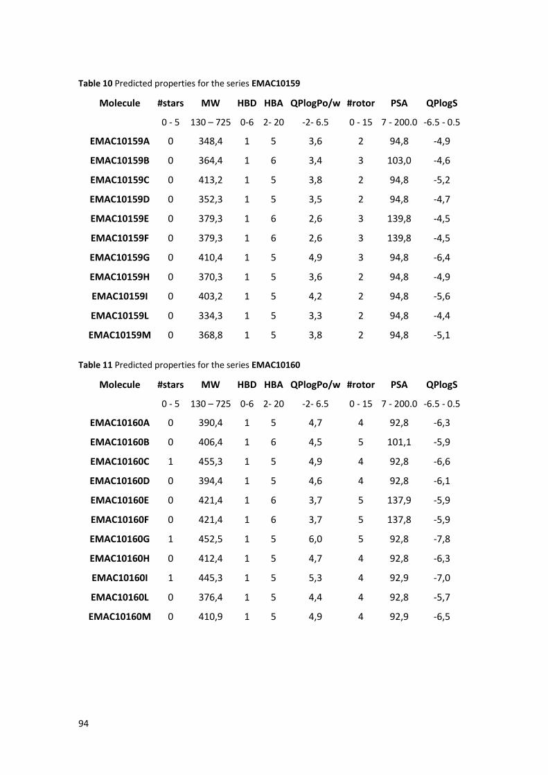
94
Table 10 Predicted properties for the series EMAC10159
Molecule #stars MW HBD HBA QPlogPo/w #rotor PSA QPlogS
0 - 5 130 – 725 0-6 2- 20 -2- 6.5 0 - 15 7 - 200.0 -6.5 - 0.5
EMAC10159A 0 348,4 1 5 3,6 2 94,8 -4,9
EMAC10159B 0 364,4 1 6 3,4 3 103,0 -4,6
EMAC10159C 0 413,2 1 5 3,8 2 94,8 -5,2
EMAC10159D 0 352,3 1 5 3,5 2 94,8 -4,7
EMAC10159E 0 379,3 1 6 2,6 3 139,8 -4,5
EMAC10159F 0 379,3 1 6 2,6 3 139,8 -4,5
EMAC10159G 0 410,4 1 5 4,9 3 94,8 -6,4
EMAC10159H 0 370,3 1 5 3,6 2 94,8 -4,9
EMAC10159I 0 403,2 1 5 4,2 2 94,8 -5,6
EMAC10159L 0 334,3 1 5 3,3 2 94,8 -4,4
EMAC10159M 0 368,8 1 5 3,8 2 94,8 -5,1
Table 11 Predicted properties for the series EMAC10160
Molecule #stars MW HBD HBA QPlogPo/w #rotor PSA QPlogS
0 - 5 130 – 725 0-6 2- 20 -2- 6.5 0 - 15 7 - 200.0 -6.5 - 0.5
EMAC10160A 0 390,4 1 5 4,7 4 92,8 -6,3
EMAC10160B 0 406,4 1 6 4,5 5 101,1 -5,9
EMAC10160C 1 455,3 1 5 4,9 4 92,8 -6,6
EMAC10160D 0 394,4 1 5 4,6 4 92,8 -6,1
EMAC10160E 0 421,4 1 6 3,7 5 137,9 -5,9
EMAC10160F 0 421,4 1 6 3,7 5 137,8 -5,9
EMAC10160G 1 452,5 1 5 6,0 5 92,8 -7,8
EMAC10160H 0 412,4 1 5 4,7 4 92,8 -6,3
EMAC10160I 1 445,3 1 5 5,3 4 92,9 -7,0
EMAC10160L 0 376,4 1 5 4,4 4 92,8 -5,7
EMAC10160M 0 410,9 1 5 4,9 4 92,9 -6,5

95
Table 12 Predicted properties for the series EMAC10161
Molecule #stars MW HBD HBA QPlogPo/w #rotor PSA QPlogS
0 - 5 130 – 725 0-6 2- 20 -2- 6.5 0 - 15 7 - 200.0 -6.5 - 0.5
EMAC10161A 0 408,5 0 6 3,8 8 108,1 -5,6
EMAC10161B 0 424,4 0 7 3,5 9 116,4 -5,2
EMAC10161C 2 473,3 0 6 4,2 8 108,1 -6,7
EMAC10161D 0 412,4 0 6 3,7 8 108,1 -5,4
EMAC10161E 1 439,4 0 7 2,7 9 153,1 -5,1
EMAC10161F 2 439,4 0 7 2,8 9 152,9 -5,8
EMAC10161G 2 470,5 0 6 5,1 9 108,1 -7,2
EMAC10161H 0 430,4 0 6 3,9 8 107,2 -5,7
EMAC10161I 0 463,3 0 6 4,4 8 106,8 -6,3
EMAC10161L 0 394,4 0 6 3,4 8 108,1 -5,0
EMAC10161M 0 428,9 0 6 4,0 8 108,1 -5,8
Table 13 Predicted properties for the series EMAC10162
Molecule #stars MW HBD HBA QPlogPo/w #rotor PSA QPlogS
0 - 5 130 – 725 0-6 2- 20 -2- 6.5 0 - 15 7 - 200.0 -6.5 - 0.5
EMAC10162A 0 362,4 1 5 3,9 3 97,2 -5,3
EMAC10162B 0 378,4 1 6 3,6 4 105,5 -4,9
EMAC10162C 0 427,3 1 5 4,1 3 97,2 -5,6
EMAC10162D 0 366,3 1 5 3,8 3 97,2 -5,1
EMAC10162E 0 393,4 1 6 2,8 4 142,2 -4,9
EMAC10162F 0 393,4 1 6 2,8 4 142,2 -4,9
EMAC10162G 1 424,5 1 5 5,2 4 97,2 -6,7
EMAC10162H 0 384,3 1 5 3,9 3 97,2 -5,3
EMAC10162I 0 417,2 1 5 4,4 3 97,2 -6,0
EMAC10162L 0 348,4 1 5 3,5 3 97,2 -4,7
EMAC10162M 0 382,8 1 5 4,0 3 97,2 -5,5

96
Table 14 Predicted properties for the series EMAC10190
Molecule #stars MW HBD HBA QPlogPo/w #rotor PSA QPlogS
0 - 5 130 – 725 0-6 2- 20 -2- 6.5 0 - 15 7 - 200.0 -6.5 - 0.5
EMAC10190aE 0 318,4 3 7 1,5 7 90,8 -3,5
EMAC10190aZ 1 318,4 3 7 1,5 7 89,7 -3,5
EMAC10190bZ 0 289,4 3 6 1,4 6 86,4 -3,2
EMAC10190bE 0 289,4 3 6 1,4 6 87,4 -3,2
EMAC10190cZ 0 365,4 3 8 1,3 9 109,8 -3,2
EMAC10190cE 0 365,4 3 8 1,4 9 110,7 -3,4
EMAC10190dZ 0 363,4 3 9 0,5 8 133,3 -3,0
EMAC10190dE 0 363,4 3 9 0,5 8 134,3 -3,2
EMAC10190eZ 0 341,4 4 7 1,3 7 104,2 -3,3
EMAC10190eE 0 341,4 4 7 1,3 7 105,0 -3,3
EMAC10190fZ 0 325,4 3 6 1,5 6 86,4 -3,8
EMAC10190fE 0 325,4 3 6 1,5 6 87,4 -3,8
EMAC10190gZ 0 325,4 3 6 1,6 6 86,7 -4,3
EMAC10190gE 0 325,4 3 6 1,5 6 87,0 -3,7
EMAC10190hZ 0 349,4 3 8 1,6 9 101,4 -3,5
EMAC10190hE 0 349,4 3 8 1,6 9 102,4 -3,8
EMAC10190iZ 0 311,3 3 6 1,5 6 86,3 -3,3
EMAC10190iE 0 311,3 3 6 1,5 6 87,4 -3,3
EMAC10190jZ 0 305,4 3 7 1,1 7 94,7 -2,9
EMAC10190jE 0 305,4 3 7 1,1 7 95,7 -3,0
EMAC10190kZ 0 293,3 3 6 1,3 6 86,4 -3,0
EMAC10190kE 0 293,3 3 6 1,3 6 87,4 -3,1
EMAC10190lZ 0 309,8 3 6 1,5 6 86,4 -3,4
EMAC10190lE 0 309,8 3 6 1,5 6 87,4 -3,4
EMAC10190mZ 0 265,3 3 7 0,5 6 93,5 -2,2
EMAC10190mE 0 265,3 3 7 0,5 6 97,1 -2,2
Table 15 Predicted properties for the series EMAC10191
Molecule #stars MW HBD HBA QPlogPo/w #rotor PSA QPlogS

97
0 - 5 130 – 725 0-6 2- 20 -2- 6.5 0 - 15 7 - 200.0 -6.5 - 0.5
EMAC10191aR 0 392,5 3 9 1,6 5 108,1 -4,5
EMAC10191bS 0 363,4 3 8 1,5 4 104,7 -4,3
EMAC10191bR 0 363,4 3 8 1,5 4 104,6 -4,2
EMAC10191cS 0 439,5 3 10 1,3 7 128,8 -2,9
EMAC10191cR 0 439,5 3 10 1,3 7 129,1 -2,8
EMAC10191dS 0 437,5 3 11 0,4 6 152,1 -2,8
EMAC10191dR 0 437,5 3 11 0,4 6 152,1 -2,7
EMAC10191eS 0 415,5 4 8 1,5 5 119,9 -4,2
EMAC10191eR 0 415,5 4 8 1,5 5 119,9 -4,2
EMAC10191fS 0 399,5 3 8 2,1 4 104,6 -4,9
EMAC10191fR 0 399,5 3 8 2,1 4 104,7 -4,8
EMAC10191gS 0 399,5 3 8 2,0 4 104,4 -4,6
EMAC10191gR 0 399,5 3 8 2,0 4 104,2 -4,6
EMAC10191hS 0 423,5 3 9 1,8 7 119,8 -4,7
EMAC10191hR 0 423,5 3 9 1,8 7 119,7 -4,7
EMAC10191iS 0 385,4 3 8 1,6 4 104,7 -4,2
EMAC10191iR 0 385,4 3 8 1,6 4 104,7 -4,3
EMAC10191jS 0 379,4 3 8 1,3 5 112,9 -4,0
EMAC10191jR 0 379,4 3 8 1,3 5 112,9 -3,9
EMAC10191kS 0 367,4 3 8 1,4 4 104,6 -4,1
EMAC10191kR 0 367,4 3 8 1,4 4 104,6 -4,0
EMAC10191lS 0 383,9 3 8 1,7 4 104,7 -4,4
EMAC10191lR 0 383,9 3 8 1,7 4 104,7 -4,4
EMAC10191mS 0 339,4 3 8 0,7 4 113,3 -3,1
EMAC10191mR 0 339,4 3 8 0,7 4 113,3 -3,6
Table 16 Ranges of pharmacokinetics properties calculated considering 95% of drugs
QPPCaco < 25 poor, >500 great
QPPMDCK < 25 poor, >500 great
PercentHumanOralAbsorption
(%OA)
> 80 % is high < 25 % is
poor

98
QPlogHERG >-5
CNS - 2 (inactive) to +2 (active)
QPlogBB -3.0 - 1.2
QPlogKhsa -1.5 - 1.5

99
Table 17 Predicted pharmacokinetics properties for the series EMAC10155
Molecule QPPCaco QPPMDCK %OA QPlogHERG CNS QPlogBB QPlogKhsa
< 25 poor,
>500 great
< 25 poor,
>500 great
> 80 % is high,
< 25 % is poor >-5
- 2 (inactive) to
+2 (active) -3.0 - 1.2 -1.5 - 1.5
EMAC10155A 752,5 363,8 93,5 -5,9 -1 -0,8 -0,2
EMAC10155B 752,4 363,7 92,1 -5,8 -1 -0,9 -0,4
EMAC10155C 752,9 962,4 95,0 -5,9 0 -0,6 -0,2
EMAC10155D 753,2 653,8 93,0 -5,8 0 -0,7 -0,3
EMAC10155E 89,7 36,5 70,7 -5,9 -2 -1,8 -0,4
EMAC10155F 89,7 36,5 70,7 -5,9 -2 -1,8 -0,4
EMAC10155G 752,3 363,7 100,0 -7,2 -1 -1,0 0,3
EMAC10155H 758,6 878,0 93,9 -5,7 0 -0,6 -0,3
EMAC10155I 826,4 1928,2 100,0 -5,9 0 -0,5 -0,1
EMAC10155L 752,6 363,8 91,6 -5,9 -1 -0,8 -0,3
EMAC10155M 753,0 896,6 94,6 -5,8 0 -0,6 -0,2
Table 18 Predicted pharmacokinetics properties for the series EMAC10156
Molecule QPPCaco QPPMDCK %OA QPlogHERG CNS QPlogBB QPlogKhsa
< 25 poor,
>500 great
< 25 poor,
>500 great
> 80 % is high,
< 25 % is poor >-5
- 2 (inactive) to
+2 (active) -3.0 - 1.2 -1.5 - 1.5

100
EMAC10156A 439,1 203,2 92,1 -6,2 -2 -1,3 0,0
EMAC10156B 443,0 205,2 90,6 -6,1 -2 -1,4 -0,2
EMAC10156C 439,2 537,4 93,6 -6,2 -2 -1,1 -0,1
EMAC10156D 439,5 365,2 91,5 -6,1 -2 -1,2 -0,2
EMAC10156E 52,1 20,3 69,2 -6,2 -2 -2,5 -0,3
EMAC10156F 52,4 20,4 69,3 -6,2 -2 -2,5 -0,3
EMAC10156G 438,7 203,1 100,0 -7,4 -2 -1,5 0,4
EMAC10156H 446,2 494,9 92,4 -6,0 -2 -1,1 -0,1
EMAC10156I 483,1 1079,6 96,5 -6,1 -1 -1,0 0,0
EMAC10156L 443,2 205,3 90,2 -6,3 -2 -1,3 -0,2
EMAC10156M 439,2 500,6 93,1 -6,2 -2 -1,1 -0,1
Table 19 Predicted pharmacokinetics properties for the series EMAC10157
Molecule QPPCaco QPPMDCK %OA QPlogHERG CNS QPlogBB QPlogKhsa
< 25 poor,
>500 great
< 25 poor,
>500 great
> 80 % is high,
< 25 % is poor >-5
- 2 (inactive) to
+2 (active) -3.0 - 1.2 -1.5 - 1.5
EMAC10157A 631,9 301,2 100,0 -6,6 -2 -1,4 0,4
EMAC10157B 628,1 299,3 100,0 -6,2 -2 -1,4 0,2
EMAC10157C 627,7 790,6 100,0 -6,3 -2 -1,1 0,4
EMAC10157D 631,3 540,2 100,0 -6,2 -2 -1,2 0,3

101
EMAC10157E 74,9 30,1 79,1 -6,3 -2 -2,5 0,1
EMAC10157F 75,3 30,2 79,1 -6,3 -2 -2,5 0,1
EMAC10157G 631,9 301,2 96,9 -7,5 -2 -1,5 0,9
EMAC10157H 635,7 726,0 100,0 -6,1 -2 -1,1 0,3
EMAC10157I 518,3 967,0 100,0 -5,4 -2 -1,0 0,3
EMAC10157L 629,0 299,7 100,0 -6,4 -2 -1,3 0,2
EMAC10157M 631,4 741,1 100,0 -6,3 -2 -1,2 0,3
Table 20 Predicted pharmacokinetics properties for the series EMAC10158
Molecule QPPCaco QPPMDCK %OA QPlogHERG CNS QPlogBB QPlogKhsa
< 25 poor,
>500 great
< 25 poor,
>500 great
> 80 % is high,
< 25 % is poor >-5
- 2 (inactive) to
+2 (active) -3.0 - 1.2 -1.5 - 1.5
EMAC10158a 214,2 112,7 100,0 -5,2 0 -0,1 0,3
EMAC10158b 214,2 112,7 100,0 -5,2 0 -0,1 0,1
EMAC10158c 214,2 298,0 100,0 -5,3 1 0,1 0,3
EMAC10158d 214,2 202,4 100,0 -5,1 1 0,1 0,2
EMAC10158e 25,4 11,3 83,7 -5,3 -1 -1,0 0,1
EMAC10158f 26,2 11,6 83,9 -5,2 -1 -1,0 0,1
EMAC10158g 214,3 112,7 100,0 -6,7 0 -0,2 0,8
EMAC10158h 214,3 290,3 100,0 -5,0 1 0,2 0,2

102
EMAC10158i 214,3 545,9 100,0 -5,1 1 0,3 0,4
EMAC10158l 214,2 112,7 100,0 -5,2 0 0,0 0,2
EMAC10158m 214,2 277,1 100,0 -5,2 1 0,1 0,3
Table 21 Predicted pharmacokinetics properties for the series EMAC10159
Molecule QPPCaco QPPMDCK %OA QPlogHERG CNS QPlogBB QPlogKhsa
< 25 poor,
>500 great
< 25 poor,
>500 great
> 80 % is high,
< 25 % is poor >-5
- 2 (inactive) to
+2 (active) -3.0 - 1.2 -1.5 - 1.5
EMAC10159a 100,3 52,4 83,7 -3,5 -1 -0,9 0,3
EMAC10159b 100,3 52,4 82,4 -3,5 -1 -1,0 0,1
EMAC10159c 100,3 138,5 85,2 -3,5 -1 -0,7 0,2
EMAC10159d 100,3 94,0 83,2 -3,4 -1 -0,8 0,1
EMAC10159e 11,9 5,2 61,2 -3,5 -2 -1,9 0,1
EMAC10159f 11,9 5,2 61,2 -3,5 -2 -1,9 0,1
EMAC10159g 100,2 52,4 91,4 -4,9 -2 -1,1 0,7
EMAC10159h 100,3 129,2 84,0 -3,3 -1 -0,7 0,2
EMAC10159i 100,4 244,9 87,2 -3,4 -1 -0,6 0,3
EMAC10159l 100,3 52,4 81,9 -3,5 -1 -0,9 0,1
EMAC10159m 100,2 128,9 84,8 -3,5 -1 -0,8 0,2

103
Table 22 Predicted pharmacokinetics properties for the series EMAC10160
Molecule QPPCaco QPPMDCK %OA QPlogHERG CNS QPlogBB QPlogKhsa
< 25 poor,
>500 great
< 25 poor,
>500 great
> 80 % is high,
< 25 % is poor >-5
- 2 (inactive) to
+2 (active) -3.0 - 1.2 -1.5 - 1.5
EMAC10160a 111,7 58,9 91,1 -3,8 -2 -1,1 0,7
EMAC10160b 111,7 58,9 89,8 -3,8 -2 -1,2 0,5
EMAC10160c 111,7 155,6 92,5 -3,9 -1 -0,9 0,6
EMAC10160d 111,7 105,7 90,6 -3,8 -1 -1,0 0,5
EMAC10160e 13,3 5,9 68,5 -3,9 -2 -2,2 0,4
EMAC10160f 13,3 5,9 68,5 -3,9 -2 -2,2 0,4
EMAC10160g 111,7 58,9 85,9 -5,1 -2 -1,3 1,1
EMAC10160h 111,7 145,2 91,3 -3,7 -1 -0,9 0,6
EMAC10160i 111,5 277,1 81,6 -3,7 -1 -0,8 0,7
EMAC10160l 111,6 58,8 89,2 -3,9 -2 -1,1 0,5
EMAC10160m 111,6 144,8 92,1 -3,8 -1 -1,0 0,6
Table 23 Predicted pharmacokinetics properties for the series EMAC10161
Molecule QPPCaco QPPMDCK %OA QPlogHERG CNS QPlogBB QPlogKhsa
< 25 poor,
>500 great
< 25 poor,
>500 great
> 80 % is high,
< 25 % is poor >-5
- 2 (inactive) to
+2 (active) -3.0 - 1.2 -1.5 - 1.5

104
EMAC10161A 383,9 175,8 95,3 -6,5 -2,0 -1,6 0,2
EMAC10161B 380,9 174,3 93,6 -6,4 -2,0 -1,7 0,03
EMAC10161C 247,2 288,8 94,1 -6,9 -2,0 -1,8 0,3
EMAC10161D 383,6 315,3 94,7 -6,4 -2,0 -1,5 0,1
EMAC10161E 45,6 17,6 72,4 -6,5 -2,0 -2,8 -0,1
EMAC10161F 31,7 11,9 70,5 -6,8 -2,0 -3,1 0,1
EMAC10161G 383,3 175,5 90,3 -7,7 -2,0 -1,8 0,7
EMAC10161H 412,8 534,8 96,6 -6,3 -2,0 -1,3 0,1
EMAC10161I 404,4 860,9 100,0 -6,3 -2,0 -1,3 0,3
EMAC10161L 381,4 174,6 93,3 -6,6 -2,0 -1,6 0,1
EMAC10161M 381,2 429,5 96,3 -6,5 -2,0 -1,4 0,2
Table 24 Predicted pharmacokinetics properties for the series EMAC10162
Molecule QPPCaco QPPMDCK %OA QPlogHERG CNS QPlogBB QPlogKhsa
< 25 poor,
>500 great
< 25 poor,
>500 great
> 80 % is high,
< 25 % is poor >-5
- 2 (inactive) to
+2 (active) -3.0 - 1.2 -1.5 - 1.5
EMAC10162A 75,4 38,5 83,1 -3,7 -2,0 -1,2 0,4
EMAC10162B 75,4 38,5 81,8 -3,7 -2,0 -1,2 0,2
EMAC10162C 75,4 101,7 84,6 -3,7 -1,0 -0,9 0,3
EMAC10162D 75,4 69,1 82,6 -3,6 -2,0 -1,0 0,2
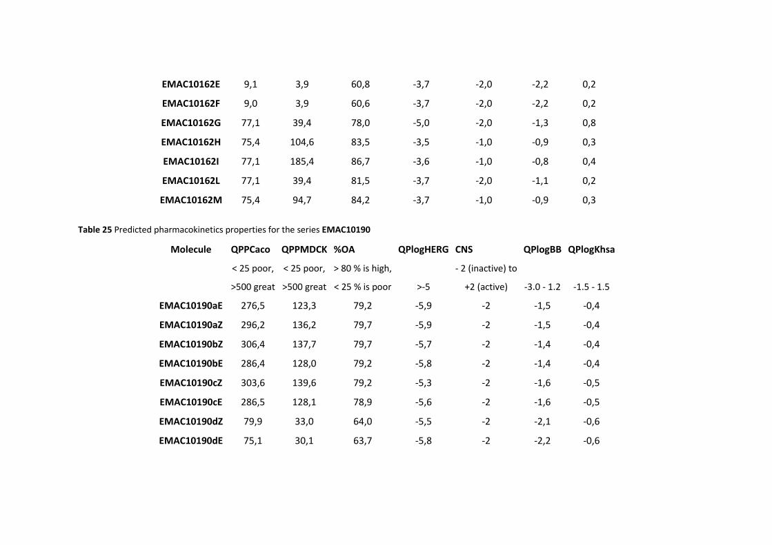
105
EMAC10162E 9,1 3,9 60,8 -3,7 -2,0 -2,2 0,2
EMAC10162F 9,0 3,9 60,6 -3,7 -2,0 -2,2 0,2
EMAC10162G 77,1 39,4 78,0 -5,0 -2,0 -1,3 0,8
EMAC10162H 75,4 104,6 83,5 -3,5 -1,0 -0,9 0,3
EMAC10162I 77,1 185,4 86,7 -3,6 -1,0 -0,8 0,4
EMAC10162L 77,1 39,4 81,5 -3,7 -2,0 -1,1 0,2
EMAC10162M 75,4 94,7 84,2 -3,7 -1,0 -0,9 0,3
Table 25 Predicted pharmacokinetics properties for the series EMAC10190
Molecule QPPCaco QPPMDCK %OA QPlogHERG CNS QPlogBB QPlogKhsa
< 25 poor,
>500 great
< 25 poor,
>500 great
> 80 % is high,
< 25 % is poor >-5
- 2 (inactive) to
+2 (active) -3.0 - 1.2 -1.5 - 1.5
EMAC10190aE 276,5 123,3 79,2 -5,9 -2 -1,5 -0,4
EMAC10190aZ 296,2 136,2 79,7 -5,9 -2 -1,5 -0,4
EMAC10190bZ 306,4 137,7 79,7 -5,7 -2 -1,4 -0,4
EMAC10190bE 286,4 128,0 79,2 -5,8 -2 -1,4 -0,4
EMAC10190cZ 303,6 139,6 79,2 -5,3 -2 -1,6 -0,5
EMAC10190cE 286,5 128,1 78,9 -5,6 -2 -1,6 -0,5
EMAC10190dZ 79,9 33,0 64,0 -5,5 -2 -2,1 -0,6
EMAC10190dE 75,1 30,1 63,7 -5,8 -2 -2,2 -0,6

106
EMAC10190eZ 162,1 70,8 74,1 -6,2 -2 -1,7 -0,4
EMAC10190eE 150,1 63,7 73,5 -6,3 -2 -1,8 -0,4
EMAC10190fZ 306,1 140,8 80,3 -6,5 -2 -1,4 -0,2
EMAC10190fE 286,3 128,0 79,8 -6,5 -2 -1,4 -0,2
EMAC10190gZ 193,2 85,6 77,0 -7,0 -2 -1,7 -0,2
EMAC10190gE 288,1 128,9 79,8 -6,5 -2 -1,4 -0,2
EMAC10190hZ 302,1 135,7 80,7 -5,8 -2 -1,6 -0,4
EMAC10190hE 286,1 127,9 80,5 -6,1 -2 -1,7 -0,4
EMAC10190iZ 303,6 395,2 80,0 -5,6 -2 -1,1 -0,5
EMAC10190iE 286,5 369,3 79,6 -5,6 -2 -1,2 -0,5
EMAC10190jZ 304,2 139,8 78,0 -5,7 -2 -1,4 -0,5
EMAC10190jE 285,7 130,7 77,6 -5,8 -2 -1,5 -0,5
EMAC10190kZ 306,0 246,9 78,8 -5,6 -2 -1,2 -0,5
EMAC10190kE 287,5 236,1 78,4 -5,8 -2 -1,3 -0,5
EMAC10190lZ 305,8 346,2 80,2 -5,8 -2 -1,2 -0,4
EMAC10190lE 285,4 314,0 79,7 -5,8 -2 -1,2 -0,4
EMAC10190mZ 290,4 133,0 74,0 -5,5 -2 -1,3 -0,7
EMAC10190mE 280,5 128,1 73,5 -5,4 -2 -1,3 -0,7

107
Table 26 Predicted pharmacokinetics properties for the series EMAC10191
Molecule QPPCaco QPPMDCK %OA QPlogHERG CNS QPlogBB QPlogKhsa
< 25 poor,
>500 great
< 25 poor,
>500 great
> 80 % is high,
< 25 % is poor >-5
- 2 (inactive) to
+2 (active) -3.0 - 1.2 -1.5 - 1.5
EMAC10191aR 155,2 115,2 75,5 -5,8 -2 -1,6 -0,2
EMAC10191bS 160,1 119,3 75,1 -5,7 -2 -1,5 -0,2
EMAC10191bR 160,3 116,8 75,1 -5,6 -2 -1,5 -0,2
EMAC10191cS 233,1 179,0 76,7 -3,7 -2 -1,2 -0,4
EMAC10191cR 225,1 168,3 76,4 -3,5 -2 -1,2 -0,4
EMAC10191dS 59,4 40,1 61,3 -3,9 -2 -1,6 -0,5
EMAC10191dR 59,4 39,1 61,2 -3,9 -2 -1,6 -0,5
EMAC10191eS 103,7 72,4 72,0 -6,0 -2 -1,7 -0,2
EMAC10191eR 103,9 72,6 72,0 -6,0 -2 -1,7 -0,2
EMAC10191fS 160,4 119,0 78,7 -6,4 -2 -1,5 -0,1
EMAC10191fR 160,3 116,7 78,6 -6,4 -2 -1,5 0,0
EMAC10191gS 163,2 111,7 78,3 -6,2 -2 -1,4 -0,1
EMAC10191gR 162,2 113,9 78,4 -6,3 -2 -1,5 -0,1
EMAC10191hS 160,1 116,5 76,9 -5,9 -2 -1,8 -0,2
EMAC10191hR 160,2 116,1 76,9 -5,9 -2 -1,8 -0,2
EMAC10191iS 160,4 324,9 75,9 -5,3 -2 -1,2 -0,3

108
EMAC10191iR 160,6 332,9 75,9 -5,4 -2 -1,2 -0,3
EMAC10191jS 161,2 120,0 74,1 -5,6 -2 -1,5 -0,3
EMAC10191jR 161,0 116,9 74,0 -5,5 -2 -1,5 -0,3
EMAC10191kS 161,6 215,9 74,9 -5,6 -2 -1,3 -0,3
EMAC10191kR 161,0 210,4 74,9 -5,6 -2 -1,3 -0,3
EMAC10191lS 160,3 287,4 76,2 -5,6 -2 -1,2 -0,3
EMAC10191lR 160,8 288,1 76,2 -5,6 -2 -1,3 -0,3
EMAC10191mS 160,2 106,0 70,3 -5,2 -2 -1,3 -0,5
EMAC10191mR 99,6 61,9 66,9 -5,8 -2 -1,7 -0,5

109
All compounds showed from 0 to 2 stars, which indicates that most of them did not exceed
the ranges allowed for the following properties: MW, dipole, IP, EA, SASA, FOSA, FISA,
PISA, WPSA, PSA, volume, #rotor, donorHB (HBD), accptHB (HBA), glob, QPpolrz,
QPlogPC16, QPlogPoct, QPlogPw, QPlogPo/w, logS, QPLogKhsa, QPlogBB, #metabol.
Furthermore, we reported in the Tables (17-26) few more ADME properties such as:
QPPCaco, QPPMDCK, %OA, QPlogHERG, that contribute to depict a more complete
picture.
In fact, tissue distribution is an important element of a drug pharmacokinetic (PK) profile
united with knowledge of the in vitro activity.
The Caco-2 cell monolayer resembles the human intestinal barrier also considering their
characteristic in terms of morphology, the monolayer is used routinely to predict drug
permeability in the intestine and the fraction of dose absorbed. QSAR models have been
developed to predict bioactive compound's passive permeability of gut blood-barrier using
data collected experimentally studying these cells, these data do not consider the Pgp
efflux.
While QSAR models based on data of compounds able to cross the membrane of MDCK
(Madin-Darby Canine Kidney) cells can help to understand the ability of the compounds to
reach the CNS.
In general, all the synthesized compounds have a high predicted oral absorption, a good
gut absorption, except for the nitro- derivatives. The permeability values are lower for the
predicted MDCK cells crossing, which indicates a predicted difficulty by these compounds
to reach the central nervous system (CNS).
The only worrying property is the predicted IC50 for human Ether-à-go-go-Related Gene
(HERG) K+ Channel Blockage in some reported series.
In certain cases, the value is borderline, in others, requires more attention and should be
better investigated experimentally.
Finally, predicted binding to human serum albumin (QPlogKhsa) are within -0,5-0 ranges,
while the reported drugs range is between -1,5-1,5. So we can be confident that,
considering these data, all the synthesized compounds can bind the plasmatic proteins but
not for a long time. This should be favorable for the duration and distribution process.

110
In conclusion, the prediction study of the physical-chemical and PK properties is
encouraging and, albeit these data should be confirmed in vitro, according to the obtained
data and considering to apply few optimization rounds, we are confident that the most of
the PK problems can be solved.
Furthermore, other strategies like proper formulations or nanotechnology applications
could ultimately help to overcome their limitation to reach the tumor localization.

111
5.4 Synthesis of new pyrazolo[3,4-d]pyrimidine compounds
5.4.1 Background
Src and Abl families are overexpressed in several tumors such as CML, colon and breast
cancer, childhood neuroblastoma, osteosarcoma, prostate and stomach cancer.
CML therapies include the use of Imatinib, but over time, resistance have been observed;
for this reason, second-generation drugs have been studied, like Dasatinib. With this new
therapies, T315I mutation has appeared, which confers resistance to the second-
generation drugs.
Compounds with pyrazolo[3,4-d]pyrimidine structure are able to inhibit the
phosphorylation of tyrosine kinase Src, and consequentially the proliferation of tumor
cells. Moreover, they are able to induce the apoptotic mechanism. Therefore , compounds
owning the pyrazolo[3,4-d]pyrimidine structure turned out to be good inhibitors for T315I
mutant cells.
With this purpose, during my stay at the Lead Discovery Siena (LDS) research company, we
have designed and synthetized new pyrazolo[3,4-d]pyrimidine structure compounds
substituted in position N-1, C-4 and C-6 (Fig. 39). According to what observed in a previous
research, performed by the LDS team, we have identified a specific substitution pattern to
be introduced in the pyrazolo[3,4-d]pyrimidine central core. In particular we have focused
our attention on the introduction of different substituents in the positions 4 and 6 of the
heterocycle. Therefore, the introduction of a chlorine atom in the position 4 was
investigated to evaluate is impact on the biological activity. Moreover, we have
investigated the effect on the biological properties of both a methylthio and an
hydroxyethylamino substituent in the position 6 of the pyrazolo[3,4-d]pyrimidine nucleus.
To prepare the desired compounds a synthetic pathway was devised (Scheme 3) based on
the so called “pyrimidine approach” according to the starting point of the synthetic
procedure.

112
Figure 39 Scaffold pyrazolo[3,4-d]pyrimidine
PYRIMIDINE APPROACH
The recent pyrimidine approach provides the use of appropriately substituted pyrimidines
as precursor of the pyrazolo-pyrimidine core. One of the classic procedures is described in
Scheme 3.
Scheme 3 Schematic synthetic pathway to pyrazole-pyrimidine core .) Reagents and conditions: (i.) KOH, MeI, H2O, reflux, 3 h; (ii). POCl3, DMF, reflux, 12 h; (iii.) TEA, N2H4·H2O, dioxane, rt, 5 h; (iv.)
R2OH, Ph3P, DIAD, anhydrous THF, μW 100 °C, 3 min
The alchylation of 2-thiobarbituric acid, under Vilsmeier-Haack conditions lead to the
introduction of an aldehyde moiety in the 5 position of the methylthiobarbituric acid
(Scheme 4).

113
Scheme 4 General mechanism of Vilsmeier-Haach formylation
The obtained compound is cycled to obtaining the pyrazolo-pyrimidine core, which in turn
is substituted in position N-1 with the opportune alcohol. To speed up the cyclization
reaction, a procedure with the use of microwave have been studied. The substitution in
position N-1 is obtained with a Mitsunobu reaction: recent studies have demonstrated
that this reaction is regioselective and efficient.
To obtain the amino substitution in position C-4 of the pyrazolo-pyrimidine core, is
necessary an aromatic nucleophilic substitution with alcohol solvent (Scheme 5).
Scheme 5 Schematic procedure for the substitution in C-4 position. Reagents and conditions: (v.) amine, EtOH, reflux, 12 h
In literature different procedure have been studied for selective substitution in position C-
6 of pyrazole-pyrimidine core. The thiomethyl group, after reaction with oxidizing agent,
take to formation of corresponding sulphone, which being replacing with the opportune
nucleophile, in this case an aliphatic amine (Scheme 7).
Vilsmeier-Haack
complex

114
Scheme 6 Schematic procedure for C-6 substitution. Reagents and conditions: (vi.) m-CPBA, DCM,
rt, 2 h; (vii.) ETA, THF, 40 °C, overnight

115
Scheme 7 (i.) KOH, MeI, H2O, reflux, 3 h; (ii). POCl3, DMF, reflux, 12 h; (iii.) TEA, N2H4·H2O, dioxane, rt, 5 h; (iv.) R2OH, Ph3P, DIAD, anhydrous THF, μW 100 °C, 3 min; (v.) amine, EtOH, reflux, 12 h; (vi.) m-CPBA, DCM, rt, 2 h; (vii.) ETA, THF, 40 °C, overnight.

116
5.4.2 Biological results and discussion
The pyrazolo[3,4-d]pyrimidine derivates were submitted to enzymatic assay to evaluate
their activity and selectivity towards Abl and c-Src, and were submitted to cells assay to
evaluate their activity toward K562 cells. The results are show in Table 27.
All of the compounds are micromolar inhibitors of Abl and c-Src. In particular, compound
8a show good value of IC50 in micromolar range against K562 cells, respect to compounds
6a and 6b, with a thiomethyl group in position C-6.
Compounds 6b and 8a, present high selectivity toward c-Src with a Ki value of 0.077 μM
and 0.0198 μM respectively.
Table 27 Inhibition data towards Abl, c-Src and K652 cells of compounds 6a, 6b and 8a
Compound X R Abl
Ki (μM)
c-Src
Ki (μM)
K562 cells
IC50 (μM)
6b OH SCH3 0.0225 0.077 7.76 ± 1.2
6a Cl SCH3 0.0192 0.1585 6.46 ± 0.5
8a Cl NHCH2CH2OH 0.0151 0.0198 0.59 ± 0.7
Imatinib 0.013 31.0 0.37±0.09
5.4.3 Conclusions
Three different pyrazolo[3,4-d]pyrimidines were synthetized and tested for their ability to
inhibit proliferation of K562 cells, a Bcr-Abl-positive human leukemia cell lines, previously
found to be dual c-Src-Abl inhibitors. These results corroborate the hypothesis that
pyrazolo[3,4-d]pyrimidines could be advantageous for design of new dual c-Src-Abl
inhibitors. In this context, further exploration and might constitute the basis for future
development for the pathways involved in the proliferation of leukemia cell lines.

117
EXPERIMENTAL SECTION

118

119
EMAC 10155 (a-m) and EMAC 10158 (a-m)
Scheme 8 Synthesis of derivatives EMAC 10155 (a-m) and EMAC 10158 (a-m): (i.) 2-bromo/2-chloro-acetophenone, K2CO3, acetone, Δ; (ii.) NaOH 1N, Δ
General method for the synthesis of compounds EMAC 10155 (a-m)
Equimolar amounts of 7-hydroxycoumarin (1,8 mmol), and potassium carbonate (1,8
mmol) were stirred in dry acetone (5 mL), then the appropriate halogen-acetophenone
(1,8 mmol) was added, and the reaction mixture was stirred at reflux until the end of the
reaction. The reaction mixture was cooled to room temperature to obtain a solid which
was dissolved in methanol and poured into crushed ice. The mixture was stirred until ice
melting, then HCl 10% was added drop to drop to obtain the desired product, that was
filtered.
EMAC 10155 A: 7-(2-oxo-2-(p-tolyl)ethoxy)-2H-chromen-2-one

120
1H NMR (400 MHz, DMSO) δ 7.99 (d, J = 9.5 Hz, 1H, CH-Ar), 7.94 (d, J = 8.2 Hz, 2H, CH-Ar),
7.63 (d, J = 8.6 Hz, 1H, CH-Ar), 7.39 (d, J = 8.0 Hz, 2H, CH-Ar), 7.07 (d, J = 2.4 Hz, 1H, CH-Ar),
7.01 (dd, J = 8.6, 2.5 Hz, 1H, CH-Ar), 6.29 (d, J = 9.5 Hz, 1H, CH-Ar), 5.70 (s, 2H, -OCH2-),
2.41 (s, 3H, -CH3).
13C NMR (101 MHz, DMSO) δ 193.20 (1C), 161.18 (1C), 160.19 (1C), 155.23 (1C), 144.40
(1C), 144.40 (1C), 131.71 (1C), 129.39 (1C), 129.32 (2C), 127.98 (2C), 112.79 (1C), 112.64
(1C), 112.59 (1C), 101.57 (1C), 70.44 (1C), 21.22 (1C).
EMAC 10155 B: 7-(2-(4-methoxyphenyl)-2-oxoethoxy)-2H-chromen-2-one
1H NMR (400 MHz, DMSO) δ 8.03 (s, 1H), 7.99 (d, J = 9.4 Hz, 2H, CH-Ar), 7.63 (d, J = 8.6 Hz,
1H, CH-Ar), 7.10 (d, J = 8.9 Hz, 2H, CH-Ar), 7.05 (d, J = 2.4 Hz, 1H, CH-Ar), 7.00 (dd, J = 8.6,
2.5 Hz, 1H, CH-Ar), 6.29 (d, J = 9.5 Hz, 1H, CH-Ar), 5.67 (s, 2H, -OCH2-), 3.87 (s, 3H, -OCH3).
13C NMR (101 MHz, DMSO) δ 192.01 (1C), 163.64 (1C), 161.23 (1C), 160.20 (1C), 155.22
(1C), 144.23 (1C), 130.25 (2C), 129.39 (1C), 127.09 (1C), 114.04 (2C), 112.80 (1C), 112.61
(1C),112.56 (1C), 101.55 (1C), 70.25 (1C), 55.62 (1C).
EMAC 10155 C: 7-(2-(4-bromophenyl)-2-oxoethoxy)-2H-chromen-2-one
1H NMR (600 MHz, DMSO) δ 8.00 (d, J = 9.5 Hz, 1H, CH-Ar), 7.98 (d, J = 8.3 Hz, 2H, CH-Ar),
7.82 (d, J = 8.3 Hz, 2H, CH-Ar), 7.65 (d, J = 8.6 Hz, 1H, CH-Ar), 7.11 (d, J = 1.6 Hz, 1H, CH-Ar),
7.03 (dd, J = 8.6, 2.0 Hz, 1H, CH-Ar), 6.31 (d, J = 9.5 Hz, 1H, CH-Ar), 5.73 (s, 2H, -OCH2-).
13C NMR (151 MHz, DMSO) δ 193.57 (1C ), 161.57 (1C ), 160.69 (1C ), 155.76 (1C), 144.72
(1C), 133.72 (1C), 132.37 (2C), 130.42 (2C), 129.92 (1C), 128.46 (1C ), 113.32 (1C ), 113.21
(1C), 113.16 (1C), 102.10 (1C), 71.04 (1C).

121
EMAC 10155 D: 7-(2-(4-fluorophenyl)-2-oxoethoxy)-2H-chromen-2-one
1H NMR (600 MHz, DMSO) δ 8.13 (dd, J = 8.7, 5.6 Hz, 2H, CH-Ar), 8.00 (d, J = 9.5 Hz, 1H, CH-
Ar Coum), 7.65 (d, J = 8.6 Hz, 1H, CH-Ar Coum), 7.43 (t, J = 8.8 Hz, 2H, CH-Ar), 7.10 (d, J =
2.3 Hz, 1H, CH-Ar), 7.03 (dd, J = 8.6, 2.4 Hz, 1H, CH-Ar), 6.31 (d, J = 9.5 Hz, 1H, CH-Ar), 5.74
(s, 2H, -OCH2-).
13C NMR (151 MHz, DMSO) δ 192.88 (1C), 166.71 (1C), 165.04 (1C), 161.62 (1C), 160.70
(1C), 155.75 (1C), 144.72 (1C), 131.49 (d, J = 9.6 Hz, 2C), 129.91 (1C), 116.38 (d, J = 22.0 Hz,
2C), 113.25 (d, J = 19.6 Hz), 113.14 (1C), 102.09 (1C), 70.99 (1C).
EMAC 10155 G: 7-(2-([1,1'-biphenyl]-4-yl)-2-oxoethoxy)-2H-chromen-2-one
1H NMR (600 MHz, DMSO) δ 8.14 (d, J = 8.4 Hz, 2H, CH-Ar), 8.01 (d, J = 9.5 Hz, 1H, CH-Ar),
7.90 (d, J = 8.4 Hz, 2H, CH-Ar), 7.79 (d, J = 7.5 Hz, 2H, CH-Ar), 7.66 (d, J = 8.6 Hz, 1H, CH-Ar),
7.54 (t, J = 7.6 Hz, 2H), 7.46 (t, J = 7.3 Hz, 1H), 7.12 (d, J = 2.4 Hz, 1H, CH-Ar Coum), 7.05
(dd, J = 8.6, 2.4 Hz, 1H, CH-Ar Coum), 6.31 (d, J = 9.5 Hz, 1H, CH-Ar Coum), 5.79 (s, 2H, -
OCH2-).
13C NMR (151 MHz, DMSO) δ 193.78 (1C), 161.69 (1C), 160.71 (1C), 155.77 (1C), 145.72
(1C), 144.74 (1C), 139.29 (1C), 133.51 (1C), 129.93 (s, 2C), 129.61 (2C), 129.15 (2C), 129.01
(2C), 127.54 (1C), 127.46 (1C), 113.33 (1C), 113.16 (d, J = 5.1 Hz, 1C), 102.13 (1C), 71.11
(1C).

122
EMAC 10155 H: 7-(2-(2,4-difluorophenyl)-2-oxoethoxy)-2H-chromen-2-one
1H NMR (600 MHz, DMSO) δ 8.04 (dd, J = 15.3, 8.5 Hz, 1H, CH-Ar), 8.00 (d, J = 9.5 Hz, 1H,
CH-Ar), 7.64 (d, J = 8.6 Hz, 1H, CH-Ar), 7.52 – 7.49 (m, 1H, CH-Ar), 7.30 (td, J = 8.5, 2.4 Hz,
1H), 7.08 (d, J = 2.4 Hz, 1H, , CH-Ar Coum), 7.02 (dd, J = 8.6, 2.5 Hz, 1H, , CH-Ar Coum), 6.30
(d, J = 9.5 Hz, 1H, CH-Ar Coum), 5.54 (d, J = 2.9 Hz, 2H, -OCH2-).
13C NMR (151 MHz, DMSO) δ 191.14 (1C), 161.62 (1C), 160.70 (1C), 155.74 (s1C145.00
(1C), 144.72 (1C), 132.78 (1C), 130.18 (1C), 129.88 (1C), 113.60 (1C), 113.24 (2C), 113.16
(d, J = 4.0 Hz, 1C), 111.75 (1C), 105.67 (t, J = 26.8 Hz, 1C), 102.64 (2C), 102.03 (1C), 73.07
(d, J = 11.2 Hz, 1C).
EMAC 10155 L: 7-(2-oxo-2-phenylethoxy)-2H-chromen-2-one
1H NMR (600 MHz, DMSO) δ 8.05 (d, J = 7.4 Hz, 2H, CH-Ar), 8.01 (d, J = 9.5 Hz, 1H, CH-Ar
Coum), 7.72 (t, J = 7.0 Hz, 1H, CH-Ar Coum), 7.65 (d, J = 8.5 Hz, 1H, CH-Ar Coum), 7.60 (t, J
= 7.3 Hz, 2H, CH-Ar), 7.10 (s, 1H, Ar Coum), 7.04 (d, J = 8.4 Hz, 1H, Ar Coum), 6.31 (d, J = 9.4
Hz, 1H, Ar Coum), 5.76 (s, 2H, -OCH2-).
13C NMR (151 MHz, DMSO) δ 194.23 (1C), 161.67 (1C), 160.71 (1C), 155.75 (1C), 144.73
(1C), 134.71 (1C), 134.38 (1C), 129.91 (1C), 129.31 (2C), 128.39 (2C), 113.31 (1C), 113.15
(d, J = 6.4 Hz), 102.11 (1C), 71.07 (s1C
EMAC 10155 M: 7-(2-(4-chlorophenyl)-2-oxoethoxy)-2H-chromen-2-one
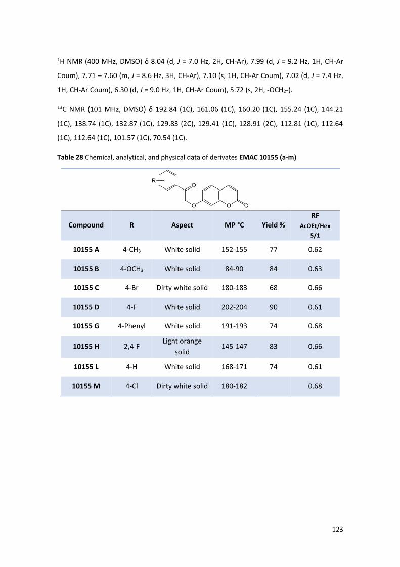
123
1H NMR (400 MHz, DMSO) δ 8.04 (d, J = 7.0 Hz, 2H, CH-Ar), 7.99 (d, J = 9.2 Hz, 1H, CH-Ar
Coum), 7.71 – 7.60 (m, J = 8.6 Hz, 3H, CH-Ar), 7.10 (s, 1H, CH-Ar Coum), 7.02 (d, J = 7.4 Hz,
1H, CH-Ar Coum), 6.30 (d, J = 9.0 Hz, 1H, CH-Ar Coum), 5.72 (s, 2H, -OCH2-).
13C NMR (101 MHz, DMSO) δ 192.84 (1C), 161.06 (1C), 160.20 (1C), 155.24 (1C), 144.21
(1C), 138.74 (1C), 132.87 (1C), 129.83 (2C), 129.41 (1C), 128.91 (2C), 112.81 (1C), 112.64
(1C), 112.64 (1C), 101.57 (1C), 70.54 (1C).
Table 28 Chemical, analytical, and physical data of derivates EMAC 10155 (a-m)
Compound R Aspect MP °C Yield % RF
AcOEt/Hex
5/1
10155 A 4-CH3 White solid 152-155 77 0.62
10155 B 4-OCH3 White solid 84-90 84 0.63
10155 C 4-Br Dirty white solid 180-183 68 0.66
10155 D 4-F White solid 202-204 90 0.61
10155 G 4-Phenyl White solid 191-193 74 0.68
10155 H 2,4-F Light orange
solid 145-147 83 0.66
10155 L 4-H White solid 168-171 74 0.61
10155 M 4-Cl Dirty white solid 180-182 0.68

124
General method for the synthesis of compounds EMAC 10158 (a-m)
5 mL of 1 N sodium hydroxide were added to a suspension of the appropriate EMAC 10155
(a-m). The reaction mixture was stirred at reflux for 4 hours. Then, it was poured into
crushed ice and water and it was stirred until ice melting. Hydrochloric acid was added
dropwise to obtain a precipitate.
EMAC 10158 A: 3-(p-tolyl)-7H-furo[3,2-g]chromen-7-one
1H NMR (600 MHz, DMSO) δ 8.45 (s, 1H, OCH-Ar Furoc), 8.32 (s, 1H, CH-Ar Furoc), 8.24 (d, J
= 9.7 Hz, 1H, CH-Ar Furoc), 7.79 (s, 1H, CH-Ar Furoc), 7.68 (d, J = 7.5 Hz, 2H, CH-Ar), 7.35 (d,
J = 7.5 Hz, 2H, CH-Ar), 6.47 (d, J = 9.5 Hz, 1H, CH-Ar Furoc), 2.38 (s, 3H, -CH3).
13C NMR (151 MHz, DMSO) δ 163.81 (1C), 160.33 (1C), 157.03 (1C), 145.57 (1C), 144.48
(1C), 137.75 (1C), 130.18 (2C), 129.84 (1C), 127.43 (2C), 123.67 (1C), 121.43 (1C), 120.71
(1C), 116.18 (1C), 114.73 (1C), 100.20 (1C), 21.31 (1C).
EMAC 10158 M: 3-(4-chlorophenyl)-7H-furo[3,2-g]chromen-7-one

125
1H NMR (600 MHz, DMSO) δ 8.45 (s, 1H, OCH-Ar Furoc), 8.22 (s, 1H, CH-Ar Furoc), 8.12 (d, J
= 9.0 Hz, 1H, CH-Ar Furoc), 7.76 – 7.67 (m, 3H, CH-Ar), 7.50 (d, J = 6.9 Hz, 2H, Ar), 6.39 (d, J
= 9.1 Hz, 1H, Ar Furoc).
13C NMR (151 MHz, DMSO) δ 160.54 (1C), 156.99 (1C), 152.20 (1C), 145.41 (1C), 145.26
(1C), 132.89 (1C), 130.06 (1C), 129.60 (2C), 129.21 (2C), 123.17 (1C), 120.58 (1C), 120.41
(1C), 116.27 (1C), 114.87 (1C), 100.27 (1C).
Table 29 Chemical, analytical, and physical data of derivates EMAC 10158 (a-m)
Compound R Aspect MP °C Yield % RF
AcOEt/Hex 5/1
10158 A 4-CH3 Light brown solid 130-133 69 0.73
10158 M 4-Cl Yellow solid 197-198 92 0.76

126
EMAC 10156 (a-m) and EMAC 10159 (a-m)
Scheme 9 Synthesis of derivatives EMAC 10156(a-m) and EMAC 10159(a-m): (i.) dimethyl acetylsuccinate, H2SO4, rT, 30’; (ii.) 2-bromo/2-chloro-acetophenone, acetone, K2CO3, Δ; (iii.)
NaOH 1N, Δ
Synthesis of compound EMAC 10156
A mixture of resorcinol (4,5 mmol) and dimethyl acetylsuccinate (4,5 mmol) was stirred at
room temperature. Sulfuric acid 98% (13,5 mmol) was added dropwise and the mixture was
vigorously stirred until the end of the reaction. The reaction mixture was then poured into
crushed ice to give a precipitate and stirred until ice melting. The formed precipitate was
filtered off to obtain a white solid. The progression of the reaction was monitored by TLC (ethyl
acetate/n-hexane 6/1).
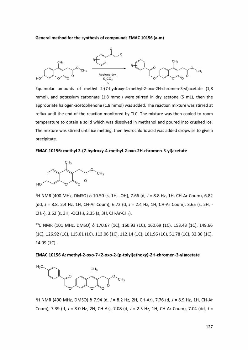
127
General method for the synthesis of compounds EMAC 10156 (a-m)
Equimolar amounts of methyl 2-(7-hydroxy-4-methyl-2-oxo-2H-chromen-3-yl)acetate (1,8
mmol), and potassium carbonate (1,8 mmol) were stirred in dry acetone (5 mL), then the
appropriate halogen-acetophenone (1,8 mmol) was added. The reaction mixture was stirred at
reflux until the end of the reaction monitored by TLC. The mixture was then cooled to room
temperature to obtain a solid which was dissolved in methanol and poured into crushed ice.
The mixture was stirred until ice melting, then hydrochloric acid was added dropwise to give a
precipitate.
EMAC 10156: methyl 2-(7-hydroxy-4-methyl-2-oxo-2H-chromen-3-yl)acetate
1H NMR (400 MHz, DMSO) δ 10.50 (s, 1H, -OH), 7.66 (d, J = 8.8 Hz, 1H, CH-Ar Coum), 6.82
(dd, J = 8.8, 2.4 Hz, 1H, CH-Ar Coum), 6.72 (d, J = 2.4 Hz, 1H, CH-Ar Coum), 3.65 (s, 2H, -
CH2-), 3.62 (s, 3H, -OCH3), 2.35 (s, 3H, CH-Ar-CH3).
13C NMR (101 MHz, DMSO) δ 170.67 (1C), 160.93 (1C), 160.69 (1C), 153.43 (1C), 149.66
(1C), 126.92 (1C), 115.01 (1C), 113.06 (1C), 112.14 (1C), 101.96 (1C), 51.78 (1C), 32.30 (1C),
14.99 (1C).
EMAC 10156 A: methyl-2-oxo-7-(2-oxo-2-(p-tolyl)ethoxy)-2H-chromen-3-yl)acetate
1H NMR (400 MHz, DMSO) δ 7.94 (d, J = 8.2 Hz, 2H, CH-Ar), 7.76 (d, J = 8.9 Hz, 1H, CH-Ar
Coum), 7.39 (d, J = 8.0 Hz, 2H, CH-Ar), 7.08 (d, J = 2.5 Hz, 1H, CH-Ar Coum), 7.04 (dd, J =

128
8.9, 2.5 Hz, 1H, CH-Ar Coum), 5.71 (s, 2H, -OCH2-), 3.67 (s, 2H, Ar-CH2), 3.62 (s, 3H, -OCH3),
2.41 (s, 3H, CH-Ar-CH3), 2.39 (s, 3H, CH-Ar-CH3).
13C NMR (101 MHz, DMSO) δ 193.25 (1C), 170.56 (1C), 160.77 (1C), 160.69 (1C), 153.21
(1C), 149.45 (1C), 144.39 (1C), 131.73 (1C), 129.35 (2C), 127.90 (2C), 126.73 (1C), 116.11
(1C), 113.52 (1C), 112.72 (1C), 101.42 (1C), 70.44 (1C), 51.81 (1C), 32.36 (s1C21.22 (1C),
15.07 (1C).
EMAC 10156 B: methyl 2-(7-(2-(4-methoxyphenyl)-2-oxoethoxy)-4-methyl-2-oxo-2H-
chromen-3-yl)acetate
1H NMR (400 MHz, DMSO) δ 8.02 (d, J = 8.8 Hz, 2H, CH-Ar), 7.75 (d, J = 8.9 Hz, 1H, CH-Ar
Coum), 7.10 (d, J = 8.8 Hz, 2H, CH-Ar), 7.06 (d, J = 2.3 Hz, 1H, CH-Ar Coum), 7.03 (dd, J = 8.8
Hz, 1H, CH-Ar Coum), 5.67 (s, 2H, -OCH2-), 3.87 (s, 3H, CH-Ar-OCH3), 3.67 (s, 2H, Ar-CH2-),
3.62 (s, 3H-OCH3), 2.38 (s, 3H, CH-Ar-CH3).
13C NMR (101 MHz, DMSO) δ 192.04 (1C), 170.56 (1C), 163.63 (1C), 160.77 (1C), 160.74
(1C), 153.20 (1C), 149.44 (1C), 130.24 (2C), 127.10 (1C), 126.71 (1C), 116.08 (1C), 114.05
(2C), 113.49 (1C), 112.71 (1C), 101.40 (1C), 70.24 (1C), 55.59 (1C), 51.81 (1C), 32.36 (1C),
15.05 (1C).
EMAC 10156 C: methyl 2-(7-(2-(4-bromophenyl)-2-oxoethoxy)-4-methyl-2-oxo-2H-
chromen-3-yl)acetate
1H NMR (400 MHz, DMSO) δ 7.97 (d, J = 8.7 Hz, 2H, CH-Ar), 7.81 (d, J = 8.7 Hz, 2H, CH-Ar),
7.76 (d, J = 8.9 Hz, 1H, CH-Ar Coum), 7.12 (d, J = 2.6 Hz, 1H, CH-Ar Coum), 7.05 (dd, J = 8.9,
2.6 Hz, 1H, CH-Ar Coum), 5.73 (s, 2H, -OCH2-), 3.67 (s, 2H, Ar-CH2-), 3.62 (s, 3H, -OCH3),
2.39 (s, 3H, CH-Ar-CH3).

129
13C NMR (151 MHz, DMSO) δ 193.62 (1C), 171.04 (1C), 161.31 (1C), 161.08 (1C), 153.69
(1C), 149.94 (1C), 133.74 (1C), 132.37 (2C), 130.42 (2C), 128.44 (1C), 127.26 (1C), 116.67
(1C), 114.10 (1C), 113.25 (1C), 101.96 (1C), 71.04 (1C), 52.32 (1C), 32.88 (1C), 15.58 (1C).
EMAC 10156 D: methyl 2-(7-(2-(4-bromophenyl)-2-oxoethoxy)-4-methyl-2-oxo-2H-
chromen-3-yl)acetate
1H NMR (400 MHz, DMSO) δ 8.15-8.10 (m, 2H, CH-Ar), 7.76 (d, J = 9.0 Hz, 1H, CH-Ar Coum),
7.42 (t, J = 8.9 Hz, 2H, CH-Ar), 7.11 (d, J = 2.5 Hz, 1H, CH-Ar Coum), 7.05 (dd, J = 8.9, 2.5 Hz,
1H, CH-Ar Coum), 5.73 (s, 2H, -OCH2-), 3.67 (s, 2H, Ar-CH2-), 3.62 (s, 3H, -OCH3), 2.39 (s, 3H,
CH-Ar-CH3).
13C NMR (101 MHz, DMSO) δ 192.57 (1C), 170.72 (1C), 166.63 (1C), 164.11 (1C), 160.91
(1C), 160.52 (1C), 153.11 (1C), 149.61 (1C), 130.95 (d, J = 9.6 Hz, 2C), 126.75 (1C), 116.06
(1C), 116.01 (1C), 115.79 (1C), 113.57 (1C), 112.78 (1C), 101.29 (1C), 70.33 (1C), 51.87 (1C),
32.30 (1C), 14.99 (1C).
EMAC 10156 F: methyl 2-(4-methyl-7-(2-(3-nitrophenyl)-2-oxoethoxy)-2-oxo-2H-
chromen-3-yl)acetate
1H NMR (400 MHz, DMSO) δ 8.75 (s, 1H, CH-Ar), 8.53 (d, J = 6.6 Hz, 1H, CH-Ar), 8.46 (d, J =
6.5 Hz, 1H, CH-Ar), 7.89 (s, 1H, CH-Ar), 7.77 (d, J = 8.0 Hz, 1H, CH-Ar Coum), 7.19 (s, 1H, CH-
Ar Coum), 7.09 (d, J = 7.2 Hz, 1H, CH-Ar Coum), 5.85 (s, 2H, -OCH2-), 3.68 (s, 2H, Ar-CH2-),
3.62 (s, 3H, -OCH3), 2.39 (s, 3H, CH-Ar-CH3).
13C NMR (151 MHz, DMSO) δ 193.06 (1C), 171.07 (1C), 161.27 (1C), 161.01 (1C), 153.77
(1C), 149.89 (1C), 148.52 (1C), 135.93 (1C), 134.66 (1C), 131.11 (1C), 128.44 (1C), 127.28

130
(1C), 122.91 (1C), 116.73 (1C), 114.17 (1C), 113.33 (1C), 101.98 (1C), 71.35 (1C), 52.33 (1C),
32.88 (1C), 15.59 (1C).
EMAC 10156 L: methyl 2-(4-methyl-2-oxo-7-(2-oxo-2-phenylethoxy)-2H-chromen-3-
yl)acetate
1H NMR (600 MHz, DMSO) δ 8.05 (d, J = 7.6 Hz, 2H, CH-Ar), 7.77 (d, J = 8.9 Hz, 1H, CH-Ar
Coum), 7.72 (t, J = 7.4 Hz, 1H, CH-Ar), 7.60 (t, J = 7.6 Hz, 2H, CH-Ar), 7.11 (s, 1H, CH-Ar
Coum), 7.07 (d, J = 8.8 Hz, 1H, CH-Ar Coum), 5.76 (s, 2H, -OCH2-), 3.69 (s, 2H, Ar-CH2-), 3.63
(s, 3H, -OCH3), 2.40 (s, 3H, CH-Ar-CH3).
13C NMR (151 MHz, DMSO) δ 194.27 (1C), 171.07 (1C), 161.29 (1C), 161.18 (1C), 153.73
(1C), 149.95 (1C), 134.73 (1C), 134.38 (1C), 129.32 (2C), 128.39 (2C), 127.24 (1C), 116.64
(1C), 114.06 (1C), 113.24 (1C), 101.96 (1C), 71.06 (1C), 52.32 (1C), 32.88 (1C), 5.58 (1C).
EMAC 10156 M: methyl 2-(7-(2-(4-chlorophenyl)-2-oxoethoxy)-4-methyl-2-oxo-2H-
chromen-3-yl)acetate
1H NMR (600 MHz, DMSO) δ 8.07 (d, J = 8.4 Hz, 2H, CH-Ar), 7.78 (d, J = 8.9 Hz, 1H, CH-Ar
Coum), 7.68 (d, J = 8.4 Hz, 2H, CH-Ar), 7.13 (d, J = 2.3 Hz, 1H, CH-Ar Coum), 7.07 (d, J = 8.9
Hz, 1H, CH-Ar Coum), 5.75 (s, 2H, -OCH2-), 3.69 (s, 2H, Ar-CH2-), 3.64 (s, 3H, -OCH3), 2.41 (s,
3H, CH-Ar-CH3).
13C NMR (151 MHz, DMSO) δ 193.39 (1C), 170.90 (1C), 161.29 (1C), 161.10 (1C), 153.75
(1C), 149.93 (1C), 139.32 (1C), 133.32 (1C), 130.34 (2C), 129.42 (2C), 128.42 (1C), 127.17
(1C), 116.70 (1C), 113.25 (1C), 102.09 (1C), 71.07 (1C), 52.32 (1C), 32.88 (1C), 15.58 (1C).

131
Table 30 Chemical, analytical, and physical data of derivates EMAC 10156 (a-m)
Compound R Aspect MP °C Yield % RF
AcOEt/Hex
5/1
10156 A 4-CH3 White solid 154-155 73 0.57
10156 B 4-OCH3 White solid 143-144 19 0.61
10156 C 4-Br Light orange solid 155-157 68 0.60
10156 D 4-F Light orange solid 90-93 80 0.61
10156 F 3-NO2 Yellow solid 150-153 59 0.58
10156 L 4-H Light yellow solid 108-110 70 0.56
10156 M 4-Cl Yellow solid 151-154 67 0.63

132
General method for the synthesis of compound EMAC 10159 (a-m)
EMAC 10156 (a-m) were stirred in a solution of 1N sodium hydroxide. The reaction mixture
was stirred at reflux until the end of the reaction. Then, it was poured into crushed ice and
water, and it was stirred until ice melting. Hydrochloric acid was added dropwise to obtain
a precipitate. The precipitate was filtered to obtain the desired product. The reaction was
monitored by TLC (ethyl acetate/n-hexane 5/1).
EMAC 10159 A: 2-(5-methyl-7-oxo-3-(p-tolyl)-7H-furo[3,2-g]chromen-6-yl)acetic acid
1H NMR (400 MHz, DMSO) δ 12.48 (bs, 1H, -OH), 8.40 (s, 1H, CH-Ar Furoc), 8.14 (s, 1H, CH-
Ar Furoc), 7.76 (s, 1H, CH-Ar Furoc), 7.69 (d, J = 8.1 Hz, 2H, CH-Ar), 7.34 (d, J = 7.9 Hz, 2H,
CH-Ar), 3.65 (s, 2H, Ar-CH2-), 2.51 (s, 3H, CH-Ar-CH3), 2.37 (s, 3H, CH-Ar-CH3).
13C NMR (101 MHz, DMSO) δ 171.51 (1C), 160.78 (1C), 155.92 (1C), 149.74 (1C), 149.32
(1C), 137.23 (1C), 129.74 (2C), 127.54 (1C), 127.04 (2C), 123.03 (1C), 121.17 (1C), 118.07
(1C), 116.60 (1C), 116.60 (1C), 99.40 (1C), 32.85 (1C), 20.75 (1C), 15.45 (1C).

133
EMAC 10159 B: 2-(3-(4-methoxyphenyl)-5-methyl-7-oxo-7H-furo[3,2-g]chromen-6-
yl)acetic acid
1H NMR (400 MHz, DMSO) δ 12.47 (bs, 1H, -OH), 8.38 (s, 1H, CH-Ar Furoc), 8.17 (s, 1H, CH-
Ar Furoc), 7.79 (s, 1H, CH-Ar Furoc), 7.75 (d, J = 8.7 Hz, 2H, CH-Ar), 7.11 (d, J = 8.8 Hz, 2H,
CH-Ar), 3.83 (s, 3H, -OCH3), 3.66 (s, 2H, Ar-CH2-), 2.54 (s, 3H, CH-Ar-CH3).
13C NMR (101 MHz, DMSO) δ 171.45 (1C), 160.75 (1C), 158.97 (1C), 155.96 (1C), 149.83
(1C), 149.35 (1C), 128.51 (2C), 123.18 (1C), 122.85 (1C), 120.96 (1C), 118.15 (1C), 116.71
(1C), 116.65 (1C), 114.65 (2C), 99.47 (1C), 55.19 (1C), 32.90 (1C), 15.56 (1C).
EMAC 10159 C: 2-(3-(4-bromophenyl)-5-methyl-7-oxo-7H-furo[3,2-g]chromen-6-yl)acetic
acid
1H NMR (400 MHz, DMSO) δ 12.46 (bs, 1H, -OH), 8.51 (s, 1H, CH-Ar Furoc), 8.17 (s, 1H, CH-
Ar Furoc), 7.78 (d (d+s), J = 7.2 Hz, 3H, CH-Ar + CH-Ar Furoc), 7.72 (d, J = 8.6 Hz, 2H, CH-Ar),
3.65 (s, 2H, Ar-CH2-), 2.53 (s, 3H, CH-Ar-CH3).
13C NMR (101 MHz, DMSO) δ 171.42 (1C), 160.67 (1C), 155.98 (1C), 149.92 (1C), 149.30
(1C), 132.06 (2C), 131.24 (1C), 129.90 (1C), 129.27 (2C), 122.53 (1C), 120.87 (1C), 120.26
(1C), 118.30 (1C), 116.84 (1C), 116.74 (1C), 99.58 (1C), 32.90 (1C), 15.57 (s).

134
EMAC 10159 D: 2-(3-(4-fluorophenyl)-5-methyl-7-oxo-7H-furo[3,2-g]chromen-6-yl)acetic
acid
1H NMR (400 MHz, DMSO) δ 12.47 (bs, 1H, -OH), 8.46 (s, 1H, CH-Ar Furoc), 8.17 (s, 1H, CH-
Ar Furoc), 7.87 (dd, J = 8.3, 5.6 Hz, 2H, CH-Ar), 7.79 (s, 1H, CH-Ar Furoc), 7.37 (t, J = 8.8 Hz,
2H, CH-Ar), 3.65 (s, 2H, Ar-CH2-), 2.53 (s, 3H, CH-Ar-CH3).
13C NMR (101 MHz, DMSO) δ 171.42 (1C), 162.97 (1C), 160.68 (1C), 160.54 (1C), 155.94
(1C), 149.90 (1C), 149.31 (1C), 129.29 (d, J = 8.2 Hz, 2C), 127.07 (d, J = 3.2 Hz, 1C), 122.80
(1C), 120.35 (1C), 118.25 (1C), 116.78 (1C), 116.71 (1C), 116.17 (1C), 115.96 (1C), 99.54
(1C), 32.90 (1C), 15.58 (s).
EMAC 10159 L: 2-(5-methyl-7-oxo-3-phenyl-7H-furo[3,2-g]chromen-6-yl)acetic acid
1H NMR (600 MHz, DMSO) δ 12.48 (bs, 1H, -OH), 8.47 (s, 1H), 8.20 (s, 1H, CH-Ar Furoc),
7.82 (dd, J = 8.2, 1.1 Hz, 2H, CH-Ar), 7.80 (s, 1H, CH-Ar Furoc), 7.55 (t, J = 7.7 Hz, 2H, CH-Ar),
7.44 (t, J = 7.7 Hz, 1H, CH-Ar), 3.66 (s, 2H, CH-Ar-CH2), 2.54 (s, 3H, CH-Ar-CH3).
13C NMR (151 MHz, DMSO) δ 171.95 (1C), 161.23 (1C), 156.53 (1C), 150.39 (1C), 149.83
(1C), 144.82 (1C), 131.14 (1C), 129.71 (2C), 128.34 (1C), 127.73 (2C), 123.41 (1C), 121.80
(1C), 118.74 (1C), 117.26 (2C), 100.05 (1C), 33.42 (1C), 16.08 (1C).
EMAC 10159 M: 2-(3-(4-chlorophenyl)-5-methyl-7-oxo-7H-furo[3,2-g]chromen-6-yl)acetic
acid

135
1H NMR (600 MHz, DMSO) δ 12.47 (bs, 1H, -OH), 8.50 (s, 1H, CH-Ar Furoc), 8.17 (s, 1H, CH-
Ar Furoc), 7.84 (d, J = 8.4 Hz, 2H, CH-Ar), 7.79 (s, 1H, CH-Ar Furoc), 7.57 (d, J = 8.4 Hz, 2H,
CH-Ar Furoc), 3.64 (s, 2H, CH-Ar-CH2), 2.52 (s, 3H, CH-Ar-CH3).
13C NMR (151 MHz, DMSO) δ 171.92 (1C), 161.18 (1C), 156.50 (1C), 150.45 (1C), 149.82
(1C), 145.25 (1C), 132.89 (1C), 130.07 (1C), 129.66 (2C), 129.50 (2C), 123.11 (1C), 120.73
(1C), 118.82 (1C), 117.36 (1C), 117.28 (1C), 100.11 (1C), 33.42 (1C), 16.10 (1C).
Table 31 Chemical, analytical, and physical data of derivates EMAC 10159 (a-m)
Compound R Aspect MP °C Yield % RF
AcOEt/Hex
5/1
10159 A 4-CH3 Dark green solid 194-196 94 0.65
10159 B 4-OCH3 Grey solid 187-190 83 0.61
10159 C 4-Br Dark brown solid 75-78 76 0.67
10159 D 4-F Light brown solid 140-143 73 0.61
10159 L 4-H Light yellow solid 205-208 75 0.66
10159 M 4-Cl Brown 172-175 87 0.73

136
EMAC 10157 (a-m) and EMAC 10160 (a-m)
Scheme 10 Synthesis of derivatives EMAC 10157(a-m) and EMAC 10160(a-m): (i.) dimethyl acetylsuccinate, H2SO4, rT, 30’; (ii.) halogeno-acetophenone, acetone, K2CO3, Δ; (iii.) NaOH 1N, Δ
Synthesis of compound EMAC 10157
Equimolar amounts of 2-propyl resorcinol and dimethyl acetylsuccinate were stirred in
sulfuric acid 98% at room temperature until the end of the reaction. A precipitate was
formed, and it was the desired compound that was used for the next step without
purification. The reaction was monitored by TLC (ethyl acetate/n-hexane 5/1).

137
General method for the synthesis of compounds EMAC 10157 (a-m)
Equimolar amounts of methyl 2-(7-hydroxy-4-methyl-2-oxo-8-propyl-2H-chromen-3-
yl)acetate (1,8 mmol), and potassium carbonate (1,8 mmol) were stirred in dry acetone (5
mL), then the appropriate halogen-acetophenone (1,8 mmol) was added and the reaction
mixture stirred at reflux until the end of the reaction. The reaction mixture was cooled to
room temperature to obtain a precipitate.
EMAC 10157: methyl 2-(7-hydroxy-4-methyl-2-oxo-8-propyl-2H-chromen-3-yl)acetate
1H NMR (400 MHz, DMSO) δ 10.33 (s, 1H, -OH), 7.52 (d, J = 8.8 Hz, 1H, Coum), 6.88 (d, J =
8.8 Hz, 1H, Coum), 3.65 (s, 2H, Ar-CH2-CO), 3.62 (s, 3H, -OCH3), 2.69 (t, J = 7.2 Hz, 2H, Ar-
CH2-CH2-), 2.34 (s, 3H, Ar-CH3), 1.58 – 1.47 (m, 2H, -CH2-CH2-CH3), 0.91 (t, J = 7.4 Hz, 3H, -
CH2-CH3).
13C NMR (101 MHz, DMSO) δ 170.76 (1C), 161.08 (1C), 158.42 (1C), 151.34 (1C), 149.95
(1C), 123.77 (1C), 115.01 (1C), 114.57 (1C), 112.16 (1C), 112.13 (1C), 51.77 (1C), 32.29 (1C),
24.22 (1C), 21.70 (1C), 15.05 (1C), 13.92 (1C).

138
EMAC 10157 A: methyl 2-(4-methyl-2-oxo-7-(2-oxo-2-(p-tolyl)ethoxy)-8-propyl-2H-
chromen-3-yl)acetate
1H NMR (400 MHz, DMSO) δ 7.93 (d, J = 8, 2H, CH-Ar), 7.61 (d, J = 9.2, 1H, CH-Ar Coum),
7.39 (d, J = 8, 2H, CH-Ar), 7.00 (d, J = 8.8, 1H, CH-Ar Coum), 5.74 (s, 2H, -OCH2-CO-), 3.68
(s, 2H, Ar-CH2-CO), 3.62 (s, 3H, -OCH3), 2.83–2.79 (m, 2H, Ar-CH2-CH2-), 2.41 (s, 3H, Ar-CH3),
2.37 (s, 3H, Ar-CH3), 1.64–1.55 (m, 2H, -CH2-CH2-CH3), 0.95–0.91 (t, 3H, -CH2-CH3).
13C NMR (101 MHz, DMSO) δ 193.70 (1C), 170.64 (1C), 160.88 (1C), 158.24 (1C), 150.61
(1C), 149.69 (1C), 144.37 (1C), 131.78 (1C), 129.34 (s, 2C), 127.96 (s, 2C), 123.92 (1C),
116.97 (1C), 115.87 (1C), 113.65 (1C), 108.77 (1C), 70.55 (1C), 51.82 (1C), 32.37 (1C), 24.34
(1C), 21.70 (1C), 21.22 (1C), 15.10 (1C), 13.96 (1C).
EMAC 10157 B: methyl 2-(7-(2-(4-methoxyphenyl)-2-oxoethoxy)-4-methyl-2-oxo-8-
propyl-2H-chromen-3-yl)acetate
1H NMR (400 MHz, DMSO) δ 8.01 (d, J = 8.8, 2H, CH-Ar), 7.61 (d, J = 8, 1H, CH-Ar Coum),
7.09 (d, J = 8, 2H, CH-Ar), 6.98 (d, J = 8, 1H, CH-Ar Coum), 5.70 (s, 2H, -OCH2-CO-), 3.86 (s,
3H, CH-Ar-OCH3), 3.68 (s, 2H, Ar-CH2-CO), 3.62, (s, 3H, -OCH3), 2.82–2.79 (m, 2H, Ar-CH2-
CH2-), 2.36 (s, 3H, Ar-CH3), 1.64–1.55 (m, 2H, -CH2-CH2-CH3), 0.95–0.91 (t, 3H, -CH2-CH3)
13C NMR (100 MHz, DMSO) δ 192.48 (1C), 170.64 (1C), 163.61 (1C), 160.89 (1C), 158.29
(1C), 150.60 (1C), 149.69 (1C), 130.23 (2C), 127.15 (1C), 123.90 (1C), 116.96v, 115.84 (1C),

139
114.05 (2C), 113.61 (1C), 108.76 (1C), 70.35 (1C), 55.60 (1C), 51.81 (1C), 32.36 (1C), 24.34
(1C), 21.70 (1C), 15.09 (1C), 13.96 (1C).
EMAC 10157 D: methyl 2-(7-(2-(4-fluorophenyl)-2-oxoethoxy)-4-methyl-2-oxo-8-propyl-
2H-chromen-3-yl)acetate
1H NMR (400 MHz, DMSO) δ 8.13–8.10 (m, 2H, CH-Ar), 7.62 (d, J = 9.2, 1H, CH-Ar Coum),
7.45–7.40 (m, 2H, CH-Ar), 7.03 (d, J = 9.2, 1H, CH-Ar Coum), 5.77 (s, 2H, -OCH2-CO-), 3.68
(s, 2H, Ar-CH2-CO-), 3.62 (s, 3H, -OCH3), 2.83–2.79 (m, 2H, Ar-CH2-CH2-), 2.37 (s, 3H, Ar-
CH3), 1.64–1.55 (m, 2H, -CH2-CH2-CH3), 0.95–0.91 (t, 3H, -CH2-CH3).
13C NMR (100 MHz, DMSO) δ 192.90 (1C), 170.65 (1C), 166.59 (1C), 164.08 (1C), 160.89
(1C), 158.15 (1C), 150.60 (1C), 149.70 (1C), 131.01 (2C), 123.94 (1C), 116.98 (1C), 116.01
(1C), 115.91 (2C), 113.70 (1C), 108.79 (1C), 70.55 (1C), 51.82 (1C), 32.36 (1C), 24.33 (1C),
21.70 (1C), 15.10 (1C), 13.95 (1C).
EMAC 10157 F: methyl 2-(4-methyl-7-(2-(3-nitrophenyl)-2-oxoethoxy)-2-oxo-8-propyl-
2H-chromen-3-yl)acetate
1H NMR (400 MHz, DMSO) δ 8.75 (s, 1H, CH-Ar), 8.53 (d, J = 7.2 Hz, 1H,CH-Ar), 8.44 (d, J =
7.2 Hz, 1H,CH-Ar), 7.89 (t, J = 7.6 Hz, 1H,CH-Ar), 7.63 (d, J = 8.6 Hz, 1H,CH-Ar Coum), 7.11
(d, J = 8.7 Hz, 1H,CH-Ar Coum), 5.88 (s, 2H, -OCH2-CO-), 3.68 (s, 2H, Ar-CH2-CO-), 3.62 (s,

140
3H, -OCH3), 2.81 (s, 2H, Ar-CH2-CH2-), 2.37 (s, 3H, Ar-CH3), 1.59 (d, J = 6.6 Hz, 2H, -CH2-CH2-
CH3), 0.92 (t, J = 6.4 Hz, 3H, -CH2-CH3).
13C NMR (101 MHz, DMSO) δ 193.11 (1C), 170.62 (1C), 160.84 (1C), 158.00 (1C), 150.60
(1C), 149.65 (1C), 147.97 (1C), 135.47 (1C), 134.13 (1C), 130.62 (1C), 127.92 (1C), 123.95
(1C), 122.43 (1C), 117.01 (1C), 116.00 (1C), 113.81 (1C), 108.89 (1C), 70.97 (1C), 51.81 (1C),
32.37 (1C), 24.35 (1C), 21.72 (1C), 15.11 (1C), 13.95 (s).
EMAC 10157 G: methyl 2-(7-(2-([1,1'-biphenyl]-4-yl)-2-oxoethoxy)-4-methyl-2-oxo-8-
propyl-2H-chromen-3-yl)acetate
1H NMR (400 MHz, DMSO) δ 8.12 (d, J = 8.4, 2H, CH-Ar), 7.88 (d, J = 8.4, 2H, CH-Ar), 7.78 (d,
J = 7.2, 2H, CH-Ar), 7.65–7.61 (m, 1H, CH-Ar), 7.53 (t, J = 7.2, 2H, CH-Ar), 7.46 (d, J = 7.3, 1H,
CH-Ar Coum), 7.05 (d, 1H, CH-Ar Coum), 5.82 (s, 2H, -OCH2-CO-), 3.68 (s, 2H, Ar-CH2-CO-),
3.62 (s, 3H, -OCH3), 2.85–2.81 (m, 2H, Ar-CH2-CH2-),2.37 (s, 3H, Ar-CH3), 1.64–1.58 (m, 2H, -
CH2-CH2-CH3), 0.96–0.92 (t, 3H, -CH2-CH3).
13C NMR (100 MHz, DMSO) δ 193.78 (1C), 170.63 (1C), 160.88 (1C), 158.22 (1C), 150.62
(1C), 149.68 (1C), 145.17 (1C), 138.77 (1C), 133.07 (1C), 129.10 (2C), 128.61 (2C), 127.02
(2C), 126.96 (2C), 123.94 (1C), 116.99 (1C), 115.90 (1C), 113.68 (1C), 108.81 (1C), 73.94
(1C), 70.69 (1C), 51.81 (1C), 32.37 (1C), 24.37 (1C), 21.73 (1C), 15.11 (1C), 13.98 (1C).

141
EMAC 10157 H: methyl 2-(7-(2-(2,4-difluorophenyl)-2-oxoethoxy)-4-methyl-2-oxo-8-
propyl-2H-chromen-3-yl)acetateC
O OO
O
O
O
CH3
CH3
CH3
F
F
1H NMR (400 MHz, DMSO) δ 8.01 (td, J = 8.6, 6.7 Hz, 1H, CH-Ar), 7.60 (d, J = 9.0 Hz, 1H, CH-
Ar Coum), 7.51 (ddd, J = 11.5, 9.3, 2.4 Hz, 1H, CH-Ar), 7.29 (td, J = 8.5, 2.4 Hz, 1H, CH-Ar),
7.02 (d, J = 9.0 Hz, 1H, , CH-Ar Coum), 5.56 (d, J = 2.7 Hz, 2H, -OCH2-CO-), 3.68 (s, 2H, Ar-
CH2-CO-), 3.62 (s, 3H, -OCH3), 2.78 (t, 2H, Ar-CH2-CH2-), 2.37 (s, 3H, Ar-CH3), 1.65 – 1.49 (m,
2H, -CH2-CH2-CH3), 0.92 (t, J = 7.4 Hz, 3H, -CH2-CH3).
13C NMR (101 MHz, DMSO) δ 191.23 (d, J = 5.0 Hz, 1C), 170.62 (1C), 160.86 (1C), 158.06
(1C), 150.59 (1C), 149.66 (1C), 123.91 (1C), 116.90 (1C), 115.94 (1C), 113.72 (1C), 108.73
(1C), 72.73 (d, J = 10.5 Hz, 1C), 51.81 (1C), 32.37 (1C), 24.34 (1C), 21.69 (1C), 15.11 (1C),
13.96 (1C).
EMAC 10157 L: methyl 2-(4-methyl-2-oxo-7-(2-oxo-2-phenylethoxy)-8-propyl-2H-
chromen-3-yl)acetate
1H NMR (600 MHz, DMSO) δ 8.04 (d, J = 7.7 Hz, 2H, CH-Ar), 7.72 (t, J = 7.4 Hz, 1H, CH-Ar),
7.63 (d, J = 9.0 Hz, 1H, CH-Ar Coum), 7.60 (t, J = 7.7 Hz, 2H, CH-Ar), 7.04 (d, J = 9.0 Hz, 1H,
CH-Ar Coum), 5.80 (s, 2H, -OCH2-CO-), 3.70 (s, 2H, Ar-CH2-CO-), 3.64 (s, 3H, -OCH3), 2.87 –
2.80 (m, 2H, Ar-CH2-CH2-), 2.38 (s, 3H, Ar-CH3), 1.62 (dq, J = 14.7, 7.3 Hz, 2H, -CH2-CH2-
CH3), 0.95 (t, J = 7.3 Hz, 3H, -CH2-CH3).

142
13C NMR (151 MHz, DMSO) δ 194.76 (1C), 171.14 (1C), 161.38 (1C), 158.72 (1C), 151.13
(1C), 150.19 (1C), 134.80 (1C), 134.35 (1C), 129.32 (2C), 128.38 (2C), 124.44 (1C), 117.51
(1C), 116.41 (1C), 114.19 (1C), 109.30 (1C), 71.18 (1C), 52.32 (1C), 32.89 (1C), 24.86 (1C),
22.22 (1C), 15.62 (1C), 14.48 (1C).
EMAC 10157 M: methyl 2-(7-(2-(4-chlorophenyl)-2-oxoethoxy)-4-methyl-2-oxo-8-propyl-
2H-chromen-3-yl)acetate
1H NMR (600 MHz, DMSO) δ 8.05 (d, J = 8.4 Hz, 2H, CH-Ar), 7.67 (d, J = 8.4 Hz, 2H, CH-Ar),
7.63 (d, J = 9.0 Hz, 1H, CH-Ar Coum), 7.05 (d, J = 9.0 Hz, 1H, CH-Ar Coum), 5.78 (s, 2H, -
OCH2-CO-),), 3.69 (s, 2H, Ar-CH2-CO-), 3.63 (s, 3H, -OCH3), 2.87 – 2.73 (m, 2H, Ar-CH2-CH2-),
2.38 (s, 3H, Ar-CH3), 1.66 – 1.54 (m, 2H, -CH2-CH2-CH3), 0.94 (t, J = 7.4 Hz, 3H, 3H, -CH2-
CH3).
13C NMR (151 MHz, DMSO) δ 193.87 (1C), 171.13 (1C), 161.37 (1C), 158.63 (1C), 151.12
(1C), 150.18 (1C), 139.22 (1C), 133.49 (1C), 130.33 (2C), 129.44 (2C), 124.45 (1C), 117.50
(1C), 116.45 (1C), 114.24 (1C), 109.32 (1C), 71.17 (1C), 52.32 (1C), 32.89 (1C), 24.86 (1C),
22.22 (1C), 15.62 (1C), 14.47 (s).

143
Table 32 Chemical, analytical, and physical data of derivates EMAC 10157 (a-m)
Compound R Aspect MP °C Yield % RF
DCM/MeOH
10/1
10157 A 4-CH3 White crystals 132-133 80 0.83
10157 B 4-OCH3 Pale brown solid 127-130 89 0.82
10157 D 4-F White crystals 164-166 90 0.82
10157 F 3-NO2 Orange solid 135-141 36 0.86
10157 G 4-Phenyl White crystals 138-140 71 0.83
10157 H 2,4-F White solid 166-170 2 0.86
10157 L 4-H White solid 147-150 90 0.84
10157 M 4-Cl Beige solid 157-159 89 0.86

144
General method for the synthesis of compound EMAC 10160 (a-m)
5 mL
of 1 N sodium hydroxide were added to a suspension of the appropriate EMAC 10161 (a-
k), the reaction mixture was stirred at reflux for 4 hours. Then it was poured into crushed
ice and water, and it was stirred until ice melting. Hydrochloric acid was added dropwise
to obtain a precipitate.
EMAC 10160 A: 2-(5-methyl-7-oxo-9-propyl-3-(p-tolyl)-7H-furo[3,2-g]chromen-6-yl)acetic
acid
O O
OH
OO
CH3
CH3
H3C
1H NMR (400 MHz, DMSO) δ 12.47 (bs, 1H, -OH), 8.41 (s, 1H, -OCH-Ar Furoc), 8.03 (s, 1H,
CH-Ar Furoc), 7.69 (d, J = 8, 2H, CH-Ar), 7.34 (d, J = 7.9, 2H, CH-Ar),3.65 (s, 2H, Ar-CH2-CO),
3.03 (t, J = 7.5, 2H, Ar-CH2-CH2-), 2.51 (s, 3H, Ar-CH3), 2.38 (s, 3H, Ar-CH3), 1.78–1.69 (m,
2H, -CH2-CH2-CH3), 0.97–0.94 (t, 3H,-CH2-CH3).
13C NMR (100 MHz, DMSO) δ 171.49 (1C), 160.72 (1C), 154.93 (1C), 149.65 (1C), 147.36
(1C), 143.62 (1C), 137.11 (1C), 129.72 (2C), 127.82 (1C), 127.11 (2C), 122.21 (1C), 121.45
(1C), 117.83 (1C), 116.71 (1C), 114.20 (1C), 112.93 (1C), 32.90 (1C), 24.85 (1C), 21.93 (1C),
20.79 (1C), 15.64 (1C), 13.83 (1C).

145
EMAC 10160 B: 2-(3-(4-methoxyphenyl)-5-methyl-7-oxo-9-propyl-7H-furo[3,2-
g]chromen-6-yl)acetic acid
1H NMR (400 MHz, DMSO) δ 12.47 (bs, 1H, -OH), 8.37 (s, 1H, OCH-Ar Furoc), 8.01 (s,1H,
CH-Ar Furoc), 7.73 (d, J = 8.2, 2H, CH-Ar), 7.10 (d, J = 8.6, 2H, CH-Ar), 3.82 (s, 3H, CH-Ar-
OCH3), 3.65 (s, 2H, Ar-CH2-CO), 3.02 (t, J = 6.8, 2H, Ar-CH2-CH2-), 2.51 (s, 3H, Ar-CH3), 1.71–
1.68 (m, 2H, -CH2-CH2-CH3), 0.97–0.94 (t, 3H,-CH2-CH3).
13C NMR (100 MHz, DMSO) δ 171.50 (1C), 160.73 (1C), 158.92 (1C), 154.89 (1C), 149.66
(1C), 147.34 (1C), 143.17 (1C), 128.47 (2C), 123.00 (1C), 122.31 (1C), 121.18 (1C), 117.79
(1C), 116.66 (1C), 114.62 (2C), 114.16 (1C), 112.89 (1C), 55.17 (1C), 32.90 (1C), 24.84 (1C),
21.92 (1C), 15.65 (1C), 13.82 (1C).
EMAC 10160 D: 2-(3-(4-fluorophenyl)-5-methyl-7-oxo-9-propyl-7H-furo[3,2-g]chromen-6-
yl)acetic acid
1H NMR (400 MHz, DMSO) δ 12.47 (bs, 1H, -OH), 8.46 (s, 1H, -OCH-Ar Furoc), 8.03 (s, 1H,
CH-Ar Furoc), 7.87–7.85 (m, 2H, CH-Ar), 7.37 (t, J = 8.8, 2H), 3.65 (s, 2H, Ar-CH2-CO), 3.03

146
(t, J = 7.5, 2H, Ar-CH2-CH2-), 2.52 (s, 3H, Ar-CH3), 1.78–1.69 (m, 2H, -CH2-CH2-CH3), 0.97–
0.94 (t, 3H, -CH2-CH3).
13C NMR (100 MHz, DMSO) δ 171.48 (1C), 162.91 (1C), 160.68 (1C), 160.51 (1C), 154.88
(1C), 149.67 (1C), 147.42 (1C), 129.23 (1C), 129.23 (1C), 127.21 (1C), 121.97 (1C), 120.59
(1C), 117.90 (1C), 116.81 (1C), 116.16 (1C), 115.94 (1C), 114.16 (1C), 112.98 (1C), 32.90
(1C), 27.45 (1C), 24.84 (1C), 21.92 (1C), 15.67 (1C), 13.82 (1C).
EMAC 10160 G: 2-(3-([1,1'-biphenyl]-4-yl)-5-methyl-7-oxo-9-propyl-7H-furo[3,2-
g]chromen-6-yl)acetic acid
1H NMR (400 MHz, DMSO) δ 12.49 (bs, 1H, -OH), 8.54 (s, 1H, -OCH-Ar Furoc), 8.13 (s, 1H,
CH-Ar Furoc), 7.92 (d, 2H, J = 8.4, 2H, CH-Ar), 7.84 (d, J = 8.4, 2H, CH-Ar), 7.75 (d, J = 7.2,
2H, CH-Ar), 7.51 (t, J = 7.2, 2H, CH-Ar), 7.40 (t, J = 7.2, 1H, CH-Ar), 3.67 (s, 2H, Ar-CH2-CO),
3.07–3.04 (m, 2H, Ar-CH2-CH2-), 2.55 (s, 3H, Ar-CH3), 1.80–1.71 (m, 2H, -CH2-CH2-CH3),
0.99–0.95 (t, 3H, -CH2-CH3).
13C NMR (100 MHz, DMSO) δ 171.49 (1C), 160.71 (1C), 155.01 (1C), 149.69 (1C), 147.44
(1C), 144.21 (1C), 139.56 (1C), 139.42 (1C), 129.93 (1C), 129.01 (2C), 127.73 (2C), 127.59
(1C), 127.37 (2C), 126.54 (2C), 122.04 (1C), 121.13 (1C), 117.92 (1C), 116.84 (1C), 114.35
(1C), 113,03 (1C), 32.92 (1C), 24.88 (1C), 21.94 (1C), 15.71 (1C), 13.85 (1C).

147
EMAC 10160 L: 2-(5-methyl-7-oxo-3-phenyl-9-propyl-7H-furo[3,2-g]chromen-6-yl)acetic
acid
1H NMR (400 MHz, DMSO) δ 12.49 (bs, 1H, OH), 8.45 (d, J = 1.6 Hz, 1H, -OCH-Ar Furoc),
8.05 (d, J = 3.6 Hz, 1H, CH-Ar Furoc ), 7.80 (d, J = 7.6 Hz, 2H, CH-Ar), 7.54 (t, J = 7.5 Hz, 2H,
CH-Ar), 7.43 (t, J = 7.3 Hz, 1H, CH-Ar), 3.65 (s, 2H, Ar-CH2-CO), 2.97-3.06 (m, 2H, Ar-CH2-
CH2-), 2.51 (s, 3H, Ar-CH3) 1.68-1.78 (m, 2H, -CH2-CH2-CH3), 0.96 (t, J = 7.2 Hz, 3H , -CH2-
CH3).
13C NMR (101 MHz, DMSO) δ 171.48 (1C), 160.69 (1C), 154.95 (1C), 149.64 (1C), 147.38
(1C), 130.77 (1C), 128.83 (2C), 127.75 (1C), 127.19 (2C), 122.04 (1C), 121.51 (1C), 117.87
(1C), 116.77 (1C), 114.20 (1C), 114.20 (1C), 112.97 (1C), 32.90 (1C), 24.85 (1C), 21.93 (1C),
15.65 (1C), 13.79 (1C).
EMAC 10160 M: 2-(3-(4-chlorophenyl)-5-methyl-7-oxo-9-propyl-7H-furo[3,2-g]chromen-
6-yl)acetic acid
1H NMR (400 MHz, DMSO) δ 8.48 (s, 1H, -OCH-Ar Furoc), 8.02 (s, 1H, CH-Ar Furoc), 7.82 (d,
J = 8.6 Hz, 2H, CH-Ar), 7.57 (d, J = 8.6 Hz, 2H, CH-Ar), 3.64 (s, 2H, Ar-CH2-CO), 3.03 – 2.96

148
(m, 2H, Ar-CH2-CH2-), 2.51 (s, 3H, Ar-CH3) 1.75 – 1.66 (m, 2H, CH2-CH2-CH3), 0.94 (t, J = 7.4
Hz, 3H, -CH2-CH3).
OH not detected.
13C NMR (101 MHz, DMSO) δ 171.47 (1C), 160.65 (1C), 154.90 (1C), 149.63 (1C), 147.41
(1C), 142.82 (1C), 132.29 (1C), 129.66 (1C), 129.11 (2C), 128.90 (2C), 121.71 (1C), 120.40
(1C), 117.92 (1C), 116.83 (1C), 114.16 (1C), 113.01 (1C), 32.89 (1C), 24.82 (1C), 21.91 (1C),
15.65 (1C), 13.81 (s).
Table 33 Chemical, analytical, and physical data of derivates EMAC 10160 (a-m)
Compound R Aspect MP °C Yield % RF
DCM/MeOH
5/1
10160 A 4-CH3 Grey crystals 226-229 98 0.86
10160 B 4-OCH3 Green crystals 202-205 97 0.88
10160 D 4-F Grey solid 135-139 92 0.87
10160 G 4-Phenyl Pale brown crystals 248-250 97 0.91
10160 L 4-H Blue solid 128-130 74 0.88
10160 M 4-Cl Dark green solid 232-235 89 0.87

149
EMAC 10161 (a-m) and EMAC 10162 (a-m)
Scheme 11 Synthesis of derivatives EMAC 10161 (a-m) and EMAC 10162 (a-m): (i.) diethyl 2-acetylglutarate, H2SO4, RT, 30’; (ii.) 2-bromo/2-chloro-acetophenone, acetone, K2CO3, Δ; (iii.) NaOH
1N, Δ
Synthesis of compound EMAC 10161
A mixture of resorcinol (4,5 mmol) and diethyl 2-acetylglutarate (4,5 mmol) was stirred at
room temperature. Sulfuric acid 98% (13,5 mmol) was added dropwise to the reaction
mixture which was vigorously stirred until the end of the reaction. After 30 minutes a
homogeneous sticky solid was obtained which was dissolved in the minimum quantity of
methanol and poured into crushed ice. The mixture was stirred until ice melting and then
filtered off to obtain a light-yellow solid. The crude product was washed with ethyl ether
giving a white powder which was recrystallized from methanol. The progression of the
reaction was monitored by TLC (ethyl acetate/n-hexane 6/1).

150
General method for the synthesis of compounds EMAC 10161 (a-m)
Equimolar amounts of ethyl 3-(7-hydroxy-4-methyl-2-oxo-2H-chromen-3-yl)propanoate
(1,8 mmol), and potassium carbonate (1,8 mmol) were stirred in dry acetone (5 mL). Then
the appropriate 2-bromo-acetophenone or 2-chloro-acetophenone (1,8 mmol) was added,
and the reaction mixture was stirred at reflux until the end of the reaction. The mixture
was cooled to room temperature to obtain a solid which was dissolved in methanol and
poured into crushed ice. The mixture was stirred until ice melting, then hydrochloric acid
was added dropwise to obtain a precipitate. The crude product was filtered, washed with
ethyl ether, and crystallized in ethanol to obtain the desired compound.
EMAC 10161: ethyl 3-(7-hydroxy-4-methyl-2-oxo-2H-chromen-3-yl)propanoate
1H NMR (600 MHz, DMSO) δ 12.13 (bs, 1H, OH), 7.62 (d, J = 8.8 Hz, 1H, CH-Ar Coum), 6.79
(dd, J = 8.8, 2.4 Hz, 1H CH-Ar Coum), 6.68 (d, J = 2.4 Hz, 1H CH-Ar Coum), 2.80-2.73 (m, 2H,
-OCH2-CH3), 2.50 (s, 3H, Ar-CH3), 2.46 (m, 2H, OCH2-CH3), 2.40 – 2.36 (m, 5H, 2H+3H,-
COCH2-CH2-CH-Ar, OCH2-CH3.
13C NMR (151 MHz, DMSO) δ 174.18 (1C), 161.30 (1C), 160.74 (1C), 153.78 (1C), 148.40
(1C), 127.14 (1C), 120.33 (1C), 113.34 (2C), 112.93 (1C), 102.36 (2C), 51.85 (1C), 32.83 (1C),
32.51 (1C), 23.23 (1C), 15.06 (1C).

151
EMAC 10161 A: ethyl 3-(4-methyl-2-oxo-7-(2-oxo-2-(p-tolyl)ethoxy)-2H-chromen-3-
yl)propanoate
1H NMR (600 MHz, DMSO) δ 7.96 (d, J = 8.2 Hz, 2H, CH-Ar), 7.74 (d, J = 8.9 Hz, 1H, CH-Ar
Coum), 7.41 (d, J = 8.0 Hz, 2H, CH-Ar), 7.06 (d, J = 2.5 Hz, 1H, CH-Ar Coum), 7.03 (dd, J =
8.8, 2.6 Hz, 1H, CH-Ar Coum), 5.71 (s, 2H, -OCH2CO), 4.06 (q, J = 7.1 Hz, 2H, OCH2-CH3), 2.83
(t, J = 7.7 Hz, 2H, -COCH2-CH2-CH-Ar), 2.51-2.47 (m, 2H, -COCH2-CH2-CH-Ar), 2.43 (s, 6H,
3H+3H, Ar-CH3, Ar-CH3), 1.17 (t, J = 7.1 Hz, 3H, OCH2-CH3).
13C NMR (151 MHz, DMSO) δ 193.80 (1C), 172.55 (1C), 161.14 (1C), 160.82 (1C), 153.57
(1C), 148.31 (1C), 144.88 (1C), 132.26 (1C), 129.83 (2C), 128.48 (2C), 126.99 (1C), 121.14
(1C), 114.28 (1C), 113.00 (1C), 101.82 (1C), 70.91 (1C), 60.42 (1C), 32.65 (1C), 23.22 (1C),
21.73 (1C), 15.15 (1C), 14.53 (1C).
EMAC 10161 B: ethyl 3-(7-(2-(4-methoxyphenyl)-2-oxoethoxy)-4-methyl-2-oxo-2H-
chromen-3-yl)propanoate
1H NMR (600 MHz, DMSO) δ 8.01 (d, J = 8.9 Hz, 2H, CH-Ar), 7.72 (d, J = 8.8 Hz, 1H, CH-Ar
Coum), 7.10 (d, J = 8.9 Hz, 2H, CH-Ar), 7.02 (d, J = 2.5 Hz, 1H, CH-Ar Coum), 7.00 (dd, J =
8.8, 2.6 Hz, 1H, CH-Ar Coum), 5.65 (s, 2H, -OCH2CO), 3.87 (s, 3H, OCH3), 3.59 (s, 3H, Ar-
CH3), 2.81 (t, J = 7.7 Hz, 2H, -COCH2-CH2-CH-Ar), 2.51 – 2.42 (m, 4H, 2H+2H, -COCH2-CH2-
CH-Ar, OCH2-CH3), 2.41 (s, 3H, OCH2-CH3).
13C NMR (151 MHz, DMSO) δ 192.61 (1C), 172.83 (1C), 164.14 (1C), 161.14 (1C), 160.88
(1C), 153.57 (1C), 148.37 (1C), 130.75 (2C), 127.64 (1C), 126.99 (1C), 121.08 (1C), 114.55
(2C), 114.25 (1C), 113.01 (1C), 101.81 (1C), 70.72 (1C), 56.13 (1C), 51.86 (1C), 40.55 (1C),
32.45 (1C), 23.20 (1C), 15.12 (s).

152
EMAC 10161 D: ethyl 3-(7-(2-(4-fluorophenyl)-2-oxoethoxy)-4-methyl-2-oxo-2H-
chromen-3-yl)propanoate
1H NMR (600 MHz, DMSO) δ 8.14 – 8.10 (m, 2H, CH-Ar), 7.72 (d, J = 8.9 Hz, 1H, CH-Ar
Coum), 7.42 (m, J = 8.9 Hz, 2H, CH-Ar), 7.06 (d, J = 2.6 Hz, 1H, CH-Ar Coum), 7.02 (dd, J =
8.9, 2.6 Hz, 1H, CH-Ar Coum), 5.72 (s, 2H, -OCH2CO), 3.59 (s, 3H, Ar-CH3), 2.82 (t, J = 7.7 Hz,
2H, -COCH2-CH2-CH-Ar), 2.48 (s, 2H, OCH2-CH3), 2.43 – 2.39 (m, 5H, 3H+2H, OCH2-CH3, -
COCH2-CH2-CH-Ar).
13C NMR (151 MHz, DMSO) δ 192.98 (1C), 173.02 (1C), 166.67 (1C), 161.13 (1C), 160.76
(1C), 153.59 (1C), 148.36 (1C), 131.51 (2C), 131.48 (1C), 127.01 (1C), 121.14 (1C), 116.45
(1C), 116.31 (1C), 114.33 (1C), 113.03 (1C), 101.83 (1C), 70.95 (1C), 51.86 (1C), 40.55 (1C),
32.44 (1C), 23.21 (1C), 15.13 (s).
EMAC 10161 G: ethyl 3-(7-(2-([1,1'-biphenyl]-4-yl)-2-oxoethoxy)-4-methyl-2-oxo-2H-
chromen-3-yl)propanoate
1H NMR (600 MHz, DMSO) δ 8.12 (d, J = 8.3 Hz, 2H, CH-Ar), 7.89 (d, J = 8.3 Hz, 2H, CH-Ar),
7.78 (d, J = 7.4 Hz, 2H, CH-Ar), 7.73 (d, J = 8.9 Hz, 1H, CH-Ar Coum), 7.53 (t, J = 7.6 Hz, 2H,
CH-Ar), 7.45 (t, J = 7.3 Hz, 1H, CH-Ar), 7.08 (d, J = 2.5 Hz, 1H, CH-Ar Coum), 7.04 (dd, J = 8.9,
2.5 Hz, 1H, CH-Ar Coum), 5.76 (s, 2H, -OCH2CO), 3.59 (s, 3H, Ar-CH3), 2.82 (t, J = 7.8 Hz, 2H,
-COCH2-CH2-CH-Ar), 2.46 (m, 2H, OCH2-CH3) 2.48 – 2.34 (m, 5H, 3H+2H, OCH2-CH3, -COCH2-
CH2-CH-Ar).
13C NMR (151 MHz, DMSO) δ 193.88 (1C), 173.03 (1C), 161.15 (1C), 160.82 (1C), 153.60
(1C), 148.36 (1C), 145.70 (1C), 139.29 (1C), 133.55 (1C), 129.61 (2C), 129.14 (2C), 129.01

153
(1C), 127.50 (d, J = 12.0 Hz, 4H), 121.13 (1C), 114.32 (1C), 113.03 (1C), 101.86 (1C), 71.06
(1C), 51.87 (1C), 40.55 (1C), 32.45 (1C), 23.21 (1C), 15.13 (1C).
EMAC 10161 H: ethyl 3-(7-(2-(2,4-difluorophenyl)-2-oxoethoxy)-4-methyl-2-oxo-2H-
chromen-3-yl)propanoate
1H NMR (600 MHz, DMSO) δ 8.02 (dd, J = 15.3, 8.5 Hz, 1H, CH-Ar), 7.71 (d, J = 8.9 Hz, 1H,
CH-Ar Coum), 7.51 (ddd, J = 11.5, 9.4, 2.4 Hz, 1H, CH-Ar), 7.29 (td, J = 8.5, 2.4 Hz, 1H, CH-
Ar), 7.04 (d, J = 2.6 Hz, 1H, CH-Ar Coum), 7.01 (dd, J = 8.9, 2.6 Hz, 1H, CH-Ar Coum), 5.52 (d,
J = 2.9 Hz, 2H, -OCH2CO), 3.58 (s, 3H, Ar-CH3), 2.81 (dd, J = 14.0, 6.0 Hz, 2H, -COCH2-CH2-
CH-Ar), 2.76 (dd, J = 13.5, 5.9 Hz 2H, OCH2-CH3), 2.41 – 2.40 (m, 2H, COCH2-CH2-CH-Ar),
2.38 (t, J = 11.8, 5.4 Hz, 3H , OCH2-CH3).
13C NMR (151 MHz, DMSO) δ 173.02 (1C), 161.14 (1C), 160.75 (1C), 153.58 (1C), 148.41
(1C), 148.34 (1C), 132.73 (1C), 127.14 (1C), 126.97 (1C), 120.33 (1C), 114.33 (1C), 113.34
(1C), 112.95 (2C), 105.68 (1C), 102.36 (1C), 101.76 (s, 2C), 73.06 (1C), 51.86 (1C), 32.83
(1C), 32.44 (1C), 23.20 (1C), 15.12 (1C).
EMAC 10161 L: ethyl 3-(4-methyl-2-oxo-7-(2-oxo-2-phenylethoxy)-2H-chromen-3-
yl)propanoate
1H NMR (600 MHz, DMSO) δ 8.04 (d, J = 7.7 Hz, 2H, CH-Ar), 7.71 (t, J = 9.3 Hz, 2H, 1H+1H,
CH-Ar, CH-Ar Coum), 7.59 (t, J = 7.7 Hz, 2H, CH-Ar), 7.06 (d, J = 2.5 Hz, 1H, CH-Ar Coum),
7.02 (dd, J = 8.8, 2.6 Hz, 1H, CH-Ar Coum), 5.74 (s, 2H, -OCH2CO), 3.59 (s, 3H, Ar-CH3), 2.82
(t, J = 7.7 Hz, 2H, -COCH2-CH2-CH-Ar), 2.49-2.46 (m, 2H, OCH2-CH3), 2.44 – 2.37 (m, 5H ,
3H+2H, OCH2-CH3, -COCH2-CH2-CH-Ar).

154
13C NMR (151 MHz, DMSO) δ 194.32 (1C), 173.02 (1C), 161.14 (1C), 160.80 (1C), 153.58
(1C), 148.36 (1C), 134.74 (1C), 134.36 (1C), 129.31 (s, 2C), 128.38 (s, 2C), 127.00 (1C),
121.12 (1C), 114.31 (1C), 113.02 (1C), 101.84 (1C), 71.02 (1C), 51.86 (1C), 40.55 (1C), 32.44
(1C), 23.20 (1C), 15.13 (1C).
EMAC 10161 M: ethyl 3-(7-(2-(4-chlorophenyl)-2-oxoethoxy)-4-methyl-2-oxo-2H-
chromen-3-yl)propanoate
1H NMR (600 MHz, DMSO) δ 8.05 (d, J = 8.6 Hz, 2H, CH-Ar), 7.72 (d, J = 8.9 Hz, 1H, CH-Ar
Coum), 7.66 (d, J = 8.6 Hz, 2H, CH-Ar), 7.07 (d, J = 2.6 Hz, 1H, CH-Ar), 7.02 (dd, J = 8.9, 2.6
Hz, 1H, CH-Ar), 5.72 (s, 2H, -OCH2CO), 4.04 (q, J = 7.1 Hz, 2H OCH2-CH3), 2.81 (t, J = 7.7 Hz,
2H-COCH2-CH2-CH-Ar), 2.49 – 2.42 (m, 2H-COCH2-CH2-CH-Ar), 2.41 (s, 3H, Ar-CH3), 1.15 (t, J
= 7.1 Hz, 3H, OCH2-CH3).
13C NMR (151 MHz, DMSO) δ 193.45 (1C), 172.55 (1C), 161.13 (1C), 160.71 (1C), 153.58
(1C), 148.31 (1C), 139.23 (1C), 133.43 (1C), 130.34 (2C), 129.42 (2C), 127.01 (1C), 121.19
(1C), 114.34 (1C), 113.03 (1C), 101.83 (1C), 71.02 (1C), 60.42 (1C), 32.65 (1C), 23.22 (1C),
15.16 (1C), 14.53 (1C).

155
Table 34 Chemical, analytical, and physical data of derivates EMAC 10161 (a-m)
Compound R Aspect MP °C Yield % RF
AcOEt/Hex
2/1
10161 A 4-CH3 Pale yellow crystals 143-144 87 0.82
10161 B 4-OCH3 Pale yellow crystals 145-147 94 0.70
10161 D 4-F Yellow crystals 144-145 83 0.76
10161 G 4-Phenyl Pale yellow crystals 159-162 92 0.82
10161 H 2,4-F Orange crystals 119-122 55 0.80
10161 L 4-H Pale yellow crystals 134-136 96 0.76
10161 M 4-Cl Yellow crystals 141-142 82 0.86

156
General method for the synthesis of compounds EMAC 10162 (b-m)
5 mL
of 1 N sodium hydroxide were added to a suspension of the appropriate EMAC 10161 (a-
m). The reaction mixture was stirred at reflux for 4 hours. Then, it was poured into crushed
ice and water, and it was stirred until ice melting. Hydrochloric acid was added dropwise
to obtain a precipitate. The product was filtered, and then recrystallized from isopropanol.
EMAC 10162 B: 3-(3-(4-methoxyphenyl)-5-methyl-7-oxo-7H-furo[3,2-g]chromen-6-
yl)propanoic acid
OH
O
O O
CH3
O
O
CH3
1H NMR (600 MHz, Acetone) δ 10.67 (bs, 1H, OH), 8.22 (s, 1H, OCH-Ar Furoc), 8.15 (s, 1H,
CH-Ar Furoc), 7.74 (m, 2H, CH-Ar), 7.56 (s, 1H, CH-Ar Furoc), 7.10 (m, 2H, CH-Ar), 3.88 (s,
3H, -OCH3), 3.01 – 2.97 (m, 2H, Ar-CH2-CH2-CO), 2.65 (s, 3H, Ar-CH3), 2.60 (m, 2H, Ar-CH2-
CH2-CO).
13C NMR (151 MHz, Acetone) δ 173.08 (1C), 160.55 (1C), 159.55 (1C), 156.61 (1C), 150.55
(1C), 147.54 (1C), 142.76 (1C), 128.70 (2C), 123.64 (1C), 123.32 (1C), 122.95 (1C), 121.69
(1C), 117.32 (1C), 116.47 (1C), 114.58 (2C), 99.14 (1C), 54.78 (1C), 31.86 (1C), 23.25 (1C),
14.62 (1C).

157
EMAC 10162 D: 3-(3-(4-fluorophenyl)-5-methyl-7-oxo-7H-furo[3,2-g]chromen-6-
yl)propanoic acid
1H NMR (600 MHz, DMSO) δ 12.24 (bs, 1H, OH), 8.51 (s, 1H, OCH-Ar Furoc), 8.15 (s, 1H, CH-
Ar Furoc), 7.86 (d, J = 8.5 Hz, 1H, CH-Ar Furoc), 7.78 (d, J = 8.6 Hz, 3H, 1H+2H, CH-Ar Furoc,
CH-Ar), 7.73 (d, J = 8.5 Hz, 1H, CH-Ar Furoc), 7.71 (d, J = 8.6 Hz, 1H, CH-Ar Furoc), 2.88 –
2.83 (m, 1H, Ar-CH2-CH2-CO), 2.57 (s, 3H, Ar-CH3), 2.46 – 2.42 (m, 2H, Ar-CH2-CH2-CO).
13C NMR (151 MHz, DMSO) δ 174.08 (2C), 163.06 (2C), 161.43 (2C), 160.96 (2C), 156.25
(2C), 150.34 (2C), 148.40 (2C), 144.73 (2C), 129.75 (d, J = 8.1 Hz, 4C), 127.60 (d, J = 3.0 Hz,
2C), 123.19 (2C), 122.98 (2C), 120.82 (2C), 117.48 (2C), 116.89 (2C), 116.64 (2C), 116.49
(2C), 99.91 (2C), 32.82 (2C), 23.51 (2C), 15.65 (2C).
EMAC 10162 G: 3-(3-([1,1'-biphenyl]-4-yl)-5-methyl-7-oxo-7H-furo[3,2-g]chromen-6-
yl)propanoic acid
1H NMR (600 MHz, DMSO) δ 12.35 (bs, 1H, OH), 8.52 (s, 1H, OCH-Ar Furoc), 8.22 (s, 1H, CH-
Ar Furoc), 7.92 (d, J = 8.1 Hz, 2H, CH-Ar), 7.84 (d, J = 8.2 Hz, 2H, CH-Ar), 7.78 (s, 1H, CH-Ar
Furoc), 7.75 (d, J = 7.6 Hz, 2H, CH-Ar), 7.51 (t, J = 7.7 Hz, 2H, CH-Ar), 7.40 (t, J = 7.4 Hz, 1H,
CH-Ar), 2.86 (t, J = 7.8, 8.4 Hz, 2H, Ar-CH2-CH2-CO), 2.59 (s, 3H, Ar-CH3), 2.44 (t, J = 7.8, 8.4
Hz, 2H, Ar-CH2-CH2-CO).

158
13C NMR (151 MHz, DMSO) δ 174.14 (1C), 161.00 (1C), 156.37 (1C), 150.36 (1C), 148.40
(1C), 144.91 (1C), 140.06 (1C), 139.95 (1C), 130.33 (1C), 129.52 (s, 2C), 128.21 (2C), 128.10
(1C), 127.89 (2C), 127.05 (1C), 123.26 (1C), 123.07 (s, 2C), 121.36 (1C), 117.53 (1C), 117.07
(1C), 99.96 (1C), 32.97 (1C), 23.59 (1C), 15.69 (s).
EMAC 10162 L: 3-(3-([1,1'-biphenyl]-4-yl)-5-methyl-7-oxo-7H-furo[3,2-g]chromen-6-
yl)propanoic acid
1H NMR (600 MHz, DMSO) δ 12.21 (bs, 1H, OH), 8.44 (s, 1H, OCH-Ar Furoc), 8.14 (s, 1H, CH-
Ar Furoc), 7.80 (d, J = 7.1 Hz, 2H, CH-Ar), 7.75 (s, 1H, CH-Ar Furoc), 7.54 (t, J = 7.7 Hz, 2H,
CH-Ar), 7.44 (t, J = 7.4 Hz, 1H, CH-Ar), 2.85 (t, J = 7.8, 8.4 Hz, 2H, Ar-CH2-CH2-CO), 2.56 (s,
3H, Ar-CH3), 2.44 (t, J = 7.8, 8.4 Hz, 2H, Ar-CH2-CH2-CO).
13C NMR (151 MHz, DMSO) δ 174.10 (1C), 160.99 (1C), 156.33 (1C), 150.31 (1C), 148.40
(1C), 144.73 (1C), 131.18 (1C), 129.69 (2C), 128.30 (1C), 127.69 (2C), 123.28 (1C), 122.96
(1C), 121.75 (1C), 117.46 (1C), 116.94 (1C), 99.91 (1C), 32.83 (1C), 23.51 (1C), 15.64 (s).
EMAC 10162 M: 3-(3-(4-chlorophenyl)-5-methyl-7-oxo-7H-furo[3,2-g]chromen-6-
yl)propanoic acid
1H NMR (600 MHz, DMSO) δ 12.21 (bs, 1H, OH), 8.51 (s, 1H, OCH-Ar Furoc), 8.15 (s, 1H, CH-
Ar Furoc), 7.85 (d, J = 8.5 Hz, 2H, CH-Ar), 7.78 (s, 1H, CH-Ar Furoc), 7.59 (d, J = 8.5 Hz, 2H,
CH-Ar), 2.86 (t, J = 7.8, 8.4 Hz, 2H, Ar-CH2-CH2-CO), 2.57 (s, 3H, Ar-CH3), 2.44 (t, J = 7.8, 8.4
Hz, 2H, Ar-CH2-CH2-CO).

159
13C NMR (151 MHz, DMSO) δ 174.09 (1C), 160.96 (1C), 156.31 (1C), 150.39 (1C), 148.43
(1C), 145.17 (1C), 132.85 (1C), 130.10 (1C), 129.67 (2C), 129.46 (2C), 123.05 (1C), 122.99
(1C), 120.69 (1C), 117.57 (1C), 116.99 (1C), 99.99 (1C), 32.82 (1C), 23.52 (1C), 15.68 (s).
Table 35 Chemical, analytical, and physical data of derivates EMAC 10162 (a-m)
Compound R Aspect MP °C Yield % RF
DCM/MeOH
3/1
10162 B 4-OCH3 Beige crystals 139-142 92 0.80
10162 D 4-F Pale brown crystals 157-160 79 0.82
10162 G 4-Phenyl Brown crystals 234-236 39 0.80
10162 L 4-H Brown crystals 172-174 65 0.80
10162 M 4-Cl Orange crystals 189-90 82 0.85

160
EMAC 10190 (b-l) and EMAC 10191 (b-l)
Synthesis of 4-hydrazineylbenzenesulfonamide
A solution of 4-chlorobenzenesulfonamide (1 mmol) and hydrazine monohydrate (1.0 mol)
was irradiated under MW at 120°C with p = 250 psi. Then the mixture was poured into
water (25 mL) and stirred for 30’ at 80° C. After cooling at room temperature, a pearl white
precipitate was formed. Then, the reaction mixture was put in the fridge overnight. The
resulting precipitate was collected by filtration, washed thoroughly with water several
times to eliminate the remaining hydrazine monohydrate, and with cold methanol, to
eliminate the remaining 4-chlorobenzenesulfonamide. Then, the solid was dried under
vacuum to give the title compound (95% yield) as a nacreous solid. The progression of the
reaction was monitored by TLC (DCM/MeOH 20/1)
General method for the synthesis of derivates EMAC 10190 (b-l) (ii)
To a solution of 4-hydrazineylbenzenesulfonamide (1 eq) and the opportune benzaldehyde
(1 eq) in MeOH, acetic acid was added dropwise. The reaction mixture was stirred at reflux
until the end of the reaction. A precipitate was formed, and it was filtered to obtain the
desired compound, that was washed in H2O to eliminate the remaining acid.

161
EMAC 10190 B: 4-(2-(4-methylbenzylidene)hydrazineyl)benzenesulfonamide
1H NMR (600 MHz, DMSO) δ 10.69 (s, 1H, NH), 7.91 (s, 1H, -NCH), 7.65 (d, J = 8.8 Hz, 2H,
CH-Ar Benz-Sulf), 7.58 (d, J = 8.0 Hz, 2H CH-Ar), 7.22 (d, J = 7.9 Hz, 2H CH-Ar), 7.13 (d, J =
8.7 Hz, 2H CH-Ar CH-Ar Benz-Sulf), 7.05 (s, 2H, SO2NH2), 2.33 (s, 3H, Ar-CH3).
13C NMR (151 MHz, DMSO) δ 148.32 (1C), 139.62 (1C), 138.64 (1C), 133.77 (1C), 133.01
(1C), 129.78 (s, 2C), 127.94 (s, 2C), 126.52 (2C), 111.54 (2C), 33.82 (s).
EMAC 10190 C: 4-(2-(3,4,5-trimethoxybenzylidene)hydrazineyl)benzenesulfonamide
1H NMR (600 MHz, DMSO) δ 10.74 (s, 1H, NH), 7.88 (s, 1H, -NCH), 7.67 (d, J = 8.9 Hz, 2H,
CH-Ar CH-Ar Benz-Sulf), 7.16 (d, J = 8.7 Hz, 2H, CH-Ar CH-Ar Benz-Sulf), 7.07 (s, 2H, CH-Ar),
7.00 (s, 2H, SO2NH2), 3.85 (s, 6H, 3H+3H, OCH3, OCH3), 3.70 (s, 3H, OCH3).
13C NMR (151 MHz, DMSO) δ 153.66 (2C), 148.24 (1C), 139.48 (1C), 138.65 (1C), 133.87
(1C), 131.30 (1C), 127.91 (2C), 111.67 (2C), 103.86 (2C), 60.58 (1C), 56.42 (2C).

162
EMAC 10190 D: 2-methoxy-4-((2-(4-sulfamoylphenyl)hydrazineylidene)methyl)phenyl
acetate
1H NMR (600 MHz, DMSO) δ 10.82 (s, 1H, NH), 7.94 (s, 1H, -NCH), 7.67 (d, J = 8.9 Hz, 2H,
CH-Ar Benz-Sulf), 7.44 (d, J = 1.7 Hz, 1H, CH-Ar), 7.27 (dd, J = 8.2, 1.7 Hz, 1H, CH-Ar ), 7.17
(d, J = 8.8 Hz, 2H, CH-Ar CH-Ar Benz-Sulf), 7.12 (d, J = 8.1 Hz, 1H, CH-Ar), 7.08 (s, 2H,
SO2NH2), 3.86 (s, 3H, OCH3), 2.27 (s, 3H, -COCH3).
13C NMR (151 MHz, DMSO) δ 168.98 (1C), 151.61 (1C), 148.10 (1C), 140.09 (1C), 138.76
(1C), 134.68 (1C), 134.06 (1C), 127.93 2C), 123.60 (1C), 119.25 (1C), 111.75 (2C), 110.02
(1C), 56.32 (1C), 20.89 (1C).
EMAC 10190 F: 4-(2-(naphthalen-2-ylmethylene)hydrazineyl)benzenesulfonamide
1H NMR (600 MHz, DMSO) δ 10.81 (s, 1H, NH), 8.70 (d, J = 8.5 Hz, 1H, CH-Ar Napht), 8.50
(s, 1H, -NCH), 7.95-7.89 (m,3H, 1H+2H, CH-Ar Napht, CH-Ar Napht), 7.70 (d, J = 8.9 Hz, 2H,
CH-Ar Benz-Sulf) 7.66 – 7.56 (m, 3H, 2H+1H, CH-Ar Napht, CH-Ar Napht), 7.57 – 7.52 (m,
2H, CH-Ar Benz-Sulf), 7.06 (s, 2H, SONH2).

163
13C NMR (151 MHz, DMSO) δ 149.70 (1C), 138.16 (1C), 133.14 (1C), 132.90 (1C), 131.80
(1C), 130.15 (1C), 129.60 (1C), 129.10 (1C), 128.70 (2C), 127.59 (1C), 126.40 (1C), 126.10
(1C), 126.02 (1C), 123.90 (1C), 114.27 (2C).
EMAC 10190 G: 4-(2-(naphthalen-1-ylmethylene)hydrazineyl)benzenesulfonamide
1H NMR (600 MHz, DMSO) δ 10.92 (s, 1H, NH) 8.73 (d, J = 8.5 Hz, 1H, CH-Ar Napht), 8.64 (s,
1H, -NCH), 8.00 (d, J = 8.0 Hz, 1H, CH-Ar Napht), 7.95 (d, J = 7.6 Hz, 2H, CH-Ar Napht), 7.72
(d, J = 8.9 Hz, 2H, CH-Ar Benz-Sulf), 7.68 (ddd, J = 8.4, 6.8, 1.3 Hz, 1H, CH-Ar Napht), 7.59
(dd, J = 14.5, 6.8 Hz, 2H, CH-Ar Napht), 7.21 (d, J = 8.7 Hz, 2H, CH-Ar Napht), 7.10 (s, 2H,
SO2NH2).
13C NMR (151 MHz, DMSO) δ 148.16 (1C), 139.03 (1C), 134.14 (1C), 134.10 (1C), 130.91
(1C), 130.30 (1C), 129.47 (1C), 129.25 (1C), 128.06 (2C), 127.49 (1C), 126.56 (1C), 126.33
(1C), 126.12 (1C), 124.32 (1C), 111.74 (2C).
EMAC 10190 H: 4-(2-(4-ethoxy-3-methoxybenzylidene)hydrazineyl)benzenesulfonamide
1H NMR (600 MHz, DMSO) δ 10.62 (s, 1H, NH), 7.88 (s, 1H, -NCH), 7.65 (d, J = 8.9 Hz, 2H,
CH-Ar Benz-Sulf), 7.33 (d, J = 1.8 Hz, 1H, CH-Ar), 7.15 (d, J = 1.8 Hz, 1H, CH-Ar), 7.14-7.12
(m, 2H, CH-Ar Benz-Sulf), 7.05 (s, 2H, SO2NH2), 6.97 (d, J = 8.3 Hz, 1H, CH-Ar), 4.05 (q, J =
7.0 Hz, 2H, OCH2-CH3), 3.84 (s, 3H, -OCH3), 1.35 (t, J = 7.0 Hz, 3H OCH2-CH3).

164
13C NMR (151 MHz, DMSO) δ 149.64 (1C), 149.33 (1C), 148.42 (1C), 139.83 (1C), 133.52
(1C), 128.48 (1C), 127.92 (2C), 120.52 (1C), 113.07 (1C), 111.45 (2C), 108.80 (1C), 64.21
(1C), 55.94 (1C), 15.18 (1C).
EMAC 10190 J: 4-(2-(4-methoxybenzylidene)hydrazineyl)benzenesulfonamide
1H NMR (600 MHz, DMSO) δ 10.62 (s, 1H, NH), 7.91 (s, 1H, -NCH), 7.65 (dd, J = 10.4, 8.9 Hz,
4H, 2H+2H, CH-Ar, CH-Ar Benz-Sulf), 7.12 (d, J = 8.7 Hz, 2H CH-Ar Benz-Sulf), 7.05 (s, 2H,
SO2NH2), 6.99 (d, J = 8.8 Hz, 2H, CH-Ar), 3.80 (s, 3H, OCH3).
13C NMR (151 MHz, DMSO) δ 160.25 (1C), 148.46 (1C), 139.55 (1C), 133.50 (1C), 128.37
(1C), 127.97 (d, J = 10.2 Hz, 4C), 114.70 (2C), 111.40 (2C), 55.71 (1C).
EMAC 10190 K: 4-(2-(4-fluorobenzylidene)hydrazineyl)benzenesulfonamide
1H NMR (600 MHz, DMSO) δ 10.78 (s, 1H, NH), 7.95 (s, 1H, -NCH), 7.75 (dd, J = 8.5, 5.7 Hz,
2H, CH-Ar), 7.68 (d, J = 8.8 Hz, 2H, CH-Ar Benz-Sulf), 7.25 (t, J = 8.8 Hz, 2H, CH-Ar), 7.16 (d, J
= 8.7 Hz, 2H CH-Ar Benz-Sulf), 7.08 (s, 2H, SO2NH2).
13C NMR (151 MHz, DMSO) δ 163.53 (1C), 161.90 (1C), 148.24 (1C), 138.33 (1C), 133.99
(1C), 132.35 (d, J = 2.9 Hz, 1C), 128.49 (d, J = 8.2 Hz, 1C), 127.95 (2C), 116.17 (d, J = 21.8 Hz,
1C), 111.66 (2C).

165
EMAC 10190 L: 4-(2-(4-chlorobenzylidene)hydrazineyl)benzenesulfonamide
1H NMR (600 MHz, DMSO) δ 10.87 (s, 1H, NH), 7.94 (s, 1H, -NCH), 7.72 (d, J = 8.5 Hz, 2H,
CH-Ar Benz-Sulf), 7.67 (d, J = 8.9 Hz, 2H, CH-Ar), 7.47 (d, J = 8.5 Hz, 2H, CH-Ar Benz-Sulf),
7.16 (d, J = 8.8 Hz, 2H, CH-Ar), 7.08 (s, 2H, SO2NH2).
13C NMR (151 MHz, DMSO) δ 157.08 (1C), 148.06 (1C), 138.05 (1C), 134.23 (1C), 133.27
(1C), 129.22 (2C), 128.02 (d, J = 21.6 Hz, 4C), 111.78 (2C).

166
Table 36 Chemical, analytical, and physical data of derivates EMAC 10190 (a-l)
Compound R Aspect MP °C Yield % RF
DCM/MeOH
20/1
10190 B 4-CH3 Optic white
crystals 220-221 65 0.29
10190 C 3,4,5-OCH3 Light yellow
crystals 222-225 95 0.24
10190 D 4-Acethoxy-3-
OCH3
Optic white
crystals 185-188 93 0.25
10190 F 2-Naphtal Light yellow
crystals 230-233 71 0.26
10190 G 1-Naphtal Green crystals 232-234 78 0.23
10190 H 4-OCH2CH3-3-
OCH3 White crystals 183-185 84 0.26
10190 J 4-OCH3 Dirty white
crystals 207-210 77 0.21
10190 K 4-F White crystals 219-221 89 0.21
10190 L 4-Cl White crystals 220-223 83 0.19

167
General method for the synthesis of derivates EMAC 10191 (b-l) (iii)
A solution of the opportune EMAC10190 (b-l) (1eq) in mercaptoacetic acid (15 eq), was
stirred at 60°C until the end of the reaction. Then the solution was colled at room
temperature, and ethyl acetate wass added; the organic layer was washed with NaHCO3 (3
× 20 mL), and then with H2O (3 × 20 mL), dried over Na2SO4 and then concentrated in
vacuo to obtain a solid. It was purify with Isopropyl ether. The product was purified by
flash chromatography (DCM/MetOH = 20/1). The progression of the reaction was
monitored by TLC (DCM/MeOH 20/1).
EMAC 10191 B: 4-((4-oxo-2-(p-tolyl)thiazolidin-3-yl)amino)benzenesulfonamide
1H NMR (600 MHz, DMSO) δ 8.64 (s, 1H, NH), 7.58 (s, 2H, CH-Ar Benz-Sulf), 7.32 (s, 2H, CH
CH-Ar), 7.19 (d, J = 7.3 Hz, 2H, CH-Ar), 7.06 (s, 2H, SO2NH2), 6.74 (d, J = 8.1 Hz, 2H, CH-Ar
Benz-Sulf), 5.85 (s, 1H, NCHS Thiaz), 4.38 (s, 3H, CH3), 3.93 (d, J = 15.8 Hz, 1H, SCH2-thiaz),
3.81 (d, J = 15.7 Hz, 1H SCH2-Thiaz).
13C NMR (151 MHz, DMSO) δ 198.63 (1C), 170.02 (1C), 149.98 (1C), 139.08 (1C), 135.10
(1C), 129.67 (2C), 127.68 (2C), 111.76 (4C), 48.26 (1C), 41.34 (1C), 25.80 (1C).

168
EMAC 10191 C: 4-((4-oxo-2-(3,4,5-trimethoxyphenyl)thiazolidin-3-
yl)amino)benzenesulfonamide
1H NMR (600 MHz, Acetone) δ 7.81 (s, 1H, NH), 7.68 (d, J = 8.7 Hz, 2H, CH-Ar Benz-Sulf),
6.85 (d, J = 8.8 Hz, 2H, CH-Ar Benz-Sulf), 6.80 (s, 2H, SO2NH2), 6.29 (s, 2H, CH-Ar), 5.89 (d, J
= 1.2 Hz, 1H, NCHS Thiaz), 3.81 (s, 6H, 3H+3H, OCH3, OCH3), 3.77-3.73 (m, 2H, SCH2-thiaz),
3.72 (s, 3H, OCH3).
13C NMR (151 MHz, Acetone) δ 196.54 (1C), 168.59 (1C), 153.74 (2C), 135.00 (1C), 127.68
(2C), 111.87 (2C), 104.79 (1C), 59.63 (1C), 55.60 (3C), 40.90 (1C).
EMAC 10191 D: 2-methoxy-4-(4-oxo-3-((4-sulfamoylphenyl)amino)thiazolidin-2-yl)phenyl
acetate
1H NMR (600 MHz, Acetone) δ 7.92 (s, 1H, NH), 7.70 (d, J = 6.8 Hz, 2H, CH CH-Ar Benz-Sulf),
7.27 (s, 1H, CH-Ar), 7.07 (s, 2H, CH-Ar), 6.91 – 6.83 (m, 2H, CH-Ar Benz-Sulf), 6.31 (s, 2H,
SO2NH2), 5.97 (s, 1H, NCHS Thiaz), 3.92 (dd, J = 15.8, 1.7 Hz, 1H, SCH2-thiaz), 3.83 (s, 3H,
OCH3), 3.78 (d, J = 15.8 Hz, 1H, SCH2-thiaz), 2.25 (s, 3H, COCH3).

169
13C NMR (151 MHz, DMSO) δ 169.05 (1C), 168.11 (1C), 151.72 (1C) 149.70 (1C), 140.48
(1C), 135.02 (1C), 127.72 (2C), 122.99 (2C), 119.62 (1C), 111.86 (2C), 111.55 (1C), 61.73
(1C), 55.48 (1C), 25.70 (1C), 19.60 (1C).
EMAC 10191 F: 4-((2-(naphthalen-2-yl)-4-oxothiazolidin-3-yl)amino)benzenesulfonamide
1H NMR (600 MHz, DMSO) δ 8.73 (s, 1H, NH), 7.98 (d, J = 8.5 Hz, 1H, CH-Ar Napht), 7.93 (d,
J = 5.2 Hz, 1H, CH-Ar Napht), 7.92 – 7.89 (m, 2H, CH-Ar Napht), 7.66 – 7.56 (m, 2H, CH-Ar
Benz-Sulph), 7.57 – 7.52 (m, 3H CH-Ar Napht), 7.06 (s, 2H, NH2), 6.77 (d, J = 8.4 Hz, 2H CH-
Ar Benz-Sulf), 6.06 (s, 1H, NCHS thiaz), 4.04 (d, J = 15.8 Hz, 1H, SCH2-thiaz), 3.87 (d, J = 15.9
Hz, 1H, SCH2-thiaz).
13C NMR (151 MHz, DMSO) δ 198.62 (1C), 169.47 (1C), 152.72 (1C), 134.71 (1C), 133.53
(1C), 132.99 (1C), 129.19 (1C), 128.43 (1C), 128.08 (1C), 127.71 (1C), 127.11 (1C), 127.03
(2C), 126.63 (1C) 125.78 (1C), 111.79 (2C), 41.34 (1C), 23.21 (1C).
EMAC 10191 G: 4-((2-(naphthalen-1-yl)-4-oxothiazolidin-3-yl)amino)benzenesulfonamide
1H NMR (600 MHz, Acetone) δ 8.13 (d, J = 7.1 Hz, 1H, NH), 8.07 (s, 1H CH-Ar Napht), 8.00
(d, J = 2.8 Hz, 1H CH-Ar Napht), 7.99 (d, J = 2.1 Hz, 1H CH-Ar Napht), 7.96 (d, J = 8.2 Hz, 1H
CH-Ar Napht), 7.73 (d, J = 8.7 Hz, 2H CH-Ar Benz-Sulf), 7.64 – 7.54 (m, 4H, 2H+2H, CH-Ar

170
Napht, CH-Ar Benz-Sulf), 6.95 (d, J = 8.7 Hz, 2H, NH2), 6.82 (s, 1H, , CH-Ar Napht), 6.32 (s,
1H, NCHS thiaz), 3.84 (q, J = 16.1 Hz, 2H, SCH2-thiaz).
13C NMR (151 MHz, Acetone) δ 181.14 (1C), 169.67 (1C), 149.78 (1C), 135.25 (1C), 134.94
(1C), 134.22 (1C), 130.63 (1C), 129.12 (1C), 128.99 (1C), 127.76 (2C), 126.67 (1C), 126.10
(1C), 125.43 (1C), 122.59 (1C), 112.02 (2C), 58.72 (1C), 29.86 (1C).
EMAC 10191 H: 4-((2-(4-ethoxy-3-methoxyphenyl)-4-oxothiazolidin-3-
yl)amino)benzenesulfonamide
1H NMR (600 MHz, DMSO) δ 7.90 (s, 1H, NH), 7.68 (d, J = 6.8 Hz, 2H, CH CH-Ar Benz-Sulf),
7.28 (s, 1H, CH-Ar), 7.04 (s, 2H, CH-Ar), 6.95 – 6.87 (m, 2H, CH-Ar Benz-Sulf), 6.28 (s, 2H,
SO2NH2), 5.92 (s, 1H, NCHS Thiaz), 4.05 (q, J = 7.0 Hz, 2H, OCH2-CH3), 3.70 (s, 3H, OCH3),
3.50 (d, J = 15.8 Hz, 1H, SCH2-thiaz), 3.45 (d, J = 15.8 Hz, 1H, SCH2-Thiaz), 2.25 (s, 3H,
COCH3).
13C NMR (151 MHz, DMSO) δ 169.98 (1C), 168.09 (1C), 151.60 (1C) 149.70 (1C), 141.60
(1C), 136.24 (1C), 128.92 (2C), 122.40 (2C), 118.67 (1C), 112.97 (2C), 111.40 (1C), 61.73
(1C), 55.48 (1C), 35.12 (1C), 19.60 (1C).

171
EMAC 10191 J: 4-((2-(4-methoxyphenyl)-4-oxothiazolidin-3-
yl)amino)benzenesulfonamide
1H NMR (600 MHz, Acetone) δ 7.74 (s, 1H, NH), 7.68 (d, J = 8.6 Hz, 2H, CH CH-Ar Benz-Sulf),
7.44 (d, J = 8.2 Hz, 2H, CH CH-Ar Benz-Sulf),), 6.96 – 6.92 (m, 2H, CH CH-Ar), 6.86 – 6.82 (m,
2H, CH-Ar), 6.29 (s, 2H, NH2), 5.90 (s, 1H, NCHS Thiaz), 3.87 (dd, J = 15.8, 1.7 Hz, 1H, SCH2-
Thiaz), 3.81 (s, 3H, OCH3), 3.77 (d, J = 15.8 Hz, 1H, SCH2-Thiaz).
13C NMR (151 MHz, Acetone) δ 160.56 (1C), 135.33 (1C), 129.28 (1C), 129.22.16 (1C),
127.63 (2C), 113.97 (2C), 111.84 (4C), 54.75 (1C), 33- (1C), 29.43 (1C),
EMAC 10191 K: 4-((2-(4-fluorophenyl)-4-oxothiazolidin-3-yl)amino)benzenesulfonamide
1H NMR (600 MHz, Acetone) δ 7.82 (s, 1H, NH), 7.69 (d, J = 8.6 Hz, 2H, CH CH-Ar Benz-Sulf),
7.62 – 7.55 (m, 2H, CH CH-Ar), 7.19 – 7.13 (m, 2H, CH CH-Ar), 6.85 (d, J = 8.8 Hz, 2H, CH CH-
Ar Benz-Sulf), 6.31 (s, 2H, SO2NH2), 5.98 (s, 1H, NCHS Thiaz), 3.91 (dd, J = 15.9, 1.7 Hz, 1H,
SCH2-Thiaz), 3.79 (d, J = 15.9 Hz, 1H SCH2-Thiaz).
13C NMR (151 MHz, Acetone) δ 169.20 – 168.48 (m), 163.76 (1C), 162.13 (1C), 149.74 (1C),
135,68 (1C), 135.09 (1C), 129.99 (1C), 127.66 (2C), 115.51 (1C), 115.37 (1C), 111.88 (2C),
61.37 (1C), 29.69 (1C).

172
EMAC 10191 L: 4-((2-(4-chlorophenyl)-4-oxothiazolidin-3-yl)amino)benzenesulfonamide
1H NMR (600 MHz, DMSO) δ 8.69 (s, 1H, NH), 7.59 (d, J = 6.6 Hz, 2H, CH CH-Ar Benz-Sulf),
7.44 (d, J = 7.5 Hz, 4H, 2H+2H, CH-Ar, CH-Ar), 7.06 (s, 2H SO2NH2), 6.73 (d, J = 8.4 Hz, 2H,
CH CH-Ar Benz-Sulf), 5.91 (s, 1H NCHS Thiaz), 3.96 (d, J = 15.7 Hz, 1H, SCH2-Thiaz), 3.81 (d, J
= 15.8 Hz, 1H, SCH2-Thiaz).
13C NMR (151 MHz, DMSO) δ 169.74 (1C) 149.69 (1C), 134.81 (1C), 133.77 (1C), 129.81
(1C), 129.13 (1C), 127.69 (2C), 127.69 (2C), 111.80 (2C), 60.47 (1C), 41.34 (1C).

173
Table 37 Chemical, analytical, and physical data of derivates EMAC 10191 (a-l)
Compound R Aspect MP °C Yield % RF
DCM/MeOH
10/1
10191 B 4-CH3 White solid 146-148 21 0.59
10191 C 3,4,5-OCH3 Light yellow
solid 151-153 27 0.53
10191 D 4-Acethoxy-3-
OCH3
Light yellow
solid 111-114 15 0.51
10191 F 2-Naphtal White solid 219-221 23 0.58
10191 G 1-Naphtal Light yellow
solid 220-223 40 0.48
10191 H 4-OCH2CH3-3-
OCH3 Yellow solid 110-113 20 0.47
10191 J 4-OCH3 White solid 144-147 19 0.47
10191 K 4-F White solid 142-145 25 0.47
10191 L 4-Cl Light yellow
solid 146-148 40 0.47

174
General information
Starting materials and reagents were obtained from commercial suppliers and were used
without purification. 1H-NMR and 13C-NMR spectra were registered on a Bruker AMX
400 MHz (chemical shifts in δ values) operating at 400 MHz and 100 MHz, respectively, or
on a Bruker Avance III HD 600 operating at 600 MHz and 151 MHz, respectively. Chemical
shifts are reported referenced to the solvent in which they were measured. Coupling
constants J are expressed in hertz (Hz). Microanalytical data of the synthesized compounds
are in agreement within ± 0.4% of the theoretical values. TLC chromatography was
performed using silica gel plates (Merck F 254), spots were visualized by UV light.
Chromatographic purifications were performed on columns packed with Merk 60 silica gel,
23-400 mesh, for flash technique. All melting points were determined on a Stuart SMP11
melting points apparatus and are uncorrected.
Microwave Irradiation Experiments
Microwave irradiation experiments were conducted using a CEM Discover SP + Explorer
Hybrid (CEM Corp., Matthews, NC). The machine consists of a continuous focused
microwave power delivery system with operator-selectable power output from 0 to 300
W. The temperature of the contents of the vessels was monitored using a calibrated
infrared temperature control mounted under the reaction vessel. All the experiments
were performed using a stirring option whereby the contents of the vessel are stirred by
means of a rotating magnetic plate located below the floor of the microwave cavity and a
Teflon-coated magnetic stir bar in the vessel.
Carbonic anhydrase inhibition assay
The CA catalyzed CO2 hydration/inhibition was measured by using a stopped-flow
instrument (Applied Photophysics, Oxford, U.K.) as the method described earlier.233 Initial
rates of the CA-catalyzed CO2 hydration reaction were followed for 10–100 s. The CO2
concentrations ranged from 1.7 to 17 mM for the determination of the inhibition
constants. For each inhibitor, at least six traces of the initial 5–10% of the reaction were
used for assessing the initial velocity. The uncatalyzed rates were subtracted from the total
observed rates. Stock solutions of inhibitors (10 mM) and dilutions up to 0.01 nM were
prepared in distilled-deionized water. Inhibitor and enzyme solutions were preincubated
together for 15 min at room temperature prior to assay, in order to allow for the

175
formation of the E–I complex. The inhibition constants were obtained by non-linear least-
squares methods using PRISM 3 as reported earlier and represent the mean from at least
three different determinations46–48. hCA I, hCA II, hCA IX (catalytic domain), and hCA XII
(catalytic domain) were recombinant proteins produced in-house using our standardized
protocol and their concentration in the assay system was in the range of 3–10 nM (and
even lower for highly effective, sub-nanomolar inhibitors).234, 235
Experimental part drug design
Theoretical 3D models of compounds were built by means of Maestro GUI or downloaded
from the PDB.236 Ligands were docked in the global minimum energy conformation as
determined by molecular mechanics conformational analysis performed with Macromodel
software version 9.2.237 All parameters were left as default. The geometry was optimized
by MMFFs (Merck molecular force fields)238 and GB/SA water implicit solvation model239
using Polak-Ribier Conjugate Gradient (PRCG) method, 5000 iterations and a convergence
criterion of 0.05 kcal/(mol Å).
QikProp232 results are not very sensitive to the conformation of the input structure.
However, the energy minimized ligands structures were evaluated for their drug-like
properties.

176
Pyrazolo[3,4-d]pyrimidine
Synthesis of 2-(methylthio)pyrimidine-4,6-diol (2)
To a solution of 2-thiobarbituric acid (1) (5.00 g, 34.7 mmol, 1.00 eq.), KOH (5.84 g, 104.1
mmol, 3.00 eq.), and H2O (50 mL), methyl iodide (2.4 mL, 38.2 mmol, 1.10 eq.) was added.
The reaction mixture was heated for 3 hours under reflux. The transparent solution was
cooled to room temperature and acidified with 3N HCl until pH 2. The product was
obtained as a light-yellow precipitate and was recovered by filtration under vacuum.
Synthesis of 4,6-dichloro-2-(methylthio)pyrimidine-5-carbaldehyde (3)
Anhydrous DMF (1.60 mL, 19.0 mmol, 3.00 eq.) was slowly added to an ice-cooled solution
of distilled POCl3 (4.40 mL, 47.5 mmol, 7.50 eq.), and the resulting solution was stirred for
1 hour at room temperature. 2-(methylthio)pyrimidine-4,6-diol (2) (1.00 g, 6.33 mmol,
1.00 eq.) was added in 3 portions within 30 minutes, and the resulting solution was stirred
under reflux overnight. The reaction mixture was cooled to room temperature, diluted
with H2O (50 mL), and then stirred overnight at room temperature. The solution was
neutralized with NaHCO3 until pH 7. The product was extracted with EtOAc (3 x 50 mL), the
organic layer was washed with brine, dried over Na2SO4, filtered and the solvent removed
under reduced pressure. The obtained oil was added to the precipitate and was purified by
flash chromatography (Hex/EtOAc = 99/1) to obtain the product as a white solid.

177
Synthesis of 4-chloro-6-(methylthio)1H-pyrazolo[3,4-d]pyrimidine (4)
To an ice-cooled solution of 4,6-dichloro-2-(methylthio)pyrimidine-5-carbaldehyde (3) (100
mg, 0.45 mmol, 1.00 eq.) and TEA (0.06 mL, 0.45 mmol, 1.00 eq) in dioxane (1 mL),
N2H4·H2O (0.015 mL, 0.50 mmol, 1.10 eq) was added. After 5 minutes, the ice bath was
removed, and the reaction was stirred at room temperature for 5 hours, until the
completion of the reaction. To acidify the reaction, 10 mL of 0.5 N HCl were added. Then,
an extraction with EtOAc (3 × 20 mL) was performed, and the organic phases were
collected and dried over Na2SO4, filtered, and evaporated under reduced pressure to
obtain a yellow solid. The product was purified by flash chromatography (DCM/MetOH =
99/1).
General Synthesis of 2-(4-arylphenyl)propan-1-ol
The appropriate 2-(4-arylphenyl)propanoic acid (6 mmol, 1 eq) was dissolved in dry THF
(46 mL), and BH3·SMe2 (33.6 mmol, 5.6 eq) was added dropwise at 0° C. The reaction
mixture was stirred for 2 hours at 0° C and then cooled to room temperature and stirred
for 24 hours, till the completion of the reaction. The mixture was quenched with NaHCO3,
then the aqueous layer was extracted with Et2O (3 x 20 mL). The combined organic layers
were washed with brine and dried over Na2SO4, filtered and concentrated in vacuo. The
product was purified by flash chromatography using DCM/MetOH 99/1

178
General synthesis of 4-chloro-1-(2-arylpropyl)-6-( methylhio)-1H-pyrazolo[3,4-
d]pyrimidine (5a-b)
Ph3P (40 mg, 0.15 mmol, 1.50 eq.) and 4-chloro-6-(methylthio)1H-pyrazolo[3,4-
d]pyrimidine (4) (20 mg, 0.10 mmol, 1.00 eq.) were dissolved in anhydrous THF while
cooling with an ice bath. Then the corresponding alcohol (10 a-b) (0.15 mmol, 1.50 eq) was
added. Immediately, DIAD (0.03 ml, 0.15 mmol, 1.50 eq.) was added dropwise over 1
minute. The reaction mixture was stirred at 4° C for 15 minutes, then allowed to warm to
room temperature. The solution was irradiated under µW at 100° C for 3 minutes, λ = 300
Wmax and p = 250 psi. Then the reaction was taken to dryness to obtain a green oil, that
was purified by flash chromatography using PE/AcOEt mixture as eluent.
General synthesis of 4-chloro-3-(1-(2-arylpropyl)-6-(methylthio)-1H-pyrazolo[3,4-
d]pyrimidin-4-ylamino)phenol (6a-b)
3-amino-4-chlorophenol (65 mg, 0.45 mmol, 3 eq.) was added to a solution of the
appropriate 4-chloro-1-(2-arylpropyl)-6-(melthio)-1H-pyrazolo[3,4-d]pyrimidine (5a-b) (53

179
mg, 0.15 mmol, 1 eq.) in EtOH (2 mL). The reaction mixture was stirred under reflux
overnight, until the completion of the reaction. The solvent was removed under reduced
pressure, and the product was purified by flash chromatography using DCM/MetOH.
General synthesis of 4-chloro-3-(1-(2-arylpropyl)-6-(2-hydroxyethylamino)-1H-
pyrazolo[3,4-d]pyrimidin-4-ylamino)phenol (8 a-b)
m-CPBA (22 mg, 0.13 mmol, 2.10 eq.) was added to a solution of the appropriate 4-chloro-
3-(1-(2-arylpropyl)-6-(methylthio)-1H-pyrazolo[3,4-d]pyrimidin-4-ylamino)phenol (6 a-b)
(28 mg, 0.06 mmol, 1.00 eq.) in DCM (3 mL) at 0°C. The reaction mixture was stirred
overnight at room temperature. m-CPBA was neutralized with NaHCO3, and an extraction
with AcOEt (3 × 20 mL) was performed. The organic phase was washed with brine and
dried over Na2SO4, filtered and the solvent removed under reduced pressure to obtain a
yellow oily residue (7 a-b) (sulfone and sulfoxide). This compound (7 a-b) was solubilized in
DMSO (1.5 ml) and Butan-1-olo (1.5 mL), then Ethanolamine (0.02 mL, 0.3 mmol, 5 eq.)
was added. The reaction mixture was stirred at reflux overnight, till the completion of the
reaction. Butan-1-olo was removed in vacuo and DMSO was removed through an
extraction in AcOEt (3 × 20 mL), the organic phase was washed with brine and dried over
Na2SO4, filtered and the solvent removed under reduced pressure. The product was
purified by flash chromatography using DCM/MetOH as eluent.

180
7l: 4-chloro-3-((1-(2-(4-hydroxyphenyl)propyl)-6-(methylthio)-1H-pyrazolo[3,4-
d]pyrimidin-4-yl)amino)phenol
1H NMR (400 MHz, Acetone): 8.70 (s, 1H, NH), 7.75 (s, 1H, CH-Pyraz), 7.61 (d, 1H, CH-Ar-
ClOH), 7.47 (d, 1H, CH-Ar-ClOH), 7.30 (s, 1H, CH-Ar-ClOH), 6.89 (dd, 4H, 2H+2H, CH-Ar-Cl,
CH-Ar-Cl), 4.69 (q, J = 12.5, 7,5 Hz, 1H, NPyraz-CH2-CH-), 4.50 (q, J = 12.5, 7,5 Hz, 1H,
NPyraz-CH2-CH-), 3.75 – 3.57 (m, 1H, NPyraz-CH2-CH(CH3)-CH-Ar), 2.60 (s, 3H, SCH3), 1.30
(d, J = 6.8 Hz, 3H, -CH-CH3).
Ar-OH and Ar-OH not detected
13C NMR (101 MHz, Acetone): 168.44 (1C), 156.72 (1C), 155.83 (1C), 154.50 (1C), 154.25
(1C), 136.46 (1C), 135.92 (1C), 130.93 (1C), 130.0 (1C)4, 129.91 (2C), 129.25(2C), 118.72
(1C), 114.91 (1C), 114.24 (1C), 98.64 (1C), 54.60 (1C), 41.14 (1C), 19.24 (1C), 13.17 (1C).
7m: 4-chloro-3-((1-(2-(4-chlorophenyl)propyl)-6-(methylthio)-1H-pyrazolo[3,4-
d]pyrimidin-4-yl)amino)phenol
1H NMR (400 MHz, Acetone): 8.76 (s, 1H, NH), 7.75 (s, 1H, CH-Pyraz), 7.59 (d, 1H, CH-Ar-
ClOH), 7.45 (d, 1H, CH-Ar-ClOH), 7.36 (s, 1H, CH-Ar-ClOH), 6.91 (dd, 4H, 2H+2H, CH-Ar-Cl,
CH-Ar-Cl), 4.75 (q, J = 12.5, 7,5 Hz, 1H, NPyraz-CH2-CH-), 4.53 (q, J = 12.5, 7,5 Hz, 1H,

181
NPyraz-CH2-CH-), 3.72 – 3.55 (m, 1H, NPyraz-CH2-CH(CH3)-CH-Ar), 2.61 (s, 3H, SCH3), 1.37
(d, J = 6.8 Hz, 3H, -CH-CH3).
Ar-OH not detected
13C NMR (101 MHz, Acetone): 168.95 (1C), 156.76 (1C), 154.74 (1C), 154.63 (1C), 142.63
(1C), 135.84 (1C), 131.72 (1C), 131.30 (1C), 130.07 (1C), 129.01 (1C), 128.30 (2C), 128.26
(2C), 118.85 (1C), 114.40 (1C), 98.39 (1C), 52.90 (1C), 39.39 (1C), 18.37 (1C), 13.20 (1C).
7n: 4-chloro-3-(1-(2-(4-chlorophenyl)propyl)-6-(2-hydroxyethylamino)-1H-pyrazolo[3,4-
d]pyrimidin-4-ylamino)phenol
1H NMR (400 MHz, Acetone) δ 7.89 (bs, 1H, Pyraz-NH-Ar), 7.73 (s, 1H, CH-Pyraz), 7.35-7.26
(m, 4H, CH-Ar-Cl), 7.20 (d, J = 7.5 Hz, 1H, CH-Ar(OH)(Cl)), 6.70 (dd, J = 7.5, 1,5 Hz, 1H, CH-
Ar(OH)(Cl)), 6.20 (d, J = 1,5 Hz, 1H, CH-Ar(OH)(Cl)), 4.81 (q, J = 12.5, 7,5 Hz, 1H, NPyraz-CH2-
CH-) 4.53 (q, J = 12.5, 7,5 Hz, 1H, NPyraz-CH2-CH-), 3.40-3.30 (m, 4H, -NH-CH2-CH2-OH),
2.96-2.90 (m, 1H, NPyraz-CH2-CH(CH3)-CH-Ar), 1.26 (t, J = 6.8 Hz, 3H, -CH-CH3).
Ar-OH, CH2-OH and Pyraz-NH-CH2 not detected.
13C NMR (101 MHz, Acetone) δ 161.54 (1C), 156.58 (2C), 155.21 (1C), 142.94 (2C), 136.39
(1C), 131.60 (1C), 131.08 (1C), 130.93 (1C), 129.77 (1C), 129.04 (1C), 128.27 (1C), 116.83
(1C), 113.08 (1C), 112.84 (1C), 96.10 (1C), 52.44 (1C), 44.18 (1C), 39.20 (1C), 18.44 (1C),
18.37 (1C).

182
Compound X R Aspect Yield
%
MS (E/S)
m/z
7l OH SCH3 White solid 25 441 [M - H]-
7m Cl SCH3 White solid 24 459 [M - H]-.
7n Cl NHCH2CH2OH White solid 20 472,12 [M - H]-

183
General information
All commercially available chemicals were used as purchased. Anhydrous reactions were
run under a positive pressure of dry N2 or argon. TLC was carried out using Merck TLC
plates silica gel 60 F254. Chromatographic purifications were performed on columns
packed with Merk 60 silica gel, 23-400 mesh, for flash technique. 1H NMR and 13C NMR
spectra were recorded at 400 MHz on a Bruker Avance DPX400. Mass spectra (MS) data
were obtained using an Agilent 1100 LC/MSD VL system (G1946C) with a 0.4 mL min-1 flow
rate using a binary solvent system of 95 : 5 = CH3OH : H2O. UV detection was monitored at
254 nm. MS were acquired IN negative mode scanning over the mass range m/z 50-1500.
The following ion source parameters were used: drying gas flow, 9 mL min-1; nebulizer
pressure, 40 psi; drying gas temperature, 350°C.
Microwave Irradiation Experiments
Microwave irradiation experiments were conducted using a CEM Discover Synthesis Unit
(CEM Corp., Matthews, NC). The machine consists of a continuous focused microwave
power delivery system with operator-selectable power output from 0 to 300 W. The
temperature of the contents of the vessels was monitored using a calibrated infrared
temperature control mounted under the reaction vessel. All the experiments were
performed using a stirring option whereby the contents of the vessel are stirred by means
of a rotating magnetic plate located below the floor of the microwave cavity and a Teflon-
coated magnetic stir bar in the vessel.
Enzymatic Assay on Isolated Abl
Recombinant human Abl was purchased from Upstate. Activity was measured in a filter-
binding assay using an Abl specific peptide substrate (Abtide, Upstate). Reaction
conditions were as follows: 0.012 μM [γ-32P]ATP, 50 μM peptide, and 0.022 μM c-Abl. The
apparent affinity (Km) values of the Abl preparation used for its peptide and ATP substrates
were determined separately and found to be 1.5 μM and 10 μM, respectively. Kinetic
analysis was performed as described for c-Src. Because of the noncompetitive mode of
action and because of the fact that the enzyme concentration was higher than the ATP
substrate concentration, ID50 values were converted to Ki by eq 3: Ki = ID50/{E0 +
[E0(Km(ATP)/S0)]}240/E0, where E0 and S0 are the enzyme and the ATP concentrations,

184
respectively. Each experiment was done in triplicate, and mean values were used for the
interpolation. Curve fitting was performed with the program GraphPad Prism.
Enzymatic Assay on Isolated Src
Recombinant human Src was purchased from Upstate (Lake Placid, NY). Activity was
measured in a filter-binding assay using a commercial kit (Src Assay Kit, Upstate),
according to the manufacturer’s protocol, using 150 μM of the specific Src peptide
substrate (KVEKIGEGTYGVVYK) and in the presence of 0.125 pmol of Src and 0.160 pmol of
[γ-32P]-ATP. The apparent affinity (Km) values of the Src preparation used for its peptide
and ATP substrates were determined separately and found to be 30 μM and 5 μM,
respectively.
Cell assay
Human CML K-562 cell lines in blast crisis were obtained from the American Type Culture
Collection and were grown in RPMI 1640 medium (Euroclone, Devon, UK), containing 10%
fetal bovine serum (FBS) and antibiotics (100 U/mL penicillin and 100 μg/mL
streptomycin).241 The cultures were free of mycoplasma. Cells were cultured in normoxic
conditions. For the experiments we used an incubator (KW Apparecchi Scientifici, Italy) set
at 5% CO2, 20% O2 (atmospheric oxygen ≈140 mmHg), and 37°C in a humidified
environment. In our experiments, the O2 tension was set and maintained constantly at 2%
(≈ 14 mmHg) by injecting N2 automatically in the chamber to bring the O2 level to the set
point. The compounds were dissolved in DMSO and used at the indicated concentrations.
At the end of the experiments, cells were promptly analyzed.

185

186

187
BIBLIOGRAPHY
1. Ferlay J, E. M., Lam F, Colombet M, Mery L, Piñeros M, et al. , Global Cancer Observatory: Cancer Today. . Lyon: International Agency for Research on Cancer 2020. 2. Sung, H.; Ferlay, J.; Siegel, R. L.; Laversanne, M.; Soerjomataram, I.; Jemal, A.; Bray, F., Global Cancer Statistics 2020: GLOBOCAN Estimates of Incidence and Mortality Worldwide for 36 Cancers in 185 Countries. CA: a cancer journal for clinicians 2021, 71 (3), 209-249. 3. Greenhough, A.; Smartt, H. J.; Moore, A. E.; Roberts, H. R.; Williams, A. C.; Paraskeva, C.; Kaidi, A., The COX-2/PGE2 pathway: key roles in the hallmarks of cancer and adaptation to the tumour microenvironment. Carcinogenesis 2009, 30 (3), 377-86. 4. Weinberg, R. A., Mechanisms of malignant progression. Carcinogenesis 2008, 29 (6), 1092-5. 5. WHO (WORLD HEART ORGANIZATION). https://www.who.int/health-topics/cancer#tab=tab_1. 6. Liu, E., Oncogenes and suppressor genes: genetic control of cancer. Philadelphia: W.B. Saunders: 2004: 1108-1116. 7. Shord S, C. L., Pharmacoterapy: a pathophysioplogic approach. 10th ed.; DiPiro J, Talbert R, Yee G et al: USA: McGraw-Hill, 2017; Vol. Cancer tratment and chemoterapy. 8. Siddiqui, I. A.; Sanna, V.; Ahmad, N.; Sechi, M.; Mukhtar, H., Resveratrol nanoformulation for cancer prevention and therapy. Annals of the New York Academy of Sciences 2015, 1348 (1), 20-31. 9. Hennings, H.; Glick, A. B.; Greenhalgh, D. A.; Morgan, D. L.; Strickland, J. E.; Tennenbaum, T.; Yuspa, S. H., Critical aspects of initiation, promotion, and progression in multistage epidermal carcinogenesis. Proceedings of the Society for Experimental Biology and Medicine. Society for Experimental Biology and Medicine (New York, N.Y.) 1993, 202 (1), 1-8. 10. Qian, B. Z.; Pollard, J. W., Macrophage diversity enhances tumor progression and metastasis. Cell 2010, 141 (1), 39-51. 11. Meiliana, A.; Dewi, N.; Wijaya, A., Cancer Stem Cell Hypothesis: Implication for Cancer Prevention and Treatment. The Indonesian Biomedical Journal 2016, 8, 21. 12. Hanahan, D.; Weinberg, R. A., Hallmarks of cancer: the next generation. Cell 2011, 144 (5), 646-74. 13. Chaffer, C. L.; Weinberg, R. A., How does multistep tumorigenesis really proceed? Cancer discovery 2015, 5 (1), 22-4. 14. DeBerardinis, R. J.; Chandel, N. S., Fundamentals of cancer metabolism. Science advances 2016, 2 (5), e1600200. 15. Moreno-Sánchez, R.; Rodríguez-Enríquez, S.; Marín-Hernández, A.; Saavedra, E., Energy metabolism in tumor cells. The FEBS journal 2007, 274 (6), 1393-418. 16. Boroughs, L. K.; DeBerardinis, R. J., Metabolic pathways promoting cancer cell survival and growth. Nature cell biology 2015, 17 (4), 351-9. 17. Akram, M., Mini-review on Glycolysis and Cancer. Journal of Cancer Education 2013, 28 (3), 454-457. 18. Kroemer, G.; Pouyssegur, J., Tumor Cell Metabolism: Cancer's Achilles' Heel. Cancer cell 2008, 13 (6), 472-482. 19. Warburg, O., The metabolism of tumours. J Physiol Chem 1910, 56:66–305. 20. Gatenby, R. A.; Gillies, R. J., Why do cancers have high aerobic glycolysis? Nature reviews. Cancer 2004, 4 (11), 891-9.

188
21. Simabuco, F. M.; Morale, M. G.; Pavan, I. C. B.; Morelli, A. P.; Silva, F. R.; Tamura, R. E., p53 and metabolism: from mechanism to therapeutics. Oncotarget 2018, 9 (34), 23780-23823. 22. Cantor, J. R.; Sabatini, D. M., Cancer cell metabolism: one hallmark, many faces. Cancer discovery 2012, 2 (10), 881-98. 23. Dibble, C. C.; Manning, B. D., Signal integration by mTORC1 coordinates nutrient input with biosynthetic output. Nature cell biology 2013, 15 (6), 555-64. 24. Bryant, J. L.; Meredith, S. L.; Williams, K. J.; White, A., Targeting hypoxia in the treatment of small cell lung cancer. Lung cancer (Amsterdam, Netherlands) 2014, 86 (2), 126-32. 25. Semenza, G. L., HIF-1: mediator of physiological and pathophysiological responses to hypoxia. Journal of applied physiology (Bethesda, Md. : 1985) 2000, 88 (4), 1474-80. 26. Thomas, D. D.; Espey, M. G.; Ridnour, L. A.; Hofseth, L. J.; Mancardi, D.; Harris, C. C.; Wink, D. A., Hypoxic inducible factor 1alpha, extracellular signal-regulated kinase, and p53 are regulated by distinct threshold concentrations of nitric oxide. Proceedings of the National Academy of Sciences of the United States of America 2004, 101 (24), 8894-9. 27. Dang, C. V.; Kim, J. W.; Gao, P.; Yustein, J., The interplay between MYC and HIF in cancer. Nature reviews. Cancer 2008, 8 (1), 51-6. 28. Benej, M.; Pastorekova, S.; Pastorek, J., Carbonic anhydrase IX: regulation and role in cancer. Sub-cellular biochemistry 2014, 75, 199-219. 29. McDonald, P. C.; Winum, J. Y.; Supuran, C. T.; Dedhar, S., Recent developments in targeting carbonic anhydrase IX for cancer therapeutics. Oncotarget 2012, 3 (1), 84-97. 30. Wilson, W. R.; Hay, M. P., Targeting hypoxia in cancer therapy. Nature reviews. Cancer 2011, 11 (6), 393-410. 31. Chiche, J.; Ilc, K.; Laferrière, J.; Trottier, E.; Dayan, F.; Mazure, N. M.; Brahimi-Horn, M. C.; Pouysségur, J., Hypoxia-inducible carbonic anhydrase IX and XII promote tumor cell growth by counteracting acidosis through the regulation of the intracellular pH. Cancer research 2009, 69 (1), 358-68. 32. Wykoff, C. C.; Beasley, N. J.; Watson, P. H.; Turner, K. J.; Pastorek, J.; Sibtain, A.; Wilson, G. D.; Turley, H.; Talks, K. L.; Maxwell, P. H.; Pugh, C. W.; Ratcliffe, P. J.; Harris, A. L., Hypoxia-inducible expression of tumor-associated carbonic anhydrases. Cancer research 2000, 60 (24), 7075-83. 33. Cebola, I.; Peinado, M. A., Epigenetic deregulation of the COX pathway in cancer. Progress in lipid research 2012, 51 (4), 301-13. 34. Khan, M. O.; Deimling, M. J.; Philip, A., Medicinal chemistry and the pharmacy curriculum. American journal of pharmaceutical education 2011, 75 (8), 161. 35. Erhardt, P. W., Medicinal chemistry in the new millennium. A glance into the future. Pure and Applied Chemistry 2002, 74 (5), 703-785. 36. EF, P. J. a. G., Rapid Review Biochemistry, 2nd ed. 2nd ed.; Philadelphia, Mosby: 2007. 37. Chen, I.; Lui, F., Physiology, Active Transport. In StatPearls, StatPearls Publishing
Copyright © 2021, StatPearls Publishing LLC.: Treasure Island (FL), 2021. 38. Wang, X.; Zhang, H.; Chen, X., Drug resistance and combating drug resistance in cancer. Cancer Drug Resist 2019, 2, 141-160. 39. Urruticoechea, A.; Alemany, R.; Balart, J.; Villanueva, A.; Viñals, F.; Capellá, G., Recent advances in cancer therapy: an overview. Current pharmaceutical design 2010, 16 (1), 3-10. 40. Baskar, R.; Lee, K. A.; Yeo, R.; Yeoh, K. W., Cancer and radiation therapy: current advances and future directions. International journal of medical sciences 2012, 9 (3), 193-9.

189
41. Khalil, D. N.; Smith, E. L.; Brentjens, R. J.; Wolchok, J. D., The future of cancer treatment: immunomodulation, CARs and combination immunotherapy. Nature reviews. Clinical oncology 2016, 13 (5), 273-90. 42. Wu, L.; Qu, X., Cancer biomarker detection: recent achievements and challenges. Chemical Society reviews 2015, 44 (10), 2963-97. 43. Golubnitschaja, O.; Flammer, J., What are the biomarkers for glaucoma? Survey of ophthalmology 2007, 52 Suppl 2, S155-61. 44. Aronson, J. K.; Ferner, R. E., Biomarkers-A General Review. Current protocols in pharmacology 2017, 76, 9.23.1-9.23.17. 45. Prasad, V.; Fojo, T.; Brada, M., Precision oncology: origins, optimism, and potential. The Lancet. Oncology 2016, 17 (2), e81-e86. 46. Schwartzberg, L.; Kim, E. S.; Liu, D.; Schrag, D., Precision Oncology: Who, How, What, When, and When Not? American Society of Clinical Oncology educational book. American Society of Clinical Oncology. Annual Meeting 2017, 37, 160-169. 47. Bao, J.; Qiao, L., New developments on targeted cancer therapy: Multi-faceted issues in targeted cancer therapy. Cancer letters 2017, 387, 1-2. 48. Rautio, J.; Kumpulainen, H.; Heimbach, T.; Oliyai, R.; Oh, D.; Järvinen, T.; Savolainen, J., Prodrugs: design and clinical applications. Nature reviews. Drug discovery 2008, 7 (3), 255-70. 49. Walther, R.; Rautio, J.; Zelikin, A. N., Prodrugs in medicinal chemistry and enzyme prodrug therapies. Advanced Drug Delivery Reviews 2017, 118, 65-77. 50. Padma, V. V., An overview of targeted cancer therapy. BioMedicine 2015, 5 (4), 19. 51. Conde-Estévez, D., Targeted cancer therapy: interactions with other medicines. Clinical & translational oncology : official publication of the Federation of Spanish Oncology Societies and of the National Cancer Institute of Mexico 2017, 19 (1), 21-30. 52. Baudino, T. A., Targeted Cancer Therapy: The Next Generation of Cancer Treatment. Current drug discovery technologies 2015, 12 (1), 3-20. 53. Kumar, M.; Ernani, V.; Owonikoko, T. K., Biomarkers and targeted systemic therapies in advanced non-small cell lung cancer. Molecular aspects of medicine 2015, 45, 55-66. 54. Farkona, S.; Diamandis, E. P.; Blasutig, I. M., Cancer immunotherapy: the beginning of the end of cancer? BMC Medicine 2016, 14 (1), 73. 55. Anna, M.; Nurrani Mustika, D.; Andi, W., Cancer Immunotherapy: a Review. Indonesian Biomedical Journal 2016, 8 (1), 1-20. 56. Mahoney, K. M.; Rennert, P. D.; Freeman, G. J., Combination cancer immunotherapy and new immunomodulatory targets. Nature reviews. Drug discovery 2015, 14 (8), 561-84. 57. Sun, W.; Shi, Q.; Zhang, H.; Yang, K.; Ke, Y.; Wang, Y.; Qiao, L., Advances in the techniques and methodologies of cancer gene therapy. Discovery medicine 2019, 27 (146), 45-55. 58. Santos, S. B.; Sousa Lobo, J. M.; Silva, A. C., Biosimilar medicines used for cancer therapy in Europe: a review. Drug Discovery Today 2019, 24 (1), 293-299. 59. Rugo, H. S.; Linton, K. M.; Cervi, P.; Rosenberg, J. A.; Jacobs, I., A clinician's guide to biosimilars in oncology. Cancer treatment reviews 2016, 46, 73-9. 60. Walton, E. L., On the road to epigenetic therapy. Biomedical journal 2016, 39 (3), 161-5. 61. Lippert, T. H.; Ruoff, H. J.; Volm, M., Intrinsic and acquired drug resistance in malignant tumors. The main reason for therapeutic failure. Arzneimittel-Forschung 2008, 58 (6), 261-4. 62. Kelderman, S.; Schumacher, T. N.; Haanen, J. B., Acquired and intrinsic resistance in cancer immunotherapy. Molecular oncology 2014, 8 (6), 1132-9.

190
63. Volm, M.; Koomagi, R.; Efferth, T., Prediction of Drug Sensitivity and Resistance of Cancer by Protein Expression Profiling. Cancer genomics & proteomics 2004, 1 (2), 157-166. 64. Quintás-Cardama, A.; Kantarjian, H. M.; Cortes, J. E., Mechanisms of primary and secondary resistance to imatinib in chronic myeloid leukemia. Cancer control : journal of the Moffitt Cancer Center 2009, 16 (2), 122-31. 65. Medina-Franco, J. L.; Giulianotti, M. A.; Welmaker, G. S.; Houghten, R. A., Shifting from the single to the multitarget paradigm in drug discovery. Drug Discov Today 2013, 18 (9-10), 495-501. 66. Ramsay, R. R.; Popovic-Nikolic, M. R.; Nikolic, K.; Uliassi, E.; Bolognesi, M. L., A perspective on multi-target drug discovery and design for complex diseases. Clinical and translational medicine 2018, 7 (1), 3. 67. Makhoba, X. H.; Viegas, C., Jr.; Mosa, R. A.; Viegas, F. P. D.; Pooe, O. J., Potential Impact of the Multi-Target Drug Approach in the Treatment of Some Complex Diseases. Drug Des Devel Ther 2020, 14, 3235-3249. 68. Talevi, A., Multi-target pharmacology: possibilities and limitations of the "skeleton key approach" from a medicinal chemist perspective. Frontiers in pharmacology 2015, 6, 205. 69. Musumeci, F.; Schenone, S.; Brullo, C.; Botta, M., An update on dual Src/Abl inhibitors. Future medicinal chemistry 2012, 4 (6), 799-822. 70. Hopkins, A. L., Network pharmacology: the next paradigm in drug discovery. Nature chemical biology 2008, 4 (11), 682-690. 71. Bottegoni, G.; Favia, A. D.; Recanatini, M.; Cavalli, A., The role of fragment-based and computational methods in polypharmacology. Drug Discov Today 2012, 17 (1-2), 23-34. 72. Morphy, R.; Rankovic, Z., Designing multiple ligands - medicinal chemistry strategies and challenges. Current pharmaceutical design 2009, 15 (6), 587-600. 73. Morphy, R.; Rankovic, Z., Designed Multiple Ligands. An Emerging Drug Discovery Paradigm. Journal of Medicinal Chemistry 2005, 48 (21), 6523-6543. 74. Morphy, R.; Rankovic, Z., The physicochemical challenges of designing multiple ligands. J Med Chem 2006, 49 (16), 4961-70. 75. Decker, M. e., Design of hybrid molecules for drug development. 2017. 76. Jéssika de Oliveira Viana, M. B. F., Mayara dos Santos Maia, Vanessa de Lima Serafim, Luciana Scotti, Marcus Tullius Scotti, Drug discovery and computational strategies in the multitarget drugs era. Brazilian Journal of Pharmaceutical Sciences 2018, 54. 77. Ma, X. H.; Shi, Z.; Tan, C.; Jiang, Y.; Go, M. L.; Low, B. C.; Chen, Y. Z., In-silico approaches to multi-target drug discovery : computer aided multi-target drug design, multi-target virtual screening. Pharmaceutical research 2010, 27 (5), 739-49. 78. Abdolmaleki, A.; Ghasemi, J. B.; Ghasemi, F., Computer Aided Drug Design for Multi-Target Drug Design: SAR /QSAR, Molecular Docking and Pharmacophore Methods. Current drug targets 2017, 18 (5), 556-575. 79. Lindskog, S., Structure and mechanism of carbonic anhydrase. Pharmacology & therapeutics 1997, 74 (1), 1-20. 80. Supuran, C. T., Carbonic anhydrases--an overview. Current pharmaceutical design 2008, 14 (7), 603-14. 81. Gilmour, K. M., Perspectives on carbonic anhydrase. Comparative biochemistry and physiology. Part A, Molecular & integrative physiology 2010, 157 (3), 193-7. 82. Hilvo, M.; Tolvanen, M.; Clark, A.; Shen, B.; Shah, G. N.; Waheed, A.; Halmi, P.; Hänninen, M.; Hämäläinen, J. M.; Vihinen, M.; Sly, W. S.; Parkkila, S., Characterization of CA XV, a new GPI-anchored form of carbonic anhydrase. The Biochemical journal 2005, 392 (Pt 1), 83-92. 83. Supuran, C. T.; Scozzafava, A.; Casini, A., Carbonic anhydrase inhibitors. Medicinal research reviews 2003, 23 (2), 146-89.
191
84. Nishimori, I.; Minakuchi, T.; Onishi, S.; Vullo, D.; Scozzafava, A.; Supuran, C. T., Carbonic anhydrase inhibitors. DNA cloning, characterization, and inhibition studies of the human secretory isoform VI, a new target for sulfonamide and sulfamate inhibitors. J Med Chem 2007, 50 (2), 381-8. 85. Nishimori, I.; Vullo, D.; Innocenti, A.; Scozzafava, A.; Mastrolorenzo, A.; Supuran, C. T., Carbonic anhydrase inhibitors. The mitochondrial isozyme VB as a new target for sulfonamide and sulfamate inhibitors. J Med Chem 2005, 48 (24), 7860-6. 86. Xu, Y.; Feng, L.; Jeffrey, P. D.; Shi, Y.; Morel, F. M., Structure and metal exchange in the cadmium carbonic anhydrase of marine diatoms. Nature 2008, 452 (7183), 56-61. 87. Hewett-Emmett, D., Evolution and distribution of the carbonic anhydrase gene families. Exs 2000, (90), 29-76. 88. Supuran, C. T., Carbonic Anhydrases as Drug Targets: General Presentation. John wiley & Sons 2010, 15-38. 89. Domsic, J. F.; Avvaru, B. S.; Kim, C. U.; Gruner, S. M.; Agbandje-McKenna, M.; Silverman, D. N.; McKenna, R., Entrapment of carbon dioxide in the active site of carbonic anhydrase II. The Journal of biological chemistry 2008, 283 (45), 30766-71. 90. Supuran, C. T., Carbonic anhydrase inhibitors. Bioorganic & medicinal chemistry letters 2010, 20 (12), 3467-74. 91. Alterio, V.; Di Fiore, A.; D'Ambrosio, K.; Supuran, C. T.; De Simone, G., Multiple binding modes of inhibitors to carbonic anhydrases: how to design specific drugs targeting 15 different isoforms? Chemical reviews 2012, 112 (8), 4421-68. 92. Smith, K. S.; Jakubzick, C.; Whittam, T. S.; Ferry, J. G., Carbonic anhydrase is an ancient enzyme widespread in prokaryotes. Proceedings of the National Academy of Sciences of the United States of America 1999, 96 (26), 15184-9. 93. Pilka, E. S.; Kochan, G.; Oppermann, U.; Yue, W. W., Crystal structure of the secretory isozyme of mammalian carbonic anhydrases CA VI: implications for biological assembly and inhibitor development. Biochemical and biophysical research communications 2012, 419 (3), 485-9. 94. Supuran, C. T., Structure-based drug discovery of carbonic anhydrase inhibitors. Journal of enzyme inhibition and medicinal chemistry 2012, 27 (6), 759-72. 95. Supuran, C. T., Carbonic anhydrases: novel therapeutic applications for inhibitors and activators. Nature reviews. Drug discovery 2008, 7 (2), 168-81. 96. Supuran, C. T. S., A., Carbonic anhydrase - Its inhibitors and activators. Conway, J. E., Ed. CRC Press: Boca Raton (FL), USA, 2004; pp 1-363. 97. Chegwidden, W. R.; Dodgson, S. J.; Spencer, I. M., The roles of carbonic anhydrase in metabolism, cell growth and cancer in animals. Exs 2000, (90), 343-63. 98. Supuran, C. T., Carbonic anhydrases as drug targets--an overview. Current topics in medicinal chemistry 2007, 7 (9), 825-33. 99. Di Fiore, A.; Truppo, E.; Supuran, C. T.; Alterio, V.; Dathan, N.; Bootorabi, F.; Parkkila, S.; Monti, S. M.; De Simone, G., Crystal structure of the C183S/C217S mutant of human CA VII in complex with acetazolamide. Bioorganic & medicinal chemistry letters 2010, 20 (17), 5023-6. 100. Duda, D. M.; Tu, C.; Fisher, S. Z.; An, H.; Yoshioka, C.; Govindasamy, L.; Laipis, P. J.; Agbandje-McKenna, M.; Silverman, D. N.; McKenna, R., Human Carbonic Anhydrase III: Structural and Kinetic Study of Catalysis and Proton Transfer. Biochemistry 2005, 44 (30), 10046-10053. 101. Alterio, V.; Hilvo, M.; Di Fiore, A.; Supuran, C. T.; Pan, P.; Parkkila, S.; Scaloni, A.; Pastorek, J.; Pastorekova, S.; Pedone, C.; Scozzafava, A.; Monti, S. M.; De Simone, G., Crystal structure of the catalytic domain of the tumor-associated human carbonic anhydrase IX.

192
Proceedings of the National Academy of Sciences of the United States of America 2009, 106 (38), 16233-8. 102. Kannan, K. K.; Ramanadham, M.; Jones, T. A., Structure, refinement, and function of carbonic anhydrase isozymes: refinement of human carbonic anhydrase I. Annals of the New York Academy of Sciences 1984, 429, 49-60. 103. Christianson, D. W.; Fierke, C. A., Carbonic Anhydrase: Evolution of the Zinc Binding Site by Nature and by Design. Accounts of chemical research 1996, 29 (7), 331-339. 104. Leitans, J.; Kazaks, A.; Balode, A.; Ivanova, J.; Zalubovskis, R.; Supuran, C. T.; Tars, K., Efficient Expression and Crystallization System of Cancer-Associated Carbonic Anhydrase Isoform IX. J Med Chem 2015, 58 (22), 9004-9. 105. Mboge, M. Y.; Chen, Z.; Wolff, A.; Mathias, J. V.; Tu, C.; Brown, K. D.; Bozdag, M.; Carta, F.; Supuran, C. T.; McKenna, R.; Frost, S. C., Selective inhibition of carbonic anhydrase IX over carbonic anhydrase XII in breast cancer cells using benzene sulfonamides: Disconnect between activity and growth inhibition. PloS one 2018, 13 (11), e0207417. 106. Zamanova, S.; Shabana, A. M.; Mondal, U. K.; Ilies, M. A., Carbonic anhydrases as disease markers. Expert opinion on therapeutic patents 2019, 29 (7), 509-533. 107. Pastorek, J.; Pastorekova, S., Hypoxia-induced carbonic anhydrase IX as a target for cancer therapy: from biology to clinical use. Seminars in cancer biology 2015, 31, 52-64. 108. Kivelä, A.-J.; Kivelä, J.; Saarnio, J.; Parkkila, S., Carbonic anhydrases in normal gastrointestinal tract and gastrointestinal tumours. World J Gastroenterol 2005, 11 (2), 155-163. 109. Hilvo, M.; Baranauskiene, L.; Salzano, A. M.; Scaloni, A.; Matulis, D.; Innocenti, A.; Scozzafava, A.; Monti, S. M.; Di Fiore, A.; De Simone, G.; Lindfors, M.; Jänis, J.; Valjakka, J.; Pastoreková, S.; Pastorek, J.; Kulomaa, M. S.; Nordlund, H. R.; Supuran, C. T.; Parkkila, S., Biochemical characterization of CA IX, one of the most active carbonic anhydrase isozymes. The Journal of biological chemistry 2008, 283 (41), 27799-27809. 110. Ivanov, S.; Liao, S. Y.; Ivanova, A.; Danilkovitch-Miagkova, A.; Tarasova, N.; Weirich, G.; Merrill, M. J.; Proescholdt, M. A.; Oldfield, E. H.; Lee, J.; Zavada, J.; Waheed, A.; Sly, W.; Lerman, M. I.; Stanbridge, E. J., Expression of hypoxia-inducible cell-surface transmembrane carbonic anhydrases in human cancer. The American journal of pathology 2001, 158 (3), 905-19. 111. Svastová, E.; Hulíková, A.; Rafajová, M.; Zat'ovicová, M.; Gibadulinová, A.; Casini, A.; Cecchi, A.; Scozzafava, A.; Supuran, C. T.; Pastorek, J.; Pastoreková, S., Hypoxia activates the capacity of tumor-associated carbonic anhydrase IX to acidify extracellular pH. FEBS letters 2004, 577 (3), 439-45. 112. Waheed, A.; Sly, W. S., Carbonic anhydrase XII functions in health and disease. Gene 2017, 623, 33-40. 113. Ivanov, S. V.; Kuzmin, I.; Wei, M. H.; Pack, S.; Geil, L.; Johnson, B. E.; Stanbridge, E. J.; Lerman, M. I., Down-regulation of transmembrane carbonic anhydrases in renal cell carcinoma cell lines by wild-type von Hippel-Lindau transgenes. Proceedings of the National Academy of Sciences of the United States of America 1998, 95 (21), 12596-601. 114. Robertson, N.; Potter, C.; Harris, A. L., Role of carbonic anhydrase IX in human tumor cell growth, survival, and invasion. Cancer research 2004, 64 (17), 6160-5. 115. Daunys, S.; Petrikaitė, V., The roles of carbonic anhydrases IX and XII in cancer cell adhesion, migration, invasion and metastasis. Biology of the cell 2020, 112 (12), 383-397. 116. Chen, Z.; Ai, L.; Mboge, M. Y.; Tu, C.; McKenna, R.; Brown, K. D.; Heldermon, C. D.; Frost, S. C., Differential expression and function of CAIX and CAXII in breast cancer: A comparison between tumorgraft models and cells. PloS one 2018, 13 (7), e0199476. 117. Parks, S. K.; Pouysségur, J., Targeting pH regulating proteins for cancer therapy-Progress and limitations. Seminars in cancer biology 2017, 43, 66-73.

193
118. Kaya, A. O.; Gunel, N.; Benekli, M.; Akyurek, N.; Buyukberber, S.; Tatli, H.; Coskun, U.; Yildiz, R.; Yaman, E.; Ozturk, B., Hypoxia inducible factor-1 alpha and carbonic anhydrase IX overexpression are associated with poor survival in breast cancer patients. Journal of B.U.ON. : official journal of the Balkan Union of Oncology 2012, 17 (4), 663-8. 119. Swayampakula, M.; McDonald, P. C.; Vallejo, M.; Coyaud, E.; Chafe, S. C.; Westerback, A.; Venkateswaran, G.; Shankar, J.; Gao, G.; Laurent, E. M. N.; Lou, Y.; Bennewith, K. L.; Supuran, C. T.; Nabi, I. R.; Raught, B.; Dedhar, S., The interactome of metabolic enzyme carbonic anhydrase IX reveals novel roles in tumor cell migration and invadopodia/MMP14-mediated invasion. Oncogene 2017, 36 (45), 6244-6261. 120. Guerrini, G.; Criscuoli, M.; Filippi, I.; Naldini, A.; Carraro, F., Inhibition of smoothened in breast cancer cells reduces CAXII expression and cell migration. Journal of cellular physiology 2018, 233 (12), 9799-9811. 121. Amiri, A.; Le, P. U.; Moquin, A.; Machkalyan, G.; Petrecca, K.; Gillard, J. W.; Yoganathan, N.; Maysinger, D., Inhibition of carbonic anhydrase IX in glioblastoma multiforme. European journal of pharmaceutics and biopharmaceutics : official journal of Arbeitsgemeinschaft fur Pharmazeutische Verfahrenstechnik e.V 2016, 109, 81-92. 122. Ward, C.; Meehan, J.; Mullen, P.; Supuran, C.; Dixon, J. M.; Thomas, J. S.; Winum, J.-Y.; Lambin, P.; Dubois, L.; Pavathaneni, N.-K.; Jarman, E. J.; Renshaw, L.; Um, I. H.; Kay, C.; Harrison, D. J.; Kunkler, I. H.; Langdon, S. P., Evaluation of carbonic anhydrase IX as a therapeutic target for inhibition of breast cancer invasion and metastasis using a series of in vitro breast cancer models. Oncotarget 2015, 6 (28), 24865-24870. 123. Svastova, E.; Pastorekova, S., Carbonic anhydrase IX: a hypoxia-controlled "catalyst" of cell migration. Cell adhesion & migration 2013, 7 (2), 226-31. 124. Jia, J.; Martin, T. A.; Ye, L.; Meng, L.; Xia, N.; Jiang, W. G.; Zhang, X., Fibroblast activation protein-α promotes the growth and migration of lung cancer cells via the PI3K and sonic hedgehog pathways. Int J Mol Med 2018, 41 (1), 275-283. 125. Maxwell, P. H.; Wiesener, M. S.; Chang, G. W.; Clifford, S. C.; Vaux, E. C.; Cockman, M. E.; Wykoff, C. C.; Pugh, C. W.; Maher, E. R.; Ratcliffe, P. J., The tumour suppressor protein VHL targets hypoxia-inducible factors for oxygen-dependent proteolysis. Nature 1999, 399 (6733), 271-5. 126. Sneddon, D.; Niemans, R.; Bauwens, M.; Yaromina, A.; van Kuijk, S. J.; Lieuwes, N. G.; Biemans, R.; Pooters, I.; Pellegrini, P. A.; Lengkeek, N. A.; Greguric, I.; Tonissen, K. F.; Supuran, C. T.; Lambin, P.; Dubois, L.; Poulsen, S. A., Synthesis and in Vivo Biological Evaluation of (68)Ga-Labeled Carbonic Anhydrase IX Targeting Small Molecules for Positron Emission Tomography. J Med Chem 2016, 59 (13), 6431-43. 127. Mboge, M. Y.; Mahon, B. P.; McKenna, R.; Frost, S. C., Carbonic Anhydrases: Role in pH Control and Cancer. Metabolites 2018, 8 (1). 128. Liang, J. Y.; Lipscomb, W. N., Binding of substrate CO2 to the active site of human carbonic anhydrase II: a molecular dynamics study. Proceedings of the National Academy of Sciences of the United States of America 1990, 87 (10), 3675-9. 129. Merz, K. M., Carbon dioxide binding to human carbonic anhydrase II. Journal of the American Chemical Society 1991, 113 (2), 406-411. 130. Mann, T.; Keilin, D., Sulphanilamide as a Specific Inhibitor of Carbonic Anhydrase. Nature 1940, 146 (3692), 164-165. 131. Jonsson, B. H.; Liljas, A., Perspectives on the Classical Enzyme Carbonic Anhydrase and the Search for Inhibitors. Biophysical journal 2020, 119 (7), 1275-1280. 132. Liljas, A.; Håkansson, K.; Jonsson, B. H.; Xue, Y., Inhibition and catalysis of carbonic anhydrase. Recent crystallographic analyses. European journal of biochemistry 1994, 219 (1-2), 1-10.

194
133. Maresca, A.; Temperini, C.; Pochet, L.; Masereel, B.; Scozzafava, A.; Supuran, C. T., Deciphering the Mechanism of Carbonic Anhydrase Inhibition with Coumarins and Thiocoumarins. Journal of Medicinal Chemistry 2010, 53 (1), 335-344. 134. Maresca, A.; Temperini, C.; Vu, H.; Pham, N. B.; Poulsen, S. A.; Scozzafava, A.; Quinn, R. J.; Supuran, C. T., Non-zinc mediated inhibition of carbonic anhydrases: coumarins are a new class of suicide inhibitors. Journal of the American Chemical Society 2009, 131 (8), 3057-62. 135. Ferraroni, M.; Carta, F.; Scozzafava, A.; Supuran, C. T., Thioxocoumarins Show an Alternative Carbonic Anhydrase Inhibition Mechanism Compared to Coumarins. J Med Chem 2016, 59 (1), 462-73. 136. De Simone, G.; Supuran, C. T., Carbonic anhydrase IX: Biochemical and crystallographic characterization of a novel antitumor target. Biochimica et biophysica acta 2010, 1804 (2), 404-9. 137. Li, B.; Lu, Y.; Yu, L.; Han, X.; Wang, H.; Mao, J.; Shen, J.; Wang, B.; Tang, J.; Li, C.; Song, B., miR-221/222 promote cancer stem-like cell properties and tumor growth of breast cancer via targeting PTEN and sustained Akt/NF-κB/COX-2 activation. Chemico-biological interactions 2017, 277, 33-42. 138. Hashemi Goradel, N.; Najafi, M.; Salehi, E.; Farhood, B.; Mortezaee, K., Cyclooxygenase-2 in cancer: A review. Journal of cellular physiology 2019, 234 (5), 5683-5699. 139. Chandrasekharan, N. V.; Dai, H.; Roos, K. L.; Evanson, N. K.; Tomsik, J.; Elton, T. S.; Simmons, D. L., COX-3, a cyclooxygenase-1 variant inhibited by acetaminophen and other analgesic/antipyretic drugs: cloning, structure, and expression. Proceedings of the National Academy of Sciences of the United States of America 2002, 99 (21), 13926-31. 140. Williams, C. S.; Mann, M.; DuBois, R. N., The role of cyclooxygenases in inflammation, cancer, and development. Oncogene 1999, 18 (55), 7908-16. 141. Van der Ouderaa, F. J.; Buytenhek, M.; Nugteren, D. H.; Van Dorp, D. A., Purification and characterisation of prostaglandin endoperoxide synthetase from sheep vesicular glands. Biochimica et biophysica acta 1977, 487 (2), 315-31. 142. Wang, M. T.; Honn, K. V.; Nie, D., Cyclooxygenases, prostanoids, and tumor progression. Cancer metastasis reviews 2007, 26 (3-4), 525-34. 143. Fitzpatrick, F. A., Cyclooxygenase enzymes: regulation and function. Current pharmaceutical design 2004, 10 (6), 577-88. 144. Blobaum, A. L.; Marnett, L. J., Structural and Functional Basis of Cyclooxygenase Inhibition. Journal of Medicinal Chemistry 2007, 50 (7), 1425-1441. 145. Simmons, D. L.; Botting, R. M.; Hla, T., Cyclooxygenase isozymes: the biology of prostaglandin synthesis and inhibition. Pharmacological reviews 2004, 56 (3), 387-437. 146. Svensson, C. I.; Yaksh, T. L., The spinal phospholipase-cyclooxygenase-prostanoid cascade in nociceptive processing. Annual review of pharmacology and toxicology 2002, 42, 553-83. 147. Kim, K. B.; Nam, Y. A.; Kim, H. S.; Hayes, A. W.; Lee, B. M., α-Linolenic acid: nutraceutical, pharmacological and toxicological evaluation. Food and chemical toxicology : an international journal published for the British Industrial Biological Research Association 2014, 70, 163-78. 148. Desai, S. J.; Prickril, B.; Rasooly, A., Mechanisms of Phytonutrient Modulation of Cyclooxygenase-2 (COX-2) and Inflammation Related to Cancer. Nutrition and cancer 2018, 70 (3), 350-375. 149. Justice, E.; Carruthers, D. M., Cardiovascular risk and COX-2 inhibition in rheumatological practice. Journal of human hypertension 2005, 19 (1), 1-5.

195
150. Dubois, R. N.; Abramson, S. B.; Crofford, L.; Gupta, R. A.; Simon, L. S.; Van De Putte, L. B.; Lipsky, P. E., Cyclooxygenase in biology and disease. FASEB journal : official publication of the Federation of American Societies for Experimental Biology 1998, 12 (12), 1063-73. 151. Gierse, J. K.; McDonald, J. J.; Hauser, S. D.; Rangwala, S. H.; Koboldt, C. M.; Seibert, K., A Single Amino Acid Difference between Cyclooxygenase-1 (COX-1) and −2 (COX-2) Reverses the Selectivity of COX-2 Specific Inhibitors*. Journal of Biological Chemistry 1996, 271 (26), 15810-15814. 152. Rumzhum, N. N.; Ammit, A. J., Cyclooxygenase 2: its regulation, role and impact in airway inflammation. Clinical and experimental allergy : journal of the British Society for Allergy and Clinical Immunology 2016, 46 (3), 397-410. 153. Cui, Y.; Shu, X. O.; Li, H. L.; Yang, G.; Wen, W.; Gao, Y. T.; Cai, Q.; Rothman, N.; Yin, H. Y.; Lan, Q.; Xiang, Y. B.; Zheng, W., Prospective study of urinary prostaglandin E2 metabolite and pancreatic cancer risk. International journal of cancer 2017, 141 (12), 2423-2429. 154. Hosseini, F.; Mahdian-Shakib, A.; Jadidi-Niaragh, F.; Enderami, S. E.; Mohammadi, H.; Hemmatzadeh, M.; Mohammed, H. A.; Anissian, A.; Kokhaei, P.; Mirshafiey, A.; Hassannia, H., Anti-inflammatory and anti-tumor effects of α-l-guluronic acid (G2013) on cancer-related inflammation in a murine breast cancer model. Biomedicine & pharmacotherapy = Biomedecine & pharmacotherapie 2018, 98, 793-800. 155. Todoric, J.; Antonucci, L.; Karin, M., Targeting Inflammation in Cancer Prevention and Therapy. Cancer prevention research (Philadelphia, Pa.) 2016, 9 (12), 895-905. 156. Singh, B.; Berry, J. A.; Shoher, A.; Lucci, A., COX-2 induces IL-11 production in human breast cancer cells. The Journal of surgical research 2006, 131 (2), 267-75. 157. Majumder, M.; Dunn, L.; Liu, L.; Hasan, A.; Vincent, K.; Brackstone, M.; Hess, D.; Lala, P. K., COX-2 induces oncogenic micro RNA miR655 in human breast cancer. Sci Rep 2018, 8 (1), 327. 158. Saha, D.; Pyo, H.; Choy, H., COX-2 inhibitor as a radiation enhancer: new strategies for the treatment of lung cancer. American journal of clinical oncology 2003, 26 (4), S70-4. 159. Sheng, H.; Shao, J.; Morrow, J. D.; Beauchamp, R. D.; DuBois, R. N., Modulation of apoptosis and Bcl-2 expression by prostaglandin E2 in human colon cancer cells. Cancer research 1998, 58 (2), 362-6. 160. Williams, C. S.; DuBois, R. N., Prostaglandin endoperoxide synthase: why two isoforms? The American journal of physiology 1996, 270 (3 Pt 1), G393-400. 161. Saha, D.; Datta, P. K.; Sheng, H.; Morrow, J. D.; Wada, M.; Moses, H. L.; Beauchamp, R. D., Synergistic induction of cyclooxygenase-2 by transforming growth factor-beta1 and epidermal growth factor inhibits apoptosis in epithelial cells. Neoplasia (New York, N.Y.) 1999, 1 (6), 508-17. 162. Huang, R.-Y.; Chen, G. G., Cigarette smoking, cyclooxygenase-2 pathway and cancer. Biochimica et Biophysica Acta (BBA) - Reviews on Cancer 2011, 1815 (2), 158-169. 163. Weylandt, K. H.; Krause, L. F.; Gomolka, B.; Chiu, C. Y.; Bilal, S.; Nadolny, A.; Waechter, S. F.; Fischer, A.; Rothe, M.; Kang, J. X., Suppressed liver tumorigenesis in fat-1 mice with elevated omega-3 fatty acids is associated with increased omega-3 derived lipid mediators and reduced TNF-α. Carcinogenesis 2011, 32 (6), 897-903. 164. Funahashi, H.; Satake, M.; Hasan, S.; Sawai, H.; Newman, R. A.; Reber, H. A.; Hines, O. J.; Eibl, G., Opposing effects of n-6 and n-3 polyunsaturated fatty acids on pancreatic cancer growth. Pancreas 2008, 36 (4), 353-62. 165. Reed, J. R.; Leon, R. P.; Hall, M. K.; Schwertfeger, K. L., Interleukin-1beta and fibroblast growth factor receptor 1 cooperate to induce cyclooxygenase-2 during early mammary tumourigenesis. Breast cancer research : BCR 2009, 11 (2), R21.

196
166. Chang, K. Y.; Shen, M. R.; Lee, M. Y.; Wang, W. L.; Su, W. C.; Chang, W. C.; Chen, B. K., Epidermal growth factor-activated aryl hydrocarbon receptor nuclear translocator/HIF-1{beta} signal pathway up-regulates cyclooxygenase-2 gene expression associated with squamous cell carcinoma. The Journal of biological chemistry 2009, 284 (15), 9908-16. 167. Liu, W.; Reinmuth, N.; Stoeltzing, O.; Parikh, A. A.; Tellez, C.; Williams, S.; Jung, Y. D.; Fan, F.; Takeda, A.; Akagi, M.; Bar-Eli, M.; Gallick, G. E.; Ellis, L. M., Cyclooxygenase-2 is up-regulated by interleukin-1 beta in human colorectal cancer cells via multiple signaling pathways. Cancer research 2003, 63 (13), 3632-6. 168. Kaul, R.; Verma, S. C.; Murakami, M.; Lan, K.; Choudhuri, T.; Robertson, E. S., Epstein-Barr virus protein can upregulate cyclo-oxygenase-2 expression through association with the suppressor of metastasis Nm23-H1. Journal of virology 2006, 80 (3), 1321-31. 169. Harris, R. E.; Beebe-Donk, J.; Alshafie, G. A., Cancer chemoprevention by cyclooxygenase 2 (COX-2) blockade: results of case control studies. Sub-cellular biochemistry 2007, 42, 193-212. 170. Shiff, S. J.; Rigas, B., The role of cyclooxygenase inhibition in the antineoplastic effects of nonsteroidal antiinflammatory drugs (NSAIDs). The Journal of experimental medicine 1999, 190 (4), 445-50. 171. Howe, L. R.; Subbaramaiah, K.; Brown, A. M.; Dannenberg, A. J., Cyclooxygenase-2: a target for the prevention and treatment of breast cancer. Endocrine-related cancer 2001, 8 (2), 97-114. 172. Singh, A.; Sharma, H.; Salhan, S.; Gupta, S. D.; Bhatla, N.; Jain, S. K.; Singh, N., Evaluation of expression of apoptosis-related proteins and their correlation with HPV, telomerase activity, and apoptotic index in cervical cancer. Pathobiology : journal of immunopathology, molecular and cellular biology 2004, 71 (6), 314-22. 173. Harris, R. E.; Beebe, J.; Alshafie, G. A., Reduction in cancer risk by selective and nonselective cyclooxygenase-2 (COX-2) inhibitors. Journal of experimental pharmacology 2012, 4, 91-6. 174. Hippisley-Cox, J.; Coupland, C., Risk of myocardial infarction in patients taking cyclo-oxygenase-2 inhibitors or conventional non-steroidal anti-inflammatory drugs: population based nested case-control analysis. BMJ (Clinical research ed.) 2005, 330 (7504), 1366. 175. Adelizzi, R. A., COX-1 and COX-2 in health and disease. Journal of Osteopathic Medicine 1999, 99 (s11), 7-12. 176. Harris, R. E., Cyclooxygenase-2 (cox-2) blockade in the chemoprevention of cancers of the colon, breast, prostate, and lung. Inflammopharmacology 2009, 17 (2), 55-67. 177. Chan, A. T.; Giovannucci, E. L., Primary prevention of colorectal cancer. Gastroenterology 2010, 138 (6), 2029-2043.e10. 178. Arber, N.; Levin, B., Chemoprevention of colorectal neoplasia: the potential for personalized medicine. Gastroenterology 2008, 134 (4), 1224-37. 179. Kismet, K.; Akay, M. T.; Abbasoglu, O.; Ercan, A., Celecoxib: a potent cyclooxygenase-2 inhibitor in cancer prevention. Cancer detection and prevention 2004, 28 (2), 127-42. 180. Penning, T. D.; Talley, J. J.; Bertenshaw, S. R.; Carter, J. S.; Collins, P. W.; Docter, S.; Graneto, M. J.; Lee, L. F.; Malecha, J. W.; Miyashiro, J. M.; Rogers, R. S.; Rogier, D. J.; Yu, S. S.; AndersonGd; Burton, E. G.; Cogburn, J. N.; Gregory, S. A.; Koboldt, C. M.; Perkins, W. E.; Seibert, K.; Veenhuizen, A. W.; Zhang, Y. Y.; Isakson, P. C., Synthesis and biological evaluation of the 1,5-diarylpyrazole class of cyclooxygenase-2 inhibitors: identification of 4-[5-(4-methylphenyl)-3-(trifluoromethyl)-1H-pyrazol-1-yl]benze nesulfonamide (SC-58635, celecoxib). J Med Chem 1997, 40 (9), 1347-65. 181. Koki, A. T.; Masferrer, J. L., Celecoxib: a specific COX-2 inhibitor with anticancer properties. Cancer control : journal of the Moffitt Cancer Center 2002, 9 (2 Suppl), 28-35.

197
182. Saxena, P.; Sharma, P. K.; Purohit, P., A journey of celecoxib from pain to cancer. Prostaglandins & Other Lipid Mediators 2020, 147, 106379. 183. Veettil, S. K.; Lim, K. G.; Ching, S. M.; Saokaew, S.; Phisalprapa, P.; Chaiyakunapruk, N., Effects of aspirin and non-aspirin nonsteroidal anti-inflammatory drugs on the incidence of recurrent colorectal adenomas: a systematic review with meta-analysis and trial sequential analysis of randomized clinical trials. BMC cancer 2017, 17 (1), 763. 184. Sproviero, D.; Julien, S.; Burford, B.; Taylor-Papadimitriou, J.; Burchell, J. M., Cyclooxygenase-2 enzyme induces the expression of the α-2,3-sialyltransferase-3 (ST3Gal-I) in breast cancer. The Journal of biological chemistry 2012, 287 (53), 44490-7. 185. Atukorala, I.; Hunter, D. J., Valdecoxib : the rise and fall of a COX-2 inhibitor. Expert opinion on pharmacotherapy 2013, 14 (8), 1077-86. 186. Chavez, M. L.; DeKorte, C. J., Valdecoxib: a review. Clinical therapeutics 2003, 25 (3), 817-51. 187. Scott, L. J.; Lamb, H. M., Rofecoxib. Drugs 1999, 58 (3), 499-505; discussion 506-7. 188. Sada, O.; Ahmed, K.; Jeldo, A.; Shafi, M., Role of Anti-inflammatory Drugs in the Colorectal Cancer. Hospital pharmacy 2020, 55 (3), 168-180. 189. Shen, K.; Hines, A. C.; Schwarzer, D.; Pickin, K. A.; Cole, P. A., Protein kinase structure and function analysis with chemical tools. Biochimica et biophysica acta 2005, 1754 (1-2), 65-78. 190. Schwartz, P. A.; Murray, B. W., Protein kinase biochemistry and drug discovery. Bioorganic chemistry 2011, 39 (5-6), 192-210. 191. M. Ho, H. N. B., D.E. Hansen, J.R. Knowles, E.T. Kaiser, Stereochemical course of the phospho group transfer catalyzed by cAMP-dependent protein kinase. J. Am. Chem. Soc. 1988, 110, 2680-2681. 192. Cohen, P., Protein kinases--the major drug targets of the twenty-first century? Nature reviews. Drug discovery 2002, 1 (4), 309-15. 193. Huse, M.; Kuriyan, J., The conformational plasticity of protein kinases. Cell 2002, 109 (3), 275-82. 194. Kornev, A. P.; Taylor, S. S., Defining the conserved internal architecture of a protein kinase. Biochimica et biophysica acta 2010, 1804 (3), 440-4. 195. Hanks, S. K.; Quinn, A. M.; Hunter, T., The protein kinase family: conserved features and deduced phylogeny of the catalytic domains. Science (New York, N.Y.) 1988, 241 (4861), 42-52. 196. Hubbard, S. R.; Till, J. H., Protein tyrosine kinase structure and function. Annual review of biochemistry 2000, 69, 373-98. 197. Waksman, G.; Shoelson, S. E.; Pant, N.; Cowburn, D.; Kuriyan, J., Binding of a high affinity phosphotyrosyl peptide to the Src SH2 domain: crystal structures of the complexed and peptide-free forms. Cell 1993, 72 (5), 779-90. 198. Xu, W.; Doshi, A.; Lei, M.; Eck, M. J.; Harrison, S. C., Crystal structures of c-Src reveal features of its autoinhibitory mechanism. Molecular cell 1999, 3 (5), 629-38. 199. Cooper, J. A.; Gould, K. L.; Cartwright, C. A.; Hunter, T., Tyr527 is phosphorylated in pp60c-src: implications for regulation. Science (New York, N.Y.) 1986, 231 (4744), 1431-4. 200. Kmiecik, T. E.; Shalloway, D., Activation and suppression of pp60c-src transforming ability by mutation of its primary sites of tyrosine phosphorylation. Cell 1987, 49 (1), 65-73. 201. Pluk, H.; Dorey, K.; Superti-Furga, G., Autoinhibition of c-Abl. Cell 2002, 108 (2), 247-59. 202. Van Etten, R. A.; Debnath, J.; Zhou, H.; Casasnovas, J. M., Introduction of a loss-of-function point mutation from the SH3 region of the Caenorhabditis elegans sem-5 gene activates the transforming ability of c-abl in vivo and abolishes binding of proline-rich ligands in vitro. Oncogene 1995, 10 (10), 1977-88.

198
203. Schenone, S.; Manetti, F.; Botta, M., Last findings on dual inhibitors of abl and SRC tyrosine-kinases. Mini Rev Med Chem 2007, 7 (2), 191-201. 204. Biscardi, J. S.; Tice, D. A.; Parsons, S. J., c-Src, receptor tyrosine kinases, and human cancer. Advances in cancer research 1999, 76, 61-119. 205. Irby, R. B.; Yeatman, T. J., Role of Src expression and activation in human cancer. Oncogene 2000, 19 (49), 5636-42. 206. Hu, Y.; Liu, Y.; Pelletier, S.; Buchdunger, E.; Warmuth, M.; Fabbro, D.; Hallek, M.; Van Etten, R. A.; Li, S., Requirement of Src kinases Lyn, Hck and Fgr for BCR-ABL1-induced B-lymphoblastic leukemia but not chronic myeloid leukemia. Nature genetics 2004, 36 (5), 453-61. 207. Schenone, S.; Manetti, F.; Botta, M., SRC inhibitors and angiogenesis. Current pharmaceutical design 2007, 13 (21), 2118-28. 208. Wisniewski, D.; Lambek, C. L.; Liu, C.; Strife, A.; Veach, D. R.; Nagar, B.; Young, M. A.; Schindler, T.; Bornmann, W. G.; Bertino, J. R.; Kuriyan, J.; Clarkson, B., Characterization of potent inhibitors of the Bcr-Abl and the c-kit receptor tyrosine kinases. Cancer research 2002, 62 (15), 4244-55. 209. Zuccotto, F.; Ardini, E.; Casale, E.; Angiolini, M., Through the "gatekeeper door": exploiting the active kinase conformation. J Med Chem 2010, 53 (7), 2681-94. 210. Liu, Y.; Gray, N. S., Rational design of inhibitors that bind to inactive kinase conformations. Nature chemical biology 2006, 2 (7), 358-64. 211. Nagar, B.; Hantschel, O.; Young, M. A.; Scheffzek, K.; Veach, D.; Bornmann, W.; Clarkson, B.; Superti-Furga, G.; Kuriyan, J., Structural basis for the autoinhibition of c-Abl tyrosine kinase. Cell 2003, 112 (6), 859-71. 212. Boggon, T. J.; Eck, M. J., Structure and regulation of Src family kinases. Oncogene 2004, 23 (48), 7918-27. 213. Schenone, S.; Brullo, C.; Botta, M., New opportunities to treat the T315I-Bcr-Abl mutant in chronic myeloid leukaemia: tyrosine kinase inhibitors and molecules that act by alternative mechanisms. Current medicinal chemistry 2010, 17 (13), 1220-45. 214. Thaimattam, R.; Daga, P. R.; Banerjee, R.; Iqbal, J., 3D-QSAR studies on c-Src kinase inhibitors and docking analyses of a potent dual kinase inhibitor of c-Src and c-Abl kinases. Bioorg Med Chem 2005, 13 (15), 4704-12. 215. Hassan, A. Q.; Sharma, S. V.; Warmuth, M., Allosteric inhibition of BCR-ABL. Cell cycle (Georgetown, Tex.) 2010, 9 (18), 3710-4. 216. Tatton, L.; Morley, G. M.; Chopra, R.; Khwaja, A., The Src-selective kinase inhibitor PP1 also inhibits Kit and Bcr-Abl tyrosine kinases. The Journal of biological chemistry 2003, 278 (7), 4847-53. 217. Prasad, V.; Mailankody, S., The accelerated approval of oncologic drugs: lessons from ponatinib. Jama 2014, 311 (4), 353-4. 218. Pemovska, T.; Johnson, E.; Kontro, M.; Repasky, G. A.; Chen, J.; Wells, P.; Cronin, C. N.; McTigue, M.; Kallioniemi, O.; Porkka, K.; Murray, B. W.; Wennerberg, K., Axitinib effectively inhibits BCR-ABL1(T315I) with a distinct binding conformation. Nature 2015, 519 (7541), 102-5. 219. Huang, W. S.; Metcalf, C. A.; Sundaramoorthi, R.; Wang, Y.; Zou, D.; Thomas, R. M.; Zhu, X.; Cai, L.; Wen, D.; Liu, S.; Romero, J.; Qi, J.; Chen, I.; Banda, G.; Lentini, S. P.; Das, S.; Xu, Q.; Keats, J.; Wang, F.; Wardwell, S.; Ning, Y.; Snodgrass, J. T.; Broudy, M. I.; Russian, K.; Zhou, T.; Commodore, L.; Narasimhan, N. I.; Mohemmad, Q. K.; Iuliucci, J.; Rivera, V. M.; Dalgarno, D. C.; Sawyer, T. K.; Clackson, T.; Shakespeare, W. C., Discovery of 3-[2-(imidazo[1,2-b]pyridazin-3-yl)ethynyl]-4-methyl-N-{4-[(4-methylpiperazin-1-yl)methyl]-3-(trifluoromethyl)phenyl}benzamide (AP24534), a potent, orally active pan-inhibitor of

199
breakpoint cluster region-abelson (BCR-ABL) kinase including the T315I gatekeeper mutant. J Med Chem 2010, 53 (12), 4701-19. 220. Choi, H. G.; Zhang, J.; Weisberg, E.; Griffin, J. D.; Sim, T.; Gray, N. S., Development of 'DFG-out' inhibitors of gatekeeper mutant kinases. Bioorganic & medicinal chemistry letters 2012, 22 (16), 5297-5302. 221. Zhang, J.; Adrián, F. J.; Jahnke, W.; Cowan-Jacob, S. W.; Li, A. G.; Iacob, R. E.; Sim, T.; Powers, J.; Dierks, C.; Sun, F.; Guo, G. R.; Ding, Q.; Okram, B.; Choi, Y.; Wojciechowski, A.; Deng, X.; Liu, G.; Fendrich, G.; Strauss, A.; Vajpai, N.; Grzesiek, S.; Tuntland, T.; Liu, Y.; Bursulaya, B.; Azam, M.; Manley, P. W.; Engen, J. R.; Daley, G. Q.; Warmuth, M.; Gray, N. S., Targeting Bcr-Abl by combining allosteric with ATP-binding-site inhibitors. Nature 2010, 463 (7280), 501-6. 222. Wu, Y.; Xu, J.; Liu, Y.; Zeng, Y.; Wu, G., A Review on Anti-Tumor Mechanisms of Coumarins. Front Oncol 2020, 10, 592853-592853. 223. Melis, C.; Distinto, S.; Bianco, G.; Meleddu, R.; Cottiglia, F.; Fois, B.; Taverna, D.; Angius, R.; Alcaro, S.; Ortuso, F.; Gaspari, M.; Angeli, A.; Del Prete, S.; Capasso, C.; Supuran, C. T.; Maccioni, E., Targeting Tumor Associated Carbonic Anhydrases IX and XII: Highly Isozyme Selective Coumarin and Psoralen Inhibitors. ACS medicinal chemistry letters 2018, 9 (7), 725-729. 224. Meleddu, R.; Deplano, S.; Maccioni, E.; Ortuso, F.; Cottiglia, F.; Secci, D.; Onali, A.; Sanna, E.; Angeli, A.; Angius, R.; Alcaro, S.; Supuran, C. T.; Distinto, S., Selective inhibition of carbonic anhydrase IX and XII by coumarin and psoralen derivatives. Journal of enzyme inhibition and medicinal chemistry 2021, 36 (1), 685-692. 225. Lipinski, C. A.; Lombardo, F.; Dominy, B. W.; Feeney, P. J., Experimental and computational approaches to estimate solubility and permeability in drug discovery and development settings. Advanced Drug Delivery Reviews 2001, 46 (1–3), 3-26. 226. Jorgensen, W. L.; Duffy, E. M., Prediction of drug solubility from structure. Advanced Drug Delivery Reviews 2002, 54 (3), 355-366. 227. Cavalli, A.; Poluzzi, E.; De Ponti, F.; Recanatini, M., Toward a pharmacophore for drugs inducing the long QT syndrome: insights from a CoMFA study of HERG K(+) channel blockers. J Med Chem 2002, 45 (18), 3844-53. 228. Lipinski, C. A.; Lombardo, F.; Dominy, B. W.; Feeney, P. J., Experimental and computational approaches to estimate solubility and permeability in drug discovery and development settings. Adv Drug Deliv Rev 2001, 46 (1-3), 3-26. 229. Bickerton, G. R.; Paolini, G. V.; Besnard, J.; Muresan, S.; Hopkins, A. L., Quantifying the chemical beauty of drugs. Nature Chemistry 2012, 4 (2), 90-98. 230. Hoekman, D., Exploring QSAR Fundamentals and Applications in Chemistry and Biology, Volume 1. Hydrophobic, Electronic and Steric Constants, Volume 2 J. Am. Chem. Soc. 1995, 117, 9782. Journal of the American Chemical Society 1996, 118 (43), 10678-10678. 231. Lagorce, D.; Reynes, C.; Camproux, A.-C.; Miteva, M. A.; Sperandio, O.; Villoutreix, B. O., In silico ADME/Tox predictions. In ADMET for Medicinal Chemists, John Wiley & Sons, Inc.: 2011; pp 29-124. 232. Walker, N.; Stuart, D., An empirical method for correcting diffractometer data for absorption effects. Acta Crystallographica, Section A 1983, 39 (1), 158-166. 233. Khalifah, R. G., The carbon dioxide hydration activity of carbonic anhydrase. I. Stop-flow kinetic studies on the native human isoenzymes B and C. The Journal of biological chemistry 1971, 246 (8), 2561-73. 234. Berrino, E.; Angeli, A.; Zhdanov, D. D.; Kiryukhina, A. P.; Milaneschi, A.; De Luca, A.; Bozdag, M.; Carradori, S.; Selleri, S.; Bartolucci, G.; Peat, T. S.; Ferraroni, M.; Supuran, C. T.; Carta, F., Azidothymidine "Clicked" into 1,2,3-Triazoles: First Report on Carbonic Anhydrase-Telomerase Dual-Hybrid Inhibitors. Journal of medicinal chemistry 2020, 63 (13), 7392-7409.

200
235. Pacchiano, F.; Carta, F.; McDonald, P. C.; Lou, Y.; Vullo, D.; Scozzafava, A.; Dedhar, S.; Supuran, C. T., Ureido-substituted benzenesulfonamides potently inhibit carbonic anhydrase IX and show antimetastatic activity in a model of breast cancer metastasis. J Med Chem 2011, 54 (6), 1896-902. 236. Berman, H. M.; Westbrook, J.; Feng, Z.; Gilliland, G.; Bhat, T. N.; Weissig, H.; Shindyalov, I. N.; Bourne, P. E., The Protein Data Bank. Nucleic Acids Research 2000, 28 (1), 235-242. 237. Mohamadi, F.; Richards, N. G.; Guida, W. C.; Liskamp, R.; Lipton, M.; Caufield, C.; Chang, G.; Hendrickson, T.; Still, W. C., MacroModel—an integrated software system for modeling organic and bioorganic molecules using molecular mechanics. Journal of Computational Chemistry 1990, 11 (4), 440-467. 238. Halgren, T. A., Merck molecular force field. II. MMFF94 van der Waals and electrostatic parameters for intermolecular interactions. Journal of Computational Chemistry 1996, 17 (5‐6), 520-552. 239. Kollman, P. A.; Massova, I.; Reyes, C.; Kuhn, B.; Huo, S.; Chong, L.; Lee, M.; Lee, T.; Duan, Y.; Wang, W.; Donini, O.; Cieplak, P.; Srinivasan, J.; Case, D. A.; Cheatham, T. E., Calculating structures and free energies of complex molecules: combining molecular mechanics and continuum models. Accounts of Chemical Research 2000, 33 (12), 889-897. 240. Chilin, A.; Battistutta, R.; Bortolato, A.; Cozza, G.; Zanatta, S.; Poletto, G.; Mazzorana, M.; Zagotto, G.; Uriarte, E.; Guiotto, A.; Pinna, L. A.; Meggio, F.; Moro, S., Coumarin as attractive casein kinase 2 (CK2) inhibitor scaffold: an integrate approach to elucidate the putative binding motif and explain structure-activity relationships. J Med Chem 2008, 51 (4), 752-9. 241. Ogura, M.; Morishima, Y.; Ohno, R.; Kato, Y.; Hirabayashi, N.; Nagura, H.; Saito, H., Establishment of a novel human megakaryoblastic leukemia cell line, MEG-01, with positive Philadelphia chromosome. Blood 1985, 66 (6), 1384-92.

201

202

203
APPENDIX
Oral Communications in Scientific Meetings
• “Multi-enzyme inhibitors for the treatment of cancer”
Serenella Deplanoa, Rita Meleddu,a Simona Distintoa, Lisa Sequeiraa, Benedetta Foisa,
Filippo Cottigliaa, Claudiu T. Supuranb, and Elias Maccionia
aDepartment of Life and Environmental Sciences, University of Cagliari,Via Ospedale 72, 09124-
Cagliari, Italy bDepartment of NEUROFARBA, Sezione di Scienze Farmaceutiche, Università degli Studi di
Firenze, Sesto Fiorentino, Florence, Italy
4th Annual meeting – MuTaLig Cost Action – Catanzaro (Italy), June 13-15th 2019
Poster Communications in Scientific Meetings
• “Exploring new scaffolds for the dual inhibition of HIV-1 rt polymerase and
ribonuclease associated functions”
Serenella Deplanoa, Rita Meleddua, Angela Coronaa, Simona Distintoa, Filippo
Cottigliaa, Lisa Sequeiraa, Daniela Seccia, Alessia Onalia, Erica Sannaa, Francesca
Espositoa, Italo Cironea, Francesco Ortusob, Stefano Alcarob, Enzo Tramontanoa, Peter
Matyusc and Elias Maccionia
a Department of Life and Environmental Sciences, University of Cagliari, Cittadella Universitaria
di Monserrato, S.P. 8 km 0.700, 09042 Monserrato Cagliari (CA ), Italy b Dipartimento di Scienze della Salute, Università Magna Graecia di Catanzaro, Campus ‘S.
Venuta’, Viale Europa, 88100 Catanzaro, Italy c Institute of Digital Health Sciences, Faculty of Health and Public Services, Semmelweis
University, Ferenc tér 15, Budapest, 1094, Hungary
Paul Ehrlich MedChem Euro PhD Network - Virtual Meeting, July 26-28th 2021
• “Benzenesulfonamide-n-ethyldihydrothiazole hybrids as inhibitors of human
carbonic anhydrase isozymes”
Serenella Deplano,a Claudiu T. Supuran,b Rita Meleddu,a Simona Distinto,a Filippo
Cottiglia,a Rossella Angius,c and Elias Maccioni a
a Department of Life and Environmental Sciences, University of Cagliari, Via Ospedale 72, 09124
Cagliari, Italy b Dipartimento NEUROFARBA, Sezione di Scienze Farmaceutiche, Università degli Studi di
Firenze, Sesto Fiorentino, Florence, Italy

204
c Laboratorio NMR e Tecnologie Bioanalitiche, Sardegna Ricerche, 09010 Pula (CA), Italy
4th Satellite Meeting on Carbonic Anhydrase - Parma (Italy), November 14-17th 2019
• “From (z)-4-(2-(2-(2-oxoindolin-3-ylidene)hydrazinyl)thiazol-4-yl)aryl to 3'-acetyl-5'-
aryl-1h-spiro[indole-3,2'-[1,3,4]oxadiazole]-2-one derivatives: structural evolution
of HIV-1 rt dual function inhibitors”
Rita. Meleddu1, Serenella Deplano1, Angela Corona1, Francesca Esposito1, Enzo
Tramontano1, Simona Distinto1 and Elias Maccioni1
1Department of Life and Environmental Sciences, University of Cagliari, Cagliari, Italy
4th IAAASS Innovative Approaches for Identification of Antiviral Agents Summer
School – September 24-29th 2018
Manuscripts in International Journals
• Rita Meleddu, Angela Corona, Simona Distinto, Filippo Cottiglia, Serenella
Deplano, Lisa Sequeira, Daniela Secci, Alessia Onali, Erica Sanna, Francesca
Esposito, Italo Cirone, Francesco Ortuso, Stefano Alcaro, Enzo Tramontano, Péter
Mátyus and Elias Maccioni. Exploring New Scaffolds for the Dual Inhibition of HIV-1
RT Polymerase and Ribonuclease Associated Functions. Molecules. 2021 Jun
23;26(13):3821. doi: 10.3390/molecules26133821
• Rita Meleddu, Serenella Deplano, Elias Maccioni, Francesco Ortuso, Filippo
Cottiglia, Daniela Secci, Alessia Onali, Erica Sanna, Andrea Angeli, Rossella Angius 4,
Stefano Alcaro, Claudiu T Supuran, Simona Distinto. Selective inhibition of carbonic
anhydrase IX and XII by coumarin and psoralen derivatives. J Enzyme Inhib Med
Chem. 2021 Dec;36(1):685-692. doi: 10.1080/14756366.2021.1887171.
• Rita Meleddu, Simona Distinto, Filippo Cottiglia, Rossella Angius, Pierluigi Caboni,
Andrea Angeli, Claudia Melis, Serenella Deplano, Stefano Alcaro, Francesco
Ortuso, Claudiu T Supuran, Elias Maccioni. New Dihydrothiazole
Benzensulfonamides: Looking for Selectivity toward Carbonic Anhydrase Isoforms I,
II, IX, and XII. ACS Med Chem Lett. 2020 Feb 13;11(5):852-856. doi:
10.1021/acsmedchemlett.9b00644. eCollection 2020 May 14.
• Benedetta Fois, Simona Distinto, Rita Meleddu, Serenella Deplano, Elias Maccioni,
Costantino Floris, Antonella Rosa, Mariella Nieddu, Pierluigi Caboni, Claudia Sissi,
Andrea Angeli, Claudiu T Supuran, Filippo Cottiglia. Coumarins from Magydaris

205
pastinacea as inhibitors of the tumour-associated carbonic anhydrases IX and XII:
isolation, biological studies and in silico evaluation. J Enzyme Inhib Med Chem.
2020 Dec;35(1):539-548. doi: 10.1080/14756366.2020.1713114.
• Rita Meleddu, Vilma Petrikaite, Simona Distinto, Antonella Arridu, Rossella Angius,
Lorenzo Serusi, Laura Škarnulytė, Ugnė Endriulaitytė, Miglė Paškevičiu̅tė, Filippo
Cottiglia, Marco Gaspari, Domenico Taverna, Serenella Deplano, Benedetta Fois,
and Elias Maccioni. Investigating the Anticancer Activity of Isatin/Dihydropyrazole
Hybrids. ACS Med. Chem. Lett. 2019, 10, 4, 571–576 doi:
10.1021/acsmedchemlett.8b00596
• Simona Distinto, Rita Meleddu, Francesco Ortuso, Filippo Cottiglia, Serenella
Deplano, Lisa Sequeira, Claudia Melis, Benedetta Fois, Andrea Angeli, Clemente
Capasso, Rossella Angius, Stefano Alcaro, Claudiu T Supuran, Elias Maccioni.
Exploring new structural features of the 4-[(3-methyl-4-aryl-2,3-dihydro-1,3-thiazol-
2-ylidene)amino]benzenesulphonamide scaffold for the inhibition of human
carbonic anhydrases. J Enzyme Inhib Med Chem. 2019 Dec;34(1):1526-1533. doi:
10.1080/14756366.2019.1654470.
• Rita Meleddu, Simona Distinto, Filippo Cottiglia, Rossella Angius, Marco Gaspari,
Domenico Taverna, Claudia Melis, Andrea Angeli, Giulia Bianco, Serenella Deplano,
Benedetta Fois, Sonia Del Prete, Clemente Capasso, Stefano Alcaro, Francesco
Ortuso, Matilde Yanez, Claudiu T Supuran, Elias Maccioni. Tuning the Dual
Inhibition of Carbonic Anhydrase and Cyclooxygenase by Dihydrothiazole
Benzensulfonamides. ACS Med Chem Lett. 2018 Sep 17;9(10):1045-1050. doi:
10.1021/acsmedchemlett.8b00352. eCollection 2018 Oct 11.

206

207