Naples Pain Conference - IRIS
231
1 PROCEEDINGS of the Naples Pain Conference Research and Therapy for Human and Animal Suffering MAY 16 th - 19 , 2010
-
Upload
khangminh22 -
Category
Documents
-
view
1 -
download
0
Transcript of Naples Pain Conference - IRIS

1
PROCEEDINGS
of the
Naples Pain Conference
Research and Therapy for Human and Animal Suffering
MAY 16th - 19 , 2010

2
3
4
Pre Congress day
THE EXPRESSION OF PAIN IN ART
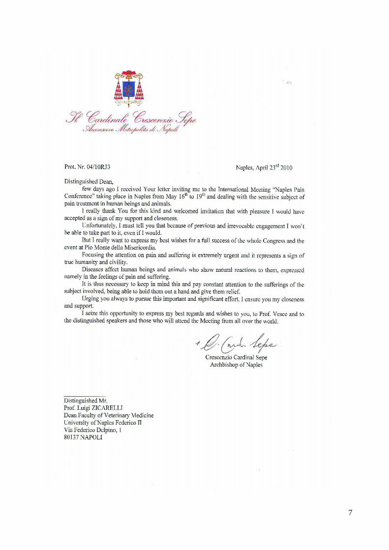
MESSAGE FOM HIS EMINENCE THE CARDINAL CRESCENZIO SEPE ARCHBISHOP OF
NAPLES
GESUALDO DA VENOSA: «Musicorum Princeps, Doloris Princeps » Kathy Toma
ANIMAL PAIN IN LATIN CHRISTIAN POETRY Antonio V. Nazzaro

5

6
7

8
“... O miei cari sospiri
Miei graditi martiri...”
GESUALDO DA VENOSA : «Musicorum Princeps, Doloris Princeps »
Kathy Toma
For the opening of this congress devoted to Pain one could not imagine or dream of a more perfect
place to evoke the remarkable individual, Carlo Gesualdo, Prince de Venosa, as it was his own
beloved uncle, the cardinal, then archbishop of Naples, Alfonso Gesualdo, who was behind the
founding of this venerable institution. The figure of the great composer of madrigals, of the
“musician-murderer,” best known for the dismal tragedy of killing his gorgeous wife and her lover,
has been shrouded in a sulfurous cloud that still contains mysteries (1). Almost forgotten for
centuries, the art of the “musician-murderer” was virtually rediscovered by Stravinsky in mid-
twentieth century and has continued to inspire, today more than ever, writers, poets,
cinematographers, composers, musicians, screen writers and, even, psychoanalysts. From Huxley
to Schnittke, Herling, Schifano, Iudica, Werner Herzog, Dominique Fernandez, Krausser, Francesco
D’Avalos, Sermonti-Francesconi, Tracanna-Guarino, Sciarrino, …and Bernardo Bertolucci, (the list
is even longer…)
Since then, the interest aroused by his modernity has steadily grown among those sensitive beings
who, after the horrors of world wars, genocides and the Holocaust, have seen ideologies crumble,
the birth of existentialism and the awareness of the absurd.
Like Giordano Bruno and Caravaggio, the Prince of Venosa belongs to the cursed breed of artists, to
the visionaries, whose thought travels at supersonic speeds in a world not ready to welcome them,
preferring to burn them on the pyre of intolerance. The dichotomy between their individuality and
their place in society in a particularly oppressive age, when Counter-reform and Inquisition reigned,
could only have been a source of heartbreak. The experience of physical and psychological pain in a
world that rejects them will carry them, through the catharsis of their art, to the realm of the
sublime.
The Prince de Venosa is the Prince of Pain. Very early, at the age of seven, he was to lose his
mother, Geronima Borromée –sister of the future saint - and the niece of Pius IV. As well, from very
young his health was fragile: he had asthma (the malady “del castrone”); later he would suffer for a
long time from an unhealed fractured leg after falling from a horse; but, more than that, he would be
sickened for life, poisoned by a horrible potion prepared by a witch at the request of a concubine
who refused to be rejected. (2) The two women would end up in the dungeons of the Gesualdo's
château after their trial.
Psychological pain would be his companion throughout his life.
Betrayed once, Gesualdo will be so doubly by his destiny which obligates him to become, a first
time at eighteen years old, head of the family at the death of his older brother, Luigi, in the year
1584: a terrible year for him marked by two other bereavements – the death of his beloved
grandfather, Luigi, who bequeathed to the family the title of Prince, and that of his uncle, Charles
Borromée.
Gesualdo, far from wanting to devote his life to a “career as prince,” thought only of assuaging his
singular passion: music.
The second betrayal, that of his wife Maria D'Avalos whom he wed in 1586, will feed all the
gossips of the time causing him to become the laughing-stock of the entire city of Naples. He will
have to face the horror of the code of honor then in force. And so the beautiful Maria D'Avalos and
her lover, the Duke Fabrizio Carafa D'Andria, caught in the act, will be killed in the palace of San
Severo in Naples the night of the 16th
of October 1590 by Carlo Gesualdo who fled Naples the same
night and sought refuge in his château in Gesualdo.
This terrible tragedy, this moment of madness when everything in his life turned upside-down at the

9
age of twenty-four, is only a prelude to a long series of misfortunes that will batter him relentlessly
like a curse (3). In 1600 Alfonsino died, the second son born of his second wife, Leonora d'Este,
whom he wed in Ferrara in 1594, quite probably poisoned, he as well, in Gesualdo by the same
witch. Carlo allows himself to die of despair, the 8th
of September 1613, after writing his will,
following the tragic death of Emanuel, his only son with Maria d'Avalos, fallen from a horse during
a hunt, the 20th
of August, at the age of twenty-six.
“Io Tacero, ma nel silenzio mio
Le lacrime e i sospiri diranno i miei martiri...”
Even if the gesualdien tears and sighs are part of the “saturnine” tradition, including the celebrated
Mélancholie by Dürer, as well as the Lamentations de Saint Pierre by Roland de Lassus, or flow my
tears by Dowland or the Lamento d'Arianne, the gesualdien pain is distinct from all the others. Its
accents cannot be coded, allying archaisms and modernism, pushing to the limits the potential of
counterpoint and dissonance, showing strong affinities with certain painters, as with El Greco or
Caravaggio, in their treatment of light, color or the increase or decrease in perspective.
(We may one day discover links between the Gesualdo family and Caravaggio not yet explored, as a
painting by Caravaggio (a Salvatore) is mentioned in the inventory of the Château Gesualdo).
Pontormo, by his science of color, the richness of his tones and the exceptional silence that
envelopes his floating world, Rosso Fiorentino whose fantastic purplish palette is like a sigh
exhaled from the Responsoria and Cosmè Tura belong to the same dream-like universe.
Who knows if Carlo had the chance to see his extraordinary painted panels from the organ of the
Ferrara Cathedral where the Princess is pictured as contorted by anguish? With a marvelous
gesturing of her hands she captures a strange light that also illuminates a portion of her face where
through her open mouth a cry escapes from between her teeth. The archaisms and the modernism of
Cosmè, the mystery and depth, the mixing of sacred and profane are of the same order: the
oxymoron's alchemy...
“…Enfer ou Ciel, qu’importe!” -La musique pour ne pas mourir-
In Carlo's last piece of sacred music, the Responsoria et Alia ad Officium Hebdomadae Santae
Spectantia ,(1611) probably his spiritual and artistic testament, we are surprised by the process of
his identifying with the passion of Christ. Through the drama of God's son, abandoned and betrayed
by all, is revealed the profound solitude of man/Carlo lambasted by pain who seeks answers to his
questions, perhaps some alleviation of his interminable suffering.
In the last two Books of the Madrigals (V and VI), a contemporary publication, where the form “a
sei voci” (for six voices) dominates, the same existential misery resonates.
There love is a corollary of death: Madrigaux et Répons intermingle, no border separates sacred
music from profane. The eroticism of religion is counterpart to the mysticism of love in a brilliant
oxymoron. The religion of love travels between sacred and profane.
Life charted the fate of the Prince: his art is the only reply/question he can offer as it rises up with a
terrible radiance from the disturbing chiaroscuro of his soul, amidst a silence yet filled with voices:
“...che nel silenzio ancor son voci e prieghi”
When we hear: “mercè grido, piangendo/ma chi m’ascolta?” The expression of man's existential
solitude reaches the infinite as in the despairing narrative of Christ in the Responsori: “Posuerunt
me in deserto solitudinis” and especially when the cry arises “quid me dereliquisti ?”. Questions
without answers.
The key word, “tradidit”, repeated insistently, expresses supreme betrayal, while “flagellatum”
beats on the martyred flesh of the Grand d’Espagne, the Prince de Venosa, known to have submitted
to masochistic sessions of flagellation, reported by Michele Giustiniani (4) and by Ferrante della
Marra (5) and for whom the famous doctor Tommaso Campanella (6) provided the diagnosis.

10
The manic tones of the “Io moro” reach the cosmic shores of unfamiliar regions. The music
becomes prayer, the interpellation is universal, the question crosses space and time to reach us, and
beyond, and remains unanswered, eternally unresolved:
“O vos omnes qui transitis per viam, attendite et videte si est dolor similis sicut dolor meus”, an
unending quest “pietas” in this piece already put to music by Carlo in the Sacrae Cantiones of 1603
that ends with the words “dolor meus”- presence of my suffering self – of an extreme softness, like
a puff of air, like the last sigh that disappears into eternity.
Notes
1- cf. Orsola Tarantino Fraternali et Kathy Toma, Gesualdo da Venosa Fasti dimenticati di un
principe del Rinascimento Luciano de Venezia, Salerno 2009
2- A. Cogliano, Il Principe, l’amante, la strega, ESI, Napoli 2004
3- G..Watkins, The Gesualdo Hex, Music, Myth, and Memory, Norton & Norton,
New-York, London 2010
4- F. Vatielli, Il Principe di Venosa e Leonora d’Este, Milan 1941
p. 67: lettera a Giulio Giustiniani del 10 ottobre 1674
5- C. Gray, and P. Heseltine, Carlo Gesualdo, Musician and Murderer, London 1926 cit.,
pp.49-50: Rovine di Case Napolitane del suo tempo, 1632
6- Ibidem, cit., p.51 Thomas Campanella, Medicinalium juxta propria principia ( lib. 111, art
12) 1635. The writer, in attributing to flagellation the virtue of curing intestinal obstructions,
adduces in proof of his assertion the case of Gesualdo: “Princeps Venusia musica
clarissimus nostro tempore cacare non poterat; nisi verberatus a servo ad id adscito”, in
chapter “Monstruosa Cura”(The Prince of Venosa, one of the best musicians of this age,
was unable to go to the stool without having been previously flogged by a valet kept
expressly for the purpose).

11
ANIMAL PAIN IN LATIN CHRISTIAN POETRY
Antonio V. Nazzaro
1. Pain is a typical condition in men and animals, our minor brothers. Over the centuries scientists
have enormously contributed to alleviate pain both in men and animals and to defeat recurrent
forms of lethal epidemics, with the result to decrease death rate mainly in the wealthier regions.
Unfortunately nothing can be done to defeat totally recurrent and devastating forms of illness.
Through the centuries the theme of pain both in men and animals has been widely dealt with in
many philosophical and theological speculations, in literature, poetry and all forms of art all over
the world.
Many thinkers and philosophers have questioned the existence of pain in the world, at times seen as
a means of redemption by Christian theologians. Poets, painters and musicians have portrayed both
individual and cosmic pain in immortal masterpieces.
In the Bible all animals, creatures of the Almighty, show signs of His divinity and share men’s
destiny on earth.
Qoèlet says: « Also to them is the event of the sons of man, and the event of the brute; one event
befalls them: as is the death of the one, so also the death of the other, ant there is one breath to all:
and what has the man more than the brute? Nothing; for all is vanity»(3, 19)
According to the Apostle Paul animals as well as men take part to redemption and spiritual
freedom, having both experienced pain and labor in delivery( Rom 8, 22).
In their writings Christian writers and poets were influenced by pagan poetry and inspired by the
Bible.
2. As to my paper subject, I’ve to mention Severus Sanctus Endelechius, friend of Paulinus
Nolanus (IV-V century), who is the author of De mortibus boum (The Death of the Cattle). This
short poem in 33 asclepiadean stanzas is closely modelled on Virgil’s Eclogue I and Georgics III
(474- 566).
In these verses the Latin poet describes a serious plague infection among oxen in the region of
Norico. He clearly had in mind De rerum natura 6, 1136-1284 of Lucrece, who deals with
pestilence in Athen in 430 b. C. In both poems pestilence causes a very high mortality rate and in
reading their lines we almost forget that the virgilians victims are animals. For he describes the
event with such pathos, suffering and emotion for the oxen’s death caused by air and water
pollution.
The poem De mortibus boum by Endelechius, considered as a poem of religious propaganda, tells
of a plague, which devastates vast areas of central and western Europe, finally reaching Gaul where
the poem is set. While the shepherd Buculus is complaining with Aegon about plague that killed
all his animals, comes on stage Tityrus who tells his cattle were saved by having the sign of the
cross made on their foreheads. Then Buculus and Aegon are converted to Christianity. At the end
of the poem three shepherds leave the pastoral landscape and set off for the city to go to church.
3. Now i’ll read to you my translation of latin christian poem about the Death of the Cattle:
<Aegon> Why do you miserably sigh, Buculus, wandering alone with your sad eyes downcast?
Why do copious tears stream down your cheeks? Please, allow your friend to know this.
< Buculus> Aegon, I beg you, let me keep my painful emotions in a deep silence, for he who
reveals his troubles opens the wound; on the contrary, he who stifles them in silence heals them.
<Aegon> The opposite of what you say is true; your claim is false. For a burden shared becomes
less heavy, but what is covered over boils up more fiercely. Talking helps to relieve pains.

12
<Buculus> You are aware, Aegon, what a large flock I had, and that my cattle grazed beside every
river: they filled even the hollow valleys, the fields and the mountain ridges. Now all hope based
on my wealth has vanished and what my long work produced throughout all my life lost in a two
days’ time. So swift is the running toward troubles!
<Aegon> This painful pestilence is now said to be spreading. Not long ago it caused the
Pannonians, Illyrians and Belgians terrible destruction, and now it is attacking us, too, in its foul
progress. But you, who know how to drive out harmful pestilence with medicinal juices, why do
you not forestall the danger you fear by applying your healing hands?
<Buculus> There are not warning signs of such fear. What the disease attacks it also destroys, it
admits no lingering, allows no delay. Thus death anticipates the pestilence. To the farm carts I’d
yoked oxen of strong build chosen as carefully as possible. Both of them shared the same thoughts,
and their bells tinkled in harmony, both the same age, with the same colour bristles, they were both
gentle, both equally strong and they had the same fate, for in mid-course the pair of them collapsed
in identical death. I was sowing the emmer deep in the softened earth, the clods were crumbling
after the copious rain: the plough moved easily through the furrows, nowhere did the ploughshare
stick. The ox on the left, who was tamed in the past summer, collapsed suddenly and fell: at once I
unyoked his grieving partner, nor fearing further misfortune now. But faster than one could say,
death seized him although he had always been healthy before. Now he, jerking his flanks with long
fits, bent his head worn out.
<Aegon> I feel anguish, and torment, sorrow and grief, for my heart is shattered by your losses, as
if they were my own; and yet I believe your herd is now safe?
<Buculus> O dear! I’m going where I must expect something far worse. For it would be some
confort in my trouble, if only a little, if a following litter had given to me what this plague had
taken away. But, who would have believed the litter were killed at the same time? I myself saw the
pregnant cow headlong: I saw two lives destroyed in the one body. Here a heifer, refusing the
spring water and neglecting the grass, wanders on wobbly legs, but she cannot escape infection,
for she falls heavily, tripping on death chains. Over there is a calf, who just now frolicking around
had outlined fanciful caprioles, goes to suckle his mother; but soon he sucks the infection from the
diseased udder. When his mother, wounded by this sorrowful pain, saw her calf closing his eyes,
she repeatedly bellowing and pitifully groaning, collapsed, longing for death. Then as if she feared
that thirst might choke the parched throat, while she lay there dying too, she moved her udder to her
calf who was already dead. Love remains strong even after death. There is the bull, husband and
father of the healthy herd, with his strong neck and broad forehead; while he
was happy and very proud of himself, he falled heavily in the grassy meadow. As many are falling
leaves of which the wood is stripped when lashed by the icy north wind, as thickly as the
snowflakes flutter in a blizzard, so numerous are the cattle which have died. Now the whole ground
is covered with carcases, their bodies swell with the bloated bellies, their eyes are white with livid
patches, their legs stiff with rigid hoof. Already flocks of baleful and grim birds are hovering;
already packs of dogs press round to tear the bowels and feed on them. Alas, why not on mine
also?
<Aegon> Why, I ask you, why the death’s grim fate is so inconstant, that it passes some over, but
striks others? Look at Tityrus, happily driving his healthy flick.
<Buculus> Yes, I see him now. Come tell us, Tityrus, which God has saved you from these
troubles, so that the plague that ravaged your neighbours’ flocks has not affected yours at all?

13
<Tityrus> The sign wich is said to represent the cross of God who alone is worshipped in the big
cities, Christ, the glory of the eternal God, whose only Son he is, this sign, made over the forheads,
brought my cattle’s sure salvation, and for thi reason this powerful God is now truly hailed as our
Saviour. The raging plague at once fled from the herd, the pestilence lost its strength. But if you
wish to pray to this God, it is enough to believe. Faith alone makes your prayer effective. His altar
is not dripping with blood, the disease has drived off by no slaughter, but simplicity and purity of
mind obtain the desired rewards.
<Buculus> If you sure about this, Tityrus, I will without delay begin to perform the rites of the true
faith, I will gladly flee from the old error, for it is deceptive and illusory.
<Tityrus> Already I am keen to hurry and visit the temple of almighty God; come, Buculus, let us
go together – it is not far – and acknowledge Christ’s divinity.
<Aegon> Please, join me to your happy plan. For how could I doubt that mankind, too, will for ever
benefit from this sign which overcame the powerful plague?
To sum up, this poem clearly shows the Christian poets are very sympathetic to animal’s pain.
Thank you!

14
Conference Opening
John Bonica memorial lecture:
“THE PAST, PRESENT, AND FUTURE STANDARDS FOR MANAGEMENT OF PAIN”
Charles E. Short DVM, PhD, DACVA, DECVA
Testimonial: John Bonica at Naples – Prof. Alberto del Genio M.D.

15
THE PAST, PRESENT, AND FUTURE STANDARDS FOR MANAGEMENT OF PAIN
Charles E. Short, DVM, PhD, DACVA, DECVA
Cornell University, College of Veterinary Medicine, Department of Clinical Sciences, Ithaca, NY
USA - [email protected]
Even before there was a standard for the management of pain, it was recognized that pain was
associated with natural conditions, trauma, and disease. The concept of pain involving the nerves
and brain can be traced back to 566-260 B.C. in ancient Greece. It was established in ancient Rome
that inflammation is a phenomenon of redness, swelling, heat, and pain. Galen (131-200 A.D.)
defined three classes of nerves: soft nerves for sensory function, hard nerves for motor function, and
“third type” for pain sensation.
The ancient standard for management of pain included combinations made from the poppy,
mandragora, hemp, or henbane as a potion or combined with wine. A major task of Jesus Christ was
to heal the sick and banish pain and suffering in the Judeo-Hebraic civilization.
The use of morphine and local anesthetics were the standard therapies with or without general
anesthetics including diethyl ether or chloroform as recently as 50 years ago. In many conditions,
the fear of uncontrolled pain was worse than the concern for the outcome of the health problem.
Has there been progress toward an acceptable present standard for management of pain? Where are
we now?
The management of pain is a major medical challenge for the relief of suffering in animal and
human patients. It is of global concern likely to effect up to 45% of the human population at some
time and countless animals both domestic and wild. In 1990, John Bonica, MD, reported the
incidence of unrelieved pain in acute and chronic conditions varied from 30-100% depending on the
condition and the therapy provided. The percent of the population requiring therapy for moderate-
to-severe pain was remarkable. The incidence of unrelieved moderate-to-severe pain in animals was
not available. Considering the available medications and the nonspeaking animal patient and the
lack of emphasis on treating animal pain, it should be obvious at that time the need for major
improvement was highly significant.
Fortunately during the last 20 years, much progress has been made. Now new and better analgesics
are available. The scientific base for concepts of biochemical and neurologic mechanisms of pain
has been much improved. Studies in the pharmacokinetics of our medications coupled with better
diagnostic and monitoring capabilities now enhance improved patient care.
Our attitude toward providing pain management also changed remarkably. Pain became a vital sign
in our patient evaluations regardless of presenting symptoms or treatment requirements. Effective
pain management became a standard of good medical practice in both medical and veterinary
medical hospitals. Educational programs within our colleges and universities coupled with
advanced scientific research expanded the new knowledge to the medical professions. The efforts of
the pharmaceutical industry and regulatory agencies provided new therapies for safer and more
effective treatments.
All the national and international pain organizations, including the International Association for the
Study of Pain, actively promoted a standard of pain practice. The American Animal Hospital
Association adopted a required standard for all accredited hospitals in 2003. This was a major
milestone in improving pain management in companion animals. Pain is a multispecies concern; but
when one or more organizations require a standard of management, other improvements will follow.
Having a standard for acute and chronic pain management would be of little value if modern
analgesics were not available. The major emphasis on new nonsteroidal analgesics has been crucial
to improving chronic pain management and a major factor in post-surgical pain for orthopedic
patients.
Morphine continues to be an effective analgesic with many indications. It is widely used in man and

16
animals especially where cost of medication is of major concern. Other opioid agonists such as
hydromorphone and members of the fentanyl family have become widely used analgesics with
multiple dose methods (oral, injectable including constant rate infusion and transdermal).
Buprenorphine and butorphanol are being used more frequently in some patients than the mu-
agonist opioids. It is not unusual for either of these to be combined with an α2 agonist for effective
pain management.
New opioids and treatment modalities have improved comfort in the dental, surgical, and cancer
patients. Fortunately, other medications including α2 agonists and anticonvulsants may be used to
supplement other treatments in difficult pain syndromes.
Dexmedetomidine used alone or in combination has been found to be effective in select animal and
human patients. Both xylazine and detomidine became major analgesics in the horse and can also
be used in cattle.
Patients with neuropathetic pain do not always get needed relief with opioids alone. Gabapentin has
shown great promise in some of these patients, especially as a supplement to more traditional pain
therapy.
Pain management can now be individualized for each patient and the presenting condition. This is
true in medicine and in veterinary medicine. In acute pain, management usually extends until the
tissues involved heal. In contrast, treatment of chronic pain extends for long duration especially in
cases of cancer or severe arthritis.
We have learned pain management should accompany all surgical procedures. Gone are the years
when it was acceptable to consider a stormy post-surgical recovery was just a bad anesthetic
response. Isoflurane or sevoflurane recoveries are very rapid. It has become evident that concurrent
pain management is the only way to assure comfort in almost all patients as they arouse from the
anesthetic state. We have learned to balance the use of analgesics and anesthetics for a high standard
of management of the surgical patient.
The populations of both people and many animals now have an extended life span. As a result,
chronic conditions are major medical issues. The treatment of cancer is not limited to surgery,
chemotherapy, or radiation. It must also include patient comfort. The primary difference between
people and animals in severe, uncomfortable cancer pain is that in animals an overdose of anesthetic
is acceptable when there is no longer hope for recovery. Sometime that is the only humane action
left as a standard of animal care.
Pain scoring is much more complicated in animals and in infants. It is difficult to determine the
level of pain by observation in many cases. As a result, the choice of medication and the dosage
often varies and in many cases appears inaccurate. Fortunately, biochemical markers and advanced
techniques to monitor the brain and nervous system have provided much needed data to help the
professions more accurately treat with a safe and effective protocol.
We now can evaluate new medications based on its uptake, distribution, and elimination; the
changes in EEG and other neurologic indicators with and without noxious stimulation; any changes
in stress hormones; the hemodynamics; and alterations in behavioral pattern. Studies of these
parameters have already been made during research and development. Similar studies of drug
combinations are necessary due to the interactions in multimodial pain medication.
Now we are at a crossroads in the management of pain. In many practices and hospitals, it is
practiced at a high standard providing comfort to both acute and chronic patients. However, in
many others, the standard of pain management is unacceptable. Why? There is a long list of
reasons. The list includes lack of effective medications in many parts of the world, economics, lack
of knowledge, customs, and in some cases a lack of concern. What is next?
Will slow release injectable opioids join the list of slow-release oral opioids? Extensive studies of
slow release injectable sufentanil showed it is possible. Will slow-release NSAIDs be significant in
the future? Two long-lasting NSAIDs (mavacoxib and robenacoxib) have been developed. The
promise of a better understanding of pain management options will contribute to a higher standard
in pain management.

17
This has especially been beneficial in canine osteoarthritis when several predominantly COX2
NSAIDs have been approved along with one dual channel NSAID. The advances for feline chronic
pain have progressed at a much slower pace. NSAIDs continue to be a major medication in equine
orthopedic pain.
NSAID usage in people continues to be a major factor in inflammatory pain despite the removal
from the market of some effective medication due to adverse side effects including cardiovascular
concerns.
The ideal standard of pain management for the future would be global in scope. It would follow the
principles of clinical pain management so well practiced and taught by Dr. John Bonica. The core
for this future standard will be knowledge and available medications worldwide. It will take
cooperative efforts by industry, government, academia, and the patient care providers. The
development of new analgesics is important, but likewise is the availability of inexpensive
analgesics to economically poor countries. It is important to learn through research the biochemical
mechanisms and the influence of new medications on specific nerve pathways. But it is also
important to teach the medical care providers how to practice practical and effective pain
management for their patients.
The selection of medications and the route of administration is not the same for all creatures great
and small. The combination of medications for the surgical patient is not the same for the patient
with severe osteoarthritis. Each patient, however, has the right to pain management and we have an
obligation to provide comfort.
We have the opportunity by working together as part of the international medical community for the
relief of pain and suffering to make a difference for countess patients. This objective is truly a part
of the one medicine concept to serve all creatures great and small including the human race. Our
efforts will be a tribute to the vision and works of Dr. Bonica, to whom we are so indebted for his
leadership.
18
PAIN and SLEEP – PAIN in ANIMALS – ACUPUNCTURE ANALGESIA
PLENARY SESSION
PAIN and SLEEP
CHAIRMEN: Lydic - Vesce - Di Marzo
NEUROCHEMICAL MODULATION OF SLEEP AND PAIN
Ralph Lydic
“BRAIN REGIONS AND NEUROTRANSMITTERS MEDIATING OPIOID INDUCED
SLEEP DISRUPTION”
Helen A. Baghdoyan
GATING OF SENSORY INFLOW THROUGH NOCICEPTIVE PATHWAYS DURING
SLEEP
Peter J. Soja
IMMUNE RESPONSE, PAIN AND SLEEP: BRAIN MECHANISMS MEDIATING
CYTOKINE EFFECTS ON SLEEP
Luca Imeri
MESOLIMBIC CIRCUITS FOR ANIMAL PAIN AND ANALGESIA.
D. Fonda

19
NEUROCHEMICAL MODULATION OF SLEEP AND PAIN
Ralph Lydic, University of Michigan
*.PPT 1
1. Acknowledgments: This work supported by National Institutes of Health Grants HL40881,
HL57120, HL65272 and by the Department of Anesthesiology.
2. Conflicts of Interest: None to declare
*.PPT 2
Outline: Sleep Relevance for Pain Management
*.PPT 3 and 4
Relevance of Sleep: If one lives to be 70 years old, 21 years will be spent in NREM sleep and 6
years will be spent in REM sleep.
*.PPT 5
Modifiable Risk Factors for Disease. It is estimated that 30% of diseases are genetic and 70% are
promoted by lack of healthy life-style issues such as sleep, diet, and exercise.
*.PPT 6
U.S. Institute of Medicine considers sleep disorders “An unmet public health problem.”
*.PPT 7 and 8
Outline: Sleep, like pain, is an altered neurobehavioral state
*.PPT 9
Multiple (not single) regions of the brain regulate sleep and wakefulness.
*.PPT 10
Pain, like sleep, is regulated by anatomically distributed neuronal networks
*.PPT 11
Sleep is actively generated by the brain. Sleep is NOT the passive loss of wakefulness.
*.PPT 12
Multiple neurotransmitters contribute to the regulation of sleep and wakefulness.
*.PPT 13 and 14
Normal sleep, like cardiac and pulmonary function, is temporally organized.
*.PPT 15
Loss of normal, restorative sleep is a key complaint of patients experiencing pain (Bonica, 1990)
*.PPT 16
Sleep, like pain, alters autonomic physiology
*.PPT 17 and 18
Outline: Pain medications disrupt normal sleep

20
*.PPT 19
Sleep disruption increases pain perception
Slide 20, 21, 22
Opioids disrupt REM sleep, in part, by decreasing acetylcholine release in the pontine reticular
formation.
*.PPT 23-24
Opioids disrupt NREM sleep, in part, by decreasing acetylcholine release in basal forebrain.
*.PPT 25
Sleep disruption caused by pain medication contributes to post-operative cognitive dysfunction in
some older patients.
*.PPT 26
Age is a risk factor for opioid-induced respiratory depression (Etches, R.C., Can J Anaesth 41: 125,
1994). In Italy and the U.S. health challenges associated with aging is a major public health
concern.
*.PPT 27
By what mechanisms to opioids disrupt sleep?
*.PPT 28, 29, 30
Opportunities for Research and Improving Patient Care
*.PPT 31 and 32
Can we achieve pain relief without sleep disruption?
Selected Readings:
- Osman, N.I., H.A. Baghdoyan, and R. Lydic. Morphine inhibits acetylcholine release in rat
prefrontal cortex when delivered systemically or by microdialysis to basal forebrain.
Anesthesiology 103: 779-787, 2005.
- Watson CJ, Lydic R, Baghdoyan HA. Sleep and GABA levels in the oral part of rat pontine
reticular formation are decreased by local and systemic administration of morphine. Neuroscience
144: 375-386, 2007.
- Nelson AM, Battersby AS, Baghdoyan HA , Lydic R. Opioid induced decreases in rat brain
adenosine levels are reversed by inhibiting adenosine deaminase. Anesthesiology 111: 1327-1333,
2009.

21
“BRAIN REGIONS AND NEUROTRANSMITTERS MEDIATING OPIOID INDUCED
SLEEP DISRUPTION”
Helen A. Baghdoyan, University of Michigan
Support and Conflicts of Interest: This work was supported by National Institutes of Health
Grants MH45361, HL57120, and HL65272, and by the Department of Anesthesiology,
University of Michigan. There are no conflicts of interest.
Background and Clinical Relevance: Sleep states, anesthetic states, and mental states are
modulated by overlapping brain regions and neurotransmitters. This talk will focus on the
sleep- and pain-related actions of GABA and adenosine in the pontine reticular formation.
Background and Clinical Relevance: Opioids disrupt sleep; pain disrupts sleep, and sleep
disruption worsens pain.
Background and Clinical Relevance: GABAergic transmission in the pontine reticular
formation promotes wakefulness: Most drugs that are used to produced sleep, sedation, or
general anesthesia increase transmission at GABA-A receptors. However, GABAergic
transmission in the pontine reticular formation actually increases wakefulness. This finding
emphasizes the need to understand drug effects on a brain-region-by-brain-region basis.
Results and Interpretation: Morphine acting at mu-opioid receptors in the pontine reticular
formation decreases endogenous GABA levels and disrupts sleep. Thus, the pontine reticular
formation may be one site where morphine acts to cause disturbed sleep.
Background and Clinical Relevance: Adenosine is a neuromodulator that promotes sleep by its
actions at the level of the pontine reticular formation and the basal forebrain. Adenosine also
contributes to pain management.
Results and Interpretation: Morphine and fentanyl decrease levels of adenosine in the pontine
reticular formation and in the substania innominata region of the basal forebrain. These data
suggest that sleep disruption caused by opioids may be mediated by decreasing the sleep-
promoting effects of adenosine.
Background and Clinical Relevance: Buprenophine is an opioid that acts as a partial agonist at
mu-opioid-receptors and an antagonist at kappa and delta opioid receptors. Buprenorphine is
used effectively as an analgesic. The effects of buprenophine on sleep an on adenosine levels
in regions of the brain that regulate sleep have not been reported.
Results and Interpretation: This presentation will report new data showing that in rat 1) an
antinociceptive dose of buprenorphine causes significant sleep disruption; 2) co-administration
of the benzodiazepine receptor agonist eszopiclone reverses the sleep disruption caused by
buprenophine; and 3) buprenophine decreases levels of adenosine in the substantia innominata
region of the basal forebrain. These findings support the interpretation that opioid-induced
disruption of sleep is caused, at least in part, by decreased adenosine levels in the basal
forebrain. These data also suggest that eszopiclone may provide adjunctive therapy for sleep
disruption caused by buprenophine.
Future Directions: Understanding the brain regions and neurotransmitter systems by which
pharmacological treatments for pain disrupt sleep will provide opportunities to develop
effective countermeasures to improve patient care.

22
Work Cited:
- Lydic R and Baghdoyan HA. Neurochemical mechanisms mediating opioid-induced REM sleep
disruption. In: Sleep and Pain, edited by G Lavigne, BJ Sessle, M Choinière, and PJ Soja, IASP
Press, Seattle, pp 99-122, 2007.
- Watson CJ, Lydic R, and Baghdoyan HA. Sleep and GABA levels in the oral part of rat pontine
reticular formation are decreased by local and systemic administration of morphine. Neuroscience
144:375-386, 2007.
- Vanini, G, Watson CJ, Lydic R, and Baghdoyan HA. -aminobutyric acid-mediated
neurotransmission in the pontine reticular formation modulates hypnosis, immobility, and breathing
during isoflurane anesthesia. Anesthesiology 109:978-988, 2008.
- Nelson AM, Battersby AS, Baghdoyan HA, and Lydic R. Opioid induced decreases in rat brain
adenosine levels are reversed by inhibiting adenosine deaminase. Anesthesiology 111:1327-1333,
2009.
- Gauthier E, Baghdoyan HA, Lydic R. Intravenous administration of buprenorphine to Sprague
Dawley rat disrupts normal sleep architecture. The Journal of Pain 11:S31, 2010.
- Guzick S, Brummett CM, Baghdoyan HA, Lydic R. Microdialysis delivery of buprenorphine to
rat pontine reticular formation does not alter adenosine levels. The Journal of Pain 11:S32, 2010.
Additional Readings:
- Chhangani B, Roehrs TA, Hassis EJ, Hyde M, Drake C, Hudgel DW, Roth T. Pain sensitivity in
sleep pain-free normals. Sleep 32:1011-1017, 2009.
- Roehrs TA, Hyde M, Blaisdell B, Greenwald M, Roth T. Sleep loss and REM sleep loss are
hyeralgesic. Sleep292:145-151, 2006.
- Shaw IR, Lavigne G, Mayer P, Choiniere M. Acute intravenous administration of morphine
perturbs sleep architecture in healthy pain-free young adults: a preliminary study. Sleep 28:667-
682, 2005.

23
GATING OF SENSORY INFLOW THROUGH NOCICEPTIVE PATHWAYS DURING
SLEEP
Peter J. Soja
Faculty of Pharmaceutical Sciences, The University of British Columbia, Vancouver, BC Canada
V6T 1Z3 E: [email protected]
It is well known that humans spend approximately one-third of their lives (~25 years) in the states
of non-rapid-eye-movement (non-REM) and REM sleep. In these states, sensory and pain
perceptions are markedly diminished when compared to the awake state 1-5
. Unfortunately, the
neural mechanisms and pathways that are responsible for the reduction in pain perception during
sleep states remain a mystery and consequently represent a prime untapped reservoir of naturally
occurring targets that future research may ultimately prove to be critical for pain control and
improved sleep.
The spinothalamic tract (STT) and related trigemino-thalamic tract (TGT) are the main ascending
somatosensory pathways in the central nervous system that convey nociceptive information leading
to the experience of pain. Data from in vivo electrophysiological studies of how nociceptive
information is transmitted through these sensory channels and then modulated by higher brain
centers remain by and large confounded due to the use of invasive surgery, anesthetic drugs, and
paralytic agents 6-11
. Hence, despite the extensive literature, relating data derived from these studies
on how the natural sleeping brain regulates itself with findings on how the anesthetized brain and
spinal cord process incoming nociceptive signals is, at best, problematic.
Albeit challenging and time-consuming, appropriately designed neurophysiological recording
studies in intact behaving animals offer a powerful way to delineate the mechanisms and neural
networks underlying the interactions between the states of sleep and pain. Over the past four
decades, sporadic attempts have been made to assess how the brain modulates prethalamic sensory
inflow during sleep and wakefulness. Understanding this process is a prerequisite for understanding
how pain affects sleep. The following surveys the current state of knowledge regarding how sleep
affects sensory inflow using a “bottom-up” systems approach, beginning at the spinal and
trigeminal brainstem levels and with peripheral evoked field potentials followed by an overview of
single-unit recording experiments.
Evoked Potential Studies In the late 1960s, pioneering studies utilizing gross evoked potentials
conducted by Pompeiano and his colleagues provided seminal evidence that spinal somatosensory
neurotransmission is attenuated during naturally occurring sleep 4, 12, 13
. These reports showed that
hindlimb nerve-evoked responses recorded from ipsilateral thoracic (T12) ventrolateral quadrants or
field potentials recorded in the cat cerebellum were suppressed only during the phasic eye
movement events that are characteristic of desynchronized or “active” REM sleep, and not during
other states.
Paradoxically, sensory inflow is also enhanced during sleep, particularly REM sleep relative to
“drowsy” wakefulness. For example, Favale et al.14
delivered low-intensity shock stimuli to the
subcutaneous tissue of one forelimb of the cat. They reported that the magnitude of peripheral
shock-evoked potentials recorded in the thalamus was enhanced by 20–50% when compared to
responses recorded during drowsy wakefulness. It was not determined whether this finding
represented an increase in the excitability of thalamic neurons, or if it was due to a change in the
excitability of STT neurons 14
.
We have reported that sensory transmission through classical ascending sensory pathways,
including the STT, spinoreticular tract (SRT), and spinomesencephalic tract (SMT), are tonically
suppressed during REM sleep 15
. In our study, additional suppression occurred phasically during
rapid eye-movements and the other events that are hallmarks of REM sleep. Our findings indicated
that the cells of origin contributing to the evoked response in the ventrolateral reticular formation
24
arose from ascending projection neurons in the contralateral lumbar spinal cord 15
. The
pharmacological basis for the REM-sleep-specific suppression and facilitation of prethalamic
sensory inflow1 that was noted in these studies remains unknown.
At the brainstem level, Hernandez-Peon et al. 16
reported that field potentials representing the
summed activity of numerous neurons, elicited by tactile stimuli applied to facial receptive fields in
behaving cats, were dependent on the animals’ level of vigilance. In their studies, the orthodromic
field potential recorded in the rostral portion of the trigeminal brainstem sensory nuclear complex
(TBSNC) was maximal in amplitude during “slow-wave,” or non-REM, sleep, but was suppressed
during alert wakefulness or REM sleep. On the other hand, Satoh et al.17
reported that tooth-pulp-
evoked field potentials recorded in the rostral portion of the TBSNC did not change during
wakefulness, non-REM sleep, or REM sleep 17
. We identified a specific area within the rostral
TBSNC in which field potentials evoked from the inferior alveolar nerve and from tooth pulp are
suppressed tonically during REM sleep in a stimulus-intensity-specific manner 18
. Comparable
studies have yet to appear that assess neuronal changes during sleep vs. wakefulness at the level of
the nucleus caudalis, an area of the TBSNC considered to be the trigeminal homologue of the spinal
dorsal horn for nociceptive pain transmission 19, 20
.
Single Neuron Recording Experiments Individual trigeminal brainstem neurons have also been
monitored in unanesthetized, intact, behaving cats 16, 18, 21
. Peripheral-nerve-evoked responses of
TGT neurons have also been monitored during the sleep/wake cycle. We discovered that responses
evoked by stimulation of the inferior alveolar nerve either increased or decreased during REM sleep
when compared to wakefulness; the population mean was not affected 22
. However, when the
smaller-diameter fibers of the tooth pulp were stimulated to activate TGT neurons, the tooth-pulp-
evoked responses recorded from each cell were markedly suppressed during REM sleep compared
to the waking state. These data strongly argue for the presence of a complex gate control
mechanism that is engaged specifically during the state of REM sleep, which has features
reminiscent of those described over 40 years ago by Melzack and Wall 23
. This REM-sleep-specific
“gate” appears to regulate sensory input in a fiber-diameter-specific (or modality-specific) manner.
State-Dependent Excitability Changes of Lumbar Sensory Neurons Relatively few published
studies have recorded the activity of dorsal horn sensory neurons during the state of wakefulness.
Kishikawa et al. 24
concluded that the air puff-evoked hair-mechanoreceptor responses of
unidentified “sensory” neurons in the lumbar dorsal horn increased during REM sleep. Kishikawa
et al. 24
hypothesized that innocuous sensory inflow via dorsal horn neurons was disinhibited by
descending systems during REM sleep. An alternative explanation that was not discussed by
Kishikawa et al. (1995) is that their recorded interneurons may actually have been premotor
inhibitory neurons receiving convergent sensory afferent inputs 25
. These neurons may participate
in the descending inhibitory pathway to motoneurons that are activated during the state of REM
sleep 26
or neural pathways conveying complex excitatory amino acid-mediated drives underlying
the myoclonic twitches and jerks that occur during phasic eye movement periods of REM sleep 26-
29.
Our own chronic single-unit recording studies have utilized antidromic identification procedures, in
situ, to examine synaptic transmission through the dorsal spinocerebellar tract (DSCT), a classic
sensory pathway conveying proprioceptive and exteroceptive information rostrally to the
cerebellum 30, 31
and to prethalamic tectal nuclei 32
. DSCT neurons display moderate ongoing spike
activity (~17 spikes/second) during the behavioral state of wakefulness 33
. Hence, proprioceptive
and exteroceptive input conveyed to higher brain centers by DSCT neurons is robust during the
state of quiet wakefulness. Sensory inflow through the DSCT was found to be consistently and
markedly suppressed during the “atonia" of REM sleep when compared with other states such as
wakefulness or non-REM sleep. Neurons that contribute to the SRT, one of several consortium pain

25
pathways studied in acute animal preparations 6, 34
, and which are located near DSCT neurons in the
lumbar L3 spinal cord segment also demonstrated REM-sleep-specific depression of their ongoing
spike activities 35, 36
. This REM suppression of sensory input was attributed to a process of
postsynaptic inhibition based on the following observations: 1) decreased spontaneous spike
activity, 2) decreased peripheral-nerve-evoked mono- and polysynaptic responses, and 3) decreased
cellular excitatory responses to juxtacellularly applied glutamate 33, 35-40
. Using these three measures
of activity, the investigations revealed that the excitability of DSCT neurons, as a distinct,
homogeneous population, did not differ dramatically between wakefulness and non-REM sleep.
Furthermore, the GABAA antagonist bicuculline and glycine antagonist strychnine, when applied in
amounts sufficient to selectively block inhibition by GABA or glycine, respectively, enhanced the
ongoing and glutamate-driven spike activities of DSCT neurons during wakefulness and non-REM
sleep. This key finding suggests that GABA- and glycine-mediated inhibitory influences tonically
control DSCT neuron excitability during these behavioral states. More importantly, the inhibition of
spontaneous and glutamate-driven spike activities of DSCT neurons during REM sleep were also
markedly reduced in the presence of bicuculline or strychnine or abolished when both antagonists
were co-administered 40
. Further studies using microdialysis probes positioned in the spinal gray
matter, where DSCT and SRT neurons are located, revealed that during naturally occurring REM
sleep, glutamate, GABA, and glycine levels were markedly increased over basal levels observed
during wakefulness, whereas dopamine levels were markedly decreased 41
.
Novel Modulatory Influences during non-REM Sleep. A characteristic hallmark feature of non-REM
sleep are the 7–14-Hz spindle oscillations in the EEG which individually last from ~1–1.5 seconds
and occur every 3–4 seconds. During non-REM sleep when sleep spindles are present, sensory
inflow from the thalamus to the cortical mantle is obliterated 42, 43
. We have recently discovered
that certain SRT neurons (~50% of a sample population), but not DSCT neurons 44, 45
, present lower
spontaneous spike rates immediately before or after sleep spindles when compared with spike rates
of the same neurons during computer-tagged sleep spindle oscillations of non-REM sleep or
wakefulness. It is tempting to speculate from such findings that the spinal cord may be the first site
in the central nervous system where afferent sensory inflow to the thalamic nuclei is subjected to
de-afferentation mechanisms that are activated during non-REM sleep 42, 43
and may even partly
explain the reduction in subjective pain sensitivity in humans to certain sensory stimuli during non-
REM sleep 1. Further studies of SRT and STT neurons are sorely needed in order to tease out the
responsible cellular mechanisms and neural networks (intersegmental or supraspinal) that are
involved during non-REM sleep. Such efforts would provide important new information that may
aid in the development of new pain therapies.
Summary and Conclusions: The functional implications of the sleep-related modulation of
identified prethalamic sensory tract neurons remain an important, fundamental, and open question.
It may now be timely to consider the possibility that sleep serves an unrecognized vital function
over one’s entire lifetime, namely, to keep somatosensory input including pain in proper check and
in a manner appropriate to the organism’s behavioral repertoire. From the human perspective, when
sleep is postponed due to societal demands or other medical reasons, this exquisite control of
sensory processing may be compromised. Future studies directed at bridging these two fields of
science hold tremendous promise for providing novel therapeutic strategies for patients suffering
from inadequate sleep, chronic pain, or both.
References
1. Lavigne G, Zucconi M, Castronovo C, Manzini C, Marchettini P, Smirne S. Sleep arousal
response to experimental thermal stimulation during sleep in human subjects free of pain and sleep
problems. Pain. 2000; 84283-290.
2. McCarley RW, Hoffman E. REM sleep dreams and the activation-synthesis hypothesis. Am
J Psychiatry. 1981; 138: 904-912.

26
3. Nielsen TA, McGregor DL, Zadra A, Ilnicki D, Ouellet L. Pain in dreams. Sleep. 1993; 16:
490-498.
4. Pompeiano O. Sensory inhibition during motor activity in sleep. In: Yahr MD, Purpura DP,
eds. Neurophysiological Basis of Normal and Abnormal Motor Activities. New York: Raven Press;
1967: 323-375.
5. Sandrini G, Milanov I, Rossi B, et al. Effects of sleep on spinal nociceptive reflexes in
humans. Sleep. 2001; 2: 13-17.
6. Ammons WS. Characteristics of spinoreticular and spinothalamic neurons with renal input. J
Neurophysiol. 1987; 58: 480-495.
7. Carstens E, Trevino DL. Laminar origins of spinothalamic projections in the cat as
determined by the retrograde transport of horseradish peroxidase. J Comp Neurol. 1978; 182: 161-
165.
8. Craig AD, Kniffki KD. Spinothalamic lumbosacral lamina I cells responsive to skin and
muscle stimulation in the cat. J Physiol. 1985; 365: 197-221.
9. Craig AD, Jr., Linington AJ, Kniffki KD. Cells of origin of spinothalamic tract projections
to the medial and lateral thalamus in the cat. J Comp Neurol. 1989; 289: 568-585.
10. Willis WD. Nociceptive pathways: anatomy and physiology of nociceptive ascending
pathways. Philos Trans R Soc Lond B Biol Sci. 1985; 308: 253-270.
11. Willis WD, Jr., Zhang X, Honda CN, Giesler GJ, Jr. A critical review of the role of the
proposed VMpo nucleus in pain. J Pain. 2002; 3: 79-94.
12. Carli G, Kawamura H, Pompeiano O. Transmission of somatic sensory volleys through
ascending spinal hindlimb pathways during sleep and wakefulness. Pflugers Arch. 1967; 298: 163-
169.
13. Willis WD, Kenshalo DR, Jr., Leonard RB. The cells of origin of the primate spinothalamic
tract. J Comp Neurol. 1979; 188:543-573.
14. Favale E, Loeb C, Manfredi M, Sacco G. Somatic Afferent Transmission And Cortical
Responsiveness During Natural Sleep And Arousal In The Cat. Electroencephalogr Clin
Neurophysiol.1965; 18: 354-368.
15. Soja P, Cairns B, Kristensen M. Transmission through ascending lumbar sensory pathways:
Dependence on behavioral state. In: Lydic R, Baghdoyan H, eds. In: Handbook of Behavioral State
Control - Cellular and Molecular Mechanisms. Boca Raton: CRC Press; 1999: 521-544.
16. Hernandez-Peon R, O'Flaherty JJ, Mazzuchelli-O'Flaherty AL. Modifications of tactile
evoked potentials at the spinal trigeminal sensory nucleus during wakefulness and sleep. Exp.
Neurol. 1965; 13: 40.
17. Satoh T, Yamada S, Yokota T, Ohshima T, Kitayama S. Modulation during sleep of the cat
trigeminal neurons responding to tooth pulp stimulation. Physiol Behav. 1987; 39:395-398.
18. Cairns BE, Fragoso MC, Soja PJ. Activity of rostral trigeminal sensory neurons in the cat
during wakefulness and sleep. J Neurophysiol. 1995; 73: 2486-2498.
19. Sessle BJ. Neurophysiology of orofacial pain. Dent Clin North Am. 1987; 31: 595-613.
20. Sessle BJ. Neural mechanisms of oral and facial pain. Otolaryngol Clin North Am. 1989;
22:1059-1072.
21. Boissonade FM, Banks D, Matthews B. A technique for recording from brain-stem neurones
in awake, unrestrained cats. J Neurosci Methods. 1991; 38: 41-46.
22. Cairns BE, Fragoso MC, Soja PJ. Active-sleep-related suppression of feline trigeminal
sensory neurons: evidence implicating presynaptic inhibition via a process of primary afferent
depolarization. J Neurophysiol. 1996; 75: 1152-1162.
23. Melzack R, Wall PD. Pain mechanisms: a new theory. Science. 1965; 150: 971-979.
24. Kishikawa K, Uchida H, Yamamori Y, Collins JG. Low-threshold neuronal activity of
spinal dorsal horn neurons increases during REM sleep in cats: comparison with effects of
anesthesia. J Neurophysiol. 1995; 74: 763-769.

27
25. Jankowska E. Interneuronal relay in spinal pathways from proprioceptors. Prog Neurobiol.
1992; 38 :335-378.
26. Chase MH, Morales FR. The atonia and myoclonia of active (REM) sleep. Annu Rev
Psychol. 1990; 41: 557-584.
27. Chase MH, Morales FR. Phasic changes in motoneuron membrane potential during REM
periods of active sleep. Neurosci Lett. 1982; 34: 177-182.
28. Chase MH, Morales FR. Subthreshold excitatory activity and motoneuron discharge during
REM periods of active sleep. Science. 1983; 221: 1195-1198.
29. Soja PJ, Lopez-Rodriguez F, Morales FR, Chase MH. Effects of excitatory amino acid
antagonists on the phasic depolarizing events that occur in lumbar motoneurons during REM
periods of active sleep. J Neurosci. 1995; 15: 4068-4076.
30. Bosco G, Poppele RE. Proprioception from a spinocerebellar perspective. Physiol Rev.
2001;81(2):539-568.
31. Walmsley B. Central synaptic transmission: studies at the connection between primary
afferent fibres and dorsal spinocerebellar tract (DSCT) neurones in Clarke's column of the spinal
cord. Prog Neurobiol. 1991; 36: 391-423.
32. Johansson H, Silfvenius H. Axon-collateral activation by dorsal spinocerebellar tract fibres
of group I relay cells of nucleus Z in the cat medulla oblongata. J Physiol. 1977; 265: 341-369.
33. Soja PJ, Fragoso MC, Cairns BE, Jia WG. Dorsal spinocerebellar tract neurons in the
chronic intact cat during wakefulness and sleep: analysis of spontaneous spike activity. J Neurosci.
1996; 16: 1260-1272.
34. Ammons WS. Responses of spinoreticular cells to graded increases in renal venous pressure.
Am J Physiol. 1991; 260: 27-31.
35. Soja PJ, Pang W, Taepavarapruk N, Cairns BE, McErlane SA. On the reduction of
spontaneous and glutamate-driven spinocerebellar and spinoreticular tract neuronal activity during
active sleep. Neuroscience 2001; 104: 199-206.
36. Soja PJ, Pang W, Taepavarapruk N, McErlane SA. Spontaneous spike activity of
spinoreticular tract neurons during sleep and wakefulness. Sleep 2001;24: 18-25.
37. Soja P, Fabian L, Pang W, Taepavarapuk N, McErlane S. Peripheral nerve-evoked activity
of dorsal spinocerebellar tract (DSCT) neurons decreases during active sleep. Society for
Neuroscience Abstracts. 2001; 27: 402.412.
38. Soja PJ, Fragoso MC, Cairns BE, Oka JI. Dorsal spinocerebellar tract neuronal activity in
the intact chronic cat. J Neurosci Methods. 1995; 60: 227-239.
39. Taepavarapruk N, McErlane SA, Chan A, Chow S, Fabian L, Soja PJ. State-dependent
GABAergic inhibition of sciatic nerve-evoked responses of dorsal spinocerebellar tract neurons. J
Neurophysiol. 2004; 92: 1479-1490.
40. Taepavarapruk N, McErlane SA, Soja PJ. State-related inhibition by GABA and glycine of
transmission in Clarke's column. J Neurosci. 2002; 22: 5777-5788.
41. Taepavarapruk N, Taepavarapruk P, John J, et al. State-dependent changes in glutamate,
glycine, GABA, and dopamine levels in cat lumbar spinal cord. J Neurophysiol. 2008; 100: 598-
608.
42. Steriade M. Brainstem activation of thalamocortical systems. Brain Res Bull. 1999; 50 :391-
392.
43. Steriade M. Sleep, epilepsy and thalamic reticular inhibitory neurons. Trends Neurosci.
2005; 28: 317-324.
44. Gharapetian A, Taepavarapruk N, McErlane SA, Soja. PJ. Sciatic nerve-evoked activity of
dorsal spinocerebellar tract (DSCT) neurons around sleep "spindles" of non-rapid eye movement
(NREM) sleep. Abstract Viewer/Itinerary Planner Online. 2004; Program No. 895.2.
45. Jang W, McErlane SA, Soja PJ. Sensory inflow through the spinoreticular tract (SRT)
around sleep spindles of Non-REM (NREM) sleep. Abstract Viewer/Itinerary Planner Online.
2005; Program No. 515.1.
28
IMMUNE RESPONSE, PAIN AND SLEEP: BRAIN MECHANISMS MEDIATING
CYTOKINE EFFECTS ON SLEEP
Luca Imeri, University of Milan Medical School, Milan, Italy
Pain is a hallmark of inflammation. The central nervous system (CNS) senses peripheral infection,
subsequent inflammation and immune activation through different mechanisms. For instance,
cytokines, such as interleukin-1 (IL-1) and tumor necrosis factor (TNF), whose production is
increased in these conditions, enter the CNS through brain areas lacking the blood brain barrier.
Cytokines can also activate primary afferent nerves, such as the vagal nerves, which project to the
nucleus tractus solitarius in the brainstem, where glutamate release is increased during peripheral
inflammation.
Once cytokines are inside the CNS, they stimulate their own production and induce what is known
as sickness behavior. Sickness behavior includes reduced intake of food and beverages, reduced
interest in the physical and social environments, and altered sleep. Rapid eye movements (REM)
sleep is inhibited, while non-REM sleep is increased and fragmented.
The increase in non-REM sleep is mediated, in part, by IL-1, which interacts with specific neuronal
circuits and neurotransmitter systems in brain. Interleukin-1, for instance, inhibits the firing rate of
wake-active serotonergic neurons in the raphe nuclei. This inhibition is the result of IL-1-induced
potentiation of GABA effects on these neurons. But IL-1 increases NREM sleep also because it
potentiates serotonin (5-HT) release from axon terminals in the anterior hypothalamus (and
adjoining basal forebrain), i.e. in the only brain region where 5-HT is necessary for sleep that
naturally follows wakefulness. At this level, 5-HT i) inhibits cortically-projecting cholinergic
neurons, which are important for electrocortical arousal, and ii) stimulates IL-1 synthesis, which
inhibits wake-active neurons.
Animal studies also show that IL-1 i) inhibits cholinergic neurons in the laterodorsal tegmental
nucleus (LDT), which is part of the neuronal circuitry responsible for REM sleep generation, ii)
inhibits evoked excitatory glutamatergic responses in the same cholinergic neurons, and iii)
significantly reduces REM sleep amount when microinjected into the LDT.
Alterations in sleep amount and architecture during infection can be seen as exquisitely tailored to
support the generation of fever, which in turn imparts survival value. For instance, shivering is
crucial for the generation of fever, but does not occur during REM sleep, because active
thermoregulation is impaired during this sleep phase. This could explain why REM sleep is
eliminated during development of fever. Moreover, IL-1 interactions with brain neurotransmitters
affect other behaviors, beside sleep, and play a role in pain modulation.
Selected readings
Dantzer R et al (2008) From inflammation to sickness and depression: when the immune system
subjugates the brain. Nature Rev Neurosci, 9:46-57
Imeri L and Opp MR (2009) How (and why) the immune system makes us asleep.Nature Rev
Neurosci, 10:199-210
Tracey KJ (2009) Reflex control of immunity. Nature Rev Immunol, 9:418-428

29
MESOLIMBIC CIRCUITS FOR ANIMAL PAIN AND ANALGESIA
D. Fonda
Veterinary Clinical Sciences Department, Università degli Studi di Milano, via Celoria 10, 20133
Milano (Italy)
E-mail: [email protected]
In humans and animals, main circuits for inhibition of pain have been found to act through the
opioid disinhibition, although other mechanisms may be involved through serotonergic or
adrenergic neurotransmission. During vertebrate evolution, the opioid receptors have been shown to
arise firstly as -receptors, afterwards as - receptors, and more recently as - receptors,
and seem to be more type-selective in mammals than in non mammalian vertebrates. In animals,
opioid receptors are nowadays demonstrated to be present in mammals, birds, reptiles, amphibians
and teleost fishes [1]. Distribution of opioid receptors in human central nervous system has been
recently clarified by positive emission tomography (PET): during acute pain, -opioid receptor
binding is increased in amygdala (AMY), nucleus accumbens (NACC), thalamus (THA) and rostral
anterior cingulate gyrus cortex (ACC) [2], particularly reflecting the gate function for the pain
threshold in THA, ACC and inferior frontal cortex (FC); the coding of pain intensity in
periaqueductal substance grey (PAG) and posterior cingulate gyrus cortex (PCC); and the encoding
of unpleasantness in posterior ACC [3]. Distribution of opioid receptors in animals has been
recently better investigated. Using radioligand autoradiography, higher binding of -opioid
receptors in the FC, somatosensory (S1), and cerebellar (CeC) cortices and in midbrain (colliculus)
were recorded in the horse than that in the dog, whereas there was higher binding of -opioid
receptors in the FC of dogs and in the CeC of horses [4]. In rats, using small animal PET combined
with the glucose analogue [18
F]-fluorodeoxyglucose, electrical high frequency stimuli applied to the
sciatic nerve was followed by induction of spinal long-term potentiation (LTP) and an acute
metabolic response in S1, but by various slower metabolic adaptations in brain regions involved in
nociception and descending inhibition, as well as AMY, PAG and dorsolateral pontine tegmentum
(DLPT) [5]. In humans, using functional magnetic resonance imaging technique, neural substrates
of classic circuits for acute pain have been recently identified in THA and S1, insular (IC) and
prefrontal orbital gyrus (PFC) cortices [6]. In humans, two components of pain [7] have been
recently diagnosed by imaging technique [8], the sensory-discriminative one situated in THA and
S1, IC, and CeC, and the affective-emotional one situated in PFC, ACC, NACC, and sublenticular
extended AMY). In animals, there is now evidence in most mammalian species for the confirmation
of the former two pain components [9], to which could be added other two, hypothesized in man,
the interoceptive autonomic one, demonstrated in primates [10], and the cognitive one, suggested
by the placebo effect [11] and supposed to act also in animals [12].
Both endogenous and exogenous opioids are inhibitory neurotransmitters so that they can act only
by disinhibition of other inhibitory neurotransmitters (e.g. GABA), producing analgesia. Analgesia
may be considered as an inhibition of pain and acute pain as an alarm system. Since pain may need,
differently from the other alarm system as temperature, touch, and proprioception, to be disactivated
(analgesia) because of survival or other requirements, all vertebrates show to share with human
beings opioid, serotonergic and noradrenergic receptors, to produce pain inhibition (analgesia) in
occasion of survival, threats or other necessities, and to process emotionally pain. Because there are
many pain pathways, so there are many types of analgesia (stimulation-induced analgesia, stress-
induced analgesia, diffuse noxious inhibitory controls, counterirritation or surround analgesia, vagal
nerve stimulation-induced analgesia, fear-induced analgesia, pain-induced analgesia), involving
many other neurotransmitters than opioids and suggesting the recent approach to multimodal
analgesia. Because there are more than one pain pathway and consequently many pain mechanisms,
30
it is now recommended, also for animals, than therapeutic protocols cope with the pain
mechanisms, no longer with the pain symptoms [13].
1. In humans and animals, the main axis of this endogenous (non pharmacologic) and
exogenous (pharmacologic) opioid inhibition seems to be the periaqueductal substance grey
(PAG) and the rostroventromedial medulla (MRVM), at the mesopontomedullary level.
In the brain, pain is regulated by multiple overlapping neuroanatomic circuitries. The most
frequently quoted for the opioid inhibition is the mesopontomedullary PAG-MRVM system. The
PAG, named also central grey, is a circumventricular organ, tapestrying the aqueduct of Silvius (De
la Boe), where it takes contact with the cerebrospinal fluid. The PAG [14] is constituted of four
columns providing important connections: dorsomedial column projecting to AMY and HYP (part
of mesolimbic circuit); dorsolateral column projecting to IC and ACC; lateral column connecting
the spinomesencephalic tract with limbic system; ventrolateral column projecting to MRVM. The
MRVM is constituted of nucleus magnus raphe (NMR), nucleus paragigantocellularis, lateral part
(NrPGil) and nucleus reticularis gigantocellularis (NrGi) [15], which have shown to be involved in
endogenous nociceptive descendent inhibitory pathways, but also to transmit facilitatory inputs, to
control homeostatic physiological processes as micturition and sleep [16] or behavioral responses
(the rostral “aggressive response” including howling, the caudal “flight response” and the
ventrolateral “freezing “ or tonic immobility response, including a sympatholitic, antipredator
defensive behaviour) [17]. In the case of pain-induced analgesia, PAG projections from MRVM
provide to transfer inhibitory serotonergic (dorsal, median raphe nuclei, NMR, nucleus pallidus and
obscurus raphe), noradrenergic (NrPGil), opioidergic (NrGi) descendent inputs to dorsal horn of
spinal cord (lamina II) in order to obtain antinociception. PAG opioid disinhibition of tonic MRVM
inhibition supplied from GABAergic neurons have shown to activate these descendent inhibitory
pathways, resulting in analgesia.
22.. IInn aanniimmaallss,, dduurriinngg aaccuuttee ppaaiinn,, eendogenous opioid release are supposed to be mainly
dependent upon both the opioidergic mesolimbic loop, and the dopaminergic reward circuit.
There are in the brain many circuits providing for release of opioids. In animals, two systems seem
to be nowadays most important than others. The opioidergic mesolimbic loop, named also the “
morphine system” is constituted of opioid cells scattered in PAG, AMY, NACC, [18] and now
supposed to be correlated with striatal enkephalin system [19] and extended to HYP [20]. The
dopaminergic reward circuit, named also mesostriatal [21] or mesolimbic system [22], is constituted
of dopaminergic cells scattered in ventral tegmental area (VTA), NACC, and PFC, that can be
activated by opioid release [23] or can produce opioid release in the presence of conflicting
motivations, such as hunger or a threatening predator [21]. Recently other structures including
AMY, hippocampus (IPP), lateral habenular nucleus, substantia nigra, pedunculopontine nucleus
and the raphe nuclei have been considered to be key components of the reward circuit [24]. Using
functional magnetic resonance imaging technique, morphine infusion resulted in increased signal
changes in reward structures including NACC, sublenticular extended AMY, IPP and orbitofrontal
cortex, a decreased signal in cortical areas (sedative effect), PAG and ACC (analgesic effect) and an
increased signal in HYP [25]. Using PET, both opioid and placebo show to involve associated brain
areas, particularly rACC, suggesting the participation of higher order cognitive networks [26].
Clinically, there is now evidence for a contribution of attention and expectation on descending
modulatory pathways. Attentional modulation of pain may be obtained by visual, auditive or other
distraction, involving PAG-MRVM system: pain intensity and reactions were predictabily lowered
by distraction. Pain expectancy or anticipation seem to be the most important pain-predictive
contextual cues. Pain expectation or prediction may have negative (fear) or positive (reward) effect,
presenting a large degree of variability between the certain and uncertain pattern. Therefore, reward
may be considered the result of a certain positive expectation, whereas being afraid of pain is
recognized to increase pain intensity, in the absence of a noxious stimulus also. Through recent
functional magnetic resonance neuroimaging studies, neural substrates of certain and uncertain
expectation have shown to be the rostral ACC and posterior cerebellum, and respectively PFC, ACC
31
and IPP [27]. Ventral striatum and particularly NACC has been studied to be involved in a
particular form of reward, considered to be the addiction, Molecular mechanisms of addiction,
through valid animal models (mouse, rat), show to involve dopaminergic [28] and opioid release in
the reward-stress circuitry (central nucleus of AMY, bed nucleus of stria terminalis, and NACC)
[29]. Support for endogenous opioid-mediated pain-modulating pathways may be obtained through
both mesolimbic and reward circuits [21] and may be demonstrated by neural substrates of the
placebo-analgesia (activation of NACC, dopamine release in NACC, triggered activation of the
endogenous opioid system, ventral loop of the basal ganglia circuitry) [30]. Interestingly, these
brain areas have shown to be important for the animal low-order consciousness. In the past, seven
basic emotional feelings (seeking, rage, fear, lust, care, panic, play) were assessed in animals [31]
and recently six subcortical midline sunstrates (PAG, superior colliculi, adjacent mesencephalic
locomotor region, preoptic areas, HYP, dorsomedial thalamus) [32] were proposed as site for
animal awareness, considered to be the so called “core self” [33].
3. In vertebrate evolution, evidence for these endogenous opioid circuits makes the basis for
the homologous circuits supposed to act in human beings for modulating acute pain and
allowing the human and animal non-pharmacological analgesia.
Different cortical and subcortical regions process different component of multidimensional nature
of pain. Neural substrates of pain pathways in animals have shown to be similar [10] to those in
humans [34]. Neural substrates implicated in conditioned analgesia in animals are the brainstem to
spinal cord circuits, which are highly conserved in a variety of mammals species, including
marsupials, rodents, carnivores, and primates [35]. Importantly, the distribution of neurotransmitters
in this circuitry is also conserved across species, including rats and humans [36]. Although more
careful investigations would be necessary to be performed in single and domestic species, some
important differences have arisen in cats about the spino-reticular and spino-mesencephalic
pathways [37], the five separate spino-PAG [38] and the larger spino-parabrachial circuits [39]. In
conclusion, this endogenous opioid-mediated pain modulatory system has been recognized to exist
firstly in mammalian species and more recently in human beings, so that there is a body of evidence
supporting the notion that pain-modulating circuitry, homologous to PAG-MRVM opioid system
demonstrated in animals, are present in humans [35].
Clinically, it is important understanding that endogenous opioid release may be an important
therapeutic opportunity for animals as well as for paedriatric patients. In the last two decades, the
use and diffusion of many methods or interventions to obtain nonpharmacological analgesia have
remarkably grown in paediatric [40] and animal patients. Together with many techniques of
complementary and alternative medicine, nonpharmacological interventions are based not only on
the opioid mechanism of the mesolimbic loop, able to induce “tender love care”, but on the
management of all environmental effects of pain on the animal five senses: comfortable body
temperature (warm-cold) and moisture (dry-wet), touch contact surface (smooth-rough), minimal
light (vision) or noise (hearing) in environment, friend smell or pheromones (olfaction). Other
nonpharmacological pain management methods are based on parental effects like maternal touch,
kangaroo care, swaddling, facilitated tucking, non-nutritive or nutritive (glucose) suckling, rocking,
or physical effects on pain like cryotherapy, hydrotherapy, massage therapy, chiropractic and
osteopathic manipulations. It is worth noting that also play, laughing, tickling, pins [41], and social
contacts [42] may contribute to relief of acute pain, not only in rats. Other non-pharmacological
therapeutic techniques, applied also in animals, have shown to involve for relief of pain
mechanisms in part opioid, like acupuncture (OPP), in part anti-inflammatory, like laser-therapy,
pulse magnetic fields therapy, transcutaneous electrical nerve stimulation, ultrasound therapy,
extracorporeal shock wave therapy.
4. During chronic pain, particularly the neuropathic one, conditions and effects of these
circuits seem to change, but in humans and animals are still to be evaluated.
To understand acute and chronic pain mechanisms, the proposal of the multidimensional theory of
the body self neuromatrix [42] has shown to be essential. From four concepts derived from the

32
analysis of phantom limb phenomena (pain may occur in the absence of any inputs from the body -
stimuli may trigger pain patterns but do not produce them – the body is perceived as the “self” - the
body-self is “built-in” by genetic specification and modification by experience), Melzack has
derived a new conceptual nervous system as the theory of a body self neuromatrix, involving four
components or steps: sensory inputs sculpt their line in the neuromatrix (thalamocorticothalamic
and limbocorticolimbic circuits); repeated cyclical processing and synthesis of these nerve impulses
create in this neuromatrix a neurosignature; this neurosignature projects both in brain regions
(sentient neural hub), where it is converted and transduced in the stream of awareness, and in spinal
cord neurons to activate pattern of movements (actions) to bring about the desired goal. Thereby,
pain experience may be considered not as a purely sensory phenomenon, but a multidimensional
experience, including sensory-discriminative, affective-emotional, cognitive and interoceptive
components. Moreover, such an interpretation may be considered to be partially shared also with
other animals [44], non-human primates and vertebrates, provided that it is proportionate to
functions and connections supplied by neural substrates.
Clinically, processing of chronic pain is different from that involving the acute one. Acute pain
processing involves more than one neural substrate, possibly in the following order: at the
mesopontomedullary level, producing involuntary neurophysiological responses; in the parietal
cortex, coding pain site, intensity, type; in the limbic system, decoding pain emotion and memory;
in the prefrontal cortex, planning behaviour; in the parietal cortex, producing voluntary cognitive
response; in the basal ganglia, producing voluntary locomotory response; in the PAG, allowing pain
facilitation or inhibition (analgesia). On the contrary, processing of chronic pain, without more
interest in coding the sensory component (localization, intensity) of this repeated pain, continues to
elaborate pain emotion and memory between cortex and limbic substrates, participating with
conditioned involuntary neurophysiological responses and without being able to produce voluntary
responses. [45].
Our understanding of acute and chronic pain mechanisms requests a growing attention not only to
involved neural substrates, but also to the contribution of cellular remodelling and biochemical
changes, regarding particularly the immune mechanisms in the inhibition of pain (analgesia). A
recent challenge to conventional concepts regarding not only the acute pain, but particularly the
chronic neuropathic one, is the contribution of immune cells and glia to the development and the
persistence of pain after nerve injury. Neurons resulted nowadays to be no more the only
responsible for the pathophysiological changes that drive maladaptive, neuropathic pain. This
shifting in our understanding suggest to consider hyperalgesia and allodinia no longer maladaptive,
but adaptive because - for example- the participation of peripheral immune cells in wallerian
degeneration could be considered as a facilitation for healing and regeneration of injured axons so
to provide novel opportunities for pain therapeutic approach [46].
References:
[1] Stevens C et al, 2004, Brain Research Reviews, 46, 204.; [2] Henriksen G, Willock F 2008,
Brain 131, 1171; [3] Tolle TR et al 1999, Ann Neurol 45, 40; [4] Hellyer PW et al 2003, Veterinary
Anaesthesia and Analgesia 30, 172; [5] Hjornevikt T et al 2008, Pain 140, 456; [6] Becerra L et al
2001, Neuron 32, 927; [7] Auvray M 2008, Neuroscience and behavioral Reviews 34, 214; [8]
Becerra L et al 2004, Journal Neurophysiology, 91, 533. [9] Boyce-Rustay JM et al 2010,
Neuropharmacology 58, 537. [10] Craig AD 2003, Current Opinion in neurobiology 13, 500; [11]
Zubieta JR et al 2005, The Journal of Neuroscience 25, 7754; [12] McMillan FD 1999, JAVMA
215, 992. [13] Scholz J, Woolf C 2002, Nature Neurosci 5, 1062; [14] Carrive P, Morgan MM
2004, Periaqueductal grey. In: Paxinos G, May JF (eds.) The human nervous system. Second
edition. 2004, Elsevier, Amsterdam, pp. 393-424; [15] Heinricher MM 1997, Organizational
characteristics if supraspinnaly mediated resonses to nociceptive input. In: (Yaksh TL et al eds.)
Anesthesia: biological foundations, Lippincott-Raven, Philadelphia (USA) pp. 643-660; [16] Mason
P 2005, The Journal of Comparative Neurology 493, 2; [17] Carrive P, Bandler R 1991,Exp Brain

33
Res 84, 599; [18] Heinricher MM 1997, in: (Yaksh TL et al eds.) Anesthesia: biological
foundations, Lippincott-Raven, Philadelphia (USA) pp. 643-660; [19] Kelley AE et al 2005, The
Journal of Comparative Neurology 493, 72; [20] Fonda D 2009, Dolore e analgesia negli animali,
Point Vétérinaire Italie, Milano, pp. 85-101; [21] Fields HL 2007, Regional Anesthesia and Pain
Medicine, 32, 242; [22] Chen BT et al 2010, Ann.N.Y.Acad Sci. 1187, 129; [23] Johnson SW North
RA 1992, J Neurosci 12, 483; [24] Haber SN, Knutson B 2010, Neuropsychopharmacology 35, 4;
[25] Becerra L et al 2006, Conscious Cogn. 14, 30; [26] Petrovic P et al 2002, Science 295, 1737;
[27] Ploghaus A et al 2003, Trends in Cognitive Sciences 7, 197. [28] Kelley AM et al 2004,
Neuron 44, 161; [29] Koob GF 2006, Addiction 101 (Suppl. 1), 23; [30] Alexander GE 1986, Annu
Rev Neurosi 9, 357; [31] Panksepp J 2005, Conscious Cogn. 14, 30; [32] Panksepp J, Northoff G.
2009, Conscious Cogn. 18, 193; [33] Northoff G, Panksepp J. 2008, Trends Cogn Sci. 12, 259; [34]
Willis WD, Westlund KN 1997, J Clin Neurophysiol 14, 2; [35] Fields HL et al 2006, in:
(McMahon SB, Koltzenburg M eds) Wall and Melzack’s Textbook of pain. Fifth edition, Elsevier
Churchill-Livingstone, Edinburgh (UK), p.125-142; [36] Pittius CW et al 1984, Brain Res 304, 127;
[37] Mouton LJ et al 2001 Brain Res Bull 54, 167-74; [38] Mouton LJ, Holstege G 2000, J Comp
Neurol. 2000 Dec 18;428(3):389-410; [39] Klop EM et al 2005, Neuroscience.134, 189; [40]
Cignacco E et al 2006, J Eur J Pain 11, 139; [41] Panksepp J, Burgdorf 2003, Physiology and
Behaviour 79, 533; [42] Langford DL et al 2006, Science 312, 1967; [43] Melzack R 2001, Journal
of Dental Education, 65, 1378. [44] Tracey I , Mantyh PW 2007, Neuron 55, 377; [45] Apkarian AV
2008, Current Opinion in Neurobiology 18, 464;. [46] Scholz J, Woolf C 2007, Nature Neurosci
10, 1361
34
PLENARY SESSION
Sessione accreditata ECM
PAIN IN ANIMALS
CHAIRMEN: LARENZA - SAVOIA - DI CESARE MANNELLI
ADVANCES IN PERIOPERATIVE PAIN RECOGNITION AND MANAGEMENT IN
ANIMALS
M. Paula Larenza
ASSESSMENT OF THE DOSE-DEPENDENT EFFECT OF BUTORPHANOL AND
MORPHINE ON THE MINIMUM ANAESTHETIC CONCENTRATION OF ISOFLURANE
IN CHICKENS
O Martin Jurado
ANALGESIC MANAGEMENT OF CHRONIC OSTEOARTHRITIC PAIN
A. Bufalari
NEUROPATHIC PAIN IN ANIMALS. PHYSIOPATHOLOGIC CONSIDERATIONS AND
THERAPEUTIC APPROACH
Giorgia della Rocca
PAIN ASSESSMENT OF INTERNAL MEDICAL DISEASES IN DOGS: PRELIMINARY
OBSERVATIONS
M.B. Conti
TARGET CONTROLLED INFUSION (TCI) OF OPIOIDS: AN OVERVIEW
Lorenzo Novello
MULTIMODAL SEDATION AND PAIN MANAGEMENT IN THE EQUINE DENTAL
PATIENT
Elizabeth F. Schilling
PAIN MANAGEMENT OF GASTRIC DILATATION-VOLVOLUS SYNDROME IN DOGS
C. Filippi
VALUE OF LOCAL ANAESTHESIA AS MEANS OF PRE-EMPTIVE ANALGESIA IN
SMALL ANIMAL SURGERY
Bruna Santangelo
LIDOCAINE CONSTANT RATE INFUSION FOR SURGICAL ANALGESIA IN DOGS
C. Cordella
ATTITUDE OF ITALIAN VETERINARIANS INTERESTED IN SMALL ANIMAL
SURGERY, TO POST-OPERATIVE PAIN IN CANINE AND FELINE PATIENTS:
PRELIMINARY RESULTS FROM A WEB-BASED SURVEY.
Sergio Maffi

35
ADVANCES IN PERIOPERATIVE PAIN RECOGNITION AND MANAGEMENT IN
ANIMALS
M. Paula Larenza, DVM, Dr.Med.Vet., DECVAA.
Assistant Professor of Anesthesiology, University of Pennsylvania School of Veterinary Medicine
Philadelphia – PA
PROGRESS IN ACUTE PAIN ASSESSMENT
When debating the nature of pain in animals, considerable parallels can be drawn with the situation
in human infants (Flecknell & Waterman-Pearson 2000). In adult humans, the ability to provide
direct verbal communication, complete pain questionnaires or scoring systems, or to directly
manage analgesic dosage using patient controlled analgesia systems allows reasonably reliable
estimates to be made of the degree of pain and the efficacy of pain control. In young human infants,
written and verbal communication is not possible; nevertheless, extrapolation from adult humans,
coupled with objective demonstrations of the adverse effects of surgical stress, has led to a huge
increase in research focused to this area (McGrath & Unruh 1989). In human infants, the most
widely used techniques have been pain scoring systems based upon criteria such as crying, facial
expression, posture and behavior.
Biochemical and endocrine parameters, such as blood cortisone concentrations or catecholamine
concentrations, have been proposed as indicating pain. A major problem in interpreting the
significance of these changes is the influence of surgery and anesthesia, which markedly alter many
of these variables, even in patients which are pain free. Therefore endocrine parameters are not
clinically helpful when assessing immediate postoperative pain. Similarly, increases in heart rate or
blood pressure that can be associated with pain, are relatively unspecific parameters as they can also
be related to hemodynamic changes as a result of surgery (Flecknell & Waterman-Pearson 2000).
The basic methodology selecting clinical signs which might be due to pain has been used to provide
pain-scoring systems during the perioperative period in veterinary clinical patients (Flecknell &
Waterman-Pearson 2000). Attempts at scoring have either used descriptive ratings converted to
numerical scores to allow for comparisons, or have used visual analogue scoring systems (VAS)
(Nolan & Reid 1993; Pascoe & Dyson 1993). Particular problems noted were considerable between
observer variation and the poor predictive value of certain of the parameters scored. It appears that
if the number of observers is restricted, and the criteria used was carefully selected, reasonable
agreement can be achieved. Even though some pain scoring systems have been efficiently used for
chronic pain evaluation, there applicability for postoperative acute pain is limited (Morton &
Griffiths 1985). A problem with many of these 0approaches is the difficulty associated with scoring
of animal behavior, motor function and food intake during the recovery phase from anesthesia.
However, scoring systems for postoperative pain assessment have been validated (Firth & Haldane
1999). More recently, a short composite pain scale has been effectively used for acute pain
assessment in dogs (Murrell et al., 2008). Although not validated, a couple of clinical studies have
used postoperative pain scales that can be used as a guide to assess postoperative pain in dogs and
cats (Rohrer Bley et al., 2004; Wenger et al., 2005).
PRACTICAL APPROACH TO POSTOPERATIVE PAIN ASSESSMENT
Pain management should be species-specific and patient-oriented, as important variations occur
among species. Not every animal will perceive pain in the same way and not always similar
procedures will cause similar painful sensations. It is important to document the results of pain
assessment and the efficacy of the treatment, as further adjustments on the overall analgesic
management strategy should be based on the individual findings. Most patients recovering from
anesthesia after surgery will experience some degree of pain. However, other patients may be pain
relieved after some procedures (i.e. repositioning of coxofemoral luxation). Some species (i.e.
rabbits, sheep) will not display easily recognizable signs of pain or discomfort and might suffer

36
quietly. Others will show dysphoria due to anesthetic or analgesic administration that might
confound pain evaluation. A good way to start is to know the normal behavior for the evaluated
species and/or to observe the patient before the surgical procedure. Monitor if the current illness
process is causing signs of pain and check the behavioral reactions to human interaction and to
foreign places. During anesthesia pre-medication check for the potential behavioral reactions to
opioids or benzodiazepines as they may cause dysphoria that might be confounded with signs of
pain during the postoperative period, particularly in some species (i.e. dogs, cats, horses).
Throughout the recovery phase from anesthesia it is important to evaluate the patient respiratory
rate and pattern and assess for anxiety. Bear in mind drugs that had been administered to distinguish
vocalization as a result of pain or side effects to these analgesics (i.e. opioids, benzodiazepines,
ketamine). Slowly approach the patient and evaluate its reaction to human interaction and palpate
gently the surgical area and also non operated areas and compare the patient reactions to both
manipulations. For species that may not display signs of pain or discomfort when approached, it is
helpful to evaluate their gait or, whenever possible, the behavior in their natural environment, from
a distance. If the patient shows abnormal behaviors (i.e. loss of appetite, decreased interaction with
environment) or is somewhat anxious and vocalizes and turns the head when palpating the surgical
area only, a test dose of an analgesic may be administered ( i.e. buprenorphine, methadone,
morphine). If administration of an analgesic diminishes the changes associated with abnormal
behaviors or manipulation of the operated area, this supports the hypothesis that the changes were,
at least in part, pain related. Clearly it is important to establish that the analgesic did not have non-
specific effects in normal animals that would influence the variable studied (i.e. extreme sedation
might be confounded with pain relief). Alternatively, if vocalization and/or agitation are observed
and a reaction to opioids or benzodiazepines is suspected, the administration of a specific reversal
might be appropriated (i.e. naloxone or butorphanol, flumazenil). Keep in mind that antagonization
of opioids might reverse their analgesic effects and other non-opioid analgesic drugs should be
considered in such situation (i.e. 2-agonists, non-steroidal anti-inflammatory drugs).
Anxiety might be the result of several factors such as pain, separation or unknown environment. If
pain has been exclude as the cause for anxiety, sedatives (acepromazine or dexmedetomidine) or
simple human interaction and tender loving care (TLC) will help in reducing the stress. It is
important to realize that many patients might be uncomfortable after anesthesia as a result of a full
bladder that needs to be evacuated. In addition, re-evaluate bandage tightness and position of the
body in the postoperative period as these factors might be also the source for stress or pain. A calm
room and/or comfortable bedding or for some species, an enriched environment will also alleviate
patient discomfort.
ANALGESIC DRUG SELECTION
The use and choice of analgesic agents are based on the species-related pharmacological profile of
these drugs and a thorough patient assessment that includes a physical exam; an evaluation of the
patient’s history, underlying or preexisting conditions, and presenting complaint; and a laboratory
evaluation of an appropriate database (Hellyer et al., 2007). A multimodal approach to pain
management allows for using lower doses of a combination of analgesic and takes advantage of
their different modes and sites of action. Commonly used analgesic protocols will include an opioid
before an elective surgery, in combination with tranquilizers or sedatives (i.e. phenothiazines, 2-
agonists) to enhance sedation and analgesia. The use a local anesthetic to block the transmission of
noxious stimuli before incision or to potentiate analgesia postoperatively, has being gaining more
adepts lately as local anesthetics have been associated with less postoperative chronic pain
incidence. Epidural catheters or wound infiltration catheters can be placed during anesthesia to
prolong the benefits of local blockade throughout the postoperative period. Prior anesthesia or
during anesthetic recovery, the use non-steroideal anti-inflammatory drugs help to decrease the
actions of inflammatory mediators released during surgery. However, the species-specific
pharmacokinetic profile and the potential side effects of these agents should be taken into
consideration. Opioids are still the most widely used agents to control postoperative pain as they

37
have minimal cardiovascular side effects. Lidocaine or ketamine infusions can be used in the
postoperative period to alleviate severe pain in combination with opioid infusions when local
blockade is not possible. Lately, tramadol has been incorporated to treat mild to moderate pain in
the postoperative period, generally associated with non-steroideal anti-inflammatory drugs. If
sedation is required together with analgesia, infusions of 2-agonists (i.e. medetomidine or
dexmedetomidine) might be a valid option.
REFERENCES
Firth, A. M. & Haldane, S. L. (1999) Development of a scale to evaluate postoperative pain in dogs.
J Am Vet Med Assoc, 214, 651-9.
Flecknell, P. & Waterman-Pearson, P. (2000) Pain Management in Animals, London, Saunders.
Mcgrath, P. J. & Unruh, A. M. (1989) Pain in children and adolescents, Amsterdam, The
Netherlands, Elsevier.
Morton, D. B. & Griffiths, P. H. (1985) Guidelines on the recognition of pain, distress and
discomfort in experimental animals and an hypothesis for assessment. Vet Rec, 116, 431-6.
Murrell, J. C., Psatha, E. P., Scott, E. M., Reid, J. & Hellebrekers, L. J. (2008) Application of a
modified form of the Glasgow pain scale in a veterinary teaching centre in the Netherlands. Vet Rec,
162, 403-8.
Nolan, A. & Reid, J. (1993) Comparison of the postoperative analgesic and sedative effects of
carprofen and papaveretum in the dog. Vet Rec, 133, 240-2.
Pascoe, P. J. & Dyson, D. H. (1993) Analgesia after lateral thoracotomy in dogs. Epidural morphine
vs. intercostal bupivacaine. Vet Surg, 22, 141-7.
Rohrer Bley, C., Neiger-Aeschbacher, G., Busato, A. & Schatzmann, U. (2004) Comparison of
perioperative racemic methadone, levo-methadone and dextromoramide in cats using indicators of
post-operative pain. Vet Anaesth Analg, 31, 175-82.
Wenger, S., Moens, Y., Jaggin, N. & Schatzmann, U. (2005) Evaluation of the analgesic effect of
lidocaine and bupivacaine used to provide a brachial plexus block for forelimb surgery in 10 dogs.
Vet Rec, 156, 639-42.

38
ASSESSMENT OF THE DOSE-DEPENDENT EFFECT OF BUTORPHANOL AND
MORPHINE ON THE MINIMUM ANAESTHETIC CONCENTRATION OF ISOFLURANE
IN CHICKENS
O Martin Juradoa, HA Haga
b, JM. Hatt
c, R Bettschart-Wolfensberger
a
aAnaesthesiology Division, Clinic for horses, Vetsuisse Faculty, University of Zurich,
Winterthurerstr. 260, 8057 Zurich, Switzerland bDepartment of Companion Animal Sciences, Norwegian School of Veterinary Science,
Ullevålsveien 72, 0033 Oslo, Norway cClinic for Zoo Animals, Exotic Pets and Wildlife ,Vetsuisse Faculty, University of Zurich,
Winterthurerstr. 260, 8057 Zurich, Switzerland
The International Association for the Study of Pain (IASP) defines that the inability to communicate
does not negate the possibility that an individual is experiencing pain and is in need of appropriate
pain-relieving treatment. Pain is a combination of both peripheral inputs and neurophysiological
processes within the central nervous system. In mammals, opioids are often chosen for moderate
pain relief,[1] while the use of opioids in avian medicine is limited and not broadly extended.[2]
The opioid drugs act on specific opioid receptors. Butorphanol is mainly acting as agonist on kappa
receptors, while the mu receptors are usually triggered by opioids such as morphine.[3-4] The
relative proportion of mu and kappa receptors in the forebrain of pigeons correspond to 14% and
76%, respectively[5] whilst in chickens (23% and 20%, respectively)[6] is remarkably close to
those in mammals (29% and 37%, respectively).[5]Therefore, depending on the similarity of other
avian species to pigeons or chickens, kappa or mu agonist opioids like butorphanol and morphine
are both options to provide analgesia in birds.
Quantification of the analgesic effect of drugs in birds is challenging and no validated scores are
available. The tools used in the avian species are based on the assessment of the evoked nocifensive
answer in response to mechanical, thermal or electrical stimulation. Quantification of changes of the
minimum anaesthetic concentration (MAC) can provide information that allows comparison
between different analgesic drugs when interacting with volatile agents. MAC is defined as the
concentration of an inhalatory anaesthetic that prevents purposeful movement following a supra
maximal painful stimulus in 50% of the individuals tested.[7] An effective analgesic dose would
lead to a reduction of the MAC.
Butorphanol has been reported to potentially have an analgesic effect based on reduction of the
requirements of inhalant anaesthetics, in cockatoos (Cacatua spp.) (1 mg/kg),[8] African grey
parrots (Psittacus erithacus) (1 mg/kg)[9] and Hispaniolan Amazon parrots (Amazona ventralis) (2
mg/kg)[10] after intramuscular administration. In chicken, the effect of various drugs on MAC has
been determined.[11] Butorphanol at a dose of 1 mg /kg IM, did not produce MAC reduction.
Morphine has been described to have an isoflurane-sparing effect in chickens after progressive
intravenous administration.[12]
However, a decrease on MAC due to the effect of an analgesic drug could lead to an inadequate
depth of anaesthesia. Objective assessment of depth of anaesthesia is performed in human medicine
by use of Bispectral Index (BIS).[13-14] In birds, first data [11] showed that BIS is a useful tool for
this purpose as well.
MAC reduction caused by opioids is not only an objective measure that quantifies the analgesic
potency of a drug, but also a goal in balance anaesthesia: if less inhalation anesthetic can be used,
cardiovascular function will be improved. This would contribute to decrease the high mortality rate
related to avian anaesthesia (5.5%) in comparison to 0.17% in dogs or 0.02 – 0.005% in human
medicine.[15]
39
The purpose of this study was to evaluate the dose-dependent changes on the MAC of isoflurane
produced by butorphanol and morphine in chickens.
Eight adult healthy chickens underwent individual MAC determination of isoflurane with the up
and down method using toe-pinch stimulation. Allowing 1-week washout period between
treatments, the individual MAC was determined after intramuscular administration of saline (0.5,
0.75 or 1 ml/kg), butorphanol (2, 3 or 4 mg/kg) or morphine (5, 7.5 or 10 mg/kg). All the chickens
received during the three first anesthesias, randomly and by a blinded observer, the lowest dosage of
every drug. Thereafter, they received the medium and the high dosage. Results are reported as
median and were analyzed using Friedman and Tukey tests when corresponded. Significance was
set at P 0.05.
The only remarkable effect measured on MAC was a 22 % reduction of isoflurane with 10 mg/kg
morphine. The MAC values for the low, medium and high dose of saline were 0.63 %, 0.60 % and
0.71 %; butorphanol 0.70 %, 0.60 % and 0.65 % and morphine 0.70 %, 0.53 % and 0.45 %,
respectively. The individual MAC of isoflurane obtained for our group of chickens was lower than
in previous studies in chickens (1.25%[12] and 1.15%[16]) and other avian species i.e. cockatoos
(Cacatua spp.) 1.44%[8], sandhill cranes (Grus canadensis) 1.34%[17], peking ducks (Anas
domesticus) 1.30%[18], or thick-billed parrots (Rhynchopsitta pachyrhyncha) 1.07%.[19]
No significant differences were observed between groups for heart rate (260 to 300 bpm) or non
invasive blood pressure (85 – 100 mmHg). Bispectral index monitored the electrical activity of the
brain revealing a correlation to the anaesthesia event; BIS for saline, butorphanol and morphine for
the low dose were 68 (45 – 80), 54 (38 – 71) and 69 (55 – 79); for the medium dose 77 (35 – 85), 72
(39 – 84) and 72 (61 – 81) and for the high dose 39 (34 – 67), 38 (34 – 46) and 36 (34 – 41),
respectively.
The dosage order was not randomized and therefore, chickens receiving the high dose of saline had
been at least 6 times under anesthesia. Further studies are needed to investigate whether repetitive
anesthesias depress the electrical activity of the brain in chickens.
We conclude that significant changes on MAC were only observed with 10 mg/kg morphine, which
produced an isoflurane-sparing effect of 22 % in the chickens of this study. These results have to be
carefully interpreted as the method is based on the reaction of the animal to supra-maximal
stimulation and thus it can be easily biased.
This study was supported by the Stiefel-Zangger Foundation and the Nachwuchsförderungskredit of
the University of Zurich.
References
1. Flecknell P. and A. Waterman-Pearson, Pain management in animals. 2000, London: W.B
Saunders.
2. Harrison and Harrison, Pain management in clinical avian medicine and surgery. 2006, Lake
Worth: Wingers Publishing. p. 233-239.
3. Atalan G. et al., Effect of medetomidine-butorphanol-ketamine anaesthesia and atipamezole
on heart and respiratory rate and cloacal temperature of domestic pigeons. J Vet Med A Physiol
Pathol Clin Med, 2002. 49(6): p. 281-285.
4. Paul-Murphy J. and J.W. Ludders, Avian analgesia. Vet Clin North Am Exot Anim Pract,
2001. 4(1): p. 35-45.
5. Mansour A. et al., Anatomy of CNS opioid receptors. Trends Neurosci, 1988. 11(7): p. 308-
14.
6. Sanyathitiseree P., Opioid receptors in the chicken brain. Thesis at the Institut für Tierzucht
der Bundesforschungsanstalt für Landwirtschaft (FAL). 2005, Tierärztliche Hochschule Hannover:
Hannover. p. 181.
7. Naganobu K., et al., Determination of the minimum anesthetic concentration and
cardiovascular dose response for sevoflurane in chickens during controlled ventilation. Vet Surg,
2000. 29(1): p. 102-5.

40
8. Curro T.G., Brunson D.B., and Paul-Murphy J., Determination of the ED50 of isoflurane and
evaluation of the isoflurane-sparing effect of butorphanol in cockatoos (Cacatua spp.). Vet Surg,
1994. 23(5): p. 429-433.
9. Paul-Murphy J.R., Brunson D.B., and Miletic V., Analgesic effects of butorphanol and
buprenorphine in conscious African grey parrots (Psittacus erithacus erithacus and Psittacus
erithacus timneh). Am J Vet Res, 1999. 60(10): p. 1218-1221.
10. Klaphake E. et al., Comparative anesthetic and cardiopulmonary effects of pre- versus
postoperative butorphanol administration in hispaniolan amazon parrots (Amazona ventralis)
anesthetized with sevoflurane. J Avian Med Sur, 2006. 20(1): p. 2-7.
11. Martin-Jurado O., Determination of the anaesthesia depth in chickens with bispectral index
(BIS). Thesis at the Clinic of Zoo Animals, Exotic Pets and Wildlife. 2007, Vetsuisse Faculty:
Zurich. p. 102.
12. Concannon K., Dodam J., and Hellyer P., Influence of a mu- and Kappa-opioid agonist on
isoflurane minimal anesthetic concentration in chickens. Am J Vet Res, 1995. 56(6): p. 806-811.
13. March P.A. and Muir W.W., Bispectral analysis of the electroencephalogram: a review of its
development and use in anesthesia. Vet Anaesth Analg, 2005. 32(5): p. 241-255.
14. Kearse L.A. et al., Bispectral analysis of the electroencephalogram correlates with patient
movement to skin incision during propofol/nitrous oxide anesthesia. Anesthesiology, 1994. 81(6): p.
1365-1370.
15. Brodbelt D.C. et al., The risk of death: the Confidential Enquiry into Perioperative Small
Animal Fatalities. Vet Anaesth Analg, 2008. 35: p. 365-373.
16. Martin-Jurado O. et al., Effect of inhalation of isoflurane at end-tidal concentrations greater
than, equal to, and less than the minimum anesthetic concentration on bispectral index in chickens.
American Journal of Veterinary Research, 2008. 69(10): p. 1254-1261.
17. Ludders J.W., Rode J., and Mitchell G.S., Isoflurane anesthesia in sandhill cranes (Grus
canadensis): minimal anesthetic concentration and cardiopulmonary dose-response during
spontaneous and controlled breathing. Anesth Analg, 1989. 68(4): p. 511-6.
18. Ludders J.W., Mitchell G.S., and Rode J., Minimal anesthetic concentration and
cardiopulmonary dose response of isoflurane in ducks. Vet Surg, 1990. 19(4): p. 304-7.
19. Mercado J.A. et al. Determination of minimum anesthetic concentration of isoflurane in
thick-billed parrots. in Procc. American Association of Zoo Veterinarians. 2006. Toronto.

41
ANALGESIC MANAGEMENT OF CHRONIC OSTEOARTHRITIC PAIN
A Bufalari, C Maggio
Sezione Clinica Chirurgica e Radiodiagnostica Veterinaria, Facoltà Medicina Veterinaria, Via S.
Costanzo, 4 06126 Perugia, Italy
Corresponding author email: [email protected]
Osteoarthritis (OA) is a progressive degenerative disease of synovial joints, characterized by
maladaptive pain during rest and joint use, lameness, destruction of articular cartilage and bony
remodelling.(1)
OA is generally secondary to congenital and developmental abnormalities (elbow
incongruety and dysplasia, osteochondritis dissecans, hip dysplasia); joint trauma such as fractures
or ligamentous injuries; inflammation such as infections or immune-mediated diseases; neuropathic,
neoplastic or iatrogenic causes.(1)
OA affects all the component of the joint including cartilage,
subchondral bone, synovial membrane, synovial fluid, ligaments and tendons as well as
periarticular soft tissue. Once the disease process begins, a progressive cascade of mechanical and
biochemical events occurs which determines maladaptive pain associated with an altered pain
processing system (peripheral sensitization and central pain pathways). OA pain is caused by
aberrant functioning of a pathologically altered nervous system, with key mechanistic drivers from
peripheral nerves and central pain pathways. Pain perception does not simply involve an analysis of
afferent noxious input relayed by a hard-wired system. Also, the neural transmission system is
plastic
and it is altered by previous experiences, leading to spontaneous pain, allodynia,
hyperalgesia.(2)
In the same way the peripheral terminal of the nociceptor can be sensitized,
resulting in amplification and facilitation of synaptic transfer and NMDA mediated cellular wind-up
which leads to central sensitization or hyperexcitability.(3)
The following modifications can occur in
this situation: synaptic-mediated activation of intracellular signal transduction pathways leading
changes in gene expression; changes in function and anatomy of spinal cord; humoral factors
potentiating central sensitization; peripherally produced factors act on central endothelial cells to
produce IL-1b, which increases COX-2 expression and facilitates production of PGs which facilitate
synaptic transmission and excitability; upregulation of COX-(2) and prostanoids centrally drives
development of central sensitization; increased sympathetic activity. When treating OA, the goals
are to control joint pain, regain normal joint function, prevent cartilage destruction and fibrosis to
preserve joint range of motion.(1)
Analgesic treatment of OA pain includes administration of anti-
inflammatory, opioids (including transdermal administration) and other analgesic drugs. In most
cases, these drugs must be used in combination with weight reduction, proper nutrition, exercise
control, physical therapy, and OA disease-modifying agents. Treatment for OA pain is effectively
limited to the available products. The number of them proved to provide safe and effective
treatment does not change rapidly. The approved pharmaceutic analgesic agent are the most
reviewed products. The decision of when and how to treat OA is often based on a combination
factors such as efficacy, product availability, cost, personal experiences, easy to administration and
success or failure of previous treatments.(4)
References:
1. McLaughlin R. Management of chronic osteoarthritic pain in Mathews KA. Management of
pain. Vet Clin North Am, 2000; 30(4), 933-950
2. Fonda D. Dolore e analgesia negli animali. 2009; Ed Le Point Veterinaire, Italia
3. Neugebauer V, Schalbe HG. Evidence for a central component in the sensitization of spinal
neurons with joint input during development of acute arthritis in cat’s knee. J Neurophysiology,
1990; 64(1)
4. Johnston SA, McLaughin RM, Budsberg SC. Non surgical management of osteoarthritis in
dogs. Vet Clin Small Anim, 2008; 38, 1449-1470

42
NEUROPATHIC PAIN IN ANIMALS. PHYSIOPATHOLOGIC CONSIDERATIONS AND
THERAPEUTIC APPROACH
Giorgia della Rocca, Alessandra Di Salvo, Maria Teresa Mandara - Centro di Studio sul Dolore
Animale (CeSDA), Facoltà di Medicina Veterinaria, Perugia, http://centri.unipg.it/cesda
Key words: neuropatic pain, animals, causes, treatment
SUMMARY
Neuropathic pain originates from a primary dysfunction of the peripheral or central nervous system
(NS). As in human beings, in animals a number of conditions during surgery or in traumatic,
inflammatory, and metabolic disorders, can lead to the development of neuropathic pain. Since
animals are unable to verbalize, neuropathic pain is more difficult to be diagnosed than in humans.
Once neuropathic pain has been diagnosed, an appropriate therapy should be prescribed,
considering not only the well known analgesic drugs such as FANS and opioids, but also molecules
such as gabapentin, amantadine, imipramine and others, as well as adjuvant techniques, addressed
to the owners with accurate instructions on observation and reporting pain behaviour.
INTRODUCTION
Pain can be classified into physiological (adaptive) and pathological (maladaptive) pain.
Pathological pain is no longer associated with tissue damage or a repairing process, but it results
from a damage (neuropathic pain) or abnormal function (functional pain) of the NS. This pain has
no longer a space-time correlation with the damage that caused it, since it is the expression of an
abnormal sensory process, and it is usually persistent (over the stimulus that caused it) or recurrent.
It is due to plastic often irreversible changes, occurring in the central nervous system, which get the
system activated even in absence of nociception (dis-nociceptive or not nociceptive pain). This pain
is characterized by loss of temporariness and auto-limitation, features of adaptive pain (1, 3, 4).
Neuropathic pain is defined as “pain initiated or caused by a primary lesion or dysfunction of the
nervous system”. This definition, however, is quite unclear because the term dysfunction is not
specific. Another proposed definition is “pain that develops as a direct result of a lesion or a disease
affecting somatosensory system”. Therefore, this pain results from an abnormal activation of neural
pathways of pain perception due to a damage or a dysfunction affecting peripheral nerves and
dorsal roots (peripheral neuropathic pain), spinal cord or brain (central neuropathic pain) (1).
With respect to inflammatory pain, neuropathic pain occurs in the absence of nociceptor
stimulation, following an ectopic activation of peripheral or central neurons (dis-nociceptive pain).
A persistent increased excitability of nociceptors (which are sensitized from an injury and do not
return to normal level of excitability after the resolution of the tissue damage), reactivation of the
action potential along the fiber (in case of dismyielination or demyelination), formation of
neuromas (characterized by neo-nociceptors synthesis, formation of ephapses and central sprouting
of A fibers terminals), degeneration of central terminals, dis-inhibition, neuronal hypersensitivity
and central sprouting of unharmed central terminals are some of the molecular mechanisms
underling peripheral neuropathic pain.
Molecular mechanisms of central neuropathic pain are probably attributed to an imbalance of
glutamatergic and GABAergic transmission in the CNS, especially in the thalamic-cortical axis, and
to the consequent hyperactivity of thalamic and cortical neurons, which follows injury to any
neuraxis level (3).
Today it is assumed that animals show the same neurophysiological pathways and pathogenic
mechanisms than of human pain. Moreover, after a consistent injury they also show plastic changes
of NS producing a transition from adaptive to maladaptive pain. Based on these premises it seems
reasonable that also animals can suffer from neuropathic pain.

43
CONDITIONS ASSOCIATED WITH NEUROPATHIC PAIN IN ANIMALS
Based on both direct clinical experiences and similarities with human medicine, it is possible to
define a number of conditions that may be associated with neuropathic pain in animals.
Neuropathic pain in animals can be due to:
1. accidental or surgical trauma;
2. primary lesions of the peripheral or central NS;
3. visceral disorders.
1. A tissue damage, such as those caused by trauma or invasive surgery, if not properly and
promptly treated, can produce plastic changes in the NS circuits which facilitate the maintenance of
pain even when the afferent stimulation ceases. Moreover, in many surgical procedures, nervous
tissue may be entrapped by accident in a suture or in fibrous tissue activating neuropathic pain, as
studied in animal models. Among the conditions potentially responsible of neuropathic pain, we can
mention: perineal and inguinal hernia reduction (pudendal nerve entrapment), sacral and pelvic
fractures or their reduction (damage to the femoral nerve and cauda equina and/or pudendal nerve
entrapment), surgical procedures on limbs (entrapment of limb nerves), amputation (formation of
neuromas), lumbo-sacral lesions (eg degenerative lumbosacral stenosis or cauda equina syndrome,
idiopathic stenosis, discospondylitis , trauma, cancer, inflammation, impaired vascular functions
and congenital abnormalities) and spinal cord injury (eg trauma, ischemia, hemorrhage,
intervertebral disc herniation, fibrocartilaginous embolic myelopathy, vertebral osteomyelitis and
discospondylitis).
2. Among the causes responsible for neuropathic pain from primary lesions of the peripheral
nervous system we can indicate tumors, diabetic neuropathy, polyradiculoneuritis and functional
disorder of vascular innervation.
Central nervous system tumors, congenital and development lesions (eg syringohydromyelia),
vasculitis and meninges inflammation (eg granulomatous meningoencephalomyelitis, aseptic
meningitis), represent some of the primary lesions causing central neuropathic pain.
3. Among the causes of visceral neuropathic pain, feline interstitial cystitis, Idiopatic Bowel Disease
(IBD), pancreatitis and pancreatic cancer are some the most implicated (2).
DIAGNOSIS OF NEUROPATHIC PAIN
Diagnosis of neuropathic pain is crucial, because the mechanisms involved in the genesis of this
type of pain will condition the therapeutic approach.
Clinically, neuropathic pain may be attributed to positive or negative signs, and autonomic
dysfunctions. Spontaneous and evoked pain, such as allodynia, hyperalgesia, loss of sensitivity and
vascular and sweat glands dysfunction are the most frequent clinical signs.
In both human and veterinary clinical practice, diagnosis of neuropathic pain is not to easy.
In human medicine, specific diagnostic tests as well as verbal indicators and scales for the
assessment of neuropathic pain play a key role in the diagnostic process. Generally, in the absence
of an actual tissue damage the diagnosis of human neuropathic pain is mainly based on anamnesis
data consistent with nerve damage and on specific clinical signs, such as allodynia, secondary
hyperalgesia, dysesthesia, and autonomic features.
Of course, given the inability of animals to verbalize, in veterinary medicine diagnosis of
neuropathic pain is even more complex than in humans,. The approach used in human medicine,
namely clinical monitoring, with particular emphasis on history and physical examination of the
sensitivity, can also be applied to veterinary medicine. The presence of neuropathic pain can be
suspected when a “personality” change of animals occurs, as a consequence of sensory perception
and emotional pain. Behavioural changes, such as numbness or aggressivity, may be noticed by the
owners along with clear signs of pain. During the diagnostic process, to interact with animals is
very useful: movement and palpation of the painful area may evoke a tenderness not proportional to
that expected, suggesting the presence of neuropathic pain. Algometric, thermal tests, and
electrodiagnostic methods are being object of study also in veterinary medicine, and it is likely that

44
with time scales for the assessment of neuropathic pain will be arranged also in veterinary patients
(1, 2).
TREATMENT OF NEUROPATHIC PAIN
Several studies showed reduced response to opioids in patients suffering from neuropathic pain.
Clinical evidence also showed that some opioids, especially pure μ-agonists, can promote spinal
excitation when used at high doses, paradoxically implementing the pain level. From the foregoing,
it is clear that opioids do not have a great therapeutic response in neuropathic pain treatement, but in
severe or breakthrough pain, such as that occurring in some cancers, they must nevertheless be
considered within the adopting multimodal analgesic protocols.
In veterinary literature there are not sufficient data on efficacy and pharmacokinetic features of
tramadol in neuropathic pain useful to provide appropriate dosage in dogs or cats; however, in some
situations its use is still recommended, with doses coming from human medicine or relied on
clinical experience carried out on animal patients.
In animal models of neuropathic pain, allodynia and hyperalgesia are often responsive to NMDA
receptor antagonists, such as ketamine, amantadine and dextrometorfane. Ketamine is a dissociative
anesthetic commonly used in veterinary medicine, that has recently been tried in sub-anaesthetic
doses to contrast neuropathic pain. Amantadine has recently been introduced in the management of
animal chronic pain. Dextrometorfane is an oral antagonist of the NMDA receptor, recommended in
the management of human neuropathic pain. Unfortunately, it is not absorbed when administered
orally to dogs, and this is why it is not recommended in this species.
The up-regulation of voltage-dependent sodium ion channels (that may occur in damaged sections
of nerve fibers) accounts for the excellent analgesic activity of local anaesthetics in neuropathic
pain. Lidocaine, tocainide, mexiletine and flecainide seem to relieve neuropathic pain in some
human patients, but at present there are no evidences in veterinary medicine about their analgesic
effects in dogs and cats with neuropathic pain.
Αlpha2-adrenergic agonists administered by spinal route have been shown to counteract dysesthesia
and allodynia observed following a peripheral nerve injury in rats and humans. Medetomidine, and
more recently dexmedetomidine, are the α2-agonists most commonly used in veterinary medicine.
The central action of NSAIDs makes these drugs able to contrast the wind-up. However, so far there
are no studies on their efficacy in the management of neuropathic pain in animals. From a clinical
point of view, it has been noticed that, under pain of presumably neuropathic origin, adding a
NSAID to an opioid improves analgesia and pain scores, if compared to the use only of the opioid.
Gabapentin, pregabalin and lamotrigine belong to the class of anticonvulsants. Recently, these drugs
have been recommended for the treatment of neuropathic pain in human patients, and they are
taking place even in veterinary practice.
Because of their ability to modulate the action of serotonin and norepinephrine at central and
peripheral level thus reinforcing the inhibitory descending system, drugs such as tricyclic
antidepressants (eg imipramine and amitriptilina) and serotonin and norepinephrine re-uptake
inhibitors (eg, fluoxetine and duloxetine) have recently found application as adjuvants in the
treatment of neuropathic pain.
Even non-pharmacological treatments, such as acupuncture and physical rehabilitation, begin to be
considered for the management of neuropathic pain (2).
CONCLUSIONS
It is now clear that neuropathic pain, frequently associated with chronic pain, may also occur as a
consequence of an acute situation involving nervous tissue.
An early recognition of an animal “at risk” of developing chronic neuropathic pain from an acute
condition is essential to establish an appropriate pharmacological approach before, during and after
surgery, with the aim to prevent the sensitization process and the stabilization of a situation that is

45
difficult to diagnose and treat later on. The use of preemptive analgesia is essential in view of
reducing afferent stimulation and a subsequent development of sensitization.
In addition, whenever an animal should be subjected to surgery, it may be careful to identify the
nervous tissue, always to handle it with great care and ensure that it has not been entrapped into a
surgical legation, thus preventing the potential development of neuropathic pain.
An accurate diagnostic process is also crucial for the identification of neuropathic pain without
apparent injury.
To manage neuropathic pain, drug therapies are in use applying different classes of molecules,
every of which acts at a specific level of the pain pathway (multimodal analgesic therapy).
“Unconventional treatments”, such as acupuncture and physical therapies, are also applicable.
After a diagnosis of neuropathic pain, the best approach we can adopte is to use a multimodal drug
regimen in order to try to alleviate or eliminate suffering in our animals, thus improving their life
quality and respecting one of the five freedoms related to animal welfare.
BIBLIOGRAFIA
1. Bennet M.I. (2007): Neuropathic pain. Bennet M. ed.; Oxford University Press, Oxford
2. Mathews K.A. (2008): Neuropathic Pain in Dogs and Cats: If Only They Could Tell Us If They
Hurt. Vet Clin North Am Small Anim Pract, 38, p.1365-1414
3. Orlandini G. (2005): La semeiotica del dolore: i presupposti teorici e la pratica clinica. Delfino
A. editore, Roma
4. Woolf C.J. (2004): Dissecting out mechanisms responsible for peripheral neuropathic pain:
implications for diagnosis and therapy. Life Sci; 74:2605–10

46
PAIN ASSESSMENT OF INTERNAL MEDICAL DISEASES IN DOGS: PRELIMINARY
OBSERVATIONS
M.B. Conti, G. della Rocca, M. Duranti
Dipartimento di Patologia, Diagnostica e Clinica Veterinaria, Facoltà di Medicina Veterinaria,
Università degli Studi di Perugia, via S. Costanzo 4, 06126 PERUGIA
Introduction - Pain is a sensory and emotional experience which is not always clearly identified,
especially in non verbilizing patients. In order to reduce the effects of variability between operators,
pain scales have been developed in humans and some of them have been adapted to dogs
undergoing surgery 1, 2. However, little has been done to assess pain associated to internal
medical diseases (IMD). The purpose of this work is to provide the results of a preliminary study,
carried out in a group of dogs referred to the Teaching Veterinary Hospital of the Faculty of Perugia,
aimed to recognize pain in subjects affected by IMD.
Methods –61 dogs that differed in age, sex, and breed were divided into 4 groups: Group A: gastro-
enteric diseases (n° 18); Group B: respiratory diseases (n° 20); Group C: urinary tract diseases (n°
13); Group D (control group): clinically healthy dogs (n° 10). Furthermore, each animal was
evaluated by 3 groups of operators whom have different levels of training (students, recent
graduates, and teachers). The subjects were observed for a time ranging from 20 to 30 minutes:
starting with the patients simply being placed in the hospital’s cages, then in the treatment room,
free to walk around and ultimately outdoors walking on a leash. The observers, whom were familiar
with the patient’s history, reviewed a questionnaire filled out by the owner (schedule 2), which
contained information on the usual behaviour of the animal, and then performed a clinical
examination. Hospitalized dogs were assessed 4-5 times per day. Observations were then recorded
in a pain score sheet developed by extrapolating pain indicators from existing animal pain scales
and contributing new ones. Obtained results were compared with those achieved by Simple
Descriptive Scale (SDS), Numerical Rating Scale (NRS) and the Glasgow Composite Pain Tool.
Results and discussion – By using SDS, 47 of the 51 dogs affected by the different group diseases
showed signs of pain (32/47 of mild intensity, 12/47 moderate, and 3/47 severe); 4 subjects showed
no pain. Moreover, considering the differences amongst the type of diseases, almost all dogs from
Group B (respiratory diseases: 15/20) and Group C (urinary tract diseases: 9/13) had mild pain;
whereas animals from Group A (gastro-enteric diseases) presented a 50% mild and moderate pain.
By using NRS, scores of 3 and 4 were assigned to more than half (n° 27) of the subjects; in 6 cases
the score was 5, and a score of 1 was attributed to the remaining animals, except for 2 dogs which
were given the score of 0. Considering each group as separate, the scores included values between 3
and 7 in Group A, between 1 and 5 (with a prevalence of 3 and 4) for Group B, and between 3 and 4
(with a prevalence of 4) in Group C. Ultimately, The Glasgow Composite Pain Tool proved to be
inappropriate as expected in the assessment of pain caused by IMD. Behavioural observations easily
allowed a differentiation between reactions due to fear and pain, furthermore keeping a record of
the animal’s behaviour in different situations. The initial impression of the different observers
inherent to the presumed level of pain after the assessment of the dogs in the cages, was generally
modified once the kennel door was opened and the animal let free to explore his/her surroundings.
As a matter of fact, almost all dogs that appeared to be depressed or aggressive, became more active
and sociable out of the cage. Only 2 cases (2 out of 4 affected by infectious gastroenteritis), which
were severely depressed, and in subjects suffering from uremic syndromes, it was not possible to
appreciate the above mentioned change. The sternal or lateral recumbency was observed in all

47
affected subjects, but not in the control group: therefore it is likely that these postures can express
pain, even if mild. However, those dogs with an increased score pain remained for a long time in the
same position or oppositely, changed position constantly. The vocalizations were associated only to
severe pain, and in the control group, associated to fear or to attention withdrawl. A good percentile
of dogs with a score that went from 3 to 6 (mostly belonging to Groups A and C), appeared to be as
calm as subjects from the control group. This attitude, if associated to an average pain score, should
lead to the suspicion of malaise. Moreover, most dogs showed to be reactive to active voices and
nursing care. The more shy reactions, except for subjects with higher scores, were often associated
to the character of the animals, as confirmed by the questionnaire of the owner and as noted in the
control group. As far as the assessment with the dog walking outdoors on a leash, which was ideally
constituted as a mean to free the animal from further anxieties and fears that are related to the
movement in a closed environment, in our experience represented a confirmation of the results of
those subjects that were free to roam in large kennels. During the clinical examination antalgic
postures, specifically false kyphosis, were evident, of course, in patients with infectious
gastroenteritis (associated with high scores) but also in some dogs with respiratory and urinary tract
diseases (score 4-5), confirming the value of reliable indicators of acute pain of various intensities.
Other parameters, such as heart and respiratory rate, were increased both in diseased subjects and in
controls with respect to basal values: moreover, scores resulted to be higher in patients with acute
gastro-intestinal and respiratory diseases.In hospitalized subjects, the possibility of serial repetitions
of observations was useful to refine previous scores: by re-evaluating the animal several times, all
operators tended to give higher scores than the initial ones.
With regard to the inter-operator variability, scores were almost superimposable in the 3 groups,
although Group 1 always gave slightly higher scores than the other ones, which may be related to
the student’s less rational approach, but also to a prolonged contact established with animals,
especially if hospitalized. In our experience, although limited, introduction of a combined approach,
including not only a semi-objective assess as obtained from scales already validated, but also a
behavioural observation and thorough clinical examination, allowed to recognize pain, also of slight
entity, in disorders in which analgesics are rarely used. Such experience must however be improved
by increasing the sample size and the number of diseases taken into account, and including both
acute and chronic pathologies.
References - 1) Hansen B.D. (2003), Assessment of pain in dogs: veterinary clinical studies. ILAR
J, 44, 197-205; 2) Mathews K.A. (2000), Pain assessment and general approach to management. Vet
Clin Nort Am: Small An Pract, 30, 729-756.

48
Schedule 2: questionnaire for the owners
1) how many hours does your dog spend playing?
one two or more none
2) normally your dog sleeps:
curled belly up outstretched
3) your dog prefers food:
wet dry homemade
4) is it possibile to approach him/her while eating?
yes no
5) while walking your dog:
remains on a leash is free to walk around
5) how does your dog behave with other dogs?
in a friendly manner at first is suspicious then friendly is afraid is aggressive
6) how does your dog behave with strangers?
is friendly at first is suspicious then friendly is afraid is aggressive
7) does your dog spend time in environments different from your house?
yes, often sometimes rarely never
8) how does he/she behave in new environments?
curious, explores and sniffs everything shy and a bit scared, it takes a time to
calm down
very scared I don’t know
9) do you seperate from your dog?
yes yes, but there is always someone in the house no, I
always bring him with me
sometimes he stays alone, but most of the time I bring him with me
10) When your dog is home alone:
he behaves well he destroys the furniture cries barks I
don’t know
11) when he is separated from you:
he behaves well he’s unconfortable I don’t know
12) when you take him/her to the veterinarian:
(tick more than one box)
he barks and moans he is frightened he’s a little nervous
he’s calm aggressive allows the vet to visit him/her
he reacts to manipulation of certain body areas (ears, eyes, feet) seeks for comforting
he/she doesn’t let anyone touch him/her
12) How would you define your dogs behaviour?
very vivacious vivacious calm shy and fearful
UUNNIIVVEERRSSIITTAA’’ DDEEGGLLII SSTTUUDDII DDII PPEERRUUGGIIAA
DDiipp.. PPaattoollooggiiaa,, DDiiaaggnnoossttiiccaa ee CClliinniiccaa
VVeetteerriinnaarriiaa
Sezione Medicina
Interna
dog’s name: ____________________________________
reason for visit: _________________________________

49
TARGET CONTROLLED INFUSION (TCI) OF OPIOIDS: AN OVERVIEW
Lorenzo Novello, Med Vet, Diplomate ESRA, MRCVS
a. Referenza Carobbi Novello, Venezia & Massa, Italy
Corresponding author: Lorenzo Novello, e-mail: [email protected]
‘One of the delights of anaesthesia is that the administrator of the drugs observes the nature and
duration of the effects produced’.1 To take most advantage of intravenous analgesics requires basic
understanding of pharmacokinetics and pharmacodynamics. Pharmacokinetics (PK) is what the
body does to the drug, i.e. the relationship between the dose administered and the concentration
generated.
Pharmacodynamics (PD) is what the drug does to the body, i.e. the relationship between the
concentration achieved and the effect generated. Therefore, a better understanding of the behaviour
of intravenous drugs will help to achieve the desired effect in a more optimal way.
Compartment models are mathematical equations used to predict plasma concentrations of drugs
based on experimental observations. In an experimental setting a drug is administered to a group of
patients, and plasma concentration measured at different time intervals since administration. The
mathematical equation that fits to data will be the compartment model describing the behaviour of
the drug administered. Compartment models are an attempt to provide physiologic insight into the
mathematical relationship between dose and concentration. With the probable exception of systemic
clearance, there is little physiologic or anatomical ‘truth’ to the volumes and clearances identified in
pharmacokinetic models.2 Multi-compartment models account for the uptake of drugs by different
tissues in the body, which depends from physicochemical properties of the drug being considered
and blood flow rates to the above tissues. Tissue sharing pharmacokinetic properties form a
compartment. Although the number of theoretical compartments is limitless, it is experimentally
impossible to distinguish more than three compartments. The central compartment is the
compartment in which the drug is administered and from which the drug distributes to the other
compartments. Elimination occurs only from the central compartment, and is described by the
elimination rate constant. An effect compartment, which is the compartment where the drug exerts
its effect, can be added to these multi-compartment models, however it is so small that does not
contribute to the total volume of distribution of the drug. The effect compartment is in equilibrium
with the central compartment. Most drugs of major interest to anaesthesia/analgesia behave like
two- or three-compartment models. In the two-compartment model there is a central compartment
connected to a peripheral compartment. The volume of the central compartment is V1, that of the
peripheral one is V2, and the total volume of distribution is the sum of these two volumes. Transfer
among compartments are described by specific rate constants. PK and PD concepts can be used in a
clinical setting to develop dosing regimens for achieving the desired effect in the desired time
frame, quickly achieving and maintaining a constant effect, predicting when to stop an infusion to
achieve the desired clinical recovery, etc. A constant drug concentration offers the advantage of a
stable anaesthetic, due to lack of peaks and troughs which may result in side effects or insufficient
anaesthesia and/or analgesia. This can be achieved manually using the BET method, first described
by Kruger-Thiemer in 1968. The method relies on an initial bolus (B) to achieve the desired
concentration, a continuous infusion to replace the drug that has been eliminated (E), and a
decreasing infusion to replace the drug that is transferred (T) to peripheral tissues. The current
target-controlled infusion (TCI) systems represent the evolution of the BET scheme. Using complex
mathematical models a TCI software calculates the drug dosage to be infused in order to achieve
and maintain a stable concentration over time. As a result, the infusion rate is updated frequently
(up to 5-10 seconds) according to the prediction of the pharmacokinetic model. A TCI software can
be used for simulation, allowing the clinician to manually set the infusion rate according to software
prediction (Manual TCI), or can be connected to a syringe driver and used to deliver a TCI.

50
Modified BET schemes have been described for fentanyl in cats,3,4,5
fentanyl in dogs,6 and propofol
in dogs.7 In addition, propofol-fentanyl TCI has been prospectively compared to isoflurane balanced
anaesthesia in dogs, and analgesic levels during ovariohysterectomy and spinal surgery propectively
investigated.8,9
Fentanyl TCI has already been reported successfully in dogs10,11
and cats.12-14
Anecdotal evidence and current literature suggest that a large variety of drugs including opioids is
commonly administered to small animals for analgesia. In humans intravenous administration of
analgesics is common practice, and Target Controlled Infusion (TCI) is actually a well established
technology that has been used for over 15 years.15
In small animals, however, intravenous
administration of analgesics is typically accomplished by manual bolus or constant rate infusion
(CRI) since major veterinary anaesthesia books do not thoroughly discuss the delivery of
intravenous drugs according to the drug’s pharmacokinetic behaviour. Reports on the use of
pharmacokinetic model-driven drug delivery in dogs and cats are therefore limited, and syringe
pumps with optional TCI capabilities implementing small animal PK-PD models are not available.
Although in the clinical setting PK model-driven infusions may offer some advantages over
constant rate infusions for perioperative antinociception and analgesia, no reports comparing these
techniques in small animals are currently available. In humans, an overall preference of
anaesthetists for propofol TCI when compared to manual infusion of propofol has been reported. In
addition, TCI resulted in more rapid induction, greater degree of titration and lower incidence of
patient movement during surgery.16
A variable computer-assisted continuous infusion of fentanyl
resulted in superior haemodynamic stability and fewer interventions with adjuvant anaesthesia or
vasoactive drugs compared to constant infusion and multiple bolus doses.17
Recently, a survey reported on the use of PK model-driven infusion in small animal anaesthesia and
analgesia in Italy.18
Surprisingly, almost 50% of respondents used a PK model-driven infusion, and
the majority of them believed that having access to pumps with TCI capability would improve
practice and patient care. PK model-driven infusions were also perceived as increasing
haemodynamic stability, requiring fewer interventions to adjust anaesthetic depth to surgical
stimulation and providing more effective analgesia with less side effects compared to CRI and
administration of bolus doses. Although TCI allowed much easier and faster titration, and resulted
in greater satisfaction and confidence compared to manual infusion regimens (i.e. Manual-TCI,
BET schemes), portability of current TCI systems was questioned, and technical problems causing
infusion failure were reported.
In conclusion, PK model-driven infusions are likely to play a major role in the provision of
effective analgesia in small animals, provided that veterinary TCI systems are available. Further
study on pharmacokinetics and concentration-response characteristics of anaesthetic and analgesic
drugs will increase TCI popularity among veterinary surgeons worldwide, and possibly help with
the release of commercial TCI systems implementing small animal pharmacokinetic models.
References
1. Norman J, Br J Anaesth 1983;55:1049-1052
2. Shafer SL. Shock values (Editorial). Anesthesiology 2004;101:567-8
3. Abbondanza S. Long-term postoperative analgesia in two cats using a fentanyl Target Guided
Infusion. Proceedings of Unified against pain – IVAPM Congress. Montreal – Canada,
November 1-3 2007, Research Volume
4. Novello L, Carobbi B. Post-thoracotomy analgesia in a cat using a manual target controlled
infusion scheme for fentanyl (abstract). Proceedings 57th
SCIVAC Congress – Perugia – Italy,
October 26th
- 28th
2007
5. Novello L, Rabozzi R. Rationalization of fentanyl based analgesia in cats by computer
simulation (abstract). Proceedings of 55th
SCIVAC Congress – Updates in feline medicine –
Milano – Italy, March 2nd
- 4th
2007
6. Novello L, Rabozzi R. Development of a modified BET scheme for fentanyl-based analgesia in
dogs (abstract). Proceedings 56th
SCIVAC Congress – Rimini – Italy, June 1st - 3
rd 2007

51
7. Rabozzi R, Novello L. A modified BET scheme to maintain two different concentrations of
propofol at the site of drug effect in dogs. Proceedings 56th
SCIVAC Congress – Rimini – Italy,
June 1st - 3
rd 2007
8. Rabozzi R, Novello L, Natale V. Prospective clinical investigation of TCI anaesthesia with
propofol and fentanyl for ovariectomy in dogs (abstract) [in Italian]. Proceedings 59° SCIVAC
Congress – Rimini – Italy, May 30th
to June 1st 2007
9. Novello L, Corletto F, Rabozzi R, Platt SR. Sparing effect of a low dose of intrathecal morphine
on fentanyl requirements during spinal surgery – A preliminary clinical investigation in dogs.
Veterinary Surgery 2008; 37:153-160
10. Novello L, Carobbi B, Bacon NJ, White RAS. Partial hemipelvectomy with rapid recovery of
ambulation and long-lasting analgesia after intrathecal administration of low-dose hypobaric
bupivacaine and morphine. Veterinary Comparative Orthopaedics and Traumatology 2008;
21:89-93
11. Novello L, Carobbi B. Short-term sedation and translaryngeal intubation after soft palate
resection in a French Bulldog with brachycephalic airway syndrome [abstr]. Proceedings 64°
SCIVAC Congress – Milano – Italy (5th - 7th March 2010), p. 187-188
12. Novello L. Intrathecal isobaric bupivacaine and fentanyl for selective spinal anaesthesia in a cat
undergoing perineal urethrostomy (abstract). Proceedings 55° SCIVAC Congress – Updates in
feline medicine – Milano – Italy, March 2nd
- 4th
2007
13. Novello L, Carobbi B. Post-thoracotomy analgesia in a cat using a manual target controlled
infusion scheme for fentanyl [abstr]. Proceedings 57° SCIVAC Congress – Perugia – Italy,
October 26th - 28th 2007
14. Abbondanza S. Long-term postoperative analgesia in two cats using a fentanyl Target Guided
Infusion [abstr]. Proceedings of Unified against pain – IVAPM Congress. Montreal – Canada,
November 1-3 2007, Research p325
15. Egan TD. Target-Controlled drug delivery - progress toward an intravenous “vaporizer” and
automated anesthetic administration. Anesthesiology 2003; 99:1214–1219
16. Russell D, Wilkes MP, Hunter SC, Glen JB, Hutton P, Kenny GNC. Manual compared with
target-controlled infusion of propofol. British Journal of Anaesthesia 1995; 75:562-566
17. Alvis MJ, Reves JG, Govier AV, Menkhaus PG, Henling CE, Spain JA, Bradley E. Computer-
assisted continuous infusion of fentanyl during cardiac anesthesia: comparison with a manual
method. Anesthesiology 1985; 63:41-49
18. Novello L, Carobbi B. A survey on the use of pharmacokinetic model-driven intravenous
infusions of propofol and opioids in Italy (abstract). Scientific proceedings BSAVA 53rd
Congress. Birmingham – UK, 8th
- 11th
April 2010, Veterinary Programme, p 442
52
MULTIMODAL SEDATION AND PAIN MANAGEMENT IN THE EQUINE DENTAL
PATIENT
Elizabeth F Schilling DVM
College of Veterinary Medicine, Western University of Health Sciences,
309 E. 2nd St, Pomona, CA USA 91766
Abstract
Multi-modal and pre-emptive analgesia are terms that have become familiar to both human and
small animal health care providers. It would seem logical that these principles should be equally
emphasised in equine medicine and dentistry. However, outside of referral centres these approaches
are not yet everyday concepts in equine practice.
One of the key concepts of multimodal analgesia is the use of multiple analgesics, most often
belonging to different mechanistic classes, to achieve optimal analgesia with smaller doses of each
agent, thus minimising the risk of adverse effects. Similarly, multimodal sedation is ideal for
standing surgery and dental procedures. Use of a pre-emptive well-thought out approach can result
in a better sedative experience with increased comfort and safety for the patient, veterinarian and
handler. In human dentistry, it is noted that fear and anxiety have a role to play in the perception of
discomfort or pain related to a dental visit[1]. Veterinary patients may not be able to verbalise
anxiety relating to anticipation of a dental procedure, but this does not negate the fact that they
likely perceive pain[2].
Multimodal analgesia has been widely addressed in the horse [3-6]. Analgesic modalities which are
particularly useful in equine dentistry include local and regional anaesthesia, opioids, NSAIDs,
Alpha-2 agonists, and acupuncture. Multimodal sedation, in conjunction with balanced analgesia,
provides for a successful sedative experience while aiming to minimise adverse effects. A rational
approach to planning sedation for equine dentistry includes: evaluation of the patient’s
temperament and level of training, assessment of the safety of the facilities, degree of existent
pathology, and expected level of pain from the planned procedure.
Introduction
Regular dental care is an important aspect in maintaining the general health of equids[7-8] just as in
any other species. It is hard to argue that appropriate pain management is an important part of
treatment planning in human and small animal dental patients, and so it should be for equine
patients as well. One major difference in equine dentistry is the need for standing sedation in order
to safely and comfortably perform dental procedures. Human patients can understand and (it is
hoped) comply with verbal explanations and requests, and in small animal dentistry general
anaesthesia has become the standard for achieving a proper oral exam. In horses and other equids
anaesthesia is more problematic vis-a vis dental procedures. Due to their size, general anaesthesia is
inherently more risky in horses than in small animals, and complications are potentially severe
(myositis, traumatic recovery, death)[9]. Additionally, it is physically very difficult to perform
quality dental work on a recumbent equine patient. Therefore the quest for the perfect “standing
sedation cocktail” is a large part of equine dental practice.
While a one-size-fits-all approach is likely to be effective most of the time, individual variation
must be taken into account. In addition to the tremendous variety in size amongst the various
breeds (80-800kg), there are differences in response to sedation between the breeds as well as
among individuals within a given breed. Even during the course of a single horse’s life, differences
in handling, environment, and existent pathology or concurrent general disease can change its
response to sedation.
Multimodal analgesia
It has been well recognised in human medicine that preemptive multimodal analgesia leads to a
much better perioperative experience, decreased post-operative pain, and improved healing[10-15].
This knowledge has applied well to small animal medicine and surgery. In 2007 the American
53
Animal Hospital Association produced guidelines for pain management, which include
recommendations for a multimodal approach[16]. It is becoming well known that these same
principles apply equally well to the equine species[3, 5, 17-19].
Common complaints after equine dental procedures include pain in the masticatory muscles,
unwillingness to eat, ptyalism, or quidding[20-21]. Some of these signs may relate to dentinal pain
secondary to exposure of odontoblast processes after routine floating[1, 22] but the majority of
post-procedural pain originates from the muscles of mastication and the temporomandibular joints,
resulting from the forces applied against the full-mouth speculum employed to gain access to the
horse’s oral cavity. Given that quality dentistry requires full access to and visualisation of the oral
structures, and therefore necessitates the use of a speculum, some degree of pain is anticipated and
it is therefore logical to apply our knowledge of perioperative pain management in other species to
the equid as well.
The primary pain pathways which concern us in equine dentistry involve the trigeminal nerve and
the sensory nucleus of the trigeminal nerve in the medullary dorsal horn of the brainstem. Dental or
oral pain may be due to a noxious stimuli, (acute dental disease or the dental procedure itself)
inflammation, or may be neuropathic in nature, or the result of chronic disease[23]. The pulp is
innervated by branches of the maxillary and mandibular alveolar nerves. Dental pulp contains both
C and A- fibres, with C-fibres being more prevalent[1]. The masticatory muscles and oral soft
tissues are likewise innervated by the trigeminal nerve. Proprioceptive information is also carried by
the trigeminal nerve; so significant changes in occlusion may lead to potential post-procedural
effects as well.
We have a variety of drugs available to effect analgesia at multiple points in the pain pathway.
Local anaesthetics (regional blocks or local infiltration [24]), NSAIDs and opioids can prevent
transduction of the nociceptive impulse. Alpha-2 adrenenoreceptor agonists, the most commonly
used sedatives in equine practice, as well as local anaesthetics block transmission. Alpha-2
agonists, NSAIDs, opioids and NMDA antagonists can all effect modulation. Lastly, alpha-2
agonists, opioids, and general anaesthetics can be used to block perception of pain.
Local anaesthetics can block surgical pain, and reduce the need for post-operative analgesia[25].
Local infiltration[24] or regional blocks can both be effective in equine dentistry. A combination of
lidocaine and bupivacaine is often used, to take advantage of both rapid onset of action and long
duration of analgesia. In addition to submucosal infiltration, regional nerve blocks commonly used
are the infraorbital (blocks incisors, canines, and 1-2 premolars), maxillary, mental (incisors and
canines) and mandibular. Techniques are described elsewhere.
The most commonly used NSAIDs in the horse in the US are phenylbutazone and flunixin
meglumine. A relatively new option is topical Diclofenac , which has some anecdotal support for
use on TMJ pain and myofascial pain of the masseter muscles.
Opioids have been shown to be effective in the horse[26]. The most commonly used is butorphanol,
a mixed agonist/antagonist. Most of the pure μ agonists are not commonly used in equids, in part
because of increased record keeping requirements, and because of side effects such as ileus or an
excitatory phase. Fear of post-operative complications such as ileus have not borne out. It is
postulated that the difference in the effects seen in painful horses vs test subjects may simply be due
to the fact that the patients needed the analgesic.[19]
Recently there has been investigation into the use of NMDA antagonists, gabapentin, tramadol etc
in the horse, with some promising results. These drugs may quite possibly hold promise for dental
cases, but have not been widely used at present. It is likely that their utility will rest more in the
treatment of more chronic pain, but may be limited to in-hospital situations.
Multimodal sedation
The principles of multimodal analgesia apply equally to sedation. The goal is to obtain optimal
sedation with minimal adverse side effects. Common side effects of sedatives in horses include
cardiovascular effects, transient ataxia and decreased GI motility which may lead to impaction,
choke or ileus[27-28]. These effects are dose-dependant so a multimodal strategy allowing lower

54
doses to be used is an appropriate approach. Combining analgesic and sedative plans allows for a
truly multimodal approach.
Most important perhaps is the fact that a comfortable patient will require less sedation. Appropriate
analgesia administered well before the initiation of any noxious stimulus will result in a quieter, less
anxious patient. The other major factors in sedation of horses relate to handling and technique.
The less habituated the horse is to being handled or restrained, and to new and different sounds, the
more sedation is typically required to safely accomplish a procedure. A quiet and safe environment
and calm confidant demeanour will help to lower the horse’s anxiety. In addition, the use of
anxiolytics or mild tranquilisers will decrease the amount of sedation needed. Acepromazine
administered 15-20 minutes prior to any stimulation will help to ensure a quiet horse. Diazepam is
another option, however its use should be limited to situation where the horse can be restrained in a
stocks or in a solid corner, as it tends to produce moderate to severe although transient ataxia, which
often results in a hyper-anxious patient.
The most commonly used sedatives in the horse are the alpha-2 agonists. Most veterinarians who
routinely use a set “cocktail” base their sedation protocol on detomidine for its greater depth of
sedation and longer duration. One common drawback to the use of detomidine alone is the ataxia it
causes, an effect that often causes anxiety in the horseowner. Xylazine is often used alone for
shorter procedures or “stacked” with detomidine in an attempt to achieve an intermediate level and
duration of sedation. Romifidine, long used in Europe, has only recently been available in the US.
Romifidine provides sedation and analgesia at a level similar to detomidine[25], but with less
ataxia.
Fortunately for the equine dental practitioner, there is not only synergism but also some crossover
effect between the alpha-2 agonists and opioids commonly used in horses. Alpha-2 agonists such as
xylazine, romifidine, and detomidine provide a degree of analgesia as well as sedation. Similarly,
while opioids such as butorphanol are primarily analgesic, there is a degree of sedation provided as
well. The mu agonists have been reported to cause an excitatory phase, an unwanted effect in the
standing horse. However, in a balanced protocol with sedatives, this is not seen[19, 29]. A
balanced multimodal sedative and analgesic approach is exemplified by the use of anxiolytics,
NSAIDs, opioids and alpha-2 agonists, and will result in a comfortable patient and a happy
clinician.
In summary, this author’s approach is as follows: First, examine the patient in a quiet setting.
Particular attention is paid to heart rate and rhythm, core temperature, GI motility, the tractability of
the patient and the presence of pain or pathology. Premedication is administered (usually a
combination of acepromazine, butorphanol, flunixin and, in nervous patients, an Alpha-2 agonist).
The patient is allowed to relax while equipment is set up or while the previous patient is worked
on. After 15-20 minutes, the patient’s level of anxiety is assessed and a small amount of an alpha-2
is administered. The speculum is applied and a comprehensive oral examination is completed.
Some patients at this point will allow routine procedures, with roughly half the label dose of an
alpha-2 agonist.
In cases of moderate or severe pre-existing pathology, or when a particularly painful or lengthy
procedure is planned (radiography, extraction, periodontal treatment)regional nerve blocks may be
administered, or a CRI may be used[30-31]. The CRI may be sedative only (detomidine), analgesic
only (butorphanol or butophanol-lidocaine) or mixed (detomidine, butorphanol). Use of a
pentafusion (Ketamine, lidocaine, morphine, acepromazine and detomidine) has been reported in
laminitic horses;[19] its use may be worth investigating in treatment of some of the more chronic
conditions such as EORTH. A welcome side effect of a lidocaine infusion in horses sedated with
alpha-2 agonists and opioids is its prokinetic properties. There remain possibilities to be explored;
tramadol and gabapentin may prove to be useful in equids. New combinations of analgesics may
provide even better levels of analgesia, allowing for less sedation, comfortable patients, shorter
recovery times, and satisfied clients.
References:

55
1. Cohen, S. and R.C. Burns, Pathways of the pulp. 8th ed. 2002, St. Louis: Mosby. xviii, 1031 p.
2. Pain, I.A.f.t.S.o. [Webpage] 2010 [cited 2010 14 March 2010]; Resource page of pain
terminology].
3. Matthews, N.S., Carroll, G.L. Review of Equine Analgesics and Pain Management. in 53rd
Annual Convention of the American Association of Equine Practitioners - AAEP, 2007 2007.
Orlando, FL, USA: Publisher: American Association of Equine Practitioners, Orlando, FL. Internet
Publisher: International Veterinary Information Service, Ithaca NY (www.ivis.org).
4. Taylor, P. Pharmacological Approaches to Pain Management in the Horse. in 51 Annual
Convention of the American Association of Equine Practitioners 2005. Seattle, WA, USA:
American Association of Equine Practitioners, Lexington KY. Internet Publisher: International
Veterinary Information Service, Ithaca NY (www.ivis.org).
5. Muir, W. and J.A.E. Hubbell, Equine anesthesia : monitoring and emergency therapy. 2nd ed.
2009, St. Louis, Mo.: Saunders/Elsevier. xvii, 478 p.
6. Gaynor, J.S. and W. Muir, Handbook of veterinary pain management. 2nd ed. 2009, St. Louis,
Mo.: Mosby/Elsevier. xvi, 641 p.
7. Scrutchfield, W., ed. Equine Dentistry. ed. B.G.a.E. J. 1999, Saunders Co: Philadelphia.
8. Easley, G.B.a.J., ed. Equine Dentistry. 1999, Saunders Co: Philadelphia.
9. Robertson, S.A., Standing sedation and pain management for ophthalmic patients. Vet Clin North
Am Equine Pract, 2004. 20: p. 485-97.
10. Gottschalk, A., New Concepts inAcute Pain Therapy: Preemptive Analgesia. American Family
Physician, 2001. 63(10): p. 1979-84.
11. Abram, S., Morphine, But Not Inhalation Anesthesia, Blocks Post-Injury Facilitation.
Anesthesiology, 1993. 78: p. 713-721.
12. Filos, K.S., Pre-Emptive Analgesia: How Important Is It in Clinical Reality? European Surgical
Research, 1998. 31: p. 122-132.
13. Ke, R., A Randomized, Double-blinded Trial of Preemptive Analgesia in Laparoscopy.
Obstetrics and Gynecology, 1998. 92(6): p. 972-5.
14. McQuay, Pre-emptive analgesia. British Journal of Anaesthesia, 1992. 69(1): p. 1-3.
15. Katz, J., Pre-emptive Analgesia: Evidence, current status and future directions. European
Journal of Anaesthesiology, 1995. 12(Suppl 10): p. 8-13.
16. AAHA, AAHA/AAFP Pain Management Guidelines for Dogs & Cats. J Am Anim Hosp Assoc,
2007. 43: p. 235-248.
17. Taylor, P.M. Pharmacological Approaches to Pain Management in the Horse. in 51st Annual
Convention of the American Association of Equine Practitioners 2005. Seattle, WA: AAEP.
18. Muir, W.W., Recognizing and treating pain in horses, in Equine Internal Medicine, S. Reed,
Bayly WM, Sellon DC Editor. 2004, WB Saunders: Philadelphia. p. 1529-41.
19. Taylor, P.M. Control of pain in horses in field practice. in Annual Meeting of the Belgian Equine
Practitioners Society. 2007. Leuven, Belgium: Belgian Equine Practitioners Society.
20. Baker, G.J. and J. Easley, Equine dentistry. 2nd ed. 2005, Edinburgh ; New York:
Elsevier/Saunders. p.
21. Harvey, C., ed. Veterinary Dentistry. 1985, WB Saunders: Philadelphia. 368.
22. Kempson, S., The Effect of Three types of Rasps on the Occlusal Surface of Equine Cheek
Teeth: a Scanning Electron microscopic Study Journal of Veterinry Dentistry, 2003. 20(1): p. 19-27.
23. Kressin, D. Pain management for the veterinary patient. in 10th World Veterinary Dental
Congress. 2007. Sao Paulo, Brazil: Pesquisa Veterinária Brasileira.
24. Klugh, D.O., Infiltration Anesthesia in Equine Dentistry. Compendium on Continuing Education
for the Veterinary Practitioner, 2004. 26(8): p. 625-630.
25. Driessen, B., Pain: Systemic and Local/Regional Drug Therapy. Clinical Techniques in Equine
Practice, 2007. 6: p. 135-44.
26. Mama, P.M.T.P.J.P.K., Diagnosing and treating pain in the horse-where are we today? . Vet Clin
North Am Equine Pract, 2002. 18: p. 1-19.

56
27. Plumb, D., Plumbs Veterinary Drug Handbook. 6th ed. 2008: Wiley & Sons. 1504.
28. Murison, P. Blood pressure and electrocardiographic effects of acepromazine in anaesthetized
horses. in Association of Veterinary Anaesthetists Autumn Meeting. 2002. Crete, Greece:
Association of Veterinary Anaesthetists.
29. Valverde, A. Pain management in the horse. in North American Veterinary Conference. 2005.
Orlando, FL: NAVC.
30. Kollias-Baker, C. New Analgesics and New Thoughts on Old Analgesics in the Horse. in North
American Veterinary Conference. 2006. Orlando, FL: North American Veterinary Conference.
31. Quandt, J. Sedation in the horse. in Minnesota Equine Dental Conference. 2005. St Paul,
Minnesota: University of Minnesota.
57
PAIN MANAGEMENT OF GASTRIC DILATATION-VOLVOLUS SYNDROME IN DOGS
C. Filippi, DVM, PhD e-mail: [email protected]
Gastric Dilation-Volvolus GDV syndrome is a surgical and medical emergency bearing high
mortality risk in dogs, even when treated. Predisposing factors that may significantly increase the
risk of GDV in dogs include breed, sex, age, feeding habits and patient temperament. In fact, large,
brachicephalic or deep-chested breeds, male gender, aging, large volumes of food per meal, one
meal for day, voracious eating and anxious temperament, favour and add their specific problems to
GDV syndrome.
Clinical presentation includes pain and discomfort hinted by suffering “facies”, arched back,
dyspnoea, restlessness due to abdominal distension, non productive vomiting, hyper salivation,
signs of shock such as tachycardia, weak pulse, crt > 2’’, pale mucous membranes. Clinical findings
depend on severity of shock.
Treatment priority must be given to symptomatic shock therapy to stabilize patient conditions.
Gastric decompression must be performed rapidly, in fact compression of caudal vena cava and
portal vein by the dilated and/or rotated stomach reduces venous return and cardiac output.
Hypovolemic shock and gastric necrosis may lead to systemic inflammatory response syndrome
(SIRS), multiple organ failure (MODS) and disseminated intravascular coagulation (DIC) (2).
Shock therapy includes oxygen and fluid-therapy. Crystalloid solutions are administrated trough
two large bore IV catheters, while the choice of solution depends on results of blood-work (high
PVC, low TS) and blood-gas analysis (metabolic acidosis and electrolyte imbalances). Monitoring
of vital signs, blood pressure and cardiac activity should be performed during shock therapy. In fact,
and inadequate tissue perfusion leads to multiple organ failure. Metabolic acidosis promotes cardiac
arrhythmias also associated with gastric necrosis. Ventricular arrhythmias, DIC and reperfusion
injury are the most common cause of increased mortality also in the postoperative period (2).
Gastric decompression can often be performed by gastric tube in case of dilation or incomplete
rotation, while gatrocentesis may be the only choice for relieving distension in case of gastric
torsion.
Pain, due to stomach wall necrosys and systemic derangement, is classified as “deep visceral pain”
characterized by autonomic signs. Such pain worsens patient clinical conditions by promoting
acetylcholine and catecholamine release, dyspnoea and cardiac arrhythmias all reducing patient’s
response to shock therapy. Unfortunately such critical scenario does reduce the choice of analgesic
drugs, precluding the administration of Non Steroidal Anti Inflammatory Drugs (NSAIDS) due to
their effects on gastric secretion and on renal perfusion already compromised by the shock. Alpha-2
agonists too are contraindicated due to their negative cardiovascular effects impairing peripheral
perfusion and cardiac work. Under these circumstances, opioids are the best analgesic option, due to
their powerful effect, their ability to spare cardiac performance and possibility of reversing them.
Their administration should be started along with shock therapy and gastric decompression. First
choice opioids are Methadone, Buthorphanol and Fentanyl, but Buprenorphine, Meperidine and
Morphine can be reasonable alternatives in expert hands, under oxygen supplementation. If opioids
are not available, Tramadol can be a choice. When surgical correction is indicated, general
anaesthesia is mandatory in spite of the ASA 4th
or 5th
risk class. To stabilize the haemodynamic
conditions can require a total of 60-90 ml/kg fluids loading dose.
A short acting hypnotic and/or Fentanyl/benzodiazepine induction are first choice options for
anaethetic induction. The use of a ketamine/benzodiazepine combination fot induction is also a
widespread reasonable alternative, followed by inhalation anaesthesia.
Before anaesthetic induction, a loading dose and a CRI of Fentanyl should be started to allow for
surgical analgesia and to benefit from its anaesthetic sparing effect. A very good alternative to
Fentanyl is provided by a CRI of Lidocaine, also to be commenced before surgery and after a
loading dose.

58
Once surgery is over, recovery can be complicated by a number of life threatening conditions
including pain, sepsis, shock, hypotension, hyperkalemia, or acute kidney and/or liver failure. Since
analgesia must be continued, Naloxone should not be administered at this time to avoid full opioid
antagonism, relying on Buprenorphine or Buthorphanolo to provide long lasting analgesia without
severe respiratory depression. Due to their considerable latency, such opioids should be injected 20
– 30 minutes before the end of anaesthesia. If Lidocaine by CRI was administered during
anaesthetic maintenance, it is wise to continue such infusion, allowing for its antiarrhythmic and
anti-inflammatory effects, also effective to prevent reperfusion injury. To provide long lasting
profound analgesia, Fentanyl patches are also a good choice.
Postoperative respiratory monitoring and oxygen supplementation, along with cardiovascular,
urinary and acid-base monitoring are compulsory during the first twelve hours In a later recovery
phase the analgesic regimen should be lessened by reducing drug dosages without prolonging their
interval above their reported half-life. Such analgesic sparing must be accomplished without patient
suffering, for humane as well as for clinical reasons grounded on negative consequences of pain in
such compromised patients.
REFERENCES
1) J Am Anim Hosp Assoc. 2010 Mar-Apr;46(2):97-102. A retrospective study of factors
influencing survival following surgery for gastric dilatation-volvulus syndrome in 306 dogs.
Mackenzie G, Barnhart M, Kennedy S, Dehoff W, Schertel E;
2) Probl Vet Med. 1989 Apr-Jun;1(2):268-80. Complications associated with the medical and
surgical management of gastric dilatation-volvulus in the dog. Whitney WO;
3) J Am Vet Med Assoc. 1996 Jun 1;208(11):1855-8. Factors associated with perioperative
mortality in dogs with surgically managed gastric dilatation-volvulus: 137 cases (1988-1993).
Brourman JD, Schertel ER, Allen DA, Birchard SJ, DeHoff WD.

59
VALUE OF LOCAL ANAESTHESIA AS MEANS OF PRE-EMPTIVE ANALGESIA IN
SMALL ANIMAL SURGERY
Bruna Santangelo
Department of Clinical Veterinary Sciences, School of Veterinary Medicine, University of Naples
Federico II
Corresponding authors: [email protected]
The goal of peri-operative pain management is to reduce pain and speed recovery while minimizing
complications originated from the analgesic technique. Preemptive analgesia, an evolving clinical
practice, entails the administration of an analgesic regimen before the onset of noxious stimuli, with
the aim of preventing central nervous system sensitization to subsequent stimuli which would
amplify pain [1].
Surgical pain results from local tissue injury, peripheral responses and central nervous system
sensitization. Local anaesthetics, by blocking nerve transmission to central nervous system, can
prevent the establishment of central hyper-excitability thus reducing or eliminating pain
enhancement due to post-operative sensitization [2]. Further to this, local anaesthesia provides
superior pain relief, powerful anaesthetic sparing effect, faster post-operative recovery and reduced
patient hospitalization compared to systemic analgesic treatments [2,3].
Local anaesthetics have been used for more than a century for analgesia in humans and animal
medicine and are generally considered safe and effective for producing complete local and regional
desensitization. In veterinary medicine, local anaesthesia has been used in most species for pain
therapy, as well as for diagnostic purposes primarily in the equines [1].
Several different techniques are used for blocking nervous transmission from a surgical field,
including central blocks (epidural and spinal anaesthesia) and peripheral local anaesthesia such as
nerve, splash, ring, line and joint blocks [3].
Until recently, loco-regional anaesthetic techniques for controlling pain in small animal hind limb
surgery have been confined to the neuraxial approach, in spite of a lower incidence of post-
operative complications reported for peripheral nerve blocks [4, 5].
In veterinary medicine peripheral nerve blocks are still typically performed using anatomical
landmarks for nerve location, while the use of peripheral nerve stimulators allows for more accurate
needle placement relative to the nerve to be blocked, reducing the risk of nerve damage or
technique failure. Such devices consist of a constant-current generator joined to an insulated
exploring needle and a remote ground lead connected to the skin. The stimulating needle is also
connected to an injection port allowing precise drug deposition next to the identified nerve.
The nerve stimulator produces motor responses whose strength depends on the current intensity and
on the distance between nerve and needle. The key rule is that evident muscular contractions
following a low-intensity current stimulation indicate that the tip of the needle is very close to the
nerve trunk [6, 7].
When the initial motor contractions of the muscles dependent from the stimulated nerve appear, the
current is slowly decreased in amperage while the needle tip is slowly advanced toward the nerve.
Persistence of motor response at 0.2 mA indicates risk of intraneural injection which may cause
nerve injury [7].
Brachial Plexus Block (BPB) provides front limb analgesia and paralysis distal to the elbow, by
blocking radial, median, ulnar, musculocutaneous, and axillar nerves [3]. The BPB puncture spot is
located at the level of the scapulo-humeral joint in a plane medial to the scapula. The needle
direction must be caudad, aiming toward the costo-chondral junctions and, once BP is located, the
anaesthetic solution is spread on a wide area while retracting the needle. Due to the high vascularity
of the area and the potential for inadvertent intravascular injection, frequent aspiration and slow
injection is recommended [5]. An alternative technique is provided by selective median, ulnar,

60
musculocutaneous, and radial (MUMR) nerve blocks to desensitize structures distal to the elbow
[3].
Hind leg local anaesthesia distal to the hip can be achieved by selective blocks of the saphenous,
common peroneal and tibial nerves, while analgesia of whole hemi-pelvis can be achieved by
blocking the ipsilateral sciatic and femoral nerves (lumbar plexus) [2, 5, 6]. A nerve stimulator is
particularly helpful for these kind of blocks. The puncture site for the proximal sciatic nerve block
is located at one-third of the distance along a line that joins the femoral greater trocanter and the
ischiatic tuberosity, closer to the greater trocanter. Needle insertion is perpendicular to skin surface.
A correct nerve stimulation produces foot dorsiflexion (peroneal component) or plantar flexion
(tibial component) applying a stimulating current of 0.5 mA [5].
To achieve lumbar plexus block, the puncture site is located 1-2 cm lateral to the midline, at level of
the L5 vertebral body. The stimulating needle is advanced ventrally, parallel to the sagittal plane
with a slightly caudal direction, until a femoral nerve response is elicited, proved by contractions of
the quadriceps muscle associated to leg extension [5, 8]. Tacking the same precautions as for the
brachial plexus block, the selected volume of anaesthetic solution should slowly be delivered.
Local anaesthetic action and duration depend on drug concentration and on a critical drug volume
delivered to achieve a lasting nerve minimal exposure length [8].
According to Campoy et al. (2008), by the accurate use of a peripheral nerve stimulator, a volume
of 0.3 ml/kg should be sufficient to produce a brachial plexus block in dogs, while volumes of 0.05
ml/kg and 0.4 ml/kg would be adequate for sciatic nerve and lumbar plexus blocks respectively [8].
Dependent on the specific anaesthetic molecule and concentration, local block duration changes
from 2 to 8 hours. The long-acting local anaesthetic molecules produce smooth recovery and less
postoperative pain.
Lidocaine and Bupivacaine.are the most popular local agents in dogs. A Lidocaine dose of 2–6
mg/kg provides an effect lasting 1–2 hours with a short latency. The dose for Bupivacaine in dogs is
1–3 mg/kg with a delayed onset (30 minutes) and a 4–8 hours lasting effect [3].
Few reports on the clinical use of Levobupivacaine and Ropivacaine exist in small animal’s
literature, although these newer long-acting agents present a potentially wider safety margin [9],
lower cardio- and neuro-toxicity compared to bupivacaine [9, 10], provide a similar dose-dependent
sensitive block with a shorter duration of the motor block [11, 12, 13].
The value of peripheral nerve blocks has been well established in humans, often in conjunction with
other drugs and techniques for a balanced approach to surgical anaesthesia and post-operative
analgesia. We support these techniques for small animal preemptive analgesia along with general
anaesthesia and as part of multimodal analgesia because they are effective, safe, easy to perform
and inexpensive. Due to their anaesthetic sparing effect, nerve blocks allow for intra-operative
reduced anaesthetic depth, while the cardiovascular, respiratory, urinary and gastrointestinal side
effects of other post-operative analgesics can be reduced or eliminated through the incorporation of
peripheral nerve blocks in a multimodal approach [1].
References:
1- Rasmussen L.M., Lipowitz A.J., Graham L.F. “Development and verification of saphenous, tibial
and common peroneal nerve block techniques for analgesia below the thigh in the
nonchondrodystrophoid dog” Veterinary Anaesthesia and Analgesia 2006, 33: 36-48.
2- Lemke K.A., Dawson S.D. “Local and regional Anesthesia.”, The Veterinary Clinics of North
America: Small Animal Practice – Management of Pain. Philadelphia: Saunders; 2000; 839-857.
3- Beale B. “Local anesthetic limb blocks for orthopedic surgery”, Proceeding of the NAVC North
American Veterinary Conference, Jan. 13-27, 2007, Orlando, Florida.
4- Capdevila X., Barthejet Y., Biboulet P., Ryckwaert Y., Rubenovitch J., D'Athis F. “Effects of
Perioperative Analgesic Technique on the Surgical Outcome and Duration of Rehabilitation after
Major Knee Surgery”, Anesthesiology, V 91, No 1, Jul 1999.
5- Campoy L.: “Fundamentals of Regional Anesthesia Using Nerve Stimulation in the Dog”. In:
61
Recent Advances in Veterinary Anesthesia and Analgesia: Companion Animals. Gleed RD, Ludders
JW (eds). http://www.ivis.org/advances/Anesthesia_Gleed/campoy/chapter.asp. Accessed 16 April
2008.
6- Mahler S.P., Adogwa A.O.: “Anatomical and experimental studies of brachial plexus, sciatic,
and femoral nerve-location using peripheral nerve stimulation in the dog.”, Veterinary Anaesthesia
and Analgesia 2008, 35, 80–89.
7- Urmey W.F. “Using the nerve stimulator for peripheral or plexus nerve blocks.” Minerva
Anestesiologica 2006; 72: 467-71.
8- Campoy L., Martin-Flores M., Looney A.L., Erb H.N., Ludders J.W., Stewart J.E., Gleed R.D.,
Asakawa M.: “Distribution of a lidocaine-methylene blue solution staining in brachial plexus,
lumbar plexus and sciatic nerve blocks in the dog.”, Veterinary Anaesthesia and Analgesia 2008, 35:
348–54.
9- Groban L. “Central Nervous System and Cardiac Effects From Long-Acting Amide Local
Anesthetic Toxicity in the Intact Animal Model.”, Regional Anesthesia and Pain Medicine, Vol 28,
No 1 (January–February), 2003, 3-11.
10- McLure H.A., Rubin A.P., “Review of local anaesthetic agents”, Minerva Anestesiologica
2005; 71: 59-74.
11- Gomez de Segura IA, Menafro A, García-Fernández P, Murillo S, Parodi EM., “Analgesic and
motor-blocking action of epidurally administered levobupivacaine or bupivacaine in the conscious
dog.”, Veterinary Anaesthesia and Analgesia 2009, 36: 485–94.
12- Dumas M. P. Y, Ravasio G., Carotenuto A. M., Boiocchi S., Jacchetti A., Bronzo V., Fonda D.,
“Post-operative analgesic effects, after orthopaedic surgery in the dog, of loco-regional ropivacaine
and bupivacaine blockade using the nerve locator technique:159 cases”, Veterinary Research
Communications 2008, Vol 32, Supplement 1: 283-86.
13- Santangelo B., Belli A., Di Martino I., Marino F., Reynaud F., Rondelli V., Cordella C., Vesce
G. “Anestesia paravertebrale dei nervi sciatico e femorale con levobupivacaina nella chirurgia
dell’arto posteriore del cane.” Atti XVI congresso nazionale SICV, Parma, 18-19 Giugno 2009.
62
LIDOCAINE CONSTANT RATE INFUSION FOR SURGICAL ANALGESIA IN DOGS
Filippi C1, Ricco A
2, Belli A
3, Cordella C
3, Marino F
3, Reynaud F
3, Vesce G
1DVM, Ph D, Rome, Italy;
2DVM, Ph D, Battipaglia, Italy;
3Department of Clinical Veterinary
Sciences, School of Veterinary Medicine, University Federico II, Naples
Lidocaine (L) is a local anaesthetic agent also used for its antiarrhythmic, central analgesic and anti-
inflammatory properties.
As a local anesthetic L is administered by topical application, tissue infiltration, nerve and plexic
blocks, paravertebral, spinal and epidural analgesia an Bier block.
As an antiarrhythmic, Lidocaine hydrochloride is highly effective against ventricular arrhythmias,
although studies in dogs demonstrated the ability of this molecule to act also on supraventricular
arrhythmias (David et all. 1990).
In humans too Lidocaine is used to treat ventricular arrhythmias even in the course of myocardial
infarction, to control tachycardia and to prevent reflex hypertension during laryngoscopy and
endotracheal intubation.
In dogs Lidocaine is used intravenously to treat most tachyarrhythmia, including those produced by
sodium Thiopental and halogenated anaesthetics during general anesthesia, as well as during
cardiac surgery, gastric torsion, and other life threatening conditions. In small animals CPR,
Lidocaine is used as means of chemical defibrillation and to reduce the recurrence of post
resuscitation ventricular arrhythmias (Paddleford and Short 1973). Moreover, an hypnotic potential
of L has been reported in experimental as well as in clinical circumstances (Hirota et al. - 2000;
Muir et al. - 2003), to produce a thiopental sparing effect and to ease endotracheal intubation by
direct action on laryngeal and pharyngeal reflexes (Björling DE, et al. 1982).
Recent studies (Cassuto, 2003) highlighted the ability of Lidocaine to act as an antioxidant and in
modulating the inflammatory response, suggesting its use to prevent the consequences of post-
ischemic reperfusion, systemic inflammatory response syndrome (SIRS) and multiple dysfunction
syndrome organ (MODS).
The central mechanisms by which Lidocaine reduces pain, although not clear, include also an
interaction with μ and κ opioid receptors (Hirota et al. - 2000). A study by Ferrante et al. (1996)
demonstrated the analgesic effect of IV Lidocaine for the treatment of neuropathic pain. Similar
effects were reported in rats by L. CRI started 24 h before sciatic nerve ligation (Smith et. Al.2002).
Systemic administration of Lidocaine is a well-known support for the treatment of intra- and post-
operative pain in dogs (Melanie, 2002; Smith, 2004; Ravasio, 2005). Such valuable analgesic effect
is highly desirable in high-risk patients which may endure excessive cardiovascular depression from
inhalation anaesthesia unless balanced anesthesia is provided. Under these circumstances systemic
Lidocaine may directly improve patient cardiovascular stability while lessening cardiovascular
depression by reducing inhalant agent requirements during painful procedures.
Muir et al. (2003) reported a 29% reduction of isoflurane requirements in dogs by administering a
Lidocaine CRI at a rate of 50 μg/kg/min. Paulo V. M. Steagall et al. (2006) compared in dogs the
Isoflurane sparing effects of Lidocaine with those of fentanyl during surgery, reporting a greater
Isoflurane sparing effect of fentanyl (54-66%) compared to Lidocaine (34-44%). On the other hand,
the Authors observed that “the low heart rate induced by fentanyl may partially offset the
improvement in mean arterial pressure that would be expected with reduced Isoflurane
requirements”.
At the recommended doses, I.V. Lidocaine has minimal effect on heart rate in dogs. While its
cardiotoxicity would reduce heart rate, coronary blood flow and myocardial oxygen consumption, it
can ooccasionally produce sinus tachycardia secondary to peripheral vasodilatation (Singh, BN et
all. 1978).
Adverse reactions associated with administration of Lidocaine at therapeutic doses (plasma

63
concentrations of 2-7 mcg/ml) are quite rare. When its serum concentration exceed 7 mcg/ml, the
observed side effects include vomiting, neurological, cardiovascular and respiratory effects, namely
drowsiness, agitation, decreased hearing, disorientation, muscle tremors, convulsions, bradycardia,
hypotension, decreased QT interval and respiratory arrest (Heissenbuttler and Bigger, 1969). These
symptoms require immediate correction of the infusion rate and symptomatic therapy.
At the Veterinary School of the University of Naples we have a fairly long experience with the CRI
of Lidocaine in dogs, mainly for peri-operative analgesia in surgical patients. In our experience, the
high safety margin and analgesic power of this drug is a strong indication for patients with a high
anesthetic risk. We studied the effects of different infusion rates ranging from 50 to 200
mcg/kg/min, following loading doses of one to two mg/kg. In such studies we never observed
significant changes in heart rate, recording rather an improved hearth rhythm by ECG, linked to the
well known direct action of Lidocaine on cardiac cells. Recovery from anesthesia after Lidocaine
CRI has always been smooth in our patients, without side effects ascribable to this molecule. We
recorded a tangible long lasting post-operative analgesia persisting long after drug administration.
One of our studies (A.Ricco et al, 2005), was run on 16 dogs undergoing surgery to investigate,
after a 1 mg/kg loading dose, the Thiopental and Isoflurane sparing effects of two (50 and 100
mcg/kg/min) CRI regimens of IV Lidocaine, along with the cardiovascular and respiratory and
postoperative analgesic effects. This study proved the safety of our protocols failing to observe
significant as well as the reported Thiopental sparing effect. In fact in our patients the
cardiovascular parameters were in the normal range, while the postoperative pain score was
obviously low in both groups.
In another study (A.Belli et al, 2009) we compared the analgesic and hypnotic effects of higher CRI
dose regimens (100 and 200 mcg/kg/min) after a loading dose of 2 mg/kg, sampling our patients for
serum Lidocaine and its metabolites concentrations. None of the patients showed signs of toxicity,
nor side effects of note, while the Isoflurane sparing effect and postoperative analgesia were
evidently dose related. In this study Lidocaine pharmacokinetics revealed blood concentrations
within the therapeutic range, gradually declining during the CRI at 100 mcg/kg/min. Plasma
concentrations found in patients receiving 200 mcg/kg/min stayed quite steady, also within the
Lidocaine therapeutic range. In both groups we failed to observe bradycardia and hypotension
which could have been offset by the lower Isoflurane concentrations. Such interpretation explained
the apparent paradox observed in this study, where the average blood pressure and heart rate were
significantly higher in subjects treated with 200 mcg/kg/min, compared with the lower dose group.
We also observed that plasma concentrations of Lidocaine and its active metabolite MEGX decline
quite slowly at the end of the CRI, with high individual variability in our patients. Presently, in
clinical setting the most widely used protocol in our practice is a Lidocaine CRI of 100 mcg/kg/min
following a loading dose of 1mg/kg. Patient selection for such analgesic treatment tends to exclude
subjects with metabolic impairment.
We recommend Lidocaine CRI as a mean of multimodal analgesia in dogs undergoing surgery,
particularly in those bearing a high anaesthetic risk.
Bibliography
1. David, D., Lang, R.M. Neuman, A., Borow, K.M., Akselrod, S., Mor-Avi, V.
Parasympathetically modulated antiarrhythmic action of lidocaine in atrial fibrillation. Am.
Heart. J. 1990, 119: 1061-1068
2. Hirota K., Okawa H., Appadu B.L., Grandy D.K., Lambert D.G.. Interaction of local anaestetics
with recombinant μ,κ, and δ-opioid receptors expressed in chinese hamster ovarycells. Br. J.
Anaesth. 2000, 85: 740-746
3. Muir W.W., Weise A.J., March P.A. Effects of morphine, lidocaine, ketamine, and morphine-
lidocaine-ketamie-drugs combination on minimum alveolar concentration in dogs anesthetized
whith isoflurane. Am. J. Vet. Res. 2003, 64:1155-1160.
64
4. L. Smith et al., Morphine or lidocaine infusion as a pre-emptive analgesic for intraocular
surgery in dogs; Blackwell Science Ltd, 2001
5. Melanie P., Briganti A., Leonardi L., Breghi G. Infusione continua di lidocaina nel cane:
valutazione della sua efficacia e sicurezza in anestesia generale. XI Congresso Nazionale
Società Italiana di Chirurgia Veterinaria, S.I.C.V. Grugliasco (TO), 2002 pg. 42-44.
6. Smith L.J., Bentley E., Andre S., Miller P.E., Systenic lidocaine infusion as an analgesic for
intraocular surgery in dogs: a pilot study. Vet. Anesth. Analg. 2004, 31:53-63.
7. Ravasio G., Ferrando A., Jacchetti A., Signorelli P.S., Bolla F., Fonda D., Effetto analgesico
della somministrazione in infusione continua di lidocaina cloridrato nella chirurgia perineale del
cane. XII Congresso Nazionale Società Italiana di Chirurgia Veterinaria, S.I.C.V. Pisa, 2005,
pg.163-165.
8. Ferrante FM, Kaufman AG, Dunbar SA, Cain CF, Cherukury S. The analgesic response to
intravenuos lidocaine in the treatment of neurophatic pain. Anesth. Analg. 1996 Jan; 82 (1):91-
7.
9. Smith LJ, Shih A, Miletic G, Miletic V Continual systemic infusion of lidocaine provides
analgesia in an animal model of neurophatic pain. Pain 2002 Jun; 97 (3): 267-73
10. Paulo V. M. Steagall, MV; Francisco J. Teixeira Neto, MV, PhD, Bruno W. Minto, MV; Daniela
Campagnol, MV; Mariana A. Corrêa, MV, MSc. Evalutation of the isofluorane-sparing effects
of lidocaine and fentanyl during surgery in dogs. Javma, Vol 229, No. 4, August 15, 2006
11. Biorlig D.E., Rawling C.A. Induction of anesthesia with thiopental-lidocaine combination in
dogs with cardiopulmonary disease. J. Am . An. Hosp. Ass. 1982, 20: 445-448.
12. Singh B.N., Mandel W.J. Antiarrhythmic drugs: Basic concepts of their actions,
pharmakocinetics characteristics, and clinical application. In Cardiac Arrhytmias. Ed. by W.J.
Mandell Philadelphia, J.B. Lippincott, 1980
13. 55. Zilberstein L. 2002 “Studio sperimentale sull’ induzione dell’ anestesia nel cane: effetti
della velocità di somministrazione del tiopentale sull’apparato cardiovascolare. Tesi di Dottorato
di ricerca in anestesiologia degli animali.
14. Briganti A., Melanie P., Leonardi L., Bianchi E., Breghi G. Stabilizzazione elettrocardiografia
durante l’anestesia mediante infusione endovenosa di lidocaina nel cane. XII SICV, Pisa 2005,
P. 25- 27
15. A.Belli et al. "Pharmacokinetic studies of lidocaine and its active metabolite (MEGX) after
continuous infusion (CRI) in dogs undergoing general anesthesia” – PhD discussion 2009.
16. A.Ricco et al. "Analgesic and clinical effects of two protocol of CRI during general anesthesia
in dogs" – PhD discussion 2005
17. Cassuto B. H., Feller R.W. Use of intravenous lidocaine to prevent reperfusion injury and
subsequent multiple organ disfunction syndrome. J. Vet. Critical Care
18. Bigger JT Jr, Heissenbuttel Rh 1969. The use of procaine amide and lidocaine in the treatment
of cardiac arrhythmias. Prog Cardiovasc Dis. 1969 May;11(6):515-34.
19. Filippi C., Ricco A., Belli A., Santangelo B., D’Avino L., Vesce G. “Effetti clinici ed
analgesici di due regimi di infusione e.v. continua di lidocaina in corso di anestesia generale
nel cane.” Atti del XIII Congresso SICV – Grosseto, 21-22 Giugno 2006.
20. Santangelo B., Belli A., Filippi C., Di Martino I., Ricco A., Vesce G. “Valutazione
comparativa degli effetti clinici ed analgesici dell’infusione endovenosa costante di Fentanil
o Lidocaina in cani anestetizzati con isoflurano e sottoposti a chirurgia spinale.” Atti LXI
S.I.S.Vet, Salsomaggiore Terme, 26-29 Settembre 2007;

65
ATTITUDE OF ITALIAN VETERINARIANS INTERESTED IN SMALL ANIMAL
SURGERY, TO POST-OPERATIVE PAIN IN CANINE AND FELINE PATIENTS:
PRELIMINARY RESULTS FROM A WEB-BASED SURVEY.
Sergio Maffi
Clinica Veterinaria Maffi, Palazzolo sull'Oglio (Brescia), Italy.
Introduction
Preventing, assessing and managing post-operative pain (POP) is essential in veterinary practice
and should be a fundamental part of the quality care we must offer to sane or ill patients, in
particular when the veterinary interest is aimed to surgery of dogs and cats, the two most human-
linked companion animals. POP treatment is a need for reducing suffering, but also for improving
recovery from anaesthesia and lessening surgery secondary effects (such as loss of appetite or self-
trauma) which can prolong the time needed for healing from surgical acts itself or from primary
lesion. Untreated POP decreases every time quality of life in all patients and pain, considered as the
fourth main vital sign to monitor together with pulse, respiration and temperature, should be
integrated into all post-surgical evaluations. Unfortunately, even if POP in companion animals is
well known in its genesis, it can be frequently difficult to be exactly assessed in its intensity and
degree and, consequently, faced in a proper way, avoiding over or under-treatment. Understanding
and managing POP requires looking for mute signs and asking for right questions at the right
moment, considering specific ethological characteristics and behaviour alterations, more than only
trusting on physiological measurable parameters. Many dogs and cats may not show obvious
indications of pain and identifying the amount of suffering associated with POP can be a challenge.
Considering this, it is reasonable to assume that the evaluation of POP and the level of its treatment
in companion animals will depend largely on the kind of attitude veterinarians have toward pain
issues and on the knowledge base, related to general pain recognition and control. In evaluating and
treating POP, great importance goes to the attitude of veterinarians toward some factors such as how
much pain they think dogs and cats likely experience postoperatively, after different surgical
procedures; how do they assess and measure POP in practice; which are their habits and concerns
about analgesic agents usage or how do they gain information and updates on pain issue. These
attitudes have been already investigated among veterinarians in Australia [18], Canada [5,6,9,10],
Finland [16], France [11], New Zealand [20], South Africa [12,13], UK [2,14] and USA [7,15].
Similar surveys have been conducted also among vet-technicians and vet-nurses in Canada [4], UK
[3] and USA [1,8], among vet students in USA [8] and even among small animal owners in Finland
[17] while few is known about Italy. There are only anecdotal informations regarding the real
attitudes of Italian veterinarians toward pain in companion animals and the way POP is recognized,
evaluated and treated in private veterinary practices in Italy, with the only good exception of a
recent graduation thesis presented at Perugia University [21].
Materials and methods
The primary purpose of the present study was to determine the attitudes of Italian veterinarians
toward postoperative pain in companion animals; to collect data we used a survey, submitted to
participants with an electronic questionnaire (EQ).
A survey is a simple data collection tool for carrying out research, capable of obtaining information
with questionnaires, presented to large samples of population, useful to describe what exists, in
what amount, and in what context and also good to establish baselines against which future or
transnational comparisons can be made. Questionnaires are usually administered through the
classical "paper and pencil" version, with face-to-face or telephone interviews, or submitted via
postal mail. Considering the increasing use of the World Wide Web, not only to present and offer,
but also to collect information about the characteristics, actions or opinions of large groups of

66
people, we chose an electronic option for our research, so offering an illustration of how survey data
of this kind can actually be gathered among Italian veterinarians via internet and e-mail. We used an
electronic option also for its economical advantage and for privacy concerns: this kind of survey
requires in fact minimum resources (staff, time and cost) and is best suited to eliciting confidential
information. Moreover, EQs allow the respondent the greatest latitude in time, pace and sequence of
response and can cause minimal interviewer and respondent measurement errors due to the absence
of a personal direct contact. However we must admit that, using EQs, bias may occur, either in the
form of non-response error from participants (since some questions may be inadvertently or
intentionally skipped) or in the nature and accuracy of the responses that are received. Intentional
misreporting of behaviours by respondents (to confound the survey results or to hide inappropriate
behaviour) are such types of error and bias may occur also when the intended respondent refers to
others in completing the survey. It is also important to remember that all kinds of survey do not
provide exact measurements of true populations, but only estimates, since they are subject to
coverage error when population lists are incomplete or out of date.
Our independent veterinary study group, called "VETPETPAIN" (VPP) - founded in 2009, with the
participation of some Italian private practitioners from the provinces of Brescia, Bergamo and
Rome and with the support of CeSDA (Animal Pain Study Centre) of Perugia -, started the present
study in January 2009. An EQ was constructed after a review of similar ones, submitted to
veterinarians in other nations [2,5,6,9-14,16,20] but conducted in those cases using only paper
documents. After ten trial interviews, sent via email to veterinarians resident in Brescia and
Bergamo provinces, we widened the research to all Italian provinces. Our EQ was set up with free-
ware programs, used to create and share web pages (“Google sites”™) or to upload, edit and store
files and documents (“Google docs”™) and was submitted to participants exclusively through VPP
Internet site, with on-line forms. In contacting all the 27442 veterinarians officially registered at
FNOVI (National Federation of Italian Veterinary Provincial Boards), we faced the problem of the
unavailability of their e-mail addresses: since a complete public email directory, useful to contact all
veterinarians, isn't obtainable in Italy, we sent e-mail with a presentation file to the 103 Italian
Veterinary Provincial Boards, asking them to act as “sponsors” for our research. Every Board
(considering also legal concerns on privacy) was asked to communicate VPP web URL to all its
members (either to their electronic or postal address) or to publish a direct link to VPP web main
page (http://sites.google.com/site/vetpetpain/) on its own Internet site or through its electronic
newsletter. On VPP site we presented the activity of our group and gave the link to fill in the EQ,
asking for a volunteer participation only to those veterinarians effectively involved in companion
animals surgery and involved in POP prevention, recognition, evaluation and relief. Our interest
was to survey just this category of veterinarians, but there was however no actual way to previously
sort out the recipients or to address only these colleagues, discerning them from all the other
veterinarians registered in Italy but interested in different activities. However, on VPP site, we
offered through a single preliminary “pain interest” question, the possibility to all connected
veterinarians to express their effective interest in pain issues, even without filling the complete EQ.
The veterinarians interested in participating the survey, found on VPP site the link to the EQ,
divided into seven parts, with a first form asking 10 questions on biographical and professional
anonymous data and other six forms to be completed with 30 questions on pain issues:
- INTEREST IN PAIN AND IN THIS SURVEY - PRELIMINARY QUESTION
- FORM 1) ANAGRAPHICAL AND PROFESSIONAL DATA - 10 questions
- FORM 2) INTEREST IN PAIN MANAGEMENT - 7 questions
- FORM 3) EVALUATION OF POP IN DOG - 2 questions
- FORM 4) EVALUATION OF POP IN CAT - 2 questions
- FORM 5) RECOGNITION AND ASSESSMENT OF POP - 2 questions
- FORM 6) USE OF DRUGS FOR POP TREATMENT - 14 questions
- FORM 7) KNOWLEDGE ACQUISITION AND UPDATE - 3 questions
In these forms, all linkable from VPP site, veterinarians found closed-end, partially closed-end or

67
open-end questions. With the first two kinds of questions the respondent was required to compare
possible different responses and select one or more among them (or choose “other” to freely express
a not already given answer), with choices ordered to form a continuum or with unordered multiple
choices. In this case the respondent was given a full comprehensive selection of responses to choose
from, while, on the opposite, the open-end questions gave the respondents the opportunity to
express their own opinion, with empty line to fill with answer. After completion, each form was
returned to VPP central database via Internet, in an anonymous and automatic way.
Results
Using a free-ware Internet statistic program (“ShinyStat™ Free” - http://www.shinystat.com/it), we
could collect data on visits to VPP site, which summarized to 543 visitors during the period January
2009 – April 2010, mostly during Mondays (25%), in the hours between 5 and 6 PM (16%). Most
parts of the contacts were made using Explorer Microsoft (51%) and a Windows OS (94%).
For the preliminary question we obtained 210 answers, with 195 veterinarians (93%) from 16
Provincial Boards, which declared themselves interested in pain issue and in participating to this
survey, even if the actual number of respondents was lower, varying among different forms and
different questions. Most answers were provided by veterinarians from Milan (42,1%), Bergamo
(11,3%) and Brescia (6,2%) provinces.
Figure 1. Biographical data of veterinarians responding FORM 1 of the survey
Figure 2. Professional data of veterinarians responding FORM 1 of the survey
Biographical data (age; gender; professional age) and professional data (size of the place of work;
number of veterinarians, technicians and presence of anaesthetists in the structure) of veterinarians
responding form 1 are shown in Figure 1 and 2, while some of the preliminary results of this still
ongoing survey, updated after 15 months of data collection, are here exposed, expressed as
percentage and summarized in tables 1 – 12.

68
Table 1. Questions 1 and 2 of FORM 2
Table 2. Question 3 of FORM 2
Table 3. Question 1 of FORMS 3 and 4 Table 4. Question 2 of FORMS 3 and 4
Table 5. Question 1 of FORM 5 Table 6. Question 2 of FORM 5
Which one, among these statements, best depicts your working reality ? %
"I'm sufficiently able to identify and evaluate POP" 30,8
"I'm not adeguately prepared to identify and evaluate POP and I would like to improve my skills " 21,0
NR 48,2
"I consider adeguate my knowledge about POP pharmacologycal theraphy" 24,6
"I don't consider adeguate my knowledge about POP pharmacologycal theraphy and I would like to improve it" 27,7
NR 47,7
"I think that POP identify and treat protocols in use in the structure were I work, are adeguate" 28,7
"I would like to change POP protocols in the structure where I work" 26,7
NR 44,6
It's important to treat POP in … dog cat
- very important 59,5 61,1
- quite important 11,3 9,7
- a little 1,0 1,0
- no 0 0
- NR 28,2 28,2
dog cat
- ovariohysterectomy 6,5 6,3
- other abdominal surgery 6,9 6,8
- orthopaedics and traumatology 7,4 7,4
- mammal surgery 8,9 8,5
- perineal and perianal surgery 8,1 7,9
- eye surgery 7,7 7,7
- nose and ear surgery 8,1 8,0
- dental procedures 6,7 6,8
- thoracic surgery 8,4 8,4
OVERALL POP MEDIUM VALUE 7,64 7,53
Give a numeric value 1 to 10 to the POP
intensity of these surgical procedures in ...
(minimal = 1 / maximal = 10)
dog cat
- quite easy 48,2 9,1
- quite uneasy 51,8 54,5
- NR 36,4
- easier then in dog 3,0
- uneasier then in dog 90,9
- the same as in dog 3,0
- NR 3,1
How do you judge POP assessing
in ...
%
- physiological parameters and appetite 24,6
- behavior 18,6
- posture 18,3
- movement 17,0
- wound palpation response 11,2
- vocalizations 10,1
- other 0,2
Which kind of alteration you consider first
in your method of POP recognition ?
Do you use any scale to assess pain ? %
- I use personal methods 47,1
- Glasgow p.s. 8,3
- Colorado p.s. 1,9
- Melbourne p.s. 1,5
- don't know any p.s. 30,1
- I use other p.s. 6,8
- I don't asess pain 4,4

69
Table 7. Question 1 of FORM 6 Table 8. Question 2 of FORM 6
Table 9. Question 3 of FORM 6 Table 10. Question 9 and 10 of FORM 6
Table 11. Question 2 of FORM 7 Table 12. Question 3 of FORM 7
Discussion
This paper presents preliminary results of a still running survey. From the data obtained until now,
it's possible to assume that the analysed sample of Italian veterinarians, interested in surgery of
companion animals, shows a fair attitude to identify, assess and treat POP in its patients. In fact,
from the data showed in previous tables, we can infer that, in general, Italian veterinarians:
5- consider very important to treat POP;
6- think of themselves as “sufficiently able to identify and evaluate POP”, using “adequate
protocols” in doing it, but with a willing to improve their knowledge about POP
pharmacological therapy, which they consider as “not adequate”;
7- consider however “uneasy” to assess POP in companion animals, with assessment in cats
more difficult than in dogs, a species that they think can have a higher POP intensity value;
8- repute alteration in physiological parameters and behaviour as the first facts to be looked at
in POP recognition, using their own “personal methods” to evaluate POP, with a low
knowledge of any validated pain scales;
9- tend to always use drugs for POP treatment, more for aiming at “reduce post-surgical pain
and improve recovery” than for any other reason, with a preference, in the opiates class, for
fentanyl in dogs and butorphanol in cats;
10- would like to update their knowledge on pain issue, via courses on-line or with full-
immersion short courses, held by “national veterinarians associations” or Veterinary
Provincial Boards.
It will be however necessary, in order to complete our study and give statistical validation and
reliable interest to our data elaboration, to:
7- collect more responses from Italian veterinarians, also to make possible a confrontation
%
- National veterinarians society 33,6
- Provincial Veterinary Boards 31,3
- University 16,4
- other 18,7
Who would you like to take care of your
continuing education in updating your
knowledge about pain in animals ?
%
- always 55,4
- frequently 38,8
- rarely 5,8
- never 0
How often do you use drugs for POP
treatment ? %
- to reduce post surgical pain and improve recovery 53,6
- for ethical reason 27,0
- to do surgery more quietly 14,4
- other 5,0
Why would you use antalgic drugs in treating POP
?
%
- I don't need to use drugs to treat POP for my surgery 22,3
- I'm afraid of side effects 12,2
- I don't know how to use drugs for POP treatment 10,8
- I have difficulties in pain assessment 10,1
- feeling POP limits movement and promotes healing 4,3
- other and NR 40,3
Why would you never or rarely use antalgic drugs
in treating POP ?
%
- through courses on-line 35,8
- with full immersion short courses 31,3
- with long time courses 17,9
- other 15,0
How would you like to update and deep your
present knowledge about pain in animals ?
The most used opiate in … ? dog cat
- fentanyl 22,7 15,8
- butorphanol 18,6 22,3
- buprenorphin 18,6 19,3
- tramadol 7,3 6,4
- others 23,6 21,3
I don't use opiates 9,1 14,9

70
among diverse regional reality (north, centre and south Italy) and with analogous surveys
made in other nations;
8- identify the statistics that could be used to relate variables or compare groups and provide
evidence either in support or in refutation of the preliminary data already obtained.
Note: The forms of our EQ are still in actual use and so they will stay until the end of 2010; if you
are an Italian veterinarian interested in small animal surgery and POP, you may respond
anonymously as a participant, connecting to VPP site.
Bibliography
All the references are available, with corresponding numbers, at the “bibliographical notes” link, on
VPP website: http://sites.google.com/site/vetpetpain/bibliografia-1

71
PLENARY SESSION
Sessione accreditata ECM
ACUPUNCTURE ANALGESIA
CHAIRMEN: Luna – Santangelo – Russo
USE OF ACUPUNCTURE FOR ANALGESIA. WHAT IS FEASIBLE?
S. P. L. Luna
NON-CONVENTIONAL TREATMENT OPTIONS FOR SURGICAL AND POST-
OPERATIVE ANALGESIA IN DOGS.
I. Di Martino
ANALGESIC EFFECT OF ELECTROACUPUNCTURE DURING TPLO IN DOG
F. Leonardi
FIBROMYALGIA: HOW TO TREAT IT BY ACUPUNCTURE.
Nicola Brizio
PAIN THERAPY BY ACUPUNCTURE IN SPORT HORSES
F. Longo

72
USE OF ACUPUNCTURE FOR ANALGESIA. WHAT IS FEASIBLE?
S. P. L. Luna
Department of Veterinary Surgery and Anaesthesiology, School of Veterinary Medicine and Animal
Science, University of São Paulo State (Unesp), 18610-000-Botucatu, São Paulo, Brazil
A substantial part of the studies designed to explain the physiological basis of acupuncture have
been involved with pain assessment. Acupuncture induced hypoalgesia can be sufficient for certain
surgical procedures and is very useful for treatment of pain originating from a variety of disorders
[1].
Acupuncture action appears to be related to a combination of neurological and humoral mechanisms
[2,3]. The acupuncture meridians usually follow the main nerves and acupoints are closely related
to the nerve branches. Acupuncture analgesia fails to develop after local anaesthetic infiltration,
nerve block of the nerves enervating the site of the needle application, spinal block or nerve section
proximal to the point, implying a dependence on peripheral sensory receptors [3].
Acupuncture stimuli from the peripheral nerve may affect many sites in the central nervous system,
both spinal and supra spinal, implicating an intricate pain complex in the CNS [4,5].
Studies have indicated that central serotonin, endogenous opioids, glycine and acetylcholine seem
to be the most important substrates for mediation of acupuncture analgesia, while GABA, dopamine
and substance P could act either inhibiting or stimulating according the region of the CNS involved,
and catecholamines, especially norepinephrine, through alpha receptors, might exert an antagonist
effect [2,5].
A humoral mechanism is involved in the acupuncture effect, because electroacupuncture analgesia
can be induced in animals not needled, but cross-perfused with animals submitted to acupuncture
[3]. In rats, rabbits, dogs and monkeys, administration of cerebrospinal fluid, serum or blood from
animals submitted to acupuncture analgesia, produced analgesia in the receptors [6].
Endogenous opioids play a significant role in the humoral component of acupuncture-induced
analgesia. Opioids are a significant component in mice, rats, sheep, horses and primates, including
man [7].
There is increasing evidence that acupuncture analgesia is not exclusively regulated by the opioid
system, as specific opiate antagonists can only reverse electroacupuncture-induced analgesia at
some, but not all, its stages [2]. The identity of the neurotransmitter involved may be partially
related to the frequency of electro-stimulation, but the precise mechanisms involved are unknown.
Among the most important neurotransmitters potentiating the effect of electroacupuncture analgesia
are serotonin and acetylcholine [5].
A major factor that might be associated with the endocrine aspects of acupuncture is stress-induced
analgesia. Electroacupuncture is a stimulus that could be considered unpleasant, and thus act as a
stressor. Needles are inserted and an electrical potential may be applied, usually in a strange
environment. Pro-opiomelanocortin-derived peptides (ACTH and ß-endorphin ) are released during
stress and these are known to produce analgesia. The hypothesis that acupuncture leads to stress-
induced analgesia has been raised and plasma cortisol, which is known to increase in response to a
stressor, was elevated in man and horses [8] submitted to acupuncture.
Luna & Taylor (1998) aimed to study the potential role of endogenous opioids in mediating some
effects of acupuncture by measuring central (cerebrospinal fluid), pituitary (pituitary venous
effluent) and peripheral opioid concentrations in response to acupuncture, including evaluation of
the relationship and relative contribution of each of these sources. Secondly, was to investigate
whether acupuncture per se caused any stress response that would suggest its effect dependent on
stress-induced analgesia, as suggested by previous studies in horses [8]. Few changes were
observed for ß-endorphin and dynorphin, but peripheral plasma met-enkephalin concentration

73
increased 100% after 50 minutes, which suggests that acupuncture caused met-enkephalin release
from peripheral sources only, showing that this opioid appears to be one of the most important for
electroacupuncture-induced analgesia. CSF, peripheral and pituitary ACTH, catecholamines and
cortisol concentrations were not modified by electroacupuncture, suggesting that electroacupuncture
is not stress-induced analgesia and does not cause any endocrine response which could be related to
stress.
Acupuncture may be used to potentiate anaesthesia or to treat acute or chronic pain.
Eletroacupuncture was susccesfully used to perform caesarean section in bitches. Besides that,
neonate dogs born from bitches submitted to caesarean section under inhalation anaesthesia showed
a greater neurological and cardiorrespiratory depression when compared to bitches undergoing EA
(eletroacupunture) [9]. A more common procedure is the use of acupuncture to potentiate both
intravenous [10,11] and inhalation anaesthesia. Injection of microdosis of anaesthetics in acupoints
[10,11] reduced the amount of anaesthetics to produce anaesthesia and also produced sedation in
horses as well as the conventional dose of sedatives [12].
A study was performed to investigate the analgesic, cardiorespiratory, behavioral and endocrine
effects of electrical stimulation of acupoints (unilateral and bilateral), comparing with false points
and a control group [13]. Dogs were submitted to four different treatments: Gfalse (n=8):
electrostimulation of false points; Gacep (n=8): no stimulation; G-EA/bil (n=8): EA in stomach 36
(St 36), gall bladder 34 (GB 34) and spleen 6 (Sp 6), bilaterally; G-EA/uni (n=8): as G-EA/bil but
unilaterally. An alternate square wave dense disperse (10-1000 Hz) electrical stimulus was applied
after sedation of all animals with 0.05 mg/kg of acepromazine IV. Analgesia was investigated by the
response to thermical and mechanical stimuli applied at the thorax and abdmonen. The thermical
stimulus was also applied to the interdigital space of the anterior limbs, measuring the time in
seconds until reaction. There were no important cardiorespiratory differences either in time in each
group or among the groups. The cutaneous pain threshold was higher after bilateral and unilateral
EA compared to false points, however the latency period was shorter and analgesia was more
intense and longer in the first group. Seventy five percent of the animals treated with both uni and
bilateral EA were calm during treatment and 62,5% of the animals treated with false points showed
signs of restlessness. Time to response to interdigital thermic stimulation was longer in bilateral EA
group compared to the other groups. There was no difference in plasma cortisol concentrations.
Bilateral electrical stimulation of St 36, GB 34 and Sp 6 acupoints produced a shorter latency
period, a greater intensity and longer duration of analgesia, than unilateral stimulation. EA did not
produce stress response as there were no cardiorespiratory and cortisol changes during treatments.
Electrical stimulation of false points did not produce any effect, suggesting that it is necessary to
stimulate proper acupoints to induce analgesia in dogs.
A great interest has recently been paid to investigate the effect of acupuncture on postoperative
analgesia, compared to conventional analgesic therapy using opioids or antiinflammatory drugs
[14]. Although the use of carprofen required less postoperative analgesic intervention, when
compared to acupuncture and morphine, acupuncture was as effective as morphine, showing that
dry needle acupuncture would have a promising effect to be used clinically to treat postoperative
pain in dogs [14]. In another study, acupuncture was compared to meloxicam for perioperative
analgesia in dogs undergoing elective ovariohysterectomy. There were no differences in pain score
for 24 hs after surgery, and both were equally effective for postoperative analgesia [15]. According
to the two studies mentioned previously, replacement of analgesic drugs in pre-anaesthetic
medication prior to painful procedures is a potential and attractive use of acupuncture, to eliminate
the side effects of conventional analgesics and reduce treatment costs. Dry needle acupuncture
appears do be as effective as AINES and opioid analgesics, however maintenance of needles prior
do anaesthesia might be cumbersome. Other advantages include the reduction in nausea, vomiting,
analgesic requirement and metabolic and sympathoadrenal responses to surgery and improvement
of blood coagulation, tissue healing and post-operative outcome.
Acupuncture has also been benefitial to treat neurological and musculoskeletal related problems,
74
most of them involving pain [16,17]. A good outcome was observed in above 60% of patients and is
important to consider that most of these patients were referred as lost cases by conventional
medicine. Electroacupuncture was more effective for recovering ambulation and improving
neurologic deficit grade than decompressive surgery in dogs with longstanding severe deficits of
thoracolumbar intervertebral disk disease [18]. The therapeutic approach in these cases, however, is
based on Tradicional Chinese Medicine, an may be achieved by using points along the region of the
meridian affected, influential and master points of certain areas, association points and points which
produce general analgesia.
References
[1] He L. Involvement of endogenous opioid peptides in acupuncture analgesia. Pain. 1987; 31:99-
121.
[10] WYNN, S. G., LUNA, S. P. L, LIU, H., XIE, H., NAM, T. C., CHIEN, C. H. Global
acupuncture research: previously untranslated studies. Studies from Brazil In: Veterinary
Acupuncture.2 ed.St Louis, Missouri, EUA : Mosby, 2000, v.1, p. 53-72.
[11] LUNA, S. P. L, MAIANTE, A., XAVIER, Fernanda, OSÓRIO, D. D. P., ENDO, Youko,
KARASAWA, A. S. M. The effect of acupuncture on the thiopental dose for induction of
anaesthesia in dogs In: 27th Annual International Congress on Veterinary Acupuncture, 2001,
Otawa. Proceedings. , 2001. v.27.
[12] Luna SPL, Angeli AL, Ferreira CL, Lettry V, Scognamillo-Szabo M. Comparison of
pharmacopuncture, aquapuncture and acepromazine for aedation of horses. Evidence-Based Compl.
Altern. Med. 2006; 5:267-272.
[13] Cassu RN, Luna SPL, Clark RMO, Kronka SN. Electroacupuncture analgesia in dogs: is there
a difference between uni- and bi-lateral stimulation? Vet. Anaesth. Analg. 2008; 35:52-61.
[14] Paes de Barros MS, Luna SPL, Scognamilli-Szabo MVR. Pre-treatment with
eletroacupuncture, morphine and carprofen for postoperative analgesia in ovariohysterectomy in
dogs. In: 32nd INTERNATIONAL CONGRESS ON VETERINARY ACUPUNCTURE, 2006,
Boston. Proceedings of the 32nd International Congress on Veterinary Acupuncture. 2006: 32.
[15] Ferrari D, Luna SPL, Lima AFM, Marucio, RL, Paparotto T, Takahira RK, Vesce G. Effetti
analgesici ed emostatici perioperatori dell’agopuntura in cagne sottoposte ad ovarioisterectmia.
Obiettivi & Documenti Veterinari. 2006; 27:11-20.
[16] JOAQUIM, J. G. F., LUNA, S. P. L, TORELLI, S R, ANGELI, A L, GAMA, E D. Study of 43
dogs with neurological disease: a western and eastern view of the neurological pattern of diseases
In: 29th International Congress on Veterinary Acupuncture, 2003, Santos-SP. Proceedings of the
29th International Congress on Veterinary Acupuncture. Santos-SP: , 2003. v.29. p.289 - 300
[17] Angeli, A L, Joaquim, J G F, Gama, E D, Luna, S. P. L Outcome of 119 dogs and cats treated at
the acupuncture unit of the Faculty of Veterinary Medicine and Animal Science of the University of
São Paulo State, Botucatu city, Brazil. Brazilian Journal of Veterinary Research and Animal
Science. , v.42, p.68 - 74, 2005.
[18] JOAQUIM, J G F, LUNA, S. P. L., BRONDANI, J T, TORELLI, S R, RAHAL, S C,
FREITAS, F. P. Comparison of decompressive surgery, electroacupuncture, and decompressive
surgery followed by electroacupuncture for the treatment of dogs with intervertebral disk disease
with longstanding severe neurologic deficits. Journal of the American Veterinary Medical
Association, 2010
[2] McLennan H, Gilfillan K, Heap Y. Some pharmacological observations on the analgesia
induced by acupuncture in rabbits. Pain. 1977; 3:229-238.
[3] Rogers PAM, White SS, Ottaway CW. Stimulation of the acupuncture points in relation to
therapy of analgesia and clinical disorders in animals. Vet. Annual. 1977; 17:258-279.
[4] Melzack R. Acupuncture and pain mechanisms. Anaesthesist. 1976; 25: 204-207.
[5] Han JS, Tang J, Ren MF, Zhou ZF, Fan SG, Qiu XC. Central neurotransmitters and acupuncture
analgesia. Am. J. Chin. Med.1980; 8:331-48.

75
[6] Kho H-G, Robertson EN. The mechanisms of acupuncture analgesia: review and update. Am. J.
Acup. 1997; 25:261-281.
[7] Luna SPL, Taylor PM. Effect of electroacupuncture on endogenous opioids , AVP, ACTH,
cortisol and catecholamine concentrations. In: 24Tth INTERNATIONAL CONGRESS ON
VETERINARY ACUPUNCTURE, 1998, Taipei, Formosa. Proceedings... 1998; 24:172-174.
[8] Bossut DFB, Leshin LS, Stromberg MW, Malven PV. Plasma cortisol and beta-endorphin in
horses subjected to electroacupuncture for cutaneous analgesia. Peptides. 1983; 4:501-557.
[9] Lopes, M. D., Luna, S. P. L, Alvarenga, F. L., Leal, A. C. R. Clinical And Neurological Signs Of
Newborn Dogs After Caesarean Section Using Inhalation Anaesthesia Or Electroacupuncture In:
24TH INTERNATIONAL CONGRESS ON VETERINARY ACUPUNTURE, 1998, Taipei,
Formosa. Proceedings. , 1998. v.24. p.132 - 133

76
NON-CONVENTIONAL TREATMENT OPTIONS FOR SURGICAL AND POST-
OPERATIVE ANALGESIA IN DOGS.
I. Di Martinoa, S.P.L. Luna
b.
a
DVM, PhD, Faculty of Veterinary Medicine of Naples, Clinical Veterinary Sciences Department,
Surgery Section, University “Federico II”, via Delpino 1, Naples, Italy. b
DVM, PhD, EECVA Dipl., Prof. of Veterinary Anaesthesia, Department of Veterinary Surgery and
Anaesthesiology, FVMZ , UNESP, 18618-000, Campus de Botucatu, SP, Brazil.
Acupuncture is a millenary Chinese technique with analgesic properties for acute, chronic, somatic
and visceral pain well known all over the world. Neuro-humoral and neuro-endocrine mechanisms
are involved in the effect of acupuncture: it modifies pain transmission and enhances pain
endogenous inhibition by endogenous opioid peptides release [1]; as well as can attenuate
inflammatory cascade [2].
Acupuncture may be used to substitute or supplement the use of conventional analgesic treatment
for surgical pain both in healthy or sick patients, as conventional drugs, like NSAIDs or opioids,
may present side effects or contraindications in some cases, especially when administered at high
dosages, for prolonged time or in sick patients.
Morphine, Carprofen or Meloxicam are widely used for pre-emptive analgesia in dogs, as they are
effective and relatively safe [3]. Although safety is one of the major advantages of opioids for pain
management, possible side effects after their administration in man and animals may be observed,
such as respiratory depression, urinary retention, excitement/dysphoria and histamine release.
NSAIDs are commonly administered due to their analgesic and anti-inflammatory effects; however
side effects may also be observed, especially gastrointestinal toxicity [4], hepatic and renal
dysfunctions and coagulation disorders, mainly in patients with a previous history of these events.
Acupuncture was compared to Meloxicam for pre-emptive analgesia in bitches submitted to elective
ovariohysterectomy. One group of patients was treated with Meloxicam and the other group was
treated with acupuncture at ST36, GB34, SP6, LIV3, GB39 acupoints. Pain scores, obtained by
behavioural and physiological responses based on an algometric scale after 1, 2, 6 and 24 hours
after surgery, failed to reveal statistical differences between treatments, showing that acupuncture
was as effective as Meloxicam [5].
Pharmacopuncture is the injection of sub-clinical doses of drugs into acupoints. This method
combines the traditional acupoint stimulation with the local delivery of a pharmacological agent and
it is supposed to potentiate and prolong the drug effects. Pharmacopuncture effectiveness was
already demonstrate in other trials: sedation degree with systemic Acepromazine 0.1 mg/kg was the
same of that produced with 1/10 of this dosage injected in GV1 in horse [6]; Xylazine 0.2 mg/kg
injected into Yin Tang acupoint caused the same sedative effects of 1 mg/kg SC in dog [7].
A preliminary study was performed to compare postoperative analgesia produced by pre-emptive
conventional doses of Carprofen, Morphine, acupuncture or pharmacopuncture with Carprofen or
Morphine, administered before elective ovariohysterectomy in dogs. Post-operative pain
measurements were recorded until 24 hours after recovery from anaesthesia, by a “blind” observer
using four different internationally recognized pain scales (DIVAS – dynamic interactive visual
analogue scale; UMPS – University of Melbourne pain scale; CSU – Colorado State University pain
scale and SF-CPS- Short Form of Glasgow composite pain scale). Morphine rescue analgesia was
administered when pain score was > nine points (above 33% the maximum score) according to
UMPS score. No statistical differences were observed between groups when the number of rescue
analgesia administered was considered. All treatments produced comparable degree of post-
operative analgesia for 24 hours.

77
According to these results the use of either acupuncture or pharmacopuncture was equally effective
as either Morphine or Carprofen to control postoperative pain in bitches submitted do OSH.
Pharmacopunture appears to be as effective as acupuncture for postoperative analgesia in dogs
submitted do ovariohysterectomy, and both were equally effective than opioid or NSAID pain
treatment. The advantage of pharmacopunture when compared to the use of the conventional doses
of drugs is the less side effects as well as cost reduction. The advantage of pharmacopunture when
compared to dry needle acupuncture is that the injections may be performed quickly without
maintaining needles before surgery.
References:
1) Okada K., Kawakita K. (2007). Analgesic action of acupuncture and moxibustion: a review of
unique technique in Japan. Evid Based Complement Alternat Med. 2009 Mar;6(1):11-7. Epub 2007
Aug 27.
2) Zhang R-X, Lao L., Wang X. et al. (2005). Electroacupuncture attenuates inflammation in a rat
model. J Altern Complement Med. 11(1)135-42.
3) Dzikiti TB, Joubert KE, Venter LJ et al. (2006) Comparison of morphine and carprofen
administered alone or in combination for analgesia in dogs undergoing ovariohysterectomy. J S Afr
Vet Assoc. Sep; 77(3):120-6.
4) Stanton ME, Bright RM. (1989) Gastroduodenal ulceration in dogs. Retrospective study of 43
cases and literature review. J Vet Intern Med. Oct-Dec; 3(4): 238-44.
5) Ferrari D., Luna SPL, Lima AF, Marucio R, Paparotto T, Takahira RK, Vesce G. (2006) Effetti
analgesici ed emostatici perioperatori dell’agopuntura in cagne sottoposte ad ovarioisterectomia.
Obiettivi e documenti veterinari, XXVII, 11-20.
6) Luna SPL, Angeli AL, Ferreira CL et al. (2006) Comparison of Pharmacopuncture, Aquapuncture
and Acepromazine for Sedation of Horses. Evid Based Complement Alt Med. 5 (3): 267-72.
7) Scognamillo-Szabo’ MVR., Faria AB., Rigueira F. (2007) Xylazine pharmacopuncture for
sedation of dogs. Proceedings 33th
International Congress on Veterinary Acupuncture. 241-57.

78
ANALGESIC EFFECT OF ELECTROACUPUNCTURE DURING TPLO IN DOG
F. Leonardi1, S Spicca
2, B Simonazzi
1, FM Martini
1, P Botti
1
1Department of Animal Health, Faculty of Veterinary Medicine of Parma, via Del Taglio n. 10,
43126, Parma, Italy 2Veterinary practitioner
Corresponding author: Dr Fabio Leonardi, e-mail [email protected]
Abstract
The aim of this work is to evaluate the analgesic effect of four electro-stimulated acupoints during
TPLO (tibial plateau leveling osteotomy) in dogs. Twelve dogs were divided in two groups: group A
(acupuncture) and group C (control). The same anesthetic and surgical management was used for
each group. In the acupuncture group, disposable sterile needles, 0.25 x 13 mm, were inserted into
the bilateral points 24 Bladder and 25 Bladder and, only contra-laterally as to the operated limb,
into the 9 Spleen and 34 Bilious Bladder. Electroacupuncture stimulation started 20 minutes before
surgery and finished at the end of surgical procedure. Cardiac and respiratory rates, end tidal CO2
(EtCO2), partial oxygen saturation (SpO2), isoflurane concentration necessary to maintain a surgical
plain of anaesthesia and analgesic supplementary treatment required during surgery (fentanyl iv in
bolus) were recorded and plasmatic cortisol level was measured before, after 40 minutes from the
beginning and at the end of electroacupuncture stimulation. Statistical analysis was performed using
t-student test and ANOVA. No differences were recorded between groups about cardiac and
respiratory rates, EtCO2 and SpO2. Mean isoflurane concentration and fentanyl required were
significantly lower in the acupuncture group than in the control one. After 40 minutes the plasmatic
cortisol was significantly lower in the acupuncture group. No difference was recorded in plasmatic
cortisol between groups at the end of electroacupuncture stimulation. This work points out that
electroacupuncture stimulation of these 4 acupuncture points is useful to increase analgesia during
TPLO in dog.
Introduction
Acupuncture is the application of small-gauge needles to various points on the body for the purpose
of eliciting physiological responses in the treatment of almost any disease, especially for relieving
pain. A primitive acupuncture-like therapy was practiced in India some 7000 years ago, but it has
been mentioned for the first time in the book “Historic memories” by MaQian in the 2nd
century
b.C. One of the earliest records of veterinary acupuncture was some 3000 years ago in India for the
treatment of elephants. The professional figure of veterinary surgeon has been appeared during
Zhong dynasty 1027-221 b.C.) and veterinary acupuncture became a separated branch of the
traditional Chinese medicine, but the father of veterinary acupuncture is generally considered to be
Shun Yang (480 b.C.) from China (Schoen 2001).
Clinical studies, on both humans and dogs, point out that acupuncture is beneficial in cases where
analgesics and anti-inflammatory medications had been ineffective. Acupuncture was employed in
dogs, cats, horses and cattle (Sánchez-Araujo et Puchi 1997, Schoen 2001, Cassu et al. 2007, Muir
et al. 2007, Lin et al. 2008). It plays an important role in racing horse to treat muscular and skeletal
diseases and reproductive and intestinal pathologies because there are no problems with anti-doping
test (Merritt et al. 2002).
Even if acupuncture has been performed for thousands of years, it was employed recently as an
anaesthetic technique. A tonsillectomy was performed, for the first time, with only this anesthetic
management in 1958 (Lee et Ernst 2005). In veterinary medicine, it is difficult to use only
acupuncture as anaesthetic regimen because electro-stimulation itself is painful and the animals do
not stand still. Wallis et al. (1974) showed that acupuncture analgesia is not adequate by itself for
surgery. Then, many studies point out that acupuncture is useful to reduce surgical pain (Lee et

79
Ernst 2005, Muir et al. 2007). Tseng et al. (2008) have demonstrated that electro-acupuncture
produce analgesia and is able to reduce halothane minimum alveolar concentration (MAC).
The aim of this work is to evaluate the analgesic effect of four electro-stimulated acupoints during
TPLO (tibial plateau leveling osteotomy) in dogs.
Materials and Methods
Twelve dogs referred to the Department of Animal Health of the Faculty of Veterinary Medicine of
Parma with the rupture of cranial cruciate ligament were considered. The dogs were treated with
TPLO (tibial plateau leveling osteotomy). The features of the patients were reported in table 1.
Table 1: Breed, age, weight and sex (M: male; F: female) of the dogs of the study.
1 Breed Age (months) Weight (kg) Sex
American Staffordshire Terrier 60 27 M
2 American Staffordshire Terrier 24 31 M
3 American Staffordshire Terrier 30 19 F
4 Boxer 84 37 M
5 Pointer 60 28.9 M
6 Dobermann 72 40 M
7 Dogue de Bordeaux 60 50 F
8 Dogue de Bordeaux 72 60 M
9 Labrador Retriever 96 32 F
10 Labrador Retriever 84 39.5 F
11 Corso 24 56.5 M
12 Corso 72 47 M
The dogs were divided in two groups: group A (acupuncture) and group C (control). The same
anesthetic and surgical management was used for each group. In the acupuncture group, disposable
sterile needles, 0.25 x 13 mm, were inserted into the bilateral points 24 Bladder and 25 Bladder
(figures 1 and 2) and, only contra-laterally at the operated limb, into the 9 Spleen (figure 3) and 34
Bilious Bladder (figure 4).
Electro-acupuncture treatment instrument SDZ III (Hwato) was employed because of its ability to
work at different frequencies to give pulsation with different frequencies for a constant activation of
acupoints. The frequency of every wave was 100 Hz while intensity was progressively increased
until reaching muscular contractions. The anesthetized dogs were treated by electro-acupuncture
stimulation from 20 minutes before surgery to the end of surgical procedure.
Figure 1: red spots show dorsoventral
localization of acupoints 24 Bladder
and 25 Bladder
Figure 2: red spots show laterolateral
localization of acupoints 24 Bladder
and 25 Bladder

80
Figure 3: red spot shows acupoint 9
Spleen
Figure 4: red spot shows acupoint 34
Bilious Bladder
The same anesthetic regimen was used for all dogs: premedication was made by a mixture of
medetomidine 10 g kg-1
(Domitor or Sedator®) and butorphanol 0.2 mg kg-1
(Dolorex or
Nargesic®) im and after 20 minutes a venous catheter was placed in the cephalic vein and a fluid
therapy with Ringer lactate solution 5-10 ml kg-1
h-1
. Carprophen 4 mg kg-1
(Rimadyl®) and
cephazoline 20 mg kg-1
(Cefazolina Dorom®) were administered iv before anesthesia induction
with propofol 2-3 mg kg-1
(Rapinovet®) iv; an orotracheal tube of Ruesch was placed and
anesthesia was maintained by isoflurane (Isoflo®) in oxygen. If necessary, fentanyl 2-4 g kg-1
(Fentanest®) iv was administered to ensure intraoperatory analgesia; buprenorphine 10 g kg-1
(Temgesic®) iv was administered 30 minutes before the end of surgery.
Cardiac and respiratory rates, end tidal CO2 (EtCO2), partial oxygen saturation (SpO2), isoflurane
concentration necessary to maintain a surgical plain of anesthesia and analgesic supplementary
treatment required during surgery (fentanyl iv in bolus) were recorded. Cardiac and respiratory data
were obtained using the monitors Guardian (Schiller) and Capnomac II (Datex). Cardiac and
respiratory rates, EtCO2 and SpO2 were recorded at five times: T0 (before surgery), T1 (skin
incision), T2 (tibial periosteum incision and retraction), T3 (tibial osteotomy) and T4 (skin suture).
Plasmatic cortisol level was measured to quantify the effect of electro-acupuncture treatment. The
blood samples were collected before (pre), after 40 minutes from the beginning (intra) and after the
end (post) of electro-acupuncture stimulation. Plasmatic cortisol level was tasted by the kit Demetic
Cortisol ELISA. The samples were refrigerated and stored and the plasmatic cortisol determinations
were made at the same time.
T-student test was employed to analyze the following data of the groups: age, weight, cardiac and

81
respiratory rates, EtCO2, SpO2, isoflurane concentration and dose of fentanyl. Statistical analysis of
plasmatic cortisol level was performed using a ONE-way ANOVA analysis of the variance.
Results
Two dogs were stormed from the study, one for each group: the former because the veterinary
anesthetist was not the same and the latter because it showed plasmatic cortisol level before electro-
stimulation higher (pre 383.1 ng ml-1
) than normal physiological values.
The mean age of the ten dogs was 39.9 ± 96 months and mean weight 59.4 ± 14.03 kg. There is no
significant differences between groups about age and weight even if the subjects of acupuncture
group are younger than the others: group A 34.7 ± 20.33 months and 42 ± 12-05 while group C 76.8
± 13.68 months and 45.2 ± 11.03 kg.
Cardiac rate ranged between 75.6 ± 12.81 (T0) and 86.4 ± 15.15 (T2) bpm (beats per minute) in
acupuncture group and between 56.4 ± 19.41 (T0) and 92.25 ± 12.29 (T2) bpm in control group.
There were no significant differences between groups, but in control group cardiac rate was
significantly higher at T2 than T0.
Respiratory rate ranged between 10 ± 4.79 (T4) and 16.8 ± 6.90 (T2) apm (acts per minute) in
acupuncture group and between 10.4 ± 4.56 (T0) and 14.8 ± 4.60 (T2) bpm in control group. There
are no significant differences between groups and inside of the groups.
No significant differences were recorded about EtCO2 and SpO2. They always showed
physiological values: EtCO2 ranged between 35 and 45 mmHg and SpO2 between 95 and 99%.
Isoflurane concentration was 1.77 ± 0.18% in group A and 1.90 ± 1.16% in group C: there is no
significant difference between groups even if the concentration in group C are higher than in group
A.
The mean dose of fentanyl was 1.43 ± 2.79 g kg-1
in group A and 2.1 ± 1.82 g kg-1
in group C:
there is no significant difference between groups even if the percentage in group C are higher than
in group A.
The plasmatic cortisol levels of the dogs of group A were 16.14 ± 16.23 ng ml-1
(pre), 16.83 ± 29.30
ng ml-1
(intra) and 61.38 ± 39.45 ng ml-1
(post). The plasmatic cortisol levels of the dogs of group
C were 16.88 ± 11.33 ng ml-1
(pre), 35.14 ± 44.02 ng ml-1
(intra) and 69.35 ± 36.24 ng ml-1
(post).
The difference between intra and pre values is significant in group C while the plasmatic cortisol
level intra was significantly higher in group C than in group A.
Discussion
Electro-acupuncture has been studied for relief of experimentally-induced pain in animals and many
studies pointed out that electro-acupuncture analgesia is adequate in mice and rabbits (Tseng).
According to Stoelting et al. (1973) there are no changes in halothane MAC in the dogs treated with
electro-acupuncture. On the contrary, Tseng et al. (1981) demonstrated that electro-acupuncture
produced a statistically significant decrease in halothane MAC in dogs. Our data show a significant
decrease in isoflurane concentration in dogs treated with acupuncture suggesting that electro-
acupuncture might be a useful adjuvant analgesic method. Besides, in contrast with a previous work
(Lee 1975), this study highlights that electro-acupuncture has no effect on cardiorespiratory system.
Analgesic effect produced by electro-acupuncture is supported also by plasmatic level of cortisol. It
is known that plasmatic level of cortisol increases during surgery in dogs and cats (Bianchi et al.
2001). Our results show that electro-acupuncture reduced plasmatic level of cortisol in dogs
suggesting that electro-acupuncture improves analgesia during surgery. Unfortunately the plasmatic
level of cortisol is the same in both groups, with or without acupuncture, at the end of electro-
stimulation: probably beneficial analgesic effect does not last over time.
However, our results encourage the use of electro-acupuncture to improve intraoperative analgesia
Acknowledgments
The authors wish to thank Mr Giuseppe Bertaccini for his technical support.

82
Reference
1. Bianchi E, Barsotti G, Melanie P, Breghi G. Sistema bibliotecario di Ateneo; Parametri
ematici per la valutazione del dolore nel cane e nel gatto. 2001.
http://www.biblio.vet.unipi.it/annali2001/265.pdf
2. Cassu RN, Luna SPL, Clark RMO, Kronka SN. Electroacupuncture analgesia in dogs: is
there a difference between uni- and bi-lateral stimulation? Veterinary Anaesthesia and
Analgesia 1 (2007): 1-10.
3. Lee H, Ernst E. Acupuncture analgesia during surgery: a sistematic review. Pain 114 (2005):
511-517.
4. Lee MO, Lee DC, Kim S, Clifford DH. Cardiovascular effects of acupuncture at Tsu San Li
(St-36) in dogs. J Surg Res 18 (1975): 51-63.
5. Lin H, Shih CH, Kaphle K, Wu LS, Tseng WY, Chiu JH, Lee T, Wu YL. Acupuncture
effects on cardiac functions measured by cardiac magnetic resonance imaging in a feline
model. Evidence-based Complementary and Alternative Medicine 23 (2008): 1-8.
6. Merritt AM, Xie H, Lester GD, Burrow JA, Lorenzo-Figueras M, Mahfoud Z. Evaluation of
a method to experimentally induce colic in horses and the effects of acupuncture applied at
the Guan-yuan-shu (similar to BL-21) acupoint. Am J Vet Res 63 (2002): 1006-1011.
7. Muir III WW, Hubbel JAE, Bednarski RM. Analgesia e agopuntura. In: Muir III WW,
Hubbel JAE, Bednarski RM. Manuale di anestesia veterinaria. Quarta edizione. Milano:
Elsevier Masson, 2007, 309-322.
8. Sánchez-Araujo M, Puchi A. Acupunture enhances the efficacy of antibiotics treatment for
canine otitis crises. Acupunct Electro Ther Res 22 (1997): 191-206.
9. Schoen AM. Veterinary Acupuncture. Seconda edizione. Missuri: Mosby, 2001.
10. Stoelting RK, Kim KC, Peterson C, Haselby KA. The influence of acupuncture on halothane
MAC in dogs. Anesthesiology 39 (1973): 661.
11. Tseng CK, Tay AAL, Pace NL, Westenskow DR, Wong KC. Electro-acupuncture
modification of halothane anesthesia in dog. Canad Anaesth Soc J 28 (2008): 125-128.
12. Wallis L, Shinder SM, Palahniuk RJ, Spively HT. An evaluation of acupuncture analgesia in
obstetrics. Anesthesiology 41 (1974): 596-610.

83
FIBROMYALGIA: HOW TO TREAT IT BY ACUPUNCTURE.
Nicola Brizio
Acupuncture and Auriculopuncture Unit
P.S.I “S. Maria di Loreto Crispi” ASL NA1
Fibromyalgia is a common type of musculoskeletal pain characterized by both wide diffusion and
fatigue.
It is by far one of the most diffused rheumatic diseases: it can be deemed that in Italy only between
2 and 4 million individuals are affected, and most of them are women.
The term Fibromyalgia or fibromyalgic syndrome means: pain in the muscles and in the fibrous
connective tissues. Fibromyalgia is actually a type of extra-articular or flesh rheumatism and,
although it is similar to an articular pathology, it is not “arthritis” and it does not cause any
deformity of articular structures.
It can be often confused with other kinds of pathologies, because some of its associated symptoms
can be found within other clinical conditions.
These studies have shown that some symptoms, like the diffused musculoskeletal pain, together
with the presence of specific areas resulting algogenic when finger pressed, the so-called “Tender
Points”, can be found within those patients affected by fibromyalgic syndrome and not in healthy
people or patients suffering from other painful rheumatic pathologies.
The fibromyalgic syndrome does not have any alterations that can be detected by lab analyses nor
causes any damage detectable by X-rays. In effect the diagnosis is mainly due to those symptoms
referred directly by the patient.
Being fibromyalgia a cronical disease that tends to get worse slowly in time, it frequently occurs
that a patient suffering from it can react to it by getting anxious and depressed.
Pain is fibromyalgia predominant symptom. Generally, it appears within the whole body, even
though its starting point can be delimited to a specific area, like the cervical rachis and the
shoulders, and from there it can subsequently diffuse to other areas.
Such a pain is described according to a great variety of ways: burning sensation, rigidity,
contracture, tension, etc. . FM is a disease affecting the muscles in such a way to cause a muscular
tension increase: all muscles undergo a constant tension and hurt.
The hypertonic muscles do not allow the patient to rest properly: the person affected by FM is a
light sleeper who often wakes up during the night and who feels more tired than the night before,
when he finally wakes up in the following morning.
The muscular tension is reflected in the tendons thus causing pain particularly at their insertion
points: these painful points, together with some muscular points which can be detected during a
medical examination by simply touching, represent a characteristic peculiar to FM.
FM disease origin is due to a multifactor perspective. The real cause of such a syndrome is still
unknown at present. A lot of various factors can induce a fibromyalgic syndrome. For example
some stressing events can lead to the disease typical symptoms. It is highly unlikely that the
fibromyalgic syndrome can be induced by a single cause.
Several studies have been carried out in order to investigate the disease causes and they have
documented various alterations of the neurotransmitters at the level of central nervous system, that
is to say those substances that are of the maximum importance within the communication among
nerve cells.
Actually the fibromyalgic syndrome would seem depending on a reduced pain threshold due to an
alteration of perception modalities relevant to somaesthetic inputs by the central nervous system
(nociceptive threshold alteration).
FM can be then considered essentially a pathology relevant to intercellular communication.

84
The two characteristics present are hyperalgia and allodynia.
One of the effects of the neurotransmitters dysfunction, and particularly of serotonin and
noradrenaline, is Neurovegetative Nervous System hyperactivity that causes a vascularization
deficiency at muscular level with subsequent pain, asthenia and tension appearance.
In addition to the two main symptoms, pain and tiredness, a lot of other clinical signs can be present
within FM outline and the association of such symptoms can partly explain the difficulties
encountered as far as FM diagnosis is concerned.
Asthenia and sleeping disorder
Headache or face pain
Tinnitus
Esthesia disorder
Gastrointestinal disorder
Urinary disorder
Dysmennorhea
Temperature alterations
Rigidity
Tachycardia
Cognitive disorder
Allergies
Pathogenesis is still the most controversial matter
One of FM characteristics is the sympathetic hyperactivity that turns particularly into alterations of
peripheral and central microcirculation.
Alterations of several neurotransmitters have been shown within FM disease, as a proof of FM
“central” origin: reduced concentration of serotonin and 5-Hidroxy-tryptophan in liquor and plasm,
melatonin reduced production, an over three-time increase of P substance concentration in the
liquor. All these neurotransmitters are involved in the pain modulation and sleep regulation.
The response to the relaxation therapy and to selective serotonin reuptake inhibitors (SSRI) allows
to think that the predominant deficiency within FM is the one associated to serotonin syndrome.
We can basically distinguish two categories of medications used to treat FM: the myorelaxing ones,
that act on FM “peripheral” appearance, that is to say on the muscular contracture, and those
intended to potentiate serotonin activity which act instead on one of the disease “central”
mechanisms.
Generally these medications are combined within the same patient.
• SSRI
• SNRI The sympathetic and parasympathetic systems can be compared to TCM Yin/Yang dualism: the first
one prepares the body to face stress or emergency situations, while the second one provides for the
functions associated to digestion and sleep.
The sympathetic main neurotransmitter is adrenalin, while acetylcholine is the one concerned
within the parasympathetic one.
The most correct therapeutic approach for FM should foresee a Sympathetic tone reduction, then a
Yang lowering by Yin stimulation in the initial phases; afterwards, in case the sympathetic
hyperactivity status goes on in time thus leading to a Yang lack (emptiness), Qi stimulation becomes
necessary.
The National Institute of Health (NIH), on the basis of reviews released within literature from 1970
to 1997, in the National Health Development Panel on Acupuncture has stated Acupuncture utility
within Fibromyalgic Syndrome therapy, and this both as an additional treatment and as an
acceptable alternative to conventional methods.
Acupuncture is associated both to a pain reduction and to an increase of pain threshold and, in
addition, the variation of neuropeptides levels that matches the acupuncture treatment, like
serotonin ( ) and P substance ( ), has been documented.

85
Acupuncture interferes in neuroendocrine physiology at several levels:
It acts on the hypothalamic-pituitary-adrenal axis (HPA axis);
It adjusts release of opioid peptides as well as other neuropeptides like orphanin, P
substance, CCK, angiotensin II, VIP, only to mention some of them;
It modifies GnRH release by means of the endorphins.
According to TCM, that is by definition global and unitary medicine, only the patient exists while
disease doesn’t and, as a consequence, a separation between physical and psychic symptoms can
only be unconceivable, even only from a theoretical point of view.
For this reason, according to the classical TCM principles, the FM equivalent, as intended by
western medicine, does not exist, therefore a standard therapy neither can nor must exist.
In the initial stage (I stage ) a condition of a lack (absence, emptiness) of Spleen Qi associated to
and made worse (more serious) by a stagnation of Hepar Qi is observed; when these energetic
alterations go on in time an emptiness of Blood is obtained with a penetration into the meridians by
Cold (II stage). If a correct therapy is not started, the disease gets worse because of the consumption
of all vital substances, mainly kidney Yang and Jing. (III stage).
A precise relationship between muscular tissues and extraordinary meridians exists. The Dai
Mai/Yang Wei Mai couple takes care of the transverse growth and it is connected to upper and
lower limbs maturation. Use of these two “curious” Dai Mai and Yang Wei Mai key points in pair is
highly active in the wandering myoarticular pains, mainly when associated to a strong psychic
engagement.
By observing the psycho-physical characteristics of fibromyalgic patients and by taking into
account the symbolic meaning of tendon-muscular secondary meridians as well as of extraordinary
Dai Mai and Yang Wei Mai meridians, we have treated 15 patients affected by FM by means of
simple acupuncture on Zulinqi (GB41) and Waiguan (YB5) points with a bilateral approach, in
addition to those points peculiar to muscles under hypertonia and those connected to the energetic
diagnosis, and we have also treated other 15 patients with false acupuncture who were at the same
time under pharmacological treatment.
12 twice-weekly somatic acupuncture and auriculo-puncture sessions, each one lasting 30 minutes
have been carried out:
GB 41, it is Dai Mai opening point and it starts its energetic sequence; specific point for head
affections; it treats occipital algia (pain); it dissolves Humidity-Heat, it helps Hepar Qi to flow
freely, it treats coxalgia when combined with SP6.
TB 5, it is Yang Wei Mai opening point; it is one of the main points to free the outside (used to treat
Tai Yang level) to discharge Wind-Heat. It is the most important point to treat Shao Yang when
pathogen energy is half inside and half outside (alternation of chilly sensation and fever, symptoms
originating from Qi stagnation) it is good for the ears; it subjugates Hepar Yang (temporal
migraines)
CV12, ST36, SP3, SP6, BL20, BL21, GB34, LR3, LR13, LR14, TB6, PC6, HT7, CV14, CV15,
CV4, CV6 are some of the points used according to the criteria of disease stage and energetic
diagnosis.
Shen Men points, 0 point and occiput in a bilateral way, and sensitive points have been used for
each patient after auricle examination.
A reduction of muscular rigidity and of spontaneous pain, in addition to the induced one was
observed after 4 weeks in 10 of the patients treated within the first group as well as an improvement
of psychic symptoms in the remaining 5 patients, while the symptoms variation within the second
group of patients under pharmacological treatment was modest (poor).
A reduction of muscular rigidity and pain as well as a mood improvement was observed after 4
weeks more in 8 patients of the second group.
Within the second group, after the first 4 weeks, we reduced pharmacological dosage and we
replaced false acupuncture with real one.
Such an acupuncture treatment has shown various parameters improvement like myalgic index,

86
number of sensitive tender points, with a consequent improvement of life quality. This has allowed
a reduction of allopathic medications use.
Although FM pathogenic etiology and mechanisms continue to be almost unknown, it seems
evident that the same pathogenic mechanism is responsible for the psychic and physical disease
symptoms. At present we know that central nervous system functions are adjusted by chemical
messengers that include neurotransmitters and neuropeptides and that the release of these
substances can be modulated by acupuncture.
The purpose of such a study, which will last for a long time in the future, will focus not only on
confirming acupuncture therapeutic effectiveness and absence of collateral effects, but also and
mainly on demonstrating how it will be possible to prevent relapse phases and disease progress for
the single patients and on the basis of their individual weakness.
This will be accomplished by means of a close follow up which will foresee, in addition to
acupuncture, also medical physical exercise, dietetics and phytotherapy, and, possibly, association
with allopathic medications.
87
PAIN THERAPY BY ACUPUNCTURE IN SPORT HORSES
F. Longo – M. Gazzola
a. Veterinary Surgeon
Specialist in Physiopathology of Reproduction
Expert in TCM and Veterinary Acupuncture
Italian Veterinary Acupuncture Society
Bologna Tel.: 0039/3471861679 E-mail: [email protected]
Website: www.siav-itvas.org
b. Veterinary Surgeon
Specialist in Animal Health
Expert in TCM and Veterinary Acupuncture
Italian Veterinary Acupuncture Society
Parma Tel.: 0039/3395811608 E-mail: [email protected]
Website: www.siav-itvas.org
Pain and suffering have a negative impact on the horse’s performance and quality of life.
Traditional Chinese Veterinary Medicine (TCVM) provides a number of clinical methods for
treating pain.
One of the most crucial parts of pain therapy consists of recognizing the changes in character and
attitudes that denote pain: licking, biting, scratching or shaking the affected area, changes in the
skin and coat, alterations in the posture and gait, excessive perspiration, and even more intensive
symptoms such as changes in the appetite or intestinal peristalsis[ ].
In hippiatrics, there is a consolidated system of diagnostic acupoints, which makes it possible to
identify the organs involved in the disorder or the seat of limb diseases, and is thus particularly
useful in sports medicine. According to E.C.I.W.O. (Embryo Containing the Information of the
Whole Organism) theory, the Ting points, which are among the Antique Shu points, the Bei Shu
points, the Mu points and the points on the ear form diagnostics systems that are linked to the
complex network of relationships between the internal organs and external areas, and can be
investigated by palpation.
Through this direct contact, we can evaluate and quantify the patient’s energy, and in this way
interact with the patient through wave functions that create an information flow[ ].
In dealing with the disease, it is essential to formulate a correct ‘energy diagnosis’ which establishes
the state criteria, or in other words the general situation affecting the animal’s organism, conditions
of excess or deficiency expressed through physical and functional signs[ ].
Treatment acupoints are selected on the basis of the traditional indications regarding their energy, as
well as experimental evidence.
Their action manifests itself at different levels: antalgic, anti-inflammatory, neuroendocrine, trophic
and vasomodulator activity in an integrated system that elicits specific functional responses.
There are different ways of stimulating the acupoints: with Chinese needle: hydro-acupuncture,
electroacupuncture, laserpuncture[ ].
The diseases that can be treated fall into eight categories:
• Recurrent lameness
• Osteoarthritis
• Navicular disease
• Acute and chronic dorsal pain
• Endocrine syndrome
• Postoperative pain
• Myofascial pain
• Visceral pain.

88
The authors have assessed the effectiveness of each way of stimulating the acupoints for each of
these clinical conditions.
Recurrent lameness: This is a common problem in horses, in which a specific joint segment which
has been subject to a prior phlogistic process is periodically affected by inflammation to a greater or
lesser extent, so that there is a relapse of lameness. Allopathic medicine treats each relapse with a
local infiltration.
Keidel (1999) notes that acupuncture modifies the engrams, i.e., the patterns of cooperation
between nerve bundles at the level of the CNS, thus promoting its ability to heal persistent
disorders[ ].
In these disorders, the best results are achieved by applying traditional acupuncture and using points
such as Neiguan and Houxi which are used specifically to treat chronic and recurrent diseases, and
can bring about a change in the engrams which helps the area concerned return to a normal
structural and functional condition.
Osteoarthritis: Especially common in elderly patients, these chronic diseases are characterized by
periodic relapses accompanied by pain and limitation of movement.
In these diseases, the best results are achieved by applying traditional acupunture to modify bone
metabolism, specifically with acupoints such as Dazhu and Huantiao; local application of
acuelectrostimulation is highly effective[ ].
Navicular disease: This is a complex syndrome that starts as a podotrochlear bursitis, i.e., as an
inflammation of the bursa between the deep digital flexor tendon and the navicular bone. As the
inflammation progresses, it causes degenerative and erosive lesions of the fibrocartilage, which
ulcerates at the sagittal ridge. The fibrils of the tendon tear at the distal edge of the navicular bone.
The early stages of the disease can be treated with traditional acupuncture and with
acuelectrostimulation, chiefly using the Qian Ti Men and Ming Tang acupoints. In more advanced
forms of the disease, the best treatment is laserpuncture, which induces significant anti-
inflammatory and analgesic effects; above all, it promotes tissue growth and increases circulation in
disorders accompanied by vessel occlusion, where it boosts capillary circulation and
microcirculation[ ].
Laser stimulation has been found to have an important effect in treating these diseases: in addition
to pain relief, the results thus obtained show a marked drop (±50%) in levels of endogenous
cortisol, confirming the lower state of stress produced in all patients[ ].
Acute and chronic dorsal pain: Biomechanically, the vertebral column is crucial for all of the
horse’s movements. From the physiological standpoint, the column must follow a series of curves
which allow the limbs to function correctly, balance to be maintained, and all of the necessary shifts
in the animal’s center of gravity to take place. In TCVM, the vertebral column is in the path of the
Du Mai curious meridian. Specific acupoints are used according to the region involved.
Cervical region: Vertebral subluxations can occur which cause disorders in the biomechanical or
neurological functions (a particular form of cervical ataxia resulting from traumatic injuries,
osteochondritis dissecans or congenital lesions of this region is Wobbler Syndrome). Treatment is
based on the use of the traditional Jiu Wei point (in reality, this is a group of nine points that form
an arc on the lateral aspect of both sides of the neck).
Thoracic region: This region of the column should be regarded as an integral part of the thoracic
cavity; it must permit flexion, extension, lateroflexion, rotation and translation, all movements that
are limited by the structure and the function of the sternum and diaphragm. Here as in the cervical
region, vertebral subluxations can occur which reduce the range of movement. Treatment is based
on the use of the corresponding acupoints of the Du Mai, the Bladder channel and, above all, the
Hwato Jiaji system of paravertebral points
Lumbar region: This entire section of the vertebral column is to be considered as the source of
movement for the whole animal. Among other movements, it produces two fundamental flexion and
extension movements that can be compared to the stages of loading and unloading a spring. The
most common disorder of this region is the Chronic Lumbar Pain Syndrome.

89
According to TCVM, this is a typical Bi-Syndrome caused by blocked Qi (energy) and Xue (blood)
circulation. Initially, this stagnation affects only the muscular component, and more specifically
involves the dorsal longissimus and the lumbodorsal fascia. When the contraction of these muscles
persists, the fascia lata tensor is involved, causing an alteration in movement. In addition,
continuing contraction generates a misalignment of the vertebral bodies, which has profound effects
on gait. When mares present with pain at this level, it is important to perform differential diagnosis
with the Endocrine Syndrome. Traditional treatment consists of using moxa (placed in a wooden
box), and the Wei Zhong, Wu Shu, Yang Ling Quan, Ming Men and Guan Yuan acupoints.
Sacral region: The sacrum, formed from the fusion of five vertebrae, is of particular importance in
TCVM, as it is the dorsal wall of the Dan Tian (Lower Cinnabar Field), a species of chamber inside
the pelvis which stores ancestral energies and contains the Jing Chamber (Uterus / Prostate). Its
function is to permit oscillation of the ilium and forward transmission of movement towards L6; in
addition, it must allow the coccygeal vertebrae to function as a rudder for the entire column. As it
acts as a aileron, the sacrum can be subject to malpositioning, with immediate effects on the rocking
motion of the pelvis that are extremely detrimental to gait. Treatment consists of using Zao Bai Hui
and the Liao acupoints, a term that means hole and refers to the sacral foramina: Shang Liao, Ci
Liao, Zhong Liao, Xia Liao. It is preferable to use traditional Chinese metal needles and apply a
moxa stick for each, thus taking advantage of the relaxing and tonic effects of heat.
Coccygeal region: This sector is made up of 17 – 21 small vertebrae whose size decreases from the
first to the last (only the first two are fully formed); the apparently random movements of the tail
enable the entire column to be mobilized. The most frequent disorders in this area are the result of
traumatic injuries that can limit movements, with major repercussions on all of the preceding
regions. The acupoints for this region are shown on the most ancient Chinese table: Wei Gen, Wei
Ben, Wei Jian[ ].
Endocrine syndrome: This is a particular syndrome that affects competition mares. The causes of
the disease lie in the type of life led by these patients and in certain psycho-behavioral factors:
stress from training, competitions, rigidly controlled diet, lack of reproductive activity and, in some
cases, the use of performance-enhancing drugs. Lumbar pain radiates to the hindlimbs, the
lumbosacral and coccygeal areas are extremely sensitive to palpation, and the mare presents rigid
movements, pain and tension in the paralumbar fossa, particularly on the left. There is often an
involvement of the neck and the associated local path of the Triple Burner channel. Treatment is
based on the use of acupoints that both regularize the endocrine axis and act on the algic
component: Shenshu, Jingmen, Mingmen, Yangchi, Huangmen, Jingming, Zhiyin. The Shenshu
acupoint is highly effective, and can also be stimulated with hydro-acupuncture. Transrectal
massage of the uterus is also beneficial: according to the classic Taoist text Su Nu Jing, this
technique restores normal uterine functions, promotes production and maturation of the eggs, and
aids the well-being of the entire female organism. Stretching and Tuina (Chinese massage) can also
be applied to advantage[ ].
Postoperative pain and visceral pain: Where possible, treatment should be preventive, with the
patient undergoing one or two sittings before the surgical procedure. In this way, a number of
traditional acupuncture techniques can be used which have an antalgic effect and improve the
trophism of the tissues involved in the operation. Where treatment is applied postoperatively,
specific techniques are used to treat the surgical wound[ ] and to eliminate pain. These techniques
chiefly employ local needling or electroacustimulation. In veterinary medicine, Kothbauer and
Schwartz performed a thorough investigation of the Bei Shu points, finding that they behave as
small areas that reflect somatic pain from the internal organs. This enables them to be used in
diagnostics and treatment.
Myofascial pain: Pain is expressed in specific areas of the body that become particularly sensitive,
to the point where they cause contractions in the muscle groups known as trigger points.
Abnormalities of the internal organs can also cause limited contractions, stiffness and hardening of
the muscles, as a means of protecting the affected organ. Cutaneous stimulation of these areas can
90
influence the condition of the internal organs through the medullary nerves, causing peristaltic
movements, organ contractions, increased liver perfusion, hormone secretion and the consequent
reduction or disappearance of the tension at the trigger points[ ].
Treatment involves special techniques for stimulating the affected areas with traditional
acupuncture and hydro-acupuncture. The affected organ is also treated via its connections with the
corresponding somatotype (usually one of the Bei Shu points).
According to these clinical experiences it is possible to infer that:
• electroacupuncture is more effective in treating acute conditions, it induces the pain
disappearance quickly, but the antalgic effect has average length;
• hydro-acupuncture produces a good level of muscle relaxation and improve the metabolic
capacities in competition horses: it is the best technique to increase the sport performance;
• traditional acupuncture (dry needles) promotes an analgesic level more slowly but more
prolonged in length, besides it induces manifest effects on organic metabolism;
• laserpuncuture determines effects like the traditional acupuncture.
Independently from the technique employed, it is very significant that acupuncture has a relevant
anti-stress effect: at the local antalgic and anti-inflammatory action corresponds a general effect on
biological functions of the organism; the acupuncture assures the complete wellbeing of the horse
and it acts on global energy of the body resulting “naturally” effective.
References
1) American College of Veterinary Anesthesiologists (2006): Position paper on the treatment of
pain in animals; www.acva.org/professional/Position/pain.htm
2) Longo F. (2002): L’esame della palpazione in agopuntura veterinaria; Yi Dao Za Zhi, n. 18
ottobre 2002; Laveno Mombello.
3) Longo F. (2006): La malattia cronica in medicina tradizionale cinese; Atti del 53° Congresso
Nazionale S.C.I.V.A.C.; 27-30 Maggio 2006, Rimini, 226-227.
4) Longo F. (2004): Formule magistrali in agopuntura veterinaria; Atti del 48° Congresso
Nazionale S.C.I.V.A.C.”; 27-30 Maggio 2004, Rimini, 226–227.
5) Longo F. (2007): Medicina Veterinaria Tradizionale Cinese; in P. Pignattelli: Medicina
Veterinaria Non Convenzionale; Edagricole – Il Sole 24 Ore, Bologna, 115-153.
6) Gazzola M.(2007): Impiego dell’agopuntura nel cavallo geriatrico; http://www.siav-
itvas.org/italiano/clinica-pratica/2009/AGOPUNTURA_CAVALLO_GERIATRICO.pdf
7) Longo F., Canavesio E., Giommi D.W. (2003): Effetti antistress dell’agopuntura nel cavallo;
in Longo: Agopuntura Veterinaria – Evidenze Cliniche; GMT2000, Laveno Mombello (VA).
8) Canavesio E., Giommi D., Longo F. (2002): Produzione di cortisolo endogeno mediante
l’impiego della laserpuntura; Rivista Italiana di Medicina Tradizionale Cinese n. 90 (4) ottobre –
dicembre 2002, Civitanova Marche (MC).
9) Longo F.(2006): Diagnosi e terapia delle patologie ortopediche nel cavallo; in Atti del I
Congresso V.A.M., 29 – 30/Set – 1/Ott 2006, Barcelona.
10) Longo F. (2007): Gestione della riproduzione equina mediante agopuntura; in Atti del II
Congresso V.A.M., 28 – 30/Set 2007, Nice.
11) Mayr G., Longo F. (2010): Il trattamento delle ferite mediante agopuntura; in Atti del
workshop “wound care in veterinaria: stato dell’arte e proposte per il futuro”, II congresso A.I.U.C.
‘Laboratorio Ulcere Cutanee’, 17 Aprile 2010, Padova.
12) Longo F. (2008): Tai Yang e Du Mai : movimento e cute nel cavallo; in Atti del III
Congresso V.A.M., 25 – 27 Aprile 2008, Mallorca.
91
PAIN TREATMENT IN MAN AND ANIMALS
PLENARY SESSION
Analgesia update in man
CHAIRMEN: Santangelo E. - Soja - Della Rocca
Renato Cuocolo Memorial Lecture:
PAIN ASSESSMENT IN NON-COMUNICATING CRITICAL ILL PATIENTS.
E.Santangelo
Testimonial: Renato Cuocolo and the Neapolitan School of Anaesthesiology –
Prof. Rosalba Tufano M.D.
PAIN PATHOPHYSIOLOGY: AN UPDATE BY FUNCTIONAL MAGNETIC RESONANCE
K. Abdalla
MOTOR CORTEX STIMULATION (MCS) AND DEEP BRAIN STIMULATION (DBS)
FOR TREATMENT OF PAIN SYNDROMES
Angelo Lavano
92
PAIN ASSESSMENT IN NON-COMUNICATING CRITICAL ILL PATIENTS.
E.Santangelo, K. Abdalla, R.Privitera, G.Russo.
Anesthesia and Oncological Intensive Care Department, “T.Campanella” Cancer Center, UMG
Catanzaro
School of Medicine - University “Magna Grecia” of Catanzaro.
Introduction
Adequate assessment of pain using validated tools appropriate to the population or individual is an
essential prerequisite of successful pain management [1 ]
Pain assessment is the first step in proper pain relief, but many factors compromise patients’ ability
to communicate verbally including the use of sedative agents, and mechanical ventilation [ 2].
Sources of pain in critically ill patients include the existing medical condition, traumatic injuries,
surgical or medical procedures, invasive instrumentation, blood draws, and other routine care such
as suctioning, turning, positioning, drain and catheter removal, and wound care. In addition,
immobility, hidden infection, and early decubiti can cause pain and discomfort [ 3].
Verbal adult patients describe a constant baseline aching pain with intermittent procedure-related
pain descriptors such as sharp, stinging, stabbing, shooting, and awful pain; thus it should be
assumed that those unable to report pain also experience these sensations.
Pain evaluation by observational (Behavioral) scales is a great challenge. Different scales have
been valitated, and each of them used facial expression, vocalization, and body movements as
indicators of pain. It's difficult to adapt one scale for every kind of patients and further sudies are
required to define the use of observational study of pain.
Behavioral Pain Assessment Tools
Behavioral tools have good psychometric properties. This instrument might prove useful to
measure pain in uncommunicative critically ill patients and to evaluate the effectiveness of
analgesic treatment and adapt it.
Although no single behavioral scale has been shown to be superior to others, clinicians should
select a scale that is appropriate to the patient and types of pain on which it has been tested [ 4].
Behavioral pain assessment is not appropriate for pharmacologically paralyzed infants, children
and adults, or those who are flaccid and cannot respond behaviorally to pain [5 ].
Use of these instruments in critical care practice is restricted because of the limitations of the
studies.
Limitations include small sample sizes, lack of validation in intubated patients,use of a subjective
scale (eg, absence, slight, moderate, and extreme intensity of behaviors),confusion in the definition
of behaviors (eg, body movements and muscle rigidity), and use of dependent observations (ie,
statistical analysis of the observations rather than of the sample of patients).
Clinical feasibility, or the ability to readily adapt an instrument for routine assessment and
documentation, may depend on a tool’s simplicity and its compatibility with other tools used in the
clinical setting,as well as on the ability to use the tool across settings or populations of patients.
In elderly dementia and other damages to the central nervous system that affect memory, language,
and higher order cognitive processing necessary to communicate the experience impact the ability
to communicate pain to health care providers. [ 6].
FLACC Behavioral Scale
FLACC Behavioral Scale has excellent psychometric properties, including reliability, criterion
validity, and construct validity, in assessing pain in these patients.
Interestingly, 4 categories (face, legs, activity, and consolability) were predictive of most of the
variance in scores.
The cry category correlated poorly with other categories and slightly lowered the internal
consistency of the tool.
These findings are not surprising, because many of the patients in this study were nonverbal and

93
many had endotracheal tubes. FLACC tool were meant to indicate some of the differences observed
from patient to patient. However, assessment of chronic or long-term pain should include other
observations such as activity, quality of sleep, and expres- sions of depression.
Variety of medical and surgical patients in the sample indicate usefulness of FLACC across critical
care settings. FLACC scores are comparable to scores generated by using 0-to-10 number rating
scales.
Patients having difficulties with verbal communication (mechanically ventilated or having been
tracheally extubated less than four hours), showed that the most frequently noted physiological
indicators of pain were increased heart rate and increased arterial blood pressure.
However, it is agreed that these physiological indicators lack specificity in the ICU and can be
influenced by many medications (vasopressors, alpha-adrenergic blockers, antiarrhythmics, sedative
drugs, etc.) and pathological conditions (sepsis states, shock, hypoxia, and fear) .
The facial scales and upper limb movements contributed to the pain rating more than compliance
with mechanical ventilation. The reason could be that this subscale might be affected by some
factors unrelated to pain, such as hypoxemia, bronchospasm, and mucous plugging, which can lead
to coughing and some fighting of the ventilator.
Newer modes of ventilation more adaptable in patients’ needs may reduce the reliability of this
category in assessing discomfort.
Further studies are required to determine whether the use of this scale can really improve
management of pain in the critical care setting [ 7].
CHEOPS (Children’s Hospital of Eastern Ontario Pain Scale)
CHEOPS (Children’s Hospital of Eastern Ontario Pain Scale) is a behavioural scale used to
monitor the effectiveness of interventions for reducing pain and discomfort and valitated initially,
in children aged 1–5 years, and subsequently validated in children from other populations and ages.
Pain score is obtained from adding points from six different parameters range from 4 to 13.
Use of either the FLACC (Face, Legs, Activity, Cry, Consolability) or the CHEOPS (Children’s
Hospital of Eastern Ontario Pain Scale) is recommended for pain associated with medical
procedures and other brief painful events. Both have been very extensively used and have excellent
evidencem of reliability, validity, and responsiveness. The FLACC comprises five items scored 0–2
as identified in the name of the scale. It is lower in burden than the CHEOPS, and the 0–10 FLACC
scores are more readily interpretable.
In any particular procedural pain context, the choice between the two instruments will depend on
how important it is to use an instrument with low burden and a commonly understood metric (in
which the FLACC has the advantage) or to avoid inferences about ‘consolability’ (in which the
CHEOPS has the advantage). The FLACC is recommended as the first choice for post-operative
pain in hospital, as it was designed and validated in this clinical context over a broader age range
than the CHEOPS. Moreover, the variable of ‘consolability,’ or response to supportive contact and
distraction, may be generally more feasible and important to assess in post-operative care (which
lasts for a longer period of time) than it is in brief procedural pain.
The COMFORT scale
The COMFORT scale is recommended for pain in children in critical care as it is the only well-
studied instrument that makes explicit accommodation for constraints placed on the behavioral
expression of pain by mechanical ventilation and physical restraint. The inter-rater reliability and
internal consistency of this scale are strong. It requires scoring 8 items from 1 to 5, for a total score
from 8 to 40; it includes a requirement for comparison of blood pressure with baseline levels which
may be problematic when no baseline is available. To score at the maximum, a child would have to
be engaged in ‘‘vigorous movement, including torso and head,’’ which might not be characteristic of
a child whose response to pain is mainly guarding and rigidity. These limitations might require
discussion in studies employing the COMFORT scale [ 8].
The BPS (Behavior Pain Scale)
The BPS (Behavior Pain Scale) could be used for sedated and ventilated patients , but also adapted

94
to non-mechanically ventilated, non- intubated critically ill patients unable to self-report their pain.
BPS-NI ( Behavior Pain Scale in Non Intubated patient) is a valid, reliable and responsive
instrument to measure pain in this population.
The BPS-NI and a Confusion Assessment score for the ICU could be used together to assess the
patient’s pain and confusion, respectively [ 9-10].
In addition to these psychometric properties, the BPS showed good feasibility, and average time of
assessment is only four minutes. The short time required will make the BPS suitable for everyday
clinical use. This instrument might prove useful to measure pain in uncommunicative critically ill
patients and to evaluate the effectiveness of analgesic treatment and adapt it.
The Critical-Care Pain Observation Tool (CPOT)
The Critical-Care Pain Observation Tool (CPOT) showed that no matter their level of
consciousness, critically ill adult patients react to a noxious stimulus by expressing different
behaviors that may be associated with pain.
Therefore, the tool could be used to assess the effect of various measures for the management of
pain.
The CPOT, developed in French, has 4 sections, each with different behavioral categories: facial
expression, body movements, muscle tension, and compliance with the ventilator for intubated
patients or vocalization for extubated patients.
Three testing periods, were completed during each patient’s early postoperative course, and each
testing periods included 3 assessments times for a total of 9 pain assessments scores obteined
with the CPOT .
CPOT scores were higher during the positioning procedure than during rest or recovery ). When
patients were intubated during the second testing period, CPOT scores differed significantly
between those who reported pain and those who did not. Moreover, when patients were extubated
during the third testing period, the higher a patient’s self- report of pain was, the higher was the
patient’s score on the CPOT.
Discriminant validity was supported by the finding that CPOT scores were higher during
positioning than at rest in the 3 testing periods. Payen et al. also found higher behavioral scores
during positioning than at rest in unconscious critically ill patients. Such results emphasize that pain
behaviors are observable even if a patient cannot report pain [ 11].
Conclusions
Evaluation of pain in ICU patients is usually not a simple task. Although not simple, it is essential.
Some patients are unable to provide a self-report or may be impaired by disease or treatments.
These patients are at high risk for unrecognized pain. The appropriate treatment of pain is based on
a clinician-performed pain evaluation process that is as accurate and detailed as possible.
ICU clinicians are encouraged to select pain evaluation methods using a hierarchical approach [ 12]
.
Behavioral indicators can be a valid and reliable measure of pain, and allow to improve pain
assessment and treatment for patients. Compliance with mechanical ventilation, adapted from the
Comfort scale , had a moderate but effective contribution to pain assessment. Facial expression is
well related to pain intensity. Use of combinated behavioral sclaes can improve pain asessment.
References
11- Assessment of pain British Journal of Anaesthesia H. Breivik et al. 101 (1): 17–24
(2008)
12- Patients' perceptions and responses to procedural pain: results from Thunder Project II.
Puntillo KA, Am J Care 2001 jul;10(4);238-51
13- Pain Assessment and Management in Critically Ill Intubated Patients: a Retrospective Study
Céline Gélinas, RN, MSc, Martine Fortier, BA, Chantal Viens, RN, PhD, Lise Fillion, RN, PhD and
Kathleen Puntillo, RN, DNSc. American Journal of Critical Care. 2004;13: 126-136
14- Pain Assessment in the Nonverbal Patient: Position Statement with Clinical Practice

95
Recommendations, Keela Herr et al. American Society for Pain Management Nursing
doi:10.1016/j.pmn.2006.02.003
15- Use of a Behavioural Pain Scale to assess pain in ventilated, unconscious and/or sedated
patients Jeanne Younga, Jo Siffleeta, Sue Nikoletti. Intensive and Critical Care Nursing (2006) 22,
32—39
16- Villanueva MR, Smith TL, Erickson JS, Lee AC, Singer CM. Pain Assessment for the
Dementing Elderly (PADE): reliability and validity of a new measure. Journal of the American
Medical Directors Association 2003;4(1):1-8
17- Reliability and Validity of the Face, Legs, Activity, Cry, Consolability Behavioral Tool in
Assessing Acute Pain in Critically Ill Patients Terri Voepel-Lewis et al. American Journal of Critical
Care. 2010;19: 55-61
18- Systematic review of observational (behavioral) measures of pain for children and
adolescents aged 3 to 18 years.von Baeyer CL et al.Pain, 2007,. Volume 127, Issue 1, Pages 140-
150
19- Validation of a Behavioral Pain Scale in Critically Ill, Sedated, and Mechanically Ventilated
Patients Youne`s Aı¨ssaoui et al. Intensive Care Med (2009) 35:2060–2067
20- Assessing pain in non-intubated critically ill patients unable to self report: an adaptation of
the Behavioral Pain Scale, Ge´rald Chanques et al. Intensive Care Medicine ; 2009 ;2060-2067
21- Validation of the Critical-Care Pain Observation Tool in Adult Patients, Céline Gélinas, Lise
Fillion, Kathleen A. Puntillo, Chantal Viens and Martine Fortier, Am J Crit Care. 2006;15: 420-427
22- Evaluation of Pain in ICU Patients Curtis N. Sessler et al.Chest 2009;135;1069-1074
96
PAIN PATHOPHYSIOLOGY: AN UPDATE BY FUNCTIONAL MAGNETIC RESONANCE
K. Abdalla, E. Santangelo, R. Privitera, S. Shrod.
Anesthesia and Oncological Intensive Care Department, “T.Campanella” Cancer Center, UMG
Catanzaro. School of Medicine - University “Magna Grecia” of Catanzaro.
Functional brain imaging with magnetic resonance imaging has been used extensively to map
regional changes in brain activity. The signal used by this technique is based on changes in local
circulation and metabolism (brain work) [1 ].
Hemoglobin is diamagnetic when oxygenated but paramagnetic when deoxygenated. The magnetic
resonance (MR) signal of blood is therefore slightly different depending on the level of
oxygenation. These differential signals can be detected using an appropriate MR pulse sequence as
blood-oxygen-level dependent (BOLD) contrast. Higher BOLD signal intensities arise from
increases in the concentration of oxygenated hemoglobin since the blood magnetic susceptibility
now more closely matches the tissue magnetic susceptibility. By collecting data in an MRI scanner
with parameters sensitive to changes in magnetic susceptibility one can assess changes in BOLD
contrast. These changes can be either positive or negative depending upon the relative changes in
both regional cerebral blood flow (rCBF) and oxygen consumption. Increases in rCBF that outstrip
changes in oxygen consumption will lead to increased BOLD signal, conversely decreases in rCBF
that outstrip changes in oxygen consumption will cause decreased BOLD signal intensity [2 ].
Anatomically pain is a complex entity involving multiple ascending pathways, different functional
projections to thalamus, and a cortical circuit comprising areas, which although playing a specific
functional role, participate in pain processing in serial and parallel manner [3 ].
The perception of pain is complicated and involves numerous areas through both the peripheral and
central nervous systems. The spinothalamic, spinoreticular, spinomesencephalic, cervicothalamic,
and spinohypothalamic tracts carry painful sensations to the amygdala, the periaqueductal gray, and
the thalamus, which then connects to the insular cortex, the primary somatosensory cortex, the basal
ganglia, the motor cortex, and the posterior parietal cortex [4,5 ].
When nociceptive stimulations reach the thalamus and limbic levels, the percept known as pain
begins to arise. This percept has three components: affective–motivational, sensory–discriminative,
and cognitive–evaluative [6 ].
The study of pain is improving because new techniques that scientists can use to define brain areas
involved in the pain processing, one of these techniques is the functional magnetic resonance
imaging (fMRI). With the reporting of pain being so subjective and patients' accounts sometimes
being called into question, the researchers set out to test a means of objectively determining whether
someone is experiencing pain or not by analyzing images from the brain.
Several studies examine pain-related activation in the brain have consistently found that, under
normal conditions, several cortical structures, including primary and secondary somatosensory areas
(SI and SII), the insular cortex (IC), and the anterior cingulate cortex (ACC), display
levels of
activation that parallel the intensity of the stimulus and the intensity of pain perceived [7-9 ]. In
addition, some of these areas, such as the ACC and possibly part of the insula, appear to be more
strongly associated with affective dimension of pain [9-11], findings that do not preclude a role for
these structures in the coding of intensity, because the perception of pain intensity and affect are
often highly correlated [12 ].
The increases in rCBF to noxious stimuli are almost constantly observed in SII and insular regions,
and in the ACC, and with slightly less consistency in the contralateral thalamus and the SI.
Activation of the lateral thalamus, SI, SII and insula are thought to be related to the sensory-
discriminative aspects of pain processing. SI is activated in roughly half of the studies, and the
probability of obtaining SI activation appears related to the total amount of body surface stimulated
(spatial summation) and probably also by temporal summation and attention to the stimulus.

97
In a number of studies, the thalamic response was bilateral, probably reflecting generalized arousal
in reaction to pain. ACC does not seem to be involved in coding stimulus intensity or location but
appears to participate in both the affective and attentional concomitants of pain sensation, as well as
in response selection. ACC subdivisions activated by painful stimuli partially overlap those
activated in orienting and target detection tasks, but are distinct from those activated in tests
involving sustained attention.
In addition to ACC, increased blood flow in the posterior parietal and prefrontal cortices is thought
to reflect attentional and memory networks activated by noxious stimulation. Less noted but
frequent activation concerns motor-related areas such as the striatum, cerebellum and
supplementary motor area, as well as regions involved in pain control such as the periaqueductal
grey.
In patients, chronic spontaneous pain is associated with decreased resting rCBF in contralateral
thalamus, which may be reverted by analgesic procedures. Abnormal pain evoked by innocuous
stimuli (allodynia) has been associated with amplification of the thalamic, insular and SII responses,
concomitant to a paradoxical rCBF decrease in ACC. It is argued that imaging studies of allodynia
should be encouraged in order to understand central reorganizations leading to abnormal cortical
pain processing. A number of brain areas activated by acute pain, particularly the thalamus and
anterior cingulate, also show increases in rCBF during analgesic procedures.
Taken together, these data suggest that hemodynamic responses to pain reflect simultaneously the
sensory, cognitive and affective dimensions of pain, and that the same structure may both respond to
pain and participate in pain control [13 ].
fMRI has demonstrated an altered state in the brain in chronic pain conditions including back pain
and neuropathic pain [14 ]. There is a possibility of differences in the cerebral activation pattern in
chronic pathological pain and experimentally induced acute pain. The brain activation pattern in
chronic pain emphasizes the emotional aspect, whereas acute experimental pain seems to include
localization or discrimination components [15 ].
In conclusion, the rapid developments of imaging methods over the past years have led to a
decrease in variability in the description of central pain responses between different studies and
have led to a definition of a central pain matrix with specialized subfunctions in human.
References:
1. Marcus E. Raichle, Mark A. Mintun. BrainWork and Brain Imaging. Annu. Rev. Neurosci. 2006.
29:449–76.
2. Ogawa, S., Lee, T.M., Nayak, A.S., and Glynn, P. Oxygenation-sensitive contrast in magnetic
resonance image of rodent brain at high magnetic fields. Magn Reson Med (1990).14, 68-78.
3. Tatiana F. Almeida, Suely Roizenblatt, Sergio Tufik. Afferent pain pathways: a neuroanatomical
review. Brain Research 1000 (2004) 40–56.
4. Basbaum AI, Jessell TM: The perception of pain, Principles of Neural Science, 4th edition. Edited
by Kandel ER, Schwartz JH, Jessell TM. New York, McGraw-Hill, 2000, pp 472–92.
5. Craig AD, Dostrovsky JO: Medulla to thalamus, Textbook of Pain, 4th
edition. Edited by Wall PD,
Melzack R. Edinburgh, Churchill Livingstone, 1999, chapter 7, pp 183–214.
6. Craig KD: Emotions and psychobiology, Textbook of Pain, 4th edition. Edited by Wall PD,
Melzack R. Edinburgh, Churchill Livingstone, 1999, chapter 12, pp 331–43.
7. Porro CA, Cettolo V, Francescato MP, Baraldi P. Temporal and intensity coding of pain in human
cortex. J Neurophysiol (1998) 80:3312–3320.
8. Coghill RC, Gilron I, Iadarola MJ. Hemispheric lateralization of somatosensory processing. J
Neurophysiol (2001) 85:2602–2612.
9. Coghill RC, McHaffie JG, Yen YF. Neural correlates of interindividual differences in the subjective
experience of pain. Proc Natl Acad Sci USA (2003) 100:8538–8542.
10. Rainville P, Duncan GH, Price DD, Carrier B, Bushnell MC. Pain affect encoded in human anterior

98
cingulated but not somatosensory cortex. Science (1997) 277:968–971.
11. Wager TD, Rilling JK, Smith EE, Sokolik A, Casey KL, Davidson RJ, Kosslyn SM, Rose RM,
Cohen JD. Placebo-induced changes in fMRI in the anticipation and experience of pain. Science
(2004) 303:1162–1167.
12. Rainville P, Feine JS, Bushnell MC, Duncan GH. A psychophysical comparison of sensory and
affective responses to four modalities of experimental pain. Somatosens Mot Res (1992) 9:265–277.
13. Peyron R, Laurent B, García-Larrea L. Functional imaging of brain responses to pain. A review and
meta-analysis. Neurophysiol Clin. (2000) 30(5):263-88.
14. David B, Eric AM, Karl FS, Lino RB. Neuroimaging revolutionizes therapeutic approaches to
chronic pain. Molecular Pain 2007, 3:25.
15. Ingvar M. Pain and functional imaging. Phil. Trans. R. Soc. Lond. B (1999) 354, 1347-1358.

99
MOTOR CORTEX STIMULATION (MCS) AND DEEP BRAIN STIMULATION (DBS)
FOR TREATMENT OF PAIN SYNDROMES
Angelo Lavano
Neurosurgical Department/Unit of Functional Neurosurgery, Campus “S. Venuta”, University
“Magna Graecia” of Catanzaro, Italy
Electrical stimulation of the brain is a tool for the treatment of chronic pain states that do not
respond to less invasive or conservative treatment options as neuropathic pain or mixed
nocipeptive/neuropathic pain: those types of pain corresponds to heterogeneous clinical syndromes
that are secondary to a wide variety of peripheral and/or central nervous system injuries.
The brain may be stimulated with two procedures: stimulation of deep brain regions (DBS) (3,4)
and direct stimulation of cortical areas (MCS) (1,2,5). Stimulation involves the implantation of
electrodes together with the insertion of internal pulse generators (IPGs) subcutaneously in thoracic
region that drive the overall system.
Deep Brain Stimulation
For the DBS multicontact wire-electrodes are implanted with stereotactic method in
somatosensory thalamus (VPL/VPM) (4), in periventricular gray region (PVG), in postero-medial
hypothalamus (PMH) (3) (Fig. 1).
Lead implantation is performed after induction of local anesthesia with intravenous
analgosedation (remifentanil an propofol), stereotactic frame fixation and target localization
performed with volumetric stereotactic CT images fused with T1-weighted MRI images. By means
of a precoronal burr hole the lead is implanted into the target but accurate target localization and
identification with intraoperative neurophysiological techniques are the key requirement for success
and long-term results. This target refinement is performed using intraoperative microelectode
recording as well as micro and macrostimulation. Intraoperative microrecording shows the tipical
neuronal activity of each target while micro and macrostimulation evocs intraoperative responces
Stimulation in the lateral somatosensory thalamus elicits paresthesias in different body areas
according to the laterality of the implanted electrode and suprathreshold stimulation is reported to
be painful by the patients expecially in those with thalamic pain. Stimulation in the PVG creates a
feeling of warmth, floating, dizziness panic and an reproducible elevation of blood pressure and
heart rate at the threshold stimulation. Stimulation in postero-medial hypothalamus produces ocular
deviation toward the stimulated side and a sensation of fear and panic. After placing the lead into
the intended target the wire is connected to external extension lead for a postoperative testing trial
for approximately 7 days. If during this trial a satisfactory pain relief is obtained the extension is
internalized and it is connected to a pulse generator implanted subcutaneously in subclavicular
region.
Fig. 1: Deep Brain Stimulation

100
Complications
All deep-brain electrode implantation procedures carry a small risk of mortality or post-operative
neurological deficits due to intracerebral haemorrhage; others possible complications are infection
or breakage of the hardware (4).
Indications for the deep brain stimulation
DBS of thalamus and periventricular gray region: Failed-Back Surgery Syndrome (FBSS),
Complex Regional Pain Syndrome (CRPS II), Trigeminal neurophatic pain and Dysestesia
Dolorosa, Phantom-Limb Pain, spinal cord injury or post-stroke pain. In FBSS PVG stimulation is
recommended for low-back pain whereas VPL stimulation mainly for radicular neuropathic pain
component.
DBS of hypothalamus: Painful syndrome of the face like Chronic Cluster Headache (CCH)
and Short-lasting Unilateral Neuralgiform Headache with Conjunctival iniection and Tearing
(SUNCT). The rationale for target choice was derived from Positron Emission Tomography
examination which showed focal activation of the posteromedial hypothalamus during cluster
headaches attacks. So the goal of high frequency stimulation of the postero-medial hypothalamus
was to induce an electrical field inhibiting the focal hyperactivity of hypothalamic neurons
responsible of cluster headache attacks.
Results
In our experience in thalamus and periventricular gray implants better result are obtained in patient
with partial deafferentation pain (FBSS and CRPS II) than in patients with complete deafferentation
pain (spinal cord injury or post-stroke pain)
The results of hypothalamic implants are very encouraging: most patients achieved stable and
notable pain reduction and many became pain free.
Motor Cortex Stimulation
For MCS a multicontact plate-electrode is implanted in the epidural cranial space on primary
Motor Cortex (M1) through a burr hole after application of local anesthetic or through a craniotomy
after induction of either local or general anesthesia. The key point of this procedure is the accurate
placement of the electrode over the motor cortex that somatotopically corresponds to the painful
area. The best electrode orientation is perpendicular to the central sulcus. Image-guided MRI
neuronavigation integrated with functional MRI (fMRI) into the targeting plan is used for precise
identification of the Motor Cortex pre and intraoperatively (2). The main body segments and their
respective motor cortex areas to be stimulated are:
1) face: lower part of the central gyrus,
2) upper limb and hand: middle part of the central gyrus between the inferior frontal sulcus and the
superior frontal sulcus,
3) lower lim and trunk: upper part of the central gyrus between the superior frontal sulcus and the
interhemispheric fissure.
The distal part of the lower limb lies on the inner surface of the hemisphere and therefore it cannot
be directly stimulated epidurally; this limb representation, however, can be extended to the upper
part of the motor gyrus.
Proper placement is confirmed with physiological testing: intraoperative neurophysiologic
localization of M1 is performed with PESS (N20/P20 phase reversal) and with macrostimulation
(hand clonic movements with 200-500 microsec, 5 Hz, increasing amplitude). An empirical
approach is used to select the optimal stimulation parameters by adjusting the combination of
contacts, polarity, frequency, pulse width and amplitude according to the patient’s pain relief.
Stimulation is always subthreshold for muscle contraction or any sensation. The most used
stimulation parameters programmed on the pulse generator are: amplitude 2,5-6 V, pulse width
150-200msec, frequency 20-50Hz, 3+/0- setting, continuous or cycling mode. Amplitudes above 6

101
V are more likely to be associated with seizures during programming. MCS produces a period of
poststimulus pain relief that can range from minutes to hours so the majority of surgerons report
the use of a cycling mode of stimulation.
The amount of cerebrospinal fluid (CSF) between the dura and the cortex underneath the
stimulating electrode is the most important factor affecting the distribution of the electrical field.
Fig. 2: Motor Cortex Stimulation
Complications
Like any neurosurgical procedure, MCS can be associated with risks and complications, including
stimulation-induced seizures, epidural hematomas, infection or breakage of the hardware but the
percentage of occurrence is very low about 3% (2). Sometime headache is provoked by stimulation
but it disappears with parameter adjustments.
After the initial benefit, which may last for several months, sometimes there is a tolerance-like
phenomenon with decrease or loss of efficacy. This phenomenon may be due to a scar formation
around the plate electrode or to a neural plasticity and reorganization of the deafferentaded cortical
area.
Indications for the motor cortex stimulation
MCS was proposed by Tsubokawa in 1991 for the treatment of central post-stroke pain. Since that
time the indications increased and now include trigeminal neuropathic pain, postherpetic neuralgia,
peripheral deafferentation pain syndromes such as brachial plexus and roots avulsions, spinal cord
injury pain, phantom limb and stump pain, and complex regional pain syndrome (CPRS).
Results
Concerning Motor cortex implants nearly 60% of the treated patients improved with a higher than
50% pain relief after several months of follow-up and sometimes of a few years. Better results are
obtained in facial neuropathic pain (more than 80% of successful results) rather than in no-facial
pain: the large somatotopic facial representation on the motor cortex compared to the other body
regions, may be an explanation for these particularly good results.
Mechanism of action
Under normal conditions noxious and non-noxious inputs from the thalamus converge at cortical
level and the non-noxious stimulus is able to inhibit the noxious afferences. When such an
inhibitory mechanism is lost as a consequence of a thalamic lesion, MCS can antidromically and
orthodromically activate large fibres reciprocal connections between the motor and the sensory
cortex, and then activate non-noxious, fourth order sensory neurons restoring the inhibitory control
102
over tha nocicptive inputs. PET studies demonstrated a significant increase in cerebral blood flow in
the ipsilateral thalamus, but also in the brainstem, cingulated gyrus, anterior insula and orbito-
frontal cortex during MCS in patients reporting a good pain relief. MCS may reinforce the control
of non-nociceptive sensory inputs on nociceptive systems not only at the thalamic level, but also at
the brainstem and at the spinal cord level.
MCS may also reduce the emotional component of chronic pain by activating the anterior
cingulated cortex and the anterior insula as demonstrated by PET studies.
Biochemical processes such as action on the endorphin sites in the brainstem or control on
GABAergic interneurons at cortical level may also be implicated in the mechanisms of action.
Conclusions
DBS and MCS are helpful treatment options in patients with neuropathic/deafferentation
pain that do not respond to less invasive or more conventional therapeutic measure. A neural
substrate for the origin of pain should be obvious; patients with diffuse pain state without a
detectable neural reason should be excluded; also pain in pelvic region do not respond. Before being
referred to a neurosurgeon for MCS/DBS patients should be treated in a multidisciplinary pain team
and with a sufficient amount of time with tricyclic antidepressants, anticonvulsivant and other
medications.
References
1. Cioni B, Meglio M. Motor cortex stimulation for chronic non malignant pain: current state
and future prospects Acta Neurochir Suppl 2007; 97: 45-9
2. Handerson JM, Lad SP. Motor cortex stimulation and neuropathic facial pain Neurosurg
Focus 2006; 21 (6) E6:1-4
3. Leone M. Deep brain stimulation in headache Lancet Neurol 2006 Oct; 5 (10): 873-7
4. Rasche D., Rinaldi PC., Young RF, Tronnier VM. Deep brain stimulation for the treatment
of various chronic pain syndromes Neurosurg Focus 2006; 21 (6) E8:1-4
5. Saitoh Y, Yoshimine T Stimulation of primary motor cortex for intractable deafferentation
pain Acta Neurochir Suppl 2007, 87:51-6

103
PLENARY SESSION
CLINICAL APPLICATIONS OF PAIN THERAPY
CHAIRMEN: Savoia - Short - Sasso
ACUTE PAIN IN CHILDREN
G. Savoia
CANCER PAIN TREATMENT
Aniello De Nicola
PAIN MANAGEMENT IN THE INTENSIVE CARE UNIT
Maria José Sucre
THORACIC EPIDURAL ANALGESIA Carlo Di Iorio
SPINAL CORD STIMULATION IN CHRONIC PAIN CONTROL
F.Ferraro
EMOTIONAL AND PHYSICAL PAIN IN WOMEN WITH NEUROLOGICAL PAIN
RELATED CONDITIONS: HEADACHE, FYBROMIALGIA AND RLS.
Silvestri R.

104
ACUTE PAIN IN CHILDREN
G. Savoia, M. Loreto, A. Geri, C. Eziandio
UOSC of Anaesthesia and Pediatric Intensive Care - AORN “A. Cardarelli”-Naples
corresponding: [email protected]
Pain is an unpleasant physical and psychological experience which if not managed appropriately in
children can lead to distress, clinical deterioration and severe functional limitations. Pain can be
associated with numerous paediatric diseases and conditions and is an important consideration in
children undergoing procedures and surgery. Acute pain in children is still undertreated in many
hospitals. This seems to be related mainly to organizational aspects. Control of Pain in Children can
be quite difficult as it hard to determine the exact level of pain present. In addition, many of the
medications used to treat pain have not been appropriately tested in children so doctors may not be
entirely sure of their effects and be reluctant to use them. In the past, pain has been poorly managed
in paediatric patients due to the false belief that neonates and children do not experience pain or
require pain relief, as commonly as adults. It was thought that neonates did not experience or
remember pain due to their immature nervous systems. Subsequent studies showed these responses
develop much earlier than expected. Furthermore, several studies have highlighted that many health
professionals underestimate the level of pain experienced by young children. More recent studies
have confirmed the benefits of appropriate pain relief in young patients and that neonates, infants,
and children can receive analgesia and anesthesia safely if the necessary dosing and administration
adjustments are made. Inadequate management of pain, particularly chronic forms can have
substantial impacts on children. A recent study proved that chronic pain in children and adolescents
can cause considerable functional limitations, particularly school absenteeism, sleep disturbance
and inability to perform sporting activities. Ongoing absences from school due to pain can lead to
poor school performance and long-term complications. Pain in the acute setting can lead to
deterioration in the patient's clinical condition (1 - 5).
Pain assessment
Infants and young children cannot verbalise their pain levels so rely on adult's and health
professional's interpretations of external manifestations of the pain. Difficulties assessing pain have
lead to the development of numerous pain assessment scales that health professionals use to grade
patient's pain. These look at behaviour pattern and what the child reports as their level of pain.
There are different tools for different age groups. Children older than eight years of age can usually
describe their pain similar to adults by rating it according to the intensity of pain on a horizontal
ruler. For younger children, doctors use series of faces and pictures (progressively becoming more
distressed) which the child can point to identify their pain level. Neonates, infants and children
under four years are assessed based on observation of behaviour and physiologic changes such as
facial expressions, motor expressions, verbal responses and vital signs (such as pulse, blood
pressure etc). The assessment tools are not entirely reliable but usually give the medical staff a
sufficient idea of whether pain is present and how severe so they can treat it appropriately.
Pain treatment
There are numerous options available for paediatric pain management outlined below. In general, a
multidisciplinary approach (that is using several different agents such as drugs and other techniques
in combination) has been proven to be the most beneficial.The pain control can be achieved through
the use of analgesics given by different administration routes, combined to sedation or general
anaesthesia, using non pharmacologic methods.Regional anaesthesia is gaining popularity in infants
and children. The addition of ultrasound guidance has broadened the potential indication and
improved the efficacy of nerve blocks in this populations of patients (6 -9).
Concerning more severe procedures (lumbar puncture, bone marrow aspiration), the administration
of vapour anaesthetic and of a topical or local anaesthetic infiltration is effective in most of the
patients. For major procedures (fracture reduction), intravenous regional blocks using a local

105
anaesthetic is effective in most of the paediatric patients; despite the potential complications and the
high incidence of adverse events, the general anaesthesia can be more appropriate in selected
groups of patients. The PCA system is very safe and effective, and can be used in cooperative
children (usually > 5 years of age). In day-surgery procedures, the wound infiltration with local
anaesthetic, the caudal block or the peripheral nerve blocks (block of dorsal nerve of penis for
fimosis, block of ilioinguinal/iliohypogastric nerve for hernia) assure a good level of analgesia (2,
10 -11).
Paracetamol and FANS
Acetaminophen and NSAIDs can be used for the treatment of moderate pain and can reduce the
need of opiates after major surgery . Acetylsalicylic acid must be carefully used in febrile children
due to the potential risk of Reye syndrome; furthermore, aspirin and NSAIDs can increase the risk
of postoperative bleeding; serious adverse events are rare in children aged > 6
months.Acetaminophen (paracetamol) is probably the most commonly used analgesic as it is very
safe. Ready-to-use i.v. paracetamol (1 g solution, dose=15 mg/Kg), used as monotherapy or in
combination with other analgesics, may be effective for alleviating postoperative pain and well
tolerated in patients undergoing ambulatory surgery (12 -14).Non-steroidal anti-inflammatory drugs
(such as ibuprofen and naproxen) are also used in children. In general, these agents are given orally
which is the preferred route of administration for most patients. A combination of individually
titrated intraoperative opioids and regularly administered perioperative mild analgesics (NSAID
and/or paracetamol) is required for management of postoperative pain.
Opioids Opioids are stronger drugs used for more severe pain such as that associated with surgery or chronic
cancer. Morphine, fentanyl, codeine and meperidine (pethidine) may be administered by various
routes. In some cases the drug can be given intravenously (into the veins and bloodstream) and may
be connected to a special pump. The patient can press a button to get a dose of medication as
required. This is called 'patient controlled analgesia' and is a good method of administering pain
medications due to the highly varied doses needed to get appropriate pain relief. It has been used in
children as young as six years. The machine has special cut off values so the patient is unable to
overdose by pressing the button too much. In other cases the nurse or parent may control the doses.
It should be recognized that infants less than 3 months old and neonates and infants with lung
disease are at particular risk of respiratory depression from some of these drugs. This is because
accumulation of the drug causes inhibition of the breathing centers in the brain. Caution must
always be taken giving these types of drugs to young patients (15).
Local anaesthetics
As far as minor procedures are concerned (venipuncture, stitches, etc), the administration of vapour
anaesthetic and/or of a topical local anaesthetic, as EMLA, can be considered a safe and efficacious
procedure. Local anaesthetics such as lidocaine and bupivacaine, are widely used in children
undergoing procedures (16). They have a narrow dose for which they are effective without causing
side effects. Maximum doses of both drugs should not be exceeded or toxic side effects will occur.
Other analgesic techniques include blocking particular nerves to stop the pain signal.
Others Tricyclic antidepressants and anti-epileptic medications can be used in the management of
neuropathic pain (pain originating from damage or irritation to a nerve pathway). Numerous
sedative drugs are also used in children undergoing surgery and other procedures. However, these
drugs are more to treat anxiety than pain per se but they are worth a mention. Chloral hydrate,
benzodiazepines (especially midazolam), ketamine, barbiturates and nitrous oxide are the main
sedative agents used in pediatrics. The safety of sedation in children has greatly increased over the
years particularly when agents were developed that could reverse some of the respiratory depressive
actions of the above drugs (4, 5).

106
Non-pharmacological Non-pharmacological agents are often used in combination with the drug classes already mentioned
in the management of chronic pain:
Hypnosis has been proven beneficial in clinical studies.
Cognitive Behavioral therapy- There is also good evidence for this treatment.
Deep breathing and relaxation exercises.
Distraction techniques- Focusing a child's attention away from something negative to
something more positive such as music, toys or bubbles.
Play therapy.
Friendly hospital environment- This can reduce anxiety and fear in young patients which has
been shown to exacerbate pain.
Pain in neonates can be helped by breast feeding, sugars, pacifiers and multisensory
stimulation of your baby (e.g. massage, voice, eye contact).
Education- If your child is adequately described the details and nature of a procedure
(including being shown equipment and being allowed to ask questions) it can reduce their fear and
help reduce pain. Only necessary procedures should be performed on your child.
Parent training programs- It can help your child if you are taught ways to identify and cope
with pain so you are able to offer positive support.
Conclusion
A subject of considerable interest recently is the discovery that the experience of pain
in early life
may lead to long-term consequences. New research findings from laboratory and clinical studies
have clearly identified possible mechanisms and provided evidence that long-term behavioral
changes can extend far beyond what would be considered the normal period of postinjury recovery.
Timing, degree of injury, and administered analgesia and its nature may be important determinants
of the long-term outcome of infant pain. Chronic pain, including neuropathic pain, is far more
common in children than was thought(16). The assessment and treatment of this pain and its
functional consequences present a considerable unmet challenge. There is
a pressing need for further
research and clinical development in the management of pain in children.
Reference
1. Berde C, Sethna N. Analgesics for the Treatment of Pain in Children, NEJM 2002; 347 (14);
1094-1103.
2. Savoia G, Ambrosio F, Paoletti F, Bertini L, Mattia C, Amantea B, Branca L, De Nicola A,
Nicosia F, Nolli M, Pagnoni R, Paolicchi A, Rossignoli L, Sansone A, Santangelo E, Tufano R,
Varrassi G, Venuti S; SIAARTI Study Group for Acute/Chronic Pain SIAARTI recommendations
for the treatment of postoperative pain. Minerva Anestesiol. 2002 Nov;68(10):735-50.
3. Chalkiadis G. Management of chronic pain in children, MJA 2001; 175: 476-479.
4. Guideline Statement: Management of Procedure-related Pain in Children and Adolescents,
Royal Australasian College of Physicians, Sydney 2005. Available at URL:
http://www.racp.edu.au/hpu/paed/pain/child_pain.pdf
5. Hay W, Hayward A, Levin M, Sondheimer J. Current Paediatric Diagnosis & Treatment, 14th
Ed, Appleton & Lange, Stamford, 1999.
6. Krauss B, Green S. Sedation and Analgesia for Procedures in Children, NEJM 2000; 342(13);
938-945.
7. Royal College of Paediatrics & Child Health, Guidelines for Good Practice- Recognition and
Assessment of acute Pain in Children, RCPCH 2001.
8. Zacharias M, Watts D. Pain relief in children. BMJ 1998; 316:1552-1560.

107
9. Khoury CE, Dagher C, Ghanem I, Naccache N, Jawish D, Yazbeck P.Combined regional and
general anesthesia for ambulatory peripheral orthopedic surgery in children. J Pediatr Orthop B.
2009 Jan;18(1):37-45.
10. Lonnqvist PA, Morton NS. Paediatric day-case anaesthesia and pain control. Curr Opin
Anaesthesiol. 2006 Dec;19(6):617-21.
11. Australian and New Zealand College of Anaesthesits and Faculty of Pain Medicine . Acute pain
management : scientific evidences. Third Edition 2010 ( available on http:// www.vdanet.it )
12. Sutters KA, Miaskowski C, Holdridge-Zeuner D, Waite S, Paul SM, Savedra MC, Lanier B,
Mahoney K. A randomized clinical trial of the efficacy of scheduled dosing of acetaminophen and
hydrocodone for the management of postoperative pain in children after tonsillectomy. Clin J Pain.
2010 Feb;26(2):95-103.
13. Göröcs TS, Lambert M, Rinne T, Krekler M, Modell S. Efficacy and tolerability of ready-to-use
intravenous paracetamol solution as monotherapy or as an adjunct analgesic therapy for
postoperative pain in patients undergoing elective ambulatory surgery: open, prospective study. Int
J Clin Pract. 2009 Jan;63(1):112-20.
14. Australian and New Zeland College of Anesthesists and faculty on Pain management. The
paediatric patient. In Acute Pain Management-Scientific Evidence. II Ed 2005; 10: 199-220.
15. Lehr V, Bevier P. Patient-controlled analgesia for the pediatric patient. Orthopaedic Nursing.
2003; 22(4): 298-304.
16. Shipton EA and Tait B. Flagging the pain: preventing the burden of chronic pain by identifying
and treating risk factors in acute pain. European Journal of Anaesthesiology 2005, 22: 405-412.

108
CANCER PAIN TREATMENT
Aniello De Nicola and Maria José Sucre
Dept. of Anaesthesia and Intensive Care, San Leonardo Hospital, Castellammare di Stabia (NA)
Cancer-related pain is a major issue of healthcare systems worldwide. The reported incidence,
considering all stages of the disease, is 51%, which can increase to 90% in the advanced and
terminal stages. For advanced cancer, pain is moderate to severe in about 40–50% and very severe
or excruciating in 25–30% of cases (1).
Pain management is still given too little attention by health professionals. Throughout the spectrum
of health care, including cancer centers, intensive care units, and nursing homes, cancer pain
continues to be both prevalent and undertreated.
Pain depends on many factors such as the type of cancer, the stage of the disease, and the patient's
tolerance. Cancer pain can result from the following:
• Blocked blood vessels causing poor circulation
• Bone fracture from metastasis
• Infection
• Inflammation
• Psychological or emotional problems
• Side effects from cancer treatments (e.g., chemotherapy, radiation)
• Tumor exerting pressure on a nerve
Initially, pain may produce physiological signs such as grimacing, rapid heart rate, sweating, and
rapid breathing. Patients with pain lasting more than 3 months (chronic pain) often do not display
physiological signs and as a result, chronic pain often is undertreated. When possible, cancer pain is
treated by removing or reducing the tumor that is causing it. When the tumor cannot be removed,
the pain can be treated in various ways.
Studies have shown that 90% of patients with advanced cancer experience severe pain and that pain
occurs in 30% of all cancer patients, regardless of the stage of the disease. As many as 50% of
patients may be undertreated for cancer pain, yet not all cancer patients feel pain, and pain is rarely
a sign of early cancer. Pain usually increases as cancer progresses.
The most common cancer pain is from tumors that metastasize to the bone. As many as 60-80% of
cancer patients with bone metastasis experience pain. The second most common cancer pain is
caused by tumors infiltrating the nerve and hollow viscus. Tumors near neural structures may cause
the most severe pain. The third most common pain associated with cancer occurs as a result of
chemotherapy, radiation, or surgery (2).
Cancer pain also may occur in different parts of the body. In one study, more than 80% of patients
experienced two distinct pains, and more than 30% experienced three distinct pains.
Effectively treating chronic pain poses a great challenge for physicians. This type of pain often
affects a person's life in many ways. It can change someone's personality, ability to function, and
quality of life.
The assessment may reveal a cause for the pain that is amenable to primary therapy. For pain
produced by tumor infiltration or compression, antineoplastic treatment with surgery, radiation
therapy, chemotherapy, or other approaches may be considered. Pain caused by infections may be
amenable to antibiotic therapy or drainage procedures
The WHO in 1986 established a stepladder approach for the treatment of patients with cancer pain.
Clearly, the WHO method has been of enormous benefit for the treatment of cancer pain worldwide.
Several case series document that the application of this analgesic regimen will achieve pain relief
in the majority of patients with cancer. Between 70–90% of patients with cancer pain, treated
according to the three-step ladder, achieve effective analgesia (3)
While the use of non-opioids for step I and "strong" opioids for step III is widely accepted, the
clinical usefulness of the "weak" opioids in the management of cancer pain has been challenged.

109
There are two systematic reviews comparing the efficacy of non steroidal anti-inflammatory drugs
(NSAID) versus a weak opioid (4). The results suggest that the transition from step I to step II drugs
does not necessarily improve analgesia. Furthermore, this transition may delay achieving optimal
pain control, especially in patients with rapidly progressive pain or in those who need quick titration
of analgesic therapy.
The World Health Organization Three-Step Analgesic Ladder provides a useful approach to drug
selection for cancer pain:
• At step 1, patients with mild to moderate cancer-related pain should be treated with a nonopioid
analgesic, which should be combined with adjuvant drugs if a specific indication for one exists
(5).
• At step 2, patients who have limited opioid exposure and present with moderate to severe pain
or who fail to achieve adequate relief after a trial of a nonopioid analgesic should be treated
with an opioid conventionally used for moderate pain'(previously termed "weak" opioids). In
the United States, these drugs include codeine, hydrocodone, dihydrocodeine, oxycodone, or
propoxyphene. This drug is typically combined with a nonopioid and may be coadministered
with an adjuvant analgesic.
• At step 3, patients who present with severe pain or who fail to achieve adequate relief following
appropriate administration of drugs on the second step of the analgesic ladder should receive an
opioid conventionally used for severe pain (previously termed "strong" opioids). These drugs
(morphine, oxycodone, hydromorphone, methadone, and fentanyl) may also be combined with a
nonopioid analgesic or an adjuvant drug (6,7).
Acetaminophen (paracetamol) or nonsteroidal anti-inflammatory drugs (NSAIDs) are effective
analgesics for patients with mild cancer pain and can be combined with opioids in patients with
moderate to severe pain (5). Experience with the use of the WHO ladder has shown that the simple
principle of escalating from non-opioid to strong opioid analgesics is safe and effective. In most
patients, side effects associated with the use of opioids can be easily managed with a combination
of patient education and reassurance about the transient nature of sedation and emesis, careful
selection of dose and route of opioid, and the use of additional drugs such as antiemetics and
laxatives.
Opioids should be administered by the least invasive and most convenient route capable of
providing adequate analgesia for the patient. In routine practice, the oral route is usually the most
appropriate. Alternative noninvasive routes, including sublingual, and transdermal, are sometimes
feasible for patients who have impaired swallowing or gastrointestinal obstruction.
Opioids are the gold-standard treatment in moderate to severe pain. Numerous studies have shown
that when the WHO treatment guidelines are followed, 90% of patients are pain-free. These pain
management guidelines suggest that the choice of analgesic pharmacotherapy should be based on
the intensity of pain reported by the patient, not simply on its specific etiology. In the WHO
guidelines, morphine remains a cornerstone for the management of cancer pain. A substantial
minority of patients treated with oral morphine (10–30%) do not have a successful outcome because
of excessive adverse effects, inadequate analgesia, or a combination of both adverse effects together
with inadequate analgesia. It is now recognized that individual patients vary greatly in their
response to different opioids. Patients who obtain poor analgesic efficacy or tolerability with one
opioid will frequently tolerate another opioid. Opioids, such as morphine, hydromorphone,
oxycodone, fentanyl, and buprenorphine, have been shown to be highly effective in alleviating
moderate to severe malignant pain (7).
Recently, the development of new drugs and formulations of different opioids has enlarged the
available therapeutic arsenal and improved their administration, thus contributing to better tolerance
of side effects. This has modified the third step in analgesia, and morphine does not remain the first-
choice drug. Experience with the use of the WHO ladder has shown that the simple principle of
escalating from non-opioid to strong opioid analgesics is safe and effective. However, the role of
the weak opioids in the treatment of moderate cancer pain has been questioned, and some experts
110
speculate that this second step of the ladder could be omitted (8).
Morphine remains the gold standard among strong opioids and is the first-line recommended
treatment in moderate to severe cancer pain. Absorption is well achieved through the
gastrointestinal tract, but its bioavailability is variable (15–65%) due to a first-step effect that
depends on the speed of its metabolization in liver. Serum levels have a peak at approximately one
hour. Clearance is variable and medium elimination half-life ranges from 3–4 hours (1–7). This
determines the way of administration.
Doses need to be individualized, depending on every single patient's pain baseline and previous
analgesic requirements. In order to assess the morphine requirements, 5–10 mg of immediate-
release morphine sulphate, up to once every four hours, should be administered until pain control is
adequate during at least 48 hours. Then, we should convert the total daily dose to controlled-release
morphine sulphate administered twice a day, every 12 hours. As a rescue treatment, one sixth of the
total daily oral morphine requirements should be administered every four hours as immediate-
release morphine.
During lastyears a wide range of fentanyl preparations are becoming available, including buccal
tablets or patches, nasal sprays, inhalers, and active transdermal patches (heat or electrical). Some
of these presentations, such as nasal sprays and inhalers, may result in a rapid response. In addition,
the expense of some of these appliances may greatly reduce their cost-effectiveness.
Oral Transmucosal Fentanyl Citrate was approved in Europe for breakthrough pain in cancer
patients who were undergoing an opioid-based treatment for their pain problem (9,10).
Fentanyl Iontophoretic Transdermal System has been authorized for the European Union since
2006, and is indicated for the treatment of postsurgical pain from moderate to severe. The system
consists of a compact electronic controller and two reservoirs containing hydrogel, one of which
contains fentanyl in gel, with no needles and on-demand. It releases from 40 μg per dose on
demand, until a maximum of 240 μg per hour (six doses, 10 minutes each) and no more than 80
doses in a 24-hour term (11).
Fentanyl Buccal Tablet employs an innovative drug delivery system, to optimize the delivery of
fentanyl across the buccal mucosa. When FBT comes in contact with saliva, it generates an
effervescent reaction, which enhances the rate and extent of fentanyl absorbed through the buccal
mucosa (12,13).
Fentanyl Sublingual Tablet is a new tablet system for sublingual administration and rapid drug
absorption: promotes rapid absorption and high bioavailability, with subsequent almost immediate
onset of pharmacologic effect. However, many oromucosal delivery systems are compromised by
the possibility of the patient swallowing the active substance before it has been released and
absorbed locally into the systemic circulation..
Fentanyl Citrate Nasal Spray For the treatment of breakthrough cancer pain, the inhaler is an ideal
drug-delivery system that can provide a noninvasive route of administration and still maintain rapid
drug delivery.
Parenteral routes of administration should be considered for patients who have impaired swallowing
or gastrointestinal obstruction, those who require the rapid onset of analgesia, and patients who
require high doses that cannot otherwise be conveniently administered. Continuous infusions, via
elastomeric pumps, avoid the problems associated with the bolus effect and may be administered IV
or SC. If continuous IV infusion must be continued on a long-term basis, a permanent central
venous port is recommended.
Continuous infusions of drug combinations may be indicated when pain is accompanied by nausea,
anxiety, or agitation. In such cases, an antiemetic, neuroleptic, or anxiolytic may be combined with
an opioid, provided it is nonirritant, miscible, and stable in combined solution.
During long-term treatment, it is often necessary to switch routes of administration. All such
changes require careful attention to relative potency. It is generally prudent to perform the switch in
a gradual stepwise manner over a two to three-day period.
Barriers to good cancer pain management may be related to health practitioners, to patients, or to

111
the health care systems.
Physicians have acknowledged that they are not properly trained in pain assessment and may not
address the issue of pain unless it is raised by the patient. To provide adequate pain control, health
professionals need to seek a patient's report of pain as the primary assessment. Many cancer patients
fear that reporting pain will distract clinicians from treating their disease and therefore do not report
it.
Inclusive pain assessment and management are poorly rewarded as they are time consuming. Cost
can be a factor in relation to other issues including the availability of medications. Most cancer
patients should be able to have their pain managed as an outpatient.
Patient attitudes about pain and its treatment can inhibit adequate pain management. Specific issues
included fear of addiction, beliefs that "good" patients do not complain about pain, and concerns
about side effects. Some patients felt that doctors were not interested in their pain, over half were
concerned about addiction, and most were anxious about constipation as a side effect of cancer pain
management. There were more concerns in those with less education, lower incomes, and higher
levels of pain and in those who were undermedicated.
Fear of addiction is a special concern for older patients and may also be a concern for their families
and health care providers. There is little rational evidence to support the fear of addiction(14).
Confusion in the terminology associated with addiction and physical dependence may contribute to
the problem. Physical dependence is a physiological phenomenon characterized by the development
of an abstinence syndrome following abrupt discontinuation of therapy, substantial dose reduction,
or the administration of an antagonist drug. Physical dependence will develop in patients who use
opioids for any length of time in a situation similar to that of any patient who has been prescribed
corticosteroids over time. When opioid use is stopped suddenly, the patient experiences
physiological withdrawal symptoms that may include fever, tachycardia, and abdominal cramps.
The onset of withdrawal symptoms has been used by many to establish the diagnosis of substance
dependence. A more appropriate definition of addiction or psychological dependence allows the
diagnosis to be made on the presence of three types of aberrant behavior:
• a patient's loss of control over drug use,
• a patient's compulsive use of the drug
• continued use of the drug despite evidence of harm to the patient. Publicity campaigns may
contribute to the public's misunderstanding of these concerns.
Three components are critical to managing cancer pain: assessing pain, establishing an appropriate
therapeutic opioid regimen, and integrating with other therapies. An appropriate therapeutic opioid
regimen involves initiating, consolidating and maintaining therapy. Other strategies (eg, advanced
pharmacological, adjuvant, interventional, and psychological) can be added to opioid therapy (15).
Pain is a symptom experienced by many cancer patients regardless of disease stage. To ensure
proper management of cancer pain, physicians must recognize the components that affect pain
relief, including the barriers to treatment of cancer pain and the factors that influence both the
pharmacodynamics and the pharmacokinetics of analgesic agents. The management of pain in
cancer patients should proceed aggressively with more frequent reassessment of both analgesia and
side effects to ensure optimal cancer pain relief.
References
1. Cleeland CS, Gonin R, Hartfield A, et al. Pain and its treatment in outpatients with metastatic
cancer. N Engl J Med. 1994;330: 592–6.
2. Van den Beuken, van Everdingen MH, de Rijke JM, et al. Prevalence of pain in patients with
cancer: a systematic review of the past 40 years. Ann Oncol. 2007;18:1437–49
3. De Conno F, Ripamonti C, Brunelli C. Opioid purchase and expenditure in nine western
European countries: "Are we killing off morphine?" Palliat Med. 2005;19:179–84.

112
4. Ripamonti C, Fagnoni E, Campa T, Brunelli C, De Conno F. Is the use of transdermal fentanyl
inappropriate according to the WHO guidelines and the EAPC recommendations? A study of
cancer patients in Italy. Support Care Cancer. 2006;14:400–7.
5. McNicol E, Strassels S, Goudas L, Lau J, Carr D. Nonsteroidal anti-inflammatory drugs, alone
or combined with opioids, for cancer pain: a systematic review. J Clin Oncol. 2004;22:1975–92.
6. Marinangeli F, Ciccozzi A, Leonardis M,et al. Use of strong opioids in advanced cancer pain: a
randomized trial. J Pain Symptom Manage. 2004;27:409–16.
7. Lehmann KA, Zech D. Transdermal fentanyl: Clinical pharmacology. J Pain Symp Manage.
1992;7:8–16.
8. Payne R, Coluzzi P, Hart L, et al. Long-term safety of oral trans-mucosal fentanyl citrate for
breakthrough cancer pain. J Pain Symptom Manage. 2001;22:575–83.
9. Streisand JB, Varvel JR, Stanski DR, et al. Absorption and bioavailability of oral transmucosal
fentanyl citrate. Anesthesiology. 1991;75:223–9.
10. Egan TD, Sharma A, Ashburn MA, Kievit J, Pace NL, Streisand JB. Multiple dose
pharmacokinetics of oral transmucosal fentanyl citrate in healthy volunteers. Anesthesiology.
2000;92:665–73.
11. Ashburn M, Stephen R, Ackerman E. Iontophoretic delivery of morphine for postoperative
analgesia. J Pain Symptom Manage. 1992;7:27–33.
12. Portenoy RK, Messina J, Xie F, Peppin J. Fentanyl buccal tablet for relief of breakthrough pain
in opioid-treated patients with chronic low back pain: a randomized, placebo-controlled study.
Curr Med Res Opin. 2007;23:223–33.
13. Slatkin NE, Xie F, Messina J, Segal TJ. Fentanyl buccal tablet for relief of breakthrough pain in
opioid-tolerant patients with cancer-related chronic pain. J Support Oncol. 2007;5:327–34.
14. World Health Organization (1996) Cancer pain relief, 2nd edn. World Health Organization,
Geneva, Switzerland).
15. Colleau SM, Joranson DE. Tolerance, physical dependence and addiction: definitions, clinical
relevance and misconceptions. Cancer Pain. 1998;11:1–5.

113
PAIN MANAGEMENT IN THE INTENSIVE CARE UNIT
Maria José Sucre and Aniello De Nicola
Dept. of Anaesthesia and Intensive Care, San Leonardo Hospital, Castellammare di Stabia, Italy
Introduction
New reports reviews the complex nature of pain experienced by a critical care patient and details
the benefits of taking a comprehensive approach to pain management, one that combines
pharmacotherapy with behavioral, social, and communication strategies, interdisciplinary teams,
and family involvement.Patients in the ICU have unique characteristics that provide significant
challenges for the critical care team. Critically ill patients may suffer disproportionately compared
with other patients, experiencing significant pain from their life-threatening illness or injury, and
additional pain associated with simple procedures, such as endotracheal suctioning or the removal
of a chest tube. Furthermore, critically ill patients are often unable to effectively communicate pain
to their caregivers, making it difficult to assess and manage pain sufficiently.
Organ failure, sepsis, and other medical complications can make it difficult to manage pain in the
critically ill. The use of opioids or related medications is the standard method of acute pain control,
and the choice of which opioid to use is carefully selected based on the uniqueness of each patient's
pain management issues, as well as potential side effects and drug interactions.
Much of the research on pain medications has been conducted in non-ICU settings. The ICU
clinician must extrapolate and apply this research to the care of critically ill patients who frequently
have tenuous and rapidly changing clinical conditions that complicate pharmacotherapy decisions
(1).
Tools for pain evaluation
Numerous scales to assess the level of consciousness exist. Regardless of the scale, patients should
be able to maintain a level of wakeful, quiet comfort; have adequate control over pain and agitation;
and be able to tolerate mechanical ventilation (2). The complex nature of caring for the critically ill,
particularly in the area of pain management, requires a more holistic approach to patient care.
Standard tools for pain assessment and evaluation, which typically rely on a patient's own verbal
report, may be ineffective for critically ill patients who are unable to communicate(3). In these
situations, the critical care team can use alternative methods for pain assessment, including the
following:
Patient Risk Profile. This tool identifies the patient's risk of pain prior to a procedure and allows the
team to administer preemptive pain management, whereby decreasing the incidence of pain.
Nonverbal Communication, Patients who cannot speak or have difficulty speaking may be able to
point or blink when referring to a pain scale.
Analgesic Trial. To verify the presence of pain, the ICU team can administer a low dose of first-line
analgesic followed by observation of the patient's pain-related behaviors.
Dosing Strategies
Sedatives and analgesics may be given intermittently or by continuous infusion(4). If continuous
infusion is used, scheduled daily interruptions should be performed. Daily interruption has been
shown to reduce the duration of mechanical ventilation, length of ICU stay, ICU complications, and
posttraumatic stress disorder (5). While beneficial, interrupted sedation may produce a
catecholamine surge, and caution should be taken in patients with hypertensive crises, traumatic
brain injuries, alcohol withdrawal, or unstable coronary artery disease. However, adverse
cardiovascular effects are rare, even among high-risk patients.
Regardless of the chosen method of sedation, titration to the desired effect is critical. This remains
the most challenging aspect of sedation, and its implementation remains controversial (6,7).
Analgesic and sedative agents
Midazolam is a potent and extremely short-acting benzodiazepine with an elimination half-life of
only 2 hours. Midazolam is indicated for preoperative and procedural sedation and induction of

114
general anesthesia (8). It is also useful for short-term management of agitated delirium. It
accumulates during continuous infusion causing persistent sedation, especially in the elderly and in
patients with renal or hepatic dysfunction. Its use is contraindicated in patients who use alcohol as
well as those with acute narrow-angle glaucoma or head injury.
Remifentanil is a potent, selective mu-opioid receptor agonist that is indicated for analgesia in
mechanically ventilated patients and during induction and maintenance of general anesthesia. Its
potency is similar to that of fentanyl, and its onset of action is approximately 1 minute (9). Unlike
other hepatically metabolized synthetic opioids, remifentanil has an ester linkage that undergoes
rapid hydrolysis by nonspecific tissue and plasma esterases, rendering it an inactive metabolite. Its
elimination half-life is 10 minutes, allowing it to be easily titrated while minimizing tissue
accumulation. However, this very short duration of effect lends itself to early tolerance.
Remifentanil's unique pharmacologic profile makes its use very advantageous in patients with
neurotrauma, renal impairment, and chronic obstructive pulmonary disease and in those undergoing
cardiac or general surgery. Adverse effects include a dose-dependent reduction in sympathetic
nervous system tone, respiratory depression, heart rate and arterial pressure. This adverse effects
can minimized with accurate titration. In our opinion is the most advantageous drug for sedation
and analgesia in ICU patients.
Propofol is a short-acting, intravenous, alkyl phenol with sedative and hypnotic properties. It has a
rapid onset of action and short duration of effect, allowing for easy titration with limited
accumulation. In addition, side effects are minimal and an absence of active metabolites make it an
ideal agent for sedation in the ICU (10,11). Its onset of action is 30 seconds, distribution half-life 2-
8 minutes, and elimination half-life between 2 and 24 hours. Its extensive peripheral tissue
redistribution leads to a rapid decline of blood concentrations, thereby requiring continuous infusion
for effective sedation. The presence of hepatic cirrhosis or renal insufficiency does not appear to
significantly alter its pharmacokinetics. Propofol is used for sedation of mechanically ventilated
patients (12,13). However, it provides no analgesia. The fast-onset, rapid awakening for neurologic
assessments and ease of titration make the use of propofol advantageous over the longer-acting
benzodiazepines (14). Hypotension and transient apnea are common side effects.
Dexmedetomidine is a centrally acting, alpha-2-receptor agonist. Although its mechanism of action
is similar to that of clonidine, it has an 8-fold greater affinity for the alpha-2 receptors. It is unique
in its ability to provide sedation without compromising respiratory drive or function (15). It has
sedative, analgesic, sympatholytic, and anxiolytic effects that blunt many of the cardiovascular
responses while reducing the anesthetic, sedative, and analgesic needs of the patient. Onset of
action is 15 minutes, and it has a half-life of 2 hours. Dexmedetomidine does not have amnesic
properties, which may be detrimental in patients requiring neuromuscular blockade (16,17). The
most commonly reported adverse effects are primarily cardiac. Continuous infusion causes
hypotension in approximately 30% of patients and can be associated with significant bradycardia.
Several studies have assessed the safety and efficacy of dexmedetomidine (18,19)
Ketamine is a phencyclidine derivative, noncompetitive N-methyl-D-aspartate (NMDA)-receptor
antagonist that produces sedation, hypnosis, analgesia, and amnesia. It is a dissociative agent that
differs from most sedatives by producing a trancelike cataleptic state in which patients experience
profound analgesia and amnesia but maintain cardiopulmonary stability (20). The effects of
ketamine are not dose-dependent, once a critical threshold has been reached, the dissociative state
occurs and additional drug does not augment this state. For intravenous formulations, the onset of
action is 1-5 minutes. It has a duration of action of 30-45 minutes and an excretion half-life of 2-3
hours. Ketamine is 12% protein-bound and is rapidly distributed into brain and other well-perfused
tissues. It is methylated to norketamine and then renally excreted. Ketamine is indicated for brief
diagnostic and surgical procedures that have the potential to be painful and emotionally disturbing
but do not require skeletal muscle relaxation. It is also indicated as an anesthetic induction agent
before administration of other general anesthetic agents, and as an adjunct to low-potency agents,
such as nitrous oxide. Ketamine produces a dose-related increase in heart rate and blood pressure
115
while preserving respiratory function, thereby minimizing the risk for cardiopulmonary compromise
(21).
Conclusion
Although pain in the ICU is inevitable, there are a number of unique interventions that critical care
professionals can use to anticipate, manage, and even prevent pain from occurring. Physicians,
nurses, pharmacists, and other members of the extended critical care team should continue to make
effective pain assessment and management a priority in the ICU.
Adequate sedation is a critical part of effective ICU medicine. Although several agents are available
to aid in sedation, the optimal regimen has yet to be determined and should be tailored to the
individual patient. While the strategy used in each patient varies, adequate analgesia and reduction
of anxiety, agitation, motor activity, and work of breathing should remain the goal for each patient.
References
1. Lewis KS, Whipple JK, Michael KA, Quebbeman EJ. Effect of analgesic treatment on the
physiological consequences of acute pain. Am J Hosp Pharm. 1994;51:1539-1554.
2. Barr J, Donner A. Optimal intravenous dosing strategies for sedatives and analgesics in the
intensive care unit. Crit Care Clin. 1995;11:827-847.
http://www.medscape.com/medline/abstract/8535981
3. Nelson BJ, Weinert CR, Bury CL, Marinelli WA, Gross CR. Intensive care unit drug use and
subsequent quality of life in acute lung injury patients. Crit Care Med 2000;28:3626-3630.
4. Kollef MH, Levy NT, Ahrens TS, Schaiff R, Prentice D, Sherman G. The use of continuous i.v.
sedation is associated with prolongation of mechanical ventilation. Chest. 1998;114:541-548.
http://www.medscape.com/medline/abstract/9726743
5. Kress JP, Pohlman AS, Hall JB. Sedation and analgesia in the intensive care unit. Am J Respir
Crit Care Med. 2002;166:1024-1028. http://www.medscape.com/medline/abstract/12379543
6. Jacobi J, Fraser GL, Coursin DB, et al. Clinical practice guidelines for the sustained use of
sedatives and analgesics in the critically ill adult. Crit Care Med. 2002;30:119-141.
http://www.medscape.com/medline/abstract/11902253
7. Gabor JY, Cooper AB, Crombach SA, et al. Contribution of the intensive care unit environment
to sleep disruption in mechanically ventilated patients and healthy subjects. Am J Respir Crit Care
Med. 2003;167:708-715. http://www.medscape.com/medline/abstract/12598213
8. Shafer A. Complications of sedation with midazolam in the intensive care unit and a comparison
with other sedative regimens. Crit Care Med. 1998;26:947-956.
http://www.medscape.com/medline/abstract/9590327
9. Cavaliere F, Antonelli M, Arcangeli A, et al. A low-dose remifentanil infusion is well tolerated
for sedation in mechanically ventilated, critically-ill patients. Can J Anaesth. 2002;49:1088-1094.
http://www.medscape.com/medline/abstract/12477685
10. Bryson HM, Fulton BR, Faulds D. Propofol. An update of its use in anaesthesia and conscious
sedation. Drugs. 1995;50:513-559. http://www.medscape.com/medline/abstract/8521772
11. De Cosmo G, Congedo E, Clemente A, Aceto P. Sedation in PACU: The role of propofol. Curr
Drug Targets. 2005;6:741-744. http://www.medscape.com/medline/abstract/16305451
12. Ronan KP, Gallagher TJ, George B, Hamby B. Comparison of propofol and midazolam for
sedation in intensive care unit patients. Crit Care Med. 1995;23:286-293.
http://www.medscape.com/medline/abstract/7867354
13. Saito M, Terao Y, Fukusaki M, Makita T, Shibata O, Sumikawa K. Sequential use of midazolam
and propofol for long-term sedation in postoperative mechanically ventilated patients. Anesth
Analg. 2003;96:834-838 http://www.medscape.com/medline/abstract/12598270
14. Muellejans B, Matthey T, Scholpp J, Schill M. Sedation in the intensive care unit with
remifentanil/propofol versus midazolam/fentanyl: A randomised, open-label, pharmacoeconomic
trial. Crit Care. 2006;10:R91.
116
15. Szumita PM, Baroletti SA, Anger KE, Wechsler ME. Sedation and analgesia in the intensive
care unit: Evaluating the role of dexmedetomidine. Am J Health Syst Pharm. 2007;64:37-44.
http://www.medscape.com/medline/abstract/17189578
16. Ingersoll-Weng E, Manecke GR Jr, Thistlethwaite PA. Dexmedetomidine and cardiac arrest.
Anesthesiology. 2004;100:738-739. http://www.medscape.com/medline/abstract/15108994
17. Shehabi Y, Ruettimann U, Adamson H, Innes R, Ickeringill M. Dexmedetomidine infusion for
more than 24 hours in critically ill patients: Sedative and cardiovascular effects. Intensive Care
Med. 2004;30:2188-2196. http://www.medscape.com/medline/abstract/15338124
18. Martin E, Ramsay G, Mantz J, Sum-Ping ST. The role of the alpha2-adrenoceptor agonist
dexmedetomidine in postsurgical sedation in the intensive care unit. J Intensive Care Med.
2003;18:29-41. http://www.medscape.com/medline/abstract/15189665
19. Riker RR, Shehabi Y, Bokesch PM, et al. Dexmedetomidine vs midazolam for sedation of
critically ill patients: A randomized trial. JAMA. 2009;301:489-499.
http://www.medscape.com/medline/abstract/19188334
20. Ng KC, Ang SY. Sedation with ketamine for paediatric procedures in the emergency
department--a review of 500 cases. Singapore Med J. 2002;43:300-304.
http://www.medscape.com/medline/abstract/12380727
21. Reich DL, Silvay G. Ketamine: An update on the first twenty-five years of clinical experience.
Can J Anaesth. 1989;36:186-197. http://www.medscape.com/medline/abstract/2650898
117
THORACIC EPIDURAL ANALGESIA
Carlo Di Iorio M.D., Head of the U.O.S.C. Anesthesia and I.C.U., AORN A. Cardarelli Napoli
The Author describe the thoracic epidural technique in thoracic surgery. Epidural anaesthesia and
analgesia are widely and successfully used to alleviate perioperative pain. The technique claims to
offer many advantages, such as improved cardio-pulmonary function, less intraoperative
anaesthetic, improved postoperative gut function, early tracheal extubation, and better mobilisation.
Moreover, there is an interest in the use of high thoracic epidural anaesthesia because experimental
and clinical studies have suggested that central neuroaxial blockade attenuates the response to
surgical stress and enables earlier extubation and a smoother postoperative course. Therefore, the
Author illustrates these different aspects of this anaesthetic technique, and the anatomical features
of the thoracic vertebral column. Finally, this contribution summarizes five years of experience in
thoracic epidural technique.
L’ANALGESIA PERIDURALE TORACICA
L’inserzione di un catetere epidurale toracico per un blocco nervoso con oppioidi e/o anestetici
locali è stata storicamente ed è tuttora ritenuta, non senza qualche ragione, come più difficoltosa
dell’inserzione lombare.
I motivi sono fondamentalmente legati alla paura per il potenziale danno midollare, alle differenze
anatomiche della regione toracica rispetto alla più usata regione lombare, che rendono più arduo
l’accesso allo spazio epidurale, ed infine alle alterazioni della fisiologia cardiovascolare e
respiratoria, prodotte dalla procedura e dai farmaci utilizzati (1).
CONSIDERAZIONI ANATOMICHE E CONSEGUENZE SULLA PROCEDURA
Alcune differenze anatomiche tra il rachide medio-toracico e lombare sono alla base del diverso
approccio con ago nello spazio epidurale del primo distretto.
I processi spinosi delle vertebre toraciche medie hanno un’inclinazione caudale di 45°, più marcata
tra T3 e T7, con un ristretto spazio interspinoso data l’embricatura degli elementi vertebrali, che
richiede una profondità d’inserzione dell’ago epidurale maggiore rispetto alla zona lombare.
I processi spinosi di T1-T2 e di T10-T11-T12 sono quasi orizzontali, come a livello lombare, e
richiedono l’inserzione dell’ago a circa 90°, rispetto al piano cutaneo, anche se essendo più corti dei
processi spinosi lombari, l’ago incontra lo spazio peridurale ad una minore profondità.
Il margine inferiore dei processi spinosi medio-toracici corrisponde all’altezza della parte media
della lamina della vertebra sottostante. Al disotto di T7 la situazione diventa progressivamente
simile a quella di L1-L2.
Se l’ago è inserito troppo vicino al processo spinoso nell’accesso paramediano e ogni volta che si
effettua la puntura sulla linea mediana, l’ago può urtare contro il processo spinoso. Tuttavia
l’ostacolo più comune alla progressione dell’ago è la lamina vertebrale inclinata posteriormente e
verso il basso.
Gli spazi toracici interlaminari sono sagomati a semiluna e più piccoli di quelli lombari, misurando
in media 15 x 5mm, anche perché le lamine vertebrali sono più larghe, anche se più corte in altezza.
I legamenti interspinosi toracici sono meno ben definiti e più stretti rispetto ai lombari e più
difficili da identificare perché i processi spinosi sono più vicini.
I legamenti gialli sono più sottili nella zona toracica (3-5mm) rispetto alla regione lombare (6mm),
così che la sensazione tattile del passaggio dell’ago è più difficile da apprezzare. Da uno studio
anatomico di Lirk si evidenzia che i legamenti gialli cervicali e toracici alti, fino a T4, presentano,
con frequenza statisticamente significativa, soluzioni di continuità che rendono ancora più difficile
percepire la resistenza alla progressione dell’ago (2).
Lo spazio epidurale varia inversamente al contenuto del canale spinale. Lo spazio epidurale è
stretto in corrispondenza dei rigonfiamenti midollari cervicale, che va da C3 a T2, per la fuoriuscita
delle radici dei plessi brachiali, e lombare, che va da T9 a L1, per la fuoriuscita delle radici dei

118
plessi lombari.
Nel tratto cervicale basso la distanza tra il legamento giallo e la dura madre è di 2mm o meno, al
disotto di C7 lo spazio si allarga, soprattutto durante la flessione del collo, per raggiungere 3-4mm a
livello di T1. Nella regione toracica media è di 3-5mm ma molto sottile lateralmente, al di sotto di
L2, dove finisce il midollo, è di 5-7mm.
Pertanto la quantità di anestetico locale, richiesta per un blocco di un segmento midollare, è
direttamente proporzionale all’area traversa dello spazio epidurale: approssimativamente 0,5-0,8ml
per un segmento toracico, 1,5ml per un segmento lombare, 3ml per un segmento sacrale.
I forami di coniugazione a livello toracico sono più stretti, rispetto al livello lombare, per cui è
minore la diffusione laterale della soluzione anestetica locale e quindi dei 2 meccanismi
responsabili dell’anestesia peridurale ( blocco delle radici spinali a livello della cuffia durale e
blocco nervoso paravertebrale per la diffusione attraverso i forami di coniugazione) a livello
toracico il primo prevale di gran lunga sul secondo.
Nell’adulto il midollo è più corto rispetto alla colonna vertebrale per cui i segmenti midollari non si
trovano esattamente in corrispondenza delle vertebre omonime. Infatti gli 8 segmenti cervicali si
trovano compresi tra le prime 6 vertebre cervicali, i primi 6 segmenti toracici vanno da C7 a T4, gli
ultimi 6 segmenti toracici vanno da T5 a T9, i 5 segmenti lombari vanno da T10 a T12, i cinque
segmenti sacrali ed il singolo coccigeo vanno da T12 a L1.
L’ottavo nervo cervicale emerge tra C7 e T1, gli altri nervi cervicali emergono al di sopra della
vertebra corrispondente. I nervi toracici, lombari e sacrali emergono al di sotto.
Le dimensioni delle radici posteriori sensitive sono massime a C8 e S1, che presentano una
superficie di sezione di 4-5mm2, mentre diminuiscono progressivamente fino a raggiungere il
minimo nella regione toracica ove le radici posteriori sono più sottili, con una superficie di sezione
di 1mm2, e quindi sono più sensibili al blocco nervoso.
Nella regione lombare le articolazioni zigo-apofisarie sono orientate su di un piano verticale,
rendendo così impossibile la rotazione di una vertebra sull’altra. Nella regione toracica invece le
faccette si articolano su di un piano orizzontale e ciò permette la rotazione: anche questo
contribuisce a rendere più difficoltosa la puntura a livello toracico.
L’irrorazione arteriosa del midollo è assicurata per il terzo posteriore da 2 arterie spinali posteriori,
che sono alimentate molto generosamente, ricevendo da 25 a 40 tributarie radicolari ben sviluppate,
mentre i 2/3 anteriori sono irrorati da una sola arteria spinale anteriore, alimentata solo da poche
arterie. La più grossa di queste è l’arteria radicularis magna di Adamkiewicz, che alimenta l’arteria
spinale anteriore nella zona del rigonfiamento midollare lombare. Essa di solito entra attraverso un
singolo forame intervertebrale, in genere sito a sinistra, fra i forami T8 e L3.
La scarsità di anastomosi verticali ed orizzontali con le arterie spinali posteriori giustifica la
maggiore fragilità dell’irrorazione anteriore del midollo che è massima in corrispondenza di T4,
zona meno vascolarizzata del midollo e più sensibile a danni chimici o fisici, in grado di
determinare un’ischemia dell’arteria spinale anteriore che si manifesta con deficit motori a
sensibilità conservata. È anche vero che il danno principale è dato dalla formazione di un ematoma
di dimensioni tali da sviluppare una pressione nello spazio epidurale di almeno 40 o più mmHg, che
rappresenta la pressione arteriosa media di perfusione del midollo toracico.
I punti di repere anatomici per l’esecuzione di una peridurale toracica e per individuare il livello
metamerico della procedura sono i seguenti:
Apofisi spinosa di C7 o apofisi prominente
Linea orizzontale che unisce le spine delle scapole: interseca l’apofisi spinosa di T3
Linea orizzontale che unisce gli apici inferiori delle scapole: interseca l’apofisi spinosa di
T7 Noi in genere sovrastimiamo la nostra capacità di pungere correttamente proprio lo spazio che ci
prefiggiamo di bloccare. A tal riguardo ricordo lo studio di Lirk (3) su cadaveri nel quale si
conclude che gli interspazi tra T8 e L4 sono individuati più correttamente di quelli tra C3 e T5 e che
il 94% delle punture viene eseguita ad almeno un interspazio più in alto del livello prescelto.

119
CONSIDERAZIONI FISIOLOGICHE E CONSEGUENZE SULLA PROCEDURA
Le pressioni toraciche e addominali, che variano con i movimenti respiratori e la posizione del
soggetto, influenzano direttamente la pressione peridurale.
La pressione endopleurica media misura –7cmH2O (da –5 a –10cm H2O) e Usubiaga ha registrato,
nel momento in cui la punta dell’ago entra nello spazio epidurale (4), pressioni epidurali toraciche
da –3fino a –10 cmH2O, con una media di -6 cmH2O.
La negatività della pressione epidurale è particolarmente evidente nella regione toracica media e
dipende dalla trasmissione allo spazio epidurale della pressione negativa endopleurica, attraverso le
vene epidurali, ma soprattutto, attraverso i forami di coniugazione intervertebrali e lo spazio
paravertebrale: essa aumenta a paziente seduto, con i muscoli addominali rilassati.
La comunicazione tra questi spazi e lo spazio peridurale è stata dimostrata con l’iniezione di mezzo
di contrasto.
Poiché la massima negatività della pressione epidurale si osserva durante l’inspirazione è
opportuno, quando si utilizza, come nella nostra esperienza la tecnica della goccia pendente di
Gutierrez (5), che l’avanzamento dell’ago avvenga specialmente durante gli atti inspiratori. Gli aghi
con alette sono ideali per tale tecnica, dato che l’impugnatura dell’ago deve essere ben distante dal
punto in cui esce la goccia. La goccia pendente non è controllabile da parte dell’anestesista: essa è
segno evidente solo dopo la penetrazione nello spazio epidurale, mentre rileva più tenuamente
l’aumento della resistenza al contatto con il legamento giallo e la relativa perdita al suo
attraversamento.
Non utilizziamo in genere la tecnica della perdita di resistenza, sia con il mandrino liquido che
gassoso, che usiamo per la regione lombare (6): in tale regione la pressione peridurale è debolmente
negativa ed è determinata principalmente dallo spostamento della dura verso l’interno, durante
l’avanzamento dell’ago, e poco dall’inspirazione. La negatività scompare in tutti i pazienti nella
regione sacrale. La tecnica, basata su perdita di resistenza, è quindi più affidabile nella regione
lombare e sacrale, ove è poco opportuno utilizzare la tecnica della goccia pendente.
Se nella regione toracica media l’ago viene inserito con direzione obliqua è meno probabile che la
dura venga distesa, come a livello lombare ove è inserito quasi perpendicolarmente, e quindi a tale
livello la pressione negativa epidurale è poco influenzata dallo spostamento della dura, indotto
dall’ago.
Considerata la sottigliezza dello spazio epidurale nella regione mediotoracica, la tecnica della
goccia pendente è senz’altro più raccomandabile.
Tuttavia nei pazienti anziani, con bassa compliance dello spazio epidurale e con pneumopatie che
aumentano la pressione media endopleurica fino a positivizzarla, prolungando anche la fase
espiratoria, la pressione negativa epidurale toracica può ridursi per cui in tal caso può essere
opportuno il metodo della perdita di resistenza.
In tal caso non sembra esserci differenza tra mandrino liquido e gassoso sull’incidenza di puntura
durale (7).
E’ opportuno applicare una pressione intermittente sul pistone della siringa quando si utilizza
l’approccio paramediano: essa consente un migliore apprezzamento tattile della perdita di
resistenza, per la più alta compliance e la più bassa pressione di apertura del muscolo paraspinoso,
laddove una pressione costante sul pistone supera la pressione di apertura del muscolo paraspinoso
e mima la perdita di resistenza. Dal colpetto intermittente sul pistone si può meglio distinguere la
compliance tissutale e la differenza tra apertura del muscolo e apertura dello spazio epidurale.
Se usiamo la perdita di resistenza con approccio mediano è opportuna invece una pressione costante
sul pistone per la bassa compliance e l’alta pressione di apertura del legamento interspinoso.
La sensazione tattile ed il click di passaggio attraverso il legamento giallo, qualunque sia la tecnica
usata, non sempre è un indicatore efficace della penetrazione dell’ago nello spazio epidurale
toracico. Essa può essere utilizzata come segno di minore importanza per valutare la correttezza
della procedura: questo è un elemento che aumenta il rischio di potenziale danno del midollo (8).

120
La facilità di inserzione del catetere è un segno importante di approdo allo spazio epidurale. Un
eventuale ostacolo al passaggio è indicativo di una malposizione del catetere. La ristrettezza dei
forami intervertebrali a livello medio toracico di solito non consente la dislocazione del catetere
nello spazio paravertebrale. Inserzioni del catetere nello spazio epidurale superiori ai 5cm possono
favorire percorsi aberranti. D’altra parte è opportuno inserire il catetere tra i 3 e i 4cm, perché talora
con una distanza inferiore a 3cm sono maggiori i rischi di dislocazione. Questo corrisponde
abitualmente, in un soggetto di corporatura normale, ad una lunghezza di circa 10cm a partire dal
punto di penetrazione cutanea fino all’estremità distale del catetere.
Un recente studio di autori coreani (9) ha controllato radiologicamente la posizione del catetere
epidurale, introdotto tramite un ago di Tuohy inserito nell’interspazio T6-T7 in 106 pazienti, e
valutato i risultati ottenuti a seconda che l’ugello sia rivolto in alto o in basso e quindi il catetere
indirizzato cranialmente o caudalmente.
I risultati non sono stati molto confortanti: nel gruppo cefalico il 63% dei cateteri è correttamente
indirizzato e posizionato, nel gruppo caudale solo il 22% dei cateteri è avanzato e posizionato
correttamente. Nel 17,6% dei casi si è visto l’arrotolamento del catetere.
LA PROCEDURA
L’approccio allo spazio peridurale toracico può essere paramediano o mediano. C’è scarsa evidenza
in grado di stabilire se l’outcome è realmente diverso con una tecnica o l’altra. Ogni approccio ha i
suoi estimatori ed i suoi detrattori. Sono pochi i dati per definire la superiorità di una tecnica
all’altra e come recita la regola sacra dell’anestesia: meglio usare la tecnica in cui si è più esperti.
Idealmente la penetrazione nello spazio peridurale dovrebbe essere rigorosamente mediana,
qualunque sia l’approccio mediano o paravertebrale. Infatti la penetrazione laterale dello spazio
aumenta i rischi di puntura della dura madre, di un vaso o di una radice. Questo ancor più a livello
mediotoracico ove lo spazio epidurale è più stretto lateralmente.
Molti autori ritengono preferibile l’approccio paramediano per un cateterismo mediotoracico sia
per l’inclinazione caudale dei processi spinosi che per la ristrettezza della semiluna interlaminare.
Tale approccio consente di pungere il legamento giallo ad un angolo più acuto e di evitare del tutto
le frequenti calcificazioni e cisti del legamento interspinoso. Il maggiore inconveniente è che la
gran parte degli anestesisti non pratica tale accesso a livello lombare e quindi è ancora meno
propensa ad usarlo più in alto. Per l’approccio paraspinoso è preferibile usare un ago di Crawford di
calibro sottile 18G: l’angolo col quale si inserisce l’ago permette una più agevole introduzione del
catetere se si adopera un ago di Crawford a punta retta che un Tuohy a punta angolata di Huber.
La via di accesso consente di eludere l’angolo acuto dei processi spinosi e dei legamenti sopra e
interspinosi. L’anestesia locale a ventaglio, che permette di verificare la profondità della lamina, è
fatta circa 1,5cm lateralmente alla punta caudale del processo spinoso dello spazio prescelto. Si
inserisce l’ago epidurale a lato del processo con un’angolazione di 15° rispetto al piano sagittale e
di 55-60° rispetto al piano cutaneo lungo l’asse della colonna.
Noi, che nasciamo come periduralisti lombari, preferiamo l’accesso mediano con il paziente,
leggermente sedato, in posizione seduta, inclinata leggermente in avanti, con il collo flesso e le
braccia incrociate, aiutato da un infermiere, a meno che non vi siano motivi clinici tali da imporre la
posizione laterale. La posizione seduta ci consente di sfruttare al massimo in inspirio la negativa
pressione endopleurica ed epidurale col metodo della goccia pendente.
Se l’ago è inserito sulla linea mediana può urtare contro il processo spinoso anche se l’ostacolo più
comune è la lamina vertebrale, inclinata posteriormente e verso il basso. L’angolo di puntura è in
genere calcolato durante l’infiltrazione dei piani superficiali con un ago di 3cm.
L’ago epidurale è introdotto con un angolo di circa 30-40° ad una profondità di circa 2,5cm, fino a
che non è fissato nel legamento interspinoso. A questo punto viene rimosso il mandrino e viene
posta una goccia di soluzione fisiologica o di anestetico locale nella calotta dell’ago, che viene fatto
avanzare verso il legamento giallo. Il pollice e l’indice di entrambe le mani assicurano per le alette
l’ago che avanza millimetro per millimetro, con le altre dita che esercitano una funzione di freno
sulla schiena del paziente. Gli occhi dell’operatore sono fissi sulla goccia pendente. Quando viene
121
raggiunto lo spazio epidurale la goccia è risucchiata verso l’interno con maggiore forza aspirativa se
si invita il paziente ad una profonda inspirazione; viene anche avvertita la perdita di resistenza
anche se di meno rispetto alla tecnica del mandrino.
EFFETTI DEL BLOCCO
È possibile titrare il livello del blocco, limitandolo ad un numero ristretto di metameri, tenendo
presente che per bloccare un segmento sono necessari 0,5-0,8ml di soluzione anestetica. Questo
consente di effettuare blocchi paucisegmentali con scarso impatto sull’emodinamica del paziente
per la minore simpaticolisi.
Al di sotto di T4 un blocco esteso provoca una simpaticolisi periferica con vasodilatazione a livello
addominale, se sono bloccate le fibre splancniche, e della pelvi e degli arti inferiori. I soggetti sani
in posizione supina compensano la riduzione della pressione arteriosa media con vasocostrizione
efferente simpatica al disopra del blocco, a carico della testa, del collo e degli arti superiori. Essa è
mediata, tramite i barocettori, dai nervi simpatici vasocostrittori cervicali da T1 a T5, e dalle
catecolamine della midollare del surrene, in quanto il blocco splancnico non è quasi mai totale.
Le arteriole maggiori rispondono prevalentemente agli stimoli nervosi, mentre le piccole arteriole e
le venule rispondono prevalentemente alle catecolamine circolanti.
Infine la capacità di autoregolazione degli sfinteri precapillari si sviluppa poco tempo dopo la
cessazione dell’attività nervosa e ciò contribuisce al recupero del tono vascolare ed al compenso.
I due stimoli simpatici (nervoso e ormonale) determinano un aumento della contrattilità e della
frequenza cardiaca in media del 22% con aumento medio della gittata cardiaca del 21%.
L’idratazione pre-blocco, insieme con i meccanismi di compenso, dianzi citati, permette il
mantenimento della pressione arteriosa media a livelli vicini alla norma, sempre che il blocco non
superi T4.
Al di sopra di T4 si parla di Epidurale Toracica Alta (HTEA) che determina i seguenti effetti:
blocco dei riflessi cardiaci segmentali nei segmenti T1-T4, blocco delle efferenze dal centro
vasomotorio, blocco dei nervi vasocostrittori del capo, del collo e degli arti superiori.
Se il blocco è esteso e va da T1 a L4 si sommeranno a questi gli effetti sopra descritti per il blocco
esteso sotto T4.
Nei blocchi paucisegmentali in soggetti sani le alterazioni associate della pressione arteriosa media
si sono rivelate sorprendentemente modeste. Poiché la HTEA si pratica quasi sempre in
cardiochirurgia è opportuno valutarne gli effetti nei pazienti cardiopatici che si devono sottoporre ad
intervento e specialmente nei coronaropatici.
I pazienti coronaropatici non rispondono allo stimolo simpatico con la normale dilatazione
coronarica, bensì con una costrizione con conseguente rischio di aritmie e di infarto. Dati
sperimentali dimostrano che in cani con legatura delle coronarie una HTEA riduce la frequenza di
aritmie e di necrosi miocardica (10). In tali pazienti la HTEA migliora la funzione ventricolare sn e
riduce gli episodi di ischemia intra e post-operatoria durante interventi di rivascolarizzazione
miocardica (11). Inoltre l’epidurale riduce la ipercoagulabilità di questi pazienti riducendo i livelli
plasmatici dell’inibitore 1 dell’attivatore del plasminogeno (12).
La migliore funzione ventricolare dipende soprattutto da una maggiore efficienza della fase
diastolica (13).Gli effetti benefici dell’epidurale toracica alta dipendono da un aumento del flusso
coronarico transmurale e da una riduzione della richiesta di O2, che non solo riducono le aree
ischemiche ed i livelli post-operatori di troponina, ma migliorano la funzione ventricolare regionale
e globale (14).Sono altresì ridotte le aritmie post-operatorie (15).
A livello respiratorio il blocco epidurale toracico oltre alle afferenze sensitive può interessare le
efferenze motorie con paralisi dei muscoli intercostali e addominali ma non del diaframma, in
quanto il rischio di un blocco del nervo frenico (C3-C5) è estremamente basso. Le modeste
riduzioni della performance ventilatoria, dovuta al blocco, sono ampiamente compensate
dall’efficace analgesia che consente una più precoce estubazione, una maggiore aderenza al
programma fisioterapico, un miglioramento della CV, della CFR, della PaO2 ed una più efficiente
espettorazione.

122
ATTUALITA’ Recenti esperienze con studi prospettici randomizzati (16) e metanalisi (17) hanno dimostrato i
vantaggi dell’epidurale toracica combinata all’anestesia generale rispetto all’anestesia generale da
sola.
Nell’Ottobre di quest’anno sono stati pubblicati sul J Cardiothoracic Vascular Anesthesia due lavori
interessanti relativi all’epidurale toracica.
Il primo lavoro, che si propone di definire l’eterno interrogativo sulla migliore posizione del
paziente per eseguire la procedura, è di autori giapponesi (18) che hanno, con uno studio prospettico
e randomizzato eseguito su 41 pazienti, sottoposti a intervento di rivascolarizzazione miocardica,
messo a confronto la posizione seduta e la posizione laterale.
I risultati sono stati i seguenti: la percentuale di successi è sovrapponibile, i tempi di inserzione del
catetere nel gruppo seduto sono significativamente più brevi rispetto al gruppo in posizione laterale,
l’accuratezza di raggiungere lo spazio previsto al primo tentativo è stata del 93% nel gruppo seduto
e del 73% nel gruppo in laterale, senza differenza statisticamente significativa, nel gruppo seduto il
20% dei pazienti ha presentato un riflesso vagale con ipotensione e bradicardia con differenza
significativa rispetto al gruppo in posizione laterale.
Questa posizione quindi si fa preferire nei pazienti cardiopatici in cui uno stimolo vagale può
precipitare una situazione di compenso emodinamico labile o nei pazienti con turbe severe della
conduzione in grado di evolvere, per stimolo vagale, verso gradi sempre più rischiosi di BSA o
BAV.
Il secondo studio pubblicato sulla stessa rivista è di autori statunitensi (19). E’ uno studio
epidemiologico sulla metodologia didattica utilizzata per l’apprendimento della procedura da parte
dei giovani anestesisti in USA. I risultati sono molto interessanti e sono ottenuti da una review
retrospettiva di 2007 epidurali: il 34% dei centri posiziona più frequentemente cateteri peridurali
toracici che lombari, il 92% dei centri utilizza il cateterismo peridurale prevalentemente per
l’analgesia post-operatoria e l’88% dei centri insegna la procedura lombare prima di passare alla
toracica, mentre il restante 12% fa viceversa. Preferiscono l’approccio mediano a quello
paravertebrale anche a livello toracico gli anestesisti che acquisiscono la loro iniziale esperienza sul
campo a livello lombare.
La seconda parte dello studio è una survey su di un programma accreditato dall’America Board of
Anesthesiology: 60 residenti sono stati divisi a random in 2 gruppi: il primo ha imparato prima la
tecnica lombare e poi quella toracica, il secondo ha svolto un programma didattico opposto. Non
sono risultate tra i 2 gruppi differenze significative dal punto di vista statistico sia per quanto
attiene il grado di difficoltà tecnica della procedura sia per l’incidenza di complicazioni ad essa
correlate.
Nel 2006 il sito della Society of Cardiovascular Anesthesiologists ha pubblicato un pro e contro sul
tema “Conscious Neuraxial Anesthesia is a Viable Alternative to General Anesthesia in Cardiac
Surgery?”.
La voce pro (20) raccomanda l’HTEA a paziente sveglio per la chirurgia cardiaca a cuore battente
specie per le recenti tecniche mini-invasive cardiochirurgiche, sottolineando i vantaggi sul circolo
coronarico e sulla funzione miocardica dianzi descritti. Raccomanda l’associazione di anestetici
locali ed oppioidi che consente di evitare la somministrazione pre e intra-operatoria di forti dosi di
sedativi, che potrebbero deprimere la funzione ventilatoria dei pazienti. In questi la sedazione sarà
garantita anche dal riassorbimento dell’oppiaceo epidurale. Si enfatizza la necessità di una accurata
selezione dei pazienti: sono arruolabili soprattutto i pazienti con stenosi della discendente anteriore
sinistra e/o della coronaria destra, facilmente accessibili per il chirurgo, mentre la
rivascolarizzazione dell’arteria circonflessa sinistra richiede la posizione di Trendelenburg e la
lussazione del cuore, di non facile performabilità a paziente sveglio.
Da escludere anche i pazienti con notevole compromissione della funzione di pompa, in cui una
residua forma di compenso è sostenuta dal tono simpatico. Notevoli sono i vantaggi legati alla

123
tecnica fast tracking per la più precoce estubazione, la migliore gestione del dolore post-operatorio,
la minore degenza in ICU e in ospedale. Si avvantaggiano in modo particolare i pazienti
coronaropatici con COPD (21) che potrebbero avere difficoltà nel weaning postoperatorio e i
pazienti con stenosi carotidee severe, con compromesse funzioni cerebrovascolari in cui il
monitoraggio verbale intra-operatorio a paziente sveglio, può risultare di particolare utilità (22).
Naturalmente ancora più vincolanti sono le controindicazioni all’epidurale in genere, specie per
situazioni cliniche a rischio di ematoma (Tempo di Tromboplastina < 80%, Tempo di Protrombina >
40sec, Piastrine < 100.000/ml e uso di farmaci antiaggreganti negli ultimi 10 giorni). Con queste
accortezze è minimo il rischio di ematoma in corso di HTEA: su 10.000 procedure è riportato 1 solo
caso di un giovane paziente, sottoposto a sostituzione valvolare aortica, in cui l’ematoma si è
sviluppato nel post-operatorio dopo uso di un farmaco trombolitico, l’alteplase, per flushare un
catetere venoso centrale ostruito (23). Un’immediata laminectomia ed evacuazione dell’ematoma
hanno consentito il completo recupero neurologico del paziente.
L’editoriale contro (24) insiste sui rischi di ematoma epidurale, particolarmente pericoloso se legato
al cateterismo e se sopraggiunto durante o dopo l’intervento cardiochirurgico, a causa delle
difficoltà tecniche legate alla necessità di un reintervento neurochirurgico in un paziente operato di
rivascolarizzazione e obbligato ad un trattamento ipo o anticoagulante. Si discute sull’opportunità di
ritirare il catetere epidurale una volta assicurato un buon assetto coagulativo. Per ridurre tali rischi si
enfatizza la scelta di posizionare il catetere il giorno precedente l’intervento e comunque almeno 1
ora prima della eparinizzazione. Viene evidenziata la casistica di Karagoz (21) con un 3% di
fallimento nella inserzione del catetere e come più tentativi aumentino i rischi di ematoma.
Altro elemento negativo è legato al fatto che il respiro spontaneo del paziente può interferire con la
chirurgia e che la frequenza di comparsa di un pneumotorace in cardiochirurgia oscilla tra il 10 e il
28% portando in alcuni casi ad una insufficienza respiratoria che richiede l’intubazione e quindi
l’anestesia generale del paziente. D’altra parte, anche se rara, è possibile per livelli di HTEA
superiori a C5, come quando è necessario portare il livello anestetico cutaneo fino al giugulo, avere
una paralisi diaframmatica che impone l’assistenza ventilatoria meccanica, previa intubazione.
Altri motivi per la conversione di una HTEA in una GA (General Anaesthesia) sono una tosse
ostinata del paziente e una importante instabilità emodinamica per emorragia intraoperatoria.
In alcuni pazienti la necessità di un prelievo della vena safena impone un blocco midollare lombare
con incremento ulteriore dei rischi di tossicità da anestetico locale, di complicanze midollari e di
instabilità cardiovascolare (25).
Molti pazienti richiedono una forte sedazione che può precipitare una insufficienza respiratoria, già
favorita da una eventuale paralisi dei muscoli intercostali. Lo stato di ansia, comune a tutti i pazienti
che si devono sottoporre a chirurgia, ancor più se delicata e complessa, è acuito dal fatto di dover
stare immobili con il torace aperto per alcune ore. Uno stato di ansia elevato, indesiderabile in un
coronaropatico, può indurre al passaggio ad una GA.
LA NOSTRA ESPERIENZA
Noi abbiamo utilizzato l’anestesia epidurale mediotoracica in un protocollo di anestesia integrata
per interventi di chirurgia toracica, posizionando il catetere al tavolo operatorio il giorno
dell’intervento con un onset medio di circa 30 minuti prima dell’inizio della chirurgia.
MATERIALI E METODI
La nostra casistica riguarda 60 pazienti, arruolati da Febbraio 2004 ad Ottobre 2006, previo
consenso informato e in base ad una chek list di inclusione ed esclusione.
Il case mix chirurgico è costituito da 4 pneumonectomie, 5 lobectomie con costectomie e 51
lobectomie.
La divisione per sesso vede la notevole prevalenza di maschi (52) sulle femmine (8).
La tipologia dei pazienti dal punto di vista del rischio anestesiologico è in media di classe 3 ASA.
Sono presenti nella casistica pazienti molto immunodepressi (leucemia), pazienti con importanti
patologie cardiache
(recente IMA, pazienti con recente rivascolarizzazione miocardica) e vascolari ( stenosi carotidea

124
del 55%, pazienti operati di by-pass aorto-bifemorale), pazienti con insufficienza respiratoria
importante con riduzione del FEV1 al disotto del 65%.
L’accesso utilizzato è stato sempre quello mediano (nasciamo come periduralisti lombari) ed è
sempre stato coronato da successo nel corretto posizionamento del catetere peridurale, fatto
proseguire per almeno 3cm nello spazio.
La posizione del paziente è sempre stata seduta ed è stato utilizzato ago di Thuoy 18G. La puntura è
avvenuta per via mediana, inclinando l’ago da 25°a 40° sul piano cutaneo e pungendo a livello di
T6-T7. Il metodo per ricercare lo spazio peridurale è stato quello della goccia pendente.
La ventilazione monopolmonare dei pazienti anestetizzati è sempre stata assicurata con
posizionamento di tubo di Carlens o di White.
I pazienti sono stati premedicati in reparto con Midazolam 0,07mg.kg-1
per via intramuscolare
almeno 1 ora prima dell’inizio dell’intervento. In sala operatoria, dopo aver assicurato un adeguato
accesso venoso periferico e arterioso radiale per l’IBP, abbiamo potenziato la premedicazione con
somministrazione endovenosa di Sufentanil 0,21mcg.Kg-1
e Atropina 0.007mg.kg-1
. L’induzione è
stata effettuata con bolo di Propofol 1,7mg.kg-1
e la miorisoluzione ottenuta con Cisatracurium
0,17mg.kg-1
. Il mantenimento è stato assicurato da inalazione di miscela di O2 e Aria al 50% con
aggiunta di vapori di Sevorane intorno all’1% e, se necessari, altri boli di Cisatracurium a dosi
dimezzate.
La decurarizzazione è stata praticata sempre con Atropina 0,014mg.kg-1
e Prostigmina 0,028mg.kg-
1.
Da Febbraio 2004 a Novembre 2005 è stato usato come anestetico locale la Ropivacaina con la
quale sono stati eseguiti 38 interventi (gruppo R: 35 M e 3 F), dal Dicembre 2005 ad Ottobre 2006
sono stati eseguiti 22 interventi (gruppo L: 17 M e 5 F) usando la Levobupivacaina, che ha
sostituito la prima molecola nel nostro prontuario.
I protocolli farmacologici ed i dosaggi dell’anestetico locale e dell’oppioide sono stati i seguenti:
Gruppo R: protocollo intra-operatorio: bolo, diviso tra dose test di 5ml attraverso l’ago e di altri
5ml, dopo il posizionamento del catetere, di Ropivacaina 50mg allo 0,5% associata a Sufentanil
1mcg per ml, seguito dopo 10 minuti da altro bolo di 5ml di Ropivacaina 25mg allo 0,5% in 5ml
associata a Sufentanil 1mcg per ml.
Quando l’intervento si è prolungato oltre i 150 minuti sono stati somministrati boli supplementari di
5ml di 25mg di Ropivacaina allo 0,5% e Sufentanil 1mcg per ml ogni 60 minuti.
Protocollo post-operatorio: infusione continua per le prime 48 ore ,con il paziente in TIPO. di
miscela di Ropivacaina 0,2% (2mg/ml) e Sufentanil 0,5–1mcg per ml in pompa elastomerica alla
velocità di 5ml/ora; nelle 48 ore successive, con il paziente in reparto, anestetico locale alla stessa
concentrazione con Clonidina alla dose di 0,625 mcg per ml sempre alla stessa velocità di infusione.
Gruppo L: protocollo intra-operatorio: bolo, dopo dose test di 5ml attraverso l’ago di altri 5ml,
dopo il posizionamento del catetere, di Levo allo 0,37% 37mg e Sufentanil 1mcg per ml, seguito
dopo 10 minuti da altro bolo di 5ml di Levo allo 0,37%, pari a 18,5mg e Sufentanil 1mcg per ml.
Quando gli interventi si sono prolungati oltre i 180 minuti sono stati somministrati boli
supplementari di Levo 18,5mg in 5ml allo 0,37% e Sufentanil 1mcg per ml ogni 60 minuti.
Protocollo post-operatorio: infusione continua per le prime 48 ore, con il paziente in TIPO, di
Levobupivacaina 0,125% e Sufentanil 0,5–1mcg per ml in pompa elastomerica alla velocità di
5ml/ora; nelle 48 ore successive, con il paziente in reparto, anestetico locale alla stessa
concentrazione con Clonidina alla dose di 0,625mcg per ml sempre alla stessa velocità di infusione.
RISULTATI
I pazienti si sono tutti risvegliati al tavolo operatorio, senza agitazione e dolore di rilievo e sono
stati trasferiti in TIPO.
Intra-operatoriamente si è registrata una eccellente stabilità cardiovascolare tranne in 1 paziente del
gruppo R, che ha presentato un episodio di bradicardia ed ipotensione, risolto con incremento del
riempimento volemico e dosi refratte di Efedrina, e 4 pazienti sempre del gruppo R, che hanno
presentato solo bradicardia, senza ipotensione, risolta con la somministrazione di dosi refratte di

125
Atropina.
Nel post-operatorio solo un paziente del gruppo R ha presentato globo vescicale in seconda
giornata.
Tutti i pazienti dei 2 gruppi hanno tenuto i cateteri in situ per i 4 giorni post-operatori programmati,
tranne:
2 rimozioni del catetere dopo 24 ore per dislocazione dello stesso, 4 pazienti che hanno richiesto
una rimozione dopo 6 giorni per consentire un ulteriore pain relief per un programma di fisioterapia
respiratoria, 1 paziente che ha tenuto in situ il catetere per 14 giorni a causa della necessità di un
reintervento.
Non è mai stata riscontrata infezione a livello del sito di inserzione né nausea e /o vomito e/o
prurito.
Per quanto attiene il dolore post-operatorio tutti i pazienti sono stati monitorati nelle 96 ore a
intervalli di 3 ore per le prime 24 ore e di 6 ore per le successiva 24 ore, trascorse in TIPO, e di 12
ore per le 48 ore, trascorse in reparto.
I risultati sono stati i seguenti:
43 pz VAS medio a riposo < 1 e VAS medio in movimento < 2.
17 pz VAS medio a riposo > 2 e VAS medio in movimento < 3.
In quasi tutti i pazienti è comparso nelle prime ore del post-operatorio dolore alla spalla dal lato
operato, risolto con 30mg di Ketorolac, somministrato per via endovenosa. La patogenesi del dolore
è legata fondamentalmente alla postura laterale del paziente che deve stare per ore con l’arto
superiore flesso al gomito ed abdotto, al fatto che talora la cupola pleurica sfugge al blocco, perché
la chirurgia interessa i lobi inferiori ed all’irritazione della pleura diaframmatica.
Non è stata riscontrata nessuna riduzione della motilità, valutata con scala di Bromage modificata.
CONCLUSIONI
Dalla nostra esperienza e dai dati della letteratura si può senz’altro concludere che l’anestesia
epidurale medio-toracica rappresenta in pazienti selezionati da sottoporre ad interventi di chirurgia
toracica un golden standard sia nell’ambito di un protocollo di anestesia integrata che per il
trattamento del dolore post-operatorio.
Questo per la notevole stabilità emodinamica intra-operatoria, per la riduzione del dosaggio
complessivo medio dei farmaci, usati per l’anestesia generale, per il notevole pain relief post-
operatorio, per il più precoce svezzamento dalla ventilazione meccanica, per la migliore compliance
al nursing ed alla fisioterapia respiratoria, per la più precoce mobilizzazione, i minori rischi di
eventi trombo-embolici, per la minore durata di degenza in TIPO ed in reparto ed infine per il
notevole abbattimento dei costi.
L’anestesia epidurale toracica alta, utilizzata quasi esclusivamente in cardiochirurgia, a paziente
sveglio negli interventi a cuore battente ed in altri casi in un protocollo di anestesia integrata, si va
sempre più diffondendo, per gli innegabili vantaggi sul circolo coronarico e sul lavoro cardiaco, ma
restano ancora forti perplessità per i rischi di ematoma, difficile da trattare in un post-operatorio che
impone un trattamento anticoagulante efficace. Di qui la necessità di definire nel modo migliore il
target di pazienti che per il profilo psicologico, per il quadro clinico, per l’indicazione chirurgica e
per l’assetto coagulativo, ottiene il meglio dal rapporto rischi/benefici, con notevole prevalenza del
denominatore e scarsa incidenza del numeratore.
BIBLIOGRAFIA
1) Bromage P.R. “Epidural Analgesia” W.B. Saunders Company, Philadelfia, London,
Toronto. 1978.
2) Lirk P, Kolbitsch C et al “Cervical and high thoracic ligamentum flavum frequently fails to
fuse in the midline” Anesthesiology. 2003 Dec;99(6):1387-90.

126
3) Lirk P, Messner H et al “Accuracy in estimating the correct intervertebral space level
during lumbar, thoracic and cervical epidural anaesthesia” Acta Anaesthesiol Scand. 2004 Mar;
48(3): 347-9.
4) Usubiaga, J.E. et al “Effect of thoracic and abdominal pressure changes on the epidural
space pressure” Br. J. Anaesth., 39:612,1967.
5) Gutierrez A. “Valor de la aspiracion liquida en el especio peridural en la anestesia
preridural” Rev. Circ. Buenos Aires 12:255, 1933.
6) Dogliotti AM. “Segmental peridural anestesia” Am. J Surg 20:107, 1933.
7) Lund, P.C. “Peridural analgesia and anesthesia” pp. 71, 93.Sprinfield, Charles C. Thomas,
1966.
8) Body S.C. “Thoracic Epidural Analgesia” pg 417-440 da “Postoperative Pain
Management” Edited by F.M. Ferrante e T.R. VadeBoncouer – Churchill Livingstone – New York
1993.
9) Choi DH, Lee SM, Cho HS, Ahn HJ. “Relationship between the bevel of the Tuohy needle
and catheter direction in thoracic epidural anesthesia” Reg Anesth Pain Med. 2006 Mar - Apr;
31(2): 105 –12.
10) Davis RF, DeBoer WL, Maroko PR. “Thoracic epidural anesthesia reduces myocardial
infarct size after coronary artery occlusion in dogs” Anesth Analg 1986; 65: 711–7.
11) Meissner A, Rolf N, Van Aken H. “Thoracic epidural anesthesia and the patient with heart
disease: benefits, risks, and controversies” Anesth Analg 1997; 85: 517–28.
12) Berendes E, Schmidt C. et al “Reversible cardiac sympathectomy by high thoracic epidural
anesthesia improves regional left ventricular function in patients undergoing coronary artery
bypass grafting: a randomized trial” Arch Surg. 2003 Dec;138(12):1283-90; discussion 1291.
13) Schmidt C, Hinder F. et al “The effect of high thoracic epidural anesthesia on systolic and
diastolic left ventricular function in patients with coronary artery disease” Anesth Analg. 2005
Jun;100(6):1561-9.
14) Eigil N, Klaus F. et al “Effects of High Thoracic Epidural Analgesia on Myocardial Blood
Flow in Patients With Ischemic Heart Disease” Circulation April 25, 2005,10:1161/01.
15) Blomberg S, Emanuelsson H, Kvist H, et al “Effects of thoracic epidural anesthesia on
coronary arteries and arterioles in patients with coronary artery disease” Anesthesiology 1990; 73:
840–7.
16) Royse C, Royse A, Soeding P, et al. “Prospective randomized trial of high thoracic epidural
analgesia for coronary artery bypass surgery” Ann Thorac Surg 2003 Jan; 75: 93-100.
17) Liu SS, Block BM, Wu CL. “Effects of Perioperative Central Neuraxial Analgesia on
Outcome after Coronary Artery Bypass Surgery -A Meta-analysis” Anesthesiology 2004; 101: 153-
163.
18) Nishi M, Usukaura A, Kidani Y, Tsubokawa T, Yamamoto K.“Which is a better position for
insertion of a high thoracic epidural catheter: sitting or lateral decubitus?”J Cardiothorac Vasc
Anesth. 2006 Oct; 20(5):656-8. Epub 2006 Aug 8.
19) Saied NN, Helwani M, Tabakian H, Srinivasan J. “Teaching practices of thoracic epidural
anesthesia in the United States: should lumbar epidurals be taught before thoracic epidurals?” J
Cardiothorac Vasc Anesth. 2006 Oct;20(5): 652-5. Epub 2006 Aug 9.
20) Paul Kessler “PRO: Conscious Neuraxial Anesthesia is a Viable Alternative to General
Anesthesia in Cardiac Surgery” Society of Cardiovascular Anesthesiologists Information-Links
2006.
21) Karagoz HY, Kurtoglu M, Bakkaloglu B, et al. “Coronary artery bypass grafting in the
awake patient: three years' experience in 137 cases” J Thorac Cardiovasc Surg 2003; 125: 1401-
1404.
22) Gerosa G, Grego F, Falasco G, di Marco F. “Simultaneous coronary artery bypass grafting
and carotid endartectomy in an awake Jehova's witness patient without endotracheal intubation”
Eur J Cardiothorac Surg 2005; 27: 168-70.

127
23) Rosen DA, Hawkinberry II DW, Rosen KR, et al. “An epidural hematoma in an adolescent
patient after cardiac surgery” Anesth Analg 2004; 98: 966-969.
24) Peggy T. Y. Li and Anthony M. H. Ho“CON: Conscious Neuraxial Anesthesia is a Viable
Alternative to General Anesthesia in Cardiac Surgery” Society of Cardiovascular
Anesthesiologists Information-Links 2006.
25) Chakravarthy M, Jawali V, Manohar M, Patil T, Jayaprakash K, Kolar S, Das JK “Conscious
off pump coronary artery bypass surgery--an audit of our first 151 cases” Ann Thorac Cardiovasc
Surg 2005; 11: 93-7.

128
SPINAL CORD STIMULATION IN CHRONIC PAIN CONTROL
F.Ferraro, MD
Dipartimento di Scienze Anestesiologiche, Chirurgiche e dell’Emergenza
II° Università degli Studi di Napoli
Epidural spinal cord stimulation (SCS) was born in the end of Sixties as reversible and non-
destructive method to treat chronic pain. Now SCS is an established treatment for intractable
neuropathic pain. SCS is performed using an implantable pulse generator connected to leads with
electrodes positioned in the dorsal epidural space. The electrical stimulation of the ascending and
descending dorsal column fibres, achieves paresthesia covering the area of pain and so it inhibits
pain transmission. It is based on the Gate Control
Theory, introduced by Melzack and Wall in 1965. The
new SCS system based on cardiac pacemaker
technology is totally implantable and with
percutaneous method(1)
(fig.1). More recent studies
indicate that SCS releases substance P serotonin,
noradrenaline and GABA in the dorsal horns;
activation of the GABAB receptor may be linked to a
decrease in the release of glutamate and other
excitatory amino acids, resulting in a decrease of
neuropathic pain. The progressive technology and
methodology evolution of the last years have spread
the technique with improving results and the
acquisition of new indications(2, 3)
. Today the clinical indications for SCS are mainly peripheral
vascular diseases (PVD)(4)
,refractory angina(5)
, failed back surgery syndrome (FBSS)(6,7)
spasticity,
complex regional pain syndrome (CRPS) type 1 and type 2, spinal cord stenosis and neuropathic
pain(8, 9)
. In addition, spinal cord stimulation suppresses visceral response to colon distension in an
animal model and case series in the literature report an effective outcome of SCS on intractable
visceral pain (10, 11)
. SCS is one of the most effective modalities for management of refractory
neuropathic pain unresponsive to conservative therapies and
in our experiece we have treated with SCS (Medtronic
Itrel3/Pisces Quad SCS System)(fig.2) 21 patients (pts.)
(14M, 7F) of age between 47 and 80 (63±14)(mean ± SD).
Patient's diseases were peripheral vascular disease (PVD)
(15 pts.), peripheral neuropathy (PN) (3 pts.),
deafferentation syndrome (DS) (2 pts.), causalgic syndrome
(CS) (pts.1); for 7 patients indication was pain control, pain
control and peripheral revascolarization for other 14. Access
sites were T10-T11 ( 1 pts.), T11-T12 ( 1 pts.),T12-L1( 3
pts.), L1-L2 ( 9 pts.), L2-L3 ( 7 pts.) level. The stimulation
parameters were pulse width 210 µ sec., amplitude 2.3mA,
frequency of 80 Hz, and between 0,8 and 8 volt (min
2,4±1,3 volt; max 6,4±1,5 volt). Mode of stimulation was
continuous for the first month after internalization of SCS,
then automatic on/off cycling mode (30 min on - 15 min
off). For pain evaluation we use visual analogical scale
(VAS) (0 no pain; 10 unbearable pain). Only 15 of 21
patients (66,67%) after time test (fig.1) between 13 and 57
days (30±18) received internalization of SCS (fig.2); this emphasizes the importance of screening
Fig.
1
Fig.2
1

129
before permanent implantation. 2pts. (10,5%) were out from the study (failed for no collaboration)
and received only pharmacological epidural therapy. In patients with internal SCS, VAS was
reduced from 7,8±1,3 to 1,8±2,3, with 75,68%±30,56 reduction of pain. Besides in one patient,
during time test we relieved a sterile inflammatory reaction to implanted material in subcutaneous
tract of percutaneous extension of electrode, treated with SCS removal(12)
; two patients were dead
for other pathology. Our experience with percutaneous placement, relieving satisfactory results,
agrees with the reported outcome results found in the international literature. We can reduce drug
administration in the pain management with SCS. Besides the percutaneous technique reduces
discomforts on the contrary of the minimal laminectomy technique, more destructive and all that
allows a life quality improvement in these patients.
SCS has proven to be an effective however an invasive and relatively expensive treatment.
On the other hand this technique has proven to be cost effective in the long term despite its high
initial cost and the new puncture trial method is less invasive and can reduce psychological
resistance of the patient for SCS manipulation(13)
. Finally the SCS has been successful in providing
analgesia, improving function, and enhancing quality of life for patients suffering from intractable
chronic pain conditions(14, 15)
.
REFERENCES: 1. Kunnumpurath S, Srinivasagopalan R, Vadivelu N Spinal cord stimulation: principles of
past, present and future practice: a reviewJ Clin Monit Comput. 2009 Oct;23(5):333-9.
2. de Vos CC, Rajan V, Steenbergen W, et al. Effect and safety of spinal cord stimulation for
treatment of chronic pain caused by diabetic neuropathy. J Diabetes Complications. 2009 Jan-
Feb;23(1):40-5. Epub 2008 Apr 16.
3. Kothari S. Neuromodulatory approaches to chronic pelvic pain and coccygodynia. Acta Neurochir
Suppl. 2007;97(Pt 1):365-71.
4. Ferraro F, et al. Percoutaneus spinal cord stimolation in cronic pain: our preliminary experience
Abstr. II Congress of the European Federation of IASP Chapters Barcelona-Spain 1997; 24-27
September 1997.
5. De Vries J, De Jongste MJ, Spincemaille G, Staal MJ. Spinal cord stimulation for ischemic heart
disease and peripheral vascular disease. Adv Tech Stand Neurosurg. 2007;32:63-89.
6. Turner JA, Hollingworth W, Comstock BA, et al. Spinal cord stimulation for failed back surgery
syndrome: outcomes in a workers' compensation setting. Pain. 2010 Jan;148(1):14-25. Epub 2009
Oct 28.
7. Frey ME, Manchikanti L, Benyamin RM, et al. Spinal cord stimulation for patients with failed back
surgery syndrome: a systematic review. Pain Physician. 2009 Mar-Apr;12(2):379-97.
8. Lee AW, Pilitsis JG. Spinal cord stimulation: indications and outcomes. Neurosurg Focus. 2006 Dec
15;21(6):E3.
9. Rod S Taylor, Jessica De Vries, Eric Buchser, et al. Spinal cord stimulation in the treatment of
refractory angina: systematic review and meta-analysis of randomised controlled trials BMC
Cardiovasc Disord. 2009; 9: 13. Meglio M. Spinal cord stimulation in chronic pain management.
Neurosurg Clin N Am. 2004 Jul;15(3):297-306.
10. Kim JK, Hong SH, Kim MH, Lee JK. Spinal Cord Stimulation for Intractable Visceral Pain due to
Chronic Pancreatitis. J Korean Neurosurg Soc. 2009 Aug;46(2):165-7. Epub 2009 Aug 31.
11. Kapural L, Narouze SN, Janicki TI, Mekhail N : Spinal cord stimulation is an effective treatment
for the chronic intractable visceral pelvic pain. Pain Med 7 : 440-443, 2006.
12. Ferraro F, et al. Elettrostimolazione midollare e rigetto: case report. Atti II Congr Naz AISD Roma-
Italy 1998; 21-23 May.
13. Bala MM, Riemsma RP, Nixon J, Kleijnen J. Systematic review of the (cost-)effectiveness of spinal
cord stimulation for people with failed back surgery syndrome. Clin J Pain. 2008 Nov-
Dec;24(9):741-56. Review.

130
14. Manca A, Kumar K, Taylor RS, et al. Quality of life, resource consumption and costs of spinal cord
stimulation versus conventional medical management in neuropathic pain patients with failed back
surgery syndrome (PROCESS trial). Eur J Pain. 2008 Nov;12(8):1047-58. Epub 2008 Mar 21.
15. Kumar K, Taylor RS, Jacques L, et al. The effects of spinal cord stimulation in neuropathic pain are
sustained: a 24-month follow-up of the prospective randomized controlled multicenter trial of the
effectiveness of spinal cord stimulation. Neurosurgery. 2008 Oct;63(4):762-70; discussion 770.

131
EMOTIONAL AND PHYSICAL PAIN IN WOMEN WITH NEUROLOGICAL PAIN
RELATED CONDITIONS: HEADACHE, FYBROMIALGIA AND RLS.
Silvestri R., Aricò I
Sleep Medicine Center, Messina Medical School Italy
Despite a significant inherent resilience, women are more affected by health conditions involving
physical as well as psychological pain. Depression as an emotional burden is definitely more
prevalent in females contributing to lower central pain threshold via specific neuromodulatory
alterations. Gender related differences in the perception/experience of pain may be related to
different brain connectivity( increased in females), thresholds, modulatory systems, genetic and
hormonal influences. Three major clinical pain related conditions best exemplify gender effects
over prevalence and symptoms characteristics.
Fybromialgia (FM) is a central pain disorder with a 2:1=F:M ratio, characterized by chronic
widespread pain due to excessive central sensitization. It relies on an increased aberrant response of
the dorsal horn neurons to painful stimuli leading to allodynia, hyperalgesia, fatigue and sleep
disruption. BDNF, substance P and glutamate are excessively released whereas there appears to be
demodulation of the descending pain pathways (NE, 5 HT). Sleep disturbance and fatigue are core
symptoms of FM. Up to 86% of FM patients experience sleep disturbance (Kranzler et al, 2002).
They usually report the following types of sleep complaints (Harding SM, 1998): insomnia, non
restorative sleep, increased awakenings. Up to 96% of FM patients suffer from fatigue, lacking the
strength or energy to function normally. Alpha sleep, typical of FM, consists of alpha intrusions in
NREM sleep; alpha-delta patterns with alteration of σ/δ ratio decay across sleep are strongly
associated with unrefreshing sleep and severity of symptoms. Also fewer spindles and a decreased
power in spindle frequency activity are reported in women with FM and pain, probably due to
impaired thalamo-cortical mechanisms.Growth hormone (GH) is reduced in FM as well as
melatonin (MLT). Data about an estroprogestinic influence on fybromialgia symptoms are scanty
and non conclusive (Okifuji A and Turk D, 2006). Possible therapeutic implications are: behavioural
measures, antidepressants, MLT, GH (impracticable), Pregabalin, Gabapentin, Sodium oxybate, anti
TNF-α medications. Several trials with amitryptiline (25-50mg) demonstrated a pain threshold
increase and sleep consolidating effects but no effect on α/δ sleep. More recently dual reuptake
inhibitors (SNaRI) such as duloxetine and venlafaxine have been more in use due to a more
favourable profile as far as side effects. Pregabalin is an α2-δ ligand that causes increase of the
cellular expression of Ca-channels and a decrease of SP and Na, consolidates nocturnal sleep, by
increasing SWS and reduces neuropathic pain. Sodium Oxybate is a GABA-β agonist recently
employed in narcolepsy. It increases SWS and decreases α-EEG sleep. Due to a high potential for
abuse and increased parasomnias, it does not hold a favourable profile to be recommended for
general practice prescriptions.
Migraine is a neurological syndrome characterized by altered bodily perceptions, severe headaches,
and nausea, more common in women than in men (F:M=2:1). Migraine strongly relies on female
hormonal asset and cyclic changes. Its frequency is usually enhanced during the
menstrual/premenstrual period, tends to decrease during pregnancy parallel to progesterone rising
values and is most often abated by menopause after a temporary rise in the perimenopausal interval,
once estrogens sources are depleted unless hormonal replacement therapy (HRT) is started. Female
hormonal effects on migraine are consistent and well known, having been extensively reported.
25% of female patients experience their first migraine attack coincidental with menarch; 60% report
worsening of migraine symptoms/frequency before and/or during menses (catamenial migraine)
especially in familial cases, whereas only 22% of migraine symptoms start or get worse during
menopause. In our Center, we observed a F/M ratio of 3:1.
18% of our sample reports altered sleep quality with females suffering from initial insomnia more
than males whereas depression, only in subjects with migraine without aura, is associated with sleep

132
complaints without gender differences.
According to the International Classification of Sleep Disorder (ICSD2, 2005) and to the standard
diagnostic criteria (Allen RP et al, 2003), restless legs syndrome (RLS) is a sleep-related movement
disorder characterized by uncomfortable sensations affecting the legs, which begins or worsens at
rest, improves or disappears with movement, and occurs or worsens in the evening or at night.
Although the etiopathogenesis of RLS is still unknown, several reports suggest a dopaminergic
system dysfunction as the basic mechanism. The neuroanatomical basis of this dysfunction is also
unclear, but a hyperexcitability of the spinal locomotor generator, due to an impairment of
inhibitory supraspinal descending neurons to the dorsal spinal gray matter, has been postulated
(Clemens S et al, 2006).
Several studies report a nearly double prevalence of RLS in females comparable to males (Berger K
et al, 2004). Studies have shown that more than a fifth of women have RLS during pregnancy; most
of them experience RLS symptoms for the first time and have a remission around the time of
delivery (Manconi et al, 2004; Tunç T et al, 2007). In one study (Berger K et al, 2004), pregnancy
was found to be a risk factor for RLS at later ages, suggesting that the degree of risk increases with
the number of children. Symptoms (11%-27% prevalence) peak in the III° trimester, strongly
associated with hormonal asset (Manconi M et al, 2004): increased estrogens, progesterone and
prolactine. Infact estrogens are known to exert in the rodent an antidopaminergic effect mediated by
alfa estrogen receptors of the medial preoptic area whereas progesterone increases neuronal
excitability and is responsible for the physiologic proprioceptive hyperreflexia. As far as prolactine
is concerned, since dopamine is known to be the main inhibitor of prolactine hypophisis secretion, a
dopamine decrease secondary to prolactine rise could be hypothesized.
In a minority of women first RLS symptoms occur at menopause. Wesström J and colleagues
(2008) demonstrated that vasomotor symptoms during the premenopausal period are strongly
associated with the presence of RLS as if a decreased ovarian estrogen secretion could alter at the
same time neuromodulation and hypothalamic thermoregulation. Also secondary cardiovascular
consequences and hypertension prevail in RLS females and are strongly related to the presence of
depressive symptoms, in association with a lowered pain threshold. In our Center, a total of 72 (46
F and 26 M, mean age 53.2 and 52.4 yrs, respectively) patients have been diagnosed as RLS during
the last 3 years(8.3% of all patients reporting to a tertiary center for sleep medicine). On all patients
available data include: anthropometric data (weight, BMI), pphysical/neurological examination,
personal and familial history, including comorbid disorders and treatment, iron status (blood count,
Hb, sideremia, ferritin, transferrin). Furthermore results from self administered scales were obtained
in all patients: Epworth Sleepiness Scale (ESS), IRLS-RS (Allen et al, 2003), Pittsburg Sleep
Quality Index (PSQI), Hamilton Anxiety Rating Scale (HAM-A), Beck Depression Inventory
(BDI), Short Form-36 2v Health Survey, (SF-36 2v). No statistical gender related differences were
found as far as age, BMI, ESS, IRLS-RS scores, mean age of onset and symptoms duration between
men and women. 38.8% of patients suffered from hypertension, 26.3% from dyslipidemia, 25%
from migraine, 11.1% from nocturnal eating disorder, 20.8% from tyreopathy, 58.3% from
mood/anxiety disorder. Patients with a positive RLS familial history were younger, mainly females,
with an earlier mean age of onset compared to sporadic cases. 29 women were menopausal, had a
mean parity index 1.8; in 17% of women symptoms started with the first pregnancy and in 17%
with menopause.
Different Dopa-agonists were prescribed to all patients with IRLS-RS drastically improving after 1
month, more in men than in women. Significant statistical differences were found for PSQI, BDI
and HARS, as more attained in women. Moreover a menopausal symptom onset seems to be
associated with a less serious form of the disorder, whereas RLS onset during pregnancy was more
related to the presence of hypertension; also an older age of onset and longer duration of symptoms
were risk factors for hypertension.
From a Multicenter Italian Study on sleep disorders in peri and postmenopausal women (data not
published yet), preliminary results confirm a strong association of hypertension and depression with

133
RLS, even stronger than seen in patients with obstructive sleep apnea (OSAS).
Women affected by chronic neurological conditions involving constant or recurrent pain, have a
disabling compromised quality of life, severely impacting their work ability and social interactions.
They are also often stigmatized and are prone to excessive albeit unsuccessful use of sanitary
resources. Family and social networks including psychotherapeutic approaches should be
considered and encouraged in order to optimize therapeutic holistic strategies.
References
Allen R.P. et al. Restless legs syndrome: diagnostic criteria, special considerations, and
epidemiology. A report from the restless legs syndrome diagnosis and epidemiology workshop
at the National Institutes of Health. Sleep Med 2003; 101-119.
American Academy of Sleep Medicine. The International Classification of Sleep Disorders,2nd
edition. Diagnostic and Coding Manual, Westchester Il 2005
Berger K et al. Sex and the risk of restless legs syndrome in the general population Arch Intern
Med 2004; 26; 164(2): 196-202.
Clemens S, Rye D, Hochman S. Restless legs syndrome: revisiting the dopamine hypothesis
from the spinal cord perspective. Neurology. 2006; 67:125-130
Harding SM. Sleep in fibromyalgia patients: subjective and objective findings. Am J Med Sci
1998 Jun; 315 (6): 367-76.
Kranzler JD et al. The psychopharmacology of fibromyalgia: a drug development perspective.
Psychopharmacol Bull 2002 Winter, 36 (1): 165-213.
Manconi M et al. Restless legs syndrome and pregnancy. Neurology 2004; 63: 1065-1069.
Manconi M et al. Pregnancy as a risk factor for restless legs syndrome. Sleep Med 2004; 5(3):
305-8.
Okifuji A, Turk D, Sex hormones and pain in regularly menstruating women with fibromyalgia
syndrome. The J of Pain, vol 7, n 11, 2006: 851-859
Tunç T et al. Predisposing factors of restless legs syndrome in pregnancy. Mov Disorder vol 22;
5, 2007: 627-631.
Walters AS et al. International Restless Legs Syndrome Study Group. Validation of the
International Restless Legs Syndrome Study Group rating scale for restless legs syndrome.
Sleep Med 2003; 4(2): 121-32.
Wesström J et al. Restless legs syndrome among women: prevalence, co-morbidity and possible
relationship to menopause. Climateric 2008; 11: 422-428

134
PLENARY SESSION
Sessione accreditata ECM
Traslational Research
CHAIRMEN: Otto - D'agostino – Ferraro
ADEQUATE DEPTH OF ANAESTHESIA - HOW CAN WE AVOID CONSCIOUS
PERCEPTION OF PAIN DURING EXPERIMENTAL SURGERY IN LABORATORY
ANIMALS?
K.A. Otto
ANALGESIC REFINEMENT OF PAINFUL EXPERIMENTS IN LABORATORY
ANIMALS
Giancarlo Vesce
“MR ANALYSES OF GLIOSIS AND GLUTAMATE EXCITOTOXICITY IN A MOUSE
NEUROPATHIC PAIN MODEL”
Carlo Cavaliere
ANALGESIC STRATEGIES IN EXPERIMENTAL SWINE SURGERY MODELS
S.Castaldo

135
ADEQUATE DEPTH OF ANAESTHESIA - HOW CAN WE AVOID CONSCIOUS
PERCEPTION OF PAIN DURING EXPERIMENTAL SURGERY IN LABORATORY
ANIMALS?
K.A. Otto
Hannover Medical School, Central Laboratory Animal Facility, Carl-Neuberg-Str. 1, D-30625
Hannover, Germany
International and national animal welfare guidelines require that general anaesthesia is maintained
at a level sufficient to [1,2]
a) minimize changes in physiologic function
b) block responses to noxious stimulation
c) produce unconsciousness as rapidly as possible in order to avoid conscious perception of pain.
Unconsciousness in animals refers to a state in which stimuli are not perceived.[1] In order to
achieve these goals, for example, the use of end-tidal anaesthetic concentrations ranging from 1.25
to 1.50 times the minimum alveolar concentration (MAC) has been suggested for maintenance of
general anaesthesia with volatile agents during surgery in laboratory animals while anaesthetic
concentrations corresponding to 1.0 MAC may be sufficient to produce unconsciousness during
non-surgical anaesthesia.[1] Clinical end-points that have been used to monitor adequacy of
anaesthetic depth in man and various animal species include eye reflex responses (e.g. pupil size,
eye movement, position of the eyeball) introduced by Guedel almost 90 years ago [3] as well as
changes in somatosensory responses, skeletal muscle tone, movement response, respiratory and
haemodynamic variables, respectively, [4] during anaesthesia and noxious stimulation.
A questionnaire in human patients [5], however, revealed that
a) the aforementioned clinical signs may be insufficient in order to assure adequate depth of
surgical anaesthesia
b) intraoperative awareness may be associated with pain perception but also with analgesia
meaning
that intraoperative analgesia can be most likely assured by a combination of both
unconsciousness
and treatment with analgesic drugs.
Consequently, the question arises how can we assess unconsciousness in animals and thereby assure
lack of pain perception? Clinical signs recommended as indicators for the onset and presence of
unconsciousness in man include
a) loss of movement [4]
b) loss of the eyelash reflex [6, 7]
c) a minimum of 1.3 MAC end-tidal volatile anaesthetic concentration supplemented with an
opioid
and nitrous oxide [8]
d) the absence of recall of intraoperative events [9, 10]
e) absence of eye opening to command [11]
f) uninhibited release of a hand-held object [7]
g) EEG median frequency of less than 6.8 Hz (propofol anaesthesia, quiescent room, no
surgical
stimuli) [11] or less than 5 Hz (methohexital) [12].

136
Many of these indicators, however, are not applicable to animals owing to the lack of verbal
communication. Therefore, maintenance of adequate anaesthetic depth may become a major ethical
issue in circumstances in which there is a potential that signs of inadequate anaesthesia might be
masked. [13] Such circumstances exist in all types of surgeries where the movement response and
the skeletal muscular tone are suppressed by neuromuscular blocking drugs (NMBDs).
Moreover, during experimental cardiac surgeries the assurance of adequate depth of surgical
anaesthesia may be even more affected due to changes associated with the employement of an
cardiopulmonary bypass (CPB). [14] The extracorporal circulation results in an isolation of the
lungs from systemic circulation leading to the loss of monitoring volatile end-tidal anaesthetic
concentrations (VET). [15] However, contradiction exists on the benefits of using end-tidal
anaesthetic concentrations for titrating anaesthetic depth. While some authors reported that end-tidal
isoflurane monitoring does not improve the titration of isoflurane during general anaesthesia [16],
other authors reported on a close correlation between the partial pressure of the volatile anaesthetic
at the oxygenator exhaust with simultaneously obtained anaesthetic blood partial pressure
suggesting that monitoring oxygenator exhaust gas may be used clinically. [17]
In addition, animal responses to noxious stimulation during CPB may be profoundly affected by
changes in the plasma concentration, distribution and elimination of administered anaesthetic and
analgesic drugs resulting from [15, 18-21]
a) haemodilution and altered plasma protein binding
b) hypotension
c) hypothermia
d) uptake of anaesthetic drugs by the bypass circuit and by the oxygenator
e) frequent use of vasoactive drugs
f) non-pulsatile blood flow during cardiac arrest
g) decreases body uptake of anaesthetic/analgesic drugs with some oxygenators.
During hypothermic CPB the decrease in body temperature will affect the anaesthetic action of
volatile agents by increasing the drugs’ blood and tissue solubility. [17, 18] In addition, cooling
results in MAC reduction of volatile anaesthetics and functional depression of the brain. [22, 23]
Based on these findings, the registration of end-points that are very likely to indicate
unconsciousness and thus a lack of conscious perception of pain during surgery would be
potentially possible by means of intraoperative monitoring of the cerebrocortical activity, e.g. by
recording of the electroencephalogram (EEG). For this purpose, for example, detection of burst
suppression (BS) pattern in the analogous EEG or EEG median frequency (MF) below 5 Hz may be
used. [12, 24-26]. In addition to MF, other quantitative variables (QEEG) derived from EEG power
spectrum analysis such as spectral edge frequency (SEF), distribution of EEG power into different
frequency bands (percent of relative delta ( ), theta ( ), alpha ( ), beta ( ) power) and power
band ratios ( ) may be used to detect increased cerebrocortical activation
resulting from noxious stimulation during surgery. [27, 28]The presence of EEG burst suppression
pattern during isoflurane anaesthesia in man was reported to indicate an anaesthetic depth which is
greater than that necessary to ensure unconsciousness and to prevent recall and awareness. [24]
Consequently, the presence of a burst suppression pattern may be used in animals as well to ensure
adequate surgical anaesthesia and hence the lack of conscious perception of pain as appearance of
BS pattern indicates a diminution of cortical metabolic activity and demand. However, BS pattern
may affect the generation of EEG variables such as MF, SEF, etc. and these variables may become
invalid.[30] Therefore, future studies are needed in order to determine the value QEEG variables for
monitoring adequacy of anaesthetic depth during surgical procedures.
In summary, most clinical signs traditionally used for monitoring depth of general anaesthesia are
not sufficient in order to assure lack of conscious perception of pain during specific types of
experimental surgeries (e.g. cardiac surgeries, muscle paralysis, long-lasting surgeries). In these
cases and based on present knowledge, intraoperative monitoring of the electroencephalogram can
provide at least some important information on the likelihood of pain perception which in turn is
137
very unlikely in the presence of burst suppression pattern.
References
1. National Research Council (1992). Recognition and alleviation of pain and distress in
laboratory animals. Washington,
DC, National Academy Press, p 53-84.
2. National Research Council (1996). Guide for the care and use of laboratory animals.
Washington, DC, National Academy
Press, p 56-70.
3. Guedel AE (1921). Third stage ether anesthesia. A subclassification, with significance of
position and movements of the
eyeball. Pennsylvania Medical Journal 375-380.
4. Prys-Roberts, C (1987). Anaesthesia: A practical or impossible construct? (editorial) Br J
Anaesth 59:1341-1345.
5. Rosen M, Lunn JN (1987). Consciousness, awareness and pain in general anaesthesia.
London, Butterworths, p 186-192.
6. Leslie K, Sessler DI, Smith WD, Larson MD, Ozaki M, Blanchard D, Crankshaw DP (1996).
Prediction of movement during propofol/nitrous oxide anesthesia: Performance of concentration,
electroencephalographic, pupillary, and hemodynamic indicators. Anesthesiology 84:52-63.
7. Glass PS, Bloom M, Kearse L, Rosow C, Sebel P, Manberg P (1997). Bispectral analysis
measures sedation and memory
effects of propofol, midazolam, isoflurane, and alfentanil in healthy volunteers (clinical
investigation). Anesthesiology
86:836-847.
8. Sandin RH, Enlund G, Samuelsson P, Lenmarken C (2000). Awareness during anaesthesia: a
prospective case study.
Lancet 355:707-711.
9. Stanski DR (1990). Monitoring depth of anesthesia. In: Miller RD (ed). Anesthesia (3rd ed).
New York, Churchill
Livingstone, 1001-1029.
10. Vernon JM, Lang E, Sebel PS, Manberg P (1995) Prediction of movement using bispectral
electroencephalographic
analysis during propofol/alfentanil or isoflurane/alfentanil anesthesia. Anesth Analg 80:780-785.
11. Forrest FC, Tooley MA, Saunders PR, Prys-Roberts C (1994). Propofol infusion and the
suppression of consciousness:
the EEG and dose requirements. Br J Anaesth 72: 35-41.
12. Schwilden H, Schüttler J, Stoeckel H (1987). Closed-loop feedback control of methohexital
anesthesia by quantitative
EEG analysis in humans. Anesthesiology 67:341-347.
13. Drummond JC, Todd MM, Saidman LJ (1996). Use of neuromuscular blocking drugs in
scientific investigations
involving animal subjects: the benefit of doubt goes to the animal. Editorial View.
Anesthesiology 85:697-699.
14. Doi M, Gajraj RJ, Mantzaridis H, Kenny GNC (1997). Effects of cardiopulmonary bypass
and hypothermia on
electroencephalographic variables. Anaesthesia 52:1048-1055.
15. Mets B (2000). The pharmacokinetics of anesthetic drugs and adjuvants during
cardiopulmonary bypass. Acta
Anaesthesiol Scand 44:261-273.
16. Liu J, Klein KW, Griffin JD, White PF (1995). Does monitoring end-tidal isoflurane
concentration improve titration

138
during general anesthesia? J Clin Anesth 7:186-191.
17. Nussmeier NA, Moskowitz GJ, Weiskopf RB, Cohen NH, Fisher DM, Eger II EI (1988). In
vitro anesthetic washin and
washout via bubble oxygenators: Influence of anesthetic solubility and rates of carrier gas inflow
and pump blood flow.
Anesth Analg 67:982-987.
18. Nussmeier NA, Lambert ML, Moskowitz GJ, Cohen NH, Weiskopf RB, Fisher DM, Eger II
EI (1989). Washin and
washout of isoflurane administered via bubble oxygenators during hypothermic cardiopulmonary
bypass. Anesthesiology
71:519-525.
19. Stern RC, Weiss CI, Steinbach JH, Evers AS (1989). Isoflurane uptake and elimination are
delayed by absorption of
anesthetic by the scimed membrane oxygenator. Anesth Analg 69:657-662.
20. Laussen PC, Murphy JA, Zurakowski D, Sullivan LJ, McGowan FX, Demaso DR (2001).
Bispectral index monitoring in
children undergoing mild hypothermic cardiopulmonary bypass. Paediatric Anaesthesia 11:567-
573.
21. Philipp A, Wiesenack C, Behr R, Schmidt FX, Birnbaum DE (2002). High risk of
intraoperative awareness during
cardiopulmonary bypass with isoflurane administration via diffusion membrane oxygenators.
Perfusion 17:175-178.
22. Eger EI II, Saidman LJ, Brandstater B (1965). Temperature dependence of halothane and
cyclopropane anesthesia in
dogs: Correlation with some theories of anesthetic action. Anesthesiology 26:764-770.
23. Michenfelder JD, Milde JH (1991). The relationship among canine brain temperature,
metabolism, and function during
hypothermia. Anesthesiology 75:130-136.
24. Schwilden H, Stoeckel H (1987). Quantitative EEG analysis during anaesthesia with
isoflurane in nitrous oxide at 1.3 and
1.5 MAC. Br J Anaesth 59:738-745.
25. Michenfelder JD (1988). Anesthesia and the brain. New York, Churchill Livingstone Inc,
pp35-48.
26. Guérit JM (1998). Neuromonitoring in the operating room: why, when, and how to monitor.
Electroencephalogr Clin
Neurophysiol 106:1-21.
27. Otto KA, Mally P (2003). Noxious stimulation during orthopaedic surgery results in EEG
‘arousal’ or ‘paradoxical
arousal’ reaction in isoflurane-anaesthetised sheep. Res Vet Sci 75: 103-112.
28. Otto KA (2008). EEG power spectrum analysis for monitoring depth of anaesthesia during
experimental surgery. Lab
Anim 42:45-61.
29. Newberg LA, Milde JH, Michenfelder JD (1983). The cerebral metabolic effects of
isoflurane at and above
concentrations that suppress cortical electrical activity. Anesthesiology 59:23-28.
30. Röpcke H, Rehberg B, Koenen-Bergmann M, Bouillon T, Bruhn J, Hoeft A (2001). Surgical
stimulation shifts EEG
concentration-response relationship of desflurane. Anesthesiology 94:390-399.

139
ANALGESIC REFINEMENT OF PAINFUL EXPERIMENTS IN LABORATORY
ANIMALS
Giancarlo Vesce
Department of Clinical Veterinary Sciences University of Napoli “Federico II”
The use of animal models in biomedical research is a social debate because of their suffering from
captivity, manipulations or invasive procedures.
In spite of such concern and of the growing effort to find alternative methods to animal
experiments, new techniques and tools have favoured a growing number of in vivo studies.
Small laboratory animals, preferred because of reduced costs, widespread genetic engineering and
scaled equipment, favour the design and study of human disease models. To comply with statistical
needs, such experiments are frequently run on a large scale, applying estimated anaesthetic
protocols to groups of individuals. On the other hand, small species diverge from larger ones and
humans for their short life spam and higher metabolic rate, upsetting exact modelling.
Anaesthesia of small laboratory animals is mostly a “one drug” procedure, neglecting the benefits
of “balanced anaesthesia”. Chemical restraint by hypnotics such as Chloral Hydrate,
Tribromoethanol, Alfa-Chloralose and Urethane should not be used for surgical anaesthesia. Depth
and duration of anaesthesia must be customized to the specific procedure and tuned to the
individual, allowing painless and prompt recovery. Inhalation as well as reversible anaesthetic
agents allow for precise recovery timing, avoiding unintentionally prolonged anaesthesia,
detrimental for the patient and for the experiment outcome.
Species specific as well as individual analgesic treatment and pain scoring are compulsory to obtain
reliable experimental results. Pre-emptive and multimodal analgesia must allow for effective pain
prevention and treatment. Skilled sedation, analgesia and anaesthesia are of paramount importance
to reduce stress, fear, pain and critical imbalances such as hypothermia, hypoglycaemia, hypoxia
and hypercarbia.
Non invasive or mini invasive experimental models should be encouraged, limiting tissue injury.
Whenever stressful handling, manipulation, invasive procedures or surgery are anticipated,
adequate sedation must be warranted before and after anaesthesia. Under these circumstances the
analgesic protocol must be tailored according to the nature, location intensity and duration of
painful stimuli.
Quality of pain in experimental models - Due to the enormous amount of diseases and their
experimental models it would be out of the reach of this presentation to fit them in specific pain
treatment groups. Nevertheless a paradigmatic approach can help grading different experimental
painful conditions and their treatment.
Acute pain is more easily anticipated and graded on the basis of the extent of tissue damage, its
location and duration of reparative processes. Non invasive procedures such as inflammation, sepsis
and shock models impose unbearable suffering seldom treated as such.
Surgery is a fitting example of acute experimental pain. Surgical analgesia must discern damage to
soft from hard tissues, somatic from visceral pain, superficial from deep pain, sensitive innervation,
movement and weight bearing functions of the injured tissues.
An example is provided by standard invasive cardiovascular research models, such as L.A.D.
Coronary Artery ligation or aortic arch stenosis. These procedures are fairly quick, impose limited
tissue incision favouring early food intake and behaviour recovery. Nevertheless temporary
tracheotomy, intercostals nerves dieresis, severe costal spreading or costotomy and ischemic left
ventricle are very painful conditions requiring skilled analgesic treatment rarely adopted in small
laboratory animal experiments.
Superficial tissues possess different innervation withholding their lesions from fitting qualitative
and quantitative pain grouping. Mechanical, thermal or chemical stimuli trigger such kind of pain

140
which can be quite severe. Being easily accessible, painful superficial tissues can be promptly
treated by topic medication, more direct and effective compared to systemic or regional treatment.
Deep pain must be ranked as somatic or visceral. Somatic, structural, or skeletal pain can originate
in muscles, bones, joints and related structures such as fascia, tendons, ligaments and capsules. It
can be acute or chronic and can alter animal behaviour when action and position do influence it.
Visceral pain can originate from any of the large body cavities, often bearing an obvious autonomic
component. Distension or obstruction of hollow organs and ducts, ischemia, compression, muscular
and serosal inflammation give rise to symptoms referred as colic, chest or head aches, which can
also be perceived as referred pain.
Neuropathic, neurogenic or central pain is a quality of any kind of pain arising as a consequence of
peripheral or central nervous system lesions. It can be very severe and continuous, tends to become
chronic and is often characterized by allodynia.
Peripheral neuropathies undergo central events such as wind-up, sensitization and plasticity.
Fibromyalgia like muscle pain arising from gamma motoneurons are graded as neuropathic pain.
Central pain has been described in man as a persistent torture featuring dysestesia with spatial
summation and hyperpathia with temporal summation.
Experimental models of neuropathic hyperpathia vary from crushing or ligating a peripheral nerve,
to lasering the spinal cord of animals after pre-treatment with erythrosin-B. Cancer is an obvious
cause of neuropathic pain when nervous structures are affected and in man is known to produce
breakthrough cancer pain.
Any kind of pain persisting for a long period of time activates central mechanisms such as wind-up,
sensitization and plasticity generating a nociceptive hyperpathia featuring symptoms somewhat
similar to neuropathic pain, very difficult to treat. Degenerative or non healing lesions more often of
hard tissues are responsible for such kind of pain. Chronic inflammation is a widespread
experimental model like primary and metastatic cancer pain, a multifaceted model of human
diseases often responsible for chronic pain.
Experimental models of chronic pain impose initial persistent deep acute soreness such as surgical
or chemical damage to articular cartilages, producing sudden and sustained inflammation consistent
with the degree of the inflicted damage. The assumption that since the animal only shows an initial
lameness fading after 2-3 days, conflicts with the histological and clinical evidence of the ongoing
chronic disease.
Pain scoring - Analgesia monitoring is compulsory in laboratory animals as well as in any suffering
being. Adequate anaesthetic depth must be assessed throughout painful procedures. Recovery must
be prompt, gradual and complete, while involuntary excitement must be prevented.
Pain relieving is a multi-step procedure. Effective pain treatment depends on the continuity of the
analgesic regimen, as well as on its intensity and on precise, species specific, pharmacokinetic drug
scheduling. Pain scoring also necessitates species specific skills; still, a species trained observer has
also to face many other behavioural variables such as breed, strain, type of experiment, pain quality
and individual response to pain and analgesics.
Pain measurement in non verbal patients it is very subjective and depends on the pain scale used.
Precise pain assessment warrants a proper analgesic regimen. Best results are given by individual
patient observation before the experiment and repeated interactive pain scoring throughout the pain
treatment period. In any instance, procedures painful to man are equally so in animals.
Choice of the pain scoring scale is critical. No pain scale fulfils all scoring requirements, while
some scales can be too complex and time wasting. Ideally more than one scale is needed for a
thorough individual pain assessment. Among others, the Dynamic Visual Analogue Scale (DIVAS),
the short form Glasgow Composite Pain S., the Melbourne Pain S. and the Colorado S., are most
used. As a rule pain scales includes objective parameters such as TPR, weight, food and water
consumption and behavioural data, such as activity level, exterior aspect, behavioural traits,
vocalizations and when existent, surgical wound or lesions appearence.
“Experiment specific” pain scales allow to focus on the anticipated pain symptoms for an

141
homogeneous group of individuals saving time and reducing bias.
When allodynia, nociceptive or neuropathic hyperpathia are present or suspected, the mechanical
nociceptive threshold can be recorded by Von Frey Filaments applying progressive pressure
stimulation from 0,05 g to 300 g.
Pain treatment: Laboratory animals used in medical research must receive skilled analgesic
treatment to prevent and cure pain and suffering. The best analgesic approach is set by considering
the painful stimuli according to their transduction, transmission, modulation and perception, in
order to fulfil the principles of multimodal analgesia combining NSAIDs, local anaesthetics,
NMDA and AMPA antagonists, opioids, α-2 agonists and other kind of analgesics.
Elective pain is best prevented at the periphery by local anaesthetics and by the systemic
administration of NSAIDs. Central analgesics, NMDA and AMPA antagonists by systemic or
Intrathecal route can be combined to easily prevent, block and treat acute pheripheral pain.
The use of local anaesthetics should be encouraged in small laboratory animals, due to their ability
to prevent stimuli from reaching central integrating structures, allowing effective analgesia in most
painful conditions. If such kind of block is produced at spinal level, sensitive and motor paralysis
spreads to dependent regions enhancing and prolonging drug action and promoting a synergistic
effect with most central acting drugs. The administration of IV Lidocaine by CRI is an exclusive
and very effective exception among all local anaesthetic molecules able to produce central analgesic
effect by systemic administration.
Experimental surgery may involve different structures and tissues as in the example on cardiac
surgery given above. Such complex pain scenario demands a full set of analgesic measures to
prevent and relieve patient suffering: a) pre anaesthetic medication consisting in systemic analgesia
by NSAIDs and/or opioids, b) anaesthetic maintenance by potent volatile anaesthetic allowing
steady, adequate depth and ease of PPV ventilation, c) local block of intercostals n. or intra-pleural
injection of L. A., which can be carried on in the postoperative phase, scheduled according to
individual pain score and to agent half-life.
Acute inflammation models such as the Carrageenan, formalin, DSS, endotoxin and other agents or
techniques such as colonic infarction models, are a concern, imposing a more cautious strategy to
relieve severe acute pain without interfering with the experiment methods and aims.
Minor lesions such as catheterization or invasive instrumentation, skin, eye and mucous membranes
incision are promptly reached by local agents providing effective, easy and safe pain prevention and
treatment. Severe superficial pain can follow burn lesions, painful scars or skin diseases falling in
the nociceptive or neuropathic hyperpathia categories.
Skeletal pain can be quite severe, mainly as a consequence of joints or bone lesions. Weight bearing
structures such as hip and legs demand deep planes of anaesthesia and powerful post operative
multimodal analgesia.
Visceral pain such as colic, chest or head pain have no chance of peripheral or direct treatment.
Such experimental models should either be studied under general anaesthesia, or be prevented and
treated by opioids, Ketamine, α-2 agonists or CRI lidocaine. Autonomic signs must be searched for
and treated according to their nature to lessen their effects on organs and systems. Craniotomy and
intra-cerebral injection or probing must include prevention, monitoring and treatment of increased
intracranial pressure.
Neuropathic pain is the worst and most difficult type of pain to treat. Experimental models
producing such kind of suffering should be judiciously restricted, warranting ethical authorization
upon sound analgesic protocol assurance. Peripheral lesions leading to wind-up, sensitization and
plasticity must be prevented by NMDA antagonists. If hyperpathia, allodynia and break trough pain,
indicating the existence of neuropathic pain are detected or suspected, must be properly prevented
and treated. Opioids, known to be scarcely effective on such kind of pain, are second choice drugs,
while Tramadol, some anticonvulsants (gabapentin, pregabalin), tricyclic antidepressants
(imipramine) and SSRI (fluoxetine) should rather be dispensed. Intravenous Lidocaine and

142
localized administration of other local anaesthetic molecules are also known to be effective on
neuropathic pain.
Chronic pain models should also be wisely guarded by ethical committees. The acute phase
following the initial lesion should be avoided by selecting less cruel and destructive methods such
as adjuvant arthritis models. NSAIDs as well as central pain relieving remedies must be adopted to
avoid/reduce the symptoms during the acute phase. The chronic pain phase must be treated
according to location and quality of pain throughout the experiment on the basis of the harm benefit
ratio for animal comfort and experiment methods and aims.
Conclusions - Refinement of anaesthetic and analgesic painful experiments in laboratory animals is
a complex task to be accomplished by veterinary anaesthesiologists.
Pre-emptive and multimodal analgesia for a given experiment in a given species be based on
stimulus nature and on transduction, transmission, modulation and perception mechanisms. Skilled
assessment and treatment of pain must be scheduled on animal metabolic rate and drug half-life.
Individual response to pain and stress must be taken into account in all instances.
Selected Readings
- Abou-Madi N. 2006: Recent Advances in Veterinary Anesthesia and Analgesia: www.ivis.org
- Chu D. K. et al., 2006: J. A. A. Lab. Anim. Sci. 45, 4 8–13.
- Cruz J. I. et al., 1998: Laboratory Animals 32, 18-22.
- Dalkara T et al., 1995: J Cereb Blood Flow Metab 15: 631–638.
- Flecknell P A., 1996: Laboratory Animal Anaesthesia. London, Academic.
- Flecknell P A., 1998: Seminars in Avian and Exotic Pet Medicine 7, 1, 41-47.
- Gardner DJ et al. 1995: Lab Animal Sci 45:199–204.
- Hart CYT et al., 2001 Am J Physiol Heart Circ Physiol 281: H1938–H1945.
- Messmer K. 1998: Res Exp Med (Berl) 198: 23–35.
- Reid WC, et al. 1999: Lab Anim Sci 49:665–667.
- Schwarte L A et al., 2000: Basic Res Cardiol 95: 510–520.
- Smith T L. 2004: Amer J Physiol 287:H1618–H1624.
- Tarin D et al., 1972: Lab Anim 6: 79–84.
- Zeller W. et al. 1998: Laboratory animals 32, 407-416.
- Zuurbier C J. et al.: 2002 Am J Physiol Heart Circ Physiol 282: H2099–H2105.
- Zuurbier C J. et al.: 2008: Anesth. Analg. 106 (1): 135-142.
143
“MR ANALYSES OF GLIOSIS AND GLUTAMATE EXCITOTOXICITY IN A MOUSE
NEUROPATHIC PAIN MODEL”
Carlo Cavaliere
Support and Conflicts of Interest: This work was supported by grants from Regione
Campania (L.R. N.5 Bando 2003 to M.P.), the Italian Minister of Research and University
(PRIN2004 to M.P.), Regione Campania (Prog. Spec art 12 E.F. 2000 to M.P.), and the CNR
(Neurobiotecnologie 2003 to M.P.)., and by the Institute of Human Anatomy, Second
University of Naples. There are no conflicts of interest.
Background and Clinical Relevance: Damage to peripheral nerves following either trauma
or
disease has several consequences including the development of neuropathic pain. This syndrome,
which is persistent and often refractory to conventional analgesic therapy, is characterized by
spontaneous burning pain (hyperalgesia) and an exquisite sensitivity to light-touch stimuli, which
are perceived as painful (allodynia). Different animal models have been developed: this talk will
discuss about astrocytic morpho-functional changes following Chronic Constriction Injury (CCI) of
the sciatic nerve.
Background and Clinical Relevance: In recent years, new technologies have become available
for imaging small animals. Noninvasive imaging in small living animal models has gained
increasing importance in preclinical research, itself becoming an independent specialty.
Recently, we designed a facility for Small Animal Imaging (SAIF), dedicated to research
studies on rodents. The SAIF is equipped with several dedicated in vivo small animal imaging
instruments like a 7T microMR system (7T Bruker), a microCT scanner (GE eXplore Locus),
microPET (Siemens), RX (Multigraph F IMX-84) and microUS (VisualSonics Vevo 770 and
now 2100). In this talk, the possibility of in vivo Magnetic Resonance Imaging in a neuropathic
pain model will be discuss.
Background and Clinical Relevance: Gliosis is strongly implicated in the development and
maintenance of persistent pain states following chronic constriction injury of the sciatic nerve.
Recently, we demonstrate that in the dorsal horn of the spinal cord, gliosis is accompanied by
changes in glial amino acid transporters examined by immunoblot, immunohistochemistry and
RT-PCR. This talk will be focused on the analysis by imaging techniques of astroglial reaction
and of neurotransmitters homeostasis following sciatic nerve injury.
Results and Interpretation: MR signal intensity in the lumbar spinal cord increases
progressively up to the end of the study. This pattern is paralleled by spinal GFAP
immunolabeling that increases on pd 7 lasting up to pd 14. Thus in vivo magnetic resonance
imaging is able to identify astrocytic reaction following chronic constriction injury of the
sciatic nerve.
Results and Interpretation: Reactive astrocytosis is paralleled by a reduced expression of glial
transporters for glycine and glutamate (GlyT1, GLT1) on pd 7 and 14. Consistent with a these
phenomena, MR spectrum analysis of lumbar spinal cord showed an increase of Glx peak on
pd 7 and 14. At the same time points, HPLC analysis revealed a net increase in glutamate and
glycine concentration in lumbar spinal cord tissue from neuropathic mice. In vivo magnetic
resonance imaging is able to quantify neurotransmitters level alterations and so functional
consequences of reactive astrocytosis.
Results and Interpretation: This presentation will report new data showing that following
chronic constriction injury gliosis, and the concurrent glial cytoskeletal rearrangement,
correlates with a marked decrease in glial aminoacid transporters, and determines a net increase
of neurotransmitters at spinal cord level, as revealed by MRS and HPLC analyses. We

144
speculate that these phenomena could contribute, via NMDA receptor overstimulation, to
generate changes in synaptic functioning responsible for persistent pain maintenance.
Future Directions: Understanding the cellular and molecular substrates that maintain
neuropathic pain will provide opportunities to develop targeted drugs and so more effective
therapies to improve neuropathic pain management.
Work Cited:
Bennett G.J. and Xie Y.K. A peripheral mononeuropathy in rat that produces disorders of pain
sensation like those seen in man. Pain 33: 87–107, 1988.
Grassi R, Cavaliere C, Cozzolino S, Mansi L, Cirillo S, Tedeschi G, Franchi R, Russo P, Cornacchia
S, Rotondo A. Small animal imaging facility: new perspectives for the radiologist. Radiol Med,
114(1):152-67, 2009
Cavaliere C, Cirillo G, Bianco MR, Rossi F, De Novellis V, Maione S and Papa M. Gliosis alters
expression and uptake of spinal glial amino acid transporters in a mouse neuropathic pain model.
Neuron Glia Biology 3, 1–14, 2007.
Rothstein JD, Dykes-Hoberg M, Pardo CA, Bristol LA, Jin L, Kuncl RW, Kanai Y, Hediger MA,
Wang Y, Shielke JP, and Welty DF. Knockout of Glutamate Transporters Reveals a Major Role for
Astroglial Transport in Excitotoxicity and Clearance of Glutamate. Neuron 6:675–686, 1996
Additional Readings:
Chaplan S.R., Bach F.W., Pogrel J.W., Chung J.M. and Yaksh T.L. Quantitative assessment of tactile
allodynia in the rat paw. Journal of Neuroscience Methods 53: 55–63. 1994.
Hargreaves K., Dubner R., Brown F., Flores C. and Joris J. A new and sensitive method for
measuring thermal nociception in cutaneous hyperalgesia. Pain 32: 77–88, 1988
Danbolt N.C. Glutamate uptake. Progress in Neurobiology 65: 1–105, 2001
Garrison C.J., Dougherty P.M. and Carlton S.M. GFAP expression in lumbar spinal cord of naive
and neuropathic rats treated with MK-801. Experimental Neurology 129: 237–243, 1994
Meyerand M.E., Cremillieux Y., Wadghiri Y.Z., Azzawi A., Hoopes P.J., Dunn J.F. In vivo gradient
echo microimaging of rodent spinal cord at 7 T. Magnetic Resonance in Medicine 40(5): 789-791

145
ANALGESIC STRATEGIES IN EXPERIMENTAL SWINE SURGERY MODELS
S.Castaldo DVM and C.Attanasio DVM, PhD
Ospedale Cardarelli Center of Biotechnologies, 9 Via Cardarelli, 80131, Naples, Italy.
The Center of Biotechnologies (CB) of Ospedale “A. Cardarelli” in Naples is equipped with an
“animal lab”, authorized by the Italian Ministry of Health, for training in surgery and microsurgery
as well as research activities. In this context animal models for biomedical and pre-clinical studies
are developed and tested [1]. The analgesic strategy underlying our approach to invasive and
painful procedures has been developed with special reference to suffering prevention and
experiment refinement, reported by Russel and Burch through the 3 R (replacement, reduction,
refinement) concept on animal welfare. Over the course of 2009, greater then 200 pigs were
anesthetized for experimental studies at the CB. Among this group we elected to use different
analgesic strategies in diverse protocols depending on whether the experiments was designed for
research or training purposes [2]. After arrival at the CB, animals are allowed to acclimate for 1-2
days and provided with health monitoring, food and water ad libitum. Pigs were fasted for 8-12
hours before surgery. On the scheduled day of surgery each pig was premedicated, based on weight,
with a combination of tiletamine and zolazepam administered via an intramuscular injection. This
mode of delivery results in a faster induction and uses a lower injection volume compared to
azaperon and ketamine when used for the same purpose. Anaesthetic induction is then performed
using propofol and ketamine administered intravenously and followed by endotracheal intubation
for inhaled isoflurane administration as needed. Throughout the experiment hemodynamic and
biochemical parameters are continuously monitored. Maintenance anaesthesia is provided using
propofol or isoflurane depending on the experimental setting. Analgesia management is specifically
designed and optimized for each procedure. Ketamine is extensively associated to analgesic drugs
in order to better control adverse effects [3]. Transdermal patch for analgesic delivery has been
abandoned because of the wide range of unexpected individual responses with a high risk of
overdose. During videolaparoscopic procedures we suggest an intravenous bolus of butorphanol for
its fast action and simple management, moreover its effective volume may be decreased by using
xylazine which can reduce the economic burden. Our experience has led us to recommend
carprofen for biomaterials related research due to its long-lasting analgesic effect; 24 hours, versus
6 hrs with butorphanol, as well as the added benefit of being able to administer with food, thus
avoiding stressful manipulations. Throughout invasive procedures such as osteosynthetic implants a
single bolus of butorphanol administered intravenously enhances carprofen effectiveness. In the
specific case of training in surgery, just after the anaesthesia induction, ketorolac via intramuscular
injection enables to induce a “delayed”, long-lasting analgesia which is perfected by butorphanol
administration as needed [4].
Taken together, our findings provide the community with important information related to
appropriate and effective anaesthetic and analgesic for use in large animal models.
1) Swindle MM., Laber K, Smith AC, Goodrich JA, Bingel SA, Biology and medicine of swine,
Reuter JD, Suckow MA (eds) 2003.
2) Swindle MM. Swine in the Laboratory: Surgery, Anaesthesia, Imaging and Experimental
techniques, 2nd
Ed. Boca Raton, FL:CRC press 2007.
3) Boschert K, Flecknell PA et al., Ketamine and its use in the pig, Laboratory animals 1996
30:209-219.
4) Andersen HEA, Fosse RT et al. Ketorolac (Toradol®) as an analgesic in swine following
transluminal coronary angioplasty, Laboratory Animals 1998 32:307-315.

146
ANIMAL WELFARE - ALTERNATIVE PAIN TREATMENT
PARALLEL SESSION
Sessione accreditata ECM
Il Benessere negli Animali da Reddito
CHAIRMAN: Limone - Iovane
Il corso si inserisce nell'ambito del progetto di ricerca finalizzata "L'adattamento degli
animali agli ambienti di allevamento: ricadute su patologie e consumo di farmaci"
(PRF 2006201, responsabile scientifico Dott. Massimo Amadori), finanziato dal Ministero
della Salutee comprendente un'unità operativa rappresentata dall'Istituto Zooprofilattico
Sperimentale del Mezzogiorno (responsabile scientifico Dott.ssa Esterina De Carlo).
LA LEGISLAZIONE IN VIGORE SUL BENESSERE DEGLI ANIMALI DA REDDITO
Rosalba Matassa
L’ APPROCCIO IMMUNOLOGICO ALLA VALUTAZIONE DEL BENESSERE
ANIMALE Massimo Amadori
PROBLEMI PRATICI DI ADATTAMENTO ALL'AMBIENTE DELLE BOVINE DA
LATTE CON PARTICOLARE RIFERIMENTO ALLO STRESS DA CALDO
N. Lacetera
VALUTAZIONE DEL BENESSERE ANIMALE IN ALLEVAMENTO CON IL METODO
WELFARE QUALITY®
G. De Rosa
ADATTAMENTO ALL’AMBIENTE DI ALLEVAMENTO DELLA SPECIE BUFALINA
E. De Carlo

147
LA LEGISLAZIONE IN VIGORE SUL BENESSERE DEGLI ANIMALI DA REDDITO
Rosalba Matassa
Nel nostro Paese la protezione degli animali allevati o custoditi per la produzione di derrate
alimentari, lana, pelli, pellicce o per altri scopi agricoli, inclusi pesci, rettili e anfibi, è
regolamentata dal decreto legislativo n. 146/2001, attuazione della direttiva 98/58/CE e da norme
specifiche relative all’allevamento dei vitelli, dei suini, delle galline ovaiole.
La decisione n. 778/2006, entrata in applicazione dal 1° gennaio 2008, stabilisce che le ispezioni
per il benessere animale debbano riguardare tutte le specie d’allevamento che rientrano nel campo
di applicazione della direttiva 98/58/CE e non solo vitelli, suini e galline ovaiole, inoltre detta
regole per armonizzare la raccolta delle informazioni nel corso delle ispezioni e le modalità di
comunicazione delle informazioni stesse alla Commissione europea.
Il presupposto fondamentale su cui si basa detta decisione è che le difformità applicative delle
norme in materia di benessere animale potrebbero da una parte compromettere il benessere degli
animali allevati e dall’altra provocare una distorsione nella leale concorrenza di mercato.
Per quanto concerne la protezione degli animali durante il trasporto il Regolamento (CE) n. 1/2005
ha determinato notevoli cambiamenti particolarmente evidenti in realtà come quella nazionale
caratterizzata dalla notevole movimentazione di animali vivi da altri Paesi comunitari.
Relativamente alla protezione degli animali durante la macellazione è stato recentemente approvato
il regolamento sulla protezione degli animali durante l’abbattimento che abrogherà la direttiva
93/119/CE.
Il Ministero della salute, in applicazione della legislazione UE nonché in coerenza con il Piano
d’azione comunitario per il benessere animale 2006 – 2010 e nel rispetto dei principi enunciati dal
trattato di Lisbona, che riconosce agli animali la qualità di esseri senzienti, ha messo in atto
numerose iniziative che vanno dall’implementazione e programmazione dei controlli alla
formazione di tutti gli operatori del settore.
In particolare è stato emanato il Piano nazionale per il benessere degli animali in allevamento
avviato con una fase sperimentale nell’agosto 2008. Il PNBA, periodicamente aggiornato, è stato
elaborato da un gruppo di lavoro istituito presso la Direzione Generale della sanità animale e del
farmaco veterinario di cui fanno parte rappresentanti dei Servizi Veterinari delle Regioni e Province
autonome nonché del Centro di Referenza nazionale per il benessere animale.
Il “piano nazionale per il benessere animale (PNBA)” nasce non solo dall’esigenza di ottemperare
alle disposizioni nazionali e comunitarie ma anche dalla necessità di rendere uniformi le modalità di
esecuzione e la programmazione dei controlli. Partendo dal presupposto di base secondo il quale è
necessario migliorare la formazione dei medici veterinari e degli allevatori relativamente alle
tematiche di benessere animale, si è ritenuto indispensabile individuare la formazione stessa come
obiettivo prioritario del PNBA, in quanto solo attraverso il miglioramento della conoscenza è
possibile garantire il rispetto delle norme vigenti e l’applicazione di tecniche di allevamento più
rispettose delle esigenze etologiche delle specie allevate.
Le iniziative di formazione finanziate e coordinate dal Ministero si prefiggono, pertanto, l’obiettivo
di creare un sistema nazionale che garantisca la conoscenza ed il rispetto delle norme di benessere
animale da parte di tutti gli attori della filiera.
Dal 2010 il PNBA prevede anche una programmazione minima dei controlli per la verifica delle
disposizioni concernenti il benessere degli animali durante il trasporto e nella macellazione.
Nel prossimo futuro il benessere animale, già oggi ricompresso tra gli obiettivi principali della
politica europea, troverà sempre maggiore spazio anche in considerazione dell’aumento della
sensibilità dei cittadini europei su tali tematiche e del riconoscimento dell’esistenza di uno stretto
legame tra benessere, sanità animale e sicurezza alimentare. In ambito UE entro il 30 giugno 2010
in tutti i Paesi membri dovrà essere data attuazione alla direttiva concernente il benessere dei polli
da carne, mentre numerose altre iniziative sono in itinere in Europa, non solo all’interno della

148
Comunità ma anche nel contesto del Consiglio d’Europa e dell’OIE.
I medici veterinari, sia pubblici che privati, e tutti gli attori della filiera devono essere pronti a
questa profonda evoluzione che comporta sicuramente un cambiamento culturale e un notevole
impegno, anche sul piano economico, ma che a lungo termine condurrà al miglioramento in termini
di qualità delle nostre produzioni zootecniche.

149
L’ APPROCCIO IMMUNOLOGICO ALLA VALUTAZIONE DEL BENESSERE ANIMALE
Massimo Amadori
Laboratorio di Immunologia Cellulare,
IZSLER, Brescia
Le risposte infiammatoria, immunitaria e da stress costituiscono un unico complesso ancestrale di
risposte biologiche sovrapposte, finalizzate alla neutralizzazione di noxae che turbino l’equilibrio
omeostatico dell’organismo animale. Tali risposte si sono via via differenziate nel corso
dell’evoluzione filogenetica sino all’assetto attuale nei vertebrati superiori. Al di là di tale aspetto, la
conseguenza più importante che deriva da tale concetto è che sussiste una notevole sovrapposizione
tra meccanismi di risposta a “stressor” infettivi di natura microbica e meccanismi implicati nella
risposta a “stressor” di natura non infettiva, associati al processo di adattamento ambientale. Si può
così comprendere pertanto come una citochina del sistema immunitario, l’interferone alfa, non sia
solo implicato nella risposta ad agenti virali, ma anche agli stress da trasporto e da svezzamento
precoce nei suini; l'importanza di tale citochina per l'omeostasi metabolica dell'ospite è inoltre
provata dalla sua presenza costitutiva, non indotta, in organi e tessuti, evidenziata anche nella specie
suina. Il sistema immunitario partecipa pertanto con il sistema neuro-endocrino ai processi di
regolazione omeostatica dell'organismo animale. Aspetti importanti della scienza immunologica
rientrano così a pieno titolo nella disciplina denominata "fisiologia dell'adattamento". A seguito ad
esempio di uno stress da trasporto e da formazione dei nuovi gruppi in allevamento, l’adattamento
si accompagna ad un progressivo ripristino di fondamentali condizioni di normalità dell’etogramma
e della regolazione metabolica, anche a livello dei parametri correlati alla competenza immunitaria
nei confronti dei patogeni ambientali. Tale competenza è pertanto un indice preciso del livello di
benessere animale, inteso per l’appunto (secondo la corrente di pensiero funzionalistica) come la
condizione di un individuo per quanto concerne i suoi tentativi di adeguarsi all’ambiente. E’
evidente infine la correlazione tra competenza immunitaria nei confronti dei patogeni ambientali e
problematiche di sanità animale. Sulla base di tali presupposti, l’immunologia veterinaria è in grado
di fornire contributi decisivi nei settori del benessere e della sanità animale, in ambiti che vanno dai
meccanismi di risposta immunitaria a infezioni naturali e vaccinazioni, sino ai fini meccanismi di
immunosoppressione e di alterata omeostasi della risposta infiammatoria, che preludono
all’insorgenza di diverse patologie condizionate di natura infettiva o dismetabolica. E’ questo il caso
di molti problemi sanitari della bovina da latte nel periodo di transizione (3 settimane prima e dopo
il parto), alla base dei quali si osserva una grave disregolazione del processo flogistico innescato da
citochine pro-infiammatorie del sistema immunitario. Sulla base di queste analisi, il veterinario
deve saper proporre idonei interventi di igiene zootecnica atti a favorire l’adattamento ambientale
degli animali; tali interventi sono finalizzati in primo luogo alla prevenzione di risposte da stress
cronico che possono a loro volta sfociare in patologie condizionate. In tale contesto, anche gli
interventi di immunomodulazione mirata possono giocare un ruolo sicuramente importante, visto
che la selezione genetica per elevate produzioni ha in genere determinato l’emergere di fenotipi
animali meno in grado di adattarsi a condizioni ambientali non ottimali.

150
PROBLEMI PRATICI DI ADATTAMENTO ALL'AMBIENTE DELLE BOVINE DA
LATTE CON PARTICOLARE RIFERIMENTO ALLO STRESS DA CALDO
N. Lacetera
Dipartimento di Produzioni Animali, Facoltà di Agraria, Università degli Studi della Tuscia, Via
San Camillo De Lellis, 01100 Viterbo
Corresponding author e-mail address: [email protected]
L’adattamento della bovina da latte all’ambiente di allevamento comporta l’attivazione di una serie
complessa di processi di natura comportamentale, neuroendocrina, metabolica ed immunologica che
può compromettere in maniera significativa il mantenimento di una condizione ottimale di
benessere e di salute.
Numerose esperienze di carattere sperimentale hanno evidenziato come le condizioni di caldo
ambientale tipiche della stagione estiva costringano la bovina a mettere in atto processi che hanno
come obiettivo fondamentale quello di evitare l’innalzamento della temperatura corporea
(ipertermia) e che l’attivazione di tali processi può avere delle ripercussioni negative sullo stato di
benessere e di salute, sulle performance riproduttive e produttive e sulle aspettative di vita (1).
L’alterazione dello stato metabolico e della funzionalità del sistema immunitario possono essere
causa rispettivamente di aumento dell’incidenza di dismetabolie (alterazioni dell’equilibrio acido-
basico, chetosi, fatty liver), di alterazioni dello stato ossidativo e di patologie di natura infettiva (2,
3, 4); l’alterazione delle performance riproduttive si sostanzia in una maggiore incidenza di calori
silenti, di ritorni in calore e in un conseguente allungamento degli intervalli interparto (5, 6); il
deficit produttivo può tradursi in riduzione sia della quantità sia della qualità del latte prodotto (7).
Le ipotesi avanzate per giustificare la maggiore incidenza di alcune infezioni durante il periodo
caldo estivo sono relative alla possibilità che le condizioni di clima caldo possano facilitare la
sopravvivenza e la moltiplicazione di microrganismi patogeni nell’ambiente di allevamento e/o
essere causa di una minore efficienza dei meccanismi difensivi che l’animale attiva nei confronti
delle aggressioni da parte di agenti infettanti. A tale ultimo riguardo, in uno studio condotto nel
corso dell’estate 2003 (8), noi stessi abbiamo evidenziato una profonda alterazione dei meccanismi
difensivi in bovine peripartorienti che ha riguardato la risposta immunitaria di tipo cellulo-mediata.
Oltre a determinare una maggiore vulnerabilità degli animali nei confronti di alcuni agenti patogeni
viventi l’immunodepressione riconducibile a situazioni di stress caldo intenso, come quello che ha
caratterizzato il periodo estivo dell’anno 2003, potrebbe pure associarsi ad una ridotta efficacia
degli interventi vaccinali nonché a diminuzione dell’attendibilità di test diagnostici basati sulla
valutazione della reattività del sistema immunitario (intradermotubercolinizzazione).
Infine, in uno studio di campo recente eseguito in Pianura Padana sono stati individuati valori soglia
di una variabile che consente di valutare congiuntamente i valori della temperatura e dell’umidità
relativa (Temeperature Humidity Index, THI) al di sopra dei quali il rischio di morte per la bovina
da latte aumenta in maniera significativa (9).
Le strategie attuabili per facilitare l’adattamento delle bovine da latte alle condizioni di caldo
ambientale sono riconducibili sostanzialmente all’effettuazione di interventi di natura ambientale
(raffrescamento) o alimentare (formulazione di razioni ad hoc per il periodo estivo) (10, 11).
La realizzazione di modifiche delle strutture e del management di allevamento e la messa a punto di
sistemi di allerta meteo potranno consentire agli allevatori di predisporre per tempo gli interventi
necessari per mitigare gli effetti di eventi climatici estremi che con frequenza sempre maggiore
tenderanno a verificarsi nel corso dei prossimi decenni.
Bibliografia
1. Nardone A, Ronchi B, Lacetera N, Bernabucci U (2006) Climate effects on productive traits in
livestock. Vet Res Commun 30(1):75-81.
2. Lacetera N, Bernabucci U, Ronchi B, Nardone A (1996). Body condition score, metabolic status
151
and milk production of early lactating dairy cows exposed to warm environment. Riv Agr Subtrop
Trop 90:43-55.
3. Bernabucci U, Ronchi B, Lacetera N, Nardone A (2002). Markers of oxidative status in plasma
and erythrocytes of transition dairy cows during the hot season. J Dairy Sci 85:2173-2179.
4. Yeruham I, Elad D, Friedman S, Perl S (2003) Corynebacterium pseudotuberculosis infection in
Israeli dairy cattle. Epidemiol Infect 131:947-55.
5. Wolfenson, D., Roth, Z., Meidan, R., 2000. Impaired reproduction in heat stressed cattle: basic
and applied aspects. Anim. Rep. Sci. 60–61, 535–547.
6. Ronchi, B., Stradaioli, G., Verini Supplizi, A., Bernabucci, U., Lacetera, N., Accorsi, P.A.,
Nardone, A., Seren, E., 2001. Influence of heat stress and feed restriction on plasma progesterone,
estradiol-17β LH, FSH, prolactin and cortisol in Holstein heifers. Liv. Prod. Sci. 68, 231–241.
7. Lacetera N, Bernabucci U, Scalia D, Ronchi B, Kuzminsky G, Nardone A (2005). Lymphocyte
functions in dairy cows in hot environment. Int J Biometeorol 50:105-110.
8. Bernabucci, U., Calamari, L., 1998. Effects of heat stress on bovine milk yield and composition.
Zoot. Nutriz. Anim. 24, 247–257.
9. Vitali A, Segnalini M, Bertocchi L, Bernabucci U, Nardone A, Lacetera L (2009) Seasonal
Pattern of Mortality and Relationships Between Mortality and Temperature Humidity Index in
Adult Dairy Cows. J Dairy Sci 92:3781-3790.
10. Hansen P.J., Aréchiga C.F. 1999. Strategies for managing reproduction in the heat-stressed dairy
cow. J Anim Sci. 77 Suppl 2:36-50.
11. West J.W. 1999. Nutritional strategies for managing the heat-stressed dairy cow. J Anim Sci. 77
Suppl 2:21-35.

152
VALUTAZIONE DEL BENESSERE ANIMALE IN ALLEVAMENTO CON IL METODO
WELFARE QUALITY®
G. De Rosa
Dipartimento di Scienze del Suolo, della Pianta, dell’Ambiente e delle Produzioni animali,
Università degli Studi di Napoli “Federico II”, Via Università 133, 80055 Portici (NA) - Italy.
Corresponding author: Giuseppe De Rosa – Email: [email protected]
Negli ultimi anni, in Europa sono stati messi a punto diversi schemi di valutazione del benessere
animale al fine di consentire al consumatore di acquistare prodotti ottenuti da soggetti che abbiano
avuto la possibilità di godere di un “elevato grado di benessere”. In tali schemi, proposti da diversi
gruppi di interesse (produttori, grande distribuzione, catene di ristorazione, organizzazioni non
governative, ecc.), viene preso in considerazione un certo numero di parametri che varia da uno
schema ad un altro, mettendo in evidenza la mancanza di standard europei, sia per la valutazione del
benessere, sia per l’acquisizione di informazioni oggettivamente trasferibili e facilmente
comprensibili dai consumatori (certificazione dei prodotti). Pertanto, a livello europeo è stata
avvertita l’esigenza di uniformare le varie procedure. In questo contesto, il progetto Welfare
Quality®, la cui denominazione ufficiale è “Integration of animal welfare in the food quality chain:
from public concern to improved welfare and trasparent quality”, della durata di 5 anni (2004-
2009), ha perseguito l’obiettivo di mettere a punto un sistema di valutazione del benessere a livello
aziendale e di individuare specifiche strategie per migliorare le condizioni di allevamento di bovini
(vacche da latte, manze, vitelli a carne bianca e vitelloni), suini (scrofe, suinetti, suini da ingrasso) e
avicoli (galline ovaiole e broilers). Sistema di valutazione che potrà essere utilizzato anche come
strumento consultivo dagli allevatori per identificare, prevenire o risolvere problemi tecnico-
gestionali influenzanti il livello di benessere degli animali allevati. Nel 2007 sono rientrate nel
progetto anche le bufale da latte. Gli istituti di ricerca europei coinvolti sono stati 50.
Del benessere esistono numerose definizioni, ma, in termini generali, un elevato livello di benessere
presuppone buone condizioni fisiche e mentali dell’animale. Molti sono concordi nel ritenere che il
benessere sia multidimensionale, dipendente da molti aspetti della vita, compresi gli eventi e gli
stati affettivi positivi e negativi vissuti da un individuo e, in particolare: emozioni negative come la
fame, la sete, il dolore, la paura, traumi e malattie, ecc. ed esperienze positive come quelle prodotte
dall’espressione di comportamenti naturali (riproduttivo, sociale, materno, ecc.). Una valutazione
generale del benessere, pertanto, deve tenere conto di tutti questi aspetti, basandosi sulle conoscenze
scientifiche relative a questa problematica.
Nel progetto Welfare Quality® sono stati individuati 12 criteri da prendere in esame in un qualsiasi
sistema di monitoraggio del benessere (Tabella 1). Al fine di ridurre il loro numero e facilitarne la
comprensione, tali criteri di benessere sono stati riuniti in 4 principi in funzione dei seguenti aspetti:
1. gli animali sono alimentati e abbeverati adeguatamente?
2. gli animali sono stabulati in modo razionale?
3. gli animali presentano un buono stato di salute?
4. il comportamento degli animali riflette stati emozionali positivi?

153
Tabella 1 - Schema per la valutazione del benessere a livello aziendale.
Principio Esempi di indicatori
Alimentazione 1. soddisfacimento delle esigenze nutrizionali Body Condition Score
2. soddisfacimento delle esigenze idriche numero e pulizia degli abbeveratoi
Stabulazione 3. comfort durante il riposo frequenza delle differenti posture
4. comfort termico polipnea, brividi e orripilazione
5. facilità di movimento tipologia di stabulazione
Salute 6. assenza di lesioni valutazione dei danni a carico del tegumento
presenza di zoppia
7. assenza di malattie diarrea, parassiti esterni, mortalità
8.assenza di dolore indotto dalle pratiche Mutilazioni routinarie (recisione della coda,
manageriali decornazione, ecc.)
Comportamento 9. espressione del comportamento sociale allogrooming , comportamento agonistico
10. espressione di altri comportamenti accesso al pascolo, comportamenti anomali
11. rapporto uomo-animale distanza di fuga, test di avvicinamento
12. assenza di paura valutazione qualitativa del comportamento
Criterio
L’impostazione del metodo è stata sottoposta all’esame di un gruppo di consumatori che ha
manifestato piena approvazione. Il passo successivo è stato quello di definire in che modo debbano
essere valutati i singoli elementi del benessere animale: gli animali differiscono in relazione al
patrimonio genetico, alle esperienze acquisite, al temperamento e ad altri fattori, per cui possono
percepire lo stesso ambiente in modo diverso. Così, la rispondenza a determinati requisiti
riguardanti la tipologia di stabulazione, la disponibilità di spazio, il tipo di pavimentazione, la
facilità di assunzione dell’alimento e dell’acqua, ecc. o quelli relativi al management (strategie
riproduttive, piani sanitari, routine di mungitura, ecc.) non sempre costituisce garanzia di un buon
benessere in una particolare situazione. Pertanto, è stato convenuto di effettuare la valutazione del
benessere fondamentalmente sulla base di misure rilevate direttamente sugli animali (stato di salute,
comportamento, ecc.). Sono stati pertanto identificati, per ciascuna specie animale, i potenziali
indicatori da utilizzare, programmando nel contempo le ricerche da condurre per aumentarne
l’affidabilità. Nel sistema di monitoraggio è stato previsto anche l’esame di elementi tecnici e
strutturali (tipo di ricovero, densità di allevamento, ecc.) e del management (strategie riproduttive,
piani sanitari, ecc.), che può contribuire alla individuazione delle cause quando l’osservazione degli
animali evidenzia che il livello di benessere in una determinata azienda è scarso o perché può
risultare utile per l’identificazione dei fattori di rischio per problemi di benessere futuri. Tutti gli
indicatori inseriti nello schema di monitoraggio Welfare Quality® sono stati sottoposti a verifica per
quanto riguarda la validità (relazione esistente tra la variabile misurata e ciò che essa dovrebbe
valutare in termini di benessere), la ripetibilità (grado di concordanza dei risultati ottenuti in
differenti misurazioni) e l’applicabilità (facilità di rilievo). Gli indicatori basati sull’osservazione
diretta degli animali maggiormente rispondenti ai suddetti requisiti sono stati rigorosamente valutati

154
sotto l’aspetto scientifico per verificare se il sistema di monitoraggio soddisfacesse le aspettative dei
consumatori e dei produttori. Il sistema di monitoraggio è stato poi testato in allevamenti e impianti
di macellazione di diversi paesi dell’UE (Italia, Spagna, Francia, ecc.).
Per poter fornire informazioni ai consumatori sullo stato di benessere degli animali, i risultati sono
stati integrati in una valutazione complessiva secondo i metodi messi a punto nella teoria
multidecisionale. Gli indicatori inclusi nello stesso criterio sono stati combinati affinché possa
ottenersi un unico valore compreso tra 0 e 100. I punteggi bassi corrispondono a livelli di benessere
inadeguati, al contrario di quelli più alti. Successivamente, i criteri di ciascun principio sono stati
aggregati utilizzando metodi che limitino la compensazione. Infine, si otterrà un giudizio globale
che potrà rientrare in quattro diverse categorie di benessere. I diversi passaggi (dagli indicatori al
giudizio globale) sono stati discussi e definiti con il contributo di tutti i rappresentanti della filiera
produttiva (allevatori, consumatori, ricercatori, politici, ecc.).

155
ADATTAMENTO ALL’AMBIENTE DI ALLEVAMENTO DELLA SPECIE BUFALINA
E. De Carlo
Istituto Zooprofilattico del Mezzogiorno- Centro di Referenza Nazionale sull’igiene e le tecnologie
dell’allevamento e delle produzioni bufaline- Sezione di Salerno
Caratteristiche generali della specie
In Italia l’allevamento della bufala è presente ormai in tutte le regioni, con valori che oscillano dal
73,3% del patrimonio nazionale per la Campania, al 18,2% per il Lazio e di poco al disopra del 2%
per la Lombardia e la Puglia, mentre la restante percentuale è distribuita su tutto il territorio
nazionale. L’allevamento di questa specie è finalizzato soprattutto alla produzione di un particolare
formaggio fresco a pasta filata, denominato mozzarella di bufala, a cui è riconosciuta la
Denominazione di Origine Protetta se prodotta nelle aree geografiche incluse nel Disciplinare di
produzione.
Per loro natura, i bufali sono animali sociali che vivono in allevamenti di varie dimensioni nel
rispetto di una rigida gerarchia di dominanza. Nel corso dell’evoluzione, i bufali hanno acquisito
alcune caratteristiche morfologiche che consentono loro di vivere in zone calde e umide. Dopo una
gestazione di circa 310 giorni, si ha la nascita di un solo vitello o, più raramente, di due gemelli
(0.6%), dal peso variabile tra i 35 e i 39 kg, anche se negli ultimi anni il peso alla nascita è
aumentato.
Principali fattori che influenzano il benessere della bufala
L’allevamento delle bufale da latte costituisce una tradizionale attività svolta per secoli secondo
sistemi estensivi in zone paludose e marginali dell’Italia centro-meridionale. L’ introduzione di
tecniche di allevamento più avanzate (mungitura meccanica, allattamento artificiale dei vitelli,
stabulazione libera con cuccette, nuovi criteri di razionamento, ecc.), anche se messe a punto per i
bovini da latte e perciò non sempre adeguate alle esigenze della bufala, ha indotto un notevole
incremento del livello produttivo, suscitando un notevole interesse per questa specie. Ma la rapida
intensivizzazione delle tecniche di allevamento ha esposto questi animali a repentini cambiamenti e
a vari stressori di natura fisica e psicologica che prima non interessavano questa specie.
Trattandosi di una specie evolutasi in zone calde e umide, si è reso necessario per la bufala acquisire
alcune caratteristiche morfologiche e funzionali che le consentissero di vivere bene e produrre in
zone con una temperatura tra 15° e 30°C. Il processo evolutivo ha reso pertanto questa specie più
attiva e più produttiva proprio nei mesi più caldi.
La fertilità della bufala infatti migliora al diminuire delle ore di luce, indipendentemente dalla
temperatura, ovvero è una specie a fotoperiodo negativo, con il picco di fertilità, nelle mandrie
destagionalizzate, tra luglio e agosto, contrariamente alla bovina che ha una fertilità a fotoperiodo
positivo (la bovina tra luglio ed agosto fa registrare i più bassi tassi di concepimento) e con picco
settembre-gennaio nelle mandrie non destagionalizzate (1). In condizioni di monta naturale, ovvero
senza adottare accorgimenti che mirano a modificare la stagionalità, come la tecnica di
destagionalizzazione dei parti, a Nord dell’equatore, sia nelle aree temperate sia nelle aree tropicali,
la stagione dei parti è compresa tra luglio e dicembre-gennaio, periodo in cui si registra circa il 70%
dei parti. Il restante 30% si distribuisce tra febbraio e giugno. In definitiva, proprio quando la
fertilità della bovina da latte è migliore (gennaio - giugno) quella della bufala è peggiore. Quando si
adotta la tecnica di destagionalizzazione dei parti, la maggior parte dei concepimenti nella bufala si
registra tra luglio e settembre, periodo dell’anno in cui la fertilità della bovina fa registrare i valori
più bassi. Ne deriva pertanto che la fertilità va valutata in funzione della stagionalità della mandria
e/o dell’adozione o meno della destagionalizzazione dei parti. E’ ovvio che risultano più fertili le
mandrie in cui non viene modificata la stagionalità rispetto a quelle in cui il calendario dei parti
viene modificato in funzione della richiesta di mercato (2). Pertanto, la fertilità va valutata in una

156
mandria a parità di tecnica adottata. La tecnica di destagionalizzazione comunque non influisce
negativamente sullo stato di benessere dell’animale, ma la nascita di vitelli nei mesi più freddi
dell’anno è da tenere in conto quando si progetta la stalla, poiché bisogna preventivare la
costruzione di vitellaie particolarmente idonee a ricoverare i nuovi nati per proteggerli dal clima
rigido.Anche il picco produttivo si colloca nel periodo estivo, ovvero tra giugno e agosto,
contrariamente a quanto accade nel bovino, proprio quando T° e THI sono simili a quelli dei paesi
tropicali, e anche l’assunzione di sostanza secca non viene penalizzata dalle alte temperature
ambientali, sempre che, come già detto, vengano forniti adeguati mezzi di protezione.
Nella specie bufalina è stato osservato che a parità di spazio (m2/capo), di ordine di parto (calving
parity) e di razionamento, la produzione lattea a 135 giorni dal parto, che corrisponde a metà
lattazione, non viene influenzata negativamente dalle temperature estive bensì da quelle invernali
(3). Durante il periodo invernale, a una minore produzione di latte corrisponde una maggiore
assunzione di sostanza secca cui consegue però una peggiore conversione di sostanza secca in latte.
E’ verosimile che parte della sostanza secca, e quindi l’energia in essa contenuta, venga utilizzata
per far fronte agli accresciuti fabbisogni derivanti dalle basse temperature ambientali, proprio
perchè il bufalo è una specie che predilige il caldo. E’ stata osservata una bassa assunzione di
sostanza secca nel periodo marzo – maggio, ascrivibile alla bassa produzione di latte, quindi
l’incremento delle ore di luce giornaliera probabilmente penalizza non solo l’attività riproduttiva ma
anche quella produttiva. Inoltre, contrariamente a ciò che avviene nella vacca da latte, la bufala non
modifica in senso negativo la qualità del latte estivo al punto che la mozzarella prodotta d’estate è
addirittura più sapida e gradita al consumatore.
La maggiore assunzione di sostanza secca durante l’estate potrebbe dipendere anche dal fatto che il
numero di passi giornalieri (registrati con il podometro) aumenta all’aumentare delle ore di luce
(aprile – luglio) sia nelle primipare sia nelle pluripare, comportando un maggior dispendio
energetico. Tale comportamento potrebbe essere determinato anche da una maggiore attività
sessuale nel periodo estivo e a una maggiore percentuale di concepimento.
Se ne deduce che le basse temperature influiscono negativamente su produzione e riproduzione del
bufalo. Il freddo infatti incrementa la perdita di calore corporeo e la mortalità neonatale ed influisce
sulla funzionalità tiroidea. Elevati livelli di T4 influenzano negativamente estrogeni e progesterone
nel liquido follicolare, riducendo la fertilità (4).
Ma oltre al freddo, anche l’alta temperatura ambientale, in associazione con un elevato THI
(temperature humidity index) e in assenza di adeguate strutture, può costituire un fattore
stressogeno. Infatti, la cute pigmentata protegge i bufali dall’azione dannosa dei raggi gi
ultravioletti, ma la densità dei peli negli adulti è pari all’incirca a 1/8 di quella dei bovini, 394
follicoli/cm2 vs 2893 del bovino, e anche il numero delle ghiandole sudoripare e sebacee è
notevolmente inferiore rispetto al bovino. Tuttavia, la secrezione di sebo nei bufali è più elevata e
svolge una funzione protettiva sulla pelle quando gli animali s’immergono nel fango. D’altro canto,
la pelle dei bufali presenta uno spessore maggiore di quella del bovino; infatti, lo strato corneo
misura circa 11µm vs 5µm nel bovino e svolge essa stessa una funzione di protezione della
superficie corporea, quasi completamente sprovvista di peli, contro agenti meccanici e chimici,
soprattutto quando gli animali sono immersi nel fango. Da quanto detto si evince che la possibilità
di immergersi in acqua o fango e l’ombreggiamento costituiscono delle condizioni fondamentali per
la termoregolazione del bufalo durante la stagione estiva (5). È pertanto indispensabile la presenza
negli allevamenti di pozze, vasche, doccette e tettoie.
La bufala, se fornita di acqua in cui immergersi, migliora la produzione lattea ed incrementa la sua
attività riproduttiva, come è attestato da numerosi studi che dimostrano anche come, nei gruppi
provvisti di pozze, sia significativamente più elevata la produzione lattea media individuale senza
differenze qualitative del latte prodotto, e sia minore la percentuale di bufale non gravide con un
intervallo parto-concepimento più breve (6,7,8). In bufale allevate senza vasca si è inoltre
dimostrato un più alto tasso di mortalità embrionale e/o cicli anomali, senza contare che la presenza
d’acqua induce le bufale ad interagire di più con i consimili, favorendo più movimento, più

157
esplorazione e più interazioni sociali, che sono attività indicatrici di un buon livello di benessere.
Un altro elemento importante per il benessere di questa specie, che non ha subito la stessa pressione
di selezione della bovina da latte, è la disponibilità di spazio.
Tutti gli studi condotti sul bufalo hanno dimostrato che la maggiore disponibilità di spazio, che a
nostro parere in un allevamento intensivo deve comunque essere il più vicino possibile ai 20
m2/capo, corrisponde sempre a una produzione di latte maggiore e a una maggiore assunzione di
sostanza secca, a un aumento della fertilità della mandria e a una diminuzione del periodo di
interparto, sempre in maniera proporzionale allo spazio disponibile (3).
L’aumento produttivo registrato negli studi effettuati è probabilmente da attribuire a una migliore
condizione di benessere di tutti i soggetti allevati.
Una maggiore disponibilità di spazio potrebbe agire in diversi modi e, principalmente, riducendo la
pressione gerarchica, che spesso si manifesta nella specie bufalina con fenomeni di aggressività nei
confronti dei soggetti più deboli, aumentando la possibilità della ginnastica funzionale e
migliorando le condizioni ambientali in termini di riduzione dei gas derivati dalle fermentazioni
delle deiezioni prodotte.
La riduzione dello spazio determina anche nei vitelli alterazioni della risposta comportamentale e
fisiologica (9).
Da quanto detto si evince che la produttività della bufala e la sua resa economica sono influenzate
dalla possibilità di riprodurre in allevamento una situazione naturale. Pertanto, nella progettazione
dell’ambiente d’allevamento del bufalo è importante garantire una stabulazione in grado di offrire
confort e movimento commisurati alle esigenze dell’animale.
Dati di laboratorio
L’incremento delle performance produttive comporta per i bufali, come avviene per le altre specie
sottoposte ad allevamento intensivo, un obiettivo aumento del rischio di patologie conclamate o di
gravi disfunzioni metaboliche. Infatti, la sovrastimolazione dei meccanismi fisiologici di omeostasi
favorisce l’insorgenza d’immunosoppressione, che predispone alle malattie. Ogni tentativo di
sovraccaricare le prestazioni fisiologiche dell’animale costituisce un elemento di stress, unitamente
ad ogni tipo di sollecitazione ambientale che coinvolga i sistemi di controllo e di regolazione.
La valutazione del management aziendale, del tasso di rimonta e delle produzioni, associata
all’osservazione comportamentale degli individui, non costituisce da sola un sistema obiettivo di
valutazione del benessere, poiché è necessario mettere a punto dei sistemi in grado di fornire un
accertamento puntuale e tempestivo del livello di benessere. In tal senso sarebbe necessario un
approccio combinato multidisciplinare, basato su competenze di clinica, zootecnia, etologia e
immuno-biochimica, al fine non più di verificare e controllare lo stato di benessere, bensì di
prevenire lo stato di stress e mantenere in piena efficienza le competenze immunitarie dei soggetti.
In tal senso rivestono un ruolo di fondamentale importanza le indagini di laboratorio atte a rivelare
lo stato immunitario dei soggetti. Lo studio dei parametri d’immunità innata, non adattiva, che
condiziona l’interazione con i più comuni patogeni ambientali, può soddisfare la necessità di
conoscere la predisposizione dei soggetti a sviluppare malattie condizionate da eventi stressanti di
qualsivoglia natura (10). Quindi, sarebbe bene corredare le valutazioni di tipo etologico e
zootecnico con valutazioni di laboratorio a carattere immunologico, ematologico e chimico-clinico,
atte a rilevare l’entità dello sforzo di adattamento ambientale da parte degli animali e le alterazioni
correlate a tale sforzo.
Nella specie bufalina, oltre alle indagini ematologiche e chimico-cliniche, effettuate di routine dai
clinici più attenti, risultano applicabili anche alcuni dei parametri immunitari già da tempo
disponibili per le specie di maggiore interesse zootecnico.
Grazie al lavoro di collaborazione iniziato nel 2007 con il Centro di Referenza Nazionale Benessere
Animale, nei laboratori del Centro di Referenza Nazionale sull’igiene e le tecnologie
dell’allevamento e delle produzioni bufaline, sono stati applicati sui sieri bufalini i metodi di prova
per la titolazione del lisozima, la titolazione semiquantitativa del complemento emolitico, la

158
determinazione della battericidia sierica in micrometodo e il dosaggio dell’aptoglobina. Tutti i
parametri indagati sembrano poter essere applicati con successo alla specie bufalina, anche se la
determinazione degli standard di riferimento specifici per la specie è ancora oggetto di studio;
infatti, sarebbe un grave errore assimilare il bufalo al bovino anche riguardo ai parametri di
laboratorio. Infatti, dagli studi effettuati in diverse tipologie di allevamento, di categorie di età e di
produttività, risulta che i livelli di aptoglobina sono in media molto più elevati dei valori riportati in
letteratura per i bovini, e quelli del lisozima sono più vicini ai valori dei bovini da carne che a quelli
delle vacche da latte. Considerazioni simili sulla difformità dei valori possono essere fatte per la
battericidia sierica che nel bufalo presenta valori inferiori a quelli dei bovini.
Secondo degli studi effettuati, in tutte le categorie di età allevate in maniera estensiva si è rilevata
una più alta concentrazione di complemento emolitico rispetto a quelle tenute in allevamento
intensivo. Questi dati indicano che gli animali che hanno una maggiore disponibilità di spazio
hanno anche una migliore capacità di affrontare la comparsa e la severità di eventuali infezioni. Alla
stessa maniera, i livelli di aptoglobina sono risultati più elevati in un allevamento intensivo che, per
motivi di gestione, veniva sottoposto a numerose manipolazioni da parte degli operatori sanitari,
eventi di certo stressogeni. Inoltre, i livelli di aptoglobina sono risultati più alti in inverno in tutte le
categorie esaminate, tranne che nei vitelli di un allevamento intensivo modello, dove erano state
prese tutte le precauzioni tecniche e strutturali per proteggerli durante l’inverno. Questo dato
conferma il fatto che le basse temperature, comprese quelle non eccessivamente rigide dei territori
del sud Italia, costituiscono un fattore di stress per la specie.
Oltre allo studio per definire i valori di riferimento dei parametri finora indagati, sono in corso gli
approfondimenti relativi alla variabilità dei livelli di immunità innata in presenza di evidenti fattori
stressogeni acuti e cronici, nonché di malattia conclamata, al fine di fornire un ulteriore strumento
valutativo del benessere della specie bufalina, attualmente in espansione sul territorio italiano.
Conclusioni
Al fine di proteggere la specie bufalina dalla progressiva intensificazione dell’allevamento, non
sempre rispettosa delle esigenze della specie, è auspicabile la realizzazione di uno schema standard
di monitoraggio, basato su rilievi zootecnici strutturali, di produttività e riproduzione, di
comportamento e di laboratorio, con lo scopo di tutelare il benessere di una specie in espansione,
nonchè di migliorare la percezione da parte del consumatore della qualità dell’intera filiera
produttiva.
References
1) Saltalamacchia, F., Tripaldi, C., Castellano, A., Napolitano, F., Musto, M., De Rosa, G.( 2007)
Human and animal behaviour in dairy buffalo at milking. Anim. Welfare 16: 139-142.
2) Zicarelli L. (2002) Advanced reproductive technologies for improving buffalo production. In
Proceedings The First Buffalo Symposium of Americas, September 01 to 08 - Estacao das Docas -
Belèm - Parà Brazil, pp. 186 – 197.
3) Zicarelli, F., Campanile, G., Gasparrini, B., Di Palo, R., Zicarelli, L. (2005) Influence of the
period and of the space on the milk production and on the consumption of dry matter in the Italian
Mediterranean Buffalo. In Proc. 3rd Congresso Nazionale sull’ Allevamento del Buffalo-1st Buffalo
Symp. Europe and the Americas, Paestum (SA), Italy, pp. 75-76.
4) Avallone, L., Parmeggiani, A., Esposito, L., Campanile, G. (1994) Correlation between
Prolactin, T3 and T4 levels in buffalo heifers during the whole year. In Proc. IV World
Buffalo Congress, Sao Paolo, June 27-30, 2: 477-479.
5) Grasso, F., De Rosa, G., Napolitano, F. (2008) Il comfort della bufala è legato all’acqua. I.Z. 2:
72-74.
6) Di Palo, R., Ariota, B., Zicarelli, F., De Blasi, M., Zicarelli, G., Gasparrini, B. (2009) Incidence
of pregnancy failures in buffaloes with different rearing system. Ital. J. Anim. Sci. 8 (suppl. 2) :
619-621.

159
7) Neglia, G., Rendina, M., Balestrieri, A., Grasso, FL., Potena, A., Russo, I., Zicarelli, L. (2009)
Influence of a swimming-pool on fertility in buffalo species. Ital. J. Anim. Sci. 8 (suppl. 2) : 637-
639.
8) Tripaldi, C., De Rosa, G., Grasso, F., Terzano, G.M., Napolitano, F. (2004) Housing system and
welfare of buffalo (Bubalus bubalis) cows. Anim. Sci. 78: 477-483.
9) Napolitano, F., De Rosa, G., Grasso, F., Pacelli, C., Bordi, A. (2004) Influence of space allowance
on the welfare of weaned buffalo (Bubalus bubalis) calves. Livest. Prod. Sci. 86:117-124.
10) Amadori, M., Archetti, I.L., Mondelli, M.M., Fazia, M. (2002) in La valutazione del benessere
nella specie bovina, Ed. Fondazione iniziative zooprofilattiche e zootecniche-Brescia, pp.13-19

160
PARALLEL SESSION
Alternative Pain Treatments
CHAIRMEN: Lettieri - Bufalari – Calignano
EFFECT OF AUTONOMIC NERVOUS SYSTEM DYSFUNCTION ON SUDDEN DEATH
IN ISCHEMIC PATIENTS WITH ANGINAL SYNDROME DIED DURING
ELECTROCARDIOGRAPHIC MONITORING IN IC
Lettieri B.
OZONE THERAPY IN THE TREATMENT OF PAIN IN VETERINARY MEDICINE Luongo C.
ACTION OF OXYGEN-OZONE MIXTURE ON ACTIVITY OF CYTOKINES D. Siniscalco
ANALGESIC EFFECTS OF OXYGEN/OZONE THERAPY IN ARTERIAL
OBSTRUCTIVE PATHOLOGY. Mascolo Luigi

161
EFFECT OF AUTONOMIC NERVOUS SYSTEM DYSFUNCTION ON SUDDEN DEATH
IN ISCHEMIC PATIENTS WITH ANGINAL SYNDROME DIED DURING
ELECTROCARDIOGRAPHIC MONITORING IN IC
Lettieri B. Delle Donne D.
Abstract
Objectives. The aim of this study was to investigate the role of sympathovagal imbalance in
patients with “ ischemic“ sudden death (arrhythmic death preceded by ST segment shift).
Background. Although heart rate variability is a powerful tool for risk stratification after
myocardial infarction, the mechanism precipitating sudden death is poorly known.
Methods. We analyzed the tapes of 10 patients who had ischemic sudden death during ECG Holter
monitoring. Thirty patients with angina and transient myocardial ischemia during Holter monitoring
served as control subjects. Arrhythmias, ST segment changes and heart rate variability were
analyzed by a computerized interactive Holter system.
Results. In eight patients the sudden death was induced by ventricular fibrillation; in two by
atrioventricular block followed by sinus arrest. All ten patients showed ST segment shift. ST
depression (maximal change 0,54 ± 0,16 mV) occurred in six and ST elevation ( maximal change
0,65 ± 0,24 mV) in four. The standard deviation of normal RR intervals (SDNN) was 92 ± 30 ms
during total Holter monitoring period vs 70 ± 10 ms and 46 ± 8 ms in epoch 1 and epoch 2
respectively. The SDNN was lower before the occurrence of ischemic sudden death: 54 ± 12 ms (p<
0,005) in epoch 3 and 26 ± 5 (p<0,005) in epoch 4 (i.e. 5 min before the onset of fatal ST segment
shift). In controls the SDNN was 108 ± 30 ms during total Holter monitoring period, whereas is
measured 58 ± 28 ms 5 min before the most significant episode of ST shift vs 26 ± 5 in the group
with sudden death.
Conclusions. Sympathovagal imbalance, as detected by a marked decrease in heart rate variability,
is present in the period (5 min) immediately preceding the onset of the ST shift precipitating
ischemic sudden death. These findings suggest that transient autonomic dysfunction may facilitate,
during acute myocardial ischemia, fatal arrhythmias precipitating in sudden death.
Key words: ischemic sudden death, ECG Holter monitoring, heart rate variability, standard
deviation of normal RR intervals(SDNN), ST segment shift, arrhythmias.
Introduction
The sudden death is the most rilevant adverse event in patients with ischemic heart disease
(1,2,3,4). More than two thirds of these deaths can be attributed to the occurrence of malignant
ventricular arrhythmias (5,6). The current phatophisiological knowledges are not able to identify the
patients at high risk eligible for an antiarrhythmic pharmacological treatment able to improve the
survival of these patients. Data from experimental research indicate that both acute myocardial
ischemia(5,6,7) and autonomic nervous system dysfunction (8) may induce fatal arrhythmias.
Previous studies have found that low heart rate variability is due to autonomic nervous system
dysfunction with high sympathetic activity and/or concomitant low vagal activity (9,10). However,
the mechanism by which sympathovagal imbalance may precipitate sudden death in humans is not
clear (11,12,13). The best source of information to evaluate the correlation between sympathovagal
imbalance and sudden death comes from the tapes of patients who died during ambulatory
electrocardiography Holter monitoring (14,15,16,17).
The aim of this study was to investigate the correlation between ischemic sudden death (arrhythmic
death preceded by ST segment shift) and autonomic nervous system activity in patients died during
Holter monitoring.

162
Materials and Methods
We studied 12 patients with anginal syndrome and ischemic sudden death during Holter monitoring
in our Intensive Unit Care (11). The sudden death was defined “ischemic” when the fatal
arrhythmia was preceded by significant transient ST segment shift. Thirty patients, gender and age-
matched, with angina and transient myocardial ischemia, but no life-threatening ventricular
arrhythmias (Lown class<4) during 24-h Holter monitoring, served as control subjects. All Holter
tapes were performed by our Department of Anesthesiological Sciences and Critical Care between
January 1996 December 2005. Tapes were analyzed by a computer-assisted Holter system for heart
rate variability (Marquette Electronics Inc). All arrhythmic events and ST segment trends were
analyzed to identify fatal arrhythmia and transient ST segment changes suggestive of myocardial
ischemia (ST shift >0,15 mV lasting >2 min). Time domain measurement of heart rate variability
indexes were assessed by an automatic program. Three-channel ECG recordings were used;
artifacts, ectopic beats and period with significant ST segment changes were removed by RR
calculations. Heart rate ranges was analyzed between 40 and 300 heats/min. The maximal allowable
beat to variation was 20%.
Measurements were obtained by active analyses of 5 and 60 min time segments (“epochs”). All
normal RR (NN) intervals and their standard deviations (SD) were used to calculate five variables
strictly related to changes in sympathovagal balance (18):
-SDNN (standard deviation of NN interval) : the square root of the variance of successive QRS
intervals. It reflects all the cyclic components responsible for variability and serves as a global
marker of HRV, encompassing vagal and sympathetic influences (mean normal value > 140 msec).
-SDANN index: the average of mean SDs for 5 min segments
-pNN50 :the percent difference of successive normal beats differing by >50 ms
-rMSSD : the mean square root of the SD.
In patients with sudden death the mean value of two heart-rate variability indexes (SD of NN
intervals and percent of NN > 50 ms), were measured as follows:
1) for the total Holter recording;
2) in the initial 1 hour ( epoch 1), including the last 5 minutes ( epoch 2) ;
3) 1 hour (epoch3), including the last 5 minutes (epoch 4), before the onset of ST segment shift,
whether or not ST segment change led to sudden death.
The remaining variables, SDNN index, SDANN index and rMSSD were calculated in the overall
24-hours Holter recording and in epochs, 1 and 3.
In the control group, the SD of NN intervals and the percent of NN>50 msec, were measured
during the overall 24-hours Holter recording and 1h and 5 min before each episode of the ST
segment shift (epoch 3 and epoch 4 respectively).
Statistical analysis
Statistical analysis within each group was performed by the student paired t-tests. Comparison
between the two groups was assessered by Mann-Whitney-Wilcox on rank sum test ; p values <
0,05 were considered significant.
Results
Patients with ischemic sudden death.
Twelve patients were included in the study: ten men and two women.
Two patients were excluded: one because taking verapamil and the other because under treatment
with flecainide.
All patients underwent Holter monitoring in our Department of Anesthesiological Sciences
and Critical Care. All patients had a history of angina pectoris ( six patients were affected by
unstable angina and four by chronic stable effort angina).
No patient had a history of diabetes or was taking antiarrhythmic drugs, including digoxin or a beta-
blockers. The general clinical characteristics and the drug treatment of the patients with sudden
death are shown in the table 1. The mean duration ± SD of ECG Holter recording was 14,3 ± 5 h (
range 5 to 22 h).
163
All patients showed ventricular ectopic beats during monitoring, only two patients had frequent (>
15 h) and complex (couplets) ventricular ectopic beats.
Sudden death was due to a ventricular fibrillation in eight patients (in one patient preceded by
torsade de pointes in the other by sustained ventricular tachycardia), on the others two patients the
sudden death was due to an advanced atrioventricular block followed by a sinus arrest. All patients
reported chest pain before dying. All patients exhibited ST segment shift suggesting for ischemia
during ECG monitoring ( ST depression in six and ST elevation in four). The mean duration of fatal
ST shift episodes was 52 ± 30 min ( range 12-105).
The mean heart rate at the onset of ST shift was 86 ±12 beats/min, vs 76± 14 heats/min, during
overall monitoring ( p = NS).
In four patients seven additional ischemic episodes occurred during ECG monitoring ≥1 h before
the onset of ischemic sudden death.
Such ST shifts ( lasting 48±27 min ; 0,32±0,8 mV) were defined nonfatal episodes of transient
myocardial ischemia and were used for analytic purposes. Tab 2 shows Holter data.
Control group
The control group comprised 30 patients ( 20 men and 10 women; mean age 68 ± 8 yr) 18
with unstable angina and 12 with stable angina. During Holter monitoring none was taking
antiarrhythmic drugs, including digoxin or beta blockers. Fourteen patients were being
treated with nitrates and sixteen with calcium channel blockers. The percent of patients
taking these antianginal medications was similar to that observed in the group with sudden
death. All patients showed ST segment changes suggestive for myocardial ischemia.The chest
pain was associated with ST segment shift in 22 of the 30 episodes. Eighteen episodes of ST
segment depression (maximum 0,38 ± 0,15 mV) and twelve of ST elevation (0,46 ± 0,30 mV)
were registered. The mean duration of such episodes was 42 ± 18 min range 8-98 min). The
mean heart rate at the onset of ST shift was 88 ± 16 beats/min whereas the it was 70 ± 7
beats/min during 24-h Holter monitoring (p<0,005).
Heart rate variability analysis in the patients with ischemic sudden death.
The mean RR interval in the patients of the group with sudden death was 773 ± 72 during the
overall ECG monitoring period. Time domain measurements of heart rate variability were as
follows:
The total SD of NN intervals was 92 ± 30 ms vs 70 ± 10 and 46 ± 8 ms in the initial epochs 1 and 2
respectively. The SD of NN intervals was lower in epoch 3: 54 ± 12 (p<0,005) and in epoch 4 (i.e. 5
min before the onset of the fatal ST segment shift): 26 ± 5 (p<0,005) (tab.3). Furthermore, the
lowest value of SD of NN intervals, in the total Holter recording period, was not statistically
different from the epoch 4 measurements: 23 ± 9 vs 26 ± 5, p= NS.
Values for the remaining variables of sympathovagal imbalance (tab.3), referring to overall and 1-h
period, gave similar results.
The parasympathetic variables of heart rate variability were slightly lower before the ST shift
leading the sudden death than in the initial time segments. The percent of NN>50ms was 9,1 ± 6,8%
in epoch 1 vs 8,8 ± 6,5 % in epoch 3, p=NS; whereas it was 9,6 ± 7 % in epoch 2 vs 6,2 ± 4,2 in
epoch 4 (p<0,001). Similarly, no statistically significant difference between the root mean square of
SD (rMSSD) in epoch 1 and 3 (33 ± 10 vs 31 ± 9; p=NS) was found. The two patients with
bradyarrhythmic death showed persistent and markedly low values of percent of NN>50 ms during
the overall ECG monitoring period (range 0 to 2,5 %).
Heart rate variability analysis in the patients of the control group
In all patients of the control group the heart rate variability was analysed before 30 episodes of ST
shift. The mean SD values of NN intervals in patients with ischemic sudden death and in controls
are depicted in fig. 1. The SD of NN intervals during 24 hr was 108 ± 30ms.The SD of intervals of
NN in epoch 3 (1 h before the most significant episode of ST shift) was 68 ± 23 ms vs 65 ± 28 ms
in epoch 4 (i.e. 5 min before the most significant ST shift episode); p=NS.
Parasympathetic variables (percent of NN>50 ms), during overall monitoring, were not

164
significantly different from those obtained in epoch 3 and 4 (6,8 ± 5,4 % vs 7,2 ± 5,5 and vs 5,4 ±
6,8%).
The total SD of NN intervals is similar in the patients with sudden death and controls, whereas the
analysis of epoch 4 (i.e. 5 min before the ischemic episode) shows a significant statistically
difference between the two groups: 30 ± 5 in the patients with sudden death vs 65 ± 28 in the
control group; p<0,001.
In the 4 patients with sudden death who had 7 additional nonfatal ischemic episodes, the total
SDNN was not significantly different from those found in the patients with sudden death. In the
same patients the measurements 5 minute before the onset of ST changes differed significantly from
those of epoch 4 (68 ± 20 vs 30 ± 12; p<0,001).
The values of SD of NN intervals of the two groups of patients are depicted in the fig.1.
Discussion
In this study the balance between sympathetic and vagal activities has been evaluated by means of
heart rate variability analysis.
Many studies demonstrated that both a reduction of vagal output and an increase in adrenergic drive
may be deleterious and lead to ventricular arrhytmogenesis (19,20). The relationship between
increased heart rate, expressing low vagal output and increased sympathetic tone, and increased risk
of death was observed in GISSI-2 study: in 8915 patients with acute myocardial infarction,
discharge heart rate was an independent predictor of total mortality (21). In the ATRAMI study
(Autonomic Tone and Reflexes After Myocardial Infarction) both heart rate variability and
baroreflex sensitivity were determined in 1284 patients in the first month after acute myocardial
infarction (22). In this trial, a SDNN<70 msec was associated with increase of death of 3.2. In this
study it was also demonstrated that the marker of vagal dysfunction such as rMSSD is an important
arrhythmogenetic factor.
The results of our study suggest that a decrease in heart rate variability in the short time period (5
min) preceded ischemic sudden death in patient with an anginal syndrome undergoing Holter
monitoring. A decrease of SDNN was found before all ischemic episodes, in both the sudden death
and control groups. Sympathovagal imbalance, as expressed by SD of NN intervals, shows
dramatic changes before the ST shift leading to sudden death, compared with findings in patients
with transient myocardial ischemica as well but without death or malignant arrhythmias.
In our study all patients who died suddenly had an SD of NN intervals value <35 msec 5 min before
the onset of fatal ischemic episode whereas all patient of control group had higher values. The
analysis in four patients with sudden death of seven non fatal ischemic episodes with values of SD
of NN intervals > 60msec, confirms the strict relation between this parameter and the ischemic
episodes related to sudden death.
All the fatal ventricular tachyarrhytmias episodes occurred in patients with ST depression whereas
the two patients with bradyarrhytmias showed an ST elevation.
A relation between ST shifts and type of arrhythmias is uncertain.
In this study the two patients with bradyarrhytmias showed persistently low levels of percent of
NN>50 msec, whereas the patients with sudden death showed a dramatic decrease in this index
before fatal ST shift. These findings suggest that different patterns of sympathovagal imbalance
may precipitate different manifestations of ischemic-induced arrhythmic sudden death.
Although the concomitance of multiple pathogenetic factors (15,23) seems to be mandatory for
precipitation of sudden death, the autonomic nervous system activity has been suggested to be an
independent risk factor for sudden death (24).
Furthermore, the low values of SD of NN intervals observed in the present study before ischemic
sudden death (< 35 msec) was similar to those found in high risk patients, whereas the value of total
SD of NN intervals observed in our study (86 msec) is similar to that found in patients with
coronary artery disease. This finding suggests that sympathovagal imbalance may have only a
functional mechanism, because patients with ischemic sudden death did not show any
cardiovascular disorders able to reduce heart rate variability, such as recent infarction, heart failure

165
or diabetes (25). Furthermore both patients with sudden death, and control subjects were taking
cardioactive drugs, such as calcium channel blockes or nitrates,that have no or little effect on heart
rate variability (23,26). Thus, the marked decrease in SD of NN intervals occurring in the 5 min
before fatal ST segment changes, found in our study, has pathogenetic implications,suggesting that
transient myocardial ischemia may trigger malignant arrhythmias when superimposed on transient
autonomic dysfunction.
In conclusion our data confirm the role of sympathovagal imbalance in patients with sudden death
and suggest that measurement of autonomic nervous system activity may play an important role in
evaluation of the risk of sudden death in patients with transient myocardial ischemia.
Fig. 1
0
20
40
60
80
100
120
140
Total 5 min. before
ST shift
SD
NN
(ms
)
pt with ISD
control group
pt with non fatal ischemic
episodes
Fig 1: Standard deviation of normal RR intervasl (SDNN) in control group, in patients with
ischemic sudden death (ISD and in patients with ischemic sudden death with seven non fatal
ischemic episodes (four of the ten patients).* * p<0.001
Table I- Clinical characteristics of patients
N pt. age/gender Type of Angina Treatment
1 65/M Unstable N+CB
2 68/M Stable N+CB
3 72/F Unstable N+ACE
4 58/M Unstable N+CB
5 55/M Stable N+ACE
6 62/M Stable N+CB
7 57/M Unstable N+CB
8 72/M Stable N+ACE
9 76/F Unstable N+CB
10 78/M Unstable N+CB
ACE: angiotensin-converting enzyme inhibitor ;
CB: channel calcium blockes. N: nitrate
Table II- Holter electrocardiographic data for patients with sudden death
No pt. Duration of
Monitoring
(h)
Fatal
arrhythmia
ST Segment
shift/ Anginal
pain
Duration/ max
ST segment
shift(min/mV)
ST
shift
episode
s (no)
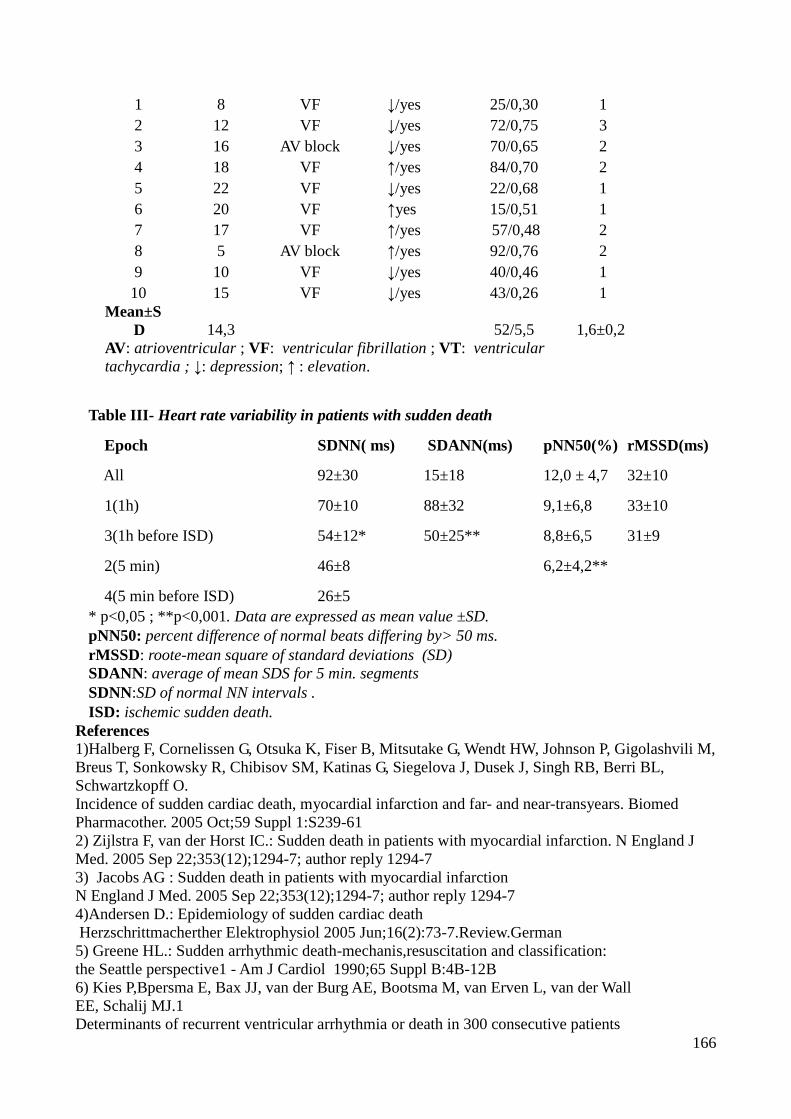
166
1 8 VF ↓/yes 25/0,30 1
2 12 VF ↓/yes 72/0,75 3
3 16 AV block ↓/yes 70/0,65 2
4 18 VF ↑/yes 84/0,70 2
5 22 VF ↓/yes 22/0,68 1
6 20 VF ↑yes 15/0,51 1
7 17 VF ↑/yes 57/0,48 2
8 5 AV block ↑/yes 92/0,76 2
9 10 VF ↓/yes 40/0,46 1
10 15 VF ↓/yes 43/0,26 1
Mean±S
D 14,3 52/5,5 1,6±0,2
AV: atrioventricular ; VF: ventricular fibrillation ; VT: ventricular
tachycardia ; ↓: depression; ↑ : elevation.
Table III- Heart rate variability in patients with sudden death
Epoch SDNN( ms) SDANN(ms) pNN50(%) rMSSD(ms)
All 92±30 15±18 12,0 ± 4,7 32±10
1(1h) 70±10 88±32 9,1±6,8 33±10
3(1h before ISD) 54±12* 50±25** 8,8±6,5 31±9
2(5 min) 46±8 6,2±4,2**
4(5 min before ISD) 26±5
* p<0,05 ; **p<0,001. Data are expressed as mean value ±SD.
pNN50: percent difference of normal beats differing by> 50 ms.
rMSSD: roote-mean square of standard deviations (SD)
SDANN: average of mean SDS for 5 min. segments
SDNN:SD of normal NN intervals .
ISD: ischemic sudden death.
References
1)Halberg F, Cornelissen G, Otsuka K, Fiser B, Mitsutake G, Wendt HW, Johnson P, Gigolashvili M,
Breus T, Sonkowsky R, Chibisov SM, Katinas G, Siegelova J, Dusek J, Singh RB, Berri BL,
Schwartzkopff O.
Incidence of sudden cardiac death, myocardial infarction and far- and near-transyears. Biomed
Pharmacother. 2005 Oct;59 Suppl 1:S239-61
2) Zijlstra F, van der Horst IC.: Sudden death in patients with myocardial infarction. N England J
Med. 2005 Sep 22;353(12);1294-7; author reply 1294-7
3) Jacobs AG : Sudden death in patients with myocardial infarction
N England J Med. 2005 Sep 22;353(12);1294-7; author reply 1294-7
4)Andersen D.: Epidemiology of sudden cardiac death
Herzschrittmacherther Elektrophysiol 2005 Jun;16(2):73-7.Review.German
5) Greene HL.: Sudden arrhythmic death-mechanis,resuscitation and classification:
the Seattle perspective1 - Am J Cardiol 1990;65 Suppl B:4B-12B
6) Kies P,Bpersma E, Bax JJ, van der Burg AE, Bootsma M, van Erven L, van der Wall
EE, Schalij MJ.1
Determinants of recurrent ventricular arrhythmia or death in 300 consecutive patients

167
with ischemic heart disease who experienced aborted sudden death: data from the
Leiden out-of- hospital cardiac arrest study.
J Cardiovasc Electrophysiol. 2005 Oct; 16(10):1049-56
7) Brembilla-Perrot B, Suty-Selton C, Claudon O,Terrier de la Chaise A, Louis P,
Nippert M, Popovic B,Blangry H, Khaldi E, Belhakem H,Beurrier D, Houriez P.
Significance of inducible ventricular flutter-fibrillation after myocardial infarction.
Pacing Clin Electrophysiol. 2005 Sep;28(9):938-43
8) Sztajzel J.
Heart rate variability: a non invasive electrocardiographic method to measure the
autonomic nervous system.
Swiss Med Wkly. 2004 Sep.4;134(35-36):514-22.
9)Salomon SD, Zelenkofske S, Mc Murray JJ,Finn PV,Velazquez E, Ertl G,
Harsanyi A, Rouleau JL,Maggioni A, Kober L, White H, Van de Werf F, Pieper
K,Califf RM, Pfeffer
MA: Sudden death in patients with myocardial infarction and left ventricular
dysfunction,heart failure,or both.
N Engl J Med 2005 Aug 18;353(7):744.
10) Cygankiewicz I, Wranicz JK, Bolinska H, Zaslonka J, Zareba W:
Relationship between heart rate turbolence and heart rate,heart rate variability,and
number of ventricular premature beats in coronary patients.
J Cardiovasc Electrophysiol. 2004 Jul; 15(7): 731-7.
11) Sredniawa B, Musialik-Lydka A,Obrebska J,Sliwinska A, Markowicz-Pawlus
E,Kalaurus Z, Polonski L.
Holter monitoring in the prognosis of sudden cardiac death-new experiences and
Possibilities
Wiad Lek 2005;58(3-4):233-7.Review.Polish.
12) Norman SE, Eager RA, Waran NK, Jeffer L,Schroter RC, Marlin DJ.
Recording of ECG signals on a portable MiniDisc recorder for time and frequency
domain heart rate variability analysis.
Physiol Behav. 2005 Jan 17;83(5):729-38
13) Juven X, Empana JP, Schwartz PJ, Desnos M, Courbon D, Ducimetiere P.
Heart rate profile during exercise as a predictor of sudden death
N Engl J Med. 2005 May 12; 352(19):1951-8
14) Makikallio TH, Barthel P, Schehneider R, Bauer A, Tapanaimen JM, Tulppo MP,
Schmidt G, Huikuri HV.
Prediction of sudden cardiac death after acute myocardial infarction : role of Holter
monitoring in the modern treatment area.
Eur Heart J 2005 Apr; 26(8):762-9. Epub 2005 Mar 18
15) Willich SN, Maclure M, Mittleman M, Arntz H-R, Muller Je.
Sudden cardiac death. Support for a role of triggering in causation.
Circulation 1993;87:1442-50.
16) Bigger TJ, Fleis Jl, Steinman RC, Rolnitzky LM, Kleiger RE, Rottman JM.
Correlations among time and frequency domain measures of heart period variability
two weeks after acute myocardial infarction.
Am J Cardiol 1992;69:891-8
17) Kleiger RE, Bigger JT, Bosner MS, et all
Stability over time of variables measuring heart rate variability in normal subjects
Am J Cardiol 1991;68:626-30
18) Schartz p, Priori S:
Sympathetic nervous system and cardiac arrhythmias.In: Zipes D, Jalife J, eds .
Cardiac electrophysiology:from cell to besides.

168
Philadelphia:WB Saunders Co,1990;330-43
19) Kannel WB, Kannel C ,Paffenbarger RS, et al.
Heart rate and cardiovascolar mortality:the Framingham study
Am Heart J 1987;113:1489-94
20) Richter S, Duray G, Gronefeld G, W Israel C, H Hohnloser S.
Prevention of sudden cardiac death:lessons from recent controlled trials
Circ J 2005 Jun; 69(6):625-9.Review
21) Zanetti G,Neilson J,Latini R et al:
Prognostic significance of heart rate variabilità in post-myocardial infarction patients in fibrinolytic
era .The Gissi-2 results.
Circulation,1996;94:432-36
22) La Rovere M,Bigger J,Marcus F et al:
Baroreflex sensitivity and heart rate variability in prediction of total cardiac mortalità after
myocardal infarction. ATRAMI (Autonomic Tone and Reflexes After Myocardal Infarction)
Investigators.
Lancet ,1998;351:478-84
23) Vanoli E, Cerati D, Pedretti R: Autonomic control of heart rate: pharmacological and
nonpharmacological intervention.
Basic Res Cardiol,1998; 93 /Suppl.1):133-42.
24) Van Ravenswaaij-Arts CMA, Kole LAA, Hopman JCW, Stoelinga GBA, van Geijin HP.
Heart rate variability.
Ann Intern Med 1993;118:436-47.
25) Kataoka M, Ito C, Sasaki H, Yamane K, Kohno N.
Low heart rate variability is a risk factor for sudden cardiac death in type 2 diabetes.
Diabetes Res Clin Pract. 2004 Apr;64(1):51-8.
26) Cowan MJ, Pike K, Burr RL, Cain KC, Narayanan SB. Description of time- and
frequency-domain-based measures of heart rate variability in individuals taking
antiarrhythmics,beta- blockers, calcium channel blockers, and/or anti-hypertensive
drugs after sudden cardiac arrest. J Electrocardiol 1993; 26 Suppl:1-13.

169
OZONE THERAPY IN THE TREATMENT OF PAIN IN VETERINARY MEDICINE
Luongo C.; Rotunno P.; Mascolo L.; Schioppi M.; Di Costanzo S. M.; Siniscalco D. and Luongo M.
Seconda Università degli studi di Napoli
Facoltà di medicina e chirurgia - Dip. Ass. Anestesia, Rianimazione e Terapia Intensiva
Ambulatorio di Ossigeno-Ozonoterapia, Direttore Biagio Lettieri
Introduction The Health Ministry has notified, by ministerial notes of 1992 and 1996, the indications and action
mechanisms of O₂-O₃ therapy.
There is already extensive international literature on the ozone effectiveness as a hemorhreological
drug [1-15], a body protector [11-14] and as analgesic medication.
In the clinic we have improved and stabilized ingravescent diseases between 60% -80% of cases. he
laboratory provided us with interesting experimental data to understand in part the ozone
therapeutic efficacy as analgesic and anti-inflammatory drug, when administered.
Ozone gas is naturally present in various layers of the atmosphere and it will give many important
effects of both physical-chemical and biochemical. First of all the power absorption of UV solar
rays make it a atmosphere key component for organic life on our planet.
At the biochemical level, the action takes place primarily on ozone coenzymes and organic
substances, and later involves the metabolism three basic lines: lipids, carbohydrates and proteins.
These particular interactions with the ozone metabolism cause various reactions in the blood, with
different effects on both the corpuscular elements and on the plasma.
Particularly important are the effects on erythrocytes, including:
peroxide (increased negative charge in the membrane)
shortening of the lipid chains (this allows the relaxation of the membrane)
increased production of 2-3 diphosphoglycerate (acceleration of glycolysis).
Just this substance have significant importance for the ozone therapy effects in vascular disease of
both man and animal, because it increases the supply of oxygen to tissues by Hb.
Discussion
Therefore considering the anti-inflammatory and analgesic action of O₂-O₃ mixture two studies
have highlighted the use of O₂-O₃ mixture as:
cardio-protective drug in ischemia reperfusion infarct;
analgesic and anti-inflammatory drug in the treatment of neuropathic pain.
Regarding the first point, in a study on rats, was given the O₂-O₃ mixture on myocardial tissue
damage due to an ischemic event. Rats treated with the O₂-O₃ mixture showed significant
cardioprotection compared with control rats.
This effect was associated with a decrease in tissue markers of oxidative stress (nitrotyrosine)
(figura 1), inflammation (CD68) and immune response (CD4 and CD8) in the rats infarcted
myocardium treated with the O₂-O₃ mixture (figura 2). These data indicate that tissue damage
associated to biochemical ischemia/reperfusion myocardium, can be countered by an acute pre-
treatment with O₃ . Regarding the second point of considerable importance for the treatment of pain in humans and
animals, is the action that ozone exerts on both the mediators and the program of cell death or
apoptosis.
Experimental data show that ozone is able to act, limiting the action and the same synthesis, on
enzymes which the caspases.
The caspases known are 13. Those we studied are 4: Caspase-1, caspase-12, caspase-8 and caspase-
3.
The first two (caspase-1 and caspase-12) involved in complex inflammasoma formation and are

170
responsible for the release of pro-inflammatory cytokines. Indeed, it was found that after exposure
to pathogenic noxa activates a series of mediators of cellular damage that induce synthesis and
release of caspases,
which promote the synthesis and release of pro-inflammatory cytokines such as IL-1b, IL-10, IL-18,
etc... (Figura 3). As noted in studies conducted in mice subjected to sciatic nerve ligation, the pain
causes gene over-Expression and then synthesis of mediators such
IL-1, pro-inflammatory cytokine, mainly in the cortex and spinal cord, through the synthesis of
caspase-1.
Ozone has the ability to inhibit the release of caspase-1 and 12 through its immunomodulatory
action also states also from studies of gene amplification (RT-PCR), which notes that the drug
reduces the synthesis of caspase -1, TNF-R, IL-1B and 10 (Figura 4).
Furthermore, another important function of ozone is linked to inhibition of the mechanism of cell
death (apoptosis) mediated by caspase-8 and caspase-3, which is amplified during chronic pain,
both cortical bone marrow for halogenated continuous stimulation (Figura 5).
It was pointed out that during the O₂-O₃ treatment, a reduction in both cortex and spinal cord of
caspases involved in this process.
Conclusion
Based on what was said and shown by various studies and results in human trials, we can say that
ozone, having both anti-inflammatory properties that inhibit the mechanism of cell death and
realizing from the clinical point of view a reduction of painful symptoms in humans, is a drug
applicable in veterinary medicine, being cheap, without all those side effects associated with the use
of various molecules and anti-inflammatory analgesic marketing, both in man and animal. From this
it requires a greater encouragement to continue research and to determine an upper extension of the
therapeutic applications of ozone in animals.
References
1. Ozonized autohaemotransfusion could be a potential rapid-acting antidepressant medication
in elderly patients. Coppola L, Luongo C, Pastore A, Masciello C, Parascandola RR,
Mastrolorenzo L, Grassia A, Coppola A, De Biase M, Lettieri B, Gombos G.: Int J Geriatr
Psychiatry. 2010 Feb; 25(2):208-13.
2. Ozonized Autohaemotransfusion does not affect the artery vasodilator function in patients
with peripheral arterial disease. Coppola A, Coppola L, Luongo C, et al.. Int J Angiol 2007; 16: 7-
11.
3. Oxygen-ozone therapy and haemorheological parameters in peripheral chronic arterial
disease. Coppola L, Verrazzo G, Giunta R, et al.. Tromb Ateroscl 1992; 3: 83-89.
4. Variazioni della viscosità ematica e dell'aggregazione piastrinica indotte dall'ozono medicale
a diverse concentrazioni. Studio in vitro. Coppola L, Verrazzo G, Luongo C, Sammartino A. Rend
Atti Accad Sci Med Chir 1993.147: 155-167.
5. Influence of ozone on haemoglobin oxygen affinity in type 2 diabetic patients with
peripheral vascular disease. Coppola L, Giunta R, Verrazzo G, et al. Diabete Metab 1995. 21: 252-
257.
6. Ozonised autohaemotransfusion and fibrinolytic balance in peripheral arterial occlusive
disease. Coppola L, Lettieri B, Cozzolino D, et al. Blood Coagul Fibrinolysis 2002. 13: 671-681.
7. Valutazione degli effetti della terapia sistemica con O2-O3 nelle arteriopatie croniche
periferiche. Luongo C, Luongo M, Cipollaro C, et al. Rivista Italiana di Ossigeno-Ozonoterapia
2002.1: 25-30.
8. Hyperbaric oxygen, oxygen-ozone therapy, and rheologic parameters of blood in patients
with peripheral occlusive arterial disease. Verrazzo G, Coppola L, Luongo C, et al. Undersea
Hyperb Med 1995. 22: 17-22.
9. Use of a non-specific immunomodulation therapy as a therapeutic vasculogenesis strategy in
no-option critical limb ischemia patients. Marfella R, Luongo C, Coppola A, Luongo M, Capodanno

171
P, Ruggiero R, Mascolo L, Ambrosino I, Sardu C, Boccardi V, Lettieri B, Paolisso G.
Atherosclerosis. 2010 Feb; 208(2):473-9.
10. Ozonized autohemotransfusion improves hemorheological parameters and oxygen delivery
to tissues in patients with peripheral occlusive arterial disease.Giunta R, Coppola A, Luongo C,
Sammartino A, Guastafierro S, Grassia A, Giunta L, Mascolo L, Tirelli A, Coppola L. Ann
Hematol. 2001 Dec;80(12):745-8.
11. Antiarrhythmic effect of acute oxygen-ozone administration to rats. Di Filippo C, Cervone
C, Rossi C, di Ronza C, Marfella R, Capodanno P, Luongo C, Rossi F, D'Amico M. Eur J
Pharmacol. 2010 Mar 10;629(1-3):89-95.
12. Acute oxygen-ozone administration to rats protects the heart from ischemia reperfusion
infarct. Di Filippo C, Marfella R, Capodanno P, Ferraraccio F, Coppola L, Luongo M, Mascolo L,
Luongo C, Capuano A, Rossi F, D'Amico M. Inflamm Res. 2008 Oct; 57(10):445-9.
13. Results of a non-specific immunomodulation therapy in chronic heart failure (ACCLAIM
trial): a placebo-controlled randomised trial
Guillermo T. A. and coll.: Lancet 2008 January 19,.
14. Prevention of renal injury after introduction of ozone tolerance in rats submitted to warm
ischaemia. Bocci V. e coll.:Mediat. Inflamm 1999; 8, 37-41.
15. A single subcutaneous injection of ozone prevents allodynia and decreases the over-
expression of pro-inflammatory caspases in the orbito-frontal cortex of neuropathic mice. Fuccio C,
Luongo C, Capodanno P, Giordano C, Scafuro MA, Siniscalco D, Lettieri B, Rossi F, Maione S,
Berrino L. Eur J Pharmacol. 2009 Jan 28;603(1-3):42-9.
16. Ameliorative effect of ozone on cytokine production in mice injected with human
rheumatoid arthritis synovial fibroblast cells. Chang et al. Rheumatol. Int. 26 (2007), pp. 142–151.
17. Chronic NMDA administration increases neuroinflammatory markers in rat frontal cortex:
cross-talk between excitotoxicity and neuroinflammation. Chang et al. Neurochem. Res. 33 (2008),
pp. 2318–2323.
18. Identification of valid housekeeping genes and antioxidant enzyme gene expression change
in the aging rat liver. Chen et al. J. Gerontol., Ser. A, Biol. Sci. Med. Sci. 61 (2006), pp. 20–27.
19. Advances in brain imaging of neuropathic pain. Chen et al. Chin. Med. J. 121 (2008), pp.
653–657.
20. Ozone oxidative preconditioning protects the rat kidney from reperfusion injury: the role of
nitric oxide. Chen et al. J. Surg. Res. 149 (2008), pp. 287–295.

172
Figura 1
CD68
CD4
CD8
Cuore infartuato Cuore infartuato + O3
↑
↑
Risultati biomolecolari dopo un’unica somministrazione di miscela O₂-O₃ e O₂ a livello rachideo dopo lesione del nervo
sciatico
Apoptosi: Casp3/HPRT
0
40
80
120
160
200
NAIVE SNI SNI O3 SNI O2
* *
p<0,05 vs naive
Potente azione anti-apoptica dell’ozono
Morte cellulare programmata: Casp8/HPRT
0
40
80
120
160
200
C SNI SNI O3 SNI O2
Potente azione anti-apoptica dell’ozono
*
*
*p<0.05 vs C
Potente azione anti-infiammatoria dell’ozono
*
*p<0.05 vs C
Azione immunomodulante dell’ozono
Figura 1 Figura 2
RT-PCR : amplificazione genica
< recettore tnf < caspasi-1
< IL-1B < IL-10
Risultati biomolecolari dopo un’unica somministrazione di miscela O₂-O₃ e O₂ a livello rachideo dopo lesione del nervo
sciatico
Apoptosi: Casp3/HPRT
0
40
80
120
160
200
NAIVE SNI SNI O3 SNI O2
* *
p<0,05 vs naive
Potente azione anti-apoptica dell’ozono
Morte cellulare programmata: Casp8/HPRT
0
40
80
120
160
200
C SNI SNI O3 SNI O2
Potente azione anti-apoptica dell’ozono
Figura 3 Figura 4

173
ACTION OF OXYGEN-OZONE MIXTURE ON ACTIVITY OF CYTOKINES
D. Siniscalco
Second University of Naples, Department of Experimental Medicine - Division of Pharmacology "L.
Donatelli". via S. Maria di Costantinopoli, 16 - 80138 - Naples – Italy. Tel. +39-81-5665880; Fax.
+39-81-566750.
Corresponding author email: [email protected]
Introduction
Neuropathic pain is defined as pain initiated or caused by a primary lesion or dysfunction in the
nervous system [1, 2]. Due to its very complex nature, neuropathic pain does not respond to
currently-used analgesic drugs, such as antiepileptic drugs, opioids, antidepressant agents.
Peripheral neuropathy triggers several biomolecular events propagating to the spinal cord and brain,
where they are integrated and processed by limbic and cortical areas [3]. Several pre-frontal cortical
areas are part of the brain network for pain [4]. Recent evidence suggests cortical neuroplastic
changes including genes activation of neurotrophic factors in chronic pain conditions [5].
The caspase family comprises thus far 13 different cysteine proteases that are mainly involved in
the apoptotic pathways. Among them, caspase-1 is less involved in the apoptotic cascade, but it is
prominent in inflammation because of its pivotal role in regulating the cellular export of pro-
inflammatory cytokines such as IL-1 and IL-18. These enzymes regulate the inflammatory
processing of CNS diseases, including spinal cord inflammation and neuropathic and chronic
inflammatory pain [6].
Ozone is a gas constituting an ozone and oxygen mixture. It has a great oxidative power and it has
been used in medicine for the treatment of several illnesses with a wide spectrum. Ozone has been
proposed as an antioxidant enzyme activator, immunomodulator, and cellular metabolic activator
[7]. Several experimental and clinical evidences have shown advantageous effects of oxygen/ozone
therapy in several pathologies characterized by a cellular oxidative and inflammatory response,
including renal injury, cardiopathy, atherosclerosis and septic shock [8]. Indeed, ozone therapy
shows long-term anti-inflammatory effects and reduces pro-inflammatory factors without toxicity or
serious side effects.
In this study, we have studied whether a single subcutaneous administration of the O3/O2 mixture
two days after sciatic nerve injury was anti-allodynic and anti-hyperalgesic in the first phase of
neuropathic pain (i.e. 7 days post-injury). In addition, we have addressed the issue of whether O3/O2
mixture treatment was capable to reduce the spinal and supra-spinal SNI-over-expression of pro-
inflammatory genes.
Materials and Methods Animals. Male C57BL/6N mice (35–40 g) were housed 3 per cage under controlled illumination
(12-h light/12-h dark cycle; light on 06:00 h) and environmental conditions (room temperature 20–
22 ◦C, humidity 55–60%) for at least 1 week before the commencement of experiments. Mouse
chow and tap water were available ad libitum. The experimental procedures were approved by the
Animal Ethics Committee of the Second University of Naples. Animal care was in compliance with
the IASP and European Community (E.C. L358/1 18/12/86) guidelines on the use and protection of
animals in experimental research. All efforts were made to minimise animal suffering and to reduce
the number of animals used. Ozone/oxygen mixture was generated by Multiossigen Medical-93
(Multitech, Milan, Italy). Ozone obtained from medicinal grade oxygen was used immediately.
Treatment.Mice were grouped (n=5) as follows:
(a) Naıve mice; (b) Sham mice; (c) Sham-operated mice treated with a single administration of
O2/O3 gas mixture (around 180 μg/kg s.c.); (d) SNI mice; (e) SNI mice treated with a single
administration of O2/O3 gas mixture (around 180 μg/kg s.c.).
Sham operated and SNI mice were assessed for the development of thermal hyperalgesia and

174
mechanical allodynia before any other examination. Behavioural testing was performed before
surgery to establish a baseline for comparison with post-surgical values. Behavioural analysis was
performed 1 h after the last injection of O2/O3 gas mixture. Ozone dose and administration
procedure were consistent with concentration and timing of oxygen/ozone treatment applied in
ozone-therapy in human neurosurgical procedures [9].
Spared nerve injury. Mononeuropathy was induced according to the method of Bourquin and
Decosterd [10]. Mice were anaesthetized with sodium pentobarbital (60 mg/kg i.p.). The sciatic
nerve was exposed at mid-thigh level distal to the trifurcation and freed of connective tissue; the
three peripheral branches (sural, common peroneal, and tibial nerves) of the sciatic nerve were
exposed without stretching nerve structures. Both tibial and common peroneal nerves were ligated
and transacted together. The sham procedure consisted of the same surgery without ligation and
transection of the nerves.
Nociceptive behaviour. Thermal hyperalgesia was evaluated by using a Plantar Test Apparatus (Ugo
Basile, Varese, Italy). On the day of the experiment each animal was placed in a plastic cage (22cm
x 17cm x 14cm; length x width x height) with a glass floor. After a 60 min habituation period, the
plantar surface of the hind paw was exposed to a beam of radiant heat through the glass floor. The
paw withdrawal latency (PWL) was automatically displayed to the nearest 0.1 sec; the cut-off time
was 20 sec in order to prevent tissue damage.
Mechanical allodynia was measured by using Dynamic Plantar Anesthesiometer (Ugo Basile,
Varese, Italy). Mice were adapted to the testing environment before any measurements were taken.
After that, the mechanical stimulus was delivered to the plantar surface of the hindpaw of the mouse
from below the floor of the test chamber by an automated testing device. A steel rod (2 mm) was
pushed with electronical ascending force (0-30 g in 10 sec). When the animal withdrawn its
hindpaw, the mechanical stimulus was automatically withdrawn and the force recorded to the
nearest 0.1 g. Nociceptive responses for thermal and mechanical sensitivity were expressed as
thermal paw withdrawal latency (PWL) in seconds and mechanical paw withdrawal threshold
(PWT) in grams.
Each mouse served as its own control, the responses being measured both before and after surgical
procedures. PWL and PWT were quantified by an observer blinded to the treatment.
RNA extraction and RT-PCR. Total RNA was extracted from homogenized pre-frontal cortex and
spinal cord using an RNA Tri-Reagent (Molecular Research Center Inc., Cincinnati, OH) according
to the manufacturer’s protocol. The extracted RNA was subjected to DNase I treatment at 37◦C for
30 min. The total RNA concentration was determined by UV spectrophotometer. The mRNA levels
of the genes under analysis were measured by RT-PCR amplification, as previously reported [11]. A
semiquantitative analysis of mRNA levels was carried out by the “Gel Doc 2000 UV System” (Bio-
Rad, Hercules, CA). The measured mRNA levels were normalised with respect to hypoxanthine-
guanine phosphoribosyltransferase (HPRT), chosen as housekeeping gene. The HPRT gene
expression did not change in several experimental conditions. To our knowledge there is no
molecular evidence for variation in HPRT mRNA-levels in SNI neuropathic pain model [6]. The
gene expression values were expressed as arbitrary units ± S.E.M. Amplification of genes of interest
and HPRT were performed simultaneously.
Statistical Analysis. Behavioural data are represented as means ± S.E.M. ANOVA, followed by
Student–Neuman–Keuls post hoc test were used to determine the statistical significance among
groups. Molecular and biochemical data are shown as means ± S.E.M., ANOVA, followed by
Student–Neuman–Keuls post hoc test, were used to determine the statistical significance among
groups. P< 0.05 was considered statistically significant.
RESULTS
Ozone prevents the development of mechanical allodynia and thermal hyperalgesia
In naïve mice, the behavioural change of mechanical allodynia and thermal hyperalgesia did not
significantly undergo modification during analgesimeter registration. SNI of the sciatic nerve
resulted in significant decrease in PWT and in PWL in the ipsilateral side of mice 7 days after

175
surgery. The increased sensitivity in the SNI sides was not present on the contralateral sides. A
single administration of ozone performed 2 days after SNI surgery, was effective in preventing the
appearance of mechanical allodynia and thermal hyperlagesia at 7 days post-SNI.
Ozone prevents the over-expression of pro-inflammatory genes
The semiquantitative analysis of mRNA levels measured by RT-PCR amplification in the pre-
frontal cortex and in spinal cord of mice, showed an increase in the pro-inflammatory caspase-1,
IL-1β, IL-10, NF-kβ, TNF-R1 and SOD2 genes 7 days after the SNI of the sciatic nerve. Ozone
inhibited the gene over-expression in 7 days SNI pre-frontal cortex and in spinal cord (Fig. 1).
Figure 1. The mRNA levels (mean ± SE) of the genes under analysis measured by RT-PCR
amplification were reported. °p<0.05 vs the corresponding naïve mice, *p<0.05 vs the SNI ozone
untreated mice, as analysed by ANOVA, followed by Student-Neuman-Keuls test. Legend: CTL
control mice; SNI neuropathic mice; SNI+ozone neuropathic mice treated with ozone.
Discussion
The present study offers evidence indicating that the pro-inflammatory caspase-1, IL-1β, IL-10, NF-
kβ, TNF-R1 and SOD2 gene expressions were significantly increased in the pre-frontal cortex and
in spinal cord seven days after SNI in the mouse.
Our data indicate that peripheral nerve injury activates a neuroinflammatory-like process in the
central structures controlling pain processing. Moreover, treatment with ozone has been clinically
effective in decrease the behaviour indication of the neuropathic pain. Ozone was able to inhibit the
transcription of the genes involved in the cellular stress and inflammation and reduce pain-like
behaviours.
Collectively, these preliminary data indicate that ozone can be an effective practice for preventing
the development of neuropathic pain through complex as yet unexplored mechanisms and, among
these, through the modulation of specific pro-inflammatory genes in the central nervous system.

176
References
1) Merskey H, Bogduk N (1994). Classification of chronic pain. IASP press, Seattle.
2) Siniscalco D, de Novellis V, Rossi F, Maione S (2005). Neuropathic pain: is the end of
suffering starting in the gene therapy?. Curr Drug Targets 6: 75-80.
3) Millecamps M, Centeno MV, Berra HH, Rudick CN, Lavarello S, Tkatch T, Apkarian AV.
(2007). D-cycloserine reduces neuropathic pain behavior through limbic NMDA-mediated circuitry.
Pain. 132(1-2):108-123.
4) Apkarian AV, Sosa Y, Sonty S, Levy RM, Harden RN, Parrish TB, Gitelman DR (2004).
Chronic back pain is associated with decreased prefrontal and thalamic gray matter density. J
Neurosci 24(46):10410-5.
5) Wei F, Zhuo M. Activation of Erk in the anterior cingulate cortex during the induction and
expression of chronic pain. Mol Pain. 2008; 4:28.
6) Fuccio C, Luongo C, Capodanno P, Giordano C, Scafuro MA, Siniscalco D, Lettieri B,
Rossi F, Maione S, Berrino L. (2009). A single subcutaneous injection of ozone prevents allodynia
and decreases the over-expression of pro-inflammatory caspases in the orbito-frontal cortex of
neuropathic mice. Eur J Pharmacol. 603(1-3):42-9.
7) Guven A, Gundogdu G, Vurucu S, Uysal B, Oztas E, Ozturk H, Korkmaz A (2009). Medical
ozone therapy reduces oxidative stress and intestinal damage in an experimental model of
necrotizing enterocolitis in neonatal rats. J Pediatr Surg. 44(9):1730-5.
8) Hernandez, S. Menendez and R. Wong (1995). Decrease of blood cholesterol and
stimulation of antioxidative response in cardiopathy patients treated with endovenous ozone
therapy. Free Radic. Biol. Med. 19: 115–119.
9) Martínez-Sánchez G, Al-Dalain SM, Menéndez S, Re L, Giuliani A, Candelario-Jalil E,
Alvarez H, Fernández-Montequín JI, León OS (2005). Therapeutic efficacy of ozone in patients
with diabetic foot. Eur J Pharmacol 523(1-3):151-61.
10) Bourquin AF, Süveges M, Pertin M, Gilliard N, Sardy S, Davison AC, Spahn DR, Decosterd
I. (2006). Assessment and analysis of mechanical allodynia-like behavior induced by spared nerve
injury (SNI) in the mouse. Pain. 122(1-2):14.e1-14.
11) Siniscalco D, Giordano C, Fuccio C, Luongo L, Ferraraccio F, Rossi F, de Novellis V, Roth
KA, Maione S. (2008). Involvement of subtype 1 metabotropic glutamate receptors in apoptosis and
caspase-7 over-expression in spinal cord of neuropathic rats. Pharmacol Res 57(3):223-33.

177
ANALGESIC EFFECTS OF OXYGEN/OZONE THERAPY IN ARTERIAL
OBSTRUCTIVE PATHOLOGY.
Mascolo Luigi, Luongo Margherita
On the basis of Fontaine classification, the arterial vascular insufficiency triggers “claudicatio
intermittens” due to ischemia/hypoxia during walking.
A study published on Circulation (2006; 113: 463-654) highlighted that drugs or factors of Level of
Evidence A in the peripheral arterial disease are: exercise activity, cilostazol (in Italy not yet
disposable) and pentoxifyline. According to this study, other drugs (i.e. beraprost, iloprost, l-
arginine, propionyl-l-carnitine) did not show a definite effectiveness. The vasculopathy is able to
trigger peripheral pain.
Ozone therapy has been shown to have therapeutic efficacy in positively change the emo-reologici
factors conditioning peripheral oxygen reperfusion. Clinic therapy has confirmed this hypothesis.
We report our experience in a vasculopathic chronic patient group of Fontaine level 3 and with
peripheral ulcer, has been administrated one cycle of systemic O2-O3 mixture treatment.
O2-O3 mixture treatment was able to decrease pain in resting state, as well as to increase free
movement in these patients.
These results indicate that O2-O3 mixture could be an optimal treatment in the arterial vascular
insufficiency care.

178
PARALLEL SESSION
OPIOID THERAPY and PAIN MEASUREMENT
CHAIRMEN: Chiefafi - Fonda – Maione
ENDOCRINE CONSEQUENCES OF OPIOID THERAPY
C. Aurilio
MULTIMODAL ANALGESIA FOR CHRONIC PAIN: RATIONALE AND FUTURE
DIRECTIONS Adiletta S
MECHANISMS UNDERLYING ANALGESIC TOLERANCE AND HYPERALGESIA TO
OPIOIDS.
Pastore A.
PELVIC PAIN EVALUATION IN HUMANS
M.B. Passavanti

179
ENDOCRINE CONSEQUENCES OF OPIOID THERAPY
C. Aurilio, M.B. Passavanti, V. Pota
Second University of Naples, Dpt of Anesthesia, Surgical and Emergency Sciences, Piazza Miraglia
2, 80100 Naples, Italy
Corresponding author: [email protected]
In the treatment of pain and particularly of the moderate and severe chronic pain, opioids remain
one of the most effective and used therapies. However too often patients achieve little or no pain
relief due to the number of side effects that limit their intake. The OPIAD, i.e. the opioid induced
androgen deficiency is one of the mostly constant syndrome. That exposure to opioids decreases
gonadal hormones in humans, as well as in experimental animals treated with different opioids, is a
quite aknowledge fact looking at the several paper present in the literature (1-3). Several hypothesis
have been suggested to explain such a possible pathogenesis , one of these considers the
inhibition by opioids of the releasing factors secretion at hypothalamic level, another a direct
inhibitory action at the pituitary level via specific binding sites on the gonadotrophs, however other
hypothesis were tested. We present in this review a series of experiments carried out in different
patients population, in experimental animals and in vitro in which the study of hypothalamo-
pituitary-adrenal/hypothalamo/pituitary/gonadal related hormones appears to significantly respond
to the different opioid actions.
Effect of i.t. morphine administration in men and women subjects
To evaluate the immediate effects of morphine spinal administration on steroid hormones and their
time course we considered male and female patients implanted with an i.t. catheter, due to a
persistent severe pain. The morphine intratecal daily dose was 0.9 mg/die and the overall
administration time was 15 days. The following hormones were considered: LH, FSH, Testosterone,
Free Testosterone, Cortisol. Blood was collected and analysed on Day 0, 1, 2, 15 (last day of
administration) and 16 (the day after the withdrawal of morphine). In females and males, in fact,
total testosterone and free testosterone blood levels as well as cortisol resulted to be greatly
reduced with respect to the basal levels since Day 1 to Day 15 and completely recovered 24 hrs
after the end of treatment (16th day). By contrast gonadotropins tended to minimally increase with
respect the basal levels in males and to decrease in females. These findings clearly show that the
administration of morphine, also via i.t., immediately affects gonadal hormones and cortisol ,
independently from the gender. Indeed, whereas in males a hypothalamo/pituitary axes inhibition
has to be excluded due to the lack of gonadotropin decrement, in females a hypothalamo-pituitary
involvement would be considered due to the decrease, even if small, of gonadotropins levels . Thus
in interpreting the testosterone decrement in males a direct action of opioids on testis and/or an
increase in testosterone metabolism has to be hypothesized.
Effect of the longlasting administration of transdermal Buprenorphine in males and females
subjects
Male and female subjects undergoing buprenorphine transdermal intake (35 mcg/ h every 72 hrs)
because of an acute/persistent pain were included in the study whose main aim was observing
the modifications on HPG and HPA axes. Estradiol, Total Testosterone, Free Testosterone, DHT,
Cortisol and SHBG plasma levels were evaluated, at basal level, and after 1, 3 and 6 months of
therapy. During the observation period in females all hormones showed various slight changes that
did not become significant except for total testosterone that resulted increased at 3 months. In males
no significant difference was observed for all hormones except for free testosterone that appeared
to be lowered after 3 months. In both sexes the HPA axis was not inhibited since cortisol
progressively increased during treatment. SHBG is a rarely measured protein, it indicates the
possibility of testosterone to be transported in the blood. In the present study its level doesn’t
change significantly in both sexes. In the present study its efficacy (buprenorphine: μ partial agonist
and κ antagonist) with different characteristics with respect morphine (μ and κ agonist), showed

180
different effects, indeed the deficiency of the androgens in the blood is not present. This means that
buprenorphine therapy may be carried out also for several months whithout falling in the
hypogonadic conditions induced by morphine.
Effect of surgical analgesia by i.v. opioid administration in men In the present study we measured the blood level of testosterone and estradiol in a sample of males
patients who were submitted to an intervention to evaluate the modifications of these hormones in
such a particular condition. It clearly appears that after the procedure the testosterone levels are
very low, particularly when morphine is implemented with fentanyl. In addition in these patients
also estradiol resulted to be lowered by treatment. In particular men treated only with morphine for
the management of transvescical prostateadenectomia showed testosterone levels to be
significantly reduced after 24 hrs, to completely recover three days later.
Effect of sc morphine administration in plasma and brain testosterone levels in male and
female rats
Male and female rats were s.c. injected with morphine to study the effects of this opoid in
testosterone plasma and brain levels. Blood and brain tissues were collected after 4 hours. It clearly
appears that a decrement of testosterone has been observed in the blood of both female and male
rats. In the brain testosterone was importantly decreased in males while in the female brain
testosterone didn’t reach detectable levels also in the saline treated animals.
Effect of morphine on testosterone levels in glia cells
A series of experiments were carried out to verify whether rat C6 glioma cells testosterone content
was affected by morphine administration and whether this effect could be modified by the addition
to culture medium of drugs interfering with testosterone degradation by acting on different
enzymes: anastrazole or finasteride. We measured aromatase activity in pools of cells treated with
ANA, morphine (10 μM) and the association of these two substances, versus control. Testosterone
was measurable in the glioma cells and its level was strongly reduced by morphine addition in a
dose dependent way. In other groups of glia cells the addition of anastrozole and finasteride
differently affected testosterone concentration. With respect to controls, anastrazole significantly
increased testosterone concentration, an effect completely abolished by the contemporary
administration of morphine. In contrast finasteride did not change significantly the testosterone
levels. Our data provide evidence that glial cells contain low, but detectable levels of testosterone
and that morphine significantly decreases testosterone cellular levels.
DISCUSSION
All the experiments presented in this paper show the different ability of opioids to affect hormones.
4The hypopgonadism induced by morphine as well as the effect on neuronal content of testosterone
have to be considered because although opioids remain the more important group of drugs
commonly used in pain therapy, physicians need to be aware about all their long term
consequences. Morphine and the other opioids, when given to treat pain, disrupt the physiological
ratio between opioids and gonadal hormones, altering many of the several functions in which
opioids and hormone act. This important interaction is probably involved in the different ability
present in the two sexes to develop chronic pain (Aloisi and Bonifazi, 2006). In the second study,
buprenorphine-treated subjects suffering acute persistent pain were studied for six months.
Testosterone as well as estradiol is involved in wound healing through its action on tissue
regeneration and immunity (i.e Engeland et al., 2008). Thus at present data are not clear on the
effects of gonadal hormones on healing, but it is plausible to advance that morphine affect post-
surgical patients not only by relieving their pain, but also changing their tissues repearing by
healing via a temporary hypogonadism able to change the ratio among the immune and hormonal
activation. In conclusion, Opioid therapy is mandatory in several pains, in particular in chronic
pains. However its administration requires a detailed understanding of all mechanisms it affects
including gonadal hormones metabolism. All the data we reported infact confirm in agreement with
the findings described by other authors that morphine which is the mostly used compound
significantly influence testosterone metabolism, also at glial cells level. Therefore the prescription

181
of an opoid treatment requires a paramount attention and awareness of these so involving side
effects.
References
1. Aloisi A.M., Ceccarelli I., Fiorenzani P., Aurilio C., Pace MC., Chiefari M., Affaitati G., Lerza
R., Giamberardino MA. Sex Differences in behavioral responses to artificial calculosis in rats.
V EFIC Congress, Instanbul Turkey, 13-16 September 2006
2. The behavioral and neuronal effects induced by repetitive nociceptive stimulation are affected
by gonadal hormones in male rats. Ceccarelli I, Scaramuzzino A, Massafra C, Aloisi AM.
Pain. 2003 Jul;104(1-2):35-47.
3. H.W. Daniell, Hypogonadism in men consuming sustained-action oral opioids, J Pain 3 (2002),
pp. 377–384
182
MULTIMODAL ANALGESIA FOR CHRONIC PAIN: RATIONALE AND FUTURE
DIRECTIONS
Palomba R, Adiletta S, Candiello A, Pennimpede M, Bonaccia P, De Martino CJ
Chronic pain is a complex disease requiring multidimensional assessment and multimodal
treatment. In chronic pain, dysregulation of one or many locations within the complex nociceptive
and pain neural circuit leads to neuroplastic reorganization and increased spontaneous activity
(spontaneous pain), as well as hyperresponsiveness to noxious (hyperalgesia) and non-noxious
(allodynia) stimuli. The neuroanatomy of the nociceptive loop is comprised grossly of nerve
endings of peripheral primary sensory ganglia, supraspinal nuclei, the thalamus, limbic and cerebral
cortices, diencephalic processing and supraspinal descending modulatory control of the initial
peripheral input at the spinal dorsal horn level.
Mechanisms regulating peripheral sensation play a critical role in the manifestation of chronic pain;
several classes of primary sensory neurons with somata located among the dorsal root or cranial
sensory ganglia have been described and respond to noxious and non-noxious mechanical
(mechanoreceptors), thermal (thermoreceptors), or chemical (chemoreceptors) stimuli. The four
broadly defined primary sensory afferent types include:
1. The largest caliber, heavily myelinated muscle spindle and Golgi tendon afferents
responsible for signaling limb position and maintaining proprioceptive sense.
2. Large-caliber, heavily myelinated Aβ axons responsible for signaling highly discriminative
mechanical stimuli transduction.
3. Small-caliber, lightly myelinated Aδ axons.
4. Smaller, unmyelinated C axons that form morphologically, simple free nerve endings
(FNE)(1) that terminate within the epidermis, around the vasculature and all other skin
appendages, as well as muscle, all visceral organs and bone.
A majority of FNE are believed to respond specifically to various noxious stimuli and are
collectively termed “nociceptors”. As many as half of the Aδ and C axons are normally
unresponsive to test stimuli used to functionally characterize sensory innervation by
elettrophysiology. Many of these unresponsive axons have nociceptive characteristics and are
believed to be driven only by severe tissue damage or prolonged experimentally induced pain, and
are therefore referred to as “silent” nociceptors. Under acute tissue injury, quiescent nociceptive
neurons may be activated by direct stimuli-mechanical, thermal or chemical, including such
inflammatory cytokines as interleukin (IL)-1β and IL-6 released both immediately following injury
and subsequently during tissue repair processes. Inflammatory cytokines thus activate nociceptive
neurons, leading to the generation of pain and nocifensive behaviors that accompany tissue injury
and repair.
Recent studies have dedemonstrated that neurons, keratinocytes (2)and vascular endothelial cells
express common signaling pathways, suggesting a bidirectional communication between the
nervous system and cells of the epidermis and vasculature. In particular, depending on the type,
intensity and duration of a stimulus, epidermal keratinocytes release different combinations and
proportions of excitatory and/or inhibitory ligands that influence neuronal activity. Studies have
demostrated a role for β-endorphin, an endogenous opiate peptide produced by superficial
keratinocytes, which may represent a tonic inhibitory tone on µ opiate receptors (µOR) expressing
epidermal FNE. Loss of this tonic inhibitory mechanism within the keratinocytes would likely lead
to hyperexcitability of the µOR- expressing fibers, heightened nociceptive-like responses to varied
stimuli and potentially to chronic pain. In addiction to β-endorphin, keratinocytes release the purine
nucleotide adenosine triphospate (ATP) in response to air contact and various mechanical stimuli;
ATP likely contributes to the excitation of FNE axons through metabotropic G protein-coupled
mechanisms (P2Y receptors) and through ionotropic direct ion-gated channels (P2X) that are
present on small-caliber axons.(2)(3)

183
A loss of FNE may result in the disregulation of epidermal chemistry and inappropiate release of
various ligands from keratinocytes, contributing to increased excitability of FNE.(3)(4)
Nociceptive primary afferent fibers use glutamate and neuropeptide transmitters to convey the
status of the peripheral receptive field (5)(6); modulation of glutamate and its cognate receptor
family are critical to nociceptive processing in the dorsal horn and likely have implications in
neuropathic and chronic pain conditions. Additionally, primary peripheral nociceptive input is under
tonic control by descending brain stem-level regulatory systems, using serotonin, norepinephrine
and opioids as transimtters/modulators which can inhibit or facilitate nociceptive transmission in the
spinal dorsal horn. Many drugs -including for example opiates, antidepressants and
anticonvulsants- can influence the signaling of these descending systems and thereby achieve
effective analgesia for patients with chronic pain. Additional alterations in signaling pathways
involved in spinal-level nociceptive processing are not only limited to neural descending
presynaptic control or to postsynaptic N-methyl-D-aspartic acid (NMDA) receptor modulation but
also include glia(7): astrocytes can modulate synaptic function and release cytokines that sensitize
the system and have been shown to discriminatively respond to active neurons, implying a
bidirectional communication not previous appreciated.
Considering the fusion of central and peripheral mechanisms in the genesis of chronic pain, an
important role for the successful treatment of patients suffered from chronic pain could be awarded
to the use of palmitoylethanolamide (PEA) (8). PEA is not a drug but a nutritional element normally
present in the human organism(9); it is a biological modulator supporting the control of the
physiological tissue reactivity. PEA produces a down-modulation of mast cells- degranulation
peripherally (10) and an activation of endocannabinoid receptor CB2 (CB2 –like) centrally.(11) (12)
In our study, we recruited 81 patients (age 28-75) with a diagnosis of low back pain,VAS>5;we
administered opioids drugs (oxycodone) and drugs used for the neuropathic pain (antidepressants,
anticonvulsants); furthermore, 41 of these 81 patients were treated with PEA (600 mg x 2/die 21
days then 600 mg/die 30 days). We valued the pain score with VAS at first examination, after 21
and 71 days; we considered effective the therapy when it caused a VAS>60% reduction , compared
to the initial value. After 21 days, we verified a VAS> 60% reduction in 21 patients treated with
PEA and 16 patients not treated with PEA; after 70 days, a VAS>60% reduction was in 32 patients
treated with PEA and 22 patients not treated with PEA. Otherwise, in the group of patients treated
with PEA, we observed a lower dose of drug used for the treatment of neuropathic pain (
gabapentin, pregabalin, lamotrigine, duloxetine), compared to the group of patients not treated with
PEA.
Bibliography
1. Rice F. et al – Handbook of the senses Vol 5- Somatosensation –Elsevier 2008
2. Zao P Pain 2008, 139:90 -105
3. Koizumi S et al-Biochem J 2007;380: 329-38
4. Malin SA –Pain 2008;138: 484-96
5. Shigetomi E et al- J Neurosci 2008
6. South SM J –Neuroscience 2006; 23: 5031-40
7. McNally L, Bhagwagar Z, Hannestad J-CNS Spectr, Vol.13, No 6, 2008
8. Lambert DM, Vandevoorde S, Jonsson KO, Fowler CJ- Curr Med Chem.2002;9(6):663-74
9. Costa B, Comelli F, Bettoni I, Colleoni M, Giagnoni G- Pain 2008; 139:541550
10. De Filippis D, D’Amico A, Iuvone T, -J Neuroendocrinol. 2008; 1:20-25
11. Conti S, Costa B, Colleoni M, Parolaro D, Giagnoni G, -Pharmacol. 2002; 135(1):181-7
12. Re G, Barbero R, Miolo A, Di Marzo V- Vet J. 2007, 173(1):21-30

184
MECHANISMS UNDERLYING ANALGESIC TOLERANCE AND HYPERALGESIA TO
OPIOIDS.
Palomba R., Pastore A., Adiletta S., Candiello A., Pennimpede M.,Viscardi D.
Opioid analgesics are effective in relieving chronic pain, but they have serious adverse effects,
including development of tolerance and hyperalgesia.
The mechanisms underlying opioid tolerance are not fully understood, but appear to be comprised
of two types of plasticity or counter-adaptation, at the cellular level and through neuronal circuits.
Current studies mostly emphasize the cellular adaptation mechanisms, which include altered gene
expression,receptor down regulation and desensitization(due to phosphorylation and endocytosis).
However, the mechanisms underlying opioid tolerance and dependence are not always explained by
cellular adaptation mechanisms alone. Recent studies have shown a counterbalance of opioid
actions through an enhancement of synaptic activities between glutamate and NMDA receptor due
to up-regulation of receptor and racemase to produce D-serine, an allosteric NMDA receptor
agonist, and down-regulation of glutamate transporter.
The opioid-induced hyperalgesia,instead,is due to up regulation of NMDA receptor and to the
apoptosys of neural cells that provide to the production of opioid receptors.
Our weapons to tolerance and hyperalgesia-opioid induced are the opioids ”switch”, the
“multimodal analgesia” and new drugs based on the opioid assoiciation agonist-antagonist.
Bibliography
Short-term infusion of the μ-opioid agonist remifentanil in humans causes hyperalgesia during
remifentanil in humans causes hyperalgesia during withdrawal W. Koppert et al. Pain Nov.2003)(
W. Koppert et al. Pain Nov.2003)
Borgland SL. Acute opioid receptor desensitization and tolerance: is there a link? Clin Exp
Pharmacol Physiol 2001 Mar;28(3):147-54
Regulator GProtein Signaling (RGS) (Guo-xi Xie et al Anaesth Analg 2005; Whistler JL et al
Neuron 1999)
Mercadante S, Ferrera P, Villari P, Arcuri E.. “Hyperalgesia: An Emerging Iatrogenic Syndrome”. J
Pain Symptom Manage 2003 Aug;26(2):1-7

185
PELVIC PAIN EVALUATION IN HUMANS
M.B. Passavanti, P. Sansone, T.L. Di Gennaro, M.C. Pace
Second University of Naples, Department of Anesthesiological, Surgical and Emergency Science –
Piazza Miraglia,2, 80100 Naples, Italy
Corresponding author: [email protected]
Summary
Pelvic Pain is a condition which occurs more in women (5% of the whole population) than men. As
pelvic pain is related to several pathologies and determines a reduction of quality of life it needs an
accurate evaluation. Many scales are used in order to measure pelvic pain; they are focused on
several aspects of symptoms and a notable number of devices guarantee precision and objective
evaluation.
Pain Evaluation
Chronic pelvic pain (CPP) is defined by the American College of Obstetricians and Gynecologists
as “non-cyclic” pain of 6 or more months duration that localizes to the anatomic pelvis, anterior
abdominal wall at or below the umbilicus, the lumbosacral back, or the buttocks and is of sufficient
severity to cause functional disability or lead to medical care (1) put forward another definition of
CPP, concerning an unpleasant, subjective, sensory experience confined to the pelvis. This would be
generally associated to sensory, motor, affective and behavioral aspects. Pelvic pain can have both a
nociceptive and neuropathic physiopathological component; in a chronic context it is not a
diagnosis but a description of a clinical condition. In an acute context it may represent the fifth vital
sign. This symptom affects women more frequently than men because of genetic, hormonal,
anthropological, sociocultural, environmental aspects (2). Because of the strong relation between
pain intensity and pain physiopathology, pain measurement awakened a lot of interest in the last 50
years. A suitable qualitative and quantitative pain analysis allows, indeed, a faster and more
complete diagnosis which is particularly important in an emergency context. (3) states that scales
developed for work for clinical disorders, such as CPP, need to demonstrate the qualities of content
validity, test- retest reliability and responsiveness, among others. Pelvic pain measurement includes
both verbal and instrumental methods to choose in relation to pain duration (acute or chronic),
pathology (whose pain is the symptom), patient sex. The Pelvic Pain Assessment Form and the
NIH- CPSI (National Institute of Health- Chronic Prostatic Symptom Index) Questionnaire are the
only not instrumental sex- related methods to assess pelvic pain.
The Visual Analogue Scale (VAS), the Numerical Rating Scale (NRS), the Pain Diary are the most
commonly one- dimensional scale used for pain measurement. In particular, VAS and NRS are
considered to be superior to the First Behavioral Index (BI- 1) and to the Second Behavioral Index
(BI- 2) in acute pelvic pain assessment because of their quickness and easiness of use. Besides, they
do not underestimate pain (4).
The Mc Gill Pain Scale, the QUID, the Brief Pain Inventory (BPI), the Oswestry Disability
Questionnaire, the Neuropathic Pain Scale, the LANSS Pain Scale, the Neuropathic Pain Symptoms
Inventory represent the most successful multidimensional methods to assess pelvic pain. The
Quality of Life Questionnaires, such as SF-36, SF-12, testing the effects of pain on the quality of
life of the patient, can provide a good contribute to pelvic pain assessment too.
Women who suffer from chronic pelvic pain occur high risk of psychiatric disorders. By the form of
quality of life (QoL) is evident how women with anxiety and depression refer a lower pain
tolerance and pain causes a higher incidence of anxiety and depression. Hamilton Psychiatric Rating
Scale for Depression and Hospital Anxiety and Depression Scale, are useful when they are
associated with pathologies of psychiatric interest in patients with an historyof physical and/or
sexual abuse.
Pelvic Pain may be evaluated using instrumental methods which offer an objective evaluation as

186
Algometry and Neurometer CPT-test.
The algometry supply an instrumental, suitable and effective measurement of pain sensitivity of
pelvic muscles (internal algometer for women), abdominal muscles (pressure portable algometer)
which has been demonstrated to be useful in both visceral and somatic pelvic pain (5). The pain
threshold, given by the device, is defined as the intensity of pressure applied at which a patient
reports a change in sensation from pressure to pain (6). Thermal algometry, giving the patients two
fast trains of painful and thermal stimuli, can demonstrate a variation of thermal pain threshold due
to central sensitization aspects likely associated to pelvic pain. In case of a neuropathic component
in chronic pelvic pain the Neurometer CPT, (7) an electrodiagnostic device, can measure the current
perception threshold (CPT) on small myelinated (A-delta) and unmyelinated (C) afferent nerve
fibers giving out stimuli as graduated, alternating current. Nowadays physicians can have a lot of
methods to assess pain quality, quantity, and diagnosis at their disposal. Their enjoyment allows to
put the pain down to analytical characteristics, converting the symptom into a sign. To objectify a
symptom strongly affected by cognitive-estimative, motivational-emotional, sensory-discriminative
characteristics, actually represents a priority objective of pain research.
Pain measurement obtained through instrumental methods such as algometer, Neurometer CPT
evaluation (8), electromyography deal with this new diagnostic trend. As concerning the non
instrumental methods of pelvic pain assessment we support the construction validity, the content
validity, the test- ratest reliability, the responsiveness, the easiness of administration, the
comprehensibility to be some essential requirements. Neurometer, electrodiagnostic sensory nerve
conduction threshold (sNCT) testing equipment is unmatched in its ability to painlessly and non-
invasively provide a functional evaluation of sensory nerve integrity. Established normative
painless Current Perception Threshold (CPT) values individually measure each the three 3 major
sub-populations of sensory nerve fibers that compose the typical sensory nerve (Aβ, Aδ and C
fibers). Neuroselectivity is achieved by using three different frequencies of an electrical sinewave
stimulus (2000 Hz, 250 Hz and 5 Hz), taking advantage of this waveform’s frequency dependent
rate of depolarization. Large diameter fibers can generate action potentials in response to the rapid
2000 Hz stimulus but small fibers require several milliseconds of continuous depolarization (i.e.
low frequency stimulation, e.g., 5 Hz), to reach threshold potential. Large fibers will generate action
potentials to the 5 Hz stimulus, but not at physiologically significant rates. Additionally, the
quantity of electrons or charge per depolarization of a 5 Hz sinewave (100 msec) is 400 times the
charge and duration of a 2000 Hz sinewave depolarization (0.25 msec). Together, these factors
result in the 2000 Hz stimulus selectively evoking physiologically significant large myelinated fiber
responses and the 5 Hz stimulus selectively evoking physiologically significant unmyelinated fiber
responses. The CPT evaluation permits the determination of neurological dysfunction from
hyperesthesia/neuritis through hypoesthesia/neuropathy as well as monitoring the progression of
nerve regeneration. The sNCT test reliably and accurately measures the function at any cutaneous
site on the body including mucosal surfaces such as in the palate and the bladder. The Neurometer
provides an objective automated non-invasive painless neuroselective evaluation of C fiber as well
as Aδ and Aβ fiber functional integrity. Neurometer CPT test, furthermore, is successfully used after
breast surgery in order to evaluate sensory dysfunction incidence after different surgical techniques
and differences in perception in women with breast prosthesis and not (9). The electrodiagnostic
measurement is conducted by the application of Electrodes positioned at the prescribed test site and
held in place with tape. Honjo et al. conducted a trial, using neurometer for diagnosis of interstitial
cystitis. Hann- Chorng Kuo et al. Underline the interest of literature in threshold study using
Neurometer of sensory afferent fibers from the bladder in painful bladder syndrome, in interstitial
cystitis and in Syndrome of unstable bladder.
Conclusion.
Pelvic pain can be evaluated by scales which offer an high level of reliability but they are related to
the perception of the subjects. Considering the variety of symptoms, the number of evaluation
scales and patients’ subjective perception, it’s difficult to be considered expert operators in this

187
subject.
The instrumental measurement allows a major precision in pain evaluation and in recognition of
pathogenetic causes, as neuropathic causes which needs specific and focused treatment.
References
1) Fenton BW, Palmieri PA,et al : Quantification of abdominal wall pain using pain pressure
threshold algometry in patients with chronic pelvic pain. Clin J Pain 2009 25(6):500-5.
2) Aloisi AM, Affaitati G, et al et al : Estradiol and testosterone differently affect visceral pain-
related behavioural responses in male and female rats. Eur J Pain. 2009 Nov 28
3) Steege JF. Pelvic pain phenotyping: the key to progress. Obstet Gynecol;2009 113:258-9
4) Fauconnier A, Dallongeville E, et al;. Measurement of acute pelvic pain intensity in
gynecology: a comparison of five methods. Obstet Gynecol 2009;113: 260-9.
5) HC Kuo, YC Chuang, et al : Neurourology, Diet, and Painful Bladder Highlights of the 37th
Annual Conference of the International Continence Society, 2008 August 24, 2- 07,
Rotterdam, Netherlands Rev Urol
6) Tu FF, Fitzgerald CM, Kuiken T, Farrell T, Harden RN. Comparative measurement of pelvic
floor pain sensitivity in chronic pelvic pain. Obstet Gynecol 2007;110(6):1244-8.
7) Katims, J.J., Engel, R.M.E.,et al:. Electrodiagnostic Evaluation of Penile Pudendal Nerve
Sensory Function. Clin Neurophysiology, 1999 Vol. 110, Supplement 1:PS-2.15:S99
8) Tu FF, Fitzgerald CM, Kuiken T, Farrell T, Norman Harden R. Vaginal pressure-pain
thresholds: initial validation and reliability assessment in healthy women. Clin J Pain; 2008
24(1): 45-50.
9) Passavanti M.B., Pace MC, Aurilio C.: Pain and sensory dysfunction after breast cancer
surgey: Neurometer CPT evaluation Anticancer Res, 2006.

188
PHARMACOLOGY and PHARMACEUTICAL RESEARCH
PLENARY SESSION
LIPID SIGNALLING in PAIN CONTROL:GENERAL CONCEPTS
CHAIRMEN: Piomelli - Baghdyan - Della Rocca
Ludovico Sorrentino Memorial Lecture:
ENDOCANNABINOID REGULATION OF PAIN, ANXIETY AND MOOD
Daniele Piomelli
Testimonial: A memory of Ludovico Sorrentino – Prof. Daniele Piomelli D.P.
ROLE OF NEUROSTEROIDS IN THE ANALGESIC AND ANTI-HYPERALGESIC
EFFECTS OF PEA
Oscar Sasso
PALMITOYLETHANOLAMIDE INDUCES NEUROSTEROIDS SYNTHESIS IN C6
CELLS TROUGH PPAR-ALPHA DEPENDENT MECHANISM. Mattace Raso G
USE OF PALMITOYLETHANOLAMIDE (PEA) FOR CONTROLLING PAIN IN SMALL
ANIMAL VETERINARY MEDICINE: PRE-CLINICAL AND CLINICAL DATA
MF della Valle

189
ENDOCANNABINOID REGULATION OF PAIN, ANXIETY AND MOOD
Daniele Piomelli, PhD.
Drug Discovery and Development, Italian Institute of Technology, Italy
Department Pharmacology, University of California, Irvine, CA, USA
Background. The major psychoactive constituent of cannabis, 9-tetrahydrocannabinol, affects
pain sensation and emotional states in humans and laboratory animals by activating brain CB1-type
cannabinoid receptors. A primary endogenous ligand of these receptors is anandamide, the amide of
arachidonic acid with ethanolamine. Anandamide is released in select regions of the brain and is
deactivated through a two-step process consisting of transport into cells followed by intracellular
hydrolysis by fatty-acid amide hydrolase (FAAH). Our lab has developed a potent and selective
FAAH inhibitor, URB597, and investigated its pharmacological properties in live animals.
Methods. Behavioral analyses were conducted as described (Kathuria et al., 2003; Gobbi et al.;
2005; LoVerme et al., 2006; Bortolato et al., 2007).
Results. URB597 inhibits FAAH activity in vivo with an ID50 of 0.15 mg/kg (ip, rat). At doses of
0.1-0.3 mg/kg (ip) URB597 exerts potent anxiolytic-like effects in the rat elevated plus maze and
isolation-induced ultrasonic vocalization tests. At the same doses, URB597 elicits marked
antidepressant-like effects in the mouse tail-suspension test, the rat forced-swim test and the chronic
mild stress test. Moreover, URB597 reduces hyperalgesia and allodynia in a rat model of
neuropathic pain (loose sciatic nerve ligature). These behavioral actions of URB597 (i) are
accompanied by increases in the firing activity of serotonergic neurons in the dorsal raphe nucleus
and noradrenergic neurons in the nucleus locus coeruleus; (ii) are prevented by the CB1 antagonist
rimonabant; and (iii) are maintained upon repeated URB597 administration. Unlike direct CB1
agonists, URB597 does not exert rewarding effects in the conditioned place preference test or
produce generalization to the discriminative effects of 9-THC in rats. Finally, URB597 does not
maintain self-administration or enhance 9-THC self-administration in squirrel monkeys.
Discussion. Pharmacological blockade of anandamide deactivation produces anxiolytic-like,
antidepressant-like and analgesic effects in rats and mice. These actions are not associated with
other behavioral responses typical of direct-acting cannabinoid agonists – such as place preference
or self-administration –– and are accompanied by changes in serotonergic and noradrenergic
transmission in select regions of the brain. These findings suggest that anandamide contributes to
the regulation of pain and emotion, and that anandamide deactivation might be a target for novel
analgesic, anxiolytic and antidepressant drugs.
References
1. M. Bortolato, R.A. Mangieri, J. Fu, J.H. Kim, O. Arguello, A. Duranti, A. Tontini, M. Mor, G.
Tarzia and D. Piomelli, 2006. Biol Psychiatry, in the press.
2. G. Gobbi, F.R. Bambico, R. Mangieri, M. Bortolato, P. Campolongo, M. Solinas, T. Cassano,
M.G. Morgese, G. Debonnel, A. Duranti, A. Tontini, G. Tarzia, M. Mor, V. Trezza, S.R. Goldberg, V.
Cuomo, and D. Piomelli, 2005. Proc Natl Acad Sci U S A, 102, 18620-18625.
3. S. Kathuria, S. Gaetani, D. Fegley, F. Valiño, A. Duranti, A. Tontini, M. Mor, G. Tarzia, G. La
Rana, A. Calignano, A. Giustino, M. Tattoli, M. Palmery, V. Cuomo and D. Piomelli, 2003. Nat Med
9, 76-81
4. J. LoVerme, R. Russo, G. La Rana, J. Fu, J. Farthing, G. Raso, R. Meli, A. Hohmann, A.
Calignano and D. Piomelli, 2006. J Pharmacol Exp Ther, 319, 1051-1061.

190
ROLE OF NEUROSTEROIDS IN THE ANALGESIC AND ANTI-HYPERALGESIC
EFFECTS OF PEA
Oscar Sasso1,a
, Giovanna La Rana1, Roberto Russo
1, Giuseppe D’Agostino
1, Giuseppina Mattace
Raso1, Anna Iacono
1, Rosaria Meli
1and Antonio Calignano
1
1
Department of Experimental Pharmacology, University of Naples Federico II, via D. Montesano
49, 80131 Naples, Italy; a Present address: Department of Drug Discovery and Development, Italian Institute of Technology,
via Morego 30, 16163 Genova, Italy.
N-Palmitoylethanolamide (PEA), the endogenous amide of palmitic acid and ethanolamine, belongs
to the superfamily of N-acylethanolamides (NAEs), a class of lipid mediators. PEA exerts
antinociceptive effects [1, 2], and inhibits peripheral inflammation and mast cell degranulation in
rodents [3, 4].
Among the molecular mechanisms of PEA effects, we have previously demonstrated that its anti-
inflammatory and analgesic effects were mediated by peroxisome-proliferator activated receptor
alpha (PPAR-α). In fact PEA failed to exert these properties in PPAR-α knockout mice [5, 6, 7, 8,
9]. PEA interacts with this receptor with a potency comparable with that of the synthetic PPAR-α
agonist GW7647, without activating the other PPAR isoforms, PPAR-β/δ or PPAR-γ [7, 8]. More
recently, we have identified PPAR-α as the molecular target responsible for the analgesic effects of
PEA [9].
Both PPAR-α and PPAR-γ receptor subtypes have been reported to regulate in vivo and in vitro
inflammatory responses [10, 11, 12]. The first indication for a role of PPAR-α in modulating
inflammation was demonstrated by that the ability of leukotriene B4, a potent chemotactic
inflammatory eicosanoid, to bind PPAR-α and, in the last ten years, it has emerged that
neurosteroids are strongly involved in several physiological cognitive and emotive functions of
the CNS [13]. In the CNS, such as in periphery, the first and rate–limiting step in the biosynthesis
of all steroid hormones is the conversion of insoluble cholesterol to soluble pregnanolone, which
is accomplished by the cleavage of the cholesterol side chain, catalyzed by the mitochondrial
cytochrome P450 enzyme, termed P450scc. This enzyme functions within the mitochondria [14]
after delivery of cholesterol to the inner mitochondrial membrane due to steroidogenic acute
regulatory protein (StAR) or peripheral benzodiazepine-type receptor (PBR) [15, 16, 17].
Very recently we have demonstated that several PEA effects are mediated in part by neurosteroids
[18]. Neurosteroids are known to exert several rapid effects, including the modulation of hypnosis,
through activation of GABAA receptors; these studies have been mainly conducted with the action
of pregnanolone-like neuroactive steroid [19], and metyrapone, a blocker of the enzyme 11 β-
hydroxylase, which is essential for the biosynthesis of corticosteroids [20]. Keller et al. [21] have
shown that 5 α-reduced neurosteroids act on GABAA, as well as PBR, modulating the GABA-
induced Cl− currents that result in neuron hyperpolarization. Among 5 α-reduced neuroactive
steroids, the 3α-hydroxy-5α-pregnan-20-one (allopregnanolone, ALLO) displays anxiolytic,
sedative, analgesic and anaesthetic properties [22], causing a great pharmacological interest.
The present study investigated the involvement of neurosteoid de novo synthesis in the mechanisms
underlying PEA analgesic and anti-hyperalgesic effects in the formalin test and carrageenan-
induced edema models in mice. Here, we report that PEA reduces nocifensive behaviours elicited in
mice by intraplantar injection of formalin or carrageenan. The involvement of PPAR-α in the
analgesic effect of PEA was evidenced in wild type mice, while was lacking in PPAR-α null mice
subjected to formalin challenge. Moreover, PEA analgesic and anti-hyperalgesic effects partially
disappeared treating mice with two inhibitors blocking the key steps of neurosteroid synthesis,
aminogluthetimide and finasteride, implying that the antinociceptive effect is dependent on the de
novo neurosteroid synthesis. In agreement with this data, the expression of StAR and P450scc, both

191
involved in neurosteroidogenesis was increased in spinal cord from PEA-treated mice subjected to
both pain models. Accordingly, ALLO levels were in turn higher in spinal cord of PEA treated mice,
as revealed by quantitative analysis using gas chromatography-mass spectrometry. Based on this
evidence, this study brings novel arguments characterizing the role of PEA in physiological and
pathological conditions, illustrating a novel aspect in PEA mechanism of action after PPAR-α
activation, candidating PEA as a preferred drug in controlling pain perception.
References
[1] Calignano A; La Rana G; Giuffrida A; Piomelli D. Control of pain initiation by endogenous
cannabinoids. Nature 1998; 394: 277-281.
[2] Calignano A, La Rana G, Piomelli D. Antinociceptive activity of the endogenous fatty acid
amide, palmitylethanolamide. Eur J Pharmacol 2001; 419: 191-8.
[3] Berdyshev E, Boichot E, Corbel M, Germain N, Lagente V. Effects of cannabinoid receptor
ligands on LPS-induced pulmonary inflammation in mice. Life Sci 1998; 63: PL125-9.
[4] Mazzari S, Canella R, Petrelli L, Marcolongo G, Leon A. N-(2-hydroxyethyl)hexadecanamide is
orally active in reducing edema formation and inflammatory hyperalgesia by down-modulating
mast cell activation. Eur J Pharmacol 1996; 300: 227-36.
[5] D'Agostino G, La Rana G, Russo R, Sasso O, Iacono A, Esposito E, Raso GM, Cuzzocrea S, Lo
Verme J, Piomelli D, Meli R, Calignano A. Acute intracerebroventricular administration of
palmitoylethanolamide, an endogenous peroxisome proliferator-activated receptor-alpha agonist,
modulates carrageenan-induced paw edema in mice. J Pharmacol Exp Ther 2007; 322: 1137-43.
[6] D'Agostino G, La Rana G, Russo R, Sasso O, Iacono A, Esposito E, Mattace Raso G, Cuzzocrea
S, Loverme J, Piomelli D, Meli R, Calignano A. Central administration of palmitoylethanolamide
reduces hyperalgesia in mice via inhibition of NF-kappaB nuclear signalling in dorsal root ganglia.
Eur J Pharmacol 2009; 613: 54-9.
[7] Lo Verme J, Fu J, Astarita G, La Rana G, Russo R, Calignano A, Piomelli D. The nuclear
receptor peroxisome proliferator-activated receptor-alpha mediates the anti-inflammatory actions of
palmitoylethanolamide. Mol Pharmacol 2005; 67: 15-9.
[8] LoVerme J, La Rana G, Russo R, Calignano A, Piomelli D. The search for the
palmitoylethanolamide receptor. Life Sci 2005; 77: 1685-1698.
[9] LoVerme J, Russo R, La Rana G, Fu J, Farthing J, Mattace-Raso G, Meli R, Hohmann A,
Calignano A, Piomelli D. Rapid broad-spectrum analgesia through activation of peroxisome
proliferator-activated receptor-alpha. J Pharmacol Exp Ther 2006; 319: 1051-61.
[10] Delerive P, Fruchart JC, Staels B. Peroxisome proliferator-activated receptors in inflammation
control. J Endocrinol 2001; 169: 453-9.
[11] Devchand PR, Keller H, Peters JM, Vazquez M, Gonzalez FJ, Wahli W. The PPARalpha-
leukotriene B4 pathway to inflammation control. Nature 1996; 384: 39-43.
[12] Kostadinova R, Wahli W, Michalik L. PPARs in diseases: control mechanisms of
inflammation. Curr Med Chem 2005; 12: 2995-3009.
[13] Jung-Testas I, Baulieu EE. Steroid hormone receptors and steroid action in rat glial cells of the
central and peripheral nervous system. J Steroid Biochem Mol Biol 1998; 65: 243-251.
[14] Black SM, Harikrishna JA, Szklarz GD, Miller WL. The mitochondrial environment is
required for activity of the cholesterol side-chain cleavage enzyme, cytochrome P450scc. Proc Natl
Acad Sci U S A. 1994; 91: 7247-7251.
[15] Hauet T, Liu J, Li H, Gazouli M, Culty M, Papadopoulos V. PBR, StAR, and PKA: partners in
cholesterol transport in steroidogenic cells. Endocr Res 2002; 28: 395-401.
[16] Stocco DM. StAR protein and the regulation of steroid hormone biosynthesis. Annu Rev
Physiol 2001; 63: 193–213.
[17] Stocco DM. Tracking the role of a star in the sky of the new millennium. Mol Endocrinol 2001;
15: 1245–1254.
[18] Sasso O, La Rana G, Vitiello S, Russo R, D'Agostino G, Iacono A, Russo E, Citraro R,

192
Cuzzocrea S, Piazza PV, De Sarro G, Meli R, Calignano A. Palmitoylethanolamide modulates
pentobarbital-evoked hypnotic effect in mice Involvement of allopregnanolone biosynthesis. Eur
Neuropsychopharmacol 2010; 20: 195-206.
[19] Mendelson WB, Martin JV, Perlis M, Wagner R. Sleep and benzodiazepine receptor sub-types.
J Neural Transm 1987; 70: 329-336.
[20] Burade VS, Jain MR, Khan FA, Saha SG, Subhedar N. Involvement of corticosteroid-like
neurosteroids in pentobarbital-induced sleep.
[21] Keller AF, Breton JD, Schlichter R, Poisbeau P. Production of 5alpha-reduced neurosteroids is
developmentally regulated and shapes GABA(A) miniature IPSCs in lamina II of the spinal cord. J
Neurosci 2004; 24: 907-915.
[22] Rupprecht R, di Michele F, Hermann B, Ströhle A, Lancel M, Romeo E, Holsboer F.
Neuroactive steroids: molecular mechanisms of action and implications for
neuropsychopharmacology. Brain Res Brain Res Rev 2001; 37: 59-67.

193
PALMITOYLETHANOLAMIDE INDUCES NEUROSTEROIDS SYNTHESIS IN C6
CELLS TROUGH PPAR-ALPHA DEPENDENT MECHANISM.
Mattace Raso G, Esposito E1, Iacono A, D’Agostino G, La Rana G, Sasso O, Russo R, Calignano A
and Meli R
Department of Experimental Pharmacology, University of Naples Federico II, Naples, Italy; 1IRCCS Centro Neurolesi "Bonino-Pulejo", Messina, Italy
Palmitoylethanolamide (PEA) regulates many pathophysiological processes, including pain
perception, convulsions, and neurotoxicity (1-3). In the central nervous system (CNS), where PEA
is present at high levels (4), increasing evidence points to its neuroprotective action: PEA
concentration-dependently protected cultured mouse cerebellar granule cells from glutamate
toxicity (3); it reduced histamine-induced cell death in hippocampal cultures (3) and enhanced
microglial cell motility (5). The anti-inflammatory and analgesic effects of PEA were suggested to
be mediated by peroxisome-proliferator activated receptor (PPAR)-a: in fact PEA failed to exert
these properties in PPAR-a knockout mice (6). Very recently it has been demonstrated that PEA,
through a PPAR-alpha-dependent mechanism, potentates pentobarbital-evoked hypnotic effect
modulating neurosteroids formation increasing allopregnanolone (Allo) levels. Among the main
enzymes involved in steroid synthesis, steroidogenic acute regulatory protein (StAR) and
cytochrome P450 side-chain cleavage (CYP 450scc) have a major role in cholesterol turnover and
neurosteroids formation in mitochondria (7). Among the neurosteroids that have involved in pain
perception, are the 3a,5a reduced metabolites of progesterone, such as 3a-hydroxy-5a-pregnane-20-
one or Allo. This latter neurosteroid, as well as tetrahydrodeoxycorticosterone, is known for its
beneficial effect in attenuating pain behaviour (8).
Here, we have hypothesized that PEA, acting as ligand of endogenous PPAR-α, may play a role in
CNS neurosteroid formation, as a mechanism underlying PEA effect. For this aim, using C6 glioma
cell line and primary murine astrocytes, we have explored the capability of PEA to affect StAR
activation and CYP450scc induction, leading to neurosteroid synthesis. Silencing of PPAR-a
expression by using RNA interference or a pre-treatment with the PPAR-a antagonist GW6471 was
performed to evaluate the involvement of this receptor in PEA induction of neurosteroid synthesis.
Low concentrations of PEA (1-3 mM) induced a weak proliferative effect, while 10 mM of PEA did
not modify cell viability. At 30-300 mM, PEA led to a significant and concentration-dependent
decrease of cell viability. Survival percentage observed after 24 h of incubation did not significantly
change during time up to 4 days. Moreover, we showed that the stimulation of C6 cells with PEA
led to an increase in StAR protein. An increase in CYP 450scc expression in the mithocondrial
fraction was also observed. Both effects were completely blunted by the pre-incubation of cells with
the PPAR-a antagonist, GW6471. Consistent with these findings down-regulation of PPAR-a in C6
cells by RNA interference led to failure of PEA in modulating StAR and CYP 450scc.
To confirm our hypothesis, we also measured the amount of allo released in the supernatant after
24h PEA stimulation. Allo was significantly increased and its synthesis was reverted at basal level
by GW6471 pre-treatment. PEA effect was also confirmed on primary murine astrocytes, showing
an increase of StAR and CYP 450scc in the mitochondrial fraction, that was blunted by GW6471
pre-incubation of astrocytes.
Further studies are needed to verify the physiological role and pharmacological properties of PEA
in CNS, where PEA is found in significant levels in whole mouse or rats brains and could play
homeostatic and protective role both in physiological and pathological conditions, when its
concentration increases (5).
1. Calignano A; et al., Nature 1998; 394: 277-281.
2. Lambert DM, et al.,. Epilepsia 2001; 42: 321-327.
3. Skaper et al., Proc Natl Acad Sci U S A. 1996; 93:3984-9.

194
4. Cadas H, et al., J Neurosci 1997; 17: 1226-1242.
5. Franklin A et al., J Neurosci. 2003; 23:7767-75.
6. D'Agostino G, et al., J Pharmacol Exp Ther 2007; 322: 1137-43.
7. Stocco DM. Annu Rev Physiol 2001; 63: 193–213.
8. Peng HY, et al., Pain 2009; 143:12-20

195
USE OF PALMITOYLETHANOLAMIDE (PEA) FOR CONTROLLING PAIN IN SMALL
ANIMAL VETERINARY MEDICINE: PRE-CLINICAL AND CLINICAL DATA
A Miolo, MF della Valle
CeDIS (Science Information and Documentation Centre), Innovet Italia srl, Milan, Italy
Corresponding Author e-mail Address: [email protected]
The ever-increasing physical and emotional relationships between human beings and pets, together
with the recent scientific breakthroughs, have currently given rise to a much greater attention to the
assessment and management of pain in dogs and cats [1-5].
Feline and canine pain can originate from a variety of conditions, ranging from osteoarthritis (OA)
to lower urinary tract diseases (LUTDs). Dermatologic disorders are also recognized as important
source of acute and chronic pain. Moreover, pruritus, i.e. the clinical sign more frequently detected
in small animal skin diseases, is known to share many similarities to pain. Unmyelinated nerve
fibres for itch and pain both originate in the skin; nociceptors and pruriceptors are morphologically
indistinguishable; itch and pain are conveyed centrally in two distinct systems that both use the
same peripheral nerve bundle and spinothalamic tract [6].
Many biologically active substances able to sensitize nociceptors and pruriceptors have been
discovered. Histamine, tryptase, chymase, and growth factors (e.g. TNF-alpha, NGF Nerve Growth
Factor) are among these mediators and they are all synthesized and released upon activation by
mast cells (MCs). MCs are in close connection with microvessels and sensory fibres [7] and MC
products may contribute to pain sensation through the activation/sensitization of innervating
primary afferent nociceptors either directly, or indirectly through sympathetic fibres [8]. A self-
reinforcing loop between mast cells and sensory/sympathetic nerves may thus act to sustain both the
neurogenic inflammatory component and the hyperalgesia typically associated with tissue
injury/noxious perturbations. A great deal of evidence actually shows that activated MCs contribute
importantly to neuropathic and inflammatory pain by releasing nociceptive, pruritogenic and
inflammatory mediators after degranulation [8,9].
Palmitoylethanolamide (PEA) is the naturally occurring amide of palmitic acid and ethanolamine,
and belongs to the superfamily of N-acylethanolamines. PEA attracted attention for the first time in
1957, when it was discovered that the compound isolated from soybeans, peanuts, and egg yolk has
anti-inflammatory activity [10]. In 1965 it was shown that N-acylethanolamines also existed in
mammalian tissues [11]. It was in the mid 1990s that PEA received renewed attention, when the
research group headed by the Nobel Prize Winner Rita Levi Montalcini coined the acronym ALIA
(Autacoid Local Injury Antagonism) to describe that the local production of PEA and congeners
may lead to inhibition of both inflammation and the sensitising effects of inflammatory products on
nociceptive processes. It has originally been proposed that the ALIA mechanism acts trough down-
regulating mast cell activation and deleterious cellular processes following pathological events
[12,13]. Since then, other hypotheses have been put forth to explain the mechanism of how PEA
and other aliamides actually work (e.g. the entourage hypothesis and the activation of particular
receptors, as the intracellular receptor PPAR-alpha) [14]. More recently, it has been shown that the
endocannabinoid-like compound PEA is produced on demand and accumulates locally during
several inflammatory and painful disorders, i.e. intestinal inflammation [15], chronic migraine [16],
neuropathic pain [17,18], focal cerebral ischemia [19] and multiple sclerosis [20]. PEA levels also
increase in response to UV-B irradiation in mouse epidermal cells [21] and in dermatitis skin lesions
[22]. Interestingly, in the lesional skin of atopic dogs PEA levels are 30 times higher than in normal
canine skin [23]. The increase in PEA levels is universally considered to play important protective
roles [24]. PEA indeed exerts anti-inflammatory and anti-hyperalgesic effects in various animal
models of inflammation and pain [24,25] (Table) and it has been suggested to function as an
endogenous regulator of nociception [26]. Notably, a symposium has recently been dedicated to
PEA within the Congress of the Italian Pharmacological Society (*).

196
TABLE – The main anti-inflammatory and anti-hyperalgesic effects of PEA
Model Dose Main effect Ref.
Infl
am
mati
on
(i
n v
itro
)
LPS-stimulated
hPBM cells
30-300 nM Inhibition of IL-4, IL-6 and, IL-
8 production; decreased release
of p75 TNF-α soluble receptor
[27]
Antigen-stimulated
RBL-2H3 cells and
RPMCs
EC50 0.3M Profound reduction of antigen-
evoked serotonin release
[28]
LPS-stimulated
macrophage cell
line RAW264.7
10M Significant inhibition of NO
production
[29]
LPS-stimulated
adipocytes
100 μmol/l Significant inhibition of TNF-α
secretion
[30]
Immunologically-
challenged canine
skin mast cells
10-5
– 10 -6
M Inhibition of histamine, PGD2,
and TNF-α release
[31]
Cultured
keratinocytes
challenged with
poly-(I:C)
0.1, 1, 10 M Strong reduction of the
chemokyne MCP-2
[22]
Acu
te i
nfl
am
mati
on
(i
n v
ivo)
Neurogenic
inflammation (SP in
the ear pinna of
rats)
0.1, 1, 5, 20
mg/kg s.c.
Significant inhibition of skin
mast cell degranulation
[12]
Neurogenic
inflammation (SP in
the ear pinna of
rats)
0.1, 1, 10 mg/kg
p.o.
Significant and dose-dependent
inhibition of skin mast cell
degranulation and plasma
extravasation
[32]
Carrageenan-
induced rat paw
oedema
3 - 10 mg/kg
p.o.
Reduction of carrageenan-
induced oedema n a time- and
dose- dependent manner
[32]
Passive cutaneous
anaphylaxis in mice
1 mg/kg p.o. Significant inhibition of PCA-
induced extravasation
[32]
Formalin-induced
rat hind paw
oedema
10 mg/kg p.o. Significant reduction of paw
oedema
[32]
Dextran-induced
oedema in the rat
0.3, 1, 3 mg/kg
p.o.
Significant and dose-dependent
reduction of hind paw oedema
formation
[32]
Carrageenan-
induced rat paw
oedema
10 mg/kg p.o. Inhibition of paw oedema [33]
Carrageenan-
induced rat paw
oedema
1, 3, 5, 10
mg/kg p.o.
Inhibition of paw oedema when
PEA is given after inflammation
is established (curative efficacy),
and inhibition of NO production
[34]
TPA-induced ear
oedema in mice
Topical, 15 and
150 nmol/cm2
Significant reduction of oedema [35]
Carrageenan-
induced paw
10 mg/kg i.p. Significant decrease of oedema [35]
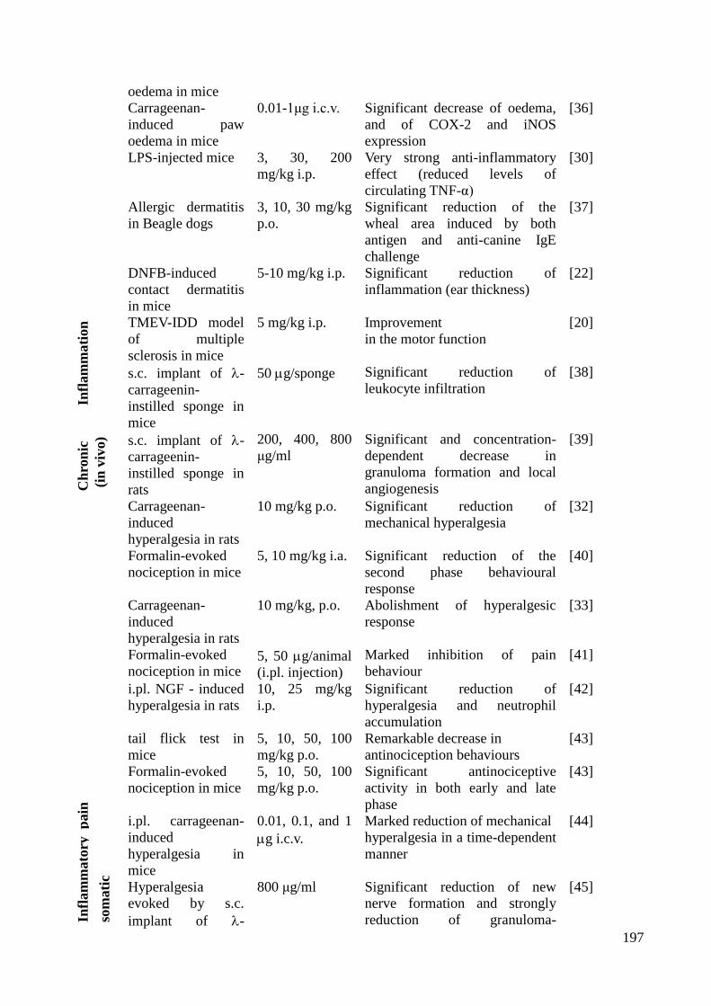
197
oedema in mice
Carrageenan-
induced paw
oedema in mice
0.01-1μg i.c.v. Significant decrease of oedema,
and of COX-2 and iNOS
expression
[36]
LPS-injected mice 3, 30, 200
mg/kg i.p.
Very strong anti-inflammatory
effect (reduced levels of
circulating TNF-α)
[30]
Allergic dermatitis
in Beagle dogs
3, 10, 30 mg/kg
p.o.
Significant reduction of the
wheal area induced by both
antigen and anti-canine IgE
challenge
[37]
DNFB-induced
contact dermatitis
in mice
5-10 mg/kg i.p. Significant reduction of
inflammation (ear thickness)
[22]
Ch
ron
ic
Infl
am
mati
on
(in
viv
o)
TMEV-IDD model
of multiple
sclerosis in mice
5 mg/kg i.p. Improvement
in the motor function
[20]
s.c. implant of -
carrageenin-
instilled sponge in
mice
50 g/sponge Significant reduction of
leukocyte infiltration
[38]
s.c. implant of -
carrageenin-
instilled sponge in
rats
200, 400, 800
μg/ml
Significant and concentration-
dependent decrease in
granuloma formation and local
angiogenesis
[39]
Infl
am
mato
ry p
ain
som
ati
c
Carrageenan-
induced
hyperalgesia in rats
10 mg/kg p.o. Significant reduction of
mechanical hyperalgesia
[32]
Formalin-evoked
nociception in mice
5, 10 mg/kg i.a. Significant reduction of the
second phase behavioural
response
[40]
Carrageenan-
induced
hyperalgesia in rats
10 mg/kg, p.o. Abolishment of hyperalgesic
response
[33]
Formalin-evoked
nociception in mice 5, 50 g/animal
(i.pl. injection)
Marked inhibition of pain
behaviour
[41]
i.pl. NGF - induced
hyperalgesia in rats
10, 25 mg/kg
i.p.
Significant reduction of
hyperalgesia and neutrophil
accumulation
[42]
tail flick test in
mice
5, 10, 50, 100
mg/kg p.o.
Remarkable decrease in
antinociception behaviours
[43]
Formalin-evoked
nociception in mice
5, 10, 50, 100
mg/kg p.o.
Significant antinociceptive
activity in both early and late
phase
[43]
i.pl. carrageenan-
induced
hyperalgesia in
mice
0.01, 0.1, and 1
g i.c.v.
Marked reduction of mechanical
hyperalgesia in a time-dependent
manner
[44]
Hyperalgesia
evoked by s.c.
implant of -
800 μg/ml Significant reduction of new
nerve formation and strongly
reduction of granuloma-
[45]

198
carrageenin-
instilled sponge in
the rat
associated hyperalgesia vis
cera
l
Turpentine
inflammation of the
rat urinary bladder
10, 20, 30
mg/kg i.a.
Significant attenuation of the
vesical hyper-reflexic response
[40]
Acetic acid-evoked
writhing in mice
1, 20 mg/kg i.p. Dose-dependent attenuation of
the writhing response
[41]
Turpentine
inflammation of the
rat urinary bladder
10, 25 mg/kg
i.p.
Attenuation of referred
hyperalgesia in a dose-dependent
fashion
[46]
Kaolin-evoked
writhing in mice
0.1 - 10 mg/kg
i.p.
Potent inhibition of the
nociceptive response
[41]
Magnesium
sulphate-evoked
writhing in mice
1-10 mg/kg i.p. Dose-dependent inhibition of the
nocifensive response
[41]
NGF-induced
inflammation of the
rat urinary bladder
2.5 mg/kg i.a. Significant increase of
micturition threshold and
significant reduction of spinal
cord Fos production
[47]
Acetic acid-evoked
writhing in mice
50, 100 mg/kg
p.o.
Significant reduction of the
number of writhes
[43]
Neu
rop
ath
ic p
ain
Spinal cord injury
in mice
10 mg/kg i.p.
before or after
surgery
Significant reduction of the
severity of spinal cord trauma
and of the spinal cord levels of
TNF-α, IL-1, iNOS
[48]
Chronic
constriction injury
of sciatic nerve in
mice
10 mg/kg i.p.
once daily for
one week
Significant relief of thermal
hyperalgesia and mechanical
allodynia [49]
Abbreviations used in the table - DNFB, 2,4-dinitrofluorobenzene; i.pl., intraplantar; LPS,
bacterial lipopolysaccharide; poly-(I:C), polyinosinic acid-polycytidylic acid; RBL-2H3, rat
basophilic leukemia cells of the secreting subline 2H3; RPMCs, rat peritoneal mast cells; TPA,
phorbol ester 12-O-tetradecanoylphorbol-13-acetate; TMEV-IDD, Theiler's murine
encephalomyelitis virus-induced demyelinating disease.
The anti-inflammatory and anti-hyperalgesic effects of PEA summarized in the Table are partly
mediated by mast cell down-modulation [47,50]. Particularly, ultramicronized PEA, whose
veterinary use is patented by Innovet Italia, down-modulates mast cell degranulation both in vitro
and in vivo [31]. A clinical study on cats with eosinophilic granuloma and eosinophilic plaque
revealed that PEA (10 mg/kg/30 days/p.o.) not only down-regulates skin mast cell degranulation,
but also reduces cutaneous signs and lesions [51]. A randomized, double blind, placebo controlled
study confirmed the ability of PEA (15 mg/kg/45 days/p.o.) to reduce inflammatory-related signs in
atopic dogs [52].
PEA is currently used to control inflammation and pain in some small animal veterinary products
indicated for skin diseases and LUTDs [14], under the trademark of Redonyl® and Urys
®
respectively.
A PEA congener, namely N-palmitoyl-D-glucosamine or Glupamid®, is included in a
chondroprotective nutraceutical for canine and feline OA, and has been shown to down-modulate
mast cell mediator release [53]. A preliminary ongoing study has recently shown that a 7-day
Glupamid® treatment (10 mg/kg, p.o., sid) significantly attenuated mechanical allodynia in a time-

199
dependent manner in a murine model of osteoarthritis pain (Costa B., personal communication).
The evidence collected so far provide a strong rational basis for the use of PEA as the management
modality of choice for canine and feline pain, of both inflammatory and neuropathic origin.
References
1. AAHA/AAFP J Am Anim Hosp Assoc. 2007; 43(5):235-48; 2. Della Rocca G. et al. J Vet
Pharmacol Ther. 2009; 32(s1):108; 3. Grubb T. Top Companion Anim Med. 2010;25(1):45-52; 4.
Fox SM. Chronic pain in small animal medicine. Manson Publishing ltd. 2010; 5. Hugonnard M. et
al. Vet Anaesth Analg. 2004; 31(3):154-63; 6. Schmelz M. Dermatol Ther. 2005;18(4):304-7; 7.
Freemont AJ et al. J Pathol. 2002;197(3):281-5; 8. Thacker MA et al. Anesth Analg. 2007;
105(3):838-47. 9. Rudick CN. et al. PLoS ONE 2008;3(5):e2096; 10. Kuehl FA Jr. et al. J Am Chem
Soc. 1957; 79(19):5577-8; 11. Bachur NR. et al. J Biol Chem. 1965;240:1019-24; 12. Aloe L. et al.
Agents Actions. 1993;39 Spec No:C145-7; 13. Levi-Montalcini R. et al. Trends Neurosci. 1996;
19(11):514-20; 14. LoVerme J. et al. Life Sci. 2005; 77(14):1685-98; 15. D'Argenio G. et al. J Mol
Med. 2007; 85(5):523-30, 16. Sarchielli P. et al. Neuropsychopharmacology. 2007; 32(6):1384-90;
17. Darmani NA. et al. Neuropharmacology. 2005;48(8):1154-63; 18. Garcia-Ovejero D. et al.
Neurobiol Dis. 2009; 33(1):57-71; 19. Franklin A. et al. J Neurosci. 2003;23(21):7767-75; 20. Loria
F. et al. Eur J Neurosci. 2008; 28(4):633-41; 21. Berdyshev EV. et al. Biochem J. 2000; 346 Pt
2:369-74; 22. Petrosino S. et al. Allergy, in press. 2009 DOI: 10.1111/j.1398-9995.2009.02254.x;
23. Petrosino S. et al. Proceedings 33rd Annual WSAVA/FECAVA Congress, Dublin, Ireland, 2008,
p.707; 24. Re G. et al. Vet J. 2007,173:23-32 ; 25. Rice AS. et al. Prostagland Leukot Essent Fatty
Acids. 2002; 66(2-3):243-56; 26. Calignano A. et al. Nature. 1998; 394(6690):277-81; 27.
Berdyshev EV. et al. Eur J Pharmacol. 1997;330(2-3):231-40; 28. Facci L. et al. Proc Natl Acad Sci
1995; 92(8):3376-80; 29. Ross RA. et al. Eur J Pharmacol. 2000;401(2):121-30; 30. Hoareau L. et
al. Obesity. 2009; 17(3):431-8; 31. Cerrato S. et al. Vet Immunol Immunopathol. 2010;133(1): 9-15;
32. Mazzari S. et al. Eur J Pharmacol. 1996; 300(3):227-36; 33. Conti S. et al., Br J Pharmacol.
2002; 135(1):181-7; 34. Costa B. et al. Br J Pharmacol. 2002; 137(4):413-20; 35. Lo Verme J. et al.
Mol Pharmacol. 2005; 67(1):15-9; 36. D'Agostino G. et al. J Pharmacol Exp Ther. 2007;
322(3):1137-43; 37. Cerrato S. et al. 29th Congr European Academy of Allergy and Clinical
Immunology, EAACI 2010, London, UK; 38. Solorzano C. et al. Proc Natl Acad Sci U S A.
2009;106(49):20966-71; 39. De Filippis D. et al. Pharmacol Res. 2010; 61(4):321-8; 40. Jaggar SI.
et al. Pain. 1998; 76(1-2):189-99; 41. Calignano A. et al. Eur J Pharmacol. 2001; 419(2-3):191-8;
42. Farquhar-Smith WP, Rice AS. Anesthesiology. 2003; 99(6):1391-401; 43. Karimi G. et al.
Pharmacologyonline. 2008;3:904-12; 44. D'Agostino G. et al. Eur J Pharmacol. 2009; 613(1-3):54-
9; 45. De Filippis D. et al. submitted; 46. Farquhar-Smith WP, Rice AS. Anesthesiology. 2001;
94(3):507-13; 47. Farquhar-Smith WP. et al. Pain. 2002; 97(1-2):11-21; 48. Genovese T. et al. J
Pharmacol Exp Ther. 2008; 326(1):12-23; 49. Costa B. et al. Pain. 2008; 139(3): 541-50; 50. Costa
B. et al. J Periph Nerv Syst. 2009;14(Suppl1):10; 51. Scarampella F. et al. Vet Dermatol.
2001;12(1):29-39; 52. Waisglass S. et al. Proceedings Congr Internaz SCIVAC, 2009, Rimini, Italy,
p. 564; 53. Miolo A. et al. Veterinaria, 2007, 21(6): 23-8.
* Symposium - Palmitoylethanolamide antinflammatory/neuroprotective properties: the
pharmacological bases for novel putative therapeutic agents, 34th
National Congress of the Italian
Pharmacological Society, Rimini, Italy, October 2009 -
http://www.sifweb.org/cong34/congresso_simposi_S-2-4.php

200
PLENARY SESSION
LIPID SIGNALLING in PAIN CONTROL:AND MOLECULAR TARGETS
CHAIRMEN: Di Marzo - Otto - De Nicola
ROLE OF TRP CHANNELS IN PAIN AND THEIR INTERACTIONS WITH
PHYTOCANNABINOIDS AND ENDOCANNABINOIDS
Vincenzo Di Marzo
INVOLVEMENT OF OPIOID AND CANNABINOID RECEPTORS IN THE OXYTOCIN-
INDUCED ANTIHYPERALGESIC EFFECT FOLLOWING ICV ADMINISTRATION R. Russo
CENTRAL PPAR-ALPHA ACTIVATION REDUCES PAIN AND HYPERALGIA IN MICE
VIA NUCLEAR FACTOR KB INHIBITION Giuseppe D’Agostino
NEUROPATHIC PAIN: LOOKING FOR A DISEASE MODIFYING AGENT
L. Di Cesare Mannelli
ENDOCRINE EFFECTS OF “NEW OPIOIDS” IN CHRONIC PAIN IN WOMEN OF
DIFFERENT AGES
M.C. Pace
201
ROLE OF TRP CHANNELS IN PAIN AND THEIR INTERACTIONS WITH
PHYTOCANNABINOIDS AND ENDOCANNABINOIDS
Vincenzo Di Marzo
Endocannabinoid Research Group, Institute of Biomolecular Chemistry, Consiglio Nazionale delle
Ricerche, 80078, Via Campi Flegrei 34, Pozzuoli (NA), Italy
The transient receptor potential (TRP) superfamily of non-selective cation channels includes 6
subfamilies: TRPC (‘Canonical’), TRPV (‘Vanilloid’), TRPM (‘Melastatin’), TRPP (‘Polycystin’),
TRPML (‘Mucolipin’), and TRPA (‘Ankyrin’) channels. TRP channels are six transmembrane (TM)
domain integral membrane proteins, with cytosolic C- and N-terminal domains, and a non-selective
cation-permeable pore region between TM5 and TM6. The various subfamilies differ particularly
for the number of ankirin repeats present in their N-terminus, which is null in TRPM and very high
in TRPA channels. Over 50 members of the TRP family have been characterized in yeast, worms,
insects, fish, and so far 28 in mammals. They are involved in the trasduction of a remarkable range
of stimuli including temperature, mechanical and osmotic stimuli, electrical charge, light, olfactive
and taste stimuli, hypotonic cell swelling, xenobiotic substances and endogenous lipids.
Importantly, mutations in different TRPs have been linked to human diseases, and their expression
in tissues affected by pathological conditions is often increased (1).
TRP channels of the vanilloid-type 1-4 (TRPV1-4), ankyrin type-1 (TRPA1) or melastatin type-8
(TRPM8) are deeply involved in thermosensation, pain transduction and inflammation. They are
expressed in sensory fibers of A and C-type, in dorsal root (DRG) and trigeminal ganglia as well
as in perivascular neurons, with TRPV1 (the “capsaicin receptor”) and TRPA1 (the “mustard
receptor”) being often co-expressed in the same neurons. Whilst TRPV1-4 are activated by high
temperatures, TRPA1 and TRPM8 (the “menthol receptor”) are activated by cold. TRPV1 is also
activated by low pH, such as during certain inflammatory conditions, as well as by several pro-
inflammatory mediators, and this leads to release of algogenic peptides (substance P, CGRP) from
sensory neurons, thus contributing to neurogenic inflammation. TRPA1, instead, is activated by
numerous irritants. TRPV1 is also expressed in central neurons, and, at the supra-spinal level, is
abundant in neurons of the periaqueductal grey (PAG) and rostral ventrolateral medulla (RVM),
where it modulates the descending pathway of antinociception. Contrary to its role in the spinal
cord and sensory afferents, TRPV1 in the PAG-RVM contributes to descending antinociception, and
it does so by enhancing both glutamatergic signalling/OFF neuron activity in the RVM and -
opioid receptor-mediated analgesia (2,3).
In DRG neurons as well as in spinal cord and in the PAG and RVM, TRPV1 is also co-expressed
with cannabinoid CB1 receptors, with which this channel shares some endogenous agonists, i.e.
anandamide and N-arachidonoyl-dopamine, known as endovanilloids/endocannabinoids. TRPV1
and CB1 can either act in concert or oppose each other at modulating pain through sensory or supra-
spinal pathways, and this cross-talk might occur through several mechanisms (4). Furthermore,
some non-psychotropic cannabinoids from Cannabis sativa with little or no activity at CB1
receptors, such as cannabidiol (CBD) or cannabichromene (CBC), activate instead TRPV1, TRPV2
and/or TRPA1 channels, or potently antagonize TRPM8 channels (5). Importantly, CBD can both
activate and desensitise TRPV1, TRPV2 and TRPA1 channels, and this property endows the
compound with the capability of potentially influencing nociception and inflammation in several
ways, also at the supraspinal level (5-7). These interactions will be described and new data on the
effects of CBD and CBC on the descending pathway of antinociception, obtained by my group
together with Prof. Maione’s group, will be presented at the conference.

202
References
1) Nelson PL, Beck A, Cheng H. Transient receptor proteins illuminated: Current views on
TRPs and disease. Vet J. 2010 Mar 27 [Epub ahead of print]
2) Starowicz K, Maione S, Cristino L, Palazzo E, Marabese I, Rossi F, de Novellis V, Di
Marzo V. Tonic endovanilloid facilitation of glutamate release in brainstem descending
antinociceptive pathways. J Neurosci. 2007, 27(50):13739-49.
3) Maione S, Starowicz K, Cristino L, Guida F, Palazzo E, Luongo L, Rossi F, Marabese I,
de Novellis V, Di Marzo V. Functional interaction between TRPV1 and micro-opioid receptors in
the descending antinociceptive pathway activates glutamate transmission and induces analgesia. J
Neurophysiol. 2009, 101(5):2411-22.
4) Di Marzo V, Cristino L. Why endocannabinoids are not all alike. Nat Neurosci. 2008,
11(2):124-6.
5) De Petrocellis L, Vellani V, Schiano-Moriello A, Marini P, Magherini PC, Orlando P, Di
Marzo V. Plant-derived cannabinoids modulate the activity of transient receptor potential channels
of ankyrin type-1 and melastatin type-8. J Pharmacol Exp Ther. 2008, 325(3):1007-15.
6) De Petrocellis L, Di Marzo V. Non-CB1, non-CB2 receptors for endocannabinoids, plant
cannabinoids, and synthetic cannabimimetics: focus on G-protein-coupled receptors and transient
receptor potential channels. J Neuroimmune Pharmacol. 2010, 5(1):103-21.
7) Palazzo E, Rossi F, Maione S. Role of TRPV1 receptors in descending modulation of
pain. Mol Cell Endocrinol. 2008, 286(1-2 Suppl 1):S79-83.

203
INVOLVEMENT OF OPIOID AND CANNABINOID RECEPTORS IN THE OXYTOCIN-
INDUCED ANTIHYPERALGESIC EFFECT FOLLOWING ICV ADMINISTRATION
R. Russo, G. La Rana, O. Sasso, G. D’Agostino, R. Meli and A. Calignano
Department of Experimental Pharmacology, University of Naples ‘‘Federico II’’, via D. Montesano
49, 80131 Naples, Italy.
Corresponding author e-mail address: [email protected]
Introduction
Oxytocin (OXT) is a nonapeptide synthesized in the paraventricular (PVN) and the supraoptical
(SON) nuclei in the hypothalamus, its receptors are widely distribute in the CNS, including cortex,
olfactory system, basal gangalia, limbic system, thalamus, hypothalamus, brain stem, and dorsal
horn of the spinal cord [1]. OXT performs its biological functions through OXT receptor
functionally coupled to Gq/11α class GTP binding proteins that stimulate together with G β/γ, the
activity of phospholipase C-β isoforms [1]. Several studies have reported that OXT modulates
analgesia or nociception [2; 3], and that the endogenous opioid system may be involved [4].
Moreover, a series of physiological and pharmacological studies have shown that OXT acts on
autoreceptors to elevate intracellular Ca2+
[5; 6]. Although many authors have reported and are in
agreement on analgesic action of OXT, the mechanism has not been well elucidated. Some studies
suggest in fact, that OXT has no effect on nociception [7] and attribute the OXT increased latency
responses on the hot plate test to the sedative and vasoconstrictive effects of this peptide [8]. On the
other hand, several manuscripts suggest that OXT-induce analgesia or nociception could be due to
the interaction with opioid system [9; 4]. Indeed, Miranda-Cardenas and co-worker [10] have show
that the endogenous released OXT induced by electrical stimulation of the paraventricular nucleus
of hypothalamus or direct application of OXT on the spinal cord produced a clear analgesic effect,
which was reduced significantly when naloxone was intrathecally injected before the stimulation of
the paraventricular nucleus and OXT administration [10]. Recently, an interesting hypothesis
suggest that OXT releases from presynaptic cell may activate local Ca2+
stores [5; 6] producing a
second molecule such as endocannabinoid [11; 12; 13], which
in turn then function as retrograde
transmitter [14].
Aim
The aim of present study was to investigate a possible role for OXT receptors at the CNS level in
mediating peripheral pain. In particular, we examined the antinociceptive and the anti-inflammatory
effects of OXT and, in particular, we studied the role of the opioid and cannabinoid systems in
OXT-induced antinociception in carrageenan-induced paw oedema.
Animals. and Chemicals.
Male Swiss mice weighing 30-33 g were purchased from Harlan (Udine, Italy). OXT, naloxone,
nor-binaltorphimine (nor-BNI), naltrindole, were purchased from Tocris Cookson Inc. (Bristol,
UK). All other chemicals were from Sigma-Aldrich (Milan, Italy).
Intracerebroventricular Injections.
Animals were briefly anesthetized with enflurane. Drugs were administered in a volume of 2 μl per
mouse by using a 25-μl glass Hamilton syringe Hamilton Co. (Reno, NV) with a stainless steel
needle modified with a shaft to control the depth of injection at 2 mm. The injection was made into
the lateral ventricle 2 mm caudal and 2 mm lateral from the bregma.
Paw Oedema Paw oedema was induced by a sub-plantar injection of 50 μl of sterile saline containing 1% λ-
carrageenan into the left hind paw. Paw volumes were measured by a plethysmometer (Ugo Basile,
Milan, Italy) at different time intervals.
Mechanical hyperalgesia
Mechanical hyperalgesia was assessed by measuring of latency (s) to withdraw the paw away from
a constant mechanical pressure exerted onto its dorsal surface.

204
Statistical Analyses.
Results are expressed as the mean ± S.E.M. of n experiments. All analyses were conducted using
Graph-Pad Prism (GraphPad Software Inc., San Diego, CA). The significance of differences
between groups was determined by two-way analyses of variance (ANOVA) followed by
Bonferroni post hoc tests.
Results and Discussion Many studies have been demonstrated that OXT is involved in pain modulation in the central
nervous system [7; 10]. Central treatment (icv) with OXT has an analgesic effect in a patient with
intractable cancer pain [15], moreover, OXT in the spinal cord relates to antinociception in the dog
[16]. Moreover, Yang and co-worker [19], proved that OXT takes part in the chronic and acute low
back pain in the human. Our studies showed that OXT increased paw withdrawal latency only
following central administration (30ng/mouse-icv), while peripheral injection (1mg/kg-i.p. and
50μg/paw-i.pl.) did not produce any effect on carrageenan-induced hyperalgesia. For confirmed
involvement of central OXT receptors, we used atosiban, a specific antagonist OXT receptors.
Central co-injection of OXT (30 ng/mouse) and atosiban (1μg/mouse), has produced a reduction
OXT-induce antihyperalgesic effect. Previous studies had proven that OXT induces the spinal cord
releasing endogenous opiate peptides, and that the OXT analgesic effect can be reversed by opiate
receptor antagonist [9; 17; 10]. The mechanism of OXT regulating analgesia may be proposed as
follows: OXT is released from PVN and/or SON during pain stimulation, and then transported to
the other brain nuclei and spinal cord, where OXT influences the other antinociceptive system such
as endogenous opiate peptide system to take part in pain modulation. Previous studies showed that
OXT-induced antinociception could be blocked by the non-selective opioid receptor antagonist
naloxone in the supraspinal level [18]. In the present study OXT-induced antinociceptive effect to
mechanical stimulation was attenuated by i.c.v. injection of the selective opioid receptor antagonists
beta-Funaltrexamine (β-FNA 1ug/mouse; μ-antagonist) and nor-binaltorphimine (nor-BNI 10
nmol/mouse; k-antagonist), but not by naltrindole (10 nmol; δ-antagonist), indicating that mu and
kappa opioid receptors, not delta opioid receptor, are involved in the OXT-induced anti-nociception
in the brain. These findings are supported by sereral papers [19; 9, 4]. Moreover, a series of
physiological and pharmacological studies have shown that OXT acts on OXT auto-receptors to
elevate intracellular Ca2+
[5; 6]. On basis of this, we have supposed that dendritically released OXT
act on presynaptic OXT cell activated Ca2+
stores
this event is well know to lead to
endocannabinoid (eCB) release [11; 12; 13]. Consequently, we explored whether activation of OXT
receptor was a prerequisite to the production and subsequent actions of eCB. Our results showed
that CB1 antagonist, SR141617A (1 μg/mouse), reduced antihyperalgesic effect of OXT, whereas, a
specific CB2 antagonist, SR144528 (1 μg/mouse), did not modify OXT activity, suggest a key role
only for the CB1 receptor. Recent reports have highlighted the interaction of the endogenous
cannabinoid (CB) system with central hormone release in the modulation of magnocellular neuron
synaptic physiology [12; 20]. It is conceivable that OXT may diffuse further eCB, and thus, allow
eCB action on many cells to co-ordinate activity, transforming a local effect to a global one.
The present study clarifies that OXT plays a key role in modulation of pain pathway. In particular,
following activates of OXT central receptors, the nonapeptide is able to reduce carrageenan-induce
hyperalgesia but it do not modify oedema development. Moreover, our results indicate that μ- k-
receptors, and CB1-receptor, are involved in the OXT-induce antihyperalgesic effect in the central
nervous system.
References
1. Gimpl G, Fahrenholz F. The oxytocin receptor system: structure, function, and regulation.
Physiol Rev (2001) 81:629-683,.
2. Yu SQ, Lundeberg T, Yu LC. Involvement of oxytocin in spinal antinociception in rats with
inflammation Brain Res. (2003) 983(1-2):13-22.
3. Han Y, Yu LC. Involvement of oxytocin and its receptor in nociceptive modulation in the central
nucleus of amygdala of rats. Neurosci Lett. (2009). 454(1):101-4.

205
4. Gu XL, Yu LC Involvement of opioid receptors in oxytocin-induced antinociception in the
nucleus accumbens of rats. J Pain. (2007). 8(1):85-90.
5. Lambert RC, Dayanithi G, Moos FC, Richard P. A rise in the intracellular Ca2++ concentration
of isolated rat supraoptic cells in response to oxytocin. J Physiol (Lond) (1994) 478:275–
287.
6. Ludwig M, Sabatier N, Bull PM, Landgraf R, Dayanithi G, Leng G. Intracellular calcium stores
regulate activity-dependent neuropeptide release from dendrites. Nature (2002) 418:85– 89.
7. Millan MJ, Schmauss C, Millan MH, Herz A, Vasopressin and oxytocin in the rat spinal cord:
analysis of their role in the control of nociception, Brain Res. 309 (1984) 384–388.
8. Xu XJ, Wiesenfeld-Hallin Z. Is systemically administered oxytocin an analgesic in rats? Pain
1994;87:193–6.
9. Yang J: Intrathecal administration of oxytocin induces analgesia in low back pain involving the
endogenous opiate peptide system. Spine (1994). 19:867-871,
10. Miranda-Cardenas Y, Rojas-Piloni G, Martinez-Lorenzana G, Rodriguez-Jimenez J, Lopez-
Hidalgo M, Freund-Mercier MJ and Condes-Lara M, Oxytocin and electrical stimulation of
the paraventricular hypothalamic nucleus produce antinociceptive effects that are reversed
by an oxytocin antagonist, Pain 122 (2006), 182–189.
11. Brenowitz SD, Regehr WG. Calcium dependence of retrograde inhibitionby endocannabinoids
at synapses onto Purkinje cells. J Neurosci 23 (2003):6373– 6384.
12. Hirasawa M, Schwab Y, Natah S, Hillard CJ, Mackie K, Sharkey KA, Pittman QJ.
Dendritically released transmitters cooperate via autocrine and retrograde actions to inhibit
afferent excitation in rat brain. J Physiol (Lond) (2004) 559:611– 624.
13. Ohno-Shosaku T, Hashimotodani Y, Maejima T, Kano M Calcium signaling and synaptic
modulation: regulation of endocannabinoidmediated synaptic modulation by calcium. Cell
Calcium (2005). 38:369 –374.
14. Oliet SH, Baimoukhametova DV, Piet R, Bains JS. Retrograde regulation of GABA
transmission by the tonic release of oxytocin and endocannabinoids governs postsynaptic
firing. J Neurosci. (2007); 27(6):1325-33.
15. Madrazo I, Franco-Bourland RE, Leon-Meza VM, Mena I. Intraventricular somatostatin-14,
arginine-vasopressin and oxytocin: analgesic effect in a patient with intractable cancer pain.
Appl Neurophysiol (1987);50:427–31
16. Brown DC, Perkowski S. Oxytocin content of the cerebrospinal fluid of dogs and its
relationship to pain induced by spinal cord compression. Vet Surg (1998) ;27: 607–11.
17. Gao L, Yu LC. Involvement of opioid receptors in the oxytocin-induced antinociception in the
central nervous system of rats. Regul Pept (2004);120:53–8.
18. Arletti R, Benelli A, Bertolini A, Influence of oxytocin on nociception and morphine
antinociception, Neuropeptides 24 (1993) 125–129.
19. Ge Y, Lundeberg T, Yu LC. Blockade effect of mu and kappa opioid antagonist on the
antinociception induced by intra-periaqueductal grey injection of oxytocin in rats. Brain Res
(2002); 927:204–7.
20. Sabatier N, Leng G. Presynaptic actions of endocannabinoids mediate alpha-MSH-induced
inhibition of oxytocin cells. Am J Physiol Regul Integr Comp Physiol. (2006); 290(3):R577-
84..
206
CENTRAL PPAR-ALPHA ACTIVATION REDUCES PAIN AND HYPERALGIA IN MICE
VIA NUCLEAR FACTOR KB INHIBITION
Giuseppe D’Agostino, Giovanna La Rana, Roberto Russo, Oscar Sasso, Carmen Avagliano, Anna
Iacono, Rosaria Meli, Antonio Calignano.
Department of Experimental Pharmacology, University of Naples Federico II, Naples, Italy.
Peroxisome proliferator-activated receptor alpha (PPAR-alpha) is a ligandactivated transcription
factor belonging to the nuclear hormone receptors and suggested to be involved in inflammation
and pain control. Little is known about its role at CNS level. We report that spinal and dorsal root
ganglia PPAR-alpha expression is modulated by a peripheral inflammatory or a painful stimulus,
and central administration of endogenous or synthetic PPAR-alpha agonists reduces both pain
perception and inflammatory hyperalgesia in mice. Under inflammatory pain state, central PPAR-
alpha activation modulates NF-kB nuclear signalling along sciatic nerve, dorsal root ganglia and
spinal cord. Moreover, we evidence that PPAR-alpha receptor may physically reduce NF-kB
activation during the early phase of pain signalling. In vivo co-immunoprecipitation experiments
reveal a physical interaction between PPAR-alpha and NF-kB complex subunits. This interaction
was strongly and rapidly increased in lumbar spinal cord of mice subjected to the formalin
nociception test. To understand if the enhanced interaction would function as a reducer of pain-
evoked intracellular machinery, we used an in vitro approach. In SH-SY5Y neuroblastoma cells
substance P stimulation induced the PPAR-alpha/NF-kB interaction in few minutes, similarly to that
observed in vivo. GW7647, a synthetic PPARalpha agonist and pain-reducing compound, increases
this complex formation. We conclude that i) PPAR-alpha-mediated reduction of persistent pain may
likely rely on reduction of the NF-kB activity and inflammatory central sensitizer synthesis, ii)
while the contribution to acute pain responses may be in part related to constitutive activity of both
these transcription factors in neurons of the nociceptive system

207
NEUROPATHIC PAIN: LOOKING FOR A DISEASE MODIFYING AGENT
L. Di Cesare Mannelli, C. Ghelardini
University of Florence, Department of Preclinical and Clinical Pharmacology, Viale Pieraccini 6,
50139, Florence, Italy
Neuropathic pain is an unpleasant, abnormal signalling associated with injury or malfunction in the
peripheral or central nervous system (CNS). These widespread disorders are induced by metabolic
insult or trauma, autoimmune diseases, infections, drug or toxin exposure, and by cancer or viral
chemotherapy. Neuropathies are extremely difficult to treat and actual therapies are generally
palliative and include conservative non-pharmacological therapies, drugs and more invasive
interventions. Available drugs for pain improvement are not able to revert the nervous alteration or
to induce tissue regeneration [1]. On the other hand, many growth factors for the nervous system do
not relieve pain. NGF, the prototypical neurotrophic factor, maintains the survival of sympathetic
and sensory neurons as well as neurite outgrowth but it also exerts profound biological effect on
nociceptors that express high-affinity NGF receptors [2]. Therefore, a great deal of interest has
evolved around the research of compounds able to decrease hyperalgesia and to induce, at the same
time, neuroprotection or neuroregeneration.
Pain is the common symptom due to variable nervous tissue alterations leading to different
neuropathies. Traumas induce morphological changes evident also further than the lesion site.
Damage to peripheral nerve are able to induce a decrease in myelin thickness, axon diameter and
number of fibers in the proximal and, at a lesser extent, in the distal part of the nerve. An important
inflammatory component is present as oedema and macrophagic infiltrate [3]. Moreover, in a model
of peripheral neuropathy induced in the rat by loose ligation of the sciatic nerve (Chronic
Constriction Injury; CCI) the activation of the apoptosis cascade has been described both in
proximal and distal portion of the nerve. A mitochondrial damage alters membrane permeability,
cytochrome C is released in the cytosol and triggers the signal up to the fragmentation of the
genome [4]. Animal treatment with Acetyl-L-Carnitine (ALCAR; 100 mgkg-1
i.p. twice daily for 14
days), but not with L-Carnitine or Gabapentin, prevents apoptosis induction. ALCAR is also able to
prevent hyperalgesia and both the anti-apoptotic and the anti-hyperalgesic effects are reverted by
the nicotinic receptor (nAChR) antagonist mecamylamine [5]. On the other hand, in the same
model, acute administration of the alpha7 nAChR agonist PNU-282987, 10 and 30 mg kg-1
p.o. (15
days after ligation), is able to reduce hyperalgesia in a methyllicaconitine-reversed manner. This
alpha7 nAChR agonist exerts no analgesic effects. Chronic PNU-282987 treatments, 30 mg kg-1
once a day for 7 days and 10 mg kg-1
for 28 days are able to decrease pain perception. Repeated
treatments with PNU-282987 reduce the presence of oedema and macrophagic infiltrate and, on the
coronal sections of the nerve, a significant higher myelin sheath, axonal diameter and number of
fibers are observable [3].
In other neuropathies, like those chemotherapy-dependent, the effect of neurotoxicity is lesser
pronounced and detectable and a smir of light signs diffuse from the peripheral to the central
nervous system has been highlighted. Characteristically, no inflammatory responses are present.
Both neuropathy groups, with inflammatory or without inflammatory alterations, show as common
characteristic a glial activation in CNS. Astrocytes and microglia have well-documented roles in
pain [6]. Although astrocytes and microglia in the CNS each have unique roles in the modulation of
neuronal function [7], they do have some overlapping actions. Both cells types are key mediators of
the CNS innate immune response. Growing evidences ascribe to glia pathological effects as
neuronal hyperexcitability and chronic inflammation. Spinal microglia have been recognized as
pivotal in the initial phases of neuropathic pain, whereas astrocytes may be involved in the
maintenance [8,9]. On the other hand glia has a number of housekeeping function, among them

208
neuroprotection [10,11]. It is important to consider that both astrocytes and microglia are necessary
for the homeostasis of the environment surrounding neurons, and also for the regulated clearance of
apoptotic cells.
A major challenge that new drug-development strategies for the treatment of neuropathic pain face
is targeting the pathological and pain trigger actions of astrocytes and microglia without altering
their protective and recuperative roles. In order to evaluate the glia role in neuropathic pain, we
recently described a decrease of the anti-hyperalgesic growth factor Artemin a member of the glial
cell line-derived neurotrophic factor (GDNF)-related family in the CCI model. The neuroprotective
compound ALCAR is able to normalize this expression level as well as to induce Artemin
expression in sham rats [12]. Topic of debate is whether this ALCAR effect is due to a nicotinic
mechanism. nAChR signalling has a major role in the glia-neuron network, in particular alpha7
nAChR is expressed in microglial cells [13] and its activation attenuates the pro-inflammatory
response of microglial cultures [14]. Also astrocytes express this receptor subtype but their possible
functional role is poorly understood [15]. In conclusion, glia is strongly involved in neuropathic
pain as well as in neuroregenerative mechanism and the stimulation of the glia-expressed alpha7
nAChR shows anti-hyperalgesic and neuroprotective effects. Further understanding of the
molecular mechanisms that underlie the effects of glia on neuropathy processing and more
evidences about the importance of nAChRs in the signal switch between pain and regeneration
should lead to the development of new and more efficient approaches for the clinical management.
References
1.Di Cesare Mannelli L, Ghelardini C, Toscano A, Pacini A, Bartolini A. Neuroscience 165:1345-
1352, 2010
2.Pezet S, McMahon SB. Annu Rev Neurosci 29:507-538, 2006
3.Di Cesare Mannelli L, Pacini A, Bonaccini L, Ronzoni S, Bartolini A, Ghelardini C. Pain, In Press
4.Di Cesare Mannelli L, Ghelardini C, Calvani M, Nicolai R, Mosconi L, Vivoli E, Pacini A,
Bartolini A. Eur J Neurosci 26:820-827, 2007
5.Di Cesare Mannelli L, Ghelardini C, Calvani M, Nicolai R, Mosconi L, Toscano A, Pacini A,
Bartolini A. J Neurosci Res 87:200-207, 2009
6.Pocock JM, Kettenmann H. Trends Neurosci 30:527-535, 2007
7.Haydon PG. Nature Rev Neurosci 2:185-193, 2001
8.Marchand F, Perretti M, McMahon SB. Nat Rev Neurosci 6:521-532, 2005
9.Scholz J, Woolf CJ. Nat Neurosci 10:1361-1368, 2007
10.Glezer I, Simard AR, Rivest S. Neuroscience 147:867-883, 2007
11.Milligan ED, Watkins LR. Nat Rev Neurosci 10:23-36, 2009
12.Vivoli E, Di Cesare Mannelli L, Salvicchi A, Bartolini A, Koverech A, Nicolai R, Benatti P,
Ghelardini C. Neuroscience, 2010. PMID: 20302919
13.Carnevale D, De Simone R, Minghetti L. CNS Neurol Disord Drug Targets 6:388-397, 2007
14.Suzuki T, Hide I, Matsubara A, Hama C, Harada K, Miyano K, Andrä M, Matsubayashi H, Sakai
N, Kohsaka S, Inoue K, Nakata Y. J Neurosci Res 83:1461-1470, 2006
15.Ono K, Toyono T, Inenaga K. Neuroscience 154:994-1001, 2008

209
ENDOCRINE EFFECTS OF “NEW OPIOIDS” IN CHRONIC PAIN IN WOMEN OF
DIFFERENT AGES
M.C. Pace, M. Iannotti, P. Sansone, V. Pota, M.B. Passavanti
Second University of Study of Naples, Department of Anesthesiologycal, Surgical and Emergency
Sciences, Piazza Miraglia 2, 80100 Naples, Italy;
Corresponding author: [email protected]
Introduction
Chronic non cancer pain affects one fourth of European population, the majority being women (1).
The need to treat pain compels physicians to use opioids for long periods even though opioids can
affect the hypothalamus-pituitary axis (2). Indeed both endogenous (β-endorphin) and exogenous
opioids are known to modulate the secretion of pituitary hormones, including gonadotropins. In
men, morphine administration was found to induce persistent, long-lasting hypogonadism. Indeed
substantial evidences indicate that gonadal hormones can influence the structure and functions of
neurons during development and also in adulthood, including alterations of their dendrites and
synaptic connections. Hajszan describes some studies showing that synaptic connectivity in the
hippocampus and prefrontal cortex of male rats, normally depends on androgens. When adult males
are castrated, the number of synapses in each region decreases. It is also suggested that androgens
might be an effective therapeutic for certain neurological disfuntions such as Alzheimer’s disease
and schizophrenia. The effects on androgens have been described in men, already the opioid-
induced hypogonadism has also been shown also in women with clinically relevant features. It is
necessary to find solutions able to support the use of opioids in pain therapy without deleterious
side effects like hypogonadism. As regards the efficacy/side effects ratio, ‘new’ opioids such as
buprenorphine are indicated as one of the best/first choices, also in view of the possibility to
administer the drug via a patch. Buprenorphine (BP) is a centrally-acting analgesic that binds to μ-
opioid receptors (MOR, partial agonist) and k-opioid receptors (antagonist) with high affinity. Many
studies have described the good analgesic effects of this drug, its high safety and the good
compliance by patients (3). Moreover Ceccarelli et al. described that buprenorphine does not affect
testosterone levels in the brain of male rats (4). We conducted a trial to assess the efficacy and the
effects of buprenorphine TDS for 6 months in women in reproductive age or in menopause.
Furthermore, a number of hormonal determinations where carried out to study the effect of
buprenorphine on HPG axis.
Methods
Subjects were recruited in the Second University of Naples, while hormonal determinations were
carried out in the Pain and Stress Neurophysiology lab of the University of Siena. The protocol, in
accordance with the ethical standards of the Declaration of Helsinki, was approved by the
Institutional Ethic Committee. Patients signed an informed consent before participation.
Female outpatients of at least 18 years of age, suffering non-cancer, musculoskeletal pain (low back
pain) with VAS > 6 were asked to participate in the study. The exclusion criteria were: neurological
pathologies; cardiac, respiratory, metabolic, hepatic, renal, gastrointestinal, heavy and/or
uncompensated pathologies; oncologic pathologies; HIV; endocrine alterations; psychiatric
disorders; skin diseases; alcoholism and drug abuse; verified hypersensitivity to opioids; patients
allergic to patches; pregnancy or lactation. This was an open prospective study for the evaluation of
the analgesic efficacy and endocrine effects induced by long-term therapy with buprenorphine TDS
35 mcg/h to be changed every 72 h. After an interview including a pathological anamnesis and
objective exam, each patient underwent an evaluation of pain through VAS, Mc Gill Pain
Questionnaire Short Form (SF-MPQ) and PPI.
During the visit a blood sample was collected to determine blood and hormone parameters. Then a
Buprenorphine TDS 35 mcg/h patch was applied.

210
The patients were instructed to use hydrochloride Buprenorphine sublingual tablets 0.2 mg, as
rescue medication. As antiemetic prophylaxis they could take metaclopromide 10 mg.
Each patient was given a diary for the daily recording of VAS, adverse effects and use of rescue
medication. After 15 days the patients made a control visit. The same procedure, the survey and
controls and the blood sampling were replicated after 1, 3 and 6 months.
Statistical analysis
ANOVA with repeated measures was applied to all parameters. Multiple comparisons were carried
with the Least Significant Difference (LSD) test when needed. The significance of the linear
correlation coefficient (r) in the pre and post-M groups was tested using the Steel and Torrie
procedure
Results
41 patients were screened to enrol 26 patients who met the inclusion criteria. 18 patients concluded
the 6-month period, 8 patients discontinued the therapy for various reasons: adverse effects
(constipation and somnolence, nausea and vomiting, n=4), patients decision (N=3) and medical
decision (N=1).
The 18 patients admitted to the study were treated with Buprenorphine for 6 months. They were
grouped into post-menopausal (10 patients, mean age 66.1 years, range 54-76), and pre-menopausal
groups (8 patients, mean age 39.5, range 26-50).
The patients did not exhibit any significant collateral effects and none resorted to metoclopramide;
the use of Buprenorphine tablets 0.2 mg as rescue medication was unnecessary and no
buprenorphine TDS adjustments were required.
All patients recorded an improvement in their pain, as showed VAS, SF-MPQ and PPI; this result
was present from the first month of treatment and persisted for the whole study (Table 1). Indeed
ANOVA revealed a significant effect of time for the three parameters, in both age groups, due to the
progressive decrease of their scores from basaline (T0) to the end.
Table 1: Pain parameters.
Pre-
M
T0
T1
T3
T6
Post-M
T0
T1
T3
T6
VAS 83.8 50.6 # 42.5 # 35.6 # 81 50.0 # 41.5 # 36.5 #
SF-MPQ 25.9 20.0 # 18.6 # 14.1 # 24.7 18.7 # 17.1 # 13.0 #
PPI 4.50 3.25 # 2.50 # 2.13 # 4.10 2.70 # 2.20 # 1.80 #
Vas: visual analog scale; SF-MPQ: short form-McQuill Questionnaire; PPI: present pain intensity
# p<0.05 vs T0 same group.
There were no statistically significant differences between the two groups in blood sugar, creatinine,
protein or red and white cells. As regards azotemia, GOT, GPT, g-GT and haematocrit, ANOVA
applied to the two groups determined at the 4 time points revealed significant differences (Table 2).
Table 2: Blood parameters.
Blood
Parameters Pre-
M
T0
T1
T3
T6
Post-M
T0
T1
T3
T6
Azotemia
mg/dl
34.3 33.6 30.6 31.8 53.1* 53.6* 52.3* 54.1*
Glicemia
mg/dl
78.0 82.6 78.1 82.4 96.3 106.7 101.4 96.6
Creatinin
mg/dl
0.86 0.82 0.84 0.83 0.93 0.94 0.98 0.98
GOT U/L 21.1 37.9# 23.3 22.6 17.6 20.1* 19.0 19.6
GPT U/L 21.9 37.4# 23.3 21.1 16.7 16.5* 15.4 19.2
g-GT U/L 15.7 28.3# 24.3 17.0 23.1 21.3 21.4 21.9

211
Protein g/L 7.3 7.2 7.3 7.1 7.0 6.9 6.9 6.9
Red cells
106/ml
4.3 4.3 4.4 4.4 4.6 4.6 4.8 4.7
White cells
103/ml
5.6 5.4 5.4 5.9 6.1 6.1 5.8 5.7
HT % 36.2 36.6 36.9 37.0 39.2* 38.2 38.9 38.4
VES mm/H 12.6 17.5 8.0 8.9 27.9* 16.5 14.3 14.4
* p<0.05 vs Pre-M group, same period
Luteinizing (LH) an Follicle-stimulating (FSH) hormones were higher in the post-M than pre-M
women (p<0,01 and p<0,03 respectively) (Table 3).
In contrast to findings obtained with other opioids, in wich testosterone was drastically decreased,
neither total testosterone (TT) nor free testosterone (fT), changed significantly for buprenorphine
TDS treatment in either group of women (table 3). TT and fT levels were slightly higher in post-
menopausal women at all determinations. Estradiol (E2) levels showed strong variation in the
younger women still in the reproductive periods due to the difficulty in collecting blood on exactly
the same day of the menstrual cycle (table 3).
Table 3: Hormones. Hormones Pre-M
T0
T1
T3
T6
Post-M
T0
T1
T3
T6
Total T ng/ml 0.12 0.16 0.20 0.20 0.20 0.20 0.21 0.21
Free T pg/dl 0.77 0.72 0.77 0.69 1.26 1.13 1.51 1.04
Estradiol pg/ml 83.5 38.9 24.4 22.1 7.2* 7.3 7.3 8.8*
Cortisol g/dl 85.5 149.3# 140.1# 170.6# 124.9 132.4 166.3 172.4
SHBG° 46.0 61.6 114.8 44.1 88.0 113.2 96.6 76.4
DHT° 26.1 29.11 32.0 42.4 23.1 21.4 16.4 10.7
# p<0.05 vs T0 same group; * p<0.05 vs Pre-M group, same period.
° (n=4 x group), in these hormones no statistics was carried out
Despite of this, as expected, E2 values were higher in younger women than in older ones.
Like testosterone, the cortisol (C) levels were not decreased by buprenorphine treatment. Indeed
ANOVA revealed a progressive increase from baseline to the end. In post-menopausal women the
increase was not significant, while in pre-menopausal women it was significant after 1 month of
treatment (p<0.05).The multivariate analysis applied to the three main hormones TT, E2 and C,
showed a significant effect over time point (p<0.01) only in the pre-menopausal group (p<0.01).
The correlation between VAS and hormones was significant in the pre-M group (R=0.34, p<0.001),
in the not significant in the Post-M one (R=0.034, p>0.05).
Discussion
The important result of this study is that women treated for 6 months with buprenorphine TDS did
not show the strong endocrine impairment observed with other opioids (5). Chronic, non cancer
pain is a very big problem in our society. It affects, about 20% of the adult population and it is
severe in 13% of cases. Moreover chronic pain mainly affects women, but few pharmacological
experimentation has been done on them because of neurohormonal interferences. For this reason we
investigate the effects of opioids on gonadal hormones, evaluating the results of the analgesic
efficacy of the therapy in both reproductive-age and menopausal patients. The high analgesic power
of these drugs makes them particularly efficient in the treatment of severe pain, but the risk of side
effects and of addiction and tolerance phenomena, often affects their clinical use in prolonged
treatments (6). OPIAD, i.e. opioid-induced androgen deficiency, mainly occurs in men treated with
opioids, but it has also been found in women. Fatigue, anemia, absence of libido, bad mood and
depression, are reported by patients treated with opioids. Buprenorphine, has repeatedly been shown
to have some features different from the other commonly used opioids. Indeed in experimental
animals and in patients it was found to cause a low level of addiction and it does not show a “roof

212
effect” if taken at therapeutic doses. Moreover recent studies have demonstrated that buprenorphine,
unlike other opioids, exerts an antihyperalgesic effect, probably due to its antagonistic properties on
k-opioid receptors (7). For these reasons we decided to use buprenorphine and we chosen the
transdermal system because patients and doctors are amenable that: it is not invasive, lasts 72 hours
and allows a constant release of the proximate principle with high therapeutic efficacy and low side
effects. These characteristics can also be seen in the results of our study, which show in women,
since the first patch application, a significant reduction in the pain symptomatology that persisted
during the treatment, without significant variations in relation to the menstrual cycle and
menopause. This early and constant positive trend made recourse to “rescue medication”
unnecessary. VAS and the other pain parameters clearly decreased already after one month of
treatment to remain low till the end of treatment.The results of the study are also encouraging in
regard to side effects, particularly the nausea and vomiting that often follow opioid administration.
In fact, in agreement with international literature, that reports a reduction of side effects with the use
of buprenorphine TDS, none of our patients reported any significant side effects that needed
treatment. This result underlines the key role of the administration technique in the genesis and the
degree of side effects from opioids. We must, of course, consider the different responses to the
therapy, often related to the patient’s conditions, which also explains why 4 patients were excluded
from the study after a short time. In fact, we believe it is very important to modulate the therapy
according to individual needs. The blood parameters have never showed significant alterations that
would justified the interruption of treatment. This is an interesting result since it reflects the absence
of detrimental effects of buprenorphine TDS on body homeostasis. In previous studies on humans
or experimental animals, gonadal hormones were found to be strongly affected by opioid intake.
These effects were present in both sexes, although with some differences (8). In the present study,
the considered steroid hormones (testosterone, DHT, cortisol) did not show any sign of decrease,
infact, they tended to increase. In this study, testosterone was inversely related to VAS but only in
pre-menopausal women. This was also true when all three hormones (testosterone, cortisol,
estradiol) were considered all together. It is not easy to explain why the correlation was found only
in the younger subjects because all recorded changes were apparently present in both groups, not
only in the pre-menopausal one. However this strong correlation suggests that the presence of the
menstrual cycle was significant. Indeed it was recently found in experimental females, that in
addition to the hormone replacement, the cycle play a strong role in worsening the behavioral
response to painful stimulation (9).
References
1) Niv D., Devor M. Position paper of the European Federation of IASP Chapters (EFIC) on the
subject of pain management. Eur J Pain 2007; 11:487-489
2) Aloisi, A.M., Affaitati G. et al. Estradiol and testosterone differently affect visceral pain-related
behavioural responses in male and female rats. Eur J Pain. 2009 Nov 28
3) Aurilio C, Pace MC et al. Opioids switching with transdermal systems in chronic cancer pain. J
Exp Clin Cancer Res. 2009 May 7;28:61.
4) Ceccarelli, I., et al. Single opioid administration modifies gonadal steroids in both the CNS and
plasma of male rats. Neuroscience 140, 929-937 (2006).
5) Daniell HW. Opioid endocrinopathy in women consuming prescribed sustained-action opioids
for control of nonmalignant pain. J Pain. 2008 Jan;9(1):28-36.
6) Furlan, A.D. et al. Opioids for chronic non cancer pain: a meta-analysis of effectiveness and
side effects. CMAJ 174, 1589-1594 (2006).
7) Kress HG. Clinical update on the pharmacology, efficacy and safety of transdermal
buprenorphine. Eur J Pain. 2009 Mar;13(3):219-30.
8) Aloisi AM, Aurilio C et al. Endocrine consequences of opioid therapy.
Psychoneuroendocrinology. 2009 Dec;34 Suppl 1:S162-8.
9) Sanoja R, Cervero F. Estrogen modulation of ovariectomy-induced hyperalgesia in adult mice.
Eur J Pain. 2008 Jul;12(5):573-81.
213
PLENARY SESSION
PERIPHERAL, SPINAL AND SUPRA-SPINAL MECHANISMS OF PAIN CONTROL
CHAIRMEN: Maione - Lydic - Vesce
FOREBRAIN PAIN MECHANISMS
Sabatino Maione
ANESTHETIC ACTIONS ON TONIC DESCENDING CONTROL OF THE
SPINOTHALAMIC TRACT
Peter J. Soja
1-(2’,4’-DICHLOROPHENYL)-6-METHYL-N-CYCLOHEXYLAMINE-1,4-
DIHYDROINDENO[1,2-C]PYRAZOLE-3-CARBOXAMIDE, A NOVEL CB2 AGONIST,
ALLEVIATES NEUROPATHIC PAIN THROUGH FUNCTIONAL MICROGLIAL
CHANGES IN MICE.
Luongo L.
THE PALMITOYLETHANOLAMIDE: AN IMPORTANT TOOL FOR PAIN
MANAGEMENT.
Calignano Antonio

214
FOREBRAIN PAIN MECHANISMS
Volker Neugebauer1, Vasco Galhardo2, Sabatino Maione3, Sean C. Mackey4,
1.Department of Neuroscience & Cell Biology, University of Texas Medical Branch, Galveston, TX
77555-1069, USA; 2.University of Porto Medical School, 4200-319 Porto, Portugal;
3.Department of Experimental Medicine, The Second University of Naples, 80138 Naples, Italy;
4.Anesthesia & Pain Management, Stanford University School of Medicine, Palo Alto, CA 94304-
1573, USA
Functional changes in chronic pain
Neuropathic pain is a devastating and difficult to manage consequence of peripheral nerve injury,
the main aspect of which is enhanced transmission of nociceptive messages [1]. In this condition,
noxious stimuli are perceived as more painful (hyperalgesia), and normal, harmless stimuli may
elicit pain (allodynia). Neuropathic pain may be considered a progressive nervous system disease
that results from poorly-defined neurophysiological and neurochemical changes. Support for this
hypothesis is obtained from animal models of chronic pain, in which a long-lasting increase in the
excitability of pain-integrating neurons is observed. More recently, multidisciplinary research
approaches have shown that chronic pain may be a consequence of long-term plastic changes along
the entire pain matrix [2]. Apart from peripheral nociceptors and the spinal cord, morphological and
functional plastic changes can also take place in cortical areas participating in pain processing.
Indeed, growing evidence suggests that long term plastic modifications in cortical networks may
represent a possible basic mechanism underlying chronic pain. Among cortical regions, anterior
cingulate cortex (ACC), insular cortex (IC), primary (S1) and secondary (S2) somatosensory cortex,
and prefrontal cortex (PFC) consistently respond to acute “physiological” pain stimuli in healthy
subjects. However, if on the one hand the majority of the progress in cortex imaging studies of pain
has been obtained in healthy subjects, much less is known about changes in cortical network
functioning or responsiveness during chronic pain, and whether or not areas associated with
physiological pain are also involved in chronic pain conditions. Intriguingly, PET or fMRI
procedures have shown that spontaneous neuropathic pain seems to be more consistently associated
with changes in thalamic activity and the medial (emotional) pain system. Thus, different “sources”
of allodynia may induce distinct patterns of brain activity, reflecting different pathophysiological
mechanisms and suggesting that specific “allodynia networks” in the cortex may exist [3].
Morphological changes in neuropathic pain
Apart from functional changes associated with chronic pain states, morphological alterations at
spinal and sovraspinal level have been reported. Neuropathic pain has been shown to be
accompanied by apoptosis of spinal cord cells [4], sprouting of nerve terminals in somatosensory
cortex [5], grey matter density decrease in the prefrontal cortex, associated with reduced cognitive
abilities [6]. In particular, Apkarian et al., showed by morphometric analysis that chronic back pain
is associated with 5-10% of brain grey matter atrophy in prefrontal cortex and thalamus [6]. It is
still not known whether the reduced grey matter density is related to projecting neurons, inhitory
interneurons, microglia or all of these, and if it is the case, in which proportions. It has been
demonstrated that mainly GABAergic interneuron population undergoes functional impairment at
spinal level [7]. Further studies have reported that microglia is hyperactivated at spinal level after
nerve injury [8] or proved inhibited in the cortex in chronic back pain [6].
Most of the data correlating cortical damage and chronic pain currently available come from human
brain imaging, where it is difficult to identify the source of neuronal activity, namely excitatory vs
inhibitory neurons. On this subject, the use of animal models remains the key approach to studying
the basic mechanisms of chronic pain.
Changes in cortical caspases and cytokines
Changes in cortical caspase levels may represent an index of cell degenerative processes leading to
cognitive deficit in chronic pain states. In particular, caspase-1 plays a pivotal role in controlling the

215
release of pro-inflammatory cytokines such as IL-1, which appears upregulated in the prefrontal
cortex of rats with neuropathic pain [9]. Cytokines and chemokines contribute directly to the
pathophysiology of neuropathic pain at peripheral, spinal and sovraspinal level [10, 11].
CB2 receptor agonists in neuropathic pain
It has been shown that cannabinoid receptor subtype 2 (CB2) attenuates the induction and
maintenance of inflammatory and neuropathic pain [12, 13] by inhibiting the release of pro-
inflammatory cytokines and chemokines. On this subject, this receptor may represent a new target
to exploit in chronic pain therapy.
A preliminary study in our laboratory has shown that the gene expression of caspase-1, caspase-12
and caspase-8 (Fig.1) were significantly increased in the prefrontal cortex of neuropathic mice.
Concomitantly, an increased staining of the IL-1ß, which correlates with astrocyte activation, and an
increase in the active form of caspase-3 in microglia have been observed in the prefrontal cortex
following spared nerve injury in mice (Fig.2). The occurrence of prefrontal cortex caspase-3
activation may imply microglial cell death and secondary neuronal functional impairment or
damage following peripheral nerve injury and consequent neuropathic pain development.
The systemic administration of JWH-133, a selective CB2 receptor agonist, reduced mechanical
allodynia and the mRNA levels of the pro-inflammatory caspase-1, caspase 8 and caspase-12.
Conclusion
These preliminary data open the way to considering changes in microglia/astrocyte activation as
possible biomarkers of neural suffering during chronic pain states. On that subject, drugs such as
CB2 agonists, which are able to change the activation of caspases and the release of particular
cytokines, may represent a source for chronic pain modulation and to avoid consequent cortical
impairment.
References
[1] Bonica J.J., 1970. Advances in Pain Research and Therapy. Raven Press, New York, p. 141-166.
[2] Zhuo, M., 2008. Cortical excitation and chronic pain. Trends in Neurosci. 31, 199-205.
[3] Apkarian, A.V., Bushnell, M.C., Treede, R.D., Zubieta, J.K., 2005. Human brain mechanism of
pain perception and regulation in health and disease. Eur. J. Pain 9, 463-484.
[4] de Novellis, V., Siniscalco, D., Galderisi, U., Fuccio, C., Nolano, M., Santoro, L., Cascino, A.,
Roth, K.A., Rossi, F., Maione, S., 2004. Blockade of glutamate mGlu5 receptors in a rat model of
neuropathic pain prevents early over-expression of pro-apoptotic genes and morphological changes
in dorsal horn lamina II. Neuropharmacology, 46, 468-479.
[5] Flor, H., Nikolajsen, L., Staehelin Jensen T., 2006. Phantom limb pain: a case of maladaptive
CNS plasticity? Nat. Rev. Neurosci. 7, 873-881.
[6] Apkarian, V.A., Sosa, Y., Sonty, S., Levy, R.M., Harden, R.N., Parrish, T.B., Gitelman, D.R.,
2004b. Cronic back pain is associated with decreased prefrontal and thalamic grey matter density. J.
Neurosci. 24, 10410-10415.
[7] Scholz, J., Broom, D.C., Youn, D.H., Mills, C.D., Kohno, T., Suter, M.R., Moore, K.A.,
Decosterd, I., Coggeshall, R.E., Woolf, C.J., 2005. Blocking caspase activity prevents transsynaptic
neuronal apoptosis and the loss of inhibition in lamina II of the dorsal horn after peripheral nerve
injury. J Neurosci. 25, 7317-7323.
[8] Tsouda, M., Shigemoto-Mogami, Y, Koizumi, S., Mizokoshi, A., Kohasaka, S., Salter, M.W.,
Inoue, K., 2003. P2X4 receptors induced in spinal microglia gate tactile allodynia after nerve injury.
Nature 424, 778-783.
[9] Apkarian, A.V., Lavarello, S., Randolf, A., Berra, H.H., Chialvo, D.R., Besedovsky, H.O., del
Rey, A., 2006. Expression of IL-1beta in supraspinal brain regions in rats with neuropathic pain.
Neurosci. Lett. 407, 176-781.
[10] Clark, A.K., Yip, P.K., Grist, J., Gentry, C., Staniland, A.A., Marchand, F., Dehvari, M.,
Wotherspoon, G., Winter, J., Ullah, J., Bevan, S., Malcangio, M., 2007. Inhibition of spinal
microglial cathepsin S for the reversal of neuropathic pain. Proc. Natl. Acad. Sci. U S A. 104,

216
10655-10660
[11] Watkins, L.R., Martin, D., Ulrich, P., Tracey, K.J., Maier, S.F., 1997. Evidence for the
involvement of spinal cord glia in subcutaneous formalin induced hyperalgesia in the rat. Pain. 71,
225-235.
[12] Jhaveri, M.D., Richardson, D., Chapman, V., 2007. Endocannabinoid metabolism and uptake:
novel targets for neuropathic and inflammatory pain. Br. J. Pharmacol. 152, 624-632.
[13] Luongo L, Palazzo E, Tambaro S, Giordano C, Gatta L, Scafuro MA, Rossi FS, Lazzari P, Pani
L, de Novellis V, Malcangio M, Maione S. 1-(2',4'-dichlorophenyl)-6-methyl-N-cyclohexylamine-
1,4-dihydroindeno[1,2-c]pyrazole-3-carboxamide, a novel CB2 agonist, alleviates neuropathic pain
through functional microglial changes in mice. Neurobiol Dis. 2010;37(1):177-85.
A
0
20
40
60
80
100
120
140
160
180
Naive Sham+veh Sham+GW SNI+veh SNI+GW
perc
enta
ge v
ariatio
n c
asp-1
2/H
PR
T
*
°
0
20
40
60
80
100
120
140
160
180
Naive Sham+veh Sham+GW SNI+veh SNI+GW
pe
rce
nta
ge
va
ria
tion
ca
sp-8
/HP
RT
*
°
0
20
40
60
80
100
120
140
160
180
200
Naive Sham+veh Sham+GW SNI+veh SNI+GW
pe
rce
nta
ge
va
ria
tion
ca
sp-1
/HP
RT
*
°
B
C
Fig.1
A B
CHoechst Casp-3
Casp-3 Iba-1 merge
merge
Fig.2
Whiteside, G.T., Munglani, R., 2001. Cell death in the superficial dorsal horn in a model of
neuropathic pain. J. Neurosci. Res. 64, 168-173.

217
ANESTHETIC ACTIONS ON TONIC DESCENDING CONTROL OF THE
SPINOTHALAMIC TRACT
Peter J. Soja
Faculty of Pharmaceutical Sciences, The University of British Columbia, Vancouver, BC Canada
V6T 1Z3 Email: [email protected]
Tonic descending inhibition (TDI) is an often reported but poorly understood property of lumbar
sensory neurons. It is usually indicated in "acute" in vivo recording experiments when the
excitability of a spinal sensory cell, i.e., its spontaneous activity and/or responses to certain afferent
inputs, is markedly enhanced during temporary removal of descending influences from the brain by
the cooling of a spinal cord segment rostral to the recorded cell site. The reversibility of this release
of descending tonic inhibitory control is revealed when the cooled spinal cord segment is
subsequently re-warmed to restore axonal conduction in the spinal cord.
TDI is a common feature of lumbar dorsal horn neurons and certain sensory tract cells recorded in
anesthetized, acutely prepared animal preparations that are subjected to extensive surgical
procedures. However, it is not known whether the anesthesia and/or surgical preparation required
for exposure of the spinal cord contribute to the development of TDI in acute experiments where
TDI is commonly revealed. Consequently, further work on TDI in acutely anesthetized animal
preparations, vis-à-vis TDI’s mechanisms, pathways, and neurotransmitters, should be reserved until
the existence and origin of TDI has been adequately documented in the conscious, intact animal and
determined not to be an epiphenomenon of surgical trauma or general anesthesia. This represents an
enormous undertaking and careful experimental design but a critical antecedant demonstration
would be to confirm that TDI actually exists on classical spinothalamic tract (STT) neurons in
conventional acute animal preparations. The following surveys the current state of knowledge
regarding the phenomenon of TDI.
Electrophysiological Studies Almost one century ago, Sherrington and Sowton1 and others
2-3
demonstrated that spinal cord transection results in augmented reflex movements suggesting that the
brainstem exerted profound suppresser drives on spinal cord function. Engberg, Lundberg, and
associates 4-5
later demonstrated that the medial medulla exerts an overall tonic suppressive action
on spinal cord neuronal activities induced by flexor reflex afferents. In 1967, Wall utilized the
technique of cooling a spinal cord segment rostral to the site of recording with frozen Ringer’s
solution to reversibly block axonal conduction within the spinal cord 6. He found individual cells
located in the lumbar dorsal horn laminae (I, IV, and VI) of the decerebrate cat preparation
increased their spontaneous and peripherally-evoked discharge activities during a cold-block of the
spinal cord over those observed in the normal state, which suggested the existence of TDI. Other
spinal cells became more responsive to specific sensory modalities during a cold-block, e.g., cells
located in laminae IV and V became more responsive to C-mechanoreceptor and polymodal
receptor input during spinal cold-block 6. TDI appears in anesthetized acute preparations to be a
consistent feature for lumbar interneurons or tract neurons comprising the spinocervical tract 7-8
and
postsynaptic dorsal column (PSDC) pathway 9 which receive convergent A- and C fiber primary
afferent input 6-8, 10-16
. Similar responses have been observed from "deep" unidentified neurons
located in laminae IV-VI and deeper laminae (VII and VIII). These spinal gray regions contain
neurons forming the spinothalamic tract (STT) 17-18
, spinoreticular tract (SRT) 19-20
, and dorsal
spinocerebellar tract (DSCT) 21
but it should be noted that TDI has been assumed to impinge on
STT neurons but has never been demonstrated for STT neurons in the rat, cat, or monkey.
Neural Mechanism(s) Underlying TDI The neural mechanisms underlying TDI of sensory tract
neurons are not known but could arise as a result of direct active postsynaptic inhibition of
ascending sensory tract neurons by relevant descending fibers or via inhibitory interneurons excited
by descending facilitatory systems. Either of these two synaptic circuitries could account, in part,
218
for the commonly observed increases in spontaneous discharge activity of neurons recorded during
cold-block of the spinal cord. Alternatively, disfacilitation, e.g., a tonic presynaptic depolarization
of primary afferent fibers 22
or postsynaptic inhibition of excitatory interneurons intercalated
between primary afferent fibers and tract neurons, could produce TDI.
Origin of TDI Experiments performed in acute, barbiturate-anesthetized preparations suggest that
TDI impinges on sensory neurons and emanates from the ventrolateral medullary reticular
formation just ventral to the facial motor nucleus 13, 23
while TDI impinging on spinal neurons that
convey reflex actions induced by the flexor reflex afferents in decerebrate, unanesthetized animals
may originate in the medial reticular formation and may be even more “powerful” 4, 21, 24
. However,
glycine and GABA applied by microiontophoresis into the ventrolateral reticular formation below
the facial motor nucleus in the awake cat does not alter synaptic transmission through the SRT,
SMT, and STT 25
suggesting that barbiturate anesthetics may shift the origin of the TDI generator(s)
and/or specific brainstem centers may selectively mediate TDI of specific sensory channels.
However, a comparative single unit recording study of TDI on separate STT neurons has not yet
been performed.
Function of TDI The function of TDI remains cryptic but could be involved in the gain of motor
reflexes 5, autonomic somatoreflex integration
26-27, or inhibition of the development of spinal
interneuron plasticity 28-30
. TDI could also arise from activity in small (un)myelinated primary
afferents 20, 31
suggesting that TDI could be an epiphenomenon of surgical procedures involved in
spinal cord exposure prior to unit recording studies 32-38
. Integration of the findings from these
acute studies with the results of chronic studies suggest that TDI could be operating omnipresently
on second order tract neurons to filter sensory information, waxing and waning in a manner
appropriate to the animal's behavioral repertoire. For example, TDI has been suggested to operate
during arousal associated with feeding, 39
attention, 40-41
locomotion, 42
or during wakefulness and
the transition to sleep. During wakefulness, TDI may dampen sensory inflow via diencephalospinal
DA pathways while during REM sleep, TDI could act in concert with other inhibitory drives, e.g.,
glycine, impinging on motoneurons 43
to suppress sensory inflow via ascending sensory pathways. 43-49
However, no direct data quantifying TDI under control and test conditions truly exists to date to
support any of these functional possibilities.
Neuropharmacology of TDI The few acute studies performed in anesthetized animals that have
used reversible cold-block techniques to directly assess the effects of drugs acting as (ant)agonists
of certain neurotransmitter substances have failed in identifying a putative neurotransmitter
mediating TDI. For example, neither the inhibitory amino acids, glycine and GABA, the biogenic
amines, NA and 5-HT 15, 50-52
nor endogenous opioids 16, 53-54
appear to mediate TDI. In fact, Soja
and Sinclair 15
have demonstrated that intrinsic noradrenergic systems may actually oppose TDI in
chloralose-anesthetized cats. Other work in anesthetized animals has shown indirect evidence that
GABAB and/or 5-HT1A/3 receptors may partly mediate TDI of unidentified, but nevertheless
functionally characterized, dorsal horn neurons 55-60
. However, these studies did not measure TDI
before and after drug administration. Clearly no consistent picture emerges here. New studies
designed to verify of TDI’s existence on STT neurons as a real physiological mechanism in the
intact animal without surgery and anesthetic-induced distortion are critical for elucidating its origin
and neurotransmitter basis.
Tonic Descending Facilitation (TDF) The majority of investigators who have documented TDI on
lumbar sensory interneurons have reported on a minority of recorded cells whose spontaneous
discharge rate and peripherally-evoked responses are markedly suppressed during the cold-blocked
state of the spinal cord relative to the normal state of the cord 6, 16, 31, 61-63
suggesting the existence of
tonic descending facilitation (TDF). Virtually nothing is known regarding the cellular mechanism
(i.e., disinhibition vs. postsynaptic excitation), underlying synaptic circuitry, pharmacology, and/or
function of TDF.
Summary & Conclusions As reviewed above, TDI is a complex phenomenon whose biological
significance remains to be determined. We report in these proceedings, that the nature of tonic
219
descending influences, i.e., inhibition or facilitation, affecting STT neurons actually depends on
which anesthetic is used to maintain the state of unconsciousness. These findings will have major
implications to current concepts of TDI controlling spinal neurons comprising classical sensory
tracts, namely the STT, as well as the importance of experimental design, including critical drug-
free controls, when assessing descending controls from the higher brain under natural conditions.
REFERENCES
1. Sherrington CS, Sowton SCM. Observations on reflex responses to single break-shocks. J.
Physiol. (Lond.). 1915; 49: 331-348.
2. Forbes A, Cobb S, Cattell H. Electrical studies in mammalian reflexes. III Immediate
changes in the flexion reflex after spinal transection. American J. Physiol. 1923; 65:1-44.
3. Liddell EGT, Sherrington CS. A comparison between certain features of the spinal flexor
reflex and of the decerebrate extensor reflex respectively. Proc. Royal Soc. Lond. [Biol.].
1923; 95: 299-339.
4. Engberg I, Lundberg A, Ryall RW. Reticulospinal inhibition of transmission in reflex
pathways. J. Physiol., (Lond.). 1968; 194: 201-223.
5. Lundberg A. Brainstem Control of Spinal Mechanisms: Elsevier, Biomedical Press; 1982.
6. Wall PD. The laminar organization of dorsal horn and effects of descending impulses. J
Physiol. 1967; 188: 403-423.
7. Brown AG. Effects of descending impulses on transmission through the spinocervical tract. J
Physiol. 1971; 219: 103-125.
8. Cervero F, Iggo A, Molony V. Responses of spinocervical tract neurones to noxious
stimulation of the skin. J Physiol. 1977; 267: 537-558.
9. Noble R, Riddell JS. Descending influences on the cutaneous receptive fields of
postsynaptic dorsal column neurones in the cat. J Physiol. 1989; 408: 167-183.
10. Dickhaus H, Pauser G, Zimmermann M. Tonic descending inhibition affects intensity coding
of nociceptive responses of spinal dorsal horn neurones in the cat. Pain. 1985; 23: 145-158.
11. Duggan AW. Brain stem inhibtion of the spinal transmission of nociceptive information-
pharmacological studies of tonic and stimulation-produced inhibition. . In: B. Sjölund aAB,
ed. In: Brainstem Control of Spinal Mechanisms. Amsterdam: Elsevier, Biomedical Press;
1982: 439-450.
12. Duggan AW. Pharmacology of descending control systems. Philos. Trans. Royal Soc. Lond.,
1985; 308: 375-391.
13. Hall JG, Duggan AW, Morton CR, Johnson SM. The location of brainstem neurones
tonically inhibiting dorsal horn neurones of the cat. Brain Res. 1982; 244: 215-222.
14. Soja PJ, Sinclair JG. The response of dorsal horn neurones of the cat to intra-arterial
bradykinin and noxious radiant heat. Neuroscience Letters. 1980; 20: 183-188.
15. Soja PJ, Sinclair JG. Evidence that noradrenaline reduce tonic descending inhibition of
nociceptor-driven neurones. Pain. 1983; 15: 71-81.
16. Soja PJ. Studies on Tonic Descending Inhibition of Cat Spinal Cord Multireceptive
Neurones. Vancouver: Pharmaceutical Sciences, The University of British Columbia; 1983
17. Standish A, Vizzard MA, Ammons WS. Tonic descending modulation of spinal neuronal
responses to activation of renal receptors. Am J Physiol. 1993; 265: 1291-1303.
18. Standish A, Vizzard MA, Ammons WS. Tonic descending modulation of spinal neurons with
renal input. Brain Res. 1992; 576: 12-24.
19. Cervero F, Shouenborg J, Sjolund BH, Waddell PJ. Cutaneous inputs to dorsal horn
neurones in adult rats treated at birth with capsaicin. Brain Res. 1984; 301:47-57.
20. Schaible HG, Neugebauer V, Cervero F, Schmidt RF. Changes in tonic descending inhibition
of spinal neurons with articular input during the development of acute arthritis in the cat. J
Neurophysiol. 1991; 66: 1021-1032.
21. Holmqvist B, Lundberg A, Oscarsson O. Supraspinal inhibitory conrol of transmission to
220
three ascending spinal pahways influened by the flexion reflex afferents. Acta Ital. Biol.
1960; 98: 60-80.
22. Calvillo O, Madrid J, Rudomin P. Presynaptic depolarization of unmyelinnated primary
afferent fibers in the spinal cord of the cat. . Neuroscience. 1982 7: 1389-1400.
23. Foong FW, Duggan AW. Brain-stem areas tonically inhibiting dorsal horn neurones: studies
with microinjection of the GABA analogue piperidine-4-sulphonic acid. Pain. 1986; 27:
361-371.
24. Wolstencroft JH, West DC. Functional characteristics of raphespinal and other projections
from nucleus raphe magnus. . In: B. Sjölund aAB, ed. Brainstem Control of Spinal
Mechanisms. Amsterdam: Elsevier, Biomedical Press; 1982: 359-380.
25. Soja PJ, Oka JI, Fragoso M. Synaptic transmission through cat lumbar ascending sensory
pathways is suppressed during active sleep. J Neurophysiol. 1993; 70: 1708-1712.
26. Dembowsky K, Czachurski J, Amendt K, Seller H. Tonic descending inhibition of the spinal
somato-sympathetic reflex from the lower brain stem. J Auton Nerv Syst. 1980; 2: 157-182.
27. Dembowsky K, Lackner K, Czachurski J, Seller H. Tonic catecholaminergic inhibition of
the spinal somato-sympathetic reflexes originating in the ventrolateral medulla oblongata. J
Auton Nerv Syst. 1981; 3:277-290.
28. Liu XG, Morton CR, Azkue JJ, Zimmermann M, Sandkuhler J. Long-term depression of C-
fibre-evoked spinal field potentials by stimulation of primary afferent A delta-fibres in the
adult rat. Eur J Neurosci. 1998; 10:3069-3075.
29. Ren K, Dubner R. Enhanced descending modulation of nociception in rats with persistent
hindpaw inflammation. J Neurophysiol. 1996; 76: 3025-3037.
30. Sandkuhler J, Fu QG, Zimmermann M. Spinal pathways mediating tonic or stimulation-
produced descending inhibition from the periaqueductal gray or nucleus raphe magnus are
separate in the cat. J Neurophysiol. 1987; 58: 327-341.
31. Cervero F, Plenderleith MB. Spinal cord sensory systems after neonatal capsaicin. Acta
Physiol Hung. 1987; 69: 393-401.
32. Clarke RW, Matthews B. The thresholds of the jaw-opening reflex and trigeminal brainstem
neurons to tooth-pulp stimulation in acutely and chronically prepared cats. Neuroscience.
1990; 36:105-114.
33. Clarke RW, Matthews B. The effects of anaesthetics and remote noxious stimuli on the jaw-
opening reflex evoked by tooth-pulp stimulation in the cat. Brain Res. 1985; 327: 105-111.
34. Duggan AW, Morton CR. Tonic descending inhibition and spinal nociceptive transmission.
Prog Brain Res. 1988; 77: 193-211.
35. Herrero JF, Headley PM. Cutaneous responsiveness of lumbar spinal neurons in awake and
halothane-anesthetized sheep. J Neurophysiol. 1995; 74: 1549-1562.
36. Herrero JF, Headley PM. The dominant class of somatosensory neurone recorded in the
spinal dorsal horn of awake sheep has wide dynamic range properties. Pain. 1995; 61: 133-
138.
37. Li HS, Monhemius R, Simpson BA, Roberts MH. Supraspinal inhibition of nociceptive
dorsal horn neurones in the anaesthetized rat: tonic or dynamic? J Physiol. 1998; 506: 459-
469.
38. Tarasiuk A, Gibbs L, Kendig JJ. Descending inhibition in neonatal rat spinal cord: actions of
pentobarbital and morphine. Brain Res Bull. 1996; 41: 39-45.
39. Sorkin LS, Morrow TJ, Casey KL. Physiological identification of afferent fibers and
postsynaptic sensory neurons in the spinal cord of the intact, awake cat. Exp Neurol. 1988;
99: 412-427.
40. Bushnell MC, Duncan GH, Dubner R, He LF. Activity of trigeminothalamic neurons in
medullary dorsal horn of awake monkeys trained in a thermal discrimination task. J
Neurophysiol. 1984; 52: 170-187.
41. Hernández-Peón R, Brust-Carmona H. nhibition of tactile and nociceptive spinal evoked
221
potentials in the cat during distraction. Acta Neurol. Latinoamer. 1961; 7: 289-298.
42. Livingston CA, Leonard RB. Tonic descending inhibition in the stingray's spinal cord. Brain
Res. 1985; 358: 339-342.
43. Chase MH, Morales FR. The atonia and myoclonia of active (REM) sleep. Annu Rev
Psychol. 1990; 41: 557-584.
44. Soja P, Cairns B, Kristensen M. Transmission through ascending lumbar sensory pathways:
Dependence on behavioral state. In: Lydic R, Baghdoyan H, eds. In: Handbook of
Behavioral State Control - Cellular and Molecular Mechanisms. Boca Raton: CRC Press;
1999: 521-544.
45. Soja PJ, Fragoso MC, Cairns BE, Jia WG. Dorsal spinocerebellar tract neurons in the
chronic intact cat during wakefulness and sleep: analysis of spontaneous spike activity. J
Neurosci. 1996; 16: 1260-1272.
46. Soja PJ, Pang W, Taepavarapruk N, Cairns BE, McErlane SA. On the reduction of
spontaneous and glutamate-driven spinocerebellar and spinoreticular tract neuronal activity
during active sleep. Neuroscience. 2001; 104:199-206.
47. Soja PJ, Pang W, Taepavarapruk N, McErlane SA. Spontaneous spike activity of
spinoreticular tract neurons during sleep and wakefulness. Sleep. 2001; 24: 18-25.
48. Taepavarapruk N, McErlane SA, Chan A, Chow S, Fabian L, Soja PJ. State-dependent
GABAergic inhibition of sciatic nerve-evoked responses of dorsal spinocerebellar tract
neurons. J Neurophysiol. 2004; 92: 1479-1490.
49. Taepavarapruk N, McErlane SA, Soja PJ. State-related inhibition by GABA and glycine of
transmission in Clarke's column. J Neurosci. 2002; 22: 5777-5788.
50. Duggan AW, Griersmith BT, Johnson SM. Supraspinal inhibition of the excitation of dorsal
horn neurones by impulses in unmyelinated primary afferents: lack of effect by strychnine
and bicuculline. Brain Res. 1981; 210: 231-241.
51. Griersmith BT, Duggan AW, North RA. Methysergide and supraspinal inhibition of the
spinal transmission of nociceptive information in the anaesthetized cat. Brain Res. 1981;
204: 147-158.
52. Soja PJ, Sinclair JG. Evidence against a serotonin involvement in the tonic descending
inhibition of nociceptor-driven neurons in the cat spinal cord. Brain Res. 1980; 199: 225-
230.
53. Duggan AW, Hall JG, Headley PM, Griersmith BT. The effect of naloxone on the excitation
of dorsal horn neurones of the cat by noxious and non-noxious cutaneous stimuli. Brain Res.
1977; 138: 185-189.
54. Sinclair JG, Fox RE, Mokha SS, Iggo A. The effect of naloxone on the inhibition of
nociceptor driven neurones in the cat spinal cord. Q J Exp Physiol Cogn Med Sci. 1980; 65:
181-188.
55. Alhaider AA, Lei SZ, Wilcox GL. Spinal 5-HT3 receptor-mediated antinociception: possible
release of GABA. J Neurosci. 1991; 11: 1881-1888.
56. Lin Q, Peng YB, Willis WD. Antinociception and inhibition from the periaqueductal gray
are mediated in part by spinal 5-hydroxytryptamine(1A) receptors. J Pharmacol Exp Ther.
1996; 276: 958-967.
57. Lin Q, Peng YB, Willis WD. Role of GABA receptor subtypes in inhibition of primate
spinothalamic tract neurons: difference between spinal and periaqueductal gray inhibition. J
Neurophysiol. 1996; 75: 109-123.
58. Peng YB, Lin Q, Willis WD. Effects of GABA and glycine receptor antagonists on the
activity and PAG-induced inhibition of rat dorsal horn neurons. Brain Res. 1996; 736: 189-
201.
59. Peng YB, Lin Q, Willis WD. The role of 5-HT3 receptors in periaqueductal gray-induced
inhibition of nociceptive dorsal horn neurons in rats. J Pharmacol Exp Ther. 1996; 276: 116-
124.

222
60. Rivot JP, Calvino B, Besson JM. Is there a serotonergic tonic descending inhibition on the
responses of dorsal horn convergent neurons to C-fibre inputs? Brain Res. 1987; 403: 142-
146.
61. Cervero F, Laird JM, Pozo MA. Selective changes of receptive field properties of spinal
nociceptive neurones induced by noxious visceral stimulation in the cat. Pain. 1992; 51:
335-342.
62. Laird JM, Cervero F. Tonic descending influences on receptive-field properties of
nociceptive dorsal horn neurons in sacral spinal cord of rat. J Neurophysiol. 1990; 63: 1022-
1032.
63. Soja PJ, Sinclair JG. Tonic descending influences on cat spinal cord dorsal horn neurons.
Somatosens Res. 1983; 1: 83-93.0
223
1-(2’,4’-DICHLOROPHENYL)-6-METHYL-N-CYCLOHEXYLAMINE-1,4-
DIHYDROINDENO[1,2-C]PYRAZOLE-3-CARBOXAMIDE, A NOVEL CB2 AGONIST,
ALLEVIATES NEUROPATHIC PAIN THROUGH FUNCTIONAL MICROGLIAL
CHANGES IN MICE.
Luongo L1, Palazzo E
1 ,Tambaro S
2,Giordano C
1, Gatta L
1., Scafuro M. A.
1, Rossi F.sca
4 , Lazzari
P2, Pani L.
2,5, Malcangio M
3, Maione S
1§.
1 Department of Experimental Medicine, Second University of Naples, via Costantinopoli 16,
80138 Naples, Italy - 2
Neuroscience PharmaNess , S.C.A.R.L., Edificio 5, Parco Scientifico e
Tecnologico della Sardegna, Località Piscinamanna, Pula, Cagliari, Italy, - 3Neurorestoration
group, Wolfson Centre for Age Related Diseases, King’s College London, London, UK . 4Department of Pediatrics, Second University of Naples, via De Crecchio 4, 80138 Naples, Italy
5 CNR, Institute of Biomedical Technology, Section of Cagliari, Italy
Introduction
Neuropathic pain is a debilitating condition which has a serious impact on the quality of life in
patients. It is a devastating and difficult-to-manage consequence of injury to the peripheral or
central nervous system (PNS or CNS), which shows an enhanced transmission of pain messages
[1]. Two cannabinoid receptor subtypes have been identified: cannabinoid receptor 1 (CB1), which
is localized preferentially in several brain areas (PAG, cerebellum, hippocampus, cortex), and CB2,
which are mainly expressed in peripheral tissue and in several inflammatory cells [2, 3]. Despite the
general opinion which had up until recently believed CB2 receptors to be exclusively expressed in
peripheral tissues, there is now convincing evidence to suggest that it is also expressed in the CNS.
Indeed their increased expression in microglial cells as well as in astrocytes in neuropathic pain
conditions has been shown [4, 5]. Microglial cells are the primary immunocompetent cell type
within the CNS, serving a major role in the immune response to tissue injury or infection and
subsequent removal of cellular debris [6]. Several studies have focused on the activation of spinal
microglia in the pathogenesis of neuropathic pain [7, 8]. Here, we have evaluated the analgesic
properties of a novel compound, 1-(2’,4’-dichlorophenyl)-6-methyl-N-cyclohexylamine-1,4-
dihydroindeno[1,2-c]pyrazole-3 carboxamide (NESS400), corresponding to compound 2a of the
derivative series, whose pharmacokinetic and pharmacodynamic profile has been investigated in
details [9]. Behavioural, biomolecular and immunohistochemical approaches have been used in
order to investigate the effect of prolonged treatment with NESS400 in a model of neuropathic pain
in mice on i) pain threshold, ii) glial and microglial activation, iii) pro/anti-inflammatory cytokine
expression in the spinal cord.
Material and Methods
Animals
Male C57BL/6N mice (35-40 g) were housed 3 per cage under controlled illumination (12:12 h
light:dark cycle; light on 06.00 h) and environmental conditions (room temperature 20-22° C,
humidity 55-60%) for at least 1 week before the commencement of experiments.
Spared nerve injury
Mononeuropathy was induced according to the method of Decostered and Woolf [10]. Mice were
anaesthetised with sodium pentobarbital (50 mg/kg, i.p.).
Pain behaviour
Thermal hyperalgesia was evaluated by using the Plantar Test Apparatus. After a 30 min habituation
period, the plantar surface of the hind paw was exposed to a beam of radiant heat through the glass
floor. Mechanical allodynia was measured by using the Dynamic Plantar Aesthesiometer.
Nociceptive responses for thermal and mechanical sensitivity were measured in seconds and grams,
respectively. Baseline thresholds were determined 6 days before starting the treatments.
Immunohistochemistry
Transverse sections (20 m) were cut using a cryostat and thaw-mounted onto glass slides. Slides

224
were incubated primary antibody solutions for the marker Iba-1, or anti-Glial Fibrillary Acid Protein
or CB2 receptor or anti-interleukin 1ß. Following incubation, sections were washed and incubated
with secondary antibody solution.
Results
NESS400 reduced thermal hyperalgesia and mechanical allodynia in mice with SNI
Chronic administration of NESS400 (4 mg kg-1
, i.p.) from day 1 to day 14 reduced mechanical
hypersensitivity at 3 and 7 but not at 14 days after nerve injury (Fig.1 A,C). However, thermal
hyperalgesia was prevented by NESS400 from day 3 to day 14 (Fig. 1B,D). The anti-allodynic and
anti-hyperalgesic effects of NESS400 were prevented by concomitant treatment with the CB2
receptor antagonist, AM630 (1 mg kg-1
,i.p.) (Fig. 1, A,B).
Fig.1
NESS400-induced analgesia is associated with a reduction of microglial and astrocyte activation 7
days after SNI induction
NESS400 chronic treatment did not decrease the number of Iba-1 positive profiles which were
increased in the ipsilateral dorsal horn of spinal cord 7 days post-SNI. However the number of the
activated microglia, defined as the cells showing the diameter of the soma > than the length of the
processes, was decreased by NESS400 treatment (Fig.2).
Fig.2
The number of astrocytes, as identified by GFAP staining, was strongly up-regulated 7 days after
the induction of SNI in the ipsilateral dorsal horn as compared to sham mice (Fig. 3 C, E). Chronic
treatment with NESS400 significantly reduced the number of GFAP positive profiles as compared
to SNI-vehicle treated animals (Fig. 3 D, E).
In order to provide a cellular site of action for NESS400, we subsequently investigated the presence
of CB2 receptor in the dorsal horn.
Fig.4

225
NESS400 treatment is associated with IL-1β reduction 7 days after SNI induction
IL-1β was over-expressed 7 days post-SNI mostly in spinal astrocytes as revealed by double
labelling with GFAP. Also microglial cells expressed IL-1β although in a lower percentage (Fig.4)
Fig.4
Discussion
In this study we show that chronic treatment with NESS400, a novel compound which has been
characterized in vitro as a selective CB2 receptor agonist (Murineddu et al., 2006), reduced
mechanical allodynia and thermal hyperalgesia in a mouse model of neuropathic pain. The same
treatment reduced microglial hypertrophic state in the ipsilateral dorsal horn of the spinal cord to
the nerve injury. Intriguingly, the total number of microglial cells was increased by NESS400
thereby suggesting that this CB2 agonist induces cell proliferation. Astrocyte up-regulation was also
decreased by NESS400 chronic treatment. The site of action of NESS400 is likely to be the CB2R
which we found up-regulated mostly in activated microglia, but also in astrocytes following
peripheral nerve injury. Beside the effect of microglial action of the NESS400, we cannot exclude
any peripheral mechanism since we administrated the drug systemically. Thus, in the puzzling
network at the base of neurodegenerative processes associated with neuropathic pain establishment
and maintenance, CB2 receptor is a great candidate to target although further investigation is
required in order to identify the contribution of supraspinal and spinal CB2 receptors within the
CNS in pain control.
[1] Bonica JJ. Pain-basic principles of management. Northwest Med. 1970; 69(8):567-8
[2] Matsuda LA, Lolait SJ, Brownstein MJ, Young AC, Bonner TI. Structure of a cannabinoid
receptor and functional expression of the cloned cDNA. Nature. 1990 9;346(6284):561-4
[3] Calignano,A., La Rana, G., Giuffrida,A. & Piomelli, D. Control of pain initiation by endogenous
cannabinoids. Nature. 1998; 394, 277-281.
[4] Zhang, J., Hoffert, C., Vu, H.K., Groblewski, T., Ahmad, S., O'Donnell, D. Induction of CB2
receptor expression in the rat spinal cord of neuropathic but not inflammatory chronic pain models.
Eur J Neurosci. 2003;17(12):2750-4.
[5] Maresz, K., Carrier, E.J., Ponomarev, E.D., Hillard, C.J., Dittel, B.N. Modulation of the
cannabinoid CB2 receptor in microglial cells in response to inflammatory stimuli. J Neurochem.
2005; 95(2):437-45.
[6] Kreutzberg GW. Microglia: a sensor for pathological events in the CNS. Trends Neurosci. 1996;
19(8):312-8. Review
[7] Coull JA, Beggs S, Boudreau D, Boivin D, Tsuda M, Inoue K, Gravel C, Salter MW, De
Koninck Y. BDNF from microglia causes the shift in neuronal anion gradient underlying
neuropathic pain. Nature 2005; 15; 438:1017-21
[8] Clark, A.K., Yip, P.K., Grist, J., Gentry, C., Staniland, A.A., Marchand, F., Dehvari, M.,
Wotherspoon, G., Winter, J., Ullah, J., Bevan, S., Malcangio, M. Inhibition of spinal microglial
cathepsin S for the reversal of neuropathic pain. Proc Natl Acad Sci U S A 2007; 19;104(25):10655-
60.
[9] Murineddu, G., Lazzari, P., Ruiu, S., Sanna, A., Loriga, G., Manca, I., Falzoi, M., Dessì, C.,
Curzu, M.M., Chelucci, G., Pani, L., Pinna, G.A. Tricyclic pyrazoles. 4. Synthesis and biological

226
evaluation of analogues of the robust and selective CB2 cannabinoid ligand 1-(2',4'-
dichlorophenyl)-6-methyl-N-piperidin-1-yl-1,4-dihydroindeno[1,2-c]pyrazole-3-carboxamide. J
Med Chem. 2006; 49(25):7502-12.
[10] Decosterd I, Woolf CJ. Spared nerve injury: an animal model of persistent peripheral
neuropathic pain. Pain. 2000; 87(2):149-58.

227
THE PALMITOYLETHANOLAMIDE: AN IMPORTANT TOOL FOR PAIN
MANAGEMENT.
Calignano Antonio
Department of Experimental Pharmacology, University of Naples, Naples 80139, Italy
Palmitoylethanolamide (PEA), the naturally occurring amide of ethanolamine and palmitic acid, is
an endogenous lipid that modulates pain and inflammation. Although the anti-inflammatory effects
of PEA were first characterized nearly 50 years ago, the identity of the receptor mediating these
actions has long remained elusive.
Here we outline the history of PEA, starting with its initial discovery in the 1950s, and discuss the
pharmacological properties of this compound, and its mechanisms of action in which the nuclear
receptor PPAR-alpha (peroxisome proliferator-activated receptor-alpha) play a crucial role in
suppressing pain behaviors , nerve damage, and inflammation. The discovery that PPAR-a mediates
the anti-inflammatory effects of PEA raises several important questions. How does endogenous
PEA interact with PPAR-a to regulate physiological inflammatory processes.and non-cannabinoid
FAEs, such as stearoylethanolamide also activate PPAR-a or related transcription factors. As these
questions are progressively answered, we may not only gain insights on the PEA signaling system,
but also identify new potential targets for analgesic and anti-inflammatory medicines
References
Aloe, L., Leon, A., Levi-Montalcini, R., 1993. A proposed autacoids mechanism controlling
mastocyte behaviour. Agents Actions 39, C145–C147
Ben-Shabat, S., Fride, E., Sheskin, T., Tamiri, T., Rhee, M.H., Vogel, Z., Bisogno, T., De Petrocellis,
L., Di Marzo, V., Mechoulam, R., 1998. An entourage effect: inactive endogenous fatty acid
glycerol esters enhance 2-arachidonoyl-glycerol cannabinoid activity. European Journal of
Pharmacology 353, 23–31.
Calignano, A., La Rana, G., Giuffrida, A., Piomelli, D., 1998. Control of pain initiation by
endogenous cannabinoids. Nature 394, 277– 281
Calignano, A., La Rana, G., Piomelli, D., 2001. Antinociceptive activity of the endogenous fatty
acid amide, palmitylethanolamide. European Journal of Pharmacology 419, 191–198.
Lo Verme J, Fu J, Astarita G, La Rana G, Russo R, Calignano A, and Piomelli D (2005) The nuclear
receptor peroxisome proliferator-activated receptor-alpha mediates the anti-inflammatory actions of
palmitoylethanolamide. Mol Pharmacol 67, 15–29.
Lo Verme J, Russo R, La Rana G, Fu J, Farthing J, Raso G, Meli R, Hohmann A, Calignano A, and
Piomelli D (2006) Rapid broad-spectrum analgesia through activation of peroxisome proliferator-
activated receptor-_. J Pharmacol Exp Ther 319, 1051–106
228
INDICE
WELCOME...........................................................................................................................................2
THE EXPRESSION OF PAIN IN ART ..........................................................................................3
Pre Congress day..............................................................................................................................4
- Message from His Eminence the Cardinal Crescenzio Sepe, Archbishop of Naples...................5
- Gesualdo Da Venosa : «Musicorum Princeps, Doloris Princeps »...............................................8
- Animal Pain In Latin Christian Poetry........................................................................................11
Conference Opening ...................................................................................................................14
The Past, Present, And Future Standards For Management Of Pain..................................................15
PAIN and SLEEP - PAIN in ANIMALS - ACUPUNCTURE ANALGESIA
Plenary Session - Pain And Sleep....................................................................................................18
Neurochemical Modulation Of Sleep And Pain ................................................................................19
“Brain Regions And Neurotransmitters Mediating Opioid Induced Sleep Disruption”.....................21
Gating Of Sensory Inflow Through Nociceptive Pathways During Sleep ........................................23
Immune Response, Pain And Sleep: Brain Mechanisms Mediating Cytokine Effects On Sleep......28
Mesolimbic Circuits For Animal Pain And Analgesia.......................................................................29
Plenary Session - Pain In Animals..................................................................................................34
Advances In Perioperative Pain Recognition And Management In Animals.....................................35
Assessment Of The Dose-Dependent Effect Of Butorphanol And Morphine On The Minimum
Anaesthetic Concentration Of Isoflurane In Chickens.......................................................................38
Analgesic Management Of Chronic Osteoarthritic Pain....................................................................41
Neuropathic Pain In Animals. Physiopathologic Considerations And Therapeutic Approach...........42
Pain Assessment Of Internal Medical Diseases In Dogs: Preliminary Observations.........................46
Target Controlled Infusion (Tci) Of Opioids: An Overview...............................................................49
Multimodal Sedation And Pain Management In The Equine Dental Patient.....................................52
Pain Management Of Gastric Dilatation-Volvolus Syndrome In Dogs..............................................57
Value Of Local Anaesthesia As Means Of Pre-Emptive Analgesia In Small Animal Surgery...........59
Lidocaine Constant Rate Infusion For Surgical Analgesia In Dogs...................................................62
Attitude Of Italian Veterinarians Interested In Small Animal Surgery, To Post-Operative Pain In
Canine And Feline Patients: Preliminary Results From A Web-Based Survey..................................65
Plenary Session – Acupuncture Analgesia......................................................................................71
Use Of Acupuncture For Analgesia. What Is Feasible?.....................................................................72
Non-Conventional Treatment Options For Surgical And Post-Operative Analgesia In Dogs...........76
Analgesic Effect Of Electroacupuncture During Tplo In Dog...........................................................78
Fibromyalgia: How To Treat It By Acupuncture................................................................................83
Pain Therapy By Acupuncture In Sport Horses..................................................................................87
PAIN TREATMENT IN MAN AND ANIMALS
Plenary Session - Analgesia Update In Man..................................................................................91
Pain Assessment In Non-Comunicating Critical Ill Patients..............................................................92
Pain Pathophysiology: An Update By Functional Magnetic Resonance...........................................96

229
Motor Cortex Stimulation (Mcs) And Deep Brain Stimulation (Dbs) For Treatment Of Pain
Syndromes..........................................................................................................................................99
Plenary Session - Clinical Applications Of Pain Therapy...........................................................103
Acute Pain In Children.....................................................................................................................104
Cancer Pain Treatment.....................................................................................................................108
Pain Management In The Intensive Care Unit.................................................................................113
Thoracic Epidural Analgesia.............................................................................................................117
Spinal Cord Stimulation In Chronic Pain Control............................................................................128
Emotional And Physical Pain In Women With Neurological Pain Related Conditions: Headache,
Fybromialgia And Rls.......................................................................................................................131
Plenary Session Traslational Research.........................................................................................134
Adequate Depth Of Anaesthesia - How Can We Avoid Conscious Perception Of Pain During
Experimental Surgery In Laboratory Animals?................................................................................135
Analgesic Refinement Of Painful Experiments In Laboratory Animals..........................................139
“Mr Analyses Of Gliosis And Glutamate Excitotoxicity In A Mouse Neuropathic Pain Model”....143
Analgesic Strategies In Experimental Swine Surgery Models.........................................................145
ANIMAL WALFARE - ALTERNATIVE PAIN TREATMENT
Parallel Session – Il Benessere negli Animali da Reddito...........................................................146
La Legislazione In Vigore Sul Benessere Degli Animali Da Reddito.............................................147
L‘ Approccio Immunologico Alla Valutazione Del Benessere Animale..........................................149
Problemi Pratici Di Adattamento All'ambiente Delle Bovine Da Latte Con Particolare Riferimento
Allo Stress Da Caldo....................................................................................................................... 150
Valutazione Del Benessere Animale In Allevamento Con Il Metodo Welfare Quality®.................152
Adattamento All‘ambiente Di Allevamento Della Specie Bufalina.................................................155
Parallel Session - Alternative Pain Treatments............................................................................160
Effect Of Autonomic Nervous System Dysfunction On Sudden Death In Ischemic Patients With
Anginal Syndrome Died During Electrocardiographic Monitoring In Ic........................................161
Ozone Therapy In The Treatment Of Pain In Veterinary Medicine.................................................169
Action Of Oxygen-Ozone Mixture On Activity Of Cytokines........................................................173
Analgesic Effects Of Oxygen/Ozone Therapy In Arterial Obstructive Pathology...........................176
Parallel Session - Opioid Therapy And Pain Measurement.......................................................178
Endocrine Consequences Of Opioid Therapy..................................................................................179
Multimodal Analgesia For Chronic Pain: Rationale And Future Directions ...................................182
Mechanisms Underlying Analgesic Tolerance And Hyperalgesia To Opioids.................................184
Pelvic Pain Evaluation In Humans...................................................................................................185
PHARMACOLOGY and PHARMACEUTICAL RESEARCH
Plenary Session Lipid Signalling In Pain Control:General Concepts.......................................188
Endocannabinoid Regulation Of Pain, Anxiety And Mood.............................................................189
Role Of Neurosteroids In The Analgesic And Anti-Hyperalgesic Effects Of Pea...........................190
Palmitoylethanolamide Induces Neurosteroids Synthesis In C6 Cells Trough Ppar-Alpha Dependent

230
Mechanism.......................................................................................................................................193
Use Of Palmitoylethanolamide (Pea) For Controlling Pain In Small Animal Veterinary Medicine:
Pre-Clinical And Clinical Data.........................................................................................................195
Plenary Session Lipid Signalling In Pain Control:And Molecular Targets..............................200
Role Of Trp Channels In Pain And Their Interactions With Phytocannabinoids And
Endocannabinoids.............................................................................................................................201
Involvement Of Opioid And Cannabinoid Receptors In The Oxytocin-Induced Antihyperalgesic
Effect Following Icv Administration................................................................................................203
Central Ppar-Alpha Activation Reduces Pain And Hyperalgia In Mice Via Nuclear Factor Kb
Inhibition..........................................................................................................................................206
Neuropathic Pain: Looking For A Disease Modifying Agent..........................................................207
Endocrine Effects Of ―New Opioids‖ In Chronic Pain In Women Of Different Ages...................209
Plenary Session Peripheral, Spinal And Supra-Spinal Mechanisms Of Pain Control.............213
Forebrain Pain Mechanisms.............................................................................................................214
Anesthetic Actions On Tonic Descending Control Of The Spinothalamic Tract.............................217
1-(2‘,4‘-Dichlorophenyl)-6-Methyl-N-Cyclohexylamine-1,4-Dihydroindeno[1,2-C]Pyrazole-3-
Carboxamide, A Novel Cb2 Agonist, Alleviates Neuropathic Pain Through Functional Microglial
Changes In Mice...............................................................................................................................223
The Palmitoylethanolamide: An Important Tool For Pain Management..........................................227
Indice per Autori...............................................................................................................................231
231
AUTHOR INDEX
Abdalla Karim; 95
Adiletta Sisto; 181
Amadori Massimo; 148
Aurilio Caterina; 178
Baghdoyan Helen A.; 20
Brizio Nicola; 82
Bufalari Antonello; 40
Calignano Antonio; 226
Castaldo Sigismondo; 144
Cavaliere Carlo; 142
Conti M. Beatrice; 45
Cordella Cinzia; 61
D‘Agostino Giuseppe; 205
De Carlo Esterina; 154
De Nicola Aniello; 107
De Rosa Giuseppe; 151
Della Rocca Giorgia; 41
Della Valle M. Federica; 194
Di Cesare Mannelli L.; 206
Di Iorio Carlo; 116
Di Martino Irene; 75
Di Marzo Vincenzo; 200
Ferraro Fausto; 127
Filippi Claudia; 56
Fonda Diego; 28
Imeri Luca; 27
Lacetera Nicola; 149
Larenza M. Paula; 34
Lavano Angelo; 98
Leonardi Fabio; 77
Lettieri Biagio; 160
Longo Francesco; 86
Luna P. L. Stelio; 71
Luongo Carlo; 168
Luongo Livio; 222
Lydic Ralph; 18
Maffi Sergio; 64
Maione Sabatino; 213
Martin Jurado Olga; 37
Mascolo Luigi; 176
Matassa Rosalba; 146
Mattace Raso G; 192
Nazzaro Antonio V.; 10
Novello Lorenzo; 48
Otto Klaus A.; 134
Pace M. Caterina; 208
Passavanti M. Beatrice; 184
Pastore Antonio; 183
Piomelli Daniele; 188
Russo Roberto; 202
Santangelo Bruna; 58
Santangelo Ermenegildo; 91
Sasso Oscar; 189
Savoia Gennaro; 103
Schilling Elizabeth F.; 51
Short Charles E.; 14
Silvestri Rosalia; 130
Siniscalco Dario; 172
Soja Peter J.; 22; 216
Sucre Maria José; 112
Toma Kathy; 7
Vesce Giancarlo; 138