TPP1 Is Required for TERT Recruitment, Telomere Elongation during Nuclear Reprogramming, and Normal...
15
Developmental Cell Article TPP1 Is Required for TERT Recruitment, Telomere Elongation during Nuclear Reprogramming, and Normal Skin Development in Mice Agueda M. Tejera, 1,4 Martina Stagno d’Alcontres, 1,4 Maria Thanasoula, 2 Rosa M. Marion, 1 Paula Martinez, 1 Chunyan Liao, 2 Juana M. Flores, 3 Madalena Tarsounas, 2 and Maria A. Blasco 1, * 1 Telomeres and Telomerase Group, Molecular Oncology Program, Spanish National Cancer Centre (CNIO), Melchor Ferna ´ ndez Almagro 3, Madrid E-28029, Spain 2 Telomere and Genome Stability Group, The CR-UK/MRC Gray Institute for Radiation Oncology and Biology, Old Campus Road, Oxford OX3 7DQ, UK 3 Animal Surgery and Medicine Department, Facultad de Veterinaria, Universidad Complutense de Madrid, 28029 Madrid, Spain 4 These authors contributed equally to this work *Correspondence: [email protected] DOI 10.1016/j.devcel.2010.03.011 SUMMARY The TPP1/ACD protein (hereafter TPP1) is a compo- nent of the shelterin complex at mammalian telo- meres. Here we find that Tpp1-deficient mouse embryonic fibroblasts (MEFs) show increased chro- mosomal instability including sister chromatid fusions and chromosomes with multitelomeric sig- nals related to telomere fragility. Tpp1 deletion decreases both TERT (the telomerase catalytic subunit) binding to telomeres in MEFs and telome- rase function at chromosome ends in vivo. Abroga- tion of Tpp1 abolished net telomere elongation in the context of nuclear reprogramming of MEFs into induced pluripotent stem cells, whereas Tpp1 dele- tion in stratified epithelia of Tpp1 D/D K5-Cre mice resulted in perinatal death, severe skin hyperpigmen- tation, and impaired hair follicle morphogenesis. p53 deficiency rescues skin hyperpigmentation and hair growth in these mice, indicating that p53 restricts proliferation of Tpp1-deficient cells. These results suggest a telomere-capping model where TPP1 protects telomere integrity and regulates telomerase recruitment to telomeres, thereby preventing early occurrence of degenerative pathologies. INTRODUCTION The TPP1/ACD protein (hereafter TPP1) is part of the shelterin complex bound to mammalian telomeres, encompassing TRF1, TRF2, POT1a/b, TPP1, TIN2, and RAP1 in the mouse (de Lange, 2005; Liu et al., 2004). Evidence from Schizosacchar- omyces pombe and mammals suggests that TPP1 interacts with POT1 and that this interaction is important for telomerase regu- lation at chromosome ends and for bridging TRF1 and TRF2 complexes at the double-stranded telomeric repeats (Miyoshi et al., 2008; Xin et al., 2007). In addition, TPP1 is required for POT1 binding and protection of telomeres (Hockemeyer et al., 2007). A spontaneous recessive mutation in a splice donor site of the Tpp1 gene is responsible for the acd mouse, a hypomorphic mouse model with decreased TPP1 levels (Keegan et al., 2005). These mice can survive to adulthood but show develop- mental defects including adrenocortical dysplasia and hypo- function, skin hyperpigmentation, and infertility (Keegan et al., 2005), as well as increased incidence of cancer in a p53 null background (Else et al., 2009). Cells derived from acd mice show increased telomere damage and telomere fusions but a normal telomere length (Else et al., 2007; Hockemeyer et al., 2007). A further understanding of the role of TPP1 in telomere regulation in vivo and in mouse development and disease has been missing to date due to lack of mouse models with complete TPP1 abrogation. Here we generated Tpp1-deficient mouse embryonic fibro- blasts (MEFs) as well as mice with targeted Tpp1 deletion to the stratified epithelia. Both MEFs and mice deleted for Tpp1 show induction of telomere damage foci (TIFs) and cell-cycle arrest, demonstrating that TPP1 protects telomeres from elicit- ing a DNA damage response (DDR). Tpp1 null mice die perina- tally and show severe skin hyperpigmentation and defective hair follicle morphogenesis. These phenotypes are rescued by p53 abrogation, indicating that p53 is a main effector of prolifer- ative defects associated with Tpp1 deletion. Tpp1 null cells also show increased sister telomere fusions and chromosome ends with more than one telomeric signal (multitelomeric signals; MTS), recently related to telomere fragility (Martinez et al., 2009; Sfeir et al., 2009). Interestingly, Tpp1 deletion resulted in decreased TERT (the telomerase catalytic subunit) binding to telomeres and acceler- ated telomere shortening both in MEFs and mice. Moreover, Tpp1 null cells fail to elongate their telomeres when reprog- rammed into pluripotent stem cells by using defined factors, the so-called induced pluripotent stem (iPS) cells (Takahashi and Yamanaka, 2006), a process that is dependent on telome- rase activity (Marion et al., 2009a, 2009b), thus indicating that TPP1 is essential for telomere elongation in vivo. Together, these results suggest a telomere-capping model where TPP1 not only Developmental Cell 18, 775–789, May 18, 2010 ª2010 Elsevier Inc. 775
-
Upload
independent -
Category
Documents
-
view
4 -
download
0
Transcript of TPP1 Is Required for TERT Recruitment, Telomere Elongation during Nuclear Reprogramming, and Normal...
Developmental Cell
Article
TPP1 Is Required for TERT Recruitment,Telomere Elongation during Nuclear Reprogramming,and Normal Skin Development in MiceAgueda M. Tejera,1,4 Martina Stagno d’Alcontres,1,4 Maria Thanasoula,2 Rosa M. Marion,1 Paula Martinez,1
Chunyan Liao,2 Juana M. Flores,3 Madalena Tarsounas,2 and Maria A. Blasco1,*1Telomeres and Telomerase Group, Molecular Oncology Program, Spanish National Cancer Centre (CNIO),
Melchor Fernandez Almagro 3, Madrid E-28029, Spain2Telomere and Genome Stability Group, The CR-UK/MRC Gray Institute for Radiation Oncology and Biology, Old Campus Road,
Oxford OX3 7DQ, UK3Animal Surgery and Medicine Department, Facultad de Veterinaria, Universidad Complutense de Madrid, 28029 Madrid, Spain4These authors contributed equally to this work*Correspondence: [email protected]
DOI 10.1016/j.devcel.2010.03.011
SUMMARY
The TPP1/ACD protein (hereafter TPP1) is a compo-nent of the shelterin complex at mammalian telo-meres. Here we find that Tpp1-deficient mouseembryonic fibroblasts (MEFs) show increased chro-mosomal instability including sister chromatidfusions and chromosomes with multitelomeric sig-nals related to telomere fragility. Tpp1 deletiondecreases both TERT (the telomerase catalyticsubunit) binding to telomeres in MEFs and telome-rase function at chromosome ends in vivo. Abroga-tion of Tpp1 abolished net telomere elongation inthe context of nuclear reprogramming of MEFs intoinduced pluripotent stem cells, whereas Tpp1 dele-tion in stratified epithelia of Tpp1D/DK5-Cre miceresulted in perinatal death, severe skin hyperpigmen-tation, and impaired hair follicle morphogenesis. p53deficiency rescues skin hyperpigmentation and hairgrowth in these mice, indicating that p53 restrictsproliferation of Tpp1-deficient cells. These resultssuggest a telomere-capping model where TPP1protects telomere integrity and regulates telomeraserecruitment to telomeres, thereby preventing earlyoccurrence of degenerative pathologies.
INTRODUCTION
The TPP1/ACD protein (hereafter TPP1) is part of the shelterin
complex bound to mammalian telomeres, encompassing
TRF1, TRF2, POT1a/b, TPP1, TIN2, and RAP1 in the mouse
(de Lange, 2005; Liu et al., 2004). Evidence from Schizosacchar-
omyces pombe and mammals suggests that TPP1 interacts with
POT1 and that this interaction is important for telomerase regu-
lation at chromosome ends and for bridging TRF1 and TRF2
complexes at the double-stranded telomeric repeats (Miyoshi
et al., 2008; Xin et al., 2007). In addition, TPP1 is required for
Deve
POT1 binding and protection of telomeres (Hockemeyer et al.,
2007).
A spontaneous recessive mutation in a splice donor site of the
Tpp1 gene is responsible for the acd mouse, a hypomorphic
mouse model with decreased TPP1 levels (Keegan et al.,
2005). These mice can survive to adulthood but show develop-
mental defects including adrenocortical dysplasia and hypo-
function, skin hyperpigmentation, and infertility (Keegan et al.,
2005), as well as increased incidence of cancer in a p53 null
background (Else et al., 2009). Cells derived from acd mice
show increased telomere damage and telomere fusions but
a normal telomere length (Else et al., 2007; Hockemeyer et al.,
2007). A further understanding of the role of TPP1 in telomere
regulation in vivo and in mouse development and disease has
been missing to date due to lack of mouse models with complete
TPP1 abrogation.
Here we generated Tpp1-deficient mouse embryonic fibro-
blasts (MEFs) as well as mice with targeted Tpp1 deletion to
the stratified epithelia. Both MEFs and mice deleted for Tpp1
show induction of telomere damage foci (TIFs) and cell-cycle
arrest, demonstrating that TPP1 protects telomeres from elicit-
ing a DNA damage response (DDR). Tpp1 null mice die perina-
tally and show severe skin hyperpigmentation and defective
hair follicle morphogenesis. These phenotypes are rescued by
p53 abrogation, indicating that p53 is a main effector of prolifer-
ative defects associated with Tpp1 deletion. Tpp1 null cells also
show increased sister telomere fusions and chromosome ends
with more than one telomeric signal (multitelomeric signals;
MTS), recently related to telomere fragility (Martinez et al.,
2009; Sfeir et al., 2009).
Interestingly, Tpp1 deletion resulted in decreased TERT (the
telomerase catalytic subunit) binding to telomeres and acceler-
ated telomere shortening both in MEFs and mice. Moreover,
Tpp1 null cells fail to elongate their telomeres when reprog-
rammed into pluripotent stem cells by using defined factors,
the so-called induced pluripotent stem (iPS) cells (Takahashi
and Yamanaka, 2006), a process that is dependent on telome-
rase activity (Marion et al., 2009a, 2009b), thus indicating that
TPP1 is essential for telomere elongation in vivo. Together, these
results suggest a telomere-capping model where TPP1 not only
lopmental Cell 18, 775–789, May 18, 2010 ª2010 Elsevier Inc. 775
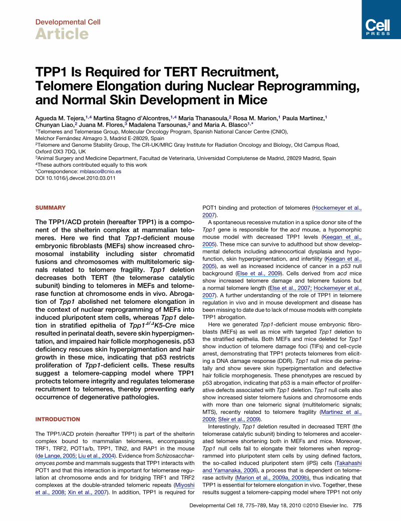
Figure 1. DDR Activation in Tpp1-Deleted
MEFs
(A) Quantification of Tpp1 mRNA levels by Q-PCR
in Tpp1D/D-Cre-LT MEFs versus control cells
infected with pBabe vector.
(B) Immunofluorescence detection of mouse TPP1
(red) in the indicated MEFs. DNA was counter-
stained with DAPI (blue).
(C) Immunofluorescence detection of gH2AX
(green) combined with TRF2 staining of the
telomeres (red) in the indicated MEFs.
(D) The percentage of metaphase nuclei exhibiting
the indicated number of TIFs per metaphase was
determined for at least 50 metaphases per
genotype prepared as in (C).
(E) Western blot detection of phospho-CHK1 and
-CHK2 in the indicated MEFs treated with Hit&Run
Cre vector. Wild-type MEFs treated with 0 or 10 Gy
of ionizing radiation served as a control for check-
point activation. Tubulin was used as a loading
control.
(F) Rescue of 53BP1 DNA damage foci in Tpp1-
depleted MEFs treated with 10 mM KU55933 (an
ATM inhibitor) or 5 mM caffeine (dual ATM/ATR
inhibitor). Two independent experiments were
performed in duplicate, and at least 100 cells
were analyzed per condition in each experiment.
The data represent mean values relative to wild-
type ± standard error. Statistical significance was
determined by Student’s t test.
Developmental Cell
TPP1 Mediates TERT Binding and Telomere Elongation
prevents the induction of a DDR at telomeres by preventing
fusions and telomere breakage but is also required for telomere
elongation by telomerase.
RESULTS
Tpp1-Deficient MEFs Undergo Rapid Proliferative ArrestConcomitant with DDR Activation at Chromosome EndsWe first established MEFs from wild-type and conditional
Tpp1flox/flox mice and infected them with a pBabe-Cre retrovirus
(Tpp1D/D-Cre MEFs), which resulted in undetectable Tpp1
mRNA levels by quantitative PCR (Q-PCR) and lack of TPP1
protein by immunofluorescence staining (Figures 1A and 1B;
see Supplemental Experimental Procedures available online).
776 Developmental Cell 18, 775–789, May 18, 2010 ª2010 Elsevier Inc.
As Tpp1-deficient MEFs failed to prolif-
erate in vitro, we downregulated the
expression of p53 by using a small hairpin
RNA (shRNA) against p53 and generated
Tpp1D/D-Cre-shp53 MEFs, or alterna-
tively suppressed both the p53 and Rb
pathways by SV40 large T (LT) antigen
expression in Tpp1D/D-Cre-LT MEFs
(Figures S1A–S1C). In both cases, we
observed a significant rescue of cell
proliferation compared to Tpp1D/D-Cre
MEFs (not shown and Figure S1C for
Tpp1D/D-Cre-shp53 MEFs). These find-
ings indicate Tpp1 deletion results in
rapid cell-cycle arrest which is mediated
by the p53 and pRB pathways.
To determine whether proliferation defects of Tpp1-deleted
MEFs are mediated by checkpoint responses induced by telo-
mere dysfunction, we next addressed whether Tpp1 abrogation
leads to telomere damage. gH2AX foci mark the presence of crit-
ically short/dysfunctional telomeres or TIFs (d’Adda di Fagagna
et al., 2003; Takai et al., 2003). After transient expression of the
Cre recombinase from the self-inactivating Hit&Run retrovirus
(Silver and Livingston, 2001), most Tpp1D/D cells showed
gH2AX foci at telomeres of spread mitotic chromosomes
(Figure 1C), reaching 6–20 TIFs per metaphase (Figure 1D). TIF
induction following Tpp1 deletion was accompanied by phos-
phorylation of the CHK1 and CHK2 checkpoint kinases
(Figure 1E), whereas this was not observed in wild-type controls.
As a positive control, wild-type MEFs exposed to 10 Gy of
Developmental Cell
TPP1 Mediates TERT Binding and Telomere Elongation
ionizing radiation showed a complete shift of the CHK2 lower
band and a robust CHK1 phosphorylation. To dissect the relative
importance of ATM and ATR pathways in the DDR induced by
Tpp1 deficiency, we determined 53BP1 DNA damage foci
upon inhibition of ATM (KU55933 inhibitor; Hickson et al.,
2004) or both ATM/ATR kinases (caffeine). As shown in
Figure 1F, 53BP1 DNA damage foci induced by Tpp1 abrogation
were rescued when either ATM or both ATM and ATR were in-
hibited, indicating that both DNA damage signaling pathways
are activated upon Tpp1 deletion.
Tpp1D/DK5-Cre Mice Show Increased Telomere Damageand G2/M Mitotic Arrest in the EpidermisTo study the impact of Tpp1 abrogation in the context of the
organism, we targeted deletion of Tpp1 to mouse stratified
epithelia. For this, we crossed conditional Tpp1flox/flox mice or
Tpp1+/flox mice with K5-Cre mice that constitutively express
the Cre recombinase under the control of the keratin 5 (K5)
promoter from day 11.5 of embryonic development onward
(Ramirez et al., 1994), thereby generating Tpp1D/DK5-Cre mice
(Figures 2A and 2B). Tpp1D/DK5-Cre mice show complete exci-
sion of Tpp1 exon 1 in the epidermis whereas no deletion can
be detected in the dermis (Figure 2B), in agreement with K5
expression pattern (Ramirez et al., 1994). Accordingly, Tpp1
expression was abrogated in Tpp1D/DK5-Cre epidermis, as
determined both by Q-PCR on skin keratinocytes (Figure 2C)
and TPP1 immunofluorescence on skin sections (Figure 2D).
In analogy to Tpp1-deficient MEFs, Tpp1D/DK5-Cre epidermis
showed increased gH2AX-positive cells compared to wild-type
mice (Figures 2E and 2F), and these gH2AX foci colocalized
with telomeres (TRF1/gH2AX colocalization) in a high per-
centage of cells (Figures 2E and 2F), indicative of persistent
DDR activation at telomeres in Tpp1-deleted mouse epidermis.
Finally, in analogy to severe cell-cycle arrest in Tpp1-deleted
MEFs, flow cytometry analysis of freshly isolated, noncultured
skin keratinocytes showed that a significant percentage of
Tpp1D/DK5-Cre keratinocytes was arrested in G2/M with a 4n
DNA content, indicating failure to undergo mitosis upon Tpp1
deletion (Figures 2G and 2H). Furthermore, Tpp1-deleted kerati-
nocytes showed increased polyploidy as indicated by the pres-
ence of >4n populations (Figures 2G and 2H). Together, these
results indicate that Tpp1 deletion in the context of the mouse
epidermis leads to a persistent telomere-originated DDR which
is accompanied by G2/M arrest and increased polyploidy.
TPP1 Prevents Sister Telomere Fusionsand Telomere FragilityTo address a role for TPP1 in telomere protection, we performed
metaphase fluorescence in situ hybridization (FISH) staining
with a telomeric probe on Tpp1 null, LT-immortalized MEFs.
Tpp1D/D-Cre-LT MEFs showed significantly increased frequen-
cies of sister telomere fusions compared to Tpp1+/+-Cre-LT
controls (Figures 3A–3C). Tpp1-deleted MEFs also showed a
significant increase in chromosome ends with MTS (2–4 per
metaphase; Figures 3A–3C), recently related to increased telo-
mere fragility (Blanco et al., 2007; Martinez et al., 2009; Munoz
et al., 2005; Sfeir et al., 2009). In agreement with this, MTS
were increased in wild-type MEFs treated with aphidicolin,
known to induce replication fork stalling and breakage at fragile
Deve
sites (Durkin and Glover, 2007; Sfeir et al., 2009), and further
increased in similarly treated Tpp1-deficient MEFs (Figure 3C),
suggesting that telomeres lacking TPP1 are prone to breakage.
Accordingly, breaks and fragments but not telomere fusions
were increased in aphidicolin-treated wild-type and Tpp1 null
MEFs (Figure 3C). Replication fork stalling results in fork frag-
mentation and resection, leading to accumulation of ssDNA.
Supporting aberrant replication and breakage of TPP1-depleted
telomeres, we observed an increased colocalization of the
replication factor PCNA and of ssDNA (as indicated by BrdU
incorporation) with telomeres (as visualized by TRF1 immunoflu-
orescence) compared with wild-type cells (Figures 3D–3I). As a
positive control, BrdU staining was increased in g-irradiated
Tpp1-depleted cells (Figure 3G). Together, these data indicate
that TPP1 deletion induces telomere uncapping leading to sister
telomere fusions, as well as increased telomere fragility and
breakage resulting in MTS. Finally, as these defects are similar,
although milder, to those induced by TRF1 deletion in MEFs
(Martinez et al., 2009), we ruled out defective TRF1 expression
in these cells as indicated by TRF1 immunofluorescence
(Figure S2).
TPP1 Is Required for TERT Binding to Telomeresand Telomere Maintenance In VivoDDR activation at chromosome ends also occurs in cells defi-
cient for other shelterin components, including POT1, TRF2,
and TRF1 (Celli and de Lange, 2005; Hockemeyer et al., 2006;
Martinez et al., 2009; Sfeir et al., 2009; Wu et al., 2006). As
TPP1 is already known to mediate POT1 binding to telomeres
(Hockemeyer et al., 2007), here we set out to address whether
binding of TRF1, TRF2, and RAP1 was affected by TPP1 deletion
(Figures 4A–4D). In chromatin fractionation assays, binding of
TRF1, TRF2, and RAP1 to chromatin was similar in wild-type
and Tpp1-deficient MEFs (Figures 4A and 4B). Chromatin
immunoprecipitation (ChIP) assays also indicated normal
binding of TRF1 and TRF2 to telomeres in the absence of
TPP1 binding (Figures 4C and 4D). In analogy with MEF results,
Tpp1-deleted epidermis showed normal TRF1 immunofluores-
cence (Figure 4E).
Based on its direct interaction with telomerase (Xin et al.,
2007), TPP1 has been proposed to regulate telomerase at chro-
mosome ends. Here we set out to address whether TPP1 is
required for TERT binding to telomeres in vivo. As shown in
Figures 4A and 4F, Tpp1-deficient MEFs showed decreased
TERT binding to chromatin in the presence of normal binding
of H3 and the other shelterin components. As controls, we per-
formed chromatin fractionation of MEFs deficient in either of
the telomerase core components TERT (Tert�/�) and Terc
(Terc�/�) (Blasco et al., 1997; Liu et al., 2000). Tert�/� MEFs
did not show detectable TERT binding to chromatin, confirming
the specificity of the TERT antibodies used (Figure 4F) (Experi-
mental Procedures). Terc�/� MEFs showed normal TERT
binding to chromatin (Figure 4F), which is also dependent on
TPP1 (Figures S3A and S3B), as indicated by using a small
hairpin RNA against Tpp1 (shTpp1) (Experimental Procedures).
We extended these findings to HeLa cells, where Tpp1 knock-
down also resulted in decreased TERT binding to chromatin
(Figure S3C). Importantly, by using telomeric ChIP assays, we
further demonstrate defective TERT binding specifically to
lopmental Cell 18, 775–789, May 18, 2010 ª2010 Elsevier Inc. 777
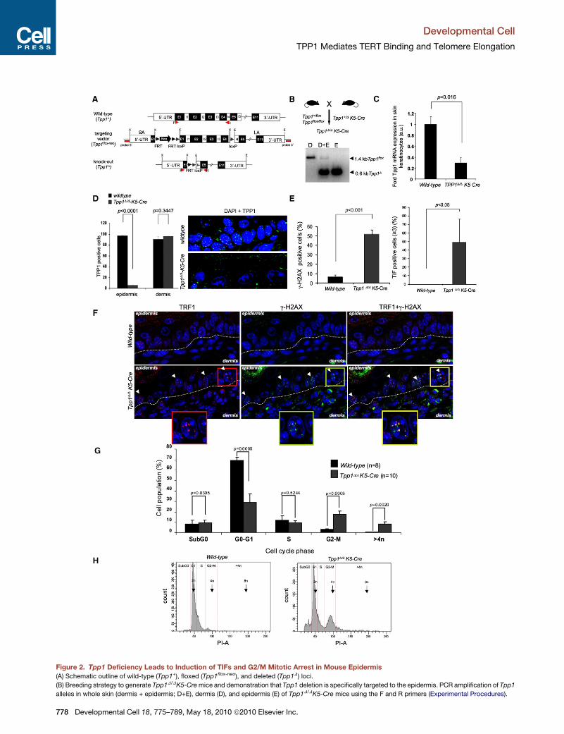
Figure 2. Tpp1 Deficiency Leads to Induction of TIFs and G2/M Mitotic Arrest in Mouse Epidermis
(A) Schematic outline of wild-type (Tpp1+), floxed (Tpp1flox-neo), and deleted (Tpp1D) loci.
(B) Breeding strategy to generate Tpp1D/DK5-Cre mice and demonstration that Tpp1 deletion is specifically targeted to the epidermis. PCR amplification of Tpp1
alleles in whole skin (dermis + epidermis; D+E), dermis (D), and epidermis (E) of Tpp1D/DK5-Cre mice using the F and R primers (Experimental Procedures).
Developmental Cell
TPP1 Mediates TERT Binding and Telomere Elongation
778 Developmental Cell 18, 775–789, May 18, 2010 ª2010 Elsevier Inc.
Developmental Cell
TPP1 Mediates TERT Binding and Telomere Elongation
telomeric repeats in the absence of TPP1 (Figures 4G and 4H).
Absence of TERT binding was not due to decreased TERT
mRNA expression in Tpp1 null cells (Figure S3D), in agreement
with normal telomerase TRAP (telomere repeat amplification
protocol) activity in these cells (Figure S3D). Together, these
results identify TPP1 as a telomerase recruitment factor in vivo.
We next addressed the impact of Tpp1 deficiency on telomere
length maintenance by performing telomere quantitative FISH
(Q-FISH) analysis on MEF metaphases. Proliferating Tpp1D/D-
Cre-shp53 and Tpp1D/D-Cre-LT MEFs showed decreased
telomere fluorescence when compared to their corresponding
wild-type controls (Figure 5A), suggesting a faster rate of
telomere shortening in the absence of TPP1. Importantly, this
shortening was not accompanied by changes in the G-strand
overhang (Figure S4). Finally, we confirmed an accelerated rate
of telomere shortening in Tpp1D/DK5-Cre epidermis compared
to wild-type skin both by using telomere Q-FISH following
correction by centromeric fluorescence (Figure 5B) and by
terminal restriction fragment (TRF) analysis (Figure 5C), a
Southern-based technique that estimates the size of telomere-
containing terminal fragments (Experimental Procedures).
TPP1 Is Required for Net Telomere Elongationduring Nuclear ReprogrammingThe results described above suggest a role for TPP1 in TERT
recruitment to telomeres and telomere maintenance, but they
do not address whether TPP1 is required for telomere elongation
by telomerase. To this end, we took advantage of the recently
described process of generation of iPS cells from parental differ-
entiated cells (Takahashi and Yamanaka, 2006). In particular,
during iPS cell generation, telomeres undergo a net elongation
compared to the parental MEFs, a process which is dependent
on telomerase activity (Marion et al., 2009a, 2009b). Here we
generated iPS cell clones from wild-type and Tpp1 null MEFs
simultaneously knocked down for p53 by using a small hairpin
RNA against p53 (shp53), which allows efficient reprogramming
of parental cells with dysfunctional telomeres (Marion et al.,
2009a) (Figures 5D–5F). Four independent wild-type Cre-shp53
iPS clones showed a net telomere elongation of approximately
10 kb at passage 2-3 after clone isolation compared to parental
MEFs (Figures 5D and 5E). In marked contrast, none of the four
Tpp1 null iPS clones studied showed detectable telomere
elongation compared to the parental MEFs; instead, a modest
telomere shortening was observed (Figures 5D and 5E). As
Tpp1 null cells have normal telomerase activity in vitro (see
(C) Tpp1 mRNA levels as determined by Q-PCR in three neonate keratinocytes per
error. Student’s t test was used for statistical analysis, and the p value is indicat
(D) Quantification of TPP1-positive cells in epidermis and dermis of three or fou
of positive cells per genotype is shown. Error bars represent standard error. Stud
representative images of immunofluorescence staining in epidermis using a TPP
(E) Percentage of skin basal-layer keratinocytes showing g-H2AX foci (left), and pe
in back skin sections of mice of the indicated genotypes. At least 300 cells from thr
error. Student’s t test was used for statistical analysis, and p values are indicate
(F) Representative images of TRF1 (red) and g-H2AX (green) fluorescence, and the
fluorescence. The dotted line separates epidermis from dermis.
(G) Cell-cycle analysis of primary keratinocytes from wild-type and Tpp1D/DK5-C
cell cycle by FACS is represented. Cells were stained with propidium iodide (P
standard error. The Student’s t test was used for statistical analysis, and p value
(H) Representative examples of the FACS histograms.
Deve
Figure S3D), these results indicate that TPP1 is necessary for
telomerase-mediated telomere elongation in vivo. As a control
for pluripotency, a similarly high expression of the endogenous
pluripotency genes Oct4 and Nanog was detected in both
wild-type and Tpp1 null iPS clones compared to a lack of expres-
sion in the parental MEFs (Figure 5F).
Tpp1-Conditional Deletion in Stratified Epithelia Leadsto Perinatal Death, Skin Hyperpigmentation,and Severe Defects in Hair Follicle MorphogenesisIn contrast to acd mice that can survive to adulthood, Tpp1D/D
K5-Cre mice showed a fully penetrant perinatal mortality with
no mice surviving more than 2 weeks (Figure 6A; see also
Figure 7D). In addition, Tpp1D/DK5-Cre mice did not gain weight
in the days following birth (Figure 6B; see also Figure 7E) due to
epithelial abnormalities in the oral mucosa (Table S1; Figure S5A)
which prevented feeding. At birth, Tpp1D/DK5-Cre mice show
severe skin hyperpigmentation, which is aggravated the
following days concomitant with severe skin scaling and dryness
(compare day 4 and day 7; Figure 6C). Histopathological analysis
of Tpp1D/DK5-Cre epidermis revealed lack of mature hair follicles
and sebaceous glands (Figures 6D–6J; Figure S5), as well as an
abnormally high expression of cytokeratin 6 compared to wild-
type epidermis (Figure 6D), a pathological condition associated
with skin diseases such as psoriasis and skin cancer (Stoler
et al., 1989). Cytokeratins 10 and 14, which mark suprabasal
and basal skin layers, respectively, showed a normal expression
pattern (Figure 6D), suggesting that Tpp1 deficiency does not
affect skin differentiation. Concomitantly with degenerative
lesions, all Tpp1D/DK5-Cre mice presented areas of focal
dysplasia in the skin (Table S1; Figure S5A) which were also
present in other stratified epithelia (Table S1; Figure S5A). Severe
skin abnormalities in Tpp1D/DK5-Cre mice were accompanied by
a defective epithelial barrier function (Figure 6E), which together
with the lack of nourishment may contribute to their fully pene-
trant perinatal mortality (McGavin and Zachary, 2007).
Severe Hair Morphogenesis Defectsin Tpp1D/DK5-Cre EpidermisLack of mature hair follicles and sebaceous glands in Tpp1D/D
K5-Cre skin suggests a defect in the hair bulge stem cell (SC)
compartment (Flores et al., 2005; Martinez et al., 2009; Stout
and Blasco, 2009). Here we set out to examine the impact of
Tpp1 abrogation on hair follicle morphogenesis by studying the
expression of the Sox9 gene, which marks hair bulge SC
genotype. Actin was used to normalize samples. Error bars represent standard
ed.
r independent wild-type and Tpp1D/DK5-Cre newborn mice. The percentage
ent’s t test was used for statistical analysis and p values are indicated. Right:
1 antibody (green). Nuclei are counterstained with DAPI (blue).
rcentage of these cells displaying g-H2AX colocalization with telomeres (TRF1)
ee independent mice were scored per genotype. Error bars represent standard
d.
combined images. Colocalization events (white arrows) are detected by yellow
re neonates. Quantification of the percentage of cells at different phases of the
I-A). n, independent newborn mice used per genotype. Error bars represent
s are indicated.
lopmental Cell 18, 775–789, May 18, 2010 ª2010 Elsevier Inc. 779
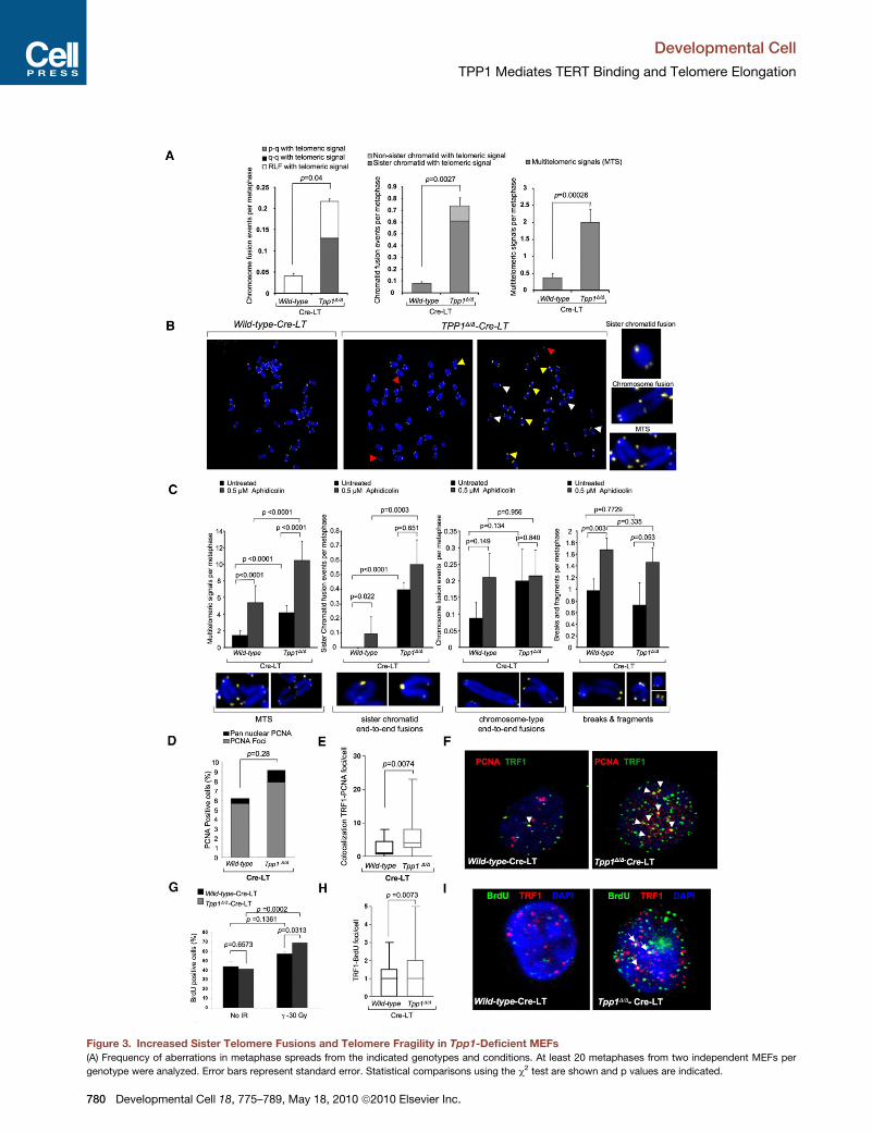
Figure 3. Increased Sister Telomere Fusions and Telomere Fragility in Tpp1-Deficient MEFs
(A) Frequency of aberrations in metaphase spreads from the indicated genotypes and conditions. At least 20 metaphases from two independent MEFs per
genotype were analyzed. Error bars represent standard error. Statistical comparisons using the c2 test are shown and p values are indicated.
Developmental Cell
TPP1 Mediates TERT Binding and Telomere Elongation
780 Developmental Cell 18, 775–789, May 18, 2010 ª2010 Elsevier Inc.
Developmental Cell
TPP1 Mediates TERT Binding and Telomere Elongation
precursors during embryonic and early postnatal development
(Nowak et al., 2008; Vidal et al., 2005). Wild-type epidermis
showed intense Sox9 expression at the hair bulge SC niche at
P4, whereas it was almost undetectable at the interfollicular
epidermis at this stage (Figure 6F). In contrast, age-matched
Tpp1-deficient epidermis showed reduced Sox9 expression at
the base of the hair primordiums whereas it was elevated in
the interfollicular epidermis (Figure 6F), indicative of lack of
mature hair bulge formation in Tpp1D/DK5-Cre skin. We also
failed to detect expression of the hair bulge marker K15 in
Tpp1D/DK5-Cre hair primordiums, whereas it was readily
detected at the hair bulge in age-matched wild-type hair follicles
(Liu et al., 2003) (Figure 6G).
A number of molecular signals are involved in instructing
epithelial cells to adopt a hair follicle fate and to establish and
maintain functional mature hair follicles (reviewed in Blanpain
and Fuchs, 2009). Wnt/b-catenin signaling is the earliest molec-
ular signal known to determine hair follicle development
(Huelsken et al., 2001; Paus et al., 1999; Zhang et al., 2008).
Interestingly, we observed no significant differences in nuclear
b-catenin staining at E16.5–E17.5 placodes and hair pegs
(Figure S5B) between wild-type and Tpp1 null mice, suggesting
that b-catenin nuclear signaling is not altered by Tpp1 abrogation
and, therefore, is not likely to be causative of hair growth defects
in these mice. Also in agreement with normal b-catenin expres-
sion, we observed no differences in the number of E17.5 placo-
des/hair pegs (Figure S5C) and P2 developing hair follicles
(Figure 6H). We next studied the ability of developing hair follicles
to downgrow into the dermis by using a dermal papilla staining
(Experimental Procedures). We observed a dramatic defect in
hair follicle downgrowth in Tpp1 null epidermis, as indicated by
dermal papilla staining restricted to the epidermis in P1 Tpp1
null epidermis in contrast to 100% internalization of the dermal
papilla in age-matched wild-type epidermis (stage 1 versus
stages 3–5 in wild-type skin; Huelsken et al., 2001; Paus et al.,
1999) (see Figure 6I). At this stage, we also failed to detect
expression of the GATA-3 differentiation marker, which is nor-
mally expressed at the inner root sheath in wild-type follicles
(white arrows; Figure S5D) (Kaufman et al., 2003). Next, we
studied whether downstream signals of Wnt known to be
involved in hair follicle downgrowth and morphogenesis, such
as Sonic hedgehog (Shh), are altered in Tpp1 null skin (Huelsken
et al., 2001; St-Jacques et al., 1998). However, Shh mRNA
(B) Representative images of metaphases of the indicated genotypes. Magnificati
fusions; white arrowheads, MTS; red arrowheads, breaks/fragments.
(C) Chromosomal aberrations upon aphidicolin treatment. At least 30 metaphase
condition. Error bars represent standard error. Magnifications of the indicated ab
(D) Immunofluorescence detection of PCNA-positive cells in wild-type and Tpp1D/
pan-nuclear staining is shown. More than 150 cells from two independent MEFs p
p values are indicated.
(E) Colocalization of TRF1-PCNA foci per cell in wild-type and Tpp1D/D MEFs trea
p values are indicated.
(F) Representative images of PCNA (red) and TRF1 (green) staining and of coloc
MEFs treated with Cre.
(G) Quantification of BrdU-positive cells in the indicated genotypes in the absence
two independent MEFs per genotype were analyzed per condition.
(H) Quantification of the number of BrdU foci that colocalize with TRF1 in two w
analyzed per genotype. Statistical significance was determined by Student’s t te
(I) Representative images of BrdU (green) and TRF1 (red) fluorescence. BrdU-TR
Deve
expression was similar at hair pegs of wild-type and Tpp1D/D
K5-Cre E17.5 embryos (Figure S5E), arguing that abnormal
Shh expression is not responsible for defective formation of
mature hair follicles in Tpp1D/DK5-Cre mice (Bitgood and McMa-
hon, 1995).
We addressed whether defective downgrowth and develop-
ment of mature hair follicles in Tpp1 null epidermis was accom-
panied by reduced proliferation specifically in the hair follicles.
Proliferation was reduced specifically at the hair primordiums
of P4 Tpp1D/DK5-Cre epidermis compared to age-matched
wild-type hair follicles, and this defect was not apparent in inter-
follicular epidermis of Tpp1D/DK5-Cre mice (Figure 6J), indicating
that hair bulge cells are more sensitive to persistent telomere
damage induced by Tpp1 deletion. We next addressed whether
decreased hair follicle SC proliferation in Tpp1D/DK5-Cre mice
was a cell-autonomous effect by using ex vivo clonogenic
assays (Experimental Procedures). The number and size of
colonies in this assay are considered to reflect, respectively,
the number of epidermal SC and their ability to proliferate
in vitro (Barrandon and Green, 1987). Tpp1D/DK5-Cre keratino-
cytes showed significantly decreased clonogenic capacity
(Figure 6K), suggesting that the proliferation defect of TPP1-
deficient SC cells is cell autonomous.
Together, these findings suggest that Tpp1 deletion does not
affect Wnt/b-catenin-dependent hair follicle specification but
instead has a profound negative impact on hair follicle down-
growth, proliferation, and differentiation, affecting the establish-
ment of a mature hair bulge SC compartment, which explains the
observations that these mice do not grow hair and show severe
skin dryness.
p53 Deficiency Rescues Skin Hyperpigmentationand Hair Growth Defects in Tpp1D/DK5-Cre p53�/� MiceAnalysis of Tpp1-deficient MEFs suggested that p53 is an
important mediator of cell-cycle arrest produced by TPP1 abro-
gation (Figure S1). To address whether the same was true
in vivo in the context of mouse epidermis, we first studied the
expression of the p53 and p21 cell-cycle inhibitors in skin
sections. Tpp1D/DK5-Cre epidermis showed increased
amounts of both cell-cycle inhibitors compared to controls
(Figures 7A and 7B), indicating that Tpp1 deletion in the context
of the mouse epidermis leads to upregulation of the p53/p21
pathways.
ons of the indicated aberrations are shown. Yellow arrowheads, sister telomere
s from four wild-type and three Tpp1D/D independent MEFs were analyzed per
errations are shown.D MEFs treated with Cre. The percentage of cells showing PCNA foci and PCNA
er genotype were scored. Statistical comparisons use the Student’s t test, and
ted with Cre. Statistical comparisons were done using the Student’s t test, and
alization events (yellow; indicated with arrowheads) in wild-type and Tpp1D/D
of treatment (no IR) and after 30 Gy of ionizing radiation. At least 200 cells from
ild-type and two Tpp1-depleted independent MEFs. At least 80 nuclei were
st, and the p value is indicated.
F1 colocalization events (yellow) are indicated with arrows.
lopmental Cell 18, 775–789, May 18, 2010 ª2010 Elsevier Inc. 781
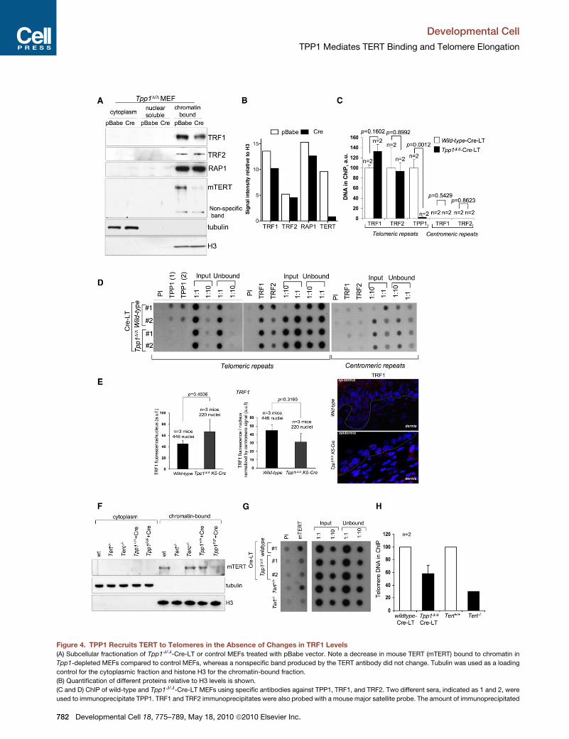
Figure 4. TPP1 Recruits TERT to Telomeres in the Absence of Changes in TRF1 Levels
(A) Subcellular fractionation of Tpp1D/D-Cre-LT or control MEFs treated with pBabe vector. Note a decrease in mouse TERT (mTERT) bound to chromatin in
Tpp1-depleted MEFs compared to control MEFs, whereas a nonspecific band produced by the TERT antibody did not change. Tubulin was used as a loading
control for the cytoplasmic fraction and histone H3 for the chromatin-bound fraction.
(B) Quantification of different proteins relative to H3 levels is shown.
(C and D) ChIP of wild-type and Tpp1D/D-Cre-LT MEFs using specific antibodies against TPP1, TRF1, and TRF2. Two different sera, indicated as 1 and 2, were
used to immunoprecipitate TPP1. TRF1 and TRF2 immunoprecipitates were also probed with a mouse major satellite probe. The amount of immunoprecipitated
Developmental Cell
TPP1 Mediates TERT Binding and Telomere Elongation
782 Developmental Cell 18, 775–789, May 18, 2010 ª2010 Elsevier Inc.
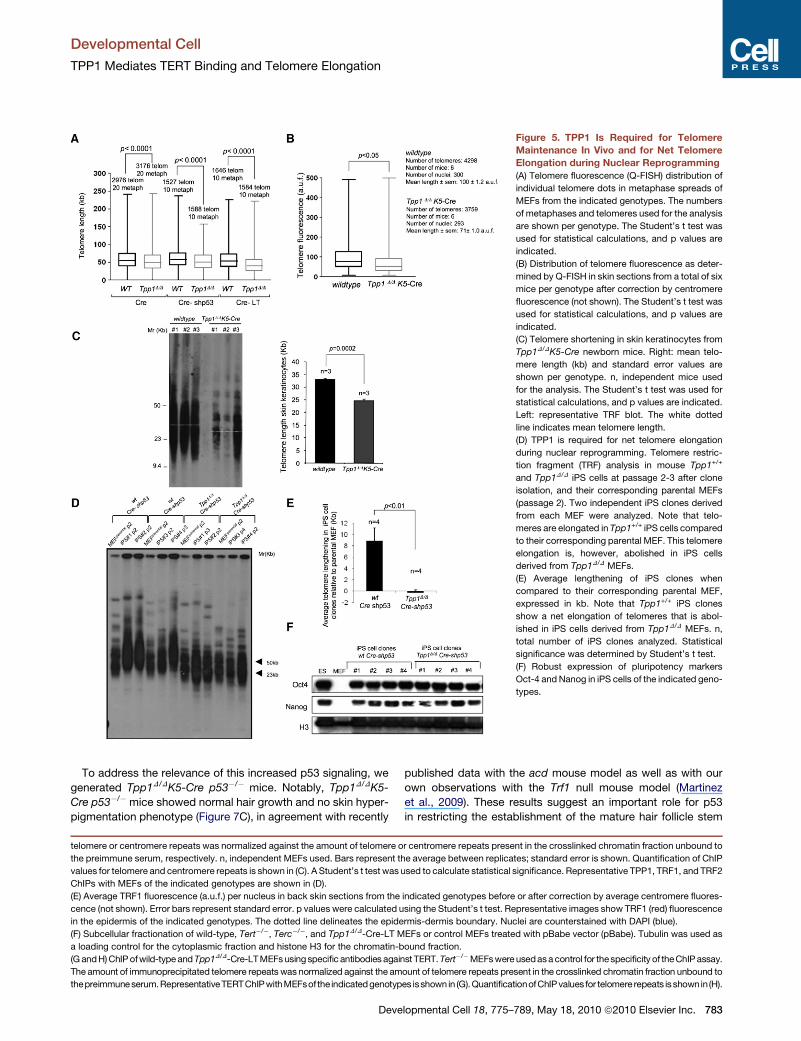
Figure 5. TPP1 Is Required for Telomere
Maintenance In Vivo and for Net Telomere
Elongation during Nuclear Reprogramming
(A) Telomere fluorescence (Q-FISH) distribution of
individual telomere dots in metaphase spreads of
MEFs from the indicated genotypes. The numbers
of metaphases and telomeres used for the analysis
are shown per genotype. The Student’s t test was
used for statistical calculations, and p values are
indicated.
(B) Distribution of telomere fluorescence as deter-
mined by Q-FISH in skin sections from a total of six
mice per genotype after correction by centromere
fluorescence (not shown). The Student’s t test was
used for statistical calculations, and p values are
indicated.
(C) Telomere shortening in skin keratinocytes from
Tpp1D/DK5-Cre newborn mice. Right: mean telo-
mere length (kb) and standard error values are
shown per genotype. n, independent mice used
for the analysis. The Student’s t test was used for
statistical calculations, and p values are indicated.
Left: representative TRF blot. The white dotted
line indicates mean telomere length.
(D) TPP1 is required for net telomere elongation
during nuclear reprogramming. Telomere restric-
tion fragment (TRF) analysis in mouse Tpp1+/+
and Tpp1D/D iPS cells at passage 2-3 after clone
isolation, and their corresponding parental MEFs
(passage 2). Two independent iPS clones derived
from each MEF were analyzed. Note that telo-
meres are elongated in Tpp1+/+ iPS cells compared
to their corresponding parental MEF. This telomere
elongation is, however, abolished in iPS cells
derived from Tpp1D/D MEFs.
(E) Average lengthening of iPS clones when
compared to their corresponding parental MEF,
expressed in kb. Note that Tpp1+/+ iPS clones
show a net elongation of telomeres that is abol-
ished in iPS cells derived from Tpp1D/D MEFs. n,
total number of iPS clones analyzed. Statistical
significance was determined by Student’s t test.
(F) Robust expression of pluripotency markers
Oct-4 and Nanog in iPS cells of the indicated geno-
types.
Developmental Cell
TPP1 Mediates TERT Binding and Telomere Elongation
To address the relevance of this increased p53 signaling, we
generated Tpp1D/DK5-Cre p53�/� mice. Notably, Tpp1D/DK5-
Cre p53�/�mice showed normal hair growth and no skin hyper-
pigmentation phenotype (Figure 7C), in agreement with recently
telomere or centromere repeats was normalized against the amount of telomere o
the preimmune serum, respectively. n, independent MEFs used. Bars represent t
values for telomere and centromere repeats is shown in (C). A Student’s t test was
ChIPs with MEFs of the indicated genotypes are shown in (D).
(E) Average TRF1 fluorescence (a.u.f.) per nucleus in back skin sections from the
cence (not shown). Error bars represent standard error. p values were calculated u
in the epidermis of the indicated genotypes. The dotted line delineates the epide
(F) Subcellular fractionation of wild-type, Tert�/�, Terc�/�, and Tpp1D/D-Cre-LT M
a loading control for the cytoplasmic fraction and histone H3 for the chromatin-b
(G and H) ChIP of wild-type and Tpp1D/D-Cre-LT MEFs using specific antibodies again
The amount of immunoprecipitated telomere repeats was normalized against the am
thepreimmuneserum.RepresentativeTERTChIPwithMEFsof the indicatedgenotyp
Deve
published data with the acd mouse model as well as with our
own observations with the Trf1 null mouse model (Martinez
et al., 2009). These results suggest an important role for p53
in restricting the establishment of the mature hair follicle stem
r centromere repeats present in the crosslinked chromatin fraction unbound to
he average between replicates; standard error is shown. Quantification of ChIP
used to calculate statistical significance. Representative TPP1, TRF1, and TRF2
indicated genotypes before or after correction by average centromere fluores-
sing the Student’s t test. Representative images show TRF1 (red) fluorescence
rmis-dermis boundary. Nuclei are counterstained with DAPI (blue).
EFs or control MEFs treated with pBabe vector (pBabe). Tubulin was used as
ound fraction.
st TERT. Tert�/�MEFs were used as a control for the specificity of the ChIP assay.
ount of telomere repeats present in the crosslinked chromatin fraction unbound to
es isshown in (G).QuantificationofChIPvalues for telomere repeats isshown in (H).
lopmental Cell 18, 775–789, May 18, 2010 ª2010 Elsevier Inc. 783
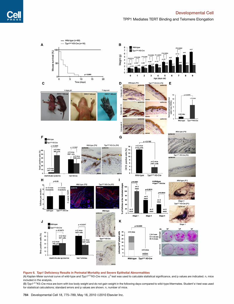
Figure 6. Tpp1 Deficiency Results in Perinatal Mortality and Severe Epithelial Abnormalities
(A) Kaplan-Meier survival curve of wild-type and Tpp1D/DK5-Cre mice. c2 test was used to calculate statistical significance, and p values are indicated. n, mice
included in the analysis.
(B) Tpp1D/DK5-Cre mice are born with low body weight and do not gain weight in the following days compared to wild-type littermates. Student’s t test was used
for statistical calculations; standard errors and p values are shown. n, number of mice.
Developmental Cell
TPP1 Mediates TERT Binding and Telomere Elongation
784 Developmental Cell 18, 775–789, May 18, 2010 ª2010 Elsevier Inc.

Developmental Cell
TPP1 Mediates TERT Binding and Telomere Elongation
cell niche, as well as in restricting hair follicle downgrowth. In
addition, p53 deficiency was able to significantly rescue both
mouse survival and body weight (Figures 7D and 7E), and this
effect was allele dose dependent as indicated by a significant
rescue of these phenotypes in Tpp1D/DK5-Cre p53+/� mice.
DISCUSSION
Here we describe a mouse model for complete Tpp1 abrogation.
Tpp1 abrogation leads to DDR activation at chromosome ends
and increased telomere fusions and fragility, suggestive of a
role for TPP1 in telomere protection. In addition, we demonstrate
that TPP1 is required for TERT recruitment to telomeres and
telomere elongation in vivo. In particular, TPP1 is essential for
telomere elongation during nuclear reprogramming of MEFs
into iPS cells, a process that involves net telomere elongation
by telomerase (Marion et al., 2009a, 2009b). Interestingly,
TERT and Tpp1 show similar patterns of mRNA expression
during embryonic development (Martin-Rivera et al., 1998; Vlan-
gos et al., 2009), further suggesting their coregulation.
When deleted in the context of the mouse stratified epithelia,
Tpp1 deficiency leads to perinatal death, severe skin hyperpig-
mentation, defective hair follicle morphogenesis, and wide-
spread epithelia dysplasia. These epithelial pathologies are
similar to, although milder than, those produced by TRF1 abro-
gation in the skin (Martinez et al., 2009), as well as to epithelial
pathologies in human diseases associated with mutations in
telomerase-related genes and the presence of dysfunctional
telomeres (Armanios et al., 2007; Mitchell et al., 1999; Tsakiri
et al., 2007; Vulliamy et al., 2001), making Tpp1-deleted mice
a useful model for understanding human disease.
In this regard, we show here that hair follicle morphogenesis
defects in Tpp1 null skin are not associated with a defective
Wnt/b-catenin pathway, a known regulator of hair follicle devel-
opment in mice (Huelsken et al., 2001; Paus et al., 1999; Zhang
et al., 2008), recently described to be regulated by TERT levels
(Park et al., 2009). Instead, Tpp1 abrogation had a profound
negative impact on hair follicle downgrowth, proliferation, and
(C) Macroscopic phenotypes. Note that the skin of a 4-day-old Tpp1D/DK5-Cre m
and tail (arrowheads, left and middle). Hyperpigmentation is aggravated in 7-day
(D) Representative images of back skin sections stained for cytokeratin 10, cy
Tpp1-depleted epidermis.
(E) Compromised epidermal barrier function in Tpp1D/DK5-Cre mice. Transepiderm
analyzed per genotype. Error bars represent standard error. Student’s t test was
(F) Percentage of Sox9-positive cells at the interfollicular epidermis and at the ha
analyzed per genotype. Right: representative images of Sox9-positive staining in
the staining in the top of the epidermis presumably represents background.
(G) Percentage of cytokeratin 15 (K15) -positive cells in back skin sections of mic
tative images of K15-positive staining in the skin of 4-day-old mice.
(H) Similar number of follicles in back skin sections from P2 mice of the indicated
represent standard deviation. Statistical analysis was carried out using Student’s
orescence staining using a b-catenin antibody (red). Nuclei are counterstained w
(I) Average number of follicles at the indicated stages of development in both w
deviation. Statistical analysis was carried out with the c2 test, and p values are in
representative images of dermal papilla (blue/brown) localization in the indicated
(J) Percentage of BrdU-positive cells in the interfollicular epidermis and in the ha
analyzed per genotype. Right: representative images of BrdU-positive staining in
(K) Quantification of size and number of epidermal SC clones obtained ex vivo
(1-2 days old) at the indicated dilutions. Clones were visualized by rhodamine s
genotype is indicated in each case. Error bars represent standard error. Student
Deve
differentiation, hindering the establishment of a mature hair
bulge SC compartment. Importantly, these defects are rescued
by p53 abrogation, supporting a key role of p53 in mediating
proliferative arrest in response to persistent telomere damage
in vivo (Chin et al., 1999; Feldser and Greider, 2007; Martinez
et al., 2009; Stout and Blasco, 2009). Together, these findings
indicate that TPP1 has a dual role in telomere protection and
telomere elongation, in this way preserving telomere function
and preventing the early onset of degenerative pathologies in
mice.
EXPERIMENTAL PROCEDURES
Cell Fractionation and Immunoblotting
Cell fractionation was performed as described (Mendez and Stillman, 2000).
Equal amounts of protein (50–100 mg) were analyzed by gel electrophoresis
followed by western blotting. Anti-tubulin or anti-histone H3 antibodies were
used as loading controls.
Immunofluorescence In Situ Hybridization
Mitotic cells were prepared and subjected to immunofluorescence staining as
described (Martinez et al., 2009).
Retroviral Infections
Retroviral supernatants were produced in 293T cells (5 3 105 cells per 100 mm
diameter dish) transfected with the ecotropic packaging plasmid pCL-Eco and
either pBabe-Cre, pRS-p53sh (Brummelkamp et al., 2002), or pZip-Neo-sv40-
LT (Brown et al., 1986) as described (Martinez et al., 2009). Deletion of Tpp1
was confirmed by PCR with primers Tpp1Up2 and Tpp1Lo2, and absence
of TPP1 expression was tested by real-time quantitative RT-PCR and immuno-
fluorescence.
Transfection of Small Interference RNAs
HeLa 1.2.11 cells were transfected with the indicated siRNAs using Oligofect-
amine (Invitrogen Life Technologies) (Supplemental Information). A GFP siRNA
was used as a control.
Tpp1 Knockdown Using Short Hairpin RNA Vectors
Terc�/�MEFs were infected with an shRNA effective in downregulating mTpp1
expression (Supplemental Information). As a control, cells were transfected
with a scrambled shRNA.
ouse shows a shiny appearance, scaling, and hyperpigmentation in the paws
-old mice.
tokeratin 14, and cytokeratin 6. Note aberrant expression of cytokeratin 6 in
al water loss (TEWL) normalized to wild-type levels (set to 1) is shown. n, mice
used for statistical analysis, and p values are indicated.
ir follicles in back skin sections from mice of the indicated genotypes. n, mice
the skin of 4-day-old mice. The dotted line separates dermis from epidermis. *,
e of the indicated genotypes. n, mice analyzed per genotype. Right: represen-
genotypes. b-catenin expression was used to identify hair follicles. Error bars
t test, and p values are indicated. Right: representative images of immunoflu-
ith DAPI (blue).
ild-type and Tpp1D/D K5-Cre tail skin sections. Error bars represent standard
dicated. Stages are quantified as described (Muller-Rover et al., 2001). Right:
genotypes.
ir follicles of back skin sections from mice of the indicated genotypes. n, mice
the skin of 4-day-old mice. The dotted line separates dermis from epidermis.
. Right: representative examples of SC clones obtained from newborn mice
taining. n, mice analyzed per genotype. The total number of cells scored per
’s t test was used for statistical analysis, and p values are shown.
lopmental Cell 18, 775–789, May 18, 2010 ª2010 Elsevier Inc. 785
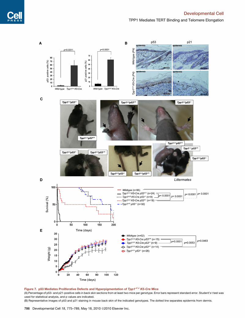
Figure 7. p53 Mediates Proliferative Defects and Hyperpigmentation of Tpp1D/D K5-Cre Mice
(A) Percentage of p53- and p21-positive cells in back skin sections from at least two mice per genotype. Error bars represent standard error. Student’s t test was
used for statistical analysis, and p values are indicated.
(B) Representative images of p53 and p21 staining in mouse back skin of the indicated genotypes. The dotted line separates epidermis from dermis.
Developmental Cell
TPP1 Mediates TERT Binding and Telomere Elongation
786 Developmental Cell 18, 775–789, May 18, 2010 ª2010 Elsevier Inc.
Developmental Cell
TPP1 Mediates TERT Binding and Telomere Elongation
Generation of Mouse iPS Cells
Tpp1+/+-Cre and Tpp1D/D-Cre primary MEFs were obtained by retroviral infec-
tion with pBabe-Cre as described above. Puromycin-selected cells were then
reprogrammed as previously described (Marion et al., 2009b). Briefly, MEFs
were seeded and infected four times in the following 2 days with a cocktail
of the retroviral constructs pMXsOct3/4, pMXsKlf4, pMXsSox2, and pRS-
p53sh. Colonies were picked after 2 weeks and expanded on feeder fibro-
blasts using standard procedures. iPS cells were transferred to gelatin-coated
plates before analysis.
Real-Time Quantitative RT-PCR
Total RNA was extracted from the indicated cells using TRIzol (Invitrogen Life
Technologies). Reverse transcription was carried out with 1 mg of total RNA
using random hexamers as primers and Superscript II reverse transcriptase
(Invitrogen Life Technologies) in accordance with the manufacturer’s instruc-
tions. Real-time PCR was performed as described (Munoz et al., 2005). Primer
sequences are shown in Supplemental Experimental Procedures.
Telomere and Centromere Fluorescence Analyses of Skin Sections
Quantitative telomere and centromere fluorescence in situ hybridization
(Q-FISH) on skin sections was performed as described (Munoz et al., 2005).
For the centromeric Q-FISH, a probe against the major satellite was used
(Flores et al., 2008).
Telomere Length and Cytogenetic Analysis Using Telomere Q-FISH
on Metaphases
Q-FISH onmetaphases from primary MEFswas performed asdescribed (Herrera
et al., 1999; Samper et al., 2000). When indicated, MEFs were treated with aphi-
dicolin at 0.5 mM for 24 hr. For analysis of chromosomal aberrations, quantitative
analysis of digital images was performed as described in Zijlmans et al. (1997).
TRF and G-Strand Overhang Analysis
Half a million keratinocytes, MEFs, or iPS cells were included in agarose plugs
and TRF analysis was performed as described (Blasco et al., 1997).
The G-strand overhang assay was carried out as described (Samper et al.,
2000). For quantitative analysis of telomere length in iPS cells, each lane was
divided into 32 identical fractions covering the entire TRF smear. The weighted
mean was calculated by measuring the signal intensity of each fraction, ex-
pressed as the percentage of the total signal intensity of the lane. The ImageJ
program was used for quantification.
Telomerase TRAP Assay
Telomerase activity was measured with a modified telomere repeat amplifica-
tion protocol (TRAP) as described (Blasco et al., 1997).
Chromatin Immunoprecipitation Assays
ChIP assays were performed as described (Garcia-Cao et al., 2004) with
the following antibodies: 8 ml of rabbit anti-mouse TRF1 serum (generated in
our lab), 4 ml of rabbit polyclonal anti-TRF2 antibody (a gift from Dr. West,
CR-UK), 8 ml of two different rabbit anti-mouse TPP1 sera (a gift from
Dr. Else, University of Michigan), and 6 ml of rabbit polyclonal anti-TERT
antibody (Calbiochem) or preimmune serum. The amount of telomeric and
pericentric DNA after ChIP was normalized to the total telomeric or centro-
meric signal, respectively, for each genotype, thus correcting for differences
in the number of telomere repeats.
Treatment with ATM and ATR Inhibitors
Wild-type and Tpp1 null MEFs were treated with either 10 mM ATM-specific
inhibitor KU55933 (Tocris Bioscience) or 5 mM caffeine (Sigma) and processed
as described (Martinez et al., 2009).
(C) p53 deficiency rescues proliferative defects and hyperpigmentation of Tpp1D/
hyperpigmentation. Arrows indicate hyperpigmentation or lack of it in paws and
(D) Survival curve of wild-type, Tpp1D/DK5-Cre, Tpp1D/DK5-Cre p53�/�, Tpp1D/DK
significance, and p values are indicated. n, number of mice included in the analy
(E) p53 deficiency rescues the low body weight of Tpp1D/DK5-Cre mice; Tpp1D/D
t test was used for statistical calculations; standard error and p values are show
Deve
Proliferation Assays
Tpp1flox/flox cells (5 3 104) infected with Cre or Cre-shp53 were distributed into
six-well plates at day 0. For direct cell counting, attached cells were trypsinized
and counted using a Neubauer chamber slide at the indicated times.
Isolation of Keratinocytes from Newborn Mice Skin
and ESC Clonogenic Assays
Primary keratinocytes from newborn mice (0–2 days old) were obtained as
described (Munoz et al., 2005) and clonogenic assays performed as described
(Flores et al., 2005).
Flow Cytometric Analysis
Keratinocytes (1 3 106) were washed twice with PBS and fixed/permeabilized
with ice-cold 70% ethanol. Fixed cells were washed with PBS and resus-
pended in 1 ml PBS containing 0.2 mg propidium iodide and 100 mg RNase.
The samples were incubated 30 min at 37�C and the fluorescence-activated
cell sorting (FACS) analysis was performed in a FACSCanto II flow cytometry
system (BD Biosciences). The data were analyzed with the software FACSDiva
v5.1.1.
Immunohistochemistry, Immunofluorescence,
and Senescence-Specific b-Gal Staining Techniques
Immunohistochemistry was performed on deparaffinated skin sections
processed with 10 mM sodium citrate (pH 6.5) cooked under pressure for
2 min. Slides were washed in water, then in Tris-buffered saline (TBS) with
0.5% Tween 20, blocked with peroxidase, washed with TBS with 0.5% Tween
20 again, and blocked with fetal bovine serum followed by another wash and
then incubated with the indicated antibodies (Supplemental Information). For
signal development, DAB (Dako) was used as a substrate. Sections were
lightly counterstained with hematoxylin and analyzed by light microscopy.
For immunofluorescence analysis of paraffin sections, skin sections
were permeabilized with 0.5% Triton X-100 buffer at room temperature for
45 min, and blocked with fetal bovine serum for 1 hr. For immunofluorescence
of OCT sections, 8 mM cryosections were fixed in 4% paraformaldehyde-PBS
for 10 min at room temperature, rinsed in PBS, and then permeabilized with
Triton X-100 0.5% twice for 15 min each at room temperature. The sections
were incubated overnight at 4�C with the indicated antibodies (Supplemental
Information). Images were obtained using a fluorescence microscope (Leica
DMRB). Quantitative image analysis of TRF1 was performed on images
obtained using a confocal microscope (Leica TCS-SP5 DOBS).
For ssDNA and TRF1 immunofluorescence, cells were attached to cover-
slips and treated with BrdU (10 mM) for 48 hr. Cells were treated with 0 or
30 Gy of ionizing radiation and left for 90 min in PBS. Then, cells were fixed
with ice-cold methanol for 1 hr at �20�C, blocked with PBS-Tween (0.1%)
for 20 min at room temperature, and probed with the indicated antibodies
(Supplemental Information), and slides were stained with DAPI and mounted
in Vectashield.
For telomere PNA-53BP1 immuno-Q-FISH, 53BP1 immunofluorescence
was carried out as described above and then the telomeric PNA probe was
hybridized following the quantitative telomere fluorescence in situ hybridiza-
tion (Q-FISH) protocol described below with the exception that 50% form-
amide was used in the washing steps.
Senescence was analyzed with the b-galactosidase staining kit (Cell
Signaling) following the manufacturer’s advice.
Dermal Papilla Detection on Skin Sections
Neonate tail skin (1–4 days old) was embedded in OCT blocks. Cryosections
(10 mM) were fixed in 4% paraformaldehyde-PBS for 10 min at room temper-
ature and then rinsed in PBS. Excess PBS was removed and 50–75 ml of devel-
oping solution (100 mM Tris [pH 9.5], 100 mM NaCl, 50 mM MgCl2), NBT (nitro
DK5-Cre mice. Tpp1D/DK5-Cre p53�/�mice develop hair and do not show skin
tail.
5-Cre p53+/�, and Tpp1+/+ p53�/�mice. c2 test was used to calculate statistical
sis.
K5-Cre p53�/� body weight is similar to that of wild-type littermates. Student’s
n. n, number of mice.
lopmental Cell 18, 775–789, May 18, 2010 ª2010 Elsevier Inc. 787
Developmental Cell
TPP1 Mediates TERT Binding and Telomere Elongation
blue tetrazolium chloride; Roche), and BCIP (5-bromo-4-chloro-3-indoxyl
phosphate; Roche) were added. Samples were incubated for up to 10 min
at room temperature, with the development of the reaction checked by micro-
scope. The reaction was stopped by washing with distilled water twice for 30 s
each. Sections were mounted with Vectashield. The number of follicles was
counted per genotype and scored according to developmental stage as
described (Muller-Rover et al., 2001) using an Olympus Provis AX70 light
microscope.
Transepidermal Water Loss
Transepidermal water loss (TEWL) was measured with a Tewameter TM 300
(CK Electronics).
Histopathological Analyses
Skin samples and other mouse tissues were fixed in 10% buffered formalin,
dehydrated, and embedded in paraffin. For histopathological analysis, 4 mm
sections were deparaffinated and stained with hematoxylin and eosin accord-
ing to standard procedures. Images were captured with a DP-10 digital
camera under an Olympus Vanox microscope at the indicated magnifications.
To study proliferation in epidermis, BrdU at a concentration of 50 mg/kg body
weight in PBS was injected into newborn mice 4 hr prior to sacrifice.
In Situ Hybridization for Detection of Shh mRNA Expression
Digoxigenin probe synthesis was performed according to the manufacturer’s
instructions (Roche). cRNA probes from a 2.8 kb mouse Shh cDNA in pBlue-
Script KS+ were made by either XbaI linearization and T7 transcription (anti-
sense) or HindIII linearization and T3 transcription (sense). In situ hybridizations
on 15 mm frozen sections obtained from E17.5 mice were performed as
described (Gat et al., 1998).
Statistical Analysis
A Student’s t test was used to calculate the statistical significance of the
observed differences in the percentage of TPP1-positive cells, RT-PCR,
body weight, gH2AX foci, 53BP1 foci, TERT expression, BrdU incorporation,
PCNA expression, p53 expression, p21 expression, Sox9 expression, cytoker-
atine 15 expression, b-catenin expression, P-cadherin expression, TRF1
expression, ChIP assays, TEWL, clonogenicity assays, flow cytometric anal-
ysis, TRAP assay, telomere length, and G-strand overhang. A c2 test was
used to calculate statistical significance in survival, pathologies, body weight
curves, chromosomal aberrations, and dermal papilla stages.
SUPPLEMENTAL INFORMATION
Supplemental Information includes Supplemental Experimental Procedures,
five figures, and one table and can be found with this article online at doi:10.
1016/j.devcel.2010.03.011.
ACKNOWLEDGMENTS
We thank L. Harrington for Tert-deficient MEFs and M. Perez-Moreno for
advice and reagents. We thank R. Serrano for mouse care and the Compara-
tive Pathology Unit at CNIO for technical assistance. P.M. is a Ramon y Cajal
senior scientist. M.A.B.’s laboratory is funded by the Spanish Ministry of Inno-
vation and Science, the Consolider-Ingenio 2010 Programme, the European
Union, the European Reseach Council (ERC Advance Grants), and the Korber
European Science Award to M.A.B. The work in M. Tarsounas’s laboratory is
funded by Cancer Research UK. Travel related to this project was funded by
a Joint International Award to M. Tarsounas and M.A.B. from The Royal
Society.
Received: July 3, 2009
Revised: February 2, 2010
Accepted: March 29, 2010
Published: May 17, 2010
788 Developmental Cell 18, 775–789, May 18, 2010 ª2010 Elsevier In
REFERENCES
Armanios, M.Y., Chen, J.J., Cogan, J.D., Alder, J.K., Ingersoll, R.G., Markin, C.,
Lawson,W.E.,Xie,M.,Vulto, I.,Phillips, J.A., III., etal. (2007). Telomerase mutations
in families with idiopathic pulmonary fibrosis. N. Engl. J. Med. 356, 1317–1326.
Barrandon, Y., and Green, H. (1987). Three clonal types of keratinocyte with
different capacities for multiplication. Proc. Natl. Acad. Sci. USA 84, 2302–2306.
Bitgood, M.J., and McMahon, A.P. (1995). Hedgehog and Bmp genes are
coexpressed at many diverse sites of cell-cell interaction in the mouse embryo.
Dev. Biol. 172, 126–138.
Blanco, R., Munoz, P., Flores, J.M., Klatt, P., and Blasco, M.A. (2007). Telome-
rase abrogation dramatically accelerates TRF2-induced epithelial carcinogen-
esis. Genes Dev. 21, 206–220.
Blanpain, C., and Fuchs, E. (2009). Epidermal homeostasis: a balancing act of
stem cells in the skin. Nat. Rev. Mol. Cell Biol. 10, 207–217.
Blasco, M.A., Lee, H.W., Hande, M.P., Samper, E., Lansdorp, P.M., DePinho,
R.A., and Greider, C.W. (1997). Telomere shortening and tumor formation by
mouse cells lacking telomerase RNA. Cell 91, 25–34.
Brown, M.,McCormack,M., Zinn, K.G.,Farrell,M.P.,Bikel, I., andLivingston, D.M.
(1986). A recombinant murine retrovirus for simian virus 40 large T cDNA trans-
forms mouse fibroblasts to anchorage-independent growth. J. Virol. 60, 290–293.
Brummelkamp, T.R., Bernards, R., and Agami, R. (2002). A system for stable
expression of short interfering RNAs in mammalian cells. Science 296, 550–553.
Celli,G.B., andde Lange,T. (2005).DNAprocessing isnot required forATM-medi-
ated telomere damage response after TRF2 deletion. Nat. Cell Biol. 7, 712–718.
Chin, L., Artandi, S.E., Shen, Q., Tam, A., Lee, S.L., Gottlieb, G.J., Greider,
C.W., and DePinho, R.A. (1999). p53 deficiency rescues the adverse effects
of telomere loss and cooperates with telomere dysfunction to accelerate
carcinogenesis. Cell 97, 527–538.
d’Adda di Fagagna, F., Reaper, P.M., Clay-Farrace, L., Fiegler, H., Carr, P., Von
Zglinicki, T., Saretzki, G., Carter, N.P., and Jackson, S.P. (2003). A DNA damage
checkpoint response in telomere-initiated senescence. Nature 426, 194–198.
de Lange, T. (2005). Shelterin: the protein complex that shapes and safeguards
human telomeres. Genes Dev. 19, 2100–2110.
Durkin, S.G., and Glover, T.W. (2007). Chromosome fragile sites. Annu. Rev.
Genet. 41, 169–192.
Else, T., Theisen, B.K., Wu, Y., Hutz, J.E., Keegan, C.E., Hammer, G.D., and
Ferguson, D.O. (2007). Tpp1/Acd maintains genomic stability through
a complex role in telomere protection. Chromosome Res. 15, 1001–1013.
Else, T., Trovato, A., Kim, A.C., Wu, Y., Ferguson, D.O., Kuick, R.D., Lucas,
P.C., and Hammer, G.D. (2009). Genetic p53 deficiency partially rescues the
adrenocortical dysplasia phenotype at the expense of increased tumorigen-
esis. Cancer Cell 15, 465–476.
Feldser, D.M., and Greider, C.W. (2007). Short telomeres limit tumor progres-
sion in vivo by inducing senescence. Cancer Cell 11, 461–469.
Flores, I., Cayuela, M.L., and Blasco, M.A. (2005). Effects of telomerase and
telomere length on epidermal stem cell behavior. Science 309, 1253–1256.
Flores, I., Canela, A., Vera, E., Tejera, A., Cotsarelis, G., and Blasco, M.A.
(2008). The longest telomeres: a general signature of adult stem cell compart-
ments. Genes Dev. 22, 654–667.
Garcia-Cao, M., O’Sullivan, R., Peters, A.H., Jenuwein, T., and Blasco, M.A.
(2004). Epigenetic regulation of telomere length in mammalian cells by the
Suv39h1 and Suv39h2 histone methyltransferases. Nat. Genet. 36, 94–99.
Gat, U., DasGupta, R., Degenstein, L., and Fuchs, E. (1998). De novo hair
follicle morphogenesis and hair tumors in mice expressing a truncated b-cat-
enin in skin. Cell 95, 605–614.
Herrera, E., Samper, E., Martin-Caballero, J., Flores, J.M., Lee, H.W., and
Blasco, M.A. (1999). Disease states associated with telomerase deficiency
appear earlier in mice with short telomeres. EMBO J. 18, 2950–2960.
Hickson, I., Zhao, Y., Richardson, C.J., Green, S.J., Martin, N.M., Orr, A.I.,
Reaper, P.M., Jackson, S.P., Curtin, N.J., and Smith, G.C. (2004). Identifica-
tion and characterization of a novel and specific inhibitor of the ataxia-telangi-
ectasia mutated kinase ATM. Cancer Res. 64, 9152–9159.
c.
Developmental Cell
TPP1 Mediates TERT Binding and Telomere Elongation
Hockemeyer, D., Daniels, J.P., Takai, H., and de Lange, T. (2006). Recent
expansion of the telomeric complex in rodents: two distinct POT1 proteins
protect mouse telomeres. Cell 126, 63–77.
Hockemeyer, D., Palm, W., Else, T., Daniels, J.P., Takai, K.K., Ye, J.Z., Kee-
gan, C.E., de Lange, T., and Hammer, G.D. (2007). Telomere protection by
mammalian Pot1 requires interaction with Tpp1. Nat. Struct. Mol. Biol. 14,
754–761.
Huelsken, J., Vogel, R., Erdmann, B., Cotsarelis, G., and Birchmeier, W. (2001).
b-catenin controls hair follicle morphogenesis and stem cell differentiation in
the skin. Cell 105, 533–545.
Kaufman, C.K., Zhou, P., Pasolli, H.A., Rendl, M., Bolotin, D., Lim, K.C., Dai, X.,
Alegre, M.L., and Fuchs, E. (2003). GATA-3: an unexpected regulator of cell
lineage determination in skin. Genes Dev. 17, 2108–2122.
Keegan, C.E., Hutz, J.E., Else, T., Adamska, M., Shah, S.P., Kent, A.E., Howes,
J.M., Beamer, W.G., and Hammer, G.D. (2005). Urogenital and caudal dysgen-
esis in adrenocortical dysplasia (acd) mice is caused by a splicing mutation in
a novel telomeric regulator. Hum. Mol. Genet. 14, 113–123.
Liu, Y., Snow, B.E., Hande, M.P., Yeung, D., Erdmann, N.J., Wakeham, A., Itie,
A., Siderovski, D.P., Lansdorp, P.M., Robinson, M.O., and Harrington, L.
(2000). The telomerase reverse transcriptase is limiting and necessary for telo-
merase function in vivo. Curr. Biol. 10, 1459–1462.
Liu, Y., Lyle, S., Yang, Z., and Cotsarelis, G. (2003). Keratin 15 promoter targets
putative epithelial stem cells in the hair follicle bulge. J. Invest. Dermatol. 121,
963–968.
Liu, D., O’Connor, M.S., Qin, J., and Songyang, Z. (2004). Telosome,
a mammalian telomere-associated complex formed by multiple telomeric
proteins. J. Biol. Chem. 279, 51338–51342.
Marion, R.M., Strati, K., Li, H., Murga, M., Blanco, R., Ortega, S., Fernandez-
Capetillo, O., Serrano, M., and Blasco, M.A. (2009a). A p53-mediated DNA
damage response limits reprogramming to ensure iPS cell genomic integrity.
Nature 460, 1149–1153.
Marion, R.M., Strati, K., Li, H., Tejera, A., Schoeftner, S., Ortega, S., Serrano,
M., and Blasco, M.A. (2009b). Telomeres acquire embryonic stem cell charac-
teristics in induced pluripotent stem cells. Cell Stem Cell 4, 141–154.
Martin-Rivera, L., Herrera, E., Albar, J.P., and Blasco, M.A. (1998). Expression
of mouse telomerase catalytic subunit in embryos and adult tissues. Proc.
Natl. Acad. Sci. USA 95, 10471–10476.
Martinez, P., Thanasoula, M., Munoz, P., Liao, C., Tejera, A., McNees, C.,
Flores, J.M., Fernandez-Capetillo, O., Tarsounas, M., and Blasco, M.A.
(2009). Increased telomere fragility and fusions resulting from TRF1 deficiency
lead to degenerative pathologies and increased cancer in mice. Genes Dev.
23, 2060–2075.
McGavin, M.D., and Zachary, J.F. (2007). Pathologic Basis of Veterinary
Disease, Fourth Edition (St. Louis: Mosby Elsevier).
Mendez, J., and Stillman, B. (2000). Chromatin association of human origin
recognition complex, Cdc6, and minichromosome maintenance proteins
during the cell cycle: assembly of prereplication complexes in late mitosis.
Mol. Cell. Biol. 20, 8602–8612.
Mitchell, J.R., Wood, E., and Collins, K. (1999). A telomerase component is
defective in the human disease dyskeratosis congenita. Nature 402, 551–555.
Miyoshi, T., Kanoh, J., Saito, M., and Ishikawa, F. (2008). Fission yeast
Pot1-Tpp1 protects telomeres and regulates telomere length. Science 320,
1341–1344.
Muller-Rover, S., Handjiski, B., van der Veen, C., Eichmuller, S., Foitzik, K.,
McKay, I.A., Stenn, K.S., and Paus, R. (2001). A comprehensive guide for
the accurate classification of murine hair follicles in distinct hair cycle stages.
J. Invest. Dermatol. 117, 3–15.
Munoz, P., Blanco, R., Flores, J.M., and Blasco, M.A. (2005). XPF nuclease-
dependent telomere loss and increased DNA damage in mice overexpressing
TRF2 result in premature aging and cancer. Nat. Genet. 37, 1063–1071.
Nowak, J.A., Polak, L., Pasolli, H.A., and Fuchs, E. (2008). Hair follicle stem
cells are specified and function in early skin morphogenesis. Cell Stem Cell
3, 33–43.
Deve
Park, J.I., Venteicher, A.S., Hong, J.Y., Choi, J., Jun, S., Shkreli, M., Chang, W.,
Meng, Z., Cheung, P., Ji, H., et al. (2009). Telomerase modulates Wnt signalling
by association with target gene chromatin. Nature 460, 66–72.
Paus, R., Muller-Rover, S., Van Der Veen, C., Maurer, M., Eichmuller, S., Ling,
G., Hofmann, U., Foitzik, K., Mecklenburg, L., and Handjiski, B. (1999).
A comprehensive guide for the recognition and classification of distinct stages
of hair follicle morphogenesis. J. Invest. Dermatol. 113, 523–532.
Ramirez, A., Bravo, A., Jorcano, J.L., and Vidal, M. (1994). Sequences 50 of the
bovine keratin 5 gene direct tissue- and cell-type-specific expression of a lacZ
gene in the adult and during development. Differentiation 58, 53–64.
Samper,E.,Goytisolo,F.A.,Slijepcevic,P., vanBuul,P.P., andBlasco,M.A. (2000).
Mammalian Ku86 protein prevents telomeric fusions independently of the length
of TTAGGG repeats and the G-strand overhang. EMBO Rep. 1, 244–252.
Sfeir, A., Kosiyatrakul, S.T., Hockemeyer, D., MacRae, S.L., Karlseder, J.,
Schildkraut, C.L., and de Lange, T. (2009). Mammalian telomeres resemble
fragile sites and require TRF1 for efficient replication. Cell 138, 90–103.
Silver, D.P., and Livingston, D.M. (2001). Self-excising retroviral vectors
encoding the Cre recombinase overcome Cre-mediated cellular toxicity.
Mol. Cell 8, 233–243.
St-Jacques, B., Dassule, H.R., Karavanova, I., Botchkarev, V.A., Li, J.,
Danielian, P.S., McMahon, J.A., Lewis, P.M., Paus, R., and McMahon, A.P.
(1998). Sonic hedgehog signaling is essential for hair development. Curr.
Biol. 8, 1058–1068.
Stoler, A., Duvic, M., and Fuchs, E. (1989). Unusual patterns of keratin expres-
sion in the overlying epidermis of patients with dermatofibromas: biochemical
alterations in the epidermis as a consequence of dermal tumors. J. Invest.
Dermatol. 93, 728–738.
Stout, G.J., and Blasco, M.A. (2009). Genetic dissection of the mechanisms
underlying telomere-associated diseases: impact of the TRF2 telomeric
protein on mouse epidermal stem cells. Dis. Model. Mech. 2, 139–156.
Takahashi, K., and Yamanaka, S. (2006). Induction of pluripotent stem cells
from mouse embryonic and adult fibroblast cultures by defined factors. Cell
126, 663–676.
Takai, H., Smogorzewska, A., and de Lange, T. (2003). DNA damage foci at
dysfunctional telomeres. Curr. Biol. 13, 1549–1556.
Tsakiri,K.D., Cronkhite, J.T., Kuan,P.J.,Xing, C., Raghu,G., Weissler, J.C.,Rose-
nblatt, R.L., Shay, J.W., and Garcia, C.K. (2007). Adult-onset pulmonary fibrosis
caused by mutations in telomerase. Proc. Natl. Acad. Sci. USA 104, 7552–7557.
Vidal, V.P., Chaboissier, M.C., Lutzkendorf, S., Cotsarelis, G., Mill, P., Hui,
C.C., Ortonne, N., Ortonne, J.P., and Schedl, A. (2005). Sox9 is essential for
outer root sheath differentiation and the formation of the hair stem cell
compartment. Curr. Biol. 15, 1340–1351.
Vlangos, C.N., O’Connor, B.C., Morley, M.J., Krause, A.S., Osawa, G.A., and
Keegan, C.E. (2009). Caudal regression in adrenocortical dysplasia (acd) mice
is caused by telomere dysfunction with subsequent p53-dependent
apoptosis. Dev. Biol. 334, 418–428.
Vulliamy, T., Marrone, A., Goldman, F., Dearlove, A., Bessler, M., Mason, P.J.,
and Dokal, I. (2001). The RNA component of telomerase is mutated in auto-
somal dominant dyskeratosis congenita. Nature 413, 432–435.
Wu, L., Multani, A.S., He, H., Cosme-Blanco, W., Deng, Y., Deng, J.M.,
Bachilo, O., Pathak, S., Tahara, H., Bailey, S.M., et al. (2006). Pot1 deficiency
initiates DNA damage checkpoint activation and aberrant homologous recom-
bination at telomeres. Cell 126, 49–62.
Xin, H., Liu, D., Wan, M., Safari, A., Kim, H., Sun, W., O’Connor, M.S., and
Songyang, Z. (2007). TPP1 is a homologue of ciliate TEBP-b and interacts
with POT1 to recruit telomerase. Nature 445, 559–562.
Zhang, Y., Andl, T., Yang, S.H., Teta, M., Liu, F., Seykora, J.T., Tobias, J.W.,
Piccolo, S., Schmidt-Ullrich, R., Nagy, A., et al. (2008). Activation of b-catenin
signaling programs embryonic epidermis to hair follicle fate. Development 135,
2161–2172.
Zijlmans, J.M., Martens, U.M., Poon, S.S., Raap, A.K., Tanke, H.J., Ward, R.K.,
and Lansdorp, P.M. (1997). Telomeres in the mouse have large inter-
chromosomal variations in the number of T2AG3 repeats. Proc. Natl. Acad.
Sci. USA 94, 7423–7428.
lopmental Cell 18, 775–789, May 18, 2010 ª2010 Elsevier Inc. 789


![Lower Rim Substituted p-tert -Butyl-Calix[4]arene. Part 15. Pb(II)-Ion-Selective Electrodes Based on p-tert -Butyl-calix[4]arene Thioamides](https://static.fdokumen.com/doc/165x107/6342a72ff9c0d1681b0ad302/lower-rim-substituted-p-tert-butyl-calix4arene-part-15-pbii-ion-selective.jpg)


![Ethene/norbornene copolymerization by [Me 2 Si(3- tert BuCp)(N tert Bu)]TiCl 2 /MAO-catalyst](https://static.fdokumen.com/doc/165x107/6312231a48b4e11f7d08cd0e/ethenenorbornene-copolymerization-by-me-2-si3-tert-bucpn-tert-buticl-2-mao-catalyst.jpg)