Towards identification of cis -acting elements involved in the replication of enterovirus and...
7
Nucleic Acids Research, Vol. 20, No. 7 1739-1745 Towards identification of cis-acting elements involved in the replication of enterovirus and rhinovirus RNAs: a proposal for the existence of tRNA-like terminal structures Evgeny V.Pilipenko, Svetlana V.Maslova, Alexander N.Sinyakov and Vadim l.Agol* Institute of Poliomyelitis and Viral Encephalitides, The USSR Academy of Medical Sciences, Moscow Region 142782 and Moscow State University, Moscow 119899, Russia Received November 8, 1991; Revised and Accepted March 3, 1992 ABSTRACT On the basis of a comparative analysis of published sequences, models for the secondary structure of the 3'-terminal [poly(A)-preceding] untranslated region of the entero- and rhinovirus RNAs were worked out. The models for all these viruses share a common core element, but there are an extra enterovirus-specific element and still an additional element characteristic of a subset of enterovirus RNAs. The two latter models were verified for poliovirus and coxsackievirus B genomes by testing with single-strand and double- strand specific enzymatic and chemical probes. A tRNA-like tertiary structure model for the 3'-terminal folding of enterovirus RNAs was proposed. A similar folding was proposed for the 3' termini of the negative RNA strands as well as for the 5' termini of the positive strand of all entero- and rhinovirus RNAs. Implications of these data for template recognition during negative and positive RNA strands synthesis and for the evolution of the picornavirus genomes are discussed. INTRODUCTION The 7.5 -8.5 kb-long single-strand (SS) RNA genome of picornaviruses (small naked icosahedral animal viruses) is replicated by the virus-coded multi-component machinery using mechanisms that are presently poorly understood (for reviews, see refs. 1, 2). Nevertheless, there is little doubt that these mechanisms include recognition of the template [positive (+) and negative (-) strands] 3' termini during initiation of the respective complementary strand synthesis; initiation of (+) strands may in principle involve also recognition of the 'left' end of the double- stranded (DS) replicative form. It could be anticipated that the recognizable signals on the (+) and (-) templates have something in common, since only a single RNA polymerase species appears to be encoded in the picornaviral genome. Nevertheless, the very 3'-termini of the (+) and (-) strands are quite different, a poly(A) tail and a heteropolymeric sequence, respectively. It seems highly unlikely that such an unspecific segment as a poly(A) tail is solely recognized in the (+) strand template; rather, certain poly(A)-preceding sequences could be supposed to contain appropriate cis-acting signals. As far as we are aware, no kinship has previously been noted, however, between the primary structures of the poly(A)-adjoining segment of the (+) strand, on the one hand, and the deduced structure of the 3' end of the (-) strand, on the other. We decided therefore to look for a possible similarity between these genomic segments at higher structural levels, those of secondary and tertiary structures. Since reliable secondary structure models for a ca. 100 nucleotides-long segment, forming a separate domain at the 5' end of the viral RNAs, have been available (3, 4), we decided first to define the secondary structure of the appropriate segment at the 3' end. Representatives of only two picomavirus genera, Enterovirus (polioviruses, coxsackieviruses, echoviruses, and other enteroviruses) and Rhinovirus (causative agents of the majority of common cold cases), were included in this study, because all of them exhibit a very similar structural organization of the entire 5' untranslated region, distinct from that in other picomaviruses (3, 5-8). The previously described approach (5) was employed. First, models for the secondary structure of the 3' ends of the entero- and rhinovirus (+) RNAs were derived on the basis of a comparative analysis of published sequences, and then these models were verified by using SS- and DS-specific probes. A further analysis allowed us to suggest that the 3' termini of enterovirus (+) RNAs could acquire a tRNA-like L-shaped tertiary folding. A similar folding appeared to be characteristic also of the 3' termini of the (-) strands and of the 5' termini of the (+) strands of all entero- and rhinovirus RNAs. Common and distinct structural features of the termini of the genomes of different representatives of entero- and rhinoviruses allowed us to speculate about their functional significance as well as evolution. MATERIALS AND METHODS The conditions used for the full-length viral RNA treatment with dimethyl sulfate (DMS) and with S1 and cobra venom (CV1) nucleases as well as for location of the modified nucleotides or cleavage sites by the primer extension technique were described previously (5). The conditions for the B. cereus and Phy M nucleases treatment (both enzymes from Pharmacia) were as * To whom correspondence should be addressed
-
Upload
independent -
Category
Documents
-
view
0 -
download
0
Transcript of Towards identification of cis -acting elements involved in the replication of enterovirus and...

Nucleic Acids Research, Vol. 20, No. 7 1739-1745
Towards identification of cis-acting elements involved inthe replication of enterovirus and rhinovirus RNAs: aproposal for the existence of tRNA-like terminal structures
Evgeny V.Pilipenko, Svetlana V.Maslova, Alexander N.Sinyakov and Vadim l.Agol*Institute of Poliomyelitis and Viral Encephalitides, The USSR Academy of Medical Sciences, MoscowRegion 142782 and Moscow State University, Moscow 119899, Russia
Received November 8, 1991; Revised and Accepted March 3, 1992
ABSTRACT
On the basis of a comparative analysis of publishedsequences, models for the secondary structure of the3'-terminal [poly(A)-preceding] untranslated region ofthe entero- and rhinovirus RNAs were worked out. Themodels for all these viruses share a common coreelement, but there are an extra enterovirus-specificelement and still an additional element characteristicof a subset of enterovirus RNAs. The two latter modelswere verified for poliovirus and coxsackievirus Bgenomes by testing with single-strand and double-strand specific enzymatic and chemical probes. AtRNA-like tertiary structure model for the 3'-terminalfolding of enterovirus RNAs was proposed. A similarfolding was proposed for the 3' termini of the negativeRNA strands as well as for the 5' termini of the positivestrand of all entero- and rhinovirus RNAs. Implicationsof these data for template recognition during negativeand positive RNA strands synthesis and for theevolution of the picornavirus genomes are discussed.
INTRODUCTION
The 7.5 -8.5 kb-long single-strand (SS) RNA genome ofpicornaviruses (small naked icosahedral animal viruses) isreplicated by the virus-coded multi-component machinery usingmechanisms that are presently poorly understood (for reviews,see refs. 1, 2). Nevertheless, there is little doubt that thesemechanisms include recognition of the template [positive (+) andnegative (-) strands] 3' termini during initiation of the respectivecomplementary strand synthesis; initiation of (+) strands mayin principle involve also recognition of the 'left' end of the double-stranded (DS) replicative form. It could be anticipated that therecognizable signals on the (+) and (-) templates have somethingin common, since only a single RNA polymerase species appearsto be encoded in the picornaviral genome. Nevertheless, the very3'-termini of the (+) and (-) strands are quite different, a
poly(A) tail and a heteropolymeric sequence, respectively. Itseems highly unlikely that such an unspecific segment as a
poly(A) tail is solely recognized in the (+) strand template; rather,certain poly(A)-preceding sequences could be supposed to contain
appropriate cis-acting signals. As far as we are aware, no kinshiphas previously been noted, however, between the primarystructures of the poly(A)-adjoining segment of the (+) strand,on the one hand, and the deduced structure of the 3' end of the(-) strand, on the other. We decided therefore to look for a
possible similarity between these genomic segments at higherstructural levels, those of secondary and tertiary structures.
Since reliable secondary structure models for a ca. 100nucleotides-long segment, forming a separate domain at the 5'end of the viral RNAs, have been available (3, 4), we decidedfirst to define the secondary structure of the appropriate segmentat the 3' end. Representatives of only two picomavirus genera,
Enterovirus (polioviruses, coxsackieviruses, echoviruses, andother enteroviruses) and Rhinovirus (causative agents of themajority of common cold cases), were included in this study,because all of them exhibit a very similar structural organizationof the entire 5' untranslated region, distinct from that in otherpicomaviruses (3, 5-8). The previously described approach (5)was employed. First, models for the secondary structure of the3' ends of the entero- and rhinovirus (+) RNAs were derivedon the basis of a comparative analysis of published sequences,and then these models were verified by using SS- and DS-specificprobes. A further analysis allowed us to suggest that the 3' terminiof enterovirus (+) RNAs could acquire a tRNA-like L-shapedtertiary folding. A similar folding appeared to be characteristicalso of the 3' termini of the (-) strands and of the 5' terminiof the (+) strands of all entero- and rhinovirus RNAs. Commonand distinct structural features of the termini of the genomes ofdifferent representatives of entero- and rhinoviruses allowed us
to speculate about their functional significance as well as
evolution.
MATERIALS AND METHODS
The conditions used for the full-length viral RNA treatment withdimethyl sulfate (DMS) and with S1 and cobra venom (CV1)nucleases as well as for location of the modified nucleotides or
cleavage sites by the primer extension technique were describedpreviously (5). The conditions for the B. cereus and Phy Mnucleases treatment (both enzymes from Pharmacia) were as
* To whom correspondence should be addressed

1740 Nucleic Acids Research, Vol. 20, No. 7
follows. One gg of RNA was preincubated in 30 mM tris-HCl,pH 7.5, 50 mM KCl, 75 mM K acetate, 2 mM MgCl2, and 2mM dithiothreitol at 300 for 6 min, followed by the incubation,in a total volume of 20 !d, with B. cereus (1 unit) or Phy M (1.5units) nucleases for 15 min at the same temperature. After twophenol deproteinizations followed by two ethanol precipitations,the modified RNA species were dried in vacuo and stored untilsequencing.
Oligodeoxynucleotide primers T14CTCCG and T14 CGCACwere used for the reverse transcription of the poliovirus (PV),type 1 and type 3, and coxsackievirus (Cox) B3 RNAs,respectively.
Nucleotide sequences of the genomes of PV 1-3, CoxB 1, B3,B4, human rhinoviruses (HRV) iB, 2, 14, and 89 were taken
(a)
from the references listed in (5), but a few positions of the CoxB33-UTR were corrected on the basis of our own determinations.The other sequences were from the following sources: CoxA9(9), CoxA21 (10), bovine enterovirus (BEV) (1 1), swine vesiculardisease virus (SVDV) (12), enterovirus 70 (EV70) (13), ECHO6(14), and ECHOll (15).
RESULTSSecondary structure models for the 3'-termini of entero- andrhinovirus RNAsAs stated in Introduction, our first goal was to derive a consensussecondary structure model for the 3'-terminal portion of entero-and rhinovirus RNAs involved in the interaction with the viral
domain Y
HRVIB :
HRV2
HRV89 :
HRV14 :
PlMah :
CoxA21:
BEV
EV70 :
aUUU
aUUU
aUUU
cUUU
aUUU
aUUU
UUUc
aWUc
Uag
Uaa
Uag
Uag
Uag
Uag
Uga
Uag
CoxBl : UUUUIUaalCoxB3 : cUUU UagICoxB4 : cUUUIUaalCoxA9: UUUcIUaaI
SVDV: cUUUIUaaIECHOll: UUUUIUaalECH06 :
a-------
aga-----
a-------
guuaacaa
uaa-----
uaa-----
auu-----
auaa----
a dom Z
a do Z
a don Z
a dom Z
aa do. Z
a dom Z
do. Z
-UAUAGAAaUAAuGAAU--gaa--------------ugAUUCuUUAaUUCUAUA poly(A)
-UAUAGAAaUAGUAAACU-ga---------------uAGWUUAUUAgUUUUAUA poly(A)
-UAUAAAACUGuUAAAUa-uagc------------ uaGWUUAuUAGWUUUAUA poly(A)-UAUAGAC-aWUUAaJUU-gag-----------u--uaGAAgUAGGaGWUUAUA poly(A)CCCUACcuCAGUGMUU-GGJAuu------------gggucauAWUGuuGUAGGG do. X
CCCUACcuCAAJGGAUUcggauu------------ggguuauACUGuuGUAGGG don X
-GCCAAcUUGAu-gAUC-Gcuuu------------aauuagcuUCAAuUUGGC- do. X
tUYUACuu-ag--gaaCC-CAAgaaCCCCucccuGGGGcauuagauuuuGUAGAG dom X
CCCUACcGCAU--aACC-GMcu------------agauaacGGUGCaGUGGGG do. X
CCCUACUGUGCU--aC-GAAcc------------agauaacGGUACAGUAGGG dom X
CCCUAUGCACU--aACC-GAACu------------agauaacGGUGCAGUAGGG do. X
CCCUAWGUACU--aACC-GMcu------------agauaacGGUGCAGUAGGC dom X
CCCUACCGCAU--gaACC-GAAcu------------ugauaaaaGUCGGUAGGG do. X
CCCUACUGCACU--UACC-GMcu------------agauaacGGUGUAGUAGGG do X
CCCUACUGCACU--UACC-GMcu------------agauaacGGUGUAGUAGGG do. X
guaaaUUUUUCUUUAAUUCGGAGAAAAA poly(A)
guaaaUUUUUCUUAAUUCGGAGAAAAA poly(A)
ccuaaUACACCCacCGGAUgGGJGUA poly(A)
guaaaUUUUGGUUaaUUGGGAAUCAAAA poly(A)
guaaaUUcucCGCAUUCGGUGCGaaaAA poly(A)
guaaaUUcucCGCAWUCGGUGCGaaaAA poly(A)
guaaaUUcUCCGCGUUCGGUGCGGAaAA poly(A)
guaaaUUCUCCGCAUUCGGUGCGGAGGA poly(A)
guaaaUUcucCGCAUUCGGUGCGaaaAA
guaaaUUcUCCGCAUUCGGUGCGGAaAA
poly(A)
poly(A)
poly(A)
(C)
CoxBl :
CoxB3 :
CoxB4:
CoxA9:
SVDV :
ECHOll:
ECH06 :
UUAgAG-aCAAUUuGAAcuaaUUUuAAUUGgCUUAA
UUAgAG-aCAAUUUGAAauaaUUUAGAUUGgCUUAA
UUAgAG-aCAAUUUGAAacaaUUUAAAUUGgCUUAA
UUaGAG-aCAAUUUGAAcugaUUUGAAUUGgCUCAA
UUaGAGCaCAAUUAg--ucaaucaUAAUUGGCUCAA
cUAgAG-aCAAUUUGAAcuaaUUUGAAUUGgCUUAa
UUaGAG-gcAAUUUGAAauaaUUUAAAUUagCUCAA
Fig. 1. Structural elements in the 3-UTRs of entero- and rhinoviruses. General organization of the 3-UTRs and the primary structure of domains Y (a), X (b) and
Z (c). Terminator codons of the polyprotein are boxed (ECH06 sequence around the terminator is not available). Oligonucleotides with a potential for base pairing
are printed in upper case letters; those of them assumed to be involved in the intra-domain secondary or inter-domain tertiary interactions are underlined and printed
in bold characters, respectively; the Us from the neighborhood of the terminator codons assumed to pair with poly(A) to form element S are also printed in upper
case letters. Presumedly unpaired nucleotides are shown in smaller case letters. Dashes are used for the alignment purposes. RNA segments separating domain Y
from the terminator codons and poly(A) are designated, in panel (a), domains (dom) Z and X, respectively. A few positions in the previously reported CoxB3 RNA
sequence were changed on the basis of the evidence obtained in this study.
(b)PlMah :
CoxA21:
BEV
EV70
CoxBl :
CoxB3 :
CoxB4:
CoxA9:
SVDV
ECHOll:
ECH06 :

Nucleic Acids Research, Vol. 20, No. 7 1741
RNA polymerase. We thought it appropriate to perform such ananalysis with the 3'-untranslated regions (3-UTRs) of the genome,where the recognizable cis-acting signals were expected to beprimarily located. The problem, however, was that the 3-UTRlengths varied considerably, from about 40 nt in rhinoviruses upto about 100 nt in some enteroviruses. We argued that the signalscommon for all the entero- and rhinoviruses (if there are suchcommon elements at all) should already be present within theshortest subset of the 3-UTRs, that of rhinoviruses. As shown inFig. la, these segments could be reasonably well aligned with eachother, despite a significant divergence of the nucleotide sequences.Importandly, all of them exhibited a potential to form a similarhairpin-loop structure (the respective complementary sequencesare underlined). The similarly-sized segments able to generate ahairpin-loop structure (though with somewhat shorter stems) werefound among the enterovirus 3-UTRs as well (Fig. la; see alsoref. 16); the presence of a unique decanucleotide insert in the EV703-UTR should perhaps be mentioned. Although the intergroupdivergence of the primary structures was considerable, severalpositions were highly conserved among the whole set of sequences.In general, however, the maintenance of the secondary structurecould be ascribed to compensating mutations. This core elementshared by all the entero- and rhinovirus 3-UTRs was designateddomain Y.
All the enteroviruses, as opposed to rhinoviruses, had at leastan additional segment, - 25 nt in length, between domain Y andthe poly(A) tail (Figs. la and lb). This segment proved to berelatively well conserved and exhibited a potential to form aseparate hairpin-loop, sometimes with the aid of a few nt from
(at) Y
*A0< \ G A U U vAv* t- AG G
_U G
G C V-*A t
C U
ss probes
* DMS
4 Phy M
tS* B.c.
ds probe
4 cv
C-GAUUk:C*G
A C(G G UAA*A , ';;~A 4
U *A AiQii A
A; A AZAAA A GUG-i>UoA I C U~U.A s 7431
7358 U*A
5' 3' poly (A)
the poly(A) tail. This element was named domain X. Theenterovirus 3-UTRs could further be subdivided, using as acriterion the distance between the termination codon (UAG,UAA, or UGA) and domain Y. The spacer was only a few ntin length in the PV, CoxA21, BEV, and EV70 genomes. Thissubset of enteroviruses will be referred to as polio-like. On theother hand, there were inserts ca. 35 nt in length between theterminator and domain Y in CoxBl, CoxB3, CoxB4, SVDV,Cox A9, ECHO6 and ECHO11 RNAs (Figs. la and Ic). Thesehighly conserved inserts, domain Z, could be unambiguouslyaligned; they also exhibited a potential to form a hairpin-loopstructure. The enteroviruses having domain Z will be referredto as CoxB-like.A characteristic feature of domains X, Y, and Z is that they
invariably have either a perfect stem or, if internal loops in thestem are present, these lops are symmetric and hence exertinga relatively mild destabilizing effect (17).An oligo(U) tract (4-5 nt in length, including U-residues of
the terminator codons) was located just upstream of the terminatorcodons. This element could be assumed to pair with A-residuesfrom the poly(A) tail; a putative helix thus generated wasdesignated S. In the rhinovirus RNAs, element S could simplyserve to extend the helix of domain Y; on the other hand, it mightensure the formation of a 'closed' structure consisting of two orthree hairpin-loops in the polio-like and CoxB-like 3-UTRs,respectively. In the latter case, the 3-UTR could be describedas having a cloverleaf-like structure (Fig. 2).The experimental verification of the model was performed
using the PV 1, PV3, and CoxB3 RNAs as examples. Nucleases
y
*- A 0AA' A*
NA(0 0 eOG&'G oA4_- 4*C UC * 4-.
U' OAA "A V,*
,UUAXUGCGAUCuA
=9G U,=>3 CG
*UAAGUUUAACAGAoA~~~r;iUU6A/~CU
A.U
ftftftft *AG~~U:Ag32' & t &C G u° °C*/ A* A G U U U A A C A G A GA U CAGA39 G U G
} z ~~~~~~~U.AS
x
(hi
Fig. 2. Secondary structure models for the 3-UTRs of PV 1 (a) and CoxB3 (b) RNAs, and their experimental verification. The positions of cleavages or modificationsinduced by SS- and DS-specific probes, as determined by the primer extension technique, are indicated using symbols given in panel a [Phy M, S, B.c. (Bacilluscereus), and CV correspond to the respective nucleases]. Relatively weak signals are represented by open symbols. The pattern of cleavages and modifications forthe PV3 3-UTR was very similar to that presented in panel a (not shown).
1742 Nucleic Acids Research, Vol. 20, No. 7
S I (attacking SS regions), PhyM (cleaving after A and U), andfrom B. cereus (cleaving after U and C) as well as dimethylsulfate served as probes for single-strandedness, while nucleaseCV I was a DS-specific reagent. Mapping of SS- and DS-specificsignals gave results that were in reasonable accord with thepredicted structures (Fig 2). CVI nuclease-generated cleavagesin the vicinity of the termination codons might be taken asevidence for the reality of helical element S. The scarcity ofsignals obtained from domain X was probably due to its closeproximity to the 3' end of the primer used for the analysis. Theonly apparent inconsistency between the experimental data andthe anticipated structure concerned domain Z in the Cox-B33-UTR (Fig. 2b), on the 3' branch of which several weak SS-specific signals were observed; due to the generation by this
domain of strong DS-specific signals as well, we are inclinedto consider the former as fortuitous ones, probably resulting fromthe destabilization of A-U- and G-U-rich hairpin Z following thecleavage of its loop.
Tertiary structure modelingAs shown in Fig 1, a highly conserved potential for inter-domaininteractions (between domains X and Y) was observed (boldfaceletters). According to the secondary structure models proposed(Fig. 2), the potentially pairing nucleotides should be confinedto the loops of the respective domains. Taking into account thatthe oligo(A) moieties of domains X and S are direct continuationsof one another, these domains should very likely form a commonstacked helical element. Combining these two considerations (the
GUAUUC (a) PV1 3'pos7400 G
G
urA G G U-A
U-AA-UTer A-UG-CC-G G
C- G 7440A-U U-AC-G U-A X
C u 7420 U-AY3U AA U-A
7380 IUAAA A U G A U-A
C-G U-AC-G U-AC-G 7367 U- A
5' 3'poly(A)
GC AA (b) PV1 3'neg
- 7380 7-CGFu,_C G-CG-C G-C
G G G-C XIY' A G U-AA A C-GJC--G U-AIA-Ul C-G
a-UA36G-C G-AU 73604 A-U
C-G C-GG-C A-U S'
C A-UC-G A-UG-C W' A-UG-C 7353U-A
G A / A 7440U G C7400 5' 3'
end
(c) CB4 3'posAA U
rCAAUAGAUU-G7360 a-CC-G G-CA-U C-G U73400-G U-A0-C U-AU-A G-ClC-a a-cC-G C-G0-G C-0 7395
'7380 U-AA-U c AA-U U-AU-A G U A A AU-A
G7300U-A A A AU-A0-G U-A
G A U-AG-C 7290U-A
Z /\U-AU-A tl 3
A-UA-u polytA)7320 A-U
U-GU-AU-A
A AA C
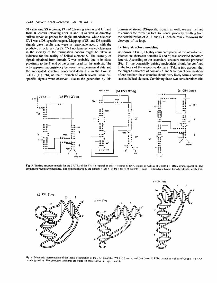
Fig. 3. Tertiary structure models for the 3-UTRs of the PV 1 (+) (panel a) and (-) (panel b) RNA strands as well as of CoxB4 (+) RNA strands (panel c). Thetermination codons are underlined. The elements shared by the domains Y and Y' of the 3-UTRs of the both (+) and (-) strands are boxed. For other details, see the text.
(c) CB4 3'pos
5'
(a) PVI 3'posx S
(b) PVI 3'negx S
Fig. 4. Schematic representation of the spatial organization of the 3-UTRs of the PV1 (+) (panel a) and (-) (panel b) RNA strands as well as of CoxB4 (+) RNA
strands (panel c). The proposed structures are based on those shown in Figs. 3 and 6.
Ter
x
S
Nucleic Acids Research, Vol. 20, No. 7 1743
interaction between loops of domains X and Y, on the one hand,and the stacking between domains X and S, on the other) wearrived at the model of the tertiary 3-UTR structure of the polio-like subset of enteroviruses shown in Fig. 3a. Since there is nointervening nucleotides between domains Y and Z in the CoxB-like subset of 3-UTRs, the stacking between the helical elementsof the respective domains could also be envisioned (Fig. 3c).Furthermore, the nucleotides paired as a result of tertiaryinteractions (element Ter) could extend, by stacking, the stemsof either domain Y or domain X. Since we had no reasons toprefer one variant of stacking to the other, we arbitrarily ascribed,solely for illustrative purposes, the former and the latter variantsto the polio-like and CoxB-like structures, respectively (Fig. 3).
(a) CB3 3' domain Z
7294 U U U A G A7300 UUAGAg7310 U U U - G A a7320 U U U A G A7326 U U - G g c
CB3 consensus:PV1:
7299A c A A 7309A U A A 7319
7325u U A A 7334
[UUUAGAnA [ l7368 U U U A G|- 7375
(b) PVI 3' domain Y
7386 G U C G a A u7396 A u7401 G U C a U A c7409 G U u G U A g
U G G 7395U G G 7400U 7408g G G 7418
(c) EV70 3' domain Y
732473327342
A g G A A CCA a G A A C C
C
C 7331C C U 7341C C U 7345
(d) PVI 3' domains Y' and X'
7359 a G G c7367 a a G g7375 c G c a7383 c G G a
GU AGU Aa U AGU A
C 7366C 7374C 7382C 7390
7414 g u G g G U A C 7421
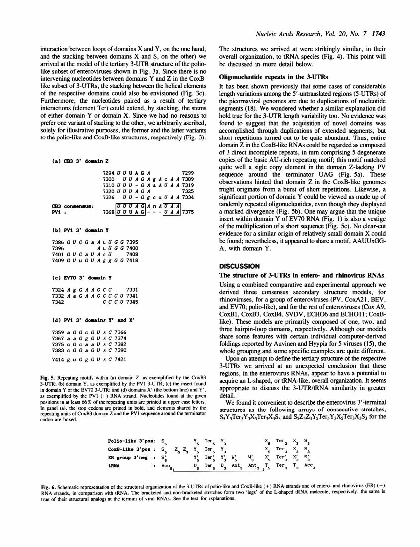
Fig. 5. Repeating motifs within (a) domain Z, as exemplified by the CoxB33-UTR; (b) domain Y, as exemplified by the PVI 3-UTR; (c) the insert foundin domain Y of the EV70 3-UTR; and (d) domains X' (the bottom line) and Y',as exemplified by the PVl (-) RNA strand. Nucleotides found at the givenpositions in at least 66% of the repeating units are printed in upper case letters.In panel (a), the stop codons are prined in bold, and elements shared by therepeating units of CoxB3 domain Z and the PV 1 sequence around the terminatorcodon are boxed.
Polio-like 3'pos: Ss Y5 Ter5CoxB-like 3'pos : Ss Z. Z3 Y5 Ter5ER group 3'neg : *S' Y' Ter'
5 5 5
tRNA : Acc5 D5 Ter5
The structures we arrived at were strikingly similar, in theiroverall organization, to tRNA species (Fig. 4). This point willbe discussed in more detail below.
Oligonucleotide repeats in the 3-UTRsIt has been shown previously that some cases of considerablelength variations among the 5'-untranslated regions (5-UTRs) ofthe picornaviral genomes are due to duplications of nucleotidesegments (18). We wondered whether a similar explanation didhold true for the 3-UTR length variability too. No evidence wasfound to suggest that the acquisition of novel domains wasaccomplished through duplications of extended segments, butshort repetitions turned out to be quite abundant. Thus, entiredomain Z in the CoxB-like RNAs could be regarded as composedof 3 direct incomplete repeats, in turn comprising 5 degeneratecopies of the basic AU-rich repeating motif; this motif matchedquite well a sigle copy element in the domain Z-lacking PVsequence around the terminator UAG (Fig. Sa). Theseobservations hinted that domain Z in the CoxB-like genomesmight originate from a burst of short repetitions. Likewise, asignificant portion of domain Y could be viewed as made up oftandemly repeated oligonucleotides, even though they displayeda marked divergence (Fig. Sb). One may argue that the uniqueinsert within domain Y of EV70 RNA (Fig. 1) is also a vestigeof the multiplication of a short sequence (Fig. Sc). No clear-cutevidence for a similar origin of relatively small domain X couldbe found; nevertheless, it appeared to share a motif, AAUUxGG-A, with domain Y.
DISCUSSIONThe structure of 3-UTRs in entero- and rhinovirus RNAsUsing a combined comparative and experimental approach wederived three consensus secondary structure models, forrhinoviruses, for a group of enteroviruses (PV, CoxA21, BEV,and EV70; polio-like), and for the rest of enteroviruses (Cox A9,CoxB1, CoxB3, CoxB4, SVDV, ECHO6 and ECHO11; CoxB-like). These models are primarily composed of one, two, andthree hairpin-loop domains, respectively. Although our modelsshare some features with certain individual computer-derivedfoldings reported by Auvinen and Hyypia for 5 viruses (15), thewhole grouping and some specific examples are quite different.Upon an attempt to define the tertiary structure of the respective
3-UTRs we arrived at an unexpected conclusion that theseregions, in the enterovirus RNAs, appear to have a potential toacquire an L-shaped, or tRNA-like, overall organization. It seemsappropriate to discuss the 3-UTR/tRNA similarity in greaterdetail.We found it convenient to describe the enterovirus 3'-terminal
structures as the following arrays of consecutive stretches,S5Y5Ter5Y3X5Ter3X3S3 and S5Z5Z3Y5Ter5Y3X5Ter3X3S3 for the
y3
Y3
3D3
X Ter5 3
X Ter5 3
W' W' X' Ter'5 3 5 3
Ant Ant T Ter5 3 i 5 3
x S3 3
x S3 3
x' S'3 3
T Acc3 3
Fig. 6. Schematic representation of the structural organization of the 3-UTRs of polio-like and CoxB-like (+) RNA strands and of entero- and rhinovirus (ER) (-)RNA strands, in comparison with tRNA. The bracketed and non-bracketed stretches form two 'legs' of the L-shaped tRNA molecule, respectively; the same istrue of their structural analogs at the termini of viral RNAs. See the text for explanations.

1744 Nucleic Acids Research, Vol. 20, No. 7
polio-like and CoxB-like RNAs, respectively (subscripts 5 and3 correspond to the 5'-proximal and 3'-proximal branches of theappropriate DS helical element). Using the same convention,we could write the structure of a tRNA molecule asAcc5D5Ter5D3Ant5Ant3T5Ter3T3Ac3, where Acc, D, Ant and Tstand for the acceptor, D, anticodon and T stems, respectively,and Ter designates the tertiary Watson-Crick interaction betweenthe D and T loops. Then, the viral and tRNA segments couldbe aligned using the Ter elements as marking points (Fig. 6).Such an alignment allowed us to consider S, Y, and X elementsin the viral 3-UTRs as structural analogs of the Acc, D, and Tstems, respectively. In the framework of this reasoning, the3-UTRs of the both polio-like and CoxB-like viral RNA specieslacked an analog of the anticodon stem, the latter RNAs havingan additional stem, Z, instead. It is noteworthy that thesedeviations of the viral structures from that of their tRNAcounterpart do not affect essential features of the L-shapedorganization. The reasons for maintaining the overall design areobvious, (i) extra-domain Z serves, by stacking on Y, merelyto elongate one 'leg' of L (Fig. 3); and (ii) the absence of theanti-codon stem merely shortens its other 'leg'.
It should be admitted that the above tertiary structure models,as opposed to the secondary structure ones, are based ontheoretical considerations only, and even though the evolutionaryapproach proved to be a powerful tool, more direct verificationof the models is needed.
Similarity between the 3'-terminal structures of (+) and (-)strandsDesigning the present study, we wanted to define commonstructural elements in the 3'-terminal regions of the (+) and (-)strands of picornaviral RNAs, as candidate cis-acting replicationelements. Obviously, the structure of the 3' end of the (-) strandshould mirror that of the 5' end of the (+) strand. Consensuscloverleaf secondary structure models for a ca. 100 nt-long regionat the 5' end of the (+) strand and, by implication, at the 3'end of the (-) strand of entero- and rhinovirus RNAs havealready been proposed on the basis of both comparativeconsiderations (3) and experimental testing (4). Upon inspectionof the appropriate structures, a conserved potential for tertiaryinteractions between two hairpin loops of the 3'-terminalcloverleaf on the (-) strand was found (illustrated for PVI inFig. 3b); a similar interaction at the 5' end of the (+) strandis also possible (not shown). As a result of such interactions andof stacking of the domains in pairs, these termini could acquirean L-shaped conformation (Fig. 4b), strikingly similar to thatproposed for the 3' end of the (+) strand of both polio-like(Fig. 4a) and CoxB-like RNAs (Fig. 4c). Using the conventiondescribed above, we designated the duplex that joins the very5'- and 3'-terminal segments of the cloverleaf on the 3' end ofthe (-) strand [or the 5' end of the (+) strand] as stem S' (stem'a' in ref. 4) and the element formed by the hairpin-hairpintertiary interaction as Ter'. The hairpin that generates a commonstacked stem with S was termed X' (stem-loop 'b'), and thehairpin that enters into the tertiary interaction with X' was termedY' (stem-loop 'd'); the remaining small domain was termed W'(stem-loop 'c') (it may be noted that domains Y and Y' sharecommon elements at the primary structure level; see boxedoligonucleotides in Fig. 3). Then, the structure of the 3'-terminalregion of the entero- and rhinovirus RNA (-) strand could bewritten as follows 5' S'5Y'5Ter'5Y'3W'5W'3X'5Ter'3X'3S'3 3'(Fig. 6). Accepting this, we could regard element W' as an analog
of the tRNA anticodon stem-loop. Again, no evidence is availableto choose between the two possible variants of stacking of Ter',either on X' or on Y'. In either case, however, the stacking wouldbe expected to stabilize the Ter' element composed of two G-Cpairs (further stabilization could in principle be achieved at theexpense of partial unwinding of Y').
Functional implicationsA major aim of the present study was to get insights into thenature of the cis-acting elements serving as recognizable signalsfor the replication machinery. Two interrelated points deservesome considerations: they concern the extent of similarity (ordiversity) of putative signals at the (i) 3' ends of the (+) strandsof different viruses; and (ii) 3' ends of the (+) and (-) RNAstrands of a given virus.The extent of the variability of the 3-UTR structures among
the entero- and rhinovirus RNA genomes is unexpected andintriguing. Since domain Y is shared by all the members of thisvirus group one may assume that it plays a major part in thetemplate recognition, other domains accomplishing someauxiliary, e. g., stabilizing, function; it could be envisioned thatsuch stabilization may be more important under certain conditionsthan under the others (due, for example, a higher optimaltemperature for the reproduction of enteroviruses than ofrhinoviruses). On the other hand, structural variability of the3-UTRs of entero- and rhinoviruses may reflect peculiarities ofinitiation of the (-) strand synthesis in each viral subgroup; thesepeculiarities may or may not be related to differences in thenatural host cells.
It is interesting to note that insertions of oligonucleotide linkersjust between Y5 and Ter5 of poliovirus type 1 RNA (cf., Figs.3a and 6) resulted in viable mutants with either wild-type or 3tsphenotypes (19). Remarkably, such insertions would not abolishthe potential for the both secondary and tertiary structureformation, but the overall stability of the mutant cis-element maybe diminished, especially in the case of the ts mutant (unpublishedobservations).There is little doubt that the same enzyme, viral RNA
polymerase, performs a key role in the synthesis of both (+)and (-) strands, though the possibility that the initiation of thetwo strands is accomplished by different mechanisms could notbe ruled out (cf., 4). One might expect that the signals recognizedby the enzyme on the 3' ends of the both kinds of templates shouldresemble each other, at least to some extent. The results presentedabove demonstrate that this is very likely to be the case for theCoxB-like subgroup of enteroviruses; here, these signals appearto be contained within very similar L-shaped structures composedof three hairpin-loop elements in each case. In polio-likeenteroviruses, the 3'-terminal structures of the (+) and (-)strands, while differing from one another in the number of theconstituent hairpin-loop elements, may nevertheless also sharea common L-shaped configuration. The 3'-terminal structuresappear to be more divergent, however, in the (+) and (-) RNAstrands of rhinoviruses, being represented by a solitary stem-loopand an L-shaped element, respectively. Although the functionalsignificance of this divergence is uncertain, it should be notedthat the overall structure of the 3' end of the (-) strand is highlyconserved among all the entero- and rhinoviruses, suggesting thatall of them share a common mechanism for the (+) strandinitiation, disregarding differences, if any, in the modes of theinitiation of the (-) strand. While considering possible differencesbetween utilization of (+) and (-) strands as templates, one

Nucleic Acids Research, Vol. 20, No. 7 1745
perhaps should take into account that the newly synthesized RNApolymerase molecule (polypeptide 3D) finds itselfjust in the closeproximity to the 3' terminal cis-element of the (+) strand; thiscircumstance may facilitate their mutual recognition.An additional puzzle was raised by the results reported by
Andino et al. (4) demonstrating that the synthesis of PV (+) RNAstrands depends primarily on the secondary structure of the 5'end of the (+) strand rather than on that of the 3' end of the(-) strand. Due to the fact that all the proposed base pairs atthe 3' end of the PV (-) RNA strands are represented by standardA-Us and G-Cs (without participation of G-Us) (Fig. 3b), thesecondary and tertiary structures of the 5' end of the (+) strandshould mirror those shown in Figs. 3b and 4b. Therefore all theconsiderations about the significance of structural peculiaritiesof the 3' end of the (-) strands could safely be extrapolated tothe 5' end of the (+) strands. The only possible distinction mayconcern a stretch of mismatches within domain Y': themismatched bases are represented solely by purines at the 3' endsof entero- and rhinovirus (-) RNA strands (Fig. 3b; positions7367-7369 and 7384-7386), whereas pyrimidines shouldoccupy this place at the (+) RNA 5' (as well as 3') ends . Aconceivable consequence of this difference is that the stem ofdomain Y should be more stable in the latter cases comparedto the former because pyrimidines could be more readily thanpurines incorporated into this stem by stacking interactions. Thiscircumstance, in addition to the difference in the primarystructure, may contribute to the preferential interaction of viralreplication proteins with the 5' ends of the (+) strands (4).A tRNA-like structure of the template termini raises an
interesting possibility that they could interact with tRNA-recognizing host proteins as is the case with some RNA phages(20) and plant viruses (21).
Evolutionary considerationsThe 3-UTRs of entero- and rhinoviruses exhibit a markedvariability. In rhinoviruses, this segment is considerably shorterthan in enteroviruses, possessing only a single secondary structuredomain (Y). It should be remembered that the rhinovirus 5-UTRsare also the shortest, lacking two duplications present in theirenterovirus counterparts (18). The difference in the 3-UTRlengths could arise from either the acquisition of novel domainsby enteroviruses, or the loss of some enterovirus-specific domainsby rhinoviruses. Although no direct evidence is available tochoose between these possibilities, we prefer the former one.
Indeed, the presence of repeats suggests that at least one of theenterovirus extra 3' domains, Z, has arisen through a burst ofduplications of a short oligonucleotide adjoining the terminatorcodon. In the framework of this reasoning, the rhinovirus genomeappears to be closer to the putative common rhino- andenterovirus ancestor, as far as the structures of the both 5- and3-UTRs are concerned (see also, ref. 18).
In turn, the enterovirus 3-UTRs could be further divided intotwo subclasses, using the presence or absence of domain Z as
a criterion. Interestingly, each subclass contains a CoxArepresentative. A close relatedness of the CoxA21 3-UTRstructure to that of PV is in good accord with the relatednessof the protein-coding parts of the respective genomes (10). Onthe other hand, capsid polypeptides (like 3-UTRs) of CoxA9appear to be more related to the CoxB viruses (22). Thus thecurrent classification of some picornaviruses into the CoxA groupdoes not appear to be supported by the primary and secondarystructures of their genomic RNAs (cf., also ref. 14).
It is hardly fortuitous that the 3'-terminal organization of sodifferent replicating molecules as genomes of picornaviruses (thisstudy), many plant RNA viruses (21), RNA bacteriophages (23)and even so called small replicating RNA species found in phageQ,B infected E. coli cells (24) bear more or less obvious similarityto tRNA. This similarity may merely reflect some advantagesof such an organization, e. g., its relative rigidity, but, in addition,it may have a more profound reason; indeed it was suggestedthat tRNA species have evolved from 3'-terminal 'tag' structuresof self-replicating inhabitants of the prehistoric 'RNA world' (25).Whether or not there is any evolutionary link between the similar5'- and 3'-terminal L-shaped structures (in other words, whetherthey came from a common ancestor RNA element), is anotherintriguing and completely open question.
Interestingly, one can find short direct repeats at the 3' endsof not only the (+) but also of the (-) strands. Thus, nearlyentire domain Y' of all the entero- and rhinovirus RNAs iscomposed of 4 tandem copies of an octanucleotide with a highlyconserved GUAC motif; domain X' also contains a copy of thismotif (Fig. Sd). Evolutionary and/or physiological significanceof this fact is unknown.
REFERENCES1. Koch,F. and Koch,G. (1985) The Molecular Biology of Poliovirus. Springer-
Verlag, Wien.2. Richards,O. and Ehrenfeld,E. (1990) Curr. Top. Microb. Immun. 161,
189-119.3. Rivera,V., Welsh,D. and Maizel,J. (1988) Virology 165, 42-50.4. Andino, R., Rieckhof, G. E., Trono, D. and Baltimore, D. (1990) Cell 463,
369-380.5. Pilipenko,E.V., Blinov,VM., Romanova,L.I., Sinyakov,A.N., Maslova,S.V.
and Agol,V.I. (1989) Virology 168, 201-209.6. Pilipenko,E.V., Blinov,V.M, Chernov,B.K., Dmitrieva,T.M. and Agol,V.I.
(1989) Nucleic Acids Res. 17, 5701-5711.7. Skinner,M.A., Raccaniello,V.R., Dunn,G., Cooper,J., Minor,P.D. and
Almond,J.W. (1989) J. Mol. Biol. 207, 379-392.8. Le,S-Y. and Zuker,M. (1990) J. Mol. Biol. 216, 729-741.9. Chang,K.H., Auvinen,P., Hyypia,T. and Stanway,G. (1989) J. Gen. Virol.
470, 3269-3280.10. Hughes,P.J., North,C., Minor,P.D. and Stanway,G. (1989) J. Gen. Virol.
470, 2943-2952.11. Earle,J.A.P., Skuce,R.A., Fleming,C.S., Hoey,E.M. and Martin,S.J. (1988)
J. Gen. Virol. 69, 253 -263.12. Inoue,T., Suzuki,T. and Sekiguchi,K. (1989) J. Gen. Virol. 70, 919-934.13. Ryan,M., Jenkins,O., Hughes,P., Brown,A., Knowles,N., Booth,D.,
Minor,P. and Almond,J. (1990) J. Gen. Virol. 71, 2291-2299.14. Auvinen,P., Stanway,G. and Hyypia,T. (1989) Arch Virol. 104, 175 - 186.15. Auvinen,P. and Hyypia,T. (1990) J. Gen. Virol. 71, 2133-2139.16. Jenkins,O., Booth,J.D., Minor,P.D. and Almond,J.W. (1987)17. Peritz,A.E., Kierzek,R., Sugimoto,N. and Turner,D.H. (1991) Biochemistry
30, 6428-6436.18. Pilipenko,E., Blinov,V. and Agol,V. (1990) Nucleic Acids Res. 18,
13371 -3375.19. Sarnow,P., Bernstein, H.D. and Baltimore,D. (1986) Proc. Natl. Acad. Sci.
83, 571-575.20. Blumenthal,T. and Carmichael,G.G. (1979) Annu. Rev. Bochem. 48,
1525 -548.21. Haenni,A-L., Joshi,S. and Chapeville,F. (1982) Progr. Nucleic Acids Res.
and Mol. Biol. 27, 85-104.22. Stanway,G. (1990) J. Gen. Virol. 71, 2483-2501.23. Adhin,M., Alblas,J. and van Duin,J. (1990) Biochim. Biophys. Acta 1050,
1110-118.24. Chetverin,A.B. and Voronin,L.A. (1987) In: Structure and Biosynthesis of
Proteins, vol. 1. Scientific Center for Biological Research, Pushchino, pp.43-52 (in Russian).
25. Weiner,A.M. and Maizels,N. (1987) Proc. NatI. Acad. Sci. USA 84,17383-7387.