Distinguishing Molecular Features and Clinical Characteristics of a Putative New Rhinovirus Species,...
12
Distinguishing Molecular Features and Clinical Characteristics of a Putative New Rhinovirus Species, Human Rhinovirus C (HRV C) Peter McErlean 1,2 , Laura A. Shackelton 3 , Emily Andrews 1,2 , Dale R. Webster 4,5,6 , Stephen B. Lambert 1,2 , Michael D. Nissen 1,2,7,8 , Theo P. Sloots 1,2,7,8 , Ian M. Mackay 1,2 * 1 Queensland Paediatric Infectious Diseases Laboratory, Sir Albert Sakzewski Virus Research Centre, Royal Children’s Hospital, Brisbane, Queensland, Australia, 2 Clinical and Medical Virology Centre, University of Queensland, Brisbane, Queensland, Australia, 3 Mueller Laboratory, Center for Infectious Disease Dynamics, Department of Biology, The Pennsylvania State University, University Park, Pennsylvania, United States of America, 4 Department of Biochemistry and Biophysics, University of California San Francisco, San Francisco, California, United States of America, 5 Biological and Medical Informatics Program, University of California San Francisco, San Francisco, California, United States of America, 6 Howard Hughes Medical Institute, University of California, San Francisco, California, United States of America, 7 Division of Microbiology, Queensland Health Pathology Service, Royal Brisbane Hospitals Campus, Brisbane, Queensland, Australia, 8 Department of Paediatrics and Child Health, Royal Children’s Hospitals, Brisbane, Queensland, Australia Abstract Background: Human rhinoviruses (HRVs) are the most frequently detected pathogens in acute respiratory tract infections (ARTIs) and yet little is known about the prevalence, recurrence, structure and clinical impact of individual members. During 2007, the complete coding sequences of six previously unknown and highly divergent HRV strains were reported. To catalogue the molecular and clinical features distinguishing the divergent HRV strains, we undertook, for the first time, in silico analyses of all available polyprotein sequences and performed retrospective reviews of the medical records of cases in which variants of the prototype strain, HRV-QPM, had been detected. Methodology/Principle Findings: Genomic analyses revealed that the six divergent strains, residing within a clade we previously called HRV A2, had the shortest polyprotein of all picornaviruses investigated. Structure-based amino acid alignments identified conserved motifs shared among members of the genus Rhinovirus as well as substantive deletions and insertions unique to the divergent strains. Deletions mostly affected regions encoding proteins traditionally involved in antigenicity and serving as HRV and HEV receptor footprints. Because the HRV A2 strains cannot yet be cultured, we created homology models of predicted HRV-QPM structural proteins. In silico comparisons confirmed that HRV-QPM was most closely related to the major group HRVs. HRV-QPM was most frequently detected in infants with expiratory wheezing or persistent cough who had been admitted to hospital and required supplemental oxygen. It was the only virus detected in 65% of positive individuals. These observations contributed to an objective clinical impact ranging from mild to severe. Conclusions: The divergent strains did not meet classification requirements for any existing species of the genus Rhinovirus or Enterovirus. HRV A2 strains should be partitioned into at least one new species, putatively called Human rhinovirus C, populated by members detected with high frequency, from individuals with respiratory symptoms requiring hospital admission. Citation: McErlean P, Shackelton LA, Andrews E, Webster DR, Lambert SB, et al. (2008) Distinguishing Molecular Features and Clinical Characteristics of a Putative New Rhinovirus Species, Human Rhinovirus C (HRV C). PLoS ONE 3(4): e1847. doi:10.1371/journal.pone.0001847 Editor: Dong-Yan Jin, University of Hong Kong, China Received December 4, 2007; Accepted February 21, 2008; Published April 2, 2008 Copyright: ß 2008 McErlean et al. This is an open-access article distributed under the terms of the Creative Commons Attribution License, which permits unrestricted use, distribution, and reproduction in any medium, provided the original author and source are credited. Funding: This work was supported by NHMRC Project grant 455905 and the Royal Children’s Hospital Foundation grants 912-011 and 922-202 sponsored by the Woolworth’s Fresh Futures Programme. PM was supported by a SASVRC PhD Scholarship. Competing Interests: The authors have declared that no competing interests exist. * E-mail: [email protected] Introduction Human rhinoviruses (HRVs) are the most frequently detected pathogens in acute respiratory tract infections (ARTIs). HRVs have been associated with lower respiratory tract (LRT) illness and more serious clinical outcomes within pediatric and other vulnerable populations [1]. Despite this, HRV strains continue to be commonly defined, en masse, by their most prolific and currently most well-defined role in causing the ‘common cold’ [2,3]. Classified within the family Picornaviridae, the genus Rhinovirus consists of 100 serotyped strains divided into two species, Human rhinovirus A (HRV A; n = 75) and Human rhinovirus B (HRV B; n = 25) [4]. Species classification was initially based on the susceptibility of strains to capsid binding antivirals [5,6] and subsequently confirmed by phylogenetic studies [4,7]. HRVs are also subdivided by receptor usage; major group HRVs use an intercellular adhesion molecule (ICAM-1; n = 88 strains), which interacts within a depression of the viral surface known as the canyon [8]. Minor group HRVs (n = 12) receive a molecule from PLoS ONE | www.plosone.org 1 April 2008 | Volume 3 | Issue 4 | e1847
Transcript of Distinguishing Molecular Features and Clinical Characteristics of a Putative New Rhinovirus Species,...

Distinguishing Molecular Features and ClinicalCharacteristics of a Putative New Rhinovirus Species,Human Rhinovirus C (HRV C)Peter McErlean1,2, Laura A. Shackelton3, Emily Andrews1,2, Dale R. Webster4,5,6, Stephen B. Lambert1,2,
Michael D. Nissen1,2,7,8, Theo P. Sloots1,2,7,8, Ian M. Mackay1,2*
1 Queensland Paediatric Infectious Diseases Laboratory, Sir Albert Sakzewski Virus Research Centre, Royal Children’s Hospital, Brisbane, Queensland, Australia, 2 Clinical
and Medical Virology Centre, University of Queensland, Brisbane, Queensland, Australia, 3 Mueller Laboratory, Center for Infectious Disease Dynamics, Department of
Biology, The Pennsylvania State University, University Park, Pennsylvania, United States of America, 4 Department of Biochemistry and Biophysics, University of California
San Francisco, San Francisco, California, United States of America, 5 Biological and Medical Informatics Program, University of California San Francisco, San Francisco,
California, United States of America, 6 Howard Hughes Medical Institute, University of California, San Francisco, California, United States of America, 7 Division of
Microbiology, Queensland Health Pathology Service, Royal Brisbane Hospitals Campus, Brisbane, Queensland, Australia, 8 Department of Paediatrics and Child Health,
Royal Children’s Hospitals, Brisbane, Queensland, Australia
Abstract
Background: Human rhinoviruses (HRVs) are the most frequently detected pathogens in acute respiratory tract infections(ARTIs) and yet little is known about the prevalence, recurrence, structure and clinical impact of individual members. During2007, the complete coding sequences of six previously unknown and highly divergent HRV strains were reported. Tocatalogue the molecular and clinical features distinguishing the divergent HRV strains, we undertook, for the first time, insilico analyses of all available polyprotein sequences and performed retrospective reviews of the medical records of cases inwhich variants of the prototype strain, HRV-QPM, had been detected.
Methodology/Principle Findings: Genomic analyses revealed that the six divergent strains, residing within a clade wepreviously called HRV A2, had the shortest polyprotein of all picornaviruses investigated. Structure-based amino acidalignments identified conserved motifs shared among members of the genus Rhinovirus as well as substantive deletionsand insertions unique to the divergent strains. Deletions mostly affected regions encoding proteins traditionally involved inantigenicity and serving as HRV and HEV receptor footprints. Because the HRV A2 strains cannot yet be cultured, we createdhomology models of predicted HRV-QPM structural proteins. In silico comparisons confirmed that HRV-QPM was mostclosely related to the major group HRVs. HRV-QPM was most frequently detected in infants with expiratory wheezing orpersistent cough who had been admitted to hospital and required supplemental oxygen. It was the only virus detected in65% of positive individuals. These observations contributed to an objective clinical impact ranging from mild to severe.
Conclusions: The divergent strains did not meet classification requirements for any existing species of the genus Rhinovirusor Enterovirus. HRV A2 strains should be partitioned into at least one new species, putatively called Human rhinovirus C,populated by members detected with high frequency, from individuals with respiratory symptoms requiring hospitaladmission.
Citation: McErlean P, Shackelton LA, Andrews E, Webster DR, Lambert SB, et al. (2008) Distinguishing Molecular Features and Clinical Characteristics of a PutativeNew Rhinovirus Species, Human Rhinovirus C (HRV C). PLoS ONE 3(4): e1847. doi:10.1371/journal.pone.0001847
Editor: Dong-Yan Jin, University of Hong Kong, China
Received December 4, 2007; Accepted February 21, 2008; Published April 2, 2008
Copyright: � 2008 McErlean et al. This is an open-access article distributed under the terms of the Creative Commons Attribution License, which permitsunrestricted use, distribution, and reproduction in any medium, provided the original author and source are credited.
Funding: This work was supported by NHMRC Project grant 455905 and the Royal Children’s Hospital Foundation grants 912-011 and 922-202 sponsored by theWoolworth’s Fresh Futures Programme. PM was supported by a SASVRC PhD Scholarship.
Competing Interests: The authors have declared that no competing interests exist.
* E-mail: [email protected]
Introduction
Human rhinoviruses (HRVs) are the most frequently detected
pathogens in acute respiratory tract infections (ARTIs). HRVs
have been associated with lower respiratory tract (LRT) illness and
more serious clinical outcomes within pediatric and other
vulnerable populations [1]. Despite this, HRV strains continue
to be commonly defined, en masse, by their most prolific and
currently most well-defined role in causing the ‘common cold’
[2,3].
Classified within the family Picornaviridae, the genus Rhinovirus
consists of 100 serotyped strains divided into two species, Human
rhinovirus A (HRV A; n = 75) and Human rhinovirus B (HRV B;
n = 25) [4]. Species classification was initially based on the
susceptibility of strains to capsid binding antivirals [5,6] and
subsequently confirmed by phylogenetic studies [4,7]. HRVs are
also subdivided by receptor usage; major group HRVs use an
intercellular adhesion molecule (ICAM-1; n = 88 strains), which
interacts within a depression of the viral surface known as the
canyon [8]. Minor group HRVs (n = 12) receive a molecule from
PLoS ONE | www.plosone.org 1 April 2008 | Volume 3 | Issue 4 | e1847

the low density lipoprotein receptor family (LDLR) at protrusions
along the 5-fold axis of the capsid surface [9–12]. Recently,
heparan sulphate was identified as a pH dependent, low efficiency
receptor for HRV-54 [13]. A relationship between receptor usage
and species identity is only evident among minor group HRVs; all
are HRV As. Major group HRVs reside in both species. Studies
have identified four neutralizing immunogenic sites on the surface
of a major group HRV (HRV-14; NImIA, NImIB, NImII and
NImIII)[14] and three sites on a minor group HRV (HRV-2; site
A, B and C) [15], with some overlap [16]. These sites consist of
discontinuous amino acid sequence within VP1, VP2 and VP3.
The human enteroviruses (HEVs) are the closest genetic
relatives of the HRVs but they belong to the genus Enterovirus.
HEV strains are classified into four species (A–D) and some have
been implicated in ARTI [17]. Similar to the major group HRVs,
some HEVs have been shown to use ICAM-1 as their primary
cellular receptor [18]. Other host molecules employed by HEVs
include the decay accelerating factor (DAF) [19,20], poliovirus
receptor (PVR) [21] and coxsackie-adenovirus receptor (CAR)
[22]. Under a proposal currently before the International
Committee on Taxonomy of Viruses (ICTV), HRV and HEV
strains will be combined into the genus Enterovirus, at the expense
of Rhinovirus, an issue of contention for decades [23].
Picornavirus genomes typically consist of a 7.2 to 7.5 kb
positive-sense, single-stranded RNA molecule located within a
non-enveloped icosahedral capsid. The genomes contain a
single open reading frame flanked by 59and 39untranslated
regions (UTR). The polyprotein is first cleaved into three
precursor polyproteins, P1, P2 and P3 (Figure 1). P1 is further
cleaved into the structural proteins VP4, VP2, VP3 and VP1.
Union of a single copy of each structural protein constitutes a
viral protomer; the HRV capsid consists of 60 protomers
arranged in a T = 1, pseudo T = 3 (i.e. T = p3) conformation. P2
and P3 are cleaved into seven non-structural proteins including
two proteases (2Apro and 3Cpro) and an RNA-dependant RNA
polymerase (3Dpol) [24].
Recent studies have generated 27 additional HRV A and 12
additional HRV B genomes [25,26] also resulting in the proposal
that individual HRV genomes are under purifying selective
pressure and that significant intra-strain variation occurs only at
the antigenic sites [26]. This is in stark contrast to the HEVs for
which recombination in the non-structural regions is a significant
contributor to viral diversification [27,28].
Untypeable rhinoviruses have been described using PCR-based
tools, since 1994 [29] but it was only in 2007 that the complete
coding sequences of a number of novel and apparently divergent
HRV strains were reported, permitting a for identification of the
first new strains in two decades [30]. The initial strain described,
HRV-QPM, was identified from children with presenting
symptoms often suggesting bronchiolitis [31]. Subsequently,
‘HRV-Xs’ were found in an adult asthma study in the United
States [32] and ‘HRV-Cs’ were described from a study of
pediatric infection by human bocavirus in Hong Kong [33].
Some notable features associated with these six putative viruses
include the reliance upon molecular methods for their detection,
their apparent endeminicity, their failure to produce cytopathic
effects in culture, their frequent detection in subjects with
expiratory wheezing and their limited sequence identity with
existing HRV strains but high shared sequence identity. Studies
employing subgenomic sequences proposed that there were many
of these putative viruses occupying a distinct position within the
genus Rhinovirus, which we collectively called HRV A2 [34]. It has
been proposed that these strains may constitute a new
picornavirus species [33]. Data have not yet been presented
which appropriately address classification using current ICTV
criteria and the scope of the clinical impact of these divergent
strains has not yet been studied in detail. Because it appears that
the members of this clade are frequently associated with LRT
illness we sought to better characterise them in the hope of
finding features that might prove useful in predicting the outcome
of HRV A2 infection.
Intensive genomic analysis and computer-based modelling
resulted in comprehensive characterisation of all HRV A2
polyprotein sequences and, by superimposing the sites of known
receptor contacts, we were able to propose a more robust
taxonomic placement for these newly identified viruses (NIVs).
Detailed medical chart reviews on HRV-QPM-positive (prototype
HRV A2) individuals were augmented by a severity scoring system
which quantified the outcome of infection by this apparently
uncharacteristically pathogenic clade of HRV strains.
Results
Genomic analysesWe undertook a comparative analysis of all HRV A2 complete
coding regions (n = 6; HRV-QPM, -X1, H2, -C024, -C025 and
C026) against all HRV A (n = 34), HRV B (n = 13), most HEV
sequences (n = 71, including species A–D) and two non-human
enteroviruses (simian, SEV and porcine, PEV).
HRV A2 sequences had a higher G+C content (average,
42.4%) than all other HRVs (average 38.7%) or HEV Ds and a
shorter coding region (average, 2144 aa) than any other
picornavirus investigated. The final residue of the HRV A2
genomes was an isoleucine, distinguishing them from all other
picornavirus genomes we investigated, which ended at the
preceding phenylalanine; the biological implications of this
feature are unknown.
We identified 10 protease sites (data not shown) dividing the
HRV A2 polyproteins into four structural and seven non-
structural proteins (Figure 1A). However, unlike previous studies
that relied on amino acid alignments to determine cleavage sites
[33], we obtained additional evidence that these sites were
recognised by 2Apro and 3Cpro by using a neural network protease
cleavage-site prediction tool. A single cleavage by 2Apro was
predicted between an A or L and a G residue at the VP1/2A
junction. The remaining cleavages, predominantly occurring
between Q and G residues, were mediated by 3Cpro. Three
motifs crucial for RNA polymerase binding (YGDD, TFLKR and
SIRWT) were identified in all HRV A2 sequences [35,36].
Using SimPlot� [37], the HRV A2 sequences were found to be
most similar to those from HRV A strains (54% identity) whereas
identity with HEV species ranged from 46%–49% (Figure 1B).
Dips in identity occurred across all regions but only the structural
region was investigated further because its components contain
antigenic sites and receptor contact residues, or ‘footprints’; the
latter being significant domains for defining the major and minor
group HRVs [38] and other picornaviruses.
Structural homology modellingAlthough identifiable using amino acid alignments[33], the
scope and impact of the many HRV A2 sequence deletions could
not be fully qualified or quantified in this form. However, the
inability to isolate the HRV A2 strains in vitro will hinder
crystallography studies seeking to identify structural features, so we
created structure-based alignments of the VP1–4 sequences from
the prototype HRV A2 strain, HRV-QPM and used them to
predict the presence of a-helices and b-sheets. These were
identified in the eight, anti-parallel b-sheet ‘jellyroll’ conformation
A New Rhinovirus Species
PLoS ONE | www.plosone.org 2 April 2008 | Volume 3 | Issue 4 | e1847

common among picornavirus proteins VP1, VP2 (including the EF
‘puff’) and VP3 [39] (Figure 2). Deletions in the VP1 protein
imparted the most dramatic changes, reducing the protrusion of
the BC, DE and HI loops compared with the other HRVs and
introducing an additional C-terminal sheet-loop-sheet structure
(arrow, Figure 2B). Similar deletions were noted in all HRV A2
strains but only HRV-C026 was predicted to encode a similar, C-
terminal structure in VP1. We next assembled the HRV-QPM
structural proteins into a protomer by associating them with the
closest sequence match (HRV-16) and then replicated this
structure in silico into a viral pentamer. RMSD values were
derived from the VP1, VP2 and VP3 structures of all HRVs with
crystallography-derived data available (HRV-1A, -2, -3, -14 and -
16) and compared to the predicted HRV-QPM structure. HRV-
QPM and HRV-16 shared the highest conformational similarity
(0.145, 0.142 and 0.147 A for the VP1, VP2 and VP3 structures
respectively), reflecting the use of HRV-16 in predicting the
structure HRV-QPM. The poorest agreements were between
HRV-1A and HRV-3 in VP1 (0.793 A) and VP3 (0.740 A) and
between HRV-2 and HRV-14 in VP2 (0.697 A). At the ICAM-1
footprint, HRV-QPM values ranged from 0.064–0.106 A and
0.709–0.779 A in comparison to the same regions on HRV-16
and -14, respectively. Values could not be determined for HRV-2
because of the impact of deletions. We also examined the
conformational agreement between an HEV footprint e.g. the
CAR site employed by CV-B3, and the predicted HRV-QPM
structure. A space filling model of the pentamer was next used to
display SimPlot data in order to visualise regions of sequence
conservation and diversification with a focus on the major (ICAM-
1; Figure 3A) and minor (VLDL-R; Figure 3C) group domains.
Overall, greater sequence identity was apparent between HRV-2
than HRV-14 reflecting the genomic similarity to HRV A strains,
however within the domains defining HRV groups, HRV-QPM
was most similar in its antigenic sites to the major group
representative, HRV-14. Most sequence diversity was found in
the VP2 protein while the most conserved protein in these
comparisons was VP3.
To study these regions in greater detail, we mapped known
HRV antigenic sites and receptor footprints onto ribbon
depictions of pentamers derived from empirically determined
structural data (major group, HRV-14, Figure 3B; minor group,
HRV-2, Figure 3D) and compared their locations and structures
to the predicted HRV-QPM pentamer. HRV-QPM and HRV-14
appeared to share a number of similar structures in the region of
the ICAM-1 footprint [6,33,40] (Figure 3B). The noteworthy
structural disparities were two small helices present in the HRV-
QPM sequences; one in the VP1 and the other in the VP3 ICAM-
1 footprint (Figure 3B; VP1 and VP3, ICAM-1 arrows). Similar
sequences were infrequent among the other HRV strains we
examined. The HRV-QPM NImIA and IB sites appeared to differ
structurally due to the shortening of the BC and DE loops in VP1
while the NImII and NImIII sites appeared structurally similar.
Comparison to HRV-2 (Figure 3D) also revealed the impact of
VP1 deletions. The resulting loops were shorter, which ablated
any analogue of antigenic site A and may hamper binding of the
VLDL-R molecule, the only LDLR family member with a well-
defined footprint. The area in VP1 forming site B was altered due
to the presence of more hydrophilic amino acids at the equivalent
positions in HRV-QPM (Figure 3D; VP1 site B, arrow). Deletions
were apparent but did not confer obvious structural changes to the
VP2 portion of site B. Furthermore, in antigenic site C (VP2), a
single deletion and a hydrophobic to hydrophilic amino acid
change contributed to a protrusion in HRV-QPM compared to
the same region in HRV-2.
Analysis of HRV-QPM in the context of HEV receptorsGiven the comparative differences we observed when locating
and comparing the HRV domains, we also investigated the HRV-
Figure 1. Characterization of HRV A2 genomes. (A) Scale representation of the coding region from prototype HRV A2 virus, HRV-QPM.Predicted protein products from the structural (VP4 to VP1) and non-structural (PRO-protease; POL-polymerase) regions are indicated. Regions areshaded alternatively for convenience and numbering indicates positions of predicted protease cleavage according to the start of the HRV-QPMpolyprotein. Dashed lines indicate regions involved in species classifications. (B) HRV A2 amino acid similarity to HRV A (n = 34) and B (n = 13), or HEV(n = 71) strains as determined by SimPlot� analysis. Plots indicate the percentage similarity of a 50% consensus sequence from each species’polyprotein compared to the HRV A2 strains. Average similarity across the polyprotein is indicated by dotted line. Averaged values for the completecoding, P1 and 2C+3CD regions are indicated within the inset of each panel.doi:10.1371/journal.pone.0001847.g001
A New Rhinovirus Species
PLoS ONE | www.plosone.org 3 April 2008 | Volume 3 | Issue 4 | e1847
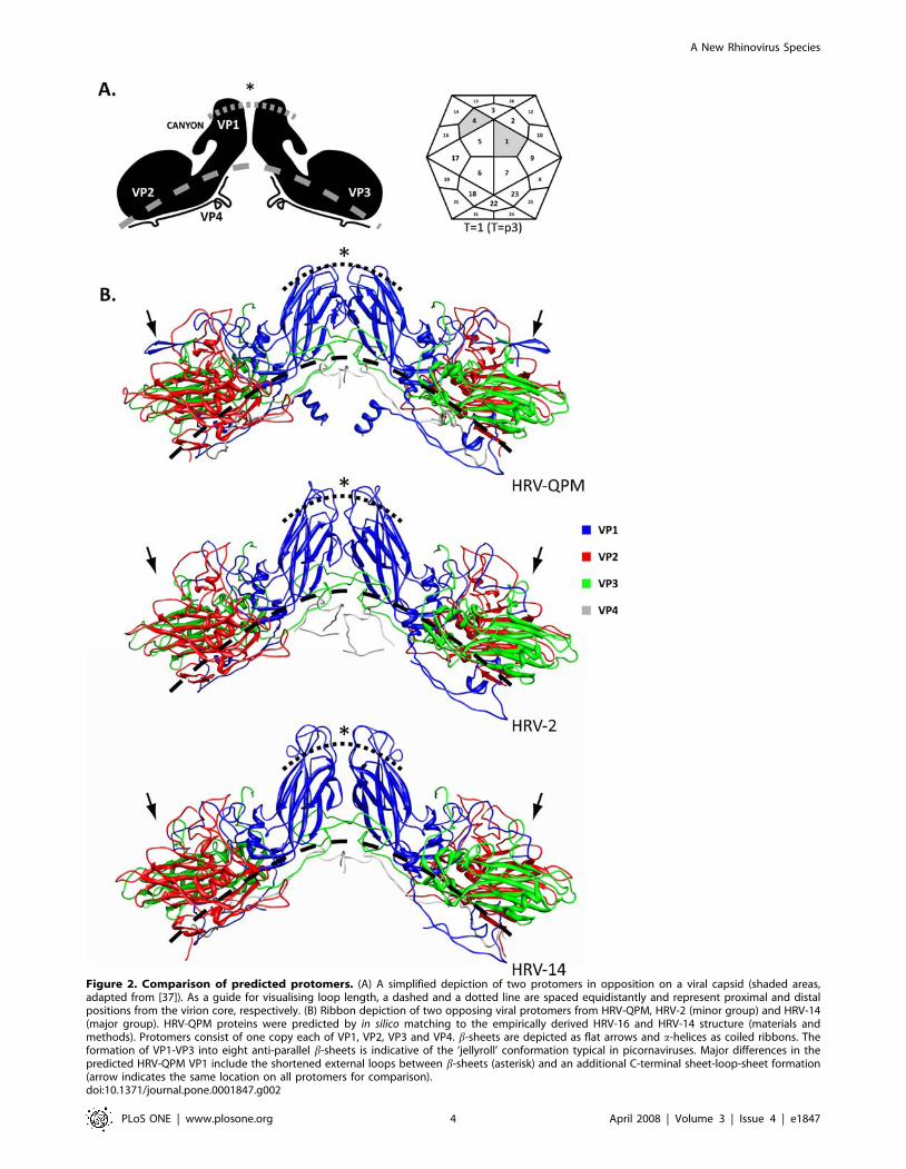
Figure 2. Comparison of predicted protomers. (A) A simplified depiction of two protomers in opposition on a viral capsid (shaded areas,adapted from [37]). As a guide for visualising loop length, a dashed and a dotted line are spaced equidistantly and represent proximal and distalpositions from the virion core, respectively. (B) Ribbon depiction of two opposing viral protomers from HRV-QPM, HRV-2 (minor group) and HRV-14(major group). HRV-QPM proteins were predicted by in silico matching to the empirically derived HRV-16 and HRV-14 structure (materials andmethods). Protomers consist of one copy each of VP1, VP2, VP3 and VP4. b-sheets are depicted as flat arrows and a-helices as coiled ribbons. Theformation of VP1-VP3 into eight anti-parallel b-sheets is indicative of the ‘jellyroll’ conformation typical in picornaviruses. Major differences in thepredicted HRV-QPM VP1 include the shortened external loops between b-sheets (asterisk) and an additional C-terminal sheet-loop-sheet formation(arrow indicates the same location on all protomers for comparison).doi:10.1371/journal.pone.0001847.g002
A New Rhinovirus Species
PLoS ONE | www.plosone.org 4 April 2008 | Volume 3 | Issue 4 | e1847

QPM capsid for the presence of footprints reportedly used by CV-
A21 (which employs ICAM-1 as a receptor), Echovirus (EV)-11
(DAF), Poliovirus-1 (PVR) and CV-B3 (CAR).
The deletions in the VP1 BC loop had a less obvious impact on
HEV receptor footprints which were mostly located on the b-
sheets rather than the loop (Figure 4). The EF loop in HRV-QPM
VP1 provided the greatest observable identity with any HEV
footprint while the C-terminal sheet-loop-sheet structure may
interfere with binding to the HEV receptors we investigated. All
the HEV VP2 receptor footprints occured in the EF ‘puff’ but in
this region of HRV-QPM a number of deletions and residue
substitutions were apparent (Figure 4) that affected the appearance
of predicted structural overlap. HEV footprints in VP3 differed
from HRV-QPM at the N-terminus and the CD loop due to
amino acid substitutions and loop-shortening deletions but they
overlapped in the GH loop.
HRV-QPM capsid prediction and visualizationHRV-QPM protomers were replicated and mapped onto an
icosahedral lattice (T = 1 configuration; Figure 5A), rendered in
3D and depth cued in order to predict capsid structures. Since
HRV-QPM protein and protomer structures were inferred by
homology to their closest available relatives, these capsids
(HRV-16 and HRV-14) were produced from the available
empirical data, using the same approach. Visual comparisons
revealed that the deletions in the HRV-QPM VP1 most
obviously reduced the size of protrusions around the 5-fold axis
of the capsid.
PhylogeneticsPreviously, phylogenies had been estimated using subgenomic
sequences from HRV A2-like strains [31–33]. To test whether this
approach accurately represented inter-strain relationships, we
Figure 3. Predicted HRV-QPM pentamers compared to representative major (HRV-14) and minor (HRV-2) group HRV pentamersderived from crystallography data. (A) HRV-QPM versus HRV-14 SimPlot data projected onto a space filling depiction of the predicted HRV-QPMpentamer. Shading represents the amino acid identity (26–69%). The yellow dashed triangle represents a single icosahedral asymmetric unit (T = p3conformation) composed of VP1 and VP2 from the same protomer and VP3 for an adjacent protomer. The major group domains of interest aredivided between two asymmetric units for ease of viewing. Receptor (white) and antigenic (red) sites are shown in outline. (B) Top view ribbondepiction of a major group HRV pentamer (HRV-14; gray) with labelled antigenic neutralisation sites (NImIA-III, green) and combined HRV A (HRV-16)and B (HRV-14)ICAM-1 receptor footprints (red) [6,37]. Magnified areas of interest (boxed) highlight computer-based predictive comparisons to theequivalent HRV-QPM (orange) structures of interest. Arrows indicate structures and corresponding sequences of interest (refer to text). (C) HRV-QPMversus HRV-2 SimPlot data projected onto the HRV-QPM pentamer. The domains of interest are mostly shown within a single asymmetric unit. (D) Aminor group pentamer (HRV-2; gray) including antigenic sites (sites A–C, green) and VLDL-R footprint (red) [10]. Attachment of the VLDL-R involvesadjacent VP1 molecules. Magnified VP1 area represents one half of a VLDL-R footprint [42]. Amino acid substitutions (arrowed) contributed to thedifferences between minor group sites B and C.doi:10.1371/journal.pone.0001847.g003
A New Rhinovirus Species
PLoS ONE | www.plosone.org 5 April 2008 | Volume 3 | Issue 4 | e1847

compared the entire polyprotein sequences from 120 previously
described and, for the first time, all six newly-identified
picornavirus strains (Figure 6). We confirmed that the newly-
identified HRVs occupy a distinct phylogenetic position with the
genus Rhinovirus [34] and are most closely related to the HRV A
strains (also supported by SimPlot mapping; Figure 3). We also
found support for our earlier data [31] that this clade is divided
into two distinct subgroups. In the 2C and 3CD regions, these
Figure 4. Comparison of HRV-QPM and HEV structures at the sites comprising HEV receptor footprints. Figures indicate comparison ofthe predicted structures of HRV-QPM (orange) with representative HEVs (gray) in regions identified as HEV receptor footprints (red) across VP1, VP2and VP3 proteins. RMSD values shown for conformational comparison of CAR and HRV-QPM structures, in angstroms.doi:10.1371/journal.pone.0001847.g004
Figure 5. Predicted virion capsid structures. (A) Replication (660) and mapping of the predicted HRV-QPM protomer onto a T = 1 icosahedrallattice (representing T = p3 configuration). (B) 3D rendering of predicted HRV capsids providing imagery similar to that obtained by cryo-electronmicroscopy reconstruction at 10 A resolution. HRV-2, -14 and -16 predicted structures were derived from crystallography data. Each viral capsid hasbeen depth cued to demonstrate canyon structure; yellow indicates surface detail and blue identifies areas of greatest depth. (C) Numberssuperimposed over the lattice indicate the fold axis. The simplified position of the canyon is indicated by a circle on the capsid. All models wererendered and oriented identically, as determined by VIPERdb [59].doi:10.1371/journal.pone.0001847.g005
A New Rhinovirus Species
PLoS ONE | www.plosone.org 6 April 2008 | Volume 3 | Issue 4 | e1847

subgroups share less than 70% amino acid identity, which also
occurs among members of the existing HRV species.
Clinical impact of HRV-QPM variantsPreviously, preliminary and potentially subjective notes made
by physicians upon first contact with ill individuals had been used
for an approximate determination of the clinical impact of HRV-
QPM [31]. To better address clinical impact, a comprehensive
review of available patient charts was undertaken and two
objective severity scoring tools were applied (Table 1). Most
HRV-QPM-positive individuals (76.5%) were admitted to hospital
(Table 2). Five patients were admitted for 96 hours or longer; two
with severe illness and only HRV-QPM detected (variants 005 and
012) and one with moderate illness positive for HRV-QPM
(variant 017) and HCoV-NL63. The remaining two had
underlying medical conditions and multiple infections (HRV-
QPM variant 009 and HCoV-229E with cystic fibrosis deterio-
ration and HRV-QPM variant 008, HBoV and HCoV-NL63 with
a viral URTI during admission for cardiac surgery). Oxygen
therapy was required by almost half of all HRV-QPM-positive
individuals studied. In addition to those described previously
[31], a new co-detection was identified with the newly identified
polyomavirus, WUPyV [41] in a patient exhibiting a persistent
cough, already positive for HRV-QPM (variant 013) and
HBoV.
Applying species criteria to the HRV A2 strainsIn silico data obtained from this study permitted an enhanced
analysis of HRV A2 strains using the ICTV criteria which assign
a member to the genus Rhinovirus or Enterovirus [42] (Table 3).
The requirement common to both genera was for .70% amino
acid identity in the P1 and 2C+3CD regions; this was not met by
the HRV A2 genomes (Figure 1B). The remaining Rhinovirus
species criterion was classification based on capsid-binding
antiviral susceptibility and we found here, building upon earlier
data [31], that all HRV A2 strains contain a Thr191. This
residue reportedly contributes to conveying resistance to the
capsid-binding antiviral Pleconaril [4,43], placing the divergent
HRVs into antiviral Group A [5].
Comparison to the Enterovirus species criteria revealed several
features that may also be applicable for classifying HRV A2
strains. Firstly, although we could not identify a specific receptor
using these in silico methods, the HRV-QPM models most closely
accommodated interaction with an ICAM-1-like molecule, similar
to some HEV C viruses [44]. Secondly, HRV A2 genomes
exhibited #2.5% variation in G+C composition compared to
HEV C and HEV D and ,5% compared to HEV A and HEV B.
Thirdly, HRV- and HEV-like protease cleavage sites and locations
suggested similar proteolytic processing (Figure 1A). Although we
have been unable to find any indication of recombination in
HRV-QPM [31], the differences in amino acid identity within the
novel HRVs, particularly in the 2C and 3CD region of HRV-X2,
-C024 and -C025 compared to HRV-QPM, -X1 and -C026 (data
not shown), may indicate a relevant event requiring further study.
rFigure 6. Neighbour-joining phylogeny based on representa-tive full-length picornavirus polyprotein sequences. Trees areunrooted and relevant nodes are labelled with bootstrap values (%) (seematerials and methods for details). Species are indicated next to verticalbars. CV-Coxsackievirus A; EV-Echovirus; HEV-Enterovirus; HPV-Poliovi-rus.doi:10.1371/journal.pone.0001847.g006
A New Rhinovirus Species
PLoS ONE | www.plosone.org 7 April 2008 | Volume 3 | Issue 4 | e1847
In sum, these data indicate that the HRV A2 strains cannot
be assigned to an existing HRV or HEV species and should
be assigned to a new species, tentatively called Human rhinovirus
C.
Discussion
In 2007, the sequences of a number of divergent HRV strains
were reported as a result of worldwide molecular investigations
into cases of suspected ARTI [26,31,33]. Because these strains
formed a distinct clade within the existing HRV As, we
previously assigned to all Australian and similar New York
strains, the title of HRV A2 [31]. Kistler et al. identified similarly
divergent strains [32] and Lau et al. used sequence-based
criteria to propose that some HRV A2-like strains could be
classified as a new species [33]. Our ongoing efforts to
characterise additional HRVs led us to catalogue the distin-
guishing molecular and clinical features of these HRV A2-like
strains and to better address their taxonomic placement. We
undertook in silico analyses, for the first time using all complete
HRV A2 coding sequences and performed a retrospective
review of the medical records of cases from which variants of the
prototype strain, HRV-QPM, had been previously detected. To
date the studies reported herein are the first of their kind to have
been conducted on a single HRV strain.
During our genomic analysis we found HRV A2 strains shared
only 50%–53% average amino acid sequence identity with other
HRV strains (Figure 1); less with any strain from an HEV species.
Average HRV A2 G+C content (42.4%) fell between the
rhinovirus (38.7%) and enterovirus (45.6%) genera, suggesting
an intermediate evolutionary path for HRV A2 strains. Nonethe-
less, a large number of HRV motifs, including those recognized by
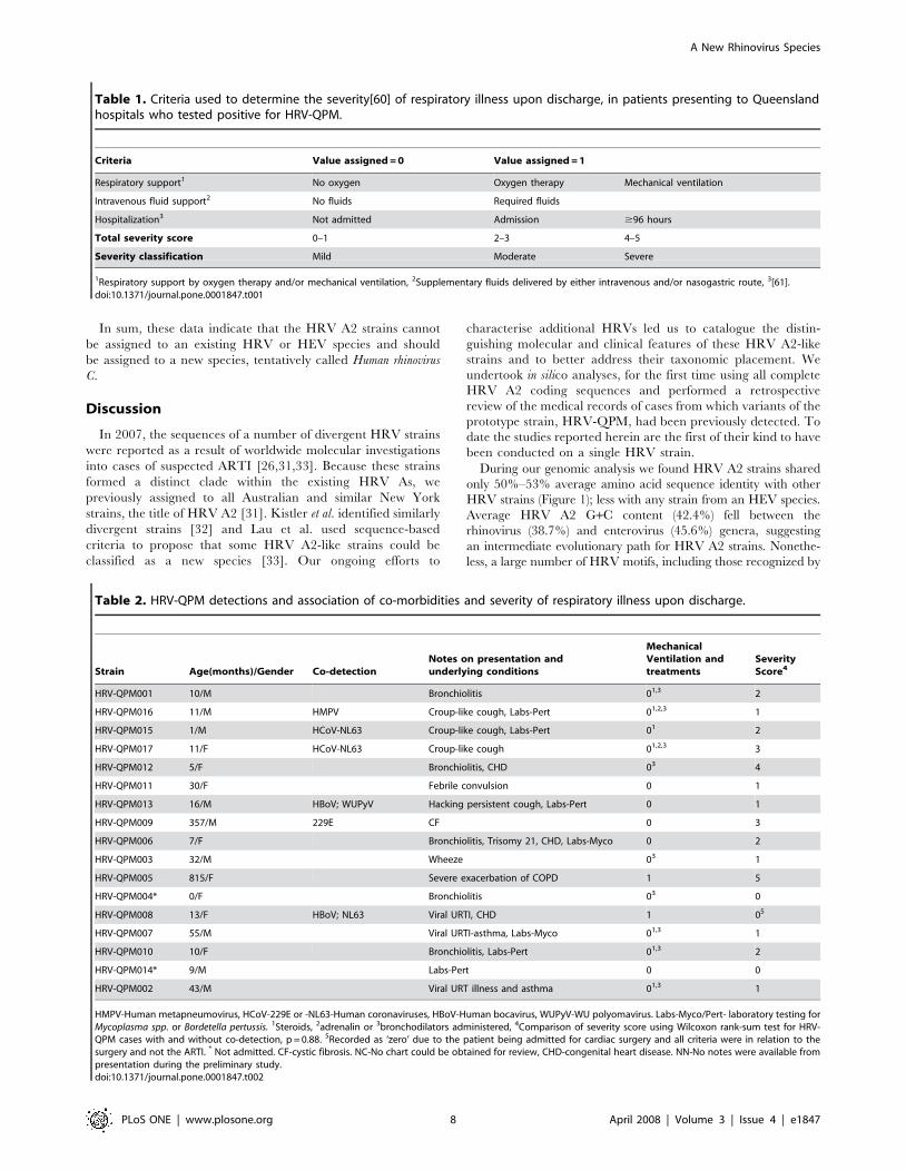
Table 1. Criteria used to determine the severity[60] of respiratory illness upon discharge, in patients presenting to Queenslandhospitals who tested positive for HRV-QPM.
Criteria Value assigned = 0 Value assigned = 1
Respiratory support1 No oxygen Oxygen therapy Mechanical ventilation
Intravenous fluid support2 No fluids Required fluids
Hospitalization3 Not admitted Admission $96 hours
Total severity score 0–1 2–3 4–5
Severity classification Mild Moderate Severe
1Respiratory support by oxygen therapy and/or mechanical ventilation, 2Supplementary fluids delivered by either intravenous and/or nasogastric route, 3[61].doi:10.1371/journal.pone.0001847.t001
Table 2. HRV-QPM detections and association of co-morbidities and severity of respiratory illness upon discharge.
Strain Age(months)/Gender Co-detectionNotes on presentation andunderlying conditions
MechanicalVentilation andtreatments
SeverityScore4
HRV-QPM001 10/M Bronchiolitis 01,3 2
HRV-QPM016 11/M HMPV Croup-like cough, Labs-Pert 01,2,3 1
HRV-QPM015 1/M HCoV-NL63 Croup-like cough, Labs-Pert 01 2
HRV-QPM017 11/F HCoV-NL63 Croup-like cough 01,2,3 3
HRV-QPM012 5/F Bronchiolitis, CHD 03 4
HRV-QPM011 30/F Febrile convulsion 0 1
HRV-QPM013 16/M HBoV; WUPyV Hacking persistent cough, Labs-Pert 0 1
HRV-QPM009 357/M 229E CF 0 3
HRV-QPM006 7/F Bronchiolitis, Trisomy 21, CHD, Labs-Myco 0 2
HRV-QPM003 32/M Wheeze 03 1
HRV-QPM005 815/F Severe exacerbation of COPD 1 5
HRV-QPM004* 0/F Bronchiolitis 03 0
HRV-QPM008 13/F HBoV; NL63 Viral URTI, CHD 1 05
HRV-QPM007 55/M Viral URTI-asthma, Labs-Myco 01,3 1
HRV-QPM010 10/F Bronchiolitis, Labs-Pert 01,3 2
HRV-QPM014* 9/M Labs-Pert 0 0
HRV-QPM002 43/M Viral URT illness and asthma 01,3 1
HMPV-Human metapneumovirus, HCoV-229E or -NL63-Human coronaviruses, HBoV-Human bocavirus, WUPyV-WU polyomavirus. Labs-Myco/Pert- laboratory testing forMycoplasma spp. or Bordetella pertussis. 1Steroids, 2adrenalin or 3bronchodilators administered, 4Comparison of severity score using Wilcoxon rank-sum test for HRV-QPM cases with and without co-detection, p = 0.88. 5Recorded as ‘zero’ due to the patient being admitted for cardiac surgery and all criteria were in relation to thesurgery and not the ARTI. * Not admitted. CF-cystic fibrosis. NC-No chart could be obtained for review, CHD-congenital heart disease. NN-No notes were available frompresentation during the preliminary study.doi:10.1371/journal.pone.0001847.t002
A New Rhinovirus Species
PLoS ONE | www.plosone.org 8 April 2008 | Volume 3 | Issue 4 | e1847

the RNA-dependent RNA polymerase, were retained by HRV A2
strains. This supported the proposal that some HRV genomic
regions are under purifying selection [26], even among the newly
identified and divergent strains. HRV A2 strains were most similar
to the HRV As overall, but amino acid sequences were as different
from any traditional HRV or HEV species as the species in those
genera were from each other. Previous subgenomic HRV A2
nucleotide phylogenies [31–33] were confirmed and expanded
herein reinforcing that these NIVs reside within a distinct clade of
the genus Rhinovirus, branching from the existing HRV A species
(Figure 6).
The inability to propagate an HRV A2 virus could be due to
many factors including the age, storage, site of origin and amount
of the inoculum, its handling during processing, how long after
symptoms appear before sampling occurs and the culture
conditions used. It is also possible that an unknown primary
receptor is employed or that binding of a secondary receptor is
required for successful viral attachment and entry; or a mix of
both. We sought to approach receptor-related issues using
homology models derived crystallography data which already
exist for a small number of HRV strains. Models of the component
structural proteins from the prototypical HRV A2 strain, HRV-
QPM, predicted that the characteristic ‘jellyroll’ conformations
were retained (Figure 2) despite frequent and distinguishing
deletions and inter- and intra-genus diversity at the sites of
receptor contact and antigenicity (Figure 3). This also supported
findings that HRV variability is mostly localized to receptor and
antigenic sites [24,33,45] and begins to extend the data to include
the more divergent strains.
The VP1 BC and HI loops receive the VLDL-R molecule
among some minor group HRV strains [12,46,47]. The
comparative reduction in size of protrusions along the 5-fold axis,
resulting from deletions in the BC, DE and HI loops of the HRV-
QPM VP1, together with structure-altering deletions and
substitutions affecting some antigenic sites previously identified
from a minor group strain (HRV-2), particularly site A, lead us to
propose that HRV A2 viruses are unlikely to behave as members
of the minor group. Mapping the known ICAM-1 contact sites
onto the predicted HRV-QPM structure revealed that most of the
footprint was retained in structure by HRV-QPM, although it
differed in sequence compared to other major group strains.
SimPlot data revealed that sequence variation was not uncommon
among strains within the major groups. Two small a-helices
predicted to form in the HRV-QPM VP1 and VP3 footprints may
disrupt receptor binding but since ICAM-1 is predicted to attach
at differing angles in different HRV strains [8], it may be suitably
flexible to overcome such obstacles. Although NImIA and NImIB,
first identified in a major group virus (HRV-14), were affected by
the deletions in VP1, additional support for HRV-QPM belonging
to the major HRV group came from structural similarities in the
NImII (VP2) and NImIII (VP3). Amino acid substitutions
occurred at these sites (ERG and GRT respectively) rather than
insertions or deletions. These variations may result in distinct
immunogenicity.
Localisation of four known HEV receptor footprints to the
predicted HRV-QPM structure identified at least one major
sequence and/or structural divergence in each (Figure 4).
However, like the ICAM-1 interactions with HRV-14 and -16,
HEV receptors could still bind at the same locations but utilize
different amino acids on the capsid of HRV A2 strains [20].
Recent findings by Bartlett et al have shown that once past the
receptor, HRV replication can occur within non-human tissues
[48] exemplifying the importance of receptors in moderating the
potential for more widespread HRV replication and illness. The
high detection frequency of HRV A2 strains from patients with
acute LRT illness may be another indication that these divergent
viruses employ a tissue-specific receptor or it may be that all HRV
strains cause similar illness, they simply have not been subjected to
sufficient individual study. The LDLR family of molecules are
located on many tissues in many species [10] and yet all these
locations do not, to our knowledge, host HRV replication.
Identifying an HRV A2 receptor in vitro cell culture system will
be important for empirically addressing receptor- and replication-
related issues and the outcomes may have broad implications for
what is currently known of the attachment, entry and replication
of HRV strains. Based on our predictive in silico studies of HRV-
QPM, an ICAM-like molecule may be an early candidate for the
role of receptor.
Table 3. Adherence of the newly identified HRV A2s to the ICTV species demarcation criteria.
ICTV Classification HRV A2 (n = 6) compared to
Genus Rhinovirus HRV A (n = 35) HRV B (n = 12)
.70% amino acid identity in P1 No No
.70% amino acid identity in 2C + 3CD No No
Receptor attachment inhibited by similar antiviral agents;
Group A Yes
Group B TD
Genus Enterovirus HEV A (n = 11) HEV B (n = 43) HEV C (n = 13) HEV D (n = 4)
.70% aa identity in P1 No No No No
.70% aa identity in 2C + 3CD No No No No
Share a limited range of host cell receptors No No Yes No
Genome G+C composition varies #2.5% No (5.2%) No (5.5%) Yes (2.3%) Yes (0.5%)
Compatibility in proteolytic processing, replication,encapsidation and genetic recombination
TD
Share limited range of natural hosts TD
Numbers next to species indicates the number of polyprotein sequences included in the comparison. Bracketed percentages indicate G+C content variation of the HRVA2 polyprotein coding region to reference species. TD-to be determined. * See Figure 6 for accession numbers.doi:10.1371/journal.pone.0001847.t003
A New Rhinovirus Species
PLoS ONE | www.plosone.org 9 April 2008 | Volume 3 | Issue 4 | e1847

Future in silico studies could examine all available HRV
polyprotein sequences to predict the extent of flexibility in
receptor interactions ‘normally’ occurring in the major and minor
groups. This would provide a context for the structural differences
we observed in HRV-QPM. The sequence of most HRV genomes
is unknown and our understanding of HRV structure is mostly due
to crystallography data derived from less than 5% of HRV strains.
There are limited structural data to support the wholesale
extrapolation of receptor binding behaviour and capsid structure
to the other HRV strains. Without data to the contrary, in silico
analyses appear to provide a suitable surrogate to address this
issue. By employing RMSD calculations to examine the
conformational similarity between predicted HRV-QPM struc-
tures and those from HRV structures determined empirically, we
could be confident that our predictions approximated experimen-
tal data. All comparative values (0.064–0.766 A) were well below
the maximum resolution used to originally determine HRV crystal
structures (2–3 A [14,15,49]). Comparison to an HEV footprint
also returned values below this threshold.
While the inability to cultivate divergent HRV strains is a
defining feature of the HRV A2s it is also a hindrance to
classifying them. In our experience (data not shown), the earliest
PCR-based methods [50] are already efficient at detecting these
divergent strains. By extrapolating from published subgenomic
sequence data, this clade is populated by a great number of
divergent strains which are distributed both geographically and
temporally, suggesting an endemic aspect to these previously
uncharacterised viruses. No evidence exists to suggest that the
HRV A2 strains are emerging viruses, rather it appears that they
are newly identified strains that have been contributing to
respiratory illness, in the absence of detection using culture-based
diagnostic methods, for many years. There has now arisen a need
for reliable protocols to characterise these putative viruses but in
the absence of an HRV A2 isolate, neutralization, acid sensitivity
and antiviral susceptibility studies, important for taxonomic
placement, cannot be obtained. We have catalogued a series of
features using methods which, while requiring further validation,
contribute to our understanding of the biology and pathogenesis of
HRV A2 strains. Additional molecular approaches will also be
useful to classify other recently described divergent HRV strains
[51]. Our data demonstrated that the newly identified HRVs,
residing within the HRV A2 clade, do not satisfy the criteria for
assignment to any existing HRV or HEV species and most likely
constitute one or more novel species in the current genus
Rhinovirus, tentatively called Human rhinovirus C (HRV C).
Until recently, the genus Rhinovirus has been frequently
neglected from clinical laboratory diagnosis and to date its
members are not sought independently. Improved diagnostic
methods, the broader application of existing PCR technology, new
sequence data and many new insights have greatly enhanced our
understanding of human rhinoviruses and the selective pressures
impacting on their evolution [25,26]. Similarly, the identification
of a putative new HRV species, HRV C, the characterisation of
HRV-QPM’s molecular features and epidemiology and the
identification of similar viruses around the world [31,34,52] have
potential to reinvigorate HRV research. We found that the
variants of one strain of the HRV Cs, HRV-QPM, sought in a well
characterised population of specimens collected over all months of
a single year, were more often associated with LRT illness than is
commonly reported. The illness frequently presented as exacer-
bation of expiratory wheezing or persistent cough, with a
requirement for supplemental oxygen, steroids and bronchodilator
treatments. Despite exhaustive PCR-based investigation for other
respiratory viral causes in our previous retrospective studies
[31,41], HRV-QPM detection, in the absence of any other
pathogen, occurred among infants with mild, moderate and severe
(in both instances) LRT illness and most cases (88.2%) were
admitted to hospital. Associated illness was usually (92.3% of the
time) scored as mild to moderate and 58.8% of the HRV-QPM
positive cases were children aged 12 months or less. The same age
group represented 43.2% of the total study population originally
screened for HRV-QPM (n = 1,244). While it is to be expected
that a paediatric hospital-based population would overestimate the
severity of HRV C clinical impact compared to a community-
based study, our study may in fact better represent the impact of
first infection with HRV strains, the major pathogens detected
during the first year of life [53].
It will be important to further characterize these NIVs and
determine whether their differences confer distinctive biological
behaviours, such as unique growth properties, antiviral resistance
and discrete clinical outcomes among infected individuals. Strain-
focussed studies could also identify how many distinct HRVs
circulate in a single respiratory season, how often a given strain
recurs in a population and just how many HRV strains there are.
Materials and Methods
Nucleotide and amino acid comparisonsNucleotide and amino acid compositions were determined using
BioEdit Sequence Alignment Editor� v7.0.5.3. Picornavirus
coding sequences (obtained from GenBank or picornaviridae.com)
were determined by multiple alignments and subsequent trunca-
tions of the 59and 39UTRs. Discrepant nucleotides were
substituted with the most frequently employed nucleotides, as
determined by consensus alignment. Alignments available upon
request. Predicted picornavirus cleavage sites were identified by
amino acid sequence submission to the NetPicoRNA World Wide
Web server [54].
Amino acid similarity was determined using SimPlot� v3.5 [37]
employing the Hamming method on a 50% consensus of each
group, a sliding window of 100 bp and a 1 bp step. Similarity data
were mapped to the HRV-QPM capsid residues on the original
sequence alignment and visualized using Chimera [55].
Phylogenetic analysesPicornavirus polyprotein sequences were compiled, translated,
and aligned with the program BioEdit [56]. A neighbour-joining
tree was generated using the Kimura two-parameter estimation in
MEGA 3.1 [57]. Nodal confidence values (%), noted at the
relevant nodes, indicate the results of bootstrap resampling
(n = 1000). Because of the sequence diversity among the viruses
analyzed, we undertook an additional analysis on a subgroup of
strains whereby we gap-stripped the alignment, removing all gaps/
highly divergent regions and re-estimated the phylogeny (data not
shown). This confirmed the phylogenetic position determined in
the first instance.
Structural homology modellingSecondary structures in the HRV A2 structural proteins were
predicted using amino acid sequences submitted to the Jpred
web server (http://www.compbio.dundee.ac.uk/,www-jpred/
submit.html). The location of a-helices and b-sheets were revealed
by comparison to other picornavirus sequences using Cn3D 4.1
(http://130.14.29.110/Structure/CN3D/cn3d.shtml) (data not
shown).
To determine the predicted protomer structure of HRV-QPM,
DeepView/Swiss-Pdb Viewer v3.7, [58] were used to locate and
individually thread HRV-QPM amino acid sequences into HRV
A New Rhinovirus Species
PLoS ONE | www.plosone.org 10 April 2008 | Volume 3 | Issue 4 | e1847

reference structures. Of the five HRV strains with empirically
determined structural data available, HRV-14 [14] and -16 [49]
shared the highest sequence identities with HRV-QPM in the regions
chosen. Proteins VP1 to VP3 were aligned and threaded through
HRV-16 (1ayn1, 1ayn2, 1ayn3 respectively) and VP4 through HRV-
14 (1na1). Models were submitted to the SWISS-MODEL server for
final threading analysis. The resulting HRV-QPM protein data bank
files (PDB) were then matched and structurally aligned to a template
HRV protomer (HRV-16, 1AYN). Other picornavirus reference
PDB files used in this study: HRV-2 (1FBN), HRV-3 (1RHI), HRV-
14 (4RHV), CV-A21 (1Z7S), E-11 (1H8T), PV-1 (1HXS) and CV-B3
(1COV). Structural matching, alignments, ribbon figures, capsid
predictions and root mean square deviation (RMSD) data were
produced using Chimera [55].
Acknowledgments
We are grateful to Dr. Amy Kistler (USA) for the HRV-87 genome and
helpful discussions and Prof. Patrick Woo (Hong Kong) for HRV C
sequences. We thank Queensland Health Pathology Service for the
provision of clinical specimens.
Author Contributions
Conceived and designed the experiments: IM PM. Performed the
experiments: IM LS PM DW. Analyzed the data: SL IM LS PM EA
MN DW. Contributed reagents/materials/analysis tools: TS IM DW.
Wrote the paper: IM PM. Other: Statistical computations and manuscript
co-author: SL. Creation of a clinical severity scoring system and medical
chart review: MN. Ethics approvals: EA. Clinical chart collection and data
collection: EA.
References
1. Kiang D, Yagi S, Kantardjieff KA, Kim EJ, Louie JK, et al. (2007) Molecular
characterization of a variant rhinovirus from an outbreak associated with
uncommonly high mortality. J Clin Virol 38: 227–237.
2. Hayden FG (2004) Rhinovirus and the lower respiratory tract. Reviews in
Medical Virology 14: 17–31.
3. Papadopoulos NG, Bates PJ, Bardin PG, Papi A, Leir SH, et al. (2000)
Rhinoviruses infect the lower airways. J Infect Dis 181: 1875–1884.
4. Ledford RM, Patel NR, Demenczuk TM, Watanyar A, Herbertz T, et al. (2004)
VP1 sequencing of all human rhinovirus serotypes: Insights into genus
phylogeny and susceptibility to antiviral capsid-binding compounds. Journal of
Virology 78: 3663–3674.
5. Andries K, Dewindt B, Snoeks J, Wouters L, Moereels H, et al. (1990) Two
groups of rhinoviruses revealed by a panel of antiviral compounds present
sequence divergence and differential pathogenicity. J Virol 64: 1117–1123.
6. Laine P, Blomqvist S, Savolainen C, Andries K, Hovi T (2006) Alignment of
capsid protein VP1 sequences of all human rhinovirus prototype strains:
conserved motifs and functional domains. J Gen Virol 87: 129–138.
7. Savolainen C, Blomqvist S, Mulders MN, Hovi T (2002) Genetic clustering of all
102 human rhinovirus prototype strains: serotype 87 is close to human
enterovirus 70. J Gen Virol 83: 333–340.
8. Rossmann MG, Bella J, Kolatkar PR, He Y, Wimmer E, et al. (2000) Cell
recognition and entry by rhino- and enteroviruses. Virology 269: 239–
247.
9. Marlovits TC, Abrahamsberg C, Blaas D (1998) Very-low-density lipoprotein
receptor fragment shed from HeLa cells inhibits human rhinovirus infection.
J Virol 72: 10246–10250.
10. Hofer F, Gruenberger M, Kowalski H, Machat H, Huettinger M, et al. (1994)
Members of the low density lipoprotein receptor family mediate cell entry of
a minor group common cold virus. Proc Natl Acad Sci U S A 91: 1839–
1842.
11. Vlasak M, Roizman B, Reithmayer M, Goesler I, Laine P, et al. (2005) The
Minor Receptor Group of Human Rhinovirus (HRV) Includes HRV23 and
HRV25, but the Presence of a Lysine in the VP1 HI Loop Is Not Sufficient for
Receptor Binding. J Virol 79: 7389–7395.
12. Hewat EA, Neumann E, Conway JF, Moser R, Ronacher B, et al. (2000) The
cellular receptor to human rhinovirus 2 binds around the 5-fold axis and not in
the canyon: a structural view. EMBO J 19: 6317–6325.
13. Khan AG, Pichler J, Rosemann A, Blaas D (2008) Human rhinovirus type 54
infection via heparan sulfate is less efficient and strictly dependent on low
endosomal pH. J Virol 81: 4625–4632.
14. Rossmann MG, Arnold E, Erickson JW, Frankenberger EA, Griffith JP, et al.
(1985) Structure of a human common cold virus and functional relationship to
other picornaviruses. Nature 317: 145–153.
15. Verdaguer N, Blaas D, Fita I (2000) Structure of human rhinovirus serotype 2
(HRV2). Journal of Molecular Biology 300: 1179–1194.
16. Blaas D, Kuechler E, Vriend G, Arnold E, Luo M, et al. (1987) Comparison of
the three-dimensional structure of two human rhinoviruses (HRV2 and
HRV14). Proteins 2: 263–272.
17. Oberste MS, Maher K, Schnurr D, Flemister MR, Lovchik JC, et al. (2004)
Enterovirus 68 is associated with respiratory illness and shares biological features
with both the enteroviruses and the rhinoviruses. J Gen Virol 85: 2577–2584.
18. Xiao C, Bator CM, Bowman VD, Rieder E, He Y, et al. (2001) Interaction of
coxsackievirus A21 with its cellular receptor, ICAM-1. J Virol 75: 2444–2451.
19. Stuart AD, McKee TA, Williams PA, Harley C, Shen S, et al. (2002)
Determination of the structure of a decay accelerating factor-binding clinical
isolate of echovirus 11 allows mapping of mutants with altered receptor
requirements for infection. J Virol 76: 7694–7704.
20. Bhella D, Goodfellow IG, Roversi P, Pettigrew D, Chaudhry Y, et al. (2004) The
structure of echovirus type 12 bound to a two-domain fragment of its cellular
attachment protein decay-accelerating factor (CD 55). J Biol Chem 279:
8325–8332.
21. Belnap DM, McDermott Jr BM, Filman DJ, Cheng N, Trus BL, et al. (2000)Three-dimensional structure of poliovirus receptor bound to poliovirus. Proc
Natl Acad Sci U S A 97: 73–78.
22. He Y, Chipman PR, Howitt J, Bator CM, Whitt MA, et al. (2001) Interaction ofcoxsackievirus B3 with the full length coxsackievirus-adenovirus receptor. Nat
Struct Biol 8: 874–878.
23. Rosen L (1965) Subclassification of picornaviruses. Bacteriological Reviews 29:
173–184.
24. Racaniello VR (2001) Picornaviridae: The viruses and their replication. In:Knipe DM, Howley PM, eds. Fields Virology. Philadelphia: Lippincott-Raven.
pp 685–722.
25. Tapparel C, Junier T, Gerlach D, Cordey S, van Belle S, et al. (2007) New
complete genome sequences of human rhinoviruses shed light on theirphylogeny and genomic features. BMC Genomics 10: 224.
26. Kistler A, Webster DR, Rouskin S, Magrini V, Credle J, et al. (2007) Genome-
wide diversity and selective pressure in the human rhinovirus. Virol J 4: 40.
27. Lukashev AN, Lashkevich VA, Ivanova OE, Koroleva GA, Hinkkanen AE, etal. (2005) Recombination in circulating human enterovirus B: independent
evolution of structural and non-structural genome regions. J Gen Virol 86:
3281–3290.
28. Simmonds P, Welch J (2006) Frequency and dynamics of recombination withindifferent species of human enteroviruses. Journal of Virology 80: 483–493.
29. Mori J, Clewley JP (1994) Polymerase chain reaction and sequencing for typing
rhinovirus RNA. J Med Virol 44: 323–329.
30. Hamparian VV, Colonno RJ, Cooney MK, Dick EC, Gwaltney Jr JM, et al.
(1987) A collaborative report: Rhinoviruses - extension of the numbering systemfrom 89 to 100. Virology 159: 191–192.
31. McErlean P, Shackleton LA, Lambert SB, Nissen MD, Sloots TP, et al. (2007)
Characterisation of a newly identified human rhinovirus, HRV-QPM,discovered in infants with bronchiolitis. J Clin Virol 39: 67–75.
32. Kistler A, Avila PC, Rouskin S, Wang D, Ward T, et al. (2007) Pan-viral
screening of respiratory tract infections in adults with and without asthma reveals
unexpected human coronavirus and human rhinovirus diversity. J Infect Dis196: 817–825.
33. Lau SKP, Yip CCY, Que T-L, Lee RA, Au-Yeung RKH, et al. (2007) Clinical
and molecular epidemiology of human bocavirus in respiratory and fecalsamples from children in Hong Kong. J Infect Dis 196: 986–993.
34. Arden KE, McErlean P, Nissen MD, Sloots TP, Mackay IM (2006) Frequentdetection of human rhinoviruses, paramyxoviruses, coronaviruses, and bocavirus
during acute respiratory tract infections. J Med Virol 78: 1232–1240.
35. Savolainen C, Laine P, Mulders MN, Hovi T (2004) Sequence analysis ofhuman rhinoviruses in the RNA-dependent RNA polymerase coding region
reveals within-species variation. J Gen Virol 85: 2271–2277.
36. Brown B, Oberste MS, Maher K, Pallansch MA (2003) Complete genomic
sequencing shows that polioviruses and members of human enterovirus species Care closely related in the noncapsid coding region. J Virol 77: 8973–8984.
37. Lole KS, Bollinger RC, Paranjape RS, Gadkari D, Kulkarni SS, et al. (1999)
Full-length human iummunodeficiency virus type 1 genomes from subtype C-infected seroconverters in India, with evidence of intersubtype recombination.
Journal of Virology 73: 152–160.
38. Palmenberg AC, Sgro J-Y (2002) Alignments and comparative profiles of
picornavirus genera. 42: 149–155.
39. Horsnell C, Gama RE, Hughes PJ, Stanway G (1995) Molecular relationshipsbetween 21 human rhinovirus serotypes. J Gen Virol 76: 2549–2555.
40. Kolatkar PR, Bella J, Olson NH, Bator CM, Baker TS, et al. (1999) Structural
studies of two rhinovirus serotypes complexed with fragments of their cellular
receptor. The EMBO Journal 18: 6249–6259.
41. Bialasiewicz S, Whiley DM, Lambert SB, Jacob KC, Bletchly C, et al. (2008)Presence of the newly discovered human polyomaviruses KI and WU in
Australian patients with acute respiratory tract infection. J Clin Virol 41: 63–68.
42. Stanway G, Brown F, Christian P, Hovi T, Hyypia T, et al. (2005) Family
Picornaviridae. In: Fauquet CM, Mayo MA, Maniloff J, Desselberger U, Ball LA,
A New Rhinovirus Species
PLoS ONE | www.plosone.org 11 April 2008 | Volume 3 | Issue 4 | e1847

eds. Virus taxonomy: eighth report of the international committee in taxonomy
of viruses. San Diego: Elsevier Academic Press. pp 757–778.43. Ledford RM, Collett MS, Pevear DC (2005) Insights into the genetic basis for
natural phenotypic resistance of human rhinoviruses to pleconaril. Antiviral Res
68: 135–138.44. Rieder E, Wimmer E (2002) Cellular receptors of picornaviruses. In: Molecular
biology of picornaviruses. Washington DC: ASM Press. pp 61–70.45. Stirk HJ, Thornton JM (1994) The BC loop in poliovirus coat protein VP1: an
ideal acceptor site for major insertions. Protein Eng 7: 47–56.
46. Neumann E, Moser R, Snyers L, Blaas D, Hewat EA (2003) A cellular receptorof human rhinovirus type 2, the very-low-density lipoprotein receptor, binds to
two neighboring proteins of the viral capsid. J Virol 77: 8504–8511.47. Vlasak M, Blomqvist S, Hovi T, Hewat E, Blaas D (2003) Sequence and
structure of human rhinoviruses reveal the basis of receptor discrimination.J Virol 77: 6923–6930.
48. Bartlett NW, Walton RP, Edwards MR, Aniscenko J, Caramori G, et al. (2008)
Mouse models of rhinovirus-induced disease and exacerbation of allergic airwayinflammation. Nature Medicine 14: 199–204.
49. Oliveira MA, Zhao R, Lee W-M, Kremer MJ, Minor I, et al. (1993) Thestructure of human rhinovirus 16. Structure 1: 51–68.
50. Gama RE, Horsnell PR, Hughes PJ, North C, Bruce CB, et al. (1989)
Amplification of rhinovirus specific nucleic acids from clinical samples using thepolymerase chain reaction. J Med Virol 28: 73–77.
51. Lee W-M, Kiesner C, Pappas T, Lee I, Grindle K, et al. (2007) A diverse groupof previously unrecognized human rhinoviruses are common causes of
respiratory illness in infants. PLoS One e966.52. Lamson D, Renwick N, Kapoor V, Liu Z, Palacios G, et al. (2006) MassTag
Polymerase-Chain-Reaction Detection of Respiratory Pathogens, Including a
New Rhinovirus Genotype, That Caused Influenza-Like Illness in New York
State during 2004–2005. J Infect Dis 194: 1398–1402.53. Kusel MMH, de Klerk NH, Holt PG, Kebadze T, Johnston SL, et al. (2006)
Role of respiratory viruses in acute upper and lower respiratory tract illness in
the first year of life. Pediatr Infect Dis J 25: 680–686.54. Blom N, Hansen J, Blaas D, Brunak S (1996) Cleavage site analysis in
picornaviral polyproteins: Discovering cellular targets by neural networks. ProtSci 5: 2203–2216.
55. Pettersen EF, Goddard TD, Huang CC, Couch GS, Greenblatt DM, et al.
(2004) UCSF chimera-A visualization system for exploratory research andanalysis. Journal of computational chemistry 25: 1605–1612.
56. Hall TA (1999) Bioedit: a user-friendly biological sequence alignment editor andanalysis program for Windows 95/98/NT. Nucleic Acids Symp Ser 41: 98.
57. Kumar S, Tamura K, Nei M (2004) MEGA3: Integrated software for molecularevolutionary genetic analysis and sequence alignment. Briefings in bioinfor-
matics 5: 150–163.
58. Guex N, Peitsch MC (1997) SWISS-MODEL and the Swiss-PdbViewer: anenvironment for comparative protein modelling. Electrophoresis 18: 2714–2723.
59. Shepherd CM, Borelli IA, Lander G, Natarajan P, Siddavanahalli V, et al.(2006) VIPERdb: a relational database for structural virology. Nucleic Acids Res
34: D386–D389.
60. Sloots TP, Mackay IM, Bialasiewicz S, Jacob KC, McQueen E, et al. (2008)Human metapneumovirus, Australia, 2001–2004. Emerg Infect Dis 12:
1263–1266.61. Wainwright C, Altamirano L, Cheney M, Cheney J, Barber S, et al. (2003) A
multicenter, randomized, double-blind, controlled trial of nebulized epinephrinein infants with acute bronchiolitis. The New England journal of medicine 349:
27–35.
A New Rhinovirus Species
PLoS ONE | www.plosone.org 12 April 2008 | Volume 3 | Issue 4 | e1847