The cis-acting replication elements define human enterovirus and rhinovirus species

The structure of human rhinovirus 16
Marcos A Oliveiral ", Rui Zhao 1, Wai-Ming Lee 2, Marcia J Kremer 1, Iwona Minor 1, Roland R Rueckert 2, Guy D Diana 3, Daniel C Pevear 3,
Frank I Dutko a, Mark A McKinlay a and Michael G Rossmann TM
1Depar tment of Biological Sciences, Purdue University, West Lafayette, Indiana 47907, USA, 2Institute for Mo lecu la r Virology, University of Wisconsin, 1525 Linden Drive, Madison, Wisconsin 53706, USA and 3Sterling W in th rop
Pharmaceuticals Research Division, 1250 S. Collegeville Road, PO Box 5000, Collegeville, Pennsylvania 19426-0900, USA
Background: Rhinoviruses and the homologous polio- viruses have hydrophobic pockets below their receptor- binding sites, which often contain unidentified elec- tron density ('pocket factors'). Certain antiviral com- pounds also bind in the pocket, displacing the pocket factor and inhibiting uncoating. However, human rhi- novirus (HRV)14, which belongs to the major group of rhinoviruses that use intercellular adhesion molecule-1 (ICAM-1) as a receptor, has an empty pocket. When anti- viral compounds bind into the empty pocket of HRV14, the roof of the pocket, which is also the floor of the receptor binding site (the canyon), is deformed, pre- venting receptor attachment. The role o f the pocket in viral infectivity is not known. Results: We have determined the structure of HRV16,
another major receptor group rhinovims serotype, to atomic resolution. Unlike HRV14, the pockets contain electron density resembling a fatty acid, eight or more carbon atoms long. Binding of the antiviral compound WIN 56291 does not cause deformation of the pocket, although it does prevent receptor attachment. Conclusions: We conjecture that the binding of the receptor to HRV16 can occur only when the pocket is temporarily empty, when it is possible for the canyon floor to be deformed downwards into the pocket. We further propose that the role of the pocket factor is to stabilize virus in transit from one host cell to the next, and that binding of ICAM-1. traps the pocket in the empty state, destabilizing the virus as required for uncoating.
Structure 15 September 1993, 1:51%8
Key words: attachment, pocket factor, receptor, rhinovirus 16, uncoating
Introduction Human rhinoviruses (HRV) are the major causative agents of the common cold and comprise one of five picomavirus genera [1]. The 102 rhinovirus serotypes are divided into three receptor groups [2,3]. The ma- jority of rhinoviruses (the major rhinovirus receptor group) use intercellular adhesion molecule-1 (ICAM-1) as the receptor [4-6]. Ten rhinoviruses (the minor re- ceptor group) use a receptor other than ICAM-1 and HRV87 uses yet a third receptor. The three-dimensional structures of two HRV serotypes have been determined: HRV14 [7], a major receptor group virus, and HRVIA, a member of the minor receptor group [8]. Among the rhinoviruses shown in Table 1, HRV14 is the least similar when compared with the other serotypes. Rhi- noviruses have also been classified into drug groups A and B based on their similarity of inhibition by a panel of antiviral agents, although the exact structural basis of this mechanism is not yet understood. HRV14 and poliovirus belong to group A, while HRV16 and HRVIA belong to group B [9]. Thus, HRV16 uses the same receptor as HRV14, but its activity with respect to capsid-binding antiviral agents is more like that of HRV1A (Table 2).
Picomaviruses form icosahedral shells encapsidating a single strand of positive sense RNA. The external dia-
Table 1. Percent identity of amino acid sequences in HRV capsid proteins for representative HRV serotypes.
HRVIA HRV2 HRV89 H R V 1 4 HRV16
HRVIA - - 73 63 41 80 HRV2 95 - - 65 43 72 HRV89 82 57 - - 41 66 HRV14 52 53 48 - - 50 HRV16 83 77 73 52 - -
The UW GCG package was used to translate nucleotide sequence into amino acid sequence. The sequences of the entire capsid protein (P1) or of the protein VP1, were aligned with the corresponding sequences of the other rhinoviruses to determine percentage of identity as described by Palmenberg [19]. Data above the empty diagonal refer to VP1; those below the diagonal, to the entire P1 protein. Cleavage sites defining VP1 were deduced using published consensus sequences for other picornaviral coat proteins [19].
meter of the particle is about 300• and its molecular weight is roughly 8.5 x 10⁶. The virus is made up of 60 protomers, each composed of four viral proteins, arranged on a pseudo T = 3 ( T = triangulation number) icosahedral surface lattice (Fig. 1). There are three ex- ternal viral proteins, VP1, VP2 and VP3, each with a molecular weight of approximately 30 kD. The small protein (VP4) lines the internal surface of the cap- sid and, thus, is presumably in contact with the RNA.
*Corresponding author. SPresent address: Depar tmen t of Chemistry and Biochemistry, Welch Hall 5.262, Universi ty of Texas, Austin, Texas 78712, USA.
(~) Current Biology Ltd ISSN 0969-2126 51

5 2 S t r u c t u r e 1993, Vol 1 No 1
Table 2. Antiviral activity of four compounds in three HRV serotypes.
WIN 52084 (~
WIN 52035
WIN 56291
Antiviral agents*
Oxazoline Phenoxy Aliphatic chain Isoxazole I I I
~ C I . I ~N..~
N--O C I / ' - - ' "
Minimal inhibitory_ 1 . concentrations (~g ml .)T
HRV14 HRV16 HRVlA
0.05 0.50 >4.5
0.92 0.38 >5.1
>8.7 0.15 0.17
R 61837 N N
M e O N
I I I Pyridazine Piperazine Pheny[
>21.8 0.28 1.37
*Drugs are shown in similar orientation as bound in HRV14 and HRV16. The inside of the pocket is on the right and the entrance to the pocket is on the left, as also shown in Fig.1. The identification of the structural components applies to WIN 52084 at the top and R 61837 at the bottom. WIN 52035 and 56291 have similar components to WIN 52084 but are reversed in the binding pocket.
tThe minimal inhibitory concentrations (i.e. the concentration required to reduce titers to 50 %) are given in nM.
The protein subunits are folded into an eight-stranded antiparallel [3-barrel motif. This fold is found not only in picornaviruses, but also in most spherical vires cap- sid structures including RNA as well as DNA viruses [10,11]. A canyon ~ 12X deep surrounds each of the 12 pentamer axes on the virus surface (Fig. 1). The neutralizing immunogenic sites in HRV14 have been localized to the rim of the canyon [12,13] and have considerable sequence variability. The most conserved region on the virus surface is the canyon, which is in accessible to antibodies due to steric hindrance [14]. It has been proposed that the cellular receptor, rec- ognized by the virus, binds to the conserved residues on the floor of the canyon, thereby avoiding the host's immune surveillance. This hypothesis is supported by site-directed mutagenesis of residues lining the canyon [15] and the inhibition of attachment caused by con formational changes in the canyon.floor when antiviral agents bind into a pocket below the canyon [16,17]. Recently, image reconstruction of a complex between HRV16 and a soluble two-domain portion of ICAMq (DID2) has confirmed this hypothesis and at the same time identified the residues in the structures of both HRV14 and HRV16 that are involved in receptor recog- nition [18].
Of the four viral proteins, VP1 exhibits the greatest se- quence variability and VP4 is the most conserved [19]. VPl is associated with several viral functions including receptor attachment [15,18] and viral uncoating [1,20].
One of the neutralizing immunogenic sites of HRV14 involves residues in the BC loop of VP1 [12,13]. The class of antiviral agents currently under investiga- tion by the Sterling Winthrop Pharmaceuticals Research Division (identified by a serial number prefixed with 'WIN') and other pharmaceutical companies bind to a hydrophobic cavity (the 'WIN' pocket) within VP1, lo- cated beneath the canyon [21-23] (Fig. 1). They block uncoating by stabilizing the virion capsid [17,24] and, in some rhinoviruses (including HRV14 and HRV16), they also inhibit attachment [16,17,24] (W-M Lee & RR Rueckert, unpublished data). In all reported structures of polioviruses [25,26] the pocket contains some elec- tron density which has tentatively been interpreted as a sphingosine-like molecule. A similar feature is present in HRVIA where the density has been interpreted as a fatty acid [27]. There has been no independent chemi- cal corroboration of the nature of these pocket factors either in poliovimses or rhinovimses. Drag binding in HRV14 triggers the movement of the 'GH' or 'FMDV loop' (up to 4.5 X in Ca coordinates) and to a lesser ex- tent the neighboring [3C and 13E strands. On the other hand, in HRVIA the pocket is already filled with the putative fatty acid, and hence, the antiviral agents only displace the pocket factor causing little conformational change in the virus structure, with no inhibition of at- tachment [16]. Indeed, the structure of HRVIA (where the pocket is filled with a pocket factor) and the struc- ture of HRV14 when complexed with an antiviral com- pound are remarkably similar [27].

The structure of human rhinovirus 16 Oliveira eta/. 53
Fig. 1. Diagrammatic view of picor- navirus with enlargement of one icosa- hedral asymmetric unit showing the outline of the canyon and the entrance to the WIN pocket. The terms 'north' (top) and 'south' rims of the canyon refer to this standard orientation. The 6S protomeric assembly unit (which dif- fers from the geometric definition of the asymmetric unit) is shown in heavy out- line on the icosahedron.
We report here the initial results of the structure de- termination of HRV16 at 3.5~ resolution as well as the complex of HRV16 with the antiviral agent WIN 56291. The latter was selected on account of its high activity (low minimal inhibitory concentration; Table 2). We chose to study HRV16 because it is a more typical rep- resentative of the major receptor group than HRV14 and because it has been used in clinical tests [28]. It is now evident that HRV16 has greater sequence (WM Lee, W Wang & RR Rueckert, unpublished data) and structural similarity to the minor receptor group viruses HRV1A and HRV2 than it does to the major receptor group HRV14 (Table 1). Greve et al. [29] and Olson et aL [18] have characterized HRV16 in terms of its interaction with soluble fragments of the ICAM-1 re- ceptor and have shown that HRV16 can form stable complexes with the receptor. HRV14 on the other hand is less stable and tends to disassemble in the presence of ICAM-1 [18]. The greater stability of HRV16 relative to HRV14 presented the opportunity to determine the structure of a complex between a whole virus and its receptor [18].
Results Interpretation of the HRV16 electron density The 3.5~ resolution electron density map was inter- preted by use of the computer graphics programs FRODO [30] and O [31]. As is usually the case when the non-crystallographic redundancy is high, the quality of the map was excellent and could easily be corre- lated to the amino acid sequence (Fig. 2). The atomic coordinates were used to compute an initial R-factor which was found to be 37.7 % for all observed reflec- tions between 3.65 and 3.50A resolution. The atomic coordinates have been deposited with the Brookhaven protein data bank.
The poorest regions of the map were those at the amino terminus of VP1 where residues 1001-1004 are not seen at all and residues 1005-1008 have only poor density (residues are numbered sequentially starting at 1000, 2000, 3000, or 4000 for VP1, VP2, VP3 or VP4, respectively); residues 215~2161 in the 'puff region (or EF loop) of VP2, for which there is no side chain density; the amino terminus of VP2 where residues
Fig. 2. The good quality of the electron density map is demonstrated by the fit of the amino acid sequence for the VP2 tripeptide 207~2080 (WWW) into the electron density.

54 Structure 1993, Vol 1 No 1
from 2001-2010 are not observed; and VP4, where only residues from 4026-4044 are visible.
Comparison with other rhinoviruses The individual viral proteins of HRV16 were superim- posed onto the corresponding structures of HRV14 and HRVIA using the procedure described by Rossmann and Argos [32]. The resultant alignment of sequences is shown in Fig. 3. The relative orientations of the viral proteins change by no more than 1.3 ° . Differences in structure greater than 3/~ in the position of Ca atoms are shown in Table 3. The smaller the separation of Cox atoms, the higher was the conservation of amino acid character, as has also been observed previously [33]. This suggests that the degree of conservation of an amino acid is proportional to the conservation of tertiary structure.
The conserved [3-barrel topology of VP1, VP2 and VP3 consists of eight I£strands (B to I) with four of the strands (B, I, D, G) making an antiparallel sheet on one side of the [3-barrel and the other four (C, H, E, F) an antiparallel opposing sheet. The barrel has a wedge-shaped end with the BC, HI, DE and FG loops forming the narrow end. In VP1, this narrow end points towards the five-fold axes while VP2 and VP3 alternate the wedge end around the three-fold axes in a quasi six-foM arrangement, in contrast to the structural con- servation of the core [£barrels, the amino and carboxyl termini as well as the connecting loops between [3- strands have little similarity in structure or sequence
amongst the VP1, VP2 and VP3 proteins of HRV16 or with any other picornaviruses (Table 3). The carboxy- terminal extensions and many of the connecting loops are on the outer surface of the virus while the amino termini ofVP1, VP2 and VP3 and the entire VP4 protein are found on the inside of the protein shell.
The surface accessible residues (Fig. 4a) were deter- mined using an algorithm developed by Rossmann and Palmenberg [14]. The deepest crevice, apart from the canyon, occurs at the two-fold axes (Fig. 4b).
The BC loop in VP1 The surface exposed BC loop of VP1 is highly vari- able in terms of its mrfino acid composition and con- formation. In HRV16, this loop has six fewer residues compared with the equivalent loop in HRV14 and three fewer relative to HRVIA (Fig. 3). The conformation of this loop in HRV16 is compared with the BC loops of HRV14 and HRVIA in Fig. 5. The BC loop is a site where neutralizing antibodies bind to HRV14 [12,13] and to poliovirus [34]. The loop has been extensively muta- genized in poliovirus, affecting the ability of the virus to interact with monoclonal antibodies raised to dif- ferent serotypes [35-37]. These loop alterations have also resulted in changes in the host range of poliovirus, giving rise to the suggestion that the loop is involved in receptor recognition [26,38].
The GH ('FMDV') loop of VP1 The OH loop (residues 1194-1216) contributes to a number of viral functions. Residues 1198-1205 on the
Table 3. Major differences in backbone structure.
Structure HRV16 range HRV14-HRVIA HRV14-HRV16 HRV1A-HRV16
VP1 Amino terminus 1001-1004 disordered disordered disordered Amino terminus 1005-1016 disordered in HRV14 disordered in HRV14 similar BC loop 1082-1091 6.9 9.7 5.5 DE loop 1132-1142 5.0 8.4 similar FG loop and ~G2 strand 1172-1183 5.9 5.3 similar GH (FMDV)loop 1208-1213 4.2 4.4 similar Near carboxy terminus 1266-1280 4.3 4.5 6.0
VP2 Puff 2136-2137, 2155, 5.1 4.6 3.9
2161-2165 HI loop 2233-2238 5.9 6.2 3.9
VP3 Knob in [3B 3060-3062 6.1 5.9 similar BC loop 3074-3077 4.8 4.8 similar HI loop 3205 3.9 3.8 similar Near carboxy terminus 3234 3.1 2.8 similar
VP4 Amino terminus 4001-4025 disordered disordered disordered Amino terminus 4025-4028 disordered in HRV14 disordered in HRV14 similar Carboxy terminus 4045-4068 disordered in HRVIA badly defined in HRV16 badly defined
The maximum deviation in each segment is shown (measured in/~) for those parts of the structure where the backbone atoms deviate by more than 3,~from their counterparts in the pairwise comparison. Structures were compared by a least-squares fit between equivalent residues of each viral protein.

The structure of human rhinovirus 16 Oliveira et al. 55
(a)
(b)
(c)
(d)
eY 1 . . . . " 60
HRVI6 NpVERYVDEVLNEVLVVPNINQSHPTTSNAAPVLDAAETGHTNKIQPEDTIETRYVQSSQ HRVIA .... NYIDEVLNEVLVVPNIKESHHTTSNSAPLLDAAETGHTSNVQPEDAIETRYVITSQ HRVI4 .................. ASISSGPKHTQKVPILTANETGATMPVLPSDSIETRTTYMHF
~Z ~B ~ ~A0 ~iA 61 ~ . . . . 112
HRVI6 TLDEMSVESFLGRSGCIHESVLDIVooDNYN.oD .... QSFTKWNINLQEMAQIRRKFEM HRVIA TRDEMSIESFLGRSGCVHISRIKV ............... NFTKWKITLQEMAQIRRKFEL HRVI4 NGSETDVECFLGRAACVHVTE .......... "° ...... LFNDWKINLSSLVQLRKKLEL
pD pE ~JB ~F 113 . . . . . 169
HRVI6 FTYARFDSEITMVPSVAAKDGHIG°°'HIVMQYMYVPPGAPIPTTRDDYAWQSGTNAS~F HRVIA FTYVRFDSEITLVPCIAGRGDDIG''°HIVMQYMYVPPGAPIPSKRNDFSWQSGTNMSIF HRVI4 FTYVRFDSEYTILATASQP .............. MYVPPGAPNPKEWDDYTWQSASNPSVF
pG1 ~ pH ~0 . . . . . 229
HRVI6 WQHGQPFPRFSLPFLSIASAYYMFYDGYDGDTYKSRYGTVVTNDMGTLCSRIVTSEQLHK HRVIA WQHGQPFPRFSIPFLSIAS ..... YDGYDGDNTSSKYGSVVTNDMGTICSRIVTEKQKLS HRVI4 FK .... ,SRFSVPYVGLASAYNCFYDGYSHDDAETQYG-TV-N-MGSMAFRI~EHDEHK
230 . . . . . 285 HRVI6 VKWTRIYHKAKHTKAWCPRPPRAVQYSHTHTTNYKL'SSEVHNDVAIRPRT.NLTTV HRVIA VVITTHIYHKAKHTKAWCPRPpRAVPYTHSHVTNYM ....... ,,TAIVP~-TITTA HRVI4 TLVKIRV ..... HVEAWIPRAPRALPYTSIGRTNYP-,N-,,,.--VIKKR-GDIKSY
HRVI6 HRVIA HRVI4
~AI ~A2 ~Z 1 . . . . . 60 SPSVEACGYSDRIIQITRGDSTITSQDVANAWGYGVWPHYLTPQDATAIDKPTQPDTSS .......... DRIMQITRGDSTITSQDVANAWGYGVWpHYLTPQDATAIDKPTQPDTSS ........... RVQQITLGNSTITTQEAANAWCYAEWPEYLPDVDASDVNKTSKPDTSV
~B cL ~o ~ pD 61 . . . . . 120
HRVI6 NRFYTLDSKMWNSTSKGWWWKLPDALKDMGIFGENMFYHFLGRSGYTVHVQCNASKFHQG
HRVIA NRFYTLESKHWNGSSKGWWWKLPDALKDMGIFGENMYYHFLGRSGYTVHVQCNASKFHQG HRVI4 CRFYTLDSKTWTTGSKGWCWKLPDALKDMGVFGQNMFFHSLGRSGYTVHVQCNATKFHSG
~E puff 121 . . . . . 179
HRVI6 TLLVVMIPEHQLATVNKGNVIqAGYKYTHPGEAGREVGTQVENE''KQPSDDNWLNFDGTLL HRVlA TLLVAMIPEHQLASA-HGSVTAGYKLTHPGEAGR-V,SQERD---RQPSDDSWLNFDGTLL
HRVI4 CLLVVVIPEHQLASH--GNVSVKYTFTHPGERGIDL'SSA-EVooGGPVKDVIYNMNGTLL
aB ~F ~GI ~G2 ~H 180 . . . . . 238
HRVI6 GNLLIFPHQFINLRSNNSATLIVPYVNAVPMDSMVRHNNWSLVIIPVCQLQSNNISNI-V HRVIA GNLLIF ....... RSNNSATLIVPyVNAVPMDSMLRHNNWCLVIIPISPLRSET-SNIoV HRVI4 GNLLIF ....... RTNNTATIVIPYINSVPIDSMTRHNNVSLMVIPIAPLTVP ...... L
239 • 261 HRVI6 PITVSISPMCAEFSGARAKTVV,Q HRVIA PITVSISPMCAEFSGARAKNIK-Q HRVI4 PITVTIAPMCTEFSGIR$KSIVPQ
HRVI6 HRVIA HRVI4
HRVI6 HRVIA HRVI4
HRVI6 HRVIA HRVI4
HRVI6 HRVIA HRVI4
HRVI6 HRVIA HRVI4
~cyl ~Z ~B 1 . . . . . 60 GLPVYVTPGSGQFMTTDDMQSPCALPWYHPTKEIFIPGEVKNLIEMCQVDTLIPINSTQS GLPVYITPGSGQFMTTDDMQSPCALPWYHPTKEISIPGEVKNLIEMCQVDTLIPVNNVGN GLPTTTLPGSGQFLTTDDRQSPSALPNYEPTPRIHIPGKVHNLLEIIQVDTLIPMNNTH-
~B C ~ ~A0 ~A BD
NVGNVSMYTVQLGNQTGMAQKVFSIKVDITSTPLATTLIGEIASYYTHWTGSLRFSFMFC -oDEVNSYLIPLNA---,NEQVFGTNLFIGDGVFKTTLLGEIVQYYTHWSGSLRFSLMYT
BE ~B ~ BGI 9~7 121 - . -- • - 180 GTANTTLKVLLAYTPPGIGKPRSRKEAMLGTHVVWDVGLQSTVSLVVpWISASQYRFTTP GTANTTLKLLLAYTPPGIDEPTTRKDAMLGTHIrVWDVGLQSTISL VvPWvSASHFRLTAD GBALSSAKLILAYTPPGARGPQDRREAMLGTHVVWDIGLQSTIVMTIPWTSGVQFRYTDP
~H ~I 181 . . . . . 238 DTYSSAGYITCWYQTNFWPPNTPNTAEMLCFVSGCNHFCLRMARDTDLHKQTGPITQ NKYSMAGYITCWYQTNLVVPPSTPQTADMLCFVSACKDFCLRMARDTDLHIQSGPIEQ DTYTSAGFLSCWYQTSLILPPETT-QVYLLSFISACPDFKLRLMKDTQTISQT-ALTE
1 . . . . . . 68 GAQVSRQNVGTHSTQNMVSNGSSLNYFNINYFKDAASSGASRLDFSQDPSKFTDPVKDVLEKGIPTLQ ......................... YFNINYFKDAASSGASRLD ........................ ............................ INYYKDAASTSSAGQS ........................
Fig. 3. Alignment comparing the amino acid sequences of viral proteins VP1 (a), VP2 (b), VP3 (c) and VP4 (d) of HRV14 and HRV1A superimposed onto HRV16. Sequence numbers are for HRV16. Sec- ondary structural elements are shown. Only those residues of HRV14 and HRVlA whose Cc~ atoms are less than 3A from the equivalent HRV16 Cc~ atom on superposition are shown. Dots within the sequences represent dele- tions. Dashes represent residues that do not superimpose onto HRV16 within the stated limits.

56 Structure 1993, Vol 1 No I
(a) O , , , , f ?A
I I I I I i i z=139A
ICAM1 footprint
i [ ]
Fig. 4. (a) Roadmap showing the amino acids covering the surface of HRV16. The boundary of the canyon is shown, arbitrar~ily assumed to be at a plane height of 139A measured along a two-fold axis, as well as the shaded footprint of the ICAM-1 receptor molecule derived from cryoelectron microscopy [18]. The footprint was determined as those residues which have any atom within 4.0 A of any atom of the modeled receptor molecule. (b) Surface topology of HRV16. Colors represent relative distances from the viral center in planes perpendicular to a two-fold axis, with blue being the lowest surface depression and white the highest surface features. ]Figures computed by the program VSurf and prepared by JY Sgro, University of Wisconsin, Madison.]

The structure of human rhinovirus 16 Oliveira et a/. 57
(a)
8 TYR
(b) .1085 VAL , ~ 8 R
83
1 ~ 8 8 TYR
~ ~1095 LYS
~ 085 VAL
83
P
~ ~ 1 0 9 3 PHE
• --'r09WTH R
~ 1 0 9 5 LYS
Fig. 5. The BC loop of VP1 from HRV16 (thick line) is compared in (a) with HRVIA (thin line) and in (b) with HRV14 (thin line). Residues are numbered as for HRV16. The view corresponds to that shown in Figs. I and 4.
south rim of the canyon participate in an epitope for neutralizing antibodies [7]. Some of the residues be- tween 1209-1216 are involved in receptor recognition at the floor of the canyon [15,18], while residue 1214 forms part of the hydrophobic 'WIN' pocket where antiviral agents bind [21,22,27,39]. The GH loop there- fore forms part of both the roof of the WIN pocket and the floor of the canyon.
In HRVIA [27] and HRV16, the WIN pocket contains density which is probably a fatty acid chain of about eight carbon atoms. The structure of the HRV16 GH loop is similar to that of HRVIA, but these both differ significantly from that found in HRV14 (Fig. 6). The difference in the HRV14 structure is primarily due to the absence of a bound pocket factor (see below) in the "WIN pocket below the canyon floor.
Termini of VP1 The first 16 residues of VP1 in HRV14 are disordered and associated with the internal RNA. However, in both HRV16 and HRVIA only residues 1-4 of VP1 are disor- dered while residues 5 12 form an s-helix. This helix has a typically amphipathic character, but surprisingly its hydrophobic surface mostly faces the RNA cavity. Of the four residues which form the hydrophobic side of the helix in HRVIA and HRV16, one is replaced by a polar residue in HRV14 which may in part account for its disordered structure in that serotype. In poliovirus, as in HRV14, these residues are disordered, but Fricks
and Hogle [40] and Flore et al. [41] showed that the amino terminus of VP1 becomes exposed after forma- tion of 'A' particles (which lack VP4) on attachment to membranes. They also suggested that this segment would be able to bind to the cellular membrane as an amphipathic helix. The prediction of such a helix is consistent with the structural observations in HRV1A and HRV16.
The carboxyl terminus of VP1 is located on the south- ern rim of the canyon. In foot-and-mouth disease vires (FMDV), it has been associated with receptor recognition [42]. The 27 carboxg-terminal residues exhibit considerable sequence variability among rhi- novirus serotypes, although the sequence PRA immedi- ately following [3I is conserved. This variable structure is outside the footprint (Fig. 4) of the ICAM-1 receptor on the surface of HRV16 [18].
The putative metal binding site on the five-fold axes and its possible role in uncoating Electron density, which has been interpreted as a pos- sible Ca 2 + ion on account of its presence in the crystal- lization liquor and its coordination geometry, has been found on the five-fold axes of HRV14, ttRV1A and now also in HRV16. In the three viruses, the position of the putative metal site differs by less than 0.5X. (In the following discussion amino acid residues are identified in general by the one-letter code and the residue num- ber followed by a subscript indicating the serotype.)

58 Structure 1993, Vol 1 No 1
• =12~1~"1~1214 MET
~ " f1195ASP t~jl,. 1200 ASP
1218 CYS
~'('~'1214 MET
' 1211THR
r " q 1195 ASP f , ~ 1 2 0 0 ASP
~ 1214 MET
"-111195 ASP 195ASP h. 2oo^s,.
Fig. 6. The GH ('FMDV') loop of VP1 from HRV16 (thick line) containing WIN 56291 is compared in (a) with HRVlA (thin line) and in (b) with HRV14 (thin line). Residues are numbered as for HRV16. The view corresponds to that shown in Figs. 1 and 4. For reference, the binding site of WIN 56291 is shown in each case.
In HRV14, the putative Ca 2+ ion is liganded .by the five symmetry-related carbonyl groups of Ql14114. In HRVIA, ligation is by the side chain of D11371A. In HRV16, however, ligation appears to be (surprisingly, as histidine has not previously been observed as a lig- and to Ca 2 +) by means of the five symmetry-related imidazole groups of Hl13416. The height of the elec- tron density of the putative metal ion is somewhat higher than the surrounding main-chain density, sug- gesting almost full occupancy. The degree of substitu- tion is sensitive to the binding of antiviral WIN com- pounds [21,27]. Difference electron density maps indi- cate that the binding of the ion increases in wild type virus with the presence of WIN compounds. However, a drug-resistant double mutant of HRV14 (N1219~S and N1145-+S) binds subnormal amounts of the puta- tive ion (MA Oliveira, D Shepard, RR Rueckert & MG Rossmann, unpublished data). Lowering the pH also causes a decrease in ion binding at the five-fold axes of HRV14 [20]. Since the binding of WIN compounds increases the capsid stability and the degree of bound ion, while acid pH decreases stability and the amount of bound ion, the putative metal ion appears to partici- pate in regulating assembly and disassembly of virions.
The DE loop, containing the metal ion ligand, is two amino acids longer in HRV14 than in HRV16 and HRVIA. In HRV1A, the loop is flanked by glycine residues. The glycine at the carboxy terminus of the loop is conserved in all known sequences of rhi-
noviruses with the exception of HRV14. HRV16 is the only virus containing a histidine residue within the DE loop. At the pH where the crystals were grown (pH 7.5) the histidine is probably deprotonated. If virus entry is through endosomic vesicles which become acidified, then the histidine may release the putative calcium ion, leading to virion destabilization. It is noteworthy that in vitro dissociation of HRV14 by ICAM-1 is not correlated with sensitivity to dissociation at low pH [43].
The EF ('puff') loop of VP2 The EF loop is the largest insertion between [£strands in VP2. It comprises 51 residues in HRV16 and HRVlA and 49 residues in HRV14. It is the site of escape mu- tations in HRV14 (i.e. mutations in this region inhibit the action of neutralizing antibodies) [7,13] and also participates in receptor recognition [18]. Comparison of the EF loop sequences from HRVl6, HRVlA and HRV14 identifies three conserved segments: PEHQLA at the amino terminus, THPGEXG in the middle portion and NXXGT at the carboxy end (Fig. 3). The structures are remarkably similar in spite of the lack of defined secondary structural elements. Surprisingly, the variable part between the first two conserved sequences partic- ipates in the receptor footprint.
VP4 The virus becomes infectious only after cleavage of the VP0 precursor into VP4 and VP2 [44,45]. An autocat- alytic mechanism has been suggested for this process
The structure of human rhinovirus 16 Oliveira et a/. 59
involving $201014 and newly packaged RNA where both approach the scissile bond between VP4 and VP2. This serine is close to the carboxy end of VP4 in HRV14, al- though the carboxy end of VP4 is disordered in HRV16 and HRVIA. Site-directed mutagenesis of this serine to an alanine in HRV14 greatly reduces but does not com- pletely inhibit the proteotysis [45]. On the other hand, mutational analysis of this serine in poliovimses does not affect cleavage of VP0 [46].
After virions binds to receptor, 'AI particles are formed, which lack VP4 [43,47,48]. Furthermore, the amino end of VP4 is myristylated [49], possibly enhancing the affinity of VP4 for cellular membranes. The amino terminus of VP4 is found near the five-fold axes. The myristate moiety forms a hydrophobic cluster beneath a 13-cylinder structure formed by five, symmetry re- lated, amino termini of VP3. This portion of the struc- ture is not ordered in HRV14. However, the present results with HRV16 combined with those on HRV14 and HRVIA provide additional evidence that the myris- late moiety occupies a similar position in the rhi- noviruses as it does in poliovirus.
The complex of HRV16 with ICAM-1 The structure of the complex of HRV16 with the two amino-terminal immunoglobulin-like domains (DID2) of ICAM-1 has been determined by cryo-electron mi- croscopy (cryo-EM) to 28~ resolution [18]. The com- plex was made with HRV16 rather than HRV14 because of the greater stability of the HRV16-ICAM complex [43]. However, at the time when the EM density was interpreted, only the structure of HRV14 was known. The most important result of the cryo-EM was that the receptor bound into the canyon as had been pre- dicted [7,50]. A difference map in which the HRV14 electron density (determined crystallographicaUy) had been subtracted from the EM density of the com- plex clearly showed the position and orientation of the DID2 fragment of ICAM-1. As the structure of ICAM-1 is not yet available [51], a homologous immunoglobulin- like two-domain fragment of CD4 [52,53] was used as a model of the DID2 domain of ICAM-1. With the availability of the HRV16 structure, the results have now been re-examined. Although the EM map was at only 28A, the fit of the rigid two-domain fragment of CD4 left little room for adjustment. Thus, the footprint of the receptor on the HRV16 surface is an accurate de- termination of which viral residues interact with the re- ceptor as shown on the surface roadmap (Fig. 4). [Fit- ting of rigid chemical units into density, where atoms are not resolved, is the standard procedure for 'atomic resolution' protein crystallography. The only difference here is that the rigid units are bigger than a peptide bond or phenyl ring.]
The receptor is in contact primarily with the floor and south side of the canyon overlapping the 'puff region (EF corner) of VP2. The abbreviated BC loop of VP1 in HRV16 makes little contact with the D1 domain of ICAM-1. In contrast, the BC loop in HRV14 is six amino
acids longer than HRV16 and is outside the electron mi- croscopy density of the HRV16-DID2 complex. Thus, the BC loop in HRV14 and in polioviruses might make some contact with 1CAM-1 or the poliovirus receptor, respectively. Specific amino acid changes within the BC loop of poliovirus 2 resulted in the reduction of neuro- virulence without affecting viral replication [26,38]. It is therefore possible that the BC loop modulates the na- ture of the interaction of receptor and virus and might be responsible for species adaptation of polioviruses to mice [25,26].
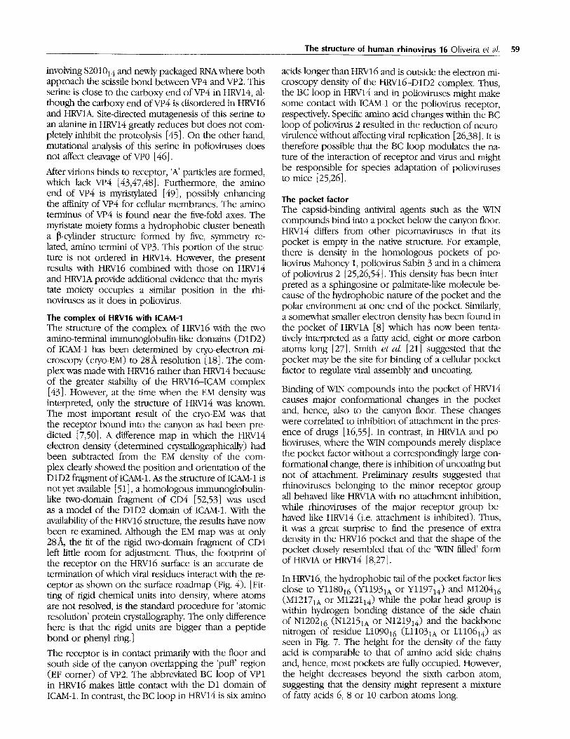
The pocket factor The capsid-binding antiviral agents such as the WIN compounds bind into a pocket below the canyon floor. HRV14 differs from other picomaviruses in that its pocket is empty in the native structure. For example, there is density in the homologous pockets of po- liovirus Mahoney 1, poliovirus Sabin 3 and in a chimera of poliovirus 2 [25,26,54]. This density has been inter- preted as a sphingosine or palmitate-like molecule be- cause of the hydrophobic nature of the pocket and the polar environment at one end of the pocket. Similarly, a somewhat smaller electron density has been found in the pocket of HRV1A [8] which has now been tenta- tively interpreted as a fatty acid, eight or more carbon atoms long [27]. Smith et al. [21] suggested that the pocket may be the site for binding of a cellular pocket factor to regulate viral assembly and uncoating.
Binding of WIN compounds into the pocket of HRV14 causes major conformational changes in the pocket and, hence, also to the canyon floor. These changes were correlated to inhibition of attachment in the pres- ence of drugs [16,55]. In contrast, in HRVlA and po- lioviruses, where the WIN compounds merely displace the pocket factor without a correspondingly large con- formational change, there is inhibition of uncoating but not of attachment. Preliminary results suggested that rhinoviruses belonging to the minor receptor group all behaved like HRVIA with no attachment inhibition, while rhinoviruses of the major receptor group be- haved like HRV14 (i.e. attachment is inhibited). Thus, it was a great surprise to find the presence of extra density in the HRV16 pocket and that the shape of the pocket closely resembled that of the 'WIN filled' form of HRVIA or HRV14 [8,27].
In HRV16, the hydrophobic tail of the pocket factor lies close to Yl18016 (Y11931A or Yl19714) and M120416 (M12171A or M122114) while the polar head group is within hydrogen bonding distance of the side chain of N120216 (N12151A or N121914) and the backbone nitrogen of residue L109016 (L11031A or Ll10614) as seen in Fig. 7. The height for the density of the fatty acid is comparable to that of amino acid side chains and, hence, most pockets are fully occupied. However, the height decreases beyond the sixth carbon atom, suggesting that the density might represent a mixture of fatty acids 6, 8 or 10 carbon atoms long.

60 Structure 1993, Vol 1 No 1
(a)
(b) ~ 1122
~1238~1098 1181
... 1122
~123~1098 1181
Fig. 7. Stereo views of the putative fatty acid in the hydrophobic interior of VP1 shown in electron density in (a) and with labels on the surrounding protein side chains in (b). The view is from out- side the virus and similar to that shown in Figs. 1 and 4.
Tablg 4. Binding site of WIN 56291.
HRV14 HRV1A HRVt6
Residue Drug moiety Residue Drug moiety Residue Drug moiety
11104 chlorine, isoxazole 11101 phenyl 11098 aliphatic, isoxazole,
chlorine
L1106 isoxazole Ll103 isoxazole Ll100 isoxazole
Yl128 chlorine, a[iphatic 11125 phenyl 11122 chlorine
11130 - - Ll127 oxazoline Ml124 - -
Al150 oxazoline Yl145 oxazoline Y1142 - -
Yl152 phenyl Yl147 chlorine Yl144 phenyl, chlorine,
isoxazole P1174 oxazoline Ml169 - - Al166 - -
Fl186 oxazoline Fl182 Fl179 phenyl, oxazoline
Vl188 chlorine 11184 - - Ll181 phenyl, chlorine
Vl191 aliphatic Ll187 chlorine Ll184 chlorine Yl197 - - Y ! !93 - - Y 1190 i~,q~n 7nt,~
Cl199 - - Ml195 isoxazole Ml192
M1221 isoxazole M1217 isoxazole M1214 isoxazole
M1224 chlorine, phenyl 11220 L1217 oxazoline
A3024 chlorine A3024 A3024 - -
Structurally equivalent residues have been aligned.
Binding of WIN 56291 to HRV16 The orientation of WIN56291 in the drug binding pocket was determined from bulges in the electron
density corresponding to the chlorine atoms on the phenyl ring (Fig. 8a,b). The phenyl and oxazoline end of the drug bind to the inner portions of the pocket

The structure of human rhinovirus 16 Oliveira et al. 61
consistent with the orientation of the same compound in HRV14 and HRV1A [8,27]. The flattened shape of the electron density suggests that the phenyl and oxa- zoline tings are coplanar and similar to the structures in HRV14 and HRVIA. The isoxazole ring is close to the entrance of the pocket but the orientation of its plane cannot be determined with certainty at present because of the poor difference density caused by displacement of the fatty acid that overlaps this portion of the WIN compound. Details of the interaction of WIN 56291 and the surrounding protein in HRV14, HRVIA and HRV16 are compared in Table 4 and the difference in position of the compound in the pocket is shown in Fig. 8c.
In the structure of HRV14, the aliphatic chain of WIN56291 bends around Yl12814 (II1251A or 1112216). In the structures of both HRV16 and HRVIA, with an isoleucine in this position, the chain can take on a more extended conformation. Another residue which may contribute to the different position of the drug in the pockets of HRV16 and HRVIA relative
to HRV14 is M122414 (I1220 in HRVIA or L1217 in HRV16). In both the HRVIA and HRV16 structures, the side chains of the equivalent residues occupy roughly the same position but fill more of the volume of the pocket; thereby reducing the pocket length. A third position which affects the site of drug binding in the pocket is residue Al16616 (M11691A or Pl17414). The corresponding bulky methionine shortens the pocket of HRV1A. These residues are probably responsible for the differences in drug binding to HRV16 and HRVIA relative to HRV14 and may also be involved in differen- tiation between the A and B drug-binding groups [9].
In HRVIA and HRV16, the more active antiviral com- pounds tend to have an aliphatic chain less than or equal to five carbon atoms long [56] corre- lating with the available space within the binding pocket [27,57,58]. In HRV14, the most active antiviral agents tend to be longer with seven-carbon aliphatic chains (Table 2). For example, WIN 56291 contains an aliphatic chain of only three carbons and is equally active against HRV16 and HRVIA but less active against
m •
(b)
(c
1100
%
y ~ . _ 11479 4
1184
- ~ ~7 1179
~ i 1 0 |
Fig. 8. (a) Difference electron density (positive contours only) where WIN 56291 binds, displacing the putative fatty acid molecule (thin line). (b) WIN 56291 in the HRV16 environment. (c) Comparison of WIN 56291 in HRV14 (wavy line), HRVIA (thin line) and HRV16 (thick line). The view is from outside the virus, similar to that shown in Figs. 1 and 4.

62 Structure 3993, Vol "1 No "1
HRV14. Thus, there is an optimal drug size which dis- plays the greatest activity and binding affinity [57,58] for each serotype and best fills the volume of the pocket. It follows that the smaller pocket factors, which can be easily displaced by VAN compounds in HRV16 and HRVIA, bind with less affinity than the antiviral compounds. Nevertheless, the pocket factors seen in the electron densities remain in the pocket even af- ter extensive dialysis of the virus sample. The VAN compounds have a binding constant comparable to their minimal inhibitory concentrations of ,-, 10- 8 M [59,60].
Discussion The role of the pocket factor The natural pocket factor found in HRV16 and HRV1A (as well as polioviruses), like VAN compounds, in- creases the thermal stability of the virus [17] by filling an internal hydrophobic cavity [61,62]. The pocket fac- tor may, therefore, be required to stabilize the virus in transit from one cell to another. However, the delivery of the infectious RNA into the cytoplasm must require a destabilizing step which might be effected by expulsion of the pocket factor during receptor-mediated uncoat- ing. There is evidence that ICAM-1 binds to the canyon only when the pocket factor has been displaced. In the case of HRV14, for example, attachment is inhibited by the presence of VAN compounds and inhibition of binding is correlated with distortion of the canyon floor (the roof of the pocket) by drug compounds (Fig. 9). These observations are also supported by site-directed mutations [15] in the deformable part of the canyon floor. In essence, there are two competing equilibria: the binding of ICAM-1 and the binding of the pocket factor to the virus. Although the sites of binding of ICAM-1 and of the pocket factor are not the same, they are in close proximity and interfere with each other. Since ICAM-1 binds to HRV14 and to HRV16, the shape of the canyon for HRV16 should be similar to that in HRV14 when ICAM-1 binding occurs. Hence, we propose that the pocket must be empty when ICAM-1 binds to HRV16 implying that the pocket factor must be displaced before the receptor can seat itself into the canyon. However, displacement of the pocket fac- tors p e r se does not cause the virus to fall apart. For instance, when HRV14 is crystallized it does not con- tain a pocket factor and the complex of HRV16 with ICAM-1 is reasonably stable. Presumably the absence of pocket factor increases the potential for disruption by, for instance, lowered pH or in cases like HRV14, by formation of the receptor-virus complex.
It has long been known that attachment of poliovirus to cells generates a major conformational change re- suiting in the immunogenically different 'A' particles, with the loss of VP4 and eventually of RNA [47]. These phenomena can be mimicked by incubating rhinovirus with soluble ICAM-1 or lowering the pH [43]. Presum-
ably this destabilization of the virus is made possible by the displacement of a sufficient number of pocket factors when the receptor competes for the overlap- ping binding site. Progressive recruitment of recep- tors is then sufficient to trigger release of the VP4s. The terminal myristate moieties of VP4 will then per- mit entry through the cell membrane, possibly by cre- ating a channel along the five-fold axes of the virus [20]. However, picornaviruses differ in stability. This may explain why the relatively more stable receptor- saturated HRV16 may require some additional stimulus, such as a decrease in pH, to trigger viral dissociation.
The current results on HRV16, taken together with the earlier reports for HRV1A [8,27] and similar results for poliovirus (J Hogle, personal communication) all show that certain antiviral compounds can displace the pocket factor. The natural pocket factor, found in HRV16, is small and has a limited number of interac- tions in the drug-binding pocket relative to WIN com- pounds. This is consistent with the observation that WIN compounds can displace the pocket factor. The relative affinities of the VAN compound for the pocket and of ICAM-1 for the receptor-binding site will deter- mine the inhibitory properties of the drug for a partic- ular rhinovirus serotype (Fig. 9).
A class of HRV14 drug-resistant (compensation) mu- tants can be selected by growing the virus in the pres- ence of antiviral VAN compounds. Such mutants occur at a frequency of about one per 104 virions. They have been shown to be mostly single mutations [17,24] and six of the seven characterized to date are situated near the walls and floor of the canyon. VAN compounds bind into the pocket of these mutant viruses and de- form the canyon floor in a similar manner to their effect on wild type viruses (M Otiveira, I Minor, RR Rueckert & MG Rossmann, unpublished data). In one of these mutants (S1223~G) the affinity of ICAM-1 for the virus is enhanced (MP Fox, DC Pevear & FJ Dutko, unpub- lished data). Thus, it is reasonable to conclude that ICAM-1 binds better to these mutant viruses than the VAN compounds (Fig. 9b).
The major structural changes induced in HRV14 by drugs are similar but differ in detail, depending on the type of compound bound [22,27]. Shorter inac- tive fragments of VAN compounds have also been shown to bind to HRV14 and these cause conforma- tional changes which are similar to those induced by the active compounds (J Bibler, MG Rossmann, DC Pevear, & GD Diana, unpublished data). Thus, the small compounds in HRV14 that produce no inhibition of attachment and that can be displaced by larger VAN compounds mimic the natural pocket factor in HRV16 and HRVIA to a certain extent (Fig. 9c).
In the case of poliovirus or HRVIA (a minor rhinovirus serotype), only uncoating is inhibited by VAN com- pounds and not attachment. Nevertheless, if the pocket factor needs to be absent for the virus to uncoat, pre- sumably binding of receptor to these viruses leads to
The structure of human rhinovirus 16 Ol iveira eta/. 63
(a) H RV14
Pocket factor
I
WiN
4
...... •
(Wl N>ICAM>>PF)
(b) HRV14 mutant
q
i
!
(ICAM>WIN>>PF)
(c) H RV16
...... •
(WIN>ICAM>PF)
displacement of the pocket factor, just as is the case for the major rhinovimses. Similarly, the WIN compounds must also be displaced by the receptor as there is no inhibition of attachment. However, the remaining WIN compounds must stabilize the virus sufficiently to in- hibit uncoating.
Biological impl ica t ions A virus needs to be stable and relatively inert dur- ing its transit from host to host or from cell to cell. However, once the virus has reached an appropri- ate target, it needs to become unstable, shedding its protein coat to allow infection. In rhinoviruses and polioviruses, this need for reversible stabi- lization appears to be fulfilled by the binding of a small cellular aliphatic molecule, called the 'pocket factor', into a hydrophobic pocket in VP1. In the major group of rhinovirus serotypes, the binding site for ICAM-1, the virus receptor, over- laps with the binding site of the stabilizing pocket
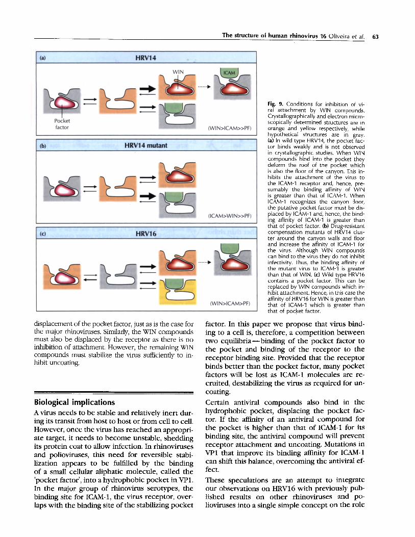
Fig. 9. Conditions for inhibition of vi- ral attachment by WIN compounds. Crystallographically and electron micro- scopically determined structures are in orange and yellow respectively, while hypothetical structures are in gray. (a) In wild type HRvq4, the pocket fac- tor binds weakly and is not observed in crystallographic studies. When WIN compounds bind into the pocket they deform the roof of the pocket which is also the floor of the canyon. This in- hibits the attachment of the virus to the ICAM-q receptor and, hence, pre- sumably the binding affinity of WIN is greater than that of ICAM-1. When ICAM-1 recognizes the canyon floor, the putative pocket factor must be dis- placed by ICAM-1 and, hence, the bind- ing affinity of ICAM-1 is greater than that of pocket factor. (b) Drug-resistant compensation mutants of HRV14 clus- ter around the canyon walls and floor and increase the affinity of ICAM-1 for the virus. Although WIN compounds can bind to the virus they do not inhibit infectivity. Thus, the binding affinity of the mutant virus to ICAM-1 is greater than that of WIN. (c) Wild type HRV16 contains a pocket factor. This can be replaced by WIN compounds which in- hibit attachment. Hence, in this case the affinity of HRV16 for WIN is greater than that of ICAM-1 which is greater than that of pocket factor.
factor. In this paper we propose that virus bind- ing to a ceil is, therefore, a competition between two equilibria--binding of the pocket factor to the pocket and binding of the receptor to the receptor binding site. Provided that the receptor binds better than the pocket factor, many pocket factors will be lost as ICAM-1 molecules are re- cruited, destabilizing the virus as required for un- coating.
Certain antiviral compounds also bind in the hydrophobic pocket, displacing the pocket fac- tor. If the affinity of an antiviral compound for the pocket is higher than that of ICAM-1 for its binding site, the antiviral compound will prevent receptor attachment and uncoating. Mutations in VP1 that improve its binding affinity for ICAM.1 can shift this balance, overcoming the antiviral ef- fect.
These speculations are an attempt to integrate our observations on HRV16 with previously pub- lished results on other rhinoviruses and po- lioviruses into a single simple concept on the role

64 Structure 1993, Vo l 1 N o 1
Table 5. Diffraction data collection.
(a) Native HRV16 data*
Resolution (,~) 00-25 25-12.5 12.5-8.33 8.33-6.25 6.25-5.0 5.0-4.17 4.17-3.57 3.57-3.13 3.13-2.28
Number of reflections 462 8635 24056 43693 71 073 106002 136879 152868 101 987 % of Data 78 87 89 84 82 83 76 65 33 Rmerg e 11.0 9.5 8.8 11.0 10.2 9.2 11.2 14.0 18.2
(b) WIN 56291 complexed with HRV161
Resolution (,g,) 30-15 15-10 10-8.5 8.5-7.5 7.5-5.5 5.5-5.0 5.0-4.5 4.5-3.5 3.5-3.0 3.0-2.75
Number of reflections 135 602 596 760 3674 1959 2912 10888 9401 4084 % of Data 2 4 4 5 5 4 4 4 3 2
*Total number of useful films = 225. tTotal number of useful imaging plates = 4. Number of crystals = 81 (1). Number of measurements = 1 332 460 (45 240). Cutoff criterion = F h 2 > 3c~ (F 2/" Number of unique reflections =645 656 (35 011). Reflections were added in abutting films. Overall Rrnerg e = 11.1 % (9.9 %),
~i IIFh a - F2)I x 100. [Data for part (b) given in parentheses after data for (a).] where Rmerge = ~ ~ F 2 h i h
of the pocket factor. Cellular proteins use cofac- tors to regulate their functions in analogous ways. Thus, it seems likely that viruses other than rhino- and enteroviruses might use reversible stabiliza- tion by a pocket factor to regulate their proper- ties at different stages o f their life cycle. The con- cept that the pocket factor plays a crucial role in the rhinovirus life cycle n o w needs to be directly tested.
Escherichia coli and cloned into the pMJ3 vector using methods described previously [45]. A nest of plasmids, prepared by delet- ing successive segments of P1 from the 3' end to the 5' end with exonuclease III, was then sequenced by primer extension using a primer purchased from New England Biolab and Sequenase (a modified T7 DNA polymerase from United States Biochemi- cal Corp., Cleveland, Ohio). Initial sequence alignments were carried out by computer using a package of programs from the University of Wisconsin Genetics Computer Group. Subse- quently these were slightly modified by the results of the three- dimensional superposition of the HRV16, HRVIA and HRV14 structures.
Mater ia ls and m e t h o d s
Tissue culture and virus growth Rhinovirus 16 was propagated in HeLa cells. These cells were grown in GIBCO S-MEM media with the addition of 0.5 % of penicillin G and streptomycin sulfate supplemented with 10 % calf serum. The optimum pH used for the growth of these cells was 7.25-7.30. HeLa cells, grown to a cell density of 5-6 x 10⁵ per ml, were centrifuged at 600 g for 10 minutes. After centrifu- gation, the pelleted cells were resuspended in 10 ml of G1BCO MEM media. The cells were infected with 10 plaque-forming units per cell [63]. The inoculated cells were kept at room tem perature for 30 min, with slow agitation every three min, during which time the virus attached to cells. The sample was diluted by a factor of ten in an Erlenmeyer flask, sealed and put into a shaking water bath at 34.5 ° C for virus propagation. The infected cells were harvested after seven to eight hours. Subsequently, it was found that by using HeLa cells grown in monolayers the yields of purified HRV16 increased from about 0.2 mg to about 1 mg of virus for 3.3 x 10⁹ ( ~ 2 x 10⁴ virions per cell) infected cells.
Determination of nucleotide sequence of coat g e n e for HRV16 and a m i n o acid sequence alignments with other rhinoviral coat proteins cDNA containing the coat (P1) region of HRV16 was copied from RNA of purified virions using AMV reverse transcriptase; the DNA duplex was then generated using polymerase I from
Antiviral assays The antiviral activity of the compounds was determined in a high capacity tissue culture infectious dose assay. Confluent monolay- ers of HeIa cells in 96 well plates were infected with a suMcient amount of virus to produce 85 100 % cytopathic effect (CPE) after incubation at 33°C for 3 days. A no-vires control column was included for each plate. After a one hour adsorption period, serial two-fold dilutions of the compound in M199 medium with 5 % fetal calf serum and 0.25 % DMSO were added to the plate in quadruplicate. The cells were incubated at 33 ° C in a humidified atmosphere with 2 % CO 2 for 3 days, fixed with a 5 % solution of glutaraldehyde and stained with a solution of 0.25 % crystal vi olet. The plates were rinsed and dried and the optical density of each well was read at 570 nm. The concentration of compound which protected 50 % of the cell monolayer from virus-induced CPE was interpolated from the optical density readings and ex- pressed as the minimal inhibitory concentration.
Table 6. Space groups for some rhino- and polioviruses.
a b c Space Virions per Crystal (A) (,a,) (,&) group unit cell
HRV14 cubic 445.18 445.18 445.18 P213 4 HRV14 orthorhombic 323.0 358.0 380.0 P21212 2 Poliovirus 1 Mahoney 322.9 358.0 380.2 P21212 2 HRV16 362.6 347 .1 334.9 P22121 2

The structure of human rhinovirus 16 Oliveira et a/. 65
b b
. . . . • . . . . . . . . ! "~ 7" ; - - .-:~+-:., J " ' : : ' - ~ , ~ " - . . , " : 7
: ..... .-,-~~:i:i:: ::!:i:i:-:-:. I.. ~ ,~.. -"~ ....... ':~.::~.:", ..... . . . . . . . . . . . "/ ~ ' ~ '~....-" .7-.,: ,~ ":~..~.:.. }.c:~.'-,.',:?~?~.i~:l. ~ Q" ,:-.:....:.)~:<. ~ " . , . : < . . ) .....
2:.'::.,~- ~?:~'.ii'~:!;:'~:~.(~, :'i:;::75:< Y::~':lS" "~: ~ : " ~' ".:.!.:::..":):::i::}.~'~.. ...5~:~:i:'.}!:}::i.: ,': ~:;:. .....
a
la
Fig. 10. The × = 72°plane of the self-rotation function (left) and its interpretation (right). Shown also at the bottom is the orientation of a standard icosahedron, The icosahedron with black symmetry elements corresponds to the reference orientation given by expression (1).
Virus purification and crystallization The harvested cells were frozen and thawed three times, homog enized and centrifuged at 10000 rpm for 10min. The su- pernatant was treated with DNAse (0.05mgm1-1) and RNAse (0.75mgm1-1) for 30min at room temperature, followed by trypsin treatment (0.5 mgm1-1) for 10rain at 37°C. Sarcosine (0.1%) (N-methyl glycine) was then added and the pH adjusted to 7.0 with ammonium hydroxide. The sample was clarified with a slow centrifugation step at 600 g for 10 min. The supernatant was then centrifuged at 45 000 rpm for two hours, through a 30 % sucrose cushion. The virus formed translucent pellets that were solubilized in 0.25M HEPES buffer, pH7.5, with 0.25M NaC1. The sample was loaded onto a sucrose gradient (7.5-45 %) and centrifuged for 90 min at 36 000 rpm. The virus bands were collected and diluted in buffer and pelleted by centrifuging for 90min at 45 000 rpm. The virus pellets were solubilized in buffer and the concentration was checked by measuring the absorbance at wavelengths of 260, 280 and 310 nm. Growth of the virus in monolayer gave a more uniform virus production than did suspension culture. The quality of the purification was determined by silver stained SDS-PAGE and negative stain elec- tron microscopy.
The hanging drop vapor diffusion method was employed in the crystallization of HRV16. The reservoir solution (0.5 ml in vol ume) contained PEG 8000 (0.5~1.5 %) in buffer. A 5 ~tl drop of virus solution, concentrated to 8-10mgm1-1, was diluted with 5 I, tl of reservoir solution. The drop was placed on a plastic coverslip which was used to seal the well. Conditions for crys-
tallization varied with respect to CaC12 concentration present in the well solution (5-20 mM). A key factor in the crystallization of HRV16 was the use of NaC1 in the buffer.
The antiviral agent WIN 56291 (Table 2) was soaked into grown crystals using the procedure described by Smith et al. [21].
Diffraction data collection and processing All diffraction data were collected by oscillation photography at the Cornell High Energy Synchrotron Source (CHESS). The high intensity of X-rays and the short wavelength (0.91 ,~,) made it possible to use small crystals (0.2 mm) and to collect more than one exposure per crystal. Exposure times were between 15 and 60 seconds. The native data were recorded on Kodak DEF X-ray film while the drug-soaked crystal data were collected on Fuji imaging plates. The crystat-to-fihn distance was 170 mm and the crystal-to-imaging plate distance was 270 mm. The films (5" x 5") were scanned on a 50 ~t raster using an Optronics 1 000 scanner and the imaging plates (8" x 10") were scanned on a 100 ~t raster using a Fuji BAS 3000 scanner. Based on es- timated cell dimensions obtained from oscillation photographs and the empirical observation of lunes, oscillation angles of 0.3 ° or 0.4 ° were chosen to avoid overlap of reflections. The crys- tals were in random orientation and the diffraction pattems ex- tended to at least 2.8& resolution.
The films and image plates were indexed using the auto-indexing algorithm developed by Kim [64]. The cell parameters derived from different crystals were averaged and used in obtaining in- tegrated intensities [65]. Post-refinement [66] was used to re-

66 Structure 1993, Vol 1 No 1
0.40
• ~ 0.35
0
0.30
"~ 0.25
0 L,) 0.20
I
0,15 I I i I p IN I i I I I r I T I i I i 0.20 0.21 0.22 0.23 0.24 0.25 0.26 0.27 0.28 0.29 0.30
x
Fig. 11. Correlation coefficients between observed and cal- culated structure factors as the particle is moved along a two-fold axis parallel to a. The position x along a is shown in fractional coordinates.
Correlation coefficient = ~h (<Fo>-Fo)(<Fc>-Fc) { ~ "11/2
(<Fo >- Fo) 2 ~(< Fc>- Fc) 2 )
where < F o > and < F c > are the mean structure factors within each resolution range.
1.0 [
).5
cyl 15
~ c cyl 69 yl 0 cy117~ =
cy120£
o
Resolution in A
0.020.01 I0.01 6.71 5.01 4fO 33
0.00 0.05 0.I0 0.15 0.20 0.25 0.30
lid ( : , q )
Fig. 12. Correlation coefficient improvement during phase ex- tension from 8A to 3.4~, resolution using 30-fold non-crystal- lographic real space electron density averaging. Cycle 0 corre- sponds to correlation coefficients calculated with structure fac- tors derived from an HRV14 model to 8A resolution. Cycle 15 shows the improvement after 15 cycles of molecular replacement averaging at 8A resolution. Cycles 69, 175 and 209 show the pro- gressive improvement of the correlation coefficients during phase extension to 7.0, 4.5 and 3.4 A resolution.
fine cell dimensions, crystal orientations and crystal mosaicity. The wavelength used was adjusted relative to the known cell di- mensions of HRV14 determined from a CuKa source. The X-ray wavelength for each of the four data trips was very slightly ad- justed during post-refinement of the complete data set to main tain the same cell dimensions, a = 362.6, b = 347.1, c = 334.9~ cz = 90.0, [3 = 90.0, 7 = 90.0°. The percentage of data with respect to resolution and final internal agreement is given in Table 5.
Structure determination The reciprocal lattice was found to have mmm symmetry. This, together with the shape of the observed unit cell, showed that the crystals were orthorhombic. Indexing of the reflections was possible using a primitive lattice with axes perpendicular to the reciprocal lattice mirror planes. However, there were insufficient data to determine the presence or absence of screw axes parallel to the principal axial directions. Assuming two particles per unit cell, the Matthews coefficient [67] was 2.53`3 per dalton. No other assumption seemed reasonable. Thus, there must be at least one direction with a two-fold axis. tater rotation function results showed that there was an icosahedral two-fold axis paral- lel to the a direction, suggesting space group P222, P2212 , P2221 or P22121. However, only space group P22121 permitted close packing of the spherical virus particles in the cell. Similar, but not identical, particle organizations have been found in crystals of HRV14 [68] and poliovirus Mahoney 1 [54] (Table 6). The orientation of the icosahedra about the a axes was determined by the use of a locked rotation function [69,70] using a data set with 64 % of the theoretically possible reflections between 15 and 83, resolution and an integration radius of 1503`. If the standard orientation of an icosahedron is defined as shown in Fig. 10 with respect to the 'b-cell' [71], then the matrix [P] re- lates this standard to the reference particle in the HRV16 crystal unit 'p-cell' such that
x = [pig
where X and Y are Cartesian coordinate systems in the h and p-cells, respectively. The locked rotation function showed that:
1.0 0.0 0.0 /
[P] = 0.0 cosO - s i nO where 0 = 86.25 ° . (1)
0.0 sinO c o s O /
This result was checked with a cross-rotation function [72] by comparing the known HRV16 structure in the />cell (a = b = c = 4003`, cz = [3 = 7 = 90°) with the unknown p-cell.
The structure of HRV14 was used both in determining the trans- lation of the particle along the a axis and as a model for de- termining the initial set of phases, although the structure and sequence of HRV16 were later found to be closer to HRVIA than HRV14. Structure factors were calculated between 10 and 8.5A resolution for the p-cell when the known HRV14 model had been initially oriented and placed into the p-cell [73]. The particle was placed at successive positions along the two-fold axis parallel to a. The best position given by the maximum correlation coefficient was found to be at x = 0.250 (Fig. 11).
The phases of the calculated structure factors were combined with the observed structure factors and used to compute an electron density map of HRV16 at 8.0 3` resolution. This map was then averaged and solvent flattened using a spherical mask with outer radius of 1653` and inner radius of 703` [71]. Where there were overlaps between particles, a tangential plane was used to separate them. The electron density in the asymmetric unit

was sampled at grid points separated by 1/3 of the resolution. After 14 cycles (see, for instance, [74]) of averaging using data between 15-8.0.& resolution, the overall correlation coefficient improved from 0.45-0.81 (Fig. 12). Phase extension proceeded by one reciprocal lattice unit at a time until 3 .4~ resolution had been attained after 209 cycles of averaging (Fig. 12). A map was then calculated with terms extending to 3.5 A resolution. Terms between 3.5 and 3.4A were omitted because of the necessarily poor phasing in the most external resolution shell.
C o m p l e x b e t w e e n HRV16 and W l N 56291
The position and conformation of the antiviral compound WIN 56291 b o u n d to the HRV16 structure was determined using difference maps with coefficients
(F D - - kFN) W N exp( ia N)
where F D and F N are the structure amplitudes of the virus--drug complex and of the native data, respectively; a N are the phases of the native structure obtained from non-crystallographic sym metry averaging; and w N are the weights dependen t on the esti- mated phase error. Simple difference maps (k = 1) were used to determine the position of the compounds in the virus structure. An atomic model was built into a map where the coefficient k was chosen to be 0.65 such that the electron densities of t h e drug and of the protein were of roughly equal height to minimize the effects of incomplete substitution and phase bias towards the native structure [21].
Acknowledgment~ We are grateful for help from Mavis Agbandje, Jodi Bibler, Michael Chapman, Hok-Kin Choi, Walter Keller, Kyung Kim, Prasanna Kolatkar, Robert McKenna, Andrew Prongay, Bill Wikoff, Hao Wu and Di Xia in the many synchrotron data collection trips. We also appreciate help from Grazyna Apostol for sample preparation. We have greatly profited by discussions with Tom Smith, Michael Chapman, Prasanna Kolatkar and Kyung Kim. We much appreciate the preparation of Figs. 4a and 4b by Jean Yves Sgro of the Univer- sity of Wisconsin in Madison. We thank Helene Prongay and Sharon Wilder for help in preparation of this manuscript. Finally, we would like to express our appreciation to Rebecca Ward for many helpful improvements to the manuscript. The work was supported by Na- tional Institutes of Health grants AI 31960 to RRR and AI 11219 to MGR. It was also supported by a Sterling Winthrop Pharmaceuticals Research Division grant to MGR as well as a research award from t h e Lucille P Markey Foundation for strengthening structural studies at Purdue University,
References 1. Rueckert, R.R. (1990). Picomaviridae and their replication. In
Virology. (Fields, B.N. & Knipe, D.M., eds), pp. 507-548, Raven Press, New York.
2. Abraham, G. & Colonno, R.J. (1984). Many rhinovirus serotypes share the same cellular receptor, a~ Virol. 51, 340-345.
3. Uncapher, C.R., DeWitt, C.M. & Colonno, R.J. (1991). The major and minor group receptor families contain all but one human rhinovirus serotype. Virology, 180, 814-817.
4. Tomassini, J.E. & Colonno, RJ. (1986). Isolation of a receptor protein involved in attachment of human rhinoviruses. J. Virol. 58, 290-295.
5. Greve, J.M., et al., & McClelland, A~ (1989). The major human rhinovirus receptor is ICAM-1. Cell, 56, 839-847.
6. Staunton, D.E., Merluzzi, V.J., Rothlein, R., Barton, R., Marling, S.D. & Springer, T~A. (1989). A cell adhesion molecule, ICAM 1, is the major surface receptor for rhinoviruses. Cell, 56, 849-853.
7. Rossmann, M.G, et al., & Vriend, G. (1985). Structure of a human common cold virus and functional relationship to other picomaviruses. Nature, 317, 145-153.
8. ICun, S., et al., & McKinlay, M.A~ (1989). The crystal structure of human rhinovirus serotype 1A (HRVIA). J. Mol. Biol. 210, 91-111.
The structure of human rhinovirus 16 Oliveira et a/. 67
9. Andries, K., et al., & Janssen, PAJ. (1990). Two groups of rhi- novimses revealed by a panel of antiviral compounds present sequence divergence and differential pathogenicity. J. Virol. 64, 1117-1123.
10. Rossmann, M.G. & Johnson, J.E. (1989). Icosahedral RNA vires structure. Annu.. Rev. Biochem. 58, 533-573.
11. Harrison, S.C.. (1992). Viruses: Curr. Opin. Struct. Biol. 2, 293-299.
12. Sherry, B. & Rueckert, R. (1985). Evidence for at least two dominant neutralization antigens on human rhinovirus 14. aT. Virol. 53, 137-143.
13. Sherry, B., Mosser, A.G., Colonno, R.J. & Rueckert, R.R. (1986). Use of monoclonal antibodies to identify four neutralization im munogens on a common cold picornavirus, human rhinovirus 14. J. ViroL 57, 246-257.
14. Rossmann, M.G. & Palmenberg, A.C. (1988). Conservation of the putative receptor attachment site in picornaviruses. Virol- ogy, 164, 373-382.
15. Colonno, RJ., Condra, J.H., Mizutani, S., Callahan, P.L., Davies, M.E. & Murcko, M.A. (1988). Evidence for the direct involve ment of the rhinovirus canyon in receptor binding. Proc. Natl. Acad ScL USA, 85, 5449-5453.
16. Pevear, D.C., .et al., & Dutko, FJ. (1989). Conformational change in the floor of the human rhinovirus canyon blocks adsorption to HeLa cell receptors. J. virol. 63, 2002-2007.
17. Heinz, B.A., Shepard, D.A. & Rueckert, R.R. (1990). Escape mutant analysis of a drug-binding site can be used to map functions in the rhinovirus capsid. In Use of X-ray Crystallog- raphy in the Design of dntiviral Agents. (Laver, W.G. & Air, G.M., eds), pp. 173-186, Academic Press, San Diego.
18. Olson, N.H., et aL, & Rossmann, M.G. (1993). Structure of a human rhinovirus complexed with its receptor molecule. Proc Natl. Acad Sci. USA, 90, 507-511.
19. Palmenberg, A.C. (1989). Sequence alignments of picornaviral capsid proteins. In Molecular Aspects of Picornavirus Infection and Detection. (Semler, B.L. & Ehrenfeld, E., eds), pp. 211-241, American Society for Microbiology, Washington, D.C.
20. Giranda, V.L., et al., & Rueckert, R.R. (1992). Acid induced structural changes in human rhinovirus 14: possible role in uncoating. Proc. Natl. Acad Sci. USA, 89, 10213-10217.
21. Smith, TJ., et al., & Otto, M.J. (1986). The site of attachment in human rhinovirus 14 for antiviral agents that inhibit uncoating. Science, 233, 1286-1293.
22. Badger, J., Minor, I., Oliveira, M.A., Smith, TO. & Rossmann, M.G. (1989). Structural analysis of antiviral agents that interact with the capsid of human rhinoviruses. Proteins, 6, 1-19.
23. Zhang, A., Nanni, R.G., Oren, D.A., Rozhon, E.J. & Arnold, E. (1992). Three-dimensional structure-activity relationships for antiviral agents that interact with picornavirus capsids. Semin. Virol. 3, 453-471.
24. Shepard, D.A., Heinz, B.A. & Rueckert, R.R. (1993). WIN com- pounds inhibit both attachment and eclipse of human rhi- novirus 14. J. virol. 67, 2245-2254.
25. Fihnan, D.J., Syed, R., Chow, M., Macadam, AJ., Minor, P.D. & Hogle, J.M. (1989). Structural factors that control conforma tional transitions and serotype specificity in type 3 poliovirus. EMBO J, 8, 1567-1579.
26. Yeates, T.O., et al., & Hogle, J.M. (1991). Three-dimensional structure of a mouse-adapted type 2/type 1 poliovirus chimera. EMBO J. 10, 2331-2341.
27. Kim, K.H., et al., & Pevear, D.C. (1993). A comparison of the anti-rhinoviral drug binding pocket in HRV14 and HRVIA. J~ Mol. Biol. 230, 20~225.
28. Lemanske, R.F. Jr., Dick, E.C., Swenson, C.A., Vrtis, R.F. & Busse, W.W. (1989). Rhinovirus upper respiratory infection increases airway hyperreactivity and late asthmatic reactions. J. Clin. In- vesL 83, 1-10.
29. Greve, J.M., et al., & McClelland, A. (1991). Mechanisms of receptor-mediated rhinovirus neutralization defined by two sol- uble forms of ICAM-1. J. Virol. 65, 6015~6023.
30. Jones, T.A. (1978). A graphics model building and refinement system for macromolecules. J. Appl. Crystallog~ 11, 268-272.
31. Jones, T.A., Zou, J.-Y., Cowan, S.W. & Kjeldgaard, M. (1991). Improved methods for building protein models in electron density maps and the location of errors in these models. Acta Crystallogr. A, 47, 110-119.

68 Structure 1993, Vol 1 No 1
32. Rossmann, M.G. & Argos, P. (1975). A comparison of the heme binding pocket in globins and cytochrome b 5. J: Biol. Chem. 250, 7525-7532.
33. Chothia, C. & Lesk, A.M. (1986). The relation between the divergence of sequence and structure in proteins. EMBO J. 5, 823426.
34. Page, G.S., Mosser, A.G., Hogle, J.M., Filman, D.J., Rueckert, R.R. & Chow, M. (1988). Three-dimensional structure of poliovirus serotype 1 neutralizing determinants../~ Virol. 62, 1781 1794.
35. Martin, A., Wychowski, C., Couderc, T., Crainic, R., Hogle, J. & Girard, M. (1988). Engineering a potiovirus type 2 antigenic site on a type 1 capsid results in a chimaeric virus which is neurovirulent for mice. ~ B O J. 7, 283%2847.
36. Minor, P.D, et al., & Almond, J.W. (1991). Antigenic struc- ture of chimeras of type 1 and type 3 polioviruses involving antigenic sites 2, 3 and 4. J. Gen. Virol. 72, 2475-2481.
37. Altmeyer, R., Murdin, A.D., Harber, J.J. & Wimmer, E. (1991). Construction and characterization of a poliovirus/rhinovirus antigenic hybrid. Virology, 184, 636~44.
38. La Monica, N., Kupsky, W.J. & Racaniello, V.R. (1987). Reduced mouse neurovirulence of potiovirus type 2 Lansing antigenic variants selected with monoclonal antibodies. Virology, 161, 429-437.
39. Chapman, M.S., Minor, I., Rossmann, M.G., Diana, G.D. & An dries, K. (1991). Human rhinovirus 14 complexed with antiviral compound R 61837. J. Mol. Biol. 217, 455-463.
40. Fricks, C.E. & Hogle, J.M. (1990). Cell-induced conformational change in poliovirus: externalization of the mnino terminus of VP1 is responsible for liposome binding. J. ViroL 64, 1934-1945.
41. Flore, O., Fricks, C.E., Filman, D.J. & Hogle, J.M. (1990). Con- formational changes in poliovirus assembly and cell entry. Semin. Virol., t, 429-438.
42. Strohmaier, K., Franze, R. & Adam, K.H. (1982). Location and characterization of the antigenic portion of the FMDV immu- nizing protein. J. Gen. ViroL 59, 295-306.
43. Hoover-Litty, H. & Greve, J.M. (1993). Formation of rhinovirus- soluble ICAM-1 complexes and conformational changes in the virion, f ViroL 67, 390-397.
44. Jaeobson, M.F. & Baltimore, D. (1968). Morphogenesis of po- liovirus. I. Association of the viral RNA with coat protein. J: Mol. Biol. 33, 369-378.
45. Lee, W.M., Monroe, S.S. & Rueckert, R.R. (1993). Role of matu ration cleavage in infectivity of picornaviruses: activation of an infectosome. J: ViroL 67, 2110-2122.
46. Harber, J.J., Bradley, J., Anderson, C.W. & Wimiiler, E. (1991). The catalysis of the poliovirus VP0 cleavage is not mediated by serine 10 of VP2. J: Virol. 65, 326-334.
47. Lonberg-Holm, K. & Noble-Harvey, J. (1973). Comparison of in vitro and cell-mediated alteration of a human rhinovirus and its inhibition by sodium dodecyl sulfate. J: Virol. 12, 819-826.
48. Kaplan, G., Freistadt, M.S. & Racaniello, V.R. (1990). Neutraliza- tion of poliovirus by cell receptors expressed in insect cells. J. Virol. 64, 4697-4702.
49. Chow, M., Newman, J.F.E., Filman, D., Hogle, J.M., Rowlands, D.J. & Brown, F. (1987). Myristylation of picornavirus cap- sid protein VP4 and its structural significance. Nature, 327, 482-486.
50. Giranda, V.L., Chapman, M.S. & Rossmann, M.G. (1990). Model- ing of the human intercellular adhesion molecule-I, the human rhinovirus major group receptor. Proteins, 7, 227-233.
51. Kolatkar, P.R., et al., & Olson, N.H. (1992). Preliminary X-ray crystallographic analysis of intercellular adhesion molecule-1. J. Mol. Biol. 225, 1127-1130.
52. Ryu, S.-E., et aL, & Hendrickson, W.A. (1990). Crystal struc- ture of an H1V-binding recombinant fragment of human CD4. Nature, 348, 419-425.
53. Wang, J., et al., & Harrison, S.C. (1990). Atomic structure of a fragment of human CD4 containing two immunoglobulin-like domains. Nature, 348, 411-418.
54. Hogle, J.M., Chow, M. & Filman, D J. (1985). Three-dimen- sional structure of poliovirus at 2.9A resolution. Science, 229, 1358-1365.
55. Heinz, B.A., et al., & Smith, T.J. (1989). Genetic and molecular analyses of spontaneous mutants of human rhinovirus 14 that are resistant to an antiviral compound..L ViroL 63, 2476-2485.
56. Mallamo, J.P, et al., & Rossmann, M.G. (1992). Conformation ally restricted analogues of disoxaril: a comparison of the ac- tivity against human rhinovirus types 14 and 1A. J. Med Chem. 35, 46904695.
57. Diana, G.D., Treasurywala, A.M., Bailey, T.R., Oglesby, R.C., Pe- vear, D.C. & Dutko, F.J. (1990). A model for compounds ac- tive against human rhinovims-14 based on X ray crystallography data. J. Med. Chem. 33, 1306-1311.
58. Diana, G.D., Kowalczyk, P., Treasurywala, A~M., Oglesby, R.C., Pevear, D.C. & Dutko, F.J. (1992). CoMFA analysis of the inter- actions of antipicornavims compounds in the binding pocket of human rhinovirus-14. J. Med. Chem. 35, 1002 1006.
59. Fox, M.P., Otto, M.J. & McKinlay, M.A. (1986). The prevention of rhinovims and poliovims uncoating by WIN 51711: a new antiviral drug. Antimicrob. Agents Chemother. 30, 110-116.
60. Fox, M.P., McKinlay, M.A., Diana, G.D. & Dutko, F.J. (1991). Binding affinities of structurally related human rhinovirus capsid-binding compounds are correlated to their activities against human rhinovims type 14. Antimicrob. Agents Chemc~ ther. 35, 1040-1047.
61. Eriksson, A.E., Baase, W.A., Wozniak, J.A. & Matthews, B.A. (1992). A cavity-containing mutant of T4 lysozyme is stabilized by buried benzene. Nature, 355, 371-373.
62. Eriksson, A.E., et aL, & Matthews, B.W. (1992). Response of a protein structure to cavity-creating mutations and its relation to the hydrophobic effect. Science, 255, 178-183.
63. Rueckert, R.R. & Pallansch, M.A. (1981). Preparation and char- acterization of encephalomyocarditis (EMC) virus. Meth. Enzo* moL 78, 315-325.
64. Kim, S. (1989). Auto indexing oscillation photographs. J. Appl Crystallogr. 22, 53~50.
65. Rossmann, M.G. (1979). Processing Oscillation diffraction data for very large unit cells with an automatic convolution tech nique and profile fitting. J. AppL Crystallogr 12, 225-238.
66. Rossmann, M.G., Leslie, A.G.W., Abdel-Meguid, S.S. & TsuMhara, T. (1979). Processing and post refinement of oscillation camera data. j~ AppL Crystallogr. 12, 570-581.
67. Matthews, B.W. (1968). Solvent content of protein crystals. J: MoL Biol. 33, 491497.
68. Erickson, J.W., Frankenberger, E.A., Rossmann, M.G., Fout, G.S., Medappa, K.C. & Rueckert, R.R. (1983). Crystallization of a common cold virus, human rhinovirus 14: 'isomorphism' with poliovirus crystals. Proc. Natl. Acad Sci. USA, 80, 931-934.
69. Rossmann, M.G., Ford, G.C., Watson, H.C. & Banaszak, L,J. (1972). Molecular symmetry of glyceraldehyde-3-phosphate de- hydrogenase, j~ Mol. Biol. 64, 237-249.
70. Tong, L. & Rossmann, M.G. (1990). The locked rotation ~nc- tion. Acta Crystallogr. A, 46, 783-792.
71. Rossmann, M.G., et al., & Lynch, R.E. (1992). Molecular replace- ment real-space averaging. L Appl. Crystallogr. 25, 166-180.
72. Rossmann, M.G. & Blow, D.M. (1962). The detection of sub- units within the crystallographic asymmetric unit. Acta Crystal- logr 15, 24-31.
73. Argos, P. & Rossmann, M.G. (1980). Molecular replacement method. In Theory and Practice of Direct Methods in Crys- tallography. (Ladd, M.F.C. & Palmer, R.A., eds), pp. 361-417, Plenum, New York.
74. Rossmann, M.G. (1990). The nlolecular replacement method. Acta Crystallogr. A, 46, 73-82.
Received: 12 July 1993; revised: 2 August 1993. Accepted: 3 August 1993.
Copyright © 2022 FDOKUMEN