Three-dimensional structure of human chromatin accessibility complex hCHRAC by electron microscopy
15
Three-dimensional structure of human chromatin accessibility complex hCHRAC by electron microscopy Minghui Hu, Yian-Biao Zhang, Luping Qian, Raymond P. Briñas, Larisa Kuznetsova, and James F. Hainfeld * Biology Department, Brookhaven National Laboratory, Upton, NY 11973, USA Abstract ATP-dependent chromatin remodeling complexes modulate the dynamic assembly and remodeling of chromatin involved in DNA transcription, replication, and repair. There is little structural detail known about these important multiple-subunit enzymes that catalyze chromatin remodeling processes. Here we report a three-dimensional structure of the human chromatin accessibility complex, hCHRAC, using single particle reconstruction by negative stain electron microscopy. This structure shows an asymmetric 15×10×12 nm disk shape with several lobes protruding out of its surfaces. Based on the factors of larger contact area, smaller steric hindrance, and direct involvement of hCHRAC in interactions with the nucleosome, we propose that four lobes on one side form a multiple-site contact surface 10 nm in diameter for nucleosome binding. This work provides the first determination of the three-dimensional structure of the ISWI family of chromatin remodeling complexes. Keywords hCHRAC; nucleosome; three-dimensional structure; electron microscopy; chromatin remodeling 1. Introduction Compaction of eukaryotic genomes into condensed chromatin fibers is required to fit over a meter of DNA within the limited volume of the nucleus (Horn and Peterson, 2002). The dynamic assembly and remodeling of chromatin involved in DNA transcription, replication, and repair is modulated by activities of remodeling complexes in living cells (Saha et al., 2006). These complexes are classified into several families, ISWI, RAD54, SWI/SNF, RSC, CHD, and INO80 (Eberharter and Becker, 2004; Lall, 2007; Lusser and Kadonaga, 2003). They share the presence of a motor subunit that belongs to the SWI2/SNF2 type of ATPase. The imitation-switch (ISWI) family of chromatin remodeling complexes also contains a non- catalytic binding protein from the BAZ/WAL family, and occasionally some other regulatory subunits. These ISWI-family chromatin remodeling complexes, such as the chromatin accessibility complex (CHRAC), the ATP-dependent chromatin assembly and remodeling factor (ACF), the nucleosome remodeling factor (NURF), and the remodeling and spacing factor (RSF), have been identified in Drosophila (Ito et al., 1997; Tsukiyama et al., 1995; *Corresponding author. Biology Department, Brookhaven National Laboratory, Upton, NY 11973, USA. Tel.: +1 631 344 3372; Fax: +1 631 344 3407; E-mail: [email protected] . Publisher's Disclaimer: This is a PDF file of an unedited manuscript that has been accepted for publication. As a service to our customers we are providing this early version of the manuscript. The manuscript will undergo copyediting, typesetting, and review of the resulting proof before it is published in its final citable form. Please note that during the production process errors may be discovered which could affect the content, and all legal disclaimers that apply to the journal pertain. NIH Public Access Author Manuscript J Struct Biol. Author manuscript; available in PMC 2009 December 1. Published in final edited form as: J Struct Biol. 2008 December ; 164(3): 263–269. doi:10.1016/j.jsb.2008.08.007. NIH-PA Author Manuscript NIH-PA Author Manuscript NIH-PA Author Manuscript
-
Upload
independent -
Category
Documents
-
view
0 -
download
0
Transcript of Three-dimensional structure of human chromatin accessibility complex hCHRAC by electron microscopy

Three-dimensional structure of human chromatin accessibilitycomplex hCHRAC by electron microscopy
Minghui Hu, Yian-Biao Zhang, Luping Qian, Raymond P. Briñas, Larisa Kuznetsova, andJames F. Hainfeld*Biology Department, Brookhaven National Laboratory, Upton, NY 11973, USA
AbstractATP-dependent chromatin remodeling complexes modulate the dynamic assembly and remodelingof chromatin involved in DNA transcription, replication, and repair. There is little structural detailknown about these important multiple-subunit enzymes that catalyze chromatin remodelingprocesses. Here we report a three-dimensional structure of the human chromatin accessibilitycomplex, hCHRAC, using single particle reconstruction by negative stain electron microscopy. Thisstructure shows an asymmetric 15×10×12 nm disk shape with several lobes protruding out of itssurfaces. Based on the factors of larger contact area, smaller steric hindrance, and direct involvementof hCHRAC in interactions with the nucleosome, we propose that four lobes on one side form amultiple-site contact surface 10 nm in diameter for nucleosome binding. This work provides the firstdetermination of the three-dimensional structure of the ISWI family of chromatin remodelingcomplexes.
KeywordshCHRAC; nucleosome; three-dimensional structure; electron microscopy; chromatin remodeling
1. IntroductionCompaction of eukaryotic genomes into condensed chromatin fibers is required to fit over ameter of DNA within the limited volume of the nucleus (Horn and Peterson, 2002). Thedynamic assembly and remodeling of chromatin involved in DNA transcription, replication,and repair is modulated by activities of remodeling complexes in living cells (Saha et al.,2006). These complexes are classified into several families, ISWI, RAD54, SWI/SNF, RSC,CHD, and INO80 (Eberharter and Becker, 2004; Lall, 2007; Lusser and Kadonaga, 2003).They share the presence of a motor subunit that belongs to the SWI2/SNF2 type of ATPase.The imitation-switch (ISWI) family of chromatin remodeling complexes also contains a non-catalytic binding protein from the BAZ/WAL family, and occasionally some other regulatorysubunits. These ISWI-family chromatin remodeling complexes, such as the chromatinaccessibility complex (CHRAC), the ATP-dependent chromatin assembly and remodelingfactor (ACF), the nucleosome remodeling factor (NURF), and the remodeling and spacingfactor (RSF), have been identified in Drosophila (Ito et al., 1997; Tsukiyama et al., 1995;
*Corresponding author. Biology Department, Brookhaven National Laboratory, Upton, NY 11973, USA. Tel.: +1 631 344 3372; Fax:+1 631 344 3407; E-mail: [email protected] .Publisher's Disclaimer: This is a PDF file of an unedited manuscript that has been accepted for publication. As a service to our customerswe are providing this early version of the manuscript. The manuscript will undergo copyediting, typesetting, and review of the resultingproof before it is published in its final citable form. Please note that during the production process errors may be discovered which couldaffect the content, and all legal disclaimers that apply to the journal pertain.
NIH Public AccessAuthor ManuscriptJ Struct Biol. Author manuscript; available in PMC 2009 December 1.
Published in final edited form as:J Struct Biol. 2008 December ; 164(3): 263–269. doi:10.1016/j.jsb.2008.08.007.
NIH
-PA Author Manuscript
NIH
-PA Author Manuscript
NIH
-PA Author Manuscript
Varga-Weisz et al., 1997), human (LeRoy et al., 1998; LeRoy et al., 2000; Poot et al., 2000;Strohner et al., 2001), and Saccharomyces cerevisiae (Iida and Araki, 2004; Tsukiyama et al.,1999), as summarized in Table 1.
Upon binding ATP, the ISWI-family chromatin remodeling complexes are capable of changingthe translational position of the histone octamer along the nucleosomal DNA (Eberharter etal., 2001; Eberharter et al., 2004; Kang et al., 2002; Langst et al., 1999). The catalytic SWI2/SNF2 ATPase subunit disrupts DNA-histone interactions (Schwanbeck et al., 2004; Zofall etal., 2006), and generates super-helical torsion, forming a diffusing twist (Havas et al., 2000)or a propagating loop (Langst and Becker, 2001). Meanwhile, the non-catalytic BAZ/WALsubunit provides anchor sites on the nucleosome through its C-terminal PHD finger (Eberharteret al., 2004), and additional binding sites to the nucleosomal DNA through its N-terminal WACdomain (Fyodorov and Kadonaga, 2002). Other small subunits might provide anchor sites onthe histone octamer and/or extranucleosomal DNA (Dang et al., 2007).
The mechanism of the ISWI-family remodeling processes is not fully understood. Structurally,only a few peptide fragments or small regulatory subunits related to the ISWI family ofremodeling complexes have been solved at atomic resolution using X-ray crystallography(Durr et al., 2005; Grune et al., 2003; Hartlepp et al., 2005; Li et al., 2006). The structure ofDrosophila ISWI C-terminus region (PDB ID 1OFC), a fragment of 301 residues out of thecomplete 1027-residue ISWI, reveals three structural domains, HAND, SANT, and SLIDE(Grune et al., 2003). The structure of the ATPase domain, close to the N-terminus, of ISWI-family complexes has not been solved yet, but the ATPase domain of RAD54-familycomplexes from Sulfolobus solfataricus was obtained (PDB ID 1Z63), showing two lobes(Durr et al., 2005). One of them is referred to as the DEXD domain that may interact with andmove along the DNA minor groove, generating DNA torsion or contraction. The structures ofthe BRD domain and PHD finger in the BAZ/WAL-family non-catalytic subunit have beensolved for the SWI/SNF-family human BPTF (PDB ID 2F6J), showing interactions betweenthe PHD finger and histone H3 tails (Li et al., 2006). The only solved structure of a completesubunit in the ISWI family of remodeling complexes is the small Drosophila CHRAC14-16heterodimer (PDB ID 2BYK) (Hartlepp et al., 2005). It shows a similar geometry andelectrostatic distribution to the histone H2B-H2A heterodimer, suggesting its binding to thenucleosomal DNA in the chromatin remodeling processes.
The three-dimensional (3D) architecture of any ISWI-family remodeling complex containingcomplete subunits has not yet been reported. The dimension and architecture of thesecomplexes are unknown, and the lack of higher order structural detail about these complexeshinders understanding of the chromatin remodeling mechanism. Electron microscopy hasemerged as a useful technique for the structural characterization of large macromolecularassemblies that are either refractory to crystallization or difficult to over-express and/or purifyin the quantities required for X-ray crystallography. In this study, we determined the 3Dstructure of human chromatin accessibility complex, hCHRAC, using single particlereconstruction by electron microscopy. hCHRAC is a homolog of the Drosophila CHRAC,and composed of four components some of which are included in multiple copies: 135 kDahSNF2H, 185 kDa hACF1, 15 kDa hCHRAC15, and 17 kDa hCHRAC17 (Poot et al., 2000).The dimension of our reconstructed hCHRAC structure suggests that it may consist of twohSNF2H, two hACF1, and one heterodimer of hCHRAC15-17. The structure has a roughlydisk shape with a little larger size than a nucleosome. We found several lobes protruding outof the hCHRAC concave surface which could form a multiple-site contact motif to thenucleosome. The 3D structural determination of hCHRAC also provides insights into themechanism of similar ISWI-family chromatin remodeling homologues, such as ACF andNURF, and SWI2/SNF2-type chromatin remodeling complexes.
Hu et al. Page 2
J Struct Biol. Author manuscript; available in PMC 2009 December 1.
NIH
-PA Author Manuscript
NIH
-PA Author Manuscript
NIH
-PA Author Manuscript
2. Materials and methods2.1. Protein purification and hCHRAC complex reconstitution
C-terminally 6-histidine tagged hACF1 and hSNF2H proteins were coexpressed in Sf9 insectcells using a recombinant baculovirus overexpression system (Aalfs et al, 2001; Phelan et al,1999). Proteins were purified using Ni-NTA affinity chromatography. The other twocomponents, hCHRAC15 and hCHRAC17 proteins were produced in E.coli BL21 (DE3) cellsusing a bacterial T7 overexpression system and purified using Ni-NTA affinity and sizeexclusion chromatography in a buffer solution (20 mM HEPES, 0.5 M NaCl, 10% v/v glycerol,0.01% v/v NP40, pH = 7.5). Figure 1 shows the in vitro reconstitution and size-exclusionchromatography (BioSep-SEC-S 3000, Phenomenex) purification of the hCHRAC complex.The peak fractions of the hCHRAC complex were analyzed by 10% SDS-PAGE gel and thecomponents visualized by Coomassie staining. Peak fractions collected from 15 to 16 mincontaining the complete complex and showing a stoichiometry of hACF1:hSNF2H ≈ 1:1 wereused for electron microscopy.
2.2. Electron microscopyhchrac was diluted to 0.02 mg/mL using a buffer solution (20 mM HEPES, 0.5 M NaCl, pH =7.5). A carbon coated copper grid (Electron Microscopy Sciences, G400) was glow-discharged(Edwards, Auto 306) for 1 min in argon to make it hydrophilic. A 5-µl drop of hCHRACsolution was applied onto the grid. After 1 min, excess buffer was removed by blotting,carefully touching the edge of the grid with a piece of filter paper (Whatman). The grid waswashed three times with the buffer, then stained with a 2% uranyl acetate solution for 1 min,and blotted and air-dried at least for 30 min. Electron microscopy imaging was performed ona JEM-1200EX (JEOL) operating at 120 kV. Micrographs were recorded on Kodak SO163film at a nominal magnification of 50,000 with an electron dose &< 10 e/Å2.
2.3. Image analysis and data processingMicrographs were digitized on a Super Coolscan 8000 scanner (Nikon) at a depth of 8 bit/pixeland a step size of 2000 pixel/inch, corresponding to 2.54 Å/pixel at the specimen level. Thepixel value histogram of each negative film was normalized by adjusting the densitometerparameters. Single particle reconstruction was performed with EMAN (Ludtke et al., 1999),SPIDER (Frank et al., 1996), and IMAGIC (van Heel et al., 1996). Seventy two micrographswere selected that displayed minimal drift and astigmatism. A total of 12,875 particles weremanually selected and extracted as 128×128 pixel images using EMAN. The optical densityhistogram of each image was normalized to the mean density. The defocus values wereestimated for each micrograph using SPIDER, showing a distribution from 0.95 to 3.08 µmwith an average of 2.04 µm (Supplementary Fig. 1). The series of defocus values gave theirfirst zero cross points ranging from 20 to 30 Å in the contrast transfer function (CTF). TheCTF parameters were determined from the computed Fourier transform of each micrograph,and phase corrections were then applied to each particle image to compensate for informationloss at the zero cross points in the CTF and to avoid resulting artifacts in structuralreconstruction. The corrected particle images were centered and rotationally aligned using thereference-free method in SPIDER (Penczek et al., 1992). These particle images were classifiedinto 100 classes based on multivariate statistical analysis in IMAGIC, where 20% of theparticles and 5% of the classes with the worst fit were discarded. The starting model forreference-based refinement was an average of low-pass filtered four intermediate models(Supplementary Fig. 2). The first and second intermediate models were obtained from a coarserefinement of a Gaussian sphere and cylinder, respectively; the third and last ones wereobtained from a coarse refinement of a common-line based initial model from two groups of20 selected class averages. The averaged model showed similar features in total shape andpositions of lobes, and cross-correlation coefficients of 0.73, 0.91, 0.93, and 0.94 with the four
Hu et al. Page 3
J Struct Biol. Author manuscript; available in PMC 2009 December 1.
NIH
-PA Author Manuscript
NIH
-PA Author Manuscript
NIH
-PA Author Manuscript

intermediate models, respectively (Supplementary Fig. 2 and Table 1). A refinement wasperformed using the reference-based back projection algorithm. The initial model bias wasminimized by three steps during the refinement. The first was to remove 20% of the particleshaving poor cross correlation with projections of the initial model before the refinement. Thesecond was to use a large angle interval (15°) during several first loops and a small interval(8°) during several last loops in the refinement. The third was to use conservative parametersduring first several loops (classiter = 15, classkeep = 1.0) and during several last loops (classiter= 4, classkeep = 0.6) in the refinement. The convergence of the refinement process wasmonitored by examining the Fourier shell correlation (FSC) between two successive iterations.The final 3D model was obtained after 20 computational iterations from a total of 10,237images. An estimate of the resolution of the final 3D model was computed by separating theclassified images into two random groups and using the FSC = 0.5 criterion (Bottcher et al.,1997; Conway et al., 1997). An isosurface threshold was set to correspond to the hCHRACmolecular weight of 670 kDa assuming a protein density of 1.35 g/cm3 (0.81 Da/Å3). Themodel was low-pass filtered to the estimated resolution and visualized by using CHIMERA(Pettersen et al., 2004). There were no significant or rapid structural changes at this resolutionas the threshold was changed from 600 to 800 kDa (Supplementary Fig. 3).
3. Results3.1. Composition analysis of hCHRAC complexes
Size-exclusion chromatography of the reconstituted hCHRAC complex showed an apparentmolecular weight of ~670 kDa (Fig. 1a). The assignment of peaks was based on Coomassieblue staining SDS-PAGE analysis of each fraction. The reconstituted complex included allfour subunits, hACF1 between 220 and 160 kDa, hSNF2H between 160 and 120 kDa,hCHRAC17 and hCHRAC15 between 20 and 10 kDa (Fig. 1b). They agree well with theirreported molecular weights of 185, 135, 17, and 15 kDa, respectively (Poot et al., 2000). It isknown that the hCHRAC complex has a stoichiometry ratio of hSNF2H to hACF1 of 1:1 (Pootet al., 2000). Recent studies on both human and Drosophila CHRAC complexes have shownthat the two small CHRAC subunits do not associate with SNF2h, but with the N-terminus ofACF1 (Hartlepp et al, 2005;Kukimoto et al., 2004). Therefore, the fractions containing thecomplete complex and showing a stoichiometry of hACF1:hSNF2H ≈ 1:1 were selected forthe complete hCHRAC complex, which minimizes the potential heterogeneity of proteincomposition.
3.2. Structural reconstruction by electron microscopySingle particle reconstruction (Penczek et al, 1992) was performed to obtain a 3D structure ofthe hCHRAC complex from negative-stain electron micrographs. The complex has a size of10–15 nm in diameter in electron microscopy images, depending on different views (Fig. 2a).The final 3D structure was reconstructed from an averaged initial model using a total of 10,237particle images (Supplementary Fig. 2). We compared 336 class averages obtained from thereference-free alignment with 2246 projections from the final model (at 3 degree intervals).The cross correlation coefficient histogram showed an average value of 0.88 with a standarddeviation of 0.05 (Supplementary Fig. 4), indicating consistency of the final model with theprimary data. Fig. 2b shows some examples of the comparison of these class averages andmodel projections. An analysis of the three eulerian angles of each particle showed a uniformdistribution of particle orientation in angular space suggesting a nearly random orientation ofhCHRAC particles on the carbon film (Fig. 2c). This ruled out significant artifacts due tomissing angles during the 3D structural reconstruction. The final refinement resulted in a 3Dstructure with a resolution of 27 Å (Fig. 2d). This was assessed by calculating the signal-to-noise ratio of the reconstructed 3D structure using the Fourier shell correlation (FSC). The FSC
Hu et al. Page 4
J Struct Biol. Author manuscript; available in PMC 2009 December 1.
NIH
-PA Author Manuscript
NIH
-PA Author Manuscript
NIH
-PA Author Manuscript

showed a smooth curve without oscillation at FSC = 0.5, suggesting the absence of seriousstructural artifacts during image processing (Cheng et al., 2006).
The reconstructed 3D model of the hCHRAC complex at this resolution shows an asymmetric15×10×12 nm disk-shaped structure with globular protrusions (Fig. 3a and SupplementaryMovie). The isosurface was set for a volume containing a mass of 670 kDa. The size of thisvolume is similar to the protein particle projections in negative-stain images, indicating thatthe surface threshold determination was reasonable. Two large surfaces are present in the disk-shaped structure. One is relatively flat, and another shows a convex contour (Fig. 3b). Thereare several prominent lobes (number 1–4) protruding out of the flat surfaces and an additionalone (number 5) out of the convex side. These lobes showed smooth density changes when theisosurface threshold was adjusted around 670 kDa (Supplementary Fig. 3). Two-axis rotationalviews showed that hCHRAC has a solid volume without any large cavity or holes (Fig. 3b).Lobes 1–4 are located on one side of hCHRAC, while lobe 5 is at the top of hCHRAC on theopposite side. At the center of the lobe 1–4 side, there are two small protrusions (numbers 6and 7).
4. Discussion4.1. Reliability of complex reconstitution and structural reconstruction
Size-exclusion chromatography showed the reconstituted hCHRAC complex has an apparentmolecular weight of ~670 kDa, which agrees with a previous report (Poot et al., 2000). It isknown that the hCHRAC complex has a stoichiometry ratio of hSNF2H to hACF1 of 1:1 (Pootet al., 2000), and the Drosophila ACF, a homologue of hCHRAC, consists of two ISWI andtwo ACF1 subunits (Strohner et al., 2005). Thus, it is reasonable to infer that hCHRAC alsoconsists of two hSNF2H, two hACF1, and one heterodimer of hCHRAC15-17, giving amolecular weight of 670 kDa. The small ambiguity in molecular weight would probably notaffect prominent structural features of the reconstructed 3D volume (Supplementary Fig. 3).Despite the ambiguity in molecular weight and minor contamination or degradation impuritiesin small quantities (Fig. 1b), there was no indication of obvious artifacts in image processing,such as poor correlation between volume projections and class averages, preferred orientationof particles, and oscillation of the FSC curve (Figs. 2b-d).
4.2. Hypothetical binding models between hCHRAC and nucleosomeWe propose a multiple-site binding model between hCHRAC and the nucleosome based ontheir structural features. The reconstructed volume of hCHRAC showed a disk-shapedarchitecture with a flat and a convex side (Fig. 3b). Because the chromatin remodelingprocesses require multiple interactions of the subunits in hCHRAC with the nucleosome andDNA, association of hCHRAC and the nucleosome should have maximum contact area andminimum steric hindrance. Among three models that possibly satisfy the above assumption,the binding mode directly involving lobes 1–4 on the flat side of hCHRAC seems mostreasonable (Fig. 4c). In this mode, lobes 1–4 form a binding surface with small protrusions 6and 7 at the center (Fig. 4a), and this region has a diameter of ~10 nm comparable to the sizeof a disk-shaped nucleosome (Fig. 4b). Other binding modes from the convex side or othersurfaces of hCHRAC either have larger steric hindrance (Fig. 4d) or involve fewer interactingsurfaces (Fig. 4e) with the nucleosome. Conformational changes upon nucleosome and/or ATPbinding might favor other binding configurations. However, considering the 3D structure ofhCHRAC found, the multiple-site contact mode (Fig. 4c) is consistent with the current three-site binding model (Cairns, 2007). In that model, the ATPase domain binds to the position oftwo helical turns from the dyad axis, i.e., superhelical location 2 (SHL2) within the nucleosome,and the HAND domain in the C-terminus binds to the entry/exist site of nucleosomal DNA onthe histone octamer (Dang and Bartholomew, 2007), together with a hypothetical hinge domain
Hu et al. Page 5
J Struct Biol. Author manuscript; available in PMC 2009 December 1.
NIH
-PA Author Manuscript
NIH
-PA Author Manuscript
NIH
-PA Author Manuscript

on the remodeling complex being affixed to the nucleosome (Eberharter and Becker,2004;Hartlepp et al., 2005). Lobes 1–4 could provide interacting translocation and stationarytracking sites to drive DNA sliding on the nucleosome in a repetitive release and rebindingprocess (Cairns, 2007). The small protrusions 6 and 7 and its surrounding surfaces couldprovide additional contact sites with the histone octamer. The conserved SANT/SLIDE domainin hSNF2H is considered to bind the linker DNA (Corona et al., 2007). Although we cannotlocalize the position of hSNF2H, some surfaces of lobes 1–4 are still exposed after hCHRACbinds to the nucleosome. Some of these exposed surfaces could enable their interaction withthe linker or extranucleosomal DNA in addition to the core histone (Hartlepp et al., 2005).
4.3. Comparison with other chromatin remodeling complexesSuch a binding geometry and remodeling mechanism may be shared by other chromatinremodeling complexes. A reconstructed 3D structure of yeast SWI/SNF complex shows a rimstructure on its surface, 15 nm in diameter and 5 nm in depth, possibly functioning as anucleosome-binding pocket (Smith et al., 2003). However, two other reconstructed 3Dstructures of SWI/SNF complexes, yeast RSC (Asturias et al., 2002; Leschzlner et al., 2007;Skiniotis et al., 2007) and human PBAF (Leschzlner et al., 2005), show a cavity inside thecomplexes to encapsulate the nucleosome. All of these SWI/SNF-family complexes arecomposed of > 10 subunits with much larger molecular weights > 1 MDa. Although the ISWI-family complex, hCHRAC, has a smaller size and molecular weight, the multiple-site motifallows maximum contact area with minimum steric hindrance between hCHRAC and thenucleosome, and leaves the other side of hCHRAC open for interactions with ATP and otherregulators.
4.4. OutlookFurther clarification of chromatin remodeling mechanism by hCHRAC requires identificationof subunits in the complex. In this regard we have labeled the His-tagged ISWI with Ni-NTA5-nm gold cluster and it can next be used to mark this protein in the complex (Reddy et al.,2005). Another approach is to dock X-ray crystallography structure of subunits into the EM3D density map. Currently, the reported X-ray ISWI structure is only a C-terminus region(Grune et al., 2003), a fragment of 301 residues (36 kDa, PDB ID 1OFC) out of the complete1027-residue ISWI (140 kDa). The CHRAC15-17 (32 kDa) structure is unknown, but itsDrosophila CHRAC14-16 homologue (30 kDa, PDB ID 2BYK) is solved (Hartlepp et al.,2005). In addition, a 802-residue ATPase domain of RAD54-family complexes fromSulfolobus solfataricus (93 kDa, PDB ID 1Z63) is reported (Durr et al., 2005). However, allof these are too small (36 and 32 kDa) to give a precise fitting into the hCHRAC 3D densitymap. The difference map method, such as structural reconstruction of hCHRAC andnucleosome binding complex (Leschzlner et al., 2005), is also useful to confirm the contactpoints and binding/releasing mode between hCHRAC and the nucleosome.
Additional structural study by higher resolution cryo-electron microscopy, gold clusterlabeling, additional X-ray crystallography, study of hCHRAC subcomplexes, and hCHRAC-nucleosome assemblies may be expected to identify the location of hCHRAC subunits andtheir precise interaction sites with the nucleosome and DNA, which will provide further insightinto the important mechanism of chromatin remodeling.
5. ConclusionsWe determined the 3D structure of the hCHRAC complex using negative-stain single particlereconstruction by transmission electron microscopy to a resolution of 27 Å. The reconstructedhCHRAC volume has an asymmetric 15×10×12 nm disk structure with globular protrusions.The assignment of molecular weight of 670 kDa is consistent with its molecular composition
Hu et al. Page 6
J Struct Biol. Author manuscript; available in PMC 2009 December 1.
NIH
-PA Author Manuscript
NIH
-PA Author Manuscript
NIH
-PA Author Manuscript
of two hSNF2H and two hACF1, and one heterodimer of hCHRAC15-17. Four large lobes(number 1–4) are present on the flat side of the disk-shaped hCHRAC structure. Because thenucleosome is slightly smaller than the hCHRAC complex, a tentative assignment of its bindingto the surfaces where lobes 1–4 are located maximizes surface contacts and interactions. Theadditional non-histone binding surfaces might provide linker and extranucleosomal DNAinteractions. While this initial structure provides a basic framework, additional higherresolution studies are required to fully identify and characterize the complex and its functionin chromatin remodeling.
Supplementary MaterialRefer to Web version on PubMed Central for supplementary material.
AcknowledgmentsWe are greatly indebted to Dr. Elena S. Lymar (BNL) for biochemical preparation, cloning, purification, andcharacterization of component proteins and reconstitution of the hCHRAC complex. This work was supported by BNLLDRD Grant 04-055 and DOE Grant 06742 and NIH Grants P41EB002181 and R01RR017545.
Appendix A. Supplementary dataSupplementary data associated with this article can be found, in the online version, at doi:
ReferencesAalfs JD, Narlikar GJ, Kingston RE. Functional differences between the human ATP-dependent
nucleosome remodeling proteins BRG1 and SNF2H. J. Biol. Chem 2001;276:34270–34278. [PubMed:11435432]
Asturias FJ, Chung WH, Kornberg RD, Lorch Y. Structural analysis of the RSC chromatin remodelingcomplex. Proc. Natl. Acad. Sci. U. S. A 2002;99:13477–13480. [PubMed: 12368485]
Bottcher B, Wynne SA, Crowther RA. Determination of the fold of the core protein of hepatitis B virusby electron cryomicroscopy. Nature 1997;386:88–91. [PubMed: 9052786]
Cairns BR. Chromatin remodeling: insights and intrigue from single-molecule studies. Nat. Struct. Mol.Biol 2007;14:989–996. [PubMed: 17984961]
Cheng Y, Wolf E, Larvie M, Zak O, Aisen P, Grigorieff N, Harrison SC, T W. Single particlereconstructions of the transferrin-transferrin receptor complex obtained with different specimenpreparation techniques. J. Mol. Biol 2006:355.
Conway JF, Cheng N, Zlotnick A, Wingfeld PT, Stahl SJ, Steven AC. Visualization of a 4-helix bundlein the hepatitis B virus capsid by cryo-electron microscopy. Nature 1997;386:91–94. [PubMed:9052787]
Corona DFV, Siriaco G, Armstrong JA, Snarskaya N, McClymont SA, Scott MP, Tamkun JW. ISWIregulate higher-order chromatin structure and histone H1 assembly in vivo. PLoS Biology2007;5:2011–2021.
Dang W, Bartholomew B. Domain architecture of the catalytic subunit in the ISW2-nucleosome complex.Mol. Cell. Biol 2007;27:8306–8317. [PubMed: 17908792]
Dang W, Kagalwala MN, Bartholomew B. The Dpb4 subunit of ISW2 is anchored to extranucleosomalDNA. J. Biol. Chem 2007;282:19418–19425. [PubMed: 17491017]
Durr H, Korner C, Muller M, Kickmann V, Hopfner KP. X-Ray structures of the Sulfolobus solfataricusSWI2/SNF2 ATPase core and its complex with DNA. Cell 2005;121:363–373. [PubMed: 15882619]
Eberharter A, Ferrari S, Langst G, Straub T, Imhof A, Varga-Weisz P, Matthias Wilm M, Becker PB.Acf1, the largest subunit of CHRAC, regulates ISWI-induced nucleosome remodelling. EMBO. J2001;20:3781–3788. [PubMed: 11447119]
Eberharter A, Becker PB. ATP-dependent nucleosome remodelling: factors and functions. J. Cell Sci2004;117:3707–3711. [PubMed: 15286171]
Hu et al. Page 7
J Struct Biol. Author manuscript; available in PMC 2009 December 1.
NIH
-PA Author Manuscript
NIH
-PA Author Manuscript
NIH
-PA Author Manuscript
Eberharter A, Vetter I, Ferreira R, Becker PB. Acf1 improves the effectiveness of nucleosomemobilization by ISWI through PHD-histone contacts. EMBO. J 2004;23:4029–4039. [PubMed:15457208]
Frank J, Radermacher M, Penczek P, Zhu J, Li Y, Ladjadj M, Leith A. Spider and web: Processing andvisualization of images in 3D electron microscopy and related fields. J. Struct. Biol 1996;116:190–199. [PubMed: 8742743]
Fyodorov DM, Kadonaga JT. Binding of Acf1 to DNA involves a WAC motif and is important for ACF-mediated chromatin assembly. Mol. Cell. Biol 2002;22:6344–6353. [PubMed: 12192034]
Grune T, Brzeski J, Eberharter A, Clapier CR, Corona DFV, Becker PB, Müller CW. Crystal structureand functional analysis of a nucleosome recognition module of the remodeling factor ISWI. Mol.Cell 2003;12:449–460. [PubMed: 14536084]
Hartlepp HF, Fernandez-Tornero C, Eberharter A, Grune T, Muller CW, Becker PB. The histone foldsubunits of Drosophila CHRAC facilitate nucleosome sliding through dynamic DNA interactions.Mol. Cell. Biol 2005;25:9886–9896. [PubMed: 16260604]
Havas K, Flaus A, Phelan M, Kingston R, Wade PA, Lilley DM, Owen-Hughes T. Generation ofsuperhelical torsion by ATP-dependent chromatin remodeling activities. Cell 2000;103:1133–1142.[PubMed: 11163188]
Horn PJ, Peterson CL. Chromatin higher order folding-wrapping up transcription. Science2002;297:1824–1827. [PubMed: 12228709]
Iida T, Araki H. Noncompetitive counteractions of DNA polymerase epsilon and ISW2/yCHRAC forepigenetic inheritance of telomere position effect in Saccharomyces cerevisiae. Mol. Cell. Biol2004;24:217–227. [PubMed: 14673157]
Ito T, Bulger M, Pazin MJ, Mobayashi R, Kadonaga JT. ACF, an ISWI-containing and ATP-utilizingchromatin assembly and remodeling factor. Cell 1997;90:145–155. [PubMed: 9230310]
Kang JG, Hamiche A, Wu C. GAL4 directs nucleosome sliding induced by NURF. EMBO. J2002;21:1406–1413. [PubMed: 11889046]
Kukimoto I, Elderkin S, Grimaldi M, Oelgeschlager T, Varga-Weisz PD. The Histone-Fold ProteinComplex CHRAC-15/17 Enhances Nucleosome Sliding and Assembly Mediated by ACF. Mol. Cell2004;13:265–277. [PubMed: 14759371]
Lall S. Primers on chromatin. Nat. Struct. Mol. Biol 2007;14:1110–1115. [PubMed: 17984971]Langst G, Bonte EJ, Corona DFV, Becker PB. Nucleosome movement by CHRAC and ISWI without
disruption or trans-displacement of the histone octamer. Cell 1999;97:842–852.Langst G, Becker PB. ISWI induces nucleosome sliding on nicked DNA. Mol. Cell 2001;8:1085–1092.
[PubMed: 11741543]LeRoy G, Orphanides G, Lane WS, Reinberg D. Requirement of RSF and FACT for transcription of
chromatin templates in vitro. Science 1998;282:1900–1904. [PubMed: 9836642]LeRoy G, Loyola A, Lane WS, Reinberg D. Purification and characterization of a human factor that
assembles and remodels chromatin. J. Biol. Chem 2000;275:14787–14790. [PubMed: 10747848]Leschzlner AE, Lemon B, Tjian R, Nogales E. Structural studies of the human PBAF chromatin-
remodeling complex. Structure 2005;13:267–275. [PubMed: 15698570]Leschzlner AE, Saha A, Wittmeyer J, Zhang Y, Bustamante C, Cairns BR, Nogales E. Conformational
flexibility in the chromatin remodeler RSC observed by electron microscopy and the orthogonal tiltreconstruction method. Proc. Natl. Acad. Sci. U. S. A 2007;104:4913–4918. [PubMed: 17360331]
Li H, Ilin S, Wang W, Duncan EM, Wysocka J, Allis CD, Patel DJ. Molecular basis for site-specific read-out of histone H2K4me3 by the BTTF PHD finger of NURF. Nature 2006;442:91–95. [PubMed:16728978]
Ludtke SJ, Baldwin PR, Chiu W. EMAN: Semi-automated software for high resolution single particlereconstructions. J. Struct. Biol 1999;128:82–97. [PubMed: 10600563]
Lusser A, Kadonaga JT. Chromatin remodeling by ATP-dependent molecular machines. BioEssays2003;25:1192–1200. [PubMed: 14635254]
Penczek P, Radermacher M, Frank J. Three-dimensional reconstruction of single particles embedded inice. Ultramicroscopy 1992;40:33–53. [PubMed: 1580010]
Hu et al. Page 8
J Struct Biol. Author manuscript; available in PMC 2009 December 1.
NIH
-PA Author Manuscript
NIH
-PA Author Manuscript
NIH
-PA Author Manuscript

Pettersen EF, Goddard TD, Huang CC, Couch GS, Greenblatt DM, Meng EC, Ferrin TE. UCSF Chimera- A visualization system for exploratory research and analysis. J. Comput. Chem 2004;25:1605–1612.[PubMed: 15264254]
Phelan ML, Sif S, Narlikar GJ, Kingston RE. Reconstitution of a core chromatin remodeling complexfrom SWI/SNF subunits. Mol. Cell 1999;3:247–253. [PubMed: 10078207]
Poot RA, Dellaire G, Hulsmann BB, Grimaldi MA, Corona DF, Becker PB, Bickmore WA, Varga-WeiszPD. HuCHRAC, a human ISWI chromatin remodelling complex contains hACF1 and two novelhistone-fold proteins. EMBO. J 2000;19:3377–3387. [PubMed: 10880450]
Reddy, V.; Lymar, E.; Hu, M.; Hainfeld, JF. 5 nm Gold-Ni-NTA binds His Tags. In: Price, R., et al.,editors. Microsc. Microanal. Vol. 11. Cambridge University Press; 2005. p. 1118-1119.
Saha A, Wittmeyer J, Cairns BR. Chromatin remodelling: the industrial revolution of DNA aroundhistones. Nat. Rev. Mol. Cell. Biol 2006;7:437–447. [PubMed: 16723979]
Schwanbeck R, Xiao H, Wu C. Spatial contacts and nucleosome step movements induced by the NURFchromatin remodeling complex. J. Biol. Chem 2004;279:39933–39941. [PubMed: 15262970]
Skiniotis G, Moazed D, Walz T. Acetylated histone tail peptides induce structural rearrangements in theRSC chromatin remodeling complex. J. Biol. Chem 2007;282:20804–20808. [PubMed: 17535815]
Smith CL, Horowitz-Scherer R, Flanagan JF, Woodcock C, Peterson CL. Structural analysis of the yeastSWI/SNF chromatin remodeling complex. Nat. Struct. Mol. Biol 2003;10:141–145.
Strohner R, Nemeth A, Jansa P, Hofmann-Rohrer U, Santoro R, Langst G, Grummt I. NoRC - a novelmember of mammalian ISWI-containing chromatin remodeling machines. EMBO. J 2001;20:4892–4900. [PubMed: 11532953]
Strohner R, Wachsmuth M, Dachauer K, Mazurkiewicz J, Hochstatter J, Rippe K, Langst G. A ‘looprecapture’ mechanism for ACF-dependent nucleosome remodeling. Nat. Struct. Mol. Biol2005;12:683–690. [PubMed: 16025127]
Tsukiyama T, Daniel C, Tamkun J, Wu C. ISWI, a member of the SWI2/SNF2 ATPase family, encodesthe 140 kDa subunit of the nucleosome remodeling factor. Cell 1995;83:1021–1026. [PubMed:8521502]
Tsukiyama T, Palmer J, Landel CC, Shiloach J, Wu C. Characterization of the imitation switch subfamilyof ATP-dependent chromatin-remodeling factors in Saccharomyces cerevisiae. Genes Dev1999;15:686–697. [PubMed: 10090725]
van Heel M, Harauz G, Orlova EV, Schmidt R, Schatz M. A new generation of the IMAGIC imageprocessing system. J. Struct. Biol 1996;116:17–24. [PubMed: 8742718]
Varga-Weisz PD, Wilm M, Bonte E, Dumas K, Mann M, Becker PB. Chromatin-remodelling factorCHRAC contains the ATPases ISWI and topoisomerase II. Nature 1997;388:598–602. [PubMed:9252192]
Zofall M, Persinger J, Kassabov SR, Bartholomew B. Chromatin remodeling by ISW2 and SWI/SNFrequires DNA translocation inside the nucleosome. Nat. Struct. Mol. Biol 2006;13:339–346.[PubMed: 16518397]
Hu et al. Page 9
J Struct Biol. Author manuscript; available in PMC 2009 December 1.
NIH
-PA Author Manuscript
NIH
-PA Author Manuscript
NIH
-PA Author Manuscript

Fig. 1.Reconstitution and composition analysis of hCHRAC complexes. (a) Size-exclusionchromatogram of the reconstituted hCHRAC complex at a flow rate of 0.2 ml/min. Thecomplete complex shows an apparent molecular weight of ~670 kDa. (b) SDS-PAGE analysisof fractions collected from the chromatography at a retention time interval of 20 s. The completecomplex showed four major bands corresponding to hACF1, hSNF2H, hCHRAC17, andhCHRAC15, respectively. Some contamination or degradation bands were visible in smallquantities. Fractions collected from 15 to 16 min were combined and used for electronmicroscopy because they contained the complete complex and showed a stoichiometry ofhACF1:hSNF2H ≈ 1:1.
Hu et al. Page 10
J Struct Biol. Author manuscript; available in PMC 2009 December 1.
NIH
-PA Author Manuscript
NIH
-PA Author Manuscript
NIH
-PA Author Manuscript

Fig. 2.Single particle reconstruction of hCHRAC complexes from electron microscopy. (a) Negative-stain electron microscopy image of hCHRAC particles visible as bright spots under defocus =2.0 µm and magnification = 50,000. (b) Typical projections of the final 3D model (left column)compared to the corresponding class averages obtained from the reference free alignment andclassification (right column), independent of the reconstruction, showing a cross correlationcoefficient of 0.92, 0.94, 0.91, 0.93, 0.92, and 0.93, respectively. (c) Eulerian angle distributionof the particle images used in the final refinement. The two out-of-plane eulerian angles, θ andφ, are plotted (the third angle ψ, which is not shown, is an in-plane rotation). (d) FSC curve ofthe two 3D models derived from the even and odd numbered particles, respectively. FSC = 0.5corresponds to a spatial frequency of 27 Å.
Hu et al. Page 11
J Struct Biol. Author manuscript; available in PMC 2009 December 1.
NIH
-PA Author Manuscript
NIH
-PA Author Manuscript
NIH
-PA Author Manuscript

Fig. 3.Three-dimensional structure of hCHRAC derived from electron microscopy single particlereconstruction. (a) A surface view of the 15×10×12 nm hCHRAC structure with five largelobes labeled from 1 to 5, and two small protrusions labeled by 6 and 7. (b) Rotational viewsaround a vertical axis (upper row) and a horizontal axis (lower row) from the orientation shownin (a), where the rotation angles are noted under each view. The flat and convex surfaces ofthe hCHRAC are indicated by red lines and curves, respectively.
Hu et al. Page 12
J Struct Biol. Author manuscript; available in PMC 2009 December 1.
NIH
-PA Author Manuscript
NIH
-PA Author Manuscript
NIH
-PA Author Manuscript

Fig. 4.Hypothetical binding of hCHRAC to a nucleosome. (a) Side view of the reconstructedhCHRAC structure. Lobes 1–4 in the hCHRAC structure form a multiple-site binding motif.The small protrusions 6 and 7 at the center may provide additional contacts with the histoneoctamer. A nucleosome outline is shown as a red annulus. (b) Face view of the nucleosomecomposed of a histone octamer and a double strand DNA (PDB ID 1AOI). The two dottedrings have a diameter of 6 and 10 nm, respectively. (c)-(e) Possible interaction modes betweenhCHRAC and nucleosome through lobes 1–4 on the flat side, lobes 2, 3, and 5 on the convexside, and lobes 1, 2, and 5 on other side of the disk-shaped hCHRAC. The structure with the
Hu et al. Page 13
J Struct Biol. Author manuscript; available in PMC 2009 December 1.
NIH
-PA Author Manuscript
NIH
-PA Author Manuscript
NIH
-PA Author Manuscript

maximum contact between hCHRAC and the nucleosome with the least common densityoverlap is shown in (c) and is therefore the proposed binding configuration.
Hu et al. Page 14
J Struct Biol. Author manuscript; available in PMC 2009 December 1.
NIH
-PA Author Manuscript
NIH
-PA Author Manuscript
NIH
-PA Author Manuscript
NIH
-PA Author Manuscript
NIH
-PA Author Manuscript
NIH
-PA Author Manuscript
Hu et al. Page 15
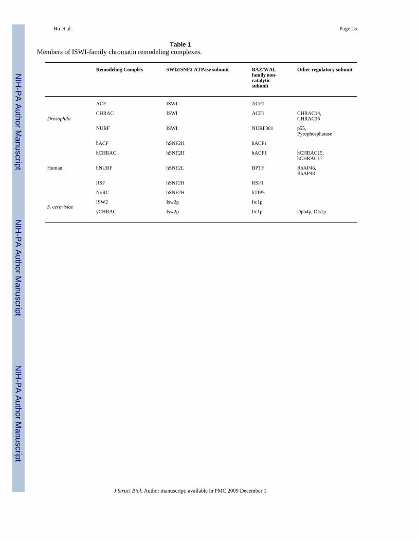
Table 1Members of ISWI-family chromatin remodeling complexes.
Remodeling Complex SWI2/SNF2 ATPase subunit BAZ/WALfamily non-catalyticsubunit
Other regulatory subunit
Drosophila
ACF ISWI ACF1
CHRAC ISWI ACF1 CHRAC14,CHRAC16
NURF ISWI NURF301 p55,Pyrophosphatase
Human
hACF hSNF2H hACF1
hCHRAC hSNF2H hACF1 hCHRAC15,hCHRAC17
hNURF hSNF2L BPTF RbAP46,RbAP48
RSF hSNF2H RSF1
NoRC hSNF2H hTIP5
S. cerevisiaeISW2 Isw2p Itc1p
yCHRAC Isw2p Itc1p Dpb4p, Dls1p
J Struct Biol. Author manuscript; available in PMC 2009 December 1.