Digital Cameras for Microscopy
16
Digital Cameras for Microscopy Camera Overview For Life Science Microscopes Versatility by Design
-
Upload
khangminh22 -
Category
Documents
-
view
1 -
download
0
Transcript of Digital Cameras for Microscopy

Digital Cameras for Microscopy
Camera OverviewFor Life Science Microscopes
Versatility by Design

2
Your project needs the right camera, and this is definitely true when it comes to matching your digital imaging
requirements with your project work. Sometimes you’ll want dazzling color fidelity and at other times you’ll need pixel-
precise monochrome images. And then there are also those occasions when you would like a microscope camera that
can do both. Add to this selection the ability to choose from a range of features and resolutions – the Olympus digital
microscope camera range really does offer you the flexibility of choice.
Perfect Color Match ............................................................................................................................................................ pp. 4 – 9
Stay true to the colors: Color fidelity has been the unreachable zenith of digital microscope cameras until now – the Olympus color
camera range provides color match and resolution capabilities for every application.
The Right Black and White .............................................................................................................................................. pp. 10 – 11
It’s all about sensitivity: Capturing the smallest intensity differences in every single pixel to build up the perfect picture of the
fluorescent scene on your sample.
Multitalented All-Rounders ............................................................................................................................................. pp. 12 – 15
The best of both worlds: The versatility to experience a dependable workhorse for all your imaging needs, from detailed brightfield to
sensitive fluorescence.
Crystalline structures in polarized illumination.
THE FLEXIBILITY OF CHOICE

3
Color Management
Olympus’ dedicated color profiling technologies, implemented across the
entire color camera range, faithfully represent the sample colors, both easily
and automatically. At every stage, from the oculars up to the monitor, the “real”
image of the sample will be displayed and recorded.
The Importance of Color
Color is one of the main methods of differentiating the relevant aspects of a sample.
The colors in the sample could be natural or imposed by the research protocol, and
the overall balance of the colors is often used to determine certain properties or even
diagnose disease. Therefore, it is essential that, as well as ensuring the optimum
resolution and clarity, colors are captured with the right hue, saturation, and intensity as
seen through the eyepiece.
Highest Fidelity
The unique Olympus color profiling technologies are individually tuned for each color
camera model. International Color Consortium (ICC) reference profiles are used to
govern the relationship between the colors at every stage of the imaging process.
This ensures the best possible color fidelity, from the specimen to the monitor, on any
Olympus color microscope camera.
Real Time, Real Color
The Olympus color profiling technologies are already at work in real time, when
you’re looking at the live image. The best color representation is then ensured from
the beginning of your acquisition process, at the highest possible speed (patented
technology): There will be no need to readjust your image after capture to compensate
for color mismatches.
Color Spaces
Different components in an imaging system offer different color spaces – also called
color gamuts by the International Color Consortium (ICC). Each component involved in
color reproduction is described by such a profile, and the resulting set of profiles is in
turn used to achieve optimum color reproducibility for the imaging system, based on
human perception. Some Olympus color cameras can also match the extended Adobe
RGB color space, for color rendering at a professional level on supported monitors and
printers.
Histological section in brightfield illumination.
On-screen image of the same histological section, without (left panel) and with (right panel) Olympus color profiling technology.
STAYING IN THE SHADE
Olympus Real-Time Color Profiles
Camera Sensor
Camera Sensor

4
PERFECT COLOR MATCH
Color reproduction presents microscope camera manufacturers with a very complex set of issues. Besides the color
itself, the intensity and weighting within the given spectral range has to be taken into account, Olympus has worked
hard to produce a range of cameras that provide perfectly balanced solutions for each and every application.
Brightfield panoramic image of an histological section of a rat embryo, processed with MIA (Multiple Image Alignment) software function.

5
XC30 and XC50
Whether you choose the 3 megapixels XC30 or the 5 megapixels XC50,
you’ll be able to take advantage of active Peltier sensor cooling to obtain an
advanced level of performance. The minimal background noise and color
fidelity provided by these cameras is especially beneficial in pathology and
histology applications, where the occasional high-intensity fluorescent sample
can also be correctly imaged.
Keeping a Cool Head
The active cooling of the Olympus XC30 and XC50 cameras employs a Peltier element
to maintain the sensor chip at a constant 10˚C in standard ambient surroundings,
guaranteeing perfect color images rich in contrast, with excellent color fidelity and
minimal background noise. The cooling also enables a diverse range of exposure times
to be covered, maximizing sensitivity in low-light applications.
Versatile Functionality
Both cameras offer a high dynamic range in all supported resolutions. When combined
with the reduced background noise afforded by the Peltier cooling system, this allows
for a sharp increase in the signal-to-noise ratio. High contrast images, which accurately
capture the essence of all sample details, hues, and intensities, are just a click away.
Frame rates to meet the needs of any application are achieved with pixel-binning
capabilities, while the partial readout mode speeds up capture. By freely defining a ROI
(Region of Interest) to read from, file sizes are also reduced – ideal for storage as well as
sharing.
Consistent Control
The Olympus XC30 and XC50 cameras are fully operated using the cellSens imaging
software, making it quick and easy to reach the camera’s full capabilities and ensuring
that the optimum solution is provided in the face of any challenge, such as image
processing, commenting, reporting, and archiving.
Algae in darkfield illumination.
Histological section in brightfield illumination.
XC30: 3 megapixels color camera with cooling.
XC50: 5 megapixels color camera with cooling.

6
PERFECT COLOR MATCH
DP22 and DP27
Lifelike digital microscopy is now a reality with the next generation of Olympus
digital cameras. Achieving an experience almost indistinguishable from the
oculars, the 5 megapixels DP27 and 3 megapixels DP22 deliver crisp and
fluid Full HD live images directly to the monitor. Introducing new multi-mode
functionality guarantees expert results with minimum effort in a wide range of
applications throughout life science.
Unmatched Quality with Full HD
The new cameras deliver detailed images with superior viewing comfort, with a Full HD
live image at 30 fps for the DP22 and 22 fps for the DP27. Thanks to the advanced
progressive scan readout technology, panning and focusing are always fluid and
natural. When a region of interest demands further investigation, the DP27 instantly
achieves precise focusing and zooming with a fast 15 fps live image at 5 megapixels –
deciphering extra detail from the most intricate of samples.
Perfect Color Balance, Every Time
Designed with the user in mind, the new cameras boast an automatic white point
balance. Regardless of changes in illumination conditions, colors are always perfectly
balanced, and color cast is automatically compensated. Without the need to constantly
adjust the settings or spend time on post-acquisition correction, producing a perfectly
balanced image couldn’t be simpler.
Expert Results in Every Task
Introducing a new level of user-friendly camera operation, three color modes are now
available, and optimizing camera settings for each sample or study has never been so
easy. The High-Fidelity mode creates a faithful image true to the oculars, while Normal
mode offers enhanced colors ideal for pale stained samples or for clearly presenting the
samples to an audience by means of various monitors and projectors. When employing
phase contrast, the dedicated Cell Culture mode generates a truly balanced image, with
clear details in both dark and bright areas.
Stand-Alone Control Option
Sample evaluation at short notice is sometimes a core part of the working day, and
having quick and easy access to an imaging system is vital. Providing unmatched
convenience and ease of use, the optional DP2-SAL stand-alone controller directly
connects the DP22 or DP27 to a monitor, removing the need for a dedicated computer.
Simplifying image acquisition with no compromise on camera quality or functionality, the
optional unit is ready to use in seconds, and includes powerful features such as movie
recording with audio and touch-screen control. Designed for maximum convenience,
the controller also provides automatic image calibration and enables quick, reliable
measurements easily exported to Microsoft Excel.
Histological section in brightfield illumination.
Pap smear in brightfield illumination.
DP22 and DP27: 3 and 5 megapixels color cameras.
Optional stand-alone controller connecting DP22 camera directly to a computer monitor. Camera is controlled via mouse and an on-screen menu.

7
DP2-SAL
The DP2-SAL controller converts the DP22 and the DP27 cameras into an
instantaneous imaging system, which can be accessed in mere seconds and is
optimized for a range of applications.
Always Ready
There is no need to wait for computer boot-up times or for software loading delays:
Simply place the sample on the microscope and capture images with one click of the
mouse. Rich in connections and with a complete range of functions, the DP2-SAL
controller brings its advantages to many different application scenarios, such as routine
work, multiple-observer discussions, or projection to an audience, and quick creation of
educational materials with audio comments. Enabling the comfort of viewing Full HD live
images on a variety of monitors, the DP2-SAL achieves complete stand-alone operation
with full camera functionality and quality.
Need Connections?
With multiple USB connections, the DP2-SAL permits storage of movies and images
onto portable storage devices, with exposure settings, magnifications, and other
parameters stored for future reference. Images can also be saved to a shared folder on
a networked drive via the Ethernet connection. The connection of a USB mouse and
keyboard allows rapid menu navigation, precise measurements and direct comment
annotation on images, if required. Finally, microphone and speakers can be directly
connected to the DP2-SAL controller, allowing the recording of comments while
capturing a movie.
Ergonomically Easy
The on-screen menu of the DP2-SAL is simplified to allow quick access to the most
important functions, while allowing intuitive execution of all operations by supporting
touch-enabled monitors. When used alongside an encoded nosepiece, precision is
automatically maintained in the measurement functions as the actual magnification is
directly read from the microscope and the reference scale adjusted in real time.
Present and Discuss
The DP2-SAL stand-alone controller takes full advantage of the DP22 and DP27
Full HD live image feed, for fast and fluid panning and focusing. Any monitor or
projector can be connected via the DVI-I port, providing the freedom to choose the ideal
output device size for a variety of applications. This affords maximum comfort together
with shared on-screen viewing, ideal for intensive sample browsing, collaborative
workflows, and prolonged use. Acquired movies and images can also be loaded via
USB storage for on-screen viewing, zooming, and panning with a single click.
In-vitro fertilization with digital live image; monitor directly connected to a DP22 camera using DP2-SAL controller.
DP2-SAL on-screen menu providing direct one-click access to functions, controlled via mouse or touch-enabled monitor.
DP2-SAL: Optional stand-alone controller for DP22 and DP27 cameras.
Stay connected: DVI, USB, Ethernet, Microphone, and Audio OUT are available.
8
PERFECT COLOR MATCH
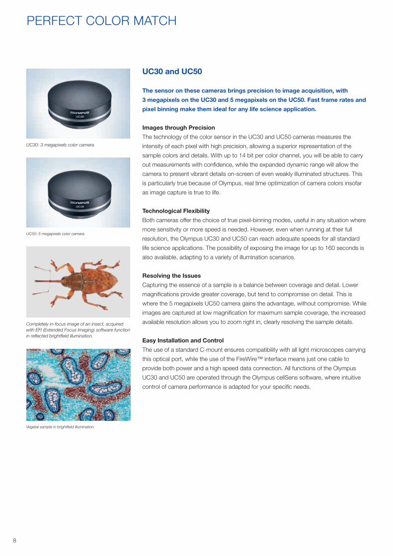
UC30 and UC50
The sensor on these cameras brings precision to image acquisition, with
3 megapixels on the UC30 and 5 megapixels on the UC50. Fast frame rates and
pixel binning make them ideal for any life science application.
Images through Precision
The technology of the color sensor in the UC30 and UC50 cameras measures the
intensity of each pixel with high precision, allowing a superior representation of the
sample colors and details. With up to 14 bit per color channel, you will be able to carry
out measurements with confidence, while the expanded dynamic range will allow the
camera to present vibrant details on-screen of even weakly illuminated structures. This
is particularly true because of Olympus, real time optimization of camera colors insofar
as image capture is true to life.
Technological Flexibility
Both cameras offer the choice of true pixel-binning modes, useful in any situation where
more sensitivity or more speed is needed. However, even when running at their full
resolution, the Olympus UC30 and UC50 can reach adequate speeds for all standard
life science applications. The possibility of exposing the image for up to 160 seconds is
also available, adapting to a variety of illumination scenarios.
Resolving the Issues
Capturing the essence of a sample is a balance between coverage and detail. Lower
magnifications provide greater coverage, but tend to compromise on detail. This is
where the 5 megapixels UC50 camera gains the advantage, without compromise. While
images are captured at low magnification for maximum sample coverage, the increased
available resolution allows you to zoom right in, clearly resolving the sample details.
Easy Installation and Control
The use of a standard C-mount ensures compatibility with all light microscopes carrying
this optical port, while the use of the FireWire™ interface means just one cable to
provide both power and a high speed data connection. All functions of the Olympus
UC30 and UC50 are operated through the Olympus cellSens software, where intuitive
control of camera performance is adapted for your specific needs.
Vegetal sample in brightfield illumination.
Completely in-focus image of an insect, acquired with EFI (Extended Focus Imaging) software function in reflected brightfield illumination.
UC30: 3 megapixels color camera.
UC50: 5 megapixels color camera.

9
SC100
The SC100 camera’s 10.5 megapixels are fully dedicated to brightfield
documentation tasks, where capturing each and every sample detail is
essential, especially when working at low magnifications.
Get the Details
With the SC100 digital color camera, you can quickly capture in-depth sample details in
a single shot, without using any pixel-shift technology. The newly implemented sensor
packs more than 10.5 megapixels, increasing its resolving power to one surpassing the
resolution of the human eye when looking through the binoculars. This frees users from
needing to take multiple, high magnification images of a sample to preserve resolution
while capturing a large area. Similarly, images can be easily investigated at high digital
magnifications at a later date, even if this was not initially intended when they were
captured using a low magnification objective. The SC100 makes sure you always see
the bigger picture, without losing the details.
Follow Your Needs
Despite the high resolution, panning and focusing are comfortable and quick thanks
to the high frame rates offered by the different live modes. Highly sensitive detection
along with pixel binning will assist in all applications, especially those involving darkfield
imaging and phase contrast. Furthermore the high-speed USB 2.0 interface allows for
an easy connection to any available computer.
SC50
Quality has never been so affordable with the 5 megapixels SC50. Designed for
everyday use and ideal under low light conditions, fast and Full HD imaging is
now easily achieved in a wide range of routine applications.
Smartly Noise-Free
The SC50 camera quickly delivers the perfect snapshot. In addition to achieving Full
HD images at 32 fps, sensitivity is now vastly improved by the sensor’s light-guide
technology, boosting the light collection capability of each pixel. Meanwhile, Olympus
Smart Image Averaging keeps noise under control, producing clean images especially
in low light. The dedicated algorithm cleverly detects sample movement and protects
against “trail” artefacts, while lateral resolution is enhanced with a new, smaller pixel
size.
Perfect Color Balance, Every Time
Designed for ease of use, the SC50 boasts an automatic white point balance.
Regardless of changes in illumination conditions, colors generated are always perfectly
balanced and devoid of any color cast – without the need to constantly adjust the
camera settings or correct images post-acquisition.
SC100: 10.5 megapixels color camera.
SC50: 5 megapixels color camera.
Detail of a butterfly wing with transmitted dark-field illumination. SC50 light-guide technology provides a brilliant picture (left panel) in contrast to a standard CMOS sensor (right panel)
Detail extraction from histological sample in brightfield.

10
Panoramic image of a mouse’s neuronal tissue sample, stained with multiple fluorochromes and processed with Extended Focus Imaging (EFI) and Multiple Image Alignment (MIA) software functions.
THE RIGHT BLACK AND WHITE
Even though fluorescence microscopy is intimately concerned with using the best combinations of excitation and
emission spectra of dyes, the cameras used for fluorescence microscopy imaging must be designed to provide
maximum sensitivity (capturing as many photons as possible) and clarity (as little background noise as possible). The
Olympus monochrome camera offers an abundance of both of these qualities.

11
XM10
The XM10 offers all of the properties required to provide dependable
fluorescence microscopy images: Extremely high sensitivity, a cooled sensor
chip, variable resolutions, and an optional external trigger function.
Designed for Fluorescence
At full resolution, the XM10 is ideal for all fluorescence acquisitions since it is extremely
sensitive, low in electronic noise, and supports long integration times of up to
160 seconds. The sensor has a pixel size of 6.45 μm x 6.45 μm, which, in combination
with the camera cooling, ensures that the XM10 is ideal for recording even the faintest
fluorescence signals in your specimen.
The Right Tool for the Job
The XM10 employs a sensor chip cooled to 10˚C (at 25˚C ambient temperature) with a
14 bit dynamic range and 15 fps (frames per second) at full resolution. It offers three
binning modes: 2x, 4x, and 8x, resulting in increased sensitivity and excellent frame
rates in live mode, which make it easier to focus and locate areas of interest within the
field of view while conserving highly sensitive fluorescence samples.
Four Models
The four different versions of the XM10 are optimized with the user application in mind.
Building on the capabilities of the basic XM10, the IR-extended version (XM10-IR) is
ideal for the entire range of fluorescent dyes, including those emitting in the near-IR
region such as CY5 and CY7. A specific model (XM10-T) guarantees precise image
capture through the presence of an external trigger input – for integration into real time
acquisition systems such as Olympus xcellence and cellSens. The most advanced
camera in this range (XM10-TIR) combines these advanced features to provide the user
with the perfect camera for all levels of fluorescence microscopy.
Easy to Integrate
The Olympus XM10 makes a great addition to any microscopy system not only because
of its great features, but also since it is easy to integrate, using a standard C-mount
adaptor to connect to the microscope, and the high-speed data transfer and power
capabilities of the FireWire™ interface. The XM10 is fully supported by the Olympus
cellSens software, ensuring that whatever the application, the information is not only
fully collected but properly analyzed, processed, and displayed. Ideal for a range of
applications, the XM10 works in synergy with Olympus cellSens software to achieve
excellent results in functions such as High Dynamic Range (HDR) acquisition, fast
deconvolution, and multi-fluorescence panoramic imaging. Maximum detail is resolved
from even the most weakly emitting fluorescence sample.
Axon and dendrite morphogenesis during neuronal maturation task.
XM10: 1.4-megapixels monochrome camera with cooling.
Deconvolution (right panel) of a gut tissue section stained with multiple fluorochromes.

12
MULTITALENTED ALL-ROUNDERS
Stereo microscopy image of a GFP-expressing Drosophila sample, overlay of brightfield and fluorescence images. DP80 precise centering mode and HDR processing of fluorescence image were used.
Image courtesy of Kei Ito, Ph.D., Institute of Molecular and Cellular Biosciences, University of Tokyo.
When there is a requirement for two or more seemingly distinct technologies in one instrument, a compromise is often
reached that makes the product good but not great. Reversing this trend, these three high-performance color and
monochrome cameras will excel at every task.

13
DP80
Thinking outside the box, Olympus has come up with the multitalented DP80.
Incorporating both a color and monochrome chip within the same housing, the
DP80 camera provides quality imaging for color and fluorescent microscopy
applications alike. The color sensor achieves bright and crisp imaging,
complementing the ultrasensitive photon detection of the monochrome sensor.
Together, these two diverse functions within a single camera offer maximum
versatility for a range of life science applications.
A Camera of Many Talents
The unique DP80 excels equally at high-resolution color documentation and high-
performance fluorescent detection alike. Olympus real-time color profiles provide lifelike
hues, while the Olympus Fine Detail Processing algorithm extracts maximum detail in
combination with the pixel-shifting capabilities of the color sensor. At the same time, the
high sensitivity up to the near-IR range of the monochrome sensor allows for precision
imaging of fluorochromes such as Cy7. With exposure settings up to 60 seconds,
photon detection is maximized for even very faint fluorescent signals. The signal-to-
noise ratio is further improved across both sensors with Peltier cooling, ideal for the
complex and detailed imaging requirements within life science.
Fast Response
Designed to meet the variable demands of a shared workplace, the DP80 removes
the need to switch camera or optical path, instead seamlessly shifting between
monochrome and color sensors in only three seconds. This is easily managed, either
automatically or at the click of an icon, and requires no recalibration of chip alignments,
saving valuable time. The readout of both chips is always progressive, for a fast and
fluid live image, free of any artefacts like striping and color-ghosting. With an on-
screen experience which truly matches the one at the oculars, the DP80 camera can
successfully also support any joint discussion or presentation need.
Creative Combinations
You can now effortlessly achieve superior combined color and fluorescent imaging,
with the ability to quickly overlay images derived from both sensors. An impressively
accurate correspondence is ensured without additional calibrations, thanks to expert
manufacturing processes, and is activated with a single click of the mouse. Now you
are free to explore exciting new paths in the lab and clinic alike, with the confidence of
an assured correspondence between color and fluorescence markers.
Confidence without Compromise
Further to cost-efficiency, the DP80 dual-sensor camera eliminates the need for
individual separate cameras and an optical switch. This makes it highly valuable in the
fast-paced research environments of today, where equipment must often be versatile
and have interchangeable functions, without compromising on quality, ease-of-use, or
performance.
DP80: Cooled dual sensor camera with 12.5 megapixels (color) and 1.4 megapixels
The central shows overlay of color (left panel) and monochrome (right panel) images of GFP-expressing drosophila sample. DP80 precise centering mode and HDR processing of fluorescence image were used.
Image courtesy of Kei Ito, Ph.D., Institute of Molecular and Cellular Biosciences, University of Tokyo.
Histological section (left image) of healing tissue also containing CY5 and CY7 labelling of collagen I and III (right image).

14
MULTITALENTED ALL-ROUNDERS
DP73
Merging Olympus expertise in pixel-shift sensor technology with the innovative
features of the high-end DSLR camera range, the DP73 opens up new paths
in capturing precise images. Quality is unsurpassed in color documentation,
and flexibility is provided for fluorescence images – for a truly all-round
performance in a variety of applications.
Simply a Better Image
The 17.2 megapixels DP73 digital camera implements the innovative Olympus Fine
Detail Processing technology, directly derived from the Olympus top-range E-5
consumer DSLR. This hardware function monitors the image pixel in real time,
preventing fine details from being obscured by surrounding structures. The result is
an image free of artefacts, where every intricate sample detail is enhanced and clearly
visible.
Evolving Technology
The top resolution of the DP73 is dramatically improved in comparison with previous
models, allowing for greater detail capture when working at low magnifications.
However, increasing the resolution is not the only factor involved in final image quality.
Olympus has applied its extensive knowledge of pixel-shifting sensors to implement the
innovative 3CCD mode, reading the actual RGB values of each shifted pixel and thus
allowing the true color to be measured directly, without any interpolation. The result
is an image free from any pixel-shifting artefact and with the color range usually only
obtained by using expensive 3CCD cameras.
DP73WDR: Responding to the Challenges
It is not uncommon for microscopy samples to present wide variations in brightness,
challenging the camera’s capability to perform correct exposure levels across the
complete field of view. The DP73WDR presents a solution in the form of its WiDeR
(Wide Dynamic Range) mode, which continuously checks the exposure of each image
pixel, applying tonal enhancements only where necessary. Dynamic exposure across
the image brings out those details previously lost to darkened or washed-out areas.
Furthermore, this mode works in real time, allowing the user to regulate the effect level
directly on the live image, for the perfect snapshot.
A True Performer, also in Fluorescence
Panning and focusing is fast and fluid, thanks to the progressive readout sensor,
allowing you to experience the same visuals on the computer screen as you would
normally get from the microscope oculars. This is particularly true thanks to the DP73
color quality, where the Olympus real time color profiling technology has been further
tuned for the hues normally used in the histology and pathology applications. You will
be able to distinguish more subtle hue variations, discovering a striking correspondence
between the image on the screen and the one at the oculars. With a generous pixel
density, and a signal to noise ratio enhanced by Peltier cooling, the DP73 also excels
in standard fluorescence imaging, while providing the versatility ideal in the diverse
applications of a shared laboratory. To further tailor performance to your needs, a
special monochrome mode is available to customize the camera sensitivity to your
fluorophore of choice.
Detail of insect sample in reflected brightfield. Image on the right shows automatic compensation of overexposed areas using DP73WDR Wide Dynamic Range mode.
Detail extraction from a histological section of a young mouse.
DP73: 17.2 megapixels color and monochrome camera with cooling.

15
XC10
With excellent image quality, high sensitivity, and long integration times
optimized for fluorescence imaging, the Olympus XC10 Peltier-cooled camera
offers users a flexible imaging setup for both monochrome and color
applications, effectively combining high sensitivity fluorescence with true color
imaging.
Fast, Smooth, and Sensitive
The powerful cooled sensor chip offers the clarity of 14 bits per color channel and has
the ability to provide very high frame rates via the use of pixel binning. In the 2x binning
mode, the camera provides more than 28 fps, which increases to nearly 50 fps when
using 4x binning. This makes the XC10 ideal for applications that require fast image
acquisition of dynamic objects. In addition, the high image frequency can be used to
focus on samples or locate areas of interest directly on the PC screen.
The high sensitivity of the XC10 is the result of a large pixel size, defining the camera’s
ability to be a well-equipped all-rounder; not only perfect for color imaging, but also for
meeting high expectations in sensitive fluorescence applications. There is no longer the
need to switch camera for the application in hand. Quality is not compromised for either
color or fluorescence imaging techniques.
Chilled
The Peltier-cooled sensor maintains a temperature of 10˚C (at ambient temperature),
enabling this multifunctional camera to provide color and black-and-white images that
are rich in detail and contrast, with extraordinarily low background noise. The extensive
exposure time range (from 100 μs to 160 s) also adds to the XC10’s appeal, ensuring
that both strong and weak signals are captured with equal fidelity.
A Team Player
Whatever the application, the XC10 can provide the images that enable research to be
pushed forward. With the ease of both C-mount optical coupling, FireWire™ data, and
power connectivity, integrating the XC10 into your imaging system is easy.
Four Models
In addition, the XC10 is available in other three versions dedicated to maximize its
performance in specific applications. The IR-extended version (XC10-IR) is ideal for the
entire range of fluorescent dyes, including those emitting in the near-IR region such as
CY5 and CY7. The Trigger version (XC10-T) guarantees precise image capture through
the presence of an external trigger input – for integration into real-time acquisition
systems such as Olympus xcellence and cellSens. The most advanced camera in this
range (XC10-TIR) combines both advanced features to provide the user with the perfect
camera for all levels of fluorescence and brightfield microscopy.
XC10: 1.4 megapixels cooled color and monochrome camera.
Sequentially recorded cell cycle.
Image courtesy of Josh Morgan, Dr. Rachel Wong, Department of Anatomy and Neurobiology, Washington University, School of Medicine,St. Louis, USA.

Color CamerasDP22 DP27 SC50 SC100
Image Sensor Color CCD Color CCD Color CMOS Color CMOS
Sensor Type Sony ICX 687 AQA Sony ICX 625 AQA Aptina MT9P006STC Aptina MT9J003
Sensor Size 1/1.8 inch 2/3 inch 1/2.5 inch 1/2.3 inch
Resolution (max.) 1920 x 1440 pixels 2448 x 1920 pixels 2560 x 1920 pixels 3840 x 2748 pixels
Pixel Size 3.7 x 3.7 μm 3.45 x 3.45 μm 2.2 x 2.2 μm 1.67 x 1.67 μm
Binning 2x 2x 2x, 4x 2x
A/D Converter 8 bit 8 bit 12 bit 12 bit
Exposure Time from 50 μs to 8 s from 50 μs to 8 s from 31 μs to 2.74 s from 0.12 ms to 14.6 s
Live Frame Rates 25 fps at 1920 x 1440 pixels 15 fps at 2448 x 1920 pixels 15 fps at 2560 x 1920 pixels 3 fps at 3840 x 2748 pixels
30 fps at 1920 x 1080 pixels 22 fps at 1920 x 1080 pixels 32 fps at 1920 x 1080 pixels 12 fps at 1920 x 1374 pixels
25 fps at 960 x 720 pixels 30 fps at 1224 x 960 pixels 55 fps at 640 x 480 pixels77 fps at 480 x 270 pixels 42 fps at 960 x 686 pixels
Cooling System No No No No
External Trigger No No No No
Data Transfer USB 3.0 USB 3.0 USB 3.0 USB 2.0
Color Profiles Olympus real time color profiles Olympus real time color profiles Olympus real time color profiles Olympus real time color profiles
Partial Readout Yes Yes No No
Remarks· Stand-alone option· Fast and Fluid Full HD Live
· Stand-alone option· Fast and Fluid Full HD Live
· Automatic White Balance· Active Noise Cancellation· Fast Full HD Live
· Single-shot 10.5 megapixels· Convenient USB interface
Operating System Compatibility Windows 7 / 8 / 8.1 32 bit and 64 bit Windows 7 / 8 / 8.1 32 bit and 64 bit Windows 7 / 8 / 8.1 32 bit and 64 bit Windows 7 / 8 / 8.1 32 bit and 64 bit
UC30 UC50 XC30 XC50
Image Sensor Color CCD Color CCD Color CCD Color CCD
Sensor Type Sony ICX 252 AQ Sony ICX 282 AQ Sony ICX 252 AQ Sony ICX 282 AQ
Sensor Size 1/1.8 inch 2/3 inch 1/1.8 inch 2/3 inch
Resolution (max.) 2080 x 1544 pixels 2588 x 1960 pixels 2080 x 1544 pixels 2576 x 1932 pixels
Pixel Size 3.45 x 3.45 μm 3.4 x 3.4 μm 3.45 x 3.45 μm 3.4 x 3.4 μm
Binning 2x, 3x 2x, 4x, 6x 2x, 3x 2x, 4x, 6x
A/D Converter 14 bit 14 bit 14 bit 14 bit
Exposure Time from 0.1 ms to 160 s from 0.1 ms to 160 s from 0.1 ms to 160 s from 0.1 ms to 10 s
Live Frame Rates 7 fps at 2080 x 1544 pixels 9 fps at 2588 x 1960 pixels 7 fps at 2080 x 1544 pixels 9 fps at 2588 x 1960 pixels
13.6 fps at 1040 x 772 pixels 9 fps at 1292 x 980 pixels 13.6 fps at 1040 x 772 pixels 9 fps at 1292 x 980 pixels
25 fps at 688 x 514 pixels 33 fps at 640 x 480 pixels 25 fps at 688 x 514 pixels 33 fps at 640 x 480 pixels
24.5 fps at 424 x 318 pixels 24.5 fps at 424 x 318 pixels
Cooling System No No Peltier 10°C at 25°C ambient Peltier 10°C at 25°C ambient
External Trigger No No No No
Data Transfer FireWire™ IEEE 1394a FireWireTM IEEE 1394a FireWire™ IEEE 1394a FireWire™ IEEE 1394a
Color Profiles Olympus real time color profiles Olympus real time color profiles Olympus real time color profiles Olympus real time color profiles
Partial Readout Yes Yes Yes No
Remarks — — · Low image noise with Peltier cooling · Low image noise with Peltier cooling
Operating System Compatibility Windows 7 / 8 / 8.1 32 bit and 64 bit Windows 7 / 8 / 8.1 32 bit and 64 bit Windows 7 / 8 / 8.1 32 bit and 64 bit Windows 7 / 8 / 8.1 32 bit and 64 bit
Color and Monochrome Cameras
DP73 DP80 XC10 XM10 (monochrome only)
Image Sensor Color CCD Color CCD + Monochrome CCD Color CCD Monochrome CCD
Sensor Type Sony ICX 274 AQ Sony ICX 275 AQ + ICX 285 AL Sony ICX 285 AQ Sony ICX 285 AL
Sensor Size 1/1.8 inch 2/3 inch (both sensors) 2/3 inch 2/3 inch
Resolution (max.) 4800 x 3600 pixels 4080 x 3072 pixels (color) 1376 x 1032 pixels 1376 x 1032 pixels
1360 x 1024 pixels (monochrome)
Pixel Size 4.4 x 4.4 µm 6.45 x 6.45 μm (both sensors) 6.45 x 6.45 µm 6.45 x 6.45 µm
Binning 2x 2x, 4x 2x, 4x 2x, 4x, 8x
A/D Converter 14 bit 14 bit (both sensors) 14 bit 14 bit
Exposure Time from 23 μs to 60 s from 23 μs to 60 s from 0.1 ms to 160 s from 0.1 ms to 160 s
Live Frame Rates 15 fps at 1600 x 1200 pixels 15 fps at 1360 x 1024 pixels 15 fps at 1360 x 1024 pixels 15 fps at 1376 x 1032
15 fps at 800 x 600 pixels 57 fps at 340 x 250 pixels 28 fps at 688 x 516 pixels 28 fps at 688 x 516
27 fps at 800 x 600 (binning 2x) 50 fps at 344 x 258 pixels 50 fps at 344 x 258
106 fps at 172 x 129
Cooling System Peltier 10°C at 25°C ambient Peltier 10°C at 25°C ambient Peltier 10°C at 25°C ambient Peltier 10°C at 25°C ambient
External Trigger Yes Yes Optional Optional
Data Transfer PCIe dedicated controller PCIe dedicated controller FireWire™ IEEE 1394a FireWire™ IEEE 1394a
Color Profiles Olympus real time color profiles Olympus real time color profiles Olympus real time color profiles N/A
Partial Readout Yes No Yes No
Operating System Compatibility Windows 7 / 8 / 8.1 64 bit Windows 7 / 8 / 8.1 64 bit Windows 7 / 8 / 8.1 32 bit and 64 bit Windows 7 / 8 / 8.1 32 bit and 64 bit
Remarks · Extended dynamic range in DP73WDR model· Fine Detail Processing· 3CCD mode· Progressive readout
· Dual CCD camera · Fast motorized sensor switching· Precise image overlay· Progressive readout· Near-IR sensitivity
· Highly sensitive both in color and monochrome· Optional Trigger and near-IR versions
· Optional Trigger and near-IR versions
· OLYMPUS CORPORATION is ISO9001/ISO14001 certified. · Illumination devices for microscopes have suggested lifetimes. Periodic inspection is required. Please visit our website for details. · All company and product names are registered trademarks and/or trademarks of their respective owners. · Images on the PC monitors are simulated. · Specifications and appearances are subject to change without any notice or obligation on the part of the manufacturer.
E04
3074
2 · 3
00 ·
01/1
5 · O
EK
G ·
OK
M
www.olympus-lifescience.com
Cover page image: YFP-H brain of a 20-week-old mouse treated by New Scale. Courtesy of: Hiroshi Hama, Atsushi Miyawaki,Laboratory for Cell Function Dynamics RIKEN Brain Science Institute.
Postbox 10 49 08, 20034 Hamburg, Germany Wendenstrasse 14–18, 20097 Hamburg, GermanyPhone: +49 40 23773-0, Fax: +49 40 233765 E-mail: [email protected]