Microscopy res. - tech-2012
9
A Simple Cryo-Holder Facilitates Specimen Observation Under a Conventional Scanning Electron Microscope CHIH-YUAN TANG, 1 RONG-NAN HUANG, 2 LING-LONG KUO-HUANG, 3 TAI-CHIH KUO, 4 YA-YUN YANG, 1 CHING-YEH LIN, 1 WANN-NENG JANE, 5 AND SHIANG-JIUUN CHEN 3 * 1 Precision Instrumentation Center, College of Science, National Taiwan University, Taipei 106, Taiwan 2 Department of Entomology and Research Center for Plant-Medicine, College of Bioresources and Agriculture, National Taiwan University, Taipei 106, Taiwan 3 Department of Life Science, Institute of Ecology and Evolutionary Biology and TechComm-5, College of Life Science, National Taiwan University, Taipei 106, Taiwan 4 Department of Biochemistry, Taipei Medical University, Taipei, 106, Taiwan 5 Plant Cell Biology Core Lab., Institute of Plant and Microbial Biology, Academia Sinica, Taipei 115, Taiwan KEY WORDS cryo-holder; specimen preparation; cryo-fixation; scanning electron microscope ABSTRACT A pre-cryogenic holder (cryo-holder) facilitating cryo-specimen observation under a conventional scanning electron microscope (SEM) is described. This cryo-holder includes a speci- men-holding unit (the stub) and a cryogenic energy-storing unit (a composite of three cylinders assembled with a screw). After cooling, the cryo-holder can continue supplying cryogenic energy to extend the observation time for the specimen in a conventional SEM. Moreover, the cryogenic energy-storing unit could retain appropriate liquid nitrogen that can evaporate to prevent frost deposition on the surface of the specimen. This device is proved feasible for various tissues and cells, and can be applied to the fields of both biology and material science. We have employed this novel cryo-holder for observation of yeast cells, trichome, and epidermal cells in the leaf of Arabi- dopsis thaliana, compound eyes of insects, red blood cells, filiform papillae on the surface of rat tongue, agar medium, water molecules, penicillium, etc. All results suggested that the newly designed cryo-holder is applicable for cryo-specimen observation under a conventional SEM with- out cooling system. Most importantly, the design of this cryo-holder is simple and easy to operate and could adapt a conventional SEM to a plain type cryo-SEM affordable for most laboratories. Microsc. Res. Tech. 75:103–111, 2012. V V C 2011 Wiley Periodicals, Inc. INTRODUCTION As early as 1665, the discovery of the cell structure by the English microscopist Robert Hooke through the use of a simple glass lens has released mankind’s curi- osity to the microcosmos. Since the work of Robert Hook, light microscopy has become a key tool for biolo- gists to observe cell structure a great deal. Ongoing advances in microscopy have made it possible to probe into the worlds invisible to human naked eyes. Compared to the light microscope, the electron microscope is a more expensive, massive, and complex instrument. It substitutes visible light with electrons as the illuminated photosource and the glass lens is replaced with a system of electromagnetic lenses to deflect electrons. Though the resolution of the electron microscope was significantly enhanced, it can only be operated under a strong and stable power source and in extremely high-vacuum environments. Moreover, the preparation of the histological specimen must meet strict requirements. For biological specimens, the his- tological protocol for light microscopy had already been well developed. The biological specimens usually con- tain considerable amounts of water. The standard steps for histological analysis of biological specimens include chemical fixation, dehydration, infiltration, embed- ding, sectioning, and staining (Afzelius, 1992). These steps serve the dual purposes of preserving and staining the organic components of the specimen as to facilitate observation. The tissue preparation protocol for the electron microscope highly resembles that of the light microscope (Allen, 2008). However, to ensure that the subcellular structures observed under the electron microscope resemble their original state, a different chemical fixative and embedding material is used (Allen, 2008; Wagenaar et al., 1993). The section- ing and staining processes have also been improved for use with the electron microscope, which pose stricter requirements for specimen preparation (Afzelius, 1992; Allen, 2008). The preprocessing of a histological specimen involves the use of various chemical reagents during fixation and infiltration. The infiltration rates of each of the reagents vary and these processes occasionally result in partial dehydration of proteins and large molecules as well as pH changes, sedimentation, or other side reactions in biological specimens after fixation or infiltration, particularly during removal of water and soluble components, and during exchange of solvents C.-Y.T. and R.-N.H. contributed equally to this work. *Correspondence to: Shiang-Jiuun Chen, Department of Life Science, Institute of Ecology and Evolutionary Biology and TechComm-5, College of Life Science, National Taiwan University, Taipei 106, Taiwan. E-mail: [email protected] Received 1 October 2010; accepted in revised form 18 April 2011 Contract grant sponsor: National Science Council of Taiwan, Republic of China; Contract grant number: NSC 97-2321-B-002-013-MY3 DOI 10.1002/jemt.21031 Published online 14 July 2011 in Wiley Online Library (wileyonlinelibrary.com). V V C 2011 WILEY PERIODICALS, INC. MICROSCOPY RESEARCH AND TECHNIQUE 75:103–111 (2012)
Transcript of Microscopy res. - tech-2012

A Simple Cryo-Holder Facilitates Specimen ObservationUnder a Conventional Scanning Electron MicroscopeCHIH-YUAN TANG,1 RONG-NAN HUANG,2 LING-LONG KUO-HUANG,3 TAI-CHIH KUO,4
YA-YUN YANG,1 CHING-YEH LIN,1 WANN-NENG JANE,5 AND SHIANG-JIUUN CHEN3*1Precision Instrumentation Center, College of Science, National Taiwan University, Taipei 106, Taiwan2Department of Entomology and Research Center for Plant-Medicine, College of Bioresources and Agriculture,National Taiwan University, Taipei 106, Taiwan3Department of Life Science, Institute of Ecology and Evolutionary Biology and TechComm-5, College of Life Science,National Taiwan University, Taipei 106, Taiwan4Department of Biochemistry, Taipei Medical University, Taipei, 106, Taiwan5Plant Cell Biology Core Lab., Institute of Plant and Microbial Biology, Academia Sinica, Taipei 115, Taiwan
KEY WORDS cryo-holder; specimen preparation; cryo-fixation; scanning electron microscope
ABSTRACT A pre-cryogenic holder (cryo-holder) facilitating cryo-specimen observation undera conventional scanning electron microscope (SEM) is described. This cryo-holder includes a speci-men-holding unit (the stub) and a cryogenic energy-storing unit (a composite of three cylindersassembled with a screw). After cooling, the cryo-holder can continue supplying cryogenic energy toextend the observation time for the specimen in a conventional SEM. Moreover, the cryogenicenergy-storing unit could retain appropriate liquid nitrogen that can evaporate to prevent frostdeposition on the surface of the specimen. This device is proved feasible for various tissues andcells, and can be applied to the fields of both biology and material science. We have employed thisnovel cryo-holder for observation of yeast cells, trichome, and epidermal cells in the leaf of Arabi-dopsis thaliana, compound eyes of insects, red blood cells, filiform papillae on the surface of rattongue, agar medium, water molecules, penicillium, etc. All results suggested that the newlydesigned cryo-holder is applicable for cryo-specimen observation under a conventional SEM with-out cooling system. Most importantly, the design of this cryo-holder is simple and easy to operateand could adapt a conventional SEM to a plain type cryo-SEM affordable for most laboratories.Microsc. Res. Tech. 75:103–111, 2012. VVC 2011 Wiley Periodicals, Inc.
INTRODUCTION
As early as 1665, the discovery of the cell structureby the English microscopist Robert Hooke through theuse of a simple glass lens has released mankind’s curi-osity to the microcosmos. Since the work of RobertHook, light microscopy has become a key tool for biolo-gists to observe cell structure a great deal. Ongoingadvances in microscopy have made it possible to probeinto the worlds invisible to human naked eyes.
Compared to the light microscope, the electronmicroscope is a more expensive, massive, and complexinstrument. It substitutes visible light with electronsas the illuminated photosource and the glass lens isreplaced with a system of electromagnetic lenses todeflect electrons. Though the resolution of the electronmicroscope was significantly enhanced, it can only beoperated under a strong and stable power source andin extremely high-vacuum environments. Moreover,the preparation of the histological specimen must meetstrict requirements. For biological specimens, the his-tological protocol for light microscopy had already beenwell developed. The biological specimens usually con-tain considerable amounts of water. The standard stepsfor histological analysis of biological specimens includechemical fixation, dehydration, infiltration, embed-ding, sectioning, and staining (Afzelius, 1992). Thesesteps serve the dual purposes of preserving andstaining the organic components of the specimen as to
facilitate observation. The tissue preparation protocolfor the electron microscope highly resembles that ofthe light microscope (Allen, 2008). However, to ensurethat the subcellular structures observed under theelectron microscope resemble their original state, adifferent chemical fixative and embedding material isused (Allen, 2008; Wagenaar et al., 1993). The section-ing and staining processes have also been improved foruse with the electron microscope, which pose stricterrequirements for specimen preparation (Afzelius, 1992;Allen, 2008).
The preprocessing of a histological specimen involvesthe use of various chemical reagents during fixationand infiltration. The infiltration rates of each of thereagents vary and these processes occasionally resultin partial dehydration of proteins and large moleculesas well as pH changes, sedimentation, or other sidereactions in biological specimens after fixation orinfiltration, particularly during removal of water andsoluble components, and during exchange of solvents
C.-Y.T. and R.-N.H. contributed equally to this work.
*Correspondence to: Shiang-Jiuun Chen, Department of Life Science, Instituteof Ecology and Evolutionary Biology and TechComm-5, College of Life Science,National Taiwan University, Taipei 106, Taiwan. E-mail: [email protected]
Received 1 October 2010; accepted in revised form 18 April 2011
Contract grant sponsor: National Science Council of Taiwan, Republic ofChina; Contract grant number: NSC 97-2321-B-002-013-MY3
DOI 10.1002/jemt.21031
Published online 14 July 2011 inWiley Online Library (wileyonlinelibrary.com).
VVC 2011 WILEY PERIODICALS, INC.
MICROSCOPY RESEARCH AND TECHNIQUE 75:103–111 (2012)

with an embedding matrix (Saga, 2005). These proce-dures could drastically damage the structure andarrangement of the biological specimen and impede theability to discern the original structure of the biologicalspecimen (Consort Ribeiro et al., 2001; Nicolas, 1991).To resolve the issue of damage to the tissue structureafter fixation or infiltration, cryogenic sectioning tech-niques were developed (Consort Ribeiro et al., 2001;Gilkey and Staehelin, 1986; Somlyo et al., 1977; Yuchiet al., 2002). These techniques mainly involve plungingfresh, hydrated samples into liquid cryogen (e.g., pro-pane, helium, or nitrogen). The detrimental effects ofchemical fixation are overcome because the tissue isnot dehydrated and specimen components are rapidlyimmobilized (Iyer et al., 2005; Saga, 2005). Afterwards,the cryo-fixed biological specimens were observedunder an electron microscope equipped with an expen-sive on-line cooling system or cryo-transfer (cryo-EM).Although the present cooling systems or cryo-transfersystems are structurally simple, their price is almosthalf that of an electron microscope and make thembeyond the reach of standard laboratories and areinconvenient for the processing of large samples.Therefore, the present article seeks to develop a simpleand accessible cryo-holder as an alternative for theexpensive cooling systems or cryo-transfer systems,which is applicable for use with scanning electronmicroscopes (SEM) to observe cryo-fixed samples.This device is simple and affordable, allowing theobservation of fresh-frozen specimens to be even moreaccessible and facilitating prescreening of large num-bers of specimens.
MATERIALS ANDMETHODSDesign of Cryo-Holder
The newly designed cryo-holder includes three cylin-ders and a screw all made of stainless steel (Fig. 1a).The three cylinders are assembled together by thescrew (Figs. 1b and 1c). Cylinder 2 serves as the pedes-tal of the cryo-holder and cylinder 3 forms a U-shapelocated on the top of the cryo-holder to support the stuband hold back liquid nitrogen (Fig. 1d). During assem-bly, a 0.5 cm interval is left for the cryo-holder to beheld by forceps in-between the pedestal (cylinder 2)and cylinder 1 in the middle (Figs. 1c and 1e). Theassembly of cylinders by a screw makes its heightadjustable for focus tuning during practice in the elec-tron microscope. The dimension of the present cryo-holder is 40.0 mm (diameter) 3 12.0–20.0 mm (height)as detailed in Figure 2. In practice, the specimenholder (stub) is screwed into the top cylinder (cylinder3) of the cryo-holder (Figs. 1d and 1e).
Specimen Preparation
Before the experiments, the cryo-holder is usuallyimmersed in liquid nitrogen for cooling (30 s to 2 min)(Fig. 3). Afterward, the specimen adhered on the stub(by using 70% ethanol) was mounted into the cyro-holder and immersed into liquid nitrogen for another30 s to 1 min for further cooling (Precooling adhesion).The 70% ethanol is used since it not only functions as acryoprotectant, the solution can also firmly stick thespecimen to the stub via hydrogen bonding through themoisture surface of the frozen specimen. Afterward,
Fig. 1. The components and assemble of the cryo-holder.
Microscopy Research and Technique
104 C.-Y. TANG ET AL.

the cryo-holder together with the cooled-stub and spec-imen are transferred immediately to the sample cham-ber of the SEM for observation. The cooled cryo-holdercan not only continue to provide cryogenic energy forthe specimen, but also can reserve a small amount ofliquid nitrogen in the top U-shaped cylinder for pre-venting frost deposition on the surface of the specimen.This could facilitate cryo-specimen observation under aconventional SEM without cooling system.
To observe particular parts of the sample (e.g.,antennae or compound eyes of insects), the specimen
was treated with a postcooling adhesion protocol. Thepostcooling adhesion was adopted by adhering thespecimen to the stub using 70% ethanol under a dissec-tion microspcope in a particular posture and mountingthis to the cooled cryo-holder that just been taken outof the liquid nitrogen tank. The cryo-holder was thentransferred immediately to the sample chamber of theSEM for observation. The advantage of this postcoolingadhesion protocol is that the specimen can be adheredto the stub in a particular posture. Moreover, thisprotocol can also avoid the occasional detachment of an
Fig. 2. The dimension of the cryo-holder. a: Side view. b: Top view.
Fig. 3. Concept of the new cryo-holder. After cooling treatment, the cryo-holder along with the speci-men were transferred to the specimen chamber of SEM for observation. Since a quantity of liquid nitro-gen was retained in the fillister (trench) of the top cylinders, it slowly evaporated to keep the specimen ina cryostate for observation under SEM long enough (ca. 30 min). LN2, liquid nitrogen.
Microscopy Research and Technique
105CRYO-HOLDER FOR CONVENTIONAL SEM
adhered specimen during the precooling adhesionprocess. Although the commercial glues can effectivelyadhere the specimen to the stub, some delicate struc-ture of specimen might be sensitive to the timing forglue drying.
Cryo-Crush of Specimens in Liquid Nitrogen
To view the interior structure, the specimens arebriefly immersed in ethanol (5–10 min depending onsize) and then directly immersed in the liquid nitrogentank for 30 s to 2 min. After cooling, the sample iscrushed into small pieces directly in liquid nitrogen byusing forceps. The crushed pieces were then trans-ferred to the stub that had already been cooledtogether with the cryo-holder in another nitrogen tank.Afterwards, the specimen, together with the cryo-holder, is transferred to the specimen chamber of theSEM for immediate observation.
Mounting of Cryo-Holder in the SpecimenChamber
The newly designed cryo-holder has a 0.5 cm intervalspace left in between the pedestal (cylinder 2) and cyl-inder 1 in the middle (Figs. 1c and 1e). Once cooled, thecyro-holder was hold with tweezers and transferred tomount onto the hole (originally for the stub loading)located on the stage inside specimen chamber of SEM(Hitachi TM-1000, Hitachi, Tokyo, Japan). Once thevacuum reached 1026 torr (around 1–1.5 min), thesample was then observed at a fixed acceleration volt-age of 15 kV. After mounting, the cooled-cryo-holdercan continuously provide cryoenergy for the specimenand keep the temperature sufficiently low (30 min) forobservation under a conventional SEM without coolingsystem. Meanwhile, liquid nitrogen is retained in thefillister (trench) of the top cylinder and can slowlyevaporate to prevent frost deposition on the surface ofthe cooled specimen before mounting into the specimenchamber and facilitate its observation (Fig. 3).
RESULTS
This study provides a novel and cheap design of cryo-holder that could facilitate specimen observation undera conventional SEM. This device can be adapted to a
conventional SEM for morphological studies of cryo-fixed specimens and has been proven feasible forseveral varieties of tissues and cells, and can be appliedin the fields of both biology and material science.
Observation of Specimens PrecooledTogether with Cryo-Holder
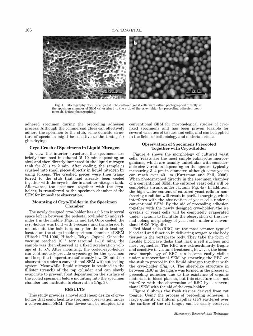
Figure 4 shows the morphology of cultured yeastcells. Yeasts are the most simple eukaryotic microor-ganisms, which are usually unicellular with consider-able size variation depending on the species, typicallymeasuring 3–4 lm in diameter, although some yeastscan reach over 40 lm (Kurtzman and Fell, 2006).When photographed directly in the specimen chamberof a conventional SEM, the cultured yeast cells will becompletely shrunk under vacuum (Fig. 4a). In addition,the high water content of cultured yeast cells in non-freezing condition will result in partial charging, whichinterferes with the observation of yeast cells under aconventional SEM. By the aid of precooling adhesiontogether with the newly designed cryo-holder, the icecrystals of yeast cells will be completely evaporatedunder vacuum to facilitate the observation of the nor-mal oblong morphology of yeast cells under a conven-tional SEM (Fig. 4b).
Red blood cells (RBC) are the most common type ofblood cell and function in delivering oxygen to the bodytissues in the vertebrate body. They take the form offlexible biconcave disks that lack a cell nucleus andmost organelles. The RBC are extraordinarily fragileand sensitive to vacuum treatment, however, the bicon-cave morphology of RBC can become easily visibleunder a conventional SEM by smearing the RBC onthe stub to precool in the liquid nitrogen together withthe cryo-holder (Fig. 5). The sheet-like structure inbetween RBC in the figure was formed in the process ofprecooling adhesion due to the existence of organicmaterials in blood plasma, but this structure does notinterfere with the observation of RBC by a conven-tional SEM with the aid of the cryo-holder.
Figure 6 shows the fresh tissues derived from rattongue. Using the process of precooling adhesion, alarge quantity of filiform papillae (FP) scattered overthe surface of the rat tongue can be easily observed
Fig. 4. Micrography of cultured yeast. The cultured yeast cells were either photographed directly inthe specimen chamber of SEM (a) or glued to the stub of the cryo-holder for precooling adhesion treat-ment (b) before photographing.
Microscopy Research and Technique
106 C.-Y. TANG ET AL.

(Fig. 6a), and even the taste buds (TB) can occasionallybe detected in the FP (Fig. 6a). Many bacteria (B) caneven be detected on the cornified surface of FP underlarger magnifications (Fig. 6b).
The current cryo-holder can also be applied to non-biological materials. Figure 7a shows the appearanceof agar medium photographed in a conventional SEMwithout precooling adhesion treatment. The high watercontent of agar medium in the nonfreezing state usu-ally results in partial charging during the course ofscanning; therefore, it is difficult to obtain a goodquality of image. Image quality can be significantlyimproved by a precooling adhesion treatment of agarmedium smeared on the stub. As shown in Figure 7b,the meshed structure of agar medium becomes evidentunder SEM with the aid of the cryo-holder. Moreover,even the crystals of water molecules can be probedafter covering the cooled stub with frost for photo-graphing under a conventional SEM (Fig. 8).
Observation of Specimens Postcooledwith Cryo-Holder
For specimens that need to be adhered to the stub ina particular position (e.g., insects or flowers), the post-cooling adhesion with the cooled cryo-holder is used.Specimens are first adhered to a stub under a dissec-tion microscope before mounting into the cooled cryo-holder. Thereafter, the assembled cryo-holder is imme-diately transferred to the specimen chamber of a SEMfor observation. Processing the specimens in this waycan also avoid detachment from the stub if preparedwith the precooling adhesion protocol. Figure 9 showsthe morphology of trichome and epidermal cells in theleaf of Arabidopsis thaliana under SEM. Although thetrichome morphology stays intact, the epidermal cellsof the leaf shrink under vacuum when observed with aconventional SEM (Fig. 9a). In contrast, both the tri-chome and epidermal cells of the leaf stay intact withthe aid of precooling adhesion treatment by using thenewly designed cryo-holder (Fig. 9b).
Figure 10 shows the compound eyes of the mosquitoAedes albopictus, which is a representative key pest in
public hygiene. Although the morphology of ommatidiawas well maintained in the scanning mode, it becamecollapsed and deformed when photographed directly inthe specimen chamber of a SEM (Fig. 10a). Thedestruction of ommatidia might be resulted from thedehydration of specimen under electron beam ignition.By the aid of the cryo-holder, the ommatidia (Fig. 10b)uniformly distributed throughout the compound eyescan be easily detected under a conventional SEM (Fig.10b) without the need of chemical fixation and coating.These results suggest that cryo-holder can effectivelyprovide cryoprotection to the specimen.
Figure 11a shows the hyphae of Penicillium thatwere severely crimped due to complete dryness undervacuum and the intact morphology of Penicillium wastherefore unavailable. By applying the Penicilliumcells directly on the surface of a stub assembled in thecooled-cryo-holder, the colony of Penicillium becomesclearly visible and numerous spores are easily detecta-ble under a conventional SEM (Fig. 11b).
Observation of Internal Structure in Specimensthat are Cryo-Crushed in Liquid Nitrogen
The current design of cryo-holder is also applicableto cryo-crushed samples. To view the freeze-fracturingsample, large samples are usually etched by a freezefracture system that is also an expensive and complexaccessory equipment item of an electron microscope.Freeze fracture is especially suited for studies of the in-ternal aspect of the lipid bilayer in the cell membrane
Fig. 5. The biconcavemicrography of red blood cells. Red blood cellsfrom rats were smeared on the stub of the cryo-holder for precoolingadhesion treatment and photographed under a conventional SEM.
Fig. 6. Micrograph of FP on rat tongue. The rat tongue tissueswere smeared on the stub of the cryo-holder for precooling adhesiontreatment and photographed under a conventional SEM. FP, filiformpapilae; TB, taste bud; B, bacteria.
Microscopy Research and Technique
107CRYO-HOLDER FOR CONVENTIONAL SEM

by using transmission electron microscopy (TEM),however, the processes of fracturing and TEM-replicaare always tedious, difficult, and hardly predictable(Walther, 2003; Walther and Muller, 1997). We adopteda simple cryo-crush method and, in combination of thenewly designed cyro-holder, the interior structure oflarge samples can be imaged in tissue level. As shownin Figure 12, the kidney tissues of a rat were cooled inliquid nitrogen and cryo-crushed using forceps. Afterbeing adhered to the cooled stub assembled in the cryo-holder, the samples were photographed under a con-ventional SEM. The interior structure of the kidney,including the proximal convoluted tubule (PCT) andcross-section of vicinal blood vessels (BV) are clearlyvisible (Fig. 12). Moreover, the renal corpuscle (RC)structure of renal and even some cross-section of nuclei(N) can be detected in the microphotograph. Thesecombined results suggest that the newly designed cryo-holder is applicable for cryo-specimen observationunder a conventional SEM without an expensivecooling system.
DISCUSSION
The preprocessing of biological specimens involvesmany chemical reagents. Even after continuousimprovements, these processes still inevitably result inalterations to the finer parts of the cell structure aftera series of chemical reactions. These artifacts not onlyintroduce challenges when viewing the capturedimages, but also hamper our efforts to discern the origi-nal appearance of the biological specimens. To mini-mize the harm caused by these chemical reagents, theyare used sparingly or not at all. Cryogenic techniquesthat are based on physical methods have developedrapidly as an alternative for chemical fixation (Janssonet al., 2000; Plattner and Bachmann, 1982; Sitte,1979). Cryogenic techniques mainly use a fresh-freez-ing method to preserve the finer parts of the cell struc-ture (Dubochet et al., 1988). It is therefore necessary touse a complete cooling system during the preparationof the specimens and the electron microscope must alsobe equipped with a cooling system (i.e., cryo-EM) whenobserving these cryo-fixed specimens. Cryogenic tech-niques have the advantage of closely preserving theoriginal cell structure and thus have been widelyemployed by various research institutes recently.Yet because the current cooling systems are extremelyexpensive, they add an additional burden to the cost ofan electron microscope (Saga, 2005). Moreover, afurther weakness of current cryogenic techniques isthe lengthy preparation required, ranging from 1 to 3days (Hoch, 1991; Refshauge et al., 2006). If largenumbers of specimens need to be observed, the timerequired to process them will prove prohibitive. Thusthe time-requirement of sample processing and costconcerns have been the main reasons for the failure ofcryogenic techniques to become the industry standard(Saga, 2005).
Cryo-fixation (freeze-fixation) is developed to mini-mize the detrimental effects of chemical fixation.Cryo-fixation is commonly followed by freeze-drying(Abascal et al., 2005; Adams, 2007), critical point dry-ing (Araujo et al., 2003; Sarmiento et al., 2006), orfreeze substitution (Quintana, 1994). Nevertheless,both freeze-drying and critical point-drying are
Fig. 7. Scanning morphology of agar. The agar plates were directly photographed under SEM (a) orpro-cooling process before observation under a conventional SEM.
Fig. 8. The hexagonal crystals of water molecules. A small amountof frost was placed on the cooled stub of cryo-holder and photographedunder a conventional SEM.
Microscopy Research and Technique
108 C.-Y. TANG ET AL.

Fig. 9. Morphology of trichome and epidermal cells in the leaf of Arabidopsis thaliana. The leaf of A.thaliana was observed directly under SEM (a) or processed with a postcooling adhesion protocol on thestub of the cryo-holder and photographed under SEM (b).
Fig. 10. The compound eye of Aedes. albopictus. The compound eye of A. albopictus was photographeddirectly in the specimen chamber of SEM (a) or glued to the stub of the cryo-holder for postcooling adhe-sion treatment and photographed under a conventional SEM (b). The arrow in (a) shows the collapsedommatidia.
Fig. 11. The hyphae and spore micrograph of Penicillium. The cultured Penicillium were either photo-graphed in the specimen chamber of SEM (a) or dropped to the cooled cryo-holder (postcooling adhesion)for observation under SEM (b).
Microscopy Research and Technique
109CRYO-HOLDER FOR CONVENTIONAL SEM

unsuitable for detailed visual examination, since theyusually introduce some artifacts to the specimens,such as cell wall collapse, movement of cell contents,and damage to delicate structures (Lindroth et al.,1988). In particular, the process of critical point dryingcan result in collapse and wrinkling of a sample simi-lar to that of chemically fixed, dehydrated, and embed-ded tissue (Gazi et al., 2005). Freeze-substitutioneffectively preserves cell ultrastructure, but necessi-tates the substitution of water and soluble compoundswith an organic solvent. In contrast, direct observa-tion of the fully hydrated frozen tissue can consis-tently produce superior preservation of the ultrastruc-ture of a specimen (Iyer et al., 2005), since it providesthe advantage of retention of water and solutes, andrapid immobilization of cell structure compared tochemical fixation. Thus, material is fixed in situ andretains a life-like appearance. Moreover, simplicityand speed of preparation (in the order of min) areadditional benefits of observing hydrated frozen mate-rial over other freezing techniques.
This study has developed a simple freezing device(cryo-holder) applicable for direct observation of thefully hydrated frozen tissue under a conventional SEMwithout equipped with an expensive cooling system(Lindroth et al., 1988). Not only will it be ease to oper-ate, it will also be affordable and appropriate for usewith the SEM, as an alternative for the more expensivecooling systems. If used in combination with a stand-ard desktop SEM, the total cost would only amount tothat of a high-end light microscope, which is within theaffordable range for most research laboratories. More-over, it will take less time and training to operate,allowing observation of frozen biological specimens tostart more efficiently (in the order of seconds or min).The system would be suitable for prescreening of largenumbers of samples.
There are two prominent features of the currentcryo-holder. First, the cooled cryo-holder can continueproviding cryoenergy in the specimen chamber of a con-
ventional SEM for about 30 min, which is long enoughfor sample observation. Though larger cryo-holdercould provide cryoenergy longer for the specimens, itwould also hamper water evaporation from the speci-mens under vacuum and slow down the instant obser-vation. We also tried to use nitrogen slush and liquidpropane as the cryoagents for specimen fixation. Theresults showed that the image qualities are not signifi-cant different from that of liquid nitrogen-fixed speci-mens. In contrast, the surface of liquid propane-fixedspecimens will cover a layer of propane slush, whichtakes more time (around 10 min) to evaporate undervacuum, and as a result will compromise the timeavailable for specimen observation. Second, the fillister(trench) in the top cylinder of the current cryo-holdercan retain small amounts of liquid nitrogen that canslowly evaporate to prevent water crystallizing on thesurface of the cooled specimens (Fig. 3) and facilitatingits observation. For the operation of traditional cryo-EM, during the loading of the specimen onto the sam-ple holder or the fixing of the sample holder onto thespecimen stage, the specimen is often inevitablyexposed to moisture in the atmosphere, which oftenresults in frost deposition (Iyer et al., 2005). The SEMused in these studies for cryo-holder demonstration is aHitachi model TM-1000, a prototypical electron micro-scope without a cooling system. However, the cooled-cryo-holder can temporarily provide cryoenergy (within30 min) for the specimen to avoid the beam energy eli-cited thermal instability of the samples. We have inves-tigated the current cryo-holder, which was also applica-ble and functioned well in different modules of SEMincluding the Hitachi S-800. We suggest this cryo-holder could be modified and workable with differenttypes of scanning electron microscope.
CONCLUSION
We have developed a simple and accessible cryo-holder as an alternative for the expensive cooling sys-tems or cryo-transfer systems, which is applicable foruse with conventional SEM to observe cryo-fixed sam-ples. This device is simple and affordable, allowing theobservation of fresh-frozen specimens to be even moreaccessible and facilitating prescreening of large num-bers of specimens. Moreover, this design has been pro-ven feasible for several varieties of tissues and cells,and can be applied to the fields of both biology and ma-terial science. Most importantly, the design of thiscryo-holder is simple and easy to operate and couldadapt a conventional SEM to a plain type cryo-EMaffordable for most laboratories.
ACKNOWLEDGMENTS
The authors would like to thank Professor JohanBillen (Katholieke Universiteit Leuven, Belgium) forcarefully reading the manuscript.
REFERENCES
Abascal K, Ganora L, Yarnell E. 2005. The effect of freeze-drying andits implications for botanical medicine: A review. Phytother Res19:655–660.
Adams G. 2007. The principles of freeze-drying. Methods Mol Biol368:15–38.
Afzelius BA. 1992. Section staining for electron microscopy using tan-nic acid as a mordant: A simple method for visualization of glycogenand collagen. Microsc Res Tech 21:65–72.
Fig. 12. Micrograph of kidney tissue. The kidneys from rats wereimmersed into liquid nitrogen for 1 min and cryo-crushed with for-ceps. The crushed pieces of kidney tissues were then glued to thecooled stub of the cryo-holder and photographed under a SEM. PCT,proximal convoluted tubule; BV, blood vessel; RC, renal corpuscle; N,nucleus.
Microscopy Research and Technique
110 C.-Y. TANG ET AL.

Allen TD, editor. 2008. Introduction to electron microscopy for biolo-gists. Burlington: Academic Press.
Araujo JC, Teran FC, Oliveira RA, Nour EA, Montenegro MA, CamposJR, Vazoller RF. 2003. Comparison of hexamethyldisilazane and criti-cal point drying treatments for SEM analysis of anaerobic biofilmsand granular sludge. J Electron Microsc (Tokyo) 52:429–433.
Consort Ribeiro K, Benchimol M, Farina M. 2001. Contribution of cryo-fixation and freeze-substitution to analytical microscopy: A study ofTritrichomonas foetus hydrogenosomes. Microsc Res Tech 53:87–92.
Dubochet J, Adrian M, Chang J, Homo J, Lepault J, McDowall AW,Schultz P. 1988. Cryo-electron microscopy of vitrified specimens. QRev Biophys 21:129–228.
Gazi E, Dwyer J, Lockyer NP, Miyan J, Gardner P, Hart C, Brown M,Clarke NW. 2005. Fixation protocols for subcellular imaging by syn-chrotron-based Fourier transform infrared microspectroscopy. Bio-polymers 77:18–30.
Gilkey JC, Staehelin LA. 1986. Advances in ultrarapid freezing forthe preservation of cellular ultrastructure. J Electron Microsc Tech3:177–210.
Hoch HC. 1991. Preservation of cell ultrastructure by freeze-substitu-tion In: Mendgen K, Lesemann DE, editors. Electron microscopyof plant pathogens. New York, NY: Springer-Verlag. pp. 1–16.
Iyer R, Arunagirinathan MA, Prabhu CS, Bellare J. 2005. Animproved specimen loader for cryo-scanning electron microscopy.Scanning 27:141–146.
Jansson HB, Persson C, Odeslius R. 2000. Growth and capture activ-ities of nematophagous fungi in soil visualized by low temperaturescanning electron microscopy. Mycologia 92:10–15.
Kurtzman CP, Fell JW. 2006. Yeast Systematics and Phylogeny-Impli-cations of Molecular Identification Methods for Studies in Ecology.Biodiversity and Ecophysiology of Yeasts. The Yeast Handbook,Springer Retrieved January 7, 2007.
Lindroth M, Bell PB Jr., Fredriksson BA. 1988. Comparison of theeffects of critical point drying and freeze-drying on cytoskeletonsand microtubules. J Microsc 151:103–114.
Nicolas G. 1991. Advantages of fast-freeze fixation followed by freeze-substitution for the preservation of cell integrity. J Electron MicroscTech 18:395–405.
Plattner H, Bachmann L. 1982. Cryofixation: A tool in biologicalultrastructural research. Int Rev Cytol 79:237–304.
Quintana C. 1994. Cryofixation, cryosubstitution, cryoembedding forultrastructural, immunocytochemical and microanalytical studies.Micron 25:63–99.
Refshauge S, Watt M, McCully ME, Huang CX. 2006. Frozen in time:A new method using cryo-scanning electron microscopy to visualizeroot-fungal interactions. New Phytol 172:369–374.
Saga K. 2005. Application of cryofixation and cryoultramicrot-omy for biological electron microscopy. Med Mol Morphol38:155–60.
Sarmiento PL, Ciarmela ML, Sanchez Thevenet P, Minvielle MC,Basualdo JA. 2006. Comparison of preparation techniques of mixedsamples (fungi-helminth eggs) for scanning electron microscopy bycritical point drying. Parasitol Res 99:455–458.
Sitte H. 1979. Cryofixation of biological material without pretreat-ment–a review. Mikroskopie 35:14–20.
Somlyo AV, Schuman H, Somlyo AP. 1977. Elemental distribution instriated muscle and the effects of hypertonicity: Electron probeanalysis of cryosections. J Cell Biol 74:828–857.
Wagenaar F, Kok GL, Broekhuijsen-Davies JM, Pol JM. 1993. Rapidcold fixation of tissue samples by microwave irradiation for use inelectron microscopy. Histochem J 25:719–725.
Walther P. 2003. Recent progress in freeze-fracturing of high-pressurefrozen samples. J Microsc 212:34–43.
Walther P, Muller M. 1997. Double layer coating for field emissioncryo SEM - Present state and applications. Scanning 19:343–348.
Yuchi H, Suganuma T, Sawaguchi A, Ide S, Kawano J, Aoki T, Kita-mura K, Eto T. 2002. Cryofixation processing is an excellent methodto improve the retention of adrenomedullin antigenicity. HistochemCell Biol 118:259–265.
Microscopy Research and Technique
111CRYO-HOLDER FOR CONVENTIONAL SEM