Elimination of fibrin γ-chain cross-linking by FXIIIa ... - PNAS
Cell Biology International ISSN 1065-6995doi: 10.1002/cbin.10171
RESEARCH ARTICLE
Three-dimensional culture of differentiated endometrial stromal cellsto oligodendrocyte progenitor cells (OPCs) in fibrin hydrogelMohammad Nabi Asmani1,2,3, Jafar Ai1,2,4*, Ghasem Amoabediny2,3,5*, Abbas Noroozi4,Mahmoud Azami1, Somayeh Ebrahimi-Barough1,4, Mona Navaei-Nigjeh1,3, Armin Ai6 andMina Jafarabadi7
1 Department of Tissue Engineering, School of Advanced Technologies in Medicine, Tehran University of Medical Sciences, Tehran, Iran2 Research Center for New Technologies in Life Science Engineering, University of Tehran, Tehran, Iran3 Faculty of New Sciences & Technologies, University of Tehran, Tehran, Iran4 Brain and Spinal Injury Research Center, Tehran University of Medical Sciences, Tehran, Iran5 Department of Chemical Engineering, School of Engineering, University of Tehran, Tehran, Iran6 Dentistry Faculty, Tehran University of Medical science, Tehran, Iran7 Reproductive Health Research Center, Tehran University of Medical Sciences, Tehran, Iran
Abstract
Neural tissue engineering is one of the most promising strategies for treatment of nerve tissue injuries. Three-dimensional (3D)environment mimics in vivo conditions for cells. 3D distribution and growth of the cells within the scaffold are both importantfor neural tissue engineering. In this study, endometrial stromal cell-derived oligodendrocyte progenitor cells (EnSC-derivedOPCs) were cultured in fibrin gel and cell differentiation and viability were evaluated after 8 days of post-culture. The structuraland mechanical characteristics of fibrin gel-like scaffold were examined with rheological analysis. EnSCs were isolated fromdonor tissue and were induced to OPCs with growth factors (FGF2/EGF/PDGF-AA) for 12 days, thenwere cultured in fibrin gelwith Triiodothyronine (T3)medium for another 8 days. The viability of cells was analyzed usingMTTassay for a period of 8 daysculturing in a fibrin matrix. Structure of fibrin matrix and cell morphology was analyzed with SEM. TEM, immunostaining andquantitative RT-PCR was performed for OPCs markers after cell culturing in fibrin matrix. Cell viability is enhanced in fibrinmatrix after 8 days. SEM and TEM show that cells are in good integration with nano-fibers. Moreover, immunohistochemistryand quantitative RT-PCR of OPCs differentiation markers showed that Olig2, Sox10, PDGFRa, CNP, and A2B5 are expressedafter 8 days culturing within fibrin matrix. Fibrin can provide a suitable 3-D scaffold for EnSCs differentiated cells for theregeneration of CNS.
Keywords: endometrial stromal cells; oligodendrocyte progenitor cells; differentiation; neural tissue engineering; fibrin gel
Introduction
Disruption of neurological structure by injuries to the spinalcord reduces personal motionability or can lead to paraplegiaand death. The CNS can heal and regenerate axons, but therate is very slow and cannot stop myelin sheath degenerationand cell death especially in the second stage of injury(Xiaosong et al., 2011). The limited nature of repair results infunctional recovery being more sporadic than organized. Celltherapy and tissue engineering could provide a therapeuticapproach to CNS regeneration (Xiaosong et al., 2011; Hatchet al., 2009). Different cells from various sources under indifferent in vivo condition have been investigated. Cell
therapy has involved animal models, with stem cells frombone marrow, neural stem cell (NSCs) and embryonic stemcells being used. Animals showed improvements in term ofaxonal regeneration and functional behavior, but a number ofproblems are associated with incorporation of cells into CNStissue (Koda et al., 2005). The differentiation, migration andhoming of the implanted stem cells into other parts of thebody, cell morbidity in the initial few days of the experimentand teratoma formation can create difficulties. Moreover, theenvironment of injury does not support the differentiation ofstem cells to neural lineage and only small percentage of thosecells will survive the graft-versus-host response (Keirsteadet al., 2005; Karimi-Abdolrezaee et al., 2006). Tissue
*Corresponding authors: e-mail: [email protected] (J.A.), [email protected] (G.A.)
1Cell Biol Int 9999 (2013) 1–10 � 2013 International Federation for Cell Biology

engineering provides alternative way for CNS regenerationusing cellular transplantation and biomaterial scaffolds(Khaing et al., 2012). Regarding the sophisticated andsensitive environment of CNS, engrafted biomaterials shouldbe biocompatible, biodegradable, with mechanical propertiesclose to host tissue, and be able to get cells to damaged area inorder to help avoid further injuries and trauma to the CNS(Tabesh et al., 2009; Xiaosong et al., 2011; Shoffstallet al., 2012). Hydrogels have such characteristics, alongwith a highly permissive microenvironment which encour-ages cell adhesion and proliferation. Additionally, hydrogelsprovide a 3D state for cell culture. The 3D setting not onlyoffers cell–cell interaction and mechanical stimulation fromevery direction similar to body’s extracellular matrix, butcloses the current gap that exists between in vitro and in vivotrials (Ishihara et al., 2011). Both synthetic and naturalbiomaterials have been used to create hydrogels fortreating spinal cord injuries, including Polyacrylamide(PAc) (NeuroGel), Polyethylene glycol (PEG), collagen,agarose, chitosan, and alginate (Tabesh et al., 2009). Allthese hydrogels seem to aid improvement, not without somesetbacks. PAc and PEG are not biodegradable and collagencan be immunogenic (Li et al., 2012). Agarose has the abilityto differentiate neural stem cell toward oligodendrocytes, butit also shows neuronal permissive resistance; chitosanpromotes cell adhesion due to its positive charge, but causesinflammation and its structure needs further modification.Alginate has a positive effect on neurons proliferation, butinhibits cellular and protein adsorption due to its negativecharge (Li et al., 2012).
Endometrial stem cells have some advantageous over othersources. In comparison to stem cells derived from bonemarrow, they have a faster and a higher proliferation rate.Low immunogenicity, safer and more convenient biopsyprocedure from donors in comparison with bone marrowand embryonic stem cells are other advantages of EnSCs.Moreover EnSCs plasticity have been proven by theirdifferentiation along other lineage, such as osteoblast,adipocytes, cardiomyocytes, neuronal, and oligodendrocytecells (Ai et al., 2012; Mobarakeh et al., 2012; Bockeriaet al., 2013; Ebrahimi-Barough et al., 2013).
In this study, endometrial stromal cells were used as a newsource for using in cell therapy. They were isolated from thedonor’s tissue; oligodendrocyte progenitor cells derived fromthese cells were cultured in fibrin gel. Fibrin has a long historyin tissue engineering applications, especially wound healing.It is a naturally occurring clotting agent in human body, so ithas a highly biocompatible scaffold and is easily degraded byenzymes inside the body. Mechanical and chemical proper-ties of this hydrogel could be adjusted by simply changingfibrinogen concentration; also there is an enzymic cross-linking mechanism that can be easily and harmlessly injected(Le et al., 2006).
Materials and methods
Endometrial stromal cell (EnSCs) isolation andcharacterization
EnSCs were obtained by the method of Ebrahimi-Baroughet al. (2013). Briefly samples were obtained from donorsusing laparoscopy procedure; all patients were informedabout the experiment and signed consent forms. Tissueswere maintained Hanks balanced salt solution (HBSS;Invitrogen, Carlsbad, CA) with 10% fetal bovine serum(FBS), and 1% (v/v) penicillin/streptomycin. Tissue wasenzymatically dissolved with collagenase type I (Sigma) at378C for 60min. The resulting suspension was neutralizedwith Dulbecco’s modified Eagle’s medium (DMEM; Invi-trogen) containing 10% FBS (Sigma), passed through 70 and40mm filters, and centrifuged to separate the supernatantfrom the pellet. Finally the pellet of stromal cells wasresuspended in medium consisting of DMEM 10% FBS and1% penicillin/streptomycin (P/S; Hyclone). Cultures werekept at sub-confluent levels in a 378C incubator in air with5% CO2. After 24 h, non-adherent cells were removed bychanging the medium. When the cells had reached 80%confluence, they were detached by incubation with 0.25%trypsin and 1mM EDTA for 5min. The media were changedevery 3 days. EnSCs at passage 3 were used for theexperiments.
Differentiation to oligodendrocyte progenitor cells
EnSCs were differentiated to OPCs using the protocol ofEbrahimi-Barough et al. (2013). Briefly 1� 105 cells wereseeded in each well of 24-well plates, and after 3 daysincubation with DMEM 10% FBS and 1% penicillin/streptomycin, the media was removed and replaced withfresh media containing DMEM-F12, FGF2 (20 ng/mL), N2and 1% penicillin/streptomycin, It was changed every2 days and after day 4. Cells were trypsinized and seeded oneach well of poly-L-ornithine coated plates at 2� 105 cell/well in DMEM-F12 containing FGF2 (20 ng/mL), EGF(20 ng/mL), N2 and 1% penicillin/streptomycin. Up to4 days with every 2 days of changing media, it was removedand substituted with DMEM-F12 plus FGF2 (20 ng/mL),PDGF (20 ng/mL), N2 and 1% penicillin/streptomycin foranother 4 days. The same cycle as previously mentionedwas repeated for this stage, completing the proliferationphase, such that the cells could be crossed to thedifferentiation phase by adding DMEM-F12, T3 (30 ng/mL), N2 and 1% penicillin/streptomycin for 6 days. Cellswere observed under a phase-contrast microscope toexamine their overall appearance. Microphotographs weretaken with X10 objective (TS-100 Nikon, Japan). The cellswere also collected for immunocytochemistry and qRT-PCR.
3D culture of EnSC-derived OPCs in fibrin hydrogel Mohammad Nabi Asmani et al.
2 Cell Biol Int 9999 (2013) 1–10 � 2013 International Federation for Cell Biology

Immunocytochemical analysis
Day 18 post-induction immunocytochemistry analysis wasdone for A2B5 antibody as a OPCs marker. The cells werecultured on wells of 24-well plate and fixed with 4%paraformaldehyde in phosphate buffer for 30min. Afterwashing with PBS, all specimens were blocked with 1%normal goat serum for 45min and incubated overnight at48C with mouse monoclonal anti-A2B5 (1:250; Abcam). Thecells were treated with Alexafluro594-conjugated goat anti-mouse IgG (1:500; Sigma) at room temperature for 45min.Nuclei stained with DAPI and the samples examined underfluorescence microscope (Olympus BX51, Japan).
Quantitative real-time reverse transcription-polymerasechain reaction
EnSCs differentiated from OPCs were removed from theplates (1.5� 106), and total RNA was extracted using theTrizol (Invitrogen). For removing genomic DNA, cells weretreated with DNase I, RNase-free kit (Takara, BIO Inc., Shiga,Japan), and cDNA was reverse transcribed using ReverseTranscriptase kit (Takara, Dalian, China). Primer pairs wereused as given in Table 1, with GAPDH as the internal control.Quantitative real-time PCR was performed using the SYBRgreen master mix (Takara, Dalian, China) on an ABI prism7300 sequence detector (Applied Biosystems, Foster City, CA,USA). Cycle number (Ct) of each reaction was calculatedfrom the amplification curve to determine the relative geneexpression by using the comparative cycle threshold method(Schmittgen and Livak, 2008). 2�DDCt was used for relativegene expression analysis. All Ct values calculated from thetarget genes were calibrated using that from the undifferen-tiated EnSCs. In each experiment there were at least threedifferent patient samples for each stage, which were done induplicate.
Fibrin gel preparation
Fibrin hydrogel was prepared as described by Esfandiari et al.(2008) and Fasciani et al. (2003). Lyophilized bovineFibrinogen (Sigma) at 2, 3, and 4mg/mL was dissolved inM199 media (Invitrogen) and 15mL thrombin (2.5 UN/mL)added to the bottom of micro-tubes which were preparedprior to scaffold construction, followed by pouring of thefibrinogen solution into the tubes. The mixture wasmaintained 15min at room temperature to clot and put inincubator at 378C for 5 h to form a 3D network structure.
Rheology
Rheological experiments were conducted using a PhysicaMCR300 rheometer (Anton-Paar, Ashland, VA, USA) equipped witha custom-made hood to eliminate dehydration of the blendsduring testing (n¼ 3). The parallel plate diameter used was25mm, and the distance between the plates was 0.05mm.Frequency sweeps were conducted on gelled hydrogel samples.Hydrogel blends were placed directly onto the parallel plate,which was heated to 378C prior to measurement. Frequencysweep was conducted between 0.1 and 10Hz. Storage modulus(G0) and loss modulus (G00) were measured for fibrins withdifferent concentrations and additional comparison was madewith spinal cord rheological measurement.
3D culture of differentiated EnSCs
After determining which fibrin gel creates better environ-ment for differentiated EnSCs, oligodendrocyte progenitorcells were trypsinized and removed from plate and suspendedat 105 cells in 500mL of prepared fibrinogen solution. Then15mL of thrombin and the above solution of suspended cellswere mixed in micro-tubes and incubated at 378C with airplus 5% CO2 at 95% humidity (Fasciani et al., 2003).
Cell viability assay
Viability of differentiated cells in conventional 2D and 3Dwere analyzed and compared by 3-[4,5-dimethyl-2-thia-zolyl]-2, 5-diphenyl-2H-tetrazolium bromide (MTT) assayafter 1, 4, and 8 days of seeding to investigate effect of fibrin3D scaffold. At each day, the cells and matrices wereincubated with 600mL 5mg/mL MTT for 4 h before 400mLprepared DMSOwas added to dissolve the crystal sufficiently.Aliquots were pipetted into the wells of a 96-well plate andtested by an enzyme labeled instrument (BioTek, USA), andthe UV absorbance at 570 nm for each well was measured.
Morphological evaluation of cultured cells by scanningelectron microscopy
Cell morphology was examined by SEM after 3 days ofculturing. The scaffolds were washed with PBS and fixed in
Table 1 Primers used for real time RT-PCR.
Gene Primer sequence (50–30)
Nestin F: AAAGTTCCAGCTGGCTGTGG
R: TCCAGCTTGGGGTCCTGAAA
PDGFRa F: AAAACGCGGTTTTTGAGCCC
R: GCTCAGCCCTGTGAGAAGAC
Olig2 F: CCAGAGCCCGATGACCTTTTT
R: CACTGCCTCCTAGCTTGTCC
SOX10 F: AAGCCTCACATCGACTTCGG
R: TCCATGTTGGACATTACCTCGT
CNP F: AGAAGAGCTCTGAGACCCTCC
R: TTAACACATCTTGTTGAGCGT
GAPDH F: TCGCCAGCCGAGCCA
R: CCTTGACGGTGCCATGGAAT
Mohammad Nabi Asmani et al. 3D culture of EnSC-derived OPCs in fibrin hydrogel
3Cell Biol Int 9999 (2013) 1–10 � 2013 International Federation for Cell Biology

2.5% glutaraldehyde aqueous solution at 48C for 1 h. Fixedsamples were rinsed twice with PBS and then dehydrated ingradient concentrations of ethanol (30, 50, 70, 80, 90, 95, and100%). After being freeze-dried in vacuum for 2 h, thecellular constructs were coated with gold sputter andexamined by SEM (Hitachi S4160, Japan) at a voltage of15 kV.
Transmission electron microscopy (TEM)
Hydrogel was washed and fixed in a phosphate buffer salineand 2.5% (v/v) glutaraldehyde, and post-fixed in 1% (w/v)osmium tetroxide for 2 h at room temperature. The fibrinhydrogel was dehydrated using graded concentrations ofethanol. After dehydration, the hydrogel was inserted inSpurr resin (Polysciences, Warrington, PA, USA). Ultrathinsections (60–90 nm) were cut with an ultramicrotome (LeicaEM UC6, Vienna, Austria) and stained with uranyl acetateand lead citrate. They were examined in a Zeiss LEO906electron microscope (Zeiss, Oberkochen, Germany).
Immunohistochemistry
Scaffolds were fixed in 4% paraformaldehyde for 1 h after7 days in culture, cut into sections by cryostat, and rinsedwith PBS. They were incubated withmousemonoclonal anti-A2B5 (1:250; Abcam) and mouse monoclonal anti-Olig2antibody (1:100; Santa Cruz Biotechnology, Santa Cruz, CA).The sections were treated with Alexafluro 488-conjugatedgoat anti-mouse IgG (1:100; Sigma) at room temperature for45min, and post-stained with DAPI. Images were taken witha fluorescence inverted microscope (Olympus_BX60).
Statistics
A difference between groups was considered statisticallysignificant if P< 0.05. For qRT-PCR, randomization testsusing REST 2009 V2.0.13 software were applied to indicatestatistically significant differences between groups and withstudent t-test for the MTT assay. Quantitative data and anysignificant differences in the latency of the each group weretested using ANOVA (one-way) test. This was used to analyzevariance using Excel software.
Results
EnSCs differentiate into cells expressing oligodendrocyteprogenitor cell markers
Stromal cells isolated from endometrial tissue have a spindlemorphology. These cells were positive for CD105, CD90,CD146, CD44 (Ebrahimi-Barough et al., 2013). To examinethe characteristics of differentiation of the EnSCs in culture
media, they were isolated and induced to OPCs phenotypeusing defined culture medium. Changes in cell morphologywere initially observed after addition of EGF, when the largeflat-shaped EnSCs becamemore slender.With the addition ofthe growth factor PDGF-AA, the cells progressively becamemore elongated and slender. Subsequently, by introducing T3hormone, cells acquired bipolar and tripolar morphologysimilar to oligodendrocytes (Figure 1).
After 18 days post treatment, the OPCs were studied byimmunocytochemistry staining for A2B5 as an oligodendro-cyte progenitor cells marker. The images showed that theEnSC-derived OPCs expressed the oligodendrocyte progeni-tor lineage marker, A2B5 (Figure 2).
Cells were examined by qRT-PCR on 18 days aftertreatment for expression of oligodendrocyte progenitor cellsmarkers that included Nestin, SOX10, Olig2, and PDGFRa.Expression of the neural progenitor cells marker (Nestin) andoligodendrocyte progenitor cells markers (SOX10, Olig2 andPDGFRa) were upregulated during OPCs differentiation(Figure 3).
Fibrin rheology
Rheological test was used to measure mechanical propertiesof the swollen gel. Plots in Figure 4 give a comparisonbetween the storage modulus (G0) and the loss modulus (G00),that represent elastic and viscous behavior of the system; thefibrin gels were prepared with different concentrations offibrinogen versus frequency sweep measurements. The plotsindicated that the storage modulus (G0, the elastic response)and the loss modulus (G00, the viscous response) values wererelatively constant. The G0 values were much greater thanzero. G0 values over the entire frequency range exceeded thoseof G00, which reflects the preservation of 3Dmicrostructure ofhydrogel. However, when viability of glial cells and neuriteextension are both higher in hydrogels with an elasticmodulus of <1,000 Pa as hydrogel prepared with fibrinogenat 4mg/mL shows such amechanical property. In addition, tobe effective for longer duration, stability plays an important
Figure 1 Differentiation to oligodendrocyte progenitor cells aftertreatment with growth factors and T3;r phase contrast microscopy.After exposure to mitogens (FGF2/EGF/PDGF-AA) and T3, human EnSC
became bipolar compared to undifferentiated cells.
3D culture of EnSC-derived OPCs in fibrin hydrogel Mohammad Nabi Asmani et al.
4 Cell Biol Int 9999 (2013) 1–10 � 2013 International Federation for Cell Biology

role, which indicates that hydrogel with 2mg/mL offibrinogen may not be a good choice since it started to loseits gel-like structure at higher frequencies. Hence it wasconcluded that further experiments used a fibrin scaffold of3mg/mL.
In vitro biocompatibility
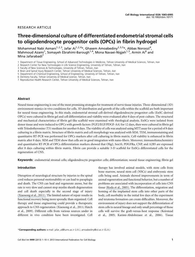
Cell viability and survivalMTTassay was used to explore the viability of EnSCs-drivedOPCs in fibrin gel (3D) and 2D plating at 1, 4, and 8 days.
Viability of OPCs decreased with time in 2D culture as well asin 3D cultures. However, OPCs showed significant improve-ment in viability in all 3 days of 3D culture rather than 2Dculture (Figure 5).
SEM micrographs of EnSCs-derived OPCs seeded in fibrin gelFigure 6 shows SEM image obtained fromOPCs on the fibrinscaffold. Scaffold structure had a meshed network of nano-fibers entanglements with large spaces between them(Figures 6a and 6b). Figures 6c and 6d show the suspendedcells in fibrin having the appearance and size of cells similar to
Figure 2 Immunocytochemical analysis of the expression of A2B5 as a marker of oligodendrocyte progenitor cells in EnSCs 18 days posttreatment with induction media. Nuclei are stained with DAPI. Scale bar is 100mm.
Figure 3 RelativemRNA expression using real-time PCR for expression of oligodendrocyte progenitor cell markers compared to non-treatedEnSCs. Human EnSCs were used as a control. GAPDH is the housekeeping gene control. Error bars show� SEM, n¼ 3 samples.
Mohammad Nabi Asmani et al. 3D culture of EnSC-derived OPCs in fibrin hydrogel
5Cell Biol Int 9999 (2013) 1–10 � 2013 International Federation for Cell Biology

oligodendrocyte progenitor cells. Moreover, the cell body andthe extensions emerging from it show the relationshipbetween fibrin and cells, which indicates that cells havebecome attached and incorporated very well in the scaffold,and that both cells and scaffold express positive interactions
which did not affect cell macrostructure. TEM images(Figure 7) show inner structure of OPCs in relation withfibers. From these figures, the cell and nuclear membraneremained intact with an even nuclear chromatin and nocytoplasmic swelling. Secretion of lipid vacuoles are also seen,
Figure 4 Rheological analysis of fibrin gels with different fibrinogen concentrations. Comparison between hydrogel with 3 and 4mg/mL
fibrinogen (A), comparison of hydrogel with 2 and 3mg/mL of fibrinogen content (B).
3D culture of EnSC-derived OPCs in fibrin hydrogel Mohammad Nabi Asmani et al.
6 Cell Biol Int 9999 (2013) 1–10 � 2013 International Federation for Cell Biology
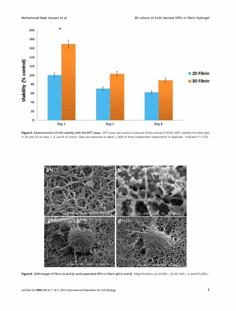
Figure 5 Determination of cell viability with theMTT assay. MTT assay was used as a measure of the survival of hEnSC-OPCs seeded into fibrin gels
in 3D and 2D on days 1, 4, and 8 of culture. Data are expressed as Mean� SEM of three independent experiments in duplicate. � Indicates P< 0.05.
Figure 6 SEM images of fibrin (a and b), and suspended OPCs in fibrin gel (c and d). Magnifications: (a) 20,000�, (b) 30, 000�, (c and d) 5,000�.
Mohammad Nabi Asmani et al. 3D culture of EnSC-derived OPCs in fibrin hydrogel
7Cell Biol Int 9999 (2013) 1–10 � 2013 International Federation for Cell Biology

therefore they shows signs of being active cells suspended infibrin hydrogel.
ImmunohistochemistryThe differentiation of EnSCs into OPCs phenotypes wasfollowed by immunohistochemistry studies after 7 daysculture in fibrin gel. Figure 8 shows staining of OPCs withspecific markers including A2B5 and Olig2 in fibrin gel.Suspended cells expressed A2B5 and Olig2 markers after
7 days cultured in fibrin gel. Differentiated cells retained theirOPCs characteristic in 3D culture and fibrin hydrogel did notexert any negative effect on the cells.
Discussion
Damage to the spinal cord which results from physicalinjuries or frommyelin degenerating diseases not only causesimpediment in everyday life, but also puts psychological and
Figure 7 TEM micrographs of EnSCs-derived OPCs cultured for 6 days inside 3D fibrin matrices. (A) TEM image of fibrin gel (magnification
3,600�) and (B) TEM image of EnSCs-derived OPCs cultured for 6 days inside of 3D fibrin matrices (magnification: 2,150�).
Figure 8 Immunohistochemistry analysis for Olig2 and A2B5 markers in suspended oligodendrocyte progenitor cells after 7 days culture infibrin gel. The results show that the differentiated cells retained their characteristics in 3D culture, and that fibrin hydrogel had no negative effects on the
cells. Scale bar: 100mm.
3D culture of EnSC-derived OPCs in fibrin hydrogel Mohammad Nabi Asmani et al.
8 Cell Biol Int 9999 (2013) 1–10 � 2013 International Federation for Cell Biology
economic burden on society’s shoulder. Different methodsused by physicians, at best, only slow down the degenerationprocess, but are incapable of restoring the patient’s life back tonormal with full functional recovery. Cell therapy as a newtechnique has its drawbacks since cells could spread to otherregions and cause more complicated conditions, not tomention unwanted differentiation of stem cells injected to theinjured site (Xiaosong et al., 2011). Tissue engineering opensup new insight in medical science and potential novel andeffective approaches to solve medical problems; many effortshave been made to help regenerate spinal cord tissue. We usefibrin as a scaffold, which is superior to other biomaterialswith regard to biodegradability and biocompatibility, plus itsphysical properties are easily adjusted to prepare suitableenvironment for OPCs and neurite extension (Karin andFoo, 2010; Xiaosong et al., 2011; Khaing et al., 2012).Endometrial stem cells were also used as a source to getinduced to OPCs. EnSCs are easier to collect, have higher rateof proliferation and are less immunogenic in comparison toother sources, such as bone marrow and embryonic stemcells. Differentiated cells are suspended in fibrin scaffold as asupport for cells to improve engraftment. The outcome ishigher functional recovery in case of animal and humantrials, and thus aspects of the in vitro biocompatibility of thetechnique have been investigated here in more detail.
In the first step, stem cells were isolated from endometrialtissue and cultured for three passages before induction toOPCs. Morphology of isolate cells and flow cytometry datafrom previous works by authors verified successful isolationof endometrial stem cells with positive expression formesenchymal markers CD105, CD90, and endometrialspecific marker CD146 (Ai et al., 2012; Ebrahimi-Baroughet al., 2013). These results are also consistent with reports onEnSCs isolation (Kato et al., 2007; Schwab and Gargett, 2007;Tsuji et al., 2008). Isolated cells were induced to oligoden-drocyte progenitor cells by protocol similar to Nistor et al.(2005) with some modifications for 21 days, and thenimmunocytochemistry analysis of A2B5 staining showedpositive expression by these cells. Quantitative RT-PCR alsoshowed positive expression of A2B5, SOX10 and, especially,the CNP marker, which oligodendrocytes express in the CNSa clear demonstration of OPCs differentiation. Since theinduced cells were not in co-culture with neurons,differentiated cells are not mature myelinated cells; express-ing A2B5 confirms that these cells are in appropriate stage ofdifferentiation, so they would complete their maturationwhen they are brought into contact with neurons insidespinal cord environment.
The fibrin gels with three different concentrationsprepared for mechanical analysis and results showed thatall preserve their 3D hydrogel structure under frequencysweep as a viscoelastic material. An environment withelasticity of <1,000 Pa is suitable for both glial and neuron
survival and proliferation; however, hydrogel of elasticity>1,000 Pa is only appropriate for glial proliferation (Uiboet al., 2009; Aurand et al., 2012). Comparing hydrogels withthree different fibrinogen concentrations, the fibrin at3mg/mL provides the best environment in both durabilityand similarity to the spinal cord environment.
MTT assay was positive for differentiated cells viability, asincrease in survival was continuous for duration of 8 days inthe 3D structure, with fibrin showing no evidence of toxicity.SEM images gave the microstructure of fibrin as a 3D meshwith nano-fibers crossed and entangled with high porosityfor nutrient transfer and cell penetration. These images gaveevidence of cell attachment and diffusion inside the fibrinstructure, without damage to the macrostructure of cells.TEM analysis of suspended induced cells verified that they areactive as their homogenous chromatin, and their cell andnuclear membranes appeared intact. Clearly there are nosigns of disruption of cells with fibrin mesh network andhence no cell death. Finally immunohistochemistry showedthat differentiated EnSCs to OPCs held in reserve their natureas positive expression of A2B5 and Olig2 markers wereevident in suspended cells after 7 days in culture.
Conclusion
EnSCs have the capacity to differentiation into OPCs; thebiocompatibility of the differentiated cells in fibrin gel wasinvestigated. The differentiated cells expressed OPCs makersof A2B5 by staining and CNP, A2B5, PDGFRa, and Olig2 byqRT-PCR, which shows their differentiation stage. Aftersuspension in fibrin scaffold, different techniques showed apositive effect of fibrin and 3D structure on differentiatedOPCs. This might aid in effective regeneration of damagedspinal cord.
Acknowledgments and funding
We thank Tehran University of Medical Sciences Research,the Iranian Council of Stem Cell Technology, and the IranNational Science Foundation (INSF) for financial supported.Also we would like to thank Dr. Babak Kafashi of the polymercharacteristics Lab at University of Tehran for support andcooperation.
References
Ai J, Shahverdi AR, Ebrahimi-Barough S, Kouchesfehani HM,
Heidari S, Roozafzoon R, Verdi J, Khoshzaban A (2012)
Derivation of adipocytes from human endometrial stem cells
(EnSCs). J Reprod Infertil 13(3): 151–57.
Aurand ER, Lampe KJ, Bjugstad KB (2012) Defining and designing
polymers and hydrogels for neural tissue engineering. Neurosci
Res 72(3): 199–213.
Mohammad Nabi Asmani et al. 3D culture of EnSC-derived OPCs in fibrin hydrogel
9Cell Biol Int 9999 (2013) 1–10 � 2013 International Federation for Cell Biology

Bockeria L, Bogin V, Bockeria O, Le T, Alekyan B,Woods EJ, Brown
A, Ichim TE, Amit PN (2013) Endometrial regenerative cells for
treatment of heart failure: a new stem cell enters the clinic. J
Trans Med 11: 56.
Ebrahimi-Barough S, Kouchesfahani HM, Ai J, Mahmodiani M,
Tavakol S, Massumi M (2013) Programming of human
endometrial-derived stromal cells (EnSCs) into pre-oligoden-
drocyte cells by overexpression of miR-219. Neurosci Lett 537:
65–70.
Esfandiari N, Nazemian Z, Casper RF (2008) Three-dimensional
culture of endometrial cells: an in vitro model of endometriosis.
Am J Reprod Immunol 60(4): 283–9.
Fasciani A, Bocci G, Xu J, Bielecki R, Greenblatt E, Leyland N,
Casper RF (2003) Three-dimensional in vitro culture of
endometrial explants mimics the early stages of endometriosis.
Fertil Steril 83: 1137–43.
Hatch MN, Nistor G, Keirstead HS (2009) Derivation of high-
purity oligodendroglial progenitors. Methods Mol Biol 549: 59–
75.
IshiharaM,Mochizuki-Oda N, Iwatsuki K, KishimaH, Iwamoto Y,
Ohnishi O, Umegaki M, Yoshimine T (2011) A new three-
dimensional axonal outgrowth assay for central nervous system
regeneration. J Neurosci Methods 198(2): 181–6.
Karimi-Abdolrezaee S, Eftekharpour E, Wang J, Morshead CM,
Fehlings MG (2006) Delayed transplantation of adult neural
precursor cells promotes remyelination and functional neuro-
logical recovery after spinal cord injury. Neuroscience 26(13):
3377–89.
Karin S, Foo S, Cheryl WP, Heilshorn SC (2010) Biomaterial
design strategies for the treatment of spinal cord injuries.
Neurotra 27(1): 1–19.
Kato K, Yoshimoto M, Adachi S, Yamayoshi A, Arima Y (2007)
Stem cells in human normal endometrium and endometrial
cancer cells: characterization of side population cells. Hum
Reprod 22: 1214–23.
Keirstead HS, Nistor G, Bernal G, Totoiu M, Cloutier F, Sharp K,
Steward O (2005) Human embryonic stem cell-derived
oligodendrocyte progenitor cell transplants remyelinate and
restore locomotion after spinal cord injury. Neuroscience
25(19): 4694–705.
Khaing ZZ, Schmidt CE (2012) Advances in natural biomaterials
for nerve tissue repair. Neurosci Lett 519(2): 103–14.
Koda M, Okada S, Nakayama T, Koshizuka S, Kamada T, Nishio Y,
Someya Y (2005) Hematopoietic stem cell and marrow stromal
cell for spinal cord injury in mice. Neuroreport 16(16): 1763–7.
Le N, Damien L, Guehennec L, Rouillon T, Pilet P, Bilban M,
Layrolle P, Daculsi G (2006) Micro-architecture of calcium
phosphate granules and fibrin glue composites for bone tissue
engineering. Biomaterials 27(13): 2716–22.
Li X, Katsanevakis E, Liu X, Zhang N, Wen X (2012) Engineering
neural stem cell fates with hydrogel design for central nervous
system regeneration. Prog Polym Sci 37(8): 1105–29.
Mobarakeh ZT, Ai J, Yazdani F, Sorkhabadi SMR, Ghanbari Z,
Noroozi A, Mortazavi-Tabatabaei SA, Massumi M, Ebrahimi-
Barough S (2012) Human endometrial stem cells as a new
source for programming to neural cells. Cell Biol Int Rep 19(1):
e00015.
Nistor GI, Totoiu MO, Haque N (2005) Human embryonic stem
cells differentiate into oligodendrocytes in high purity and
myelinate after spinal cord transplantation. Glia 49: 385–96.
Schmittgen TD, Livak KJ (2008) Analyzing real-time PCR data by
the comparative C(T) method. Nat Protoc 3: 1101–8.
Schwab KE, Gargett CE (2007) Co-expression of two perivascular
cell markers isolates mesenchymal stem-like cells from human
endometrium. Hum Reprod 22: 2903–11.
Shoffstall AJ, Dawn MT, Erin BL (2012) Engineering therapies in
the CNS: what works and what can be translated. Neurosci Lett
519(2): 147–54.
Tabesh H, Amoabediny GH, Salehi NN, Heydari M, Yosefifard M,
Ranaei SO, Mottaghy K (2009) The role of biodegradable
engineered scaffolds seeded with Schwann cells for spinal cord
regeneration. Neurochem Int 54(2): 73–83.
Tsuji S, Yoshimoto M, Takahashi K, Noda Y, Nakahata T, Heike T
(2008) Side population cells contribute to the genesis of human
endometrium. Fertil Steril 90: 1528–37.
Uibo R, Laidmäe I, Sawyer ES, Flanagan L, Georges PC, Winer JP,
Janmey P (2009) Soft materials to treat central nervous system
injuries: evaluation of the suitability of non-mammalian fibrin
gels. Biochim Biophys Acta 1793(5): 924–30.
Xiaosong GU, Fei D, Yumin Y, Jie L (2011) Construction of tissue
engineered nerve grafts and their application in peripheral nerve
regeneration. Prog Neurobiol 93(2): 204–30.
Received 13 April 2013; accepted 22 July 2012.
3D culture of EnSC-derived OPCs in fibrin hydrogel Mohammad Nabi Asmani et al.
10 Cell Biol Int 9999 (2013) 1–10 � 2013 International Federation for Cell Biology
Copyright © 2022 FDOKUMEN