Oligodendrocyte Precursor Cells Are Accurate Sensors of Local K+ in Mature Gray Matter
11
Cellular/Molecular Oligodendrocyte Precursor Cells Are Accurate Sensors of Local K in Mature Gray Matter Paloma P. Maldonado, 1,2,3 Mateo Ve ´lez-Fort, 1,2,3 Franc¸oiseLevavasseur, 1,2,3 and María Cecilia Angulo 1,2,3 1 INSERM U603, 75006 Paris, France, 2 CNRS UMR 8154, 75006 Paris, France, and 3 Universite ´ Paris Descartes, Sorbonne Paris Cite ´, 75006 Paris, France Oligodendrocyte precursor cells (OPCs) are the major source of myelinating oligodendrocytes during development. These progenitors are highly abundant at birth and persist in the adult where they are distributed throughout the brain. The large abundance of OPCs after completion of myelination challenges their unique role as progenitors in the healthy adult brain. Here we show that adult OPCs of the barrel cortex sense fine extracellular K increases generated by neuronal activity, a property commonly assigned to differentiated astrocytes rather than to progenitors. Biophysical, pharmacological, and single-cell RT-PCR analyses demonstrate that this ability of OPCs establishes itself progressively through the postnatal upregulation of Kir4.1 K channels. In animals with advanced cortical myelination, extracellular stimulation of layer V axons induces slow K currents in OPCs, which amplitude correlates with presynaptic action potential rate. Moreover, using paired recordings, we demonstrate that the discharge of a single neuron can be detected by nearby adult OPCs, indicating that these cells are strategically located to detect local changes in extracellular K concentration during physio- logical neuronal activity. These results identify a novel unitary neuron–OPC connection, which transmission does not rely on neurotrans- mitter release and appears late in development. Beyond their abundance in the mature brain, the postnatal emergence of a physiological response of OPCs to neuronal network activity supports the view that in the adult these cells are not progenitors only. Introduction Oligodendrocyte precursor cells (OPCs) are a class of progenitors that generate myelinating oligodendrocytes in the CNS during development and after demyelinating injury through a myelin repair process (Richardson et al., 2011). Recent Cre-Lox fate- mapping studies have also suggested that the fate of OPCs is relatively flexible, producing astrocytes and neurons in specific brain regions, although the multipotent capacity of OPCs is still a matter of intense debate (Richardson et al., 2011). In any case, OPCs have always been regarded as progenitors, even in the healthy adult brain where they constitute 2–3% of total cells in gray matter and 5– 8% in white matter (Dawson et al., 2003). Nevertheless, their large abundance and widespread distribution have suggested that, in addition to their role as progenitors, OPCs reside in the mature brain to play other functions. In support of this view, a few scattered reports have shown that OPCs undergo important physiological modifications during postnatal develop- ment. In the hippocampus, the biophysical phenotype of OPCs, characterized by an outward rectifying I–V relationship during the two first postnatal weeks, progressively changes toward a lin- ear shape (Kressin et al., 1995; Zhou et al., 2006). Developmental alterations in membrane receptor expression also occur in these cells. This is the case for the subunit composition of AMPA re- ceptors (Ziskin et al., 2007; Mangin et al., 2008; Maldonado et al., 2011) and for the postnatal downregulation of nicotinic receptors (Ve ´lez-Fort et al., 2009). Finally, complex changes have been re- ported concerning the mechanisms governing neuron-to-OPC communication in the brain. Glutamatergic synaptic responses of OPCs (Bergles et al., 2000; Karadottir et al., 2005; Kukley et al., 2007; Ziskin et al., 2007) increase in amplitude during the first three postnatal weeks, probably indicating a maturation process of these synapses (Ziskin et al., 2007; Mangin et al., 2008; De Biase et al., 2010). In contrast, direct GABAergic synaptic inputs of OPCs in the barrel cortex are lost early in postnatal development and replaced in the adult by an extrasynaptic mode of transmis- sion, mediated solely by GABA spillover (Ve ´lez-Fort et al., 2010). Despite all these recent indications of divergent properties between young and adult OPCs, more efforts are needed to deci- pher developmental physiological changes that govern so far un- recognized functions of these cells in the brain. In particular, little information exists on neuron-to-OPC signaling in mature gray matter. In this report, we investigated the properties of neuron- to-OPC communication when myelination is advanced in the barrel cortex. We found that, beyond neurotransmitter transmis- sion, adult OPCs are able to efficiently detect local [K ] o in- Received April 23, 2012; revised Nov. 7, 2012; accepted Dec. 7, 2012. Author contributions: P.P.M., F.L., and M.C.A. designed research; P.P.M., M.V.-F., and M.C.A. performed research; F.L. contributed unpublished reagents/analytic tools; P.P.M. analyzed data; P.P.M., M.V.-F., and M.C.A. wrote the paper. This work was supported by Grant R11077KK from Agence Nationale de la Recherche and Grants R09206KK and R11077KK from Fondation pour l’aide a ` la recherche sur la Scle ´rose en Plaques. P.P.M. was supported by a fellowship from Ecole des Neurosciences de Paris. M.V-F. was supported by a fellowship from Ligue Franc¸aise contre la Scle ´rose en Plaques. We thank D.E. Bergles and F. Lesage for the gift of NG2-DsRed and TWIK1 / transgenic mice, respectively; Isabel Llano and Troy W. Margrie for helpful comments on the manuscript; E. Audinat for advice in single-cell RT-PCR and discussions; and N. Corte ´s, J. Montanaro, Q. Bourgeois, and the SCM Imaging Platform of the Sts-Pe `res Biomedical Sciences site of Paris Descartes University for technical assistance. The authors declare no competing financial interests. Correspondence should be addressed to Dr. María Cecilia Angulo, Laboratoire de Neurophysiologie et Nouvelles Microscopies, INSERM U603, CNRS UMR 8154, Universite ´ Paris Descartes, Sorbonne Paris Cite ´, 45 rue des Saints- Pe `res, 75006 Paris, France. E-mail: [email protected]. M. Ve ´lez-Fort’s present address is Division of Neurophysiology, National Institute for Medical Research, Mill Hill, London NW7 1AA, United Kingdom. DOI:10.1523/JNEUROSCI.1961-12.2013 Copyright © 2013 the authors 0270-6474/13/332432-11$15.00/0 2432 • The Journal of Neuroscience, February 6, 2013 • 33(6):2432–2442
Transcript of Oligodendrocyte Precursor Cells Are Accurate Sensors of Local K+ in Mature Gray Matter
Cellular/Molecular
Oligodendrocyte Precursor Cells Are Accurate Sensors ofLocal K� in Mature Gray Matter
Paloma P. Maldonado,1,2,3 Mateo Velez-Fort,1,2,3 Francoise Levavasseur,1,2,3 and María Cecilia Angulo1,2,3
1INSERM U603, 75006 Paris, France, 2CNRS UMR 8154, 75006 Paris, France, and 3Universite Paris Descartes, Sorbonne Paris Cite, 75006 Paris, France
Oligodendrocyte precursor cells (OPCs) are the major source of myelinating oligodendrocytes during development. These progenitorsare highly abundant at birth and persist in the adult where they are distributed throughout the brain. The large abundance of OPCs aftercompletion of myelination challenges their unique role as progenitors in the healthy adult brain. Here we show that adult OPCs of thebarrel cortex sense fine extracellular K � increases generated by neuronal activity, a property commonly assigned to differentiatedastrocytes rather than to progenitors. Biophysical, pharmacological, and single-cell RT-PCR analyses demonstrate that this ability ofOPCs establishes itself progressively through the postnatal upregulation of Kir4.1 K � channels. In animals with advanced corticalmyelination, extracellular stimulation of layer V axons induces slow K � currents in OPCs, which amplitude correlates with presynapticaction potential rate. Moreover, using paired recordings, we demonstrate that the discharge of a single neuron can be detected by nearbyadult OPCs, indicating that these cells are strategically located to detect local changes in extracellular K � concentration during physio-logical neuronal activity. These results identify a novel unitary neuron–OPC connection, which transmission does not rely on neurotrans-mitter release and appears late in development. Beyond their abundance in the mature brain, the postnatal emergence of a physiologicalresponse of OPCs to neuronal network activity supports the view that in the adult these cells are not progenitors only.
IntroductionOligodendrocyte precursor cells (OPCs) are a class of progenitorsthat generate myelinating oligodendrocytes in the CNS duringdevelopment and after demyelinating injury through a myelinrepair process (Richardson et al., 2011). Recent Cre-Lox fate-mapping studies have also suggested that the fate of OPCs isrelatively flexible, producing astrocytes and neurons in specificbrain regions, although the multipotent capacity of OPCs is still amatter of intense debate (Richardson et al., 2011). In any case,OPCs have always been regarded as progenitors, even in thehealthy adult brain where they constitute 2–3% of total cells ingray matter and 5– 8% in white matter (Dawson et al., 2003).Nevertheless, their large abundance and widespread distributionhave suggested that, in addition to their role as progenitors, OPCs
reside in the mature brain to play other functions. In support ofthis view, a few scattered reports have shown that OPCs undergoimportant physiological modifications during postnatal develop-ment. In the hippocampus, the biophysical phenotype of OPCs,characterized by an outward rectifying I–V relationship duringthe two first postnatal weeks, progressively changes toward a lin-ear shape (Kressin et al., 1995; Zhou et al., 2006). Developmentalalterations in membrane receptor expression also occur in thesecells. This is the case for the subunit composition of AMPA re-ceptors (Ziskin et al., 2007; Mangin et al., 2008; Maldonado et al.,2011) and for the postnatal downregulation of nicotinic receptors(Velez-Fort et al., 2009). Finally, complex changes have been re-ported concerning the mechanisms governing neuron-to-OPCcommunication in the brain. Glutamatergic synaptic responsesof OPCs (Bergles et al., 2000; Karadottir et al., 2005; Kukley et al.,2007; Ziskin et al., 2007) increase in amplitude during the firstthree postnatal weeks, probably indicating a maturation processof these synapses (Ziskin et al., 2007; Mangin et al., 2008; De Biaseet al., 2010). In contrast, direct GABAergic synaptic inputs ofOPCs in the barrel cortex are lost early in postnatal developmentand replaced in the adult by an extrasynaptic mode of transmis-sion, mediated solely by GABA spillover (Velez-Fort et al., 2010).
Despite all these recent indications of divergent propertiesbetween young and adult OPCs, more efforts are needed to deci-pher developmental physiological changes that govern so far un-recognized functions of these cells in the brain. In particular, littleinformation exists on neuron-to-OPC signaling in mature graymatter. In this report, we investigated the properties of neuron-to-OPC communication when myelination is advanced in thebarrel cortex. We found that, beyond neurotransmitter transmis-sion, adult OPCs are able to efficiently detect local [K�]o in-
Received April 23, 2012; revised Nov. 7, 2012; accepted Dec. 7, 2012.Author contributions: P.P.M., F.L., and M.C.A. designed research; P.P.M., M.V.-F., and M.C.A. performed research;
F.L. contributed unpublished reagents/analytic tools; P.P.M. analyzed data; P.P.M., M.V.-F., and M.C.A. wrote thepaper.
This work was supported by Grant R11077KK from Agence Nationale de la Recherche and Grants R09206KK andR11077KK from Fondation pour l’aide a la recherche sur la Sclerose en Plaques. P.P.M. was supported by a fellowshipfrom Ecole des Neurosciences de Paris. M.V-F. was supported by a fellowship from Ligue Francaise contre la Scleroseen Plaques. We thank D.E. Bergles and F. Lesage for the gift of NG2-DsRed and TWIK1 �/� transgenic mice,respectively; Isabel Llano and Troy W. Margrie for helpful comments on the manuscript; E. Audinat for advice insingle-cell RT-PCR and discussions; and N. Cortes, J. Montanaro, Q. Bourgeois, and the SCM Imaging Platform of theSts-Peres Biomedical Sciences site of Paris Descartes University for technical assistance.
The authors declare no competing financial interests.Correspondence should be addressed to Dr. María Cecilia Angulo, Laboratoire de Neurophysiologie et Nouvelles
Microscopies, INSERM U603, CNRS UMR 8154, Universite Paris Descartes, Sorbonne Paris Cite, 45 rue des Saints-Peres, 75006 Paris, France. E-mail: [email protected].
M. Velez-Fort’s present address is Division of Neurophysiology, National Institute for Medical Research, Mill Hill,London NW7 1AA, United Kingdom.
DOI:10.1523/JNEUROSCI.1961-12.2013Copyright © 2013 the authors 0270-6474/13/332432-11$15.00/0
2432 • The Journal of Neuroscience, February 6, 2013 • 33(6):2432–2442
creases generated by action potential discharges of individualcortical neurons. We also show that this process is mediated bythe progressive upregulation of Kir4.1 channels in OPCs duringpostnatal development. The emergence of Kir4.1 channels inadult OPCs makes these cells accurate sensors of K� efflux duringneuronal physiological activity.
Materials and MethodsSlice preparation and electrophysiology. All experiments followed Euro-pean Union and institutional guidelines for the care and use of laboratoryanimals. Acute parasagittal slices (300 �m) of the barrel cortex weremainly performed at the 2 postnatal (PN) week and 4 –5 PN weeks aspreviously described (Velez-Fort et al., 2010). OPCs were identified inNG2-DsRed BAC transgenic mice (560 nm excitation and 620 nm emis-sion wavelengths; Green OptoLed, Cairn Research). Patch-clamp re-cordings were performed at 33°C using an extracellular solutioncontaining the following (in mM): 126 NaCl, 2.5 KCl, 1.25 NaH2PO4, 26NaHCO3, 20 glucose, 5 pyruvate, 2 CaCl2, and 1 MgCl2 (95% O2, 5%CO2). The intracellular solution was either a CsCl-based intracellularsolution containing the following (in mM): 130 CsCl, 10 4AP, 5 TEA-Cl,5 EGTA, 0.5 CaCl2, 2 MgCl2, 10 HEPES, 2 Na2ATP, 0.2 Na-GTP, 10Na2-phosphocreatine, or a KCl-based intracellular solution containingthe following (in mM): 130 KCl, 5 EGTA, 0.5 CaCl2, 2 MgCl2, 10 HEPES,2 Na2ATP, 0.2 Na-GTP, 10 Na2-phosphocreatine (pH � 7.3, 296mOsm). When isoflurane was used in the perfusion, it was dissolved inthe extracellular solution immediately before its bath application at roomtemperature and maintained in a sealed bottle in which bubbling wasstopped, as previously described (Taverna et al., 2005). Changes in [K �]o
from 2.5 to 10 mM were obtained by isotonic replacement of Na �.Extracellular stimulations were obtained using a monopolar electrode
(glass pipette) placed in layer V of the barrel cortex (100 �s stimulations;Iso-Stim 01D, npi electronic). Paired-pulse and trains of stimulation ofneurons were performed at 40 V, unless otherwise indicated. Neuronswere recorded in voltage-clamp and current-clamp modes with an intra-cellular solution containing the following (in mM): 130 K-gluconate, 5EGTA, 0.5 CaCl2, 2 MgCl2, 10 HEPES, 2 Na2ATP, 0.2 Na-GTP, and 10Na2-phosphocreatine. They were held at �70 or �90 mV after correc-tion for a liquid junction potential (10 mV). In simultaneous recordingsbetween a neuron and an OPC, depolarizing current pulses of 200 or 400pA were applied during 400 ms to the neuron, each 3–5 s to elicit actionpotential discharges. In simultaneous neuron-neuron recordings, theduration of this pulse was also increased to 800 ms, and the postsynapticneuron was held to �90 mV as OPCs. Recordings were made withoutseries resistance (Rs) compensation; Rs was monitored during record-ings, and cells showing a change of �30% in Rs were discarded. Whole-cell recordings were obtained using Multiclamp 700B, filtered at 2– 4kHz, and digitized at 20 kHz. Digitized data were analyzed off-line usingpClamp 10.1 (Molecular Devices) and Igor Pro 6.0.
Because the decay of inward K � currents evoked by neuronal stimu-lation was not well described by a single exponential function, we esti-mated the time required to decay by measuring the half-decay time of thecurrent evoked by paired-pulse stimulation. The paired-pulse ratio ofthese currents was obtained by dividing the current amplitude of thesecond pulse by that of the first pulse. In simultaneous patch-clamprecordings, currents evoked in OPCs by single neuron firing were con-sidered as a response only if they were on average larger than 2 times theSD of noise and if their amplitude distribution was significantly differentfrom that of the noise.
Steady-state I–V curves were obtained by applying voltage steps of 400ms from �40 mV to �140 mV and by measuring current amplitudes at390 ms from the beginning of the steps. It is noteworthy that we alreadyshowed that neither the capacitance nor the morphology of OPCs of thebarrel cortex changes during development (Velez-Fort et al., 2010). I–Vcurves of currents sensitive to different agents were obtained by subtract-ing currents in the presence of a drug from their controls and normaliz-ing with respect to the current elicited at �40 mV in control. Todetermine the percentage of OPCs having a young phenotype in 4 –5PNweeks, we plotted the distribution of current amplitudes elicited at a
voltage step of �40 mV in all recorded cells of the 2PN week. In thedistribution graph, 90% of OPCs had a current amplitude inferior to 420pA, which corresponds to the mean current amplitude plus the SD(mean � SD: 220 � 198; n � 26). We thus considered as a young phe-notype all cells having a current �420 pA and as an adult phenotypethose having currents �420 pA at a voltage step of �40 mV. Only cellsrecorded in the CsCl-based intracellular solution with an adult pheno-type were considered for analysis (70% of OPCs in 4 –5PN weeks).
Immunohistochemistry. For MBP and vGluT2 immunostainings, micewere perfused intracardially with PBS alone followed by 0.15 M phos-phate buffer, pH 7.4 (PB) containing 4% paraformaldehyde at PN7,PN13–14, and PN27–31 (n � 3 animals for each developmental stage).Brains were removed and placed in a 4% paraformaldehyde solutionovernight. Then, brain slices (50 �m) were prepared in PBS ice-coldsolution (4°C), permeabilized with 1% Triton X-100 and 2% BSA for 1 h,and incubated two nights with antibodies diluted in a 0.2% Triton X-100solution. Double immunostainings were performed by combiningguinea pig polyclonal vGluT2 (1:1000; Millipore) with mouse monoclo-nal anti-MBP (1:100; Millipore). Primary antibodies were washed threetimes in PBS and incubated in secondary antibodies coupled to AlexaFluor 488 or Alexa Fluor 633 (1:500; Invitrogen) for 2 h at room temper-ature.
For simultaneous recordings between neurons and OPCs, recordedcells were injected with 5.4 mM biocytin (Sigma-Aldrich). The slices con-taining the injected cells were then fixed overnight in 4% paraformalde-hyde at 4°C, rinsed three times in PBS for 10 min, and incubated with 1%Triton X-100 and 2% BSA during 1 h. The slices were incubated over-night with rabbit polyclonal anti-NG2 antibody (1:400; Millipore) di-luted in a 0.2% Triton X-100 solution. Then, they were washed threetimes in PBS and incubated in the secondary antibody coupled to AlexaFluor 633 and streptavidin–Alexa Fluor 488 (Invitrogen) for 2 h.
Immunofluorescence was visualized with a confocal microscope (LSM510, Carl Zeiss). Optical sections of confocal images were sequentiallyacquired using a 10� or 63� oil objectives (NA � 0.3 and 1.4, respec-tively) with the LSM-510 software. Images were processed and analyzedusing ImageJ 1.44c (Wayne Rasband, National Institutes of Health). Be-cause cortical layer I were poorly myelinated, we eliminated the back-ground noise of MBP immunostainings by subtracting the fluorescenceintensity of this layer to the whole image.
Single-cell RT-PCR and genotyping of NG2-DsRed::TWIK1 �/� trans-genic mice. The analysis of single-cell transcripts for Kir4.1 channels wasperformed as previously described with modifications (Angulo et al.,1997; Seifert et al., 2009). Briefly, we harvested the cytoplasm of recordedcells with a patch pipette filled with 6 �l of an autoclaved DEPC-treatedinternal solution containing the following (in mM): 130 CsCl, 5 4-AP, 10TEA-Cl, 2 MgCl2, 0.2 EGTA, and 10 HEPES, pH 7.4, 290 mOsm. Thecontent of the pipette was expelled into a test tube, and the reverse tran-scription (RT) was performed for 2 h at 50°C in a 10 �l reaction volumeby adding the following components at final concentrations as indicated:10 ng/�l hexamer random primers, 0.5 mM each dNTP, 5 mM DTT, 20 Uof RNaseOut (Invitrogen), 100 U of Superscript III Reverse Transcriptase(Invitrogen), and 1� buffer supplied with the enzyme. After incubation,the tubes were kept at �20°C. The RT products were then amplified withprimers for a two-round nested PCR (Seifert et al., 2009). The first PCRwas done for 35 cycles (94°C, 30 s; 54°C, 30 s; 72°C, 45 s) after adding Taqpolymerase (2.5 U; QIAGEN), 1� buffer supplied by the manufacturer,100 �M dNTP, and the following primers (0.3 �M each): 5 TATCAGAG-CAGCCACTTCACCTTC 3 and 5 GGATCGTCTCGGCCCTCTTCT-TAG 3 (final volume, 100 �l). An aliquot of 1.5 �l of the PCR productwas then used as a template for the second PCR (35 cycles; 94°C, 30 s;58°C, 30 s; 72°C, 30 s) performed with the following nested primers: 5TTCACCTTCGAGCCAAGATGACG 3 and 5 AGGCGTGTGGTTG-GCAGGAG 3 (final volume, 25 �l). The positive control consisted ofrunning in parallel RT-PCR for single cells and for total RNA (50 pg)from mouse hippocampus. Two negative controls were obtained by run-ning RT-PCR either after omitting RT or for harvested cortical layer I-IIIneurons.
NG2-DsRed::TWIK1 �/� animals were generated by crossingheterozygous NG2-DsRed mice with homozygous TWIK1 knock-out
Maldonado et al. • OPCs Are K� Sensors J. Neurosci., February 6, 2013 • 33(6):2432–2442 • 2433
mice. Heterozygous double-transgenic micewere then crossed with TWIK1 �/� mice to ob-tain TWIK1 �/� mice expressing DsRed in lit-termates. This last generation was genotypedby PCR with primers designed to amplify a 434bp and a 600 bp fragment for wild-type andknock-out sequences, respectively. PCR wasdone for 40 cycles (94°C, 30 s; 55.4°C, 30 s;72°C, 1 min 30 s) with the following primers: 5TCCTTCCTACCGACAGCGAGAGA 3 and5 GGGATGCCGATGACAGAGTAGAT 3for wild-type; 5 TGCCTTTACGGTGTCTGTTC 3 and 5 GAGAAGGCAGAAATGGAAACTG 3 for knock-out. After patch-clampexperiments, we confirmed that killed animalswere TWIK1 �/� by PCR.
Statistics. Data are expressed as mean � SEMThe nonparametric Mann–Whitney U testfor independent samples was used to deter-mine statistical differences between dataobtained in different cells. Comparisonswithin single cells were performed with theWilcoxon signed-rank test for related sam-ples when n � 6; otherwise, we used a pairedt test (GraphPad InStat software, Version3.06). Amplitude distributions of inducedcurrents in simultaneous recordings werecompared using the Kolmogorov–Smirnovtest (http://www.physics.csbsju.edu/stats).
ResultsOPCs acquire a linear phenotypeduring postnatal development of thebarrel cortexThe properties of OPCs of the barrel cor-tex during postnatal development wereexamined in layer V DsRed� cells re-corded during 2PN and 4 –5PN weeks inNG2-DsRed transgenic mice (Ziskin etal., 2007). These developmental stageswere chosen to cover the critical period of
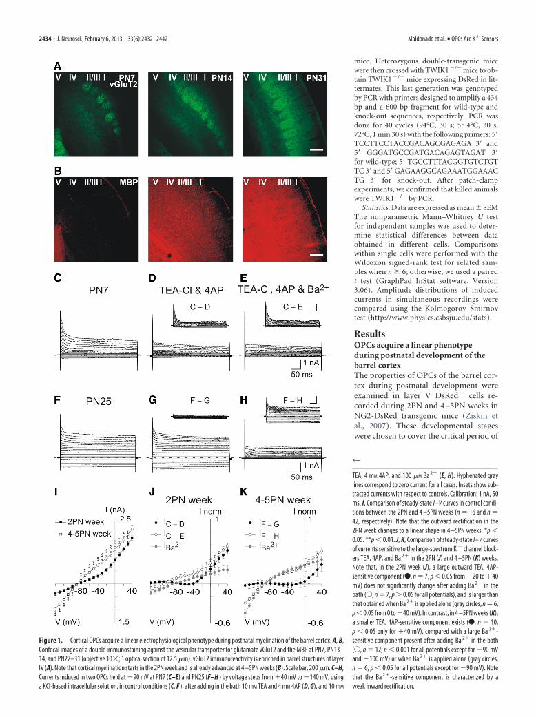
Figure 1. Cortical OPCs acquire a linear electrophysiological phenotype during postnatal myelination of the barrel cortex. A, B,Confocal images of a double immunostaining against the vesicular transporter for glutamate vGluT2 and the MBP at PN7, PN13–14, and PN27–31 (objective 10�; 1 optical section of 12.5 �m). vGluT2 immunoreactivity is enriched in barrel structures of layerIV (A). Note that cortical myelination starts in the 2PN week and is already advanced at 4 –5PN weeks (B). Scale bar, 200 �m. C–H,Currents induced in two OPCs held at �90 mV at PN7 (C–E) and PN25 (F–H ) by voltage steps from �40 mV to �140 mV, usinga KCl-based intracellular solution, in control conditions (C, F ), after adding in the bath 10 mM TEA and 4 mM 4AP (D, G), and 10 mM
4
TEA, 4 mM 4AP, and 100 �M Ba 2� (E, H). Hyphenated graylines correspond to zero current for all cases. Insets show sub-tracted currents with respect to controls. Calibration: 1 nA, 50ms. I, Comparison of steady-state I–V curves in control condi-tions between the 2PN and 4 –5PN weeks (n � 16 and n �42, respectively). Note that the outward rectification in the2PN week changes to a linear shape in 4 –5PN weeks. *p �0.05. **p � 0.01. J, K, Comparison of steady-state I–V curvesof currents sensitive to the large-spectrum K � channel block-ers TEA, 4AP, and Ba 2� in the 2PN (J) and 4 –5PN (K) weeks.Note that, in the 2PN week (J), a large outward TEA, 4AP-sensitive component (F, n � 7, p � 0.05 from �20 to �40mV) does not significantly change after adding Ba 2� in thebath (E, n � 7, p � 0.05 for all potentials), and is larger thanthat obtained when Ba 2� is applied alone (gray circles, n � 6,p�0.05 from 0 to�40 mV). In contrast, in 4 –5PN weeks (K),a smaller TEA, 4AP-sensitive component exists (F, n � 10,p � 0.05 only for �40 mV), compared with a large Ba 2�-sensitive component present after adding Ba 2� in the bath(E, n � 12; p � 0.001 for all potentials except for �90 mVand �100 mV) or when Ba 2� is applied alone (gray circles,n � 6; p � 0.05 for all potentials except for �90 mV). Notethat the Ba 2�-sensitive component is characterized by aweak inward rectification.
2434 • J. Neurosci., February 6, 2013 • 33(6):2432–2442 Maldonado et al. • OPCs Are K� Sensors
myelination of deep layers of the barrelcortex (Fig. 1A,B). Indeed, cortical myeli-nation starts in the 2PN week and is al-ready advanced at PN30 (Fig. 1B), whenthe conduction velocity of action poten-tials of the thalamocortical pathwayreaches a plateau (Salami et al., 2003) andintracortical circuits are relatively mature(Angulo et al., 1999; Morales et al., 2002).
OPCs were first recorded with a KCl-based intracellular solution and held at�90 mV. Application of voltage pulsesfrom �40 mV to �140 mV revealed a dra-matic developmental change in the I–Vrelationship. Comparison of steady-stateI–V curves showed that the outward rec-tification observed in the 2PN weekchanges to a linear shape in 4 –5PN weeks(Fig. 1C,F, I). This modification of the ioncurrent profile was observed at least untilPN54 (n � 4) and was accompanied by amore hyperpolarized resting membranepotential (�77.9 � 2.7 mV in the 2 PNweek vs �88.1 � 2.5 mV in 4 –5PN weeks,respectively; p � 0.01) and a decreased in-put resistance (281.8 � 59.7 M in the2PN week against 70.9 � 6.5 M in the4 –5PN weeks, respectively; p � 0.001).
The sensitivity of currents evoked byvoltage steps to the large-spectrum K�
channel blockers TEA and 4AP was alsodifferent between both developmentalstages. These antagonists blocked a largeproportion of outward currents in the2PN week (Fig. 1D, J) but affected to asmaller extent those in the 4 –5PN weeks(Fig. 1G,K). In contrast, a subsequentapplication of Ba 2� inhibited modestlyadditional currents in young mice (Fig.1E, J) but suppressed substantial currentsin older mice (Fig. 1H,K). This is consistentwith the effect of Ba2� applied alone. SmallBa2�-sensitive currents were revealed in the2PN week (Fig. 1J), whereas large currentshad similar amplitudes than those recordedin the presence of TEA, 4AP, and Ba2� in4–5PN weeks (Fig. 1K).
These results indicate that a Ba2�-sensitive, TEA-insensitive, and 4AP-
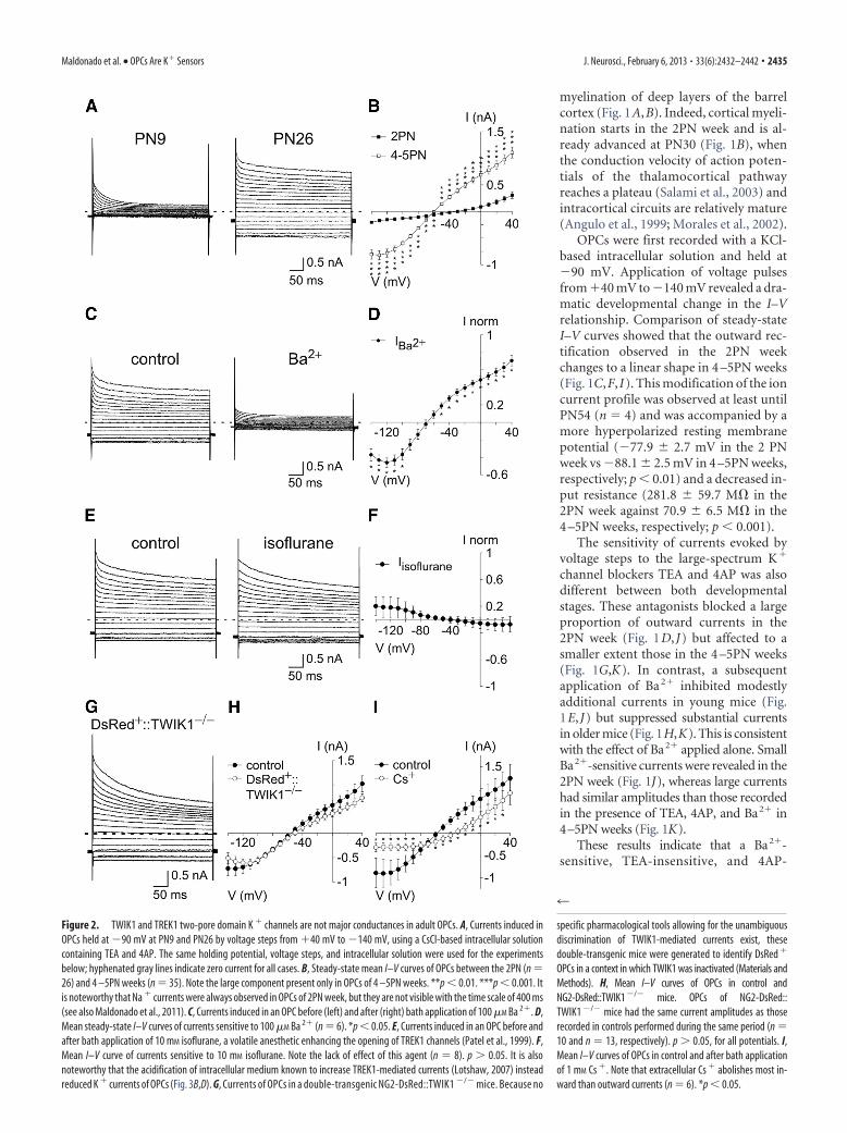
Figure 2. TWIK1 and TREK1 two-pore domain K � channels are not major conductances in adult OPCs. A, Currents induced inOPCs held at �90 mV at PN9 and PN26 by voltage steps from �40 mV to �140 mV, using a CsCl-based intracellular solutioncontaining TEA and 4AP. The same holding potential, voltage steps, and intracellular solution were used for the experimentsbelow; hyphenated gray lines indicate zero current for all cases. B, Steady-state mean I–V curves of OPCs between the 2PN (n �26) and 4 –5PN weeks (n � 35). Note the large component present only in OPCs of 4 –5PN weeks. **p � 0.01. ***p � 0.001. Itis noteworthy that Na � currents were always observed in OPCs of 2PN week, but they are not visible with the time scale of 400 ms(see also Maldonado et al., 2011). C, Currents induced in an OPC before (left) and after (right) bath application of 100 �M Ba 2�. D,Mean steady-state I–V curves of currents sensitive to 100 �M Ba 2� (n � 6). *p � 0.05. E, Currents induced in an OPC before andafter bath application of 10 mM isoflurane, a volatile anesthetic enhancing the opening of TREK1 channels (Patel et al., 1999). F,Mean I–V curve of currents sensitive to 10 mM isoflurane. Note the lack of effect of this agent (n � 8). p � 0.05. It is alsonoteworthy that the acidification of intracellular medium known to increase TREK1-mediated currents (Lotshaw, 2007) insteadreduced K � currents of OPCs (Fig. 3B,D). G, Currents of OPCs in a double-transgenic NG2-DsRed::TWIK1 �/� mice. Because no
4
specific pharmacological tools allowing for the unambiguousdiscrimination of TWIK1-mediated currents exist, thesedouble-transgenic mice were generated to identify DsRed �
OPCs in a context in which TWIK1 was inactivated (Materials andMethods). H, Mean I–V curves of OPCs in control andNG2-DsRed::TWIK1 �/� mice. OPCs of NG2-DsRed::TWIK1 �/� mice had the same current amplitudes as thoserecorded in controls performed during the same period (n �10 and n � 13, respectively). p � 0.05, for all potentials. I,Mean I–V curves of OPCs in control and after bath applicationof 1 mM Cs �. Note that extracellular Cs � abolishes most in-ward than outward currents (n � 6). *p � 0.05.
Maldonado et al. • OPCs Are K� Sensors J. Neurosci., February 6, 2013 • 33(6):2432–2442 • 2435
insensitive conductance, the identity of which is unknown, isupregulated in OPCs during postnatal development of the barrelcortex. The weakly inward rectification of this conductancecounteracts the outward rectification observed in the 2PN week,conferring a linear phenotype on OPCs in the adult (Fig. 1 I,K).
Identification of Kir4.1 channels as a major postnatalupregulated conductance in OPCsThe emergence of a linear I–V curve has been previously de-scribed in hippocampal OPCs (Kressin et al., 1995; Zhou et al.,2006), but the conductance conferring a linear phenotype in theadult has not been identified yet. To isolate the Ba 2�-sensitivecurrent upregulated in OPCs, we tested a CsCl-based intracellu-lar solution containing TEA and 4AP, known to suppress fromthe inside most currents mediated by voltage-gated K� channels.This solution almost completely abolished intrinsic currents inyoung mice, whereas significant currents still remained in 70% ofOPCs of 4 –5PN weeks (considered as OPCs with an adult phe-notype; see Materials and Methods; Fig. 2A,B). Furthermore,bath application of 100 �M Ba 2� almost completely blocked cur-rents of intrinsic I–V curves in adult OPCs (Fig. 2C,D), indicatingthat this intracellular solution is convenient to block voltage-gated K� channels and isolate the currents of our interest.
To identify the K� conductance postnatally upregulated inOPCs, we examined the effect of different blockers for K� chan-nels insensitive to TEA and 4AP and sensitive to Ba 2� (i.e., small-conductance Ca 2�-activated potassium [SK] channels) (Sah andFaber, 2002), the two-pore domain K� channels TREK1 andTWIK1 (Lesage et al., 1996; Lotshaw, 2007), and inwardly recti-fying (Kir) channels (Hibino et al., 2010). First, we excluded thecontribution of SK channels in OPCs of 4 –5PN weeks by apply-ing their selective antagonist apamin and detecting no effects onthe recorded currents (200 nM; p � 0.05; n � 5) (Sah and Faber,2002). Then, we examined whether TWIK1 and TREK1 werefunctionally expressed. According to a transcriptome database,the mRNAs of the two-pore domain K� channels TWIK1 andTREK1 are preferentially enriched in acutely isolated purifiedOPCs (Cahoy et al., 2008). However, the lack of effect of isoflu-rane in these currents, an anesthetic activating TREK1 (Fig.2E,F), and the unchanged electrophysiological phenotype ofOPCs in a transgenic mouse expressing inactivated TWIK1 (Fig.2G,H) (Nie et al., 2005) show that, contrary to astrocytes (Seifertet al., 2009; Zhou et al., 2009), two-pore domain K� channels arepoorly expressed in adult OPCs.
It is well known that Cs� blocks Kir channels from the outside ina voltage-dependent manner (Hibino et al., 2010; Tang et al., 2010).In a CsCl-based intracellular solution, bath applications of 1 mM
Cs� blocked currents of the I–V curve from the outside as expectedfor Kir channels (i.e., abolishing most inward currents and onlyweakly outward currents; Fig. 2I) (Hibino et al., 2010; Tang et al.,2010). Although the effect of intracellular Cs� is poorly character-ized for Kir channels, some effect was evident because currents, par-ticularly inward currents, appeared reduced compared with thoserecorded in KCl-based intracellular solution (Fig. 2B for CsCl vs Fig.1I for KCl) and Erev was shifted from �88.14 � 2.46 mV to�59.30 � 1.30 mV (p � 0.001). These data strongly suggest a majorexpression of a Kir conductance in adult OPCs. In support of thisand as mentioned above, 100 �M Ba2�, a well-known Kir blocker atthis concentration (Hibino et al., 2010), abolished currents of intrin-sic I–V curves in adult OPCs (Fig. 2C,D).
Among Kir channels, Kir4.1 channels are found almost exclu-sively in glial cells (Poopalasundaram et al., 2000; Higashi et al.,2001; Seifert et al., 2009; Tang et al., 2009). Because the deletion of
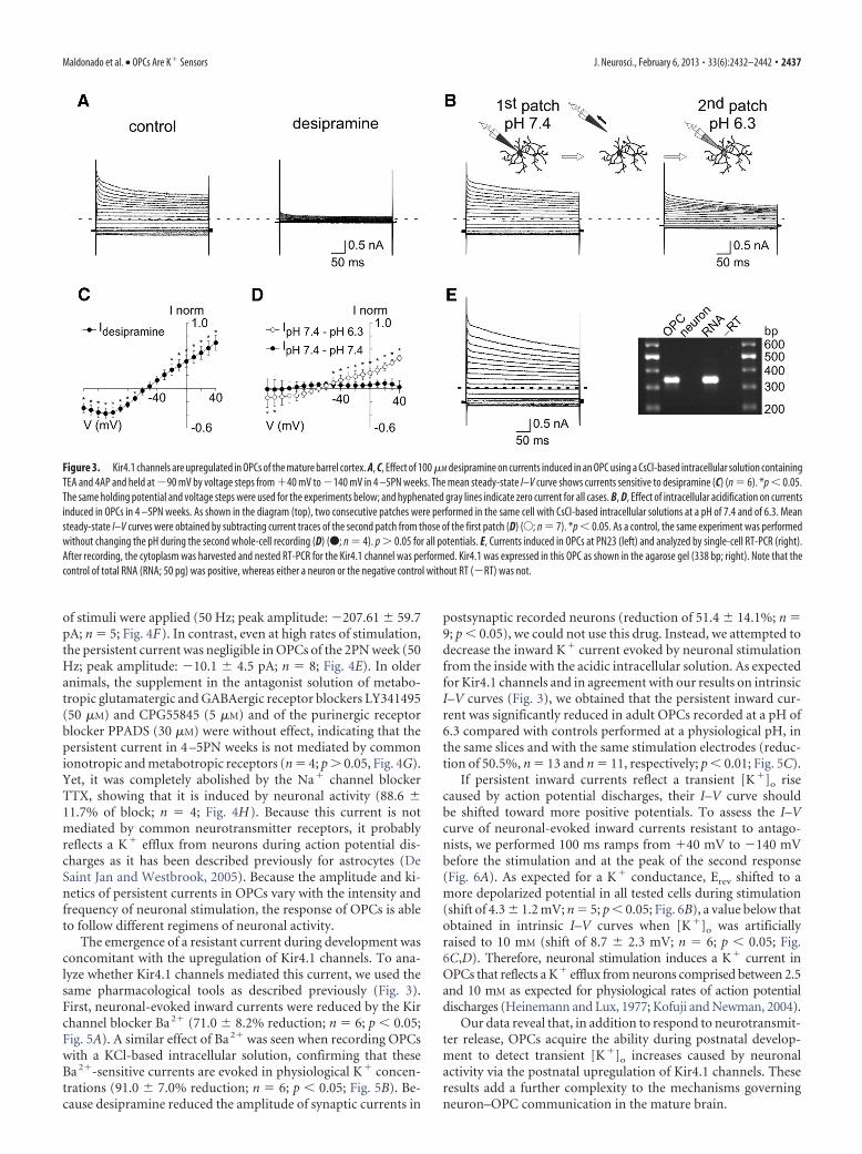
Kir4.1 channels in transgenic mice is lethal around the end of thethird PN week (Kofuji et al., 2000; Djukic et al., 2007), we usedpharmacological tools to examine its functional expression inOPCs of the 4 –5PN weeks. First, we assessed the effect of thetricyclic antidepressant desipramine, an inhibitor of Kir4.1 cur-rents that affects only slightly other Kir currents in heterologousexpression systems (Su et al., 2007). In adult OPCs recorded inCsCl-based intracellular solution, this agent almost completelyabolished K� currents (Fig. 3A,C). Previous works also demon-strated a partial sensitivity of Kir4.1 channels to an intracellularpH of 6.5 (Tucker et al., 2000; Pessia et al., 2001). To test for theeffect of acidic pH, we performed two consecutive whole-cellpatch-clamp recordings on the same cell with intracellular solu-tions at pH of 7.4 and 6.3, respectively (Fig. 3B,D). We observeda decrease of current amplitudes to voltage steps during the sec-ond whole-cell recording, an effect compatible to the acidic pHsensitivity reported for homomeric Kir4.1 channels (reduction of51.0 � 5.9% at �40 mV; Fig. 3D) (Tucker et al., 2000; Pessia et al.,2001). To verify that this effect was not caused by degradation ofthe cell by removing the first patch pipette, we performed thesame experiments without changing the pH during the secondwhole-cell recording. In that case, no changes on the I–V rela-tionship were observed, confirming the effect of acidic pH (Fig.3D, black circles). Finally, we performed single-cell nested RT-PCR experiments to detect transcripts for Kir4.1 channels inOPCs of the barrel cortex (Fig. 3E; see Materials and Methods).Kir4.1 transcripts were found in 7 of 9 recorded adult OPCs(78%), whereas they were amplified in only 1 of 10 cortical neu-rons (10%). Biophysical, pharmacological, and molecular analy-ses indicate that Kir4.1 channels are by far the major K�
conductance developmentally upregulated in OPCs of the ma-ture barrel cortex and that this conductance contribute to thelinear phenotype of I–V relationships in the adult.
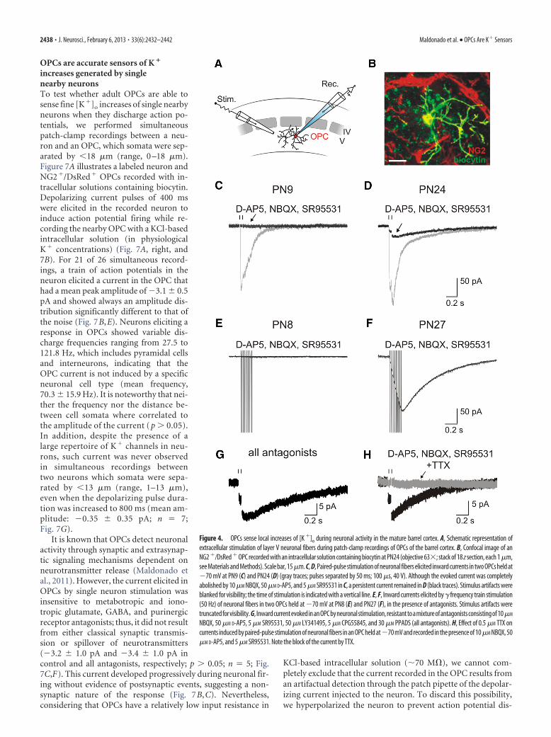
Adult OPCs detect local K � increases through upregulatedKir4.1 channelsAstrocytes are accurate detectors of neuronal activity through aKir4.1 conductance (De Saint Jan and Westbrook, 2005; Djukic etal., 2007). Because Kir4.1 channels are postnatally upregulated inOPCs, we tested whether, similarly to astrocytes, these cells areable to sense extracellular [K�]o increases through these channelsin the mature neuronal network. For this, an extracellular elec-trode placed in layer V was used to stimulate neuronal fiberswhile NG2�/DsRed� OPCs were recorded at �70 mV with theCsCl-based intracellular solution to accurately isolate Kir4.1-mediated currents (Fig. 4A,B). Experiments in older mice wereperformed in OPCs having an adult phenotype. Paired-pulsestimulation easily elicited currents in OPCs of the 2PN and4 –5PN weeks (mean amplitude of first pulse: �136.8 � 41.5 pAand �159.5 � 45.0 pA; n � 8 and n � 13, respectively; Fig. 4C,D,gray traces) (Velez-Fort et al., 2010). However, whereas the iono-tropic glutamatergic and GABAergic receptor antagonists inyoung mice abolished these currents, a persistent current re-mained in older mice (1.7 � 0.6% and 17.7 � 4.4% in 2PN and4 –5PN weeks, respectively; p � 0.001; Fig. 4C,D, black traces).This persistent current in older mice was characterized by long-lasting kinetics with a half-decay time of 0.45 � 0.03 s and a muchweaker paired-pulse depression compared with control condi-tions (paired-pulse ratio of 0.93 � 0.03 in antagonists against0.22 � 0.03 in controls, n � 13; p � 0.001; see Materials andMethods). Moreover, its amplitude increased an average whenthe stimulation intensity varied from 20 to 40 V (increment of70.1%; n � 13 and n � 23, respectively; p � 0.01) or when trains
2436 • J. Neurosci., February 6, 2013 • 33(6):2432–2442 Maldonado et al. • OPCs Are K� Sensors
of stimuli were applied (50 Hz; peak amplitude: �207.61 � 59.7pA; n � 5; Fig. 4F). In contrast, even at high rates of stimulation,the persistent current was negligible in OPCs of the 2PN week (50Hz; peak amplitude: �10.1 � 4.5 pA; n � 8; Fig. 4E). In olderanimals, the supplement in the antagonist solution of metabo-tropic glutamatergic and GABAergic receptor blockers LY341495(50 �M) and CPG55845 (5 �M) and of the purinergic receptorblocker PPADS (30 �M) were without effect, indicating that thepersistent current in 4 –5PN weeks is not mediated by commonionotropic and metabotropic receptors (n � 4; p � 0.05, Fig. 4G).Yet, it was completely abolished by the Na� channel blockerTTX, showing that it is induced by neuronal activity (88.6 �11.7% of block; n � 4; Fig. 4H). Because this current is notmediated by common neurotransmitter receptors, it probablyreflects a K� efflux from neurons during action potential dis-charges as it has been described previously for astrocytes (DeSaint Jan and Westbrook, 2005). Because the amplitude and ki-netics of persistent currents in OPCs vary with the intensity andfrequency of neuronal stimulation, the response of OPCs is ableto follow different regimens of neuronal activity.
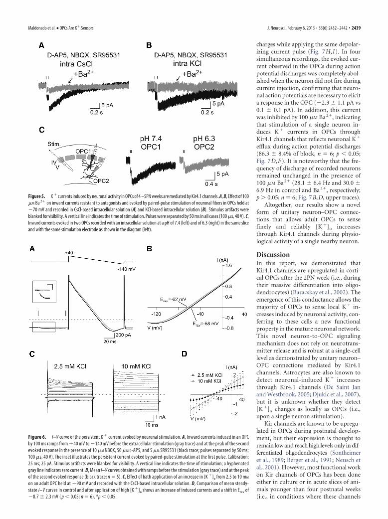
The emergence of a resistant current during development wasconcomitant with the upregulation of Kir4.1 channels. To ana-lyze whether Kir4.1 channels mediated this current, we used thesame pharmacological tools as described previously (Fig. 3).First, neuronal-evoked inward currents were reduced by the Kirchannel blocker Ba 2� (71.0 � 8.2% reduction; n � 6; p � 0.05;Fig. 5A). A similar effect of Ba 2� was seen when recording OPCswith a KCl-based intracellular solution, confirming that theseBa 2�-sensitive currents are evoked in physiological K� concen-trations (91.0 � 7.0% reduction; n � 6; p � 0.05; Fig. 5B). Be-cause desipramine reduced the amplitude of synaptic currents in
postsynaptic recorded neurons (reduction of 51.4 � 14.1%; n �9; p � 0.05), we could not use this drug. Instead, we attempted todecrease the inward K� current evoked by neuronal stimulationfrom the inside with the acidic intracellular solution. As expectedfor Kir4.1 channels and in agreement with our results on intrinsicI–V curves (Fig. 3), we obtained that the persistent inward cur-rent was significantly reduced in adult OPCs recorded at a pH of6.3 compared with controls performed at a physiological pH, inthe same slices and with the same stimulation electrodes (reduc-tion of 50.5%, n � 13 and n � 11, respectively; p � 0.01; Fig. 5C).
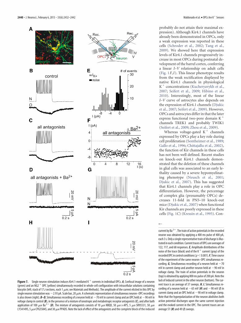
If persistent inward currents reflect a transient [K�]o risecaused by action potential discharges, their I–V curve shouldbe shifted toward more positive potentials. To assess the I–Vcurve of neuronal-evoked inward currents resistant to antago-nists, we performed 100 ms ramps from �40 mV to �140 mVbefore the stimulation and at the peak of the second response(Fig. 6A). As expected for a K� conductance, Erev shifted to amore depolarized potential in all tested cells during stimulation(shift of 4.3 � 1.2 mV; n � 5; p � 0.05; Fig. 6B), a value below thatobtained in intrinsic I–V curves when [K�]o was artificiallyraised to 10 mM (shift of 8.7 � 2.3 mV; n � 6; p � 0.05; Fig.6C,D). Therefore, neuronal stimulation induces a K� current inOPCs that reflects a K� efflux from neurons comprised between 2.5and 10 mM as expected for physiological rates of action potentialdischarges (Heinemann and Lux, 1977; Kofuji and Newman, 2004).
Our data reveal that, in addition to respond to neurotransmit-ter release, OPCs acquire the ability during postnatal develop-ment to detect transient [K�]o increases caused by neuronalactivity via the postnatal upregulation of Kir4.1 channels. Theseresults add a further complexity to the mechanisms governingneuron–OPC communication in the mature brain.
Figure 3. Kir4.1 channels are upregulated in OPCs of the mature barrel cortex. A, C, Effect of 100 �M desipramine on currents induced in an OPC using a CsCl-based intracellular solution containingTEA and 4AP and held at �90 mV by voltage steps from �40 mV to �140 mV in 4 –5PN weeks. The mean steady-state I–V curve shows currents sensitive to desipramine (C) (n � 6). *p � 0.05.The same holding potential and voltage steps were used for the experiments below; and hyphenated gray lines indicate zero current for all cases. B, D, Effect of intracellular acidification on currentsinduced in OPCs in 4 –5PN weeks. As shown in the diagram (top), two consecutive patches were performed in the same cell with CsCl-based intracellular solutions at a pH of 7.4 and of 6.3. Meansteady-state I–V curves were obtained by subtracting current traces of the second patch from those of the first patch (D) (E; n � 7). *p � 0.05. As a control, the same experiment was performedwithout changing the pH during the second whole-cell recording (D) (F; n � 4). p � 0.05 for all potentials. E, Currents induced in OPCs at PN23 (left) and analyzed by single-cell RT-PCR (right).After recording, the cytoplasm was harvested and nested RT-PCR for the Kir4.1 channel was performed. Kir4.1 was expressed in this OPC as shown in the agarose gel (338 bp; right). Note that thecontrol of total RNA (RNA; 50 pg) was positive, whereas either a neuron or the negative control without RT (�RT) was not.
Maldonado et al. • OPCs Are K� Sensors J. Neurosci., February 6, 2013 • 33(6):2432–2442 • 2437
OPCs are accurate sensors of K �
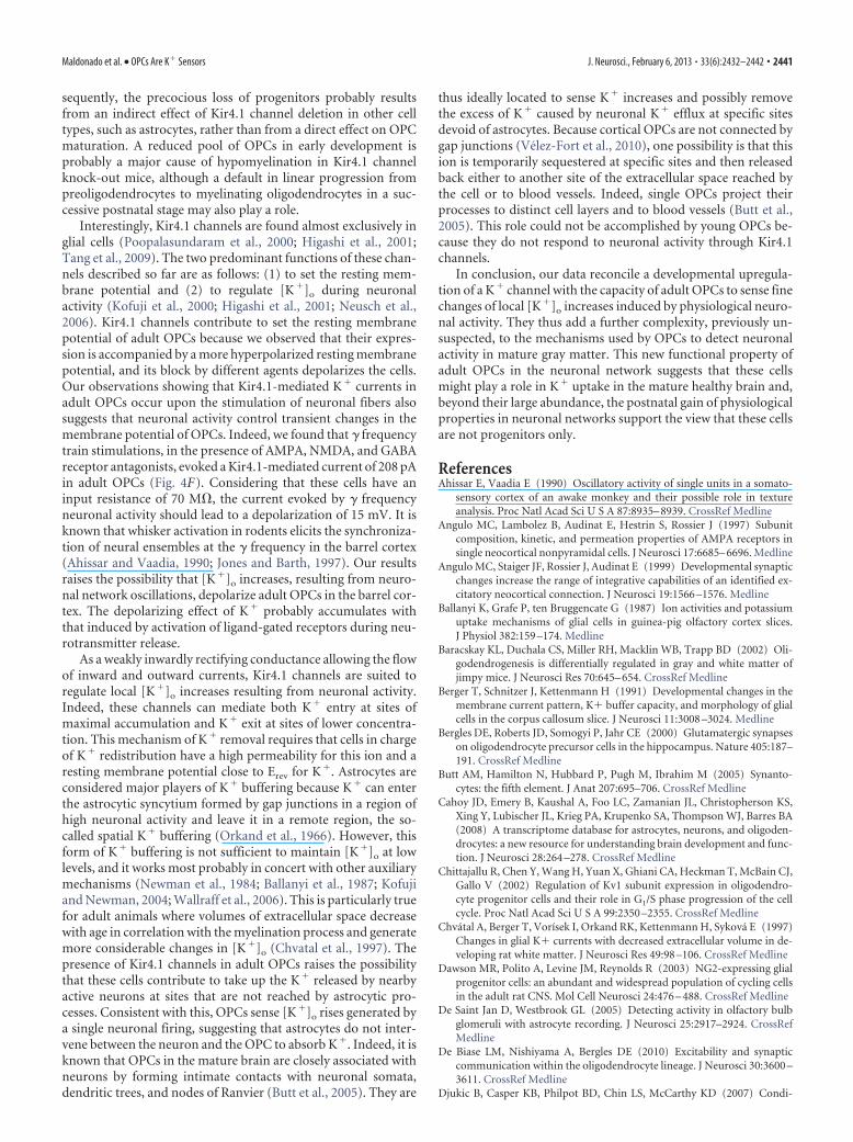
increases generated by singlenearby neuronsTo test whether adult OPCs are able tosense fine [K�]o increases of single nearbyneurons when they discharge action po-tentials, we performed simultaneouspatch-clamp recordings between a neu-ron and an OPC, which somata were sep-arated by �18 �m (range, 0 –18 �m).Figure 7A illustrates a labeled neuron andNG2�/DsRed� OPCs recorded with in-tracellular solutions containing biocytin.Depolarizing current pulses of 400 mswere elicited in the recorded neuron toinduce action potential firing while re-cording the nearby OPC with a KCl-basedintracellular solution (in physiologicalK� concentrations) (Fig. 7A, right, and7B). For 21 of 26 simultaneous record-ings, a train of action potentials in theneuron elicited a current in the OPC thathad a mean peak amplitude of �3.1 � 0.5pA and showed always an amplitude dis-tribution significantly different to that ofthe noise (Fig. 7B,E). Neurons eliciting aresponse in OPCs showed variable dis-charge frequencies ranging from 27.5 to121.8 Hz, which includes pyramidal cellsand interneurons, indicating that theOPC current is not induced by a specificneuronal cell type (mean frequency,70.3 � 15.9 Hz). It is noteworthy that nei-ther the frequency nor the distance be-tween cell somata where correlated tothe amplitude of the current ( p � 0.05).In addition, despite the presence of alarge repertoire of K � channels in neu-rons, such current was never observedin simultaneous recordings betweentwo neurons which somata were sepa-rated by �13 �m (range, 1–13 �m),even when the depolarizing pulse dura-tion was increased to 800 ms (mean am-plitude: �0.35 � 0.35 pA; n � 7;Fig. 7G).
It is known that OPCs detect neuronalactivity through synaptic and extrasynap-tic signaling mechanisms dependent onneurotransmitter release (Maldonado etal., 2011). However, the current elicited inOPCs by single neuron stimulation wasinsensitive to metabotropic and iono-tropic glutamate, GABA, and purinergicreceptor antagonists; thus, it did not resultfrom either classical synaptic transmis-sion or spillover of neurotransmitters(�3.2 � 1.0 pA and �3.4 � 1.0 pA incontrol and all antagonists, respectively; p � 0.05; n � 5; Fig.7C,F). This current developed progressively during neuronal fir-ing without evidence of postsynaptic events, suggesting a non-synaptic nature of the response (Fig. 7B,C). Nevertheless,considering that OPCs have a relatively low input resistance in
KCl-based intracellular solution (�70 M), we cannot com-pletely exclude that the current recorded in the OPC results froman artifactual detection through the patch pipette of the depolar-izing current injected to the neuron. To discard this possibility,we hyperpolarized the neuron to prevent action potential dis-
Figure 4. OPCs sense local increases of [K �]o during neuronal activity in the mature barrel cortex. A, Schematic representation ofextracellular stimulation of layer V neuronal fibers during patch-clamp recordings of OPCs of the barrel cortex. B, Confocal image of anNG2 �/DsRed � OPC recorded with an intracellular solution containing biocytin at PN24 (objective 63�; stack of 18 z section, each 1�m,see Materials and Methods). Scale bar, 15�m. C, D, Paired-pulse stimulation of neuronal fibers elicited inward currents in two OPCs held at�70 mV at PN9 (C) and PN24 (D) (gray traces; pulses separated by 50 ms; 100 �s, 40 V). Although the evoked current was completelyabolished by 10 �M NBQX, 50 �M D-AP5, and 5 �M SR95531 in C, a persistent current remained in D (black traces). Stimulus artifacts wereblanked for visibility; the time of stimulation is indicated with a vertical line. E, F, Inward currents elicited by � frequency train stimulation(50 Hz) of neuronal fibers in two OPCs held at �70 mV at PN8 (E) and PN27 (F), in the presence of antagonists. Stimulus artifacts weretruncatedforvisibility.G, InwardcurrentevokedinanOPCbyneuronalstimulation,resistanttoamixtureofantagonistsconsistingof10�M
NBQX, 50 �M D-AP5, 5 �M SR95531, 50 �M LY341495, 5 �M CPG55845, and 30 �M PPADS (all antagonists). H, Effect of 0.5 �M TTX oncurrents induced by paired-pulse stimulation of neuronal fibers in an OPC held at�70 mV and recorded in the presence of 10�M NBQX, 50�M D-AP5, and 5 �M SR95531. Note the block of the current by TTX.
2438 • J. Neurosci., February 6, 2013 • 33(6):2432–2442 Maldonado et al. • OPCs Are K� Sensors
charges while applying the same depolar-izing current pulse (Fig. 7H, I). In foursimultaneous recordings, the evoked cur-rent observed in the OPCs during actionpotential discharges was completely abol-ished when the neuron did not fire duringcurrent injection, confirming that neuro-nal action potentials are necessary to elicita response in the OPC (�2.3 � 1.1 pA vs0.1 � 0.1 pA). In addition, this currentwas inhibited by 100 �M Ba 2�, indicatingthat stimulation of a single neuron in-duces K� currents in OPCs throughKir4.1 channels that reflects neuronal K�
efflux during action potential discharges(86.3 � 8.4% of block, n � 6; p � 0.05;Fig. 7D,F). It is noteworthy that the fre-quency of discharge of recorded neuronsremained unchanged in the presence of100 �M Ba 2� (28.1 � 6.4 Hz and 30.0 �6.9 Hz in control and Ba 2�, respectively;p � 0.05; n � 6; Fig. 7B,D, upper traces).
Altogether, our results show a novelform of unitary neuron–OPC connec-tions that allows adult OPCs to sensefinely and reliably [K �]o increasesthrough Kir4.1 channels during physio-logical activity of a single nearby neuron.
DiscussionIn this report, we demonstrated thatKir4.1 channels are upregulated in corti-cal OPCs after the 2PN week (i.e., duringtheir massive differentiation into oligo-dendrocytes) (Baracskay et al., 2002). Theemergence of this conductance allows themajority of OPCs to sense local K� in-creases induced by neuronal activity, con-ferring to these cells a new functionalproperty in the mature neuronal network.This novel neuron-to-OPC signalingmechanism does not rely on neurotrans-mitter release and is robust at a single-celllevel as demonstrated by unitary neuron–OPC connections mediated by Kir4.1channels. Astrocytes are also known todetect neuronal-induced K � increasesthrough Kir4.1 channels (De Saint Janand Westbrook, 2005; Djukic et al., 2007),but it is unknown whether they detect[K�]o changes as locally as OPCs (i.e.,upon a single neuron stimulation).
Kir channels are known to be upregu-lated in OPCs during postnatal develop-ment, but their expression is thought toremain low and reach high levels only in dif-ferentiated oligodendrocytes (Sontheimeret al., 1989; Berger et al., 1991; Neusch etal., 2001). However, most functional workon Kir channels of OPCs has been doneeither in culture or in acute slices of ani-mals younger than four postnatal weeks(i.e., in conditions where these channels
Figure 6. I–V curve of the persistent K � current evoked by neuronal stimulation. A, Inward currents induced in an OPCby 100 ms ramps from �40 mV to �140 mV before the extracellular stimulation (gray trace) and at the peak of the secondevoked response in the presence of 10 �M NBQX, 50 �M D-AP5, and 5 �M SR95531 (black trace; pulses separated by 50 ms;100 �s, 40 V). The inset illustrates the persistent current evoked by paired-pulse stimulation at the first pulse. Calibration:25 ms; 25 pA. Stimulus artifacts were blanked for visibility. A vertical line indicates the time of stimulation; a hyphenatedgray line indicates zero current. B, Mean I–V curves obtained with ramps before the stimulation (gray trace) and at the peakof the second evoked response (black trace; n � 5). C, Effect of bath application of an increase in [K �]o from 2.5 to 10 mM
on an adult OPC held at �90 mV and recorded with the CsCl-based intracellular solution. D, Comparison of mean steady-state I–V curves in control and after application of high [K �]o shows an increase of induced currents and a shift in Erev of�8.7 � 2.3 mV ( p � 0.05; n � 6). *p � 0.05.
Figure 5. K � currents induced by neuronal activity in OPCs of 4 –5PN weeks are mediated by Kir4.1 channels. A, B, Effect of 100�M Ba 2� on inward currents resistant to antagonists and evoked by paired-pulse stimulation of neuronal fibers in OPCs held at�70 mV and recorded in CsCl-based intracellular solution (A) and KCl-based intracellular solution (B). Stimulus artifacts wereblanked for visibility. A vertical line indicates the time of stimulation. Pulses were separated by 50 ms in all cases (100 �s, 40 V). C,Inward currents evoked in two OPCs recorded with an intracellular solution at a pH of 7.4 (left) and of 6.3 (right) in the same sliceand with the same stimulation electrode as shown in the diagram (left).
Maldonado et al. • OPCs Are K� Sensors J. Neurosci., February 6, 2013 • 33(6):2432–2442 • 2439
probably do not attain their maximal ex-pression). Although Kir4.1 channels havealready been demonstrated in OPCs, onlya weak expression was reported in thesecells (Schroder et al., 2002; Tang et al.,2009). We showed here that expressionlevels of Kir4.1 channels progressively in-crease in most OPCs during postnatal de-velopment of the barrel cortex, conferringa linear I–V relationship on adult cells(Fig. 1F, I). This linear phenotype resultsfrom the weak rectification displayed bynative Kir4.1 channels in physiologicalK� concentrations (Kucheryavykh et al.,2007; Seifert et al., 2009; Hibino et al.,2010). Interestingly, most of the linearI–V curve of astrocytes also depends onthe expression of Kir4.1 channels (Djukicet al., 2007; Seifert et al., 2009). However,OPCs and astrocytes differ in that the laterexpress functional two-pore domain K�
channels TREK1 and probably TWIK1(Seifert et al., 2009; Zhou et al., 2009).
Whereas voltage-gated K� channelsexpressed by OPCs play a key role duringcell proliferation (Sontheimer et al., 1989;Gallo et al., 1996; Chittajallu et al., 2002),the function of Kir channels in these cellshas not been well defined. Recent studieson knock-out Kir4.1 channels demon-strated that the deletion of these channelsin glial cells was associated to an early le-thality caused by a severe hypomyelinat-ing phenotype (Neusch et al., 2001;Djukic et al., 2007). This has suggestedthat Kir4.1 channels play a role in OPCdifferentiation. However, the percentageof complex glia (presumably OPCs) de-creases 11-fold in PN5–10 knock-outmice (Djukic et al., 2007) when functionalKir channels are poorly expressed in thesecells (Fig. 1C) (Kressin et al., 1995). Con-
Figure 7. Single neuron stimulation induces Kir4.1-mediated K � currents in individual OPCs. A, Confocal image of a neuron(green) and an NG2 � OPC (yellow) simultaneously recorded in whole-cell configuration with intracellular solutions containingbiocytin (left; stack of 17 z sections, each 1 �m; see Materials and Methods). The amplitude of the current elicited in this OPC bysingle neuron stimulation was �2.93 pA. Scale bar, 20 �m. A schematic representation of simultaneous neuron–OPC recordingsis also shown (right). B–D, Simultaneous recording of a neuron held at �70 mV in current clamp and an OPC held at �90 mV involtage clamp in control (B), in the presence of a mixture of ionotropic and metabotropic receptor antagonists (C), and after bathapplication of 100 �M Ba 2� (D). The mixture of antagonists consists of 10 �M NBQX, 50 �M D-AP5, 5 �M SR95531, 50 �M
LY341495, 5 �M CPG55845, and 30 �M PPADS. Note the lack of effect of the antagonists and the complete block of the induced
4
current by Ba 2�. The train of action potentials in the recordedneuron was obtained by applying a 400 ms pulse of 400 pA,each 3 s. Only a single representative train of discharge is illus-trated in each condition. Current traces of OPCs are averages of122, 117, and 68 responses. E, Amplitude distributions of thenoise of the trace (black) and of the K � current (gray) of therecorded OPC in control conditions ( p�0.001). F, Time courseof the experiment of the same neuron–OPC simultaneous re-cording. G, Simultaneous recording of a neuron held at �70mV in current clamp and another neuron held at �90 mV involtage clamp. The train of action potentials in the neuron(top) is obtained by applying 800 ms pulse of 200 pA. Note thelack of induced current in the other neuron (bottom). The cur-rent trace is an average of 37 sweeps. H, I, Simultaneous re-cording of a neuron held at �65 mV (H) and �90 mV (I) incurrent clamp and an OPC held at �90 mV in voltage clamp.Note that the hyperpolarization of the neuron abolishes bothaction potential discharges upon the same current injectionand the evoked current in the OPC. The current traces are anaverage 51 (H) and 49 (I) sweeps.
2440 • J. Neurosci., February 6, 2013 • 33(6):2432–2442 Maldonado et al. • OPCs Are K� Sensors
sequently, the precocious loss of progenitors probably resultsfrom an indirect effect of Kir4.1 channel deletion in other celltypes, such as astrocytes, rather than from a direct effect on OPCmaturation. A reduced pool of OPCs in early development isprobably a major cause of hypomyelination in Kir4.1 channelknock-out mice, although a default in linear progression frompreoligodendrocytes to myelinating oligodendrocytes in a suc-cessive postnatal stage may also play a role.
Interestingly, Kir4.1 channels are found almost exclusively inglial cells (Poopalasundaram et al., 2000; Higashi et al., 2001;Tang et al., 2009). The two predominant functions of these chan-nels described so far are as follows: (1) to set the resting mem-brane potential and (2) to regulate [K�]o during neuronalactivity (Kofuji et al., 2000; Higashi et al., 2001; Neusch et al.,2006). Kir4.1 channels contribute to set the resting membranepotential of adult OPCs because we observed that their expres-sion is accompanied by a more hyperpolarized resting membranepotential, and its block by different agents depolarizes the cells.Our observations showing that Kir4.1-mediated K� currents inadult OPCs occur upon the stimulation of neuronal fibers alsosuggests that neuronal activity control transient changes in themembrane potential of OPCs. Indeed, we found that � frequencytrain stimulations, in the presence of AMPA, NMDA, and GABAreceptor antagonists, evoked a Kir4.1-mediated current of 208 pAin adult OPCs (Fig. 4F). Considering that these cells have aninput resistance of 70 M, the current evoked by � frequencyneuronal activity should lead to a depolarization of 15 mV. It isknown that whisker activation in rodents elicits the synchroniza-tion of neural ensembles at the � frequency in the barrel cortex(Ahissar and Vaadia, 1990; Jones and Barth, 1997). Our resultsraises the possibility that [K�]o increases, resulting from neuro-nal network oscillations, depolarize adult OPCs in the barrel cor-tex. The depolarizing effect of K� probably accumulates withthat induced by activation of ligand-gated receptors during neu-rotransmitter release.
As a weakly inwardly rectifying conductance allowing the flowof inward and outward currents, Kir4.1 channels are suited toregulate local [K�]o increases resulting from neuronal activity.Indeed, these channels can mediate both K� entry at sites ofmaximal accumulation and K� exit at sites of lower concentra-tion. This mechanism of K� removal requires that cells in chargeof K� redistribution have a high permeability for this ion and aresting membrane potential close to Erev for K�. Astrocytes areconsidered major players of K� buffering because K� can enterthe astrocytic syncytium formed by gap junctions in a region ofhigh neuronal activity and leave it in a remote region, the so-called spatial K� buffering (Orkand et al., 1966). However, thisform of K� buffering is not sufficient to maintain [K�]o at lowlevels, and it works most probably in concert with other auxiliarymechanisms (Newman et al., 1984; Ballanyi et al., 1987; Kofujiand Newman, 2004; Wallraff et al., 2006). This is particularly truefor adult animals where volumes of extracellular space decreasewith age in correlation with the myelination process and generatemore considerable changes in [K�]o (Chvatal et al., 1997). Thepresence of Kir4.1 channels in adult OPCs raises the possibilitythat these cells contribute to take up the K� released by nearbyactive neurons at sites that are not reached by astrocytic pro-cesses. Consistent with this, OPCs sense [K�]o rises generated bya single neuronal firing, suggesting that astrocytes do not inter-vene between the neuron and the OPC to absorb K�. Indeed, it isknown that OPCs in the mature brain are closely associated withneurons by forming intimate contacts with neuronal somata,dendritic trees, and nodes of Ranvier (Butt et al., 2005). They are
thus ideally located to sense K� increases and possibly removethe excess of K� caused by neuronal K� efflux at specific sitesdevoid of astrocytes. Because cortical OPCs are not connected bygap junctions (Velez-Fort et al., 2010), one possibility is that thision is temporarily sequestered at specific sites and then releasedback either to another site of the extracellular space reached bythe cell or to blood vessels. Indeed, single OPCs project theirprocesses to distinct cell layers and to blood vessels (Butt et al.,2005). This role could not be accomplished by young OPCs be-cause they do not respond to neuronal activity through Kir4.1channels.
In conclusion, our data reconcile a developmental upregula-tion of a K� channel with the capacity of adult OPCs to sense finechanges of local [K�]o increases induced by physiological neuro-nal activity. They thus add a further complexity, previously un-suspected, to the mechanisms used by OPCs to detect neuronalactivity in mature gray matter. This new functional property ofadult OPCs in the neuronal network suggests that these cellsmight play a role in K� uptake in the mature healthy brain and,beyond their large abundance, the postnatal gain of physiologicalproperties in neuronal networks support the view that these cellsare not progenitors only.
ReferencesAhissar E, Vaadia E (1990) Oscillatory activity of single units in a somato-
sensory cortex of an awake monkey and their possible role in textureanalysis. Proc Natl Acad Sci U S A 87:8935– 8939. CrossRef Medline
Angulo MC, Lambolez B, Audinat E, Hestrin S, Rossier J (1997) Subunitcomposition, kinetic, and permeation properties of AMPA receptors insingle neocortical nonpyramidal cells. J Neurosci 17:6685– 6696. Medline
Angulo MC, Staiger JF, Rossier J, Audinat E (1999) Developmental synapticchanges increase the range of integrative capabilities of an identified ex-citatory neocortical connection. J Neurosci 19:1566 –1576. Medline
Ballanyi K, Grafe P, ten Bruggencate G (1987) Ion activities and potassiumuptake mechanisms of glial cells in guinea-pig olfactory cortex slices.J Physiol 382:159 –174. Medline
Baracskay KL, Duchala CS, Miller RH, Macklin WB, Trapp BD (2002) Oli-godendrogenesis is differentially regulated in gray and white matter ofjimpy mice. J Neurosci Res 70:645– 654. CrossRef Medline
Berger T, Schnitzer J, Kettenmann H (1991) Developmental changes in themembrane current pattern, K� buffer capacity, and morphology of glialcells in the corpus callosum slice. J Neurosci 11:3008 –3024. Medline
Bergles DE, Roberts JD, Somogyi P, Jahr CE (2000) Glutamatergic synapseson oligodendrocyte precursor cells in the hippocampus. Nature 405:187–191. CrossRef Medline
Butt AM, Hamilton N, Hubbard P, Pugh M, Ibrahim M (2005) Synanto-cytes: the fifth element. J Anat 207:695–706. CrossRef Medline
Cahoy JD, Emery B, Kaushal A, Foo LC, Zamanian JL, Christopherson KS,Xing Y, Lubischer JL, Krieg PA, Krupenko SA, Thompson WJ, Barres BA(2008) A transcriptome database for astrocytes, neurons, and oligoden-drocytes: a new resource for understanding brain development and func-tion. J Neurosci 28:264 –278. CrossRef Medline
Chittajallu R, Chen Y, Wang H, Yuan X, Ghiani CA, Heckman T, McBain CJ,Gallo V (2002) Regulation of Kv1 subunit expression in oligodendro-cyte progenitor cells and their role in G1/S phase progression of the cellcycle. Proc Natl Acad Sci U S A 99:2350 –2355. CrossRef Medline
Chvatal A, Berger T, Vorísek I, Orkand RK, Kettenmann H, Sykova E (1997)Changes in glial K� currents with decreased extracellular volume in de-veloping rat white matter. J Neurosci Res 49:98 –106. CrossRef Medline
Dawson MR, Polito A, Levine JM, Reynolds R (2003) NG2-expressing glialprogenitor cells: an abundant and widespread population of cycling cellsin the adult rat CNS. Mol Cell Neurosci 24:476 – 488. CrossRef Medline
De Saint Jan D, Westbrook GL (2005) Detecting activity in olfactory bulbglomeruli with astrocyte recording. J Neurosci 25:2917–2924. CrossRefMedline
De Biase LM, Nishiyama A, Bergles DE (2010) Excitability and synapticcommunication within the oligodendrocyte lineage. J Neurosci 30:3600 –3611. CrossRef Medline
Djukic B, Casper KB, Philpot BD, Chin LS, McCarthy KD (2007) Condi-
Maldonado et al. • OPCs Are K� Sensors J. Neurosci., February 6, 2013 • 33(6):2432–2442 • 2441
tional knock-out of Kir4.1 leads to glial membrane depolarization, inhi-bition of potassium and glutamate uptake, and enhanced short-termsynaptic potentiation. J Neurosci 27:11354 –11365. CrossRef Medline
Gallo V, Zhou JM, McBain CJ, Wright P, Knutson PL, Armstrong RC (1996)Oligodendrocyte progenitor cell proliferation and lineage progression areregulated by glutamate receptor-mediated K� channel block. J Neurosci16:2659 –2670. Medline
Heinemann U, Lux HD (1977) Ceiling of stimulus induced rises in extracel-lular potassium concentration in the cerebral cortex of cat. Brain Res120:231–249. CrossRef Medline
Hibino H, Inanobe A, Furutani K, Murakami S, Findlay I, Kurachi Y (2010)Inwardly rectifying potassium channels: their structure, function, andphysiological roles. Physiol Rev 90:291–366. CrossRef Medline
Higashi K, Fujita A, Inanobe A, Tanemoto M, Doi K, Kubo T, Kurachi Y(2001) An inwardly rectifying K(�) channel, Kir4.1, expressed in astro-cytes surrounds synapses and blood vessels in brain. Am J Physiol CellPhysiol 281:C922–C931. Medline
Jones MS, Barth DS (1997) Sensory-evoked high-frequency (�-band) oscil-lating potentials in somatosensory cortex of the unanesthetized rat. BrainRes 768:167–176. CrossRef Medline
Karadottir R, Cavelier P, Bergersen LH, Attwell D (2005) NMDA receptorsare expressed in oligodendrocytes and activated in ischaemia. Nature 438:1162–1166. CrossRef Medline
Kofuji P, Newman EA (2004) Potassium buffering in the central nervoussystem. Neuroscience 129:1045–1056. CrossRef Medline
Kofuji P, Ceelen P, Zahs KR, Surbeck LW, Lester HA, Newman EA (2000)Genetic inactivation of an inwardly rectifying potassium channel (Kir4.1subunit) in mice: phenotypic impact in retina. J Neurosci 20:5733–5740.Medline
Kressin K, Kuprijanova E, Jabs R, Seifert G, Steinhauser C (1995) Develop-mental regulation of Na� and K� conductances in glial cells of mousehippocampal brain slices. Glia 15:173–187. CrossRef Medline
Kucheryavykh YV, Pearson WL, Kurata HT, Eaton MJ, Skatchkov SN, Nich-ols CG (2007) Polyamine permeation and rectification of Kir4.1 chan-nels. Channels (Austin) 1:172–178. Medline
Kukley M, Capetillo-Zarate E, Dietrich D (2007) Vesicular glutamate re-lease from axons in white matter. Nat Neurosci 10:311–320. CrossRefMedline
Lesage F, Guillemare E, Fink M, Duprat F, Lazdunski M, Romey G, Bar-hanin J (1996) TWIK-1, a ubiquitous human weakly inward rectify-ing K� channel with a novel structure. EMBO J 15:1004 –1011.Medline
Lotshaw DP (2007) Biophysical, pharmacological, and functional charac-teristics of cloned and native mammalian two-pore domain K� channels.Cell Biochem Biophys 47:209 –256. CrossRef Medline
Maldonado PP, Velez-Fort M, Angulo MC (2011) Is neuronal communica-tion with NG2 cells synaptic or extrasynaptic? J Anat 219:8 –17. CrossRefMedline
Mangin JM, Kunze A, Chittajallu R, Gallo V (2008) Satellite NG2 progenitorcells share common glutamatergic inputs with associated interneurons inthe mouse dentate gyrus. J Neurosci 28:7610 –7623. CrossRef Medline
Morales B, Choi SY, Kirkwood A (2002) Dark rearing alters the develop-ment of GABAergic transmission in visual cortex. J Neurosci 22:8084 –8090. Medline
Neusch C, Rozengurt N, Jacobs RE, Lester HA, Kofuji P (2001) Kir4.1 po-tassium channel subunit is crucial for oligodendrocyte development andin vivo myelination. J Neurosci 21:5429 –5438. Medline
Neusch C, Papadopoulos N, Muller M, Maletzki I, Winter SM, Hirrlinger J,Handschuh M, Bahr M, Richter DW, Kirchhoff F, Hulsmann S (2006)Lack of the Kir4.1 channel subunit abolishes K� buffering properties ofastrocytes in the ventral respiratory group: impact on extracellular K�regulation. J Neurophysiol 95:1843–1852. CrossRef Medline
Newman EA, Frambach DA, Odette LL (1984) Control of extracellular po-tassium levels by retinal glial cell K � siphoning. Science 225:1174 –1175.CrossRef Medline
Nie X, Arrighi I, Kaissling B, Pfaff I, Mann J, Barhanin J, Vallon V (2005)Expression and insights on function of potassium channel TWIK-1 inmouse kidney. Pflugers Arch 451:479 – 488. CrossRef Medline
Orkand RK, Nicholls JG, Kuffler SW (1966) Effect of nerve impulses on themembrane potential of glial cells in the central nervous system of am-phibia. J Neurophysiol 29:788 – 806. Medline
Patel AJ, Honore E, Lesage F, Fink M, Romey G, Lazdunski M (1999) Inha-lational anesthetics activate two-pore-domain background K � channels.Nat Neurosci 2:422– 426. CrossRef Medline
Pessia M, Imbrici P, D’Adamo MC, Salvatore L, Tucker SJ (2001) Differen-tial pH sensitivity of Kir4.1 and Kir4.2 potassium channels and theirmodulation by heteropolymerisation with Kir5.1. J Physiol 532:359 –367.CrossRef Medline
Poopalasundaram S, Knott C, Shamotienko OG, Foran PG, Dolly JO, GhianiCA, Gallo V, Wilkin GP (2000) Glial heterogeneity in expression of theinwardly rectifying K(�) channel, Kir4.1, in adult rat CNS. Glia 30:362–372. CrossRef Medline
Richardson WD, Young KM, Tripathi RB, McKenzie I (2011) NG2-glia asmultipotent neural stem cells: fact or fantasy? Neuron 70:661– 673.CrossRef Medline
Sah P, Faber ES (2002) Channels underlying neuronal calcium-activatedpotassium currents. Prog Neurobiol 66:345–353. CrossRef Medline
Salami M, Itami C, Tsumoto T, Kimura F (2003) Change of conductionvelocity by regional myelination yields constant latency irrespective ofdistance between thalamus and cortex. Proc Natl Acad Sci U S A 100:6174 – 6179. CrossRef Medline
Schroder W, Seifert G, Huttmann K, Hinterkeuser S, Steinhauser C (2002)AMPA receptor-mediated modulation of inward rectifier K� channels inastrocytes of mouse hippocampus. Mol Cell Neurosci 19:447– 458.CrossRef Medline
Seifert G, Huttmann K, Binder DK, Hartmann C, Wyczynski A, Neusch C,Steinhauser C (2009) Analysis of astroglial K� channel expression inthe developing hippocampus reveals a predominant role of the Kir4.1subunit. J Neurosci 29:7474 –7488. CrossRef Medline
Sontheimer H, Trotter J, Schachner M, Kettenmann H (1989) Channelexpression correlates with differentiation stage during the develop-ment of oligodendrocytes from their precursor cells in culture. Neu-ron 2:1135–1145. CrossRef Medline
Su S, Ohno Y, Lossin C, Hibino H, Inanobe A, Kurachi Y (2007) Inhibitionof astroglial inwardly rectifying Kir4.1 channels by a tricyclic antidepres-sant, nortriptyline. J Pharmacol Exp Ther 320:573–580. CrossRef Medline
Tang X, Taniguchi K, Kofuji P (2009) Heterogeneity of Kir4.1 channel ex-pression in glia revealed by mouse transgenesis. Glia 57:1706 –1715.CrossRef Medline
Tang X, Schmidt TM, Perez-Leighton CE, Kofuji P (2010) Inwardly rectify-ing potassium channel Kir4.1 is responsible for the native inward potas-sium conductance of satellite glial cells in sensory ganglia. Neuroscience166:397– 407. CrossRef Medline
Taverna S, Tkatch T, Metz AE, Martina M (2005) Differential expression ofTASK channels between horizontal interneurons and pyramidal cells ofrat hippocampus. J Neurosci 25:9162–9170. CrossRef Medline
Tucker SJ, Imbrici P, Salvatore L, D’Adamo MC, Pessia M (2000) pH de-pendence of the inwardly rectifying potassium channel, Kir5.1, and local-ization in renal tubular epithelia. J Biol Chem 275:16404 –16407.CrossRef Medline
Velez-Fort M, Audinat E, Angulo MC (2009) Functional � 7-containingnicotinic receptors of NG2-expressing cells in the hippocampus. Glia57:1104 –1114. CrossRef Medline
Velez-Fort M, Maldonado PP, Butt AM, Audinat E, Angulo MC (2010)Postnatal switch from synaptic to extrasynaptic transmission betweeninterneurons and NG2 cells. J Neurosci 30:6921– 6929. CrossRef Medline
Wallraff A, Kohling R, Heinemann U, Theis M, Willecke K, Steinhauser C(2006) The impact of astrocytic gap junctional coupling on potassiumbuffering in the hippocampus. J Neurosci 26:5438 –5447. CrossRefMedline
Zhou M, Schools GP, Kimelberg HK (2006) Development of GLAST(�)astrocytes and NG2(�) glia in hippocampus CA1: mature astrocytes areelectrophysiologically passive. J Neurophysiol 95:134 –143. CrossRefMedline
Zhou M, Xu G, Xie M, Zhang X, Schools GP, Ma L, Kimelberg HK, Chen H(2009) TWIK-1 and TREK-1 are potassium channels contributing signif-icantly to astrocyte passive conductance in rat hippocampal slices. J Neu-rosci 29:8551– 8564. CrossRef Medline
Ziskin JL, Nishiyama A, Rubio M, Fukaya M, Bergles DE (2007) Vesicularrelease of glutamate from unmyelinated axons in white matter. Nat Neu-rosci 10:321–330. CrossRef Medline
2442 • J. Neurosci., February 6, 2013 • 33(6):2432–2442 Maldonado et al. • OPCs Are K� Sensors