Thèse de doctorat NNT
271
Diversité des bactéries halophiles dans l'écosystème fromager et étude de leurs impacts fonctionnels Diversity of halophilic bacteria in the cheese ecosystem and the study of their functional impacts Thèse de doctorat de l'université Paris-Saclay École doctorale n° 581 Agriculture, Alimentation, Biologie, Environnement et Santé (ABIES) Spécialité de doctorat: Microbiologie Unité de Recherche : Micalis Institute, Jouy-en-Josas, France Référent : AgroParisTech Thèse présentée et soutenue à Paris-Saclay, le 01/04/2021 par Caroline Isabel KOTHE Composition du Jury Michel-Yves MISTOU Directeur de Recherche, INRAE centre IDF - Jouy-en-Josas - Antony Président Monique ZAGOREC Directrice de Recherche, INRAE centre Pays de la Loire Rapporteur & Examinatrice Nathalie DESMASURES Professeure, Université de Caen Normandie Rapporteur & Examinatrice Françoise IRLINGER Ingénieure de Recherche, INRAE centre IDF - Versailles-Grignon Examinatrice Jean-Louis HATTE Ingénieur Recherche et Développement, Lactalis Examinateur Direction de la thèse Pierre RENAULT Directeur de Recherche, INRAE (centre IDF - Jouy-en-Josas - Antony) Directeur de thèse Thèse de doctorat NNT : 2021UPASB014
-
Upload
khangminh22 -
Category
Documents
-
view
0 -
download
0
Transcript of Thèse de doctorat NNT

Diversité des bactéries halophiles dans
l'écosystème fromager et étude de leurs
impacts fonctionnels Diversity of halophilic bacteria in the cheese ecosystem and
the study of their functional impacts
Thèse de doctorat de l'université Paris-Saclay
École doctorale n° 581 Agriculture, Alimentation, Biologie, Environnement
et Santé (ABIES)
Spécialité de doctorat: Microbiologie
Unité de Recherche : Micalis Institute, Jouy-en-Josas, France
Référent : AgroParisTech
Thèse présentée et soutenue à Paris-Saclay,
le 01/04/2021 par
Caroline Isabel KOTHE
Composition du Jury
Michel-Yves MISTOU
Directeur de Recherche, INRAE centre IDF - Jouy-en-Josas - Antony Président
Monique ZAGOREC
Directrice de Recherche, INRAE centre Pays de la Loire Rapporteur & Examinatrice
Nathalie DESMASURES
Professeure, Université de Caen Normandie Rapporteur & Examinatrice
Françoise IRLINGER
Ingénieure de Recherche, INRAE centre IDF - Versailles-Grignon Examinatrice
Jean-Louis HATTE
Ingénieur Recherche et Développement, Lactalis Examinateur
Direction de la thèse
Pierre RENAULT
Directeur de Recherche, INRAE (centre IDF - Jouy-en-Josas - Antony) Directeur de thèse
Th
èse
de d
oct
ora
t N
NT : 2
021U
PA
SB
014

“A master in the art of living draws no sharp distinction between her work and her play; her
labor and her leisure; her mind and her body; her education and her recreation. She hardly knows
which is which. She simply pursues her vision of excellence through whatever she is doing, and
leaves others to determine whether she is working or playing. To herself, she always appears to
be doing both.”
Adapted to Lawrence Pearsall Jacks

REMERCIEMENTS
Remerciements
L'opportunité de faire un doctorat, en France, à l’Unité mixte de recherche MICALIS de
Jouy-en-Josas a provoqué de nombreux changements dans ma vie : un autre pays, une autre langue,
une autre culture et aussi, un nouveau domaine de recherche. Pendant ces trois ans et demi de
thèse, j’ai eu l’occasion et le plaisir de rencontrer nombre de personnes, au laboratoire et sur mes
lieux de recherche, les unes supervisant mes travaux, les autres m’apportant une aide précieuse par
leurs conseils, encouragements et humour.
Mes remerciements et ma reconnaissance vont d’abord à mon Directeur de thèse, Pierre
Renault. Je me sens vraiment privilégiée d'avoir bénéficié des conseils d’un Superviseur au grand
cœur, si patient et (presque) toujours de bonne humeur ! Je vous remercie de m'avoir accueillie
dans votre laboratoire et, surtout, pour votre disponibilité, votre aide et les nombreux échanges sur
les sujets les plus variés, qu'il s'agisse de soins aux plantes ou de questions scientifiques.
Un grand merci à Bochra-Farah Kraïem pour avoir initié ce projet, ainsi qu'à Victoria
Meslier et Bedis Dridi, pour mon insertion dans le monde de la biologie moléculaire et pour
m'avoir aidée, notamment au laboratoire et dans la clarification et le développement des protocoles
d'extraction d'ADN.
Je remercie également Monique Zagorec, Nathalie Desmasures, Françoise Irlinger, Michel-
Yves Mistou et Jean-Louis Hatte d’avoir acceptés de faire partie du jury, ainsi que Céline Delbès
et Delphine Passerini pour leurs contributions fréquentes, tout au long de chaque année de mon
parcours. Merci aussi à tous les membres de l'équipe FME, pour les réunions riches en échanges
scientifiques, leurs encouragements, sans oublier les moments sympas que nous avons passés
ensemble, surtout à Catane ! Je remercie tout particulièrement Marine et Gwendoline d'avoir rendu
nos réunions d'équipe plus joyeuses et d'avoir toujours été disponibles pour aider ou dialoguer.
Merci à Stéphane Chaillou et Mathieu Almeida qui m'ont accueillie et permis de participer
aux cours de M2, sur le traitement des données métagénomiques, informations très utiles pour ma
formation professionnelle. Dans le même ordre d'idée, pour son aide en bioinformatique, je
remercie Alexandre Bolotine (Sacha), toujours prêt à effectuer l'analyse pangénomique, un volet
important de ma thèse. Et, un grand merci et une immense reconnaissance à mon collègue du
bureau, Nacer, qui a beaucoup apporté à mon travail, très gentil et toujours prêt à m'expliquer et à
m'aider en tout, surtout en ce qui concerne les lignes de commandes.

REMERCIEMENTS
Un grand merci aux collègues de Grignon, pour avoir collaboré significativement à ce
projet de thèse. Tout d'abord à Sandra, pour les trajets jusqu'au Centre, ce qui m'a fait gagner
beaucoup de temps de transport, sans mentionner l’aspect très agréable de sa compagnie. Un
immense merci à Christophe Monnet, qui m'a aidée dans la manipulation des fromages-agar,
expériences toujours réalisées avec rigueur, sans compter son enseignement aux nombreuses
techniques microbiologiques et sa participation active à mon travail de thèse. Je remercie
également Françoise Irlinger de m'avoir accueillie avec tant d'empathie, en plus de son soutien et
de ses encouragements dans toutes les réunions traitant de la caractérisation des nouvelles
espèces ! La charge de travail à Grignon a également été assurée grâce à l'aide technique d'Anne-
Sophie et Gwendoline pour le prélèvement des échantillons, d’Anne-Claire, dans l’analyse des
chromatogrammes, et de Sophie Landaud pour son enseignement pertinent concernant l'analyse
des composés volatils et l'organisation des données. Merci à tous (toutes).
Au cours de ces années, j'ai eu l'occasion de partager des expériences avec certains
stagiaires, qui ont rendu l'environnement de travail plus convivial, tout en complétant ma formation
en ce qui concerne la gestion d’une équipe de recherche, associant empathie et patience. Parmi
elles, Magali et Marine, qui ont contribué à l'isolement des souches halophiles, Keneza et Céline
dans la caractérisation des nouvelles espèces, et Aurélie dans les analyses génomiques
comparatives. Je suis également ravie de connaître Racha, qui a travaillé sur un autre sujet, mais
qui est devenue une amie très chère.
Je tiens également à remercier le CNPq pour le financement de ma bourse et encore Pierre,
mon Directeur de thèse, pour m'avoir permis de réaliser toutes les expériences souhaitables pour
le bon déroulement de la thèse.
Je ne pourrais pas oublier de remercier Antoine Bouvier, qui a relu mon travail et m’a
permis de contrôler le genre et l’orthographe de nombre de mots français.
Mes remerciements vont aussi aux amis ou amis d’amis que j'ai rencontrés à la Gym ou
dans les couloirs, au moment des repas, pour leurs conseils, les échanges amicaux passés en
commun, et en particulier, à Léa, Iris, Eloise, Mélanie, Lucie et Ludovica (et tous ceux que j'ai pu
oublier). Un merci tout particulier à Cécile, qui a été une super amie pendant ma thèse, que ce soit
lors d’un déjeuner, pour un thé, pour le partage d'expériences, une amitié qui demeurera, au-delà
de la thèse.

REMERCIEMENTS
Je veux remercier aussi tout le personnel de l’INRAE-Jouy pour m'avoir toujours accueillie
de manière amicale, à l’Accueil et au RH, pour les clarifications et la fourniture des documents
administratifs ; merci aussi à l'équipe « Prepa », pour la préparation et la mise à disposition des
matériaux utilisés dans les expériences.
Je remercie également mes voisins de laboratoire, en particulier l'équipe de BioRetroSynth,
pour les cafés et les déjeuners pris ensemble et aussi l'équipe de MIHA pour les prêts de matériel
ou, simplement, pour un bonjour et un sourire dans les couloirs.
Loin de chez nous, les amis rencontrés et qui parlent notre langue deviennent pratiquement
des membres de notre famille. Je remercie donc tout particulièrement Gabriela, Camila, Samar et
Angelo, pour les moments et les expériences, partagés en France en tant qu'étrangers. Et même si
tous sont dispersés aujourd’hui, je me souviendrai toujours d’eux avec beaucoup d'affection.
Non moins important, un merci additionnel à toutes mes amies brésiliennes mais surtout à
Camila et Domênica. Nos échanges et nos réflexions sincères, presque quotidiennes, m’ont fait
prendre conscience que je suis la personne la plus chanceuse au monde d'avoir des gens comme
elles à mon côté. Merci pour tous ces partages, pour avoir écouté mes craintes, sans me juger, et
pour avoir célébré avec moi toutes les avancées !
Un grand merci à ma famille, mes neveux, mon père, ma mère, ma belle-sœur, mon frère
et ma sœur. Pour les longues heures d'appels vidéo et surtout pour la visite et le voyage que nous
avons fait avant l'arrivée du Covid. Merci de m'avoir communiquée les nouvelles de la croissance
des enfants, même à distance ! Je remercie également ma deuxième mère, Marinez, d'être toujours
prête à aider avec un énorme cœur. Et un merci tout particulier à ma sœur, pour m'avoir fait
découvrir la pratique du yoga et pour avoir partagé des recettes et échangé en philosophies.
Enfin, mes remerciements illimités vont à mon mari, Evandro ! Pour avoir toujours été là,
à m'écouter, à m'aider et à me réconforter. Pour avoir cru en moi et m'avoir redonnée confiance
dans les moments où je me sentais la moins sûre de moi, mais surtout pour m'avoir motivée pour
être, chaque jour, une meilleure personne.
Les remerciements sont ici un peu longs, mais nécessaires et sincères. Tous ceux qui sont
passés dans ma vie m'ont fait évoluer positivement et je me sens vraiment chanceuse d'avoir
rencontré des gens qui étaient prêts à m'aider, dans ce parcours. Je suis très reconnaissante à tous
et les remercie encore pour leur indéfectible soutien durant ces trois années et demie.

PUBLICATIONS
Liste de publications
Publications associés au projet de thèse dans des revues scientifiques :
Kothe, C. I.; Bolotin, A.; Kraïem, B. F.; Dridi, B.; FoodMicrobiome Team, Renault, P.
Unraveling the world of halophilic and halotolerant bacteria in cheese by combining cultural,
genomic and metagenomic approaches. (under review in International Journal of Food
Microbiology)
Kothe, C. I.; Renault, P. Metagenomic driven characterization of halophilic bacteria in
cheese rinds (to be submitted)
Kothe, C. I.; Monnet, C.; Irlinger, F.; Renault, P. Halomonas citridevorans sp. nov.,
Halomonas minus sp. nov., Halomonas casei sp. nov. and Halomonas flavum sp. nov., four novel
species isolated from French cheese rinds (under review in International Journal of Systematic and
Evolutionary Microbiology).
Kothe, C. I.; Monnet, C.; Irlinger, F.; Renault, P. Psychrobacter casei sp. nov. and
Psychrobacter translucens sp. nov., two new species isolated from French cheese rinds (under
review in International Journal of Systematic and Evolutionary Microbiology).
Kothe, C. I.; Monnet, C.; Landaud, S.; Renault, P. Functional properties of food and
environmental halophilic Gammaproteobacteria strains on cheese-agar models (to be submitted)
Kothe, C. I.; Delbarre-Ladrat, C.; Renault, P.; Passerini, D. (2020). Draft-genome
sequence data and phylogenomic comparison of two marine-sourced bacterial strains
Pseudoalteromonas sp. MIP2626 and Psychrobacter sp. BI730. Data in Brief, 31, 105898.
https://doi.org/10.1016/j.dib.2020.105898
Kothe, C. I.; Renault, P. Meta-analysis of five traditional Brazilian cheese varieties (to be
submitted)
Communications lors de congrès :
Kothe, C. I.; Dridi, B.; Renault, P. (2019). Origin and evolution of halophilic bacteria:
from environment to cheese. 5th International Conference on microbial diversity, September 25-
27, Italy (oral communication).

LISTE DES ABRÉVIATIONS
Liste des abréviations
ADN: Acide désoxyribonucléique
ANI: Average Nucleotide Identity
ARN: Acide ribonucléique
aw: Activité de l’eau
BLAST: Basic Local Alignment Search Tool
GRAS: Generally Recognized As Safe
GYPB: Glucose, Yeast extract, Peptone and Beef extract
GC-MS: Gas chromatography–mass spectrometry
EFFCA: European Food and Feed Cultures Association
EFSA: European Food Safety Authority
KCl: Chlorure de potassium
IDF: International Dairy Federation
ISO: International Organization for Standardization
LAB: Lactic Acid Bacteria
MB: Marine Broth
MAG: Metagenome-Assembled-Genome
MGM: Modified Growth Medium
MFCs: Microbial Food Cultures
MGL: Methionine gamma-lyase
NaCl: Chlorure de sodium
NCBI: National Center for Biotechnology Information
NSLAB: Non-Starter Lactic Acid Bacteria
PCR: Polymerase Chain Reaction

LISTE DES FIGURES ET TABLEAUX
Liste des figures (hors publication)
Figure 1. Classification des fromages basée sur la technologie et les caractéristiques microbiologiques . 18
Figure 2. Possibles sources de développement de communautés microbiennes spécifiques dans le processus
de production et d’affinage du fromage ...................................................................................................... 20
Figure 3. Étapes de la fabrication du fromage impliquant l'ajout de sel et les méthodes de salage. .......... 31
Figure 4. Fromage à croûte lavée ............................................................................................................... 32
Figure 5. Les sept sels marins artisanaux et leur pouvoir respectifs de détection de microorganismes..... 35
Figure 6. Classification des bactéries en fonction du pourcentage de NaCl nécessaire à leur croissance
optimale ...................................................................................................................................................... 37
Figure 7. Stratégies utilisés par les halophiles pour maintenir leur structure cellulaire ............................. 38
Figure 8. Arbre phylogénétique global des organismes halophiles............................................................ 40
Figure 9. Les génomes halophiles signalés dans l'écosystème fromager ................................................. 200
Figure 10. Heatmap illustrant l'abondance relative (%) de 27 espèces halophiles dans 101 croûtes de
fromage ..................................................................................................................................................... 204
Figure 11. Espèces halophiles identifiées dans l’écosystème fromager et génomes d'autres habitats,
étroitement liés .......................................................................................................................................... 206
Figure 12. Arbre phylogénétique basé sur les alignements des gènes de l'ARNr 16S de souches marines et
fromagères................................................................................................................................................. 214
Liste des figures (publications)
Publication 1
Figure 1. Cheese processing including examples of the salting process. ................................................... 53
Figure 2. ANI and pan-genome analyses of 25 B. aurantiacum cheese strains ......................................... 62
Figure 3. ANI and pan-genome analyses of 43 S. equorum strains. .......................................................... 64
Figure 4. Heatmap depicting the relative abundance (%) of halophilic and halotolerant species in cheeses
.................................................................................................................................................................... 66
Figure 5. Relationships between halophilic and halotolerant bacteria detected by metagenomic analysis in
cheese rinds ................................................................................................................................................. 68

LISTE DES FIGURES ET TABLEAUX
Publication 2
Figure 1. Taxonomic classification of microbial population in ten cheese rinds by marker gene analysis.
.................................................................................................................................................................... 88
Figure 2. Probe design and culture strategies to isolate targeted species ................................................... 89
Publication 3
Figure 1. Phylogenetic tree based on 16S rRNA gene sequences of 12 strains from this study and closely
related Halomonas type strains. ................................................................................................................ 105
Figure 2. Phylogenetic tree based on rpoB gene sequences of 12 strains from this study and closely related
Halomonas type strains ............................................................................................................................. 106
Figure 3. Phylogenomic tree based on the whole-genome sequence alignment of all the Halomonas strains
.................................................................................................................................................................. 110
Figure 4. Heatmap depicting the average nucleotide identity of the cheese-associated strains investigated
in the present study and closely related Halomonas species..................................................................... 111
Publication 4
Figure 1. Phylogenetic tree based on 16S rRNA gene sequences of the four strains from this study and
closely related Psychrobacter type strains ................................................................................................ 124
Figure 2. Phylogenetic tree based on rpoB gene sequences of the four strains from this study and closely
related Psychrobacter type strains.. .......................................................................................................... 125
Figure 3. Phylogenomic tree and biochemical and enzymatic properties ................................................ 129
Publication 5
Figure 1. Phylogenomic tree of 43 halophilic strains used in this study .................................................. 141
Figure 2. Halophilic bacterial counts and pH measures at day 21 on cheese-agar model ....................... 143
Figure 3. Heatmap depicting the intensity perceived in sniffing analysis by twelve non-trained panelists.
.................................................................................................................................................................. 145
Figure 4. Overview of volatiles profiles of 43 halophilic strains on cheese-agar models........................ 147
LISTE DES FIGURES ET TABLEAUX
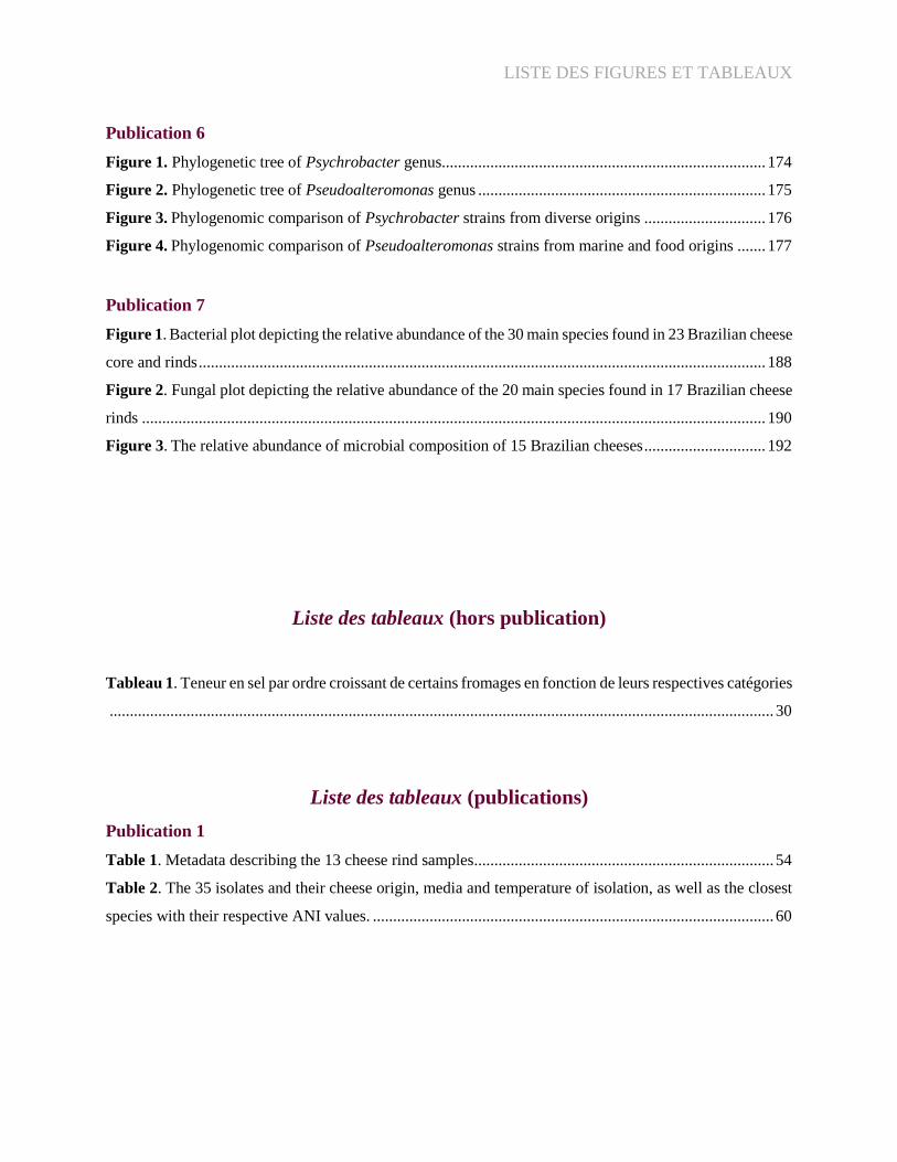
Publication 6
Figure 1. Phylogenetic tree of Psychrobacter genus................................................................................ 174
Figure 2. Phylogenetic tree of Pseudoalteromonas genus ....................................................................... 175
Figure 3. Phylogenomic comparison of Psychrobacter strains from diverse origins .............................. 176
Figure 4. Phylogenomic comparison of Pseudoalteromonas strains from marine and food origins ....... 177
Publication 7
Figure 1. Bacterial plot depicting the relative abundance of the 30 main species found in 23 Brazilian cheese
core and rinds ............................................................................................................................................ 188
Figure 2. Fungal plot depicting the relative abundance of the 20 main species found in 17 Brazilian cheese
rinds .......................................................................................................................................................... 190
Figure 3. The relative abundance of microbial composition of 15 Brazilian cheeses .............................. 192
Liste des tableaux (hors publication)
Tableau 1. Teneur en sel par ordre croissant de certains fromages en fonction de leurs respectives catégories
.................................................................................................................................................................... 30
Liste des tableaux (publications)
Publication 1
Table 1. Metadata describing the 13 cheese rind samples .......................................................................... 54
Table 2. The 35 isolates and their cheese origin, media and temperature of isolation, as well as the closest
species with their respective ANI values. ................................................................................................... 60

LISTE DES FIGURES ET TABLEAUX
Publication 2
Table 1. Characteristics of PCR primers designed to isolate the targeted species of two cheese samples..
.................................................................................................................................................................... 84
Table 2. The ten isolates from this study and their respective strategy, media and temperature of isolation,
and general features of their sequenced genomes ....................................................................................... 89
Table 3. Genomic features are summarized for each metagenome-assembled genome (MAG) including
number of contigs, size, GC content, and completeness and contamination estimation by CheckM ......... 92
Publication 3
Table 1. Genomic features of Halomonas alkaliphila and Halomonas venusta type strains and the 12 strains
representative of the novel Halomonas species proposed......................................................................... 108
Table 2. Distinctive physiological, enzymatic and biochemical properties that differentiate the novel
Halomonas strains from closely related species of the genus Halomonas ............................................... 113
Publication 4
Table 1. Genomic features and isolation sources of the novel proposed Psychrobacter strains and four
Psychrobacter type strains ........................................................................................................................ 122
Table 2. ANIb values (%) of novel strains and closely related strains of the genus Psychrobacter ........ 127
Publication 7
Table 1. Metadata describing the 23 cheese samples ............................................................................... 185

SOMMAIRE
Sommaire
CONTEXTE DU PROJET ........................................................................................................... 14
CHAPITRE I – INTRODUCTION BIBLIOGRAPHIQUE ......................................................... 16
1. Le fromage: un écosystème à découvrir ................................................................................... 16
1.1. Classification des fromages ................................................................................................ 17
1.2. Diversité microbienne des fromages .................................................................................. 20
1.2.1. Les microorganismes ajoutés intentionnellement ........................................................ 21
1.2.2. Les microorganismes adventices ................................................................................. 24
2. Le sel dans l’alimentation ......................................................................................................... 26
2.1. Le sel comme conservateur ................................................................................................ 27
2.2. Le rôle technologique du sel dans la production des aliments ........................................... 28
2.3. Le sel et la salage des fromages ......................................................................................... 29
2.3.1 Quels types de sel pour la fromagerie? ......................................................................... 33
2.3.2. Le sel comme source de développement de communautés microbiennes ................... 34
3. Halophiles et halotolérants ........................................................................................................ 36
3.1. Où résident les microorganismes halophiles? .................................................................... 38
3.1.1 Eukarya ......................................................................................................................... 40
3.1.2. Archaea ........................................................................................................................ 41
3.1.3. Bacteria ........................................................................................................................ 42
3.2. Les protéobactéries dans les fromages ............................................................................... 45
CHAPITRE II – RESULTATS ..................................................................................................... 49
1. Les bactéries halophiles et halotolérantes dans les croûtes de fromages .............................. 49
1.1. Contexte ......................................................................................................................... 49
1.2. Publication 1 ................................................................................................................... 50
1.3. Conclusion ...................................................................................................................... 96
2. Stratégies pour isoler les espèces halophiles, rarement cultivées .......................................... 97
2.1. Contexte ......................................................................................................................... 97

SOMMAIRE
2.2. Publication 2 ................................................................................................................... 98
2.3. Conclusion .................................................................................................................... 122
3. Description de nouvelles espèces ........................................................................................ 123
3.1. Contexte ....................................................................................................................... 123
3.2. Publication 3 ................................................................................................................. 124
3.3. Publication 4 ................................................................................................................. 146
3.4. Conclusion .................................................................................................................... 161
4. Propriétés fonctionnelles des souches halophiles alimentaires et environnementales dans un
modèle fromage-agar .................................................................................................................. 162
4.1. Contexte ....................................................................................................................... 162
4.2. Publication 5 ................................................................................................................. 163
4.3. Conclusion .................................................................................................................... 198
CHAPITRE III – DISCUSSION ET PERSPECTIVES ............................................................. 199
1. Les halophiles: une avancée dans la compréhension des écosystèmes fromagers .............. 199
2. Peut-on retracer l’évolution des bactéries halophiles dans l’écosystème fromager? .......... 205
3. Quel est le rôle des halophiles dans la technologie fromagère ? ......................................... 207
ANNEXE 1 ................................................................................................................................. 214
1. Analyse génomique de deux souches halophiles marines ................................................... 214
1.1. Contexte ........................................................................................................................... 214
1.2. Publication 6 ..................................................................................................................... 215
1.3. Conclusion ........................................................................................................................ 223
ANNEXE 2 ................................................................................................................................. 224
1. Méta-analyse de la communauté microbienne de fromages traditionnels brésiliens .......... 224
1.1. Contexte ....................................................................................................................... 224
1.2. Publication 7 ................................................................................................................. 225
1.3. Conclusion .................................................................................................................... 247
ANNEXE 3 ................................................................................................................................. 248
RÉFÉRENCES ........................................................................................................................... 256

CONTEXTE DU PROJET
Page | 14
CONTEXTE DU PROJET
Les communautés microbiennes sont considérées comme l'un des principaux déterminants
de la diversité et de la qualité du fromage [1]. Ces écosystèmes sont composés d'une grande variété
de microorganismes (tels que des levures, des moisissures et des bactéries), qui peuvent être
ajoutés par l'homme, selon la technologie ou les caractéristiques souhaitées, ou qui peuvent
s'insérer de manière fortuite tout au long de la chaîne de production. Au cours des dernières années,
les progrès de la biologie moléculaire ainsi que l'utilisation de la technologie de séquençage de
nouvelle génération ont permis d'établir une image de plus en plus précise de la biodiversité des
communautés microbiennes dans les aliments et de leur évolution au cours des étapes de
production [2]. En particulier, pour les croûtes de fromages affinées, des études récentes basées
sur des méthodes indépendantes des cultures (comme la métagénomique) ont montré la dominance
de certains genres halophiles tels que Halomonas, Pseudoalteromonas et Psychrobacter [3, 4].
Ces bactéries sont généralement rencontrées dans des environnements naturels salés, comme les
lacs, les mers et les sols. Bien que les halophiles soient aussi occasionnellement isolés des aliments
salés, comme les produits de la mer, le sauce de soja et les croûtes de fromages, il existait, au début
de ce Projet, une carence en espèces alimentaires disponibles dans les cultures de collections
publiques pour des études physiologiques.
Dans ce contexte, les objectifs de ce Projet de thèse étaient d’isoler et caractériser des
représentants halophiles dominants dans les croûtes de fromage et étudier leurs effets potentiels
technologiques. Les axes de recherche ont été explorés pour les thèmes suivants :
Encadré 1 : Synopsis
Ce Projet vise à explorer les bactéries halophiles présentes, et parfois abondantes, dans les
croûtes de fromage. Ainsi, étant donné la faible quantité d'isolats disponibles dans des centres
de ressources microbiennes, nationaux et internationaux, pour ce groupe de bactéries dans
l'alimentation, nous avons utilisé des stratégies pour isoler une diversité d'espèces
représentantes des surfaces de fromage. Ces espèces ont ensuite été caractérisées par la
génomique et la métagénomique. Enfin, l'impact technologique des souches halophiles
environnementales et alimentaires a été évalué dans un modèle de fromage synthétique.

CONTEXTE DU PROJET
Page | 15
Isolement systématique des souches fromagères avec des milieux de culture à base de sel
et leur distribution dans les métagénomes de croûtes de fromage;
Stratégies ciblées pour acquérir des représentants halophiles, faiblement cultivés, en
utilisant les données de la métagénomique shotgun;
Caractérisation taxonomique des souches isolées et proposition de nouvelles espèces;
Impact technologique de souches halophiles alimentaires et environnementales des genres
Halomonas, Pseudoalteromonas et Psychrobacter dans un modèle fromage-agar.
Ce manuscrit est composé de trois chapitres. Le premier chapitre est une introduction générale
divisée en trois parties. La première décrit la classification des fromages et la diversité des
microorganismes présents dans cet aliment. La deuxième partie concerne le sel et ses fonctions
potentielles dans les aliments, en plus de l'exploiter comme une source probable de sélection de
microorganismes. La troisième catégorise les principaux genres halophiles dans les écosystèmes
naturels et alimentaires, en approfondissant la connaissance des espèces présentes dans les
fromages et leurs rôles potentiels.
Le deuxième chapitre présente les résultats obtenus au cours de ce Projet et comporte quatre
parties, qui correspondent aux axes de recherche développés suivantes: (i) utilisation de méthodes
de culture, de la génomique et de la métagénomique pour révéler les halophiles et les halotolérants
dans les fromages; (ii) application de stratégies en associant les données de la métagénomique et
de la culture pour isoler les microorganismes faiblement cultivés; (iii) proposition de nouvelles
espèces halophiles; (iv) impact technologique des souches environnementales et alimentaires dans
un modèle fromage-agar. Ces résultats sont présentés sous la forme d'articles scientifiques, rédigés
en anglais, et accompagnés d'une introduction et d'une conclusion, en français. Dans le dernier
chapitre les principaux résultats sont récapitulés et discutés de manière générale, en proposant de
nouvelles perspectives pour ce thème de recherche.
En outre, deux autres articles réalisés pendant mon travail de thèse sont présentés en Annexes.
Le premier est un article de données, réalisé en collaboration avec l'Ifremer (Institut français de
recherche pour l'exploitation de la mer), dans lequel ont été effectuées des analyses génomiques
sur deux génomes marins halophiles. Le deuxième article concerne l’étude des fromages brésiliens
traditionnels, dont la composition taxonomique a été exploré par des approches métagénomiques.

CHAPITRE I – INTRODUCTION BIBLIOGRAPHIQUE
Page | 16
CHAPITRE I – INTRODUCTION BIBLIOGRAPHIQUE
1. Le fromage: un écosystème à découvrir
Les fromages comptent parmi les aliments les plus anciens fabriqués par les êtres humains.
Ils ont été produits et consommés pendant des milliers d'années et leurs processus de fabrication
sont adaptés à l'évolution des technologies, des contextes sociaux et économiques dans diverses
parties du monde [5]. Une large gamme de fromages est transformée à partir d'une seule matière
première - le lait - entraînant la croissance de bactéries, de levures et de moisissures qui jouent un
rôle essentiel dans le développement de ce produit [1]. Ainsi, la production de fromage est
inextricablement liée à la microbiologie, ce qui rend son histoire et la vaste science du fromage et
des microorganismes particulièrement uniques.
Actuellement, il existe plus de 1.400 types de fromages différents dans le monde, avec une
grande variété de textures, de saveurs et d'arômes. Ces caractéristiques sont attribuées au
développement de communautés microbiennes complexes et spécifiques selon la technologie
employée et les ingrédients utilisés, mais aussi selon les facteurs locaux tels que l'origine du lait et
les pratiques agricoles [5-7].
Encadré 2 : Sources de microorganismes dans les fromages
Au-delà des ferments ajoutés au fromage pour des caractéristiques technologiques
spécifiques, l'environnement fromager héberge une variété de microorganismes provenant de
diverses sources (caves d’affinage, équipements, ingrédients, etc). En effet, Wolfe et al. (2014)
ont estimé que 60% des genres bactériens et 25% des genres fongiques détectés dans les croûtes
de fromages (n=137) proviennent de sources externes. Une grande partie de cette flore
adventice correspond à des bactéries peu étudiées, comme les Halomonas, Pseudoalteromonas,
Psychrobacter et Vibrio, genres qui sont généralement associés aux écosystèmes naturels
comme l’eau de mer. Ainsi, une des sources probables de la sélection de cette communauté
dans les aliments est le sel marin, ingrédient utilisé lors de la fabrication du fromage.

CHAPITRE I – INTRODUCTION BIBLIOGRAPHIQUE
Page | 17
1.1. Classification des fromages
La diversité et la complexité des variétés de fromage génèrent des difficultés en ce qui
concerne sa classification et caractérisation. L'approche « européenne » (plutôt française) utilise
les procédés technologiques comme critères de classification [8] et le modèle « anglo-saxon » est
principalement basé sur les propriétés de la texture (fermeté) [9]. Bien que la catégorisation
proposée par Lenoir et al. (1985) soit largement utilisée [8], elle est particulièrement adaptée aux
fromages français et ne représente pas une vision globale de la production fromagère. Par exemple,
il y a deux points principaux dont cette version fait abstraction: (i) la coagulation par la chaleur et
l’acidification (ii) matières premières telles que le lactosérum, la crème et le colostrum pour la
production de fromages. Almena-Aliste et Mietton (2014) ont donc proposé des modèles plus
intégratifs de classification afin de mieux décrire la diversité des fromages, en tenant compte des
caractéristiques technologiques, microbiologiques, chimiques et sensorielles [10]. Ici, nous
associons le modèle de Lenoir et al. [8] et le diagramme proposé par Almena-Aliste et Mietton
[10], afin de fournir un panorama des catégories de fromage existantes (Fig. 1).
La coagulation du lait, première étape de la fabrication du fromage, correspond à une
déstabilisation de l’état micellaire originel des caséines du lait et peut être réalisée principalement
de deux manières (i) par voie fermentaire, à l’aide de bactéries lactiques (ii) par voie enzymatique,
à l’aide d’enzymes coagulantes, en particulier la présure [11]. Dans les techniques fromagères, les
deux méthodes sont normalement utilisées ensemble, sous forme d’une coagulation mixte.
En général, les fromages avec coagulation lactique et/ou enzymatique représentent ∼75%
de la production totale de fromage et presque 100% des fromages affinés [12]. Les fromages avec
large dominance de bactéries lactiques se caractérisent par une coagulation lente (environ 16
heures) et une synérèse très courte (le caillé est coupé peu avant le moulage). Après un égouttage
et un salage à sec, les fromages à pâtes lactiques peuvent être affinés, soit par l’ajout de moisissures
(comme Penicillium candidum au fromage Chaource), soit par des lavages d’eaux salées ou non-
salées (fromages à croûte lavée).

CHAPITRE I – INTRODUCTION BIBLIOGRAPHIQUE
Page | 18
Figure 1. Classification des fromages basée sur la technologie et les caractéristiques microbiologiques (Adpteé de
Almena-Aliste & Mietton [10] et Lenoir et al. [8]).
CHAPITRE I – INTRODUCTION BIBLIOGRAPHIQUE
Page | 19
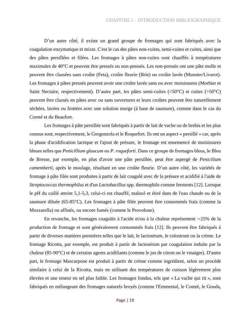
D’un autre côté, il existe un grand groupe de fromages qui sont fabriqués avec la
coagulation enzymatique et mixte. C'est le cas des pâtes non-cuites, semi-cuites et cuites, ainsi que
des pâtes persillées et filées. Les fromages à pâtes non-cuites sont chauffés à températures
maximales de 40°C et peuvent être pressés ou non-pressés. Les non-pressés ont une pâte molle et
peuvent être classées sans croûte (Feta), croûte fleurie (Brie) ou croûte lavée (Munster/Livarot).
Les fromages à pâtes pressés peuvent avoir une croûte lavée sans ou avec moisissures (Morbier et
Saint Nectaire, respectivement). D’autre part, les pâtes semi-cuites (<50°C) et cuites (>50°C)
peuvent être classés en pâtes avec ou sans ouvertures et leurs croûtes peuvent être naturellement
séchées, lavées ou frottées avec une solution morge (à base de saumure), comme dans le cas du
Comté et du Beaufort.
Les fromages à pâte persillée sont fabriqués à partir de lait de vache ou de brebis et les plus
connus sont, respectivement, le Gorgonzola et le Roquefort. Ils ont un aspect « persillé » car, après
la phase d'acidification lactique et l'ajout de présure, le fromage est ensemencé de moisissures
bleues telles que Penicillium glaucum ou P. roqueforti. Dans ce groupe de fromages bleus, le Bleu
de Bresse, par exemple, en plus d'avoir une pâte persillée, peut être aspergé de Penicilium
camemberti, après le moulage, résultant en une croûte fleurie. D’un autre côté, les variétés de
fromage à pâte filée sont produites à partir de lait coagulé avec de la présure et acidifié à l'aide de
Streptococcus thermophilus et d'un Lactobacillus spp. thermophile comme ferments [12]. Lorsque
le pH du caillé atteint 5,1-5,3, celui-ci est chauffé, malaxé et étiré dans de l'eau chaude ou de la
saumure diluée (65-85°C). Les fromages à pâte filée peuvent être consommés frais (comme la
Mozzarella) ou affinés, ou encore fumés (comme le Provolone).
En revanche, les fromages coagulés à l'acide et/ou à la chaleur représentent ∼25% de la
production de fromage et sont généralement consommés frais [12]. Ils peuvent être fabriqués à
partir de diverses matières premières telles que le lait, le lactosérum, le colostrum ou la crème. Le
fromage Ricotta, par exemple, est produit à partir de lactosérum par coagulation induite par la
chaleur (85-90°C) et de certains agents acidifiants (comme le jus de citron ou le vinaigre). D'autre
part, le fromage Mascarpone est produit à partir de crème comme ingrédient, selon un procédé
similaire à celui de la Ricotta, mais en utilisant des températures de cuisson légèrement plus
élevées et une teneur en sel plus faible. Les fromages fondus, tels que « La vache qui rit », sont
fabriqués en mélangeant des fromages naturels broyés (comme l'Emmental, le Comté, le Gouda,

CHAPITRE I – INTRODUCTION BIBLIOGRAPHIQUE
Page | 20
etc) avec des sels émulsifiants et d'autres ingrédients laitiers, et en chauffant le mélange sous vide
(120°C) sous agitation constante, jusqu'à obtention d'un mélange homogène.
Tous ces différents types de fromage sont composés de différents microorganismes
d'origines diverses (ingrédients, ferments ensemencés, saumures, équipements et matériels de la
fromagerie, caves d'affinage, etc). Ces microorganismes peuvent contribuer à la vaste diversité des
caractéristiques organoleptiques des cœurs et des croûtes/surfaces des fromages.
1.2. Diversité microbienne des fromages
Au cours des dernières années, de nouveaux outils ont permis d'étudier les fromages d'une
manière auparavant inconcevables. Ainsi, l'utilisation de ces nouvelles technologies, comme le
séquençage à haut débit, a révolutionné le domaine de l'écologie microbienne, en permettant une
identification plus exhaustive des microorganismes, y compris ceux qui sont difficiles à cultiver
et/ou qui sont présents en faible abondance [13]. Cette science émergente a apporté de nouvelles
perspectives sur la biodiversité microbienne, en particulier, pour les fromages artisanaux [3, 4, 14]
et, par conséquent, en développant une connaissance et un intérêt accrus pour les aliments
traditionnels plus anciens.
Ces microorganismes présents dans les fromages peuvent être ajoutés par l'homme (ferments),
selon la technologie ou les caractéristiques souhaitées, ou ils peuvent provenir de l'extérieur tout
au long de la chaîne de production (microorganismes adventices) (Fig. 2). Les deux procédés,
intentionnels ou non, en combinaison avec les transformations chimiques et biochimiques,
définissent les caractéristiques et l'identité du fromage [15].
Figure 2. Possibles sources de développement de communautés microbiennes spécifiques dans le processus de
production et d’affinage du fromage (Adapté de Wolfe et al.[3]).

CHAPITRE I – INTRODUCTION BIBLIOGRAPHIQUE
Page | 21
1.2.1. Les microorganismes ajoutés intentionnellement
Les ferments (encore appelés agents de fermentation ou levains) sont des souches bien
définies et caractérisées, ajoutés intentionnellement, utilisés lors de la fabrication du fromage pour
contrôler la fermentation et assurer la régularité de la production du produit [1]. La norme ISO
27205:2010 [16] définit une culture initiale pour la production de fromage comme suit: « culture
préparée qui contient une ou plusieurs souches de microorganismes en nombre élevé (en général
plus de 108 UFC/g ou ml de bactéries viables) qui sont ajoutées pour provoquer une réaction
enzymatique souhaitable (par exemple, fermentation du lactose entraînant la production d'acide,
dégradation de l'acide lactique en acide propionique, ou autres activités métaboliques directement
liées aux propriétés spécifiques du produit) ».
Les cultures microbiennes d’origine alimentaire se sont directement ou indirectement
insérées dans plusieurs cadres réglementaires au cours des dernières années, que ce soit en mettant
en évidence l'historique d'utilisation, les aliments traditionnels ou la présentation d’une parfaite
innocuité pour le consommateur et l’environnement (GRAS: Generally recognized as safe) [17].
En 2002, pour classer les ferments traditionnellement utilisés comme ingrédients alimentaires
sécurisés, l’International Dairy Federation (IDF) - en collaboration avec l’European Food and
Feed Cultures Association (EFFCA) - a compilé un inventaire des microorganismes tenant en
compte la littérature scientifique documentée de leur utilisation dans l'alimentation (Bulletin de
l’IDF n° 377/2002). Cet inventaire est devenu une référence pour l'utilisation pratique des souches
alimentaires. En 2012, Bourdichon et al. [17] ont mis à jour cet inventaire des microorganismes
en se concentrant sur les aliments fermentés, y compris le fromage. Les principaux
microorganismes qui peuvent être utilisés lors de la production de fromage comme ferments sont
brièvement présentés ci-dessous.
Bactéries lactiques
Les bactéries de l'acide lactique (LAB) sont parmi les microorganismes les plus étudiés dans
le monde. Actuellement, avec les développements des technologies de séquençage de la nouvelle
génération, des milliers de génomes sont disponibles et, en conséquence, de nombreuses
caractéristiques importantes des souches LAB sont connues, y compris les aspects de la
fermentation du sucre, de la formation d'arômes, de la production de substances texturées, des
réponses au stress, de la colonisation et de la survie chez l'hôte, des interactions entre cellules et
CHAPITRE I – INTRODUCTION BIBLIOGRAPHIQUE
Page | 22
de la pathogénicité [13]. Une recherche du terme « lactic acid bacteria » dans le titre, les mots clés
et le résumé dans la base de données scientifique Scopus a donné environ 24.895 résultats d’études
(novembre 2020), montrant que les LAB ont reçu beaucoup d'attention de la part de la communauté
scientifique. De plus, en utilisant « lactic acid bacteria » ET food, « lactic acid bacteria » ET
cheese, et « lactic acid bacteria » ET environment comme termes de recherche, 10.802, 2.359 et
1.293 documents ont été retrouvés, indiquant que les aliments sont largement étudiés en
association avec les LAB.
Ainsi, au cours des trois dernières décennies, les recherches sur les espèces LAB ont contribué
à une sélection et application bien conçues de ferments pour l'industrie alimentaire [18, 19]. En ce
qui concerne les fromages, la plupart ne peut pas être fabriquées sans ajout de certaines bactéries
lactiques dont les principales fonctions sont d'acidifier le produit pendant la fabrication et de
générer des modifications biochimiques pendant l'affinage. L’ajout intentionnel de ces bactéries
peut aussi contribuer à développer le goût ou la qualité des textures, ajoutant de la valeur aux
produits alimentaires [20].
Les LAB - telles que Lactococcus lactis et Streptococcus thermophilus - sont indispensables à
la fabrication de la grande majorité des fromages. Elles possèdent généralement un métabolisme
homo-fermentaire et génèrent l'acide lactique comme principal produit final [21, 22]. Les « non-
starter » LAB (NSLAB) constituent également une partie importante de la flore lactique
secondaire qui se développe au cours de l’affinage des fromages et sont utilisées comme ferments
de fromagerie depuis les années 90 [23]. Ces bactéries correspondent principalement à des
lactobacilles hétéro-fermentaires facultatifs, comme les espèces suivantes: Lacticaseibacillus
paracasei et casei, Lactiplantibacillus plantarum et Pediococcus acidilactici [5]. Les
métabolismes hétéro-fermentaires génèrent certains métabolites, autres que l'acide lactique, tels
que l'éthanol, le dioxyde de carbone ou l'acide acétique, et contribuent à améliorer les arômes du
fromage [5, 22, 24]. En général, les fournisseurs vendent des mélanges de ferments homo et
hétéro-fermentaires avec des proportions différentes suivant les caractéristiques à privilégier.
Les ferments ajoutés pendant la fabrication du fromage peuvent également être classés en
fonction de leur température de croissance optimale (mésophiles ou thermophiles). Par définition,
le terme « mésophile » désigne les organismes dont la température de croissance optimale se situe
entre 20 et 37°C [25]. Les cultures mésophiles sont normalement utilisées dans la fabrication de
fromages frais, de pâtes molles et de pâtes semi-cuites [26]. D’autre part, pour les fromages à pâtes

CHAPITRE I – INTRODUCTION BIBLIOGRAPHIQUE
Page | 23
cuites, l’emploi des cultures thermophiles est indiqué. Elles sont composées de bactéries qui se
développent à températures plus élevées (entre 40 et 50ºC) [27]. Ces cultures ont la particularité
de donner de la souplesse aux pâtes pressées ou générer des enzymes texturants en pâtes filées [28,
29]. Comme exemple, Lactococcus lactis ssp. lactis et L. lactis ssp. cremoris sont les principaux
ferments mésophiles largement utilisés dans la fabrication du fromage. Ils sont fréquemment
combinés avec d'autres cultures mésophiles (Leuconostoc mesenteroides, L. pseudomesenteroides)
ou thermophiles (Streptococcus thermophilus) [5, 27].
Cultures d’affinage
Au-delà des bactéries lactiques (LAB ou NSLAB), d’autres cultures fromagères peuvent
contribuer à la définition des caractéristiques des fromages. Dans certains produits traditionnels à
croûte lavée, par exemple, les fromages plus anciens sont frottés au contact de fromages jeunes
pour transférer leurs microorganismes [30]. Aujourd'hui, cependant, les mélanges commerciaux
de bactéries et de levures sont plus couramment utilisés dans les productions à large échelle afin
de produire un produit plus standardisé. Les kits de microorganismes d'affinage peuvent être
constitués de champignon ascomycètes telles que Penicilium spp. et Geotrichum candidum, de la
levure Debaromyces hansenii et des bactéries comme Brevibacterium linens/aurantiacum, B.
casei, Staphylococcus xylosus, S. equorum et Glutamicibacter arilaitensis [31, 32]. La
composition et la proportion de microorganismes dans chaque mix dépend du type de fromage
fabriqué et des caractéristiques souhaitées. Par exemple, le Penicilium roqueforti est utilisé dans
la fabrication de fromages bleus et un mélange de G. candidum, D. hansenii, B. linens, G.
arilaitensis et S. xylosus est commercialisé pour la fabrication de fromages à croûte lavées [32].
Ces dernières cultures sont souvent utilisées dans les fromages jeunes et peuvent être pulvérisées
sur la surface du fromage ou incorporées à la saumure [33].
Toutes les espèces bactériennes mentionnées ci-dessus appartiennent au groupe des Gram-
positifs et peuvent être utilisées comme cultures d’affinage, bien qu'elles puissent également être
fortuites. À ce jour, la seule bactérie Gram-négatif utilisée comme culture commerciale est
l’Hafnia alvei [5], notamment pour les fromages type Camembert au lait cru [34, 35]. La présence
de certaines souches de cette espèce augmente le niveau de composés soufrés volatils et peut,
conséquemment, contribuer au goût du fromage [36].

CHAPITRE I – INTRODUCTION BIBLIOGRAPHIQUE
Page | 24
Néanmoins, selon Delbès et al. (2015), les microorganismes ajoutés intentionnellement dans
les fromages peuvent entrer en compétition avec leur microbiote endogène et se montrer
faiblement adaptés aux étapes de fabrication de ce produit [6]. Rea et ses collègues (2007) ont
évalué la production de six lots d'un fromage à croûte lavée irlandais (Gubbeen) et ont noté que
des souches commerciales, délibérément inoculées, peuvent être présentes en tant que sous-
populations, mais ne font pas partie de la microflore dominante [37]. En effet, des études menées
au cours des 20 dernières années ont montré que les levures et/ou les ferments bactériens ajoutés
intentionnellement, notamment dans les fromages à croûte lavée, ne se développent pas
nécessairement lors de la fabrication du fromage et qu'une flore adventice peut être dominant à la
surface de ces produits fermentés [31, 38-40].
1.2.2. Les microorganismes adventices
Le lait, principal ingrédient de la production de fromage, est riche en nutriments, présente une
humidité élevée et un pH presque neutre, ce qui en fait un excellent substrat de croissance non
seulement pour les ferments, mais aussi pour les microorganismes adventices [41]. Bien que le lait
sorte de la mamelle des animaux, essentiellement stérile, il peut être contaminé par des
microorganismes provenant du trayon et du sphincter, de l'environnement de la ferme, des
équipements de traite et des récipients de conditionnement [42]. Dans les temps anciens, avant
l’utilisation de la réfrigération, les bactéries mésophiles Gram-positives, en particulier les LAB
comme Streptococcus, Enterococcus, Lactobacillus, Leuconostoc, Lactococcus et Pediococcus,
dominaient tout développement bactérien dans le lait et contribuaient donc à leur acidification
[43]. Avec l'utilisation des récipients à refroidissement rapide et au stockage réfrigéré, les basses
températures (<7°C) ont réduit les populations de bactéries lactiques dans le lait et ont favorisé la
croissance de bactéries Gram-négatives psychrophiles telles que Pseudomonas et Acinetobacter
[44, 45]. La forte activité protéolytique et lipolytique de certaines espèces de ces genres peut
entraîner de nombreux inconvénients pour le fromage, notamment des odeurs désagréables, de
l'amertume et une saveur rance. En outre, des coliformes tels que Citrobacter, Escherichia,
Enterobacter et Klebsiella font également partie de la flore psychrophile et peuvent provoquer des
saveurs désagréables ainsi que des défauts dans le lait et les fromages [45, 46].
D'autre part, le microbiote indigène présent dans les matières premières ou dans
l'environnement de production, comme les caves d'affinage, peut produire des fromages

CHAPITRE I – INTRODUCTION BIBLIOGRAPHIQUE
Page | 25
d'excellente qualité. Mounier et al. (2005) confirment l'importance de la flore adventice et de leur
présence dans les fromages affinés [40]. Les chercheurs ont isolé des microorganismes à partir de
quatre types de fromages irlandais à croûte lavée (Gubeen, Durrus, Ardrahan et Mileens), dont
aucun n'appartenait aux cultures commerciales d’affinage, ajoutées intentionnellement à la surface
des fromages. Ils ont obtenu Debaryomyces hansenii, comme levure dominante, dans les quatre
fromages évalués, et une grande diversité bactérienne, parmi laquelle des espèces appartenant aux
genres Agrococcus, Arthrobacter, Bacillus, Brevibacterium, Corynebacterium, Halomonas,
Microbacterium, Micrococcus, Staphylococcus et Vibrio.
Grâce à des techniques indépendantes de la culture, comme la métagénomique, une vue plus
complète de la biodiversité peut être mieux établie dans les écosystèmes fromagers. En 2014, par
exemple, une étude américaine menée dans le laboratoire de Rachel Dutton, a analysé par
metabarcoding 137 croûtes de fromages provenant de dix pays différents et ils ont constaté qu'au
moins 60% des genres bactériens et 25% des champignons présents dans ces produits sont des
microorganismes adventices [3]. Parmi les groupes non inoculés, ils ont identifié deux genres
bactériens, Nocardiopsis et Yaniella, qui n'avaient jamais été signalés dans les écosystèmes
alimentaires microbiens. En outre, ils ont montré que les protéobactéries telles que les Halomonas,
Pseudoalteromonas, Psychrobacter et Vibrio sont très répandues dans les communautés
fromagères, ce qui a également été observé dans d'autres études métagénomiques sur les croûtes
de fromage [3, 4, 47]. De plus, Dugat-Bony et al. (2016) ont évalué 12 variétés de fromages
français avec la métagénétique (amplicon bactérien et fongique), et ont également souligné
l'importance des microorganismes indigènes dans l’écosystème fromager [48]. Alors que la plupart
des échantillons étaient dominés par les espèces fongiques Geotrichum candidum, Debaryomyces
hansenii et Candida sake, la composition de la communauté bactérienne globale était beaucoup
plus variable parmi les échantillons. Les éventuelles bactéries adventices trouvées dans le cœur et
croûtes des fromages étudiés appartenaient aux genres Halomonas, Pseudoalteromonas,
Pseudomonas, Psychrobacter et Psychroflexus. La prédominance de ces bactéries psychrophiles
dans les fromages peut être due aux conditions de stockage appliquées pour le transport et la
distribution aux supermarchés (température de 4-8°C pendant des jours ou des semaines) [48].
Ainsi, ces études confirment que la microflore adventice qui se développe dans les fromages
peut être assez complexe, selon l'endroit où ils sont affinés et la façon dont ils sont préparés. Les
sources potentielles de ce microbiote résidant dans les fromages sont liées à l'environnement de la

CHAPITRE I – INTRODUCTION BIBLIOGRAPHIQUE
Page | 26
ferme ou de la cave d’affinage, aux récipients et équipements utilisés, ainsi qu’aux ingrédients
utilisés tels que le lait et le sel ajoutés pendant la production [5, 49-51].
2. Le sel dans l’alimentation
En tant que produit naturel, le sel (chlorure de sodium, NaCl) a été récolté depuis
l'Antiquité sur des lacs et des oasis, ainsi qu’au long des littoraux, puis de manière plus industrielle
dès la protohistoire du 5-6ème millénaire avant J.-C., dans des mines telles que Hallstatt (Autriche),
Lunca (Roumanie), Duzdagi (Azirbadjan) ou Shabwa (Yémen) [52-54]. Aujourd'hui, il existe trois
grandes méthodes pour obtenir du sel : (i) l'évaporation de l'eau du mer ; (ii) l'extraction des
cristaux de sel gemme et (iii) la création de saumures de sel (sel ignigène) [55, 56].
Le sel représente environ 3,5 % des océans du monde et peut être produit naturellement
lorsque les étangs et les baies peu profondes s'assèchent au soleil [57]. Lors de la production de
sel marin à l'échelle industrielle, l'eau du mer est placée dans de grands bassins pour permettre une
évaporation efficace, face au soleil et au vent [58, 59]. Ainsi, pour obtenir une évaporation
complète de l'eau, le sel marin est, de préférence, fabriqué sous climats secs et dans les pays à
faible pluviométrie, comme le Brésil, le Cap-Vert, la Colombie, le Mexique, l’Afrique du Sud, le
Venezuela et l’Australie occidentale [59]. Le sel de mer peut également être produit selon des
techniques anciennes et artisanales, à une échelle beaucoup plus petite, comme la Fleur de sel. Ce
sel léger et gastronomiquement réputé, est fabriqué dans de petits lagons en France, seulement,
pendant les mois d'été, de juin à août [60, 61].
Encadré 3 : Le sel comme agent de sélection des microorganismes
Le sel joue généralement un rôle central dans la production d'aliments fermentés, qui
non seulement ont une longue durée de conservation, mais développent également des
caractéristiques technologiques comme la saveur et la texture, faisant ainsi partie du patrimoine
culturel de plusieurs pays. La teneur en sel dans les fromages peut varier de 0,3 à 7% et cet
environnement « salé » peut favoriser la croissance de microorganismes tolérants au sel.

CHAPITRE I – INTRODUCTION BIBLIOGRAPHIQUE
Page | 27
Le sel gemme (aussi appelé halite) s’est formé lors de l’évaporation des mers géologiques
il y a plusieurs millions d’années, et est présent dans les couches rocheuses inférieures de la surface
de la Terre [62]. Il est extrait à la dynamite, de la même manière que tout autre minéral, ou d’une
manière plus artisanale à la main, et est ensuite broyé en différentes tailles selon les besoins
respectifs. Le sel gemme alimentaire est également connu sous le nom de sel de l’Himalaya, de sel
rose ou de sel de Khewra (importante mine de sel, au Pakistan) et c’est l’un des types de sel les
moins transformés et disponibles sur le marché [63].
Le sel ignigène est obtenu de gisements souterrains et est dissous par injection d’eau douce
afin d’obtenir une saumure remontée en surface pour être évaporée [64]. Alors que l'océan est une
saumure naturelle, l'extraction hydraulique (ou extraction par dissolution) du sel consiste à pomper
de l'eau souterraine pour dissoudre les dépôts de sel et créer une saumure. Cette saumure est ensuite
évaporée pour créer le sel. La saumure salée peut être traitée avant l'évaporation pour réduire sa
teneur en minéraux, ce qui donne un cristal de chlorure de sodium presque pur. Comme cette
méthode est peu coûteuse, a un rendement élevé et produit un sel très propre, la plupart du sel de
table est produit de cette façon [65].
Selon l’U.S. Geological Survey (2020), la production mondiale de sel est dominée
spécialement par trois pays : la Chine (20%), les États-Unis (14%) et l'Inde (10%) [66]. Sur les
290 millions de tonnes de sel produites annuellement, environ 43% sont utilisés pour le déglaçage
des routes, 37% pour l'industrie chimique et seulement 9% sont utilisés pour la consommation
humaine [66]. Alors que les sels utilisés à des fins industrielles sont principalement obtenus à partir
de l'exploitation minière, la plupart des sels de table sont produits à partir de la saumure, tandis
que les sels spéciaux ou gastronomiques sont presque toujours produits par évaporation de l'eau
de la mer. Indépendamment de la manière dont il est extrait et de son origine, le sel est un ingrédient
largement utilisé dans l’alimentation en raison de son rôle technologique, de préservation et
d'appréciation sensorielle.
2.1. Le sel comme conservateur
Le processus de salage est utilisé comme principale méthode de conservation depuis
l'antiquité [67], et il peut se faire de deux manières : par l’ajout de cristaux de sel ou par la saumure.
Les Sumériens, par exemple, utilisaient le sel pour conserver toutes sortes d'aliments, y compris le
poisson, les graisses, la viande, l'orge et le blé, en 3.000 avant J.-C. [68].

CHAPITRE I – INTRODUCTION BIBLIOGRAPHIQUE
Page | 28
Le sel est efficace comme conservateur car il réduit l'activité de l'eau (aw) des aliments, qui
est un critère de la disponibilité d’eau « libre » d’une matrice alimentaire, susceptible d’intervenir
dans des réactions chimiques, biochimiques ou microbiologiques [69]. Alors que les aliments
ayant un aw élevé (>0,95) comprennent la viande fraîche, les œufs et les légumes frais, des valeurs
d'aw faibles (<0,90) peuvent être trouvées dans les fromages du type Parmesan, les jambons et les
pains [70]. Ainsi, le NaCl peut réduire l’aw et conséquemment les taux de multiplication
microbienne dans les aliments, grâce à la capacité des ions sodium et chlorure à s'associer aux
molécules d'eau et à provoquer un choc osmotique dans les cellules microbiennes, entraînant une
perte d'eau et donc la mort des cellules ou le ralentissement de leur développement [71-73]. De
plus, des études suggèrent que, pour certains microorganismes, le sel peut limiter la solubilité de
l'oxygène, interférer avec les enzymes cellulaires, ou forcer les cellules à dépenser de l'énergie
pour exclure les ions sodium de la cellule, ce qui peut réduire leur croissance [74].
Le sel reste un ingrédient couramment utilisé pour assurer un environnement résistant à la
détérioration et pour empêcher la croissance des agents pathogènes, ce qui renforce la sécurité et
le shelf-life des produits, tels que le jambon, le poisson et le fromage. Cependant, au-delà du simple
ajout de sel, d’autres méthodes de conservation sont normalement combinées afin de permettre
l'obtention d'un produit de qualité, stable et sécurisé [69]. Par exemple, l'utilisation du sel dans la
formulation de la viande prête à consommer, en combinaison avec un conservateur chimique
(comme le nitrate), des valeurs de pH et de température adéquates, peut réduire le risque de
germination des spores d’espèces qui provoquent des intoxications alimentaires comme le
Clostridium perfringens [75, 76] et le Clostridium botulinum [77, 78].
Ainsi, alors que des niveaux de sel élevés inhibent complètement la croissance de la plupart
des microorganismes, des niveaux de sel modérés engendrent un environnement inhospitalier pour
la plupart des agents pathogènes et peuvent aussi favoriser les aspects organoleptiques et
technologiques bénéfiques pour divers aliments fermentés.
2.2. Le rôle technologique du sel dans la production des aliments
Bien que de nombreuses industries alimentaires cherchent notamment à réduire la teneur
en sel de leurs produits, les aspects technologiques et sensoriels, tels que la texture et le goût,
peuvent affecter les formulations [79, 80] et influencer l'acceptation du consommateur [81]. En ce

CHAPITRE I – INTRODUCTION BIBLIOGRAPHIQUE
Page | 29
qui concerne l'aspect organoleptique, le sel peut faciliter la libération de certaines molécules,
conférant des arômes perceptifs au niveau du palais et diminuer la diffusion du goût amer [82, 83].
Dans les produits transformés à base de viande, par exemple, le sel peut être un élément
essentiel qui contribue à la texture. Ruusunen et al. (2001) ont rapporté que le jambon cuit dont la
teneur en sel ajoutée était inférieure à 1,4% présentait des pertes à la cuisson plus importantes que
les jambons dont la teneur en sel était supérieure à 1,7% [84]. Des auteurs soulignent que dans les
produits à faible teneur en sel et à forte teneur en eau, il est nécessaire d'ajouter plus de protéines
ou d'autres ingrédients fonctionnels pour augmenter le rendement [80, 84].
Le NaCl aussi facilite la manipulation et la transformation de nombreux produits fermentés,
comme les pains, les légumes et les fromages [70]. Dans le pain, le sel a comme fonction de rendre
le gluten plus stable et moins extensible et collant, en plus de réguler le taux de fermentation. Sans
sel, la fermentation de la levure est très rapide, produisant de grandes cellules d'air irrégulières
dans la structure de la mie. Une fermentation rapide peut également consommer énormément de
sucre, ingrédient essentiel pour les réactions de brunissement telles que la caramélisation et la
réaction de Maillard qui contribuent à la couleur du pain [85]. Dans les légumes fermentés tels que
la choucroute et les pickles, le sel favorise la texture croquante car il diminue l'activité de la
pectinase, une enzyme qui rend les végétaux pâteux. En outre, le sel peut ralentir le processus de
fermentation, ce qui permet un développement complet du goût et la réduction du risque lié à la
présence de microorganismes indésirables [69]. Le fromage est un aliment dont le sel peut aussi
réguler la fermentation et permettre la croissance de microorganismes tolérants au sel. En plus, le
NaCl peut affecter directement le corps/la texture du fromage en modifiant la structure des
protéines ou, indirectement, en éliminant l'humidité. En fait, la réduction de la teneur en NaCl dans
les fromages à pâtes semi-rigides, tels que le Cheddar, entraîne une augmentation de l'amertume
et le côté désagréable du goût, ainsi qu'une réduction de la rigidité [86-88].
2.3. Le sel et la salage des fromages
En général, les différents types de fromage présentent une très large gamme de sel. Le Tableau
1 a été adaptée à partir des données provenant de l’Agence nationale de sécurité sanitaire de
l’alimentation, de l’environnement et du travail (Anses, 2020) [89] et présente la teneur en sel de
certains fromages vendus en France, sachant que les valeurs peuvent varier selon chaque fromager.

CHAPITRE I – INTRODUCTION BIBLIOGRAPHIQUE
Page | 30
Tableau 1. Teneur en sel, par ordre croissant, de certains fromages en fonction de leurs respectives catégories
(Adapté de Anses [89]).
Catégorie Fromage NaCl (g/100 g)
Blancs Faisselle 0,11
Blancs Fromage blanc nature 0,11
Spécialités Ricotta 0,30
Spécialités Mozzarella 0,49
Pâte pressée Emmental 0,75
Pâte pressée Comté 0,80
Pâte pressée Gruyère 0,80
Pâte molle Langres 0,99
Pâte molle Crottin de chèvre 1,01
Pâte molle Reblochon 1,27
Pâte pressée Beaufort 1,27
Pâte molle Pont l'Évêque 1,30
Pâte pressée Morbier 1,41
Pâte molle Pélardon 1,43
Pâte persillée Bleu de Gex 1,44
Pâte molle Camembert 1,45
Pâte molle Livarot 1,50
Pâte molle Brie 1,51
Pâte pressée Grana Padano 1,56
Pâte pressée Parmesan 1,57
Pâte pressée Cheddar 1,61
Pâte pressée Ossau-Iraty 1,68
Pâte pressée Saint-Nectaire 1,70
Pâte molle Époisses 1,76
Pâte persillée Gorgonzola 1,77
Pâte molle Munster 1,78
Pâte pressée Cantal entre-deux 1,85
Pâte pressée Gouda 2,00
Pâte pressée Provolone 2,19
Pâte pressée Edam 2,22
Spécialités Feta 2,27
Pâte pressée Mimolette 2,84
Pâte persillée Bleu d'Auvergne 2,85
Pâte persillée Roquefort 3,22
Pâte pressée Pecorino 4,73
En dehors des fromages blancs (sans ajoute de sel), la Ricotta et la Mozzarella sont les
fromages moins salés avec un taux de 0,3 et 0,5%, respectivement. La majorité des fromages
varient entre 1-2% de sel et certains, comme le Roquefort et le Pecorino, contiennent plus de 3%
de sel dans leur composition. Il existe aussi des fromages, comme le Domiati et le Sikma, très
consommés en Égypte et en Turquie, respectivement, qui peuvent contenir jusqu'à 7 % (p/p) de sel
[90, 91].

CHAPITRE I – INTRODUCTION BIBLIOGRAPHIQUE
Page | 31
Pour incorporer le sel au fromage, lors de la production, quatre approches différentes
peuvent être utilisées. Une première méthode d'application consiste à mélanger le sel directement
avec les particules de caillé, avant qu’il ne soit pressé, pour former le fromage final [92] (Fig. 3).
L'ajout de sel aux caillés de fromage génère une force motrice osmotique qui attire le lactosérum
vers la surface, où il est libéré [92]. Ainsi, au-delà des étapes de fabrication du fromage comme la
découpe, l'égouttage et le pressage, le salage est une autre étape utilisée pour réduire l'humidité du
fromage. Cette technique permet d'incorporer uniformément de fortes concentrations de sel dans
toute la masse et d'éliminer efficacement le lactosérum du caillé au cours du processus. L'expulsion
du lactosérum obtenue par cette méthode de salage permet également aux fromagers de produire
des variétés à faible teneur en humidité, comme le Cheedar, sans avoir besoin de températures de
cuisson élevées, ce qui constitue un grand avantage pratique [11].
Figure 3. Étapes de la fabrication du fromage impliquant l'ajout de sel (bleu) et les méthodes de salage (vert).
La deuxième méthode peut être appliquée au fromage avant ou pendant l'affinage, soit par
ajout de sel en cristaux ou par saumure, et ces procédés peuvent être utilisés une ou plusieurs fois
et à des différents échelles de temps (Fig. 3). Le sel sec peut être frotté sur la surface des fromages,
où il se dissout à l'intérieur et, parallèlement, l'humidité est transférée à la surface et s'évapore. Les
fromagers traditionnels appliquent le sel de manière répétée avec un frottement légèrement abrasif
afin de développer une croûte lisse, dense et imperméable qui rend le fromage très stable et durable.
Bien que cette méthode fonctionne bien pour les petits fromages (comme le Limburger), la

CHAPITRE I – INTRODUCTION BIBLIOGRAPHIQUE
Page | 32
technique devient problématique pour les gros fromages, car le salage à sec déshydrate trop
rapidement la surface [93]. Par conséquent, la couche extérieure salée peut devenir épaisse et
empêcher la migration du sel vers l'intérieur du fromage (Fig. 4)
Figure 4. Fromage à croûte lavée (Adapté de Wolfe et al. [3]).
La troisième méthode consiste à submerger le fromage dans une saumure concentrée (qui
peut varier de 10 à 20% de sel) [11]. Ce salage est particulièrement utile pour la fabrication de
grands fromages à croûte lavée, car il permet une plus grande absorption du sel tout en
déshydratant progressivement la surface en vue de la formation de la croûte. L’immersion en
saumure entraîne des gradients importants et persistants de sel et d'humidité, s'étendant de la
surface vers le centre du fromage, ce qui affecte l'écologie microbienne de manière localisée: les
microorganismes sensibles au sel peuvent se trouver en abondance dans le centre (faible teneur en
sel) et les types tolérants en surface (forte teneur en sel) [11]. Cette méthode est utilisée pour les
fromages comme le Livarot et l’Ossau Iraty.
Parallèlement à l'immersion dans la saumure, la quatrième méthode de salage consiste à
frotter le fromage pendant l’affinage avec une solution appelée morge, dont les ingrédients de base
sont l'eau et le sel. La quantité de sel à incorporer dans l'eau est très variable (5-35%) et dépend du
type de fromage et de la charge de contamination dans les caves d'affinage. Il est également
possible de diminuer le pH de la morge (aux alentours de 5,0-5,2) en ajoutant un acide (comme du
vinaigre blanc) afin d'éliminer les levures indésirables et de stimuler le développement de levures
tolérantes aux acides [94]. Le frottage de la solution de morgeage dans les fromages peut se faire
avec un torchon, un pinceau/brosse ou une éponge (Fig. 3), en commençant par les fromages les
plus anciens et en terminant par les plus jeunes, sans croûte. C'est ainsi que les microorganismes
s'incorporent et donnent l'originalité de la solution, qui peut être conservée plusieurs jours et
contribuer à des frottages successifs. Cette méthode est très artisanale et fréquente dans les petits
ateliers, mais elle tend à disparaître dans les grandes industries, qui ont tendance à privilégier une
CHAPITRE I – INTRODUCTION BIBLIOGRAPHIQUE
Page | 33
nouvelle solution de frottage à chaque traitement. Dans ce cas, l'ajout de souches commerciales à
la morge initiale peut être utile pour permettre l'ensemencement de la surface des fromages et ainsi
favoriser la formation de la croûte. Ces cultures peuvent être proposées sous forme lyophilisée ou
liquide par les fournisseurs de ferments et peuvent se constituer principalement des levures
Debaryomyces hansenii et Geotrichum candidum, ainsi que de bactéries aérobies non lactiques
telles que les corynebactéries (en particulier Brevibacterium linens/aurantiacum) et les
microcoques [94].
La phase de salage, lors de la fabrication du fromage, pourra donc générer une forte
concentration de sel, favoriser la croissance de microorganismes adaptés à ces environnements et
favoriser le développement des propriétés organoleptiques comme le goût, l'apparence et la texture
du produit final. Une meilleure compréhension de l'origine et de la manière dont ces organismes
évoluent, pendant les différentes étapes du développement du fromage, pourrait être d'un grand
intérêt pour déterminer comment le sel pourrait favoriser la composition du microbiote du
fromage.
2.3.1 Quels types de sel pour la fromagerie?
Selon le Décret n° 2007-588 du 24 avril 2007, relatif aux sels destinés à l'alimentation
humaine, le sel de qualité alimentaire est « un produit cristallin se composant principalement de
chlorure de sodium, provenant de marais salants, de sel gemme ou de saumures provenant de la
dissolution du sel gemme » [95]. Cette réglementation indique aussi que les sels doivent avoir au
moins 97% d’extraits secs de chlorures de sodium (NaCl), additifs non inclus, et contenir moins
de 2 mg/kg de cuivre ou de plomb, 0,5 mg/kg d’arsenic ou de cadmium et de 0,1 mg/kg de mercure.
Bien que l'iode soit, parfois, ajouté au sel afin de réduire les troubles thyroïdiens [96, 97],
il est considéré comme un agent antimicrobien [98] et pourrait donc inhiber des microorganismes
souhaitables dans l’affinage des fromages. Ainsi, les sels raffinés, tels que les sels de table qui
contiennent de l’iode, ne sont pas appropriés pour la fromagerie [99]. Cependant, il n'y a pas
beaucoup d'informations dans la littérature sur les types de sels utilisés pour la fabrication du
fromage. Une recherche sur des sites de vente d'ingrédients de fromagerie, dans différents pays,
confirment l'utilisation de sels marins ou du sel Himalaya, exempt d'iode. Ces deux types de sel
sont riches en oligo-éléments (minéraux) et peuvent apporter un goût salé moins prononcé, en
comparaison avec les sels de table. Enfin, en ce qui concerne le sel fromager, il est essentiel de
CHAPITRE I – INTRODUCTION BIBLIOGRAPHIQUE
Page | 34
savoir à quelle vitesse (ou avec quelle lenteur) il est absorbé et contribue au processus d'extraction
du lactosérum, à la formation de la croûte ainsi qu’au développement de microorganismes
(bénéfiques ou néfastes).
2.3.2. Le sel comme source de développement de communautés microbiennes
Comme mentionné précédemment, les producteurs d'aliments utilisent le sel pour apporter des
caractéristiques sensorielles bénéfiques ainsi que pour la conservation des aliments. Récemment,
Haastrup et al. (2018) ont révélé la présence d'un microbiote complexe dans la saumure de quatre
fromageries danoises, y compris les genres Chromahalobacter, Debaryomyces, Microbacterium,
Psychrobacter, Tetragenococcus et Debaryomyces [100]. Cette étude suggère que certaines
bactéries et levures, présentes dans la surface des fromages, pourraient être introduites par le sel
marin. Par conséquent, s’il existe des microorganismes dans le sel utilisé, ils pourraient se
retrouver aussi dans le produit final. Mais pourrait-il y avoir des microorganismes viables dans le
sel ?
Le Boston Magazine [101] a présenté, en 2013, au grand public (non scientifique), une étude
réalisée par Ben Wolfe (un microbiologiste de Harvard) et Scott Jones (un chef de cuisine), qui a
demontré que certains sels renferment un grand nombre de microorganismes. Dans cette étude, ils
ont utilisé sept types différents de sels de mer, minimalement traités, incluant les marques Celtic,
Amagansett, Wellfleet, French Grey, DeLallo, Bob’s Red Mill et Maine, ainsi que des techniques
de microbiologie classique avec étalement des échantillons sur des boîtes de Petri (milieu de
culture inconnu). De cette expérience, il a été constaté que les sels qui ont été très peu traités -
humides, avec des cristaux plus gros et de couleur gris clair - contenaient le plus grand nombre de
cultures viables. La plupart de ces bactéries sont des protéobactéries (genres/ espèces non
mentionnés), un groupe de bactéries que l'on trouve couramment dans les océans. Il est aussi
intéressant de noter que chaque sel avait son propre mélange de microorganismes, comme illustré,
Figure 5. Parmi les sept sels, trois étaient plus raffinés, d'un blanc pur, et ne comportaient pas de
microorganismes détectables.

CHAPITRE I – INTRODUCTION BIBLIOGRAPHIQUE
Page | 35
Figure 5. Les sept sels marins artisanaux analysés par Ben Wolfe and Scott Jones et leur pouvoir respectifs de
détection de microorganismes (Boîtes de Petri avec milieu de culture inconnu) (Adapté de Boston Magazine [101]).
Une série d'isolats de sels commerciaux (ou alimentaires) a été obtenue au cours des dernières
années, concernant principalement le groupe des Archaea (plus de détails dans le chapitre suivant,
section 3.1, Fig. 8). Cependant, il existe peu d'études indépendantes de la culture, dans la
littérature, ce qui fait que le microbiote complet des sels alimentaires est encore faiblement exploré
[55, 102]. Henriet et al. (2014) ont évalué la présence et la diversité des archées, par des méthodes
de culture dépendantes et indépendantes, dans 26 sels de qualité alimentaire produits dans le
monde (Amérique, Europe, Afrique, Asie et Océanie) [55]. Des comptages viables (en utilisant
quatre milieux de culture pour Archaea) ont été observés pour plus de la moitié des sels (14), et
ces comptages étaient supérieurs à 105 UFC/g dans les sels provenant de l'île de Gozo en Malte,
de Sel de Guérande en France et de La Palma Flor de Sal, en Espagne. L'analyse métagénétique
(gène 16S ARNr pour Archaea) réalisé pour neuf sels a montré la présence de 21 à 27 genres
différents, la majorité correspondant à des membres de la famille des Halobacteriaceae, soit
Haloarcula, Halobacterium soit Halorubrum, genres les plus représentés. D’autre part, Gibtan et
al. (2017) ont évalué, par métagénétique (gènes 16S ARNr), la diversité de bactéries et archées
pour quatre types de sel alimentaire, provenant d'Éthiopie et de Corée, produits à partir de bassins,
de plaines salées, de vasières et de matériaux synthétiques [102]. Les résultats ont indiqué que les
communautés microbiennes étaient très variables entre les sels évalués et contenaient environ 48-
61% de bactéries et 38-51% d’archées. Parmi le groupe des Archaea, 91,6% appartenaient à la
classe des halobactéries et les genres Halarchaeum, Haloarcula, Halobacterium, Halonotius,
Halomicrobium, Halorubrum, Natromonas et Salarchaeum ont été répertoriés dans tous les sels.

CHAPITRE I – INTRODUCTION BIBLIOGRAPHIQUE
Page | 36
Parmi les bactéries, les communautés étaient plus variables et Aeromonas, Arthrobacter,
Brachybacterium, Cronobacter, Enterobacter, Halovibrio, Leclercia, Microbacterium,
Planococcus, Psychroflexus, Salicola et Salinibacter figurent parmi les genres détectés dans un ou
plusieurs sels.
Ainsi, tous les microorganismes tolérants au processus de traitement et à la forte concentration
en sel peuvent persister en tant que cellule dormante et, lorsqu'ils trouvent des conditions
favorables, peuvent se développer. Dans le cas d’études susmentionnées, les microorganismes ont
été « réveillés » dans des milieux de culture de laboratoire, mais il est possible qu'ils se manifestent
également dans les aliments.
3. Halophiles et halotolérants
Les microorganismes peuvent être classifiés en fonction de la concentration particulière de
sel nécessaire à leur croissance optimale. Les groupes appartenant à cette définition sont appelés
halotolérants, pour ceux qui tolèrent le NaCl, et halophiles, pour ceux qui ont besoin de ce composé
pour leur prolifération [103, 104]. Il est difficile de définir ces termes, parce que leur classification
dépend de plusieurs propriétés : la concentration minimale de sel requise pour la croissance des
bactéries, la salinité optimale et la limite supérieure de sel tolérée dans l'environnement. De plus,
ces paramètres dépendent souvent de la composition du milieu et de la température de croissance
[105].
Encadré 3 : Halophiles et halotolérants : de l’environnement aux aliments
Les microorganismes peuvent être classés en fonction de la concentration de sel
nécessaire à leur développement: (i) halophiles : ils ont besoin de sel pour se développer et (ii)
halotolérants : ils sont capables de se développer en présence ou en l'absence de sel. Des
produits tels que les conserves de légumes, les fromages, les crustacés asiatiques et les viandes
fermentées doivent une grande partie de leurs caractéristiques à l'action de microorganismes,
dont la croissance donne forme à différents procédés technologiques, notamment l'ajout de sel.

CHAPITRE I – INTRODUCTION BIBLIOGRAPHIQUE
Page | 37
Les microorganismes halotolérants sont définis comme capables de survivre en présence
ou en absence de sel [103]. Il existe plusieurs catégories d’halotolérants : (i) les non tolérants, ceux
qui ne tolèrent qu'une faible concentration de sel (environ 1% p/v) ; (ii) les légèrement tolérants
avec tolérance jusqu'à 8% (p/v) ; (iii) les modérément tolérants, de tolérance jusqu'à 20% (p/v) ; et
(iv) les extrêmement tolérants, ceux qui se développent dans toute la gamme de concentrations de
sel, comprises entre zéro à la saturation [103] (Fig. 6A). LeFevre et Round mentionnent, pour la
première fois en 1919, la présence de microorganismes halotolérants isolés à partir de saumures
de fermentation de concombres, à l'aide d'un milieu de gélose au jus de concombre avec rajout de
10% de sel [106].
D’un autre part, les bactéries aimant le sel, formellement appelés halophiles, sont des
microorganismes qui ont besoin d’environnements salins pour survivre [103, 104]. La définition
précise d'halophile diverge selon les auteurs, certains incluant tout organisme nécessitant des
pourcentages autour de 3,5% de sel [103, 107], comme dans l'eau de mer, tandis que d'autres ne
considèrent que ceux qui se développent de manière optimale à 5% ou plus, et tolèrent au moins
10% de sel [105]. Kushner, en 1978, a utilisé des définitions qui distinguaient différentes
catégories : les halophiles légers (3% p/v de NaCl), les halophiles modérés (de 3 à 15% p/v) et les
halophiles extrêmes (25% p/v) (Fig. 6B) [104].
Figure 6. Classification des bactéries en fonction du pourcentage de NaCl nécessaire à leur croissance optimale.
Halotolérants (A). Halophiles (B). (Adapté de Larsen [103]).
La vie, dans les environnements à forte concentration en sel, exige des stratégies pour
équilibrer la pression osmotique cellulaire [107, 108], Fig. 7. La stratégie utilisée par ces
microorganismes consiste à maintenir de faibles concentrations de sel dans leur cytoplasme, en
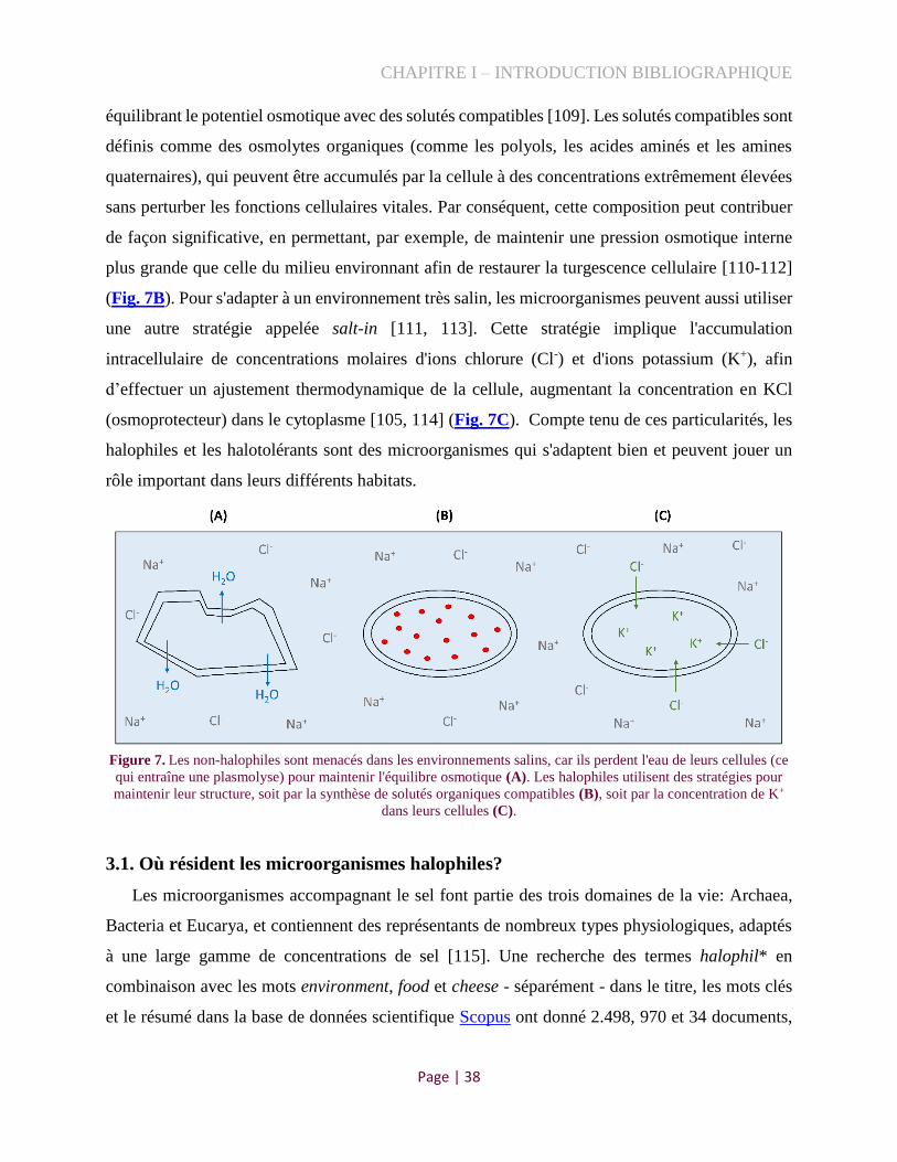
CHAPITRE I – INTRODUCTION BIBLIOGRAPHIQUE
Page | 38
équilibrant le potentiel osmotique avec des solutés compatibles [109]. Les solutés compatibles sont
définis comme des osmolytes organiques (comme les polyols, les acides aminés et les amines
quaternaires), qui peuvent être accumulés par la cellule à des concentrations extrêmement élevées
sans perturber les fonctions cellulaires vitales. Par conséquent, cette composition peut contribuer
de façon significative, en permettant, par exemple, de maintenir une pression osmotique interne
plus grande que celle du milieu environnant afin de restaurer la turgescence cellulaire [110-112]
(Fig. 7B). Pour s'adapter à un environnement très salin, les microorganismes peuvent aussi utiliser
une autre stratégie appelée salt-in [111, 113]. Cette stratégie implique l'accumulation
intracellulaire de concentrations molaires d'ions chlorure (Cl-) et d'ions potassium (K+), afin
d’effectuer un ajustement thermodynamique de la cellule, augmentant la concentration en KCl
(osmoprotecteur) dans le cytoplasme [105, 114] (Fig. 7C). Compte tenu de ces particularités, les
halophiles et les halotolérants sont des microorganismes qui s'adaptent bien et peuvent jouer un
rôle important dans leurs différents habitats.
Figure 7. Les non-halophiles sont menacés dans les environnements salins, car ils perdent l'eau de leurs cellules (ce
qui entraîne une plasmolyse) pour maintenir l'équilibre osmotique (A). Les halophiles utilisent des stratégies pour
maintenir leur structure, soit par la synthèse de solutés organiques compatibles (B), soit par la concentration de K+
dans leurs cellules (C).
3.1. Où résident les microorganismes halophiles?
Les microorganismes accompagnant le sel font partie des trois domaines de la vie: Archaea,
Bacteria et Eucarya, et contiennent des représentants de nombreux types physiologiques, adaptés
à une large gamme de concentrations de sel [115]. Une recherche des termes halophil* en
combinaison avec les mots environment, food et cheese - séparément - dans le titre, les mots clés
et le résumé dans la base de données scientifique Scopus ont donné 2.498, 970 et 34 documents,

CHAPITRE I – INTRODUCTION BIBLIOGRAPHIQUE
Page | 39
respectivement. Ce résultat souligne que ces microorganismes ont été principalement recherchés
dans les écosystèmes naturels isolés des salines solaires artificielles, des lacs salés, des marais
salants, des sols salins ou des mines de sel profondes.
En 2018, Loukas et al. [116] ont proposé une base de données halophiles appelée HaloDom.
Cette étude avait pour but de rechercher toutes les espèces halophiles documentées dans la
littérature scientifique et d'intégrer ces informations sous la forme d'une base de données en ligne.
Actuellement, HaloDom compte plus de 1.200 espèces halophiles (~475 genres), et englobe tous
les domaines de la vie, dont environ 21,9% appartiennent à Archaea, 27,9% à Eukaryotes et 50,1%
à Bacteria. Cette banque de données contient des informations de base telles que la taxonomie
complète de chaque espèce, la zone de salinité où elles se développent, la classification selon le
degré d'halotolérance (léger, modéré ou extrême), la disponibilité du génome et la bibliographie
respective pour chaque espèce. Ainsi, les données accessibles à HaloDom ont été utilisées pour
établir une phylogénie globale des microorganismes halophiles, au niveau du genre, et les mettre
en relations avec leurs habitats. Pour cela, les genres qui n'avaient pas de génomes de référence
séquencés dans la base de données du NCBI ont été filtrés (environ 40%). De plus, certains
halophiles présents dans les aliments (et absents dans la base de données HaloDom), tels que
Brachybacterium, Marinomonas, Mucor, Pseudoalteromonas, Yarrowia et Trichoderma ont été
ajoutés à nos analyses, ce qui a conduit à un total de 276 genres halophiles (16 Eukarya, 55 Archaea
and 205 Bacteria). Les principaux isolats de ces halophiles sélectionnés (ceux caractérisés par
génomique ou décrits comme nouvelles espèces) ont été recherchés dans la littérature (surtout dans
l’International Journal of Systematic and Evolutionary Microbiology), ainsi que dans les articles
exploratoires ou de revue [55, 117, 118]. Ils ont alors été classés en fonction de leurs origines: (i)
environnementale (sols, mer, lacs salins, mines de sel, etc), (ii) sel alimentaire, (iii) fruits de mer
(principalement produits fermentés asiatiques), (iv) végétaux fermentés (olives, kimchi, algues
salés, etc) ou (v) fromage/produits laitiers (Fig. 8).

CHAPITRE I – INTRODUCTION BIBLIOGRAPHIQUE
Page | 40
Figure 8. Arbre phylogénétique global des organismes halophiles. L'arbre a été construit par l’alignement du gène
YchF comme marqueur, dans le logiciel MEGA 7.0.26 [119], en utilisant la méthode neighbor-joining, et finalisé
avec iTOL v5. Les annotations des couches extérieures indiquent les sources des représentants présents pour chaque
genre. Les zones orange, verte et rose clair soulignent, respectivement, les domaines Eukarya, Archaea et Bacteria,
et les différentes couleurs des branches représentent les principaux groupes dans chaque domaine.
3.1.1 Eukarya
La Figure ci-dessus mentionne peu de représentants pour le domaine Eukarya, bien que la
base de données HaloDom contienne 172 genres différents pour ce groupe. En effet, environ 90%
d'entre eux appartiennent aux royaumes Animalia, Plantae et Protista, et leurs génomes complets
sont absents dans la base de données publique du NCBI. Pour cette raison, ici, nous nous sommes
concentrés sur le royaume des Fungi, qui comprend trois principaux phylums : Ascomycota,

CHAPITRE I – INTRODUCTION BIBLIOGRAPHIQUE
Page | 41
Basidiomycota et Zygomycota [120]. Ces champignons halophiles ont été principalement isolés
de la mer, des lacs salés et des biofilms microbiens [121]. Bien que souvent imperceptibles, ils
sont présents dans tous les environnements de la Terre et peuvent être en interaction avec d'autres
êtres vivants (archée, bactéries, etc.) dans les écosystèmes naturels ou alimentaires. Les espèces
fréquemment rencontrées dans les aliments (particulièrement les fromages), incluent
Debaryomyces hansenii, Galactomyces candidum et Mucor spp., ainsi que les moisissures
Penicillium roqueforti et P. camemberti, qui sont ajoutées intentionnellement dans les fromages
Roquefort et Camembert, respectivement [122]. Certaines espèces d'Aspergillus sont également
présentes dans les produits alimentaires, mais sont généralement considérées comme contaminants
et peuvent produire des aflatoxines qui sont des mycotoxines potentiellement dangereuses pour la
santé humaine [123, 124]. En outre, Rhodotorula glutinis, une espèce du phylum Basidiomycota,
peut être aussi considérée comme agent de détérioration dans les produits laitiers, légumes et fruits
de mer [125, 126]. Cependant, cette espèce est une levure pigmentée particulièrement importante
pour les industries alimentaires, en raison de son potentiel biotechnologique, notamment la
production de caroténoïdes [126]. De plus, le genre Mucor regroupe de nombreuses espèces
omniprésentes dans l'environnement (sol, air, poussière) [127], et peut aussi, régulièrement, se
trouver, comme contaminant, dans la viande, les fruits, les légumes ou les produits laitiers ; leur
croissance incontrôlée peut y induire des effets indésirables au niveau du goût, de la texture et de
la coloration [128]. Cependant, plusieurs espèces de Mucor présentent également un intérêt
technologique, agissant fortement sur la texture et les caractéristiques sensorielles de nombreux
produits fermentés tels que les fromages [129, 130] ou le sufu (caillé d’haricots fermenté) [131].
Ainsi, certains représentants du royaume Fungi peuvent être bénéfiques pour les écosystèmes
naturels et alimentaires et jouer un rôle écologique ou biotechnologique, mais ils peuvent aussi
produire des effets néfastes ou indésirables.
3.1.2. Archaea
En ce qui concerne les halophiles du domaine Archaea, environ 90% des génomes complets
sont disponibles (à partir de la base de données HaloDom), tous appartenant au phylum
Euryarchaeota, et peuvent être répartis entre les classes Methanobacteria ou Halobacteria [132].
Les archées sont, en général, adaptées aux environnements extrêmes tels que les sources chaudes,
les lacs salés ou les milieux très acides [133]. Celles qui produisent du méthane (méthanogènes)

CHAPITRE I – INTRODUCTION BIBLIOGRAPHIQUE
Page | 42
ne sont rencontrées que dans les écosystèmes naturels comme les sols [134], les lacs salins [135]
et les sédiments marins [136]. Au-delà des sources environnementales, la plupart des espèces
archées halophiles sont aussi souvent isolées à partir de divers aliments salés, comme le sel
alimentaire [102], les produits de mer salés et fermentés [137-139], la sauce de poisson [140-142]
et les algues salées [143-146]. Henriet et al. (2014) ont montré que le sel non raffiné constitue un
habitat favorable à de nombreuses et diverses espèces d'Archaea [55], dont la plupart ont été
identifiées et appartiennent à la famille Halobacteriaceae (mentionnée précédemment dans la
section 2.3.2). Ces organismes peuvent être « enfermés » dans des « inclusions fluides », qui sont
de petites poches d'humidité qui se forment lorsque les cristaux de sel sont produits [147]. En effet,
une étude, menée en 2010, montre que les genres Halorubrum, Natronomonas et Haloterrigena
ont été piégés de cette manière dans des cristaux d'halite, en Californie (États-Unis), âgés de plus
de 22.000 ans et ont pu être cultivés en laboratoire, en utilisant des milieux de culture et des
conditions appropriés [148]. Concernant les aliments, Han-Seung (2013), dans un article de revue,
a synthétisé la diversité des archées halophiles dans les produits fermentés [118]. En bref, les
espèces des genres Haladaptatus Halalkalicoccus, Haloarcula, Halobacterium, Halorubrum,
Haloterrigena, Natrinema et Natronococcus, sont traditionnellement présentes dans les milieux
naturels, mais on les retrouve aussi dans les poissons, les crevettes et des sauces salées orientales
(en Corée et en Thaïlande). Ainsi, les archées ne sont pas seulement abondantes dans les
environnements naturels, mais elles peuvent également se développer et survivre dans divers
aliments à forte concentration en sel. Cependant, il n'y a pas de mention dans la littérature du
domaine Archaea dans le fromage, possiblement parce qu’il est un produit contenant des quantités
modérées de sel (environ 1-2%, section 2.3), valeurs insuffisantes pour développer une sélection
nécessaire de ces organismes (communément) extrêmophiles.
3.1.3. Bacteria
Pour les bactéries, environ 90% des génomes sont accessibles via NCBI, en sachant que la
banque de données utilisée (HaloDom) dispose d'une gamme plus large d'espèces pour ce groupe,
en comparaison aux domaines Eukarya et Archaea. Comme pour les eucaryotes et pour les archées,
les espèces bactériennes ont été, en général, isolées à partir dans différents écosystèmes naturels
tels que le sol, les lacs et les mers [149]. Certaines souches halophiles environnementales sont des
sources naturelles de biomolécules qui peuvent présenter un intérêt en tant que productrices de

CHAPITRE I – INTRODUCTION BIBLIOGRAPHIQUE
Page | 43
pigments et d'activités antibactériennes [150, 151], ainsi que divers composés bioactifs, comme
les antioxydants [152, 153]. Ces biomolécules présentent un intérêt commercial pour les industries
alimentaires, pharmaceutiques, biomédicales et environnementales [154]. Relativement à
l’isolement de bactéries dans les sels alimentaires, peu d’études sont mentionnées dans la
littérature. Henriet et al. (2014), par exemple, ont isolé quelques espèces bactériennes originaires
de sels en France (sels de Camargue et de Guérande) et à Malte (sel île de Gozo), comme
Actinopolyspora halophila, Halobacillus alkaliphilus, Pontibacillus marinus, Salinibacter
ruber et Staphylococcus hominis [55]. Concernant les isolats bactériens alimentaires, il est
possible d’identifier leur présence parmi les différentes branches taxonomiques, en particulier dans
les classes Actinobacteria (clade rouge), Bacilli (clade violet) et Gammaproteobacteria (clade
rose) (Fig. 8).
Pour les Actinobacteria, plusieurs espèces de Kocuria et de Brachybacterium ont été
isolées à partir de fruits de mer ou de produits laitiers. Kocuria atrinae, K. koreensis et K.
salsicia sont des exemples d'espèces isolées à partir de jeotgal et gajami-sikhae, plats traditionnels
coréens à base de crevettes, d'huîtres, de poissons et de céréales [155-157], tandis que K.
rhizophila, K. kristinae et K. rosea, ont été isolées à partir du lait cru pour la production du Saint
Nectaire, en France, et du fromage Parmigiano-Reggiano, en Italie [158, 159]. De la même
manière, Brachybacterium squillarum a été isolé pour la première fois à partir de petites crevettes
de Corée du Sud [160], alors que Br. alimentarium et le Br. tyrofermentans ont été isolés à la
surface des fromages français, tels que le Gruyère et le Beaufort, et ont aussi été détectés, plus ou
moins abondants, dans des croûte de fromages à pâte molle, semi-dure et dure [3, 14, 30, 161].
Toujours dans le groupe des actinobactéries, l'espèce Microbacterium gubbeenense, décrite pour
la première fois à partir d’un fromage à croûte lavée irlandais (Gubbeen) [162], a été retrouvée en
grand nombre à la surface de divers fromages européens, tels que le Livarot, la Raclette et le Tilsit
[30, 163], tandis que le Microbacterium lacticum est sporadiquement détecté dans les fromages à
pâte dure ou semi-dure [164].
En ce qui concerne la classe Bacilli, plusieurs représentants de ce groupe sont détectés dans
des poissons et des crevettes fermentées thaïlandaises (espèces de Piscibacillus [165],
Lentibacillus [166] et Tetragenococcus [167]), ainsi que dans des fruits de mer fermentés
traditionnels coréens (espèces de Jeotgalicoccus [168], Salinicoccus [169], Planococcus [170],
Virgibacillus [171] et Halanaerobium [172]). Diverses espèces d'Oceanobacillus ont été aussi

CHAPITRE I – INTRODUCTION BIBLIOGRAPHIQUE
Page | 44
détectées dans des aliments fermentés (à base de légumes ou de fruits de mer) en Corée, au Japon
et en Thaïlande [173-176], tandis que ce genre est occasionnellement présent dans les produits
laitiers [177]. En plus de cela, les bactéries lactiques halophiles et alcalophiles d’origine marine,
comme les espèces Marinilactibacillus psychrotolerans et Alkalibacterium spp., peuvent
contribuer à la flore bactérienne de diverses croûtes de fromages [39, 178-180], ainsi que de
poissons, crevettes ou algues fermentées, au Japon et en Thaïlande [181, 182]. Parmi les genres
bactériens, seulement deux, appartenant à la classe Bacilli, ont des représentants dans tous les
habitats évalués (environnement, sel, aliments fermentés): Bacillus et Staphylocococus [183-186].
Ces deux genres comprennent des espèces pathogènes (comme B. cereus et S. aureus) [187] ou
bien des espèces considérées comme bénéfiques (B. coagulans et S. equorum) [188, 189].
Certaines peuvent encore être utilisées pour les manipulations génétiques (en particulier B. subtilis)
[190]. En bref, l'association de plusieurs espèces halophiles Gram-positives, membres des
actinobactéries et bacilles, est bien étudiée comme ayant soit un rôle technologique bénéfique
(qualité organoleptique, valeur nutritionnelle) soit nuisible (pathogène), dans les aliments tels que
les produits laitiers, les végétaux fermentées, la viande et les produits de poisson [17, 191, 192].
D'une autre part, la classe des gamma-protéobactéries, qui comporte des espèces Gram-
négatives, est largement étudiée dans les écosystèmes naturels en ce qui concerne ses applications
industrielles et environnementales potentielles. Un grand succès de la biotechnologie est la
production et l'application de l'ectoïne (principalement, grâce à Halomonas elongata) [193], un
ingrédient actif dans de nombreux produits cosmétiques et de soins de la peau, qui devient de plus
en plus important dans les préparations médicales [194]. Parallèlement, certains genres de ce
groupe de bactéries peuvent être aussi isolés à partir d’aliments. Les espèces comme
Chromohalobacter, Pseudomonas et Halomonas, par exemple, sont souvent présentes dans les
sauces asiatiques de soja fermenté, les légumes et les fruits de mer [117, 195-199] ainsi que dans
la saumure de fromages danoise [100] ou les croûtes de fromage [3, 40, 178, 200]. D'autres genres,
communément présents dans les environnements marins, tels que Cobetia, Psychrobacter,
Marinomonas, Pseudoalteromonas et Vibrio, sont sporadiquement isolés à partir d’aliments
comme les fruits de mer et les végétaux fermentés traditionnels en Corée [201-204], et les fromages
à croûte lavée [4, 205],
Bien que la plupart des halophiles soit connue pour vivre au sein d’écosystèmes naturels,
nous avons identifié, ci-dessus (Fig. 8), la présence de certains représentants, régulièrement

CHAPITRE I – INTRODUCTION BIBLIOGRAPHIQUE
Page | 45
détectée dans les produits alimentaires salés et fermentés, suggérant que les microorganismes des
lacs, mer et sols peuvent s’adapter aux aliments. Cela soulève la question suivante : comment ces
microorganismes environnementaux se sont-ils retrouvés dans les aliments ? Une première réponse
possible, qui peut servir de base à notre compréhension de la répartition des halophiles dans
différents habitats, peut être étayée par la célèbre loi de Baas Becking [206] : « Everything is
everywhere: but, the environment selects ». De plus, Selosse (2017) dans le livre « Jamais seul »
rappelle que nous sommes entourés de multiples interactions mutuelles avec les microorganismes,
que ce soit dans l'environnement, les animaux, les plantes ou même les aliments [207]. Ainsi, les
microorganismes peuvent être répartis dans le monde entier et peuvent se manifester ou s'adapter
à la niche alimentaire en gagnant ou en perdant des gènes, selon leurs besoins. Un paramètre
potentiel pour la sélection de cette flore halophile dans les aliments est, particulièrement, l'ajout
de sel.
3.2. Les protéobactéries dans les fromages
Bien que les espèces halophiles de la classe Gammaproteobacteria aient été isolées de
manière occasionnelle dans les aliments (et largement dans les écosystèmes naturels, comme
illustré, Fig. 8), des études récentes, basées sur des méthodes indépendantes de la culture ont
montré que ces bactéries peuvent être le microbiote dominant dans les surfaces de certains
Encadré 4 : Impact technologique des halophiles dans les fromages
Certaines études considèrent que les bactéries halophiles tels que Halomonas,
Pseudoalteromonas et Psychrobacter sont impliquées dans l’altération des aliments et sont
considérées comme indésirables dans les environnements de transformation des aliments.
Cependant, d'autres auteurs signalent que certaines espèces/souches produisent des quantités
importantes de composés volatils, ce qui suggère qu'elles ont une fonction non négligeable dans
l’affinage des fromages. Une gamme plus large d'espèces et de souches mérite d'être étudiée au
cours du processus d'affinage du fromage et pourrait faire l'objet d'une étude plus approfondie
afin de mieux comprendre la fonction concrète de ces gamma-protéobactéries dans les
fromages.

CHAPITRE I – INTRODUCTION BIBLIOGRAPHIQUE
Page | 46
fromages [3, 4], suggérant que leur rôle a été sous-estimé jusqu'à présent. Parmi les membres
mentionnés ci-dessus (section 3.1.3), les Pseudomonas spp. sont les plus fréquemment impliqués
dans l'altération des aliments et sont particulièrement importants dans les aliments réfrigérés, car
certaines souches sont psychrotolérantes [208]. De nombreuses espèces de ce genre ont déjà été
détectées dans les fromages, telles que P. pseudoalcaligenes, P. alaligenes, P. aeruginosa, P.
fluorescens, P. lundensis, P. brenneri, P. graminis et P. putida [209-211]. La plupart d'entre elles
sont déjà bien étudiées pour entraîner une altération des produits laitiers, due à la production
d'enzymes extracellulaires thermostables et d'enzymes lipolytiques [212], qui peuvent également
nuire à la qualité du fromage, en provoquant des saveurs amères ou un goût de rance (déjà
mentionnées dans la section 1.2.2).
Si les bactéries Gram-négatives sont généralement considérées comme des indicateurs
d'hygiène et d'altération des aliments, des études sur certaines d’entre elles ont montré que leur
présence peut avoir un impact écologique et sensoriel, contribuant de manière significative à la
qualité organoleptique du fromage [159, 213]. C'est le cas des genres marins Halomonas,
Pseudoalteromonas, Psychrobacter et Vibrio, qui ont été régulièrement identifiés dans les croûtes
de fromage [4, 5] et dans les surfaces des chambres d’affinage (seau de lavage, table de drainage,
cuve de saumure, grille et drain) d'installations artisanales de fabrication du fromage aux États-
Unis [7], ainsi que dans les équipements d'une fromagerie italienne [200]. Certains auteurs
signalent que plusieurs espèces de ces genres produisent des quantités importantes de composés
volatils tels que les sulfures, l'acétone, l'ammoniac et l'éthanol, ce qui suggère qu'elles participent
à la production d'arômes [214-216]. Cependant, d’autres études considèrent que ces gamma-
proteobactéries halophiles sont impliquées dans l’altération des aliments et sont, donc, considérées
comme indésirables dans les environnements de transformation alimentaire [217-219].
La présence d’Halomonas, par exemple, dans les produits laitiers, a fait l'objet de
discussions controversées. Quelques auteurs ont suggéré la présence d’Halomonas spp. comme
indicateur des carences hygiéniques dans les installations productrices de fromage [178], pendant
que d’autres études ont abouti au résultat indiquant que certaines espèces (H. venusta/ alkaliphila/
hydrothermalis, H. boliviensis et H. variabilis) peuvent avoir une fonction importante, lors de
l’affinage du fromage, en particulier des jeunes fromages, qui sont lavées à la saumure plusieurs
fois [4, 220]. Toutefois, il n’y a toujours pas de consensus sur l’interaction d’Halomonas
concernant le goût ou la texture des fromages. Les espèces du genre Pseudoalteromonas, telles

CHAPITRE I – INTRODUCTION BIBLIOGRAPHIQUE
Page | 47
que P. prydzensis [48] et P. haloplanktis/nigrifaciens [221] ont été déjà détectées dans des
fromages, avec parfois une forte présence (>30% d'abondance relative dans une croûte de Munster
[48]). Il a été récemment établi que certains représentants de Pseudoalteromonas, obtenus à partir
de métagénomes de fromages à croûte lavée de la France et des États-Unis, peuvent avoir une
activité lipolytique et protéolytique, contribuant aux propriétés aromatiques du fromage [3]. En
plus, ces espèces (proches de P. haloplanktis TAC125) contiennent des lipases et protéases
adaptées au froid, impliquées dans la production d’arôme, ainsi que le gène MGL (méthionine
gamma lyase) qui transforme la méthionine en méthanethiol, un composé soufré [3]. Enfin, les
espèces de Psychrobacter tels que P. celer, P. cibarius/immobilis, P. faecalis et P. namhaensis
sont fréquemment rencontrées dans des échantillons de lait et de fromage [163, 221, 222]. Les
membres de ce genre, en plus de tolérer le sel, sont psychrophiles et s'adaptent bien à
l'environnement laitier. Des études ont montré que les souches fromagères P. celer 91 et
Psychrobacter sp. 580 (espèce inconnue), contribuent, de manière significative, à l’affinage du
fromage, car ils produisent des aldéhydes, des esters, des cétones et des composés soufrés [36,
214]. Ces souches ont été testées soit dans un modèle de fromage de croûte lavée, en interaction
avec sept autres bactéries et quatre levures (souvent trouvées dans ce type de fromage), avec un
affinage à 12°C, pendant 35 jours (souche 91) [36], soit dans un milieu liquide stérile, à base de
caillé de fromage et sel, en association avec la levure Debaryomyces hansenii, et incubé à 14°C,
en conditions aérobies pendant 41 jours [214]. Les espèces de ces trois genres (Halomonas,
Pseudoalteromonas et Psychrobacter), se retrouvent généralement ensemble dans les fromages à
croûte lavée, tels que le Livarot, l'Epoisses, le Langres et le Munster, indiquant qu'ils partagent la
même niche écologique et, potentiellement, développent des interactions [48, 221].
D'autre part, le genre Vibrio suscite beaucoup de controverses, car il comporte trois espèces
principales, V. parahaemolyticus, V. cholerae et V. vulnificus, qui provoquent des intoxications
d'origine alimentaire, en particulier dans les préparations à base de poisson [223]. Cependant, le
genre Vibrio présente une grande variabilité génomique [224] et, certaines espèces, telles que V.
casei et V. litoralis ont été détectées sur les communautés fromagères et peuvent avoir une
contribution à l'affinage [3, 4, 7, 47]. Peu de choses sont connues sur les fonctions technologiques
de ces espèces dans l'alimentation, mais Sawabe et al. [225], en 2013, ont proposé différents clades
pour le genre Vibrio, en sachant que ces espèces présentes dans les fromages appartiennent au
CHAPITRE I – INTRODUCTION BIBLIOGRAPHIQUE
Page | 48
clade Rumoiensis, qui est phylogénétiquement distant des pathogènes (clades Harveyi, Cholerae
et Vulnificus) [224].
En bref, nous avons encore beaucoup à apprendre sur le rôle de ces microorganismes halophiles
dans les aliments fermentés qui utilisent le sel naturel comme ingrédient. Étant donnée la grande
abondance des gamma-protéobactéries dans certaines croûtes de fromage, leur rôle potentiel dans
la technologie, le goût, l'arôme et leurs interactions avec la flore qui les accompagne est d'un grand
intérêt, non seulement sur le plan scientifique, mais aussi sur le plan industriel et pour les
consommateurs. De nouvelles recherches sont donc nécessaires pour identifier, avec précision,
leur potentiel fonctionnel dans chaque écosystème alimentaire et, en particulier, le fromage.

CHAPITRE II – RESULTATS – Publication 1
Page | 49
CHAPITRE II – RESULTATS
1. Les bactéries halophiles et halotolérantes dans les croûtes de fromages
1.1. Contexte
Comme démontré dans l’introduction bibliographique (section 3.1, Fig. 8), les bactéries
halophiles sont généralement considérées comme vivant dans les environnements naturels, bien
qu'elles puissent également être présentes dans les aliments fermentés, tels que le fromage. En
effet, elles sont à même de constituer une partie importante des communautés microbiennes qui se
développent à la surface des fromages [3], dont l’étude est encore peu développée, à ce jour. Le
sel, l'un des ingrédients ajoutés aux aliments pour améliorer le goût et empêcher la multiplication
des agents pathogènes, peut provoquer de profondes modifications au sein du fromage et constituer
un facteur clé dans le développement d'écosystèmes particuliers, dominés par les bactéries
halophiles et/ou halotolérantes. Bien que plusieurs études, celles des laboratoires de Rachel Dutton
ou Paul Cotter en particulier, aient mis en évidence l'abondance de ces bactéries dans les croûtes
de fromage, en utilisant la métagénétique et la métagénomique [3, 47], aucune ne s'est concentrée
réellement sur leur isolement systématique. Effectivement, une recherche dans les collections de
cultures, au niveau national et international, a démontré l’absence d’isolats halophiles alimentaires.
Compte tenu de leur abondance et de leur fréquence dans certains fromages, nous réalisons que
l'étude de ces bactéries peut être un outil d'un grand intérêt, à la fois industriel et scientifique.
Ainsi, ce travail vise à :
isoler les bactéries halophiles de forme systématique à partir de 13 croûtes de fromage ;
séquencer les isolats obtenus, en définissant de nouvelles espèces éventuelles ;
estimer la prévalence des génomes halophiles, originaires de produits laitiers, sur les
métagénomes de croûtes de fromages.
Les résultats obtenus ont été présentés sous la forme d'un article scientifique, dans la revue
International Journal of Food Microbiology (partie 1.2).

CHAPITRE II – RESULTATS – Publication 1
Page | 50
1.2. Publication 1
Unraveling the world of halophilic and halotolerant bacteria in cheese by combining
cultural, genomic and metagenomic approaches
Caroline Isabel Kothea, Alexander Bolotina, Bochra-Farah Kraïema, Bedis Dridia,
FoodMicrobiome Teamb, Pierre Renaulta
a Université Paris-Saclay, INRAE, AgroParisTech, Micalis Institute, 78350 Jouy-en-Josas, France.
b [Anne-Laure Abraham, Nacer Mohellibi, Sandra Dérozier and Valentin Loux], MaIAGE, INRAE, Université Paris-Saclay, 78350 Jouy-
en-Josas, France.
Abstract
Halophilic and halotolerant bacteria are generally assumed to live in natural environments,
although they may also be found in foods such as cheese and seafood. These salt-loving bacteria
have been occasionally characterized in cheese, and studies on their ecological and technological
functions are still scarce. We therefore selected 13 traditional cheeses to systematically
characterize these microorganisms on their rinds via cultural, genomic and metagenomic methods.
Using different salt-based media, we identified 35 strains with unique 16S rRNA and rpoB gene
sequences, whose whole genome was sequenced. The most frequently isolated species are the
halotolerant Gram-positive bacteria Brevibacterium aurantiacum (6) and Staphylococcus equorum
(3), which are also frequently added as starters. Their genomic analyses confirm the high genetic
diversity of B. aurantiacum and reveal the presence of two subspecies in S. equorum, as well as
the genetic proximity of several cheese strains to bovine isolates. Additionally, we isolated 15
Gram-negative strains, potentially defining ten new species of halophilic cheese bacteria, in
particular for the genera Halomonas and Psychrobacter. The use of these genomes as a reference
to complement those existing in the databases allowed us to study the representativeness of 66
species of halophilic and halotolerant bacteria in 74 cheese rind metagenomes. The Gram-negative
species are particularly abundant in a wide variety of cheeses with high moisture, such as washed-
rind cheeses. Finally, analyses of co-occurrences reveal assemblies, including the frequent
coexistence of several species of the same genus, forming moderately complex ecosystems with
functional redundancies that probably ensure stable cheese development.

CHAPITRE II – RESULTATS – Publication 1
Page | 51
Keywords: salt, cheese surface, microbial diversity, halophile, ecology, microbiology.
1 Introduction
Salt frequently plays a central role in the production of fermented foods (i.e., pickles,
sauerkraut, cheeses, Asian seafood, fermented meats, etc.), not only contributing to an extended
shelf life, but also leading to the development of particular aroma, texture, nutritional and
beneficial health properties, eventually becoming part of the cultural patrimony in many countries.
Although high levels of salt totally inhibit the growth of most microorganisms, moderate levels
create an inhospitable environment for the majority of pathogens and promote the growth of certain
microorganisms in various food products. Whereas many studies have focused on lactic
fermentation in recent decades, the interest of the salt-driven development of non-pathogens in
fermented food is more recent and at its beginning stages.
Microorganisms that grow in the presence of salt may be subdivided into two categories: (i)
halotolerants, which are able to grow in the presence or absence of salt; and (ii) halophiles, which
require salt to develop [1,2]. The precise definition of a halophile diverges depending on the
authors, some of whom include any organism that requires percentages of around 3.5% of salt, as
in seawater [1,3], while others consider only those that grow optimally at 5% or above, and tolerate
at least 10% of salt [4]. Such halophilic bacteria have been widely described and isolated in
different natural ecosystems such as soil, salt lakes and seas, and are of interest as producers of
pigments and antibacterial activities, as well as a variety of bioactive compounds [5,6]. Halophilic
and halotolerant bacteria have also been isolated from foods such as salt meat, shrimp, fermented
fish sauce, sea food, poultry and cheese [7-10]. Several strains found in these foods have already
been characterized, but their functions are still unknown. Certain studies consider that halophilic
bacteria are involved in food spoilage and are thus considered to be undesirable in food processing
environments [8,9,11]. Other authors report that several species produce significant quantities of
volatile compounds such as sulfides, acetone, ammonia and ethanol, suggesting that they have a
potential function in aroma production [7,12].
Overall, studies on halophilic and halotolerant bacteria in food are still scarce. A
comprehensive overview of all ecosystem agents is therefore necessary in order to understand their
functions and manage their evolution in the process. Cheese is part of these ancient fermented

CHAPITRE II – RESULTATS – Publication 1
Page | 52
foods in which salt addition may lead to major changes in its processing. There are more than
1,000 distinct cheese types worldwide, with a variety of textures, appearances, aromas and flavors
that can be attributed to the technological development of complex and specific microbial
communities, as well as to local factors such as milk source and farming practices [13-15]. The
salting process might thus be one of the factors that strongly influence the way a microbial
community will develop in cheese. Salt can be added in different ways - in crystal form or in
solution, by brining or applying brine directly to the curd, by rubbing, smearing or scraping the
surface, with clear salt solutions or historical old brines - and each of these processes may be
applied once or several times and at different time scales (Fig. 1). Indeed, recent studies based on
non-cultivable methods have shown that halophilic bacteria may be the dominant microbiota on
cheese rinds, suggesting that their role has been underestimated until now [16,17]. A better
understanding of their origin and how these organisms evolve during the different stages of cheese
development could be of major interest to determine the role of salt in cheese ecosystem
organization.
We therefore decided to enlarge the data repository of these salt-tolerant and salt-dependant
bacteria in cheese in order to determine and produce a precise overview of their presence in cheeses
produced using different technologies. For this purpose, we used culture methods to isolate and
identify - in a systematic way - halophilic and halotolerant bacteria in 13 artisanal cheese rinds
produced by different salting processes. We then combined genomic and metagenomic approaches
that revealed potential new species, a wide diversity of halophilic and halotolerant bacteria in
cheese rinds, and the coexistence of these species in this food ecosystem.

CHAPITRE II – RESULTATS – Publication 1
Page | 53
Figure 1. Cheese processing including examples of the salting process. Processes were classified as proposed by [55]. The
effect of water activity depending on the process, in particular, on drainage and temperature, is primarily dependent on the
moisture in non-fat substances (MNFS), whereas the salting level decreases this effect. Examples of cheeses are given to
illustrate this figure.
2 Materials and methods
2.1 Cheese sampling
A total of 13 cheese samples were selected in this study (Table 1). The cheeses were purchased
from a local supermarket and their rinds were sampled in portions (1 g) with a sterile knife and
frozen at -20°C until further analysis.

CHAPITRE II – RESULTATS – Publication 1
Page | 54
Table 1. Metadata describing the 13 cheese rind samples.
Cheese Cheese Technology Animal Rind Type Origin (France) Salting Ripening*
A Lactic paste Cow Washed Bourgogne
Champagne
Washed one to three times
a week in brine short
B Lactic paste Cow Washed Bourgogne
Champagne
Washed one to three times
a week in brine short
C Unpressed uncooked soft Cow Washed Alsace-Lorraine Washed three times a week
in brine short
D Pressed uncooked soft Cow Washed Savoie Dry salt or brine before
ripening short
E Pressed uncooked
semihard Ewe Washed
Aquitane Midi-
Pyrenées
Turned and brushed with
dry salt or brine medium
F Lactic paste Ewe Washed Méditerranée Unkown short
G Blue cheese Cow Natural Auvergne Dry salt medium
H Pressed uncooked soft Cow Washed Auvergne Washed several times in
brine and regularly returned medium
I Pressed uncooked soft Cow Natural Franche-Comté Dry salt medium
J Lactic paste Goat Bloomy Méditerranée Exclusively with dry salt short
K Pressed uncooked soft Cow Washed Alsace-Lorraine Washed twice a week in
brine medium
L Pressed uncooked
semihard Ewe Washed
Aquitane Midi-
Pyrenées
Turned and brushed with
dry salt or brine medium
M Pressed cooked hard Cow Washed Franche-Comté Turned and brushed with
dry salt or brine long
*short: several weeks; medium: several months; long: over 6 months
2.2 Enumeration and isolation of halophilic bacteria from cheeses
To enumerate and isolate halophilic strains, we used Marine Broth (MB; Difco, Sparks, USA),
Long and Hammer Agar (LH; [18]) and Halomonas Medium (HM; [19]). Different concentrations
of salt were supplemented in MB (0, 4, 6 and 8% NaCl). In order to prevent fungal growth,
Amphotericin B (Sigma-Aldrich, St. Louis, MO, USA) was added to a final concentration of 20
μg/ml (50 mg/ml stock solution of Alfa Aesar ™ Amphotericin B from Streptomyces nodosus in
DMSO). Cultivable strains were enumerated using serial dilutions of homogenized cheese samples
in sterile 0.9% NaCl solution. Population counts of cheese rinds were determined by 10-3 to 10-7
dilutions and incubated 48-72 h at 20, 25 and 30°C. For each cheese, the plates with a bacterial
count comprised between 20 and 200 clones were selected for isolate characterization. An initial
selection of apparently different isolates (morphotypes) was performed based on colony
morphology (color, shape, elevation, pigmentation and opacity). A representative of each
morphotype was then restreaked on a new plate for subsequent DNA extraction.

CHAPITRE II – RESULTATS – Publication 1
Page | 55
2.3 Identification of isolated morphotypes
The selected clones were collected with a sterile loop and mixed in a tube containing 300 µl
biomol water, 100 mg of 0.1 mm-diameter zirconium beads and 100 mg of 0.5 mm-diameter
(Sigma, St. Louis, MO, USA). The tube was then vigorously shaken in a bead-beater (FastPrep-
24, MP Biomedicals Europe, Illkirch, France) for 20 s at 4.5 m/s. The supernatant of this lysis was
used directly for DNA amplification. The species assignation was performed by sequencing the
16S rRNA and the rpoB genes. The 16S rRNA gene was amplified using 27-F (5′-
AGAGTTTGATCATGGCTCA-3′) and 1492-R (5′-TACGGTTACCTTGTTACGACTT-3′) [20].
The rpoB gene was amplified using primers VIC4 (5′-GGCGAAATGGCDGARAACCA-3′) and
VIC6 (5′-GARTCYTCGAAGTGGTAACC-3′) [21,16].
Thermal cycling conditions applied for both were (i) 1 min at 94°C to initial denaturation; (ii)
30 cycles of 1 min at 94°C to denaturation, 0.5 min at 56°C to primer annealing, 1.5 min at 72°C
to initial elongation; and (iii) 5 min at 72°C to final elongation. DNA amplifications were separated
on 0.8% agarose gel. The PCR products were purified using the ExoSAP-IT (Thermo Fisher
Scientific, Waltham, MA, USA) and sent for sequencing to the service provider (Eurofins
Genomics, Ebersberg, Germany). Once received, the sequences were analyzed via the NCBI
BLAST tool [22] to obtain a taxonomic classification for each isolate.
2.4 Genomic DNA extraction and sequencing
For each unique isolate, after cultivation in MB for 48 h at 25°C, DNA was extracted from the
bacterial cells according to the protocol described by Almeida et al. [16] with some modifications.
Briefly, we used an enzymatic lysis step followed by protein precipitation by adding potassium
acetate. DNA was precipitated at −20°C after the addition of 0.1 volume of 3 M sodium acetate
and two volumes of cold absolute ethanol to the upper phase. After centrifugation (30 min at
12,000 g and 4°C), the DNA was dried in a laboratory hood and resuspended in TE 1X buffer. The
DNA concentration and quality was evaluated using a NanoDrop ND-1000 spectrophotometer
(NanoDrop Technology Inc., Wilmington, DE, USA). Additionally, 5 μL of DNA were loaded on
0.8% agarose gel and visualized after migration by ethidium bromide staining.
DNA sequencing was carried out on an Illumina HiSeq at GATC-Biotech (Konstanz,
Germany) in order to generate paired-end reads (150 bases in length). For each strain, the paired-
end reads were merged and de novo assembly was performed using SPAdes, version 3.9 [23]. Only

CHAPITRE II – RESULTATS – Publication 1
Page | 56
contigs with length >300 bp and coverage >100 were considered for further study. Annotations
were performed using the Rapid Annotation using Subsystem Technology server [24].
2.5 Phylogenetic analysis
For species assignation, evolutionary trees were built using the 16S rRNA and rpoB genes. To
delineate species, we used a threshold of over 99% identity for 16S rRNA genes with type or well-
defined strains [25], and of above 97.7% identity for rpoB nucleotide sequences [26]. Further
phylogenetic analyses were performed using ClustalX 2.1 [27] and MEGA7 [28] and using the
Neighbor-Joining method [29] with 1,000 bootstrap replicates [30]. Lastly, using genomic
sequences, we determined the Average Nucleotide Identity (ANI) using JSpeciesWS [31] to
confirm speciation of the different isolates. For new species delineation, we used the recommended
cut-off point of 95% ANI [32].
2.6 Pan-genome of Brevibacterium aurantiacum and Staphylococcus equorum
ANI and pan-genome analyses were estimated for 25 genomes of Brevibacterium aurantiacum
and 43 genomes of Staphylococcus equorum (Table S4). The ANI was performed using the ANIm
method [33] and implemented in the Python module PYANI version 0.2.6. The pan-genomes of
both species were performed with Roary software version 3.11.2 [34] and the gene-based genome-
wide association using Scoary [35]. Interactive visualization of genome phylogenies was done
with Phandango version 1.3.0 [36].
2.7 Cheese DNA extraction and sequencing
From the 13 samples used to isolate halophilic and halotolerant bacteria, ten were selected to
analyze their total DNA. The DNAs were prepared from cell pellets obtained from each cheese
sample, following a method that combines enzymatic and mechanical treatments for cell lysis and
treatment with phenol/chroloroform/isoalmyl alcohol to extract and purify DNA, as previously
described [16]. The ten DNA samples were sequenced using Illumina HiSeq2500 technology at
GATC-Biotech, which yielded 6-8 million paired-end reads of 150-nucleotide length. Moreover,
39 additional samples from different types of cheese were sequenced using SOLiD technology,
which yielded 11-19 million single-reads of 50-nucleotide length. The raw read data for all samples
are available under the accession numbers listed in Table S5.

CHAPITRE II – RESULTATS – Publication 1
Page | 57
2.8 Quantification of species in metagenomic samples
First, species present in each metagenomic sample were identified with the Food-Microbiome
Transfert tool, an in-house designed tool managing the following tasks. Each of the 66 reference
genomes (one genome per species) was mapped on the metagenomic samples with Bowtie [37]
(adapted to SOLiD data, parameters were adapted to take into account intra-species
polymorphisms and choose at most one mapping position per read: first 35 nucleotides mapped; 3
mismatches allowed; --all --best --strata -M 1). In order to discard reads that could have been
aligned on conserved regions on a more distant genome (same genus for example) or repeated
regions, BEDtools [38] and SAMtools [39] were used to filter reads and compute genome
coverage. Reads mapping genomic regions that were less informative and/or that could have been
acquired by gene transfer (intergenic regions, tRNA, rRNA, genes annotated as “transposase”,
“integrase”, “IS”, “phage/prophage” or “plasmids”) were not taken into account. In order to select
genomes whose species is present in the sample, we selected genomes with at least 50% of their
genes covered by at least one read. Food-Microbiome Transfert tool was used via a web interface
developed via the Python Django framework as well as web technologies such as HTML and
JavaScript. Genome, metagenome and analysis data are stored on a PostgreSQL relational
database. Computations were performed on the Migale platform’s calculation cluster via the
Bioblend API and the Galaxy portal.
Then, to determine the abundance of the different halophilic and halotolerant species,
metagenomic reads were mapped on a database containing all 66 reference genomes with Bowtie
(same parameters). We selected only reads mapping on genomes selected at the previous step.
Quantification was done by counting the number of read for each genome. In order to obtain
comparable results between metagenomes, we downsized the samples to 5 million reads. The
metadata of metagenomic samples are presented in Table S5.
2.9 Statistical analysis of co-occurrence relationships
The relationship between the halophilic species was examined by performing a correlation
matrix using Pearson’s test. The function ‘rcorr’ (in ‘Hmisc’ package) was used to compute the
significance levels (p<0.05) and the graph were plotted using the ‘corrplot’ package for R. Only

CHAPITRE II – RESULTATS – Publication 1
Page | 58
species with a relative abundance ≥1% were used to generate matrix and network correlations.
Bacterial networks were explored and visualized using Gephi software 0.9.2 [40].
2.10 Data availability
Raw genomic reads were deposited to the Europenan Nucleotide Archive under the project
accesion number PRJNA501839, while Illumina and SoliD metagenomic reads under
PRJNA642396 and PRJEB39332, respectively.
3 Results
3.1 Abundance of halophilic and halotolerant bacteria in cheese rinds
For this study, we selected 13 artisanal cheese rinds whose main technological features are
presented in Table 1, in order to systematically characterize their halophilic and halotolerant
microorganisms by culture methods. The bacterial population level was estimated by plate counts
on several media and temperatures in order to optimize their growth and maximize the potential to
isolate diverse strains. Overall, our results demonstrated the presence of dense bacterial
populations (around 7-8 log CFU/g) on the surface of most cheese rind samples, with a minimum
of 4.0 and a maximum of 9.5 log CFU/g (Figure S1). No major differences in bacterial counts were
found between LH and MB media, whereas lower counts were obtained with HM medium for
most cheeses. The effect of increasing salt was tested on MB medium and its increase negatively
affected bacterial counts, in particular for Cheeses A, B and F. Interestingly, counts in MB+8%
salt do not present major differences with those obtained in HM. The lower counts obtained in
these two media, compared with other ones, could be due to a higher salt content: 8-10%, vs. 1
and 2% in LH and MB, respectively. Finally, the counts were similar at the three incubation
temperatures tested (20, 25 and 30°C).
3.2 Defining potential new food halophilic species
Two marker genes, 16S rRNA and rpoB, were used to identify and differentiate the bacterial
isolates. From ~320 isolates, we finally obtained 35 strains tentatively assigned to species, as
described in Additional File 1 and presented in Table S1. Of these strains, 20 belong to the Gram-
positive group and 15 to the Gram-negative group, 17 and three isolates of which are assigned at
the species level, respectively. To obtain reliable taxonomic data, we determined the genomic

CHAPITRE II – RESULTATS – Publication 1
Page | 59
sequences of the 35 strains (Table S2) and performed an ANI analysis with closely related strains,
as shown in Table S3.
Genomic analyses confirmed the assignation of the 17 Gram-positive bacteria carried out by
the marker genes, while three remaining isolates belong to undescribed species (Table 2).
Brachybacterium strain FME24 displays an ANI value of 83.17% with its closest relative B.
tyrofermentans, indicating that it belongs to a new species. Similar analysis of Brevibacterium
strains FME17 and FME37 showed that these two strains and Brevibacterium sp. 239c share an
ANI of over 97% of each other (Table S3), but less than 87% with the closest reference species,
indicating that they belong to a new species.
Concerning Gram-negative species, ANI analysis could assign only five isolates, while ten
remained ambiguously or not assigned (Table 2). Advenella sp. FME57, isolated in this study, and
Advenella sp. 3T5F (formerly referred to as kashmirensis) appear to belong to the same species
(ANI=98.57%) but significantly differ from the A. kashmirensis type strain (WT001T) with which
they share an ANI < 90% (Table S3). These two strains should therefore belong to a new species.
Concerning the Halomonas genus, ANI analysis showed that none of the strains isolated here could
be assigned to an already described species, including the FME20 strain whose 16S rRNA and
rpoB analyses suggested its assignation to H. zhanjiangensis. The ANI analysis demonstrated that
the Pseudoalteromonas strain FME14 could be assigned to P. prydzensis (ANI=95.96%) and
FME53 to P. nigrifaciens (best score ANI=98.31%) (Table 2). Furthermore, Proteus sp. FME41
and Pseudomonas sp. FME51 belongs to new species, since they share ANI<90% with their closest
relatives (Table 2). Regarding the Psychrobacter genus, FME2 shares ANI~95% with five strains
of Psychrobacter, including P. immobilis and P. cibarius type strains (Table S3), leaving its
assignation unresolved. Finally, Psychrobacter strains FME5, FME6 and FME13 could not be
assigned to any already known species by both markers and ANI analyses (Table S1 and Table
S3).

CHAPITRE II – RESULTATS – Publication 1
Page | 60
Table 2. The 35 isolates and their cheese origin, media and temperature of isolation, as well as the closest
species with their respective ANI values.
Therefore, from these 35 isolates, we obtained strains belonging to 26 different species, ten
of which potentially belong to new species: two to the Gram-positive group and eight to the Gram-
negative group.
Isolate in this study Cheese Media
Temperature
isolated (°C) Closest reference strains of whole-genome
ANIb
score
(%)
Gra
m-p
osi
tive
stra
ins
Brachybacterium sp. FME24 K MB+6%NaCl 30 Brachybacterium tyrofermentans CNRZ926T 83.17
Brachybacterium tyrofermentans FME25 J MB+8%NaCl 25 Brachybacterium tyrofermentans CNRZ926T 95.89
Brevibacterium aurantiacum FME34 G MB+8%NaCl 20 Brevibacterium aurantiacum ATCC 9175T 96.27
Brevibacterium aurantiacum FME43 K MB+8%NaCl 30 Brevibacterium aurantiacum ATCC 9175T 96.29
Brevibacterium aurantiacum FME45 K HM 25 Brevibacterium aurantiacum ATCC 9175T 96.62
Brevibacterium aurantiacum FME48 C HM 25 Brevibacterium aurantiacum ATCC 9175T 96.67
Brevibacterium aurantiacum FME49 L MB+4%NaCl 20 Brevibacterium aurantiacum ATCC 9175T 96.69
Brevibacterium aurantiacum FME9 J MB 25 Brevibacterium aurantiacum ATCC 9175T 96.17
Brevibacterium sp. FME17 M MB+8%NaCl 20 Brevibacterium aurantiacum ATCC 9175T 85.7
Brevibacterium sp. FME37 K MB 25 Brevibacterium aurantiacum ATCC 9175T 86.18
Corynebacterium casei FME59 L MB+4%NaCl 25 Corynebacterium casei LMG S-19264T 97.92
Glutamicibacter arilaitensis FME22 H MB 25 Glutamicibacter arilaitensis Re117T 98.05
Carnobacterium mobile FME4 A MB 25 Carnobacterium mobile DSM 4848T 97.42
Marinilactibacillus psychrotolerans
FME56 C MB+4%NaCl 25 Marinilactibacillus psychrotolerans NBRC 100002T 97.07
Oceanobacillus oncorhynchi FME55 A MB+8%NaCl 30 Oceanobacillus oncorhynchi Oc5 98.04
Staphylococcus equorum FME18 H MB 25 Staphylococcus equorum NCTC 12414T 99.45
Staphylococcus equorum FME19 G MB+6%NaCl 25 Staphylococcus equorum NCTC 12414T 94.85
Staphylococcus equorum FME58 C MB+8%NaCl 25 Staphylococcus equorum NCTC 12414T 99.05
Staphylococcus vitulinus FME39 F HM 25 Staphylococcus vitulinus DSM 15615 T 98.67
Staphylococcus succinus FME10 D MB 25 Staphylococcus succinus DSM 14617 T 97.77
Gra
m-n
egati
ve
stra
ins
Advenella sp. FME57 I MB 25 Advenella incenata DSM 23814T 90.42
Hafnia alvei FME31 B MB 25 Hafnia alvei ATCC 13337 T 99.58
Halomonas sp. FME1 I MB 25 Halomonas boliviensis LC1T 79.97
Halomonas sp. FME16 F HM 25 Halomonas zhanjiangensis DSM 21076T 82.23
Halomonas sp. FME20 H MB+4%NaCl 25 Halomonas zhanjiangensis DSM 21076T 93.22
Proteus sp. FME41 C LH 20 Proteus cibarius JCM 30699T 88.62
Pseudoalteromonas prydzensis FME14 H MB 25 Pseudoalteromonas prydzensis DSM 14232T 95.96
Pseudoalteromonas nigrifaciens FME53 B MB+6%NaCl 25 Pseudoalteromonas nigrifaciens NCTC 10691T 98.31
Pseudomonas lundensis FME52 B MB 25 Pseudomonas lundensis DSM6252T 98.33
Pseudomonas sp. FME51 C LH 20 Pseudomonas litoralis 2SM5T 87.7
Psychrobacter sp. FME13 F MB 25 Psychrobacter fozi CECT 5889T 82.65
Psychrobacter sp. FME2 A MB 25 Psychrobacter cibarius JG-219T 95.58
Psychrobacter sp. FME5 G MB 25 Psychrobacter faecalis Iso-46T 80.7
Psychrobacter sp. FME6 J MB 25 Psychrobacter faecalis Iso-46T 80.5
Vibrio casei FME29 A MB+4%NaCl 25 Vibrio casei DSM 22364 T 99.87

CHAPITRE II – RESULTATS – Publication 1
Page | 61
3.3 Genomic diversity of B. aurantiacum and S. equorum
Among the Gram-positive bacteria, we isolated six B. aurantiacum strains, which, in addition
to the 19 already sequenced genomes available in the NCBI database (Table S4), gave a total of
25 genomes. ANI analysis shows that they share over 97% identity and clustering analysis
indicates the presence of four groups (indicated as A, B, C and D; Fig. 2A). The pan-genome
analysis revealed an open pan-genome for 25 strains of B. aurantiacum with a total of 10,823 genes
(Fig. S2A). We also analyzed the number of genes present in different numbers of k genomes,
yielding two major groups. The first corresponds to the core genome (k = 25 genomes) and the
second to the orphan genes (k = 1 genome), with 1,871, and 4,000 genes, respectively (Fig. S2B).
Furthermore, we constructed the maximum likelihood tree from the accessory genome elements,
which makes it possible to visualize the relatedness of strains based on their pan-genome
composition and genes shared by different strains (Fig. 2B). This analysis indicates that strains
belonging to groups B, C and D are also clustered together and shows that SMQ-1335 and 862_7
strains (isolated from cheese made in different regions) are very closely related, differing only by
a few genes (Fig. 2B) and presenting an ANI of 99.94% (Fig. 2A). Moreover, FME43 and FME45
strains, both isolated from the same cheese in this study, belong to group B with an ANI of ~98.8%,
while their gene content differs by about 10%. These strains are thus closely related but their pan-
genome differs significantly.
In previous studies, potential horizontal gene transfer events were proposed to have occurred
between Brevibacterium and several Actinobacteria [41-43]. From these studies, we selected ten
regions containing genes involved in different metabolic functions and studied their distribution
within the 25 B. aurantiacum genomes available (Fig. 2B). This analysis shows that, except for
islands 3, 4 and 5, which are present in only one strain each, the other islands are widely spread
out in the different B. aurantiacum strains. Concerning iron transport systems, which are carried
out by islands 1 and 2, they are distributed in six and nine strains, respectively, and seem to exclude
each other (Fig. 2B), probably to avoid the cost of their overload [44]. Eleven strains do not contain
any of these additional genes, indicating that although they may confer a selective advantage,
alternative systems exist.

CHAPITRE II – RESULTATS – Publication 1
Page | 62
Figure 2. ANI and pan-genome analyses of 25 B. aurantiacum cheese strains. Strains from this study are
highlighted in bold. (A) Phylogeny based on ANI values showing the presence of four groups marked in green
(Group A), violet (Group B), orange (Group C) and red (Group D). (B) Pan-genome analysis. Left panel: maximum
likelihood tree constructed from the accessory genome elements; middle panel: distribution of several horizontal
gene transfer (HGT) regions described as islands [41]; Right panel: gene presence-absence matrix showing the
presence (blue) and absence (white) of orthologous gene families.

CHAPITRE II – RESULTATS – Publication 1
Page | 63
Finally, we characterized three S. equorum strains, which, together with the other genomes of
this species available in the NCBI database (Table S4), make a total of 43 strains. The tree based
on ANI analysis revealed two well-separated groups of 39 and four strains, indicated as Groups I
and II, respectively (Fig. 3A). Strains belonging to Groups I and II display an ANI > 98% within
their group, but an ANI of ~95% with those of the other group. Group I, which is the largest, may
be subdivided into three subgroups (A, B and C), sharing an average of 99.5% ANI intra-subgroups
and differentiated by over 98.7% ANI inter-subgroups. While Group II contains only dairy strains,
Group I also contains strains of cattle and several other environments (Fig. 3A). Pan-genome
analysis of S. equorum indicated an open pan-genome with up to 7,000 genes (Fig. S2C). The
analysis of the number of genes present in different numbers of k genomes revealed two major
groups consisting of the core genome (k = 43 genomes) and the orphan genes (k = 1 genome), with
1,868, and 2,500 genes, respectively (Fig. S2D), which reflects a moderate level of genetic
diversity in this species. Pan-genome clustering produced several groups, which were tentatively
linked to metadata and ANI (Fig. 3B). First, it confirmed the distinction of Group II (FME19,
White_SAM, OffWhite_SAM and BC9), whose strains mainly differ from each other by their
content of mobile elements (prophages, potential plasmids, etc.), while the rest of their genome is
nearly identical (2,292 genes > 99.9% identity). Furthermore, seven strains isolated in different
cheeses and countries (France and the U.S.) appear to be highly related (908_10, Mu_2, 876_5,
862_5, 962_6, 947_12 and 738_7; Fig. 3B). They share ~2,500 almost identical proteins (>
99.7%), compared to ~1,400 with the other strains of this subgroup. Their pan-genomes mainly
differ by mobile elements, including potential prophages, plasmids and the number of hypothetical
proteins. Finally, while several cattle strains may also form distinct groups such as those of the
ANI group B, several cheese and cattle strains appear to be related.

CHAPITRE II – RESULTATS – Publication 1
Page | 64
Figure 3. ANI and pan-genome analyses of 43 S. equorum strains. The origin of the strain is indicated by color: blue
(dairy), red (cattle) and black (other). Strains from this study are highlighted in bold. (A) Phylogeny based on ANI
values showing the presence of two groups potentially representing two subspecies; four subgroups in Group I (A,
B, C and D) are presented by colored lines. (B) Pan-genome analysis. Left panel: maximum likelihood tree
constructed from the accessory genome elements; right panel: gene presence-absence matrix showing the presence
(blue) and absence (white) of orthologous gene families.

CHAPITRE II – RESULTATS – Publication 1
Page | 65
3.4 Overview of halophilic species in cheese rind metagenomes
The availability of the genomes sequenced in this study opens the possibility to detect and
quantify their presence in shotgun metagenomic data. We therefore selected a set of 74
metagenomic samples corresponding to cheese rinds from different types, including 49 from this
study and 25 from former studies [16,17] (Table S5). In order to provide a comprehensive
overview of halophilic and halotolerant species in these samples, we completed our set with 40
supplementary genomes of related species isolated from cheese in previous studies. The
percentages of reads corresponding to the 66 reference genomes, which were mapped on the 74
metagenomic samples, are presented in Table S6. Among the cheese rinds analyzed, only five have
no detectable level of halophilic or halotolerant bacteria. Interestingly, more than half of the
samples (42) present more than 10% of reads from these bacteria, showing their importance in
cheeses.
Among Gram-positive bacteria, Brevibacterium and Brachybacterium species are widely
distributed, especially in natural and washed cheese rinds (Fig. 4). B aurantiacum is the most
frequently detected, in about 70% of the samples, and its amount exceeds 10% in six cheese rinds.
Interestingly, the new species of Brevibacterium isolated here, represented by FME37, is also
frequently detected in cheese rind metagenomes (half of the samples) and exceeds 5% of the reads
in three cheese rinds (Table S6). Similarly, the potential new species represented by
Brachybacterium sp. FME24 is detected in 12% of the samples, showing the potential relevance
of this species in cheese ecosystems. Both species of Corynebacterium, G. arilaitensis, A. casei
and M. gubbeenense are also frequent (present in more than 17% of the samples) and sometimes
abundant (more than 5% of the reads) in our dataset (Table S6). Additionally, coagulase-negative
Staphylococci are particularly present in several natural cheese rinds (Fig. 4). Among the four
species, S. equorum is the most frequent and abundant one.

Page | 66
Figure 4. Heatmap depicting the relative abundance (%) of halophilic and halotolerant species in 74 cheeses. Samples are ordered according to rind types, as
indicated by upper labels (bloomy, natural and washed). Bacterial species are ordered according to their taxonomical class and whether they belong to the Gram-
positive or Gram-negative groups, by the color of their name, black and blue, respectively.

CHAPITRE II – RESULTATS – Publication 1
Page | 67
Furthermore, several Gram-negative species appear to be frequent and dominant in a
significant number of samples, especially in washed cheese rinds (Fig. 4). In particular, different
species of Halomonas, Pseudoalteromonas and Psychrobacter are detected in 13 to 32% of the
samples, and several species, including new species isolated in our study, exceed 10% of the reads
(Table S6). Three additional Gram-negative species, V. casei, V. litoralis and H. alvei, are also
relatively frequent (11 to 18% of the samples) and sometimes abundant (more than 5% of the reads
mapped). Both Vibrio species are mainly found in washed rinds, whereas H. alvei is present in
bloomy rinds (Fig. 4). Additionally, the Pseudomonas FME51-like species is only detected in the
cheese sample it was isolated from, while P. helleri and P. ludensis are detected in seven and four
samples, respectively, and sometimes at high levels (up to 25% of the reads, Table S6).
3.5 Co-occurrence relationships among bacterial species
Exploratory network and correlation analyses were performed to investigate the co-
occurrence among cheese halophilic and halotolerant bacteria in order to identify combinations of
species and ecosystem structuration (Fig. 5). As previously demonstrated (Fig. 4), Gram-positive
species are more closely related in cheese with natural and washed rinds, while Gram-negative
species are more closely related in cheese with washed rinds (Fig. 5A). Overall, the different
species appear to present a higher level of co-occurrence within their group than outside (Fig. 5A
and 5B; P value < 0.05). We observed that species belonging to the same genus are often found
together, such as Brevibacterium, Corynebacterium, Pseudoalteromonas, Halomonas,
Psychrobacter, Pseudomonas and Vibrio species (Fig. 5B). Additionally, we noted a positive
correlation with Brevibacterium species, Brachybacterium tyrofermentans and Agrococcus casei.
Finally, we highlight here the high level of co-occurrence between some species of Psychrobacter
and Vibrio, as well between several species of Halomonas and Pseudoalteromonas (Fig. 5B; p
value < 0.05).

CHAPITRE II – RESULTATS – Publication 1
Page | 68
Figure 5. Relationships between halophilic and halotolerant bacteria detected by metagenomic analysis in cheese
rinds. (A) Network summarizing the relationships between bacterial species and 74 cheese rind samples. For sample
nodes, different colors (green, orange and red) are used to differentiate cheese rinds (natural, washed and bloomy,
respectively). For species nodes, grey and blue are used to differentiate Gram-positive and Gram-negative strains,
respectively. (B) Correlation matrix between halophilic and halotolerant species described in this study. Bacterial
species are ordered according to their taxonomical class and whether they belong to the Gram-positive or Gram-
negative groups, by the color of their name, black and blue, respectively.

CHAPITRE II – RESULTATS – Publication 1
Page | 69
4 Discussion
We isolated halophilic and halotolerant bacteria from the rinds of 13 traditional French cheeses
by selecting colonies of different morphotypes on media containing 1 to 10% salt. Total halophilic
population count was evaluated on media with three different basic compositions (HM, LH and
MB), usually used in the study of environmental halophiles, and the effect of increased amounts
of salt was tested on MB. Counts obtained with MB and LH media were similar, whereas they
were lower on HM and MB+8% NaCl (Figure S1), possibly due to its higher salt concentration.
In most rind samples, halophilic and halotolerant populations were 7-8 log CFU/g on the former
media, which is a range similar to those reported in other studies using different culture media
supplemented with salt, such as Milk Plate Count Agar (MPCA) with 5% salt [45,46], Trypticase
Soy Agar (TSA) supplemented with 4% NaCl [47] and Brain Heart Infusion (BHI) [48]. These
data, together with the fact that most isolates were able to grow on all tested salt-based media,
would indicate that the choice of the media is not determinant in the study of halophilic and
halotolerant bacteria in cheese. Nevertheless, we did not always obtain the same species in the
parallel plate isolation, suggesting that the use of different media could favor, differently and
sufficiently (without being sharply selective), the growth of different populations and thus allow a
greater variety of species to be isolated. From ~320 isolates, we selected 35 strains with unique
16S-rpoB sequences corresponding to 20 Gram-positive and 15 Gram-negative strains (Table 2).
Their genome sequences were determined and ANI analysis showed that 12 of these isolates
belong to two and eight potentially new species of Gram-positive and Gram-negative bacteria,
respectively.
4.1 Halophilic and halotolerant as food bacteria
The availability of the genomic sequences of halophilic and halotolerant cheese bacteria offers
the opportunity to reliably detect and quantify their corresponding populations in cheese
metagenomic samples at a level of 0.1% of DNA. This relative abundance level corresponds to
subdominant populations, which may reach 105-106 CFU/g for cultivable species in several types
of cheeses, a level compatible with a significant metabolic activity that could impact cheese
technology.

CHAPITRE II – RESULTATS – Publication 1
Page | 70
The development of Gram-positive species belonging to Actinobacteria and Firmicutes in
cheese rinds during ripening is well established and has been previously reported in different types
of cheeses all over the world (Additional File 2). A metagenomic search showed that species like
B. tyrofermentans, B. aurantiacum, Corynebacterium species and G. airilaitensis were detected in
25 to 70% of samples at maximal levels of 6 to 45% of the reads (Table S6), confirming their wide
distribution in cheese rinds and their potential importance in cheese technology. Interestingly, we
isolated a new species of Brevibacterium (FME37 strain) that is widely distributed in metagenomic
samples and sometimes detected at high levels (>20%), suggesting a role in cheese.
B. aurantiacum is the most frequent and abundant Actinobacteria found in our metagenomic
samples. This frequency was probably boosted by the over 40-year-old history of the use of this
bacteria as an adjunct to take advantage of its various technological properties [49,50], although
the persistence of adjunct strains is questioned [51,52]. This species was subject to the most
detailed genomic analysis, and 19 cheese genome sequences were available at the time of this
study. The comparison of these genomes, in addition to the six provided here, confirmed the
substantial genetic diversity within B. aurantiacum and its plasticity, which might be reflected in
the diversity of color, aroma, lipolytic and other technological factors described for this bacterium.
Finally, the study of the different genomic islands – characterized earlier – suggests that their roles
are not crucial for the development of this species in cheese since a significant number of strains
are not concerned by these additional factors. In particular, from the two strains FME43 and
FME45 (isolated from the same cheese), only one contains iron acquisition genes on ISL2 (Fig.
2B), which were described as being important to develop within the cheese surface habitat [42,53].
The alternative distribution of the two different iron acquisition systems on ISL1 and ISL2 in about
half of the strains may suggest that their presence is also a metabolic load, whereas alternative
strategies probably exist.
Furthermore, we isolated three coagulase-negative staphylococci species, a type of bacteria
commonly isolated from cheese. In agreement with a previous metagenomic study [54], we found
that S. equorum is the most frequent and abundant species, while S. succinus and S. vitulinus are
more scarce (Fig. 4). S. equorum, which is also used as an adjunct in cheese to improve its texture
and contribute particular flavors (Additional File 2), has been extensively studied for safety
reasons, and 40 genomes from food, cattle and clinical samples were available at the time of this
study. Their ANI analyses, together with our three isolates, suggest that S. equorum could be

CHAPITRE II – RESULTATS – Publication 1
Page | 71
subdivided into two subspecies (Figure 3A), the second one being represented by four cheese
strains. The combined ANI and pan-genome analyses of these strains indicates that they mainly
differ by their mobile element content (prophages and potential plasmids), whereas the rest of their
genomes are nearly identical, suggesting a recent common origin for their use in cheese. Further
investigations will be required to determine if the two potential subspecies express different
technological properties, including phage resistance. In the Group IA, many isolates from cheese
could not be clearly differentiated from those from cattle, suggesting their animal origin, except a
group of seven cheese strains (Fig. 3B). The latter ones mainly differ by their mobile elements,
which could be the result of starter culture selection. However, further studies will be necessary to
demonstrate that these strains followed a drift and are now specifically growing in cheese rinds.
Additionally, we isolated three other less documented Firmicutes, including
Marinilactibacillus psychrotolerans, Carnobacterium mobile and Oceanobacillus oncorhynchi
(Additional File 2). Whereas M. psychrotolerans was detected in around 13% of our metagenomic
data with a maximum of 4% of the reads, C. mobile and O. oncorhynchi were not detected,
including the samples from which they were isolated (Table S6). These results indicate that the
size of the population of these two species was very small and that they may not play a significant
role in cheese technology.
In addition to these halotolerant Gram-positive bacteria, we obtained several Gram-negative
species that have not yet aroused keen interest in technological developments. Remarkably,
Gammaproteobacteria from the genera Halomonas, Pseudoalteromonas and Psychrobacter were
the most frequently isolated (representing 60% of the Gram-negative isolates obtained in this
study), and they often represent high relative abundance in washed cheese rinds (Fig. 4). This
observation is in agreement with recent culture-independent analyses [16,17], and their presence
in cheeses of good quality supports the possibility that they may have a positive impact during
ripening, while several authors consider them as contaminants (Additional File 2). Nevertheless,
the characterization of the potential role of these genera in cheese technology remains to be
explored. Finally, we isolated species of Pseudomonas, Proteus, Hafnia, Vibrio and Advenella.
All these genera have already been reported in cheese rind communities and the abundance of
several of these species may support further interest for their role in cheese ripening (Additional
File 2). Currently, Hafnia alvei is the only Gram-negative bacterium used as a ripening starter in

CHAPITRE II – RESULTATS – Publication 1
Page | 72
the cheese-making process and it was detected here at level of 4.5 and 8.5% in two bloomy cheese
rinds (Table S6), where it was probably added as an adjunct culture.
4.2 New insights into cheese microbial ecology
Interestingly, the development of Gram-negative bacteria appears to be greater in soft cheese,
and also favored by the washing and smearing processes of the rind, which correspond to cheeses
displaying higher moisture [55], as roughly depicted in Figure 1.
Remarkably, the present metagenomic analysis discriminates populations at the level of
species, which is not always possible with amplicon sequencing that is limited by the lack of
significant divergence, especially for Gram-negative species such as those presented in this study.
Consequently, the present set of data uncovers the world of halophilic and halotolerant bacteria in
cheese rinds with a high level of resolution, and reveals, in particular, the co-occurence of a number
of species. For example, species of Brevibacterium, Corynebacterium, Halomonas,
Psychrobacter, Pseudoalteromonas and Vibrio are found co-occurring with at least another species
of the same genus (Fig. 5B). The coexistence of such a variety of species reflects the fact that these
ecosystems are open to the microbial environment, which is likely to be resilient to their production
conditions and develop with salt as a key driver. The presence of related species, which are thus
likely to carry out similar metabolic functions, will lead to a functional redundancy, a factor that
was proposed to be of primary importance in the resilience of ecosystems submitted to changes or
pressures [56,57]. In the context of traditional productions, changes could encompass modification
of milk quality, technological issues and phage attacks, thus structuring the ecosystem and
maintaining a variety of microorganisms. However, while the presence of diverse microorganisms
in the processing environment may increase the ecosystem’s capacity to respond to changes, it
may also modify the organoleptic properties of cheeses [58]. Further studies will be required to
understand the interactions that occur between these microorganisms and their role in the
development of cheese, as well as the development of the rich and various organoleptic properties
of traditional products.
In conclusion, salt is commonly added to food to prevent the growth of pathogens by
decreasing water activity, which leads to profound changes in the environment and leads to the
development of particular ecosystems dominated by halophilic and halotolerant bacteria. This is
the case of cheese, where these communities have not been thoroughly explored and whose origins

CHAPITRE II – RESULTATS – Publication 1
Page | 73
are probably multiple. Using a combined approach that includes cultures, genomics and
metagenomics, we have identified ten potential new species that are widely distributed in 74 cheese
rind samples. Co-occurrence and distribution network analysis indicates that phylogenetically
related species coexist and probably together contribute to a safe and stable development of cheese.
The isolation of new species that we have used here as references to advance the understanding of
these ecosystems, will also allow functional studies of this particular world structured by the
addition of salt.
Acknowledgments
This work was co-funded by the French Dairy Interbranch Organization (CNIEL, Centre
National Interprofessionnel de l’Economie Laitière), Paris, France. CIK’s grant was supported by
“Conselho Nacional de Desenvolvimento Científico e Tecnológico (CNPq)” – Brazil [grant
number 202444/2017-1].
The authors thank all the members of CNIEL's committee on dairy microbiology, Françoise
Irlinger, Christophe Monnet, Céline Delbès and Delphine Passerini for stimulating discussions.
The authors are grateful to Mathieu Almeida and Nicolas Pons for their help with SoliD data
management and Thibaut Guirimand, Quentin Cavaillé and Charlie Pauvert for their involvement
in the Food-Microbiome Transfert tool.
Authors contributions
PR conceived the study and its experimental design. BD and BFK collected samples, isolated
and identified strains. CIK performed genomic and metagenomic analysis. AB contributed to
genomic analysis. The Food Microbiome team supported overall cheese rind metagenomic
analysis. CIK and PR analyzed the data and wrote the manuscript. PR supervised the project.
Competing interests
The authors declare that they have no competing interests.

CHAPITRE II – RESULTATS – Publication 1
Page | 74
Supplementary Material
The supplementary Table S1-S6 and the Additional Files 1 and 2 can be found at
http://dx.doi.org/10.17632/7vc3vndc3r.1.
Additional File 1. This supplementary information documents the identification of halophilic and
halotolerant isolates using the phylogenetic analysis of 16S rRNA and rpoB genes.
Among the 20 Gram-positive bacteria isolated here, twelve belongs to Actinobacteria and eight to
Bacilli class (Table S1). The Actinobacteria were distributed in four genera: Brachybacterium (2),
Brevibacterium (8), Corynebacterium (1) and Glutamicibacter (1). Among these strains, nine could be
assigned unambiguously at the level of species to yield Brachybacterium tyrofermentans (1),
Brevibacterium aurantiacum (6), Corynebacterium casei (1) and Glutamicibacter arilaitensis (1).
However, the second Brachybacterium isolated in this study (FME24) could not be assigned reliably to a
species althought its 16S rRNA share around 99% identity with the to B. tyrofermentans type strain, since
its rpoB nucleotide sequence only shares 93.43% with this strain (Table S1, Fig. A).
Figure A. Phylogenetic tree of Brachybacterium genus based on 16S rRNA and rpoB gene alignments, including
FME24 and FME25 strains (isolated in this study) and their closest relatives.
Beside, the two last Brevibacterium (FME17 and FME37) 16S rRNA displayed only ~98% identity
with the type strain of B. antiquum, a level which is insufficient to assign them reliably at this species.
Moreover, their assignation to this species could not be confirmed by the analysis of their rpoB sequences,
which share less than 96% identity (Table S1, Figure B).

CHAPITRE II – RESULTATS – Publication 1
Page | 75
Figure B. Phylogenetic tree of Brevibacterium genus based on 16S rRNA and rpoB gene alignments, including
FME17 and FME37 strains (isolated in this study) and their closest relatives.
Finally, isolates belonging to Bacilli class were distributed in four genera that could be unambiguously
assigned at the level of species as follows, Carnobacterium mobile (1), Marinilactibacillus psychrotolerans
(1), Oceanobacillus orcorhynchi (1), Staphylococcus equorum (3), Staphylococcus succinus (1) and
Staphylococcus vitulinus (1) (Table S1).
Among the 15 strains from the Gram-negative group, one belongs to Betaproteobacteria and 14 to
Gammaproteobacteria class. The Betaproteobacteria isolated in this study (FME57), possess high level of
identity with the 16S rRNA of Advenella kashmirensis, A. incenata and A. mimigardefordensis type strains
(respectively 98.9, 99.25 and 99.48%) as presented Figure C.
Figure C. Phylogenetic tree of Advenella genus based on 16S rRNA and rpoB gene alignments, including FME57
strain (isolated in this study) and its closest relatives.
Remarkably, the 16S rRNA of FME57 strain is 99.92% identical to that of Advenella kashmirensis
3T5F and 99.74% from Advenella sp. S44, which were isolated from cheese. Furthermore, its rpoB gene
sequence best hit at NCBI was with Advenella incenata DSM 23814T (96.5%) (Table S1), and it was only
96.26% with the Advenella kashmirensis WT001T, while still is 100% identical with that of Advenella
kashmirensis 3T5F. These data indicated that 3T5F and FME57 strains belongs to the same species, but
may not be assigned reliably to A. kashmirensis (Figure C). Concerning isolates belonging to the
Gammaproteobacteria group, they were distributed in seven genera: Hafnia (1), Halomonas (3), Proteus
(1), Pseudoalteromonas (2), Psychrobacter (4) and Vibrio (1). Among these strains, three could be assigned
unambiguously at the level of species considering the two marker genes data to yield Hafnia alvei (1),
Pseudomonas lundensis (1) and Vibrio casei (1) (Table 2). In the case of Halomonas FME20 strain, its 16S
rRNA and rpoB gene sequences display high homology, respectively 99.5 and 98.5%, with those of
Halomonas zhanjiangensis DSM 21076T (Table 2). FME20 may thus belong to this species or be closely
related to, and this hypothesis deserve further analysis to be confirmed. The two last Halomonas strains,
FME1 and FME16, remain unassigned to the species level since their 16S rRNA and rpoB genes display

CHAPITRE II – RESULTATS – Publication 1
Page | 76
levels of identity below the thresholds to all the defined species; however, they are closely related to JB37
and JB380 strains, respectively, which were isolated from cheese (Table S1, Fig. D).
Figure D. Phylogenetic tree of Halomonas genus based on 16S rRNA and rpoB gene alignments, including FME1,
FME16 and FME20 strains (isolated in this study) and their closest relatives.
Beside this, the Proteus isolate FME41 also remained unassigned because, althought its 16S rRNA
shares around 99% identity with the one of type strain, Proteus hauseri DSM 21076T , the rpoB nucleotide
sequences of these two strains share only 93.92% (Table 2, Figure E).
Figure E. Phylogenetic tree of Proteus genus based on 16S rRNA and rpoB gene alignments, including FME41
strain (isolated in this study) and its closest relatives.
In the same way, Pseudomonas FME51 strain could not be assigned reliably, althought its 16S rRNA
gene sequence shares around 99% identity with the one of Pseudomonas litoralis 2SM5T, since its rpoB
nucleotide sequence shares only 93.27% with this type strain (Table S1, Figure F).
Figure F. Phylogenetic tree of Pseudomonas genus based on 16S rRNA and rpoB gene alignments, including
FME51 and FME52 strains (isolated in this study) and their closest relatives.

CHAPITRE II – RESULTATS – Publication 1
Page | 77
Furthermore, Pseudoalteromonas FME14 and FME53 strains could not be reliably attributed to one
species because the two marker genes share high identity (over the threshold) with different species and
consequently their result are not congruent (Table S1, Figure G).
Figure G. Phylogenetic tree of Pseudoalteromonas genus based on 16S rRNA and rpoB gene alignments, including
FME14 and FME53 strains (isolated in this study) and their closest relatives.
Finnaly, the four Psychrobacter strains remain unassigned because their marker gene sequences display
insufficient levels of homology and/or non congruent results to delineate them as defined species (Table
S1, Figure H).
Figure H. Phylogenetic tree of Psychrobacter genus based on 16S rRNA and rpoB gene alignments, including
FME2, FME5, FME6 and FME13 strains (isolated in this study) and their closest relatives.

CHAPITRE II – RESULTATS – Publication 1
Page | 78
Additional File 2. This supplementary information documents the main bacterial species of halophilic and halotolerant bacteria in food
and their potential roles.
Phylum Genus Food sources Main species Potential role or contribution to food Authors
Gra
m-p
osi
tiv
e
Actinobacteria Glutamicibacter Cheese, milk G. arilaitensis, G. bergerei Contribution to the colour, flavour and texture properties in cheese or
contaminant/ spoilage. [1-3]
Actinobacteria Brevibacterium Cheese, milk, salted food B. aurantiacum, B. antiquum, B.
linens, B. casei, B. spp.
Used as adjunct in cheese; proteolytic, lipolytic and esterase activities;
production of antmicrobial substances, pigments and aromatic compounds. [1, 4-10]
Actinobacteria Corynebacterium Cheese, milk C. casei, C. variabile Contribution to the aroma and colour of surface-ripened cheeses. [11-15]
Actinobacteria Brachybacterium Fermented food, especially in
cheese
B. tyrofermentans, B.
alimentarium Contribution to the flavour and colour in cheeses. [16-18]
Firmicutes Staphylococcus
(coagulase-negative)
Fermented foods, sausages and
meat products
S. equorum, S. succinus, S.
xylosus
Used as adjunct in cheese to improve its texture and bring particular
flavors, rind coloration, proteolytic and lipolytic activities. [5, 13, 19-22]
Firmicutes Oceanobacillus Korean fermented food O. kimchii, O. gochujangensis Unknown function in food, likely derived from solar salts. [23-25]
Firmicutes Carnobacterium Cheese, milk, meat, fish, shrimp C. maltaromaticum, C. mobile,
C. divergens
Production of antimicrobial peptides and bacteriocins and production of
volatile compounds that contribute for aroma or spoilage in food. [26-29]
Firmicutes Marinilactibacillus Cheese rinds, salted food, cheese
brine M. psychrotolerans Unknown function in cheese, associated to spoilage in dry-cured hams. [30-33]
Gra
m-n
ega
tiv
e
Proteobacteria Advenella Cheese, milk A. incenata, A. kashmirensis, A.
spp. Potential to contribute positively to cheese ripening. [34-36]
Proteobacteria Pseudoalteromonas Cheese, Korean fermented food P. haloplanktis, P. spp Suggestion of play key roles in cheese rind microbial communities. [37-40]
Proteobacteria Hafnia Cheese, milk H. alvei Used as adjunct culture in cheese to improve cheese flavor. [29, 39, 41, 42]
Proteobacteria Proteus Cheese, milk P. vulgaris, P. mirabilis, P.
hauseri, P. spp
Technological interest as contribution to the organoleptic properties of
cheese, but also spoiler or potential pathogen. [43-47]
Proteobacteria Halomonas Cheese, milk, meat products H. alkaliphila, H. variabilis, H.
venusta, H. spp. Suggestions of positive impact during cheese ripening or spoilage bacteria. [14, 34, 37, 48-50]
Proteobacteria Psychrobacter Cheese, sea food, meat products P. celer, P. cibarius, P. spp. Production of volatile compounds could have a positive impact during the
cheese ripening or negative impact as in the food spoilage. [5, 47, 50-52]
Proteobacteria Pseudomonas Meat, poultry, milk and dairy
products
P. aeruginosa, P. lundensis, P.
fluorescens, P. spp.
Strongly proteolytic activity that cause undesirable odor and flavors,
frequently associated as contaminant or spoilage. [53-57]
Proteobacteria Vibrio Cheese, seafood V. casei, V. litoralis, V. spp. Suggestion of play a role in the cheese ripening process. [14, 30, 58-60]

CHAPITRE II – RESULTATS – Publication 1
Page | 79
1. Cogan, T.M., Bacteria, Beneficial: Brevibacterium linens, Brevibacterium aurantiacum and Other Smear
Microorganisms☆, in Reference Module in Food Science. 2016, Elsevier.
2. Sutthiwong, N. and L. Dufosse, Production of carotenoids by Arthrobacter arilaitensis strains isolated from smear-
ripened cheeses. Fems Microbiology Letters, 2014. 360(2): p. 174-181.
3. Monnet, C., et al., The Arthrobacter arilaitensis Re117 Genome Sequence Reveals Its Genetic Adaptation to the Surface
of Cheese. Plos One, 2010. 5(11).
4. Ryser, E.T., et al., Isolation and Identification of Cheese-Smear Bacteria Inhibitory to Listeria Spp. International Journal
of Food Microbiology, 1994. 21(3): p. 237-246.
5. Deetae, P., et al., Production of volatile aroma compounds by bacterial strains isolated from different surface-ripened
French cheeses. Applied Microbiology and Biotechnology, 2007. 76(5): p. 1161-1171.
6. Dufosse, L., P. Mabon, and A. Binet, Assessment of the coloring strength of Brevibacterium linens strains:
Spectrocolorimetry versus total carotenoid extraction/quantification. Journal of Dairy Science, 2001. 84(2): p. 354-360.
7. Lambrechts, C. and P. Galzy, Esterase Activities of Brevibacterium sp. R312 and Brevibacterium linens 62. Bioscience,
Biotechnology, and Biochemistry, 1995. 59(8): p. 1464-1471.
8. Adamitsch, B.F. and W.A. Hampel, Formation of lipolytic enzymes by Brevibacterium linens. Biotechnology Letters,
2000. 22(20): p. 1643-1646.
9. Frings, E., C. Holtz, and B. Kunz, Studies About Casein Degradation by Brevibacterium-Linens. Milchwissenschaft-
Milk Science International, 1993. 48(3): p. 130-133.
10. Coskun, H. and T. Sienkiewicz, Degradation of milk proteins by extracellular proteinase from Brevibacterium linens
FLK-61. Food Biotechnology, 1999. 13(3): p. 267-275.
11. Brennan, N.M., et al., Biodiversity of the bacterial flora on the surface of a smear cheese. Applied and Environmental
Microbiology, 2002. 68(2): p. 820-830.
12. Bockelmann, W., et al., Cultures for the ripening of smear cheeses. International Dairy Journal, 2005. 15(6-9): p. 719-
732.
13. Bockelmann, W., Development of defined surface starter cultures for the ripening of smear cheeses. International Dairy
Journal, 2002. 12(2-3): p. 123-131.
14. Mounier, J., et al., Surface microflora of four smear-ripened cheeses. Applied and Environmental Microbiology, 2005.
71(11): p. 6489-6500.
15. Irlinger, F. and J. Mounier, Microbial interactions in cheese: implications for cheese quality and safety. Current Opinion
in Biotechnology, 2009. 20(2): p. 142-148.
16. Schubert, K., et al., Two coryneform bacteria isolated from the surface of French Gruyere and Beaufort cheeses are new
species of the genus Brachybacterium: Brachybacterium alimentarium sp nov and Brachybacterium tyrofermentans sp nov.
International Journal of Systematic Bacteriology, 1996. 46(1): p. 81-87.
17. Bockelmann, W., Secondary Cheese Starter Cultures. Technology of Cheesemaking, 2010: p. 193-230.
18. Irlinger, F., et al., Cheese rind microbial communities: diversity, composition and origin. Fems Microbiology Letters,
2015. 362(2).
19. Sondergaard, A.K. and L.H. Stahnke, Growth and aroma production by Staphylococcus xylosus, S. carnosus and S.
equorum--a comparative study in model systems. Int J Food Microbiol, 2002. 75(1-2): p. 99-109.
20. Iacumin, L., et al., Molecular and technological characterization of Staphylococcus xylosus isolated from naturally
fermented Italian sausages by RAPD, Rep-PCR and Sau-PCR analysis. Meat Sci, 2006. 74(2): p. 281-8.
21. Irlinger, F., Safety assessment of dairy microorganisms: coagulase-negative staphylococci. Int J Food Microbiol, 2008.
126(3): p. 302-10.
22. Soares, J.C., et al., Biodiversity and characterization of Staphylococcus species isolated from a small manufacturing dairy
plant in Portugal. Int J Food Microbiol, 2011. 146(2): p. 123-9.
23. Whon, T.W., et al., Oceanobacillus kimchii sp. nov. isolated from a traditional Korean fermented food. J Microbiol,
2010. 48(6): p. 862-6.
24. Jung, W.Y., et al., Functional Characterization of Bacterial Communities Responsible for Fermentation of Doenjang: A
Traditional Korean Fermented Soybean Paste. Frontiers in Microbiology, 2016. 7.
25. Jang, S.J., et al., Oceanobacillus gochujangensis sp. nov., isolated from gochujang a traditional Korean fermented food.
J Microbiol, 2014. 52(12): p. 1050-5.
26. Coventry, M.J., et al., Detection of bacteriocins of lactic acid bacteria isolated from foods and comparison with pediocin
and nisin. Journal of Applied Microbiology, 1997. 83(2): p. 248-258.
27. Leisner, J.J., et al., Carnobacterium: positive and negative effects in the environment and in foods. Fems Microbiology
Reviews, 2007. 31(5): p. 592-613.
28. Monnet, C., et al., Assessment of the anti-listerial activity of microfloras from the surface of smear-ripened cheeses. Food
Microbiology, 2010. 27(2): p. 302-310.
29. Montel, M.C., et al., Traditional cheeses: Rich and diverse microbiota with associated benefits. International Journal of
Food Microbiology, 2014. 177: p. 136-154.
30. Feurer, C., et al., Assessment of the rind microbial diversity in a farm house-produced vs a pasteurized industrially
produced soft red-smear cheese using both cultivation and rDNA-based methods. Journal of Applied Microbiology, 2004. 97(3):
p. 546-556.

CHAPITRE II – RESULTATS – Publication 1
Page | 80
31. Delbes, C., L. Ali-Mandjee, and M.C. Montel, Monitoring bacterial communities in raw milk and cheese by culture-
dependent and -independent 16S rRNA gene-based analyses. Applied and Environmental Microbiology, 2007. 73(6): p. 1882-
1891.
32. Rastelli, E., et al., Identification and characterisation of halotolerant bacteria in spoiled dry-cured hams. Meat Science,
2005. 70(2): p. 241-246.
33. Haastrup, M.K., et al., Cheese brines from Danish dairies reveal a complex microbiota comprising several halotolerant
bacteria and yeasts. International Journal of Food Microbiology, 2018. 285: p. 173-187.
34. Schornsteiner, E., et al., Cultivation-independent analysis of microbial communities on Austrian raw milk hard cheese
rinds. International Journal of Food Microbiology, 2014. 180: p. 88-97.
35. Schmitz-Esser, S., et al., Abundance and potential contribution of Gram-negative cheese rind bacteria from Austrian
artisanal hard cheeses. International Journal of Food Microbiology, 2018. 266: p. 95-103.
36. Amato, L., et al., Microbial composition of defect smear - A problem evolving during foil-prepacked storage of red-
smear cheeses. International Dairy Journal, 2012. 27(1-2): p. 77-85.
37. Almeida, M., et al., Construction of a dairy microbial genome catalog opens new perspectives for the metagenomic
analysis of dairy fermented products. Bmc Genomics, 2014. 15.
38. Wolfe, B.E. and R.J. Dutton, Towards an Ecosystem Approach to Cheese Microbiology. Microbiol Spectr, 2013. 1(1).
39. Quigley, L., et al., High-Throughput Sequencing for Detection of Subpopulations of Bacteria Not Previously Associated
with Artisanal Cheeses. Applied and Environmental Microbiology, 2012. 78(16): p. 5717-5723.
40. Morya, V.K., W. Choi, and E.K. Kim, Isolation and characterization of Pseudoalteromonas sp from fermented Korean
food, as an antagonist to Vibrio harveyi. Applied Microbiology and Biotechnology, 2014. 98(3): p. 1389-1395.
41. Bourdichon, F., et al., Food fermentations: microorganisms with technological beneficial use (vol 154, pg 87, 2012).
International Journal of Food Microbiology, 2012. 156(3): p. 301-301.
42. Mounier, J., et al., Assessment of the microbial diversity at the surface of Livarot cheese using culture-dependent and
independent approaches. International Journal of Food Microbiology, 2009. 133(1-2): p. 31-37.
43. Yu, F.C., et al., Alcohol dehydrogenases from Proteus mirabilis contribute to alcoholic flavor. Journal of the Science of
Food and Agriculture, 2019. 99(8): p. 4123-4128.
44. Imran, M., et al., Safety assessment of Gram-negative bacteria associated with traditional French cheeses. Food
Microbiology, 2019. 79: p. 1-10.
45. Kamelamela, N., et al., Indigo- and indirubin-producing strains of Proteus and Psychrobacter are associated with purple
rind defect in a surface-ripened cheese. Food Microbiology, 2018. 76: p. 543-552.
46. Coton, M., et al., Diversity and assessment of potential risk factors of Gram-negative isolates associated with French
cheeses. Food Microbiology, 2012. 29(1): p. 88-98.
47. Deetae, P., et al., Growth and aroma contribution of Microbacterium foliorum, Proteus vulgaris and Psychrobacter sp
during ripening in a cheese model medium. Applied Microbiology and Biotechnology, 2009. 82(1): p. 169-177.
48. Wolfe, B.E., et al., Cheese Rind Communities Provide Tractable Systems for In Situ and In Vitro Studies of Microbial
Diversity. Cell, 2014. 158(2): p. 422-433.
49. Maoz, A., R. Mayr, and S. Scherer, Temporal stability and biodiversity of two complex antilisterial cheese-ripening
microbial consortia. Applied and Environmental Microbiology, 2003. 69(7): p. 4012-4018.
50. Ferrocino, I., et al., RNA-Based Amplicon Sequencing Reveals Microbiota Development during Ripening of Artisanal
versus Industrial Lard d'Arnad. Applied and Environmental Microbiology, 2017. 83(16).
51. Irlinger, F., et al., Ecological and aromatic impact of two Gram-negative bacteria (Psychrobacter celer and Hafnia alvei)
inoculated as part of the whole microbial community of an experimental smear soft cheese. International Journal of Food
Microbiology, 2012. 153(3): p. 332-338.
52. Pacova, Z., E. Urbanova, and E. Durnova, Psychrobacter immobilis isolated from foods: characteristics and identification.
Veterinarni Medicina, 2001. 46(4): p. 95-100.
53. Morales, P., E. Fernandez-Garcia, and M. Nunez, Volatile compounds produced in cheese by Pseudomonas strains of
dairy origin belonging to six different species. Journal of Agricultural and Food Chemistry, 2005. 53(17): p. 6835-6843.
54. Arslan, S., A. Eyi, and F. Ozdemir, Spoilage potentials and antimicrobial resistance of Pseudomonas spp. isolated from
cheeses. Journal of Dairy Science, 2011. 94(12): p. 5851-5856.
55. Carrascosa, C., et al., Blue pigment in fresh cheese produced by Pseudomonas fluorescens. Food Control, 2015. 54: p.
95-102.
56. Gallo, L., R.E. Schmitt, and W. Schmidtlorenz, Microbial Spoilage of Refrigerated Fresh Broilers .1. Bacterial-Flora and
Growth during Storage. Lebensmittel-Wissenschaft & Technologie, 1988. 21(4): p. 216-223.
57. Gram, L. and H.H. Huss, Microbiological spoilage of fish and fish products. Int J Food Microbiol, 1996. 33(1): p. 121-
37.
58. Thompson, F.L., T. Iida, and J. Swings, Biodiversity of vibrios. Microbiol Mol Biol Rev, 2004. 68(3): p. 403-31, table
of contents.
59. Bleicher, A., K. Neuhaus, and S. Scherer, Vibrio casei sp nov., isolated from the surfaces of two French red smear soft
cheeses. International Journal of Systematic and Evolutionary Microbiology, 2010. 60: p. 1745-1749.
60. El-Baradei, G., A. Delacroix-Buchet, and J.C. Ogier, Biodiversity of bacterial ecosystems in traditional Egyptian Domiati
cheese. Applied and Environmental Microbiology, 2007. 73(4): p. 1248-1255.

CHAPITRE II – RESULTATS – Publication 1
Page | 81
Table S1. Potential assignment of 35 representative isolates using 16S rRNA and rpoB genes.
Isolate in this study Phylum Class Order
Best score blast of 16S rRNA [Ref RNA Sequences] Best score blast of rpoB [Nucleotide]
Strain
Query
cover
(%)
Identity
(%) Strain
Query
cover
(%)
Identity
(%)
Gra
m-p
osi
tive
stra
ins
Brachybacterium sp. FME24 Actinobacteria Actinobacteria Micrcoccales Brachybacterium tyrofermentans CNRZ 926 T 99 99.07 Brachybacterium tyrofermentans CNRZ 926 T 100 93.43
Brachybacterium tyrofermentans FME25 Actinobacteria Actinobacteria Micrcoccales Brachybacterium tyrofermentans CNRZ 926 T 99 99.6 Brachybacterium tyrofermentans CNRZ 926 T 100 100
Brevibacterium aurantiacum FME34 Actinobacteria Actinobacteria Actinomycetales Brevibacterium aurantiacum NCDO 739 T 95 98.58 Brevibacterium aurantiacum ATCC 9175 T 100 100
Brevibacterium aurantiacum FME43 Actinobacteria Actinobacteria Actinomycetales Brevibacterium aurantiacum NCDO 739 T 95 98.44 Brevibacterium aurantiacum ATCC 9175 T 100 99.83
Brevibacterium aurantiacum FME45 Actinobacteria Actinobacteria Actinomycetales Brevibacterium aurantiacum NCDO 739 T 95 100 Brevibacterium aurantiacum ATCC 9175 T 100 100
Brevibacterium aurantiacum FME48 Actinobacteria Actinobacteria Actinomycetales Brevibacterium aurantiacum NCDO 739 T 95 98.85 Brevibacterium aurantiacum ATCC 9175 T 100 99.71
Brevibacterium aurantiacum FME49 Actinobacteria Actinobacteria Actinomycetales Brevibacterium aurantiacum NCDO 739 T 96 98.19 Brevibacterium aurantiacum ATCC 9175 T 100 99.63
Brevibacterium aurantiacum FME9 Actinobacteria Actinobacteria Actinomycetales Brevibacterium aurantiacum NCDO 739 T 95 98.58 Brevibacterium aurantiacum ATCC 9175 T 100 99.97
Brevibacterium sp. FME17 Actinobacteria Actinobacteria Actinomycetales Brevibacterium antiquum KM Ac-2118 T 96 98.46 Brevibacterium antiquum KM Ac-2118 T 100 95.23
Brevibacterium sp. FME37 Actinobacteria Actinobacteria Actinomycetales Brevibacterium antiquum KM Ac-2118 T 96 98.26 Brevibacterium antiquum KM Ac-2118 T 100 95.26
Corynebacterium casei FME59 Actinobacteria Actinobacteria Corynebacteriales Corynebacterium casei LMG S-19264 T 99 99.8 Corynebacterium casei LMG S-19264 T 100 99.49
Glutamicibacter arilaitensis FME22 Actinobacteria Actinobacteria Actinomycetales Glutamicibacter arilaitensis Re117 T 99 100 Glutamicibacter arilaitensis Re117 T 100 99.89
Carnobacterium mobile FME4 Firmicutes Bacilli Lactobacillales Carnobacterium mobile DSM 4848 T 94 99.8 Carnobacterium mobile DSM 4848 T 100 99.78
Marinilactibacillus psychrotolerans
FME56 Firmicutes Bacilli
Lactobacillales Marinilactibacillus psychrotolerans NBRC 100002 T 94 99.93 Marinilactibacillus psychrotolerans NBRC 100002 T 100 98.33
Oceanobacillus oncorhynchi FME55 Firmicutes Bacilli Bacillales Oceanobacillus oncorhynchi subsp. incaldanensis T 95 99.6 Oceanobacillus oncorhynchi Oc5 100 99.83
Staphylococcus equorum FME18 Firmicutes Bacilli Bacillales Staphylococcus equorum PA 231 T 95 100 Staphylococcus equorum NCTC 12414 T 100 99.92
Staphylococcus equorum FME19 Firmicutes Bacilli Bacillales Staphylococcus equorum PA 231 T 95 100 Staphylococcus equorum NCTC 12414 T 100 98.76
Staphylococcus equorum FME58 Firmicutes Bacilli Bacillales Staphylococcus equorum PA 231 T 95 100 Staphylococcus equorum NCTC 12414 T 100 99.63
Staphylococcus vitulinus FME39 Firmicutes Bacilli Bacillales Staphylococcus vitulinus ATCC 51145 T 95 99.93 Staphylococcus vitulinus DSM 15615 T 100 99.77
Staphylococcus succinus FME10 Firmicutes Bacilli Bacillales Staphylococcus succinus subsp. succinus AMG-D1 T 98 99.81 Staphylococcus succinus subsp. succinus DSM 14617 T 100 99.5
Gra
m-n
egati
ve
stra
ins
Advenella sp. FME57 Proteobacteria Betaproteobacteria Burkholderiales Advenella kashmirensis WT001 T 99 98.9 Advenella incenata DSM 23814 T 100 96.48
Hafnia alvei FME31 Proteobacteria Gammaproteobacteria Enterobacterales Hafnia alvei ATCC 13337 T 95 99.66 Hafnia alvei ATCC 13337 T 100 100
Halomonas sp. FME1 Proteobacteria Gammaproteobacteria Oceanospirillales Halomonas alkaliantarctica CRSS T 98 98.75 Halomonas sp. JB37 100 100
Halomonas sp. FME16 Proteobacteria Gammaproteobacteria Oceanospirillales Halomonas alkaliantarctica CRSS T 98 98.09 Halomonas sp. JB380 100 99.63
Halomonas sp. FME20 Proteobacteria Gammaproteobacteria Oceanospirillales Halomonas zhanjiangensis DSM 21076 T 93 99.52 Halomonas zhanjiangensis DSM 21076 T 100 98.55
Proteus sp. FME41 Proteobacteria Gammaproteobacteria Enterobacterales Proteus hauseri DSM 14437 T 97 99.07 Proteus hauseri ATCC 700826 T 100 93.92
Pseudoalteromonas sp. FME14 Proteobacteria Gammaproteobacteria Alteromonadales Pseudoalteromonas mariniglutinosa KMM 3635 T 99 99.44 Pseudoalteromonas prydzensis DSM 14232 T 100 99.33
Pseudoalteromonas sp.FME53 Proteobacteria Gammaproteobacteria Alteromonadales Pseudoalteromonas distincta KMM 638 T 97 99.73 Pseudoalteromonas nigrifaciens KMM 661T 100 99.73
Pseudomonas lundensis FME52 Proteobacteria Gammaproteobacteria Pseudomonadales Pseudomonas lundensis ATCC 49968 T 97 99.21 Pseudomonas lundensis W22b 100 99.85
Pseudomonas sp. FME51 Proteobacteria Gammaproteobacteria Pseudomonadales Pseudomonas litoralis 2SM5 T 93 99.19 Pseudomonas litoralis 2SM5 T 100 93.27
Psychrobacter sp. FME13 Proteobacteria Gammaproteobacteria Pseudomonadales Psychrobacter arcticus 273-4 T 99 98.18 Psychrobacter sp. 4Dc 99 91.14
Psychrobacter sp. FME2 Proteobacteria Gammaproteobacteria Pseudomonadales Psychrobacter cibarius JG-219 T 96 100 Psychrobacter cibarius W1 100 96.38
Psychrobacter sp. FME5 Proteobacteria Gammaproteobacteria Pseudomonadales Psychrobacter arcticus 273-4 T 99 98.96 Psychrobacter proteolyticus HAMBI_2948 99 90.34
Psychrobacter sp. FME6 Proteobacteria Gammaproteobacteria Pseudomonadales Psychrobacter arcticus 273-4 T 99 99.03 Psychrobacter proteolyticus HAMBI_2948 99 90.34
Vibrio casei FME29 Proteobacteria Gammaproteobacteria Vibrionales Vibrio casei DSM 22364 T 99 98.91 Vibrio casei DSM 22364 T 100 100

CHAPITRE II – RESULTATS – Publication 1
Page | 82
Table S2. General features of the 35 genomes sequenced in this study.
Species Strain No of
contigs
Size
(base
pairs)
GC
content
(%)
N50a L50b Number of
subsystems
Number of
coding
sequences
Number
of
RNAs
Accesion
Gra
m-p
osi
tiv
e s
train
s
Brachybacterium sp. FME24 67 4811703 69.3 240808 8 296 4540 51 JABUXV000000000
Brachybacterium
tyrofermentans FME25
108 4609434 70.0 165603 12 285 4364 56
JABUXW000000000
Brevibacterium aurantiacum FME34 65 4124821 62.8 199214 7 302 3869 52 JABUXX000000000
Brevibacterium aurantiacum FME43 85 4167564 62.8 169753 8 306 3925 52 JABUXY000000000
Brevibacterium aurantiacum FME45 115 4191006 62.8 153278 8 304 3987 53 JABUXZ000000000
Brevibacterium aurantiacum FME48 75 3975013 62.9 192326 7 305 3698 53 JABUYA000000000
Brevibacterium aurantiacum FME49 92 4096764 62.8 198846 7 307 3833 53 JABUYB000000000
Brevibacterium aurantiacum FME9 60 4084029 62.7 194502 7 305 3811 53 JABUYC000000000
Brevibacterium sp. FME17 87 4439845 62.2 160515 9 305 4256 52 JABUYD000000000
Brevibacterium sp. FME37 65 4321034 62.1 216643 7 312 4133 54 JABUYE000000000
Corynebacterium casei FME59 90 3041289 55.4 106800 8 261 2926 54 JABUYG000000000
Glutamicibacter arilaitensis FME22 183 3760962 59.3 63743 18 300 3784 64 JABUYH000000000
Carnobacaterium mobile FME4 98 2542362 36.7 53528 14 231 2471 43 JABUYF000000000
Marinilactibacillus
psychrotolerans FME56 473 2534696 35.5 30687 26 228 2555 48 JABUYJ000000000
Oceanobacillus orcorhynchi FME55 76 4914074 38.8 153756 10 362 5127 66 JABUYK000000000
Staphylococcus equorum FME18 25 2676219 32.9 461095 3 288 2588 51 JABUYO000000000
Staphylococcus equorum FME19 57 2849813 33.0 410660 3 292 2774 51 JABUYP000000000
Staphylococcus equorum FME58 33 2776149 32.9 308559 4 289 2738 34 JABUYQ000000000
Staphylococcus vitulinus FME39 141 2655894 32.7 38983 15 293 2715 44 JABUYR000000000
Staphylococcus succinus FME10 18 2722055 32.8 554036 2 296 2585 51 JABUYS000000000
Gra
m-n
ega
tiv
e st
rain
s
Advenella sp. FME57 59 4738585 54.7 221123 8 345 4886 41 JABUXU000000000
Hafnia alvei FME31 99 4876576 48.9 188191 8 401 4749 71 JABUYI000000000
Halomonas sp. FME1 57 3858166 53.8 227665 7 352 3696 45 RRZD00000000
Halomonas sp. FME16 62 3710911 53.9 157431 7 364 3606 59 RRZC00000000
Halomonas sp. FME20 194 4221656 54.2 67999 20 366 4065 63 RRZB00000000
Proteus sp. FME41 83 4176124 38.0 182411 8 366 3863 77 JABUYL000000000
Pseudoalteromonas prydzensis FME14 243 4899689 41.0 56441 29 358 4635 61 RRZA00000000
Pseudoalteromonas nigrifaciens FME53 232 4279332 39.8 55370 27 356 4092 62 RRYZ00000000
Pseudomonas lundensis FME52 230 5279217 58.2 60527 28 383 5193 59 JABUYM000000000
Pseudomonas sp. FME51 57 4786847 54.2 360315 5 374 4621 63 JABUYN000000000
Psychrobacter sp. FME13 419 3708697 41.8 20335 50 286 3557 47 RRYV00000000
Psychrobacter sp. FME2 59 3367099 42.7 154167 7 287 3007 47 RRYY00000000
Psychrobacter sp. FME5 295 3373895 42.8 24719 38 284 3117 45 RRYX00000000
Psychrobacter sp. FME6 268 3361643 42.8 30966 35 277 3067 45 RRYW00000000
Vibrio casei FME29 115 3833673 40.5 106021 11 352 3631 64 JABUYT000000000
aN50 is the length of contigs that collectively cover at least 50% of the assembly. bL50 is the number of contigs equal to or longer than N50.

CHAPITRE II – RESULTATS – Publication 1
Page | 83
Table S3. Average Nucleotide Identity (ANI) with close relatives of strains that marker genes (16S rRNA and rpoB) were unable to
reliably assign to a described species. ANI values greater than or equal to 95% are shown in green.
Brachybacterium
sp. FME24
Brachybacterium
sp. FME25
Brachybacterium
tyrofermentans CNRZ926T
Brachybacterium
alimentarium 908_11
Brachybacterium sp. FME24 82.38 83.17 81.28
Brachybacterium sp. FME25 95.89 82.78
Brachybacterium tyrofermentans CNRZ926T 84.99
Brachybacterium alimentarium 908_11
Brevibacterium
sp. FME17
Brevibacterium
sp. FME37
Brevibacterium
sp. 239c
Brevibacterium
antiquum CNRZ 918
Brevibacterium
sandarakinum
DSM 22082T
Brevibacterium
aurantiacum
ATCC 9175T
Brevibacterium sp. FME17 97.93 97.09 85.47 84.58 85.7
Brevibacterium sp. FME37 97.38 85.76 84.65 86.18
Brevibacterium sp. 239c 85.53 84.52 85.33
Brevibacterium antiquum CNRZ 918 85.42 86.72
Brevibacterium sandarakinum DSM 22082T 85.21
Brevibacterium aurantiacum ATCC 9175T
Advenella sp.
FME57
Advenella
kashmirensis 3T5F
Advenella incenata
DSM 23814T
Advenella
kashmirensis WT001T
Advenella
mimigardefordensis DPN7T
Advenella sp.
S44
Advenella sp. FME57 98.57 90.42 88.68 84.4 83.92
Advenella kashmirensis 3T5F 90.57 89 84.19 83.81
Advenella incenata DSM 23814T 88.89 83.85 82.77
Advenella kashmirensis WT001T 83.81 82.5
Advenella mimigardefordensis DPN7T 80.47
Advenella sp. S44
Halomonas
sp. FME16
Halomonas
sp. JB380
Halomonas
sp. 1M45
Halomonas
sp. FME20
Halomonas zhanjiangensis
DSM 21076T
Halomonas songnenensis
CGMCC 1.12152
Halomonas sp.
JCM 18142
Halomonas sp. FME16 97.46 95.89 82.24 82.23 78.6 80.44
Halomonas sp. JB380 95.01 81.93 82.18 78.59 80.11
Halomonas sp. 1M45 82.61 82.52 78.68 80.47
Halomonas sp. FME20 93.22 79.48 81.62
Halomonas zhanjiangensis DSM 21076T 79.76 81.98
Halomonas songnenensis CGMCC 1.12152 80.97
Halomonas sp. JCM 18142

CHAPITRE II – RESULTATS – Publication 1
Page | 84
Halomonas sp.
FME1
Halomonas sp.
JB37
Halomonas sp.
KO116
Halomonas
titanicae BH1T
Halomonas
boliviensis LC1T
Halomonas ventosae
NRS2HaP1
Halomonas sp. FME1 99.27 79.64 79.69 79.97 79.78
Halomonas sp. JB37 79.73 79.78 79.98 79.84
Halomonas sp. KO116 85.33 84.17 85.91
Halomonas titanicae BH1T 86.63 87.76
Halomonas boliviensis LC1T 86.93
Halomonas ventosae NRS2HaP1
Proteus sp.
FME41
Proteus vulgaris
ATCC 49132
Proteus cibarius
JCM 30699T
Proteus hauseri
ATCC 700826T
Proteus mirabilis
ATCC 29906T
Proteus sp. FME41 88.28 88.62 82.91 82.26
Proteus vulgaris ATCC 49132 89.42 82.75 82.12
Proteus cibarius JCM 30699 82.94 82.36
Proteus hauseri ATCC 700826T 81.3
Proteus mirabilis ATCC 29906T
Pseudomonas
sp. FME51
Pseudomonas
litoralis 2SM5T
Pseudomonas
bauzanensis
DSM 22558T
Pseudomonas
saudimassiliensis
12M76 airT
Pseudomonas
sp. MYb185
Pseudomonas
formosensis
JCM 18415T
Pseudomonas sp. FME51 87.70 82.28 80.67 80.54 77.67
Pseudomonas litoralis 2SM5T 82.37 81.24 80.87 78.08
Pseudomonas bauzanensis DSM 22558T 83.42 81.88 79.55
Pseudomonas saudimassiliensis 12M76 airT 81.29 79.58
Pseudomonas sp. MYb185 80.12
Pseudomonas formosensis JCM 18415T
Pe.
nigrifaciens
FME53
Pe.
nigrifaciens
NCTC
10691T
Pe.
translucida
KMM 520T
Pe.
arctica
A37-1-2T
Pe.
distincta
ATCC
700518T
Pe.
prydzensis
FME14
Pe.
prydzensis
DSM 14232T
Pe.
elyakovii
ATCC
700519T
Pe.
mariniglutinosa
KCTC 22327
Pe.
haloplanktis
ATCC
14393T
Pe. nigrifaciens FME53 98.31 96.12 81.21 80.81 76.71 76.09 70.28 75.58 75.72
Pe. nigrifaciens NCTC 10691T 95.86 81.28 80.64 76.58 76 70.6 75.72 75.83
Pe. translucida KMM 520T 80.75 80.62 76.07 75.76 70.59 75.56 75.85
Pe. arctica A37-1-2T 93.31 76.22 75.8 70.51 75.44 75.43
Pe. distincta ATCC 700518T 75.78 75.45 70.44 75.2 75.17
Pe. prydzensis FME14 95.97 70.38 85.31 85.27
Pe. prydzensis DSM 14232T 70.2 85.49 85.42
Pe. elyakovii ATCC 700519T 70.07 70.15
Pe. mariniglutinosa KCTC 22327 99.98
Pe. haloplanktis ATCC 14393T

CHAPITRE II – RESULTATS – Publication 1
Page | 85
Psychrobacter
sp. FME2
Psychrobacter
immobilis
DSM 7229T
Psychrobacter
cibarius JG-
219T
Psychrobacter
glacincola
BNF20
Psychrobacter
sp. JCM
18902
Psychrobacter
sp. P11F6
Psychrobacter sp. FME2 95.40 95.58 95.42 95.23 95.49
Psychrobacter immobilis DSM 7229T 95.45 95.66 95.77 95.66
Psychrobacter cibarius JG-219 95.67 95.52 95.59
Psychrobacter glacincola BNF20 95.53 95.64
Psychrobacter sp. JCM 18902 95.63
Psychrobacter sp. P11F6
Psychrobacter
sp. FME5
Psychrobacter
sp. FME6
Psychrobacter
sp. FME13
Psychrobacter
faecalis Iso-46T
Psychrobacter
fozii CECT 5889
Psychrobacter
aquaticus CMS 56T
Psychrobacter pacificensis
DSM 23406T
Psychrobacter sp. FME5 98.28 80.87 80.67 79.48 79.85 78.88
Psychrobacter sp. FME6 80.56 80.50 79.38 79.71 78.72
Psychrobacter sp. FME13 79.42 82.67 79.28 79.26
Psychrobacter faecalis Iso-46T 78.24 78.63 78.2
Psychrobacter fozii CECT 5889 79.82 79.09
Psychrobacter aquaticus CMS 56T 79.21
Psychrobacter pacificensis DSM 23406T

CHAPITRE II – RESULTATS – Publication 1
Page | 86
Table S4. Metadata of 25 Brevibacterium aurantiacum and 43 Staphylococcus equorum used for pan-genome analyses.
Species Strain Graph
Reference Source Country Bioproject BioSample
GenBank accession
number
No of
contigs Size (bp)
Min
contig
size
(bp)
Authors
B. aurantiacum FME9 Ba_FME9 Cheese rind France PRJNA501839 SAMN15150938 JABUYC000000000 60 4084029 327 This study
B. aurantiacum FME34 Ba_FME34 Cheese rind France PRJNA501839 SAMN15150933 JABUXX000000000 65 4124821 301 This study
B. aurantiacum FME43 Ba_FME43 Cheese rind France PRJNA501839 SAMN15150934 JABUXY000000000 85 4167564 302 This study
B. aurantiacum FME45 Ba_FME45 Cheese rind France PRJNA501839 SAMN15150935 JABUXZ000000000 115 4191006 307 This study
B. aurantiacum FME48 Ba_FME48 Cheese rind France PRJNA501839 SAMN15150936 JABUYA000000000 75 3975013 315 This study
B. aurantiacum FME49 Ba_FME49 Cheese rind France PRJNA501839 SAMN15150937 JABUYB000000000 92 4096764 347 This study
B. aurantiacum 6 (3) Ba_6_3 Cheese France PRJNA224116 SAMEA103892641 FXYZ01000001 91 4162132 1028 [1]
B. aurantiacum 8 (6) Ba_8_6 Cheese France PRJNA224116 SAMEA103892642 FXZI01000001 97 4455163 1002 [1]
B. aurantiacum ATCC 9175 Ba_ATCC_9175 Cheese France PRJNA224116 SAMEA103737418 FXZB00000000 70 4147494 1029 [1]
B. aurantiacum CNRZ 920 Ba_CNRZ_920 Cheese France PRJEB19800 SAMEA103586668 FXZG00000000 73 3991882 1049 [1]
B. aurantiacum SMQ-1335 Ba_SMQ_1335 Industrial dairy
strain Canada PRJNA340866 SAMN05714647 CP017150 1 4209935 4209935 de Melo et al.
(direct submission)
B. aurantiacum SMQ-1419 Ba_SMQ_1419 Cheese Canada PRJNA421979 SAMN08162086 CP025333 1 4038634 4038634 [2]
B. aurantiacum SMQ-1420 Ba_SMQ_1420 Cheese Canada PRJNA421981 SAMN08162094 CP025334 1 4328723 4328723 [2]
B. aurantiacum SMQ-1418 Ba_SMQ_1418 Cheese Canada PRJNA421978 SAMN08162084 CP025331 1 4193206 4193206 [2]
B. aurantiacum SMQ-1417 Ba_SMQ_1417 Cheese Canada PRJNA421977 SAMN08162081 CP025330 1 4424341 4424341 [2]
B. aurantiacum SMQ-1421 Ba_SMQ_1421 Cheese Canada PRJNA421980 SAMN08162087 CP025332 1 4085228 4085228 [2]
B. aurantiacum 341_13 Ba_341_13 Cheese rind Spain PRJNA387187 SAMN07141149 NRHB00000000 45 3875658 1080 [3]
B. aurantiacum 876_7 Ba_876_7 Cheese rind France PRJNA387187 SAMN07141152 QPGD00000000 52 4107072 1139 [3]
B. aurantiacum JB5 Ba_JB5 Cheese rind USA PRJNA387187 SAMN07141170 NRGX00000000 1 4310614 4310614 [3]
B. aurantiacum 962_8 Ba_962_8 Cheese rind USA PRJNA387187 SAMN07141156 NRGQ00000000 68 4182269 1002 [3]
B. aurantiacum 947_7 Ba_947_7 Cheese rind USA PRJNA387187 SAMN07141155 NRGP00000000 58 4099087 1000 [3]
B. aurantiacum 908_7 Ba_908_7 Cheese rind France PRJNA387187 SAMN07141154 QPGE00000000 59 3994456 1052 [3]
B. aurantiacum 738_8 Ba_738_8 Cheese rind USA PRJNA387187 SAMN07141150 NRHA00000000 56 4128221 1034 [3]
B. aurantiacum 862_7 Ba_862_7 Cheese rind USA PRJNA387187 SAMN07141151 QPGC00000000 103 4058475 1002 [3]
B. aurantiacum 900_6 Ba_900_6 Cheese rind France PRJNA387187 SAMN07141153 NRGO00000000 58 4029828 1122 [3]

CHAPITRE II – RESULTATS – Publication 1
Page | 87
References
1. Pham, N.P., et al., Comparative genomic analysis of Brevibacterium strains: insights into key genetic determinants involved in adaptation to the cheese habitat. Bmc Genomics, 2017. 18.
2. Levesque, S., et al., Mobilome of Brevibacterium aurantiacum Sheds Light on Its Genetic Diversity and Its Adaptation to Smear-Ripened Cheeses. Frontiers in Microbiology, 2019. 10.
3. Bonham, K.S., B.E. Wolfe, and R.J. Dutton, Extensive horizontal gene transfer in cheese-associated bacteria. Elife, 2017. 6. 4. Kastman, E.K., et al., Biotic Interactions Shape the Ecological Distributions of Staphylococcus Species. Mbio, 2016. 7(5).
Species Strain Graph Reference Source Country Bioproject BioSample GenBank accession
number
No of
contigs
Size
(bp)
Min
contig size
(bp)
Authors
S. equorum FME18 Se_FME18 Cheese rind France PRJNA501839 SAMN15150950 JABUYO000000000 25 2676219 308 This study
S. equorum FME19 Se_FME19 Cheese rind France PRJNA501839 SAMN15150951 JABUYP000000000 57 2849813 302 This study
S. equorum FME58 Se_FME58 Cheese rind France PRJNA501839 SAMN15150952 JABUYQ000000000 33 2776149 387 This study
S. equorum equorum Mu2 Se_Mu2 Cheese rind France PRJEA88899 SAMEA2272469 CAJL01000030 62 2927855 223 [4]
S. equorum 908_10 Se_908_10 Cheese rind France PRJNA301343 SAMN04244248 LNPY00000000 30 2927171 658 [4]
S. equorum 876_5 Se_876_5 Cheese rind France PRJNA301343 SAMN07333888 NMOI00000000 54 2820997 236 [4]
S. equorum 900_4 Se_900_4 Cheese rind France PRJNA301343 SAMN04244257 LNDI00000000 73 2881045 230 [4]
S. equorum 862_5 Se_862_5 Cheese rind USA PRJNA301343 SAMN04244252 LNMY00000000 53 2957130 220 [4]
S. equorum 962_6 Se_962_6 Cheese rind USA PRJNA301343 SAMN04244251 LNQA00000000 87 2909961 208 [4]
S. equorum 947_12 Se_947_12 Cheese rind USA PRJNA301343 SAMN04244250 LNPZ00000000 52 2909320 203 [4]
S. equorum 738_7 Se_738_7 Cheese rind USA PRJNA301343 SAMN04244247 LNPX00000000 117 3070780 229 [4]
S. equorum 341_10 Se_341_10 Cheese rind Spain PRJNA301343 SAMN04244258 LNND00000000 64 2765265 220 [4]
S. equorum BC9 Se_BC9 Cheese rind USA PRJNA301343 SAMN04244255 LNNB00000000 46 2899774 221 [4]
S. equorum linens DSM 15097 Se_DSM_15097 Cheese rind Germany PRJNA339206 SAMN05978022 PPQL00000000 101 2768268 269 [4]
S. equorum OffWhite_SAM Se_White_SAM Cheese rind France PRJNA301343 SAMN04244254 LNNA00000000 47 2821632 517 [4]
S. equorum White_SAM Se_OffWhite_SAM Cheese rind France PRJNA301343 SAMN04244253 LNMZ00000000 90 2847069 576 [4]
S. equorum BC3 Se_BC3 Cheese rind USA PRJNA301343 SAMN04244256 LNNC00000000 84 2757703 506 [4]
S. equorum UMC-CNS-924 Se_UMC_CNS924 Milk USA PRJNA211360 SAMN02471902 AVBD00000000 39 2700865 293 Calcutt et al. (direct submission)
S. equorum KS1039 Se_KS1039 Salted fermented food South Korea PRJNA299343 SAMN04196999 CP013114 1 2822193 2822193 Jeong et al. (direct submission)
S. equorum C2014 Se_C2014 Salted fermented food South Korea PRJNA306198 SAMN04363544 CP013714 6 2930519 7313 Jeong et al. (direct submission)
S. equorum KM1031 Se_KM1031 Salted fermented food South Korea PRJNA306199 SAMN04364120 CP013980 4 2792203 2638 Jeong et al. (direct submission)
S. equorum RE2.35 Se_RE2.35 Rice seed India PRJNA316783 SAMN04590070 LWJU00000000 98 2687155 586 Chaudhry et al. (direct submission)
S. equorum RE2.24 Se_RE2.24 Rice seed India PRJNA316783 SAMN04590068 LWJS00000000 142 2874390 505 Chaudhry et al. (direct submission)
S. equorum RE2.40 Se_RE2.40 Rice seed India PRJNA316783 SAMN04590072 LWJW00000000 91 2783269 613 Chaudhry et al. (direct submission)
S. equorum SNUC 2835 Se_SNUC_2835 Bovine Canada PRJNA342349 SAMN06173018 QXSF00000000 194 2749917 212 Naushad (direct submission)
S. equorum SNUC 3944 Se_SNUC_3944 Bovine Canada PRJNA342349 SAMN06173019 PYYI00000000 313 2920986 380 Naushad (direct submission)
S. equorum SNUC 1644 Se_SNUC_1644 Bovine Canada PRJNA342349 SAMN06173015 PYYK00000000 303 2874640 204 Naushad (direct submission)
S. equorum SNUC 2121 Se_SNUC_2121 Bovine Canada PRJNA342349 SAMN06173017 PYYJ00000000 337 2789163 202 Naushad (direct submission)
S. equorum SNUC 5474 Se_SNUC_5474 Bovine Canada PRJNA342349 SAMN06173021 PYYG00000000 113 2730474 212 Naushad (direct submission)
S. equorum SNUC 4019 Se_SNUC_4019 Bovine Canada PRJNA342349 SAMN06173020 PYYH00000000 163 2719872 386 Naushad (direct submission)
S. equorum SNUC 115 Se_SNUC_115 Bovine Canada PRJNA342349 SAMN06173006 QXSK00000000 197 2797521 212 Naushad (direct submission)
S. equorum SNUC 1937 Se_SNUC_1937 Bovine Canada PRJNA342349 SAMN06173016 QXSG00000000 172 2738866 207 Naushad (direct submission)
S. equorum SNUC 1023 Se_SNUC_1023 Bovine Canada PRJNA342349 SAMN06173014 QXSH00000000 184 2709348 203 Naushad (direct submission)
S. equorum SNUC 5082 Se_SNUC_5082 Bovine Canada PRJNA342349 SAMN06173022 PYYF00000000 49 2765966 207 Naushad (direct submission)
S. equorum SNUC 742 Se_SNUC_742 Bovine Canada PRJNA342349 SAMN06173013 PYYL00000000 163 2707883 213 Naushad (direct submission)
S. equorum SNUC 144 Se_SNUC_144 Bovine Canada PRJNA342349 SAMN06173008 PYYO00000000 157 2707401 225 Naushad (direct submission)
S. equorum SNUC 476 Se_SNUC_476 Bovine Canada PRJNA342349 SAMN06173011 PYYM00000000 319 2879564 209 Naushad (direct submission)
S. equorum SNUC 193 Se_SNUC_193 Bovine Canada PRJNA342349 SAMN06173009 PYYN00000000 66 2660622 209 Naushad (direct submission)
S. equorum SNUC 116 Se_SNUC_116 Bovine Canada PRJNA342349 SAMN06173007 PYYP00000000 151 2767656 205 Naushad (direct submission)
S. equorum SNUC 614 Se_SNUC_614 Bovine Canada PRJNA342349 SAMN06173012 QXSI00000000 158 2759429 206 Naushad (direct submission)
S. equorum NCTC12414 Se_NCTC12414 Horse skin Belgium PRJEB6403 SAMEA103980427 UHDI01000001 4 2758007 6077 Schleifer et al. (direct submission)
S. equorum equorum G8HB1 Se_G8HB1 Human associated India PRJNA275686 SAMN03352179 LAKE00000000 22 2799869 507 Nair et al. (direct submission)
S. equorum AR8-13 Se_AR8_13 Insect China PRJNA338661 SAMN05560422 MDJP00000000 64 2746763 504 Pei et al. (direct submission)

CHAPITRE II – RESULTATS – Publication 1
Page | 88
Table S5. Metadata of 74 cheese rind metagenomes used to map the halophilic and halotolerant
genomes selected in this study.
This
study
ID
Cheese Technology RindType Country Region or State Animal Sequencing
Technology Accesion Reference
1 Lactic paste Washed France
Bourgogne
Champagne Cow Illumina SRR12103075 This study
2 Lactic paste Washed France
Bourgogne
Champagne Cow Illumina SRR12103074 This study
3
Unpressed uncooked
soft Washed France Alsace-Lorraine Cow Illumina SRR12103073 This study
4
Pressed uncooked
soft Washed France Savoie Cow Illumina SRR12103072 This study
5
Pressed uncooked
semihard Washed France
Aquitane Midi-
Pyrenées Ewe Illumina SRR12103071 This study
6 Lactic paste Washed France Méditerranée Ewe Illumina SRR12103070 This study
7 Blue cheese Natural France Auvergne Cow Illumina SRR12103069 This study
8
Pressed uncooked
soft Natural France Auvergne Cow Illumina SRR12103068 This study
9
Pressed uncooked
soft Natural France Franche-Comté Cow Illumina SRR12103067 This study
10 Lactic paste Bloomy France Méditerranée Goat Illumina SRR12103066 This study
11 Unknown Natural USA Connecticut Cow Illumina 4524482.3 1
12 Unknown Washed France
Burgundy-
Champagne Cow Illumina 4524483.3 1
13 Unknown Bloomy France Savoie Goat Illumina 4524484.3 1
14 Unknown Bloomy France Normandy Cow Illumina 4524485.3 1
15 Unknown Washed Ireland Co._Cork Cow Illumina 4524486.3 1
16 Unknown Washed France Doubs_county Cow Illumina 4524487.3 1
17 Unknown Natural England Nottinghamshire Cow Illumina 4524488.3 1
18 Unknown Bloomy England Somerset Goat Illumina 4524489.3 1
19 Unknown Natural USA Vermont Cow Illumina 4524490.3 1
20 Unknown Natural USA Vermont Cow Illumina 4524491.3 1
21 Unknown Natural USA Massachusetts Cow Illumina 4524493.3 1
22 Unknown Washed USA California Cow Illumina 4524494.3 1
23 Unknown Washed USA Virginia Cow Illumina 4524495.3 1
24 Unknown Washed France Auvergne Cow Illumina 4524496.3 1
25 Unknown Natural France Auvergne Cow Illumina 4524497.3 1
26 Unknown Natural Spain Catalunya Goat Illumina 4524498.3 1
27 Unknown Washed USA Vermont Cow Illumina 4524499.3 1
28 Unknown Washed Switzerland Tufertschwil Cow Illumina 4524500.3 1
29 Unknown Natural USA Vermont Goat Illumina 4524501.3 1
30 Unknown Washed France Rhone-Alpes Cow Illumina 4524502.3 1
31 Unknown Bloomy Italy Piedmont Sheep Illumina 4524504.3 1
32 Unknown Washed USA Wisconsin Cow Illumina 4524505.3 1
33 Blue cheese Natural France Auvergne Cow SOLiD SAMEA7202606 This study
34 Blue cheese Natural France Auvergne Cow SOLiD SAMEA7202608 This study
35 Blue cheese Natural France Midi-Pyrenees Cow SOLiD SAMEA7202610 This study
36 Blue cheese Natural France Auvergne Cow SOLiD SAMEA7202612 This study
37 Blue cheese Natural France Auvergne Cow SOLiD SAMEA7202614 This study
38 Blue cheese Natural Italy Lombardia Cow SOLiD SAMEA7202617 This study
39 Blue cheese Natural Italy Lombardia Cow SOLiD ERS627441 2
40 Blue cheese Natural France Midi-Pyrenees Ewe SOLiD SAMEA7202618 This study
41
Unpressed uncooked
soft Bloomy France Brie Cow SOLiD SAMEA7202619 This study
42
Unpressed uncooked
soft Bloomy France Brie Cow SOLiD SAMEA7202620 This study
43
Unpressed uncooked
soft Bloomy France Brie Cow SOLiD SAMEA7202621 This study
44
Unpressed uncooked
soft Bloomy France Normandie Cow SOLiD SAMEA7202623 This study
45
Unpressed uncooked
soft Bloomy France Normandie Cow SOLiD SAMEA7202625 This study

CHAPITRE II – RESULTATS – Publication 1
Page | 89
46
Unpressed uncooked
soft Bloomy France Champagne Cow SOLiD SAMEA7202626 This study
47
Unpressed uncooked
soft Bloomy France Normandie Cow SOLiD SAMEA7202627 This study
48
Unpressed uncooked
soft Bloomy France Poitou Goat SOLiD SAMEA7202628 This study
49
Unpressed uncooked
soft Bloomy France
Centre-Val de
Loire Goat SOLiD SAMEA7202629 This study
50 Lactic paste Washed France
Bourgogne
Champagne Cow SOLiD ERS627440 2
51
Unpressed uncooked
soft Washed France Alsace Cow SOLiD SAMEA7202631 This study
52
Unpressed uncooked
soft Washed France Normandie Cow SOLiD ERS627442 2
53 Pressed cooked hard Natural France Savoie Cow SOLiD SAMEA7202632 This study
54 Pressed cooked hard Natural France Franche-Comté Cow SOLiD SAMEA7202634 This study
55 Pressed cooked hard Natural France Franche-Comté Cow SOLiD SAMEA7202635 This study
56 Pressed cooked hard Natural France Hauts-de-France Cow SOLiD SAMEA7202636 This study
57 Pressed cooked hard Natural France Hauts-de-France Cow SOLiD SAMEA7202637 This study
58 Pressed cooked hard Natural Italy Toscane Cow SOLiD SAMEA7202638 This study
59
Pressed uncooked
semihard Natural France Auvergne Cow SOLiD SAMEA7202639 This study
60
Pressed uncooked
semihard Natural France Auvergne Cow SOLiD SAMEA7202640 This study
61
Pressed uncooked
semihard Natural France
Aquitane Midi-
Pyrenées Ewe SOLiD SAMEA7202641 This study
62
Pressed uncooked
semihard Natural France
Aquitane Midi-
Pyrenées Ewe SOLiD SAMEA7202642 This study
63
Pressed uncooked
soft Natural Spain Unknown Cow SOLiD SAMEA7202644 This study
64
Pressed uncooked
soft Natural France Aveyron Cow SOLiD SAMEA7202645 This study
65
Pressed uncooked
soft Natural France Franche-Comté Cow SOLiD SAMEA7202646 This study
66
Pressed uncooked
soft Natural France Unknown Cow SOLiD SAMEA7202647 This study
67
Pressed uncooked
soft Natural France Savoie Cow SOLiD SAMEA7202648 This study
68
Pressed uncooked
soft Natural France Savoie Cow SOLiD SAMEA7202649 This study
69
Pressed uncooked
soft Washed France Auvergne Cow SOLiD SAMEA7202650 This study
70
Pressed uncooked
soft Washed France Auvergne Cow SOLiD SAMEA7202652 This study
71
Pressed uncooked
soft Washed France Auvergne Cow SOLiD SAMEA7202653 This study
72
Pressed uncooked
semihard Natural France Auvergne Cow SOLiD SAMEA7202654 This study
73
Pressed uncooked
semihard Natural France Savoie Cow SOLiD SAMEA7202655 This study
74
Pressed uncooked
semihard Natural France Savoie Cow SOLiD SAMEA7202656 This study

CHAPITRE II – RESULTATS – Publication 1
Page | 90
Table S6. Abundance (%) of each halophilic and halotolerant species in 74 cheese rinds. The table includes the abundances corresponding to the taxonomical order of the species, the type of
bacteria (Gram-positive or Gram-negative) and the total halophilic and halotolerants in each sample. Corresponding statistics on the number of samples containing more than 0.1, 0.5, 1, 5 and
10% are also available.
Species/ Cheese 1 2 3 4 5 6 7 8 9 10 11 12 13 14 15 16 17 18 19 20 21 22 23 24 25 26 27 28 29 30 31 32 33 34 35 36 37 38 39 40 41 42 43 44 45 46 47 48 49 50 51 52 53 54 55 56 57 58 59 60 61 62 63 64 65 66 67 68 69 70 71 72 73 74
Glutamicibacter arilaitensis FME22 0 0 0.82 0 0.17 7.15 0 0.6 0.19 0 0 0.57 0 0 1.56 0 0.76 0 0 0 0 0.3 0 0 0 0 0 0 0 0 0 0.26 0.65 0.27 0.44 0 0 0 0 0 0.32 0 0 0 0 0 0 0 0 45 0 0.51 0.29 0.31 0 0 0 0 0.2 0 0.3 0 0 0 0.63 2.16 0 0 3.6 0.48 1.53 0 0.62 0.71
Micrococcus luteus Mu201 0 0 0 0 0 0 0 0 0 0 0 0 0 0 0 0 0 0 0 0 0 0 0 0 0 0 0 0 0 0 0 0 0 0 0 0 0 0 0 0 0 0 0 0 0 0 0 0 0 0 0 0 0 0 0 0 0 0 0 0 0 0 0 0 0 0 0 0 0 0 0 0 0 0
Brevibacterium antiquum CNRZ 918 0 0 0.32 0 0 0 0.23 0 0.3 0 0.99 0 3.25 0 0 1.4 2.8 0 1.21 1.1 0 0 0 0.91 1.18 2.41 2.03 0 1.75 1.96 0 0 0 0.34 0 0 0 0 0 0.9 0.62 0 0 0 0 0 0 0 0 0 0 0 1.72 0.48 0.47 2.3 0 0.32 0.85 0 0 0.98 0 0.47 0 0.29 0 0 0.17 0 0 0.4 1.22 0.69
Brevibacterium aurantiacum FME9 0 0 1.98 0 0 0 1.17 0.44 1.91 0.5 2.34 0.44 22.2 1.06 0 4.82 20 0 7.29 1.48 0 0 0 5.93 1.75 15.2 14.3 0.39 12.5 5.45 0 0.81 0.36 2.63 0.44 0.29 0.53 0.26 0.58 0.37 5.05 0 0 0 0 0 0 0 0 0 0 0 6.7 3.62 2.91 17.2 0.37 0.76 7.94 3.59 0.16 8.12 0 3.88 2.05 1.98 0.45 0.13 1.23 0.23 0 3.47 9.83 5.72
Brevibacterium casei CIP 102111 0 0 0 0 0 0 0 0 0 0 0 0 0 0 0 0 0 0 0 0 0 0 0 0 0 0 0 0 0 0 0 0 0 0 0 0 0 0 0 0 0 0 0 0 0 0 0 0 0 0 0 0 0 0 0 0 0 0 0 0 0 0 0 0 0 0 0 0 0 0 0 0 0 0
Brevibacterium linens ATCC 9172 0 0 0 0 0 0 0 0 0 0 0 0 0 0 0 0.66 0.92 0 0.44 0 0 0 0 0 0 0 0 0.42 0.53 0 0 0 0 0 0 0 0.28 0 0 0 0 0 0 0 0 0 0 0 0 0 0 0 0 0 0 0 0 0 0 0 0 0 0 0 0 0 0 0 0 0 0 0 0 0
Brevibacterium sp. FME37 0 0 0.29 0 0 0 0.24 0 0.26 0.17 25.5 0 3.21 0 0 5.78 2.77 0 4.56 4.74 0 0.28 0.15 0.79 0.9 2.63 2.1 0 6.6 0.86 0 0 0 0.33 0 0 0 0 0 0 0 0 0 0 0 0 0 0 0 0 0 0 0.99 0.47 0.55 2.84 0 0.31 0.96 0.63 0 0.91 0 2.05 0 0.23 0 0 0.16 0 0 0.66 1.66 0.76
Brevibacterium sp. Mu109 0 0 0 0 0 0 0 0 0.27 0 0 0 0 0 0 1.78 0.62 0 0 0 0 0 0 0 0 0.41 5.65 2.84 0.78 3.03 0 0 0 0 0 0 0 0 0 0 0 0 0 0 0 0 0 0 0 0 0 0 1.44 0.58 1.02 0 0 0 0.77 0.4 0 0 0 0.74 0 0 0 0 0 0 0 0.92 0 0
Agrococcus casei LMG 22410 0 0 2.65 0 0 0 0 0 1.5 0 0 0 0 0 0 6.15 0 0 0 0 0 0 0.53 0 0 0 24.7 1.77 0.93 1.13 0 0 0 0 0 0 0 0 0 0 0 0 0 0 0 0 0 0 0 0 0 0 0.66 0.92 0 0 0 0 0 0 0 0 0 0 1.02 0.43 0 0 0 0 0 0 0.29 0
Gulosibacter sp. 10 0 0 0 0 0 0 0 0 0 0 0 0 0 0 0 0 0 0 0 0 0 0 0 0 0 0 0 0 0 0 0 0 0 0 0 0 0 0 0 0 0 0 0 0 0 0 0 0 0 0 0 0 0 0 0 0 0 0 0 0 0 0 0 0 0 0 0 0 0 0 0 0 0 0
Leucobacter komagatae 1L36 0 0 0.21 0 0 0 0 0.27 0 0 0 0 0 0 0 0 0 0 0 0 0 0 0 0 0 0 0 0 0 0 0 0 0 0 0 0 0 0 0 0 0 0 0 0 0 0 0 0 0 0 0 0 0 0 0 0 0 0 0 0 0 0 0 0 0 0 0 0 0 0 0 0 0 0
Microbacterium gubbeenense Mu201 0 0 5.98 0 0 0 0 0.17 1.5 0 0 0.99 1.32 0 1.24 0.49 0.79 0 0 0 0 0 0.24 0 0 0 0.43 0.77 0.21 0.27 0 7.41 0 0.25 0 0 0.32 0 0 0 0 0 0 0 0 0 0 0 0 0 0 0 0 0.87 0 0 0 0 0 0 0 0 0 0 1.54 1.43 0 0 0.54 0 0 0 0.39 0
Corynebacterium casei FME59 0 0 17.2 0 0 0 0 0 2.64 0 0 0 0 0 1.78 3.37 1.41 0 0 0 0 0 6.07 0 0 0 1.49 1.44 3.34 2.68 0 0.15 0 0 0 0 2.03 0 0 0 0 0 0 0 0 0 0 0 0 0 0 0 1.26 1.11 0.15 0 0 0 0 0 0 0 0 0 2.08 2.88 0 0 0 0 0 0 0.89 0.12
Corynebacterium variabile Mu292 0 0 6.53 0 0 0 0 0 0.18 0 0 0 0 0 1.43 7.73 2.5 0 0 0.52 0 0 0 0 0 0 2.55 1.99 0 2.87 0 0 0.17 0 0 0 21.6 0 0 0 0 0 0 0 0 0 0 0 0 0 0 0 0 1.98 0 0 0 0 0 0 0 0 0 0 0.71 9.58 0 0 0 0 0 0 0 0
Brachybacterium alimentarium 908_11 0 0 0.16 0 0 0 0.11 0 0.11 1.39 0.57 0.56 4.89 41.7 0 5.57 0.96 0 10.7 6.62 0 0 0 0.24 0 0.72 0.53 0.23 7.73 0.29 0 0.35 0 0 0 0 0 0 0 0 0 0 0 0 0 0 0 0 0 0 0 0 0.47 0 0.36 0 0 0 0 0.68 0 0.26 0 4.55 0 0 0 0 0 0 0 0.48 0.94 1.39
Brachybacterium sp. FME24 0 0 0.11 0 0 0 0 0 0 0.14 0 0 0.52 0 0 0.61 0.33 0 0 0 0 0 0 0 0 0.71 0.4 0 0.32 0.26 0 0 0 0 0 0 0 0 0 0 0 0 0 0 0 0 0 0 0 0 0 0 0 0 0 0 0 0 0 0 0 0 0 0 0 0 0 0 0 0 0 0 0 0
Brachybacterium tyrofermentans FME25 0 0 0.28 0 0 0.19 0 0.56 1.03 1.1 0 0.38 4.83 1.86 0.15 0.83 1.48 0 0.75 0 0 0 0 0.39 1.62 6.64 6.36 0 2.21 0.42 0 0 0 2.37 0 0 0 0.18 0 0 0.21 0 0 0 0 0 0 0 0 0 0 0 1.12 0.83 0 0.32 0 0 1.92 0.96 0 1.08 0 0.93 0.3 0.3 0 0 0 0 0 0.28 0.31 0.75
Jeotgalicoccus psychrophilus CRBMD21 0 0 0 0 0 0 1.36 0 0 0 0 0 0 0 0 0 0 0 0 0 0 0 0 0 0 0 0 0 0 0 0 0 0 0 0 0 0 0 0 0 0 0 0 0 0 0 0 0 0 0 0 0 0 0 0 0 0 0 0 0 0 0 0 0 0 0 0 0 0 0 0 0 0 0
Staphylococcus equorum FME18 0 0 0.11 0 0 0.15 2.78 0.58 0 0 1.18 0 0 0 0 0.53 0.11 0 2.22 13.8 0.13 0 0 3.21 1.57 0.51 0.27 0.45 7.4 0.65 0 0.36 0 0 0.87 0 0 0 0 0.31 0 0 0 0 0 0 0 0 0 0 0 0 0 0 0 0.67 0.13 0 0 0 0 0.1 0 0 0 0 0 0 0 0 0 0 0 0
Staphylococcus succinus FME10 0 0 0 0.18 0 0 0 0 0 0 0 0 0 0 0 0 0 0 0.64 0.6 0 0 0 0 0 0 0 0 0.18 0 0 0 0 0 0 0 0 0 0 0 0 0 0 0 0 0 0 0 0 0 0 0 0 0 0 0 0 0 0 0 0 0 0 0 0 0 0 0 0 0 0 0 0 0
Staphylococcus vitulinus FME39 0 0 0 0 0 0.13 0.18 0 0 0 0 0 0 0 0 0 0 0 0 0.66 0 0 0 0 0 0.53 0 0 0 0 0 0 0 0 0 0 0 0 0 0 0 0 0 0 0 0 0 0 0 0 0 0 0 0 0 0 0 0 0 0 0 0 0 0 0 0 0 0 0 0 0 0 0 0
Staphylococcus xylosus SMQ-121 0 0 0 0 0 0 0.13 0 0 0 0.21 0 0 0.1 0 0 0 0 1.05 0.56 7.35 0 0 0 0 0 0 0 0.11 0 0 0 0 0 0 0 0 0 0 0 0 0 0 0 0 0 0 0 0 0 0 0 0 0 0 0 0 0 0 0 0 0 0 0 0 0 0.13 0.19 0 0 0 0 0 0
Oceanobacillus oncorhynchi FME55 0 0 0 0 0 0 0 0 0 0 0 0 0 0 0 0 0 0 0 0 0 0 0 0 0 0 0 0 0 0 0 0 0 0 0 0 0 0 0 0 0 0 0 0 0 0 0 0 0 0 0 0 0 0 0 0 0 0 0 0 0 0 0 0 0 0 0 0 0 0 0 0 0 0
Brochothrix thermosphacta cH814 0 0 0 0 0 0 0 0 0 0 0 0 0 0 0 0 0 0 0 0 0 0 0 0 0 0 0 0 0 0 0 0 0 0 0 0 0 0 0 0 0 0 0 0 0 0 0 0 0 0 0 0 0 0 0 0 0 0 0 0 0 0 0 0 0 0 0 0 0 0 0 0 0 0
Brevibacillus parabrevis CIP 110335 0 0 0 0 0 0 0 0 0 0 0 0 0 0 0 0 0 0 0 0 0 0 0 0 0 0 0 0 0 0 0 0 0 0 0 0 0 0 0 0 0 0 0 0 0 0 0 0 0 0 0 0 0 0 0 0 0 0 0 0 0 0 0 0 0 0 0 0 0 0 0 0 0 0
Alkalibacterium kapii FAM208 38 0 0 0 0 0 0 0 0 0 0 0 0 0 0 0 0 0.49 0 0 0 0 0 0 0.5 0 0 0.34 0 0 0.44 0 0 0 0 0.59 0 0 0 0 0.14 0 0 0 0 0 0 0 0 0 0 0 0 0 0 0 0 0 0 0 0 0 0 0 0 0 0 0 0 0 0 0 0 0 0
Carnobacterium maltaromaticum LMA28 0 0 0 0 0 0 0.37 0 0 0.16 0 0 0 0 0 0 0 0 0 0 0 0 0 0 0 0 0 0 0 0 0 0 0 0 0 0 0 0 0 0 0 0 0 0 0 0 0 0 0 0 0 0 0 0 0 0 0 0 0 0 0 0 0 0 0 0 0 0 0 0 0 0 0 0
Carnobacterium mobile FME4 0 0 0 0 0 0 0 0 0 0 0 0 0 0 0 0 0 0 0 0 0 0 0 0 0 0 0 0 0 0 0 0 0 0 0 0 0 0 0 0 0 0 0 0 0 0 0 0 0 0 0 0 0 0 0 0 0 0 0 0 0 0 0 0 0 0 0 0 0 0 0 0 0 0
Marinilactibacillus psychrotolerans FME56 0 0 0.32 0 0 0.15 0 0 0.13 0 0 0 0 0 4.69 0 0 0 0 0 0 0 0.11 1.07 0.27 0 0.17 0 0 0.13 0 0 0 0 0.13 0 0 0 0 0 0 0 0 0 0 0 0 0 0 0 0 0 0 0 0 0 0 0 0 0 0 0 0 0 0 0 0 0 0 0 0 0 0 0
Bavariicoccus seileri ws4 0 0 0 0 0 0 0 0 0 0 0 0 0 0 0 0.91 0 0 0 0 0 0 0 0 0 0 0 0 0 0 0 0 0 0 0 0 0.11 0 0 0 0 0 0 0 0 0 0 0 0 0 0 0 0 0 0 0 0 0 0 0 0 0 0 0 0 0 0 0 0 0 0 0 0 0
Enterococcus malodoratus FAM208 0 0 0 0.33 0 0 0 0 0 0 0 0 0.17 0 0 0 0 0 0 0 0 0 0 0 0 0 0 0 0 0 0 0 0 0 0 0 0 0 0 0 0 0 0 0 0 0 0 0 0 0 0 0 0 0 0 0 0 0 0 0 0 0 0 0 0 0 0 0 0 0 0 0 0 0
Psychroflexus halocasei DSM 23581 0 0 0.91 0 0 0 0 0.59 0 0 0 0 0 0 0 0 0 0 0 0 0 0 1.56 0 0 0 0 0 0 0.45 0 0 0 0 0 0 0 0 0 0 0 0 0 0 0 0 0 0 0 0 0 0 0 0 0 0 0 0 0 0 0 0 0 0 0 0 0 0 0 0 0 0 0 0
Brevundimonas diminuta 3F5N 0 0 0 0 0 0 0 0 0 0 0 0 0 0 0 0 0 0 0 0 0 0 0 0 0 0 0 0 0 0 0 0 0 0 0 0 0 0 0 0 0 0 0 0 0 0 0 0 0 0 0 0 0 0 0 0 0 0 0 0 0 0 0 0 0 0 0 0 0 0 0 0 0 0
Advenella sp. FME57 0 0 0 0 0 0 0 0 0.14 0 0 0 0 0 0 0.53 0 0 0 0 0 0 0 0 0 0 0 0 0 0 0 0 0 0 0 0 0 0 0 0 0 0 0 0 0 0 0 0 0 0 0 0 0 0 0 0 0 0 0 0 0 0 0 0 0 0 0 0 0 0 0 0 0 0
Advenella sp. S44 0 0 0 0 0 0 0 0 0.92 0 0 0 0 0 0 0.2 0 0 0 0 0 0 0 0 0 0 0 0 0.57 0 0 0 0 0 0 0 0 0 0 0 0 0 0 0 0 0 0 0 0 0 0 0 0 0.15 0 0 0 0 0 0 0 0 0 0 0 0 0 0 0 0 0 0 0.98 0
Alcaligenes faecalis 2L10 0 0 0 0 0 0 0 0 0 0 0 0 0 0 0 0 0 0 0 0 0 0 20.4 0 0 0 0 0 0 0 0 0 0 0 0 0 0 0 0 0 0 0 0 0 0 0 0 0 0 0 0 0 0 0 0 0 0 0 0 0 0 0 0 0 0 0 0 0 0 0 0 0 0 0
Pseudoalteromonas prydzensis FME14 0.44 0 0 0 0 0 0 2.64 0 0 0 0.74 0 0 3.98 0 0 0 0 0 0 15.3 0.38 2.25 0 0 0 0 0 0 0 0 0 0 0 0 0 0 0 0 0 0 0 0 0 0 0 0 0 0 0 5.44 0 0 0 0 0 0 0 0 0 0 0 0 0 0 0.38 0 0 1.28 0 0 0 0
Pseudoalteromonas nigrifaciens FME53 0.92 0.11 0 0 0 1.02 0 4.37 0 0 0 15.7 0 0 4.82 0 0 0 0 0 0 24.4 0.61 3.19 0.3 0 0 0 0 0 0 0 0 0 0 0 0 0.98 2.03 0 0 0 0 0 0 0 0 0 0 5.53 6.68 32.1 0 0 0 0 0 0 0 0 0 0 0 0.18 0.23 0 0.58 0 0 1.68 0 0 0 0
Hafnia alvei FME31 0 0.17 0 0 0 0 0 0 0 0 0 0 0 0.34 0 0 0 0 0 0 0 0 0 0 0 0 0 0 0 0 0 0 0.3 0.11 0.21 0 0 0 0 0 8.66 0.67 0 4.54 0.76 0 0 0 0 0 0 0.4 0 0.13 0 0 0 0 0 0 0 0 0 0 0 0 0 0 0.26 0.19 0 0 0 0
Citrobacter freundii Pi15 0 0 0 0 0 0.5 0 0 0 0 0.89 0 0 0 0 0 0 0 0 0 0 0 0 0 0 0 0 0 0 0 0 0 0 0 0 0 0 0 0 0.55 0 0 0 0 0 0 0 0 0 0 0 0 0 0 0 0 0 0 0 0 0 0 0 0 0 0 0 0 0 0 0 0 0 0
Raoultella ornithinolytica TL332 0 0.12 0 0 0 0.19 0 0 0 0 0.98 0 0 0 0 0 0 0 0 0 0 0 0 0 0 0 0 0 0 0 0 0 0 0 0 0 0 0 0 1.85 0 0 0 0 0 0 0 0 0 0 0 0 0 0 0 0 0 0 0 0 0 0 0 0 0 0 0 0 1.23 0 0 0 0 0
Morganella morganii subsp. siboni 3A5A 0 0 0.12 0 0 0 0 0 0 0 0 0.16 0 0 0 0 0 0 0 0 0 0 0.11 0 0 0 0 0 0 0 0 0 0 0 0 0 0 0 0 0 0 0 0 0 0 0 0 0 0 0 0 0 0 0 0 0 0 0 0 0 0 0 0 0 0 0 0 0 0 0 0 0 0 0
Proteus alimentorum 08MAS0041 0 0 0 0 0 0 0 0 0 0 0 0 0 0 0 0 0 0 0 0 0 0 0 0 0 0 0 0 0 0 0 0 0 0 0 0 0 0 0 0 0 0 0 0 0 0 0 0 0 0 0 0 0 0 0 0 0 0 0 0 0 0 0 0 0 0 0 0 0.12 0 0 0 0 0
Proteus hauseri 1M10 0 0 0 0 0 0 0 0 0 0 0 0.74 0 0 0 0 0 0 0 0 0 0 0.38 0 0 0 0 0 0 0 0 0 0 0 0 0 0 0 0 0 0 0 0 0 0 0 0 0 0 0 0 0 0 0 0 0 0 0 0 0 0 0 0 0 0 0 0 0 0 0 0 0 0 0
Proteus sp. FME41 0 0 0.74 0 0 0 0 0 0 0 0 0 0 0 0.19 0 0 0 0 0 0 0 0 0 0 0 0 0 0 0 0 0 0 0 0 0 0 0 0 0 0 0 0 0 0 0 0 0 0 0 0 0 0 0 0 0 0 0 0 0 0 0 0 0 0 0 0 0 0 0 0 0 0 0
Proteus vulgaris 1M25 0 0 0 0 0 0 0 0 0 0 0 0 0 0 0 0 0.47 0 0 0 0 0 0.74 0 0 0 0 0 0 0 0 0 0 0 0 0 0 0 0 0 0 0 0 0 0 0 0 0 0 0 0 0 0 0 0 0 0 0 0 0 0 0 0 0 0 0 0 0 0.1 0 0 0 0 0
Ewingella americana ATCC 33852 0 3.96 0 0 0 0.59 0 0 0 0 0 0 0 0 0 0 0 0 0 0 0 0 0 0 0 0 0 0 0 0 0.54 0 0 0 0 0 0 0 0 0 0 0 0 0 0 0 0 0 0 0 0 0 0 0 0 0 0 0 0 0 0 0 0 0 0 0 0 0 0 0 0 0 0 0
Serratia grimesii 3M25 0.43 0 0 0.58 0 0 0 0 0 0 0 0 0 0 0 0 0 0 0 0 0 0 0 0 0 0 0 0 0 0 0 0 0 0 0 0 0 0 0 0 0 0 0 0 0 0 0 0 0 0 0 0 0 0 0 0 0 0 0 0 0 0 0 0 0 0 0 0 0.2 0 0 0 0 0
Serratia marescens 448 0 0 0 0.14 0 0 0 0 0 0 0 0 0 0 0 0 0 0 0 0 0 0 0 0 0 0 0 0 0 0 0 0 0 0 0 0 0 0 0 0 0 0 0 0 0 0 0 0 0 0 0 0 0 0 0 0 0 0 0 0 0 0 0 0 0 0 0 0 0 0 0 0 0 0
Serratia proteamaculans 1C2F 0.27 0 0 6.04 0 0.12 0 0 0 0 0 0 0 0 0 0 0 0.44 0 0 0 0 0 0 0 0 0 0 0 0 0 0.92 0 0 0 0 0 0 0 0 0 0 0 0 0 0 0 0 0 0 0 0 0 0 0 0 0 0 0 0 0 0 0 0 0 0 0 0 0.89 0 0 0 0 0
Halomonas alkaliphila 3A7M 0 0 0.96 0 0 0.79 0 2.62 1.32 0 0 0 0 0 0.26 0 0 0 0 0 0 0.67 0 4.62 0 0 0 0 0 1.12 0 2.9 0 0 0 0 2.48 0 0 0 0 0 0 0 0 0 0 0 0 0 0 0 0 0 0 0 0 0 0 0 0 0 0 0 0 0 0 0 0 0 0 0 0 0
Halomonas sp. FME1 0 0 0.88 0 0 0.77 0 6.28 33.3 0 0 0 0.63 0 0 0 1.18 3.11 0 0 0 0 0.48 21.5 0.82 0 0.58 0 1.75 1.86 0 2.02 0 0 0 0 0 0 0 0 0 0 0.52 0 0 0 0 0 0 0 0 0 0.2 0 0 0 0 0 0.16 0 0 0 0 0 0.76 0 0 0 0.32 0 0 0 0 0.16
Halomonas sp. FME16 0 0 0.2 0 0 0.58 0 4.14 0.81 0 0 9.41 0 0 3.83 0 0.33 0.46 0 0 0 9.59 0.36 0.5 0 0 0 0 0 0 0 22.9 0 0 0 0 0 0 0 0 0.41 0 0 0 0 0 0 0 0 1.67 0 10.7 0 0 0 0 0 0 0 0 0 0 0 0 0 0 0.14 0 0 0 0 0 0 0
Halomonas sp . FME20 0 0 0.33 0 0 0.32 0 2.78 1.27 0 0 19.3 0 0 0 0 0 0 0 0 0 1.45 0.25 0.74 0 0 0 0 0 0 0 2.3 0 0 0 0 0 0 0 0 0 0 0 0 0 0 0 0 0 0 0 0 0 0 0 0 0 0 0 0 0 0 0 0 0 0 0 0 0 0 0 0 0 0
Psychrobacter aquimaris ER15 174 BHI7 2.85 0 0.12 0 0.21 9.79 0.14 0 0 0.23 0 0 2.46 0 0.71 0 0 0 0 0 0 0 0 0.35 0 0 0 0 0 0 0 0 0 0 0 0 0 0.29 0 0.4 0 0 0 0 0 0 0 0 0 1.74 0 0 0 0 0 0 0 0.32 0 0 0 0 0 0 0 0 0 0 0.32 10.3 7.37 0.37 0.33 0
Psychrobacter celer 91 0.44 0 0.23 0 0 0.71 0 0 0 0 0 0 0 0 2.53 0 1.35 0 0 0 0 0 0 0 0 0 0.11 0 0 0 0 0 0 0 0 0 1.55 0 0 0 0 0 0 0 0 0 0 0 0 0 13.9 3.25 0 0 0 0 0 0 0 0 0 0 0 0 0 0 0 0 0 0 0 0 0 2.67
Psychrobacter faecalis H5 0.82 0 0.61 0 0 1 0.25 0 0 1.31 0 0 0.46 1.96 0.98 0 0 0 0 0 0 0 0 0 0 0 0 0 0 0 0 0 0 0 0 0 0 0 0 0 0.12 0 0 0 0 0 0 0 0 0 0 0 0 0 0 0 0 0 0 0 0 0 0 0 0 0 0 0 0 0 0 0 0 1.04
Psychrobacter namhaensis 1439 0.45 0 0 0 0 0.63 0 0 0 0 0 0 0.33 0 0.61 0 0 0 0 0 0 0 0 0 0 0 0 0 0 0 0 0 0 0 0 0 0 0 0 0 0.22 0 0 0 0 0 0 0 0 0 0 0 0 0 0 0 0 0 0 0 0 0 0 0 0 0 0 0 0 0 0 0 0 0
Psychrobacter sp . FME13 12 0 2.38 0 0.56 24.2 0.55 0.17 0.23 0.14 0 0.76 0 0 20.9 0 13.3 0 0 0 0 2.95 0.31 3.09 0 0 0 0 0 0 0 20.9 0 0 0 0 0 2.02 0 0 0 0 0 0 0 0 0 0 0 0.44 0 0 0 0 0 0 0 0 0 0 0 0 0 0 0.42 0 0 0 0 0 0 0 0 0
Psychrobacter sp. FME2 23 0 0.12 0 0 2.39 0.18 0 0 0.66 0 0.23 12.5 0 0.67 0 0 0.93 0 0 0 0 0 0.6 0 0 0 0 0 0 0 0 0.23 0 0.17 0 0 0.15 0 0.15 0.39 0 0 0 0 0 0 0 0 14 0 0 0 0.1 0 0 0 0.11 0 0 0.11 0 0 0 0 0 0 0 0.12 3.67 3.63 0.14 0.21 0
Psychrobacter sp . FME5 1.11 0 0.83 0 0 1.46 11.3 0 0 1 0 0 1.59 11.4 1.74 0 0 0 0 0 0 0 0 0 1.1 0 0 0 0 0 0 0 0 0 0.39 0 0 0 0 0 0 0 1.35 0 0 0 0 0 0 0 0 0 0 0 0 0 0 0 0 0 0 0.15 0 0 0 0 0 0 0 0 0 0 0 0
Pseudomonas helleri DSM 29165 0 21.5 0 0 0 0 0 0 0 0.23 0 2.63 0 0 0 0 0 0 0 0 0 0 0 0 0 0 0 0 0 0 19.2 0 0 0 0 0 0 0 0 0 0 0 0 0 0 0 0 0 0 0 0 0 0 0 0 0 0 0 0 0 0 0 0 0 0 0 0 0 0.2 0.58 2.36 0 0 0
Pseudomonas lundensis FME52 0 24.3 0 0 0 0 0 0 0 0.34 0 0.31 0 0 0 0 0 0 0 0 0 0 0 0 0 0 0 0 0 0 2.71 0 0 0 0 0 0 0 0 0 0 0 0 0 0 0 0 0 0 0 0 0 0 0 0 0 0 0 0 0 0 0 0 0 0 0 0 0 0 0 0 0 0 0
Pseudomonas sp. FME51 0 0 6.12 0 0 0 0 0 0 0 0 0 0 0 0 0 0 0 0 0 0 0 0 0 0 0 0 0 0 0 0 0 0 0 0 0 0 0 0 0 0 0 0 0 0 0 0 0 0 0 0 0 0 0 0 0 0 0 0 0 0 0 0 0 0 0 0 0 0 0 0 0 0 0
Vibrio casei FME29 8.92 0 0 0 0 3.9 0 2.24 0.29 0 0 4.95 0 0 1.59 0 0 0 0 0 0 2 0 0 0 0 0 0 0 0 0 0 0 0 0 0 0 0 0 0 0 0 0 0 0 0 0 0 0 0 0 0 0 0 0 0 0 0 0 0 0 0 0 0 0 0 0 0 0.5 0 0 0 0 0
Vibrio litoralis B4 2.38 0.29 0 0 0 4.16 0 0.35 0 0 0 0.49 0 0 0 0 0.11 0 0 0 0 1.8 0 0.37 0 0 0 0 0 0 0 0 0.16 0 0 0 0.21 0 0 0 0 0 0 0 0 0 0 0 0 9.55 0 1.35 0 0 0 0 0 0 0 0 0 0 0 0 0 0 0 0 0 0.26 0 0 0 0
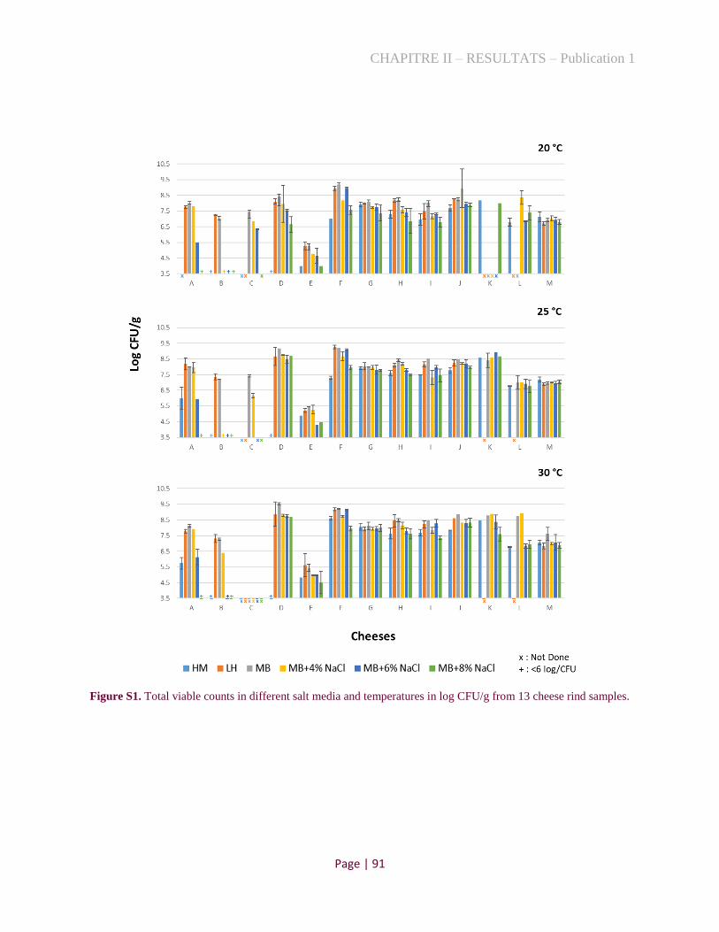
CHAPITRE II – RESULTATS – Publication 1
Page | 91
Figure S1. Total viable counts in different salt media and temperatures in log CFU/g from 13 cheese rind samples.

CHAPITRE II – RESULTATS – Publication 1
Page | 92
Figure S2. Pan- and core-genome size prediction with all combinations of 25 B. aurantiacum (A, B) and 43 S.
equorum (C, D) strains and accumulated number of genes when an additional genome is introduced.

CHAPITRE II – RESULTATS – Publication 1
Page | 93
5 References
[1]. Larsen H (1986) Halophilic and Halotolerant Microorganisms - an Overview and Historical-Perspective. Fems
Microbiol Lett 39 (1-2):3-7.
[2]. Kushner D (1978) Microbial life in extreme environments. Academic Press, London; New York
[3]. Ventosa A, Nieto JJ, Oren A (1998) Biology of moderately halophilic aerobic bacteria. Microbiol Mol Biol R 62
(2):504-44.
[4]. Oren A (2008) Microbial life at high salt concentrations: phylogenetic and metabolic diversity. Saline Systems
4:2-2. doi:10.1186/1746-1448-4-2.
[5]. Mai-Prochnow A, Webb JS, Ferrari BC, Kjelleberg S (2006) Ecological advantages of autolysis during the
development and dispersal of Pseudoalteromonas tunicata biofilms. Appl Environ Microbiol 72 (8):5414-5420.
doi:10.1128/AEM.00546-06.
[6]. Vynne NG, Mansson M, Nielsen KF, Gram L (2011) Bioactivity, chemical profiling, and 16S rRNA-based
phylogeny of Pseudoalteromonas strains collected on a global research cruise. Mar Biotechnol (NY) 13 (6):1062-
1073. doi:10.1007/s10126-011-9369-4.
[7]. Broekaert K, Noseda B, Heyndrickx M, Vlaemynck G, Devlieghere F (2013) Volatile compounds associated
with Psychrobacter spp. and Pseudoalteromonas spp., the dominant microbiota of brown shrimp (Crangon
crangon) during aerobic storage. Int J Food Microbiol 166 (3):487-493. doi:10.1016/j.ijfoodmicro.2013.08.013.
[8]. Busconi M, Zacconi C, Scolari G (2014) Bacterial ecology of PDO Coppa and Pancetta Piacentina at the end of
ripening and after MAP storage of sliced product. Int J Food Microbiol 172:13-20.
doi:10.1016/j.ijfoodmicro.2013.11.023.
[9]. Ferrocino I, Bellio A, Romano A, Macori G, Rantsiou K, Decastelli L, Cocolin L (2017) RNA-Based Amplicon
Sequencing Reveals Microbiota Development during Ripening of Artisanal versus Industrial Lard d'Arnad. Appl
Environ Microb 83 (16). doi:10.1128/AEM.00983-17.
[10]. Pacova Z, Urbanova E, Durnova E (2001) Psychrobacter immobilis isolated from foods: characteristics and
identification. Vet Med-Czech 46 (4):95-100. doi:10.17221/7866-Vetmed.
[11]. Stellato G, De Filippis F, La Storia A, Ercolini D (2015) Coexistence of Lactic Acid Bacteria and Potential
Spoilage Microbiota in a Dairy Processing Environment. Appl Environ Microb 81 (22):7893-7904.
doi:10.1128/Aem.02294-15.
[12]. Deetae P, Bonnarme P, Spinnler HE, Helinck S (2007) Production of volatile aroma compounds by bacterial
strains isolated from different surface-ripened French cheeses. Appl Microbiol Biot 76 (5):1161-1171.
doi:10.1007/s00253-007-1095-5.
[13]. Delbès C. MC, Irlinger F. (2015) Des communautés microbiennes au service de la qualité des fromages:
Diversité et dynamique adaptative et fonctionnelle des populations endogènes et ensemencées Innovations
Agronomiques 44:69-86. doi:10.15454/1.462263595954793E12.
[14]. Irlinger F, Layec S, Helinck S, Dugat-Bony E (2015) Cheese rind microbial communities: diversity,
composition and origin. Fems Microbiol Lett 362 (2). doi:10.1093/femsle/fnu015.
[15]. Bokulich NA, Mills DA (2013) Facility-Specific "House" Microbiome Drives Microbial Landscapes of Artisan
Cheesemaking Plants. Appl Environ Microb 79 (17):5214-5223. doi:10.1128/Aem.00934-13.
[16]. Almeida M, Hebert A, Abraham AL, Rasmussen S, Monnet C, Pons N, Delbes C, Loux V, Batto JM, Leonard
P, Kennedy S, Ehrlich SD, Pop M, Montel MC, Irlinger F, Renault P (2014) Construction of a dairy microbial
genome catalog opens new perspectives for the metagenomic analysis of dairy fermented products. Bmc Genomics
15. doi:10.1186/1471-2164-15-1101.
[17]. Wolfe BE, Button JE, Santarelli M, Dutton RJ (2014) Cheese Rind Communities Provide Tractable Systems
for In Situ and In Vitro Studies of Microbial Diversity. Cell 158 (2):422-433. doi:10.1016/j.cell.2014.05.041.
[18]. Broekaert K, Heyndrickx M, Herman L, Devlieghere F, Vlaemynck G (2011) Seafood quality analysis:
Molecular identification of dominant microbiota after ice storage on several general growth media. Food
Microbiology 28 (6):1162-1169. doi:10.1016/j.fm.2011.03.009.
[19]. Maskow T, Babel W (2001) Calorimetrically obtained information about the efficiency of ectoine synthesis
from glucose in Halomonas elongata (vol 1527, pg 4, 2001). Bba-Gen Subjects 1528 (1):60-60. doi:10.1016/S0304-
4165(01)00115-5.
[20]. Acinas SG, Klepac-Ceraj V, Hunt DE, Pharino C, Ceraj I, Distel DL, Polz MF (2004) Fine-scale phylogenetic
architecture of a complex bacterial community. Nature 430 (6999):551-554. doi:10.1038/nature02649.

CHAPITRE II – RESULTATS – Publication 1
Page | 94
[21]. Tayeb LA, Lefevre M, Passet V, Diancourt L, Brisse S, Grimont PAD (2008) Comparative phylogenies of
Burkholderia, Ralstonia, Comamonas, Brevundimonas and related organisms derived from rpoB, gyrB and rrs gene
sequences. Res Microbiol 159 (3):169-177. doi:10.1016/j.resmic.2007.12.005.
[22]. Camacho C, Coulouris G, Avagyan V, Ma N, Papadopoulos J, Bealer K, Madden TL (2009) BLAST plus :
architecture and applications. Bmc Bioinformatics 10. doi:10.1186/1471-2105-10-421.
[23]. Bankevich A, Nurk S, Antipov D, Gurevich AA, Dvorkin M, Kulikov AS, Lesin VM, Nikolenko SI, Pham S,
Prjibelski AD, Pyshkin AV, Sirotkin AV, Vyahhi N, Tesler G, Alekseyev MA, Pevzner PA (2012) SPAdes: A New
Genome Assembly Algorithm and Its Applications to Single-Cell Sequencing. J Comput Biol 19 (5):455-477.
doi:10.1089/cmb.2012.0021.
[24]. Aziz RK, Bartels D, Best AA, DeJongh M, Disz T, Edwards RA, Formsma K, Gerdes S, Glass EM, Kubal M,
Meyer F, Olsen GJ, Olson R, Osterman AL, Overbeek RA, McNeil LK, Paarmann D, Paczian T, Parrello B, Pusch
GD, Reich C, Stevens R, Vassieva O, Vonstein V, Wilke A, Zagnitko O (2008) The RAST server: Rapid
annotations using subsystems technology. Bmc Genomics 9. doi:10.1186/1471-2164-9-75.
[25]. Stackebrandt E Taxonomic parameters revisited: tarnished gold standards. In, 2006.
[26]. Adekambi T, Drancourt M, Raoult D (2009) The rpoB gene as a tool for clinical microbiologists. Trends
Microbiol 17 (1):37-45. doi:10.1016/j.tim.2008.09.008.
[27]. Thompson JD, Gibson TJ, Plewniak F, Jeanmougin F, Higgins DG (1997) The CLUSTAL_X windows
interface: flexible strategies for multiple sequence alignment aided by quality analysis tools. Nucleic Acids Res 25
(24):4876-4882. doi:10.1093/nar/25.24.4876.
[28]. Kumar S, Stecher G, Tamura K (2016) MEGA7: Molecular Evolutionary Genetics Analysis Version 7.0 for
Bigger Datasets. Mol Biol Evol 33 (7):1870-1874. doi:10.1093/molbev/msw054.
[29]. Saitou N, Nei M (1987) The Neighbor-Joining Method - a New Method for Reconstructing Phylogenetic
Trees. Mol Biol Evol 4 (4):406-425. doi:10.1093/oxfordjournals.molbev.a040454.
[30]. Felsenstein J (1985) Confidence-Limits on Phylogenies - an Approach Using the Bootstrap. Evolution 39
(4):783-791. doi:10.1111/j.1558-5646.1985.tb00420.x.
[31]. Richter M, Rossello-Mora R, Glockner FO, Peplies J (2016) JSpeciesWS: a web server for prokaryotic species
circumscription based on pairwise genome comparison. Bioinformatics 32 (6):929-931.
doi:10.1093/bioinformatics/btv681.
[32]. Goris J, Konstantinidis KT, Klappenbach JA, Coenye T, Vandamme P, Tiedje JM (2007) DNA-DNA
hybridization values and their relationship to whole-genome sequence similarities. Int J Syst Evol Micr 57:81-91.
doi:10.1099/ijs.0.64483-0.
[33]. Richter M, Rossello-Mora R (2009) Shifting the genomic gold standard for the prokaryotic species definition.
P Natl Acad Sci USA 106 (45):19126-19131. doi:10.1073/pnas.0906412106.
[34]. Page AJ, Cummins CA, Hunt M, Wong VK, Reuter S, Holden MTG, Fookes M, Falush D, Keane JA, Parkhill
J (2015) Roary: rapid large-scale prokaryote pan genome analysis. Bioinformatics 31 (22):3691-3693.
doi:10.1093/bioinformatics/btv421.
[35]. Brynildsrud O, Bohlin J, Scheffer L, Eldholm V (2016) Rapid scoring of genes in microbial pan-genome-wide
association studies with Scoary (vol 17, 238, 2016). Genome Biol 17. doi:10.1186/s13059-016-1132-8.
[36]. Hadfield J, Croucher NJ, Goater RJ, Abudahab K, Aanensen DM, Harris SR (2018) Phandango: an interactive
viewer for bacterial population genomics. Bioinformatics 34 (2):292-293. doi:10.1093/bioinformatics/btx610.
[37]. Langmead B, Trapnell C, Pop M, Salzberg SL (2009) Ultrafast and memory-efficient alignment of short DNA
sequences to the human genome. Genome Biol 10 (3). doi:10.1186/gb-2009-10-3-r25.
[38]. Quinlan AR, Hall IM (2010) BEDTools: a flexible suite of utilities for comparing genomic features.
Bioinformatics 26 (6):841-842. doi:10.1093/bioinformatics/btq033.
[39]. Li H, Handsaker B, Wysoker A, Fennell T, Ruan J, Homer N, Marth G, Abecasis G, Durbin R, Subgroup
GPDP (2009) The Sequence Alignment/Map format and SAMtools. Bioinformatics 25 (16):2078-2079.
doi:10.1093/bioinformatics/btp352.
[40]. Bastian M, Heymann S, Jacomy M (2009) Gephi: an open source software for exploring and manipulating
networks. vol 8.
[41]. Levesque S, de Melo AG, Labrie SJ, Moineau S (2019) Mobilome of Brevibacterium aurantiacum Sheds Light
on Its Genetic Diversity and Its Adaptation to Smear-Ripened Cheeses. Front Microbiol 10.
doi:10.3389/fmicb.2019.01270.
[42]. Pham NP, Layec S, Dugat-Bony E, Vidal M, Irlinger F, Monnet C (2017) Comparative genomic analysis of
Brevibacterium strains: insights into key genetic determinants involved in adaptation to the cheese habitat. Bmc
Genomics 18. doi:10.1186/s12864-017-4322-1.

CHAPITRE II – RESULTATS – Publication 1
Page | 95
[43]. Bonham KS, Wolfe BE, Dutton RJ (2017) Extensive horizontal gene transfer in cheese-associated bacteria.
Elife 6. doi:10.7554/eLife.22144.
[44]. Mackenzie EL, Iwasaki K, Tsuji Y (2008) Intracellular iron transport and storage: From molecular
mechanisms to health implications. Antioxid Redox Sign 10 (6):997-1030. doi:10.1089/ars.2007.1893.
[45]. Rea MC, Gorges S, Gelsomino R, Brennan NM, Mounier J, Vancanneyt M, Scherer S, Swings J, Cogan TM
(2007) Stability of the biodiversity of the surface consortia of Gubbeen, a red-smear cheese. J Dairy Sci 90 (5):2200-
2210. doi:10.3168/jds.2006-377.
[46]. Mounier J, Gelsomino R, Goerges S, Vancanneyt M, Vandemeulebroecke K, Hoste B, Scherer S, Swings J,
Fitzgerald GF, Cogan TM (2005) Surface microflora of four smear-ripened cheeses. Appl Environ Microb 71
(11):6489-6500. doi:10.1128/Aem.71.11.6489-6500.2005.
[47]. Gori K, Ryssel M, Arneborg N, Jespersen L (2013) Isolation and Identification of the Microbiota of Danish
Farmhouse and Industrially Produced Surface-Ripened Cheeses. Microb Ecol 65 (3):602-615. doi:10.1007/s00248-
012-0138-3.
[48]. Mounier J, Monnet C, Jacques N, Antoinette A, Irlinger F (2009) Assessment of the microbial diversity at the
surface of Livarot cheese using culture-dependent and independent approaches. Int J Food Microbiol 133 (1-2):31-
37. doi:10.1016/j.ijfoodmicro.2009.04.020.
[49]. Ades GL, Cone JF (1969) Proteolytic Activity of Brevibacterium linens during Ripening of Trappist-Type
Cheese. J Dairy Sci 52 (7):957-961. doi:10.3168/jds.S0022-0302(69)86674-9.
[50]. Sharpe ME, Law BA, Phillips BA, Pitcher DG (1977) Methanethiol Production by Coryneform Bacteria -
Strains from Dairy and Human-Skin Sources and Brevibacterium linens. J Gen Microbiol 101 (Aug):345-349.
doi:10.1099/00221287-101-2-345.
[51]. Brennan NM, Ward AC, Beresford TP, Fox PF, Goodfellow M, Cogan TM (2002) Biodiversity of the bacterial
flora on the surface of a smear cheese. Appl Environ Microb 68 (2):820-830. doi:10.1128/Aem.68.2.820-830.2002.
[52]. Feurer C, Irlinger F, Spinnler HE, Glaser P, Vallaeys T (2004) Assessment of the rind microbial diversity in a
farm house-produced vs a pasteurized industrially produced soft red-smear cheese using both cultivation and rDNA-
based methods. J Appl Microbiol 97 (3):546-556. doi:10.1111/j.1365-2672.2004.02333.x.
[53]. Monnet C, Landaud S, Bonnarme P, Swennen D (2015) Growth and adaptation of microorganisms on the
cheese surface. Fems Microbiol Lett 362 (1). doi:10.1093/femsle/fnu025.
[54]. Kastman EK, Kamelamela N, Norville JW, Cosetta CM, Dutton RJ, Wolfe BE (2016) Biotic Interactions
Shape the Ecological Distributions of Staphylococcus Species. Mbio 7 (5). doi:10.1128/mBio.01157-16.
[55]. Almena-Aliste M, Mietton B (2014) Cheese Classification, Characterization, and Categorization: A Global
Perspective. Microbiol Spectr 2 (1):CM-0003-2012. doi:10.1128/microbiolspec.CM-0003-2012.
[56]. Louca S, Polz MF, Mazel F, Albright MBN, Huber JA, O'Connor MI, Ackermann M, Hahn AS, Srivastava
DS, Crowe SA, Doebeli M, Parfrey LW (2018) Function and functional redundancy in microbial systems. Nat Ecol
Evol 2 (6):936-943. doi:10.1038/s41559-018-0519-1.
[57]. Pan QM, Tian DS, Naeem S, Auerswald K, Elser JJ, Bai YF, Huang JH, Wang QB, Wang H, Wu JG, Han XG
(2016) Effects of functional diversity loss on ecosystem functions are influenced by compensation. Ecology 97
(9):2293-2302. doi:10.1002/ecy.1460.
[58]. Gladstone-Gallagher RV, Pilditch CA, Stephenson F, Thrush SF (2019) Linking Traits across Ecological
Scales Determines Functional Resilience. Trends Ecol Evol 34 (12):1080-1091. doi:10.1016/j.tree.2019.07.010.

CHAPITRE II – RESULTATS – Publication 1
Page | 96
1.3. Conclusion
Pour cette étude, nous avons séquencé 35 génomes bactériens, correspondant à 20 Gram-
positifs et 15 Gram-négatifs. Parmi eux, les espèces Gram-positifs, les plus fréquemment isolées
étaient les bactéries halotolérantes Brevibacterium aurantiacum (six souches) et Staphylococcus
equorum (trois souches), qui sont, généralement, ajoutées comme ferments pour la production de
fromages. Les analyses génomiques réalisées pour ces deux espèces confirment la grande diversité
génétique entre les souches fromagères de B. aurantiacum et révèlent la présence de deux sous-
espèces chez S. equorum, ainsi que la proximité phylogénétique de plusieurs souches de fromage
avec des isolats bovins. Parmi les souches Gram-négatifs, nous avons identifié huit nouvelles
espèces potentielles, en particulier aux genres Halomonas et Psychrobacter, ce qui confirme la
faiblesse des études les concernant. De plus, le mapping des espèces halophiles et halotolérantes
concernant 74 croûtes de fromage a montré que presque tous les échantillons, sauf cinq, possèdent
dans leurs croûtes, ces bactéries tolérantes au sel. De tels résultats renforcent l’idée selon laquelle
les bactéries halophiles jouent un rôle dans l’écosystème fromager. Il est intéressant de noter que
les espèces Gram-négatifs d’Halomonas, Pseudoalteromonas et Psychrobacter, qui sont souvent
associées en tant que contaminants, possèdent ensemble un niveau d’abondance supérieur à 10 %
pour plusieurs métagénomes de croûtes de fromage (n=18). Enfin, la disponibilité de ces isolats
ouvre la possibilité d'explorer les caractéristiques physiologiques, ainsi que les propriétés
fonctionnelles, potentielles, de ces bactéries dans le domaine de la technologie fromagère.

CHAPITRE II – RESULTATS – Publication 2
Page | 97
2. Stratégies pour isoler les espèces halophiles, rarement cultivées
2.1. Contexte
La diversité microbienne, à la surface des fromages, est étudiée depuis plusieurs décennies en
appliquant des méthodes liées à la culture et, depuis la fin du 20ème siècle, en utilisant des méthodes
indépendantes de la culture, comme la métagénomique [5, 226]. Des études récentes, basées sur
des technologies de séquençage à haut débit, ont démontré la grande diversité concernant les
espèces halophiles dans les croûtes de fromage provenant de différents pays, européens et
américains [3, 4]. À partir de ces résultats, et en observant l'absence d'isolats halophiles
alimentaires dans la base de données (cf. la publication précédente), nous avons effectué un
isolement systématique de ces souches, à partir de croûtes de fromage, en utilisant des techniques
de culture, traditionnelles et touchant aussi différents milieux de culture à base de sel. Cette
méthode a permis d'isoler une grande variété d'espèces, sans être très sélective. Cependant, l'accès
à certains microorganismes halophiles par des méthodes de culture est encore peu développé et
très limité dans les aliments, étant donné qu’ils peuvent être difficiles à cultiver. Cela peut justifier
le manque constaté d’isolats halophiles alimentaires dans la littérature. Par conséquent, une partie
de la diversité microbienne des fromages ne fait pas, jusqu'à présent, l’objet de cultures et des
stratégies ciblées peuvent donc aider à leur isolement. Ainsi, les objectifs de cette étude consistent
à :
• estimer la composition taxonomique des mêmes croûtes de fromages utilisées dans la
Publication 1 ;
• développer une stratégie ciblée pour isoler les espèces halophiles prédominantes non
détectées lors de la première étude ;
• comprendre les possibles échecs ou difficultés affectant la culture de ces bactéries.
Les résultats obtenus sont présentés sous la forme d'un article en cours de préparation (cf. la
partie 2.2).

CHAPITRE II – RESULTATS – Publication 2
Page | 98
2.2. Publication 2
Metagenomic driven characterization of halophilic bacteria in cheese rinds
Caroline Isabel Kothea and Pierre Renaulta
a Université Paris-Saclay, INRAE, AgroParisTech, Micalis Institute, 78350, Jouy-en-Josas, France.
Abstract
Recent studies, based on high-throughput sequencing technologies, have demonstrated the
abundance of poorly characterized halophilic bacteria on the surface of ripened cheeses. In this
article, we investigated the results obtained by an extensive cultural study aiming the isolation of
halophilic species, such as those belonging to the genera Halomonas, Marinomonas,
Pseudoalteromonas, Psychrobacter and Vibrio, with metagenomic analyses. It indicated that
number of species were not recovered previously, challenging a comprehensive recovery of these
species. To achieve this task, we designed a strategy based on metagenomic-based probes,
targeting the species of interest, coupled with an incremental cultural approach with pools. This
strategy was performed on two cheese rinds from which many species were not isolated previously,
targeting species displaying a relative abundance >3%, as calculated by coverage of the
metagenomic assemblies. This method allowed the extensive isolation of targeted species,
including potentially new cheese species, except two, which were dominant in each cheese with a
relative abundance of 20.4 and 26.7%. These species corresponded to a new species of
Pseudoalteromonas, without isolated representative yet, and to Marinomonas foliarum, isolated
from marine environment. The possible reasons of their non-recovery were discussed with their
metagenome-assembled genomes (MAGs) in comparison with closely related cultivable strains.
Keywords: food, targeted probes, genomic reconstruction, MAG, culture method
1 Introduction
In recent years, next-generation sequencing has been enabling the development of new
approaches to study genetic diversity in microbial communities [1, 2]. Among them,

CHAPITRE II – RESULTATS – Publication 2
Page | 99
metagenomics is a powerful tool to analyze all microorganisms present in samples such as soil,
sediments, food or water [3, 4]. This independent cultivation technique allows highlighting the
existence of unexpected populations, including in already well-studied food ecosystems [5].
However, culture-independent techniques are unable to provide the biological material necessary
to study the development of the ecosystems and its functionality [6].
The surface microbiota of cheese has been studied for several decades by conventional
culture-based analysis and, since the end of the 20th century, by culture-independent methods [7,
8]. Recent studies, based on high-throughput sequencing technologies, have demonstrated a large
species diversity and dominance of halophilic bacteria in several cheese rinds from different
European and American countries [5, 9]. Based on these results and the poor availability of food
halophilic isolates in the culture collections, Kothe et al. (2020) performed a systematic isolation
of halophilic species from cheese rinds, using cultural techniques, on different salt-based media
[10]. This method allowed the characterization of 26 species of halophilic and halotolerant species,
including potentially ten undescribed species, which appeared to be frequent on cheese rinds, as
measured by their genomic profiling. However, this analysis indicated that we failed to isolate all
the existing species in each cheese, questioning the coverage of our analysis.
While profiling approach highlights species when reference genome is available, assembly-
based metagenomics provides information independently of any reference set, at least for species
whose abundance is enough to yield a good coverage [11-13]. Therefore, in this work, we carried
out a metagenomic assembly-based analysis of ten previously studied cheeses and we showed that
several abundant species had not been isolated previously. Consequently, we developed a fast and
easy screening strategy guided by metagenomic assembly data to facilitate their isolation. As a
proof of concept, we applied this strategy on two cheeses with important failure in our previous
study with the aim to isolate their corresponding halophilic species.
2 Methodology
2.1 Cheese samples and taxonomic diversity
Ten cheese rind metagenomes, sequenced by Kothe et al. (2020) [10], were analyzed in
this study (Table S1). For this purpose, we first estimated microbial community composition by
mapping the samples reads against the representative clade-specific marker catalog contained on
the MetaPhlAn tool v. 3.0.4 [14]. As an additional method for taxonomic profiling, we used a
marker gene prediction, which allows binning of assembled metagenomic contigs without the need
for reference sequence. Therefore, the reads were merged and de novo assembly was performed
using SPAdes v. 3.9 [15]. Furthermore, genes were predicted using Prodigal v. 2.6.3 and the

CHAPITRE II – RESULTATS – Publication 2
Page | 100
marker genes were extracted using fetchMG v. 1.0 [16, 17]. By default, fetchMG searched for 40
universal marker genes. We focus our research on the COG0012 (ychF gene) and blasts were
performed on the all-available sequences of the NCBI databases on nucleic and coding DNA
sequences. Summary species composition plots were created in R v. 3.6.1 using the package
ggplot2 v. 3.3.2.
2.2 Probe design and culture conditions
Species of two cheese rinds were chosen as proof of concept to build probes and for a
subsequent targeted isolation. Briefly, we aligned the ychF sequences of interest species using
ClustalX 2.1 [18] and we searched variable regions. The probes were designed with Benchling
Software according to the following criteria: (i) primer size between 18-25 bp; (ii) product size
between 90-700 bp; (iii) melting temperature (Tm) of both forward and reverse primers between
50-56°C; (iv) GC content of primers between 40-60%. The software PCR Primer Stats was applied
to prevent the hairpins formations and self-annealing between them. The main features of the
designed primers are presented in Table 1.
Table 1. Characteristics of PCR primers designed to isolate the targeted species of two cheese samples.
Closely species target
(best blast)
Probe
name
%
reads
Amplicon
size (bp) Forward Primer Sequence Reverse Primer Sequence
Ch
eese
9
Psychrobacter cibarius A1 29.53 273 GTTAAGCCTGAGCGTGTATTACC GGTTTGGTTGTGAATGGCAC
Marinomonas foliarum A2 26.73 440 CCTGGATGCTTTGGCTGAG GAAGGTGTAGAGAGCGAACTTC
Psychrobacter translucens A3 16.36 364 GCCAGTACCAGATCCACGTC GAACACATCTAGCATGGCTGC
Vibrio casei A4 10.70 204 GTTTCTAGCCGACTTAGGCATAG ATAACTTCTGCACGGATGAAGC
Vibrio litoralis A5 4.28 657 CTACGTCTAGATGCGTTAGCTG GTTTAGGCCTGGTTCTTCAATACC
Pseudoalteromonas
(unclassified) A6* 3.61 383 GTTTGTTGACATAGCAGGC GAAAGTTAACGGAGGAAATCG
Ch
eese
10
Pseudoalteromonas
(unclassified) B1* 20.38 383 GTTTGTTGACATAGCAGGC GAAAGTTAACGGAGGAAATCG
Halomonas citridevorans B2 8.71 477 TGAACGAGTCCTGCCTAC AGGTAAGGGTTGTTCTCAAAC
Halomonas minus B3 8.48 90 ACGCTCTATGTTCTTAAGCG GTTTGAAGGCCCAATAATGAG
Pseudoalteromonas undina B4 5.06 636 ACGACATTGACGTGATCAAC CGCTTTCGCCTTTACAAG
Halomonas casei B5 3.81 540 CGACTTGATAAGCTTGCG TTCTTCTGCCGCAATCTC
Halomonas flavum B6 3.59 294 ATCCGCCATTGTAAAACCC TTCACGGAGCGTAACAAG
Pseudoalteromonas
nigrifaciens B7 2.56 294 ATCCGCCATTGTAAAACCC TTCACGGAGCGTAACAAG
*Probe A6 = B1

CHAPITRE II – RESULTATS – Publication 2
Page | 101
All probes were checked in their corresponding cheese DNA in order to be a positive
control. Then, the two cheese samples, collected by Kothe et al. (2020) [10], were homogenized
in 0.9% NaCl sterile solution, diluted in series and spread on different salt-based agar media to
display their cultivable cells. We tested the following media: Marine Agar (MA; Difco, Sparks,
USA), MA added of 2, 4, 6 and 8% NaCl (Merck, Darmstadt, Germany), Halomonas Medium
(HM; [19]) and Long and Hammer Agar (LH; [20]). The plates were incubated in a range of
temperatures varying between 15 and 25°C.
Isolation were performed on plates where bacterial counts were comprised between 20 and
200 clones. Different morphotypes were selected and restreaked into a new plate to increase the
amount of bacterial cells for fast DNA extraction. For this purpose, several colonies were mixed
with a sterile loop in a tube containing 300 µl biomol water, 100 mg of 0.1 mm-diameter zirconium
beads and 100 mg of 0.5 mm-diameter (Sigma, St. Louis, MO, USA) for their DNA extraction.
The tube was agitated in a bead-beater (FastPrep-24, MP Biomedicals Europe, Illkirch, France)
for 20 s at 4.5 m/s to release the DNA from the bacteria. The supernatant of this lysis was amplified
using the targeted probes elaborated for each specific species. Thermal cycling conditions used
were (i) 1 min at 94°C to initial denaturation, (ii) 30 cycles of 1 min at 94°C to denaturation, 0.5
min at 56°C to primer annealing, 1.5 min at 72°C to initial elongation, and (iii) 5 min at 72°C to
final elongation. DNA amplification was checked on 0.8% agarose gel.
When the amplification signal was positive, the probable species corresponding was
confirmed by sequencing its 16S rRNA gene. For this, we used the same thermal cycling
conditions previously cited and the primers 27-F (5′-AGAGTTTGATCATGGCTCA-3′) and
1492-R (5′-TACGGTTACCTTGTTACGACTT-3′) [21]. Once received, the sequences were
analyzed via NCBI BLAST tool [22] to obtain a taxonomic classification for each isolate. Finally,
the extraction of genomic DNAs were performed using the protocol described by Almeida et al.
(2014) [9], and their sequencing were carried out on the Illumina HiSeq platform at GATC-Biotech
(Konstanz, Germany).
2.3. Metagenome-assembled genomes (MAGs)
The species unable to be cultivated, with the strategies used in this study, were reconstituted
with MetaBAT2-2.12.1, an automated metagenomic binning tool [23], with minimum contig size
of 1,500 nucleotides and other default settings. First, the genome binning was performed using the
raw reads and the corresponding assembled-metagenomes. However, to improve the quality of
MAGs, we also used a method assisted by the use of genome data of isolated strains in this study
for each corresponding cheese (Psychrobacter FME60, FME61, Vibrio FME29 and FME62 for

CHAPITRE II – RESULTATS – Publication 2
Page | 102
Cheese 9, and Halomonas FME63, FME64, FME65, FME66, Pseudoalteromonas FME67 and
FME68 for Cheese 10). Briefly, the reads of known genomes (isolated in this study or reliable
sequences from database) were mapped against the raw reads of their corresponding metagenomes
using Bowtie2-2.4.1 [24]. We filtered these reference genomes mapped, the remaining reads were
assembled using SPAdes v. 3.9 [15] and new MAGs were recovered with MetaBAT2. The quality
of the resulting bins were assessed with CheckM [25]. Finally, the presence of core genes from
Marinomonas foliarum was also checked by read profiling using Bowtie2-2.4.1.
2.4 Genomic analyses
We performed genomic comparisons of the MAG strains with closely related genomes
using the “Compare” and “Sequenced based” tools of SEED Viewer in RAST server v2.0 [26, 27].
The output was a table that includes the genes of the reference organism (MAGs) in chromosomal
order and of the compared organisms correspondingly, with additional information such as the
protein sequence identity (%) and if best-hits are bidirectional or unidirectional.
Additionally, prophage activity of all species obtained were annotated using Prophage
Hunter [28], which is able to differentiate active from inactive prophages. Only active prophages
were selected (region scoring > 0.8). CRISPRCasFinder was used with default parameters to
discover CRISPR loci in halophilic genomes, predict CRISPR repeats and spacers and search for
Cas proteins [29, 30].
2.5. Presence of new genomes on cheese rind metagenomes
The relative abundance of the halophilic genomes obtained in this study was determined in
more than 90 cheese rind metagenomes from different countries and technologies, using the Food-
Microbiome tool, as described previously [10]. The metadata of the samples are presented in Table
S2.

CHAPITRE II – RESULTATS – Publication 2
Page | 103
3 Results and Discussion
3.1 Evidencing absence of isolation of highly representative species by classical isolation
procedures
The determination of the taxonomical composition of ten French cheese rinds with
reference-based metagenomics tools, such as MetaPhlAn, indicates that several abundant species
were not isolated during our previous work using cultural methods [10] (Table S3). For example,
Marinomonas foliarum appears to be among the dominant species in Cheese 9, but have not been
isolated. Furthermore, the pourcentage of reads aligned to the whole reference set was sometimes
low, suggesting that several references were missing. Therefore, we employed a metagenomic
reference-independent method based on assembly followed by marker gene analysis to point out
all dominant species present in these cheeses. As results, 79 bacterial and three fungal species were
identified in the ten cheese surface samples as shown Fig. 1 and Table S4. Overall, MetaPhlAn
and the assembly-derived method gave similar results on number of species. However, marker
gene analysis pointed the presence of several species belonging to the genus Psychrobacter,
Pseudoalteromonas, Halomonas, Pseudomonas and Corynebacterium, which were not detected
by MetaPhlAn. These species correspond mostly to halophiles, which are still poorly studied. For
example, the Cheeses 9 and 10 contain an unclassified Pseudoalteromonas at estimated levels of
3.6 and 20%, repectively. Similarly, Cheeses 6, 7 and 9 contains the newly characterized species,
Psychrobacter translucens, at levels of 2.7, 27 and 16%, respectively.
Finally, comparison of species isolated from the ten cheeses in our previous study [10] with
the compostion obtained by the marker analysis indicates that several species were not isolated
(Fig. S1, hatched species). Therefore, we decided to develop targeted strategies to isolate strains
corresponding to poorly characterized species in cheeses such as the still unclassified
Pseudoalteromonas pointed out in Cheeses 9 and 10.
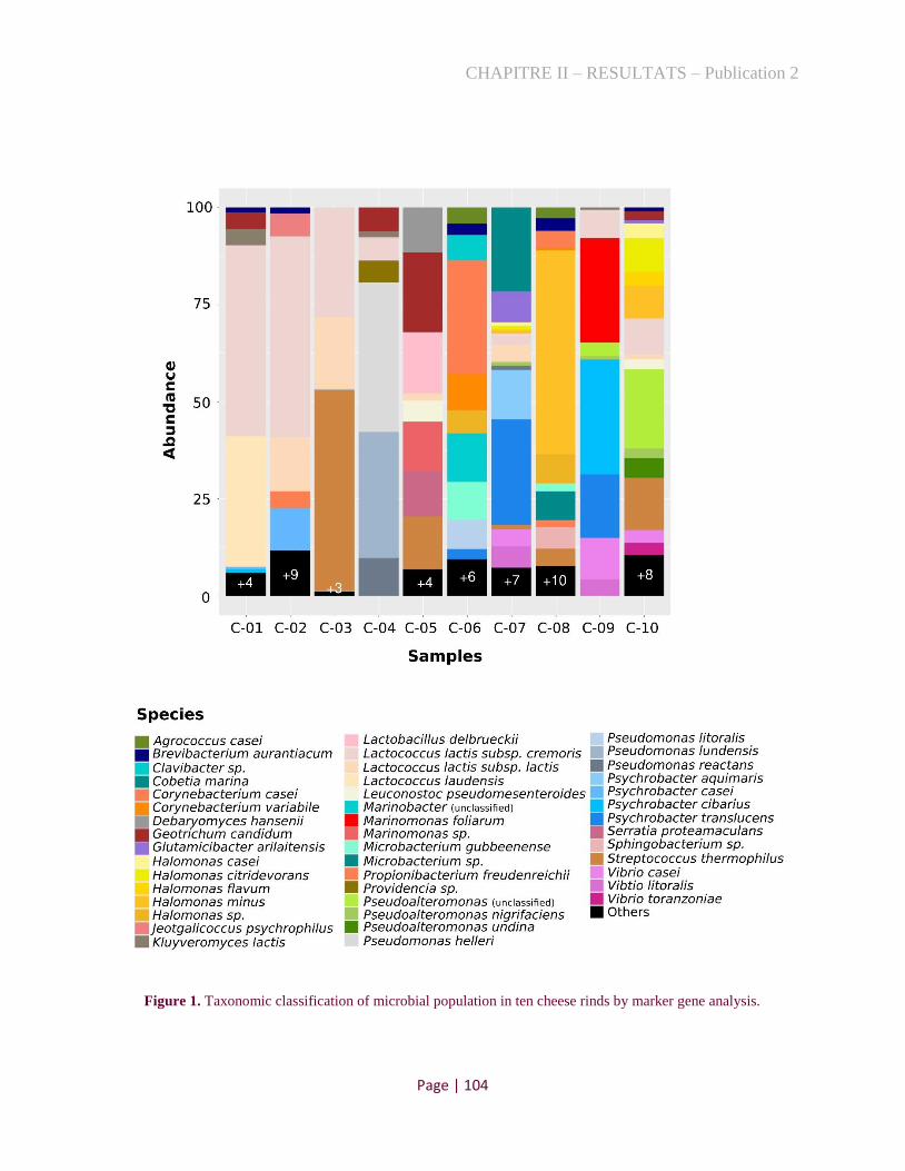
CHAPITRE II – RESULTATS – Publication 2
Page | 104
Figure 1. Taxonomic classification of microbial population in ten cheese rinds by marker gene analysis.

CHAPITRE II – RESULTATS – Publication 2
Page | 105
3.2 Strategy to target specific halophilic bacteria
In order to isolate the species missing in the previous cultural approach, some of which
being less abundant or poorly cultivable, we defined a strategy based on a fast screening technique
guided by the metagenomic analysis. The screening was based on the design of specific PCR
primers to target the different species identified by the assembly-based metagenomic analysis. To
design specific primers, metagenomic reads were assembled on contigs and sequences of marker
genes were defined for each present species. Subsequently, a well-conserved gene, such as
ribosome-binding ATPase YchF (COG0012), was selected to target any species. Therefore, their
corresponding nucleotide sequences were aligned to define variable regions from which primers
were designed to obtain pairs that allowed specific amplification for each species (Fig. 2A).
The above designed primers were finally tested on the metagenomic DNAs to confirm their
efficiency. They can then be used to detect the presence of the targeted species through DNA
extracted from single colony or from pools of colonies, as exemplified in Fig. 2B.
Figure 2. Probe design and culture strategies to isolate targeted species.

CHAPITRE II – RESULTATS – Publication 2
Page | 106
3.3 Targeted isolation of halophiles present in Cheeses 9 and 10
In this study, probes were designed to isolate halophilic bacteria present down to 2-3%
from two cheeses, corresponding to six species of Cheese 9 and seven of Cheese 10 (Table 1).
Interestingly, the dominant uncharacterized Pseudoalteromonas species in Cheese 10 was present
as sub-dominant species in Cheese 9, as indicated by the 100% identity of their ychF nucleotide
sequence (Probe A6 = B1).
In a first attempt, we applied an individual screening where colonies with different
morphotypes (size, contour, relief, opacity, pigmentation and colony consistency) were picked and
their DNA amplified with the different primers pairs. For those exhibiting a positive signal
amplified, their identification was confirmed by 16S rRNA gene sequencing. Subsequently, for
species of which no isolate was found in this first round, we developed a second screening attempt
by testing pools of 5-10 mixed colonies, and by varying the culture conditions. The pools that
exhibited a positive amplification, for a given primer pair, were separated and the clones were
tested individually to isolate the targeted species. Finally, in case of negative result in this second
attempt, we tested the cultivability of the targeted species on different medium and conditions in
order to define suitable procedure for their isolation.
In this study, the first attempt was performed in MA plates at 25°C from which we isolated
two out of the six targeted species in Cheese 9 (Psy. cibarius and Psy. translucens represented by
the probes A1 and A3) and three out of the seven targeted species in Cheese 10 (H. minus, H. casei
and H. flavum with B3, B5 and B6 probes; Table 2). Curiously, these species were not the most
abundant in the cheese samples, as indicated in Fig. 1.
For the species that were not isolated in this first attempt, we used the second isolation
procedure with pool strategies. Here, in addition to MA media, we also tested HM and LH media,
which are frequently used to isolate environmental and food halophiles, as well as MA added of
2, 4, 6 and 8% NaCl. Considering the tendency of halophilic bacteria to be psychrophilic, we also
tested temperatures of 15 and 20°C to expand the conditions to isolate them. By this way, we
isolated H. citridevorans (B2) and P. undina (B4) from Cheese 10 on MA media at 25°C.
Afterward, by using MA+4% of NaCl at 20 and 25°C, we isolated V. casei and V. litoralis (probes
A4 and A5), respectively, from Cheese 9. Finally, still on MA+4% NaCl, but incubating at lower
temperature (15°C), we isolated P. nigrifaciens from Cheese 10 (B7, Table 2).

CHAPITRE II – RESULTATS – Publication 2
Page | 107
Table 2. The ten isolates from this study and their respective strategy, media and temperature of isolation, and
general features of their sequenced genomes.
Despite the screening of over 2,000 colonies per cheese owing the use of pools, with
different media and temperatures, we did not isolate M. foliarum co-dominant in Cheese 9 and the
unclassified Pseudoalteromonas, which was the dominant species in Cheese 10 and sub-dominant
in Cheese 9. In order to test the growth potential of these species under different conditions, we
inoculated low dilution of cheese rind (~106 UFC/mL) on agar plates using the different culture
media mentioned above and incubating at 15, 20 and 25°C. However, we did not obtain any PCR
signal after the extraction of DNA from these cultures, indicating that these microorganisms were
not able to develop in these conditions and were overgrown by other strains. Interestingly, strains
corresponding to M. foliarum and this unclassified species of Pseudoalteromonas have not yet
been isolated from cheese. Indeed, growth of Pseudoalteromonas species often appear to be
fastidious, suggesting that usual laboratory media are not well adapted to isolate these species [31].
However, M. foliarum was isolated from the seagrass Posidonia oceanica on Marine Agar, and
was therefore expected to grow in our tests, especially since it represented about 30% of the initial
population.
Targeted species Probe Strategy Media T
(°C)
No of
contigs
Size (base
pairs)
GC
content
(%)
Genome accession
Ch
eese
9
Psychrobacter cibarius FME60
A1 morphotype MA 25 85 3,469,065 42.6 JABUYU000000000
Psychrobacter translucens FME61
A3 morphotype MA 25 452 3,569,320 41.7 JABUYV000000000
Vibrio casei FME29 A4 pool MA+4%NaCl 20 115 3,833,673 40.5 JABUYT000000000
Vibrio litoralis
FME62 A5 pool MA+4%NaCl 25 171 3,855,466 42.1 JABUYW000000000
Ch
eese
10
Halomonas
citridevorans FME63 B2 pool MA 25 77 3,927,099 54.2 JABUYX000000000
Halomonas minus
FME64 B3 morphotype MA 25 48 3,871,780 53.8 JABUYY000000000
Pseudoalteromonas
undina FME67 B4 pool MA 25 127 4,193,916 40.1 JABWDP000000000
Halomonas casei FME65
B5 morphotype MA 25 143 4,336,586 53 JABUYZ000000000
Halomonas flavum FME66
B6 morphotype MA 25 122 4,506,877 54.2 JABUZA000000000
Pseudoalteromonas
nigrifaciens FME68 B7 pool MA+4%NaCl 15 137 3,961,476 39.8 JABUZB000000000
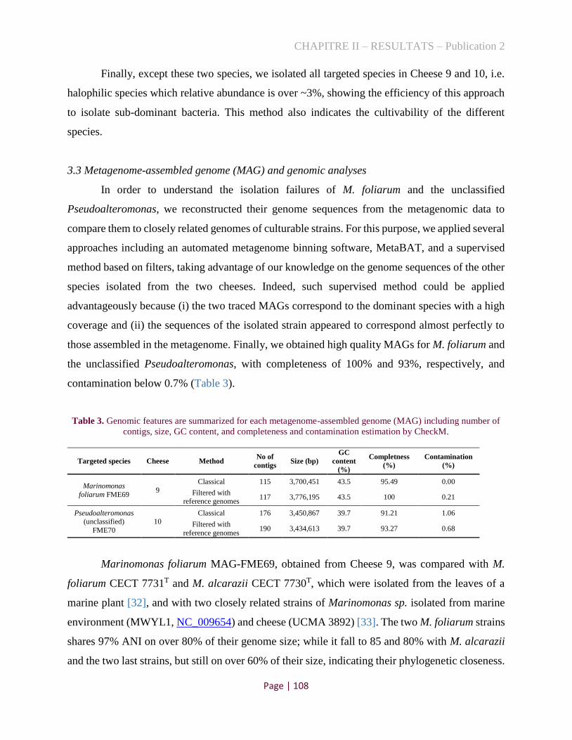
CHAPITRE II – RESULTATS – Publication 2
Page | 108
Finally, except these two species, we isolated all targeted species in Cheese 9 and 10, i.e.
halophilic species which relative abundance is over ~3%, showing the efficiency of this approach
to isolate sub-dominant bacteria. This method also indicates the cultivability of the different
species.
3.3 Metagenome-assembled genome (MAG) and genomic analyses
In order to understand the isolation failures of M. foliarum and the unclassified
Pseudoalteromonas, we reconstructed their genome sequences from the metagenomic data to
compare them to closely related genomes of culturable strains. For this purpose, we applied several
approaches including an automated metagenome binning software, MetaBAT, and a supervised
method based on filters, taking advantage of our knowledge on the genome sequences of the other
species isolated from the two cheeses. Indeed, such supervised method could be applied
advantageously because (i) the two traced MAGs correspond to the dominant species with a high
coverage and (ii) the sequences of the isolated strain appeared to correspond almost perfectly to
those assembled in the metagenome. Finally, we obtained high quality MAGs for M. foliarum and
the unclassified Pseudoalteromonas, with completeness of 100% and 93%, respectively, and
contamination below 0.7% (Table 3).
Table 3. Genomic features are summarized for each metagenome-assembled genome (MAG) including number of
contigs, size, GC content, and completeness and contamination estimation by CheckM.
Targeted species Cheese Method No of
contigs Size (bp)
GC
content
(%)
Completness
(%)
Contamination
(%)
Marinomonas
foliarum FME69 9
Classical 115 3,700,451 43.5 95.49 0.00
Filtered with reference genomes
117 3,776,195 43.5 100 0.21
Pseudoalteromonas (unclassified)
FME70
10 Classical 176 3,450,867 39.7 91.21 1.06
Filtered with
reference genomes 190 3,434,613 39.7 93.27 0.68
Marinomonas foliarum MAG-FME69, obtained from Cheese 9, was compared with M.
foliarum CECT 7731T and M. alcarazii CECT 7730T, which were isolated from the leaves of a
marine plant [32], and with two closely related strains of Marinomonas sp. isolated from marine
environment (MWYL1, NC_009654) and cheese (UCMA 3892) [33]. The two M. foliarum strains
shares 97% ANI on over 80% of their genome size; while it fall to 85 and 80% with M. alcarazii
and the two last strains, but still on over 60% of their size, indicating their phylogenetic closeness.

CHAPITRE II – RESULTATS – Publication 2
Page | 109
CECT 7731T strain contains 3,980 genes against 3,651 for MAG-FME69, indicating its genomes
encodes slightly more genes, or that several genes were not recovered by metagenomic binning.
Importantly, they share 3,158 orthologous genes, which represent around 80% of their genes. Bi-
directional hit analysis between CECT 7731T and MAG-FME69 genomes indicated that the
environmental genome contain 750 specific genes against 493 of the cheese MAG. About 75% of
CECT 7731T specific genes were clustered in 38 islands (with over than 6 genes), and 50%
annotated as “hypothetical protein” (Table S5). The main islands are (i) a potential plasmid (67
genes encoding dicarboxylic and lipid metabolic genes) (ii) phages (89 genes in four islands), (iii)
dicarboxylic acids, sugar transport and metabolism (spread in 12 islands, totaling 128 genes),
surface factors such as polysaccharides (one island with 26 genes). Additionally, three islands of
39, 37, and 33 genes are encoding significant metabolic functions. However, when compared with
MWYL1, UCMA 38921 and CECT 7730T strains, only 73 genes were missing in MAG-FME69,
including two sugar assimilation systems, two histidine transport genes, two genes involved in
glycogen synthesis and urease degradation genes. The absence of these genes could not explain
growth defect of the cheese strain on the different tested media, which contain abundant nitrogen
and sugar sources. Finally, the potential of the 493 specific genes of MAG-FME69 vs CECT 7731T
were searched and indicated the presence of at least three prophages, two plasmids and only few
miscellaneous metabolic functions (Table S5). It contains also CRISPRs genes, including 185
spacers, showing an intense phage metabolism. In order to characterize eventual phages
replications in the metagenomic assembly, we search for phage genes in contigs displaying higher
coverage than MAG-FME69 (Table S5). From this analysis, we found integrated prophage genes
from “Moraxella”, and interestingly, lactococcal or streptococcal phage genes at coverage of 68
and 444 against 18 for L. lactis genome, suggesting their replication. These data indicated that
phages could be responsible of the lack of cultivability of this Marinomonas strain.
A similar analysis was performed with Pseudoalteromonas MAG-FME70 in comparison
with Pseudoalteromonas sp. 3D05, P. mariniglutinosa KCTC 22327T, P. haloplanktis ATCC
14393T, which are among its closest environmental relatives (ANI ~77% on 47% of their length,
and 62-68% of FME70 length), completed with two Pseudoalteromonas cheese strains (FME14
and JB197). The set of reference strains shared 2,456 orthologous genes, among which 244 were
missing in MAG-FME70 (Table S6). An inspection of these genes indicated that MAG-FME70
strain do not contain genes encoding for ferric ion ABC transporter (3), cobalamine synthesis (3)

CHAPITRE II – RESULTATS – Publication 2
Page | 110
and agmatine-arginine pathway as well defined functions. Moreover, the main other functions
potentially lacking in MAG-FME70 are linked to signal transduction (30), flagellar assembly (44)
and transport system (12), completed by 54 hypothetical proteins.
Conversely, MAG-FME70 contains 680 specific genes compared to 3D05 strain (the
closest relative strain), including primarily, 20 related to iron metabolism, 6 to sulfate metabolism,
11 for the purine/uric acid/allantoin catabolic super‐pathway [34] and 15 for pili formation.
Moreover, it contain 11 genes annotated as ton-dependent receptor, 29 as involved in the transport
and 339 hypothetical proteins, among which were presented phage proteins (Table S6). Similar
results were obtained with the MAG from the same species constructed in another study [11],
suggesting that this list is robust, although it cannot be excluded that several of these genes may
have been eliminated due to metagenomic procedure-dependent analysis. Overall, missing genes
compared to other Pseudoalteromonas species could not sharply show a particular metabolic
defect in the cheese strain that would completely impair its development. Nevertheless, it would
be worth testing cobalamin supplementation, as well as addition of new nitrogen sources related
to arginine/agmatine and allantoin/purine pathways and sulfate sources. Possible presence of
bacteriophages was also tested in MAG-FME70. Nevertheless, while staphylococcal phage was
detected at high level of coverage (about 20 times higher than S. equorum mean coverage), other
phage genes potentially targeting Gammaproteobacteria were present on contig of comparable
level of coverage than those of MAG-FME70, suggesting their docking in the chromosomal DNA
(Table S6).
3.4 Representativeness of new genomes in cheese rinds
Finally, we analyzed the presence of these still uncultured species in cheeses by using their
MAGs as genome reference, as well the other isolated species, on more than 90 cheese rind
metagenomes from four studies (Table S7) [5, 10, 11, 35]. This analysis confirms a high relative
abundance of species belonging to the genera Psychrobacter, Halomonas and Pseudoalteromonas,
as previously demonstrated [10]. Concerning the MAGs recovered in this study, Marinomonas
foliarum was present in only two additional cheese metagenomes evaluated with relative
abundance of 2.6 and 4.4%. Additionally, the MAG of Pseudoalteromonas was present in seven
other cheese rinds, two of which were highly represented (13.5 and 16.8%; Table S7).

CHAPITRE II – RESULTATS – Publication 2
Page | 111
4 Conclusion
Cultures provide a very biased view of the microbial composition of cheese. This fact is
likely due to the different potential of revival of microorganisms once replicated after a long
stationary phase to a new and different culture medium. In the present study, we showed a strategy
to recover representative clones of each species present down to few percent’s (2-3%), as measured
by their DNA quantification. In particular, we isolated strains of P. undina and V. litoralis, which
are, for our best knowledge, the first cheese isolates characterized yet. Nevertheless, we failed to
isolate clones belonging to two species, both as part of the dominant species, according to DNA
quantification. The first one, M. foliarum, have already been characterized from marine
environment, and possess a phylogenetically closely related species isolated from cheese [32]. The
second one was a Pseudoalteromonas species, which was not reported in culture collections
neither in genome databases. The fact that the DNA sequence of both species could be assembled
efficiently indicates that their DNAs were of good quality upon extraction and thus likely preserved
in physically intact cells.
Metabolic features that explain their lack of growth were searched in their MAGs. In both
cases, no direct missing genes could explain the growth defect of both strains in the media used.
Consequently, the easiest explanation for their inability of revival would be their susceptibility to
freezing. However, while relatively few metabolic features extracted from its MAG appears to
distinguish the M. foliarum from its characterized relative isolated from environment, the
Pseudoaltermonas MAG presented several features deserving further attention. The size of its
genome may be significantly smaller than the average, including those isolated from cheeses,
although MAG procedure likely underestimates its size. Interestingly, comparing MAG-FME70
to several Pseudoalteromonas species, a set of genes encoding metabolic pathways appeared
absent in the MAG, while it contains specific genes that may express interesting metabolic
abilities, such as allantoin/purine pathways. These original features open the challenge to design
adapted media for its further characterization.
Finally, this work present a relevant strategy to isolate sets of representative strains present
in a food ecosystem and serve as a basis for testing their role in the community and their impact
on product development.

CHAPITRE II – RESULTATS – Publication 2
Page | 112
Supplementary material
The supplementary Table S1-S7 can be found at http://dx.doi.org/10.17632/7vc3vndc3r.1.
Table S1. Metadata of ten cheese analyzed in this study.
Table S2. Metadata of cheese metagenomes used in the mapping analysis.
Cheese
ID
Cheese
Technology RindType Country Region or State Animal
Sequencin
g
Technolog
y
Accesion Reference
1 Soft Bloomy France Méditerranée Goat Illumina SRR12103066 This study
2 Blue Natural France Auvergne Cow Illumina SRR12103069 This study
3 Semihard Washed France Aquitane Midi-
Pyrenées Ewe Illumina SRR12103071 This study
4 Soft Washed France
Bourgogne
Champagne Cow Illumina SRR12103074 This study
5 Soft Washed France Savoie Cow Illumina SRR12103072 This study
6 Soft Washed France Alsace-Lorraine Cow Illumina SRR12103073 This study
7 Soft Washed France Méditerranée Ewe Illumina SRR12103070 This study
8 Soft Washed France Franche-Comté Cow Illumina SRR12103067 This study
9 Soft Washed France Bourgogne Champagne Cow Illumina SRR12103075 This study
10 Soft Washed France Auvergne Cow Illumina SRR12103068 This study
11 Unknown Natural USA Connecticut Cow Illumina 4524482.3 1
12 Unknown Washed France Burgundy-Champagne Cow Illumina 4524483.3 1
13 Unknown Bloomy France Savoie Goat Illumina 4524484.3 1
14 Unknown Bloomy France Normandy Cow Illumina 4524485.3 1
15 Unknown Washed Ireland Co._Cork Cow Illumina 4524486.3 1
16 Unknown Washed France Doubs_county Cow Illumina 4524487.3 1
17 Unknown Natural England Nottinghamshire Cow Illumina 4524488.3 1
18 Unknown Bloomy England Somerset Goat Illumina 4524489.3 1
19 Unknown Natural USA Vermont Cow Illumina 4524490.3 1
20 Unknown Natural USA Vermont Cow Illumina 4524491.3 1
21 Unknown Natural USA Massachusetts Cow Illumina 4524493.3 1
22 Unknown Washed USA California Cow Illumina 4524494.3 1
23 Unknown Washed USA Virginia Cow Illumina 4524495.3 1
Sample Cheese technology and origin SampleName NCBI Accesion numer Biosample
C-01 Soft-ripened goat's milk cheese from Mediterranee region Cheese_FME61 SRR12103066 SAMN15394673
C-02 Semi-soft blue cheese rind with unpasteurized milk from Auvergne-Rhone-Alpes region Cheese_FME58 SRR12103069 SAMN15394670
C-03 Semi-hard ewe's milk cheese rind from Aquitane Midi-Pyrenees region Cheese_FME56 SRR12103071 SAMN15394668
C-04 Soft smear-ripened cheese rind from Champagne region Cheese_FME53 SRR12103074 SAMN15394665
C-05 Semi-soft cheese rind from Savoie region Cheese_FME55 SRR12103072 SAMN15394667
C-06 Semi-soft smear-ripened cheese rind with unpasteurized milk from Vosges-Alsace region Cheese_FME54 SRR12103073 SAMN15394666
C-07 Soft ewe's milk cheese rind from Mediterranee region Cheese_FME57 SRR12103070 SAMN15394669
C-08 Semi-soft cow's milk cheese rind from Franche-Comte region Cheese_FME60 SRR12103067 SAMN15394672
C-09 Soft smear-ripened cheese rind from Bourgogne region Cheese_FME52 SRR12103075 SAMN15394664
C-10 Semi-soft cow's milk cheese rind from Auvergne region Cheese_FME59 SRR12103068 SAMN15394671
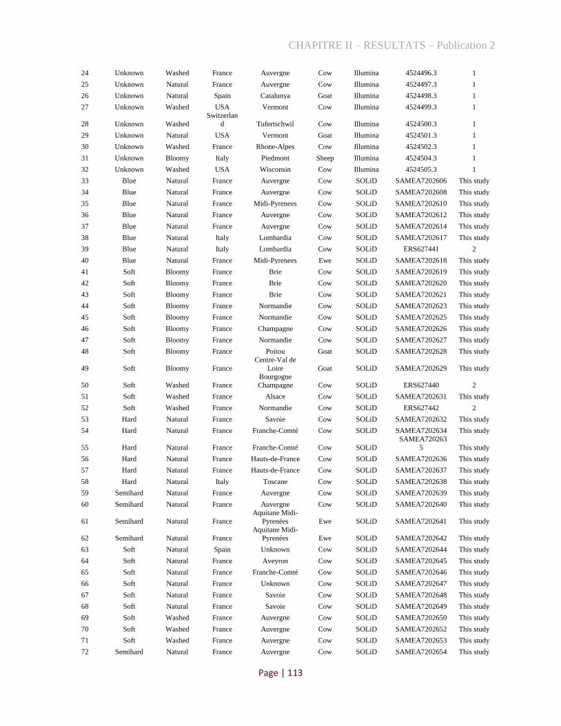
CHAPITRE II – RESULTATS – Publication 2
Page | 113
24 Unknown Washed France Auvergne Cow Illumina 4524496.3 1
25 Unknown Natural France Auvergne Cow Illumina 4524497.3 1
26 Unknown Natural Spain Catalunya Goat Illumina 4524498.3 1
27 Unknown Washed USA Vermont Cow Illumina 4524499.3 1
28 Unknown Washed
Switzerlan
d Tufertschwil Cow Illumina 4524500.3 1
29 Unknown Natural USA Vermont Goat Illumina 4524501.3 1
30 Unknown Washed France Rhone-Alpes Cow Illumina 4524502.3 1
31 Unknown Bloomy Italy Piedmont Sheep Illumina 4524504.3 1
32 Unknown Washed USA Wisconsin Cow Illumina 4524505.3 1
33 Blue Natural France Auvergne Cow SOLiD SAMEA7202606 This study
34 Blue Natural France Auvergne Cow SOLiD SAMEA7202608 This study
35 Blue Natural France Midi-Pyrenees Cow SOLiD SAMEA7202610 This study
36 Blue Natural France Auvergne Cow SOLiD SAMEA7202612 This study
37 Blue Natural France Auvergne Cow SOLiD SAMEA7202614 This study
38 Blue Natural Italy Lombardia Cow SOLiD SAMEA7202617 This study
39 Blue Natural Italy Lombardia Cow SOLiD ERS627441 2
40 Blue Natural France Midi-Pyrenees Ewe SOLiD SAMEA7202618 This study
41 Soft Bloomy France Brie Cow SOLiD SAMEA7202619 This study
42 Soft Bloomy France Brie Cow SOLiD SAMEA7202620 This study
43 Soft Bloomy France Brie Cow SOLiD SAMEA7202621 This study
44 Soft Bloomy France Normandie Cow SOLiD SAMEA7202623 This study
45 Soft Bloomy France Normandie Cow SOLiD SAMEA7202625 This study
46 Soft Bloomy France Champagne Cow SOLiD SAMEA7202626 This study
47 Soft Bloomy France Normandie Cow SOLiD SAMEA7202627 This study
48 Soft Bloomy France Poitou Goat SOLiD SAMEA7202628 This study
49 Soft Bloomy France
Centre-Val de
Loire Goat SOLiD SAMEA7202629 This study
50 Soft Washed France
Bourgogne
Champagne Cow SOLiD ERS627440 2
51 Soft Washed France Alsace Cow SOLiD SAMEA7202631 This study
52 Soft Washed France Normandie Cow SOLiD ERS627442 2
53 Hard Natural France Savoie Cow SOLiD SAMEA7202632 This study
54 Hard Natural France Franche-Comté Cow SOLiD SAMEA7202634 This study
55 Hard Natural France Franche-Comté Cow SOLiD
SAMEA720263
5 This study
56 Hard Natural France Hauts-de-France Cow SOLiD SAMEA7202636 This study
57 Hard Natural France Hauts-de-France Cow SOLiD SAMEA7202637 This study
58 Hard Natural Italy Toscane Cow SOLiD SAMEA7202638 This study
59 Semihard Natural France Auvergne Cow SOLiD SAMEA7202639 This study
60 Semihard Natural France Auvergne Cow SOLiD SAMEA7202640 This study
61 Semihard Natural France
Aquitane Midi-
Pyrenées Ewe SOLiD SAMEA7202641 This study
62 Semihard Natural France
Aquitane Midi-
Pyrenées Ewe SOLiD SAMEA7202642 This study
63 Soft Natural Spain Unknown Cow SOLiD SAMEA7202644 This study
64 Soft Natural France Aveyron Cow SOLiD SAMEA7202645 This study
65 Soft Natural France Franche-Comté Cow SOLiD SAMEA7202646 This study
66 Soft Natural France Unknown Cow SOLiD SAMEA7202647 This study
67 Soft Natural France Savoie Cow SOLiD SAMEA7202648 This study
68 Soft Natural France Savoie Cow SOLiD SAMEA7202649 This study
69 Soft Washed France Auvergne Cow SOLiD SAMEA7202650 This study
70 Soft Washed France Auvergne Cow SOLiD SAMEA7202652 This study
71 Soft Washed France Auvergne Cow SOLiD SAMEA7202653 This study
72 Semihard Natural France Auvergne Cow SOLiD SAMEA7202654 This study

CHAPITRE II – RESULTATS – Publication 2
Page | 114
73 Semihard Natural France Savoie Cow SOLiD SAMEA7202655 This study
74 Semihard Natural France Savoie Cow SOLiD SAMEA7202656 This study
75 Soft Unknown Ireland Unknown Sheep Illumina ERS3466744 3
76 Soft Bloomy Ireland Unknown Cow Illumina ERS3466745 3
77 Semihard Unknown Ireland Unknown Cow Illumina ERS3466746 3
78 Semihard Washed Ireland Unknown Cow Illumina ERS3466771 3
79 Semihard Washed Ireland Unknown Cow Illumina ERS3466747 3
80 Soft Unknown Ireland Unknown Cow Illumina ERS3466772 3
81 Soft Bloomy Ireland Unknown Cow Illumina ERS3466773 3
82 Hard Washed Ireland Unknown Goat Illumina ERS3466748 3
83 Hard Natural Ireland Unknown Sheep Illumina ERS3466774 3
84 Semihard Unknown Ireland Unknown Cow Illumina ERS3466775 3
85 Soft Natural Ireland Unknown Cow Illumina ERS3466749 3
86 Soft Washed Ireland Unknown Cow Illumina ERS3466777 3
87 Soft Bloomy Ireland Unknown Cow Illumina ERS3466789 3
88 Soft Natural Ireland Unknown Cow Illumina ERS3466790 3
89 Semihard Bloomy Ireland Unknown Goat Illumina ERS3466791 3
90 Hard Natural Ireland Unknown Cow Illumina ERS3466792 3
91 Hard Natural Ireland Unknown Goat Illumina ERS3466794 3
92 Hard Natural Ireland Unknown Cow Illumina ERS3466750 3
93 Hard Natural Ireland Unknown Cow Illumina ERS3466751 3
Table S3. The relative abundance of microbial composition of ten French cheese metagenomes
using MetaPhlAn.
Species C-01 C-02 C-03 C-04 C-05 C-06 C-07 C-08 C-09 C-10
Actinomycetaceae_bacterium_sk1b4 0 0 0 0 0 0.29109 0 0 0 0
Actinomycetales_bacterium_JB111 0 0 0 0 0 0 0 0.35251 0 0
Agrococcus_casei 0 0 0 0 0 8.19255 0 8.34963 0 0
Corynebacterium_casei 0 0.00369 0 0 0 51.2108 0 15.4885 0 0
Corynebacterium_flavescens 0 0 0 0 0.25998 0 0 0 0 0
Corynebacterium_variabile 0 0 0 0 0 19.6526 0 0.61929 0 0
Brevibacterium_antiquum 0 0.202 0 0 0 0 0 0 0 0
Brevibacterium_aurantiacum 0.96757 1.7033 0.02625 0 0 4.23944 0.09189 4.91215 0 1.19903
Brevibacterium_linens 1.48648 2.25694 0.00925 0 0 4.25165 0.06659 5.15942 0 1.09536
Brevibacterium_sp_239c 0.14179 0.03488 0 0 0 0 0 0 0 0
Brachybacterium_alimentarium 8.5708 0.15666 0.02215 0 0 0.02724 0.0338 0 0 0
Leucobacter_sp_G161 0 0 0 0 0 0.05465 0 0 0 0.38738
Arthrobacter_rhombi 0.44266 0 0 0 0 0 0 0 0 0
Glutamicibacter_arilaitensis 0 0 0.17068 0 0 2.15304 21.4939 1.65116 0 2.15174
Glutamicibacter_sp 0 0 0.06487 0 0 1.51103 16.6753 0.09404 0 2.28822
Glutamicibacter_sp_BW77 0 0 0 0 0 0 0 0.22941 0 0
Propionibacterium_freudenreichii 0 11.0583 0 0 0.87758 2.65119 0 6.18512 0 0
Psychroflexus_halocasei 0 0 0 0 0 0.06859 0 0 0 0.19547
Sphingobacterium_sp_JB170 0 0 0 0 0 0.73966 0 0.01869 0 0
Staphylococcus_equorum 0.10597 6.31793 0.004 0 0 0.34662 0.31234 0.44169 0 3.47745
Staphylococcus_succinus 0 0 0 0 1.36185 0 0.00499 0 0 0
Staphylococcus_vitulinus 0 0.13625 0 0 0 0.00257 0.41267 0 0 0
Staphylococcus_warneri 0 0 0 0 0 0 0 0 1.01311 0
Aerococcaceae_bacterium 0 0.00112 0 0 0 0.24194 0 0.07455 0 0

CHAPITRE II – RESULTATS – Publication 2
Page | 115
Alkalibacterium_gilvum 0 0.0038 0 0 0 0.00175 0 0.10198 0 0
Carnobacterium_maltaromaticum 0.12848 0.45563 0 0 0 0 0 0 0 0
Marinilactibacillus_psychrotolerans 0 0 0 0 0 1.04585 0.37135 0.71485 0 0
Enterococcus_faecalis 0.1171 0.00576 0 0.15746 0.57712 0 0 0 0 0.37182
Enterococcus_malodoratus 0 0 0 0.00849 1.29636 0 0 0.0235 0 0
Lactobacillus_brevis 0 1.5744 0 0 0 0.03413 0 0 0 0
Lactobacillus_curvatus 0 3.48061 0 0 0 0.61776 0 0.11868 0 0
Lactobacillus_delbrueckii 0.05914 0 0 0 31.1279 0 0 0.60611 0 0
Lactobacillus_helveticus 0.19328 0 0 0 0 0 0 0 0 0
Lactobacillus_nodensis 0 0.21142 0 0 0 0 0 0 0 0
Lactobacillus_parabuchneri 0 0.00352 0 0 0.55665 0.04258 0 0 0 0
Lactiplantibacillus plantarum 0 7.02383 0 0 0 0 0 0.07707 0 0
Lactobacillus_rhamnosus 0 0 1.05034 0 0.17931 0 0 0 0 0
Leuconostoc_mesenteroides 6.086 0.43638 0 0.07074 4.24848 0 0.01118 0.16069 0.99066 0.33437
Leuconostoc_pseudomesenteroides 0.16573 0.62153 0 0.08176 15.9537 0 0 0 1.4621 6.67973
Lactococcus_garvieae 0 0 0 0 1.71479 0 0 0 0 0
Lactococcus_lactis 75.0842 64.0248 34.2544 2.66912 5.18375 0.41942 7.2111 2.04215 15.3166 11.1713
Lactococcus_petauri 0 0 0 0 6.18607 0 0 0 0 0
Lactococcus_raffinolactis 0.28672 0.04814 0 0 0 0 0 0 0 0
Streptococcus_parauberis 0 0 0 0 0.14246 0 0 0 0 0
Streptococcus_thermophilus 0.08563 0.23908 64.033 0 30.1165 0 1.2383 11.572 0 22.9015
Coprococcus_eutactus 0 0 0 0 0 0 0.01082 0 0.15969 0
Brucella_abortus 0 0 0.12591 0 0 0 0 0 0 0
Advenella_kashmirensis 0 0 0 0 0 0 0 0.30599 0 0
Advenella_sp_S44 0 0 0 0 0 0 0 3.31624 0 0
Pseudoalteromonas_nigrifaciens 0 0 0 0.01582 0 0.00807 0.39101 0 0.73761 2.05761
Pseudoalteromonas_sp_A601 0 0 0 0 0 0 0 0 0 0.10447
Pseudoalteromonas_tetraodonis 0 0 0 0.01459 0 0 0 0 0.00476 1.59606
Citrobacter_braakii 0 0 0 0 0 0 0.80788 0 0 0
Raoultella_terrigena 0 0 0 0.10152 0 0 0 0 0 0
Hafnia_paralvei 0 0 0 0.51781 0 0 0.19772 0 0 0
Morganella_psychrotolerans 0 0 0 0 0 0.10715 0 0.01083 0.26594 0
Ewingella_americana 0 0 0 0.85361 0 0 0.10766 0 0 0
Serratia_fonticola 0 0 0 1.12286 0 0 0 0 0 0
Serratia_grimesii 0 0 0 0 0 0 0 0 1.09529 0
Serratia_liquefaciens 0 0 0 0 0.20565 0 0.42422 0 0 0
Cobetia_marina 0 0 0.19187 0 0 0 38.1055 0 0.02608 0
Halomonas_campaniensis 0 0 0.00217 0 0 0.75616 1.68787 0 0 8.90725
Halomonas_sp_JB37 0 0 0.00099 0 0 0 1.50231 36.452 0 21.1857
Marinomonas_foliarum 0 0 0 0 0 0 0 0 30.4574 0
Actinobacillus_pleuropneumoniae 0 0 0 0.22591 0 0 0 0 0 0
Acinetobacter_johnsonii 0 0 0 0 0 0 0.81098 0 0 0
Psychrobacter_alimentarius 0 0 0 0 0 0 0.17587 0 0 0
Psychrobacter_immobilis 0.17503 0 0 0 0 0 0.22203 0 0 0
Psychrobacter_sp_JB385 0 0 0 0 0 0 0.14573 0 0 0
Psychrobacter_sp_JCM_18900 0 0 0 0 0 0 0.01392 0 0.16575 0
Pseudomonas_bauzanensis 0 0 0 0 0 1.33246 0 0 0 0
Pseudomonas_helleri 0 0 0 53.5006 0 0 0 0 0 0
Pseudomonas_lundensis 0 0 0 40.6597 0.01181 0 0 0 0 0
Pseudomonas_taetrolens 5.90346 0 0 0 0 0 0 0 0 0
Vibrio_casei 0 0 0.04408 0 0 0 7.47305 0.92178 48.305 8.05754
Vibrio_kanaloae 0 0 0 0 0 0 0 0 0 5.83791

CHAPITRE II – RESULTATS – Publication 2
Page | 116
Table S4. The relative abundance of microbial composition of ten French cheese metagenomes by
marker gene analysis (ychF gene).
Species C-01 C-02 C-03 C-04 C-05 C-06 C-07 C-08 C-09 C-10
Agrococcus casei 0 0 0 0 0 4.12969 0 2.69727 0 0
Leucobacter sp. 0 0 0 0 0 0 0 0.49041 0 0.96772
Microbacterium gubbeenense 0 0 0 0 0 9.84096 0 2.03521 0 0
Microbacterium sp. 0 0 0 0 0 0 0 7.41504 0 0
Pseudoclavibacter sp. 0 0 0 0 0 0 0 0.81408 0 0
Corynebacterium casei 0 0 0 0 0 29.2241 0 4.50689 0 0
Corynebacterium variabile 0 0 0 0 0 9.34891 0 0.55907 0 0
Corynebacterium xerosis 0 0 0 0 0 0 0.47494 1.25055 0 1.1385
Brachybacterium alimentarium 1.30828 0 0 0 0 0 0 0 0 0
Brevibacterium aurantiacum 1.22458 1.46187 0 0 0 2.95229 0 3.2073 0 0.95065
Clavibacter sp. 0 0 0 0 0 6.50206 0 0 0 0
Glutamicibacter ardleyensis 0 0 0 0 0 0 0 0.82389 0 0
Glutamicibacter arilaitensis 0 0 0 0 0 0 7.85112 0 0 0.79695
Propionibacterium freudenreichii 0 4.2812 0 0 0 0 0 1.83414 0 0
Sphingobacterium sp. 0 0 0 0 0 0 0 5.49262 0 0
Jeotgalicoccus psychrophilus 0 5.89971 0 0 0 0 0 0 0 0
Staphylococcus equorum 0 2.1406 0 0 0 0 0 0 0 1.53697
Atopostipes suicloacalis 0 0.65262 0 0 0 0 0 0 0 0
Carnobacterium maltaromaticum 0 0.41768 0 0 0 0 0 0 0 0
Marinilactibacillus psychrotolerans 0 0 0 0 0 1.31799 0 0.84841 0 0
Enterococcus malodoratus 0 0 0 0 1.21517 0 0 0 0 0
Enterococcus sp. 0 0.30543 0 0 0 0 0 0 0 0
Vagococcus sp. 0 0 0 0 0 3.11045 0.24232 0 0 0
Lactiplantibacillus plantarum 0 2.61049 0 0 0 0 0 0 0 0
Lactobacillus brevis 0 0.81447 0 0 0 0 0 0 0 0
Lactobacillus curvatus 0 1.63417 0 0 0 0 0 0 0 0
Lactobacillus delbrueckii 0 0 0 0 15.6889 0 0 0 0 0
Lactobacillus paracasei 0 0 0 0 0 0 0 0.53945 0 0
Lactobacillus rhamnosus 0 0 0.59496 0 0 0 0 0 0 0
Leuconostoc mesenteroides 1.33309 0.5221 0 0 2.14396 0 0 0 0 0
Leuconostoc pseudomesenteroides 0 0 0 0 5.45666 0 0 0 0 2.56162
Lactococcus garvieae 0 0 0 0 3.49071 0 0 0 0 0
Lactococcus lactis subsp. cremoris 49.1629 51.7529 28.2436 6.10034 0 0 2.85936 0 7.3713 9.22184
Lactococcus lactis subsp. lactis 0 13.94 18.4154 0 1.90402 0 4.45866 0.8337 0 1.19542
Lactococcus laudensis 33.5938 0 0 0 0 0 0 0 0 0
Lactococcus raffinolactis 0 2.55045 0 0 0 0 0 0 0 0
Streptococcus gallolyticus 0 0 0.31448 0 0 0 0 0 0 0
Streptococcus thermophilus 0 0 51.8068 0 13.6765 0 1.11467 4.46275 0 13.3774
Arenibacter (unclassified) 0 0 0 0 0 0 0 0.58849 0 0
Psychroflexus sp. 0 0 0 0 0 1.42343 0 0 0 1.08157
Advenella sp. 0 0 0 0 0 0 0 1.09852 0 0

CHAPITRE II – RESULTATS – Publication 2
Page | 117
Marinobacter (unclassified) 0 0 0 0 0 12.4154 0 0 0 0
Pseudoalteromonas (unclassified ) 0 0 0 0 0 0 0 0 3.6085 20.3791
Pseudoalteromonas nigrifaciens 0 0 0 0 0 0 1.01774 0 0.8524 2.56162
Pseudoalteromonas sp. 0 0 0 0 0 0 0 0 0 1.70775
Pseudoalteromonas undina 0 0 0 0 0 0 0 0 0 5.06632
Klebsiella michiganensis 0 0 0 0 0 0 0.82388 0 0 0
Kluyvera intermedia 0 0 0 0 0 0 1.0662 0 0 0
Proteus hauseri 0 0 0 0 0 1.441 0 0 0 0
Providencia heimbachae 0 0 0 0 0 0 0 0.49041 0 0
Providencia sp. 0 0 0 5.64509 0 0 0 0 0 0
Serratia proteamaculans 0 0 0 0 11.726 0 0 0 0 0
Cobetia marina 0 0 0 0 0 0 21.6148 0 0 0
Halomonas casei 0 0 0 0 0 0 1.0662 0 0 3.81397
Halomonas citridevorans 0 0 0 0 0 0 0.87235 0 0 8.70951
Halomonas flavum 0 0 0 0 0 0 0 0 0 3.58627
Halomonas minus 0 0 0 0 0 0 0.96927 52.6016 0 8.48181
Halomonas sp. 0 0 0 0 0 6.00123 0 7.41013 0 0
Marinomonas foliarum 0 0 0 0 0 0 0 0 26.725 0
Marinomonas polaris 0 0 0 0 0 0 0 0 0 1.42312
Marinomonas sp. 0 0 0 0 12.6161 0 0 0 0 0
Acinetobacter sp. 0 0 0 0 0 0 0.46525 0 0 0
Psychrobacter aquimaris 0 0 0.25215 0 0 0 12.7944 0 0 0
Psychrobacter casei 0.76265 11.0163 0 0 0 0 0 0 0 0
Psychrobacter cibarius 0.78745 0 0 0 0 0 0 0 29.534 0
Psychrobacter pacificensis 0 0 0 0 0 0 1.35698 0 0 0
Psychrobacter sp. 1.90352 0 0.21674 0 0 0 2.81089 0 0 0
Psychrobacter translucens 0 0 0 0 0 2.70626 27.1397 0 16.358 0
Pseudomonas bauzanensis 0 0 0 0 0 2.07363 0 0 0 0
Pseudomonas helleri 0 0 0 38.423 0 0 0 0 0 0
Pseudomonas litoralis 0 0 0 0 0 7.51252 0 0 0 0
Pseudomonas lundensis 0 0 0 32.4137 0 0 0 0 0 0
Pseudomonas reactans 0 0 0 9.81517 0 0 1.0662 0 0 0
Pseudomonas sabulinigri 0 0 0 0 0 0 0 0 0 1.08157
Pseudomonas taetrolens 1.42609 0 0 0 0 0 0 0 0 0
Vibrio casei 0 0 0.15582 0 0 0 4.36173 0 10.7 3.35857
Vibrio litoralis 0 0 0 0 0 0 5.57333 0 4.2824 0
Vibrio sp. 0 0 0 0 0 0 0 0 0 1.5939
Vibrio toranzoniae 0 0 0 0 0 0 0 0 0 3.13087
Debaryomyces hansenii 0 0 0 0 11.4551 0 0 0 0 0
Geotrichum candidum 4.24107 0 0 6.10034 20.6269 0 0 0 0 2.277
Kluyveromyces lactis 4.25657 0 0 1.50232 0 0 0 0 0.5683 0

CHAPITRE II – RESULTATS – Publication 2
Page | 118
Table S5. Genomic analysis of Marinomonas foliarum MAG-FME69 and closely related
cultivable species.
Table S6. Genomic analysis of Pseudoalteromonas sp. MAG-FME70 and closely related species.
Table S7. Mapping of 12 species obtained in this study on 93 cheese rind metagenomes.
Mar
inom
onas a
lcara
zii st
rain
CEC
T 77
30
Mar
inom
onas f
oliaru
m st
rain
CEC
T 77
31
Mar
ino_F
ME6
9_m
etab
at_fil
ter.b
in.5
.fa
Mar
inom
onas s
p. MW
YL1
Mar
inom
onas U
CMA
Mar
ino_C
heese
Seq_R42_
S43_b
in.5
.fa
Mar
ino_C
heese
Seq_R46_
S38_b
in.3
.fa
Mar
ino_C
heese
Seq_R47_
S50_b
in.7
.fa
Mar
inoba
cter
hyd
roca
rbonocla
sticu
s VT8
Marinomonas alcarazii strain CECT 7730 * 86.04 [76.65] 85.94 [74.24] 80.29 [75.84] 80.26 [75.43] 80.18 [72.44] 80.19 [75.03] 80.21 [74.69] 64.77 [13.14]
Marinomonas foliarum strain CECT 7731 85.82 [73.86] * 96.91 [79.05] 80.86 [71.87] 81.19 [73.44] 81.05 [71.12] 80.88 [71.76] 80.88 [71.52] 65.12 [13.09]
Marino_FME69_metabat_filter.bin.5.fa 85.97 [78.55] 97.06 [87.38] * 81.00 [77.01] 81.21 [78.16] 81.58 [77.60] 80.98 [76.36] 80.98 [76.12] 64.51 [12.67]
Marinomonas sp. MWYL1 80.15 [61.07] 80.71 [60.25] 80.79 [57.78] * 98.05 [86.33] 97.90 [79.76] 98.17 [83.88] 98.12 [83.55] 65.53 [10.99]
Marinomonas UCMA 79.94 [60.65] 80.83 [61.89] 80.95 [59.22] 98.06 [86.65] * 97.76 [82.43] 98.10 [85.63] 98.09 [85.13] 64.78 [10.71]
Marino_CheeseSeq_R42_S43_bin.5.fa 79.95 [62.20] 80.72 [63.79] 81.19 [62.77] 97.96 [86.08] 97.75 [87.90] * 98.12 [85.31] 98.09 [84.56] 64.49 [10.95]
Marino_CheeseSeq_R46_S38_bin.3.fa 79.95 [63.27] 80.70 [62.99] 80.80 [60.50] 98.21 [89.56] 98.15 [89.95] 98.08 [84.20] * 99.91 [98.01] 64.59 [10.98]
Marino_CheeseSeq_R47_S50_bin.7.fa 79.80 [62.78] 80.54 [62.42] 80.62 [59.86] 98.02 [88.38] 97.94 [88.84] 97.88 [82.85] 99.73 [97.02] * 64.65 [11.06]
Marinobacter hydrocarbonoclasticus VT8 65.04 [11.31] 65.04 [11.82] 64.47 [10.54] 64.81 [11.19] 65.58 [12.47] 64.52 [10.75] 64.38 [10.83] 64.37 [10.81] *
Species C-09 C-10 C-11 C-12 C-22 C-23 C-24 C-25 C-50 C-77 C-85 C-86 C-87
Vibrio_litoralis_FME62 3.98 0 0 0 0 0 0 0 3.62 0 3.41 0 0
Vibrio_casei_FME29 8.55 2.33 0 5.01 2.12 0 0 0 0 0 0 0 2.62
Pseudoalteromonas_sp._MAG-FME70 2.83 12.7 0 2.61 13.5 0 16.8 2.5 2.15 0.89 0 0 3.05
Pseudoalteromonas_undina_FME67 0 5.54 0 0 0 0 0 0 0 0 0 0 0
Pseudoalteromonas_nigrifaciens_FME68 0.94 4.16 0 12.7 23.2 0 0 0 2.28 0 16.5 0 4.36
Marinomonas_foliarum_MAG-FME69 20.6 0 0 2.61 0 0 0 0 0 0 4.4 0 0
Psychrobacter_translucens_FME61 15.8 0.16 0 0 2.61 0 2.71 0 0 0 0 0 11.8
Psychrobacter_cibarius_FME60 26.2 0 0 0 0 0 1.19 0 5.52 0 0 0 0
Halomonas_minus_FME64 0 6.92 0 0 0 0 20.5 0.84 0 0 0 0 0
Halomonas_flavum_FME66 0 4.66 0 20.4 0 0 0 0 0 0 0 0 0
Halomonas_citridevorans_FME63 0 6.45 0 14.2 11.6 0 0 0 0 0.79 0 0 6.67
Halomonas_casei_FME65 0 3.41 0 0 0 0 5.85 0 0 0 0 0.42 0
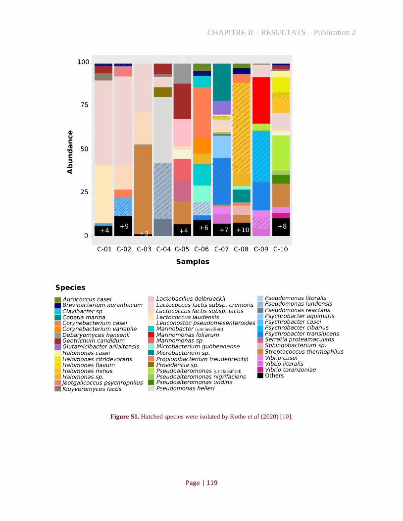
CHAPITRE II – RESULTATS – Publication 2
Page | 119
Figure S1. Hatched species were isolated by Kothe et al (2020) [10].
CHAPITRE II – RESULTATS – Publication 2
Page | 120
5 References
[1]. Jagadeesan, B., et al., The use of next generation sequencing for improving food safety: Translation into
practice. Food Microbiology, 2019. 79: p. 96-115. doi:10.1016/j.fm.2018.11.005.
[2]. van Dijk, E.L., et al., Ten years of next-generation sequencing technology. Trends in Genetics, 2014. 30(9): p.
418-426. doi:10.1016/j.tig.2014.07.001.
[3]. Quince, C., et al., Shotgun metagenomics, from sampling to analysis. Nature Biotechnology, 2017. 35(9): p.
833-844. doi:10.1038/nbt.3935.
[4]. Riesenfeld, C.S., P.D. Schloss, and J. Handelsman, Metagenomics: Genomic Analysis of Microbial
Communities. Annual Review of Genetics, 2004. 38(1): p. 525-552. doi:10.1146/annurev.genet.38.072902.091216.
[5]. Wolfe, B.E., et al., Cheese Rind Communities Provide Tractable Systems for In Situ and In Vitro Studies of
Microbial Diversity. Cell, 2014. 158(2): p. 422-433. doi:10.1016/j.cell.2014.05.041.
[6]. Lewis, W.H., et al., Innovations to culturing the uncultured microbial majority. Nature Reviews Microbiology,
2020. doi:10.1038/s41579-020-00458-8.
[7]. Irlinger, F., et al., Cheese rind microbial communities: diversity, composition and origin. FEMS Microbiology
Letters, 2015. 362(2): p. 1-11. doi:10.1093/femsle/fnu015.
[8]. Cocolin, L., et al., Culture independent methods to assess the diversity and dynamics of microbiota during food
fermentation. International Journal of Food Microbiology, 2013. 167(1): p. 29-43.
doi:10.1016/j.ijfoodmicro.2013.05.008.
[9]. Almeida, M., et al., Construction of a dairy microbial genome catalog opens new perspectives for the
metagenomic analysis of dairy fermented products. BMC Genomics, 2014. 15(1): p. 1101. doi:10.1186/1471-2164-
15-1101.
[10]. Kothe, C.I., et al., Unraveling the world of halophilic and halotolerant bacteria in cheese by combining
cultural, genomic and metagenomic approaches. bioRxiv, 2020: p. 2020.11.03.353524.
doi:10.1101/2020.11.03.353524.
[11]. Walsh, A., et al., Meta-analysis of cheese microbiomes highlights contributions to multiple aspects of quality.
Nature Food, 2020. 1: p. 500-510. doi:10.1038/s43016-020-0129-3.
[12]. Parks, D.H., et al., Recovery of nearly 8,000 metagenome-assembled genomes substantially expands the tree of
life. Nature Microbiology, 2017. 2(11): p. 1533-1542. doi:10.1038/s41564-017-0012-7.
[13]. Tully, B.J., E.D. Graham, and J.F. Heidelberg, The reconstruction of 2,631 draft metagenome-assembled
genomes from the global oceans. Scientific Data, 2018. 5(1): p. 170203. doi:10.1038/sdata.2017.203.
[14]. Truong, D.T., et al., MetaPhlAn2 for enhanced metagenomic taxonomic profiling. Nature Methods, 2015. 12:
p. 902-903. doi:10.1038/nmeth.3589.
[15]. Bankevich, A., et al., SPAdes: a new genome assembly algorithm and its applications to single-cell
sequencing. Journal of computational biology : a journal of computational molecular cell biology, 2012. 19(5): p.
455-477. doi:10.1089/cmb.2012.0021.
[16]. Ciccarelli, F.D., et al., Toward Automatic Reconstruction of a Highly Resolved Tree of Life. Science, 2006.
311(5765): p. 1283. doi:10.1126/science.1123061.
[17]. Sunagawa, S., et al., Metagenomic species profiling using universal phylogenetic marker genes. Nature
methods, 2013. 10. doi:10.1038/nmeth.2693.
[18]. Thompson, J.D., et al., The CLUSTAL_X Windows Interface: Flexible Strategies for Multiple Sequence
Alignment Aided by Quality Analysis Tools. Nucleic Acids Research, 1997. 25(24): p. 4876-4882.
doi:10.1093/nar/25.24.4876.
[19]. Maskow, T. and W. Babel, Calorimetrically obtained information about the efficiency of ectoine synthesis
from glucose in Halomonas elongata. Biochimica et Biophysica Acta (BBA) - General Subjects, 2001. 1527(1): p.
4-10. doi:10.1016/s0304-4165(01)00115-5.
[20]. Broekaert, K., et al., Seafood quality analysis: Molecular identification of dominant microbiota after ice
storage on several general growth media. Food Microbiology, 2011. 28(6): p. 1162-1169.
doi:10.1016/j.fm.2011.03.009.
[21]. Acinas, S.G., et al., Fine-scale phylogenetic architecture of a complex bacterial community. Nature, 2004.
430(6999): p. 551-554. doi:10.1038/nature02649.
[22]. Camacho, C., et al., BLAST+: architecture and applications. BMC Bioinformatics, 2009. 10(1): p. 421.
doi:10.1186/1471-2105-10-421.

CHAPITRE II – RESULTATS – Publication 2
Page | 121
[23]. Kang, D.D., et al., MetaBAT, an efficient tool for accurately reconstructing single genomes from complex
microbial communities. PeerJ, 2015. 3: p. e1165. doi:10.7717/peerj.1165.
[24]. Langmead, B. and S.L. Salzberg, Fast gapped-read alignment with Bowtie 2. Nature Methods, 2012. 9(4): p.
357-359. doi:10.1038/nmeth.1923.
[25]. Parks, D.H., et al., CheckM: assessing the quality of microbial genomes recovered from isolates, single cells,
and metagenomes. Genome Research, 2015. 25(7): p. 1043-1055. doi:10.1101/gr.186072.114.
[26]. Aziz, R.K., et al., The RAST Server: Rapid Annotations using Subsystems Technology. BMC Genomics,
2008. 9(1): p. 75. doi:10.1186/1471-2164-9-75.
[27]. Overbeek, R., et al., The Subsystems Approach to Genome Annotation and its Use in the Project to Annotate
1000 Genomes. Nucleic Acids Research, 2005. 33(17): p. 5691-5702. doi:10.1093/nar/gki866.
[28]. Song, W., et al., Prophage Hunter: an integrative hunting tool for active prophages. Nucleic Acids Research,
2019. 47(W1): p. W74-W80. doi:10.1093/nar/gkz380.
[29]. Grissa, I., G. Vergnaud, and C. Pourcel, CRISPRFinder: a web tool to identify clustered regularly interspaced
short palindromic repeats. Nucleic Acids Research, 2007. 35(suppl_2): p. W52-W57. doi:10.1093/nar/gkm360.
[30]. Couvin, D., et al., CRISPRCasFinder, an update of CRISRFinder, includes a portable version, enhanced
performance and integrates search for Cas proteins. Nucleic Acids Research, 2018. 46(W1): p. W246-W251.
doi:10.1093/nar/gky425.
[31]. Nichols, D., et al., Short Peptide Induces an “Uncultivable” Microorganism To Grow In Vitro. Applied and
Environmental Microbiology, 2008. 74(15): p. 4889. doi:10.1128/AEM.00393-08.
[32]. Lucas-Elío, P., et al., Marinomonas alcarazii sp. nov., M. rhizomae sp. nov., M. foliarum sp. nov., M.
posidonica sp. nov. and M. aquiplantarum sp. nov., isolated from the microbiota of the seagrass Posidonia oceanica.
International Journal of Systematic and Evolutionary Microbiology, 2011. 61(9): p. 2191-2196.
doi:10.1099/ijs.0.027227-0.
[33]. Larpin-Laborde, S., et al., Surface microbial consortia from Livarot, a French smear-ripened cheese. Canadian
Journal of Microbiology, 2011. 57(8): p. 651-660. doi:10.1139/w11-050.
[34]. Linder, T., A genomic survey of nitrogen assimilation pathways in budding yeasts (sub-phylum
Saccharomycotina). Yeast, 2019. 36(5): p. 259-273. doi:10.1002/yea.3364.
[35]. Kastman, E.K., et al., Biotic Interactions Shape the Ecological Distributions of Staphylococus Species. mBio,
2016. 7(5): p. e01157-16. doi:10.1128/mBio.01157-16.

CHAPITRE II – RESULTATS – Publication 2
Page | 122
2.3. Conclusion
Au cours de cette étude, nous avons utilisé une stratégie ciblée pour l'isolement de bactéries
halophiles, basée sur une technique de criblage rapide, guidée par l'analyse métagénomique,
stratégie qui s'est avérée efficace. En bref, la détermination de la composition taxonomique de dix
métagénomes de croûtes de fromages a indiqué la prédominance de plusieurs espèces qui n’étaient
pas isolées par des méthodes classiques (Publication 1). Pour isoler ces espèces manquantes, dont
certaines sont potentiellement peu cultivables, nous avons défini une stratégie axée sur la
conception d'amorces PCR spécifiques. Ainsi, deux fromages ont été choisis pour être incubés à
différentes températures, dans des milieux de culture à base de sel ; et dans une première approche,
un screening individuel de différents clones a été testé avec les amorces ciblées. Pour les espèces
qui n'ont pas été isolées au cours de cette première phase, nous avons mis au point un deuxième
niveau de criblage, basé sur le pool de cinq à dix colonies, augmentant ainsi la probabilité d’isoler
ces espèces manquantes. Utilisant ces stratégies, nous avons pu isoler dix espèces présentes, avec
des niveaux distincts d’abondance relative, dans deux fromages à croûte lavée. Cependant, nous
avons échoué dans la culture de deux espèces représentées par une Pseudoalteromonas (nouvelle
espèce) et une souche de Marinomonas foliarum, dominantes dans les fromages avec des reads
métagénomiques de valeur respective 20 et 27%. Cherchant à comprendre le problème du non-
isolement de ces souches, leurs MAGs ont été construits et comparés aux génomes, étroitement
liés à des souches cultivables. Cependant, notre analyse génomique n’a pas révélé de différence
notable entre les MAGs et les souches cultivables. Ainsi, l’hypothèse qui nous parait la plus
plausible réside dans le fait que les cellules bactériennes pourraient avoir été détruites par la
congélation, sachant que les échantillons ont été conservés à une température de -20°C. Pour les
travaux futurs, nous suggérons de tester, à la fois, leur isolement sans congeler les échantillons, et
une gamme plus large de milieux de culture.

CHAPITRE II – RESULTATS – Publications 3 et 4
Page | 123
3. Description de nouvelles espèces
3.1. Contexte
Précédemment (Publication 1 et Publication 2), nous avons isolé plusieurs souches à partir de
croûtes de fromages qui, selon les analyses génomiques, représentent onze nouvelles espèces
potentielles (ANI<95% avec des souches type d’espèces bien définies [227]). Elles comprennent
une nouvelle espèce de chacun des genres suivants : Advenella, Brachybacterium, Brevibacterium,
Pseudomonas et Proteus ; quatre nouvelles espèces d’Halomonas et deux de Psychrobacter.
Comme les espèces Gram-négatifs d’Halomonas et de Psychrobacter étaient les plus nombreuses
et avec un degré d’abondance élevé dans plusieurs croûtes de fromages, nous avons focalisé notre
étude sur la proposition de nouvelles taxonomies d’espèces chez ces deux genres. De plus, pour
augmenter la variabilité des souches fromagères au sein d'une même espèce, nous avons également
analysé les isolats obtenus par Almeida et al. [4] et Wolfe et al. [3], représentant 12 souches
d’Halomonas et quatre de Psychrobacter. Ainsi, cette étude représente une avancée en :
démontrant, à travers l'analyse phylogénomique que ces isolats fromagers forment un clade
d’espèces non encore décrit ;
identifiant les caractéristiques physiologiques, enzymatiques et biochimiques particulières
au sein de chaque nouvelle espèce étudiée ;
proposant de nouvelles taxonomies (au niveau de l’espèce) pour les genres Halomonas et
Psychrobacter.
Les résultats obtenus ont été présentés sous la forme de deux articles dans l'International
Journal of Systematic and Evolutionary Microbiology ; ils constituent respectivement la partie
3.2 and 3.3 des nouvelles espèces d’Halomonas et Psychrobacter.

CHAPITRE II – RESULTATS – Publication 3
Page | 124
3.2. Publication 3
Halomonas citridevorans sp. nov., Halomonas minus sp. nov., Halomonas casei sp. nov. and
Halomonas flavum sp. nov., four novel species isolated from French cheese rinds
Caroline Isabel Kothea (ORCID: 0000-0003-3703-5699), Christophe Monnetb (ORCID: 0000-
0003-2647-272X), Françoise Irlingerb, Pierre Renault (ORCID: 0000-0001-6540-053X)a
a Université Paris-Saclay, INRAE, AgroParisTech, Micalis Institute, 78350 Jouy-en-Josas, France.
b Université Paris-Saclay, INRAE, AgroParisTech, UMR SayFood, 78850 Thiverval-Grignon, France.
Keywords: Taxonomy, sequence analysis, whole-genome sequencing, food.
Repositories:
The whole-genome and 16S gene sequences of the novel type strains of Halomonas have
been deposited at DDBJ/ENA/GenBank and are available under the accession numbers
JABUYX000000000, JABUYY000000000, JABUYZ000000000 and JABUZA000000000, and
MT573208, MT573212, MT573213 and MT573210, respectively.
ABSTRACT
Twelve Gram-negative bacterial strains were isolated from cheese rinds in France. On the basis
of 16S rRNA gene sequence analysis, all isolates were assigned to the genus Halomonas, their
closest relatives being Halomonas alkaliphila, H. zhanjiangensis, H. hydrothermalis and H.
venusta, with similarities of 99.86, 99.78, 99.62 and 99.52%, respectively. Phylogenetic
analyses of 16S rRNA and rpoB genes and average nucleotide identity (gANI), calculated using
the complete genome sequences of the strains, revealed that they constituted four novel and
well-supported clusters. The strains isolated herein differed from the previously described
species by ANI < 95-96% and several biochemical, enzymatic and colony characteristics. In
conclusion, the results of phenotypic and phylogenetic analyses indicated that the isolates
belonged to four novel Halomonas species, for which the names Halomonas citridevorans sp.

CHAPITRE II – RESULTATS – Publication 3
Page | 125
nov., Halomonas minus sp. nov., Halomonas casei sp. nov. and Halomonas flavum sp. nov. are
proposed, with isolates FME63T (= CIRM-BIA2430T = CIP 111880T = LMG 32013T), FME64T
(=CIRM-BIA2431T = CIP 111877T = LMG 32015T), FME65T (=CIRM-BIA2432T = CIP
111881T = LMG 32012T) and FME66T (=CIRM-BIA2433T = CIP 111876T = LMG 32014T) as
type strains, respectively.
Halomonas is a genus of the Halomonadaceae family that belongs to the Oceanospirillales
order, Gammaproteobacteria class and Proteobacteria phylum [1, 2]. In total, 69 whole-genome
sequences of defined Halomonas species were available at the time of writing (NCBI public
database). Corresponding strains are predominantly halophilic [3, 4] and isolated from saline
ecosystems such as soils, saline lakes and salterns [5, 6]. Some species were also recently described
in fermented foods known to contain high salt concentrations, such as traditional Korean fermented
seafood (H. jeotgali, H. alimentaria and H. cibimaris) [7-9], and in 'Quargel' cheese (H.
nigrificans) [10].
In this paper, we describe 12 strains that belong to the Halomonas genus that were isolated
from French cheese rinds. Since these strains differed by some unusual genetic and phenotypic
features from the closely related Halomonas type strains, we established that they belong to four
new Halomonas species referred to as H. citridevorans sp. nov., H. minus sp. nov., H. casei sp.
nov. and H. flavum sp. nov.
Isolation and ecology
During the course of several projects that explored the diversity of bacteria associated
with artisanal cheese rinds, we isolated 12 strains that belong to potentially new Halomonas
species. The sources of isolation and their corresponding growth media and temperature are
presented in Table S1. Bacterial strains obtained from pure colonies were stored at -80°C in their
respective growth media supplemented with 20% (v/v) glycerol and deposited in culture collection
centers.

CHAPITRE II – RESULTATS – Publication 3
Page | 126
16S rRNA and rpoB phylogenies
Genomic DNA was extracted from the bacterial cells according to the protocol described
by Almeida et al. (2014) [11]. Their 16S rRNA genes were amplified using 27-F (5′-
AGAGTTTGATCATGGCTCA-3′) and 1492-R (5′-TACGGTTACCTTGTTACGACTT-3′)
primers [12], and sequences were compared with those of type strains of species with validly
published names using EzBioCloud (https://www.ezbiocloud.net/). Their accession numbers are
presented in Table S2. In order to expand the taxonomy of strains, rpoB (DNA-directed RNA
polymerase beta subunit), used as an alternative marker gene, was amplified using primers VIC4
(5′-GGCGAAATGGCDGARAACCA-3′) and VIC6 (5′-GARTCYTCGAAGTGGTAACC-3′)
[13]. The rpoB sequences were compared to known bacterial species in the BLASTn GenBank
database of the National Center for Biotechnology Information (NCBI). Further phylogenetic
analyses were performed for 16S rRNA and rpoB sequences using MEGA 7.0.26 software, and
trees were generated using the Neighbor-Joining algorithm with 1,000 bootstrap iterations [14]
(Fig. 1 and Fig. 2). To delineate species, we used a threshold of above 99% identity for 16S rRNA
genes with type strains [15], and above 97.7% identity for rpoB nucleotidic sequences [16].
The strains FME63T, 1M45, JB380 and FME16 are closely related to each other, sharing
identities ranging from 99.08 to 99.91% for 16S rRNA and from 97.25 to 100% for rpoB gene
sequences (Table S3A). These strains could thus be representatives of a new genomospecies since
their level of identity of 16S rRNA and rpoB with the closest related type strains is below the given
thresholds. Moreover, the inferred phylogenetic trees confirmed the unambiguous divergence of
the four strains with previously described Halomonas species, as well as the fact that they might
form two distinct genomic groups (Fig. 1 and Fig. 2) that require further genomic analyses to
confirm their correct assignment.
Similarly, analyses of 16S rRNA and rpoB genes indicated that FME64T and FME1 belong
to the same species since their sequences share identities of 100 and 98.14%, respectively.
However, they remain unassigned since their sequences display sub-threshold levels of identity
with all defined species (Table S3B).
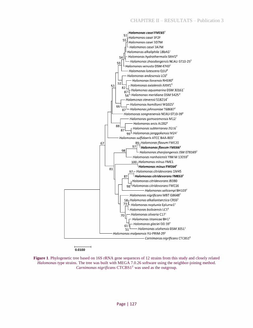
CHAPITRE II – RESULTATS – Publication 3
Page | 127
Figure 1. Phylogenetic tree based on 16S rRNA gene sequences of 12 strains from this study and closely related
Halomonas type strains. The tree was built with MEGA 7.0.26 software using the neighbor-joining method.
Carnimonas nigrificans CTCBS1T was used as the outgroup.

CHAPITRE II – RESULTATS – Publication 3
Page | 128
The Halomonas strains - FME65T, 3F2F, 3D7M and 3A7M - shared 16S rRNA and rpoB
gene sequence identities >99.71 and 100%, respectively (Table S3C). They are also closely related
to four environmental species, H. alkaliphila DSM 16354T, H. venusta DSM 4743T, H.
hydrothermalis Slthf2T and H. zhaodongensis NEAU-ST10-25T, showing the highest similarity of
between 99.27 and 99.86% for 16S rRNA sequences and of between 94.53 and 98.36% for rpoB
sequences. This led to the assumption that these four strains isolated from cheese might be asigned
to the species H. alkaliphila, but further analyses are required.
The 16S rRNA and rpoB gene sequences of Halomonas FME66T and FME20 strains share
100 and 99.68% of homology with each other, respectively, and also display high identity (> 99.5%
for 16S and > 98.5% for rpoB) with those of Halomonas zhanjiangensis JSM 078169T (Table
S3D). As a result, it was suggested that FME20 and FME66T belong to this species, and the
hypothesis that they form a new closely related species requires further investigations.
Figure 2. Phylogenetic tree based on rpoB gene sequences of 12 strains from this study and closely related
Halomonas type strains. The tree was built with MEGA 7.0.26 software using the neighbor-joining method.
Carnimonas nigrificans CTCBS1T was used as the outgroup.

CHAPITRE II – RESULTATS – Publication 3
Page | 129
Genome features and phylogenomic analysis
To establish if cheese isolates were affiliated with one of the closely related species
previously described or are probable new species of Halomonas, average nucleotide identity
(gANI) analyses with whole-genome sequences were performed. The genome of 12 strains were
sequenced and annotated as described below. In order to enable a whole-genome-based
reconstruction of its phylogeny, genomes of two Halomonas type strains (H. alkaliphila DSM
16354T and H. venusta DSM 4743T) not available so far, were also sequenced and annotated. DNA
sequencing was carried out on an Illumina HiSeq device at GATC-Biotech (Konstanz, Germany)
in order to generate at least 5 million paired-end reads per genome (150 bases in length). For each
strain, de novo assembly was performed using SPAdes version 3.9 [17]. Only contigs with
length >300 bp and coverage >100 were considered for further study. Annotations were performed
using the RAST (Rapid Annotation using Subsystem Technology) server [18]. Basic features of
these new whole-genome sequences are presented in Table 1.
The whole genomes of all Halomonas species available in the NCBI database were
collected, and information on these genomes such as their origin of isolation, numbers of
Bioproject and BioSample are shown in Table S4. The sequences of these genomes were aligned
with those of the strains obtained in this study and with that of Carnimonas nigrificans ATCC
BAA-78T, as an outgroup, using Progressive Mauve software (version 2.4.0) with the default
settings. The phylogenomic tree obtained was visualized with Interactive Tree of Life (iTOL v5)
(http://itol.embl.de). Finally, the gANI values of the Halomonas strains isolated here and that of
closely related species were calculated using JSpeciesWS [19] in order to confirm speciation of
the different isolates. For species delineation, we used the recommended cut-off point of 95-96%
ANI, which replaces the DDH analysis [20].

Table 1. Genomic features of Halomonas alkaliphila and Halomonas venusta type strains and the 12 strains representative of the novel Halomonas species
proposed.
Species Strain No of
contigs
Size
(base
pairs)
GC
content
(%)
N50a L50b
Number of
subsystems
(RAST)
Number
of coding
sequences
Bioproject BioSample Accesion
Halomonas citridevorans FME63T 77 3,927,099 54.2 131,964 6 358 3,767 PRJNA501839 SAMN15150959 JABUYX000000000
Halomonas citridevorans 1M45 78 3,759,973 54.2 116,969 10 339 3,641 PRJNA501839 SAMN15150965 JABUZC000000000
Halomonas citridevorans FME16 62 3,710,911 53.9 157,431 7 364 3,606 PRJNA501839 SAMN10346435 RRZC00000000
Halomonas citridevorans JB380 65 3,978,282 53.8 160,683 7 476 3,788 PRJEB19045 SAMEA80403418 ERS1545319
Halomonas minus FME64T 48 3,871,780 53.8 236,760 7 362 3,695 PRJNA501839 SAMN15150960 JABUYY000000000
Halomonas minus FME1 57 3,858,166 53.8 227,665 7 352 3,696 PRJNA501839 SAMN10346434 RRZD00000000
Halomonas casei FME65T 143 4,336,586 53 86,850 19 366 4,168 PRJNA501839 SAMN15150961 JABUYZ000000000
Halomonas casei 3F2F 197 4,339,107 53 NA* NA* 455 4,081 PRJNA501839 SAMN15150967 JABUZE000000000
Halomonas casei 3D7M 278 4,231,009 52.9 NA* NA* 426 3,916 PRJNA501839 SAMN15150968 JABUZF000000000
Halomonas casei 3A7M 175 4,120,177 52.9 56,190 22 340 3,992 PRJNA501839 SAMN15150966 JABUZD000000000
Halomonas flavum FME66T 122 4,506,877 54.2 76,473 18 379 4,365 PRJNA501839 SAMN15150962 JABUZA000000000
Halomonas flavum FME20 194 4,221,656 54.2 67,999 20 366 4,065 PRJNA501839 SAMN10346436 RRZB00000000
Halomonas alkaliphila DSM
16354T 45 4,095,328 52.6 321,391 5 357 3,909 PRJNA625506 SAMN14601836 JABASV000000000
Halomonas venusta DSM 4743T 59 4,307,261 52.7 313,561 6 355 4,089 PRJNA625506 SAMN14601835 JABASW000000000

CHAPITRE II – RESULTATS – Publication 3
Page | 131
Figure 3 presents a phylogenomic tree obtained with Halomonas whole-genome sequences
with the indication of their environmental origins such as aquatic environments (sea, salt lakes),
terrestrial environments (metal sulfide rock and soil) as well as food (Table S4). Whole-genome
ANI values of cheese strains and phylogenetically related species showed that the cheese strains
were clustered in four different genomic clades probably affiliated with four new species (Fig. 4).
The four groups of cheese strains represented by FME63T, FME64T, FME65T and FME66T shared
82.4, 79.9, 95.3 and 93.1 % ANI with the relative closest type strains of H. zhanjiangensis (JSM
078169T), H. boliviensis (LC1T), H. alkaliphila (DSM 16354T) and H. zhanjiangensis (JSM
078169T), respectively.
In the group of the four strains represented by FME63T, strains JB380 and FME16 display
ANI values at the limit of definition of a new species (≈95%) with FME63T, and splitting or
merging to a species is subject to interpretation by taxonomists [21]. In the present case,
physiological analyses will be essential to definitively determine their status.
Additionally, the group of four strains represented by FME65T share ANI of 93.5% with
H. venusta DSM 4743T and 95.3% with H. alkaliphila DSM 16354T (Fig. 4). This last value is also
in the boundary range (95-96%) of species delimitation. ANI analyses with additional genomes of
strains belonging to H. venusta and H. alkaliphila species showed three distinct homogeneous
clades with ANI value > 98% (Table S5). This data, in agreement with the phylogenomic tree (Fig.
3), indicates that the four cheese isolates form a separate group with the closest related species (H.
alkaliphila and H. venusta), suggesting that each clade could be considered as a different species.
Nevertheless, since ANI values are at the boundary range, we consider it essential to supplement
these data with physiological analyses to secure the classification of these strains.

CHAPITRE II – RESULTATS – Publication 3
Page | 132
Figure 3. Phylogenomic tree based on the whole-genome sequence alignment of all the Halomonas strains for
which these data are available and of the 12 isolates from this study (highlighted in the tree). Carnimonas nigrificans
CTCBS1T was used as the outgroup.
Finally, we point out that H. hydrothermalis Slthf2T, described by Kaye et al. (2004) [22]
shares an ANI value > 96% with H. venusta DSM 4743T (Fig. 4), first described - in 1983 - as
Deleya venusta [23] and later classified within the Halomonas genus [2]. This result indicates that
these two type strains form a homogeneous genomic group and belong to the same species.
Therefore, since H. venusta was described earlier, we suggest reclassifying H. hydrothermalis as
this species.
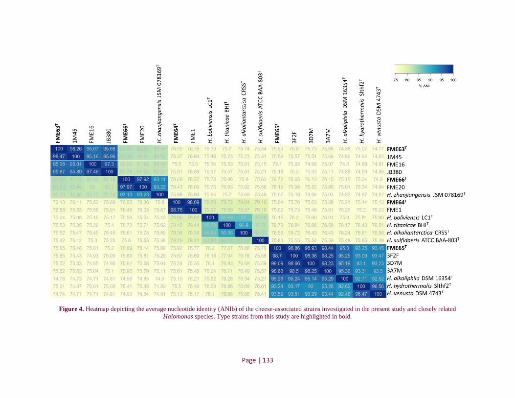
Page | 133
Figure 4. Heatmap depicting the average nucleotide identity (ANIb) of the cheese-associated strains investigated in the present study and closely related
Halomonas species. Type strains from this study are highlighted in bold.

CHAPITRE II – RESULTATS – Publication 3
Page | 134
Physiological, enzymatic and biochemical properties
Physiological characteristics, including cell morphology and motility, were observed using
an optical microscope after culture for 48 h on Tryptic Soy Agar (TSA) plates. Growth was tested
at various temperatures (4, 10, 20, 25, 30 and 37°C), and NaCl concentrations (0, 0.5, 2, 5, 8, 12,
16 and 20%) and pH values (4, 5, 6, 7, 8, 9 and 10) on TSA and TSB after culture for 72 h.
Additionally, enzymatic activities were tested with the API ZYM system (bioMérieux, Bruz,
France) according to the manufacturer’s instructions, except that the strips were incubated for 6 h
at 25°C. The API 20NE system (bioMérieux, Bruz, France) was used to test biochemical properties
and was prepared according to the manufacturer’s instructions.
Distinctive characteristics that differentiate the newly described strains from type strains
of closely related phylogenetic species are shown in Table 2, and supplementary characteristics
are available in Table S6. All Halomonas strains tested are rods or short rods, aerobic, can grow
at pH 6-10 and support up to 16% NaCl. Overall, on the basis of the physiological, enzymatic and
biochemical data, we noted that the species tested shared a high similarity in terms of these
properties (Table S6). Mata et al. (2002) [24] confirmed the difficulty of distinguishing the species
of the genus Halomonas by its phenotypic characteristics. However, we found several properties
that are critical to discriminate the different species, in combination with the genomic analyses
previously described.
The group consisting of the strains FME63T, 1M45, FME16 and JB380 belongs to a new
species because it presents a significant distance from any defined species (closest ANI ≈82% with
H. zhanjiangensis). The lowest value of ANI within this group of 95% (Fig. 4) suggests the
presence of two different genomic clusters. Nevertheless, physiological analyses showed high
similarity between these four strains, crucial for defining them as a single species. Additionally,
this group contains the only cheese strains capable of assimilating trisodium citrate and differs
from H. zhanjiangensis by the non-assimilation of potassium gluconate and by the cream colour
of their colonies (Table 2).

Page | 135
Table 2. Distinctive physiological, enzymatic and biochemical properties that differentiate the novel Halomonas strains from closely related species of the genus
Halomonas.
Characteristics FME63T 1M45 FME16 JB380 FME66T FME20
H.
zhanjiangensis
JSM 078169T
FME64T FME1
H.
boliviensis
LC1T*
FME65T 3F2F 3D7M 3A7M
H.
alkaliphila
DSM
16354T
H.
venusta
DSM
4743T
Colony coulor cream cream cream cream yellow yellow yellow cream/
white
cream/
white cream cream cream cream cream cream cream
Colony
diameter (mm) 2 2.5 3 3 3 2 3 1.5 2 ND 2.5 2.5 3 2 3 3
Oxidase - - - - - - - - - ND + + + + + +
Esterase + + + - + + + + + ND + + + + - -
Valine
arylamidase + - + + - + - - - ND + + + + + +
α-glucosidase - - - - - - - - - ND + + + + + +
Nitrate
reduction - + + + - - - - - + - + + - + +
Mannitol assimilation
- - - - - - - - - ND - - - - - +
N-acetyl-
glucosamine assimilation
- - - - - - - - - ND - - - - + +
Maltose
assimilation - - - - - - - - - ND - - - - + +
Potassium
gluconate
assimilation
- - - - + + + - - ND - - - - + -
Adipic acid
assimilation - - - + - + + - + ND - - - - + -
Trisodium citrate
assimiliation
+ + + + - - + - - ND - - - - + -
* Data from Quillaguamán et al. (2004)
ND: Not determined
CHAPITRE II – RESULTATS – Publication 3
Page | 136
The FME64T and FME1 strains belong to a group that is taxonomically distant from those
in the database (ANI < 80% with the closest outside species), clearly indicating their status as a
new species. Additionally, their colonies present relatively smaller diameters in comparison with
the other evaluated species, and nitrate reduction is a reaction that distinguishes this group from
the closest species (H. boliviensis LC1T).
The group of strains represented by FME65T, 3F2F, 3D7M and 3A7M shares ANI values
in the boundary zone of the species delimitation (95-96%) with H. alkaliphila DSM 16354T,
raising doubts about their assignment to this species. Nevertheless, the combination of ANI (Fig.
4 and Table S5), and phylogenomic analyses showed that the isolates of this study belong to a
distinct group (Fig. 3). Moreover, the cheese strains exhibit a combination of reactions that
discriminate them from H. alkaliphila, including esterase positive test and non-assimilation of N-
acetyl-glucosamine, maltose, potassium gluconate, adipic acid and trisodium citrate (Table 2).
These results confirm the fact that they effectively correspond to a distinct species.
Finally, the FME66T and FME20 cheese strains can be discriminated from the closest
species (H. zhanjiangensis) by ANI analysis (<95%, Fig. 4). These strains are distinguished by the
yellow pigmentation of their colonies, while the other cheese strains are white or cream-colored.
Moreover, the non-assimilation of trisodium citrate differentiates these strains from the closest
related species H. zhanjiangensis, indicating that they can be assigned to a new species.
Therefore, based on the results presented here, these 12 cheese strains are considered as
being representative of four new Halomonas species, for which the names Halomonas
citridevorans sp. nov. (strains FME63T, 1M45, FME16 and JB380), Halomonas minus sp. nov.
(strains FME64T and FME1), Halomonas casei sp. nov. (strains FME65T, 3F2F, 3D7M and
3A7M) and Halomonas flavum sp. nov. (strains FME66T and FME20) are proposed.

CHAPITRE II – RESULTATS – Publication 3
Page | 137
Description of Halomonas citridevorans sp. nov.
Halomonas citridevorans sp. nov (ci.tri.de.vo’rans. L. n. citrus, lemon tree; L. n. acidunz,
acid; L. neut. n. acidum citri, citric acid; L. part. adj. devorans, consuming, devouring; referring
to trisodium citrate assimilation).
Cells are Gram-negative, motile, aerobic, catalase-positive and oxidase-negative. Colonies
are cream-colored, non-translucent and raised with entire margin. Colonies grown on TSA medium
at 25°C for 72 h have a diameter of 2-3 mm. Strain growth occurs from 4 to 30°C, but not at 37°C,
in a pH range of 6.0 to 10.0, and in the presence of 0.5 to 16% (w/v) NaCl, but not at 0 and 20%.
This species is positive for leucine arylamidase and assimilation of trisodium citrate.
The type strain FME63T was isolated from semi-soft cow's milk cheese in France.
Description of Halomonas minus sp. nov.
Halomonas minus sp. nov (mi’nus. L. neut. adj. minus smaller, referring to the colony size).
Cells are Gram-negative, aerobic and oxidase-negative. Colonies are cream-colored/white,
circular, non-translucent and raised with entire margin. Colonies grown on TSA medium at 25°C
for 72 h have a diameter of 1.5-2 mm. Strain growth occurs from 4 to 25°C, but not at 37°C, in a
pH range of 6.0 to 10.0, and in the presence of 0.5 to 16% (w/v) NaCl, but not at 0 and 20%. This
species is positive for esterase, esterase lipase, leucine arylamidase and Naphthol AS-BI-
phosphate.
The type strain FME64T was isolated from semi-soft cow's milk cheese in France.
Description of Halomonas casei sp. nov.
Halomonas casei sp. nov (ca.se’i. L. gen. n. casei of cheese, referring to origin of isolation).
Cells are Gram-negative, aerobic, catalase- and oxidase-positive. Colonies are beige,
circular, non-translucent and raised with entire margin. Colonies grown on TSA medium at 25°C
for 72 h have a diameter of 2-3 mm. Strain growth occurs from 10 to 30°C, in a pH range of 6.0

CHAPITRE II – RESULTATS – Publication 3
Page | 138
to 10.0, and in the presence of 0.5 to 16% (w/v) NaCl, but not at 0 and 20%. This species is positive
for esterase, leucine arylamidase and α-glucosidase.
The type strain FME65T was isolated from semi-soft cow's milk cheese in France.
Description of Halomonas flavum sp. nov.
Halomonas flavum (fla’vum. L. neut. adj. flavum yellow, referring to the colony color).
Cells are Gram-negative, aerobic, catalase-positive and oxidase-negative. Colonies are
yellow pigmented, circular, non-translucent and raised with entire margin. Colonies grown on TSA
medium at 25°C for 72 h have a diameter of 2-3 mm. Strain growth occurs from 4 to 30°C, but not
at 37°C, in a pH range of 6.0 to 10.0, and in the presence of 0.5 to 16% (w/v) NaCl, but not at
20%. This species is positive for esterase, esterase lipase, leucine arylamidase and Naphthol AS-
BI-phosphate, and was able to assimilate potassium gluconate and malate.
The type strain FME66T was isolated from semi-soft cow's milk cheese in France.
AUTHOR STATEMENTS
Authors and contributors
Caroline Isabel Kothe: Conceptualization, Data curation, Formal analysis, Investigation,
Methodology, Validation, Writing-original draft, Writing-review & editing.
Christophe Monnet: Conceptualization, Investigation, Writing-review & editing.
Françoise Irlinger: Conceptualization, Investigation, Writing-review & editing.
Pierre Renault: Conceptualization, Investigation, Funding acquisition, Methodology, Project
administration, Resources, Supervision, Writing-review & editing.
Conflicts of interest
The authors declare that there are no conflicts of interest.
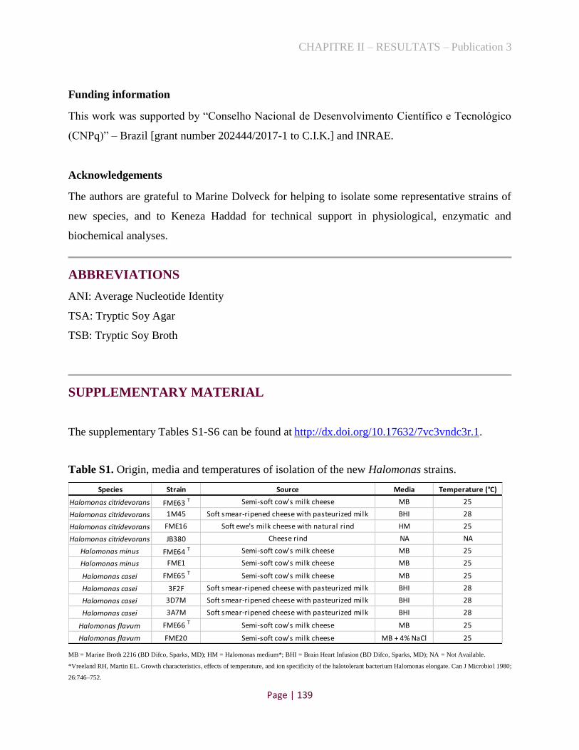
CHAPITRE II – RESULTATS – Publication 3
Page | 139
Funding information
This work was supported by “Conselho Nacional de Desenvolvimento Científico e Tecnológico
(CNPq)” – Brazil [grant number 202444/2017-1 to C.I.K.] and INRAE.
Acknowledgements
The authors are grateful to Marine Dolveck for helping to isolate some representative strains of
new species, and to Keneza Haddad for technical support in physiological, enzymatic and
biochemical analyses.
ABBREVIATIONS
ANI: Average Nucleotide Identity
TSA: Tryptic Soy Agar
TSB: Tryptic Soy Broth
SUPPLEMENTARY MATERIAL
The supplementary Tables S1-S6 can be found at http://dx.doi.org/10.17632/7vc3vndc3r.1.
Table S1. Origin, media and temperatures of isolation of the new Halomonas strains.
MB = Marine Broth 2216 (BD Difco, Sparks, MD); HM = Halomonas medium*; BHI = Brain Heart Infusion (BD Difco, Sparks, MD); NA = Not Available.
*Vreeland RH, Martin EL. Growth characteristics, effects of temperature, and ion specificity of the halotolerant bacterium Halomonas elongate. Can J Microbiol 1980;
26:746–752.
Species Strain Source Media Temperature (°C)
Halomonas citridevorans FME63 T Semi-soft cow's milk cheese MB 25
Halomonas citridevorans 1M45 Soft smear-ripened cheese with pasteurized milk BHI 28
Halomonas citridevorans FME16 Soft ewe's milk cheese with natural rind HM 25
Halomonas citridevorans JB380 Cheese rind NA NA
Halomonas minus FME64 T Semi-soft cow's milk cheese MB 25
Halomonas minus FME1 Semi-soft cow's milk cheese MB 25
Halomonas casei FME65 T Semi-soft cow's milk cheese MB 25
Halomonas casei 3F2F Soft smear-ripened cheese with pasteurized milk BHI 28
Halomonas casei 3D7M Soft smear-ripened cheese with pasteurized milk BHI 28
Halomonas casei 3A7M Soft smear-ripened cheese with pasteurized milk BHI 28
Halomonas flavum FME66 T Semi-soft cow's milk cheese MB 25
Halomonas flavum FME20 Semi-soft cow's milk cheese MB + 4% NaCl 25

CHAPITRE II – RESULTATS – Publication 3
Page | 140
Table S2. Accession numbers of 16S rRNA gene sequences used to build the phylogenetic tree
shown in Fig. 1.
Table S3. Identities of 16S rRNA and rpoB gene sequences (%) between the cheese-associated
strains investigated in the present study and closely related species. Identity values are highlighted
in green when species delineation is above the threshold (>99% for 16S rRNA and >97.7% for
rpoB gene sequences). (A) H. citridevorans; (B) H. minus; (C) H. casei; (D) H. flavum.
(A) H. citridevorans
(B) H. minus
FME63T1M45 JB380 FME16 FME63T
1M45 JB380 FME16
H. citridevorans FME63T 99.91 99.48 99.22 100 97.25 97.38
1M45 99.91 99.26 99.08 100 97.25 99.63
JB380 99.48 99.26 99.48 97.25 97.25 97.38
FME16 99.22 99.08 99.48 97.38 97.38 99.63
H. nigrificans MBT G8648T 98.15 98.16 98.35 98.21 88.26 88.26 88.11 88.21
H. titanicae BH1T 98.08 98.25 97.73 97.53 88.41 88.53 88.02 88.12
H. alkaliantarctica CRSST 97.46 97.70 97.73 98.08 88.53 88.53 88.06 88.13
H. boliviensis LC1T 97.32 97.52 97.59 97.94 87.62 87.62 87.67 87.16
H. olivaria C17T 97.19 97.43 97.32 97.66 88.25 88.25 87.67 87.80
H. sulfidaeris ATCC BAA-803T96.87 97.50 96.87 97.28 87.49 87.49 87.32 87.49
H. glacei DD 39T 98.08 98.53 97.73 98.08
H. utahensis DSM 3051T 97.94 98.44 97.59 97.94
H. neptunia Eplume1T97.32 97.52 97.46 97.80
Species and/or Strains16S rRNA gene rpoB gene
FME64TFME1 FME64T
FME1
H. minus FME64T 100 98.14
FME1 100 98.14
H. alkaliantarctica CRSST 98.76 98.76 90.75 90.73
H. olivaria C17T 98.49 98.49 90.11 90.18
H. boliviensis LC1T98.56 98.56 89.85 89.92
Species and/or Strains16S rRNA gene rpoB gene

CHAPITRE II – RESULTATS – Publication 3
Page | 141
(C) H. casei
(D) H. flavum
FME65T 3F2F 3D7M 3A7M FME65T 3F2F 3D7M 3A7M
H. casei FME65T 100 100 99.71 100 100 100
3F2F 100 100 99.81 100 100 100
3D7M 100 100 99.71 100 100 100
3A7M 99.71 99.81 99.71 100 100 100
H. alkaliphila DSM 16354T 99.86 99.81 99.86 99.64 98.36 98.36 98.36 98.36
H. venusta DSM 4743T 99.52 99.52 99.52 99.5 96.15 96.15 96.15 96.15
H. hydrothermalis Slthf2T 99.57 99.62 99.57 99.28 94.53 94.53 94.53 94.53
H. zhaodongensis NEAU-ST10-25T99.35 99.33 99.35 99.27
rpoB geneSpecies and/or Strains
16S rRNA gene
FME66T FME20 FME66T FME20
H. flavum FME66T 100 99.68
FME20 100 99.68
H. zhanjiangensis JSM 078169T 99.78 99.52 98.68 98.55
H. nanhaiensis YIM M 1305T98.57 98.63
Species and/or Strains16S rRNA gene rpoB gene

CHAPITRE II – RESULTATS – Publication 3
Page | 142
Table S4. Information concerning Halomonas strains whose whole-genome sequences were used
to build the phylogenomic tree in Fig. 3.
Species Strain Type
strain Origin Country
Halomonas aestuarii Hb3 Yes Tidal flat South Korea
Halomonas alimentaria DSM 15356 Yes Fermented food South Korea
Halomonas alkaliantarctica CRSS Yes Saline lake Ross Sea
Halomonas alkaliphila DSM 16354 Yes Salt pool Italy
Halomonas andesensis DSM 19434 Yes Saline lake water Bolivia
Halomonas anticariensis DSM 16096 Yes Soil from a saline wetland Spain
Halomonas aquamarina 558 Yes Seawater Unknown
Halomonas arcis CGMCC 1.6494 Yes Salt lake China
Halomonas axialensis Althf1 Yes Sulfide rock Pacific Ocean
Halomonas beimenensis NTU-111 No Unkown Taiwan
Halomonas boliviensis LC1 Yes Soil Bolivia
Halomonas borealis ATF 5.2 Yes Sea water Norway
Halomonas caseinilytica JCM 14802 Yes Soil China
Halomonas chromatireducens AGD 8-3 No Soil Russia
Halomonas coralii 362.1 Yes Coral Brazil
Halomonas cupida DSM 4740 Yes Seawater USA
Halomonas daqiaogenesis CGMCC 1.9150 Yes Littoral saltern China
Halomonas daqingensis CGMCC 1.6443 Yes Oilfield soil China
Halomonas denitrificans DSM 18045 Yes Saline water South Korea
Halomonas desiderata FB2 Yes Sewage treatment plant Germany
Halomonas elongata DSM 2581 Yes Solar salt facility Netherlands Antilles
Halomonas endophytica MC28 Yes Storage liquid in the stem China
Halomonas eurihalina MS1 No Saline soils Spain
Halomonas gudaonensis CGMCC 1.6133 Yes Saline soil contaminated China
Halomonas halmophila NBRC 15537 No Unkown Unkown
Halomonas halocynthiae DSM 14573 Yes Sea peach Japan
Halomonas halodenitrificans DSM 735 Yes Meat Canada
Halomonas halophila NBRC 102604 No Hypersaline soil Spain
Halomonas heilongjiangensis DSM 26881 Yes Soil China
Halomonas huangheensis BJGMM-B45 Yes Soil China
Halomonas hydrothermalis Slthf2 Yes Deep-sea hydrothermal vent Pacific Ocean
Halomonas ilicicola DSM 19980 Yes Saline water from a solar saltern Spain
Halomonas jeotgali Hwa Yes Fermented seafood South Korea
Halomonas korlensis CGMCC 1.6981 Yes Saline and alkaline soil China
Halomonas lionensis RHS90 Yes Sea sediments Mediterranean Sea
Halomonas litopenaei SYSU ZJ2214 Yes Shrimp water China
Halomonas lutea DSM 23508 Yes Salt lake China
Halomonas massiliensis Marseille-P2426 Yes Human gut France
Halomonas meridiana ACAM 246 Yes Organic lake Antarctica
Halomonas muralis DSM 14789 Yes Mural painting Austria
Halomonas nigrificans MBT G8648 Yes Cheese Germany
Halomonas niordiana ATF 5.4 Yes Seawater Norway

CHAPITRE II – RESULTATS – Publication 3
Page | 143
Halomonas nitroreducens 11S Yes Solar saltern Chile
Halomonas olivaria C17 Yes Salted olive-processing effluents Morocco
Halomonas pacifica NBRC 102220 No Seawater USA
Halomonas pantelletiensis AAP Yes Hard sand of the lake of Venere Italy
Halomonas saccharevitans CGMCC 1.6493 Yes Water sample China
Halomonas salina B6 No Human gut France
Halomonas saliphila LCB169 Yes Saline soil China
Halomonas shengliensis GCMCC 1.6444 Yes Contaminated saline soil China
Halomonas smyrnensis AAD6 Yes Soil from salt production ponds Turkey
Halomonas socia CKY01 No Marine soil South Korea
Halomonas songnenensis CGMCC 1.12152 Yes Saline and alkaline soils China
Halomonas stevensii S18214 Yes Blood from a renal care patient USA
Halomonas subglaciescola ACAM 12 Yes Organic lake Antarctica
Halomonas subterranea CGMCC 1.6495 Yes Hypersaline water China
Halomonas sulfidaeris ATCC BAA-803 Yes Metal sulfide rock Northeast Pacific Ocean
Halomonas taeanensis BH539 Yes Solar saltern Korea
Halomonas titanicae BH1 Yes Rusticles of the RMS Titanic wreck Canada
Halomonas urmiana TBZ3 Yes Lake Iran
Halomonas urumqiensis BZ-SZ-XJ27 Yes Sediment from saline-alkaline lake China
Halomonas utahensis P-halo No Human gut Unknown
Halomonas ventosae CECT 5797 Yes Soil with high salt content Spain
Halomonas venusta DSM 4743 Yes Seawater Unknown
Halomonas xianhensis CGMCC 1.6848 Yes Soil Unknown
Halomonas xinjiangensis TRM 0175 Yes Salt lake China
Halomonas zhanjiangensis JSM 078169 Yes Sea urchin China
Halomonas zincidurans B6 Yes Marine sediment South Atlantic
Novel cheese strains from this study Halomonas citridevorans FME63 Yes Semi-soft cow's milk cheese France
Halomonas citridevorans 1M45 No
Soft smear-ripened cheese with pasteurized
milk France
Halomonas citridevorans FME16 No Soft ewe's milk cheese with natural rind France
Halomonas citridevorans JB380 No Cheese rind USA
Halomonas minus FME64 Yes Semi-soft cow's milk cheese France
Halomonas minus FME1 No Semi-soft cow's milk cheese France
Halomonas casei FME65 Yes Semi-soft cow's milk cheese France
Halomonas casei 3F2F No Soft smear-ripened cheese with pasteurized
milk France
Halomonas casei 3D7M No
Soft smear-ripened cheese with pasteurized
milk France
Halomonas casei 3A7M No
Soft smear-ripened cheese with pasteurized
milk France
Halomonas flavum FME66 Yes Semi-soft cow's milk cheese France
Halomonas flavum FME20 No Semi-soft cow's milk cheese France
Outgroup Carnimonas nigrificans ATCC BAA-78 Yes Cured meat product Unknown

CHAPITRE II – RESULTATS – Publication 3
Page | 144
Table S5. ANIb values (%) between the strains of the FME65T- group and the strains of the
Halomonas alkaliphila-venusta group. The ANI values are highlighted in green when above 96%
and in yellow when ranging between 95 and 96%.
H.
venusta
NBRC
102221
H.
venusta
MA-
ZP17-13
H.
venusta
DSM
4743T
H.
alkaliphila
DSM
16354T
H.
alkaliphila
X3
FME65T 3F2F 3D7M 3A7M
H. venusta NBRC
102221 * 98.68 99.99 92.58 92.57 93.46 93.5 93.25 93.4
H. venusta MA-
ZP17-13 98.58 * 98.58 92.63 92.58 93.43 93.45 93.17 93.32
H. venusta DSM
4743T 99.9 98.54 * 92.48 92.55 93.52 93.51 93.29 93.44
H. alkaliphila
DSM 16354T 92.53 92.68 92.52 * 98.02 95.29 95.24 95.14 95.29
H. alkaliphila X3 92.59 92.62 92.62 98.01 * 95.53 95.48 95.39 95.5
FME65T 93.45 93.37 93.45 95.3 95.4 * 98.86 98.93 98.44
3F2F 93.46 93.39 93.47 95.25 95.33 98.7 * 98.38 98.25
3D7M 93.24 93.18 93.23 95.19 95.28 99.09 98.66 * 98.23
3A7M 93.5 93.45 93.5 95.36 95.46 98.63 98.5 98.25 *
95>ANI>96%
ANI>96%
Table S6. Physiological, enzymatic and biochemical characteristics of the cheese-associated
strains investigated in the present study and of closely related species.
REFERENCES
[1]. Vreeland RH, et al. Halomonas elongata, a new genus and species of extremely salt-tolerant bacteria.
Int J Syst Evol Microbiol 1980; 30(2):485-495. doi:10.1099/00207713-30-2-485.
[2]. Dobson SJ, Franzmann PD. Unification of the genera Deleya (Baumann et al 1983), Halomonas
(Vreeland et al 1980), and Halovibrio (Fendrich 1988) and the species Paracoccus halodenitrificans
(Robinson and Gibbons 1952) into a single genus, Halomonas, and placement of the genus Zymobacter in
the family Halomonadaceae. Int J Syst Bacteriol 1996; 46(2):550-558. doi:10.1099/00207713-46-2-550.
[3]. Ventosa A, Nieto JJ, Oren A. Biology of moderately halophilic aerobic bacteria. Microbiol Mol Biol
Rev 1998; 62(2):504-44. PMID: 9618450. doi:10.1128/MMBR.62.2.504-544.1998.
[4]. Arahal DR, Ventosa A. The Family Halomonadaceae. Prokaryotes: A Handbook on the Biology of
Bacteria, Vol 6, Third Edition, 2006:811-835.
[5]. Quillaguaman J, et al. Halomonas boliviensis sp. nov., an alkalitolerant, moderate halophile isolated
from soil around a Bolivian hypersaline lake. Int J Syst Evol Microbiol 2004; 54:721-725.
doi:10.1099/ijs.0.02800-0.
[6]. James SR, et al. Halomonas meridiana, a new species of extremely halotolerant bacteria isolated
from Antarctic saline lakes. Syst Appl Microbiol 1990; 13(3):270-278. doi:10.1016/S0723-
2020(11)80198-0.

CHAPITRE II – RESULTATS – Publication 3
Page | 145
[7]. Yoon JH, et al. Halomonas alimentaria sp. nov., isolated from jeotgal, a traditional Korean fermented
seafood. Int J Syst Evol Microbiol 2002; 52:123-30. doi:10.1099/00207713-52-1-123.
[8]. Jeong SH, et al. Halomonas cibimaris sp. nov., isolated from jeotgal, a traditional Korean fermented
seafood. Anton Leeuw 2013; 103(3):503-12. doi:10.1007/s10482-012-9832-x.
[9]. Kim MS, Roh SW, Bae JW. Halomonas jeotgali sp. nov., a new moderate halophilic bacterium
isolated from a traditional fermented seafood. J Microbiol 2010; 48(3): 404-10. doi:10.1007/s12275-010-
0032-y.
[10]. Oguntoyinbo FA, et al. Halomonas nigrificans sp. nov., isolated from cheese. Int J Syst Evol
Microbiol 2018; 68(1):371-376. doi:10.1099/ijsem.0.002515.
[11]. Almeida M, et al. Construction of a dairy microbial genome catalog opens new perspectives for the
metagenomic analysis of dairy fermented products. BMC Genomics 2014; 15(1): 1101.
doi:10.1186/1471-2164-15-1101.
[12]. Acinas SG, et al. Fine-scale phylogenetic architecture of a complex bacterial community. Nature
2004; 430(6999):551-554. doi:10.1038/nature02649.
[13]. Tayeb LA, et al. Comparative phylogenies of Burkholderia, Ralstonia, Comamonas, Brevundimonas
and related organisms derived from rpoB, gyrB and rrs gene sequences. Res Microbiol 2008; 159(3):169-
177. doi:10.1016/j.resmic.2007.12.005.
[14]. Kumar S, Stecher G, Tamura K. MEGA7: molecular evolutionary genetics analysis version 7.0 for
bigger datasets. Mol Biol Evol 2016; 33(7):1870-1874. doi:10.1093/molbev/msw054.
[15]. Stackebrandt E. Taxonomic parameters revisited: tarnished gold standards. Microbiol Today 2006;
33:152-155.
[16]. Adekambi T, Drancourt M, Raoult D. The rpoB gene as a tool for clinical microbiologists. Trends
Microbiol 2009; 17(1):37-45. doi:10.1016/j.tim.2008.09.008.
[17]. Bankevich A, et al. SPAdes: a new genome assembly algorithm and its applications to single-cell
sequencing. J Comput Biol 2012; 19(5):455-77. doi:10.1089/cmb.2012.0021.
[18]. Aziz RK, et al. The RAST server: Rapid annotations using subsystems technology. BMC Genomics
2008; 9:75. doi:10.1186/1471-2164-9-75.
[19]. Richter M, et al. JSpeciesWS: a web server for prokaryotic species circumscription based on
pairwise genome comparison. Bioinformatics 2016; 32(6):929-931. doi:10.1093/bioinformatics/btv681.
[20]. Richter M, Rossello-Mora R. Shifting the genomic gold standard for the prokaryotic species
definition. Proc Natl Acad Sci U S A 2009; 106(45):19126-19131. doi:10.1073/pnas.0906412106.
[21]. Portier P, et al. Elevation of Pectobacterium carotovorum subsp. odoriferum to species level as
Pectobacterium odoriferum sp. nov., proposal of Pectobacterium brasiliense sp. nov. and Pectobacterium
actinidiae sp. nov., emended description of Pectobacterium carotovorum and description of
Pectobacterium versatile sp. nov., isolated from streams and symptoms on diverse plants. Int J Syst Evol
Microbiol 2019; 69(10):3207-3216. doi:10.1099/ijsem.0.003611.
[22]. Kaye JZ, et al. Halomonas neptunia sp. nov., Halomonas sulfidaeris sp. nov., Halomonas axialensis
sp. nov. and Halomonas hydrothermalis sp. nov.: halophilic bacteria isolated from deep-sea
hydrothermal-vent environments. Int J Syst Evol Microbiol 2004; 54:499-511. doi:10.1099/ijs.0.02799-0.
[23]. Baumann L, Bowditch RD, Baumann P. Description of Deleya gen. nov. created to accommodate
the marine species Alcaligenes aestus, Alcaligenes pacificus, Alcaligenes cupidus, Alcaligenes venustus,
and Pseudomonas marina. Int J Syst Bacteriol 1983; 33(4):793-802. doi:10.1099/00207713-33-4-793.
[24]. Mata JA, et al. A detailed phenotypic characterisation of the type strains of Halomonas species. Syst
Appl Microbiol 2002; 25(3):360-375. doi:10.1078/0723-2020-00122.
CHAPITRE II – RESULTATS – Publication 4
Page | 146
3.3. Publication 4
Psychrobacter casei sp. nov. and Psychrobacter translucens sp. nov., two new species isolated
from French cheese rinds
Caroline Isabel Kothea (ORCID: 0000-0003-3703-5699), Christophe Monnetb (ORCID: 0000-
0003-2647-272X) Françoise Irlingerb, Pierre Renault a (ORCID: 0000-0001-6540-053X)
a Université Paris-Saclay, INRAE, AgroParisTech, Micalis Institute, 78350, Jouy-en-Josas, France.
b Université Paris-Saclay, INRAE, AgroParisTech, UMR SayFood, F-78850, Thiverval-Grignon, France.
Keyword: Taxonomy, sequence analysis, whole-genome sequencing, food.
Repositories:
The whole-genome and 16S sequences of four novel strains of Psychrobacter have been
deposited at DDBJ/ENA/GenBank and are available under the accession numbers listed in Table
1 and Figure 1, respectively.
ABSTRACT
Four Gram-negative bacterial strains were isolated from cheese rinds in France. On the basis of
16S rRNA gene sequence analysis, all isolates were assigned to the genus Psychrobacter, and
their closest relatives were Psychrobacter glacei, P. fozii and P. aquimaris with similarities
between 98.63 to 99.25%. Their taxonomic status were investigated using a polyphasic
taxonomic approach comprising 16S rRNA and rpoB phylogenies, determination of average
nucleotide identity (gANI) and analyses of physiological and biochemical features.
Phylogenetics and gANI analyses revealed the existence of two novel and well-supported
clusters. The novel isolates differed from the previously described species by ANI <83% and
some biochemical and enzymatic properties. In conclusion, the results of phenotypic and
phylogenetic analyses indicated that the isolates belonged two novel Psychrobacter species, for
which the names Psychrobacter casei sp. nov. and Psychrobacter translucens sp. nov. are

CHAPITRE II – RESULTATS – Publication 4
Page | 147
proposed, with isolates FME6T (=CIRM-BIA2434T = CIP 111878T= CIRM-BIA2435T) and
FME13T (=CIRM-BIA2435T = CIP 111879T = CIRM-BIA2435T) as type strains, respectively.
Psychrobater's taxonomic status appears to have been under constant review over the past
few years. The first proposed species of this genus was Psychrobacter immobilis (in 1986), that
was added as part of the Neisseriaceae family [1]. However, in 1991, Rossau et al. demonstrated
that Psychrobacter and related organisms (as Moraxella and Acinetobacter) form a separate cluster
in this taxonomy, and the Moraxellaceae family was proposed [2, 3]. Therefore, Psychrobacter
belongs nowadays to the family of Moraxellaceae within the class of Gammaproteobacteria.
Species from this genus are aerobic, Gram-negative, oxidase and catalase-positive, halotolerant,
psychrophilic or psychrotolerant [2]. They have been mainly isolated from marine and terrestrial
environments, including food, soil, seawater, sea ice and air [3, 4].
Here we describe four strains belonging to the Psychrobacter genus, which were isolated
from French cheese rinds. Since these strains differed by several unusual genetic and phenotypic
features from the closely related Psychrobacter type strains, we established they belong to two
new species of Psychrobacter, named P. casei sp. nov. and P. translucens sp. nov.
Isolation and Ecology
In the course of a project exploring the diversity of bacteria associated with French artisanal
cheese rinds, we isolated four strains, which belong to the genus Psychrobacter. These strains
(FME5, FME6, FME61 and FME13) were isolated from different types of cheeses (the isolation
sources are represented in Table 1) on Marine Agar (MA) at 25°C. Samples obtained from pure
colonies were stored at -80°C in Marine Broth (MB) medium supplemented with 20% (v/v)
glycerol and deposited in culture collection centers.

Page | 148
Table 1. Genomic features and isolation sources of the novel proposed Psychrobacter strains and four Psychrobacter type strains (P. cibarius DSM 16327T, P.
aquimaris DSM 16329T, P. faecalis DSM 14664T and P. namhaensis DSM 16330T).
Species Psychrobacter
casei
Psychrobacter
casei
Psychrobacter
translucens
Psychrobacter
translucens
Psychrobacter
aquimaris
Psychrobacter
cibarius
Psychrobacter
faecalis
Psychrobacter
namhaensis
Strain FME5 FME6T FME61 FME13T DSM 16329T DSM 16327T DSM 14664T DSM 16330T
Isolation
source
Semi-soft blue
cheese rind with
unpasteurized milk from
Auvergne-Rhone-
Alpes region
Soft-ripened goat's milk cheese
from
Mediterranean region
Soft smear-ripened
cheese rind from
Bourgogne region
Soft ewe's milk
cheese rind from Mediterranean
region
Sea water of the South Sea in Korea
Jeotgal, a traditional
Korean fermented
seafood
Bioaerosol of the Germany
Sea water of the South Sea in Korea
Number of
contigs 295 268 452 419 41 78 106 45
Size (base
pairs) 3,373,895 3,361,643 3,569,320 3,708,697 3,427,332 3,242,921 3,081,191 2,900,373
GC content
(%) 42.8 42.8 41.7 41.8 43.0 42.8 43.6 45
N50a 24,719 30,966 18,000 20,335 199,424 97,459 67,147 180,725
L50b 38 35 55 50 6 7 15 5
Number of
subsystems
(RAST)
284 277 284 286 279 272 272 275
Number of
coding
sequences
3,117 3,067 3,396 3,557 3,109 2,879 2,737 2,581
Bioproject PRJNA501839 PRJNA501839 PRJNA501839 PRJNA501839 PRJNA625506 PRJNA625506 PRJNA625506 PRJNA625506
BioSample SAMN10346440 SAMN10346441 SAMN15150957 SAMN10346442 SAMN14601840 SAMN14601841 SAMN14601838 SAMN14601839
Accesion RRYX00000000 RRYW00000000 JABUYV000000000 RRYV00000000 JABASR000000000 JABASQ000000000 JABAST000000000 JABASS000000000

CHAPITRE II – RESULTATS – Publication 4
Page | 149
16S rRNA and rpoB phylogenies
Genomic DNA was extracted from the bacterial cells according to the protocol described
by Almeida et al. (2014) [5]. Their 16S rRNA genes were amplified using 27-F (5′-
AGAGTTTGATCATGGCTCA-3′) and 1492-R (5′-TACGGTTACCTTGTTACGACTT-3′)
primers [6] and sequences were compared with those of type strains of species with validly
published names using EzBioCloud (https://www.ezbiocloud.net/). In order to expand the
taxonomy of strains, rpoB (DNA-directed RNA polymerase beta subunit), as alternative marker
gene, was amplified using primers VIC4 (5′-GGCGAAATGGCDGARAACCA-3′) and VIC6 (5′-
GARTCYTCGAAGTGGTAACC-3′) [7]. The rpoB sequences were compared to known bacterial
species in the National Center for Biotechnology Information (NCBI) GenBank database. Further
phylogenetic analyses were performed for 16S rRNA and rpoB sequences using MEGA 7.0.26
software and trees were generated using the Neighbor-Joining algorithm with 1,000 bootstrap
iterations [8] (Fig. 1 and Fig. 2). To delineate species, we used as threshold over than 99% identity
for 16S rRNA gene with type strains [9] and above 97.7% identity for rpoB nucleotidic sequence
[10].
The strains FME5 and FME6T are closely related to each other, sharing identities of 99.94%
for 16S rRNA (Fig. 1) and 100% for rpoB gene sequences (Fig. 2). These strains could be
representative of a new genomospecies since their level of identity with the closest related type
strains is at the limit of the threshold for 16S rRNA (99.0-99.2%) and below the thresholds for
rpoB gene (89.0-90.6%) sequences (Table S1A).
Similarly, analyses of 16S rRNA and rpoB genes indicated that FME13T and FME61
strains belong to the same species since their sequences share identities of 99.42 and 100%,
respectively. However, they represent probably a new species since their rpoB sequences display
sub-thresholds levels of identity with all defined species (<92%), despite their 16S rRNA genes is
at the limit of species delineation (98.63-99.25%) with P. aquimaris SW-210T (Table S1B).

CHAPITRE II – RESULTATS – Publication 4
Page | 150
Figure 1. Phylogenetic tree based on 16S rRNA gene sequences of the four strains from this study and closely
related Psychrobacter type strains. The tree was performed by MEGA 7.0.26 software using the maximum
likelihood method. Acinetobacter calcoaceticus DSM 30006T and Moraxella lacunata CCUG 4441T were used as
outgroups.
Although the inferred phylogenetic trees confirmed the two well-supported clusters (Fig.
1 and Fig. 2), we performed further investigations to confirm the assignation of these strains.

CHAPITRE II – RESULTATS – Publication 4
Page | 151
Figure 2. Phylogenetic tree based on rpoB gene sequences of the four strains from this study and closely related
Psychrobacter type strains. The tree was performed by MEGA 7.0.26 software using the maximum likelihood
method. Acinetobacter calcoaceticus DSM 30006T and Moraxella lacunata CCUG 4441T were used as outgroups.
Genome features and phylogenomic analysis
To establish if cheese isolates are belonging to new species of Psychrobacter, the genome
of four strains was sequenced and annotated as described below with the aim of performing an
average nucleotide identity (gANI) analyses. Moreover, the genomes of four Psychrobacter type
strains (P. cibarius DSM 16327T, P. aquimaris DSM 16329T, P. faecalis DSM 14664T and P.
namhaensis DSM 16330T), closely related to the cheese strains and which genomes were not
available so far, were also determined and annotated in order to complete a whole-genome-based
reconstruction of their phylogeny. DNA sequencing was carried out on the Illumina HiSeq
platform at GATC-Biotech (Konstanz, Germany) in order to generate at least 5 million paired-end
reads per genome (150 bases in length). For each strain, de novo assembly was performed using

CHAPITRE II – RESULTATS – Publication 4
Page | 152
SPAdes version 3.9 [11]. Only contigs with length >300 bp and coverage >100 were considered
for further study. Annotations were performed using the RAST (Rapid Annotation using
Subsystem Technology) server [12]. Basic features of these new whole-genome sequences are
presented in Table 1. Finally, these genomes and those of all other Psychrobacter species available
in the NCBI database were collected and related information such as their origin of isolation,
numbers of Bioproject and BioSample are presented in Table S2.
In order to propose a phylogenomic analysis, the available Psychrobacter genome
sequences were aligned, with those of Acinetobacter calcoaceticus DSM 30006T and Moraxella
lacunata CCUG 4441T as outgroups, using the software Progressive Mauve (version 2.4.0) with
the default settings. The phylogenomic tree obtained was visualized with Interactive Tree of Life
(iTOL v5) (http://itol.embl.de). In addition, gANI values of the Psychrobacter strains isolated here
and that of closely related species were calculated by using JSpeciesWS [13] in order to determine
the speciation of the different isolates. For species delineation, we used the recommended cut-off
point of 95-96% ANI, which substitutes the DDH analysis [14].
Figure 3 presents the phylogenomic tree obtained with Psychrobacter whole-genome
sequences with the indication of their origins classified as marine environments, terrestrial
environments (sediment, permafrost and soil), human, animal and food (Table S2). Analyses of
gANI values of cheese strains and phylogenetically related species showed that the cheese strains
are clustered in two different genomic groups of gANI>98% (Table 2). The two groups of cheese
strains represented by FME13T and FME6T share ≈82.6 and 80.3% ANI with the relative closest
type strains of P. fozii CECT 5889T and P. faecalis DSM 14664T (Fig. 3), respectively, a value
clearly below the threshold of 95%, indicating their belonging to new species .
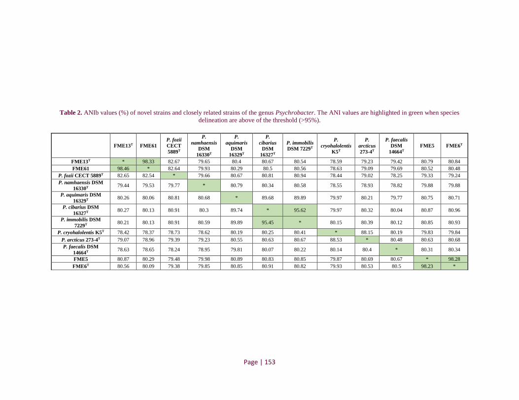
Page | 153
Table 2. ANIb values (%) of novel strains and closely related strains of the genus Psychrobacter. The ANI values are highlighted in green when species
delineation are above of the threshold (>95%).
FME13T FME61
P. fozii
CECT
5889T
P.
namhaensis
DSM
16330T
P.
aquimaris
DSM
16329T
P.
cibarius
DSM
16327T
P. immobilis
DSM 7229T
P.
cryohalolentis
K5T
P.
arcticus
273-4T
P. faecalis
DSM
14664T
FME5 FME6T
FME13T * 98.33 82.67 79.65 80.4 80.67 80.54 78.59 79.23 79.42 80.79 80.84
FME61 98.46 * 82.64 79.93 80.29 80.5 80.56 78.63 79.09 79.69 80.52 80.48
P. fozii CECT 5889T 82.65 82.54 * 79.66 80.67 80.81 80.94 78.44 79.02 78.25 79.33 79.24
P. namhaensis DSM
16330T 79.44 79.53 79.77 * 80.79 80.34 80.58 78.55 78.93 78.82 79.88 79.88
P. aquimaris DSM
16329T 80.26 80.06 80.81 80.68 * 89.68 89.89 79.97 80.21 79.77 80.75 80.71
P. cibarius DSM
16327T 80.27 80.13 80.91 80.3 89.74 * 95.62 79.97 80.32 80.04 80.87 80.96
P. immobilis DSM
7229T 80.21 80.13 80.91 80.59 89.89 95.45 * 80.15 80.39 80.12 80.85 80.93
P. cryohalolentis K5T 78.42 78.37 78.73 78.62 80.19 80.25 80.41 * 88.15 80.19 79.83 79.84
P. arcticus 273-4T 79.07 78.96 79.39 79.23 80.55 80.63 80.67 88.53 * 80.48 80.63 80.68
P. faecalis DSM
14664T 78.63 78.65 78.24 78.95 79.81 80.07 80.22 80.14 80.4 * 80.31 80.34
FME5 80.87 80.29 79.48 79.98 80.89 80.83 80.85 79.87 80.69 80.67 * 98.28
FME6T 80.56 80.09 79.38 79.85 80.85 80.91 80.82 79.93 80.53 80.5 98.23 *

CHAPITRE II – RESULTATS – Publication 4
Page | 154
Physiological, enzymatic and biochemical properties
Physiological characteristics including cell morphology and motility were observed using
an optical microscope after culture for 48 h on Tryptic Soy Agar (TSA) plates. Growth was tested
at various temperatures (4, 10, 20, 25, 30 and 37°C), NaCl concentration (0, 0.5, 2, 5, 8, 12, 16
and 20%) and pH (4, 5, 6, 7, 8, 9 and 10) on TSA after culture for 72 h. Additionally, enzymatic
activities were tested with the API ZYM system (bioMérieux, Bruz, France) according to the
manufacturer’s instructions, except that the strips were incubated for 6h at 25°C. The API 20NE
system (bioMérieux, Bruz, France) was used to test biochemical properties and prepared according
to the manufacturer’s instructions.
The novel and closely related Psychrobacter strains are rods or short rods, aerobic, can
grow at pH 6-10 and between 10 and 25°C, and support up to 12% NaCl. All these strains are
positive (or weakly positive) for esterase (C4), esterase lipase (C8), leucine arylamidase, naphthol-
AS-BI-phosphohydrolase and cytochrome oxidase. All strains are negative for the following
enzymatic and biochemical properties: trypsin, α-chymotrypsin, α-galactosidase, β-glucuronidase,
α-glucosidase, β-glucosidase, N-acetyl-β-glucosaminidase, α-mannosidas, N2 production from
nitrates, tryptophanase (indole production), fermentation, arginine dihydrolase, protease (gelatin
hydrolysis) and β-galactosidase. The strains were also negative for assimilation of d-glucose, d-
mannose, d-mannitol, N-acetylglucosamine, d-maltose, potassium gluconate, capric acid, adipic
acid and phenylacetic acid. Distinctive characteristics between the newly described strains and
type strains representative of closely related phylogenetic species are shown in Fig. 3.

Page | 155
Fig. 3. Phylogenomic tree and biochemical and enzymatic properties. Left: Phylogenomic tree based on whole-genome sequence alignment of all the
Psychrobacter strains for which this data are available and of the four isolates from this study (highlighted in the tree). Right: Distinctive enzymatic and
biochemical properties that differentiate the novel Psychrobacter strains from closely related species. Black squares correspond to positive, white squares to
negative and grey squares to weakly positive reactions. *Data from Psychrobacter cryohalolentis, P. arcticus and P. fozii were collected from literature
(Bakermans et al., 2006 and Bozal et al., 2003).

CHAPITRE II – RESULTATS – Publication 4
Page | 156
The group consisting of the strains FME6T and FME5 belongs to a new species because
they present significant distance from any defined species (ANI<81% with the closest outside
species). Moreover, these strains are distinguished by the absence of alkaline phosphatase and
lipase (C14) and the non-assimilation of arabinose, while in the closest related species (Fig. 3, P.
faecalis) these reactions are all positive. These results highlight that FME6T and FME5 strains
correspond to a new species.
The FME13T and FME61 strains were also shown to belong to a taxonomically distant
group from those described so far (ANI≈82.6 with P. fozii), which clearly indicates they belong to
a new species. Additionally, the phenotypic properties that distinguish these two strains from P.
fozii are the absence of alkaline phosphatase, cysteine arylamidase, acid phosphatase and urease
and the non-assimilation of malic acid. Furthermore, the colonies of the FME13T and FME61
strains are translucent, while those of the closely related species have a cream or beige coloration.
Therefore, based on the results presented here, the four cheese strains are considered as
representatives of two new Psychrobacter species, for which the names P. casei sp. nov. (FME5
and FME6T strains) and P. translucens sp. nov. (FME13T and FME61 strains), are proposed.
Description of Psychrobacter casei sp. nov.
Psychrobacter casei sp. nov (ca.se’i. L. gen. n. casei of cheese, referring to origin of
isolation).
Cells are Gram-negative, cocci or short rods, non-motile, aerobic, catalase and oxidase
positives. Colonies are cream, non-translucent and raised with entire margin. Colonies grown on
TSA medium at 25°C for 72h have a size of 2 mm in diameter. The strains growth occurs between
10 to 30°C, but not at 4 and 37°C, in a pH range of 6.0 to 10.0 and in the presence of 0.5 to 12%
(w/v) NaCl, but not at 0 and 15%. Strains are weakly positive for reduction of nitrate to nitrite and
for esterase (C4). The strains of this group are positive in the following biochemical testes: esterase
lipase (C8), leucine arylamidase and naphthol-AS-BI-phosphohydrolase. The G+C content of
DNA is 42.9 mol% and the genome size is near to 3.4 Mb.
The type strain FME6T was isolated from soft-ripened goat's milk cheese rind from
Mediterranean region in France.

CHAPITRE II – RESULTATS – Publication 4
Page | 157
Description of Psychrobacter translucens sp. nov.
Psychrobacter translucens (trans.lu’cens L. part. adj. translucens, being translucent).
Cells are Gram-negative, cocci or short rods, non-motile, aerobic, catalase and oxidase
positive. Colonies are translucent and raised with entire margin. Colonies grown on TSA medium
at 25°C for 72h have a size of 1 to 1.5 mm in diameter. The strains growth occurs between 10 to
30°C, but not at 4 and 37°C, in a pH range of 6.0 to 10.0 and in the presence of 0.5 to 12% (w/v)
NaCl, but not at 0 and 15%. Strains are negative for reduction of nitrate to nitrite and weakly
positive for naphthol-AS-BI-phosphohydrolase. The strains of this group are positive in the
following biochemical testes: esterase (C4), esterase lipase (C8) and leucine arylamidase. The
G+C content of DNA is 41.8 mol% and genome size is about 3.6-3.7 Mb.
The type strain FME13T was isolated from soft ewe's milk cheese rind from Mediterranean
region in France.
AUTHOR STATEMENTS
Authors and contributors
Caroline Isabel Kothe: Conceptualization, Data curation, Formal analysis, Investigation,
Methodology, Validation, Writing-original draft, Writing-review & editing.
Christophe Monnet: Conceptualization, Investigation, Writing-review & editing.
Françoise Irlinger: Conceptualization, Investigation, Writing-review & editing.
Pierre Renault: Conceptualization, Investigation, Funding acquisition, Methodology, Project
administration, Resources, Supervision, Writing-review & editing.
Conflicts of interest
The authors declare that there are no conflicts of interest.
Funding information
This work was supported by “Conselho Nacional de Desenvolvimento Científico e Tecnológico
(CNPq)” – Brazil [grant number 202444/2017-1 to C.I.K.] and INRAE.
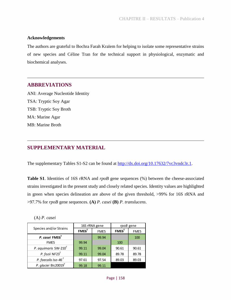
CHAPITRE II – RESULTATS – Publication 4
Page | 158
Acknowledgements
The authors are grateful to Bochra Farah Kraïem for helping to isolate some representative strains
of new species and Céline Tran for the technical support in physiological, enzymatic and
biochemical analyses.
ABBREVIATIONS
ANI: Average Nucleotide Identity
TSA: Tryptic Soy Agar
TSB: Tryptic Soy Broth
MA: Marine Agar
MB: Marine Broth
SUPPLEMENTARY MATERIAL
The supplementary Tables S1-S2 can be found at http://dx.doi.org/10.17632/7vc3vndc3r.1.
Table S1. Identities of 16S rRNA and rpoB gene sequences (%) between the cheese-associated
strains investigated in the present study and closely related species. Identity values are highlighted
in green when species delineation are above of the given threshold, >99% for 16S rRNA and
>97.7% for rpoB gene sequences. (A) P. casei (B) P. translucens.
(A) P. casei
FME6TFME5 FME6T
FME5
P. casei FME6T 99.94 100
FME5 99.94 100
P. aquimaris SW-210T 99.11 99.04 90.61 90.61
P. fozii NF23T 99.11 99.04 89.78 89.78
P. faecalis Iso-46 T 97.61 97.54 89.03 89.03
P. glaciei BIc20019T99.18 99.11
16S rRNA geneSpecies and/or Strains
rpoB gene

CHAPITRE II – RESULTATS – Publication 4
Page | 159
(B) P. translucens
Table S2. Metadata related to Psychrobacter strains whose whole-genome sequences were used
to build the phylogenomic tree in Fig. 3.
Species Strain Type strain
Isolate from Country/ Region
Psychrobacter alimentarius PAMC 27889 No Rocky desert Antarctica
Psychrobacter aquaticus CMS 56 Yes Cyanobacterial mats Antarctica
Psychrobacter aquimaris DSM 16329 Yes Sea water Republic of Korea
Psychrobacter arcticus 273-4 Yes Permafrost soil Russia
Psychrobacter cibarius DSM 16327 Yes Jeotgal, seafood Republic of Korea
Psychrobacter cryohalolentis K5 Yes Cryopeg within permafrost Russia
Psychrobacter faecalis DSM 14664 Yes Bioaerosol Germany
Psychrobacter fozii CECT 5889 Yes Sediment Antarctica
Psychrobacter frigidicola ACAM 304 Yes Ornithogenic soil Antarctica
Psychrobacter immobilis DSM 7229 Yes Poultry carcass Unknown
Psychrobacter lutiphocae DSM 21542 Yes Feces of a seal Germany
Psychrobacter namhaensis DSM 16330 Yes Sea water Republic of Korea
Psychrobacter nivimaris LAMA 639 No Deep-sea sediment South Atlantic Ocean
Psychrobacter pacificensis DSM 23406 Yes Seawater Japan
Psychrobacter pasteurii CIP 110853 Yes Human origin France
Psychrobacter phenylpyruvicus DSM 7000 Yes Human blood Belgium
Psychrobacter piechaudii CIP 110854 Yes Human origin France
Psychrobacter piscatorii LQ58 No Deep-sea hydrothermal vent
sediment East Pacific Rise
Psychrobacter proteolyticus HAMBI_2948 No Unknown Unknown
Psychrobacter pygoscelis I-STPP5b Yes Penguin Chilean Antarctic
Psychrobacter sanguinis SM868 No Sea lion New Zeland
Psychrobacter urativorans R10.10B No Soil sample Antarctica
Nouvel cheese strains from this study
Psychrobacter casei FME5 No Semi-soft blue cheese rind France
Psychrobacter casei FME6 Yes Soft-ripened goat's milk cheese France
Psychrobacter translucens FME61 No Soft smear-ripened cheese rind France
Psychrobacter translucens FME13 Yes Soft ewe's milk cheese rind France
Outgroup
Acinetobacter calcoaceticus DSM 30006 Yes Soil Netherlands
Moraxella lacunata CCUG 4441 Yes Human conjunctivitis France
FME13TFME61 FME13T
FME61
P. translucens FME13T 99.42 100
FME61 99.42 100
P. aquimaris SW-210T 98.63 99.25 90.2 90.2
P. fozii NF23T 97.95 98.56 91.36 91.36
P. nivimaris 88/2-7T98.63 98.84
rpoB geneSpecies and/or Strains
16S rRNA gene

CHAPITRE II – RESULTATS – Publication 4
Page | 160
REFERENCES
[1]. Juni, E. and G.A. Heym, Psychrobacter immobilis Gen-Nov, Sp-Nov - Genospecies Composed of
Gram-Negative, Aerobic, Oxidase-Positive Coccobacilli. International Journal of Systematic
Bacteriology, 1986. 36(3): p. 388-391.
[2]. Rossau, R., et al., Taxonomy of Moraxellaceae Fam-Nov, a New Bacterial Family to Accommodate
the Genera Moraxella, Acinetobacter, and Psychrobacter and Related Organisms. International Journal of
Systematic Bacteriology, 1991. 41(2): p. 310-319.
[3]. Bowman, J.P., The Genus Psychrobacter. Prokaryotes: A Handbook on the Biology of Bacteria, Vol
6, Third Edition, 2006: p. 920-930.
[4]. Pacova, Z., E. Urbanova, and E. Durnova, Psychrobacter immobilis isolated from foods:
characteristics and identification. Veterinarni Medicina, 2001. 46(4): p. 95-100.
[5]. Almeida, M., et al., Construction of a dairy microbial genome catalog opens new perspectives for the
metagenomic analysis of dairy fermented products. Bmc Genomics, 2014. 15.
[6]. Acinas, S.G., et al., Fine-scale phylogenetic architecture of a complex bacterial community. Nature,
2004. 430(6999): p. 551-554.
[7]. Tayeb, L.A., et al., Comparative phylogenies of Burkholderia, Ralstonia, Comamonas,
Brevundimonas and related organisms derived from rpoB, gyrB and rrs gene sequences. Research in
Microbiology, 2008. 159(3): p. 169-177.
[8]. Kumar, S., G. Stecher, and K. Tamura, MEGA7: Molecular Evolutionary Genetics Analysis Version
7.0 for Bigger Datasets. Molecular Biology and Evolution, 2016. 33(7): p. 1870-1874.
[9]. Stackebrandt, E. Taxonomic parameters revisited: tarnished gold standards. 2006.
[10]. Adekambi, T., M. Drancourt, and D. Raoult, The rpoB gene as a tool for clinical microbiologists.
Trends in Microbiology, 2009. 17(1): p. 37-45.
[11]. Bankevich, A., et al., SPAdes: A New Genome Assembly Algorithm and Its Applications to Single-
Cell Sequencing. Journal of Computational Biology, 2012. 19(5): p. 455-477.
[12]. Aziz, R.K., et al., The RAST server: Rapid annotations using subsystems technology. Bmc
Genomics, 2008.
[13]. Richter, M., et al., JSpeciesWS: a web server for prokaryotic species circumscription based on
pairwise genome comparison. Bioinformatics, 2016. 32(6): p. 929-931.
[14]. Richter, M. and R. Rossello-Mora, Shifting the genomic gold standard for the prokaryotic species
definition. Proceedings of the National Academy of Sciences of the United States of America, 2009.
106(45): p. 19126-19131.

CHAPITRE II – RESULTATS – Publications 3 et 4
Page | 161
3.4. Conclusion
Toutes les souches d’Halomonas et de Psychrobacter étudiées ici diffèrent des espèces décrites
précédemment par un ANI<95-96% et possèdent quelques caractéristiques biochimiques,
enzymatiques et physiologiques spécifiques. Parmi le genre Halomonas, ont été identifiés quatre
nouvelles espèces. En plus des caractéristiques génomiques, nous avons attribué un groupe de
souches à Halomonas citridevorans, les identifiant comme souches fromagères, capables
d'assimiler le citrate de sodium, et un autre groupe à Halomonas minus, car elles présentent des
diamètres relatifs plus petits que les autres espèces. De plus, quatre souches fromagères partagent
leurs valeurs ANI, dans la zone limite d’identification d’une nouvelle espèce (95-96%) avec H.
alkaliphila DSM 16354T. Cependant, comme ces souches présentent une combinaison de réactions
qui les différencient de la souche type environnementale, nous les avons rapportées à Halomonas
casei. Enfin, deux autres souches fromagères, avec colonies jaunes, ont été attribuées à Halomonas
flavum bien que distantes génétiquement des espèces existantes. Concernant le genre
Psychrobacter, nous avons identifié deux nouvelles espèces. Un groupe de souches, distantes
génétiquement de toutes les espèces identifiées, a été défini comme Psychrobacter casei, et se
distingue de l’espèce la plus proche par l'absence de certaines enzymes. De même, deux autres
souches fromagères de Psychrobacter, appartenant à un groupe taxonomiquement distant de ceux
de la base de données, ont été assignés sous l’appelation Psychrobacter translucens, puisqu’elles
possèdent des colonies translucides.
La définition de ces nouvelles espèces aide à catégoriser les microorganismes afin de pouvoir
transmettre plus facilement les informations biologiques, et rendre compte de la diversité des
espèces présentes dans les fromages. Les cinq autres nouvelles espèces potentielles, isolées lors de
nos études précédentes, n'ont pas encore été caractérisées et méritent d'être valorisées lors de futurs
travaux, car elles sont largement répandues dans les croûtes de fromage (Publication 1).

CHAPITRE II – RESULTATS – Publication 5
Page | 162
4. Propriétés fonctionnelles des souches halophiles alimentaires et
environnementales dans un modèle fromage-agar
4.1. Contexte
Au-delà de l'isolement d'espèces fromagères, les travaux réalisés ici (incluant l’article en
annexe, réalisé en partenariat avec l’Ifremer, Publication 6), ont montré que certaines souches
alimentaires sont très proches, phylogénétiquement, des souches environnementales,
principalement d'origine marine. Par ailleurs, étant donné la faible quantité d’informations dans la
littérature sur les fonctions des protéobactéries halophiles dans le fromage (mentionné dans
l’introduction bibliographique, section 3.2), et les controverses de certains auteurs qui les
identifient comme des altérations alimentaires ou indicateurs de manque d’hygiène, nous avons
décidé de progresser sur la connaissance des impacts technologiques que ces bactéries peuvent
générer dans les fromages. Ainsi, nous avons rassemblé (donations, achats dans des collections de
cultures ou isolées au cours de ce travail de thèse) une collection de 31 souches alimentaires et 12
souches environnementales des genres Halomonas, Pseudoalteromonas et Psychrobacter afin
d’évaluer leurs fonctions éventuelles dans un modèle de fromage synthétique. Par conséquent,
l’objectif de ce travail consiste à :
évaluer la croissance de 43 souches halophiles, individuellement, dans un modèle
caillé-agar ;
estimer les propriétés fonctionnelles produits par les souches alimentaires et
environnementales ;
identifier les voies métaboliques potentielles, utilisés par ces souches dans le modèle
construit.
Les résultats obtenus sont présentés sous la forme d'un article en cours de préparation et
constituant la partie 4.2.

CHAPITRE II – RESULTATS – Publication 5
Page | 163
4.2. Publication 5
Functional properties of food and environmental halophilic gamma-proteobacteria strains
on cheese-agar models
Caroline Isabel Kothea, Christophe Monnetb, Sophie Landaudb, Pierre Renaulta
a Université Paris-Saclay, INRAE, AgroParisTech, Micalis Institute, 78350 Jouy-en-Josas, France.
b Université Paris-Saclay, INRAE, AgroParisTech, UMR SayFood, 78850 Thiverval-Grignon, France.
Abstract
In this study, we investigated the technological impact of 43 food and environmental halophilic
strains belonging to the genera Halomonas, Pseudoalteromonas and Psychrobacter on cheese-agar
plates. Each strain was inoculated (~6 log CFU/ cm2) on the synthetic cheese model, individually,
and the plates were incubated at 15°C during 21 days. After this time, we performed sensory
analyses (sniffing, visual color and textures) as well microbiological counts, pH measures and
quantification of volatile compounds. Results showed that several strains have an intensity of
aroma perceived by evaluators and some of them changed the visual aspect on the surface of the
plates. Additionally, the majority of strains tested were able to grow on the cheese-agar, including
the environmental strains, and several of them are able to change the pH of the medium (increase
or decrease). Particularly, four cheese strains were emphasized concerning the production of
volatile compounds. Among them, two Psychrobacter strains (Psy. casei FME6 and Psy.
namhaensis 1439) generated a variety of carboxylic acids and two strains of Pseudoalteromonas
nigrifaciens (FME68 and FME53) produced a variety of pyrazines, methyl ketones and sulphur
compounds, which may contribute to the aroma of cheeses. This study is a first approach of
investigation of the functional properties effect of a large collection of halophilic strains on cheese.
It may serve as a basis for choosing strains that could have an impact on the sensory quality of
cheeses, in order to carry out more in-depth analyses and explore the interactions with other
bacteria and yeasts present in this fermented food.
Keywords: aroma compounds, gram-negative, cheese rinds, halophiles

CHAPITRE II – RESULTATS – Publication 5
Page | 164
1 Introduction
Currently, over 1,400 different types of cheeses are produced in various parts of the world,
with a wide variety of textures, appearance, flavours and aromas. In addition to local and
technological factors, these characteristics are due to the development of complex and specific
microbial communities, which can be added as starter or ripening cultures or can be inserted
incidentally along the production chain [1,2]. Lactic acid-bacteria (LAB) are the main
microorganisms found inside cheeses and have been well studied for their functional properties,
either by acidifying the medium or by producing aroma compounds [3,4]. However, the microbiota
of the cheese surface is more diverse and complex, and can be influenced by the ripening room,
equipments, materials and ingredients used in production. Indeed, Wolfe et al. (2014) showed that
around 60% of bacterial genera present in cheese rinds are adventitious [5]. In recent years,
metagenomic and amplicon-sequencing studies showed that certain marine halophilic genera, such
as Halomonas, Pseudoalteromonas and Psychrobacter, can be abundant in cheese communities,
especially in washed-rinds [5-7], suggesting they may have an impact on the organoleptic
properties of cheeses.
Although Gram-negative bacteria have been often associated with food as contaminants or
causing spoilage, some studies showed that they could play an important role in cheese ripening,
contributing to its organoleptic characteristics. Some authors suggested, for example, that the
presence of Halomonas spp. was considered an indicator of hygienic deficiencies in cheese
production facilities [8], while others suggested that some species can play an important role in
cheese ripening, especially for cheeses washed with brine [7,9]. Concerning Pseudoalteromonas
species, it was recently established that some genomes of strains associated to the cheese habitat
encode lipases and proteases adapted to the cold, which may contribute to the production of flavour
compounds, as well as the MGL gene (methionine gamma-lyase), which transforms methionine
into methanethiol, a sulphur compound [5]. Finally, it has been reported that some Psychrobacter
strains produce significant amounts of volatile compounds [10-12], which may contribute to the
flavour of cheeses. However, in general, the ability of halophilic Gram-negative strains to be used
as cheese ripening component and their contribution to colour, texture and flavour is still poorly
understood.

CHAPITRE II – RESULTATS – Publication 5
Page | 165
Recently, Kothe et al. (2020) performed a systematic isolation of halophilic strains in
cheese rinds, obtaining a collection of diverse species [13]. The isolation of such a representative
set of strains opens the opportunity to explore the physiological characteristics, as well as the
potential functional properties of these bacteria in cheese technology. Here, we gathered a
collection of cheese strains of the genera Halomonas, Psychrobacter and Pseudoalteromonas, as
well as closely related strains from other environments, to evaluate their characteristics, such as
growth capacity, pH changes and the production of volatile compounds, on the surface of cheese-
agar models and discuss their possible roles in cheese technology.
2 Methodology
2.1 Halophilic strains and culture preparation
In this study, we analysed 43 food and environmental halophilic strains belonging to the
genera Halomonas, Psychrobacter and Pseudoalteromonas (Table S1). Most of the cheese isolates
were obtained from previous studies exploring the diversity of halophilic bacteria in artisanal
cheeses [13,7]. The other strains were acquired via donations or bought from DSMZ (German
Collection of Microorganisms), and are phylogenetically closely related to the cheese strains.
From a stock at -80°C, each strain was grown in 14 ml tubes containing 2 ml of Tryptic
Soy Broth (TSB, Sigma Aldrich)+2% NaCl medium for 72 h at 28°C under aerobic condition (120
rpm). Afterwards, 0.2 ml of this medium was inoculated in a second preculture containing 10 mL
of TSB+2% salt in a 50 ml erlenmeyer flask closed with cellulose stoppers and incubated 72 h at
28°C under aerobic condition (200 rpm). Then, the cells were recovered by centrifugation and
washed with physiological water (NaCl 9g/L).
2.2 Cheese-agar model and strain inoculations
The unsalted and unripened curd (pH~5), used for the cheese model production, was
provided by an industrial cheesemaker. It was produced from pasteurized milk using a mesophilic
starter culture composed of Lactococcus strains, and according to the typical Raclette-type
manufacturing process. Blocks of approximately 500g were packed in plastic bags and stored at -
20°C, until use. The defrosted cheese (24 hours at 4°C) was grated with an electric grater and a
saline solution (NaCl 35.4 g/L) was added to the curd to obtain a final concentration equivalent to
1.7% of NaCl. The mixture was then homogenized with a mechanical blender (Ultra-Turrax model

CHAPITRE II – RESULTATS – Publication 5
Page | 166
T25, IKA Labortechnik, Staufen, Germany) at 24,000 tr/min for 8 min. A volume of 5 M NaOH
equivalent to 14 ml per kg of salted curd mixture was added progressively to increase the pH of
the medium, and the content was mixed again for a 3 min period. Finally, agar (Type E agar,
Biokar Diagnostics, Beauvais, France) was added to a final concentration of 10g/L and the mixture
was transferred in 500-mL bottles which were autoclaved at 110°C for 10 min. The subsequent
stages of cheese model production were carried out under aseptic conditions. A volume of about
40 ml of the molten cheese-agar (65°C) was poured into petri dishes (8.5 cm in diameter) and
allowed to solidify at room temperature.
A volume of 100 µl of halophilic strain suspension was inoculated onto the cheese-agar
plates using glass beads, representing an inoculation rate of about of 106 CFU/cm2. For each strain,
nine inoculated cheese-agar plates were incubated for 21 days at 15°C. Three of these plates were
used for microbiological analyses, three for quantifying flavour compounds and the last three for
sensory qualitative analysis. Blank uninoculated cheese-agar plates were incubated under the same
conditions.
2.3 Determination of bacterial growth
The surface biomass of the cheese-agar plates after 21 days of incubation was recovered
(2-3g) with sterile spatulas and transferred in physiological water (9g/L NaCl). After
homogenization with an Ultra-Turrax blender for 1 min at 24,000 tr/min, 10-fold serial dilutions
were prepared in physiological water, and spread in duplicate on TSA+2% NaCl agar. Colonies
were enumerated after 72 h of incubation at 28°C.
2.4 Qualitative sensory description
Twelve non-trained panellists from our laboratory staff carried out qualitative aroma
description of cultures inoculated on cheese-agar models. The participants sniffed the non-
inoculated plates and the samples after 21 days of incubation, and evaluated the intensity of the
smell between 0 (corresponding to the non-inoculated plates) and 10 for each strain, and choose
their own descriptors to define the odours perceived. They also described other characteristics of
each strain, such as colour and aspect changes on the surface of the cheese-agar model.

CHAPITRE II – RESULTATS – Publication 5
Page | 167
2.5 Sampling for volatile compound analysis and pH measurements
The sampling to quantify the volatile compounds produced by the halophilic strains in the
cheese-agar models were carried out in a cold room (4°C) to prevent the loss of volatile
compounds. The cheese-agar of each plate was well homogenized and approximately 7g of sample
were then transferred into sterile amber glass bottles, which were subsequently frozen at -80°C.
The remaining of the samples was used to measure the pH values, with a standard pH-meter (Jenco
Vision 6071, San Diego, CA, USA).
2.6 Identification of volatile compounds by GC-MS
To identify the aroma compounds present in the non-inoculated and inoculated cheese agar
models, GC/MS analysis was performed. These analyses were only realized for the strains with
high aromatic intensity perceived by the evaluators in sniffing analysis. Volatile compounds were
extracted using the purge and trap method by means of a Gerstel Dynamic Headspace System
(DHS) coupled with a Gerstel Multipurpose Sampler (MPS) Autosampler (Mulheim an der Ruhr,
Denmark) as described previously [14]. Nevertheless, in this study, only three grams of the
samples were weighed in a vial.
2.7 Statistical analyses
Barplot and boxplot were elaborated with ggplot package of R (v. 3.6.3) to depict the
microbial counts and pH values, respectively. Statistical analysis for pH values were performed
via examination of variance (ANOVA) applying the Tukey test (p<0.05). The function
multcompLetters from multcompView package of R was used to assign different letters to the
group exhibiting statistically significant difference. The heatmap with participants' evaluations at
sniffing analysis were elaborated using pheatmap package of R. Furthermore, the Principal
Coordinates Analysis (PcoA) of volatile compounds detected in the samples was build using the
‘FactoMineR’ package [15].
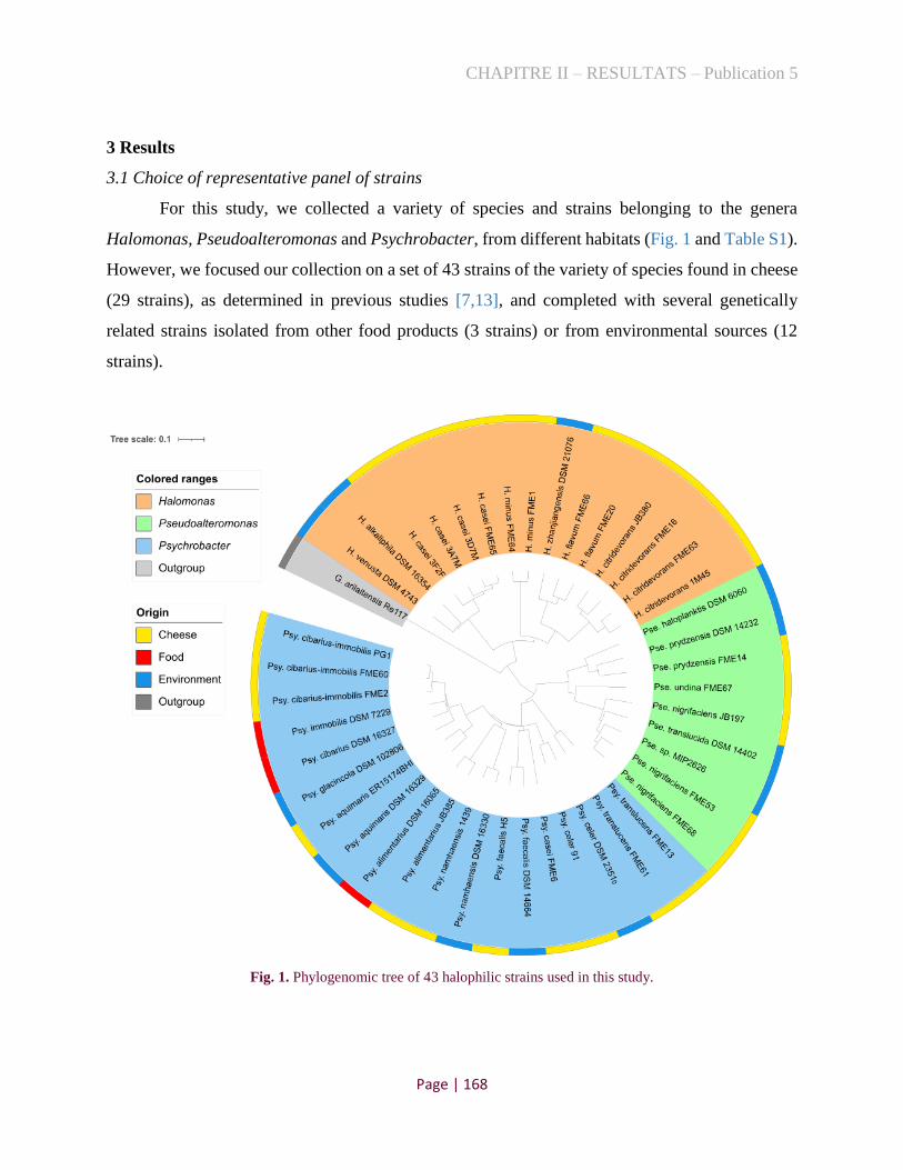
CHAPITRE II – RESULTATS – Publication 5
Page | 168
3 Results
3.1 Choice of representative panel of strains
For this study, we collected a variety of species and strains belonging to the genera
Halomonas, Pseudoalteromonas and Psychrobacter, from different habitats (Fig. 1 and Table S1).
However, we focused our collection on a set of 43 strains of the variety of species found in cheese
(29 strains), as determined in previous studies [7,13], and completed with several genetically
related strains isolated from other food products (3 strains) or from environmental sources (12
strains).
Fig. 1. Phylogenomic tree of 43 halophilic strains used in this study.

CHAPITRE II – RESULTATS – Publication 5
Page | 169
As shown in Figure 1, based on ANI analysis (Table S2, considering 95.5% of ANI cut-
off), these strains may be grouped in 19 potential species. The genus Halomonas is represented by
four species isolated from cheese, with two to four strains, and three strains related species
originating from marine environment. Similarly, Pseudoalteromonas are represented by three
species with one to three strains isolated from cheese, and four strains from marine environment.
Lastly, Psychrobacter is the largest group and contains strains from eight species isolated from
cheese, three strains from other type of food and five strains from marine environment.
Nevertheless, this panel of strains was further assessed to test their ability to develop in cheese-
agar model and produce metabolic compounds of interest for cheese technology.
3.2 Growth and pH changes in the cheese-agar model
The bacterial counts after 21 days of incubation are shown in Fig. 2A. Overall, we
observed that most halophilic strains were able to grow on the cheese-agar model, including the
environmental strains. The average cell counts was between 6.5 and 9.6 log CFU/cm2, except for
three Pseudoalteromonas strains, one of cheese origin (JB197) and two isolated from the sea (DSM
14232 and DSM 14402), which counts were <6 log CFU/cm2. Concerning Halomonas strains,
most of them alkalinise the cheese-agar, including three environmental strains (Fig. 2B). Several
Psychrobcater strains (six) slightly acidify the model (especially 1439, FME6 and DSM 16329),
while three strains - belonging to different species - considerably increase the pH. Moreover, two
of the Pseudoalteromonas strains increase the pH, corresponding to the strains of this genus that
has the greatest development on cheese-agar (Fig. 2A).

CHAPITRE II – RESULTATS – Publication 5
Page | 170
Fig. 2. Halophilic bacterial counts (A) and pH measures (B) at day 21 on cheese-agar model. The error bars
represent the standard deviations (three replicates) and the asterisks indicate the low cell counts lower than 6 log
CFU/cm2) (A). The different letter indicate a significant difference in pH (p<0.05) (B).

CHAPITRE II – RESULTATS – Publication 5
Page | 171
3.3 Sensory analysis
We conducted a sensory analysis where non-trained panellists scored the intensity of the
odours perceived for each strain, and described them qualitatively, as well as the visual aspects
observed. It resulted in the formation of two clusters that separated the intense aromatic strains
from poor, according to the scores of evaluators' sniffing (Fig. 3). Based on these results, we
observed that, overall, the Halomonas strains showed less perceived odours, while the
Psychrobacter strains, whether from food or environmental origins, tended to display more
intense. Additionally, two cheese strains of Pseudoalteromonas deserve emphasis (FME68 and
FME53), as they have been described by all evaluators as having a strong odour (descriptors such
as ammonia, ripened cheese rind and sour are cited). Moreover, they exhibited a brown colour
with white spots on the surface of the cheese-agar models (Additional File 1). We also observed
that several strains formed a biofilm on the surface of cheese-agar medium with white spots (Pse.
prydzensis FME14 and Psy. translucens FME13) or with slightly yellow colour (FME66 and DSM
21076 Halomonas strains). Finally, several Psychrobacter strains formed a white biofilm and
wrinkled textures (especially highlighted in DSM 16065, JB385, PG1, FME2, FME60 and DSM
102806). These observations suggest that several halophilic strains may have potential
contributions for visual aspects on cheese rinds.
3.4 Production of volatile compounds
From the above-mentioned results, especially from sniffing (Fig. 3), 37 strains were
selected to evaluate their potential production of volatile compounds. Overall, we decided to
perform a triplicate analysis for the cheese strains with higher notes in sniffing analysis and only
one replicate for the others and for some environmental strains, particularly those with the highest
aroma intensity. We also evaluated the volatile compounds generated for the controls (without
bacterial inoculum) (i) after 21 days of incubation and (ii) before incubation; totalling 71 samples
(replicates are described in Table S3). We identified a total of 582 molecules produced by GC-MS
analysis. In order to reduce these volatile compounds to odour-active compounds, a filter was
applied using the reference site: www.thegoodscentscompany.com and the literature. Finally 216
aromatic molecules were kept.

CHAPITRE II – RESULTATS – Publication 5
Page | 172
Fig. 3. Heatmap depicting the intensity perceived in sniffing analysis by twelve non-trained panelists.
Figure 4A highlights two groups with particular aromatic characteristics. The first one was
composed by Pse. nigrifaciens FME68 and FME53 strains, which produced volatile compounds
such as pyrazine, ketones (acetone, 2-butanone, methyl isobutyl ketone, etc), benzaldehyde and
dimethyl disulfide, that can contribute to the aroma in cheese ripening [16]. The second group with

CHAPITRE II – RESULTATS – Publication 5
Page | 173
attractive characteristic aromas included mainly the cheese strains of Psy. casei (FME6) and Psy.
namhaensis (1439) (Fig. 4B), which produced carboxylic acid compounds such as 2-methyl
butanoic acid, 3-methyl butanoic acid, decanoic acid, 2-methyl propanoic acid and acetic acid. In
addition of these carboxylic acids, they also produced ketones such as 2,3-butanedione, acetoin,
2-3 pentanedione, 2H-pyran-2-one tetrahydro-6-propyl and 2H-pyran-2-one tetrahydro-6-methyl.
In order to better explore other halophilic strains with a potential flavouring role in cheeses,
statistical analyses were carried out for each genus, separately (Fig. S1). Although Fig. 4B
clustered all Halomonas strains with the controls (without bacteria inoculation), we observed here
that they presented distinct groups (Fig. S1-A). For example, DSM 16354 strain displayed a
different volatile compound profile, with emphasis on the production of pyrazines, ketones and
sulphur compounds. Indeed, this strain had the most intense odour (among Halomonas) in the
sniffing analysis (Fig. 3), and odors of mushroom, soil, plant and vegetables were described,
consistent with the description of ketone and pyrazine compounds detected (Table S4). Similarly,
Pseudoalteromonas prydzensis FME14 presented different volatile compound profile from the
control, with production of acetoin and 2-propanone, 1-hydroxy compounds (Fig. S1-B).
Additionally, this strain also generated white spots on the biofilm and was described by the
evaluators as containing the odour of milk and cheese. Finally, we observed also other group of
Psychrobacter strains that deserve emphasis, which include the strain JB385 and two strains of
environmental origin (DSM 16065 and DSM 16330) (Fig. S1-C). These strains produced
especially ester and alcohol molecules and the evaluators noted aromas of Parmesan, Gruyère and
Emmental cheeses in the sniffing analyses. The result of the quantification of the 216 volatile
compounds in the 71 samples, as well the odour description of each molecule, can be visualised in
Table S4.

CHAPITRE II – RESULTATS – Publication 5
Page | 174
Fig. 4. Overview of volatiles profiles of 43 halophilic strains on cheese-agar models. (A) Principal component
analysis (PCA) showing the mainly aromatic compounds produced by different group of strains (cos2: square
cosine). (B) Clustering samples by similarity of aromatic profiles.

CHAPITRE II – RESULTATS – Publication 5
Page | 175
4 Discussion
A few number of studies, until now, has focused in the investigation of Gram-negative
bacteria during cheese ripening [11,17,10]. Here, we collected a large number of strains of
Halomonas, Pseudoalteromonas and Psychrobacter, to test their functional properties on a cheese-
agar model.
Overall, most of the cheese and environmental strains were able to grow on the proposed
model (Fig. 2A). Additionally, the three genera evaluated have strains that are capable of
increasing the cheese-agar pH (Fig. 2B), and this alkalinisation may be related to the consumption
of lactic acid from the curds and/or to their strong proteolytic activity [18]. Interestingly, six strains
of Psychrobacter and one of Pseudoalteromonas presented pH slightly lower than the control, and
white spots were observed on the cheese-agar surfaces of two of them: Psy. translucens FME13
and Pse. prydzensis FME14 (Additional File 1). These spots may be associated with the formation
of calcium lactate crystals, which may be formed in cheeses with low pH due the development of
insoluble lactate [19]. Finally, we have not observed any clear correlation between the growth of
the strains and the evolution of the pH on the model surface, since several strains presented counts
above 8 log CFU/cm2 and no pH changes.
Among the 43 strains evaluated, two groups were distinguished by their production of
volatile compounds. The first one, composed of Pse. nigrifaciens FME68 and FME53 strains,
showed significant production of pyrazines in the cheese-agar model (Fig. 4A), that can be
explained by isomerization of the aminoacetone derived from the degradation of alanine and
methylglyoxal (Strecker degradation) [20]. The formation of different pyrazines was described in
Parmesan cheeses and associated with a brown discoloration [20]. Interestingly, we observed a
brown color on the cheese-agar surfaces from these strains, which could be associated with the
pyrazine compounds generated by them (Additional File 1). Pyrazines were also reported to
display baked, roasted, and nutty flavor characters [21]. Moreover, both Pseudoalteromonas
strains also produced ketones, which can be formed from the fatty acids in the cheese by beta-
oxidation and can generate secondary alcohols. These compounds have been commonly reported
in blue cheeses [22,23]. Benzaldehyde, another compound generated by the two strains, was
probably formed from phenylpyruvic acid, derived from the aminotransferase activity in
phenylalanine [24] and has already been described as having an aromatic note of bitter almond and
reported in the volatile fractions of several cheeses, such as Brie and Camembert [25,26].

CHAPITRE II – RESULTATS – Publication 5
Page | 176
Additionally, FME68 and FME53 strains had high levels of disulfide dimethyl, a volatile
compound formed from methanethiol, one of the degradation products of L-methionine
metabolism. This sulphur compound was already detected in cheeses such as Camembert, Livarot,
Langres and Epoisses [27-29], and can bring aromatic notes of cabbage, garlic and cheesy.
Although evaluators in sniffing analysis did not perceive nutty and almond flavours, all of them
highlighted an intense sour and pungent smell to the nose for both strains, and described ammonia
and ripened cheese odours, which may be related to proteolytic activity of FME68 and FME53
strains. This production of ammonia may mask the perception of other odours.
The other group, composed by FME6 and 1439 Psychrobacter strains, showed intense
production of carboxylic acids and ketones compounds (Fig. S1-C). Among the carboxylic acids,
the butanoic acid, 2-methyl and butanoic acid, 3-methyl (isovaleric acid), originate from isoleucine
and leucine catabolism, respectively, are branched-chain volatile acids associated to fruity
aromatic notes of Comté, Abondance and Swiss cheeses [30-33]. On the other hand, FME6 and
1439 strains produced decanoic acid as one of the most prevalent fatty acid, a compound which
was reported with high abundance in semi-hard and hard goat cheeses [34,35], but related as
undesirable volatile compound found in Parmesan cheese due to their soapy and waxy aromas
[36]. Although fruity cheeses such as Parseman Grueyère and Emmental cheeses were mentioned
by several evaluators in the sniffing analysis, other ones mentioned notes of rancidity, which may
be related to the identified carboxylic and fatty acids, respectively. Interestingly, these
Psychrobacter strains slightly lowered the pH on cheese-agar models compared to the control (Fig.
2B), and can be related with the production of acid compounds such as acetic acid and propanoic
acid, 2-methyl. These volatile compounds were already reported in cheeses and were associated to
the vinegar and vomit-foot odors [37]. Finally, carboxylic acids can also play an essential role in
the production of aromas, since they could be precursors of other volatile compounds such as
ketones, alcohols, lactones, aldehydes and esters [38]. Among the ketones produced by FME6 and
1439 strains, diacetyl (2,3-butanedione) and its reduced forms (acetoin) can be produced by the
metabolism of sugars via pyruvate and acetolactate synthase. These compounds are commonly
produced by LAB metabolism of citrate (i.e. by Leuconostoc mesenteroides cremoris and
Lactococcus lactis subsp. diacetylactis) and present buttery or butterscotch note in foods [39,40].
Indeed, the evaluators of the sniffing analysis noticed odours of butter for both strains.

CHAPITRE II – RESULTATS – Publication 5
Page | 177
Previous studies have also shown that Psychrobacter strains, such as Psy. celer 91 and
Psychrobacter sp. 580, can contribute significantly to cheese ripening through the production of
aldehydes, esters, ketones and sulphur compounds [10,11]. Interestingly, we also tested the strain
91, and although this strain showed good implementation on our cheese-agar model and increased
the pH of the medium (Fig. 2), its production of aromatic compounds did not have large differences
with the control. In the previous studies, Psy. celer 91 was tested on a washed cheese model in
interaction with seven other bacteria and four yeasts [10]. It is thus likely that Psy. celer 91 requires
interactions of other bacteria and yeasts to produce aroma, while in our study this strain was tested
individually.
5 Conclusion
In this study, we assessed the potential of a large variety of halophilic Gram-negative
strains, isolated from cheese and from different habitats to produce aroma in a model of cheese. It
shows that most strains were able to develop in this cheese agar model. Moreover, we found that
the potential roles of the strains differed significantly. Two strains of Pse. nigrifaciens displayed
a high aromatic potential, which was highlighted by sensory analysis and could be associated with
the production of a variety of volatile compounds. Two Psychrobacter strains also stand out of the
rest of the strains by producing a variety of carboxylic acids and lowering the pH in the cheese-
agar model. This study highlight the potential of Gammaproteobacteria, which are part of the
dominant bacteria on several washed rind cheeses, to produce aroma, texture and color in cheese
technology.
Acknowledgements
The authors are grateful to Gwendoline Coeuret and Anne-Claire Peron for technical support in
sampling and analysis of chromatograms, respectively.
Supplementary Material
The supplementary Table S1-S4 and the Additional Files 1 can be found at
http://dx.doi.org/10.17632/7vc3vndc3r.1.

CHAPITRE II – RESULTATS – Publication 5
Page | 178
Table S1. Halophilic strains used in this study and their respective origin and references of
isolation.
Sample Genera Species Strain Origin Obtained from
1 Halomonas citridevorans 1M45 Cheese Almeida et al (2014)
2 Halomonas citridevorans FME63 Cheese Kothe et al (2021)
3 Halomonas citridevorans FME16 Cheese Kothe et al (2020)
4 Halomonas citridevorans JB380 Cheese Donation from Dutton lab
5 Halomonas flavum FME20 Cheese Kothe et al (2020)
6 Halomonas flavum FME66 Cheese Kothe et al (2021)
7 Halomonas zhanjiangensis DSM 21076 Environment DSMZ
8 Halomonas minus FME1 Cheese Kothe et al (2020)
9 Halomonas minus FME64 Cheese Kothe et al (2021)
10 Halomonas casei FME65 Cheese Kothe et al (2021)
11 Halomonas casei 3F2F Cheese Almeida et al (2014)
12 Halomonas casei 3D7M Cheese Almeida et al (2014)
13 Halomonas casei 3A7M Cheese Almeida et al (2014)
14 Halomonas alkaliphila DSM 16354 Environment DSMZ
15 Halomonas venusta DSM 4743 Environment DSMZ
16 Psychrobacter cibarius-immobilis PG1 Cheese Almeida et al (2014)
17 Psychrobacter cibarius-immobilis FME2 Cheese Kothe et al (2020)
18 Psychrobacter cibarius-immobilis FME60 Cheese Kothe et al (2021)
19 Psychrobacter glacincola DSM 102806 Environment DSMZ
20 Psychrobacter immobilis DSM 7229 Food DSMZ
21 Psychrobacter cibarius DSM 16327 Food DSMZ
22 Psychrobacter casei FME6 Cheese Kothe et al (2020)
23 Psychrobacter translucens FME61 Cheese Kothe et al (2021)
24 Psychrobacter translucens FME13 Cheese Kothe et al (2020)
25 Psychrobacter aquimaris ER15174BHI Cheese Almeida et al (2014)
26 Psychrobacter aquimaris DSM 16329 Environment DSMZ
27 Psychrobacter faecalis H5 Cheese Almeida et al (2014)
28 Psychrobacter faecalis DSM 14664 Environment DSMZ
29 Psychrobacter namhaensis 1439 Cheese Almeida et al (2014)
30 Psychrobacter namhaensis DSM 16330 Environment DSMZ
31 Psychrobacter celer 91 Cheese Almeida et al (2014)
32 Psychrobacter celer DSM 23510 Environment DSMZ
33 Psychrobacter alimentarius DSM 16065 Food DSMZ
34 Psychrobacter alimentarius JB385 Cheese Donation from Dutton lab
35 Pseudoalteromonas prydzensis FME14 Cheese Kothe et al (2020)
36 Pseudoalteromonas prydzensis DSM 14232 Environment DSMZ
37 Pseudoalteromonas haloplanktis DSM 6060 Environment DSMZ
38 Pseudoalteromonas nigrifaciens JB197 Cheese Donation from Dutton lab
39 Pseudoalteromonas nigrifaciens FME68 Cheese Kothe et al (2021)
40 Pseudoalteromonas nigrifaciens FME53 Cheese Kothe et al (2020)
41 Pseudoalteromonas sp. (not defined) MIP2626 Environment Donation from Ifremer
42 Pseudoalteromonas translucida DSM 14402 Environment DSMZ
43 Pseudoalteromonas undina FME67 Cheese Kothe et al (2021)

CHAPITRE II – RESULTATS – Publication 5
Page | 179
Table S2. ANI analysis of the 43 strains used in this study.
Cheese
Other food
Environment
01_Halomonas_citridevorans_1M45.fasta02_Halomonas_citridevorans_FME63.fasta03_Halomonas_citridevorans_FME16.fasta04_Halomonas_citridevorans_JB380.fasta05_Halomonas_flavum_FME20.fasta06_Halomonas_flavum_FME66.fasta07_Halomonas_zhanjiangensis_DSM_21076.fasta08_Halomonas_minus_FME1.fasta09_Halomonas_minus_FME64.fasta10_Halomonas_casei_FME65.fasta11_Halomonas_casei_3F2F.fasta12_Halomonas_casei_3D7M.fasta13_Halomonas_casei_3A7M.fasta14_Halomonas_alkaliphila_DSM_16354.fasta15_Halomonas_venusta_DSM_4743.fasta
01_Halomonas_citridevorans_1M45.fasta
02_Halomonas_citridevorans_FME63.fasta 98.61
03_Halomonas_citridevorans_FME16.fasta 96.31 96.17
04_Halomonas_citridevorans_JB380.fasta 95.54 95.44 97.64
05_Halomonas_flavum_FME20.fasta 84.2 84.46 83.97 83.81
06_Halomonas_flavum_FME66.fasta 84.54 84.62 84.04 83.76 98.33
07_Halomonas_zhanjiangensis_DSM_21076.fasta 84.17 84.2 83.88 83.84 94.14 94.08
08_Halomonas_minus_FME1.fasta 79.76 80.68 79.77 79.28 79.29 79.97 79.44
09_Halomonas_minus_FME64.fasta 79.82 79.88 79.79 79.24 79.67 79.99 79.37 99.07
10_Halomonas_casei_FME65.fasta 79.85 79.98 79.61 79.46 80.12 81.06 79.37 79.56 79.6
11_Halomonas_casei_3F2F.fasta 79.84 80.26 79.64 79.42 80.02 80.2 79.54 79.6 79.55 99.03
12_Halomonas_casei_3D7M.fasta 79.9 80.12 79.51 79.41 80.11 80.51 79.32 79.33 79.36 99.31 99
13_Halomonas_casei_3A7M.fasta 80.01 79.87 79.59 79.47 80.08 80.28 79.34 79.33 79.41 98.69 98.63 98.6
14_Halomonas_alkaliphila_DSM_16354.fasta 79.14 79.13 79.36 79.05 79.21 79.24 79.2 79.12 79.17 95.55 95.52 95.59 95.56
15_Halomonas_venusta_DSM_4743.fasta 79.14 79.02 79.15 79.12 79.17 79.17 79.33 79.21 79.14 93.76 93.81 93.69 93.72 92.83
16_Psychrobacter_immobilis_PG1.fasta17_Psychrobacter_cibarius-immobilis_FME2.fasta18_Psychrobacter_cibarius_FME60.fasta19_Psychrobacter_glacincola_DSM_102806.fasta20_Psychrobacter_immobilis_DSM_7229.fasta21_Psychrobacter_cibarius_DSM_16327.fasta22_Psychrobacter_casei_FME6.fasta23_Psychrobacter_translucens_FME61.fasta24_Psychrobacter_translucens_FME13.fasta25_Psychrobacter_aquimaris_ER15_174_BHI7.fasta26_Psychrobacter_aquimaris_DSM_16329.fasta27_Psychrobacter_faecalis_H5.fasta28_Psychrobacter_faecalis_DSM_14664.fasta29_Psychrobacter_namhaensis_1439.fasta30_Psychrobacter_namhaensis_DSM_16330.fasta31_Psychrobacter_celer_91.fasta32_Psychrobacter_celer_DSM_23510.fasta33_Psychrobacter_alimentarius_DSM_16065.fasta34_Psychrobacter_alimentarius_JB385.fasta
16_Psychrobacter_immobilis_PG1.fasta
17_Psychrobacter_cibarius-immobilis_FME2.fasta 99.49
18_Psychrobacter_cibarius_FME60.fasta 99.64 99.57
19_Psychrobacter_glacincola_DSM_102806.fasta 96.12 96.25 96.11
20_Psychrobacter_immobilis_DSM_7229.fasta 95.95 96.05 95.97 96.32
21_Psychrobacter_cibarius_DSM_16327.fasta 96.13 96.21 96.15 96.35 96.28
22_Psychrobacter_casei_FME6.fasta 82.36 82.17 82.43 82.05 82.21 82.08
23_Psychrobacter_translucens_FME61.fasta 81.75 81.53 81.81 81.6 81.62 81.65 80.82
24_Psychrobacter_translucens_FME13.fasta 81.84 81.79 81.94 81.74 81.81 81.6 81.52 98.61
25_Psychrobacter_aquimaris_ER15_174_BHI7.fasta 91.63 91.3 91.51 91.19 91.26 91.26 82.49 81.84 82.01
26_Psychrobacter_aquimaris_DSM_16329.fasta 91.21 91.1 91.23 91.15 91.26 91.19 82 81.45 81.64 97.18
27_Psychrobacter_faecalis_H5.fasta 81.86 81.79 81.89 81.9 81.96 81.91 82.22 80.23 80.8 81.94 81.45
28_Psychrobacter_faecalis_DSM_14664.fasta 81.73 81.77 81.82 81.65 81.92 81.83 81.62 80.4 80.34 81.48 81.46 97.47
29_Psychrobacter_namhaensis_1439.fasta 81.93 81.85 81.94 81.76 82.01 81.87 81.07 81.07 80.84 82.26 82.37 80.45 80.59
30_Psychrobacter_namhaensis_DSM_16330.fasta 81.95 81.9 82.01 81.88 82.19 81.92 81.09 81.04 81.01 82.12 82.28 80.4 80.55 97.65
31_Psychrobacter_celer_91.fasta 80.76 80.61 80.7 80.64 80.55 80.51 80.26 80.27 79.97 80.7 80.45 81.01 80.89 80.28 80.44
32_Psychrobacter_celer_DSM_23510.fasta 80.64 80.69 80.69 80.52 80.84 80.53 80.24 80.6 80.33 80.53 80.72 80.92 80.86 80.89 80.92 97.54
33_Psychrobacter_alimentarius_DSM_16065.fasta 80.85 80.9 81.03 80.95 80.89 81 80.56 80.64 80.55 81.38 81.49 80.06 80.08 81.91 81.81 80.11 80.03
34_Psychrobacter_alimentarius_JB385.fasta 80.85 80.87 80.96 80.83 80.86 80.97 80.44 80.46 80.39 81.19 81.32 79.91 80.03 81.85 81.84 80.01 79.92 97.12
35_Pseudoalteromonas_prydzensis FME14.fasta36_Pseudoalteromonas_prydzensis_DSM_14232.fasta37_Pseudoalteromonas_haloplanktis_DSM_6060.fasta38_Pseudoalteromonas_nigrifaciens_JB197.fasta39_Pseudoalteromonas_nigrifaciens_FME68.fasta40_Pseudoalteromonas_nigrifaciens_FME53.fasta41 Pseudoalteromonas sp. MIP2626.fasta42_Pseudoalteromonas_translucida_DSM_14402.fasta43_Pseudoalteromonas_undina_FME67.fasta
35_Pseudoalteromonas_prydzensis FME14.fasta
36_Pseudoalteromonas_prydzensis_DSM_14232.fasta 97.06
37_Pseudoalteromonas_haloplanktis_DSM_6060.fasta 86.56 86.67
38_Pseudoalteromonas_nigrifaciens_JB197.fasta 81.22 81.33 80.31
39_Pseudoalteromonas_nigrifaciens_FME68.fasta 80.77 80.59 80.07 97.98
40_Pseudoalteromonas_nigrifaciens_FME53.fasta 80.68 80.54 79.94 97.95 98.89
41 Pseudoalteromonas sp. MIP2626.fasta 80.25 80.26 79.92 97.71 98.12 98.06
42_Pseudoalteromonas_translucida_DSM_14402.fasta 80.07 80.12 80.01 96.02 96.21 96.22 96.25
43_Pseudoalteromonas_undina_FME67.fasta 81.04 80.52 80.19 82 82.5 82.19 81.64 81.46

CHAPITRE II – RESULTATS – Publication 5
Page | 180
Table S3. Sample replicates analysed by GC-MS.
Sample Genera Species Strain Origin Sniffing
(media)
Volatile analysis
(number of
replicates)
39 Pseudoalteromonas nigrifaciens FME68 Cheese 7.75 3
40 Pseudoalteromonas nigrifaciens FME53 Cheese 7.75 3
34 Psychrobacter alimentarius JB385 Cheese 2.916667 3
35 Pseudoalteromonas prydzensis FME14 Cheese 4.083333 3
27 Psychrobacter faecalis H5 Cheese 3.916667 3
30 Psychrobacter namhaensis DSM 16330 Environment 3.666667 1
28 Psychrobacter faecalis DSM 14664 Environment 4.5 1
29 Psychrobacter namhaensis 1439 Cheese 4.916667 3
16 Psychrobacter cibarius-immobilis PG1 Cheese 5.666667 3
33 Psychrobacter alimentarius DSM 16065 Food 5.083333 1
24 Psychrobacter translucens FME13 Cheese 5.75 3
26 Psychrobacter aquimaris DSM 16329 Environment 5.666667 1
12 Halomonas casei 3D7M Cheese 3.5 3
14 Halomonas alkaliphila DSM 16354 Environment 3.916667 1
21 Psychrobacter cibarius DSM 16327 Food 4 1
22 Psychrobacter casei FME6 Cheese 4.5 3
17 Psychrobacter cibarius-immobilis FME2 Cheese 4.75 3
18 Psychrobacter cibarius-immobilis FME60 Cheese 4.583333 3
19 Psychrobacter glacincola DSM 102806 Environment 4.5 1
20 Psychrobacter immobilis DSM 7229 Food 4.833333 1
15 Halomonas venusta DSM 4743 Environment 1.916667 -
32 Psychrobacter celer DSM 23510 Environment 2.916667 1
23 Psychrobacter translucens FME61 Cheese 1.75 1
8 Halomonas minus FME1 Cheese 2.333333 1
13 Halomonas casei 3A7M Cheese 2.5 1
41 Pseudoalteromonas sp. (not defined) MIP2626 Environment 1.25 -
42 Pseudoalteromonas translucida DSM 14402 Environment 1.166667 -
36 Pseudoalteromonas prydzensis DSM 14232 Environment 1.083333 -
37 Pseudoalteromonas haloplanktis DSM 6060 Environment 0.916667 -
1 Halomonas citridevorans 1M45 Cheese 0.333333 1
31 Psychrobacter celer 91 Cheese 0.916667 1
38 Pseudoalteromonas nigrifaciens JB197 Cheese 1 1
43 Pseudoalteromonas undina FME67 Cheese 1 3
25 Psychrobacter aquimaris ER15174BHI Cheese 2.5 3
7 Halomonas zhanjiangensis DSM 21076 Environment 2.583333 -
10 Halomonas casei FME65 Cheese 2.416667 1
11 Halomonas casei 3F2F Cheese 1.583333 1
6 Halomonas flavum FME66 Cheese 1.166667 3
9 Halomonas minus FME64 Cheese 1.75 1
3 Halomonas citridevorans FME16 Cheese 1.833333 1
4 Halomonas citridevorans JB380 Cheese 1.25 1
2 Halomonas citridevorans FME63 Cheese 1 1
5 Halomonas flavum FME20 Cheese 1.166667 1
Control (t=21
days) 3
Control (t=0) 1
Total 71

CHAPITRE II – RESULTATS – Publication 5
Page | 181
Table S4. Quantification of volatile compounds detected in the halophilic strains and their class,
odor description, formula and CAS number (simplified).
Sample Control FME66 3D7M PG1 FME2 FME60 FME6 FME13 ER15174 H5 1439 JB385 FME14 FME68 FME53 FME67
1-Propanol, 2-methyl- 254625 18574.3 1703.33 506598 383797 705554 1393646 11586.7 73638.7 530998 851980 1052889 11705.3 34201.3 15685.7 11165.3
2,3-Butanediol 0 0 0 2326117 739422 566205 1089039 0 0 0 0 0 0 0 0 0
1-Butanol, 3-methyl- 803415 33385.7 0 1897027 1977240 3292058 6344694 167808 3874676 6458063 1.4E+07 2.1E+07 210954 2400584 3305227 171311
1-Pentanol 83684.3 196037 0 0 0 0 0 0 0 0 0 114334 13436 0 0 53188
2-Heptanol 502536 793855 0 6294708 5304525 1.1E+07 720390 0 2232768 693544 820362 2815117 0 1488610 0 0
2-Undecanol 0 0 0 240335 0 316184 0 0 0 0 0 0 0 0 0 0
Phenylethyl Alcohol 0 0 0 0 0 1414623 1.2E+07 0 0 2486760 6.2E+07 3.9E+07 0 0 0 0
2-Pentanol 0 0 0 0 0 459401 0 0 0 0 0 0 0 688483 0 0
1-Butanol, 2-methyl- 0 0 0 0 0 0 0 0 163166 0 0 813250 0 0 0 0
Propanal, 2-methyl- 49168.7 0 0 311504 171179 105641 2155106 29848.7 0 351807 434813 35502.7 6135.33 10419.7 51075.7 0
Butanal, 3-methyl- 1214233 0 0 1388519 1129133 1078552 3496099 4916163 112471 1130859 1548635 899510 3188380 1961588 2622457 0
Benzeneacetaldehyde 22116.3 0 0 145482 265117 141983 376574 23726.3 0 349463 98417.7 125208 0 282541 263904 0
Acetic acid 34694 19082.7 12035.3 368738 268872 672587 4337299 4013876 50213.7 992508 1.5E+07 1493442 1330535 184386 841242 21587
Butanoic acid 0 0 72520.3 6726919 5455288 2E+07 2.7E+07 4636669 838222 4645878 2.4E+07 6134441 7834636 3438211 312005 0
Butanoic acid, 3-methyl- 0 0 34074.3 1.1E+07 1E+07 3.8E+07 3.5E+07 1.3E+07 38652.7 1.3E+07 6.3E+07 8017655 5234425 7679895 1.1E+07 0
Butanoic acid, 2-methyl- 0 0 0 4466313 3781490 8645341 1.4E+07 2385927 0 1137284 2.2E+07 2922321 1353114 4320671 6909254 0
Hexanoic acid 0 0 0 220690 321562 1652743 2.4E+07 1.9E+07 50294.7 2301640 1.6E+07 1767673 5424461 131921 83789.3 0
Propanoic acid, 2-methyl- 0 0 21838 3315704 3549414 1.2E+07 2.6E+07 3967378 0 5088349 1.8E+07 3351767 0 1944489 1061460 0
Octanoic acid 0 0 0 0 0 448682 1.5E+07 4798815 42498.3 582681 1.3E+07 1400757 2411003 0 115444 0
n-Decanoic acid 0 0 0 0 0 70053.7 1994684 546012 0 95107.3 3153982 183579 403027 0 21576 0
Pentanoic acid 0 0 0 0 0 0 1088038 490273 231027 124088 1007519 26687.7 103128 0 153227 0
Heptanoic acid 0 0 0 0 0 0 212450 93783.3 0 0 251400 0 0 0 0 0
Nonanoic acid 0 0 0 0 0 0 87772 0 0 0 73390 0 0 0 0 0
Propanoic acid 0 0 0 0 0 0 0 137717 0 0 0 0 0 73853.3 0 0
4-Methyloctanoic acid 0 0 0 0 0 0 0 89798.3 0 0 0 0 122223 0 0 0
4-Methylnonanoic acid 0 0 0 0 0 0 0 86458.3 0 0 0 0 0 0 0 0 Acetic acid, 2-phenylethyl
ester 126246 17590.3 58200.3 133172 199144 395625 157136 0 0 269983 1802324 1662613 0 0 76478.7 69961.7
Acetic acid, hexyl ester 336865 134524 108208 79712.3 67455.7 0 53199 0 0 0 132956 296527 0 35588 43917.3 0
2-Heptanol, acetate 161632 0 0 1954652 1454874 2942612 133523 0 401358 336314 283114 435002 0 0 0 0
Isobutyl acetate 90946.3 0 0 572797 609648 0 0 0 0 499835 0 401521 0 0 0 0 Butanoic acid, methyl
ester 0 0 0 283596 371729 365639 1257559 0 0 104653 0 221262 0 0 187365 0 1-Butanol, 3-methyl-,
propanoate 0 0 0 64416.3 46075.3 102594 0 0 140599 224422 0 2738871 0 0 0 0
2-Pentanol, acetate 0 0 0 156825 0 0 0 0 0 50256 0 0 0 0 0 0 1-Butanol, 3-methyl-,
acetate 0 0 0 0 0 1388524 0 0 2747030 4076108 4239149 9781603 0 0 0 0
Hexanoic acid, butyl ester 0 0 0 0 0 0 51608.3 0 0 36570.7 85651.3 0 0 0 0 0
Acetic acid, pentyl ester 0 0 0 0 0 0 0 0 0 171873 0 125557 0 0 0 0

CHAPITRE II – RESULTATS – Publication 5
Page | 182
Propanoic acid, ethyl ester 0 0 0 0 0 0 0 0 0 0 0 1.5E+07 0 0 0 0
1-Butanol, 2-methyl-, propanoate 0 0 0 0 0 0 0 0 0 0 0 700781 0 0 0 0
Butanoic acid, ethyl ester 0 0 0 0 0 0 0 0 0 0 0 191806 0 0 0 0 Propanoic acid, butyl
ester 0 0 0 0 0 0 0 0 0 0 0 52282.7 0 0 0 0 1-Butanol, 2-methyl-,
acetate 0 0 0 0 0 0 0 0 0 0 0 68740.7 0 0 219078 0 Propanoic acid, 2-methyl-
, ethyl ester 0 0 0 0 0 0 0 0 0 0 0 0 0 0 0 0
Octanoic acid, ethyl ester 0 0 0 0 0 0 0 0 0 0 0 0 0 0 0 0
Ethyl Acetate 14642 203436 9476.67 16169.7 17747.3 14963.3 212771 10867.3 59187.7 81392.7 109332 745307 9139.33 7612 7061 7157
Acetic acid, butyl ester 164491 37120.7 582965 120285 143300 126924 0 1173813 283940 157658 0 161244 124699 92614.3 105444 114122
Acetone 647276 3178832 1398009 1168944 1042082 2319850 1496200 7084425 1840954 1287319 2735618 1438585 8617485 1.6E+07 2.3E+07 8115582
2-Butanone 189126 911262 347992 459441 421346 396215 341801 3615428 341298 601776 875425 310760 4223548 2E+07 1.8E+07 2599510
2,3-Butanedione 160003 9060 7066.67 3751109 4301353 3894575 1.4E+07 1893017 725012 4225038 2.7E+07 256186 2909988 326542 1064820 335118
2,3-Pentanedione 0 0 0 712904 389277 1450296 1629613 0 0 455398 2568765 0 0 2067016 2517785 0
Acetoin 636108 3825.33 3585.67 9394899 5130574 1.3E+07 1.7E+07 1.7E+07 1682368 9502456 3.4E+07 709443 1.9E+07 558493 1277835 597110
2-Hexanone 50885.7 64388 28856.3 201900 128728 260249 349689 139546 94084 159296 167626 89968.7 196407 1393019 1652708 78363.3
2-Heptanone 1.8E+07 2.6E+07 2E+07 3.1E+07 2.5E+07 3.8E+07 3.5E+07 2.7E+07 2.4E+07 3.2E+07 3.7E+07 2.4E+07 2.5E+07 3E+07 2.3E+07 1.7E+07
2-Octanone 294127 279495 249611 530254 291402 829445 0 411413 238777 309857 516845 129745 363746 874645 904891 198660
2-Nonanone 9252190 2.7E+07 1.1E+07 1.3E+07 9245829 1.7E+07 1.5E+07 1.2E+07 8610692 1.1E+07 1.2E+07 1.1E+07 1E+07 1.1E+07 1E+07 8573459
2-Decanone 115584 85186.3 84706.3 150989 32406.3 200564 205150 0 79110.3 125312 169541 0 0 227009 284315 64412.3
2-Undecanone 3371906 2583899 2888098 3487594 3274411 4775508 4786718 3738873 2415134 3629677 3928355 3814021 3231035 3524542 3084261 2651821 Pyrazine, 2-methoxy-3-(1-
methylpropyl)- 158045 0 3944755 59098.7 713666 780718 412338 0 0 0 0 0 0 0 0 0
Pyrazine, 2,5-dimethyl- 7337 499210 581583 0 139591 0 30277 409371 987738 0 434311 15475 970263 1.8E+08 1.3E+08 124179
Pyrazine, methyl- 0 0 0 0 0 0 0 0 370847 0 0 0 0 4089953 2537399 0
Pyrazine, 2,3-dimethyl- 0 0 0 0 0 0 0 0 0 0 0 0 0 105817 179120 0
Pyrazine, trimethyl- 0 0 0 0 0 0 0 0 0 0 66101.7 0 0 1.1E+07 1.9E+07 0
Pyrazine, 2,6-dimethyl- 0 0 0 0 0 0 0 0 0 0 0 15475 0 0 0 0 2-Methyl-3-
isopropylpyrazine 0 0 0 0 0 0 0 0 0 0 0 0 0 108692 74630 0 Pyrazine, 3-ethyl-2,5-
dimethyl- 0 0 0 0 0 0 0 0 0 0 0 0 0 507970 511567 0 Pyrazine, 2-ethyl-3,5-
dimethyl- 0 0 0 0 0 0 0 0 0 0 0 0 0 954347 1957111 0 Pyrazine, 2,5-dimethyl-3-
propyl- 0 0 0 0 0 0 0 0 0 0 0 0 0 49403.3 44401.7 0 Pyrazine, 3,5-dimethyl-2-
propyl- 0 0 0 0 0 0 0 0 0 0 0 0 0 34853 44401.7 0 Pyrazine, 2-ethyl-5-
methyl- 0 0 0 0 0 0 0 0 0 0 0 0 0 0 0 0
Disulfide, dimethyl 118790 97715.3 71901.7 20299 30390 15586.3 29841.7 1741602 536957 621992 531181 347381 1794154 4453774 4059120 959064
Dimethyl trisulfide 0 0 0 0 0 0 0 841596 0 0 0 0 634109 236999 178317 221224 1-Propanol, 3-(methylthio)- 0 0 0 0 0 0 0 0 0 0 0 222941 0 0 0 0
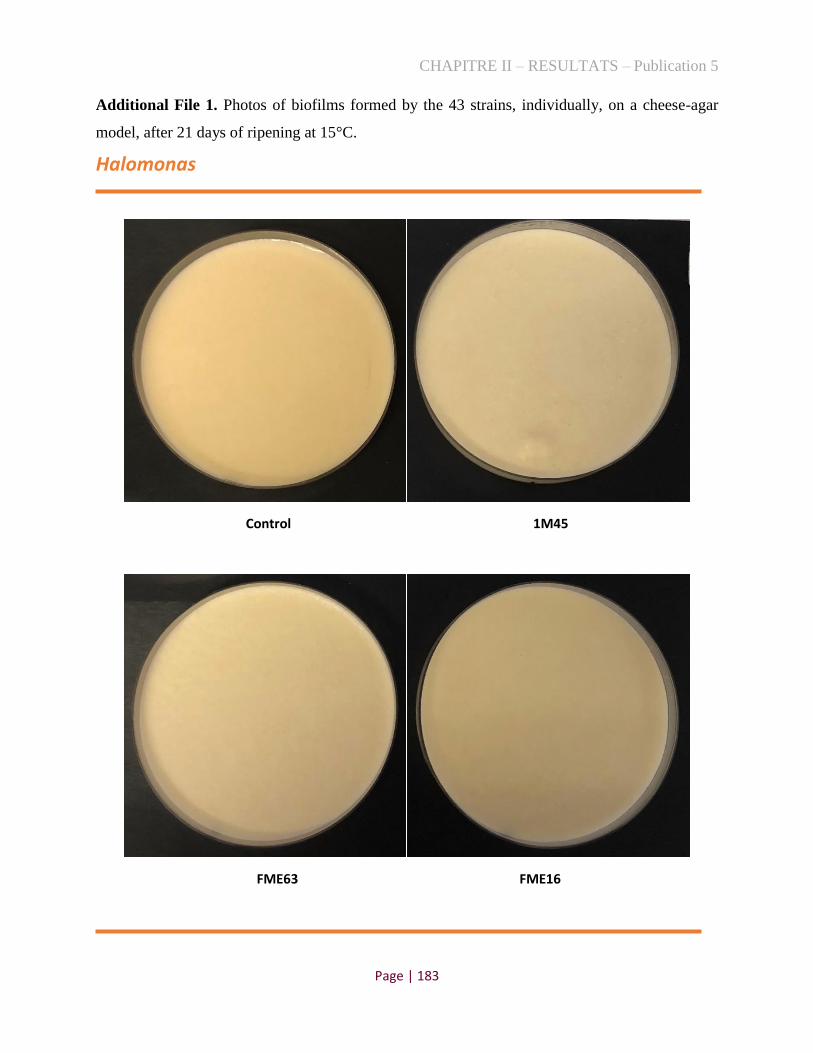
CHAPITRE II – RESULTATS – Publication 5
Page | 183
Additional File 1. Photos of biofilms formed by the 43 strains, individually, on a cheese-agar
model, after 21 days of ripening at 15°C.
Halomonas
Control 1M45
FME63 FME16
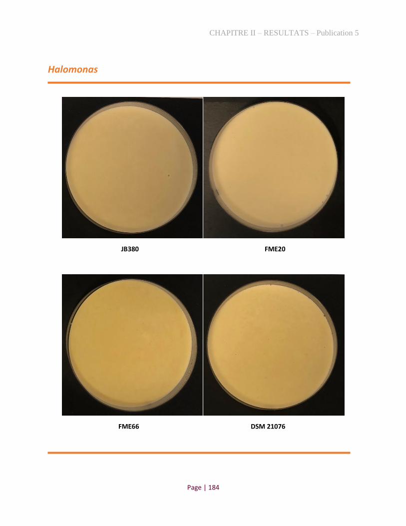
CHAPITRE II – RESULTATS – Publication 5
Page | 184
Halomonas
JB380 FME20
FME66 DSM 21076

CHAPITRE II – RESULTATS – Publication 5
Page | 185
Halomonas
FME1 FME64
FME65 3F2F

CHAPITRE II – RESULTATS – Publication 5
Page | 186
Halomonas
3D7M 3A7M
DSM 16354 DSM 4743

CHAPITRE II – RESULTATS – Publication 5
Page | 187
Psychrobacter
PG1 FME2
FME60 DSM 102806

CHAPITRE II – RESULTATS – Publication 5
Page | 188
Psychrobacter
DSM 7229 DSM 16327
FME6 FME61

CHAPITRE II – RESULTATS – Publication 5
Page | 189
Psychrobacter
FME13 ER15174BHI
DSM 16329 H5

CHAPITRE II – RESULTATS – Publication 5
Page | 190
Psychrobacter
DSM 14664 1439
DSM 16330 91

CHAPITRE II – RESULTATS – Publication 5
Page | 191
Psychrobacter
DSM 23510 DSM 16065
JB385

CHAPITRE II – RESULTATS – Publication 5
Page | 192
Pseudoalteromonas
FME14 DSM 14232
DSM 6060 JB197

CHAPITRE II – RESULTATS – Publication 5
Page | 193
Pseudoalteromonas
FME68 FME53
MIP2626 DSM 14402

CHAPITRE II – RESULTATS – Publication 5
Page | 194
Pseudoalteromonas
FME67
Fig. S1. PCA showing the main volatile compounds produced by Halomonas (A) strain.

CHAPITRE II – RESULTATS – Publication 5
Page | 195
Fig. S1. PCA showing the main volatile compounds produced by Pseudoalteromonas (B) and Psychrobacter (C)
strains.

CHAPITRE II – RESULTATS – Publication 5
Page | 196
6 References
[1]. Irlinger F, Layec S, Hélinck S, Dugat-Bony E (2015) Cheese rind microbial communities: diversity,
composition and origin. Fems Microbiol Lett 362 (2):1-11. doi:10.1093/femsle/fnu015.
2]. Delbes C, Monnet C, Irlinger F (2015) Des communautés microbiennes au service de la qualité des fromages :
diversité et dynamique adaptative et fonctionnelle des populations endogènes et ensemencées.
[3]. Smit G, Smit BA, Engels WJM (2005) Flavour formation by lactic acid bacteria and biochemical flavour
profiling of cheese products. FEMS Microbiology Reviews 29 (3):591-610. doi:10.1016/j.fmrre.2005.04.002.
[4]. Lortal S, Chapot-Chartier MP (2005) Role, mechanisms and control of lactic acid bacteria lysis in cheese. Int
Dairy J 15 (6):857-871. doi:10.1016/j.idairyj.2004.08.024.
[5]. Wolfe BE, Button JE, Santarelli M, Dutton RJ (2014) Cheese Rind Communities Provide Tractable Systems for
In Situ and In Vitro Studies of Microbial Diversity. Cell 158 (2):422-433. doi:10.1016/j.cell.2014.05.041.
[6]. Dugat-Bony E, Garnier L, Denonfoux J, Ferreira S, Sarthou AS, Bonnarme P, Irlinger F (2016) Highlighting the
microbial diversity of 12 French cheese varieties. International Journal of Food Microbiology 238:265-273.
doi:10.1016/j.ijfoodmicro.2016.09.026.
[7]. Almeida M, Hébert A, Abraham A-L, Rasmussen S, Monnet C, Pons N, Delbès C, Loux V, Batto J-M, Leonard
P, Kennedy S, Ehrlich SD, Pop M, Montel M-C, Irlinger F, Renault P (2014) Construction of a dairy microbial
genome catalog opens new perspectives for the metagenomic analysis of dairy fermented products. BMC Genomics
15 (1):1101. doi:10.1186/1471-2164-15-1101.
[8]. Maoz A, Mayr R, Scherer S (2003) Temporal stability and biodiversity of two complex antilisterial cheese-
ripening microbial consortia. Applied and Environmental Microbiology 69 (7):4012-4018.
doi:10.1128/Aem.69.7.4012-4018.2003.
[9]. Schornsteiner E, Mann E, Bereuter O, Wagner M, Schmitz-Esser S (2014) Cultivation-independent analysis of
microbial communities on Austrian raw milk hard cheese rinds. International Journal of Food Microbiology 180:88-
97. doi:10.1016/j.ijfoodmicro.2014.04.010.
[10]. Irlinger F, In Yung SAY, Sarthou A-S, Delbès-Paus C, Montel M-C, Coton E, Coton M, Helinck S (2012)
Ecological and aromatic impact of two Gram-negative bacteria (Psychrobacter celer and Hafnia alvei) inoculated as
part of the whole microbial community of an experimental smear soft cheese. International Journal of Food
Microbiology 153 (3):332-338. doi:10.1016/j.ijfoodmicro.2011.11.022.
[11]. Deetae P, Spinnler H-E, Bonnarme P, Helinck S (2009) Growth and aroma contribution of Microbacterium
foliorum, Proteus vulgaris and Psychrobacter sp. during ripening in a cheese model medium. Appl Microbiol Biot
82 (1):169-177. doi:10.1007/s00253-008-1805-7.
[12]. Deetae P, Bonnarme P, Spinnler HE, Helinck S (2007) Production of volatile aroma compounds by bacterial
strains isolated from different surface-ripened French cheeses. Appl Microbiol Biot 76 (5):1161-1171.
doi:10.1007/s00253-007-1095-5.
[13]. Kothe CI, Bolotin A, Kraïem B-F, Dridi B, Team F, Renault P (2020) Unraveling the world of halophilic and
halotolerant bacteria in cheese by combining cultural, genomic and metagenomic approaches.
bioRxiv:2020.2011.2003.353524. doi:10.1101/2020.11.03.353524.
[14]. El Youssef C, Bonnarme P, Fraud S, Péron A-C, Helinck S, Landaud S (2020) Sensory Improvement of a Pea
Protein-Based Product Using Microbial Co-Cultures of Lactic Acid Bacteria and Yeasts. Foods 9 (3):349.
doi:10.3390/foods9030349.
[15]. Lê S, Josse J, Husson F (2008) FactoMineR: An R Package for Multivariate Analysis. 2008 25 (1):18.
doi:10.18637/jss.v025.i01
[16]. Curioni PMG, Bosset JO (2002) Key odorants in various cheese types as determined by gas chromatography-
olfactometry. Int Dairy J 12 (12):959-984. doi:10.1016/S0958-6946(02)00124-3.
[17]. Deetae P, Bonnarme P, Spinnler H-E, Helinck S (2007) Production of volatile aroma compounds by bacterial
strains isolated from different surface-ripened French cheeses. Appl Microbiol Biot 76:1161-1171.
doi:10.1007/s00253-007-1095-5.
[18]. Watkinson P, Coker C, Crawford R, Dodds C, Johnston K, McKenna A, White N (2001) Effect of cheese pH
and ripening time on model cheese textural properties and proteolysis. Int Dairy J 11 (4):455-464.
doi:10.1016/S0958-6946(01)00070-X.

CHAPITRE II – RESULTATS – Publication 5
Page | 197
[19]. Agarwal S, Powers JR, Swanson BG, Chen S, Clark S (2006) Cheese pH, Protein Concentration, and
Formation of Calcium Lactate Crystals. J Dairy Sci 89 (11):4144-4155. doi:10.3168/jds.S0022-0302(06)72459-6.
[20]. Divine RD, Sommer D, Lopez-Hernandez A, Rankin SA (2012) Short communication: Evidence for
methylglyoxal-mediated browning of Parmesan cheese during low temperature storage. J Dairy Sci 95 (5):2347-
2354. doi:10.3168/jds.2011-4828.
[21]. Qian M, Reineccius G (2002) Identification of aroma compounds in Parmigiano-Reggiano cheese by gas
chromatography/olfactometry. J Dairy Sci 85 (6):1362-1369. doi:10.3168/jds.S0022-0302(02)74202-1.
[22]. Jackson HW, Hussong RV (1958) Secondary Alcohols in Blue Cheese and Their Relation to Methyl Ketones. J
Dairy Sci 41 (7):920-924. doi:10.3168/jds.S0022-0302(58)91023-3.
[23]. King RD, Clegg GH (1979) The metabolism of fatty acids, methyl ketones and secondary alcohols by
penicillium roqueforti in blue cheese slurries. Journal of the Science of Food and Agriculture 30 (2):197-202.
doi:10.1002/jsfa.2740300215.
[24]. Nierop Groot MN, de Bont JAM (1998) Conversion of Phenylalanine to Benzaldehyde Initiated by an
Aminotransferase in Lactobacillus plantarum. Appl Environ Microb 64 (8):3009. doi:10.1128/AEM.64.8.3009-
3013.1998.
[25]. Bosset JO, Gauch R (1993) Comparison of the volatile flavour compounds of six european ‘AOC’ cheeses by
using a new dynamic headspace GC-MS method. Int Dairy J 3 (4):359-377. doi:10.1016/0958-6946(93)90023-S.
[26]. Molimard P, Spinnler HE (1996) Review: Compounds involved in the flavor of surface mold-ripened cheeses:
Origins and properties. J Dairy Sci 79 (2):169-184. doi:10.3168/jds.S0022-0302(96)76348-8.
[27]. Dumont J, P., Roger S, Adda J (1974) Etude des composés volatils neutres présents dans les fromages à pâte
molle et à croûte lavée. Lait 54 (531-532):31-43
[28]. Kubickova J, Grosch W (1998) Quantification of potent odorants in Camembert cheese and calculation of their
odour activity values. Int Dairy J 8 (1):17-23. doi:10.1016/S0958-6946(98)00014-4.
[29]. Landaud S, Helinck S, Bonnarme P (2008) Formation of volatile sulfur compounds and metabolism of
methionine and other sulfur compounds in fermented food. Appl Microbiol Biot 77 (6):1191-1205.
doi:10.1007/s00253-007-1288-y.
[30]. Bugaud C, Buchin S, Hauwuy A, Coulon JB (2001) Relationships between flavour and chemical composition
of Abondance cheese derived from different types of pastures. Lait 81 (6):757-773. doi:10.1051/lait:2001162.
[31]. Preininger M, Warmke R, Grosch W (1996) Identification of the character impact flavour compounds of Swiss
cheese by sensory studies of models. Z Lebensm Unters For 202 (1):30-34. doi:10.1007/Bf01229680.
[32]. Grappin R, Berdagué JL (1989) Affinage et qualité du gruyère de Comté VIII. Synthèse et conclusions. Lait 69
(3):183-196
[33]. Thierry A, Richoux R, Kerjean JR (2004) Isovaleric acid is mainly produced by Propionibacterium
freudenreichii in Swiss cheese. Int Dairy J 14 (9):801-807. doi:10.1016/j.idairyj.2004.02.002.
[34]. Attaie R, Richter RL (1996) Formation of Volatile Free Fatty Acids During Ripening of Cheddar-like Hard
Goat Cheese. J Dairy Sci 79 (5):717-724. doi:10.3168/jds.S0022-0302(96)76418-4.
[35]. Poveda JM, Sanchez-Palomo E, Perez-Coello MS, Cabezas L (2008) Volatile composition, olfactometry
profile and sensory evaluation of semi-hard Spanish goat cheeses. Dairy Sci Technol 88 (3):355-367.
doi:10.1051/dst:2007021.
[36]. Qian MC, Burbank HM (2007) Hard Italian cheeses: Parmigiano-Reggiano and Grana Padano. Woodhead Publ
Food S (142):421-443. doi:10.1533/9781845693053.4.421.
[37]. Sympoura F, Cornu A, Tournayre P, Massouras T, Berdagué JL, Martin B (2009) Odor compounds in cheese
made from the milk of cows supplemented with extruded linseed and α-tocopherol. J Dairy Sci 92 (7):3040-3048.
doi:10.3168/jds.2008-1802.
[38]. Collins YF, McSweeney PLH, Wilkinson MG (2003) Lipolysis and free fatty acid catabolism in cheese: a
review of current knowledge. Int Dairy J 13 (11):841-866. doi:10.1016/S0958-6946(03)00109-2.
[39]. Cogan T Flavour production by dairy starter cultures. In, 1995.
[40]. Rincon-Delgadillo MI, Lopez-Hernandez A, Wijaya I, Rankin SA (2012) Diacetyl levels and volatile profiles
of commercial starter distillates and selected dairy foods. J Dairy Sci 95 (3):1128-1139. doi:10.3168/jds.2011-4834.

CHAPITRE II – RESULTATS – Publication 5
Page | 198
4.3. Conclusion
Dans cet article, nous avons évalué le potentiel fonctionnel de 43 souches halophiles Gram-
négatifs, isolées du fromage et provenant de différents habitats, sur un modèle de fromage. Les
résultats indiquent que la plupart des souches, soit alimentaires soit environnementales, ont pu se
développer dans le caillé-agar. De plus, nous avons constaté que les rôles potentiels des souches
différaient considérablement dans la production de composés volatils, variations de pH et aspects
visuels à la surface des modèles. De manière générale, les souches fromagères d’Halomonas
n'avaient pas d'odeurs perceptibles, mais plusieurs d'entre elles augmentaient le pH du fromage-
agar. Deux souches de Pse. nigrifaciens (FME68 et FME53) présentaient un potentiel aromatique
élevé, mis en évidence par l'analyse sensorielle et probablement associé à la production d'une
variété de composés volatils, tels que les pyrazines, cétones et composés soufrés. Ces deux souches
ont aussi révélé une coloration brune à la surface des modèles, qui peut être liée à la production de
pyrazines. Deux souches de Psychrobacter (Psy. casei FME6 et Psy. namhaensis 1439) se
distinguent également des autres souches, en produisant une variété d'acides carboxyliques et en
acidifiant le pH dans le caillé-agar. Ces souches ont produit du diacétyle et de l'acétone, des
composés volatils qui génèrent une odeur beurrée, décrite par les évaluateurs lors de l'analyse
sniffing. De plus, quelques souches de Psychrobacter, notamment les souches DSM 16065 et
JB385, ont formé un biofilm blanc accompagné de textures froissées à la surface des modèles de
fromage.
En conclusion, cette étude a mis en évidence le rôle de plusieurs protéobactéries qui font partie
de la flore dominante dans plusieurs fromages à croûte lavée et dont l’influence au niveau des
arômes, textures et couleurs, peut intéresser la technologie fromagère.

CHAPITRE III – DISCUSSION ET PERSPECTIVES
Page | 199
CHAPITRE III – DISCUSSION ET PERSPECTIVES
L’objectif de cette thèse consistait à la fois, à étudier les espèces halophiles, présentes sur
les croûtes de fromages, pour lesquelles les études sont, à ce jour, peu développées, et à mieux
comprendre leurs interactions potentielles dans la technologie fromagère. Dans cette perspective,
la flore bactérienne tolérante vis-à-vis du sel a d’abord été mise en évidence par des méthodes
culturales et métagénomiques, puis, par une connaissance approfondie de ses caractéristiques
génotypiques et phénotypiques ainsi que de son rôle potentiel dans les fromages. Les informations
obtenues ici, au-delà de leur valeur scientifique, présentent aussi un intérêt technologique pour les
fromagers et culturel pour les consommateurs de ces anciens produits fermentés.
1. Les halophiles: une avancée dans la compréhension des écosystèmes
fromagers
Jusqu'à présent, les halophiles ont été étudiés principalement au sein des milieux
environnementaux (mer, lacs, sols, etc). Cependant, des études récentes révèlent, par des méthodes
indépendantes de la culture, que certaines protéobactéries tolérantes au sel sont présentes
également dans les aliments. Nous avons ainsi constaté, en début de thèse, un déficit d’isolats
halophiles alimentaires dans les centres de ressources biologiques, nationaux et internationaux.
Cela peut être associé au manque d’intérêt des chercheurs pour isoler ces bactéries dans la
nourriture, considérées comme éléments contaminants, contrairement à la grande faveur
manifestée pour les bactéries lactiques. Une deuxième raison pourrait résider dans l’utilisation de
milieux de culture inappropriés pour les cultiver (sans ajout de sel, par exemple). En effet, lors de
ce travail, nous avons été amenés à développer des stratégies destinées à isoler des bactéries
halophiles à partir de croûtes de fromage.
Bilan des bactéries halophiles dans les fromages
Pour établir le bilan de la diversité des bactéries halophiles dans les croûtes de fromage,
nous avons compilé tous les génomes des genres Halomonas, Marinomonas, Pseudoalteromonas,
Psychrobacter et Vibrio, isolés dans l’ensemble des études portées à notre connaissance
concernant l’écosystème fromager [3, 4, 228, 229], ainsi que des MAGs correspondant à des
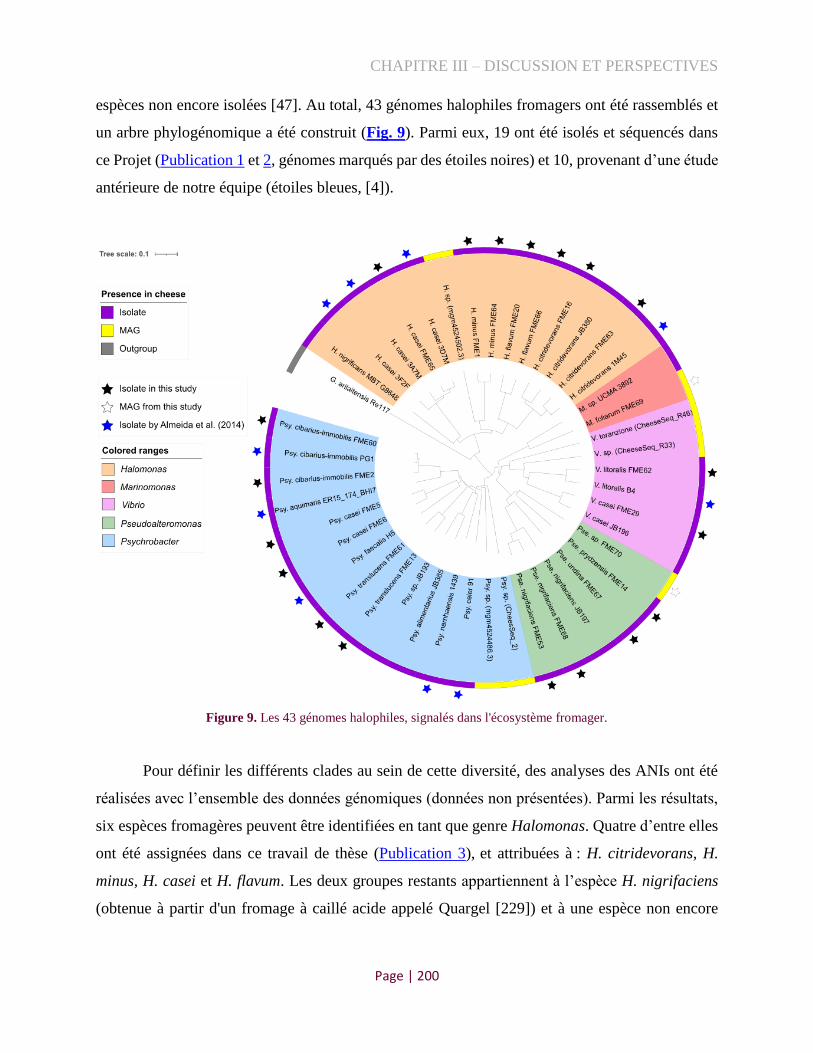
CHAPITRE III – DISCUSSION ET PERSPECTIVES
Page | 200
espèces non encore isolées [47]. Au total, 43 génomes halophiles fromagers ont été rassemblés et
un arbre phylogénomique a été construit (Fig. 9). Parmi eux, 19 ont été isolés et séquencés dans
ce Projet (Publication 1 et 2, génomes marqués par des étoiles noires) et 10, provenant d’une étude
antérieure de notre équipe (étoiles bleues, [4]).
Figure 9. Les 43 génomes halophiles, signalés dans l'écosystème fromager.
Pour définir les différents clades au sein de cette diversité, des analyses des ANIs ont été
réalisées avec l’ensemble des données génomiques (données non présentées). Parmi les résultats,
six espèces fromagères peuvent être identifiées en tant que genre Halomonas. Quatre d’entre elles
ont été assignées dans ce travail de thèse (Publication 3), et attribuées à : H. citridevorans, H.
minus, H. casei et H. flavum. Les deux groupes restants appartiennent à l’espèce H. nigrifaciens
(obtenue à partir d'un fromage à caillé acide appelé Quargel [229]) et à une espèce non encore

CHAPITRE III – DISCUSSION ET PERSPECTIVES
Page | 201
isolée (MAG), obtenue à partir d’un fromage à croûte lavée, fabriqué en Rhône-Alpes, France
[47].
Concernant les Marinomonas, ils sont moins fréquemment signalés dans l'alimentation et
ne sont représentés que par deux espèces dans les fromages (Fig. 9). À notre connaissance, un seul
génome a été séquencé à partir d’une souche du fromage pour ce genre (Marinomonas sp. UCMA
3802, isolé du Livarot [228]), dont l’assignation à une espèce reste à établir. La deuxième espèce
de Marinomonas est actuellement composée d'un seul représentant alimentaire, correspondant au
génome reconstitué dans cette étude (Publication 2, souche FME69), qui peut être attribué à
l’espèce Marinomonas foliarum en raison de sa proximité avec la souche type respective CECT
7731T (ANI=97%).
Dans le groupe des Pseudoalteromonas, trois espèces, parmi les quatre détectées, ont des
représentants isolés. L’espèce Pse. nigrifaciens est celle qui est le plus souvent détectée dans les
croûtes de fromage, mais nous avons aussi isolé des souches représentatives des espèces Pse.
prydzensis et Pse. undina. La dernière espèce de Pseudoalteromonas, représentée ici par un MAG
(souche FME70) ne correspond à aucun représentant isolé, mais son génome a été reconstitué dans
notre étude (Publication 2). Walsh et al. [47] ont aussi obtenu un MAG de cette même espèce. Il
est possible que ces MAGs proviennent d’un fromage avec la même appellation, puisque les deux
MAGs sont originaires de croûtes de fromage au lait de vache non pasteurisé d'Auvergne, France.
Nous avons observé une très grande diversité d'espèces dans les fromages au sein du genre
Psychrobacter, constitué de onze clades (Fig. 9). Deux souches obtenues dans ce travail, FME2 et
FME60, partagent des ANI avec un pourcentage très proche, entre 95-96%, avec Psy. cibarius et
Psy. immobilis, indiquant qu'elles sont soit au seuil de résolution d'une nouvelle espèce, soit
qu'elles pourraient être assignées à l'une de ces deux espèces. Une analyse plus approfondie,
incluant des caractéristiques phénotypiques, génomiques et chimiotaxonomiques, doit être
effectuée pour vérifier leur taxonomie. En outre, deux espèces détectées dans les fromages ont été
attribuées au cours de ce travail, à : Psy. casei et Psy. translucens (Publication 4). D'autres espèces
de Psychrobacter, décrites notamment par Almeida et al. [4], sont également identifiées dans les
croûtes de fromage, telles que Psy. alimentarius, Psy. aquimaris, Psy. celer, Psy. faecalis et Psy.
namhaensis. Cependant, trois groupes restent encore sans assignation d’espèce dans ce genre, dont
deux correspondent aux MAGs récemment reconstituées par Walsh et al. [47], à partir de
métagénomes de fromages irlandais, et le troisième, identifié comme la souche JB193, qui a été

CHAPITRE III – DISCUSSION ET PERSPECTIVES
Page | 202
isolée par Wolfe et al. [3] (fromage non mentionné) dont le représentant le plus proche est Psy.
alimentarius (ANI=90 %).
Nous avons également mis en évidence la présence de plusieurs espèces de Vibrio dans les
fromages, dont quatre clades distincts ont été identifiés. Les deux groupes les plus répertoriés dans
les fromages correspondent à V. casei et V. litoralis et disposent de représentants isolés, y compris
dans cette étude (FME29 et FME62, respectivement). Les deux autres espèces ne comportent
aucun isolat alimentaire, seulement des MAGs récupérés à partir de fromages irlandais [47]: l'un
d'eux peut être attribué à Vibrio toranzoniae (ANI>97,7% avec la souche type CECT 7225T),
tandis que l’autre espèce reste encore sans assignation.
En conclusion, ce bilan nous a permis d'identifier une grande diversité de génomes
halophiles fromagers appartenant à 27 espèces distinctes (6 Halomonas, 2 Marinomonas, 4
Pseudoalteromonas, 11 Psychrobacter et 4 Vibrio). Les isolats disponibles forment un ensemble
de références d’espèces de bactéries alimentaires, qui mériterait d’être exploré pour leur potentiel
métabolique, leur rôle technologique et leurs applications biotechnologiques. Cependant, sept
espèces n'ont pas encore été cultivées et ne sont présentées que sous forme de MAGs. Afin de
guider l’isolement de ces espèces et de connaître la représentativité des autres espèces pour évaluer
leurs potentiels, il est utile de connaître dans quels types de fromages il faut aller les chercher.
Ainsi, dans la section suivante, nous discuterons de la fréquence et de l'abondance de ces 27
espèces halophiles dans les croûtes de fromage.
Les protéobactéries comme population bactérienne dominante dans les croûtes de fromages
Afin de fournir une vision complète des bactéries halophiles présentes dans les croûtes de
fromage, un génome de chaque espèce a été utilisé comme référence pour les détecter et les
quantifier au niveau des données métagénomiques. Pour cela, nous avons sélectionné un ensemble
de 114 échantillons correspondant à des croûtes de fromage de différents types, dont 49 provenant
de notre étude (Publication 1) et 65 d'études précédentes [3, 4, 47, 230] (Tableau S1, Annexe 3).
Parmi les croûtes de fromage analysées, seules 13 ne présentent pas de niveaux détectables
(pourcentage au moins égal à 0,1%), pour les espèces halophiles des genres Halomonas,
Marinomonas, Pseudoalteromonas, Psychrobacter et Vibrio. Celles-ci correspondent à différents
types de croûtes, mais plus particulièrement, les naturelles et fleuries, avec pâtes molles à dures,
incluant un fromage à pâte persillée. Les abondances relatives des reads, correspondantes aux 27

CHAPITRE III – DISCUSSION ET PERSPECTIVES
Page | 203
génomes de référence cartographiés sur les 101 métagénomes restants, sont présentées dans la Fig.
10. En particulier, 32 échantillons à pâte molle et croûte lavée, présentent des abondances relatives
supérieures à 10 % des reads de ces bactéries, ce qui montre leur importance dans ces produits. Il
est intéressant de noter que ces espèces se retrouvent non seulement dans les fromages français,
mais aussi dans les fromages irlandais, anglais et nord-américains.
Bien que différentes méthodes de regroupement aient été testées et une tendance des
bactéries halophiles a été observée dans les pâtes molles et croûtes lavées, la classification par
mode technologique et par type de croûte de fromage n’a pas permis de faire ressortir des
corrélations claires. De plus, certaines études ne mentionnent pas toutes les caractéristiques des
métagénomes (marquées comme « unknown »), et la définition des catégories est alors délicate.
Cette incertitude peut concerner, par exemple, les fromages qui renferment des levures dans leur
croûte (que l'on pourrait définir comme une croûte fleurie), mais qui ont aussi été frottés avec de
l'eau salée (croûte lavée) (Cahier de charges du Saint Nectaire, 2017). Ainsi, cette classification
peut rester très subjective selon la personne qui la catégorise, et, également, présenter des
tendances différentes, selon le type de pâte. De plus, nous ne disposons pas d'information sur le
temps d'affinage de ces fromages, ce qui pourrait être une donnée intéressante pour déduire la
cinétique d’implantation des halophiles à la surface des fromages, depuis le début de la production
jusqu’à leur sortie des caves d'affinage. Des recherches futures peuvent être menées pour étudier
l'évolution de ces microorganismes tout au long de la production, ainsi que pour identifier quels
matériaux et ingrédients, utilisés dans la fromagerie, sont les vecteurs des bactéries halophiles.
Parmi les espèces analysées, les dominantes (au moins cinq échantillons >10%) et
fréquentes (au moins 30 échantillons > 0,1%), dans les croûtes de fromages, sont : Psy. cibarius-
immobilis, Psy. translucens, H. citridevorans e Pse. nigrifaciens, dont certaines pourraient
interagir, car elles sont détectées fréquemment ensemble et potentiellement associées. Concernant
les espèces non encore isolées, à l'exception de Psychrobacter sp. (CS_2), elles apparaissent
fréquemment et parfois en abondance, en particulier les espèces d’Halomonas sp.
(mgm4524502.3), Psychrobacter sp. (mgm4524486.3) et Vibrio sp. (CS_R33). Elles ont été
détectées principalement dans les croûtes lavées ou naturelles des fromages à pâte molle français
ou irlandais. Un travail plus approfondi dans ce sens devrait contribuer à reconstituer des
écosystèmes fromagers afin d’étudier le rôle de cette flore tolérante au sel.

Page | 204
Figure 10. Heatmap illustrant l'abondance relative (%) de 27 espèces halophiles dans 101 croûtes de fromage.
CHAPITRE III – DISCUSSION ET PERSPECTIVES
Page | 205
Les résultats obtenus peuvent également guider la culture des espèces non encore isolées,
en ciblant les fromages dont elles sont dominantes. Ensuite, nous avons proposé, dans cette étude,
une stratégie pour faciliter et cibler l’isolement de représentants d’espèces détectés par la présence
de leur séquence, en concevant des sondes spécifiques pour les révéler rapidement à partir de
clones isolés sur boîte. Cette stratégie s’est montrée efficace dans de nombreux cas. Cependant,
nous n'avons pas réussi à cultiver deux espèces (Marinomonas foliarum FME69 et
Pseudoalteromonas sp. FME70), malgré des conditions initiales favorables dont leur forte
abondance (Publication 2). Compte tenu du fait que notre analyse génomique n’a pas révélé de
différence notable avec leurs plus proches parentes cultivables, concernant les cellules
bactériennes, l’hypothèse qui nous parait la plus plausible est que celles-ci aient été détruites par
la congélation, sachant que les échantillons ont été conservés à une température de -20 °C. Ainsi,
nous suggérons, pour les travaux futurs, de tester leur présence dans une série d’échantillons de
croûtes de fromages, sans les congeler, et aussi, d’explorer une plus large gamme de milieux de
culture. En effet, même si le milieu Marine Agar s’est avéré être le plus efficace, lors de notre
travail pour isoler les bactéries halophiles, d'autres milieux pourraient être utilisés pour augmenter
la probabilité d'isolement. Par exemple, une étude récente sur la flore halophile des saumures de
fromage a utilisé deux milieux de culture à forte concentration en sel : (i) GYPB, composé de
glucose, extrait de levure, peptone, extrait de bœuf, avec sel et (ii) MGM, composé d'extrait de
malt, d'extrait de levure et de sel [100].
2. Peut-on retracer l’évolution des bactéries halophiles dans l’écosystème
fromager?
Dans le but de mieux caractériser la diversité des halophiles du fromage appartenant aux
genres Halomonas, Marinomonas, Pseudoalteromonas, Psychrobacter et Vibrio, les génomes de
représentants de différentes espèces issues d’autres écosystèmes ont été collectés dans la base de
données NCBI. Des arbres phylogénomiques ont été construits, en indiquant leurs habitats
(fromage, autres aliments, environnement et contexte humain, figures supplémentaires S1-S5,
Annexe 3). À l'exception de Psychrobacter, pour lequel les représentants alimentaires semblent
dominer, les autres genres sont majoritairement issus d’écosystèmes naturels.

CHAPITRE III – DISCUSSION ET PERSPECTIVES
Page | 206
De plus, il est intéressant de noter que la plupart des souches issues des fromages sont très
proches génétiquement de certaines espèces environnementales. Pour illustrer plus simplement
cette proximité, un arbre simplifié, comprenant les 27 espèces halophiles identifiées dans les
fromages, ainsi que des souches proches d'autres habitats (dont l'ANI représente plus de 90%) a
été construit (Fig. 11).
Figure 11. Espèces halophiles identifiées dans l’écosystème fromager et génomes d'autres habitats, étroitement liés
(ANI>90%).
On constate ici que la plupart des génomes provenant des fromages sont apparentés avec
des souches environnementales (principalement des écosystèmes terrestres et marins), parfois au
niveau de l’espèce. En ce qui concerne les Halomonas, la souche H. zhanjiangensis DSM 21076T,
isolée à partir d'un oursin de mer, présente un ANI de 93% avec l'espèce fromagère H. flavum,
toutes deux ayant des caractéristiques similaires, comme la coloration jaune, visible dans leurs

CHAPITRE III – DISCUSSION ET PERSPECTIVES
Page | 207
colonies. En outre, H. casei est étroitement apparenté (ANI~95-96%) aux espèces H.
hydrothermalis, H. venusta, H. alkaliphila et H. campaniensis, toutes, isolées du milieu marin.
Pour les autres genres étudiés, la majorité des espèces de fromage sont également représentés dans
les habitats naturels (Fig. 11), à l’exception de V. casei et Psy. casei. Enfin, il est intéressant de
noter que les MAGs dont les représentants restent à isoler (Halomonas sp., Vibrio sp.,
Pseudoalteromonas sp. et Psychrobacter spp.) n'ont pas non plus de représentants au niveau d’un
génome proche isolé. Il est pertinent de se demander si ces absences révèleraient une difficulté
particulière concernant la culture de souches appartenant à ces espèces. Enfin, nous constatons la
présence d'espèces isolées au sein d'autres aliments, génétiquement proches des souches
fromagères et environnementales. C'est le cas de la Pse. nigrifaciens DSM 6063T, isolée à partir
du beurre, et de la Psy. alimentarius JG-102T et Psy. cibarius JG -219T, tous deux isolées du
jeotgal, un aliment fermenté traditionnel coréen, à base de fruits de mer.
Cette analyse préliminaire indique que les bactéries alimentaires sont peut-être issues de
souches appartenant à l'environnement naturel. Pour mieux répondre à cette hypothèse, des
analyses comparatives de génomes pourraient être effectuées pour mettre en évidence les gènes
présents dans les bactéries du fromage et absents dans les autres milieux, ou vice-versa, et ainsi
indiquer les processus d'adaptation potentiels pour coloniser les écosystèmes alimentaires.
3. Quel est le rôle des halophiles dans la technologie fromagère ?
La fréquence et parfois l'abondance de certaines espèces halophiles dans les croûtes de fromage
(Fig. 10) suggèrent que celles-ci pourraient jouer un rôle technologique sous-estimé jusqu'à
présent. Cette constatation a motivé une première approche pour tester les impacts technologiques
potentiels des souches halophiles dans un modèle fromage-agar. Pour cela, nous avons d'abord
rassemblé un certain nombre d'isolats des genres Halomonas, Pseudoalteromonas et
Psychrobacter, provenant des écosystèmes alimentaires et naturels, soit un total de 43 souches
(Tableau 1, Publication 5). Les souches issues d’environnement non fromager ont été introduites
pour tester en parallèle leurs potentiels. À partir de cette collection, nous avons testé la capacité
d'implantation de chaque souche, individuellement, dans un modèle de fromage synthétique, et
leur impact sur le pH et la production de composés volatils, après 21 jours d'incubation à 15 °C
(en simulant un affinage initial). En général, on observe une bonne capacité de croissance des

CHAPITRE III – DISCUSSION ET PERSPECTIVES
Page | 208
souches halophiles dans le modèle fromage-agar, y compris pour les souches environnementales.
En ce qui concerne les souches fromagères, on observe que certaines d'entre elles (notamment les
souches de Psy. casei FME6 et Psy. namhaensis 1439) peuvent légèrement acidifier le milieu et
produire une série d'acides carboxyliques et de cétones qui pourraient renforcer la qualité
aromatique des fromages. De plus, deux souches de Pseudoalteromonas nigrifaciens (FME68 et
FME53) produisent des pyrazines et des cétones ainsi que des composés soufrés, éléments pouvant
contribuer à l'arôme lors de l'affinage du fromage. Ces classes de composés volatils ont déjà été
décrites dans l’étude des fromages [231]. Selon la quantité produite ainsi que les préférences des
consommateurs en matière de goût, elles peuvent être considérées comme des odeurs déplaisantes
ou souhaitables.
Ces recherches préliminaires nous permettent d'envisager des analyses plus approfondies
concernant les souches qui ont démontré des potentiels technologiques prononcés. Par ailleurs,
est-il possible de considérer que certaines souches halophiles peuvent être inoculées, comme
ferments, dans les surfaces de fromage ? Bien que certains auteurs puissent être sceptiques, face à
une réponse affirmative à cette question, puisque les espèces étudiées appartiennent au groupe des
protéobactéries qui leur sont normalement associées comme contaminants ou élèments d’altération
des aliments, certaines souches étudiées ici ont montré une utilisation potentielle dans la
technologie fromagère. Toutefois, les perspectives de travail suivantes restent ouvertes à une
exploration détaillée :
a) Comprendre l’implantation et le rôle des halophiles dans l’écosystème fromager
Selon le heatmap présenté ci-dessus, les bactéries halophiles sont dominantes essentiellement
dans les fromages à croûte lavée (Fig. 10), tels que le Livarot, l'Epoisses et le Saint Nectaire. Parmi
les espèces dominantes dans ces fromages figurent les levures Geotrichum candidum et
Debaryomyces hansenii, et les espèces gram-négatives H. citridevorans, Pse. nigrifaciens, Psy.
cibarius/immobilis et Psy. translucens [3, 4, 48, 232]. Ces fromages sont fabriqués à partir de la
coagulation lactique ou mixte avec des ferments mésophiles, tels que L. lactis subsp. cremoris sans
chauffage ou avec un chauffage léger (<40 °C) et sont principalement des fromages à pâte molle
(Fig. 1). Par conséquent, la surface de ces caillés reste relativement humide, ce qui favorise
probablement la croissance des bactéries halophiles. Dans le présent travail, nous avons eu pour
approche de sélectionner des fromages finis et de déconstruire leurs croûtes microbiologiquement.
CHAPITRE III – DISCUSSION ET PERSPECTIVES
Page | 209
Il aurait sans doute été souhaitable de réaliser des mesures supplémentaires pour mieux caractériser
les paramètres environnementaux qui ont permis le développement des différents microbiotes dans
chaque fromage et pour comprendre les éventuelles successions d’espèces au cours de la
production.
Notre travail a, cependant, fourni une « image » finale contenant, exhaustivement, les
différents acteurs qui se sont développés à la surface des fromages. De plus, nous avons sauvegardé
dans nos congélateurs un ensemble de souches isolées, qui pourraient être utilisées pour des
expériences de reconstruction d’écosystèmes fromagers afin de mieux comprendre leur rôle dans
ce produit. Par exemple, différentes souches d’espèces halophiles que nous avons isolées
pourraient être testées en interaction entre elles et avec les levures citées précédemment dans des
modèles de fromages afin d'effectuer un premier criblage des interactions positives (comme la
croissance de la souche halophile stimulée par la présence de la levure) ou négatives (souche
halophile inhibée).
En effet, il a été montré dans les fromages que les LAB pouvaient générer un large spectre de
molécules telles que les acides organiques, le dioxyde de carbone, l'éthanol, le peroxyde
d'hydrogène et le diacétyle, dont les actions antimicrobiennes sont bien connues [233]. Il en est
sans doute de même pour les bactéries halophiles et il est intéressant de noter que dans notre étude
(Publication 5), deux souches de Psychrobacter (FME6 et 1439) ont produit, en quantité
importante, plusieurs acides organiques ainsi que du diacétyle, qui pourraient avoir une fonction
d'interférence dans la croissance d'autres microorganismes présents dans les fromages. Ainsi,
certaines molécules sécrétées par ces bactéries pourraient exercer des actions positives telles que
la production de substrats ou de facteurs de croissance sur d'autres bactéries. Ces possibilités
méritent d'être vérifiées, non seulement pour les bactéries contaminantes et pathogènes, mais aussi
en présence d’autres espèces technologiques que l'on peut trouver à la surface des fromages, telles
que Brevibacterium aurantiacum, Brachybacterium tyrofermentans, Corynebacterium casei,
Glutamicibacter arilaitensis et Staphylococcus equorum. On peut également se demander si les
échecs répétés dans l'isolement des souches Pseudoalteromonas sp. MAG-FME70 et
Marinomonas foliarum MAG-FME69 pourraient être dus à une telle action d'une (ou plusieurs)
bactérie(s) présente(s) dans l'écosystème fromager ?
Nous avons aussi remarqué que certaines espèces halophiles formaient des biofilms différents
à la surface de nos modèles de caillé-agar, ce qui suggère la production d’autres type de composés

CHAPITRE III – DISCUSSION ET PERSPECTIVES
Page | 210
importants dans la construction des écosystèmes, comme les polysaccharides [234, 235]. Ces
molécules pourraient avoir un rôle important non seulement pour la construction de l'écosystème,
mais aussi pour la capacité de rétention d'humidité du fromage et sa texture. En outre, il a été
démontré que de nombreuses bactéries halophiles environnementales synthétisent et accumulent
souvent des polysaccharides extracellulaires (EPS) en tant que stratégies adaptatives face aux
fortes pressions salines, qui diffèrent considérablement par leurs propriétés physiques, chimiques
et structurelles [236].
Enfin, ces interactions, lors de leur développement, pourraient être explorées dans des modèles
de fromage avec des analyses simultanées de métatranscriptomes (RNA-Seq) et de composés
volatils (GC-MS), afin de mieux caractériser les facteurs impliqués dans ces interactions et le rôle
des bactéries halophiles, d’un point de vue écologique mais aussi technologique. Il est important
de noter que toutes les souches appartenant à la même espèce n'ont pas nécessairement le même
comportement physiologique. Ce fait a, par exemple, été observé dans nos analyses des composés
volatils (Publication 5), pour lesquels des souches appartenant à Pseudoalteromonas nigrifaciens
présentaient des capacités de croissance, des valeurs de pH et de production de composés volatils,
très distinctes entre elles.
b) Définir l’aptitude technologique des halophiles
Après avoir mieux compris le rôle et la place des halophiles dans l’écosystème des surfaces
des fromages, il sera possible d’en définir leurs potentialités technologiques. Des tests
préliminaires sur les propriétés fonctionnelles tel que celui que nous avons conduit (Publication
5), devraient aussi être pris en compte pour proposer différentes formulations qui pourraient être
testées sur des modèles de fromages, dont les caractéristiques technologiques s’approcheraient de
ceux mentionnées ci-dessus (fromages à pâtes molles et croûtes lavées). Faisant suite à
l'élaboration de ces modèles de fromage, les analyses microbiologiques et métagénomiques, ainsi
que les analyses de la composition du fromage (teneur en sel, humidité, matières grasses, protéines,
pH) et les analyses biochimiques (activités enzymatiques, protéolyse, acides aminés libres)
pourraient être effectuées à différents intervalles de temps d’affinage (entre 21 et 90 jours, par
exemple). Afin de mieux cerner leurs propriétés et leurs potentialités technologiques, il serait
également important de compléter ces études par des essais de détection de profils aromatiques
pour associer « métabolisme » et « arôme ».

CHAPITRE III – DISCUSSION ET PERSPECTIVES
Page | 211
Les activités métaboliques produites par les souches halophiles peuvent être étudiées via des
approches métabolomiques, à coupler avec des analyses métatranscriptomiques, comme
mentionné précédemment, permettant l'exploration des gènes exprimés par ces bactéries, lors de
l'affinage des fromages, et, en phase ultime, l’étude de leurs assignations fonctionnelles. Il serait
également intéressant de découvrir si ces souches produisent des molécules inhibitrices et/ou ont
un effet compétitif face aux bactéries pathogènes et bactéries d'altération. Par exemple, de
nombreuses souches de bactéries lactiques (telles que L. lactis et Lactiplantibacillus plantarum),
ajoutées comme ferments dans la production de fromages, produisent de la nisine et de la pédiocine
[237-239], des bactériocines qui peuvent inhiber le développement de Listeria monocytogenes et
de Staphylococcus aureus, qui comptent parmi les agents pathogènes les plus préoccupants pour
l'industrie laitière [240]. Certaines souches de Pseudoalteromonas ont des activités
antibactériennes et antivirales, comme Pse. undina [241], espèce qui a été détectée dans certains
fromages, dans le cadre de cette étude (Fig. 10). De plus, une souche de Pse. aliena (LCCM
11207P), isolée à partir d'un produit fermenté coréen, a montré un effet inhibiteur significatif
contre le Vibrio harvery [202].
D’un autre côté, certaines études considèrent que les bactéries halophiles sont impliquées dans
l’altération des aliments et sont donc considérées comme indésirables dans les environnements de
transformation des aliments [217-219]. C'est le cas, par exemple, d’espèces de
Pseudoalteromonas, isolés à partir de la crevette brune, en particulier, Pse. elyakovii et Pse.
nigrifaciens, qui ont été associées à la production de grandes quantités de composés soufrés
(hydrure de soufre, méthanethiol et disulfure de diméthyle), ainsi que d’acétone, d’acide acétique
et d’ammoniac, ce qui peut entraîner un rejet sensoriel du produit [216]. De plus, cette même étude
a indiqué que les espèces de Pseudoalteromonas et Psychrobacter ont la capacité de dégrader les
lipides et d'hydrolyser les acides aminés et les protéines, ce qui peut augmenter le potentiel
d’altération des aliments. Cependant, d'autres auteurs signalent que les composés volatils produits
par les halophiles peuvent avoir un impact positif pendant l'affinage du fromage [215, 242, 243].
En outre, certaines souches de Psychrobacter, isolées à partir de la surface de fromages défectueux
(espèces Psy. pulmonis, Psy. aquimaris et Psy. okhotskensis), ont été identifiées comme
responsables des pigments violets sur les croûtes, ainsi que certaines espèces de Proteus. Ce défaut
a été associé à la production de composés indigoïdes (indigo et indirubine), bien que la provenance
de ses précurseurs métaboliques dans le fromage soit actuellement inconnue [244]. Dans ce cas,

CHAPITRE III – DISCUSSION ET PERSPECTIVES
Page | 212
les souches productrices de pigments ne sont pas mentionnées comme une menace pour les
consommateurs, mais comme ayant un impact négatif sur la qualité du fromage.
c) S’assurer de l’innocuité des halophiles et de leur impact sur la santé
Bien que certaines espèces halophiles soient dominantes dans les croûtes de fromage et que la
population en consomme régulièrement, ce qui suggère qu'elles ne sont pas pathogènes, il est
important de s’assurer pleinement que la présence de ces microorganismes n'est pas nuisible à la
santé humaine. La littérature scientifique est peu abondante sur ce sujet, mais apporte quand même
quelques éléments concernant l’utilisation de quelques espèces d’halophiles des genres
Halomonas, Pseudoalteromonas et Psychrobacter ayant des effets potentiels bénéfiques ou
indésirables dans l'alimentation.
Concernant le genre Halomonas, Bourdichon et al. (2012) considèrent que l'espèce H. elongata
joue un rôle technologique dans les viandes fermentées [17]. En effet, cette espèce peut être une
bonne candidate comme ferment dans la saumure utilisée pour la préparation du jambon cru affiné
(Brevet allemand, DE 4035836 C2), car certaines souches peuvent contribuer à améliorer la qualité
sensorielle de ce produit [245]. En revanche, d’autres espèces d’Halomonas telles que H. stevensii,
H. hamiltonii et H. johnsoniae ont été isolées dans un centre de dialyse sur deux patients souffrant
de bactériémie, et pourraient donc constituer un facteur pathogène chez l'homme [246].
D'autre part, certaines espèces de Pseudoalteromonas produisent des toxines qui peuvent
causer la mortalité ou l'empoisonnement des poissons (Pe. piscicida et Pe. tetraodonis) [247, 248].
En revanche, il serait intéressant de tester le rôle de certaines souches au niveau intestinal en faisant
le parallèle avec la souche de Pse. aliena (LCCM 11207P), cité précédemment, qui a un effet
inhibiteur sur le Vibrio harvery, un pathogène grave chez les poissons et les invertébrés, en
mariculture [202].
Le genre Psychrobacter est rarement considéré comme opportuniste chez l'homme, mais
une étude a indiqué que la souche 310 (proche de l'espèce Psy. sanguinis), isolée chez un patient
atteint de méningite, possède plusieurs gènes de virulence, caractéristiques des bactéries
pathogènes, notamment des gènes jouant un rôle dans la résistance aux antimicrobiens, l'invasion
des cellules, l'absorption du fer et le système de sécrétion de type IV [249]. En outre, d'autres
espèces telles que Psy. phenylpyruvicus, Psy. pasteurii et Psy. piechaudii, ont également été isolées
chez l'homme et peuvent être responsables d'infections oculaires et d'endocardites [250, 251].

CHAPITRE III – DISCUSSION ET PERSPECTIVES
Page | 213
Enfin, il faut tenir compte, qu’au sein d’un même genre, chaque espèce peut avoir des
caractéristiques très différentes, comme dans le cas du Staphylococcus. Par exemple, S. aureus est
considéré comme un pathogène dangereux [252], tandis que S. equorum est un ferment avec une
utilisation technologique dans l'affinage du fromage [253]. Heureusement, nous avons observé
qu'aucune espèce isolée chez l’homme n'est étroitement associée aux génomes alimentaires (Fig.
11, Fig. S1-S5, Annexe 3), suggérant qu’il y a des différences significatives entre les souches
présentes dans l’alimentation et celles détectées dans des environnements hospitaliers.

ANNEXE 1– Publication 6
Page | 214
ANNEXE 1
1. Analyse génomique de deux souches halophiles marines
1.1. Contexte
L'intérêt que nous portons à l'origine potentielle des bactéries alimentaires halophiles au sein
les milieux naturels nous a conduits à une collaboration avec Ifremer (Institut Français de
Recherche pour l'Exploitation de la Mer). Dans le cadre de cette coopération, il a été fourni huit
séquences d'ARNr 16S, issues d’espèces non définies de bactéries marines des genres Halomonas,
Psychrobacter et Pseudoalteromonas. Ces séquences ont été placées dans un arbre phylogénétique
avec les espèces fromagères isolées dans les Publications 1 et 2, (voir Figure 12 ci-dessous).
Figure 12. Arbre phylogénétique basé sur les alignements des gènes de l'ARNr 16S de souches marines (en bleu) et
fromagères (en noir).
Parmi ces souches, Pseudoalteromonas sp. MIP2626 et Psychrobacter sp. BI730 ont été
choisies pour effectuer une analyse génomique plus approfondie, fonction de leurs proximités avec
des espèces fromagères.
Psychrobacter cibarius-immobilis
Psychrobacter casei
Psychrobacter translucens
Psychrobacter sp. BI730
Halomonas casei
Halomonas sp. 729
Halomonas minus
Halomonas flavum
Halomonas citridevorans
Pseudoalteromonas sp. 791
Pseudoalteromonas sp. 895
Pseudoalteromonas sp. 738
Pseudoalteromonas prydzensis
Pseudoalteromonas sp. 974
Pseudoalteromonas sp. 883
Pseudoalteromonas undina
Pseudoalteromonas nigrifaciens
Pseudoalteromonas sp. MIP2626
94
42
100
62
76
64
100
100 78
100
79
98
82
100
55
0.020

ANNEXE 1– Publication 6
Page | 215
1.2. Publication 6
Draft-genome sequence data and phylogenomic comparison of two marine-sourced
bacterial strains Pseudoalteromonas sp. MIP2626 and Psychrobacter sp. BI730
Caroline Isabel Kothea, Christine Delbarre-Ladratb, Pierre Renaulta, Delphine Passerinib
a Micalis Institute, INRAE, AgroParisTech, Université Paris-Saclay, Jouy-en-Josas, 78350, France
b Ifremer, BRM, EM3B Laboratory, F-44311 Nantes, France
Abstract
Halophilic and psychrophilic marine bacteria are source of interesting bioactive molecules for
biotechnology. We report here the whole-genome sequences of two of them, Pseudoalteromonas
sp. MIP2626 isolated from tropical peeled shrimps and Psychrobacter sp. BI730 isolated from
deep-sea hydrother-mal vent. Sequencing of both genomes was performed by Illumina HiSeq
platform (2×150 pb). De novo assemblies using SPAdes v3.9 generated 136 contigs for
Pseudoalteromonas MIP2626 and 42 contigs for Psychrobacter BI730, representing a genome
size of 3.9 Mb and 3.2 Mb, respectively. Phylogenetic based on 16S rRNA gene sequence and
phylogenomic analyses were reported to compare the new sequences with Pseudoalteromonas and
Psychrobacter representative strains available in the public databases. The genome sequences have
been deposited at GenBank under the accession num-bers JAATTW000000000 for
Pseudoalteromonas sp. MIP2626 and JAATTV000000000 for Psychrobacter sp. BI730.
Keywords: Aquatic, Genomic, Phylogenetic analysis, Halophilic
Les résultats obtenus ont été publiés sous la forme d’un article de données, dans la revue
Data in Brief et constituent la section 1.2 de l’Annexe 1.

ANNEXE 1– Publication 6
Page | 216
Specifications Table
Subject Molecular biology
Specific subject area Microbiology and Genomics
Type of data Draft genome sequences in FASTA format
Tables
Figures
Mtsx tree files (phylogenetic 16S)
Newick tree and svg files (phylogenomic)
How data were acquired We extracted the genomic DNA using phenol-chloroform protocol [1] and whole
genome sequencing was performed through the Illumina HiSeq plataform
Data format Raw and analyzed data
Parameters for data
collection Genomic DNAs were extracted from pure cultures of MIP2626 and BI730 strains
Description of data
collection
Whole-Genome sequencing, assembly, annotation, phylogenetic and
phylogenomic comparisons.
Genomes were assembled with de novo assembly using SPAdes version 3.9
[2] and annotated with Rapid Annotations Subsystems Technology (RAST) [3].
Phylogenetic analysis of 16S rRNA were performed using MEGA 7.0.26 [4].
Multiple Alignment of Conserved Genomic Sequence With Rearrangements
(MAUVE 2.4.0) was used for aligned the whole-genome sequences [5]. For the
annotation and management of phylogenomic trees, we used iTOL v5
(http://itol.embl.de).
Data source location Pseudoalteromonas sp. MIP2626 was isolated in France from tropical peeled
shrimps in 2009 and Psychrobacter sp. BI730 from deep-sea hydrothermal vent
site in Lau Basin, in the Southwestern Pacific Ocean, in 1989.
Data accessibility The genome of the MIP2626 strain has been deposited in NCBI database under
the Bioproject PRJNA604092, the Biosample SAMN13951749 and the genome
accession number JAATTW000000000.
The genome of the BI730 strain has been deposited in NCBI database under
the Bioproject PRJNA604092, the Biosample SAMN13951813 and the genome
accession number JAATTV000000000
All data are available in a public repository:
Repository name: Draft-genome sequence data of Pseudoalteromonas sp.
MIP2626 and Psychrobacter sp. BI730
Direct URL to data: http://dx.doi.org/10.17632/kbdfk2d7vy.1
Value of the Data
The data contribute to describe the genomic diversity of the Pseudoalteromonas and
Psychrobacter species;
Genome sequence data of halophilic strains from marine origin are useful for comparative
genomic analysis and highlight the genetic adaptation of these species in food ecosystems;
Genome sequences provide new knowledge on Pseudoalteromonas and Psychrobacter
species of biotechnological importance.

ANNEXE 1– Publication 6
Page | 217
1. Data Description
Pseudoalteromonas and Psychrobacter species are Gram-negative moderate halophilic
bacteria that could be isolated in different natural ecosystems like soil, salt lake and marine
environment [6], but also in salted food such as seafood, processed meat and cheese [7–9]. As they
are psychrophilic bacteria, both genera are interesting for biotechnological applications [10–13].
In this paper, we present the whole-genome sequences of two strains collected by Ifremer (Nantes,
France) from marine samples. The first strain, Psychrobacter sp. BI730 was isolated in 1989 from
a deep-sea hydrothermal vent site in Lau Basin, in the Southwestern Pacific Ocean, explored
during BIOLAU oceanographic cruise. The second one, Pseudoalteromonas sp. MIP2626, was
isolated in 2009 from tropical peeled shrimps.
Based on the homology of 16S rRNA gene sequences with those of closely type strains
(Additional File 1), Psychrobacter sp. BI730 shares high similarity with Psychrobacter nivimaris
88/2-7T (99.93% for a 1461 bp sequence) (Fig. 1). In the same way, Pseudoalteromonas sp.
MIP2626 strain appears closely related to Pseudoalteromonas nigrifaciens KMM 661T (100% for
a 1458 bp sequence) and Pseudoalteromonas haloplanktis ATCC 14393T (99.18% for a 1467 bp
sequence) (Fig. 2).
Figure 1. Phylogenetic tree of Psychrobacter genus, including Psychrobacter sp. BI730 and Psychrobacter type
strains. The phylogenetic tree is based on 16S rRNA gene alignments obtained by MEGA 7.0.26 software using the
neighbor-joining method. Halomonas elongata DSM 2581T was used as the outgroup.
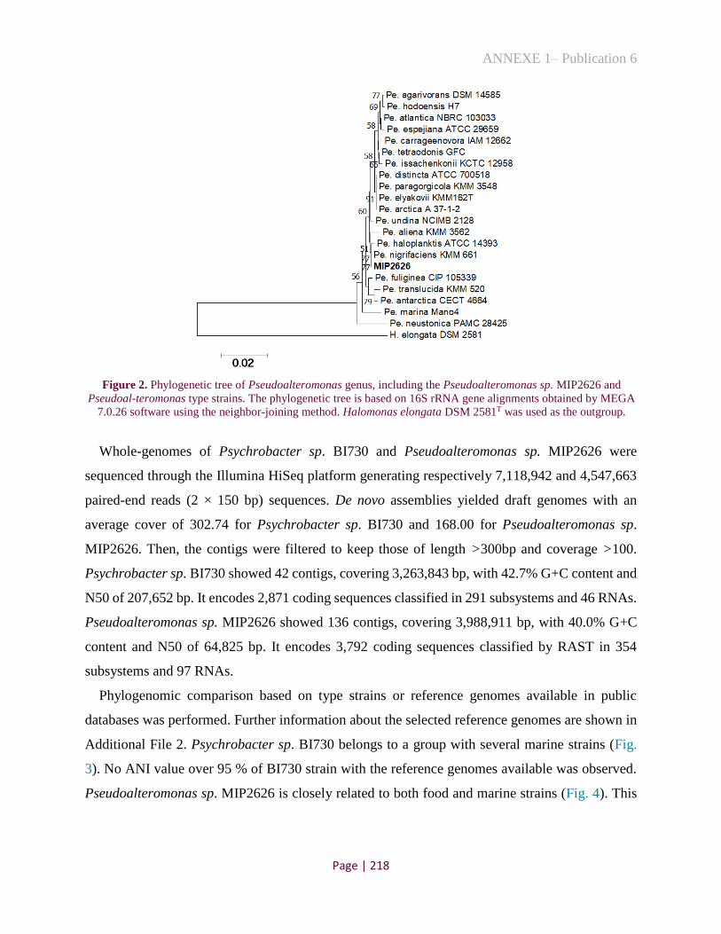
ANNEXE 1– Publication 6
Page | 218
Figure 2. Phylogenetic tree of Pseudoalteromonas genus, including the Pseudoalteromonas sp. MIP2626 and
Pseudoal-teromonas type strains. The phylogenetic tree is based on 16S rRNA gene alignments obtained by MEGA
7.0.26 software using the neighbor-joining method. Halomonas elongata DSM 2581T was used as the outgroup.
Whole-genomes of Psychrobacter sp. BI730 and Pseudoalteromonas sp. MIP2626 were
sequenced through the Illumina HiSeq platform generating respectively 7,118,942 and 4,547,663
paired-end reads (2 × 150 bp) sequences. De novo assemblies yielded draft genomes with an
average cover of 302.74 for Psychrobacter sp. BI730 and 168.00 for Pseudoalteromonas sp.
MIP2626. Then, the contigs were filtered to keep those of length >300bp and coverage >100.
Psychrobacter sp. BI730 showed 42 contigs, covering 3,263,843 bp, with 42.7% G+C content and
N50 of 207,652 bp. It encodes 2,871 coding sequences classified in 291 subsystems and 46 RNAs.
Pseudoalteromonas sp. MIP2626 showed 136 contigs, covering 3,988,911 bp, with 40.0% G+C
content and N50 of 64,825 bp. It encodes 3,792 coding sequences classified by RAST in 354
subsystems and 97 RNAs.
Phylogenomic comparison based on type strains or reference genomes available in public
databases was performed. Further information about the selected reference genomes are shown in
Additional File 2. Psychrobacter sp. BI730 belongs to a group with several marine strains (Fig.
3). No ANI value over 95 % of BI730 strain with the reference genomes available was observed.
Pseudoalteromonas sp. MIP2626 is closely related to both food and marine strains (Fig. 4). This

ANNEXE 1– Publication 6
Page | 219
strain showed an ANI value higher than 95 % with the type strains Pseudoalteromonas nigri-
faciens NCTC10691T (97.84 %) and Pseudoalteromonas translucida KMM 520T (96.1 %).
Figure 3. Phylogenomic comparison of Psychrobacter strains from diverse origins using the software Mauve 2.4.0
and visu-alized with iTOL v5. Halomonas elongata DSM 2581T was used as the outgroup.

ANNEXE 1– Publication 6
Page | 220
Figure 4. Phylogenomic comparison of Pseudoalteromonas strains from marine and food origins using the software
Mauve 2.4.0 and visualized with iTOL v5. Halomonas elongata DSM 2581T was used as the outgroup.
2. Experimental Design, Materials, and Methods
2.1. Genomic DNA extraction
BI730 and MIP2626 strains were grown in Zobell medium at 30°C under aeration and total
genomic DNA was extracted using phenol-chloroform protocol [1].
2.2. Phylogenetic analysis of 16S rRNA
The 16S rRNA gene was amplified using 27-F (5’-AGAGTTTGATCATGGCTCA-3’) and
1492-R (5’-TACGGTTACCTTGTTACGACTT-3’) primers. Thermal cycling conditions were
applied as follow (i) 1 min at 94°C for initial denaturation, (ii) 30 cycles of 1 min at 94°C for

ANNEXE 1– Publication 6
Page | 221
denaturation, 0.5 min at 56°C for primer annealing, 1.5 min at 72°C for elongation, and (iii) 5 min
at 72°C to ensure final elongation. DNA amplicons were separated on 0.8% agarose gel, purified
by using the ExoSAP-IT (Thermo Fisher Scientific, Waltham, Massachusetts, USA) and sent for
sequencing to the service provider (Eurofins Genomics, Ebersberg, Germany). Sequences were
analyzed to obtain a preliminary taxonomic classification for each isolate.
We elaborated an initial phylogeny of the isolates BI730 and MIP2626 using the closest related
16S rRNA gene sequences generated by EzBiocloud blast (https://www.ezbiocloud.net/). The
GenBank accession numbers of these species are shown in Additional File 1. Further phylogenetic
analysis were performed using MEGA 7.0.26 software and trees were generated using the
Neighbor-Joining algorithm with 1000 bootstrap iterations [4].
2.3. Genome sequencing and assembly
Whole genome sequencing was performed through the Illumina HiSeq platform at GATC-
Biotech (Konstanz, Germany) The genomes were assembled with de novo assembly using SPAdes
version 3.9 [2]. Then, the contigs were filtered to keep those of length >300bp and coverage >100.
Annotations were produced using the Rapid Annotations using Subsystems Technology server [3].
2.4. Phylogenomic analysis
Representative complete genome sequences of Psychrobacter and Pseudoalteromonas were
collected from the NCBI database. Information about these genomes such as isolation origin,
Bioproject and BioSample numbers, and Average Nucleotide Identity (ANI) with BI730 and
MIP2626 are shown in Additional File 2. ANI values, were determined by JSpeciesWS [14].
Psychrobacter and Pseudoalteromonas genome sequences selected from the public database were
aligned with the BI730 and MIP2626 strains, respectively, using the software Progressive Mauve
(version 2.4.0) with the default settings [5]. The phylogenomic trees were visualized with
Interactive Tree of Life (iTOL v5) (http://itol.embl.de).
Declaration of Competing Interest
The authors declare that they have no known competing financial interests or personal
relationships which have, or could be perceived to have, influenced the work reported in this
article.

ANNEXE 1– Publication 6
Page | 222
Acknowledgments
This work was supported by “Conselho Nacional de Desenvolvimento Científico e Tecnológico
(CNPq)” – Brazil [grant number 202444/2017-1 to C.I.K.], INRAE and Ifremer.
Supplementary materials
Supplementary material associated with this article can be found, in the online version, at
doi:10.1016/j.dib.2020.105898.
References
[1] F.M. Ausubel, Short protocols in molecular biology: a compendium of methods from Current protocols in
molecular biology, 5th ed., Wiley, New York, 2002.
[2] A. Bankevich, S. Nurk, D. Antipov, et al., SPAdes: a New Genome Assembly Algorithm and Its Applications to
Single-Cell Sequencing, J Comput Biol 19 (2012) 455–477, doi:10.1089/cmb.2012.0021.
[3] R.K. Aziz, D. Bartels, A.A. Best, et al., The RAST server: Rapid annotations using subsystems technology,
BMC Genomics 9 (2008) 75, doi:10.1186/1471-2164-9-75.
[4] S. Kumar, G. Stecher, K. Tamura, MEGA7: Molecular Evolutionary Genetics Analysis Version 7.0 for Bigger
Datasets, Mol Biol Evol 33 (2016) 1870–1874, doi:10.1093/molbev/msw054.
[5] A.C. Darling, B. Mau, F.R. Blattner, N.T. Perna, Mauve: Multiple alignment of conserved genomic sequence
with rearrangements, Genome Res 14 (2004) 1394–1403, doi:10.1101/gr.2289704.
[6] H. Zahran, Diversity, adaptation and activity of the bacterial flora in saline environments, Biol Fertil Soils 25
(1997) 211–223, doi:10.1007/s003740050306.
[7] K. Broekaert, B. Noseda, M. Heyndrickx, G. Vlaemynck, F. Devlieghere, Volatile compounds associated with
Psy-chrobacter spp. and Pseudoalteromonas spp., the dominant microbiota of brown shrimp (Crangon crangon)
during aerobic storage, Int J Food Microbiol 166 (2013) 487–493, doi:10.1016/j.ijfoodmicro.2013.08.013.
[8] I. Ferrocino, A Bellio, A. Romano, G. Macori, K. Rantsiou, L. Decastelli, L. Cocolin, RNA-based amplicon
sequencing reveals microbiota development during ripening of artisanal versus industrial Lard d’Arnad, Appl Environ
Microbiol 83 (2017) e00983-17https://doi.org/, doi:10.1128/AEM.00983-17.
[9] M. Almeida, A. Hébert, A. Abraham, et al., Construction of a dairy microbial genome catalog opens new per-
spectives for the metagenomic analysis of dairy fermented products, BMC Genomics 15 (2014) 1101, doi:10.1186/
1471-2164-15-1101.
[10] S. Bruno, D. Coppola, G. di Prisco, D. Giordano, C. Verde, Enzymes from Marine Polar Regions and Their
Biotechno-logical Applications, Mar. Drugs. 17 (2009) 544, doi:10.3390/md17100544.
[11] R.N. Lima, A.L.M. Porto, Recent Advances in Marine Enzymes for Biotechnological Processes, Adv Food
Nutr Res 78 (2016) 153–192, doi:10.1016/bs.afnr.2016.06.005.
[12] C.A. Mancuso Nichols, S. Garon, J.P. Bowman, G. Raguénès, J. Guézennec, Production of
exopolysaccharides by Antarctic marine bacterial isolates, J. Appl. Microbiol. 96 (2004) 1057–1066,
doi:10.1111/j.1365-2672.2004.02216.x.
[13] A.A. Aullybux, D. Puchooa, T. Bahorun, R. Jeewon, Phylogenetics and antibacterial properties of
exopolysaccha-rides from marine bacteria isolated from Mauritius seawater, Ann Microbiol 69 (2019) 957–972,
doi:10.1007/ s13213-019-01487-2.
[14] M. Richter, R. Rosselló-Móra, F. Oliver Glöckner, J. Peplies, JSpeciesWS: a web server for prokaryotic
species circumscription based on pairwise genome comparison, Bioinformatics 32 (2016) 929–931,
doi:10.1093/bioinformatics/ btv681.

ANNEXE 1– Publication 6
Page | 223
1.3. Conclusion
Concernant les données, nous avons décrit ici deux génomes marins appartenant aux genres
Pseudoalteromonas et Psychrobacter. Malgré le séquençage génomique, il n'a pas été possible de
les assigner à des espèces connues, selon les critères de l'ANI. Cela est la conséquence soit de
l’absence de génomes d'espèces, définis dans la base de données qui sont proches de la souche
Psychrobacter sp. BI730 (pas d’ANI>95% avec un génome de référence), soit du conflit existant
avec la taxonomie de la souche Pseudoalteromonas sp. MIP2626 (c'est-à-dire deux espèces
définies avec un ANI>95%). Cependant, la souche MIP2626 pourrait être définie comme
appartenant à l’espèce nigrifaciens, car elle présente la valeur la plus élevée d’ANI (97.84%) avec
la souche type Pseudoalteromonas nigrifaciens NCTC 10691T. Il est intéressant de noter qu’il
existe une grande diversité d’espèces Psychrobacter dans la base de données provenant de
différentes origines (marine, humaine, alimentaire, animale, etc…), tandis que pour le genre
Pseudoalteromonas, la majorité des espèces séquencées sont issues des environnements marins.
De plus, les souches alimentaires identifiées pour ce dernier genre (souches JB197 et NCTC
10691T) sont étroitement liées à la souche présentée dans cet article (MIP2626). Ce résultat suggère
que l’origine des souches alimentaires peut être d’ordre environnemental. Ainsi, de manière
générale, ces données contribuent à décrire la diversité génomique des espèces de
Pseudoalteromonas et de Psychrobacter et peuvent faciliter l’étude de l'adaptation des espèces
environnementales aux écosystèmes alimentaires.

ANNEXE 2 – Publication 7
Page | 224
ANNEXE 2
1. Méta-analyse de la communauté microbienne de fromages traditionnels
brésiliens
1.1. Contexte
Les fromages artisanaux, produits au Brésil, sont largement appréciés et consommés.
Cependant, la biodiversité des espèces naturellement présentes dans ces produits fermentés est
encore loin d'avoir révélé tous ses secrets et son potentiel. Bien que de nombreuses études aient
été consacrées à la caractérisation microbiologique de fromages brésiliens, à l'aide de méthodes de
culture, classiques, ces techniques ne permettent pas d'avoir une vision complète du microbiote
existant. Récemment, Kamimura et al. [254] ont réalisé une étude pionnière concernant la diversité
bactérienne des fromages artisanaux de différentes régions du Brésil, en utilisant une approche
métagénétique (séquençage du gène ARNr 16S). Ils ont identifié principalement les bactéries
lactiques des genres Enterococcus, Lactobacillus, Lactococcus, Leuconostoc et Streptococcus,
ainsi que la présence de certains contaminants possibles des genres Enterobacteriaceae et
Staphylococcus. Toutefois, cette étude s’est limitée à l'assignation des bactéries au niveau du genre
et n'a pas exploré la communauté fongique existante dans les fromages brésiliens. Ainsi, compte
tenu de l'importance économique, sociale et culturelle des fromages artisanaux produits au Brésil,
la compréhension de leur microbiote complet devient nécessaire pour améliorer la qualité et la
sécurité du produit. Pour cela, cette étude vise à :
identifier le microbiote bactérien et fongique des croûtes et des cœurs de 23 fromages
artisanaux, produits au Brésil, en utilisant des approches métagénetiques (amplification
des gènes ARNr 16S et ITS) ;
analyser les croûtes et cœurs de certains fromages avec la métagénomique shotgun ;
identifier les espèces caractéristiques des fromages brésiliens.
Les résultats obtenus sont présentés sous la forme d'un article en cours de préparation, et
devant constituer la partie 1.2 de l’Annexe 2.

ANNEXE 2 – Publication 7
Page | 225
1.2. Publication 7
Meta-analysis of five traditional Brazilian cheese varieties
Caroline Isabel Kothea, Pierre Renaulta
a Micalis Institute, INRAE, AgroParisTech, Université Paris-Saclay, Jouy-en-Josas, 78350, France
Abstract
In this study, we applied independent-culture methods to characterize the microbiota of 23
Brazilian artisanal cheeses, corresponding to five varieties (Araxá, Canastra, Serro, Colonial and
Serrano). For assessing the bacterial and fungal diversity, we divided the rind and core of cheeses
and, at first, we performed the high-throughput amplicon sequencing of 16S rRNA and ITS genes.
The results showed a dominance of Lactococcus lactis, Streptococcus thermophilus and
Streptococcus infantarius in the core of the cheeses and abundance of Corynebacterium variabile
and of yeasts Debaryomyces hansenii, Diutina catenulata, Kodamaea ohmeri, Trichosporon sp.
and Moniella sp. on the cheese rinds. Some rinds (n=9) and cores (n=6) were selected to improve
the accuracy of species and diversity detection of Brazilian cheeses, using shotgun metagenomic
sequencing. From the 15 metagenomes, we recovered 60 metagenomic-assembled genomes
(MAGs), including 50 prokaryotes and 10 eukaryotes. With these data, we identified four potential
new species belonging to Brevibacterium, Corynebacterium and Streptococcus, as well as three
other strains belonging to the Micrococcaceae and Lactobacillaceae families, which could not be
assigned at genus level. Additionally, we reconstituted yeast genomes of Debaryomyces hansenii
and Geotrichum candidum from Brazilian cheese rinds. Nevertheless, six other fungal MAGs,
representatives of two different species belonging to the Saccharomycetales class, did not have
close references in the public database. The availability of these data opens the opportunity to
identify the typical genomic traits of Brazilian strains, in addition to pointing out new potential
species, which can influence the characteristics of cheeses produced in Brazil.
Keywords: metagenomics, microbial diversity, Brazilian fermented food, artisanal cheese,
metagenome-assembled genomes, Brazil.

ANNEXE 2 – Publication 7
Page | 226
1 Introduction
Microbial communities are considered among the main determinants of the diversity and
quality of cheese [1]. These ecosystems are formed by a wide variety of microorganisms (such as
yeasts, moulds and bacteria), which can be added by humans, depending on the technology or
desired characteristics, or can be inserted incidentally throughout the manufacturing process. Over
the last few years, advances in molecular biology and the use of next-generation sequencing
technology have made possible an accurate image of the biodiversity in food and their evolution
during the production chain [2]. Currently, there are more than 1,400 different types of cheese in
the world, with a wide variety of textures, flavours and aromas. These characteristics are attributed
to the development of complex and specific microbial communities, depending on the technology
and ingredients applied, as well as on local factors such as the origin of the milk and agricultural
practices.
In Brazil, there are more than 30 varieties of artisanal cheeses that are produced in micro
regions of the Southeast (Araxá, Campo das Vertentes, Cerrado, Canastra and Serro), South
(Colonial and Serrano), Center (Caipira), North (Marajó) and Northeast (Butter and Curd) of the
country [3]. The cultural patrimony and the savoir-faire of each type of traditional cheese, as well
as its sensory characteristics, can be associated with its microbial richness. However, these cheeses
are usually produced with raw milk, which can be a transmission route for pathogens such as
Escherichia coli, Listeria monocytogenes, Salmonella spp. and Staphylococcus aureus [4].
Although many studies have focused on the microbiological characterisation of these Brazilian
fermented products using dependent-culture methods [5-9], these techniques fail to provide a
complete perspective of the existent microbiota, because several species are poorly cultivate or
require appropriate culture media. Recently, Kamimura et al. (2019) [10] conducted a pioneering
study on the application of culture-independent methods in Brazilian cheeses. They demonstrated,
by sequencing the 16S rRNA gene, the bacterial diversity of artisanal cheese cores from different
regions of Brazil, and identified mainly the lactic acid bacteria of the genera Enterococcus,
Lactobacillus, Lactococcus, Leuconostoc and Streptococcus, as well as some possible
contaminants such as Enterobacteriaceae and Staphylococcus. However, this study was limited to
the bacterial assignation at the genus level and did not explore the fungal diversity.
Considering the economic, social and cultural importance of the artisanal cheeses produced in
Brazil, the comprehension of their complete microbiota becomes important to improve the quality

ANNEXE 2 – Publication 7
Page | 227
and safety of them. Therefore, the focus of this study was to identify the bacterial and fungal
diversity of rinds and cores of five different types of cheeses (Araxá, Canastra, Serro, Colonial and
Serrano) by metagenetic methods (amplification of 16S rRNA and ITS genes) and by using the
shotgun metagenomic sequencing. The scientific data generated here can be used to assist in the
market expansion, ensuring the protection of historical and sanitary aspects, in addition to
supporting the elaboration of technical regulations, legislation or standardization parameters for
the different types of traditional Brazilian cheeses.
2 Methodology
2.1 Sample collection and DNA extraction
A total of 23 artisanal cheese were obtained from Southeast and South of Brazil from five
varieties: Araxá, Canastra, Serro, Colonial and Serrano (Table 1). Samples were collected from
local producers, artisan markets and fairs, and sent by post to France. The rinds were separated
from the cores, using sterile knives, and both fractions were analyzed to obtain a more detailed
overview of the microbial diversity for those cheeses. The samples were diluted 1:1 (w/v) in
guanidinium thiocyanate 4M solution (Sigma-Aldrich, MO, USA) with Tris-HCl 0.1M and mixed
in an Ultra Turrax T25 (Labortechnik) at 8,000 rpm for 2 min. The mixture was added of 10% N-
Lauryl sarcosine (Sigma-Aldrich, MO, USA) in a proportion of 1:0.08 (v/v), vortexed and
centrifuged at 4 °C, 14,000 g during 30 min. The fat and supernatant were eliminated and the pellet
was used for DNA extraction using the protocol described by Almeida et al. (2014) [11]. DNA
quality was visualized on 0.8% agarose gel and the quantification was measured with Qubit 2.0
fluorometer (Life Technologies) using Qubit dsDNA HS (Hig Sensitivity) Assay Kit.
2.2 PCR-amplification of 16S rRNA and ITS genes
Bacterial diversity was analyzed by sequencing the amplified region V3-V4 of the 16S rRNA
gene using primers V3F (5'-ACGGRAGCWGCAGT-3') and V4R (5'-
TACCAGGGTATCTAATCCT-3'). Additionally, fungal diversity was evaluated in rinds using
ITS3 (5'-GCATCGATGAAGAACGCAGC-3') and ITS4 (5'-TCCTCCGCTTWTGWTWTGC-3')
primers. The PCR was performed with MTP Taq DNA Polymerase (Sigma-Aldrich, St. Louis,
MO, USA) and the cycling conditions were: 94°C for 1 min, followed by 30 cycles of amplification
at 94°C for 1 min, 65°C for 1 min, and 72°C for 1 min, with a final extension step of 10 min at
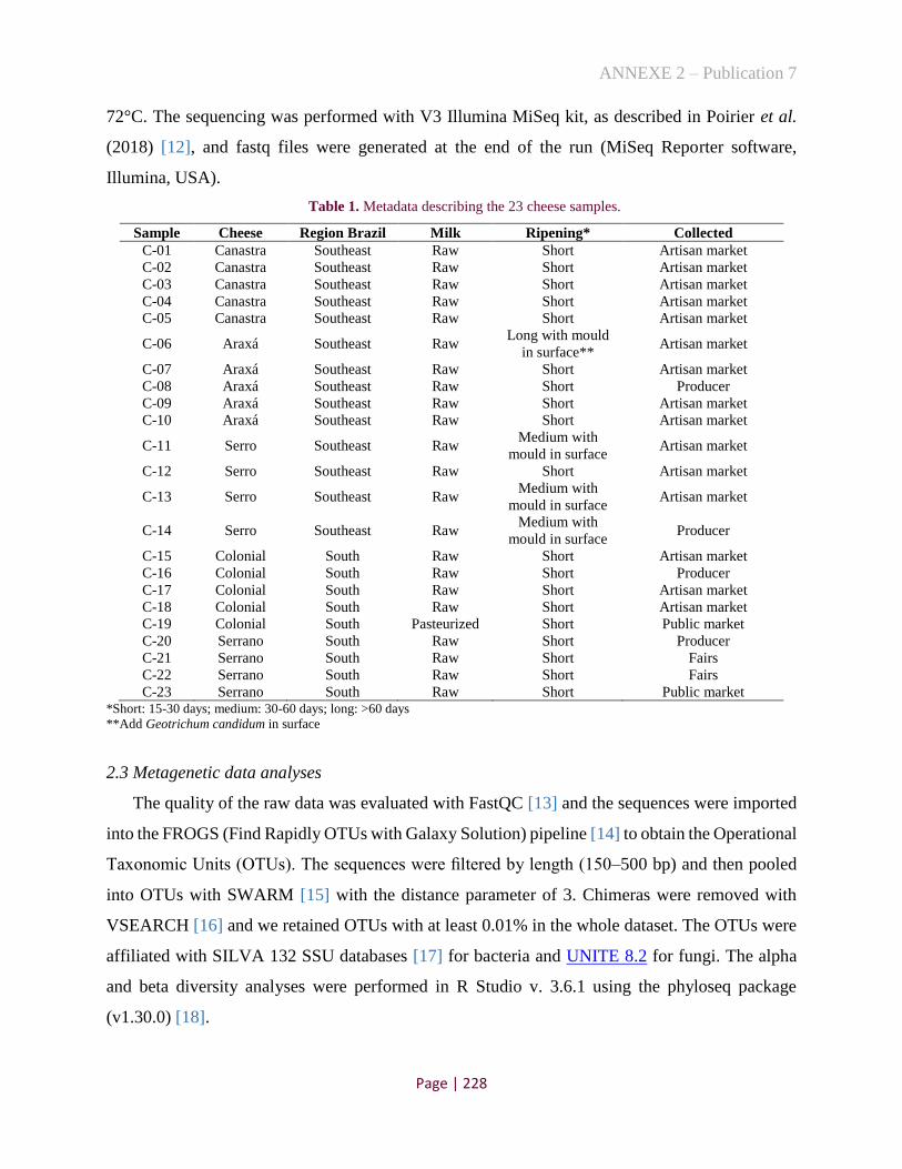
ANNEXE 2 – Publication 7
Page | 228
72°C. The sequencing was performed with V3 Illumina MiSeq kit, as described in Poirier et al.
(2018) [12], and fastq files were generated at the end of the run (MiSeq Reporter software,
Illumina, USA).
Table 1. Metadata describing the 23 cheese samples.
Sample Cheese Region Brazil Milk Ripening* Collected
C-01 Canastra Southeast Raw Short Artisan market
C-02 Canastra Southeast Raw Short Artisan market
C-03 Canastra Southeast Raw Short Artisan market
C-04 Canastra Southeast Raw Short Artisan market
C-05 Canastra Southeast Raw Short Artisan market
C-06 Araxá Southeast Raw Long with mould
in surface** Artisan market
C-07 Araxá Southeast Raw Short Artisan market
C-08 Araxá Southeast Raw Short Producer
C-09 Araxá Southeast Raw Short Artisan market
C-10 Araxá Southeast Raw Short Artisan market
C-11 Serro Southeast Raw Medium with
mould in surface Artisan market
C-12 Serro Southeast Raw Short Artisan market
C-13 Serro Southeast Raw Medium with
mould in surface Artisan market
C-14 Serro Southeast Raw Medium with
mould in surface Producer
C-15 Colonial South Raw Short Artisan market
C-16 Colonial South Raw Short Producer
C-17 Colonial South Raw Short Artisan market
C-18 Colonial South Raw Short Artisan market
C-19 Colonial South Pasteurized Short Public market
C-20 Serrano South Raw Short Producer
C-21 Serrano South Raw Short Fairs
C-22 Serrano South Raw Short Fairs
C-23 Serrano South Raw Short Public market *Short: 15-30 days; medium: 30-60 days; long: >60 days
**Add Geotrichum candidum in surface
2.3 Metagenetic data analyses
The quality of the raw data was evaluated with FastQC [13] and the sequences were imported
into the FROGS (Find Rapidly OTUs with Galaxy Solution) pipeline [14] to obtain the Operational
Taxonomic Units (OTUs). The sequences were filtered by length (150–500 bp) and then pooled
into OTUs with SWARM [15] with the distance parameter of 3. Chimeras were removed with
VSEARCH [16] and we retained OTUs with at least 0.01% in the whole dataset. The OTUs were
affiliated with SILVA 132 SSU databases [17] for bacteria and UNITE 8.2 for fungi. The alpha
and beta diversity analyses were performed in R Studio v. 3.6.1 using the phyloseq package
(v1.30.0) [18].

ANNEXE 2 – Publication 7
Page | 229
2.4 Taxonomic composition by shotgun metagenomics
From the metagenetic results, 15 cheese samples (nine surfaces and six core) were selected for
a more in-depth analysis. The DNA of those cheeses were sequenced using Illumina HiSeq2500
technology at GATC-Biotech (Konstanz, Germany), which yielded between six and eight million
paired-end reads of 150-nucleotide length. From the fastq file generates, we first estimated
microbial composition by mapping the samples reads against the representative clade-specific
marker catalog contained on the MetaPhlAn tool v. 3.0.4 [19]. As an additional method for
taxonomic profiling, we used a marker gene prediction, which allows binning of assembled
metagenomic contigs without the need for reference sequences. Therefore, the reads were merged
and de novo assembly was performed using SPAdes v. 3.9 [20]. Then, genes were predicted using
Prodigal v. 2.6.3 and single copy marker genes are extracted using fetchMG v. 1.0 [21,22]. By
default, fetchMG searched for 40 universal marker genes. We focused our research on the
COG0012 (ychF gene) and blasts were performed on the all-available sequences in the NCBI
databases on nucleic sequences and on the coding DNA sequences. Summary species composition
plots were created in R v. 3.6.1 using the package ggplot2 v. 3.3.2. Additionally, all metagenomic
reads were also mapped against Bos taurus genome with Bowtie2 [23] and visulalized with
Samtools flagstat [24].
2.5 Metagenome-assembled genome (MAG) quality and their taxonomic identification
Genome binning was performed using MetaBAT2-2.12.1 [25], with minimum contig size of
1,500 nucleotides and other default settings. The quality of the resulting prokaryotic bins were
assessed with CheckM [26] and MAGs <80% completeness and/or >10% contamination were
excluded. For species assignation of MAGs, we determined the Average Nucleotide Identity (ANI)
with the closest reference using mummer-3.23 [27]. Moreover, for eukaryotes, we predicted the
genes of MAGs and species closely related using Prodigal v. 2.6.3 and then we searched for 40
universal marker genes using fetchMG v. 1.0 [21,22]. From this analysis, ten marker genes were
identified in all MAGs (ychF, argS, rpsL, rpsB, rplK, rplC,rplB, rplF, rplP and leuS). These
proteins were concatenate to construct a phylogeny. The phylogenetic analyses were performed
using ClustalX 2.1 [28] and MEGA7 [29] and using the Neighbor-Joining method [30] with 1,000
bootstrap replicates [31].

ANNEXE 2 – Publication 7
Page | 230
2.5 Data availability
Raw sequences of amplification of 16S rRNA and ITS genes and raw metagenomic reads were
deposited to European Nucleotide Archive (ENA) under the BioProject ID PRJNA693797. The
MAGs are available in a public repository under the URL:
http://dx.doi.org/10.17632/nj3dbvg6ty.1
3 Results and Discussion
3.1 Taxonomic diversity of Brazilian cheeses using amplicon sequencing approaches
The bacterial diversity of 23 Brazilian core and rind cheeses was assessed with 16S rRNA
amplicon sequencing and the sequences were clustered in 157 bacterial OTUs, which taxonomic
assignment was possible up to the species level in the majority of the cases (Fig. 1). In these
analyses, we discard the sample C-02 due to its low depth of sequences. The mainly species present
in core of cheeses were the lactic acid bacteria (LAB) commonly used as starters, such as
Lactococcus lactis and Streptococcus thermophilus. We observed also a high abundance of
Streptococcus infantarius in two samples of Araxá cheeses, from Brazilian Southeast. Previous
studies showed that this species is highly prevalent and predominant among LABs in
spontaneously fermented African dairy products from cow, goat and camel milk [32,33]. S.
infantarius was also isolated from European and South American cheeses [34,35], including the
Coalho cheese from Brazil [36,37]. Additionally, in the core of C-R22 sample, we observed the
predominance of the species Kluyvera cryocrescens (pattern of an E. coli-like strain, [38]),
indicating the poor quality of this cheese.

ANNEXE 2 – Publication 7
Page | 231
Figure 1. Bacterial plot depicting the relative abundance of the 30 main species found in 23 Brazilian cheese
core and rinds.
The asterisk (*) indicates low depth of the sequences.
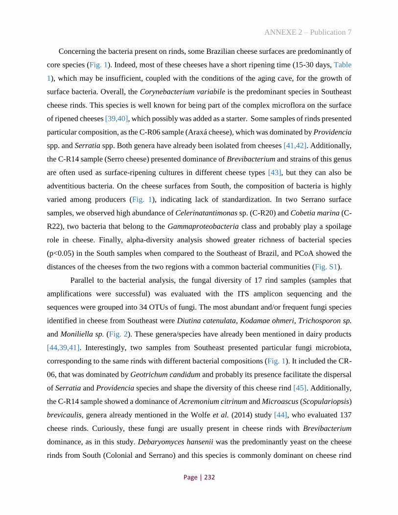
ANNEXE 2 – Publication 7
Page | 232
Concerning the bacteria present on rinds, some Brazilian cheese surfaces are predominantly of
core species (Fig. 1). Indeed, most of these cheeses have a short ripening time (15-30 days, Table
1), which may be insufficient, coupled with the conditions of the aging cave, for the growth of
surface bacteria. Overall, the Corynebacterium variabile is the predominant species in Southeast
cheese rinds. This species is well known for being part of the complex microflora on the surface
of ripened cheeses [39,40], which possibly was added as a starter. Some samples of rinds presented
particular composition, as the C-R06 sample (Araxá cheese), which was dominated by Providencia
spp. and Serratia spp. Both genera have already been isolated from cheeses [41,42]. Additionally,
the C-R14 sample (Serro cheese) presented dominance of Brevibacterium and strains of this genus
are often used as surface-ripening cultures in different cheese types [43], but they can also be
adventitious bacteria. On the cheese surfaces from South, the composition of bacteria is highly
varied among producers (Fig. 1), indicating lack of standardization. In two Serrano surface
samples, we observed high abundance of Celerinatantimonas sp. (C-R20) and Cobetia marina (C-
R22), two bacteria that belong to the Gammaproteobacteria class and probably play a spoilage
role in cheese. Finally, alpha-diversity analysis showed greater richness of bacterial species
(p<0.05) in the South samples when compared to the Southeast of Brazil, and PCoA showed the
distances of the cheeses from the two regions with a common bacterial communities (Fig. S1).
Parallel to the bacterial analysis, the fungal diversity of 17 rind samples (samples that
amplifications were successful) was evaluated with the ITS amplicon sequencing and the
sequences were grouped into 34 OTUs of fungi. The most abundant and/or frequent fungi species
identified in cheese from Southeast were Diutina catenulata, Kodamae ohmeri, Trichosporon sp.
and Moniliella sp. (Fig. 2). These genera/species have already been mentioned in dairy products
[44,39,41]. Interestingly, two samples from Southeast presented particular fungi microbiota,
corresponding to the same rinds with different bacterial compositions (Fig. 1). It included the CR-
06, that was dominated by Geotrichum candidum and probably its presence facilitate the dispersal
of Serratia and Providencia species and shape the diversity of this cheese rind [45]. Additionally,
the C-R14 sample showed a dominance of Acremonium citrinum and Microascus (Scopulariopsis)
brevicaulis, genera already mentioned in the Wolfe et al. (2014) study [44], who evaluated 137
cheese rinds. Curiously, these fungi are usually present in cheese rinds with Brevibacterium
dominance, as in this study. Debaryomyces hansenii was the predominantly yeast on the cheese
rinds from South (Colonial and Serrano) and this species is commonly dominant on cheese rind
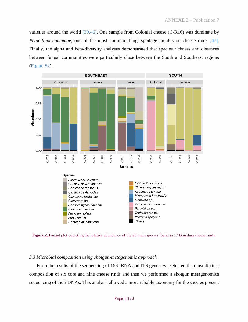
ANNEXE 2 – Publication 7
Page | 233
varieties around the world [39,46]. One sample from Colonial cheese (C-R16) was dominate by
Penicilium commune, one of the most common fungi spoilage moulds on cheese rinds [47].
Finally, the alpha and beta-diversity analyses demonstrated that species richness and distances
between fungal communities were particularly close between the South and Southeast regions
(Figure S2).
Figure 2. Fungal plot depicting the relative abundance of the 20 main species found in 17 Brazilian cheese rinds.
3.3 Microbial composition using shotgun-metagenomic approach
From the results of the sequencing of 16S rRNA and ITS genes, we selected the most distinct
composition of six core and nine cheese rinds and then we performed a shotgun metagenomics
sequencing of their DNAs. This analysis allowed a more reliable taxonomy for the species present

ANNEXE 2 – Publication 7
Page | 234
in the samples, as well as the proportion of bacteria and fungi together. Initially, the taxonomical
composition of the 15 cheese samples was determined using MetaPhlAn. From this analysis, a
total of 128 bacterial species and only one fungal species (Candida parapsilosis) were observed
in the cheeses evaluated (Table S1). Overall, the bacterial results of our metagenomes were in
accordance with the 16S rRNA amplicon methodology. However, ITS amplicon approach showed
larger fungal diversity, and we assumed that this tool could have a lack of eukaryotic reference
genomes. Then, we applied a marker gene analysis in our metagenomic dataset. As results, 56
bacterial species and seven fungal species were identified in the 15 cheeses (Table S2) and their
microbial composition is shown in Fig. 3. We observed a more accurate taxonomy with this
method, with the dominance of Streptococcus thermophilus, S. infantarius subsp. infantarius and
Lactococcus lactis subsp. lactis in cheese cores and Corynebacterium variabile and
Staphylococcus saprophyticus on rinds. We also pointed out a low relative abundance of
eukaryotes in relation to bacteria on cheese rinds, and the predominant species of fungi in Brazilian
cheese could not be assigned.
Concerning consumer risks, several samples, especially for the core (C-13, C-15 and C-18),
presented more than 30% of reads mapped on the Bos taurus genome (Table S3). For this reason,
these samples have low sequencing depth and consequently the taxonomic composition is less
accurate (Fig. 3, marked with an asterisk). The high presence of animal reads in some
metagenomes could be associated with the presence of cow somatic cells in the milk, which
indicates herd health problems [48]. Furthermore, we identified the presence of S. aureus in the C-
R02 sample (abundance relative of 0.86%, Table S2), indicating a potential risk of enterotoxin
production and foodborne outbreaks.

ANNEXE 2 – Publication 7
Page | 235
Fig. 3. The relative abundance of microbial composition of 15 Brazilian cheeses (six core and nine rinds). Results
were based on ychF marker gene, extracted from the assembled metagenomes.
The asterisks (*) indicates low depth of the sequences.

ANNEXE 2 – Publication 7
Page | 236
3.4 MAGs from Brazilian traditional cheeses
From the 15 metagenomes sequenced, a total of 50 prokaryotic metagenome-assembled
genomes (MAGs) with high-quality (over 80% complete and less than 10% contamination) were
reconstituted (Table S4). The majority of MAGs correspond to Lactococcus lactis subsp. lactis
(10) and Corynebacterium variabile (8). Moreover, six MAGs represent four potential novel
species, mainly from the C-R14 sample, since they have less than 95% average nucleotide identity
(ANI) with reference genomes, it includes two species of Brevibacterium, one Streptococcus and
one Corynebacterium. Three others prokaryotic MAGs, belonging to Micrococcaceae and
Lactobacillaceae families could not be identified at the genus level (ANI<80%), suggesting they
are characteristic bacteria of Brazilian cheeses. Additionally, ten fungal species were reconstitute
(Table S5), being two Debaryomyces hansenii, two Geotrichum candidum and six other MAGs
without closely related reference strains. Those last ones are representatives of two different
species (Fig. S3). Therefore, the lack of reference genomes closely related to MAGs obtained in
this study shows that the biodiversity of Brazilian cheeses still has many secrets, which can
contribute to the organoleptic, cultural and historical characteristics of these artisanal fermented
products.
4 Conclusion
The data obtained in this study expand and qualify the metagenomic database of Brazilian
cheeses. Furthermore, these data can contribute to improve the production practices (quality and
safety) and the acquisition of this know-how can support agro-industries and local economies.
Additionally, the identification of potential new species opens the opportunity to isolate them and
improve understanding at genomic and technological levels, since they can play an important role
in the ecology and sensory aspects of Brazilian cheeses. At the same time as new strains are
characterized, possibilities arise to develop inoculants maintaining cultural and historical
identities, as well as to study microbial interactions and assist to establish standards of quality for
cheeses produced in Brazil.

ANNEXE 2 – Publication 7
Page | 237
Acknowledgement
The authors are grateful to Mathieu Almeida, Nacer Mohellebi and Stéphane Chaillou for their
general teaching on metagenomic data analysis.
Supplementary Material
The supplementary Tables S1-S5 can be found at http://dx.doi.org/10.17632/7vc3vndc3r.1.
Table S1. The relative abundance of microbial composition of 15 cheese metagenomes from
Brazil using MetaPhlAn taxonomic assignment method.
Species C-03 C-10 C-13 C-15 C-18 C-20 C-
R02 C-
R03 C-
R04 C-
R06 C-
R07 C-
R09 C-
R11 C-
R13 C-
R14 Actinomycetaceae_bacterium_sk1b4 0 0 0 0 0 0 0.353 0.269 0 0 0 2.951 0.451 0.033 0
Bifidobacterium_mongoliense 0 0 0 0 0.28 0 0 0 0 0 0 0 0 0 0 Corynebacterium_casei 0 0 0 0 0 0 0.311 0 0.296 0.059 0.148 0.512 0.199 12.35 0.009
Corynebacterium_flavescens 0 0 0 0 0 0 0.05 0 0.037 0 0.129 0.013 0 0 0 Corynebacterium_glyciniphilum 0 0 0 0 0 0 0 0 0 0 0 0 0 0 0.228
Corynebacterium_nuruki 0 0 0 0 0 0 0 0 0 0 0 0 0.019 0 2.987 Corynebacterium_sp_CNJ_954 0 0 0 0 0 0 0 0 0 0 0 0 0 0 5.642
Corynebacterium_variabile 0 0.006 0.024 0 0 0 72.48 72.26 53.58 0.365 37.94 53.62 80.19 44.21 25.6 Dietzia_timorensis 0 0 0 0 0 0 0 0 0 0 0 0 0 0 0.14
Brevibacterium_aurantiacum 0 0 0 0 0 0 0 0 0 2.983 0 0 0 4.435 0.033 Brevibacterium_iodinum 0 0 0 0 0 0 0 0.008 0.007 0 0 0.01 0.006 0 0
Brevibacterium_linens 0 0 0 0 0 0 0 0 0 7.101 0 0 0 8.913 0.418 Brachybacterium_alimentarium 0 0 0 0 0 0 0 0 0 17.77 0 0.005 0.041 0 47.22
Kocuria_kristinae 0 0 0 0 0.201 0 0.346 0.621 0 0 0.059 0 0 0 0 Rothia_sp 0 0 0 0 0.107 0.435 0 0 0 0 0 0 0 0 0
Acidipropionibacterium_acidipropionici 0 0 0 0 0 0 0.878 0.407 0 0 0 14.01 1.553 0.806 0 Propionibacterium_freudenreichii 0 0 0 0 0 0 0 0 0 1.328 0 0.34 0 1.137 0
Nocardiopsis_alba 0 0 0 0 0 0 0 0 0 0 0 0 0 0 0.131 Chryseobacterium_bovis 0 0 0 0 0.047 0.482 0 0 0 0 0 0 0 0 0
Chryseobacterium_haifense 0 0 0 0 0 0.149 0 0 0 0 0 0 0 0 0 Chryseobacterium_sp_FP211_J200 0 0 0 0 0 0.638 0 0 0 0 0 0 0 0 0
Myroides_odoratimimus 0 0 0 0 0 0 0 0 0 0.015 0 0 0 0 0 Kurthia_sp_11kri321 0 0 0 0.124 0 0.137 0 0 0 0 0 0 0 0 0
Macrococcus_caseolyticus 0 0 0 0 0 0.039 0.098 0 0 0 0 0 0 0 0 Staphylococcus_argenteus 0 0 0 0 0 0 0.008 0 0 0 0 0 0 0 0
Staphylococcus_aureus 0 0 0.003 0 0 0 1.084 0 0 0 0 0 0 0 0 Staphylococcus_carnosus 0 0 0 0 0 0 0.045 0 0 0 0 0 0 0 0 Staphylococcus_equorum 0 0 0 0 0 0 0 0 0 0 0 0.03 0 0 0
Staphylococcus_lentus 0 0 0 0 0 0 0 0 0 0 0 0 0 0 3.963 Staphylococcus_nepalensis 0 0 0 0 0 0 0 0 0 0 0 0 0 0 2.84
Staphylococcus_saprophyticus 0 0 0 0 0 0 8.594 19.65 12.39 0 23.6 4.415 0.545 0 1.587 Staphylococcus_xylosus 0 0 0 0 0 0 0 0 0 0 0 0 0 0 4.212
Carnobacterium_divergens 0 0 0 0 0 0.401 0 0 0 0 0 0 0 0 0 Carnobacterium_maltaromaticum 0 0 0 0 0 1.939 0 0 0 0 0 0 0 0 0
Marinilactibacillus_psychrotolerans 0 0 0 0 0 0 0 0 0 0.074 0 0 0 0 0 Bavariicoccus_seileri 0 0 0 0 0 0 0.717 1.907 0 0.271 0.448 5.08 0.989 0 0
Enterococcus_devriesei 0 0 0 0 0 0 0 0 0 0.121 0 0 0.01 0.024 0 Enterococcus_dispar 0 0 0 0 0 0 0 0 0 0 0 0 0.001 0 0
Enterococcus_faecalis 0 0 0 0.083 0.038 0 0 0 0 1.592 0 0.04 0 0.485 1.868 Enterococcus_italicus 0 0 0 0 0.181 0.241 0.007 0 0 2.793 0 0 0 0 0
Enterococcus_malodoratus 0 0 0 0 0 0 0 0 0 0.287 0 0 0 0.134 0 Tetragenococcus_halophilus 0 0 0 0 0 0 0 0 0 0 0 0.474 0 0.094 0
Lactobacillus_acidipiscis 0 0 0 0 0 0 0 0 0 0 0 0.031 0 0 0 Lactobacillus_brevis 0.155 0.435 0.213 0 0 0 0.454 0.687 0.484 0.304 0.098 5.628 0.707 0.284 0
Lactobacillus_curvatus 0 0 0 0 0 0 0.453 1.66 0.017 0.171 0.408 1.939 0.055 0.028 0 Lactobacillus_delbrueckii 0 0 0 0.568 0.426 0 0 0 0 0 0 0 0 0 0
Lactobacillus_futsaii 0 0 0 0 0 0 0.011 0 0 0 0 0.02 0 0 0 Lactobacillus_kefiranofaciens 0 0.235 0 0 0 0 0 0 0 0 0 0 0 0 0
Lactobacillus_kefiri 0 0 0 0 0 0 0 0 0.074 0 0 0 0 0 0 Lactobacillus_otakiensis 0 0.051 0 0 0 0 0 0 0 0 0 0 0 0 0
Lactobacillus_parabuchneri 0 0.044 0.036 0 0.838 0 0 0 0 0 0 0 0 0.111 0 Lactobacillus_parafarraginis 0 0 0 0 0 0 0 0 0.016 0 0 0 0 0 0
Lactiplantibacillus_paraplantarum 0.183 0.553 0 0 0 0 0.65 0.707 0 0.088 0 6.536 0.674 0 0 Lactobacillus_paucivorans 0 0 0 0 0 0 0 0 0 0 0 0.168 0 0 0
_Lactiplantibacillus plantarum 0.033 1.494 1.017 0 0 0 0.488 0 0.441 0 1.439 0.045 0.157 6.032 0.12

ANNEXE 2 – Publication 7
Page | 238
Lactobacillus_rhamnosus 0 0.006 0 0 0.079 0 0 0 0 0 0 0 0.434 0 0 Lactobacillus_rossiae 0 0.004 0 0 0 0 0 0 0 0 0 0 0 0 0
Lactobacillus_senmaizukei 0 0 0 0 0 0 0 0 0 0 0 0.189 0 0 0 Lactobacillus_tucceti 0 0 0 0 0 0 0 0 0 0 0 0.062 0 0 0
Lactobacillus_versmoldensis 0 0 0 0 0 0 0.064 0 0 0 0 0.03 0.015 0 0 Pediococcus_parvulus 0 0 0 0 0 0 0 0.004 0 0 0 0.009 0 0 0
Pediococcus_pentosaceus 0.072 0 0 0 0 0 0 0 0 0 0 0 0 0 0 Pediococcus_sp 0 0 0 0 0 0 0 0.02 0 0 0 0.012 0 0 0
Leuconostoc_citreum 0 0 0 0 0 0 0.019 0 0 0 0 0 0 0 0 Leuconostoc_garlicum 0 0 0 0 0 0.123 0 0 0 0 0 0 0 0 0 Leuconostoc_gelidum 0 0 0 0 0 1.107 0 0 0 0 0 0 0 0 0
Leuconostoc_lactis 0 0 0 0 0 0.25 0 0 0 0 0 0 0 0 0 Leuconostoc_mesenteroides 0 0 0 0.257 2.522 3.744 11.29 0 0 0.051 0.991 0.939 0 0 0.063
Leuconostoc_pseudomesenteroides 0 0 0.412 0 0 0 0 0.033 0 0 0 0 0 0.292 0 Weissella_jogaejeotgali 0 0 0 0 0 0 0 0.12 0.076 0 0.756 0.038 0 0 0
Weissella_paramesenteroides 0 0 0 0 0 0 0 0.048 0 0 0 0 0 0 0 Weissella_thailandensis 0 0 0 0 0 0 0 0.141 0.029 0 0.659 0.004 0 0 0
Lactococcus_chungangensis 0 0 0 0 0 0.138 0 0 0 0 0 0 0 0 0 Lactococcus_garvieae 0 0 0 0 0.978 0.666 0 0 0 0 0 0 0 0 0
Lactococcus_lactis 61.98 19.5 80.7 4.051 7.951 6.091 1.553 1.237 31.4 5.335 33.02 2.738 13.52 10.76 2.937
Lactococcus_petauri 0 0 9E-04 0 0.453 0.304 0 0 0 0 0 0 0 0.009 0
Lactococcus_piscium 0 0 0 0 4.415 13.75 0 0 0 0 0 0 0 0 0 Lactococcus_raffinolactis 0 0 0 0 1.398 8.108 0 0 0 0 0 0 0 0 0 Streptococcus_agalactiae 0 0.247 0.026 0 0 0 0 0 0 0 0 0 0 0 0
Streptococcus_hongkongensis 0 0 0 0 0.03 0.047 0 0 0 0 0 0 0 0 0 Streptococcus_infantarius 0.955 77.38 0 0 0.372 0 0 0.089 0 0 0 0 0 0 0 Streptococcus_lutetiensis 0 0.046 0 0 0 0 0 0 0 0 0 0 0 0 0
Streptococcus_macedonicus 0 0 0 0.239 0 1.219 0 0 0 0 0 0 0 0 0 Streptococcus_parauberis 0 0 0 0 0.937 6.394 0 0 0 0 0 0 0 0 0
Streptococcus_pluranimalium 0 0 0 0 0.154 0 0 0 0 0 0 0 0 0 0 Streptococcus_salivarius 17.71 0 5.317 0 2.478 0 0 0.043 0 0 0 0 0 1.736 0
Streptococcus_thermophilus 18.91 0 12.19 93.61 56.6 37.43 0 0.028 0 0 0 0 0 2.334 0 Streptococcus_uberis 0 0 0 0 0.019 0 0 0 0 0 0 0 0 0 0
Veillonella_parvula 0 0 0 0 0.847 0 0 0 0 0 0 0 0 0 0 Enhydrobacter_aerosaccus 0 0 0 0 1.324 0.384 0 0 0 0 0 0 0 0 0
Aeromonas_allosaccharophila 0 0 0 0 0.806 0 0 0 0 0 0 0 0 0 0 Aeromonas_caviae 0 0 0 0 0.74 0 0 0 0 0 0 0 0 0 0 Aeromonas_media 0 0 0 0 1.934 0 0 0 0 0 0 0 0 0 0
Citrobacter_freundii 0 0 0 0 0.3 0 0 0 0 0 0 0 0 0 0 Citrobacter_youngae 0 0 0 0 0.976 0.636 0 0 0 0 0 0 0 0.016 0
Enterobacter_cloacae_complex 0.004 0 0.011 0.265 0.104 0 0 0 0 0 0 0 0 0 0 Escherichia_coli 0 0 0 0.317 0 5.997 0 0 0 0 0 0.009 0 0 0
Klebsiella_michiganensis 0 0 0 0 0.215 0.522 0 0 0 0 0 0 0 0 0 Klebsiella_oxytoca 0 0 0 0 0.093 0 0 0 0 0 0 0 0 0 0
Klebsiella_pneumoniae 0 0 0 0 0.349 0 0 0 0 0 0 0 0 0 0 Klebsiella_variicola 0 0 0 0.052 0 0 0 0 0 0 0 0 0 0 0
Kluyvera_cryocrescens 0 0 0 0.393 0 0 0 0 0 0 0 0 0 0 0 Raoultella_ornithinolytica 0 0 0 0.046 8.201 1.705 0 0 0 0 0 0 0 0.16 0
Raoultella_terrigena 0 0 0 0 0 0 0 0 0 0.096 0 0 0 0 0 Hafnia_alvei 0 0 0 0 1.291 0 0 0 0 0 0 0 0 0 0
Hafnia_paralvei 0 0 0 0 0 0 0 0 0 1.956 0 0 0 0 0 Morganella_morganii 0 0 0 0 0 0 0 0 0 0 0 0 0 0.072 0
Proteus_hauseri 0 0 0 0 0 0 0 0 0 0.571 0 0 0 0.384 0 Proteus_mirabilis 0 0 0 0 0 0 0 0 0 0 0 0 0 3.48 0 Proteus_vulgaris 0 0 0 0 0 0 0 0 0 4.327 0 0 0 1.493 0
Providencia_rettgeri 0 0 0 0 0 0 0 0 0 0 0 0 0 0.155 0 Serratia_liquefaciens 0 0 0 0 0 0 0 0 0 49.91 0 0 0 0 0 Serratia_marcescens 0 0 0 0 0.048 0.668 0 0 0 0.45 0 0 0 0.025 0
Serratia_rubidaea 0 0 0 0 0 0 0.008 0 0 0.257 0 0 0 0.016 0 Halomonas_campaniensis 0 0 0 0 0 0 0 0 0 0 0 0 0.05 0 0 Marinomonas_sp_MWYL1 0 0 0 0 0 0 0 0 0 0.007 0 0 0 0 0 Acinetobacter_bereziniae 0 0 0 0 0 0.543 0 0 0 0 0 0 0 0 0 Acinetobacter_johnsonii 0 0 0 0 0.218 2.265 0 0 0 0 0 0 0 0 0
Acinetobacter_junii 0 0 0 0 0 0.514 0 0 0 0 0 0 0 0 0 Acinetobacter_parvus 0 0 0 0 0.023 0 0 0 0 0 0 0 0 0 0 Acinetobacter_ursingii 0 0 0.046 0 0 1.309 0 0 0 0 0 0 0 0 0 Moraxella_osloensis 0 0 0 0 1.578 0.907 0 0 0 0 0 0 0 0 0
Pseudomonas_fluorescens_group 0 0 0 0 0.261 0.725 0 0 0 0 0 0 0 0 0 Pseudomonas_putida_group 0 0 0 0 0 0 0 0 0 0.38 0 0 0 0 0
Vibrio_alginolyticus 0 0 0 0 0 0 0 0 0 0 0 0 0.264 0 0 Vibrio_furnissii 0 0 0 0 0 0 0 0 0 0 0 0 0.118 0 0
Stenotrophomonas_maltophilia 0 0 0 0 0.192 0 0 0 0 1.317 0 0 0 0 0 Stenotrophomonas_rhizophila 0 0 0 0 0 0 0 0 0 0.019 0 0 0 0 0
Candida_parapsilosis 0 0 0 0 0 0 0.033 0.055 1.147 0 0.309 0.105 0 0 0

ANNEXE 2 – Publication 7
Page | 239
Table S2. The relative abundance of microbial composition of 15 cheese metagenomes from
Brazil using ychF marker gene assignation.
Species C-03 C-10 C-13 C-15 C-18 C-20 C-
R02 C-
R03 C-
R04 C-
R06 C-
R07 C-
R09 C-
R11 C-
R13 C-
R14
Acidipropionibacterium acidipropionici 0 0 0 0 0 0 0.929 0 0 0 0 11.08 1.54 0 0
Acidipropionibacterium unclassified 0 0 0 0 0 0 0 0 0 0 0 0 1.543 0 0
Acinetobacter albensis 0 0 0 0 0 19 0 0 0 0 0 0 0 0 0
Ancrocorticia populi 0 0 0 0 0 0 0.861 0 0 0 0 2.659 0 0 0
Arthrobacter unclassified 0 0 0 0 0 0 3.23 0 0 0 0 0 0 0 0
Bavariicoccus seileri 0 0 0 0 0 0 1.859 3.113 0 0.871 0 15.23 3.575 0 0
Brachybacterium alimentarium 0 0 0 0 0 0 0 0 0 11.43 0 0 0 0 14.9
Brachybacterium paraconglomeratum 0 0 0 0 0 0 0 0 0 0 0 0.881 0 0 0
Brevibacterium aurantiacum 0 0 0 0 0 0 0 0 0 5.952 0 0 0 7.381 0
Brevibacterium linens 0 0 0 0 0 0 0 0 0 0 0 0.792 1.537 0 14.95
Brevibacterium unclassified (1) 0 0 0 0 0 0 0 1.002 0 0 0 0 0 0 0
Brevibacterium unclassified (2) 0 0 0 0 0 0 0 0 0 0 0 0 2.443 0 37.9
Corynebacterium casei 0 0 0 0 0 0 0 0 0 0 0 0.78 0 7.297 0
Corynebacterium glyciniphilum 0 0 0 0 0 0 0 0 0 0 0 0 0 0 12.12
Corynebacterium variabile 0 0 0 0 0 0 61.97 53.47 33.15 0 24.81 42.72 64.06 26.48 7.707
Debaryomyces hansenii 0 0 0 0 0 0 0 2.374 7.696 0 5.379 0 0 0 0
Debaryomyces unclassified 0 0 0 0 0 0 3.335 5.223 0 0 1.478 0 0 0 0
Dietzia timorensis 0 0 0 0 0 0 0 0 0 0 0 0 0 0 1.829
Enterococcus faecalis 0 0 0 0 0 0 0 0 0 1.005 0 0 0 0 0
Enterococcus italicus 0 0 0 0 0 0 0 0 0 2.381 0 0 0 0 0
Geotrichum candidum 0 0 0 0 0 0 0 0 0 3.658 0 0 0 2.86 0
Hafnia alvei 0 0 0 0 0 0 0 0 0 1.309 0 0 0 0 0
Kluyveromyces lactis 0 0 0 0 0 0 0 0 2.774 0 1.947 0 0 1.783 0
Kocuria unclassified 0 0 0 0 0 0 0 0 0 0 0 0 0 0 1.63
Lacticaseibacillus paracasei 0 0 0 0 17.75 0 0 0 0 0 0 0 0 0 0
Lactiplantibacillus plantarum/ paraplantarum 0 1.62 0 0 0 0 1.713 0 0 0 0 4.156 0 3.712 0
Lactiplantibacillus unclassified 0 0 0 0 0 0 0 0 0.905 0 0 0 0 0 0
Lactobacillus curvatus 0 0 0 0 0 0 0 2.076 0 0 0.894 1.834 0 0 0
Lactobacillus huananensis 0 0 0 0 0 0 0 0 0 0 0 2.638 0 0 0
Lactobacillus kefiranofaciens 0 0.387 0 0 0 0 0 0 0 0 0 0 0 0 0
Lactobacillus rhamnosus 0 0 0 0 0 0 0 0 0 0 0 0 0.621 0 0
Lactococcus lactis subsp. lactis 31.17 17.15 54.33 0 11.95 0 2.119 1.028 29.58 5.137 30.39 3.166 19.13 12.18 0
Lactococcus piscium 0 0 0 0 0 14.49 0 0 0 0 0 0 0 0 0
Lactococcus raffinolactis 0 0 0 0 0 9.857 0 0 0 0 0 0 0 0 0
Lactococcus unclassified 0 0 0 0 0 0 0 0 0 0 0 0 0 0 1.65
Lentilactobacillus hilgardii 0 0.293 0 0 0 0 0 0 0 0 0 0 0 0 0
Leuconostoc mesenteroides 0 0 0 0 0 13.01 10.21 0 0 0 1.131 1.005 0 0 0
Levilactobacillus brevis 0 1.142 0 0 0 0 0 0 0 0 0 6.853 1.078 0 0
Microbacterium unclassified 0 0 0 0 0 0 0 0 0 9.112 0 0 0 7.701 0
Moraxellaceae bacterium 0 0 0 0 0 5.486 0 0 0 0 0 0 0 0 0
Propionibacterium freudenreichii 0 0 0 0 0 0 0 0 0 0 0 0.723 0 2.612 0
Proteus mirabilis 0 0 0 0 0 0 0 0 0 0 0 0 0 2.991 0
Proteus vulgaris 0 0 0 0 0 0 0 0 0 2.851 0 0 0 0 0
Providencia rettgeri 0 0 0 0 0 0 0 0 0 17.18 0 0 0 0 0
Pseudomonas putida 0 0 0 0 0 0 0 0 0 1.335 0 0 0 0 0
Raoultella planticola 0 0 0 2.294 8.88 0 0 0 0 0 0 0 0 0 0
Rothia kristinae 0 0 0 0 0 0 0.682 1.154 0 0 0 0 0 0 0
Serratia liquefaciens 0 0 0 0 0 0 0 0 0 32.54 0 0 0 0 0
Serratia unclassified 0 0 0 0 0 6.629 0 0 0 0 0 0 0 0 0
Sphingomonas unclassified 0 0 0 0 0 0 0 0 0.414 0 0 0 0 0 0
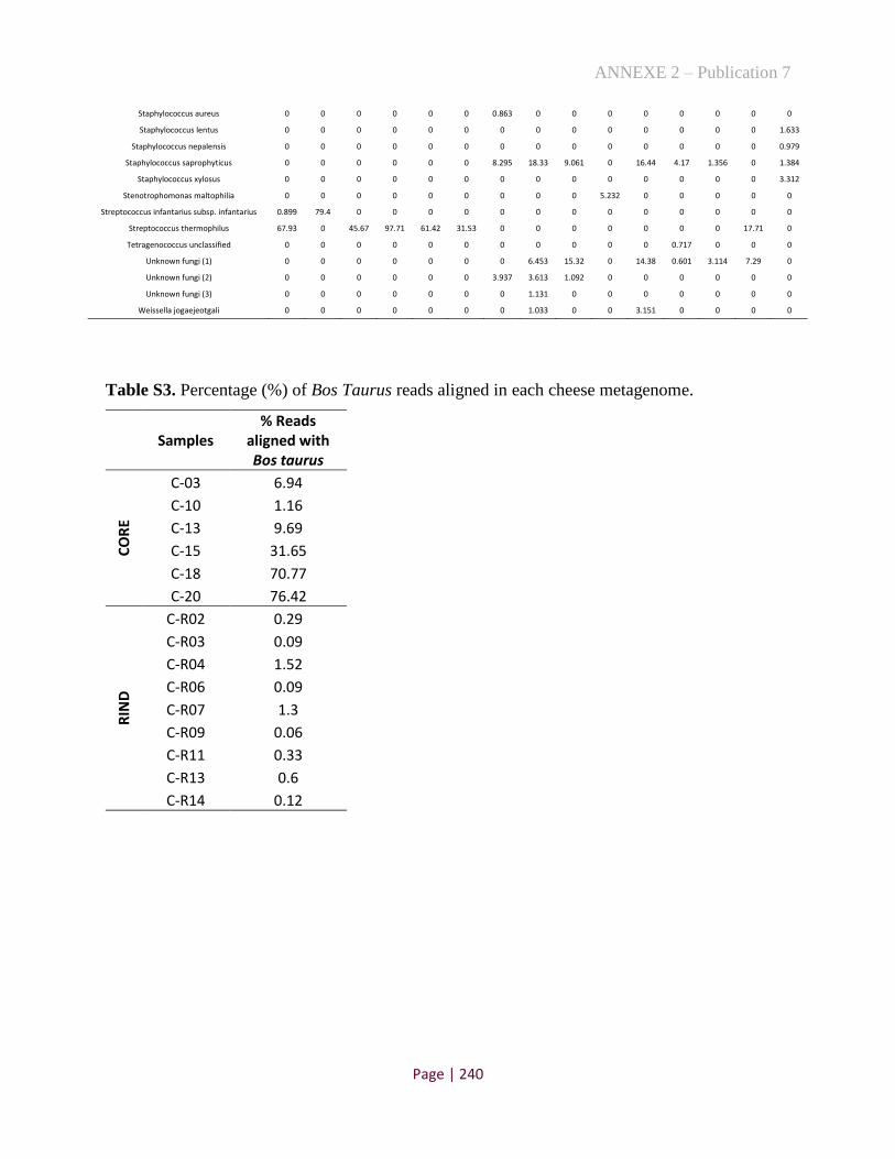
ANNEXE 2 – Publication 7
Page | 240
Staphylococcus aureus 0 0 0 0 0 0 0.863 0 0 0 0 0 0 0 0
Staphylococcus lentus 0 0 0 0 0 0 0 0 0 0 0 0 0 0 1.633
Staphylococcus nepalensis 0 0 0 0 0 0 0 0 0 0 0 0 0 0 0.979
Staphylococcus saprophyticus 0 0 0 0 0 0 8.295 18.33 9.061 0 16.44 4.17 1.356 0 1.384
Staphylococcus xylosus 0 0 0 0 0 0 0 0 0 0 0 0 0 0 3.312
Stenotrophomonas maltophilia 0 0 0 0 0 0 0 0 0 5.232 0 0 0 0 0
Streptococcus infantarius subsp. infantarius 0.899 79.4 0 0 0 0 0 0 0 0 0 0 0 0 0
Streptococcus thermophilus 67.93 0 45.67 97.71 61.42 31.53 0 0 0 0 0 0 0 17.71 0
Tetragenococcus unclassified 0 0 0 0 0 0 0 0 0 0 0 0.717 0 0 0
Unknown fungi (1) 0 0 0 0 0 0 0 6.453 15.32 0 14.38 0.601 3.114 7.29 0
Unknown fungi (2) 0 0 0 0 0 0 3.937 3.613 1.092 0 0 0 0 0 0
Unknown fungi (3) 0 0 0 0 0 0 0 1.131 0 0 0 0 0 0 0
Weissella jogaejeotgali 0 0 0 0 0 0 0 1.033 0 0 3.151 0 0 0 0
Table S3. Percentage (%) of Bos Taurus reads aligned in each cheese metagenome.
Samples % Reads
aligned with Bos taurus
CO
RE
C-03 6.94
C-10 1.16
C-13 9.69
C-15 31.65
C-18 70.77
C-20 76.42
RIN
D
C-R02 0.29
C-R03 0.09
C-R04 1.52
C-R06 0.09
C-R07 1.3
C-R09 0.06
C-R11 0.33
C-R13 0.6
C-R14 0.12

ANNEXE 2 – Publication 7
Page | 241
Table S4. Quality of prokaryotic MAGs and their ANIs with the closest reference genome found
in the NCBI database.
MAG ID Completeness Contamination Strain
Heterogeneity Reference strain
ANI (%)
Aligned nucleotides
(%)
C-R09.bin.5 99.67 0 0 Acidipropionibacterium acidipropionici ATCC
4875 98.39 [89.52]
C-R03.bin.7 98.91 0 0 Bavariicoccus seileri WCC 4188 98.12 [85.30]
C-R11.bin.5 97.13 0.55 0 Bavariicoccus seileri WCC 4188 98.57 [91.37]
C-R09.bin.3 96.17 0.68 100 Bavariicoccus seileri WCC 4188 97.8 [74.46]
C-R02.bin.9 90.71 1.54 20 Bavariicoccus seileri WCC 4188 98.54 [88.92]
C-R06.bin.12 100 0.64 50 Brachybacterium alimentarium 908_11 98.99 [91.23]
C-R14.bin.9 95.55 3.28 66.67 Brachybacterium alimentarium 908_11 98.86 [90.70]
C-R13.bin.5 99.71 0 0 Brevibacterium aurantiacum SMQ-1420 97.81 [88.03]
C-R11.bin.7 94.86 7.11 40 Brevibacterium linens ATCC 9172 81.49 [60.10]
C-R14.bin.5 87.65 2.81 71.43 Brevibacterium linens ATCC 9172 83.78 [63.54]
C-R14.bin.10 93.05 3.25 80 Brevibacterium oceani BBH7 81.15 [65.11]
C-R13.bin.8 93.47 0.88 0 Corynebacterium casei LMG S-19264 98.52 [88.49]
C-R14.bin.7 89.7 2.29 20 Corynebacterium glyciniphilum AJ-3170 88.03 [76.19]
C-R13.bin.7 98.87 0.45 0 Corynebacterium variabile DSM 44702 99.13 [93.60]
C-R07.bin.11 97.97 0.9 33.33 Corynebacterium variabile DSM 44702 99 [96.10]
C-R02.bin.10 97.82 1.95 16.67 Corynebacterium variabile DSM 44702 99 [93.29]
C-R11.bin.11 97.3 0.54 0 Corynebacterium variabile DSM 44702 99.11 [94.04]
C-R03.bin.6 97.07 0.5 50 Corynebacterium variabile DSM 44702 99.26 [96.34]
C-R09.bin.6 96.4 0.65 66.67 Corynebacterium variabile DSM 44702 99.3 [96.53]
C-R14.bin.2 96.38 1.28 50 Corynebacterium variabile DSM 44702 99.07 [94.76]
C-R04.bin.8 96.17 0.5 100 Corynebacterium variabile DSM 44702 99.36 [97.46]
C-R06.bin.9 92.5 3.2 5.56 Enterococcus italicus DSM 15952 98.7 [87.96]
C-R02.bin.1 92.75 1.03 0 Galactobacter caseinivorans JZ R-183 72.65 [36.24]
C-R13.bin.11 96.21 2.98 0 Lactiplantibacillus plantarum DSM 20174 99.04 [94.57]
C-10.bin.2 95.58 4.98 38.1 Lactiplantibacillus plantarum DSM 20174 98.5 [92.64]
C-R09.bin.11 94.16 1.09 0 Lactobacillus curvatus JCM 1096 98.86 [89.79]
C-R11.bin.2 100 0.5 100 Lactococcus lactis subsp. lactis Il1403 99.28 [86.71]
C-10.bin.1 99.62 0 0 Lactococcus lactis subsp. lactis Il1403 99.13 [87.68]
C-13.bin.2 99.62 0 0 Lactococcus lactis subsp. lactis Il1403 99.3 [89.31]
C-R13.bin.13 99.62 5.41 4.55 Lactococcus lactis subsp. lactis Il1403 98.76 [84.78]
C-03.bin.1 99.25 0.38 100 Lactococcus lactis subsp. lactis Il1403 99.4 [90.12]
C-R09.bin.10 96.6 2.17 0 Lactococcus lactis subsp. lactis Il1403 99.24 [88.85]
C-R04.bin.1 94.72 0 0 Lactococcus lactis subsp. lactis Il1403 99.22 [85.98]
C-R07.bin.3 94.72 0 0 Lactococcus lactis subsp. lactis Il1403 99.27 [86.61]
C-R02.bin.5 94.5 0.94 25 Lactococcus lactis subsp. lactis Il1403 99.42 [91.64]
C-R06.bin.11 90.98 0 0 Lactococcus lactis subsp. lactis Il1403 99.32 [90.78]
C-R02.bin.7 100 1.05 50 Leuconostoc mesenteroides ATCC 8293 98.85 [90.53]
C-R09.bin.9 82.74 0.99 33.33 Levilactobacillus brevis ATCC 367 73.52 [56.00]
C-R13.bin.1 93.13 1.11 16.67 Microbacterium excoecariae CBS5P-1 77.63 [44.68]
C-R13.bin.10 92.84 2.24 62.5 Proteus mirabilis HI4320 96.13 [85.63]
C-R06.bin.10 91.62 1.35 33.33 Proteus vulgaris ATCC 49132 99.63 [94.95]
C-R06.bin.5 97.21 7.21 17.65 Providencia rettgeri RB151 98.7 [85.62]
C-R06.bin.4 98.47 4.17 21.43 Serratia liquefaciens ATCC 27592 98.35 [91.11]
C-R07.bin.7 99.45 0 0 Staphylococcus saprophyticus ATCC 15305 99.36 [94.05]
C-R04.bin.7 98.9 0 0 Staphylococcus saprophyticus ATCC 15305 99.51 [94.66]
C-R09.bin.8 98.9 0 0 Staphylococcus saprophyticus ATCC 15305 99.62 [95.08]
C-R06.bin.7 94.43 4.92 32.26 Stenotrophomonas maltophilia SM 866 96.29 [86.80]
C-13.bin.1 89.12 0.5 100 Streptococcus salivarius ATCC 27945 93.77 [82.97]
C-R13.bin.3 90.51 0.75 0 Streptococcus salivarius ATCC 27945 93.78 [74.37]
C-R07.bin.8 98.19 2.34 12.5 Weissella jogaejeotgali FOL01 98.25 [86.40]

ANNEXE 2 – Publication 7
Page | 242
Table S5. Eukaryotic MAGs aligned with closely related reference genomes.
Figure S1. Bacterial species richness in cheese samples in the South and Southeast Brazilian regions (A) and
Principal Coordinates Analysis (PCoA) based on Unifrac distances among bacterial communities of the two
regions (B).
Figure S2. Fungal species richness in cheese samples (A) and Principal Coordinates Analysis (PCoA) based on
Unifrac distances among bacterial communities of the two regions (B).

ANNEXE 2 – Publication 7
Page | 243
Figure S3. Phylogeny of eukaryotic MAGs and closely related references based on ten amino acid (ychF, argS,
rpsL, rpsB, rplK, rplC, rplB, rplF, rplP and leuS). The tree was build with Neighbor-Joining method with 1,000
bootstrap replicates.

ANNEXE 2 – Publication 7
Page | 244
5 References
[1]. Donnelly CW (2014) Cheese and microbes. ASM Press, Washington, DC.
[2]. Ercolini D (2013) High-Throughput Sequencing and Metagenomics: Moving Forward in the Culture-
Independent Analysis of Food Microbial Ecology. Appl Environ Microb 79 (10):3148-3155.
doi:10.1128/Aem.00256-13.
[3]. Kamimura BA, Magnani M, Luciano WA, Campagnollo FB, Pimentel TC, Alvarenga VO, Pelegrino BO, Cruz
AG, Sant'Ana AS (2019) Brazilian Artisanal Cheeses: An Overview of their Characteristics, Main Types and
Regulatory Aspects. Comprehensive Reviews in Food Science and Food Safety 18 (5):1636-1657.
doi:10.1111/1541-4337.12486.
[4]. Oliver SP, Boor KJ, Murphy SC, Murinda SE (2009) Food Safety Hazards Associated with Consumption of
Raw Milk. Foodborne Pathog Dis 6 (7):793-806. doi:10.1089/fpd.2009.0302.
[5]. Lima CDLC, Lima LA, Cerqueira MMOP, Ferreira EG, Rosa CA (2009) Bactérias do acido láctico e leveduras
associadas com o queijo-de-minas artesanal produzido na região da Serra do Salitre, Minas Gerais. Arquivo
Brasileiro de Medicina Veterinária e Zootecnia 61:266-272. doi:10.1590/S0102-09352009000100037.
[6]. Pontarolo GH, Melo FD, Martini CL, Wildemann P, Alessio DRM, Sfaciotte RAP, Neto AT, Vaz EK, Ferraz
SM (2017) Quality and safety of artisan cheese produced in the serrana region of Santa Catarina. Semin-Cienc Agrar
38 (2):739-748. doi:10.5433/1679-0359.2017v38n2p739.
[7]. Resende MFS, Costa HHS, Andrade EHP, Acurcio LB, Drummond AF, Cunha AF, Nunes AC, Moreira JLS,
Penna CFAM, Souza MR (2011) Influence of altitute on lactic acid bacteria population of minas artisanal cheese
from Serra da Canastra. Arquivo Brasileiro De Medicina Veterinaria E Zootecnia 63 (6):1567-1573.
doi:10.1590/S0102-09352011000600039.
[8]. Perin LM, Sardaro MLS, Nero LA, Neviani E, Gatti M (2017) Bacterial ecology of artisanal Minas cheeses
assessed by culture-dependent and -independent methods. Food Microbiology 65:160-169.
doi:10.1016/j.fm.2017.02.005.
[9]. Luiz L, Castro R, Silva J, Oliveira L, Sales G, Nunes AC, Souza M, Sandes S (2016) Isolation and identification
of lactic acid bacteria from Brazilian Minas artisanal cheese. CyTA - Journal of Food 15:125.
doi:10.1080/19476337.2016.1219392.
[10]. Kamimura BA, De Filippis F, Sant’Ana AS, Ercolini D (2019) Large-scale mapping of microbial diversity in
artisanal Brazilian cheeses. Food Microbiology 80:40-49. doi:10.1016/j.fm.2018.12.014.
[11]. Almeida M, Hébert A, Abraham A-L, Rasmussen S, Monnet C, Pons N, Delbès C, Loux V, Batto J-M,
Leonard P, Kennedy S, Ehrlich SD, Pop M, Montel M-C, Irlinger F, Renault P (2014) Construction of a dairy
microbial genome catalog opens new perspectives for the metagenomic analysis of dairy fermented products. BMC
Genomics 15 (1):1101. doi:10.1186/1471-2164-15-1101.
[12]. Poirier S, Rué O, Peguilhan R, Coeuret G, Zagorec M, Champomier-Vergès M-C, Loux V, Chaillou S (2018)
Deciphering intra-species bacterial diversity of meat and seafood spoilage microbiota using gyrB amplicon
sequencing: A comparative analysis with 16S rDNA V3-V4 amplicon sequencing. PLOS ONE 13 (9):e0204629.
doi:10.1371/journal.pone.0204629.
[13]. Wingett S, Andrews S (2018) FastQ Screen: A tool for multi-genome mapping and quality control.
F1000Research 7:1338. doi:10.12688/f1000research.15931.1.
[14]. Escudié F, Auer L, Bernard M, Mariadassou M, Cauquil L, Vidal K, Maman S, Hernandez-Raquet G, Combes
S, Pascal G (2018) FROGS: Find, Rapidly, OTUs with Galaxy Solution. Bioinformatics 34 (8):1287-1294.
doi:10.1093/bioinformatics/btx791.
[15]. Mahe F, Rognes T, Quince C, de Vargas C, Dunthorn M (2014) Swarm: robust and fast clustering method for
amplicon-based studies. Peerj 2. doi:10.7717/peerj.593.
[16]. Rognes T, Flouri T, Nichols B, Quince C, Mahe F (2016) VSEARCH: a versatile open source tool for
metagenomics. Peerj 4. doi:10.7717/peerj.2584.
[17]. Quast C, Pruesse E, Yilmaz P, Gerken J, Schweer T, Yarza P, Peplies J, Glöckner FO (2013) The SILVA
ribosomal RNA gene database project: improved data processing and web-based tools. Nucleic Acids Research 41
(D1):D590-D596. doi:10.1093/nar/gks1219.
[18]. McMurdie PJ, Holmes S (2013) phyloseq: An R Package for Reproducible Interactive Analysis and Graphics
of Microbiome Census Data. PLOS ONE 8 (4):e61217. doi:10.1371/journal.pone.0061217.

ANNEXE 2 – Publication 7
Page | 245
[19]. Truong DT, Franzosa E, Tickle T, Scholz M, Weingart G, Pasolli E, Tett A, Huttenhower C, Segata N (2015)
MetaPhlAn2 for enhanced metagenomic taxonomic profiling. Nature Methods 12:902-903. doi:10.1038/nmeth0116-
101b.
[20]. Bankevich A, Nurk S, Antipov D, Gurevich AA, Dvorkin M, Kulikov AS, Lesin VM, Nikolenko SI, Pham S,
Prjibelski AD, Pyshkin AV, Sirotkin AV, Vyahhi N, Tesler G, Alekseyev MA, Pevzner PA (2012) SPAdes: a new
genome assembly algorithm and its applications to single-cell sequencing. J Comput Biol 19 (5):455-477.
doi:10.1089/cmb.2012.0021.
[21]. Ciccarelli FD, Doerks T, von Mering C, Creevey CJ, Snel B, Bork P (2006) Toward Automatic Reconstruction
of a Highly Resolved Tree of Life. Science 311 (5765):1283. doi:10.1126/science.1123061.
[22]. Sunagawa S, Mende D, Zeller G, Izquierdo-Carrasco F, Berger S, Kultima J, Coelho LP, Arumugam M, Tap J,
Nielsen H, Rasmussen S, Brunak S, Pedersen O, Guarner F, De Vos W, Wang J, Li J, Dore J, Ehrlich S, Bork P
(2013) Metagenomic species profiling using universal phylogenetic marker genes. Nature methods 10.
doi:10.1038/nmeth.2693.
[23]. Langmead B, Salzberg SL (2012) Fast gapped-read alignment with Bowtie 2. Nature Methods 9 (4):357-359.
doi:10.1038/nmeth.1923.
[24]. Li H, Handsaker B, Wysoker A, Fennell T, Ruan J, Homer N, Marth G, Abecasis G, Durbin R, Subgroup
GPDP (2009) The Sequence Alignment/Map format and SAMtools. Bioinformatics 25 (16):2078-2079.
doi:10.1093/bioinformatics/btp352.
[25]. Kang DD, Froula J, Egan R, Wang Z (2015) MetaBAT, an efficient tool for accurately reconstructing single
genomes from complex microbial communities. Peerj 3:e1165. doi:10.7717/peerj.1165.
[26]. Parks DH, Imelfort M, Skennerton CT, Hugenholtz P, Tyson GW (2015) CheckM: assessing the quality of
microbial genomes recovered from isolates, single cells, and metagenomes. Genome Res 25 (7):1043-1055.
doi:10.1101/gr.186072.114.
[27]. Kurtz S, Phillippy A, Delcher AL, Smoot M, Shumway M, Antonescu C, Salzberg SL (2004) Versatile and
open software for comparing large genomes. Genome Biology 5 (2):R12. doi:10.1186/gb-2004-5-2-r12.
[28]. Thompson JD, Gibson TJ, Plewniak F, Jeanmougin F, Higgins DG (1997) The CLUSTAL_X windows
interface: flexible strategies for multiple sequence alignment aided by quality analysis tools. Nucleic Acids Research
25 (24):4876-4882. doi:10.1093/nar/25.24.4876.
[29]. Kumar S, Stecher G, Tamura K (2016) MEGA7: Molecular Evolutionary Genetics Analysis Version 7.0 for
Bigger Datasets. Mol Biol Evol 33 (7):1870-1874. doi:10.1093/molbev/msw054.
[30]. Saitou N, Nei M (1987) The Neighbor-Joining Method - a New Method for Reconstructing Phylogenetic
Trees. Mol Biol Evol 4 (4):406-425. doi:10.1093/oxfordjournals.molbev.a040454.
[31]. Felsenstein J (1985) Confidence-Limits on Phylogenies - an Approach Using the Bootstrap. Evolution 39
(4):783-791. doi:10.1111/j.1558-5646.1985.tb00420.x.
[32]. Jans C, Meile L, Kaindi DWM, Kogi-Makau W, Lamuka P, Renault P, Kreikemeyer B, Lacroix C, Hattendorf
J, Zinsstag J, Schelling E, Fokou G, Bonfoh B (2017) African fermented dairy products – Overview of predominant
technologically important microorganisms focusing on African Streptococcus infantarius variants and potential
future applications for enhanced food safety and security. International Journal of Food Microbiology 250:27-36.
doi:10.1016/j.ijfoodmicro.2017.03.012.
[33]. Gboko KDT, Traoré SG, Sanhoun AR, Kirioua J, Otaru N, Kurt F, Jaeger FN, Isenring J, Kaindi DWM,
Kreikemeyer B, Renault P, Hattendorf J, Meile L, Jans C, Nguetta R, Bonfoh B (2019) Risk factors for the carriage
of Streptococcus infantarius subspecies infantarius isolated from African fermented dairy products. PLOS ONE 14
(11):e0225452. doi:10.1371/journal.pone.0225452.
[34]. Todaro M, Francesca N, Reale S, Moschetti G, Vitale F, Settanni L (2011) Effect of different salting
technologies on the chemical and microbiological characteristics of PDO Pecorino Siciliano cheese. European Food
Research and Technology 233 (6):931-940. doi:10.1007/s00217-011-1593-7.
[35]. Pacini F, Cariolato D, Andrighetto C, Lombardi A (2006) Occurrence of Streptococcus macedonicus in Italian
cheeses. Fems Microbiol Lett 261 (1):69-73. doi:10.1111/j.1574-6968.2006.00330.x.
[36]. Medeiros RS, Araújo LM, Queiroga Neto V, Andrade PP, Melo MA, Gonçalves MMBP (2016) Identification
of lactic acid bacteria isolated from artisanal Coalho cheese produced in the Brazilian Northeast. CyTA - Journal of
Food 14 (4):613-620. doi:10.1080/19476337.2016.1185468.
[37]. Brito LPd, Silva ECd, Calaça PRdA, Medeiros RSd, Soares MTCV, Porto ALF (2020) Lactic acid bacteria
isolated from Coalho cheese from northeast Brazil in dairy production: A screening for technological application.
Research, Society and Development 9 (10):e5249108457. doi:10.33448/rsd-v9i10.8457.
[38]. Sarria JC, Vidal AM, Kimbrough RC, III (2001) Infections Caused by Kluyvera Species in Humans. Clinical
Infectious Diseases 33 (7):e69-e74. doi:10.1086/322686.

ANNEXE 2 – Publication 7
Page | 246
[39]. Dugat-Bony E, Garnier L, Denonfoux J, Ferreira S, Sarthou AS, Bonnarme P, Irlinger F (2016) Highlighting
the microbial diversity of 12 French cheese varieties. International Journal of Food Microbiology 238:265-273.
doi:10.1016/j.ijfoodmicro.2016.09.026.
[40]. Bertuzzi AS, Walsh AM, Sheehan JJ, Cotter PD, Crispie F, McSweeney PLH, Kilcawley KN, Rea MC (2018)
Omics-Based Insights into Flavor Development and Microbial Succession within Surface-Ripened Cheese.
mSystems 3 (1):e00211-00217. doi:10.1128/mSystems.00211-17.
[41]. Irlinger F, Layec S, Hélinck S, Dugat-Bony E (2015) Cheese rind microbial communities: diversity,
composition and origin. Fems Microbiol Lett 362 (2):1-11. doi:10.1093/femsle/fnu015.
[42]. Coton M, Delbés-Paus C, Irlinger F, Desmasures N, Le Fleche A, Stahl V, Montel M-C, Coton E (2012)
Diversity and assessment of potential risk factors of Gram-negative isolates associated with French cheeses. Food
Microbiology 29 (1):88-98. doi:10.1016/j.fm.2011.08.020.
[43]. Bockelmann W, Willems KP, Neve H, Heller KH (2005) Cultures for the ripening of smear cheeses. Int Dairy
J 15 (6):719-732. doi:10.1016/j.idairyj.2004.08.022.
[44]. Wolfe BE, Button JE, Santarelli M, Dutton RJ (2014) Cheese Rind Communities Provide Tractable Systems
for In Situ and In Vitro Studies of Microbial Diversity. Cell 158 (2):422-433. doi:10.1016/j.cell.2014.05.041.
[45]. Zhang Y, Kastman EK, Guasto JS, Wolfe BE (2018) Fungal networks shape dynamics of bacterial dispersal
and community assembly in cheese rind microbiomes. Nature Communications 9 (1):336. doi:10.1038/s41467-017-
02522-z.
[46]. Banjara N, Suhr MJ, Hallen-Adams HE (2015) Diversity of Yeast and Mold Species from a Variety of Cheese
Types. Curr Microbiol 70 (6):792-800. doi:10.1007/s00284-015-0790-1.
[47]. Kure CF, Skaar I (2019) The fungal problem in cheese industry. Current Opinion in Food Science 29:14-19.
doi:10.1016/j.cofs.2019.07.003.
[48]. Moradi M, Omer AK, Razavi R, Valipour S, Guimarães JT (2021) The relationship between milk somatic cell
count and cheese production, quality and safety: A review. Int Dairy J 113:104884.
doi:10.1016/j.idairyj.2020.104884.

ANNEXE 2 – Publication 7
Page | 247
1.3. Conclusion
Cette étude nous a permis d’évaluer, par des approches métagénétiques et
métagénomiques, la microbiota de cinq variétés de fromages brésiliens, produites au Sud-est
(Araxá, Canastra, Serro) et au Sud (Colonial et Serrano) de ce pays. Les principales espèces
présentes dans les cœurs de ces fromages correspondaient à des bactéries lactiques communément
utilisées comme ferments, telles que Lactococcus lactis subsp. lactis et Streptococcus
thermophilus. Il est intéressant de noter que deux échantillons du fromage Araxá contenaient
également une grande quantité de Streptococcus infantarius. D'autre part, trois échantillons de
cœurs de fromages du Sud ont présenté plus de 30% de leurs reads métagénomiques, mappés sur
le génome de la vache, ce qui pourrait suggérer des problèmes de santé chez ces animaux. Les
différences entre microorganismes identifiés dans les fromages des deux régions sont plus
apparentes au sein de leurs croûtes, où existait une prédominance de Corynebacterium variabile
dans les fromages du Sud-est, alors que la distribution des bactéries était très variée dans les croûtes
du Sud, ce qui indique un manque de standardisation. De plus, nous avons noté une prédominance
de la levure Debaryomyces hansenii dans les fromages du Sud et, en particulier, deux espèces
fongiques inconnues dans les croûtes des fromages produites au Sud-est du Brésil. A partir des
analyses métagénomiques shotgun, nous avons obtenu la liste de 60 génomes assemblés par
métagénomique (MAG), dont 50 procaryotes et 10 eucaryotes, qui peuvent être utilisés pour mieux
comprendre les caractéristiques typiques des souches brésiliennes. De plus, la disponibilité de ces
données ouvre la possibilité d’isoler spécifiquement ces nouvelles espèces pour améliorer nos
connaissances aux niveaux génomique et technologique, car elles peuvent jouer un rôle important
dans l'écologie et les aspects sensoriels des fromages brésiliens. Parallèlement à la caractérisation
de nouvelles souches, ces données sont à même d’établir des normes de qualité pour les fromages
produits au Brésil, en préservant leurs identités culturelles et historiques.

ANNEXE 3 – Matériel supplémentaire
Page | 248
ANNEXE 3
Table S1. Méta-données de 114 métagénomes de croûte de fromage utilisées pour faire le mapping des 27 espèces
halophiles.
Cheese
ID
Cheese
Technology RindType Country Animal
Sequencing
Technology Repository Accesion Reference
1 Soft Washed France Cow Illumina SRA SRR12103075 This study
2 Soft Washed France Cow Illumina SRA SRR12103074 This study
3 Soft Washed France Cow Illumina SRA SRR12103073 This study
4 Soft Washed France Cow Illumina SRA SRR12103072 This study
5 Semihard Washed France Ewe Illumina SRA SRR12103071 This study
6 Soft Washed France Ewe Illumina SRA SRR12103070 This study
7 Blue Natural France Cow Illumina SRA SRR12103069 This study
8 Soft Washed France Cow Illumina SRA SRR12103068 This study
9 Soft Washed France Cow Illumina SRA SRR12103067 This study
10 Soft Bloomy France Goat Illumina SRA SRR12103066 This study
11 Unknown Natural USA Cow Illumina MG-RAST 4524482.3 1
12 Unknown Washed France Cow Illumina MG-RAST 4524483.3 1
13 Unknown Bloomy France Goat Illumina MG-RAST 4524484.3 1
14 Unknown Bloomy France Cow Illumina MG-RAST 4524485.3 1
15 Unknown Washed Ireland Cow Illumina MG-RAST 4524486.3 1
16 Unknown Washed France Cow Illumina MG-RAST 4524487.3 1
17 Unknown Natural England Cow Illumina MG-RAST 4524488.3 1
18 Unknown Bloomy England Goat Illumina MG-RAST 4524489.3 1
19 Unknown Natural USA Cow Illumina MG-RAST 4524490.3 1
20 Unknown Natural USA Cow Illumina MG-RAST 4524491.3 1
21 Unknown Natural USA Cow Illumina MG-RAST 4524493.3 1
22 Unknown Washed USA Cow Illumina MG-RAST 4524494.3 1
23 Unknown Washed USA Cow Illumina MG-RAST 4524495.3 1
24 Unknown Washed France Cow Illumina MG-RAST 4524496.3 1
25 Unknown Natural France Cow Illumina MG-RAST 4524497.3 1
26 Unknown Natural Spain Goat Illumina MG-RAST 4524498.3 1
27 Unknown Washed USA Cow Illumina MG-RAST 4524499.3 1
28 Unknown Washed Switzerland Cow Illumina MG-RAST 4524500.3 1
29 Unknown Natural USA Goat Illumina MG-RAST 4524501.3 1
30 Unknown Washed France Cow Illumina MG-RAST 4524502.3 1
31 Unknown Bloomy Italy Sheep Illumina MG-RAST 4524504.3 1
32 Unknown Washed USA Cow Illumina MG-RAST 4524505.3 1
33 Blue Natural France Cow SOLiD SRA SAMEA7202606 This study
34 Blue Natural France Cow SOLiD SRA SAMEA7202608 This study
35 Blue Natural France Cow SOLiD SRA SAMEA7202610 This study
36 Blue Natural France Cow SOLiD SRA SAMEA7202612 This study
37 Blue Natural France Cow SOLiD SRA SAMEA7202614 This study
38 Blue Natural Italy Cow SOLiD SRA SAMEA7202617 This study
39 Blue Natural Italy Cow SOLiD SRA ERS627441 2
40 Blue Natural France Ewe SOLiD SRA SAMEA7202618 This study
41 Soft Bloomy France Cow SOLiD SRA SAMEA7202619 This study
42 Soft Bloomy France Cow SOLiD SRA SAMEA7202620 This study
43 Soft Bloomy France Cow SOLiD SRA SAMEA7202621 This study
44 Soft Bloomy France Cow SOLiD SRA SAMEA7202623 This study
45 Soft Bloomy France Cow SOLiD SRA SAMEA7202625 This study
46 Soft Bloomy France Cow SOLiD SRA SAMEA7202626 This study

ANNEXE 3 – Matériel supplémentaire
Page | 249
Cheese
ID
Cheese
Technology RindType Country Animal
Sequencing
Technology Repository Accesion Reference
47 Soft Bloomy France Cow SOLiD SRA SAMEA7202627 This study
48 Soft Bloomy France Goat SOLiD SRA SAMEA7202628 This study
49 Soft Bloomy France Goat SOLiD SRA SAMEA7202629 This study
50 Soft Washed France Cow SOLiD SRA ERS627440 2
51 Soft Washed France Cow SOLiD SRA SAMEA7202631 This study
52 Soft Washed France Cow SOLiD SRA ERS627442 2
53 Hard Natural France Cow SOLiD SRA SAMEA7202632 This study
54 Hard Natural France Cow SOLiD SRA SAMEA7202634 This study
55 Hard Natural France Cow SOLiD SRA SAMEA7202635 This study
56 Hard Natural France Cow SOLiD SRA SAMEA7202636 This study
57 Hard Natural France Cow SOLiD SRA SAMEA7202637 This study
58 Hard Natural Italy Cow SOLiD SRA SAMEA7202638 This study
59 Semihard Natural France Cow SOLiD SRA SAMEA7202639 This study
60 Semihard Natural France Cow SOLiD SRA SAMEA7202640 This study
61 Semihard Natural France Ewe SOLiD SRA SAMEA7202641 This study
62 Semihard Natural France Ewe SOLiD SRA SAMEA7202642 This study
63 Soft Natural Spain Cow SOLiD SRA SAMEA7202644 This study
64 Soft Natural France Cow SOLiD SRA SAMEA7202645 This study
65 Soft Natural France Cow SOLiD SRA SAMEA7202646 This study
66 Soft Natural France Cow SOLiD SRA SAMEA7202647 This study
67 Soft Natural France Cow SOLiD SRA SAMEA7202648 This study
68 Soft Natural France Cow SOLiD SRA SAMEA7202649 This study
69 Soft Washed France Cow SOLiD SRA SAMEA7202650 This study
70 Soft Washed France Cow SOLiD SRA SAMEA7202652 This study
71 Soft Washed France Cow SOLiD SRA SAMEA7202653 This study
72 Semihard Natural France Cow SOLiD SRA SAMEA7202654 This study
73 Semihard Natural France Cow SOLiD SRA SAMEA7202655 This study
74 Semihard Natural France Cow SOLiD SRA SAMEA7202656 This study
75 Soft Bloomy Ireland Cow Illumina ENA ERS3466788 3
76 Soft Unknown Ireland Sheep Illumina ENA ERS3466744 3
77 Soft Bloomy Ireland Cow Illumina ENA ERS3466745 3
78 Semihard Unknown Ireland Cow Illumina ENA ERS3466746 3
79 Semihard Washed Ireland Cow Illumina ENA ERS3466771 3
80 Semihard Washed Ireland Cow Illumina ENA ERS3466747 3
81 Soft Unknown Ireland Cow Illumina ENA ERS3466772 3
82 Soft Bloomy Ireland Cow Illumina ENA ERS3466773 3
83 Hard Washed Ireland Goat Illumina ENA ERS3466748 3
84 Hard Natural Ireland Sheep Illumina ENA ERS3466774 3
85 Semihard Unknown Ireland Cow Illumina ENA ERS3466775 3
86 Soft Natural Ireland Cow Illumina ENA ERS3466749 3
87 Hard Washed Ireland Cow Illumina ENA ERS3466776 3
88 Soft Washed Ireland Cow Illumina ENA ERS3466777 3
89 Soft Bloomy Ireland Cow Illumina ENA ERS3466789 3
90 Soft Natural Ireland Cow Illumina ENA ERS3466790 3
91 Semihard Bloomy Ireland Goat Illumina ENA ERS3466791 3
92 Hard Natural Ireland Cow Illumina ENA ERS3466792 3
93 Soft Natural Ireland Goat Illumina ENA ERS3466793 3
94 Hard Natural Ireland Goat Illumina ENA ERS3466794 3
95 Hard Natural Ireland Cow Illumina ENA ERS3466750 3
96 Hard Natural Ireland Cow Illumina ENA ERS3466751 3
97 Unknown Natural USA Unknown Illumina MG-RAST 4675678.3 4
98 Unknown Natural France Unknown Illumina MG-RAST 4675679.3 4
99 Unknown Washed Spain Unknown Illumina MG-RAST 4675680.3 4
100 Unknown Natural USA Unknown Illumina MG-RAST 4675681.3 4
101 Unknown Washed USA Unknown Illumina MG-RAST 4675682.3 4
102 Unknown Natural Spain Unknown Illumina MG-RAST 4675683.3 4
103 Unknown Natural USA Unknown Illumina MG-RAST 4675684.3 4

ANNEXE 3 – Matériel supplémentaire
Page | 250
Cheese
ID
Cheese
Technology RindType Country Animal
Sequencing
Technology Repository Accesion Reference
104 Unknown Natural England Unknown Illumina MG-RAST 4675685.3 4
105 Unknown Natural USA Unknown Illumina MG-RAST 4675686.3 4
106 Unknown Washed USA Unknown Illumina MG-RAST 4675687.3 4
107 Unknown Natural Italy Unknown Illumina MG-RAST 4675688.3 4
108 Unknown Washed France Unknown Illumina MG-RAST 4675689.3 4
109 Unknown Natural USA Unknown Illumina MG-RAST 4675690.3 4
110 Unknown Natural USA Unknown Illumina MG-RAST 4675691.3 4
111 Unknown Washed France Unknown Illumina MG-RAST 4675692.3 4
112 Unknown Natural France Unknown Illumina MG-RAST 4675693.3 4
113 Unknown Natural USA Unknown Illumina MG-RAST 4675694.3 4
114 Unknown Natural France Unknown Illumina MG-RAST 4675695.3 4
1. Wolfe, B. E., Button, J. E., Santarelli, M. & Dutton, R. J. Cheese rind communities provide tractable systems for in situ and in
vitro studies of microbial diversity. Cell 158, 422-433, doi:10.1016/j.cell.2014.05.041 (2014).
2. Almeida, M., et al., Construction of a dairy microbial genome catalog opens new perspectives for the metagenomic analysis of
dairy fermented products. Bmc Genomics, 15(1):1101. doi: 10.1186/1471-2164-15-1101 (2014) 3. Walsh, A.M., Macori, G., Kilcawley, K.N. et al. Meta-analysis of cheese microbiomes highlights contributions to multiple
aspects of quality. Nat Food 1, 500–510 (2020). https://doi.org/10.1038/s43016-020-0129-3 4. Kastman EK, et al. 2016. Biotic interactions shape the ecological distributions of Staphylococcus species. mBio 7(5):e01157-16

ANNEXE 3 – Matériel supplémentaire
Page | 251
Halomonas
Fig. S1. Topologie phylogénétique pour 82 génomes d’Halomonas. Les souches isolées du fromage sont
indiquées en jaune, celles d'autres aliments en rouge, celles provenant d'écosystèmes environnementaux en bleu et
celles d'origine humaine en rose.

ANNEXE 3 – Matériel supplémentaire
Page | 252
Marinomonas
Fig. S2. Topologie phylogénétique pour 36 génomes de Marinomonas. Les souches du fromage sont
représentées en jaune et celles des écosystèmes environnementaux, en bleu.
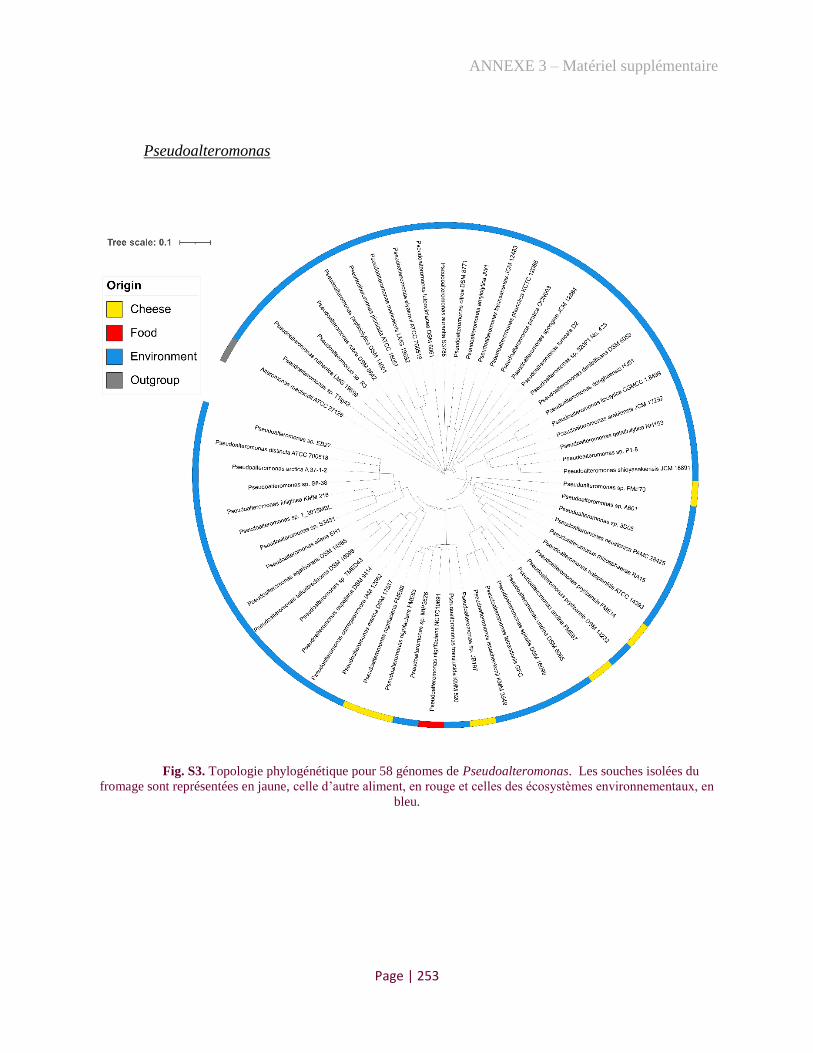
ANNEXE 3 – Matériel supplémentaire
Page | 253
Pseudoalteromonas
Fig. S3. Topologie phylogénétique pour 58 génomes de Pseudoalteromonas. Les souches isolées du
fromage sont représentées en jaune, celle d’autre aliment, en rouge et celles des écosystèmes environnementaux, en
bleu.
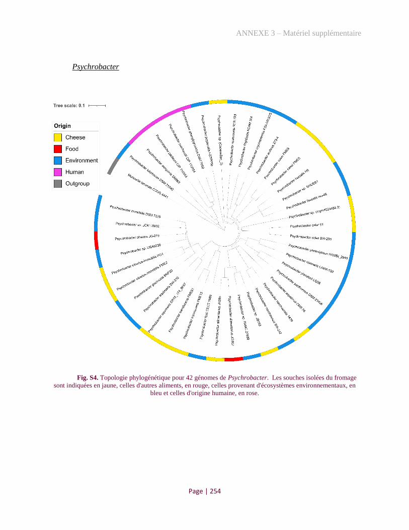
ANNEXE 3 – Matériel supplémentaire
Page | 254
Psychrobacter
Fig. S4. Topologie phylogénétique pour 42 génomes de Psychrobacter. Les souches isolées du fromage
sont indiquées en jaune, celles d'autres aliments, en rouge, celles provenant d'écosystèmes environnementaux, en
bleu et celles d'origine humaine, en rose.

ANNEXE 3 – Matériel supplémentaire
Page | 255
Vibrio
Fig. S5. Topologie phylogénétique pour 54 génomes de Vibrio. Les souches isolées du fromage sont
indiquées en jaune, celles provenant d'écosystèmes environnementaux, en bleu et celles d'origine humaine, en rose.

RÉFÉRENCES
Page | 256
RÉFÉRENCES
1. Donnelly, C.W., Cheese and microbes. 2014, Washington, DC: ASM Press. xii, 333 pages.
2. Ercolini, D., High-Throughput Sequencing and Metagenomics: Moving Forward in the
Culture-Independent Analysis of Food Microbial Ecology. Applied and Environmental
Microbiology, 2013. 79(10): p. 3148-3155.
3. Wolfe, B.E., et al., Cheese Rind Communities Provide Tractable Systems for In Situ and
In Vitro Studies of Microbial Diversity. Cell, 2014. 158(2): p. 422-433.
4. Almeida, M., et al., Construction of a dairy microbial genome catalog opens new
perspectives for the metagenomic analysis of dairy fermented products. Bmc Genomics,
2014. 15.
5. Irlinger, F., et al., Cheese rind microbial communities: diversity, composition and origin.
Fems Microbiology Letters, 2015. 362(2).
6. Delbès C., M.C., Irlinger F., Des communautés microbiennes au service de la qualité des
fromages: Diversité et dynamique adaptative et fonctionnelle des populations endogènes
et ensemencées Innovations Agronomiques, 2015. 44: p. 69-86.
7. Bokulich, N.A. and D.A. Mills, Facility-Specific "House" Microbiome Drives Microbial
Landscapes of Artisan Cheesemaking Plants. Applied and Environmental Microbiology,
2013. 79(17): p. 5214-5223.
8. Lenoir, J., et al., Control of cheese bioreactor. Biofutur, 1985(41): p. 13 p.
9. McSweeney, P.L.H., G. Ottogalli, and P. Fox. Diversity and Classification of Cheese
Varieties: An Overview. 2017.
10. ALMENA-ALISTE, M. and B. Mietton, Cheese Classification, Characterization, and
Categorization: A Global Perspective. Microbiology spectrum, 2014. 2 1: p. CM-0003-
2012.
11. Kindstedt, P.S., The Basics of Cheesemaking. Microbiology Spectrum, 2013. 1(1).
12. McSweeney, P.L.H., G. Ottogalli, and P.F. Fox, Chapter 31 - Diversity and Classification
of Cheese Varieties: An Overview, in Cheese (Fourth Edition), P.L.H. McSweeney, et al.,
Editors. 2017, Academic Press: San Diego. p. 781-808.
13. Sogin, M.L., et al., Microbial diversity in the deep sea and the underexplored "rare
biosphere". Proceedings of the National Academy of Sciences of the United States of
America, 2006. 103(32): p. 12115-12120.
14. Quigley, L., et al., High-Throughput Sequencing for Detection of Subpopulations of
Bacteria Not Previously Associated with Artisanal Cheeses. Applied and Environmental
Microbiology, 2012. 78(16): p. 5717-5723.
15. Kindstedt, P. and Vermont Cheese Council., American farmstead cheese : the complete
guide to making and selling artisan cheeses. 2005, White River Junction, Vt.: Chelsea
Green Pub. 276 p.
16. International Organization for Standardization (ISO), Fermented milk products : bacterial
starter cultures : standard of identity. 2010, Geneva; Brussels: ISO 27205:2010 [IDF
149:2010], International Dairy Federation, 11 p.
17. Bourdichon, F., et al., Food fermentations: Microorganisms with technological beneficial
use. International Journal of Food Microbiology, 2012. 154(3): p. 87-97.

RÉFÉRENCES
Page | 257
18. Papadimitriou, K., et al., Editorial: Omics and Systems Approaches to Study the Biology
and Applications of Lactic Acid Bacteria. Frontiers in Microbiology, 2020. 11.
19. De Filippis, F., E. Pasolli, and D. Ercolini, The food-gut axis: lactic acid bacteria and their
link to food, the gut microbiome and human health. FEMS Microbiology Reviews, 2020.
44(4): p. 454-489.
20. Parente, E., et al., FoodMicrobionet: A database for the visualisation and exploration of
food bacterial communities based on network analysis. International Journal of Food
Microbiology, 2016. 219: p. 28-37.
21. Harper, W.J. and R.M. Huber, Formation of Acidic Carbonyl Compounds by Some
Homofermentative Lactic Acid Bacteria. Applied Microbiology, 1961. 9(3): p. 184-&.
22. Holzapfel, W.H. and B.J.B. Wood, Introduction to the LAB. Lactic Acid Bacteria:
Biodiversity and Taxonomy, 2014: p. 1-12.
23. Peterson, S.D. and R.T. Marshall, Nonstarter Lactobacilli in Cheddar Cheese - a Review.
Journal of Dairy Science, 1990. 73(6): p. 1395-1410.
24. Settanni, L. and G. Moschetti, Non-starter lactic acid bacteria used to improve cheese
quality and provide health benefits. Food Microbiology, 2010. 27(6): p. 691-697.
25. Roissart, H.d. and F.M. Luquet, Bactéries lactiques : aspects fondamentaux et
technologiques. 1994, Uriage: Lorica.
26. Cogan, T., M., Les levains lactiques mésophiles. Une revue. Lait, 1980. 60(597): p. 397-
425.
27. Johnson, M.E., Mesophilic and Thermophilic Cultures Used in Traditional Cheesemaking.
Microbiology Spectrum, 2013. 1(1).
28. Auclair, J. and J.P. Accolas, Use of Thermophilic Lactic Starters in the Dairy-Industry.
Antonie Van Leeuwenhoek Journal of Microbiology, 1983. 49(3): p. 313-326.
29. Neyers, F., Les ferments lactiques. Généralités., in Revue des ENIL. 2016: Poligny, France.
30. Cogan, T.M., et al., Biodiversity of the Surface Microbial Consortia from Limburger,
Reblochon, Livarot, Tilsit, and Gubbeen Cheeses, in Cheese and Microbes. 2014,
American Society of Microbiology.
31. Goerges, S., et al., Commercial Ripening Starter Microorganisms Inoculated into Cheese
Milk Do Not Successfully Establish Themselves in the Resident Microbial Ripening
Consortia of a South German Red Smear Cheese. Applied and Environmental
Microbiology, 2008. 74(7): p. 2210-2217.
32. Bertuzzi, A.S., et al., Omics-Based Insights into Flavor Development and Microbial
Succession within Surface-Ripened Cheese. Msystems, 2018. 3(1).
33. Petersen, K.M., S. Westall, and L. Jespersen, Microbial succession of Debaryomyces
hansenii strains during the production of Danish surfaced-ripened cheeses. Journal of
Dairy Science, 2002. 85(3): p. 478-486.
34. Mourges, R., et al., Origine et développement des bactéries coliformes dans les fromages
à pâte molle. Lait, 1977. 57(563-564): p. 131-149.
35. Richard, J. and H. ZADI, Inventaire de la flore bactérienne dominante des Camemberts
fabriqués avec du lait cru. Lait, 1983. 63(623-624): p. 25-42.
36. Irlinger, F., et al., Ecological and aromatic impact of two Gram-negative bacteria
(Psychrobacter celer and Hafnia alvei) inoculated as part of the whole microbial
community of an experimental smear soft cheese. International Journal of Food
Microbiology, 2012. 153(3): p. 332-338.
RÉFÉRENCES
Page | 258
37. Rea, M.C., et al., Stability of the biodiversity of the surface consortia of Gubbeen, a red-
smear cheese. Journal of Dairy Science, 2007. 90(5): p. 2200-2210.
38. Brennan, N.M., et al., Biodiversity of the bacterial flora on the surface of a smear cheese.
Applied and Environmental Microbiology, 2002. 68(2): p. 820-830.
39. Feurer, C., et al., Does Smearing Inoculum Reflect the Bacterial Composition of the Smear
at the End of the Ripening of a French Soft, Red-Smear Cheese? Journal of Dairy Science,
2004. 87(10): p. 3189-3197.
40. Mounier, J., et al., Surface microflora of four smear-ripened cheeses. Applied and
Environmental Microbiology, 2005. 71(11): p. 6489-6500.
41. D'Amico, D.J., Adventitious microbes can affect the safety and quality of cheese. Microbe,
2014. 9: p. 99-104.
42. Addis, M.F., et al., The bovine milk microbiota: insights and perspectives from -omics
studies. Molecular Biosystems, 2016. 12(8): p. 2359-2372.
43. Wouters, J.T.M., et al., Microbes from raw milk for fermented dairy products. International
Dairy Journal, 2002. 12(2-3): p. 91-109.
44. von Neubeck, M., et al., Biodiversity of refrigerated raw milk microbiota and their
enzymatic spoilage potential. International Journal of Food Microbiology, 2015. 211: p.
57-65.
45. Martin, N.H., K.J. Boor, and M. Wiedmann, Symposium review: Effect of post-
pasteurization contamination on fluid milk quality. Journal of Dairy Science, 2018. 101(1):
p. 861-870.
46. Boor, K.J., et al., A 100-Year Review: Microbiology and safety of milk handling. Journal
of Dairy Science, 2017. 100(12): p. 9933-9951.
47. Walsh, A., et al., Meta-analysis of cheese microbiomes highlights contributions to multiple
aspects of quality. Nature Food, 2020. 1: p. 500-510.
48. Dugat-Bony, E., et al., Highlighting the microbial diversity of 12 French cheese varieties.
International Journal of Food Microbiology, 2016. 238: p. 265-273.
49. Didienne, R., et al., Characteristics of microbial biofilm on wooden vats ('gerles') in PDO
Salers cheese. International Journal of Food Microbiology, 2012. 156(2): p. 91-101.
50. Verdier-Metz, I., et al., Cow Teat Skin, a Potential Source of Diverse Microbial
Populations for Cheese Production. Applied and Environmental Microbiology, 2012.
78(2): p. 326-333.
51. Somers, E.B., M.E. Johnson, and A.C.L. Wong, Biofilm formation and contamination of
cheese by nonstarter lactic acid bacteria in the dairy environment. Journal of Dairy
Science, 2001. 84(9): p. 1926-1936.
52. Weller, O. and G. Dumitroaia, The earliest salt production in the world: An early Neolithic
exploitation in Poiana Slatinei-Lunca, Romania. 2005. 79.
53. Marro, C., et al., Archaeological investigations on the salt mine of Duzdaǧi (Nakhchivan,
Azerbaïjan). TUBA-AR, 2010. 13: p. 229-244.
54. McIntosh, J., Handbook to life in prehistoric Europe. 2009, Oxford ; New York: Oxford
University Press. xii, 404 p.
55. Henriet, O., et al., Exploring the diversity of extremely halophilic archaea in food-grade
salts. International Journal of Food Microbiology, 2014. 191: p. 36-44.
56. Abu-Khader, M., Viable engineering options to enhance the NaCl quality from the Dead
Sea in Jordan. Journal of Cleaner Production - J CLEAN PROD, 2006. 14: p. 80-86.

RÉFÉRENCES
Page | 259
57. Karleskint, G., R.L. Turner, and J.W. Small, Introduction to marine biology. 3rd ed. 2010,
Australia ; Belmont, CA: Brooks/Cole Cengage Learning. ix, 581 p.
58. Wang, H., X. Xu, and G. Zhu, Landscape Changes and a Salt Production Sustainable
Approach in the State of Salt Pan Area Decreasing on the Coast of Tianjin, China.
Sustainability, 2015. 7: p. 10078-10097.
59. Davis, J.S. Structure, function, and management of the biological system for seasonal solar
saltworks.
60. Dufossé, L., et al., Determination of speciality food salt origin by using 16S rDNA
fingerprinting of bacterial communities by PCR–DGGE: An application on marine salts
produced in solar salterns from the French Atlantic Ocean. Food Control, 2013. 32(2): p.
644-649.
61. Sainz-Lopez, N., T. Boski, and D.M.R. Sampath, Fleur de Sel Composition and
Production: Analysis and Numerical Simulation in an Artisanal Saltern. Journal of Coastal
Research, 2019. 35(6): p. 1200-1214.
62. Schwab, F.L., Sedimentary Petrology, in Encyclopedia of Physical Science and
Technology (Third Edition), R.A. Meyers, Editor. 2003, Academic Press: New York. p.
495-529.
63. Alam, G.S., A. Hussain, and Asrarullah, The mapping of Warcha and Kalabagh salt mines
and potash investigations, Punjab, Pakistan. 1975, Quetta: Geological Survey of Pakistan.
64. Martin, C., Obtenir du sel par la méthode ignigène : approche expérimentale. 2015. p.
1075-1088.
65. Morere, N., "Les problématiques du sel dans l´Hispanie préromaine et romaine. Folia
Electronica Classica, 2013. 26.
66. Mineral commodity summaries 2020, in Mineral Commodity Summaries. 2020: Reston,
VA. p. 204.
67. Joardder, M.U.H and Masud, M.H. A Brief History of Food Preservation. In: Food
Preservation in Developing Countries: Challenges and Solutions. Springer, Cham.
https://doi.org/10.1007/978-3-030-11530-2_3.
68. Goldblith, S.A., Condensed history of the science and technology of thermal processing.
Food technology, 1972. 44: p. 48-50.
69. Institute of Medicine (U.S.). Committee on Strategies to Reduce Sodium Intake., et al.,
Strategies to reduce sodium intake in the United States. 2010, Washington, D.C.: National
Academies Press. xii, 493 p.
70. Ravishankar, S. and V.K. Juneja, PRESERVATIVES | Traditional Preservatives – Sodium
Chloride, in Encyclopedia of Food Microbiology (Second Edition), C.A. Batt and M.L.
Tortorello, Editors. 2014, Academic Press: Oxford. p. 131-136.
71. Davidson, P.M., J.N. Sofos, and A.L. Branen, Antimicrobials in food. 3rd ed. Food science
and technology. 2005, Boca Raton: Taylor & Francis. 706 p.
72. Fennema, O.R., Food chemistry. 3rd ed. Food science and technology. 1996, New York:
Marcel Dekker. xii, 1069 p.
73. Potter, N.N. and J.H. Hotchkiss, Food science. 5th ed. Food science texts series. 1995, New
York: Chapman & Hall. xiii, 608 p.
74. Shelef, L.A. and J. Seiter, Indirect and miscellaneous antimicrobials. 2005. p. 573-598.
75. Juneja, V.K. and H. Thippareddi, Inhibitory effects of organic acid salts on growth of
Clostridium perfringens from spore inocula during chilling of marinated ground turkey
breast. International Journal of Food Microbiology, 2004. 93(2): p. 155-163.
RÉFÉRENCES
Page | 260
76. Miller, A.J., J.E. Call, and R.C. Whiting, Comparison of Organic-Acid Salts for
Clostridium-Botulinum Control in an Uncured Turkey Product. Journal of Food Protection,
1993. 56(11): p. 958-962.
77. Lalitha, K.V. and K. Gopakumar, Combined Effect of Sodium Chloride, pH and Storage
Temperature on Growth and Toxin Production by Clostridium botulinum. Journal of
Aquatic Food Product Technology, 2007. 16(2): p. 27-39.
78. Roberts, T.A., Jarvis, B. and Rhodes, A.C. Inhibition of Clostridium botulinum by curing
salts in pasteurized pork slurry. International Journal of Food Science & Technology,
1976. 11(1): p. 25-40.
79. Hutton, T., Sodium Technological functions of salt in the manufacturing of food and drink
products. British Food Journal, 2002. 104: p. 126-152.
80. Desmond, E., Reducing salt: A challenge for the meat industry. Meat Science, 2006. 74(1):
p. 188-196.
81. Saint-Eve, A., et al., Reducing salt and fat content: Impact of composition, texture and
cognitive interactions on the perception of flavoured model cheeses. Food Chemistry,
2009. 116(1): p. 167-175.
82. Breslin, P.A.S. and G.K. Beauchamp, Salt enhances flavour by suppressing bitterness.
Nature, 1997. 387(6633): p. 563-563.
83. Breslin, Paul A.S., An Evolutionary Perspective on Food and Human Taste. Current
Biology, 2013. 23(9): p. R409-R418.
84. Ruusunen, M., M. Sarkka-Tirkkonen, and E. Puolanne, Saltiness of coarsely ground
cooked ham with reduced salt content. Agricultural and Food Science in Finland, 2001. 10.
85. Pasqualone, A., et al., Effect of salt reduction on quality and acceptability of durum wheat
bread. Food Chemistry, 2019. 289: p. 575-581.
86. Akkerman, M., et al., Interaction between sodium chloride and texture in semi-hard Danish
cheese as affected by brining time, dl-starter culture, chymosin type and cheese ripening.
International Dairy Journal, 2017. 70: p. 34-45.
87. Johnson, M.E., et al., Reduction of Sodium and Fat Levels in Natural and Processed
Cheeses: Scientific and Technological Aspects. Comprehensive Reviews in Food Science
and Food Safety, 2009. 8(3): p. 252-268.
88. McMahon, D.J., et al., Effect of sodium, potassium, magnesium, and calcium salt cations
on pH, proteolysis, organic acids, and microbial populations during storage of full-fat
Cheddar cheese. Journal of Dairy Science, 2014. 97(8): p. 4780-4798.
89. Anses (Agence nationale de sécurité sanitaire de l’alimentation). Table de composition
nutritionnelle des aliments Ciqual 2020. 2020 [cited 2020 15/11/2020]; Available from:
https://ciqual.anses.fr/
90. Ceylan, Z., H. Turkoglu, and K. Dayısoylu, The Microbiological and Chemical Quality of
Sikma Cheese Produced in Turkey. Pakistan Journal of Nutrition, 2003. 2.
91. Elsanhoty, R., H. Mahrous, and G. Ghanaimy, Chemical, Microbial Counts and Evaluation
of Biogenic Amines During the Ripening of Egyptian Soft Domiati Cheese Made from Raw
and Pasteurized Buffaloes Milk. International Journal of Dairy Science, 2009. 4: p. 80-90.
92. Fox, P.F., et al., Fundamentals of Cheese Science. 2017, Springer US : Imprint: Springer,:
New York, NY. p. 1 online resource (XV, 799 pages 271 illustrations, 78 illustrations in
color.

RÉFÉRENCES
Page | 261
93. Guinee, T.P. and P.F. Fox, Chapter 13 - Salt in Cheese: Physical, Chemical and Biological
Aspects, in Cheese (Fourth Edition), P.L.H. McSweeney, et al., Editors. 2017, Academic
Press: San Diego. p. 317-375.
94. Actalia, M.r.d.l.é., Fiche technique: Le soin des fromages à croute morgée, F.f. fermières,
Editor. 2019: France.
95. Décret n°2007-588 du 24 avril 2007 relatif aux sels destinés à l'alimentation humaine.
2007.
96. Sun, X., Z. Shan, and W. Teng, Effects of increased iodine intake on thyroid disorders.
Endocrinology and metabolism (Seoul, Korea), 2014. 29(3): p. 240-247.
97. Zimmermann, M.B., P.L. Jooste, and C.S. Pandav, Iodine-deficiency disorders. The
Lancet, 2008. 372(9645): p. 1251-1262.
98. Bradshaw, C.E., An in vitro comparison of the antimicrobial activity of honey, iodine and
silver wound dressings. Bioscience Horizons: The International Journal of Student
Research, 2011. 4(1): p. 61-70.
99. Morge, S., La salage des fromages lactiques fermiers. 2017, Journée portes ouvertes:
Pradel, France.
100. Haastrup, M., et al., Cheese brines from Danish dairies reveal a complex microbiota
comprising several halotolerant bacteria and yeasts. International Journal of Food
Microbiology, 2018. 285.
101. Wolfe, S.J.B., Chefology: There Might Be Bacteria in Your Artisan Sea Salt, in Boston
Magazine. 2013, Lynne Montesanto: Boston.
102. Gibtan, A., et al., Diversity of Extremely Halophilic Archaeal and Bacterial Communities
from Commercial Salts. Frontiers in microbiology, 2017. 8: p. 799-799.
103. Larsen, H., Halophilic and halotolerant microorganisms‐an overview and historical
perspective. FEMS Microbiology Reviews, 1986. 39: p. 3-7.
104. Kushner, D.J., Life in high salt and solute concentrations: halophilic bacteria. Microbial
life in extreme environments. 1978: London, United Kingdom: Academic Press, Ltd.
105. Oren, A., Microbial life at high salt concentrations: phylogenetic and metabolic diversity.
Saline Systems, 2008. 4: p. 2.
106. Lefevre, E. and L.A. Round, A Preliminary Report Upon Some Halophilic Bacteria. J
Bacteriol, 1919. 4(2): p. 177-82.
107. Ventosa, A., J.J. Nieto, and A. Oren, Biology of moderately halophilic aerobic bacteria.
Microbiol Mol Biol Rev, 1998. 62(2): p. 504-44.
108. Grant, W.D., Life at low water activity. Philos Trans R Soc Lond B Biol Sci, 2004.
359(1448): p. 1249-66; discussion 1266-7.
109. Wohlfarth, A., J. Severin, and E.A. Galinski, The spectrum of compatible solutes in
heterotrophic halophilic eubacteria of the family Halomonadaceae. 1990. 136(4): p. 705-
712.
110. Shivanand, P. and G. Mugeraya, Halophilic bacteria and their compatible solutes –
osmoregulation and potential applications. Current science 2011. 100(10): p. 1516-1521.
111. Melgar-Lalanne, G., et al., Water Stress in Biological, Chemical, Pharmaceutical and
Food Systems. Importance of halophilic and halotolerant lactic acid bacteria in cheeses.
2015: Springer, New York, NY. 279-287.
112. Zhao, B.-S., et al., Study progress on compatible solutes in moderately halophilic bacteria.
Wei sheng wu xue bao, Acta microbiologica Sinica, 2007. 47(5): p. 937-941.

RÉFÉRENCES
Page | 262
113. Siglioccolo, A., et al., Structural adaptation of extreme halophilic proteins through
decrease of conserved hydrophobic contact surface. BMC Struct Biol, 2011. 11: p. 50.
114. Oren, A., M. Heldal, and S. Norland, X-ray microanalysis of intracellular ions in the
anaerobic halophilic eubacterium Haloanaerobium praevalens. 1997. 43(6): p. 588-592.
115. Oren, A., Industrial and environmental applications of halophilic microorganisms.
Environmental Technology, 2010. 31(8-9): p. 825-834.
116. Loukas, A., I. Kappas, and T.J. Abatzopoulos, HaloDom: a new database of halophiles
across all life domains. Journal of Biological Research-Thessaloniki, 2018. 25.
117. Tamang, J.P., K. Watanabe, and W.H. Holzapfel, Review: Diversity of Microorganisms in
Global Fermented Foods and Beverages. Frontiers in microbiology, 2016. 7: p. 377-377.
118. Han-Seung, L., Diversity of Halophilic Archaea in Fermented Foods and Human Intestines
and Their Application. Journal of Microbiology and Biotechnology, 2013. 23(12): p. 1645-
1653.
119. Kumar, S., G. Stecher, and K. Tamura, MEGA7: Molecular Evolutionary Genetics
Analysis Version 7.0 for Bigger Datasets. Molecular Biology and Evolution, 2016. 33(7):
p. 1870-1874.
120. Guarro, J., J. Gené, and A.M. Stchigel, Developments in Fungal Taxonomy. Clinical
Microbiology Reviews, 1999. 12(3): p. 454.
121. Gunde-Cimerman, N., J. Ramos, and A. Plemenitaš, Halotolerant and halophilic fungi.
Mycological Research, 2009. 113(11): p. 1231-1241.
122. Hymery, N., et al., Filamentous Fungi and Mycotoxins in Cheese: A Review.
Comprehensive Reviews in Food Science and Food Safety, 2014. 13(4): p. 437-456.
123. Lie, J.L. and E.H. Marth, Formation of Aflatoxin in Cheddar Cheese by Aspergillus flavus
and Aspergillus parasiticus1. Journal of Dairy Science, 1967. 50(10): p. 1708-1710.
124. Casquete, R., et al., The growth and aflatoxin production of Aspergillus flavus strains on a
cheese model system are influenced by physicochemical factors. Journal of Dairy Science,
2017. 100(9): p. 6987-6996.
125. Fleet, G.H. and M.A. Mian, The Occurrence and Growth of Yeasts in Dairy-Products.
International Journal of Food Microbiology, 1987. 4(2): p. 145-155.
126. Hernández-Almanza, A., et al., Rhodotorula glutinis as source of pigments and metabolites
for food industry. Food Bioscience, 2014. 5: p. 64-72.
127. Nadhum Trat, A., Isolating and Diagnosis Some Genus of Fungi that Found in
Environment Air and Soil in Different Region of Wassit City. Indian Journal of Forensic
Medicine & Toxicology, 2019. 13(4): p. 1414-1418.
128. Morin-Sardin, S., et al., Effect of temperature, pH, and water activity on Mucor spp. growth
on synthetic medium, cheese analog and cheese. Food Microbiology, 2016. 56: p. 69-79.
129. Zhang, N. and X.H. Zhao, Study of Mucor spp. in semi-hard cheese ripening. Journal of
Food Science and Technology-Mysore, 2010. 47(6): p. 613-619.
130. Hermet, A., et al., Molecular systematics in the genus Mucor with special regards to
species encountered in cheese. Fungal Biology, 2012. 116(6): p. 692-705.
131. Hesseltine, C.W., Microbiology of oriental fermented foods. Annual Review of
Microbiology, 1983. 37(1): p. 575-601.
132. Antón, J., Halophile, in Encyclopedia of Astrobiology, M. Gargaud, et al., Editors. 2011,
Springer Berlin Heidelberg: Berlin, Heidelberg. p. 725-727.
133. Chaban, B., S. Ng, and K. Jarrell, Archaeal habitats - From the extreme to the ordinary.
Canadian journal of microbiology, 2006. 52: p. 73-116.

RÉFÉRENCES
Page | 263
134. Wagner, D., et al., Methanosarcina soligelidi sp. nov., a desiccation- and freeze-thaw-
resistant methanogenic archaeon from a Siberian permafrost-affected soil. Int J Syst Evol
Microbiol, 2013. 63(Pt 8): p. 2986-2991.
135. Sorokin, D.Y., et al., Methanosalsum natronophilum sp. nov., and Methanocalculus
alkaliphilus sp. nov., haloalkaliphilic methanogens from hypersaline soda lakes. Int J Syst
Evol Microbiol, 2015. 65(10): p. 3739-3745.
136. Sowers, K.R., S.F. Baron, and J.G. Ferry, Methanosarcina acetivorans sp. nov., an
Acetotrophic Methane-Producing Bacterium Isolated from Marine Sediments. Appl
Environ Microbiol, 1984. 47(5): p. 971-8.
137. Roh, S.W., M.L. Lee, and J.W. Bae, Haladaptatus cibarius sp nov., an extremely halophilic
archaeon from seafood, and emended description of the genus Haladaptatus. International
Journal of Systematic and Evolutionary Microbiology, 2010. 60: p. 1187-1190.
138. Roh, S.W., et al., Halalkalicoccus jeotgali sp nov., a halophilic archaeon from shrimp
Jeotgal, a traditional Korean fermented seafood. International Journal of Systematic and
Evolutionary Microbiology, 2007. 57: p. 2296-2298.
139. Yim, K.J., et al., Halorubrum halophilum sp. nov., an extremely halophilic archaeon
isolated from a salt-fermented seafood. Antonie Van Leeuwenhoek, 2014. 105(3): p. 603-
12.
140. Tapingkae, W., et al., Natrinema gari sp nov., a halophilic archaeon isolated from fish
sauce in Thailand. International Journal of Systematic and Evolutionary Microbiology,
2008. 58: p. 2378-2383.
141. Namwong, S., et al., Halococcus thailandensis sp nov., from fish sauce in Thailand.
International Journal of Systematic and Evolutionary Microbiology, 2007. 57: p. 2199-
2203.
142. Namwong, S., et al., Haloarcula salaria sp. nov. and Haloarcula tradensis sp. nov.,
isolated from salt in Thai fish sauce. International Journal of Systematic and Evolutionary
Microbiology, 2011. 61: p. 231-236.
143. Han, D., L. Zhu, and H.L. Cui, Halorussus litoreus sp. nov., isolated from the salted brown
alga Laminaria. International Journal of Systematic and Evolutionary Microbiology, 2019.
69(3): p. 767-772.
144. Han, D. and H.L. Cui, Halosimplex pelagicum sp nov and Halosimplex rubrum sp nov.,
isolated from salted brown alga Laminaria, and emended description of the genus
Halosimplex. International Journal of Systematic and Evolutionary Microbiology, 2014.
64: p. 169-173.
145. Han, D. and H.L. Cui, Halostella pelagica sp. nov. and Halostella litorea sp. nov., isolated
from salted brown alga Laminaria. International Journal of Systematic and Evolutionary
Microbiology, 2020. 70(3): p. 1969-1976.
146. Cui, H.L., X. Yang, and Y.Z. Mou, Salinarchaeum laminariae gen. nov., sp. nov.: a new
member of the family Halobacteriaceae isolated from salted brown alga Laminaria.
Extremophiles, 2011. 15(6): p. 625-31.
147. Norton, C.F. and W.D. Grant, Survival of Halobacteria within Fluid Inclusions in Salt
Crystals. Journal of General Microbiology, 1988. 134: p. 1365-1373.
148. Schubert, B.A., et al., Halophilic Archaea cultured from ancient halite, Death Valley,
California. Environmental Microbiology, 2010. 12(2): p. 440-454.
149. Oren, A., Halophilic microorganisms and their environments. Cellular origin and life in
extreme habitats. 2002, Dordrecht ; Boston: Kluwer Academic. xxi, 575 p.

RÉFÉRENCES
Page | 264
150. Vynne, N.G., et al., Bioactivity, chemical profiling, and 16S rRNA-based phylogeny of
Pseudoalteromonas strains collected on a global research cruise. Mar Biotechnol (NY),
2011. 13(6): p. 1062-73.
151. Rodriguez-Saiz, M., et al., Engineering the halophilic bacterium Halomonas elongata to
produce beta-carotene. Applied Microbiology and Biotechnology, 2007. 77(3): p. 637-
643.
152. Mai-Prochnow, A., et al., Ecological advantages of autolysis during the development and
dispersal of Pseudoalteromonas tunicata biofilms. Appl Environ Microbiol, 2006. 72(8):
p. 5414-20.
153. Holmstrom, C., et al., Pseudoalteromonas tunicata sp. nov., a bacterium that produces
antifouling agents. International Journal of Systematic Bacteriology, 1998. 48: p. 1205-
1212.
154. Waditee-Sirisattha, R.W., Kageyama, H., Takabe, T., Halophilic microorganism resources
and their applications in industrial and environmental biotechnology. AIMS
Microbiology, 2016. 2(1): p. 42-54.
155. Park, E.-J., et al., Kocuria koreensis sp. nov., isolated from fermented seafood.
International Journal of Systematic and Evolutionary Microbiology, 2010. 60(1): p. 140-
143.
156. Park, E.-J., et al., Kocuria atrinae sp. nov., isolated from traditional Korean fermented
seafood. International Journal of Systematic and Evolutionary Microbiology, 2010. 60(4):
p. 914-918.
157. Yun, J.-H., et al., Kocuria salsicia sp. nov., isolated from salt-fermented seafood.
International journal of systematic and evolutionary microbiology, 2010. 61: p. 286-9.
158. Coppola, R., et al., Microbiological characteristics of Parmigiano Reggiano cheese during
the cheesemaking and the first months of the ripening. Lait, 2000. 80(5): p. 479-490.
159. Delbès, C., L. Ali-Mandjee, and M.-C. Montel, Monitoring bacterial communities in raw
milk and cheese by culture-dependent and -independent 16S rRNA gene-based analyses.
Applied and environmental microbiology, 2007. 73(6): p. 1882-1891.
160. Park, S.-K., et al., Brachybacterium squillarum sp. nov., isolated from salt-fermented
seafood. International Journal of Systematic and Evolutionary Microbiology, 2011. 61(5):
p. 1118-1122.
161. Schubert, K., et al., Two Coryneform Bacteria Isolated from the Surface of French Gruyère
and Beaufort Cheeses Are New Species of the Genus Brachybacterium: Brachybacterium
alimentarium sp. nov. and Brachybacterium tyrofermentans sp. nov.r†. International
Journal of Systematic and Evolutionary Microbiology, 1996. 46(1): p. 81-87.
162. Brennan, N.M., et al., Microbacterium gubbeenense sp. nov., from the surface of a smear-
ripened cheese. International Journal of Systematic and Evolutionary Microbiology, 2001.
51(6): p. 1969-1976.
163. Mounier, J., et al., Assessment of the microbial diversity at the surface of Livarot cheese
using culture-dependent and independent approaches. International Journal of Food
Microbiology, 2009. 133(1-2): p. 31-37.
164. Galli, A., L. Franzetti, and G. Ottogalli, Characterization of surface bacterial microflora
of Taleggio cheese. Annali Di Microbiologia Ed Enzimologia, 1996. 46: p. 369-377.
165. Tanasupawat, S., et al., Piscibacillus salipiscarius gen. nov., sp. nov., a moderately
halophilic bacterium from fermented fish (pla-ra) in Thailand. International Journal of
Systematic and Evolutionary Microbiology, 2007. 57(7): p. 1413-1417.

RÉFÉRENCES
Page | 265
166. Pakdeeto, A., et al., Lentibacillus kapialis sp. nov., from fermented shrimp paste in
Thailand. International journal of systematic and evolutionary microbiology, 2007. 57: p.
364-9.
167. Thongsanit, J., et al., Characterization and Identification of Tetragenococcus halophilus
and Tetragenococcus muriaticus Strains from Fish Sauce (Nam-pla). Japanese Journal of
Lactic Acid Bacteria, 2002. 13(1): p. 46-52.
168. Yoon, J.-H., et al., Jeotgalicoccus halotolerans gen. nov., sp. nov. and Jeotgalicoccus
psychrophilus sp. nov., isolated from the traditional Korean fermented seafood jeotgal.
International Journal of Systematic and Evolutionary Microbiology, 2003. 53(2): p. 595-
602.
169. Jung, M.-J., et al., Salinicoccus carnicancri sp. nov., a halophilic bacterium isolated from
a Korean fermented seafood. International journal of systematic and evolutionary
microbiology, 2009. 60: p. 653-8.
170. Yoon, J.H., et al., Planomicrobium koreense gen. nov., sp. nov., a bacterium isolated from
the Korean traditional fermented seafood jeotgal, and transfer of Planococcus
okeanokoites (Nakagawa et al. 1996) and Planococcus mcmeekinii (Junge et al. 1998) to
the genus Planomicrobium. International Journal of Systematic and Evolutionary
Microbiology, 2001. 51(4): p. 1511-1520.
171. Kim, J., et al., Virgibacillus alimentarius sp. nov., isolated from a traditional Korean food.
International Journal of Systematic and Evolutionary Microbiology, 2011. 61(12): p. 2851-
2855.
172. Jung, J.Y., et al., Microbial succession and metabolite changes during fermentation of
saeu-jeot: Traditional Korean salted seafood. Food Microbiology, 2013. 34(2): p. 360-
368.
173. Akuzawa, S., et al., Draft Genome Sequence of Oceanobacillus picturae Heshi-B3, Isolated
from Fermented Rice Bran in a Traditional Japanese Seafood Dish. Genome
announcements, 2016. 4(1): p. e01621-15.
174. Namwong, S., et al., Oceanobacillus kapialis sp. nov., from fermented shrimp paste in
Thailand. International journal of systematic and evolutionary microbiology, 2009. 59 Pt
9: p. 2254-9.
175. Whon, T.W., et al., Oceanobacillus kimchii sp nov Isolated from a Traditional Korean
Fermented Food. Journal of Microbiology, 2010. 48(6): p. 862-866.
176. Jang, S.J., et al., Oceanobacillus gochujangensis sp nov., Isolated from gochujang a
Traditional Korean Fermented Food. Journal of Microbiology, 2014. 52(12): p. 1050-
1055.
177. Wang, J., et al., Application of PacBio Single Molecule Real-Time (SMRT) sequencing in
bacterial source tracking analysis during milk powder production. Food Control, 2018.
93: p. 226-234.
178. Maoz, A., R. Mayr, and S. Scherer, Temporal stability and biodiversity of two complex
antilisterial cheese-ripening microbial consortia. Applied and Environmental
Microbiology, 2003. 69(7): p. 4012-4018.
179. Ishikawa, M., et al., Marinilactibacillus psychrotolerans gen. nov., sp. nov., a halophilic
and alkaliphilic marine lactic acid bacterium isolated from marine organisms in temperate
and subtropical areas of Japan. International Journal of Systematic and Evolutionary
Microbiology, 2003. 53(3): p. 711-720.

RÉFÉRENCES
Page | 266
180. Ishikawa, M., et al., Presence of halophilic and alkaliphilic lactic acid bacteria in various
cheeses. Letters in Applied Microbiology, 2007. 44(3): p. 308-313.
181. Ishikawa, M., et al., Alkalibacterium thalassium sp. nov., Alkalibacterium pelagium sp.
nov., Alkalibacterium putridalgicola sp. nov. and Alkalibacterium kapii sp. nov., slightly
halophilic and alkaliphilic marine lactic acid bacteria isolated from marine organisms and
salted foods collected in Japan and Thailand. International Journal of Systematic and
Evolutionary Microbiology, 2009. 59(5): p. 1215-1226.
182. Miyashita, M., Y. Nakagawa, and K.-i. Suzuki, Diversity of lactic acid bacteria in
fermented foods in Thailand. Japanese Journal of Lactic Acid Bacteria, 2012. 23: p. 77-85.
183. Yılmaz, E. and M. Birbir, Characterization of halotolerant bacillus species isolated from
salt samples collected from leather factories in Turkey. Journal of the American Leather
Chemists Association, 2019. 114: p. 118-130.
184. Chhetri, V., C. Prakitchaiwattana, and S. Settachaimongkon, A potential protective culture;
halophilic Bacillus isolates with bacteriocin encoding gene against Staphylococcus aureus
in salt added foods. Food Control, 2019. 104: p. 292-299.
185. indra, R., et al., Isolation and Characterization of Predominant Bacteria, Staphylococcus
piscifermentans Associated with Traditional Fermented Fish Products of Northeast India.
International Journal of Current Microbiology and Applied Sciences, 2018. 7: p. 1758-
1771.
186. Heo, S., J.-H. Lee, and D.-W. Jeong, Food-derived coagulase-negative Staphylococcus as
starter cultures for fermented foods. Food science and biotechnology, 2020. 29(8): p. 1023-
1035.
187. Walker-York-Moore, L., S.C. Moore, and E.M. Fox, Characterization of Enterotoxigenic
Bacillus cereus sensu lato and Staphylococcus aureus Isolates and Associated Enterotoxin
Production Dynamics in Milk or Meat-Based Broth. Toxins, 2017. 9(7): p. 225.
188. Ehsannia, S. and M. Sanjabi, Quality Characterization of Processed Cheese Inoculated by
Bacillus coagulans During Cold Storage: Compositional and Sensorial Attributes and
Probiotic Microorganism Viability. Journal of Food Processing and Preservation, 2015.
40: p. n/a-n/a.
189. Stavropoulou, D.A., L. De Vuyst, and F. Leroy, Nonconventional starter cultures of
coagulase-negative staphylococci to produce animal-derived fermented foods, a SWOT
analysis. Journal of Applied Microbiology, 2018. 125(6): p. 1570-1586.
190. Perkins, J., et al., Genetic engineering of Bacillus subtilis for the commercial production
of riboflavin. Journal of Industrial Microbiology and Biotechnology, 1999. 22: p. 8-18.
191. Martín, B., et al., Molecular, technological and safety characterization of Gram-positive
catalase-positive cocci from slightly fermented sausages. International Journal of Food
Microbiology, 2006. 107(2): p. 148-158.
192. Coton, E., et al., Biodiversity of Coagulase-Negative Staphylococci in French cheeses, dry
fermented sausages, processing environments and clinical samples. International Journal
of Food Microbiology, 2010. 137(2): p. 221-229.
193. Nakayama, H., R. Kawamoto, and K. Miyoshi, Ectoine production from putrefactive non-
volatile amines in the moderate halophile Halomonas elongata. IOP Conference Series:
Earth and Environmental Science, 2020. 439: p. 012001.
194. Graf, R., et al., The multifunctional role of ectoine as a natural cell protectant. Clinics in
dermatology, 2008. 26: p. 326-33.
RÉFÉRENCES
Page | 267
195. Kim, M.-S. and E.-J. Park, Bacterial Communities of Traditional Salted and Fermented
Seafoods from Jeju Island of Korea Using 16S rRNA Gene Clone Library Analysis. Journal
of Food Science, 2014. 79(5): p. M927-M934.
196. Jung, J.Y., B.H. Chun, and C.O. Jeon, Chromohalobacter is a Causing Agent for the
Production of Organic Acids and Putrescine during Fermentation of Ganjang, a Korean
Traditional Soy Sauce. Journal of Food Science, 2015. 80(12): p. M2853-M2859.
197. Liu, D. and C. Tong, Bacterial community diversity of traditional fermented vegetables in
China. LWT, 2017. 86: p. 40-48.
198. Beutling, D.M., J. Peçonek, and H. Stan-Lotter, Chromohalobacter beijerinckii: a
psychrophilic, extremely halotolerant and enzymatically active microbe from salted food
with the capacity for biogenic amine production. European Food Research and
Technology, 2009. 229(5): p. 725-730.
199. Jung, W.Y., H.J. Lee, and C.O. Jeon, Halomonas garicola sp. nov., isolated from saeu-
jeot, a Korean salted and fermented shrimp sauce. International Journal of Systematic and
Evolutionary Microbiology, 2016. 66(2): p. 731-737.
200. Calasso, M., et al., Relationships among house, rind and core microbiotas during
manufacture of traditional Italian cheeses at the same dairy plant. Food Microbiology,
2016. 54: p. 115-126.
201. Kim, M.-S., S.W. Roh, and J.-W. Bae, Cobetia crustatorum sp. nov., a novel slightly
halophilic bacterium isolated from traditional fermented seafood in Korea. International
Journal of Systematic and Evolutionary Microbiology, 2010. 60(3): p. 620-626.
202. Morya, V.K., W. Choi, and E.-k. Kim, Isolation and characterization of
Pseudoalteromonas sp. from fermented Korean food, as an antagonist to Vibrio harveyi.
Applied Microbiology and Biotechnology, 2014. 98(3): p. 1389-1395.
203. Yoon, J.-H., et al., Psychrobacter alimentarius sp. nov., isolated from squid jeotgal, a
traditional Korean fermented seafood. International Journal of Systematic and
Evolutionary Microbiology, 2005. 55(1): p. 171-176.
204. Jung, S.-Y., et al., Psychrobacter cibarius sp. nov., isolated from jeotgal, a traditional
Korean fermented seafood. International journal of systematic and evolutionary
microbiology, 2005. 55: p. 577-82.
205. Bleicher, A., K. Neuhaus, and S. Scherer, Vibrio casei sp. nov., isolated from the surfaces
of two French red smear soft cheeses. International Journal of Systematic and Evolutionary
Microbiology, 2010. 60(8): p. 1745-1749.
206. Baas Becking, L.G.M., et al., Baas Becking's: geobiology, or, introduction to
environmental science. 2016, Chichester, West Sussex ; Hoboken, NJ: Wiley Blackwell.
x,136 pages.
207. Selosse, M.-A., « Jamais seul » Ces microbes qui construisent les plantes, les animaux et
les civilisations. 2017, France: Actes Sud. 357.
208. Walker, S., Major spoilage micro‐organisms in milk and dairy products. International
Journal of Dairy Technology, 2007. 41: p. 91-92.
209. Morales, P., E. Fernández-García, and M. Nuñez, Volatile Compounds Produced in Cheese
by Pseudomonas Strains of Dairy Origin Belonging to Six Different Species. Journal of
Agricultural and Food Chemistry, 2005. 53(17): p. 6835-6843.
210. Eleboudy, A., et al., Occurrence and Behavior of Pseudomonas Organisms in White Soft
Cheese. Alexandria Journal of Veterinary Sciences, 2015. 44.

RÉFÉRENCES
Page | 268
211. Arslan, S., A. Eyi, and F. Özdemir, Spoilage potentials and antimicrobial resistance of
Pseudomonas spp. isolated from cheeses. Journal of Dairy Science, 2011. 94(12): p. 5851-
5856.
212. Stoyanova, V., et al., Lipolytic activity of genus Pseudomonas. J. BioSci. Biotech, 2012: p.
163-168.
213. Schmitz-Esser, S., et al., Abundance and potential contribution of Gram-negative cheese
rind bacteria from Austrian artisanal hard cheeses. International Journal of Food
Microbiology, 2018. 266: p. 95-103.
214. Deetae, P., et al., Growth and aroma contribution of Microbacterium foliorum, Proteus
vulgaris and Psychrobacter sp during ripening in a cheese model medium. Applied
Microbiology and Biotechnology, 2009. 82(1): p. 169-177.
215. Deetae, P., et al., Production of volatile aroma compounds by bacterial strains isolated
from different surface-ripened French cheeses. Applied microbiology and biotechnology,
2007. 76: p. 1161-71.
216. Broekaert, K., et al., Volatile compounds associated with Psychrobacter spp. and
Pseudoalteromonas spp., the dominant microbiota of brown shrimp (Crangon crangon)
during aerobic storage. International journal of food microbiology, 2013. 166: p. 487-493.
217. Busconi, M., C. Zacconi, and G. Scolari, Bacterial ecology of PDO Coppa and Pancetta
Piacentina at the end of ripening and after MAP storage of sliced product. International
Journal of Food Microbiology, 2014. 172: p. 13-20.
218. Ferrocino, I., et al., RNA-based amplicon sequencing highlights the impact of the
processing technology on microbiota composition of artisanal and industrial PDO Lard
d'Arnad. Applied and Environmental Microbiology, 2017. 83: p. AEM.00983-17.
219. Stellato, G., et al., Coexistence of Lactic Acid Bacteria and Potential Spoilage Microbiota
in a Dairy Processing Environment. Applied and Environmental Microbiology, 2015.
81(22): p. 7893-7904.
220. Schornsteiner, E., et al., Cultivation-independent analysis of microbial communities on
Austrian raw milk hard cheese rinds. International Journal of Food Microbiology, 2014.
180: p. 88-97.
221. Coton, M., et al., Diversity and assessment of potential risk factors of Gram-negative
isolates associated with French cheeses. Food Microbiology, 2012. 29(1): p. 88-98.
222. Gori, K., et al., Isolation and Identification of the Microbiota of Danish Farmhouse and
Industrially Produced Surface-Ripened Cheeses. Microbial Ecology, 2013. 65(3): p. 602-
615.
223. Bhunia, A.K. Vibrio cholerae, V. parahaemolyticus, V. vulnificus, in Foodborne Microbial
Pathogens: Mechanisms and Pathogenesis, 2008, Editor Springer New York: New York,
NY. p. 241-252.
224. Tanaka, M., et al., Genomic characterization of closely related species in the Rumoiensis
clade infers ecogenomic signatures to non-marine environments. Environmental
Microbiology, 2020. 22(8): p. 3205-3217.
225. Sawabe, T., et al., Updating the Vibrio clades defined by multilocus sequence phylogeny:
proposal of eight new clades, and the description of Vibrio tritonius sp. nov. Frontiers in
microbiology, 2013. 4: p. 414-414.
226. Cocolin, L., et al., Culture independent methods to assess the diversity and dynamics of
microbiota during food fermentation. International Journal of Food Microbiology, 2013.
167(1): p. 29-43.

RÉFÉRENCES
Page | 269
227. Goris, J., et al., DNA–DNA hybridization values and their relationship to whole-genome
sequence similarities. International Journal of Systematic and Evolutionary Microbiology,
2007. 57(1): p. 81-91.
228. Larpin-Laborde, S., et al., Surface microbial consortia from Livarot, a French smear-
ripened cheese. Canadian Journal of Microbiology, 2011. 57(8): p. 651-660.
229. Oguntoyinbo, F.A., et al., Halomonas nigrificans sp. nov., isolated from cheese.
International Journal of Systematic and Evolutionary Microbiology, 2018. 68(1): p. 371-
376.
230. Kastman, E.K., et al., Biotic Interactions Shape the Ecological Distributions of
Staphylococcus Species. mBio, 2016. 7(5): p. e01157-16.
231. Curioni, P.M.G. and J.O. Bosset, Key odorants in various cheese types as determined by
gas chromatography-olfactometry. International Dairy Journal, 2002. 12(12): p. 959-984.
232. Kothe, C.I., et al., Unraveling the world of halophilic and halotolerant bacteria in cheese
by combining cultural, genomic and metagenomic approaches. bioRxiv, 2020: p.
2020.11.03.353524.
233. Vandenbergh, P.A., Lactic acid bacteria, their metabolic products and interference with
microbial growth. FEMS Microbiology Reviews, 1993. 12(1): p. 221-237.
234. Zeidan, A.A., et al., Polysaccharide production by lactic acid bacteria: from genes to
industrial applications. FEMS Microbiology Reviews, 2017. 41(Supp_1): p. S168-S200.
235. Giavasis, I., 16 - Production of microbial polysaccharides for use in food, in Microbial
Production of Food Ingredients, Enzymes and Nutraceuticals, B. McNeil, et al., Editors.
2013, Woodhead Publishing. p. 413-468.
236. Biswas, J. and P.A. Paul, Diversity and Production of Extracellular Polysaccharide by
Halophilic Microorganisms. Biodiversity International Journal, 2017. 1.
237. Loessner, M., et al., A pediocin-producing Lactobacillus plantarum strain inhibits Listeria
monocytogenes in a multispecies cheese surface microbial ripening consortium. Applied
and Environmental Microbiology, 2003. 69(3): p. 1854.
238. Rodrı́guez, E., et al., Inhibitory activity of a nisin-producing starter culture on Listeria
innocua in raw ewes milk Manchego cheese. International Journal of Food Microbiology,
1998. 39(1): p. 129-132.
239. Rodrı́guez, E., et al., Antimicrobial activity of pediocin-producing Lactococcus lactis on
Listeria monocytogenes, Staphylococcus aureus and Escherichia coli O157:H7 in cheese.
International Dairy Journal, 2005. 15(1): p. 51-57.
240. Oliver, S.P., B.M. Jayarao, and R.A. Almeida, Foodborne Pathogens in Milk and the Dairy
Farm Environment: Food Safety and Public Health Implications. Foodborne Pathogens
and Disease, 2005. 2(2): p. 115-129.
241. Maeda, M., et al., The concept of biological control methods in aquaculture.
Hydrobiologia, 1997. 358(1): p. 285-290.
242. Deetae, P., et al., Growth and aroma contribution of Microbacterium foliorum, Proteus
vulgaris and Psychrobacter sp. during ripening in a cheese model medium. Applied
Microbiology and Biotechnology, 2009. 82(1): p. 169-177.
243. Irlinger, F., et al., Ecological and aromatic impact of two Gram-negative bacteria
(Psychrobacter celer and Hafnia alvei) inoculated as part of the whole microbial
community of an experimental smear soft cheese. International Journal of Food
Microbiology, 2012. 153(3): p. 332-338.

RÉFÉRENCES
Page | 270
244. Kamelamela, N., et al., Indigo- and indirubin-producing strains of Proteus and
Psychrobacter are associated with purple rind defect in a surface-ripened cheese. Food
Microbiology, 2018. 76: p. 543-552.
245. Hammes, W.P. and C. Hertel, New developments in meat starter cultures. Meat Science,
1998. 49: p. S125-S138.
246. Stevens, D.A., et al., Halomonas, a Newly Recognized Human Pathogen Causing
Infections and Contamination in a Dialysis Center: Three New Species. Medicine, 2009.
88(4).
247. Simidu, U., et al., Taxonomy of Four Marine Bacterial Strains That Produce Tetrodotoxin.
International Journal of Systematic and Evolutionary Microbiology, 1990. 40(4): p. 331-
336.
248. Holmström, C. and S. Kjelleberg, Marine Pseudoalteromonas species are associated with
higher organisms and produce biologically active extracellular agents. FEMS
Microbiology Ecology, 1999. 30(4): p. 285-293.
249. Joanna María, O.-A., et al., Fatal Psychrobacter sp. infection in a pediatric patient with
meningitis identified by metagenomic next-generation sequencing in cerebrospinal fluid.
Archives of Microbiology, 2016. 198(2): p. 129-135.
250. Deschaght, P., et al., Psychrobacter isolates of human origin, other than Psychrobacter
phenylpyruvicus, are predominantly Psychrobacter faecalis and Psychrobacter pulmonis,
with emended description of P. faecalis. International Journal of Systematic and
Evolutionary Microbiology, 2012. 62(Pt_3): p. 671-674.
251. Hurtado-Ortiz, R., et al., Psychrobacter pasteurii and Psychrobacter piechaudii sp. nov.,
two novel species within the genus Psychrobacter. International Journal of Systematic and
Evolutionary Microbiology, 2017. 67(9): p. 3192-3197.
252. Archer, G.L., Staphylococcus aureus: A Well-Armed Pathogen. Clinical Infectious
Diseases, 1998. 26(5): p. 1179-1181.
253. Bockelmann, W., et al., Cultures for the ripening of smear cheeses. International Dairy
Journal, 2005. 15(6): p. 719-732.
254. Kamimura, B.A., et al., Large-scale mapping of microbial diversity in artisanal Brazilian
cheeses. Food Microbiology, 2019. 80: p. 40-49.

Maison du doctorat de l’Université Paris-Saclay 2ème étage aile ouest, Ecole normale supérieure Paris-Saclay 4 avenue des Sciences, 91190 Gif sur Yvette, France
Title : Diversity of halophilic bacteria in the cheese ecosystem and the study of their functional impacts
Keywords : halophilic, halotolerant, genomic, metagenomic, high-throughput sequencing, functional potential
Abstract : Halophilic and halotolerant bacteria are
usually isolated in natural environments, although they
are also present in foods such as cheese rinds and
seafood. However, studies on their ecological and
technological functions in cheese are still
underdeveloped. In order to fill the data gap in the
literature on this subject, we have used cultural,
genomic and metagenomic approaches to deconstruct
these ecosystems in cheese rinds. The cultural approach,
assisted by the development of targeted strategies to
facilitate the isolation of sub-dominant species, allowed
us to isolate representatives of 28 species, including four
new species of Halomonas and two of Psychrobacter.
Based on a mapping with the genomes of halophilic
species available as reference on more than 100 cheese
rind metagenomes, we showed that gamma-
proteobacteria, mainly from the genera Halomonas,
Psychrobacter and Pseudoalteromonas, are abundant
in a wide variety of soft and washed-rind cheeses.
Since this fact suggests that these microorganisms
could play a technological role in foods, we evaluated,
in parallel with strains from other environments, their
ability to produce aromatic compounds and pigments
in a synthetic cheese model. These experiments
showed that these Gammaproteobacteria could have
a technological impact in addition to their ecological
role. The results obtained in this work should
stimulate the development of functional studies of
this salt-tolerant flora in the cheese ecosystem.
Titre : Diversité des bactéries halophiles dans l'écosystème fromager et étude de leurs impacts fonctionnels
Mots clés : halophiles, halotolérants, génomique, métagénomique, séquençage haut débit, potentiel fonctionnel
Résumé : Les bactéries halophiles et halotolérantes
sont généralement isolées dans des environnements
naturels, bien qu'elles soient également présentes dans
les aliments, comme les croûtes de fromage et les fruits
de mer. Cependant, les études sur leurs fonctions
écologiques et technologiques dans le fromage sont
encore peu développées. Afin de combler le manque de
données dans la littérature sur ce sujet, nous avons
utilisé des approches culturales, génomiques et
métagénomiques pour déconstruire ces écosystèmes
dans les croûtes de fromages. L’approche culturale,
assistée par le développement de stratégies ciblées
pour faciliter l’isolement des espèces sous-dominantes,
nous a permis d'isoler des représentants de 28 espèces,
dont quatre nouvelles espèces d’Halomonas et deux de
Psychrobacter. À partir d’un mapping avec les génomes
des espèces halophiles disponibles comme références
sur plus de 100 métagénomes de croûtes de fromages,
nous avons montré que les gamma-protéobactéries,
principalement des genres Halomonas, Psychrobacter
et Pseudoalteromonas, sont abondantes dans une
grande variété de fromages à pâte molle et croûte
lavée. Comme ce fait suggère que ces
microorganismes pourraient exercer un rôle
technologique dans les aliments, nous avons évalué,
en parallèle avec des souches issues d’autres
environnements, leurs capacités à produire des
composés aromatiques et des pigments dans un
modèle de fromage synthétique. Ces expériences ont
montré que ces gamma-protéobactéries pourraient
avoir un impact technologique en plus de leur rôle
écologique. Les résultats obtenus dans ce travail
devraient stimuler le développement d’études
fonctionnelles de cette flore tolérante au sel dans
l’écosystème fromager.