The shell-forming proteome of Lottia gigantea reveals both deep conservations and lineage-specific...
19
The shell-forming proteome of Lottia gigantea reveals both deep conservations and lineage-specific novelties Benjamin Marie 1, *, Daniel J. Jackson 2 , Paula Ramos-Silva 1,3 , Isabelle Zanella-Cl eon 4 , Nathalie Guichard 1 and Fr ed eric Marin 1 1 UMR 6282 (ex 5561) CNRS Biog eosciences, Universit e de Bourgogne, Dijon, France 2 Courant Research Centre Geobiology, Georg-August University of G€ ottingen, G€ ottingen, Germany 3 Section of Computational Science, University of Amsterdam, Amsterdam, the Netherlands 4 IFR 128 BioSciences Gerland-Lyon Sud, Universit e de Lyon, Lyon, France Keywords biomineralization; evolution; mantle; mollusc shell matrix proteins; proteome Correspondence B. Marie, UMR 6282 (ex 5561) CNRS Biog eosciences, Universit e de Bourgogne, Dijon, France 1 Fax: xxxx Tel: xxxxx E-mail: [email protected] F. Marin, UMR 6282 (ex 5561) CNRS Biog eosciences, Universit e de Bourgogne, Dijon, France Fax: xxxx Tel: xxxxx 2 E-mail: [email protected] *Present address UMR 7245 CNRS MCAM, MNHN, Paris, France (Received 9 October 2012, revised 1 November 2012, accepted 7 November 2012) doi:10.1111/febs.12062 Proteins that are occluded within the molluscan shell, the so-called shell matrix proteins (SMPs), are an assemblage of biomolecules attractive to study for several reasons. They increase the fracture resistance of the shell by several orders of magnitude, determine the polymorph of CaCO 3 depos- ited, and regulate crystal nucleation, growth initiation and termination. In addition, they are thought to control the shell microstructures. Under- standing how these proteins have evolved is also likely to provide deep insight into events that supported the diversification and expansion of metazoan life during the Cambrian radiation 543 million years ago. Here, we present an analysis of SMPs isolated form the CaCO 3 shell of the lim- pet Lottia gigantea, a gastropod that constructs an aragonitic cross-lame- llar shell. We identified 39 SMPs by combining proteomic analysis with genomic and transcriptomic database interrogations. Among these proteins are various low-complexity domain-containing proteins, enzymes such as peroxidases, carbonic anhydrases and chitinases, acidic calcium-binding proteins and protease inhibitors. This list is likely to contain the most abundant SMPs of the shell matrix. It reveals the presence of both highly conserved and lineage-specific biomineralizing proteins. This mosaic evolu- tionary pattern suggests that there may be an ancestral molluscan SMP set upon which different conchiferan lineages have elaborated to produce the diversity of shell microstructures we observe nowadays. Database Novel protein sequences reported in this article have been deposited in Swiss-Prot database under accession nos. B3A0P1– B3A0S4 Introduction Over the last 543 million years, molluscs have evol- ved a wide variety of mineralized shell structures to serve a range of biological functions. The evolutionary success of this morphological innovation is reflected in their presence in almost every ecological niche on the planet. The broad morphological diversity of the Abbreviations AIM, acid-insoluble matrix; ASM, acid-soluble matrix; BMSP, blue mussel shell protein; CA, carbonic anhydrase; EGF, epidermal growth factor; EST, expressed sequence tag; IGF–BP, insulin-growth factor-binding protein; kbp, kilo-base pair; LamGL, laminin G-like; LUSP, Lottia uncharacterized shell protein; RLCD, repeated low-complexity domain; SCP, secreted cysteine-rich protein; SMP, shell matrix protein; WAP, whey-acidic protein; ZP, zona pellucida. 1 2 3 4 5 6 7 8 9 10 11 12 13 14 15 16 17 18 19 20 21 22 23 24 25 26 27 28 29 30 31 32 33 34 35 36 37 38 39 40 41 42 43 44 45 46 47 48 49 50 51 52 53 FEBS Journal (2012) ª 2012 The Authors Journal compilation ª 2012 FEBS 1 F E B S 1 2 0 6 2 B Dispatch: 26.11.12 Journal: FEBS CE: Geetha M. Journal Name Manuscript No. Author Received: No. of pages: 19 PE: Gomathi V
-
Upload
independent -
Category
Documents
-
view
3 -
download
0
Transcript of The shell-forming proteome of Lottia gigantea reveals both deep conservations and lineage-specific...

The shell-forming proteome of Lottia gigantea reveals
both deep conservations and lineage-specific novelties
Benjamin Marie1,*, Daniel J. Jackson2, Paula Ramos-Silva1,3, Isabelle Zanella-Cl�eon4,
Nathalie Guichard1 and Fr�ed�eric Marin1
1 UMR 6282 (ex 5561) CNRS Biog�eosciences, Universit�e de Bourgogne, Dijon, France
2 Courant Research Centre Geobiology, Georg-August University of G€ottingen, G€ottingen, Germany
3 Section of Computational Science, University of Amsterdam, Amsterdam, the Netherlands
4 IFR 128 BioSciences Gerland-Lyon Sud, Universit�e de Lyon, Lyon, France
Keywords
biomineralization; evolution; mantle; mollusc
shell matrix proteins; proteome
Correspondence
B. Marie, UMR 6282 (ex 5561) CNRS
Biog�eosciences, Universit�e de Bourgogne,
Dijon, France1
Fax: xxxx
Tel: xxxxx
E-mail: [email protected]
F. Marin, UMR 6282 (ex 5561) CNRS
Biog�eosciences, Universit�e de Bourgogne,
Dijon, France
Fax: xxxx
Tel: xxxxx2
E-mail: [email protected]
*Present address
UMR 7245 CNRS MCAM, MNHN, Paris,
France
(Received 9 October 2012, revised 1
November 2012, accepted 7 November
2012)
doi:10.1111/febs.12062
Proteins that are occluded within the molluscan shell, the so-called shell
matrix proteins (SMPs), are an assemblage of biomolecules attractive to
study for several reasons. They increase the fracture resistance of the shell
by several orders of magnitude, determine the polymorph of CaCO3 depos-
ited, and regulate crystal nucleation, growth initiation and termination. In
addition, they are thought to control the shell microstructures. Under-
standing how these proteins have evolved is also likely to provide deep
insight into events that supported the diversification and expansion of
metazoan life during the Cambrian radiation 543 million years ago. Here,
we present an analysis of SMPs isolated form the CaCO3 shell of the lim-
pet Lottia gigantea, a gastropod that constructs an aragonitic cross-lame-
llar shell. We identified 39 SMPs by combining proteomic analysis with
genomic and transcriptomic database interrogations. Among these proteins
are various low-complexity domain-containing proteins, enzymes such as
peroxidases, carbonic anhydrases and chitinases, acidic calcium-binding
proteins and protease inhibitors. This list is likely to contain the most
abundant SMPs of the shell matrix. It reveals the presence of both highly
conserved and lineage-specific biomineralizing proteins. This mosaic evolu-
tionary pattern suggests that there may be an ancestral molluscan SMP set
upon which different conchiferan lineages have elaborated to produce the
diversity of shell microstructures we observe nowadays.
Database
Novel protein sequences reported in this article have been deposited in Swiss-Prot database
under accession nos. B3A0P1–B3A0S4
Introduction
Over the last � 543 million years, molluscs have evol-
ved a wide variety of mineralized shell structures to
serve a range of biological functions. The evolutionary
success of this morphological innovation is reflected in
their presence in almost every ecological niche on the
planet. The broad morphological diversity of the
Abbreviations
AIM, acid-insoluble matrix; ASM, acid-soluble matrix; BMSP, blue mussel shell protein; CA, carbonic anhydrase; EGF, epidermal growth
factor; EST, expressed sequence tag; IGF–BP, insulin-growth factor-binding protein; kbp, kilo-base pair; LamGL, laminin G-like; LUSP, Lottia
uncharacterized shell protein; RLCD, repeated low-complexity domain; SCP, secreted cysteine-rich protein; SMP, shell matrix protein; WAP,
whey-acidic protein; ZP, zona pellucida.
1
2
3
4
5
6
7
8
9
10
11
12
13
14
15
16
17
18
19
20
21
22
23
24
25
26
27
28
29
30
31
32
33
34
35
36
37
38
39
40
41
42
43
44
45
46
47
48
49
50
51
52
53
FEBS Journal (2012) ª 2012 The Authors Journal compilation ª 2012 FEBS 1
FE
BS
12
06
2B
Dispa
tch:
26.11.12
Jour
nal:
FEBS
CE:G
eeth
aM
.
JournalName
ManuscriptNo.
Autho
rRec
eive
d:No.
ofpa
ges:
19PE
:Gom
athi
V
philippemarie
Barrer
philippemarie
Texte inséré
+ 33 (0)3 80 39 63 87
philippemarie
Barrer
philippemarie
Texte inséré
+ 33 (0)3 80 39 63 87
philippemarie
Barrer
philippemarie
Texte inséré
+ 33 (0)3 80 39 63 72
philippemarie
Barrer
philippemarie
Texte inséré
+ 33 (0)3 80 39 63 72
philippemarie
Texte inséré
EF-hand, Calcium-binding motif (E-helix-loop-F-helix) in a ''hand'' configuration
philippemarie
Texte surligné
Provided
philippemarie
Texte surligné
Provided

100 000+ species of shell-bearing molluscs [1] extends
to a tremendous diversity of mineralogical textures
found within the shell, including ‘prismatic’, ‘nacreous’,
‘foliated’, ‘cross-lamellar’, ‘granular’ and ‘homoge-
neous’ structures [2–5]. Despite this morphological and
mineralogical diversity, all molluscan shells are synthe-
sized by a deeply conserved mechanism; they are the
result of the secretory activity of an evolutionarily
homologous tissue known as the mantle which extrudes
inorganic ions and/or amorphous mineral precursors,
together with an extracellular organic matrix. All these
ingredients self-assemble very precisely in an acellular
medium at the interface between the mantle epithelium
and the mineralization front. The organic matrix is
incorporated into, and surrounds nascent CaCO3 crys-
tals during the shell layer deposition.
Although the organic matrix represents only a frac-
tion of the total shell weight (usually between 0.1 and
5% w/w), it is known to be essential for both control-
ling shell formation [6], and for imparting many of the
remarkable physical properties (such as fracture resis-
tance) on the mature biomineral. The biochemical
characteristics of the organic matrix, usually purified
and studied following decalcification of the shell, indi-
cate that it is comprised of a heterogeneous set of
macromolecules including mainly proteins, together
with variable amounts of polysaccharides and, to a les-
ser extent, lipids and pigments [7–15].
The protein fraction of this organic matrix has been
the subject of much research [16,17]. Since the elucida-
tion of the full-length primary structure of nacrein [18],
the first molluscan shell matrix protein (SMP) to be
described (from the pearl oyster), the number of SMPs
appearing in public sequence databases has gradually
increased. More recently, various high-throughput
sequencing approaches based on the screening of man-
tle-derived cDNA libraries and next-generation
sequencing methodologies such as RNA-seq, have been
employed increasing this rate of discovery [19–22].
Although these DNA- and RNA-based techniques have
significantly increased the number of shell-forming can-
didate protein sequences, they must be cross-referenced
with alternative methods in order to identify true shell-
forming proteins. Proteomic analyses focused on the
characterization of organic material extracted directly
from the shell, combined with the interrogation of man-
tle-derived nucleic acid datasets constitutes one such
approach. This strategy has led to the description and
robust identification of numerous novel SMPs from var-
ious molluscan species [23–26] (B. Marie et al., unpub-
lished).
One key question concerning the evolution of the
Mollusca is whether the diversity of extant shell
structures, most of which appeared early during the
evolution of this phylum [3,27,28], are in fact con-
structed from similar SMP assemblages, i.e. whether
they truly share a common origin. There is little evi-
dence for the existence of homologous SMPs shared
within and between the various bivalve and gastropod
models studied to date [20,23,24] (B. Marie et al.,
unpublished).
In this study, we employed a proteomic approach
to investigate the SMPs of an emerging model for
biomineralization [29,30], the giant limpet Lottia gi-
gantea. The significant advantage of conducting such a
proteomic investigation on SMPs of L. gigantea is that
this is the first mollusc for which a draft genome and
significant expressed tag sequence (EST) resources are
publicly available. We describe the primary structure
of 39 SMPs associated with the calcified shell, and
based on conserved motifs we discuss the putative
functions of these proteins in the calcifying matrix. We
also search for homologues of these SMPs in other
conchiferan molluscs, and discuss possible scenarios of
molecular evolution of SMP genes and the origin of
cross-lamellar shell structures.
Results
The shell of L. gigantea
Like other Lottiidae [31], the shell of L. gigantea is a
multilayered organomineral structure (Fig. 1). The thin
nonmineralized outermost periostracum is comprised
of only organic components. The rest of the shell is
highly calcified and is composed of five distinct layers,
named according to their position relative to the myos-
tracum layer (M): M + 3 (outermost), M + 2, M + 1,
M and M � 1 (innermost). The outermost M + 3
layer is calcitic and consists of an assemblage of large
irregular spherulitic and prismatic structures composed
of a mosaic of granular submicron grains [29]. The
M + 2 layer consists of aragonitic small microneedle
prisms, stacked obliquely to the surface. The M + 1
and M � 1 layers possess a characteristic cross-lamel-
lar construction consisting of complicated hierarchical
aragonite structures with first, second- and third-order
lamellae [30]. The M layer contains large prismatic
aragonite structures that are perpendicular to the shell
surface.
In order to remove all potential bacterial, protein
and soft tissue contaminants, and to investigate only
proteins that are intimately associated with the mineral
phase (e.g. SMPs), aragonitic shell layers of L. gigan-
tea (M + 2, M + 1, M and M–1) were carefully
cleaned with mechanical abrasion of the periostracum
2 FEBS Journal (2012) ª 2012 The Authors Journal compilation ª 2012 FEBS
Shell matrix proteome of Lottia gigantea B. Marie et al.
1
2
3
4
5
6
7
8
9
10
11
12
13
14
15
16
17
18
19
20
21
22
23
24
25
26
27
28
29
30
31
32
33
34
35
36
37
38
39
40
41
42
43
44
45
46
47
48
49
50
51
52
53
philippemarie
Barrer
philippemarie
Texte inséré
23-27
philippemarie
Barrer
philippemarie
Texte inséré
,27
philippemarie
Barrer
philippemarie
Barrer
philippemarie
Texte inséré
,28,29
philippemarie
Barrer
philippemarie
Texte inséré
[30-31]
philippemarie
Barrer
philippemarie
Texte inséré
[32]
philippemarie
Barrer
philippemarie
Texte inséré
30
philippemarie
Barrer
philippemarie
Texte inséré
[31]

and the outermost M + 3 layer, and crushed into min-
ute fragments that were thoroughly decontaminated
with sodium hypochlorite. Following decalcification of
this powdered shell material with cold acetic acid (5%
at 4 °C), we subsequently extracted SMPs associated
with the combined aragonitic layers (M + 2, M + 1, M
and M–1). Proteins associated with the acid-insoluble
matrix (AIM) represented~0.5% of dry powdered shell
weight, whereas the proteins associated with the acid-
soluble matrix (ASM) represents only 0.05%.
Lottia gigantea shell matrix proteins
When analysed using 1D denaturing SDS/PAGE,
ASM and AIM proteins displayed few discrete bands
(Fig. 2). ASM and AIM protein banding patterns
shared few components, such as the prominent AIM
bands that were found around 35, 25 and 13 kDa.
Twelve gel bands (b1–b12) were excised from the AIM
SDS/PAGE and analysed by LC-MS/MS for pro-
tein identification. The rest of the AIM SDS/PAGE
A B C
Fig. 1. Shell layers of the giant limpet L. gigantea. (A) Low magnification SEM view of a transverse cross-section of the shell, and
schematic representations of the different layers. (B) SEM of the cross-sectional area (boxed area in A) showing the five calcified shell
layers (M + 3, M + 2, M + 1, M, M–1). (C) SEM detailing the different calcified layers. The outermost M + 3 layer consists of calcitic large
irregular spherulitic and prismatic structures. The M + 2 layer consists of aragonitic small microneedle prisms. The M + 1 and M–1 layers
possess a characteristic cross-lamellar structure. The M layer, the myostracum, contains large prismatic aragonite structures perpendicular
to the shell surface.
Fig. 2. Main shell matrix proteins of L. gigantea. SDS/PAGE separation of acid-insoluble and acid-soluble SMPs. ASM and AIM SMPs were
separated on a 4–15% gradient SDS/PAGE gel under denaturing conditions and stained with Coomassie Brilliant Blue. The 12 most
intensely stained bands of the AIM (b1–12) were excised for further analysis by MS/MS. A schematic representation of the identified
proteins is shown on the right. Grey shaded domains indicate RLCDs. The Asp-rich protein (indicated by *) is likely to possess extensive
glycosylation. Red bars indicate signal peptide sequences as determined by SIGNALP 3.0.
COLOR
COLOR
FEBS Journal (2012) ª 2012 The Authors Journal compilation ª 2012 FEBS 3
B. Marie et al. Shell matrix proteome of Lottia gigantea
1
2
3
4
5
6
7
8
9
10
11
12
13
14
15
16
17
18
19
20
21
22
23
24
25
26
27
28
29
30
31
32
33
34
35
36
37
38
39
40
41
42
43
44
45
46
47
48
49
50
51
52
53

profile (without b1–b12 bands) was similarly analysed,
without supplementary protein identification. Unfrac-
tionated ASM and AIM proteins were also analysed
by LC-MS/MS following cleavage by trypsin. Peak
lists generated from the MS/MS spectra were
directly interrogated against the draft genome assem-
bly Lotgi1_GeneModels_AllModels_20070424_aa (http://
genome.jgi-psf.org/Lotgi1/Lotgi1.home.html) using
MASCOT software. The resulting data were investigated
manually and filtered in order to remove redundant
protein entries. In this manner, we could unambigu-
ously identify 39 SMPs (Table 1 and Appendix S1).
The full-length or partial sequences of 34 of these 39
SMPs are also present in L. gigantea EST datasets,
and have now been deposited into the Swiss-Prot
database (accession numbers B3A0P1–B3A0S4). We
notice that almost all conceptually translated genomic
sequences that match our MS/MS peptides possess a
predicted signal peptide (Table 1 and Appendix S1).
This indicates that these bioinformatically predicted
proteins are likely to represent their entire N–terminus
and to be genuinely secreted by the mantle.
Our proteomic analysis of L. gigantea SMPs reveals
a diversity of SMP structures that can be broadly
categorized into one of the following seven classes:
repetitive low-complexity domain-containing (RLCD),
extracellular matrix-related, enzymes, acidic (low pre-
dicted pI), calcium-binding, protease inhibitor and
finally orphan proteins with no identifiable domains
(Table 1). Although this list of SMPs is not exhaustive
(indeed other proteins are known to be present in the
L. gigantea shell matrix, see [32]), we believe it is likely
to contain most of the abundant SMPs of the arago-
nitic shell layers because we were able to identify the
predominant SDS/PAGE protein bands with a striking
match between the expected and observed molecular
masses (Fig. 2 and Appendix S1). Indeed, most, if not
all, of the peptide analysed from the bands corre-
sponded to the identified proteins. Furthermore, most
of the SMPs we identified appear to be the predomi-
nant SMPs in Mann et al.’s dataset [32] (Fig. S1).
Interestingly, some of these SMPs (e.g. peroxidase-1,
-2, and -3, LUSP–1 and -9) were not, or were only
partially, detected by Mann et al. (Fig. S1). For exam-
ple, we were able to identify three full-length peroxid-
ases (Table 1) that were a minor fraction of the Mann
et al. dataset. In addition, LUSP–1, which appears to
be one of the main components of the L. gigantea
AIM (Fig. 2) was not detected by Mann et al. (Fig.
S1). These differences may reflect genuine biological
variation in the organic contents of the shells of
L. gigantea, because Mann et al. investigated the
whole shell layers (comprising the calcitic M + 3 layer,
together with other aragonitic layers), whereas we
restricted our analysis to the aragonitic shell layers
(M + 2, M + 1, M and M–1), and/or may this be the
result of subtle differences in shell cleaning, matrix
extraction and analysis methods.
RLCD-containing SMPs
One of the most striking results of our analysis is the
qualitative abundance (at least 13 of 39) of proteins
possessing blocks of similar or identical amino acids
(Table 1; Fig. 3). These RLCD-containing proteins
can be subdivided into three groups.
The first group possess, in addition to RLCDs, con-
served enzymatic domains such as peroxidase, carbonic
anhydrase (CA) or glycosidase domains (Fig. 3A).
Lgi-peroxidase–1 and -2, contain recognizable RLCDs
rich in the following amino acids: aspartic acid, lysine,
glycine, serine, proline, arginine and glutamic acid. We
also detected an RLCD domain rich in Gly and Glu
within glycosidase–2. Similarly, the CA–2 protein pos-
sesses supernumerary Asp- and Glu-rich domains in its
C–terminus. Several previously described SMPs also
combine such RLCDs with enzymatic domains. For
example, the CA domain of nacrein (first isolated from
the pearl oyster Pinctada fucata) is split by the inser-
tion of a RLCD rich in Gly and Asn [18]. This super-
numerary RLCD domain of nacrein has been
proposed to regulate the activity of the CA domain,
acting as an inhibitor of the precipitation of calcium
carbonate [33]. It is possible that these RLCDs,
embedded within or adjacent to enzymatically func-
tional domains, may be responsible for conferring on
these protein isoforms their specificity for biomineral-
ization purposes. However, this hypothesis awaits
further investigation.
Glutamine-rich domains characterize the second
group of RLCD-containing proteins (Fig. 3B). We iden-
tified six L. gigantea uncharacterized shell proteins
(LUSP) with high Gln contents, some of which had addi-
tional RLCDs rich in other residues. SMPs rich in Gln
have also been found in bivalves, for example MPN88
was previously characterized from the oyster Pinct-
ada margaritifera [19], but to date no clear function has
been attributed to such Gln-rich SMPs. Interestingly,
vertebrate teeth contain various Gln-rich proteins
belonging to the secreted calcium-binding phosphopro-
tein families, including amelotin, amelogenin and ename-
lin. Secretory calcium-binding phosphoproteins are
believed to interact with calcium ions and regulate miner-
alization processes in vertebrates [34].
The third group of RLCD proteins contains three
members, none of which exhibit any sequence similarity
4 FEBS Journal (2012) ª 2012 The Authors Journal compilation ª 2012 FEBS
Shell matrix proteome of Lottia gigantea B. Marie et al.
1
2
3
4
5
6
7
8
9
10
11
12
13
14
15
16
17
18
19
20
21
22
23
24
25
26
27
28
29
30
31
32
33
34
35
36
37
38
39
40
41
42
43
44
45
46
47
48
49
50
51
52
53
philippemarie
Barrer
philippemarie
Texte inséré
33
philippemarie
Barrer
philippemarie
Texte inséré
33
philippemarie
Barrer
philippemarie
Texte inséré
34
philippemarie
Barrer
philippemarie
Texte inséré
35

Table
1.Lottia
giganteashellmatrix
proteins.Forcomplete
data
includingMASCOTscores,
BLASTresultsandsequencedetails
seeTable
S1.Maj.,majorprotein;Min.,minorprotein;
Abs.,absentfrom
thedetectedprotein
list.
Function
Protein
name(domain)
Fraction
(band/M
W3;4;5
)aCalc.moleculasr
massandpI
SP
Lgig
Acc.b
ESTTissue
SWPAcc.
Mann
etal.[32]
Enzyme
Peroxidase-like1(Peroxidase,
G-rich,K-richdomains)
AIM
(b1–b12)
ASM
191kDa
pI=5.0
Yes
162078
Mantle
B3A0P1
Min.
Peroxidase-like2(Peroxidase,
D-rich,G-richdomains)
AIM
(b1–b12)c
ASM
158kDa
pI=8.4
Yes
162082
Mantle
B3A0P3
Min.
Peroxidase-like3(Peroxidasedomain)
AIM
(b2�120kDa)
ASM
117kDa
pI=7.6
Yes
162084
Mantle
B3A0Q8
Min.
CA–1(CA
domain)
AIM
(b6�35kDa)
ASM
42kDa
pI=6.5
Yes
172265
Mantle
B3A0P2
Maj.
CA–2(CA,D-richdomains)
AIM
(b4�70kDa)
ASM
69kDa
pI=5.9
Yes
174599
Mantle
B3A0Q6
Min.
Glycosidase1(Glyco_hydro_23domain)
AIM
(b5�55kDa)
–
43kDa
pI=5.0
Yes
158966
Mantle
B3A0P5
Maj.
Glycosidase2(Glyco_hydro_31,
DE-richdomains)
AIM
(b5�55kDa)
–
57kDa
pI=4.8
Yes
174920
Notfound
–Min.
Cyclophilin(Pro-isomerasedomain)
AIM
(b8�18kDa)
–
21kDa
pI=4.8
Yes
151175
Mantle
B3A0R0
Abs.
Extracellular
matrix-related
BMSP-liked(2
vonWillebrandA,
2CBM_14,LamGLdomains)
AIM
(b1�160kDa)
ASM
173kDa
pI=8.5
?New
ORFd
Larvae
B3A0P4
Maj.
Uncharacterizedprotein
2
/LUSP–2(SCPdomain)
AIM
(b6�35kDa)
ASM
35kDa
pI=9.1
Yes
162766
Mantle
B3A0P7
Maj.
Uncharacterizedprotein
3
/LUSP-3
(SCPdomain)
AIM
(b6�35kDa)
ASM
33kDa
pI=9.6
Yes
162768
Mantle
B3A0P8
Maj.
Uncharacterizedprotein
17
/LUSP–17(2
EGF-like,
ZPdomains)
AIM
–
53kDa
pI=4.7
Yes
235548
Mantle
B3A0R6
Maj.
Uncharacterizedprotein
24
/LUSP–24(2
EGF-like,
ZPdomains)
AIM
–
50kDa
pI=4.8
Yes
167423
Mantle
B3A0S3
Abs.
Uncharacterizedprotein
14
/LUSP–14(Chitin_bind_3domain)
– ASM
89kDa
pI=7.5
Yes
174428
Larvae
–Abs.
Uncharacterizedprotein
20
/LUSP–20(3
CBM_14,
LamGLdomains)
– ASM
81kDa
pI=6.9
Yes
175684
Mantle
–Min.
Perlustrin
(IGF–BPdomain)
AIM
(b9�13kDa)
–
11kDa
pI=4.0
Yes
174064
Mantle
B3A0Q9
Maj.
RLCD-containing
Uncharacterizedprotein
1
/LUSP–1(G-,Q-,M-richRLCDs)
AIM
(b4�70kDa)
ASM
68kDa
pI=9.2
Yes
174645
Larvae
B3A0P6
Abs.
FEBS Journal (2012) ª 2012 The Authors Journal compilation ª 2012 FEBS 5
B. Marie et al. Shell matrix proteome of Lottia gigantea
1
2
3
4
5
6
7
8
9
10
11
12
13
14
15
16
17
18
19
20
21
22
23
24
25
26
27
28
29
30
31
32
33
34
35
36
37
38
39
40
41
42
43
44
45
46
47
48
49
50
51
52
53
philippemarie
Barrer
philippemarie
Texte inséré
[33]

Table
1.(Continued).
Function
Protein
name(domain)
Fraction
(band/M
W3;4;5
)aCalc.moleculasr
massandpI
SP
Lgig
Acc.b
ESTTissue
SWPAcc.
Mann
etal.[32]
Uncharacterizedprotein
11
/LUSP–11(M
-andG-richdomains)
AIM
(b10�10kDa)
ASM
13kDa
pI=10.8
Yes
174538
Mantle
B3A0R1
Maj.
Uncharacterizedprotein
12
/LUSP–12(M
-and
G-richdomains)
AIM
(b10�10kDa)
ASM
13kDa
pI=9.8
Yes
174534
Mantle
B3A0R2
Maj.
Uncharacterizedprotein
5
/LUSP-5
(A-richdomains)
AIM
(b9�13kDa)
ASM
23kDa
pI=10.4
Yes
173828
Mantle
B3A0Q0
Maj.
Uncharacterizedprotein
7
/LUSP-7
(PTTGGQ-repeatdomains)
AIM ASM
28kDa
pI=5.4
Yes
167535
Mantle
B3A0Q2
Maj.
Uncharacterizedprotein
22
/LUSP–22(Q
-richrepeatdomains)
–
ASM
45kDa
pI=6.9
Yes
169441
Mantle
B3A0S0
Min.
Uncharacterizedprotein
25
/LUSP–25(M
-richrepeatdomains)
AIM
–
18kDa
pI=9.9
Yes
237996
Larvae
B3A0S4
Min.
Acidic
Asp-richprotein
(D-richdomains)
AIM
(b3�80kDa)
ASM
42kDa
pI=3.5
Yes
163374
Mantle
B3A0Q3
Maj.
Uncharacterizedprotein
23
/LUSP–23(D-andE-rich
repeatdomains)
AIM
–
22kDa
pI=3.6
Yes
172797
Larvae
B3A0S2
Min.
Uncharacterizedprotein
9
/LUSP–9(nocharacterizeddomain)
AIM
(b5�55kDa)
–
29kDa
pI=3.7
Yes
166269
Mantle
B3A0Q7
Abs.
Uncharacterizedprotein
10
/LUSP–10(D-andQ-rich
repeatdomains)
AIM
–
68kDa
pI=3.8
Yes
163637
Mantle
–Min.
Ca-binding
EF-handcontaining
protein-1
(2EF-handdomains)
–
ASM
23kDa
pI=6.5
Yes
157030
Mantle
B3A0Q5
Maj.
EF-handcontaining
protein-2
(2EF-handdomains)
–
ASM
12kDa
pI=4.7
Yes
231427
Mantle
B3A0R9
Min.
Proteaseinhibitor
Perlwapin
(5WAPdomains)
–
ASM
43kDa
pI=7.9
Yes
239121
Mantle
B3A0S1
Abs.
Unknown
Uncharacterizedprotein
4
/LUSP-4
(nocharacterizeddomain)
AIM
(b8�18kDa)
–
17kDa
pI=8.8
Yes
168547
Mantle
B3A0P9
Maj.
Uncharacterizedprotein
6
/LUSP-6
(nocharacterizeddomain)
AIM
(b7�25kDa)
ASM
20kDa
pI=9.6
Yes
167317
Mantle
B3A0Q1
Maj.
Uncharacterizedprotein
8
/LUSP-8
(nocharacterizeddomain)
AIM
(b8�18kDa)
ASM
18kDa
pI=10.1
Yes
228268
Mantle
B3A0Q4
Maj.
Uncharacterizedprotein
13
/LUSP–13(nocharacterizeddomain)
AIM
(b6
�35kDa)
ASM
26kDa
pI=8.5
Yes
234885
Mantle
B3A0R3
Maj.
Uncharacterizedprotein
15
/LUSP–15(nocharacterizeddomain)
AIM
(b6
�35kDa)
ASM
37kDa
pI=6.0
Yes
167515
Mantle
B3A0R4
Min.
6 FEBS Journal (2012) ª 2012 The Authors Journal compilation ª 2012 FEBS
Shell matrix proteome of Lottia gigantea B. Marie et al.
1
2
3
4
5
6
7
8
9
10
11
12
13
14
15
16
17
18
19
20
21
22
23
24
25
26
27
28
29
30
31
32
33
34
35
36
37
38
39
40
41
42
43
44
45
46
47
48
49
50
51
52
53
philippemarie
Barrer
philippemarie
Texte inséré
[33]

with any other proteins. LUSP–11, LUSP–12 and
LUSP–25, contain Met- and Gly-rich domains
(Fig. 3C). Putative full-length ORFs for these three
proteins were deduced from L. gigantea EST and geno-
mic resources. Similar to Gln-rich domains, the signifi-
cance of Met- and Gly-rich domains in CaCO3
biomineralization is unknown. However, we have
noticed that the shell matrices of the gastropod Halio-
tis asinina [23] and the bivalves P. margaritifera and
P maxima also contain noticeable Met-rich proteins,
such as MRNP34 [35].
Peroxidases
We detected three different peroxidase-domain-con-
taining proteins in L. gigantea shell matrices. Pep-
tides of the RLCD-containing Lgi-peroxidase–1 and
-2 were detected in all MS/MS experiments derived
from SDS/PAGE bands b1 to b12 (Fig. 2 and
Table S1). This suggests that Lgi-peroxidase–1 and
-2 are either extremely abundant in the shell matrix,
and/or are cleaved into a wide range of peptide
lengths after being secreted from the mantle and
incorporated into the calcifying shell matrix. Interest-
ingly, all three Lgi-peroxidases cluster together in
our phylogenetic reconstruction (Fig. S2). Because no
other peroxidase breaks this strongly supported clade
(posterior probability 0.98), these three limpet perox-
idases may have been produced by two gene duplica-
tion events in an ancestor that directly gave rise to
the Lottia lineage. In addition, these three L. gigan-
tea peroxidase-encoding genes are all located on the
same genomic scaffold (sca_32; Table S2) within
157 kbp of each other.
Interestingly, a similar peroxidase (H2A0M7) has
been recently retrieved from the shell matrix of the
prismatic layer of the pearl oyster P. margaritifera
(B. Marie et al., unpublished). Peroxidases catalyse the
oxidation of many aromatic amines and phenols by
hydrogen peroxide. These enzymes have long been
associated with molluscan shell formation [36]. The
function of such peroxidases within the calcifying shell
matrix, or even whether they exhibit peroxidase activ-
ity once secreted by the mantle, is unknown. One
hypothesis would be that these enzymes act in the
same way as the melanogenic peroxidase found in the
ink gland of the cuttlefish Sepia officinalis, serving to
cross-link proteins [37]. Biomineral-associated peroxid-
ases might therefore be involved in biomineral–hydro-
gel formation via protein matrix framework assembly
[38]. Similar functional activity is thought to be mostly
provided by two tyrosinases in the Pinctada shell
matrix [39].Table
1.(Continued).
Function
Protein
name(domain)
Fraction
(band/M
W3;4;5
)aCalc.moleculasr
massandpI
SP
Lgig
Acc.b
ESTTissue
SWPAcc.
Mann
etal.[32]
Uncharacterizedprotein
16
/LUSP–16(nocharacterizeddomain)
AIM
–
23kDa
pI=9.8
Yes
158439
Mantle
B3A0R5
Min.
Uncharacterizedprotein
18
/LUSP–18(nocharacterizeddomain)
–
ASM
55kDa
pI=5.7
Yes
167518
Mantle
-Maj.
Uncharacterizedprotein
19
/LUSP–19(nocharacterizeddomain)
–
ASM
27kDa
pI=9.3
Yes
173200
Mantle
B3A0R7
Maj.
Uncharacterizedprotein
21
/LUSP–21(nocharacterizeddomain)
–
ASM
16kDa
pI=9.7
Yes
157064
Mantle
B3A0R8
Maj.
aBands(b1–b12)were
excised
from
AIM
SDS/PAGE,asdescribed
inFig.2.
bThe
Lotgi1_GeneModels_AllM
odels_20070424_aacomplete
genomic
database
used
forthe
proteomic
searcheswasdownloadedfrom
Lottia
giganteav0.1
genomeprojectwebsite(http://genome.jgi-psf.org/Lotgi1/Lotgi1.home.htm
l);cWenoticethatboth
peroxidase-like1andperoxidase-
like2proteins,ortheirfragmentedpeptides,were
detectedin
allMS/M
SanalysedSDS/PAGEbandsfrom
b1to
b12(Table
S1).
dForthis
protein
anew
ORFmodelwasdeducedfrom
thegenomic
sequence(Fig.S3).
FEBS Journal (2012) ª 2012 The Authors Journal compilation ª 2012 FEBS 7
B. Marie et al. Shell matrix proteome of Lottia gigantea
1
2
3
4
5
6
7
8
9
10
11
12
13
14
15
16
17
18
19
20
21
22
23
24
25
26
27
28
29
30
31
32
33
34
35
36
37
38
39
40
41
42
43
44
45
46
47
48
49
50
51
52
53
philippemarie
Texte inséré
MW, molecular weight; SP, signal peptide; Lgig acc., Lottia gigantea genomic database accession number; SWP acc., Swiss-Prot accession number.
philippemarie
Texte surligné
Abbreviations provided in the footnotes of the table 1.
philippemarie
Barrer
philippemarie
Texte inséré
[33]
philippemarie
Barrer
philippemarie
Texte inséré
36
philippemarie
Barrer
philippemarie
Texte inséré
37
philippemarie
Barrer
philippemarie
Texte inséré
38
philippemarie
Barrer
philippemarie
Texte inséré
39
philippemarie
Barrer
philippemarie
Texte inséré
40
philippemarie
Barrer
philippemarie
Texte inséré
[27.]

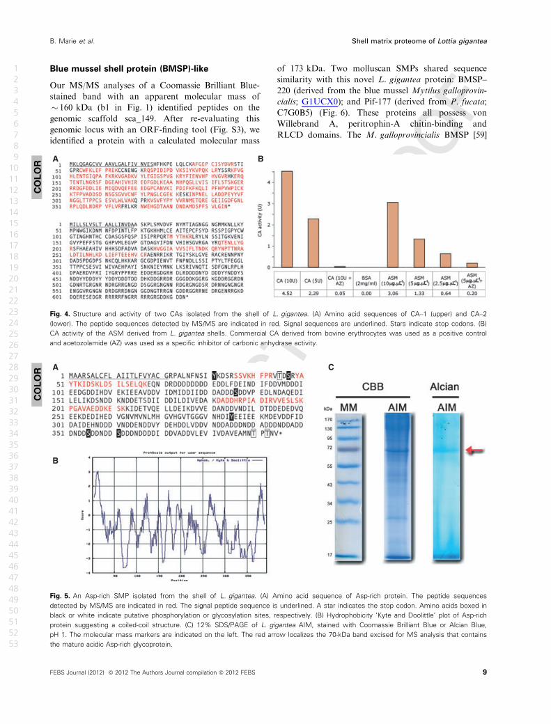
Carbonic anhydrases
CA is a ubiquitous metalloenzyme found in animals,
plants and bacteria which catalyses the reversible
hydration of carbon dioxide, according to the equation
CO2 + H2O ↔ HCO3�
+ H+. This enzyme is believed
to be essential for biomineral formation because bicar-
bonate, the product of the catalytic process, can directly
react with calcium ions to form calcium carbonate.
Furthermore, CA has been found in the organic matri-
ces of various metazoan skeletons [40–44]. We detected
two different CAs, Lgi-CA–1 and Lgi-CA–2, in
L. gigantea shell AIMs and ASMs. Both of these pro-
teins possess a highly conserved a–CA domain in addi-
tion to a Gly- and Glu-rich RLCD present in the
C–terminus of Lgi-CA–2 (Fig. 4A). Their CA domains
possess the conserved active residues known from well-
studied a–CAs [45], suggesting that these two Lottia
CAs are active enzymes. In support of this, we were
able to significantly detect a specific CA activity in the
ASM fraction (Fig. 4B).
Asp-rich, low pI proteins
Another group of proteins that emerged from our analy-
ses were the acidic Asp-rich proteins ‘Asp-rich’
(Fig. 5A), LUSP–23, LUSP–9 and LUSP–10 with
predicted pI values of 3.5, 3.6, 3.7 and 3.8, respectively
(Table 1). According to Coomassie Brilliant Blue-
stained SDS/PAGE gels, the abundant protein ‘Asp-
rich’ (which also has the lowest predicted pI) has an
apparent molecular mass of 80 kDa (Fig. 5). In contrast
to this, the predicted molecular mass for the nonglycosy-
lated mature form is only 42 kDa. A likely explanation
for this discrepancy is the observation that this band
was intensively stained with the cationic dye Alcian
Blue, suggesting that ‘Asp-rich’ bears extensive acidic
polysaccharide moieties. The hydrophobicity ‘Kyte and
Doolittle’ [46] plot of the ‘Asp-rich’ protein suggests that
it might also exhibit a coiled-coil structure (Fig. 5B).
The presence of such unusually acidic proteins in the
molluscan shell matrix is known from the pioneering
work of Meenakshi et al. [47], Crenshaw [7] and Wei-
ner and Hood [48], and has been further confirmed by
several investigations [49–53]. However, because of the
technical challenges of isolating and purifying these
acidic proteins, reports of their primary sequence are
rare [54–57]. To our knowledge, the Asp-rich protein
detected here, together with MSP–1 extracted from the
calcitic foliated layer of Patinopecten shell [54], is one
of the most acidic molluscan SMPs described to date.
Although there are several theoretical models regarding
the function that these acidic proteins play in the pro-
cess of shell formation [58], to date only a few in vivo
functional studies that have tested these theories [55].
A
C
B
Fig. 3. Schematic summary of L. gigantea’s RLCD-containing SMPs. Schematic representations of the primary structure of RLCD-containing
SMPs isolated from the shell of L. gigantea. (A) RLCD domains of peroxidase–1 and peroxidase–2. (B) LUSP–1, -7, -8, -10 and -22 possess
noticeable Q-rich repeats or domains. (C) LUSP–11, -12 and -25 exhibit both M- and G-rich domains. Each protein sequence possesses a
signal sequence indicated by a red bar. RLCDs are indicated in light grey, with specific repeats indicated by small white boxes.
COLOR
8 FEBS Journal (2012) ª 2012 The Authors Journal compilation ª 2012 FEBS
Shell matrix proteome of Lottia gigantea B. Marie et al.
1
2
3
4
5
6
7
8
9
10
11
12
13
14
15
16
17
18
19
20
21
22
23
24
25
26
27
28
29
30
31
32
33
34
35
36
37
38
39
40
41
42
43
44
45
46
47
48
49
50
51
52
53
philippemarie
Barrer
philippemarie
Texte inséré
41-45
philippemarie
Barrer
philippemarie
Texte inséré
46
philippemarie
Barrer
philippemarie
Texte inséré
47
philippemarie
Barrer
philippemarie
Texte inséré
48
philippemarie
Barrer
philippemarie
Texte inséré
49
philippemarie
Barrer
philippemarie
Texte inséré
50-54
philippemarie
Barrer
philippemarie
Texte inséré
55-58
philippemarie
Barrer
philippemarie
Texte inséré
55
philippemarie
Barrer
philippemarie
Texte inséré
59
philippemarie
Barrer
philippemarie
Texte inséré
56
Blue mussel shell protein (BMSP)-like
Our MS/MS analyses of a Coomassie Brilliant Blue-
stained band with an apparent molecular mass of
� 160 kDa (b1 in Fig. 1) identified peptides on the
genomic scaffold sca_149. After re-evaluating this
genomic locus with an ORF-finding tool (Fig. S3), we
identified a protein with a calculated molecular mass
of 173 kDa. Two molluscan SMPs shared sequence
similarity with this novel L. gigantea protein: BMSP–
220 (derived from the blue mussel Mytilus galloprovin-
cialis; G1UCX0); and Pif-177 (derived from P. fucata;
C7G0B5) (Fig. 6). These proteins all possess von
Willebrand A, peritrophin-A chitin-binding and
RLCD domains. The M. galloprovincialis BMSP [59]
A
B
C
Fig. 5. An Asp-rich SMP isolated from the shell of L. gigantea. (A) Amino acid sequence of Asp-rich protein. The peptide sequences
detected by MS/MS are indicated in red. The signal peptide sequence is underlined. A star indicates the stop codon. Amino acids boxed in
black or white indicate putative phosphorylation or glycosylation sites, respectively. (B) Hydrophobicity ‘Kyte and Doolittle’ plot of Asp-rich
protein suggesting a coiled-coil structure. (C) 12% SDS/PAGE of L. gigantea AIM, stained with Coomassie Brilliant Blue or Alcian Blue,
pH 1. The molecular mass markers are indicated on the left. The red arrow localizes the 70-kDa band excised for MS analysis that contains
the mature acidic Asp-rich glycoprotein.
A B
Fig. 4. Structure and activity of two CAs isolated from the shell of L. gigantea. (A) Amino acid sequences of CA–1 (upper) and CA–2
(lower). The peptide sequences detected by MS/MS are indicated in red. Signal sequences are underlined. Stars indicate stop codons. (B)
CA activity of the ASM derived from L. gigantea shells. Commercial CA derived from bovine erythrocytes was used as a positive control
and acetozolamide (AZ) was used as a specific inhibitor of carbonic anhydrase activity.
COLOR
COLOR
FEBS Journal (2012) ª 2012 The Authors Journal compilation ª 2012 FEBS 9
B. Marie et al. Shell matrix proteome of Lottia gigantea
1
2
3
4
5
6
7
8
9
10
11
12
13
14
15
16
17
18
19
20
21
22
23
24
25
26
27
28
29
30
31
32
33
34
35
36
37
38
39
40
41
42
43
44
45
46
47
48
49
50
51
52
53
philippemarie
Barrer
philippemarie
Texte inséré
60

and the L. gigantea BMSP-like proteins also possess a
laminin G-like (LamGL) domain and a poly(T)
domain between the von Willebrand A domain and
the LamGL domain. The M. galloprovincialis BMSP
and the L. gigantea BMSP-like proteins also share the
highest sequence similarity in these domains. The
P. fucata Pif protein was recently shown to bind both
CaCO3 and chitin, and by RNAi to play a role in
nacre formation in vivo [55]. Given that L. gigantea
does not form nacre it will be interesting to determine
the function of the L. gigantea BMSP-like protein.
Epidermal growth factor and sona pellucida
domain-containing SMPs
We also detected two similar proteins (LUSP–17 and
LUSP–24) each containing two epidermal growth fac-
tor (EGF)-like domains, and one zona pellucida (ZP)
domain in their C–termini. Although separate EGF-
like and ZP domains are commonly encountered in
organic matrix proteins associated with calcification
processes [20,60,61], the presence of both domains in
one protein is more uncommon. Previous proteomic
investigations have described one similar protein from
the shell matrix of the pacific oyster Crassostrea gigas
[25], and two from the Pinctada shell matrix (B. Marie
et al., unpublished). A sequence alignment of these lat-
ter proteins with the two EGF-like SMPs of L. gigan-
tea is presented in Fig. 7, and illustrates the strong
conservation of each domain. LUSP–17 and LUSP–24
are also located on the same genomic scaffold
(sca_66). This, in combination with their high degree
of sequence identity (79%), strongly suggests that they
originated from a gene duplication event.
EGF-like domains are involved in a wide variety of
functions such as protein/protein recognition, protein
aggregation, molecular signalling or Ca2+-binding
ability [62]. ZP domains are present in a range of extra-
cellular filament or matrix proteins from a wide variety
of eukaryotic organisms, and are characterized by eight
conserved cysteine residues, which are involved in pro-
tein polymerization processes [63]. Furthermore, the
urine-secreted protein, uromodulin (Tamm–Horsfall
protein, Q91X17) that exhibits three EGF domains and
one ZP domain can potentially contribute to colloid
osmotic pressure and modulates formation of supersatu-
rated salts and their crystals [64]. Such similar functions
could easily be credited to the EGF- and ZP-containing
SMPs and be integrated into a theoretical model of cal-
cified shell biomineralization. However, these hypothe-
ses await validation by functional experiments.
Other SMPs
Cyclophilin
We also detected a protein in the L. gigantea shell
matrix presenting sequence similarities with cyclophi-
lins (Fig. S4). Cyclophilins are peptidyl–prolyl isome-
rases that are believed to mostly facilitate protein
folding. In mice, the absence of expression of cyclophi-
lin B has been shown to induce severe osteogenesis im-
perfecta [65]. Although the specific role of this enzyme
in calcium carbonate mineralization is not known,
Jackson and co-workers [20] described a cyclophilin
gene highly expressed in the nacre forming cells of the
pearl oyster P. maxima.
Glycosidases
Two different glycosidase-related proteins were also
detected in Lottia’s shell matrix (Fig. S5). The first,
named Lgi-glycosidase–1, contains a characteristic
glycosyl_hydrolase_23 domain and shares significant
Fig. 6. BMSP-like SMPs isolated from the shell of L. gigantea. Schematic representations of the primary structure of L. gigantea BMSP-like,
M. galloprovincialis BMSP and P. fucata Pif proteins. von Willanbrand A, peritrophin-A chitin-binding (CB), RLCDs and LamGL domains are
indicated. Sequence similarity scores between selected domains are the percentage of amino acid identity.
COLOR
10 FEBS Journal (2012) ª 2012 The Authors Journal compilation ª 2012 FEBS
Shell matrix proteome of Lottia gigantea B. Marie et al.
1
2
3
4
5
6
7
8
9
10
11
12
13
14
15
16
17
18
19
20
21
22
23
24
25
26
27
28
29
30
31
32
33
34
35
36
37
38
39
40
41
42
43
44
45
46
47
48
49
50
51
52
53
philippemarie
Barrer
philippemarie
Texte inséré
56
philippemarie
Barrer
philippemarie
Texte inséré
61-62
philippemarie
Barrer
philippemarie
Texte inséré
[27.]
philippemarie
Barrer
philippemarie
Texte inséré
63
philippemarie
Barrer
philippemarie
Texte inséré
64
philippemarie
Barrer
philippemarie
Texte inséré
65
philippemarie
Barrer
philippemarie
Texte inséré
66

sequence similarity with patent lysozyme 2 proteins
described from other molluscs [66]. The second, named
Lgi-glycosidase–2, possesses DE6 -rich domains and a
conserved glyco_hydro_31 domain. Chitin and other
insoluble polysaccharides are major nonprotein com-
ponents of molluscan shells [67–69]. In classical models
of mollusc shell biomineralization, these molecules
form a framework of parallel layers between which
silk-like and acidic proteins are sandwiched [11,58].
Lottia’s glycosidase SMPs might reasonably be
expected to modify this chitin/polysaccharide frame-
work during biomineral formation.
EF7 -hand containing proteins
We also identified two short proteins containing two
EF-hand domains (Fig. S6). Similar proteins have
been described previously from the shell matrix of the
bivalves Pinctada and Venerupis [26] (B. Marie et al.,
unpublished). Two consecutive EF-hand domains are
known to bind Ca2+ ions with high affinity [70], and
are observed in many extracellular matrix proteins,
such as calmodulins, troponin–C or S–100, often in
association with other domains.
Secreted cysteine-rich protein-like proteins
Two similar L. gigantea SMPs, LUSP–2 and LUSP-3,
contain characteristic secreted cysteine-rich protein
(SCP) domains (Fig. S6). We also found that the genes
encoding LUSP–2 and LUSP–3 are located at adjacent
genomic loci (sca_35). Interestingly, three additional
SCP-domain-containing proteins, which were not
detected in our MS/MS analyses, are also present on
this scaffold (Fig. S7). Interestingly, a similar SCP-
containing protein has recently been described from
the nacre of P. margaritifera (B. Marie et al., unpub-
lished). Because SCP domains have also been
described in association with a variety of extracellular
matrix proteins, no clear function has been yet
assigned to such domains in the context of biomineral-
ization.
Perlustrin
We also detected a protein in the shell matrix of L. gi-
gantea with sequence similarities to perlustrin, a pro-
tein containing an insulin-like growth factor binding
protein (IGF–BP) domain first isolated from the nacre
of the abalone Haliotis laevigata (Fig. S8) [71]. This
Fig. 7. Two EGF-like SMPs isolated from the shell of L. gigantea. A sequence alignment of EGF-like proteins retrieved from the shell of
L. gigantea (B3A0R6 and B3A0S3) against C. gigas (P86785), and P maxima (P86953 and P86954). Signal peptides (yellow), EGF-like (green)
and ZP (blue) domains are highlighted. Stars at the end of each sequence indicate a stop codon.
COLOR
FEBS Journal (2012) ª 2012 The Authors Journal compilation ª 2012 FEBS 11
B. Marie et al. Shell matrix proteome of Lottia gigantea
1
2
3
4
5
6
7
8
9
10
11
12
13
14
15
16
17
18
19
20
21
22
23
24
25
26
27
28
29
30
31
32
33
34
35
36
37
38
39
40
41
42
43
44
45
46
47
48
49
50
51
52
53
philippemarie
Barrer
philippemarie
Texte inséré
Asp- and Glu-rich
philippemarie
Texte surligné
Provided
philippemarie
Texte surligné
Provided in the abbreviations of the page1.
philippemarie
Barrer
philippemarie
Texte inséré
67
philippemarie
Barrer
philippemarie
Texte inséré
68-70
philippemarie
Barrer
philippemarie
Texte inséré
59
philippemarie
Barrer
philippemarie
Texte inséré
71
philippemarie
Barrer
philippemarie
Texte inséré
[27].
philippemarie
Barrer
philippemarie
Texte inséré
72
philippemarie
Texte inséré
,27
philippemarie
Barrer

Lgi-IGF–BP is characterized by a pattern of 12 con-
served Cys residues. Interestingly, vertebrate bone
matrix contains IGF–BPs, which are involved in bone
formation, possess an effective affinity for growth fac-
tors of the insulin type, and function by modulating
IGF metabolism.
Perlwapin
We detected in Lottia’s shell matrix one protein con-
taining five whey-acidic protein (WAP) domains, and
with high overall sequence similarity to the perlwapin
family (Fig. S8). WAP consists of two ‘four-disulfide
core’ domains that are present in various serine-pro-
teinase inhibitors. Perlwapin proteins, containing such
WAP domains, have been identified in Haliotis
[23,72,73] and from the shell of the blue mussel
M. galloprovincialis [24]. However, whereas Lgi-per-
lwapin contains five WAP domains, the other perlwa-
pins from the species listed above possess only oen to
three WAP domains.
Orphans
Nine other L. gigantea SMPs do not display any
sequence similarity with previously described proteins,
or possess recognizably conserved domains (Table 1).
These proteins were categorized as orphans. Compara-
tive metazoan genome analyses suggest that every tax-
onomic group contains 10–20% of these so-called
‘orphan’ or ‘taxonomically restricted’ genes. Such
genes are thought to underlie mechanisms that can
support the generation of morphological novelties [74].
Interestingly, all molluscan shell matrices broadly
investigated at the ‘-omic’ level (genomic, transcrip-
tomic or proteomic) contain such orphan proteins.
The presence of such orphans may reflect the evolv-
ability of the molluscan shell matrix, suggesting that
the appearance of such new proteins within the SMP
set could potentially be related to modification of the
biomineral structure through evolutionary time. Per-
haps more than any other, this class of biomineral-
associated proteins highlights the need for in vivo gene
function assays to be developed for molluscan biomin-
eralizing systems.
Discussion
RLCD-containing SMPs
RLCD proteins are a prominent feature of all shell-
forming proteomes studied to date. Most, if not all, of
the RLCD-containing SMPs we have detected appear
to be lineage-specific proteins, supporting the idea that
such biomineralizing proteins have evolved indepen-
dently in the different molluscan models. Various
RLCD-containing proteins are present in a wide range
of metazoan-secreted structures, for example silk fibroin
[75], the mussel byssus [76] or the insect chorion [77].
Molluscan shell-forming proteins with RLCDs include
nacrein and lustrin–A which contain GN- or GS-rich
domains [18,73], MSI60 and CL10Contig2 contain poly
(G) and poly(A) blocks [78], Pif-177 contains D-rich
domains [55], MPN88 contains Q-, M- and G-rich
repeated sequences [19], and the Shematrin family bear
numerous GY-rich domains [79]. RLCDs are likely to
represent regions with intrinsically disordered confor-
mations thought to be structurally unstable [80]. Such
domains possess low binding affinity for other organic
macromolecules (such as proteins or polysaccharides),
but weakly bind mineral surfaces and ions in aqueous
phases. Indeed, GY or GN repeats of the nacrein and
shematrins have been proposed to weakly bind Ca2+
ions [33,79], whereas the D-rich domains of Pif-177
were shown to directly bind aragonitic mineral surfaces
[55]. It has also been proposed that the poly(G), poly
(A), or poly(S) regions of MSI60, CL10Contig2 or
lustrin–A may confer elastomeric properties to the
mature biomineral [23,73,78,81]. Given that RLCD pro-
teins are a major component of the protein fraction
within a wide range of molluscan shells, it is clear that
they are likely to be playing crucial roles in either shell
formation, and/or imparting to the shell certain physi-
cal properties such as fracture resistance.
Conservation of SMPs and their evolution
Given that L. gigantea does not form nacre, one of the
most surprising results of our study was the detection
of various proteins that share high sequence similarities
with SMPs previously identified from the nacro-
prismatic shells of Pinctada bivalves. Figure 8 summa-
rizes the co-occurrence of SMPs known from various
molluscan models of biomineralization: bivalves of the
genus Pinctada [19] (B. Marie et al., unpublished); aba-
lone (genus Haliotis) [23]; and L. gigantea (comprising
the proteins reported here together with the 23 main
shell proteins identified by Mann et al. [32]). Protein
sequence alignments and overall domain conservation
suggest that most of the eight proteins shared between
Lottia and Pinctada (CAs, BMSP and EGF-like in par-
ticular) may be true orthologues (Figs 3, 4, 6 and 7
and Fig. S6). For the two proteins shared between
L. gigantea and the Haliotids, IGF–BP (perlustrin) and
perlwapin, accurate evolutionary relationships (orthol-
ogy versus paralogy) are difficult to assign because the
12 FEBS Journal (2012) ª 2012 The Authors Journal compilation ª 2012 FEBS
Shell matrix proteome of Lottia gigantea B. Marie et al.
1
2
3
4
5
6
7
8
9
10
11
12
13
14
15
16
17
18
19
20
21
22
23
24
25
26
27
28
29
30
31
32
33
34
35
36
37
38
39
40
41
42
43
44
45
46
47
48
49
50
51
52
53
philippemarie
Barrer
philippemarie
Texte inséré
73-74
philippemarie
Barrer
philippemarie
Texte inséré
75
philippemarie
Barrer
philippemarie
Texte inséré
76
philippemarie
Barrer
philippemarie
Texte inséré
77
philippemarie
Barrer
philippemarie
Texte inséré
78
philippemarie
Barrer
philippemarie
Texte inséré
74
philippemarie
Barrer
philippemarie
Texte inséré
79
philippemarie
Barrer
philippemarie
Texte inséré
56
philippemarie
Barrer
philippemarie
Texte inséré
80
philippemarie
Barrer
philippemarie
Texte inséré
81
philippemarie
Barrer
philippemarie
Texte inséré
34,80
philippemarie
Barrer
philippemarie
Texte inséré
56
philippemarie
Barrer
philippemarie
Texte inséré
74,79,82
philippemarie
Barrer
philippemarie
Texte inséré
33
philippemarie
Texte inséré
,27
philippemarie
Barrer

sequence based similarities between these proteins are
restricted to amino acid positions that are specific to
the IGF–BP and WAP families (Fig. S8).
Counterintuitively, we have found more SMPs
shared between L. gigantea and bivalve species Pinct-
ada than between the gastropods L. gigantea and
A
B
Fig. 8. A comparison of molluscan SMPs isolated from Lottia, Pinctada and Haliotis. A broad comparison of molluscan SMPs. (A) A
summary of the shared and lineage-specific SMPs described to date from L. gigantea and various Pinctada and Haliotis species. Numbers
correspond to the number of different SMPs detected to date for each model, for example we can distinguish 32 different SMPs from the
39 different SMPs we have identified for L. gigantea (e.g. when considering one entry for the two CAs). (B) After categorizing these SMPs
into eight broad categories it is clear that proteins with RLCDs are a common feature of the molluscan shell-forming secretome. Most
proteins shared between L. gigantea and Pinctada species fall into the extracellular matrix category. Grey boxes indicate proteins detected
by Mann et al. [32] to be minor components of the shell matrix.
COLOR
FEBS Journal (2012) ª 2012 The Authors Journal compilation ª 2012 FEBS 13
B. Marie et al. Shell matrix proteome of Lottia gigantea
1
2
3
4
5
6
7
8
9
10
11
12
13
14
15
16
17
18
19
20
21
22
23
24
25
26
27
28
29
30
31
32
33
34
35
36
37
38
39
40
41
42
43
44
45
46
47
48
49
50
51
52
53
philippemarie
Barrer
philippemarie
Texte inséré
33

abalone. This trend was also independently described
in a transcriptomic comparison of the mantle tissues
of L. gigantea, P maxima and H. asinina [20]. One
potential explanation for these observations is that
the shell-forming secretome of the abalone has accu-
mulated more changes since its divergence from a
limpet–abalone ancestor than the limpet has since its
divergence from a bivalve–gastropod ancestor. Given
the fundamental crystallographic differences between
the limpet and abalone shells (presence/absence of
nacre and crossed lamellae, for example), such a sce-
nario is conceivable. Complicating this issue is the fact
that beyond the species and genus level, molluscan
shell microstructures are notoriously evolutionary plas-
tic. To a large degree this plasticity must be the result
of the evolution of the organic molecules that coordi-
nate deposition of the shell (past ocean chemistries
and temperatures would also affect shell evolution).
Our molecular data are compatible with the hypothesis
of a genuine affiliation between cross-lamellar struc-
tures and nacre [3]. However, a well-resolved, robust
and taxonomically well-represented phylogenetic tree
for the Conchifera is essential before any scenarios of
shell evolution can be proposed and then tested. For-
tunately, recent genomic efforts are moving towards
this goal [82,83]. In addition to such a resource, better
taxon sampling of mantle tissue transcriptomes and
shell proteomes would allow us to better understand
how this shelled diversity has been generated over the
last 550 million years.
Conclusions
The availability of genome, proteome and transcrip-
tome scale datasets from non-model organisms is
enabling more complete assessments of complex bio-
logical processes to be performed. Molluscan shell for-
mation is certainly such a process that will benefit
from such analyses. By combining a proteomic analy-
sis of SMPs extracted from the shell of L. gigantea
with a draft genome assembly, we have identified sev-
eral new biomineralizing proteins, and further charac-
terized several others. Many of these proteins are
characterized by apparently lineage-specific arrange-
ments of RLCDs and highly conserved enzymatic
domains such as CA, peroxidase and glycosidase. Even
when combined with a recent analysis by Mann et al.
[32], the complete shell-forming proteome of L. gigan-
tea is unlikely to have been described, and further
work will probably identify additional components.
Indeed, it remains possible that the trypsin hydrolysis
of few SMPs generate only peptides of unsuitable
length (too short or too long) for MS analysis, being
undetectable by classical proteomic approach [84 8].
However many of the primary shell-forming proteins
are likely to be in hand, and it is becoming increas-
ingly clear that the challenge that now faces the field is
to characterize the function of these proteins using
in vivo techniques.
Materials and methods
Sampling
Fresh L. gigantea shells (5–7 cm in length) were collected
from the West Pacific coast of the USA (California). Shell
microstructure was observed with a scanning electron
microscope Philips XL-30 LaB6 under back-scattered elec-
tron mode.
Shell matrix extraction
The external organic layer, the periostracum, and the outer-
most M + 3 calcified layer that presents burrowing traces
were mechanically remove under cold water in order to
avoid shell heating, then the rest of the shell, comprising
the M + 2, M + 1, M and M � 1 layers were crushed into
fragments of ~1 mm2. Any other superficial organic con-
taminants were removed by incubating shell fragments in
NaOCl (1%, v/v) for 24 h, and which were then thoroughly
rinsed with water and subsequently ground into a fine pow-
der that was sieved (> 200 lm). All protein extractions
were performed at 4 °C, as previously described [51].
Briefly, powdered samples were decalcified overnight in
cold dilute acetic acid (5%, v/v), which was slowly added
by an automated titrator (Titronic Universal, Mainz, Ger-
many) at a flow rate of 100 lL every 5 s. The solution
(final pH ~4.2) was centrifuged at 3900 g (30 min). The
resulting pellet, corresponding to the AIM, was rinsed six
times with MilliQ water, freeze-dried and weighed. The
supernatant containing ASM was filtered (5 lm) and con-
centrated with an Amicon ultra-filtration system on a Milli-
pore® membrane (10 kDa cut-off). The final solution
(> 5 mL) was extensively dialysed against 1 L of MilliQ
water (six water changes) before being freeze-dried and
weighed.
CA activity measurement
The miniaturized colorimetric method developed by Maren
[85] was employed for measuring the CA activity (EC 4.2.1.1)
of the shell ASM. The experiment was carried out under stabi-
lized flow of CO2, in an ice-containing vessel. Four hundred
microlitres of phenol red (12.5 mg�L�1 in 2.6 mM NaHCO3)
were mixed with 200 lL of water and 100 lL of sample. The
reaction was initiated by adding 100 lL of freshly made
carbonate buffer (0.6 M Na2CO3, 0.412 M NaHCO3) and the
14 FEBS Journal (2012) ª 2012 The Authors Journal compilation ª 2012 FEBS
Shell matrix proteome of Lottia gigantea B. Marie et al.
1
2
3
4
5
6
7
8
9
10
11
12
13
14
15
16
17
18
19
20
21
22
23
24
25
26
27
28
29
30
31
32
33
34
35
36
37
38
39
40
41
42
43
44
45
46
47
48
49
50
51
52
53
philippemarie
Texte surligné
Okey.
philippemarie
Barrer
philippemarie
Texte inséré
83,84
philippemarie
Barrer
philippemarie
Texte inséré
33
philippemarie
Barrer
philippemarie
Texte inséré
85
philippemarie
Barrer
philippemarie
Texte inséré
52
philippemarie
Barrer
philippemarie
Texte inséré
86

time interval until the colour changed from red to yellow was
monitored. This colour change characterizes the pH decrease
of the solution (from 8.2 to 7.3), resulting from the production
of protons during the reaction catalysed by the CA
(CO2 + H2O ↔ HCO3�
+ H+). The enzyme unit (EU) activ-
ity was calculated according to the following equation: activ-
ity units (EU) = (T0 � T)/T; where T and T0 are the reaction
times required for the pH change with and without a catalyst,
respectively. Acetozolamide was used as a specific inhibitor of
the reaction. Commercial bovine CA and BSA were used as
positive and negative controls, respectively.
SDS/PAGE
The fractionation of matrix macromolecules was per-
formed under denaturing conditions by monodimensional
SDS/PAGE (Mini-Protean 3; Bio-Rad). One milligram of
matrix (both ASM and AIM) was suspended in 200 lL
of Laemmli sample buffer [86], heat denatured (10 min,
100 °C) then centrifuged for 1 min at 12 000 g. Ten
microlitres of the supernatant, representing a maximum
of 50 lg of matrix, were loaded onto gels. Following
SDS/PAGE under denaturing conditions (4–15% acrylam-
ide gel), proteins were visualized with Coomassie Brilliant
Blue (CBB G-250; Biosafe, Bio-Rad). Alternatively, puta-
tive glycosylations were investigated by staining with
Alcian Blue 8GX [87], at pH 1 in order to specifically
stain sulfated sugars.
Sample preparation for proteomic analysis
An in-gel digestion procedure was performed for 12 predom-
inant protein bands visualized from the electrophoresis gel
of the AIM (Fig. 2). These bands were excised from Coo-
massie Brilliant Blue-stained gels and completely
destained by a wash with 400 lL of 50 mM NH4HCO3/
CH3CN (50/50) mixture for 15 min at 37 °C. Reduction was
performed with 50 lL of 10 mM dithiothreitol in 50 mM
NH4HCO3 for 15 min at 50 °C. Alkylation was performed
with 50 lL of 100 mM iodoacetamide for 15 min at room
temperature in the dark. The reagents were taken away and
the gel pieces were dried using 100 lL of CH3CN. Gel pieces
were then treated with 0.4 lg trypsin (Sequence grade; Pro-
mega, Madison, WI, USA) in 20 lL of 50 mM NH4HCO3
for 45 min at 50 °C under 800 rpm agitation. The superna-
tant was removed and stored. The gel pieces were extracted
with 30 lL of H2O:CH3CN:HCOOH (68 : 30 : 2) mixture
for 30 min at 30 °C. Finally, both supernatant extracts were
pooled, dried in a vacuum concentrator and resuspended in
13 lL of 0.1% trifluoroacetic acid.
In-solution digestion of unfractionated L. gigantea ASM
and AIM was also performed. These samples (0.1 and
1 mg, respectively) were reduced with 50 lL of 10 mM
dithiothreitol in 50 mM NH4HCO3 for 30 min at 50 °C.
Alkylation was performed with 50 lL of 100 mM iodoaceta-
mide in 50 mM NH4HCO3 for 30 min at room temperature
in the dark. The solution was then treated with 1 lg of tryp-
sin (Sequence grade; Promega) in 10 lL 50 mM NH4HCO3
overnight at 37 °C. The sample was dried in a vacuum con-
centrator and resuspended in 30 lL of 0.1% trifluoroacetic
acid and 2% CH3CN.
Peptide fractionation and data acquisition
MS was performed using a Q-Star XL nanospray quadru-
pole/time-of-flight tandem mass spectrometer, nanospray-
Qq-TOF-MS/MS (Applied Biosystems, France 9), coupled to
an online nano liquid chromatography system (Ultimate
Famos Switchos from Dionex, The Netherlands 10). One mi-
crolitre of each sample was loaded onto a trap column
(PepMap100 C18; 5 lm; 100 �A; 300 lm 9 5 mm; Dionex),
washed for 3 min at 25 lL�min�1 with 0.05% trifluroacetic
acid/2% acetonitrile, then eluted onto a C18 reverse phase
column (PepMap100 C18; 3 lm; 100 �A; 75 lm 9 150 mm;
Dionex). Peptides were separated at a flow rate of
0.300 lL�min�1 with a linear gradient of 5–80% acetonitrile
in 0.1% formic acid over 120 min. MS data were acquired
automatically using ANALYST QS 1.1 software (Applied Bio-
systems). Following a MS survey scan over m/z 400–1600
range, MS/MS spectra were sequentially and dynamically
acquired for the three most intense ions over m/z 65–2000
range. The collision energy was set by the software accord-
ing to the charge and mass of the precursor ion. MS and
MS/MS data were recalibrated using internal reference ions
from a trypsin autolysis peptide at m/z 842.51 [M + H]+
and m/z 421.76 [M + 2H]2+.
MS data analysis
Protein identification was performed using the MASCOT
search engine (version 2.1; Matrix Science, London, UK)
against protein databases derived from the EST and the
genomic libraries of L. gigantea comprising 252 091 and
23 851 sequences, and downloaded (March 2010) from the
NCBI server (http://www.ncbi.nlm.nih.gov) and the L. gi-
gantea genome website (http://genome.jgi-psf.org/Lotgi1/
Lotgi1.download.ftp.html), respectively. LC-MS/MS data
were searched using carbamidomethylation as a fixed modi-
fication, and methionine oxidation as a variable modifica-
tion. The peptide mass and fragment ion tolerances were
set to 0.5 Da. Only protein identifications with at least two
different peptide hits and/or that were independently
obtained from two different samples were considered to be
valid. The peptide hits were manually confirmed by the
interpretation of the raw LC-MS/MS spectra with ANALYST
QS software (Version 1.1). Quality criteria were the peptide
MS value, the assignment of major peaks to uninterrupted
y- and b-ion series of at least three to four consecutive
amino acids and the match with the de novo interpretations
proposed by the software.
FEBS Journal (2012) ª 2012 The Authors Journal compilation ª 2012 FEBS 15
B. Marie et al. Shell matrix proteome of Lottia gigantea
1
2
3
4
5
6
7
8
9
10
11
12
13
14
15
16
17
18
19
20
21
22
23
24
25
26
27
28
29
30
31
32
33
34
35
36
37
38
39
40
41
42
43
44
45
46
47
48
49
50
51
52
53
philippemarie
Texte inséré
Villebon-sur-Yvette,
philippemarie
Texte inséré
Amsterdam,
philippemarie
Texte surligné
Provided
philippemarie
Texte surligné
Provided
philippemarie
Barrer
philippemarie
Texte inséré
87
philippemarie
Barrer
philippemarie
Texte inséré
88

Sequence analysis
Protein sequence identification was performed using BLASTP
and TBLASTN analyses performed against Swiss-Prot, Gen-
Bank’s nr db and dbEST using the online tool provided by
UniProt (www.uniprot.org) and NCBI (http://blast.ncbi.
nlm.nih.gov/blast.cgi) servers. Signal peptides were pre-
dicted using SIGNALP 3.0 (http://www.cbs.dtu.dk/services/
SignalP/), and conserved domains were predicted using
SMART (http://smart.embl-heidelberg.de/) and INTERPROSCAN
(http://www.ebi.ac.uk/Tools/InterProScan/). Following pep-
tide signal removal, theoretical masses and pI values were
determined using the EXPASY PROTPARAM tool (www.expasy.
org/tools/protparam.html).
Sequence alignment and phylogenetic analysis
Representative full-length sequences of the major nonverte-
brate metazoan peroxidases were selected from the results
of a BLAST searche performed with the three peroxidases
from L. gigantea SMPs, using UniProt and NCBI online
tools, against Swiss-Prot, GenBank’s nr db and dbEST.
The multiple alignment was created using T-COFFEE 6.85
[88] set to standard parameters. Phylogenetic reconstruc-
tions were performed using the maximum likelihood
method implemented in PHYML from the www.phylogeny.fr
server [89].
Acknowledgements
We thank Eric Edsinger-Gonzales for providing shell
material. BM thanks J�erome Thomas for handling
shell pictures of Lottia and Pinctada. The work of BM
was supported by the GDR ADEQUA consortium
(Coordinator Nathalie Cochennec-Loreau/Yannick
Gueguen), while the work of NG and FM was sup-
ported via ANR Accro-Earth (ref. BLAN06-2_159971,
coordinator Gilles Ramstein, LSCE). Additional sup-
ports include ITN BIOMINTEC and INTERRVIE program
(2010). DJJ is supported by DFG funding to the Uni-
versity of G€ottingen through the German Excellence
Initiative.
References11
1 Ponder W & Lindberg DR (2008) Phylogeny and
Evolution of the Mollusca. University of California
Press, Berkley, 469 p.
2 Bøggild O (1930) The shell structure of the mollusks.K
Dan Vidensk Selsk Skr NaturvidenskMath Afd 9, 233–326.
3 Taylor J, Kennedy WJ & Hall A (1973) The shell
structure and mineralogy of the Bivalvia, Part II,
Lucinacea–Clavagellacea. Bull Brit Mus Zool 22,
253–294.
4 Carter JG (1990) Skeletal Biomineralization: Patterns,
Processes and Evolutionary Trends. Van Nostrand
Reinhold, New York.
5 Chateigner D, Hedegaard C & Wenk HR (2000)
Mollusc shell microstructures and crystallographic
textures. J Struct Geol 22, 1723–1735.
6 Mann S (2001) Biomineralization: Principles and
Concepts in Bioinorganic Materials Chemistry. Oxford
University Press, New York.
7 Crenshaw MA (1972) The soluble matrix from
Mercenaria mercenaria shell. Biomineralization 6, 6–11.
8 Weiner S & Traub W (1984) Macromolecules in
mollusc shells and their functions in biomineralization.
Phil Trans R Soc Lond 304, 425–434.
9 Lowenstam HA & Weiner S (1989) On
Biomineralization. Oxford University Press, New York.
10 Keith J, Stockwell S, Ball D, Remillard K, Kaplan D,
Thanhauser T & Sherwood R (1993) Comparative
analysis of macromolecules in mollusc shells. Comp
Biochem Physiol B 105, 487–496.
11 Levi-Kalisman Y, Falini G, Addadi L & Weiner S
(2001) Structure of the nacreous organic matrix of a
bivalve mollusk shell examined in the hydrated state
using cryo-TEM. J Struct Biol 135, 8–17.
12 B�edouet L, Schuller MJ, Marin F, Milet C, Lopez E &
Giraud M (2001) Soluble proteins of the nacre of the
giant oyster Pinctada maxima and of the abalone
Haliotis tuberculata: extraction and partial analysis of
nacre proteins. Comp Biochem Physiol B 128, 389–400.
13 Marie B, Luquet G, Pais De Barros J-P, Guichard N,
Morel S, Alcaraz G, Bollache L & Marin F (2007) The
shell matrix of the freshwater mussel Unio pictorum
(Paleoheterodonta, Unionoida). Involvement of acidic
polysaccharides from glycoproteins in nacre
mineralization. FEBS J 274, 2933–2945.
14 Marie B, Marin F, Marie A, B�edouet L, Dubost L,
Alcaraz G, Milet C & Luquet G (2009) Evolution of
nacre: biochemistry and proteomics of the shell organic
matrix of the cephalopod Nautilus macromphalus.
ChemBioChem 10, 1495–1510.
15 Farre B & Dauphin Y (2009) Lipids from the nacreous
and prismatic layers of two pteriomorpha mollusc
shells. Comp Biochem Physiol B 152, 103–109.
16 Marin F, Luquet G, Marie B & Medakovic D (2008)
Molluscan shell proteins: primary structure, origin, and
evolution. Curr Top Dev Biol 80, 209–276.
17 Marin F, Le Roy N & Marie B (2012) The formation
and mineralization of mollusc shell. Front Biosci 4,
1099–1125.
18 Miyamoto H, Miyashita T, Okushima M, Nakano S,
Morita T & Matsushiro A (1996) A carbonic anhydrase
from the nacreous layer in oyster pearls. Proc Natl
Acad Sci USA 93, 9657–9660.
19 Joubert C, Piquemal D, Marie B, Manchon L, Pierrat
F, Zanella-Cl�eon I, Cochennec-Laureau N, Gueguen Y
16 FEBS Journal (2012) ª 2012 The Authors Journal compilation ª 2012 FEBS
Shell matrix proteome of Lottia gigantea B. Marie et al.
1
2
3
4
5
6
7
8
9
10
11
12
13
14
15
16
17
18
19
20
21
22
23
24
25
26
27
28
29
30
31
32
33
34
35
36
37
38
39
40
41
42
43
44
45
46
47
48
49
50
51
52
53
philippemarie
Texte surligné
This article is now accepted in PNAS and will be published online in the 3th of Decembre week (according to the editor). So, Please would you re-inject this reference (n° 27) within the text and the reference list and modify the following reference number accordingly. The reference details are :27 - Marie B, Joubert C, Tayale A, Zanella-Cléon I, Belliard C, Cochennec-Loreau N, Piquemal D., Marin F, Gueguen Y, Montagnani C. Different secretory repertoires control the biomineralization processes of prism and nacre deposition of the pearl oyster shell. Proc Natl Acad Sci. Accepted (doi:10.1073/pnas.1210552109).
philippemarie
Barrer
philippemarie
Texte inséré
89
philippemarie
Barrer
philippemarie
Texte inséré
90
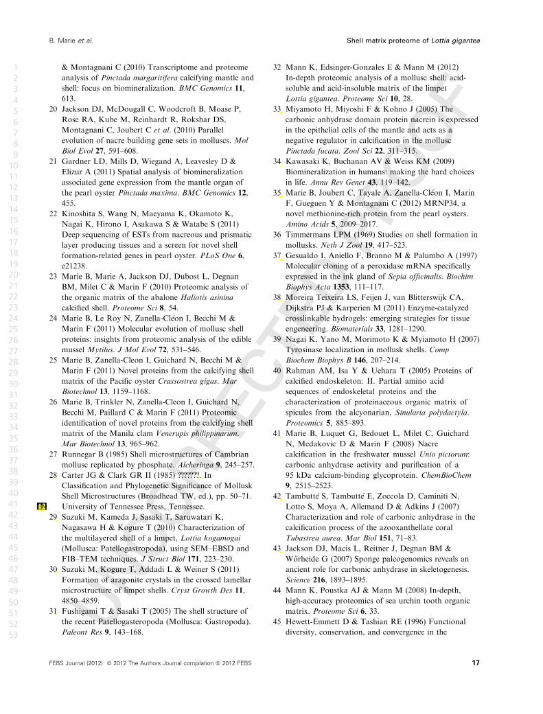
& Montagnani C (2010) Transcriptome and proteome
analysis of Pinctada margaritifera calcifying mantle and
shell: focus on biomineralization. BMC Genomics 11,
613.
20 Jackson DJ, McDougall C, Woodcroft B, Moase P,
Rose RA, Kube M, Reinhardt R, Rokshar DS,
Montagnani C, Joubert C et al. (2010) Parallel
evolution of nacre building gene sets in molluscs. Mol
Biol Evol 27, 591–608.
21 Gardner LD, Mills D, Wiegand A, Leavesley D &
Elizur A (2011) Spatial analysis of biomineralization
associated gene expression from the mantle organ of
the pearl oyster Pinctada maxima. BMC Genomics 12,
455.
22 Kinoshita S, Wang N, Maeyama K, Okamoto K,
Nagai K, Hirono I, Asakawa S & Watabe S (2011)
Deep sequencing of ESTs from nacreous and prismatic
layer producing tissues and a screen for novel shell
formation-related genes in pearl oyster. PLoS One 6,
e21238.
23 Marie B, Marie A, Jackson DJ, Dubost L, Degnan
BM, Milet C & Marin F (2010) Proteomic analysis of
the organic matrix of the abalone Haliotis asinina
calcified shell. Proteome Sci 8, 54.
24 Marie B, Le Roy N, Zanella-Cl�eon I, Becchi M &
Marin F (2011) Molecular evolution of mollusc shell
proteins: insights from proteomic analysis of the edible
mussel Mytilus. J Mol Evol 72, 531–546.
25 Marie B, Zanella-Cleon I, Guichard N, Becchi M &
Marin F (2011) Novel proteins from the calcifying shell
matrix of the Pacific oyster Crassostrea gigas. Mar
Biotechnol 13, 1159–1168.
26 Marie B, Trinkler N, Zanella-Cleon I, Guichard N,
Becchi M, Paillard C & Marin F (2011) Proteomic
identification of novel proteins from the calcifying shell
matrix of the Manila clam Venerupis philippinarum.
Mar Biotechnol 13, 965–962.
27 Runnegar B (1985) Shell microstructures of Cambrian
mollusc replicated by phosphate. Alcheringa 9, 245–257.
28 Carter JG & Clark GR II (1985) ???????. In
Classification and Phylogenetic Significance of Mollusk
Shell Microstructures (Broadhead TW, ed.), pp. 50–71.
University of Tennessee Press, Tennessee.12
29 Suzuki M, Kameda J, Sasaki T, Saruwatari K,
Nagasawa H & Kogure T (2010) Characterization of
the multilayered shell of a limpet, Lottia kogamogai
(Mollusca: Patellogastropoda), using SEM–EBSD and
FIB–TEM techniques. J Struct Biol 171, 223–230.
30 Suzuki M, Kogure T, Addadi L & Weiner S (2011)
Formation of aragonite crystals in the crossed lamellar
microstructure of limpet shells. Cryst Growth Des 11,
4850–4859.
31 Fushigami T & Sasaki T (2005) The shell structure of
the recent Patellogasteropoda (Mollusca: Gastropoda).
Paleont Res 9, 143–168.
32 Mann K, Edsinger-Gonzales E & Mann M (2012)
In-depth proteomic analysis of a mollusc shell: acid-
soluble and acid-insoluble matrix of the limpet
Lottia gigantea. Proteome Sci 10, 28.
33 Miyamoto H, Miyoshi F & Kohno J (2005) The
carbonic anhydrase domain protein nacrein is expressed
in the epithelial cells of the mantle and acts as a
negative regulator in calcification in the mollusc
Pinctada fucata. Zool Sci 22, 311–315.
34 Kawasaki K, Buchanan AV & Weiss KM (2009)
Biomineralization in humans: making the hard choices
in life. Annu Rev Genet 43, 119–142.
35 Marie B, Joubert C, Tayale A, Zanella-Cl�eon I, Marin
F, Gueguen Y & Montagnani C (2012) MRNP34, a
novel methionine-rich protein from the pearl oysters.
Amino Acids 5, 2009–2017.
36 Timmermans LPM (1969) Studies on shell formation in
mollusks. Neth J Zool 19, 417–523.
37 Gesualdo I, Aniello F, Branno M & Palumbo A (1997)
Molecular cloning of a peroxidase mRNA specifically
expressed in the ink gland of Sepia officinalis. Biochim
Biophys Acta 1353, 111–117.
38 Moreira Teixeira LS, Feijen J, van Blitterswijk CA,
Dijkstra PJ & Karperien M (2011) Enzyme-catalyzed
crosslinkable hydrogels: emerging strategies for tissue
engeneering. Biomaterials 33, 1281–1290.
39 Nagai K, Yano M, Morimoto K & Myiamoto H (2007)
Tyrosinase localization in mollusk shells. Comp
Biochem Biophys B 146, 207–214.
40 Rahman AM, Isa Y & Uehara T (2005) Proteins of
calcified endoskeleton: II. Partial amino acid
sequences of endoskeletal proteins and the
characterization of proteinaceous organic matrix of
spicules from the alcyonarian, Sinularia polydactyla.
Proteomics 5, 885–893.
41 Marie B, Luquet G, Bedouet L, Milet C, Guichard
N, Medakovic D & Marin F (2008) Nacre
calcification in the freshwater mussel Unio pictorum:
carbonic anhydrase activity and purification of a
95 kDa calcium-binding glycoprotein. ChemBioChem
9, 2515–2523.
42 Tambutt�e S, Tambutt�e E, Zoccola D, Caminiti N,
Lotto S, Moya A, Allemand D & Adkins J (2007)
Characterization and role of carbonic anhydrase in the
calcification process of the azooxanthellate coral
Tubastrea aurea. Mar Biol 151, 71–83.
43 Jackson DJ, Macis L, Reitner J, Degnan BM &
W€orheide G (2007) Sponge paleogenomics reveals an
ancient role for carbonic anhydrase in skeletogenesis.
Science 216, 1893–1895.
44 Mann K, Poustka AJ & Mann M (2008) In-depth,
high-accuracy proteomics of sea urchin tooth organic
matrix. Proteome Sci 6, 33.
45 Hewett-Emmett D & Tashian RE (1996) Functional
diversity, conservation, and convergence in the
FEBS Journal (2012) ª 2012 The Authors Journal compilation ª 2012 FEBS 17
B. Marie et al. Shell matrix proteome of Lottia gigantea
1
2
3
4
5
6
7
8
9
10
11
12
13
14
15
16
17
18
19
20
21
22
23
24
25
26
27
28
29
30
31
32
33
34
35
36
37
38
39
40
41
42
43
44
45
46
47
48
49
50
51
52
53
philippemarie
Texte inséré
27 - Marie B, Joubert C, Tayale A, Zanella-Cléon I, Belliard C, Cochennec-Loreau N, Piquemal D., Marin F, Gueguen Y, Montagnani C. Different secretory repertoires control the biomineralization processes of prism and nacre deposition of the pearl oyster shell. Proc Natl Acad Sci. Accepted (doi:10.1073/pnas.1210552109).
philippemarie
Barrer
philippemarie
Texte inséré
29
philippemarie
Barrer
philippemarie
Texte inséré
30
philippemarie
Barrer
philippemarie
Texte inséré
31
philippemarie
Barrer
philippemarie
Texte inséré
32
philippemarie
Barrer
philippemarie
Texte inséré
33
philippemarie
Barrer
philippemarie
Texte inséré
34
philippemarie
Barrer
philippemarie
Texte inséré
35
philippemarie
Barrer
philippemarie
Texte inséré
36
philippemarie
Barrer
philippemarie
Texte inséré
37
philippemarie
Barrer
philippemarie
Texte inséré
38
philippemarie
Barrer
philippemarie
Texte inséré
39
philippemarie
Barrer
philippemarie
Texte inséré
40
philippemarie
Barrer
philippemarie
Texte inséré
41
philippemarie
Barrer
philippemarie
Texte inséré
42
philippemarie
Barrer
philippemarie
Texte inséré
43
philippemarie
Barrer
philippemarie
Texte inséré
44
philippemarie
Barrer
philippemarie
Texte inséré
45
philippemarie
Barrer
philippemarie
Texte inséré
46
philippemarie
Texte inséré
Knoxville,
philippemarie
Texte surligné
Knoxville
philippemarie
Barrer
philippemarie
Texte inséré
Classification and phylogenetic significance of mlluscan shell microstructure.

evolution of the alpha-, beta-, and gamma-carbonic
anhydrase gene families. Mol Phyl Evol 5, 50–77.
46 Kyte J & Doolittle RF (1982) A simple method for
displaying the hydropathic character of a protein.
J Mol Biol 157, 105–132.
47 Meenakshi VR, Hare PE & Wilbur KM (1971) Amino
acids of the organic matrix of neogastropod shells.
Comp Biochem Physiol 40, 1037–1043.
48 Weiner S & Hood L (1975) Soluble proteins of the
organic matrix of mollusk shells: a potential template
for shell formation. Science 190, 987–989.
49 Weiner S (1979) Aspartic acid-rich proteins: major
components of the soluble organic matrix of mollusk
shells. Calcif Tissue Int 29, 163–167.
50 Weiner S (1983) Mollusk shell formation: isolation of
two organic matrix proteins associated with calcite
deposition in the bivalve Mytilus californianus.
Biochemistry 22, 4139–4145.
51 Marin F, Amons R, Guichard N, Stiger M, Hecker A,
Luquet G, Layrolle P, Alcaraz G, Riondet C &
Woestbroek P (2005) Caspartin and calprismin, two
proteins of the shell calcitic prisms of the
mediterranean fan mussel Pinna nobilis. J Biol Chem
280, 33895–33908.
52 Marin F & Luquet G (2007) Unusually acidic proteins
in biomineralization. In Handbook of Biomineralization
Vol. 1: The Biology of Biominerals Structure
Formation (Baeuerlein E, ed.), pp. 273–290.
Wiley-VCH, Weinheim.13
53 Nudelman F, Gotliv B, Addadi L & Weiner S
(2006) Mollusk shell formation: mapping the
distribution of organic matrix components underlying
a single aragonitic tablet in nacre. J Struct Biol 153,
176–187.
54 Sarashina I & Endo K (1998) Primary structure of a
soluble matrix protein of the scallop shell: implications
for calcium carbonate biomineralization. Am Miner 83,
1510–1515.
55 Suzuki M, Saruwatari K, Kogure T, Yamamoto Y,
Nishimura T, Kato T & Nagasawa H (2009) An acidic
matrix protein, Pif, is a key macromolecules for nacre
formation. Science 325, 1388–1390.
56 Gotliv B-A, Kessler N, Sumerel JL, Morse DE, Tuross
N, Addadi L & Weiner S (2005) Asprich: a novel
aspartic acid-rich protein family from the prismatic
shell matrix of the bivalve Atrina rigida. ChemBioChem
6, 304–314.
57 Tsukamoto D, Sarashina I & Endo K (2004) Structure
and expression of an unusually acidic matrix protein of
pearl oyster shells. Biochem Biophys Res Commun 320,
1175–1180.
58 Addadi L, Joester D, Nudelman F & Weiner S (2006)
Mollusk shell formation: a source of new concepts for
understanding biomineralization processes. Chem Rev
12, 980–987.
59 Suzuki M, Iwasima A, Tsutsui N, Ohira T, Kogure T
& Nagasawa H (2011) Identification and
characterisation of a calcium carbonate-binding protein,
blue mussel shell protein (BMSP), from the nacreous
layer. ChemBioChem 16, 278–287.
60 Inoue K, Takeuchi Y, Miki D & Odo S (1995) Mussel
adhesive plaque protein gene is a novel member of
epidermal growth factor-like gene family. J Biol Chem
270, 6698–6701.
61 Mann K, Poustka A & Mann M (2010)
Phosphoproteomes of Strongylocentrotus purpuratus
shell and tooth matrix: identification of a major acidic
sea urchin tooth phosphoprotein, phosphodontin.
Proteome Sci 8, 6.
62 Maurer P & Hohebester E (1997) Structural and
functional aspects of calcium binding in extracellular
matrix proteins. Matrix Biol 15, 569–580.
63 Jovine L, Darie CC, Litscher ES & Wassarman PM
(2005) Zona pellucida domain proteins. Annu Rev
Biochim 74, 83–114.
64 Mo L, Hunag HY, Zhu XH, Sapiro E, Hasty DL &
Wu WR (2004) Tamm–Horsfall protein is a critical
renal defense factor protecting against calcium oxalate
crystal formation. Kidney Int 66, 1159–1166.
65 Choi JW, Sutor SL, Lindquist L, Evans GL, Madden
BJ, Bergen HR, Hefferan TE, Yaszemski MJ & Bram
RJ (2009) Severe osteogenesis imperfect in
cyclophilin B-deficient mice. PLoS Genet 5, e1000750.
66 Xue QG, Itoh N, Schey KL, Li YL, Cooper RK &
La Peyre JF (2007) A new lysozyme from the eastern
oyster (Crassostrea virginica) indicates adaptative
evolution of I-type lysozymes. Cell Mol Life Sci 64,
82–95.
67 Goffinet G & Jeuniaux C (1979) Distribution and
quantitative importance of chitin in Mollusca shells.
Cah Biol Mar 20, 341–349.
68 Zentz F, B�edouet L, Almeida MJ, Milet C, Lopez E &
Giraud M (2001) Characterization and quantification of
chitosan extracted from nacre of the abalone
Haliotis tuberculata and the oyster Pinctada maxima.
Mar Biotechnol 1, 36–44.
69 Palpandi C, Shanmugam V & Shanmugem A (2009)
Extraction of chitin and chitosan from shell and
operculum of mangrove gastropod Nerita crepidularia
Lamarck. Int J Med Medic Sci 1, 198–205.
70 Kretzinger RH (1976) Calcium-binding proteins. Annu
Rev Biochem 45, 239–266.
71 Weiss IM, G€ohring W, Fritz M & Mann K (2001)
Perlustrin, a Haliotis laevigata (abalone) nacre protein,
is homologous to the insulin-like growth factor binding
protein N–terminal module of vertebrates. Biochem
Biophys Res Commun 285, 244–249.
72 Treccani L, Mann K, Heinemann F & Fritz M (2006)
Perlwapin, an abalone nacre protein with three-four-
disulfide core (whey acidic protein) domains, inhibits
18 FEBS Journal (2012) ª 2012 The Authors Journal compilation ª 2012 FEBS
Shell matrix proteome of Lottia gigantea B. Marie et al.
1
2
3
4
5
6
7
8
9
10
11
12
13
14
15
16
17
18
19
20
21
22
23
24
25
26
27
28
29
30
31
32
33
34
35
36
37
38
39
40
41
42
43
44
45
46
47
48
49
50
51
52
53
philippemarie
Barrer
philippemarie
Texte inséré
47
philippemarie
Barrer
philippemarie
Texte inséré
48
philippemarie
Barrer
philippemarie
Texte inséré
49
philippemarie
Barrer
philippemarie
Texte inséré
50
philippemarie
Barrer
philippemarie
Texte inséré
51
philippemarie
Barrer
philippemarie
Texte inséré
52
philippemarie
Barrer
philippemarie
Texte inséré
53
philippemarie
Barrer
philippemarie
Texte inséré
54
philippemarie
Barrer
philippemarie
Texte inséré
55
philippemarie
Barrer
philippemarie
Texte inséré
56
philippemarie
Barrer
philippemarie
Texte inséré
57
philippemarie
Barrer
philippemarie
Texte inséré
58
philippemarie
Barrer
philippemarie
Texte inséré
59
philippemarie
Barrer
philippemarie
Texte inséré
60
philippemarie
Barrer
philippemarie
Texte inséré
61
philippemarie
Barrer
philippemarie
Texte inséré
62
philippemarie
Barrer
philippemarie
Texte inséré
63
philippemarie
Barrer
philippemarie
Texte inséré
64
philippemarie
Barrer
philippemarie
Texte inséré
65
philippemarie
Barrer
philippemarie
Texte inséré
66
philippemarie
Barrer
philippemarie
Texte inséré
67
philippemarie
Barrer
philippemarie
Texte inséré
68
philippemarie
Barrer
philippemarie
Texte inséré
69
philippemarie
Barrer
philippemarie
Texte inséré
70
philippemarie
Barrer
philippemarie
Texte inséré
71
philippemarie
Barrer
philippemarie
Texte inséré
72
philippemarie
Barrer
philippemarie
Texte inséré
73
philippemarie
Texte inséré
, Germany.
philippemarie
Texte surligné
Germany

the growth of calcium carbonate crystals. Biophys J 91,
2601–2608.
73 Shen X, Belcher AM, Hansma PK, Stucky GD &
Morse DE (1997) Molecular cloning and
characterization of lustrin–A, a matrix protein from
shell and pearl nacre of Haliotis rufescens. J Biol Chem
272, 32472–32481.
74 Khalturin K, Hemmrich G, Fraune S, Augustin R &
Bosch T (2009) More than just orphans: are
taxonomically-restricted genes important in evolution?
Trend Genet 35, 404–413.
75 Gatesy J, Hayashi C, Motruik D, Woods J & Lewis R
(2001) Extreme diversity, conservation, and
convergence of spider silk fibroin sequences. Science
291, 2603–2605.
76 Sagert J & Waite JH (2009) Hyperunstable matrix
protein in the byssus of Mytilus galloprovincialis. J Exp
Biol 212, 2224–2236.
77 Waring GL, Hawley RJ & Schoenfejd T (1990)
Multiple proteins are produced from DEC–1 eggshell
gene in Drosophila by alternative RNA splicing and
proteolytic cleavage events. Dev Biol 142, 1–12.
78 Sudo S, Fujikawa T, Nagakura T, Ohkubo T,
Sakaguchi K, Tanaka M, Nakashima K & Takahashi T
(1997) Structure of mollusc shell framework proteins.
Nature 387, 563–564.
79 Yano M, Nagai K, Morimoto K & Miyamoto H (2006)
Shematrin: a family of glycin-rich structural proteins in
the shell of the pearl oyster. Comp Biochem Physiol 144,
254–262.
80 Tompa P (2002) Intrinsically unstructured proteins.
Trends Biochem Sci 27, 527–533.
81 Smith BL, Sch€affer TE, Viani M, Thompson JB,
Frederick NA, Kindt J, Belcher AM, Stucky GD,
Morse DE & Hansma PK (1999) Molecular
mechanistic origin of the toughness of natural
adhesives, fibres and composites. Nature 399, 761–763.
82 Kocot KM, Cannon JT, Todt C, Citarella MR, Kohn
AB, Meyer A, Santos SR, Schander C, Moroz LL, Lied
B et al. (2011) Phylogenomics reveals deep molluscan
relationships. Nature 477, 452–456.
83 Smith SA, Wilson NG, Goetz FE, Feehery C, Andrade
SCS, Rouse GW, Giribert G & Dunn CW (2011)
Resolving the evolutionary relationships of molluscs
with phylogenomic tools. Nature 480, 364–367.
84 B�edouet L, Marie A, Berland S, Bordenave-Auzoux S,
Marie B, Marin F & Milet C (2008) Identification of
novel shell matrix proteins in Haliotis tuberculata by
proteomics using trypsin digestion and protein ladder
sequencing. Mar Biotechnol 414, 446–458.
85 Maren TH (1960) A simplified micromethod for the
determination of carbonic anhydrase and its inhibitiors.
J Pharmacol Exp 130, 26–29.
86 Laemmli UK (1970) Cleavage of structural proteins
during the assembly of the head of bacteriophage T4.
Nature 227, 680–685.
87 Wall RS & Gyi TJ (1988) Alcian Blue staining of
proteoglycans in polyacrylamide gels using the ‘critical
electrolyte concentration’ approach. Anal Biochem 175,
298–299.
88 Notredame C, Higgins DG & Heringa J (2000) T-
Coffee: a novel method for fast and accurate multiple
sequence alignment. J Mol Biol 302, 205–217.
89 Dereeper A, Guignon V, Blanc G, Audic S, Buffet S,
Chevenet F, Dufayard J-F, Guindon S, Lefort V,
Lescot M et al. (2008) Phylogeny.fr: robust
phylogenetic analysis for the non-specialist. Nucleic
Acid Res 36, 465–469.
Supporting information
Additional supporting information may be found in
the online version of this article at the publisher’s web
site:
Appendix S1. Excel file of Lottia gigantea shell matrix
proteomic dataset.
Fig. S1. Comparison of Lottia gigantea SMPs identi-
fied in this study or by Mann et al. (2012).
Fig. S2. Phylogenetic analysis of three peroxidase
SMPs.
Fig. S3. Reinterpretation of the BMSP-like L. gigantea
ORF.
Fig. S4. Comparison of L. gigantea’s SMP and various
metazoan cyclophilins.
Fig. S5. Alignments of L. gigantea glycosidase SMPs
with homologous sequences.
Fig. S6. Alignments of L. gigantea SMPs with putative
homologous SMP sequences from bivalve shell matri-
ces.
Fig. S7. Organization of the gene cluster containing
LUSP–2 and -3 on L. gigantea genomic scaffold
sca_35.
Fig. S8. Alignments of L. gigantea SMPs with putative
homologous SMP sequences from Haliotis.
Table S1. Proteomic detection of L. gigantea peroxi-
dase-1 and -2 in various AIM SDS/PAGE bands.
Table S2. Genomic localization of Lottia gigantea
SMP genes.
FEBS Journal (2012) ª 2012 The Authors Journal compilation ª 2012 FEBS 19
B. Marie et al. Shell matrix proteome of Lottia gigantea
1
2
3
4
5
6
7
8
9
10
11
12
13
14
15
16
17
18
19
20
21
22
23
24
25
26
27
28
29
30
31
32
33
34
35
36
37
38
39
40
41
42
43
44
45
46
47
48
49
50
51
52
53
philippemarie
Barrer
philippemarie
Texte inséré
74
philippemarie
Barrer
philippemarie
Texte inséré
75
philippemarie
Barrer
philippemarie
Texte inséré
76
philippemarie
Barrer
philippemarie
Texte inséré
77
philippemarie
Barrer
philippemarie
Texte inséré
78
philippemarie
Barrer
philippemarie
Texte inséré
79
philippemarie
Barrer
philippemarie
Texte inséré
80
philippemarie
Barrer
philippemarie
Texte inséré
81
philippemarie
Barrer
philippemarie
Texte inséré
82
philippemarie
Barrer
philippemarie
Texte inséré
83
philippemarie
Barrer
philippemarie
Texte inséré
84
philippemarie
Barrer
philippemarie
Texte inséré
85
philippemarie
Barrer
philippemarie
Texte inséré
86
philippemarie
Barrer
philippemarie
Texte inséré
87
philippemarie
Barrer
philippemarie
Texte inséré
88
philippemarie
Barrer
philippemarie
Texte inséré
89
philippemarie
Barrer
philippemarie
Texte inséré
90