Identification of Mycobacterium DNA in an Egyptian Pott's disease of 5400 years old
Upload
khangminh22Category
view
2download
0
The role of DNA modifications in
development and disease
Sebastian Bultmann
Dissertation
an der Fakultat fur Biologie
der Ludwig-Maximilians-Universitat Munchen
vorgelegt von
Sebastian Bultmann
aus Munchen
Munchen, den 21. Mai 2012


Erstgutachter: Prof. Dr. Heinrich Leonhardt
Zweitgutachter: Prof. Dr. Thomas Lahaye
Tag der mundlichen Prufung: 25.6.2012

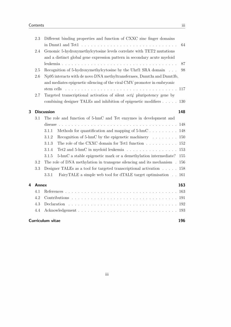
Contents
Summary iii
1 Introduction 2
1.1 Epigenetic information . . . . . . . . . . . . . . . . . . . . . . . . . . . . 2
1.2 Histone modifications and Histone variants . . . . . . . . . . . . . . . . . 2
1.3 DNA methylation . . . . . . . . . . . . . . . . . . . . . . . . . . . . . . . 4
1.3.1 DNA methylation in development and disease . . . . . . . . . . . 5
1.3.2 Mammalian DNA methyltransferases . . . . . . . . . . . . . . . . 6
1.3.3 Factors that bind methylated CpGs . . . . . . . . . . . . . . . . . 12
1.4 Mouse embryonic stem cells as a model of early mammalian development 16
1.4.1 Self-renewal and pluripotency of ESCs . . . . . . . . . . . . . . . 17
1.4.2 Role of Oct4, Sox2, and Nanog in pluripotency . . . . . . . . . . . 17
1.4.3 Embryonic stem cell di↵erentiation . . . . . . . . . . . . . . . . . 19
1.5 DNA hydroxymethylation . . . . . . . . . . . . . . . . . . . . . . . . . . 21
1.5.1 Ten-eleven-translocation protein family . . . . . . . . . . . . . . . 21
1.5.2 5-hmC and DNA demethylation . . . . . . . . . . . . . . . . . . . 22
1.5.3 Biological function of 5-hmC . . . . . . . . . . . . . . . . . . . . . 28
1.6 Transcription activator-like e↵ectors . . . . . . . . . . . . . . . . . . . . . 29
1.6.1 Biology of TAL e↵ectors . . . . . . . . . . . . . . . . . . . . . . . 29
1.6.2 Designer TAL e↵ectors as a tool for genome editing . . . . . . . . 31
1.7 Aims of this work . . . . . . . . . . . . . . . . . . . . . . . . . . . . . . . 33
2 Results 35
2.1 Sensitive enzymatic quantification of 5-hydroxymethylcytosine in genomic
DNA . . . . . . . . . . . . . . . . . . . . . . . . . . . . . . . . . . . . . . 35
2.2 Characterization of PvuRts1I endonuclease as a tool to investigate ge-
nomic 5-hydroxymethylcytosine . . . . . . . . . . . . . . . . . . . . . . . 42
ii
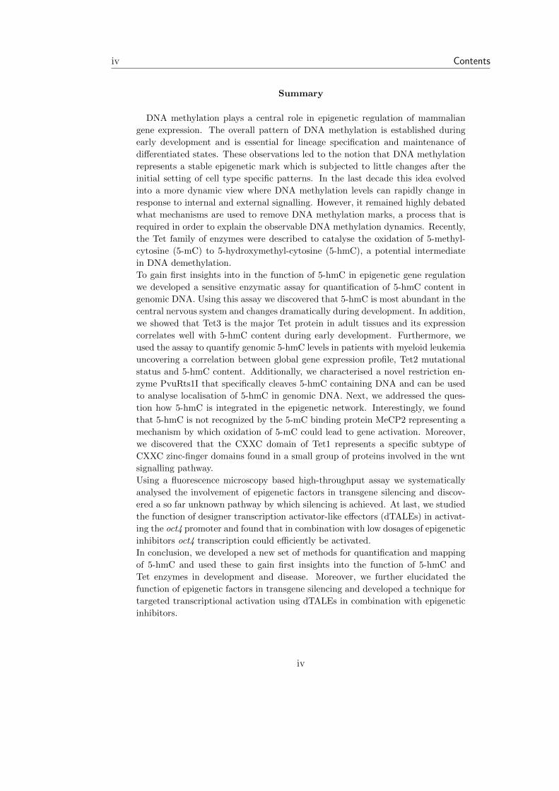
Contents iii
2.3 Di↵erent binding properties and function of CXXC zinc finger domains
in Dnmt1 and Tet1 . . . . . . . . . . . . . . . . . . . . . . . . . . . . . . 64
2.4 Genomic 5-hydroxymethylcytosine levels correlate with TET2 mutations
and a distinct global gene expression pattern in secondary acute myeloid
leukemia . . . . . . . . . . . . . . . . . . . . . . . . . . . . . . . . . . . . 87
2.5 Recognition of 5-hydroxymethylcytosine by the Uhrf1 SRA domain . . . 98
2.6 Np95 interacts with de novo DNAmethyltransferases, Dnmt3a and Dnmt3b,
and mediates epigenetic silencing of the viral CMV promoter in embryonic
stem cells . . . . . . . . . . . . . . . . . . . . . . . . . . . . . . . . . . . 117
2.7 Targeted transcriptional activation of silent oct4 pluripotency gene by
combining designer TALEs and inhibition of epigenetic modifiers . . . . . 130
3 Discussion 148
3.1 The role and function of 5-hmC and Tet enzymes in development and
disease . . . . . . . . . . . . . . . . . . . . . . . . . . . . . . . . . . . . . 148
3.1.1 Methods for quantification and mapping of 5-hmC . . . . . . . . . 148
3.1.2 Recognition of 5-hmC by the epigenetic machinery . . . . . . . . 150
3.1.3 The role of the CXXC domain for Tet1 function . . . . . . . . . . 152
3.1.4 Tet2 and 5-hmC in myeloid leukemia . . . . . . . . . . . . . . . . 153
3.1.5 5-hmC a stable epigenetic mark or a demethylation intermediate? 155
3.2 The role of DNA methylation in transgene silencing and its mechanism . 156
3.3 Designer TALEs as a tool for targeted transcriptional activation . . . . . 158
3.3.1 FairyTALE a simple web tool for dTALE target optimisation . . 161
4 Annex 163
4.1 References . . . . . . . . . . . . . . . . . . . . . . . . . . . . . . . . . . . 163
4.2 Contributions . . . . . . . . . . . . . . . . . . . . . . . . . . . . . . . . . 191
4.3 Declaration . . . . . . . . . . . . . . . . . . . . . . . . . . . . . . . . . . 192
4.4 Acknowledgement . . . . . . . . . . . . . . . . . . . . . . . . . . . . . . . 193
Curriculum vitae 196
iii
iv Contents
Summary
DNA methylation plays a central role in epigenetic regulation of mammaliangene expression. The overall pattern of DNA methylation is established duringearly development and is essential for lineage specification and maintenance ofdi↵erentiated states. These observations led to the notion that DNA methylationrepresents a stable epigenetic mark which is subjected to little changes after theinitial setting of cell type specific patterns. In the last decade this idea evolvedinto a more dynamic view where DNA methylation levels can rapidly change inresponse to internal and external signalling. However, it remained highly debatedwhat mechanisms are used to remove DNA methylation marks, a process that isrequired in order to explain the observable DNA methylation dynamics. Recently,the Tet family of enzymes were described to catalyse the oxidation of 5-methyl-cytosine (5-mC) to 5-hydroxymethyl-cytosine (5-hmC), a potential intermediatein DNA demethylation.To gain first insights into in the function of 5-hmC in epigenetic gene regulationwe developed a sensitive enzymatic assay for quantification of 5-hmC content ingenomic DNA. Using this assay we discovered that 5-hmC is most abundant in thecentral nervous system and changes dramatically during development. In addition,we showed that Tet3 is the major Tet protein in adult tissues and its expressioncorrelates well with 5-hmC content during early development. Furthermore, weused the assay to quantify genomic 5-hmC levels in patients with myeloid leukemiauncovering a correlation between global gene expression profile, Tet2 mutationalstatus and 5-hmC content. Additionally, we characterised a novel restriction en-zyme PvuRts1I that specifically cleaves 5-hmC containing DNA and can be usedto analyse localisation of 5-hmC in genomic DNA. Next, we addressed the ques-tion how 5-hmC is integrated in the epigenetic network. Interestingly, we foundthat 5-hmC is not recognized by the 5-mC binding protein MeCP2 representing amechanism by which oxidation of 5-mC could lead to gene activation. Moreover,we discovered that the CXXC domain of Tet1 represents a specific subtype ofCXXC zinc-finger domains found in a small group of proteins involved in the wntsignalling pathway.Using a fluorescence microscopy based high-throughput assay we systematicallyanalysed the involvement of epigenetic factors in transgene silencing and discov-ered a so far unknown pathway by which silencing is achieved. At last, we studiedthe function of designer transcription activator-like e↵ectors (dTALEs) in activat-ing the oct4 promoter and found that in combination with low dosages of epigeneticinhibitors oct4 transcription could e�ciently be activated.In conclusion, we developed a new set of methods for quantification and mappingof 5-hmC and used these to gain first insights into the function of 5-hmC andTet enzymes in development and disease. Moreover, we further elucidated thefunction of epigenetic factors in transgene silencing and developed a technique fortargeted transcriptional activation using dTALEs in combination with epigeneticinhibitors.
iv

1 Introduction
1.1 Epigenetic information
“One can say. . . that the elucidation of the genetic code is indeed a great achievement. It
is, in a sense, the key to molecular biology because it shows how the great polymer lan-
guages, the nucleic acid language and the protein language, are linked together.”[Crick,
1958].
Francis Crick was right to predict a breakthrough in molecular biology by the discovery
of the genetic code. However, the “key to molecular biology” has proven to open a
door into a room (or rather a hall) filled with many answers but even more questions.
The direct connection between “nucleic acid language” and “protein language” which
seemed so obvious and straight forward in the late 1950’s turned out to be an extremely
complex, inter-dependent relationship.
Considering the enormous variety of cell types, their di↵erent functions and morpholo-
gies it became clear that knowing the nucleotide sequence alone is only a small part of
the puzzle. Although all cells of a given multicellular organism contain the same genetic
information, they di↵er in their function and gene expression profiles; hence the distinct
properties of the cells do not reside in the nucleotide sequence but in how the cells
make use of their common genomic background. This level of information was termed
epigenetic (epi (Greek): over, above). Epigenetic mechanisms control cell-, tissue-, and
development- specific gene expression and are therefore responsible for the identity of
di↵erent cell types. Moreover, epigenetic information is heritable and thus can be passed
on from one cell to its progeny.
1.2 Histone modifications and Histone variants
Eukaryotic DNA is organized into a higher order structure called chromatin. The ba-
sic unit of chromatin is the nucleosome, which consists of 147 base pairs (bp) of DNA
wrapped around an octamer of core histones. This histone octamer is composed of two
2
Introduction 3
heterodimers of histone H3 and H4 associated with two heterodimers of histones H2A
and H2B [Finch et al., 1977; Dubochet and Noll, 1978]. These proteins share related
globular domains that mediate histone-histone interaction and DNA binding. Besides,
each histone also harbors a 20-35 amino acid long N-terminal peptide that extends from
the surface of the nucleosome. The histone“tails” and to a lower extend the core of the
protein are subject to a large number and variety of posttranslational modifications, in-
cluding methylation and acetylation of lysines and arginines, phosphorylation of serines
and threonines, ubiquitinylation and sumoylation of lysines, as well as ribosylation. It is
believed that many of these modifications play important roles in the regulation of tran-
scription. In principle, this can be achieved in two ways. Some modifications may lead
to alterations in structure and charge of the nucleosome which cause changes in DNA
binding and nucleosome packaging. For example acetylation of lysine residues neutral-
izes their positive charge which may weaken the interaction with the negatively charged
DNA backbone and thus lead to an open chromatin state where transcription factors can
access DNA more e�ciently. Although this might be a possibility by which histone mod-
ifications can regulate transcriptional activity, it is likely that most act by controlling
the recruitment of regulatory factors. For example, the chromodomain of heterochro-
matin protein 1 (HP1) binds to histone H3 when lysine (K) 9 is methylated [Lachner
et al., 2001] and this can lead to repression of transcription [Danzer and Wallrath, 2004].
Furthermore, the bromodomains of several proteins involved in transcriptional activa-
tion bind to acetylated lysines of histone H3 and H4 [Jacobson et al., 2000]. Histone
modifications can also lead to the recruitment of DNA methyltransferases and thereby
to DNA methylation and transcriptional repression [Tachibana et al., 2008].
Another mechanism by which histones modulate chromatin is via histone variants. While
the major histone proteins are encoded by multiple copies of histone genes, histone vari-
ants are usually present as single-copy genes. Furthermore, histone variants exhibit
significant di↵erences in the primary sequence compared to the major histones. Some
variants have distinct biophysical characteristics that are thought to alter the properties
of nucleosomes, while others localize to specific regions of the genome. Some histone
variants are exchanged with the pre-existing histones during development and di↵er-
entiation leading to tissue-specific expression patterns. These observations have led to
the suggestion that the histone variants have specialized functions in regulation of chro-
matin dynamics. Several histone variants have been shown to function in transcription,
particularly in repression. One example is the H2A variant MacroH2A which local-
izes to the inactive X-chromosome and some models suggest that the C-terminal tail of
3

4 1.3. DNA methylation
MacroH2A can repress transcription enzymatically. Other variants have been shown to
aid in transcriptional activation like H2A-Bbd which facilitates nucleosome displacement
by destabilizing the nucleosome [Kamakaka and Biggins, 2005].
While nucleosomes have long been viewed as stable complexes, there is strong evidence
that they are highly dynamic, being constantly altered in their composition, structure,
and location along the DNA. Chromatin-remodeling complexes contain ATPase sub-
units and are know to slide nucleosomes, replace histones, or alter the histone-DNA
interactions [Kamakaka and Biggins, 2005; Langst and Becker, 2004].
1.3 DNA methylation
DNA methylation in mammals refers to the addition of a methyl-group to the 5’ carbon
atom of cytosine which leads to the formation of 5-methyl cytosine (5-mC). It occurs
predominantly at CpG dinucleotides but is also found at non-CpG sites albeit to a lesser
degree [Lister et al., 2009; Laurent et al., 2010]. In mammalian somatic cells 4% of
cytosines are methylated, which accounts for 70%-80% of all CpG dinucleotides in the
genome [Ehrlich et al., 1982]. The remaining 20%-30% mainly comprise CpG islands,
regions with a high CpG density which are associated with most promoters of constitu-
tively expressed genes and 40% of genes that display a tissue-specific expression profile
[Larsen et al., 1992]. While methylation in promoter regions is thought to be associated
with gene silencing [Colot and Rossignol, 1999], there is emerging evidence that highly
transcribed genes carry methylation marks in the gene body, but the functional conse-
quences of this are unknown so far [Ball et al., 2009; Laurent et al., 2010].
Several mechanisms have been proposed on how global DNA methylation patterns are
established. While there is evidence that DNA-binding factors are involved in creating
and keeping regions from being methylated especially in the context of CpG islands
[Brandeis et al., 1994; Macleod et al., 1994; Dickson et al., 2010], other studies could
show that certain chromatin marks and DNA methylation occur in relation to each
other [Weber et al., 2007; Meissner et al., 2008; Hawkins et al., 2010]. A number of
factors have been identified that could mediated the functional interplay between DNA
methylation and chromatin modifications. These factors have been shown to bind to
histone modifications and CpG sites thereby connecting both epigenetic mechanisms
[Zhao et al., 2009; Hashimoto et al., 2010; Rottach et al., 2010; Pichler et al., 2011].
A recent study revealed that in addition to trans acting mechanisms mentioned above,
promoter sequences contain metyhlation-determining regions (MDRs) that are su�cient
4

Introduction 5
to mediate both hypomethylation and de novo methlyation in cis [Lienert et al., 2011].
1.3.1 DNA methylation in development and disease
DNA methylation has several important biological functions. During embryonic devel-
opment the genome experiences large changes in methylation levels. While the genomes
of egg and sperm cells are highly methylated [Sanford et al., 1987], the methylation is
rapidly lost after fertilization by passive [Rougier et al., 1998] and active [Mayer et al.,
2000; Gu et al., 2011; Wossidlo et al., 2011] mechanisms. After implantation embryonic
DNA methylation patterns are re-established through lineage-specific de novo methyla-
tion [Kafri et al., 1992; Santos et al., 2002]. The importance of DNA methylation during
embryonic development is supported by the discovery that embryos, which have defects
in DNA methylation show severe developmental deficiencies and die before birth [Okano
et al., 1999]. Another important function of CpG methylation is the maintenance of
mono-allelic expression of imprinted genes [Li et al., 1993]. In Embryos lacking the
maintenance DNA methyltransferase Dnmt1, alleles of both Igf2 and Igf2r, which are
normally paternally and maternally expressed, respectively, are silenced. Furthermore,
the H19 gene, which is normally maternally transcribed, is bi-allelically expressed. In
addition to its crucial role in imprinting, DNA methylation is also important for the
X-inactivation in female mammals as the expression of Xist is controlled by methylation
[Norris et al., 1994]. Moreover, DNA methylation is crucial for chromosomal stabil-
ity. Patients with the ICF syndrome (Immunodeficiency, Centromere Instability, Facial
Anomalies syndrome) carry a hypomorphic germline mutation in the gene coding for the
de novo methyltransferase DNMT3B and exhibit, besides other defects, a loss of DNA
methylation in centromeric and pericentromeric repeat regions [Miniou et al., 1997]. This
leads to pericentromeric decondensation and chromosomal instability. Consistent with
these findings, mouse embryonic stem cells (ESCs) lacking the two de novo methyltrans-
ferases dnmt3a/dnmt3b, exhibit elevated rates of centromeric sister chromatid exchange
[Jaco et al., 2008]. In addition, CpG methylation is crucial for the silencing of retro-
viruses and transposon inactivation [Cherry et al., 2000].
Aberrant changes in global DNA methylation patterns are characteristic for many cancer
types. In many cases a combination of global hypomethylation and promoter-localized
hypermethlyation is observed. However, the hypermethylation is not always confined
to promoter regions but can be spread over large gene “neighborhoods” up to whole
chromosome bands resulting in severe changes of gene expression patterns [Miremadi
et al., 2007].
5
6 1.3. DNA methylation
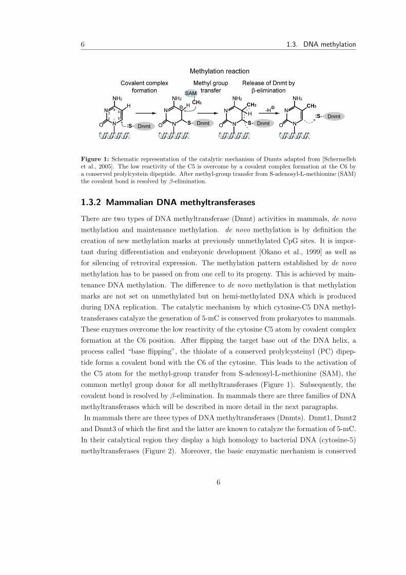
Figure 1: Schematic representation of the catalytic mechanism of Dnmts adapted from [Schermellehet al., 2005]. The low reactivity of the C5 is overcome by a covalent complex formation at the C6 bya conserved prolylcystein dipeptide. After methyl-group transfer from S-adenosyl-L-methionine (SAM)the covalent bond is resolved by �-elimination.
1.3.2 Mammalian DNA methyltransferases
There are two types of DNA methyltransferase (Dnmt) activities in mammals, de novo
methylation and maintenance methylation. de novo methylation is by definition the
creation of new methylation marks at previously unmethylated CpG sites. It is impor-
tant during di↵erentiation and embryonic development [Okano et al., 1999] as well as
for silencing of retroviral expression. The methylation pattern established by de novo
methylation has to be passed on from one cell to its progeny. This is achieved by main-
tenance DNA methylation. The di↵erence to de novo methylation is that methylation
marks are not set on unmethylated but on hemi-methylated DNA which is produced
during DNA replication. The catalytic mechanism by which cytosine-C5 DNA methyl-
transferases catalyze the generation of 5-mC is conserved from prokaryotes to mammals.
These enzymes overcome the low reactivity of the cytosine C5 atom by covalent complex
formation at the C6 position. After flipping the target base out of the DNA helix, a
process called “base flipping”, the thiolate of a conserved prolylcysteinyl (PC) dipep-
tide forms a covalent bond with the C6 of the cytosine. This leads to the activation of
the C5 atom for the methyl-group transfer from S-adenosyl-L-methionine (SAM), the
common methyl group donor for all methyltransferases (Figure 1). Subsequently, the
covalent bond is resolved by �-elimination. In mammals there are three families of DNA
methyltransferases which will be described in more detail in the next paragraphs.
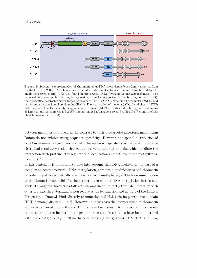
In mammals there are three types of DNA methyltransferases (Dnmts). Dnmt1, Dnmt2
and Dnmt3 of which the first and the latter are known to catalyze the formation of 5-mC.
In their catalytical region they display a high homology to bacterial DNA (cytosine-5)
methyltransferases (Figure 2). Moreover, the basic enzymatic mechanism is conserved
6
Introduction 7
Figure 2: Schematic representation of the mammalian DNA methyltransferase family adapted from[Rottach et al., 2009]. All Dnmts have a similar C-terminal catalytic domain characterized by thehighly conserved motifs (I-X) also found in prokaryotic DNA (cytosine-5) methyltransferases. TheDnmts di↵er, however, in their regulatory region. Dnmt1 contains the PCNA binding domain (PBD),the pericentric heterochromatin targeting sequence (TS), a CXXC-type zinc finger motif (ZnF) , andtwo bromo adjacent homology domains (BAH). The start codon of the long (ATGL) and short (ATGS)isoforms, as well as the seven lysine-glycine repeat linker (KG7) are indicated. The regulatory domainsof Dnmt3a and 3b comprise a PWWP domain named after a conserved Pro-Trp-Trp-Pro motif of theplant homeodomain (PHD)
.
between mammals and bacteria. In contrast to their prokaryotic ancestors, mammalian
Dnmts do not exhibit strong sequence specificity. However, the spatial distribution of
5-mC in mammalian genomes is vital. The necessary specificity is mediated by a large
Nterminal regulatory region that contains several di↵erent domains which mediate the
interaction with proteins that regulate the localization and activity of the methyltrans-
ferases. (Figure 2).
In this context it is important to take into account that DNA methylation is part of a
complex epigenetic network. DNA methylation, chromatin modifications and chromatin
remodeling pathways mutually a↵ect each other in multiple ways. The N-terminal region
of the Dnmts is responsible for the correct integration of DNA methylation in this net-
work. Through its direct cross-talk with chromatin or indirectly through interaction with
other proteins the N-terminal region regulates the localization and activity of the Dnmts.
For example, Dnmt3L binds directly to unmethylated H3K4 via its plant homeodomain
(PHD domain) [Jia et al., 2007]. However, in most cases the interpretation of chromatin
signals is achieved indirectly and Dnmts have been shown to interact with a variety
of proteins that are involved in epigenetic processes. Interactions have been described
with histone 3 lysine 9 (H3K9) methyltransferases (HMTs), Suv39h1, SetDB1 and G9a,
7
8 1.3. DNA methylation
components of the Polycomb repressive complex 2, histone deacetylases (HDACs) and
the heterochromatin protein 1 (HP1) [Cedar and Bergman, 2009].
Dnmt1
Dnmt1 was the first eukaryotic DNAmethyltransferase to be discovered [Bestor, 1988]. It
has been shown that it methylates hemimethylated DNA much more e�ciently then un-
methylated substrates [Bestor and Ingram, 1983] which led to the assignment of Dnmt1
as a maintenance DNA methyltransferase. Although there is evidence that Dnmt1 has
de novo methylation activity in vitro [Pradhan et al., 1997], it is most likely that its
main biological role in vivo is maintaining genomic methylation patterns. This is sup-
ported by the finding that Dnmt1 colocalizes with the replication machinery [Leonhardt
et al., 1992]. At replication sites hemimethylated DNA is formed when the newly syn-
thesized unmethylated strand pairs with the methylated template strand. Although
the (transient) association with the replication machinery makes it possible that Dnmt1
could directly methylate newly forming hemimethylated CpG sites, it seems not to be
essential for maintaining postreplicative methylation levels [Schermelleh et al., 2007;
Spada et al., 2007]. Intriguingly, Dnmt1 alone is not su�cient to stably maintain DNA
methylation as in ESCs lacking both de novo methyltransferases, Dnmt3a and Dnmt3b,
global methylation levels slowly decrease during long term culture, although they still
express Dnmt1 [Chen et al., 2003a]. Furthermore, Dnmt1 is ubiquitously expressed and
its presence is essential for the survival of somatic cells where apoptosis is induced via
a p53 mediated pathway, when Dnmt1 is depleted [Jackson-Grusby et al., 2001]. The
importance of Dnmt1 during development is shown by the fact that mice lacking Dnmt1
do not develop correctly and exhibit a growth arrest prior the 8-somite stage [Li et al.,
1992; Lei et al., 1996]. Moreover, Dnmt1 plays a crucial role in the maintenance of
chromosomal stability as mice expressing Dnmt1 at strongly reduced levels are viable
at birth but soon develop aggressive T cell lymphomas with a high frequency of chro-
mosome 15 trisomy [Gaudet et al., 2003]. The murine somatic form of Dnmt1 consist
of an 1100 amino acid long N-terminal regulatory region and a 500 amino acid long
C-terminal catalytic domain (Figure 2). The latter is common to all eukaryotic Dn-
mts and consists basically of ten conserved motifs which are crucial for the catalytic
activity. The N-terminal region is build up by a number of functional domains that
have regulatory functions. The first 125 amino acids mediate the interaction with the
DMAP1 transcriptional repressor [Rountree et al., 2000]. Dnmt1, except in early devel-
opment, exhibits a nuclear localization and has several nuclear localization signals (NLof
8
Introduction 9
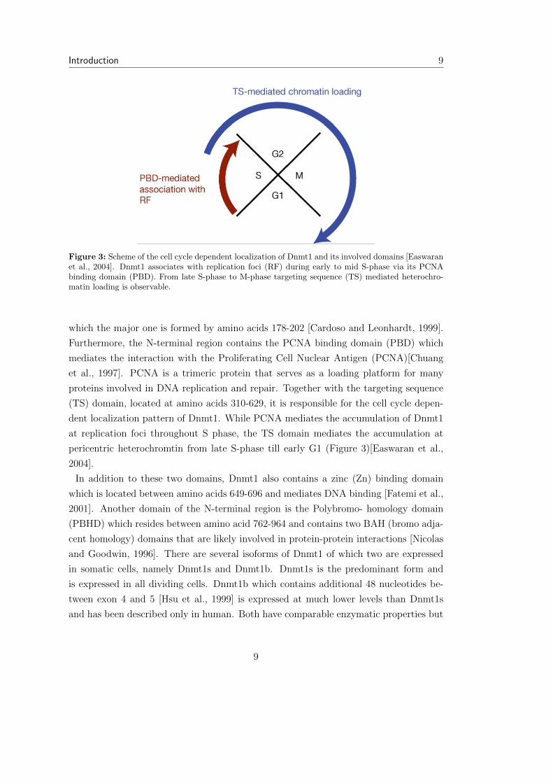
Figure 3: Scheme of the cell cycle dependent localization of Dnmt1 and its involved domains [Easwaranet al., 2004]. Dnmt1 associates with replication foci (RF) during early to mid S-phase via its PCNAbinding domain (PBD). From late S-phase to M-phase targeting sequence (TS) mediated heterochro-matin loading is observable.
which the major one is formed by amino acids 178-202 [Cardoso and Leonhardt, 1999].
Furthermore, the N-terminal region contains the PCNA binding domain (PBD) which
mediates the interaction with the Proliferating Cell Nuclear Antigen (PCNA)[Chuang
et al., 1997]. PCNA is a trimeric protein that serves as a loading platform for many
proteins involved in DNA replication and repair. Together with the targeting sequence
(TS) domain, located at amino acids 310-629, it is responsible for the cell cycle depen-
dent localization pattern of Dnmt1. While PCNA mediates the accumulation of Dnmt1
at replication foci throughout S phase, the TS domain mediates the accumulation at
pericentric heterochromtin from late S-phase till early G1 (Figure 3)[Easwaran et al.,
2004].
In addition to these two domains, Dnmt1 also contains a zinc (Zn) binding domain
which is located between amino acids 649-696 and mediates DNA binding [Fatemi et al.,
2001]. Another domain of the N-terminal region is the Polybromo- homology domain
(PBHD) which resides between amino acid 762-964 and contains two BAH (bromo adja-
cent homology) domains that are likely involved in protein-protein interactions [Nicolas
and Goodwin, 1996]. There are several isoforms of Dnmt1 of which two are expressed
in somatic cells, namely Dnmt1s and Dnmt1b. Dnmt1s is the predominant form and
is expressed in all dividing cells. Dnmt1b which contains additional 48 nucleotides be-
tween exon 4 and 5 [Hsu et al., 1999] is expressed at much lower levels than Dnmt1s
and has been described only in human. Both have comparable enzymatic properties but
9

10 1.3. DNA methylation
the function of the Dnmt1b isoform is so far not clear. In oocytes and preimplantation
embryos another isoform, Dnmt1o, is expressed. It is transcribed from an oocyte specific
promoter and its first exon di↵ers from the one of Dnmt1s and Dnmt1b [Gaudet et al.,
1998].
Dnmt2
Dnmt2 shows a high homology to other DNA methyltransferases. The inferred protein
sequence contains all 10 catalytic motifs in the canonical order (Figure 2). In contrast
to other eukaryotic DNA methyltransferases, Dnmt2 homologues do not possess a reg-
ulatory N-terminal region and in this respect resemble more closely bacterial cytosine
methyltransferases. In addition, the crystal structure of human DNMT2 revealed that
the structures of DNMT2 and the bacterial restriction methyltransferase M.HhaI are
essentially superimposable [Dong et al., 2001]. In fact, the Dnmt2 family is the most
strongly conserved and most widely distributed family of eukaryotic cytosine methyl-
transferase homologues [Goll and Bestor, 2005]. Despite all similarities to other cyto-
sine methyltransferases, the functional role of the Dnmt2 family remained enigmatic as
Dnmt2 homologues could not be shown to possess considerable DNA methyltransferase
activity and mice lacking Dnmt2 do not exhibit DNA methylation abnormalities [Okano
et al., 1998b]. In 2006 it was shown that DNMT2 methylates the aspartic acid transfer
RNA (tRNAAsp) at cytosine 38 in the anticodon loop [Goll et al., 2006].
Interestingly, analysis of tRNAAsp sequences showed complete conservation of the anti-
codon loop in species whose genomes encode Dnmt2 homologues, whereas in C. elegans
and S. cerevisiae, which lack a Dnmt2 homologue, tRNAAsp anticodon loops have di-
verged. These findings indicate coevolution of Dnmt2 and the anticodon loop of tRNA
Asp. However, the functional consequence of the tRNAAsp methylation remains unclear.
The only phenotypic e↵ect of Dnmt2 depletion reported so far was found in zebrafish,
where a knockdown in embryos results in di↵erentiation defects in particular organs,
including retina, liver, and brain. In agreement with its role in tRNAAsp methylation,
cytoplasmatically located Dnmt2 could rescue this phenotype [Rai et al., 2007]. Dnmt2
seems to have an additional role in some organisms. In Drosophila very low levels of cy-
tosine methylation are present [Gowher et al., 2000]. Its genome encodes only a Dnmt2
methyltransferase and lacks any of the canonical de novo or maintenance Dnmts. In
this organism, as well as in Dictyostelium [Kuhlmann et al., 2005], Dnmt2-dependent
DNA methylation was shown to be necessary for retrotransposon silencing and telomere
integrity [Phalke et al., 2009].
10
Introduction 11
Dnmt3
The mammalian genome encodes two functional Dnmt3 methyltransferases, namely
Dnmt3a and Dnmt3b, and a third homologue, Dnmt3L which lacks cytosine methyl-
transferase activity. Dnmt3a and Dnmt3b are closely related proteins that, similar to
Dnmt1, possess an N- terminal regulatory region and a C-terminal catalytic domain
(Figure 2). Both were found to methylate CpG dinucleotides in vitro without preference
for hemimethylated DNA and thereby assigning their possible role as de novo Dnmts
[Okano et al., 1998a]. This was confirmed in vivo, by using a stable episomal system that
employs plasmids as targets for de novo DNA methylation [Hsieh, 1999] and by the find-
ing that dnmt3a/dnmt3b double knockout ESCs exhibit an inability to de novomethylate
newly introduced retroviral elements while the maintenance of imprinted methylation
pattern is not a↵ected [Okano et al., 1999].
The N-terminal regions of Dnmt3a and Dnmt3b harbor a PWWP domain which is found
in many chromatin-associated proteins. By mutagenesis analysis this domain was shown
to be required for pericentric heterochromatin association. Furthermore, disruption of
the PWWP domain abolishes the ability of Dnmt3a and Dnmt3b to methylate major
satellite repeats at pericentric heterochromatin [Chen et al., 2004]. Both proteins also
contain an ATRX-homology domain, a cystein rich zinc-binding domain mainly found
in proteins involved in eukaryotic transcription regulation. It has been shown that the
ATRX-homology domain of Dnmt3a is su�cient to repress transcription, independently
of the methyltransferase activity, by associating with the histone deacetylase HDAC1
[Fuks et al., 2001]. In addition, this domain has been shown to mediate the binding to
symmetrically di-methylated arginine 3 at Histone 4 (H4R3) [Zhao et al., 2009]. Dnmt3a
and Dnmt3b have both been shown to be important in mouse embryonic development
and di↵erentiation. Both genes are expressed in ESCs and form a complex in vivo. Single
knockout of either Dnmt3a or Dnmt3b in ESC results in reduction of promoter methy-
lation of the pluripotency markers Oct-4 and Nanog upon di↵erentiation via retinoic
acid treatment. Simultaneous knockout of Dnmt3a and 3b completely abolishes de novo
methylation at these loci [Li et al., 2007]. Besides their similarities and synergistic func-
tion, Dnmt3a and Dnmt3b also have some non- overlapping functions which become
obvious by the phenotypic di↵erences of Dnmt3a and Dnmt3b single knockout embryos.
While the latter die at around E9.5, dnmt3a-/- appear normal at birth and die not be-
fore 4 weeks of age. Global methylation patterns seem to be normal in dnmt3a deficient
mice [Okano et al., 1999]. Deletion of Dnmt3a in the female germ line leads to hy-
pomethylation at di↵erentially methylated regions (DMR) of all maternally imprinted
11
12 1.3. DNA methylation
genes examined so far. In contrast, dnmt3b knockout in germ cells does not result in such
a phenotype [Kaneda et al., 2004]. Inactivation of dnmt3b, but not dnmt3a, in mouse
embryonic fibroblasts (MEF), results in partial loss of genome wide DNA methylation.
This suggests that, in addition to the major maintenance methyltransferase Dnmt1,
Dnmt3b is required for maintaining DNA methylation in somatic cells [Dodge et al.,
2005]. In ESCs however, both de novo methyltransferases need to be absent in order
to achieve a gradual loss of global DNA methylation [Li et al., 2007]. Dnmt3L shows
high homology to Dnmt3a and Dnmt3b in its N- and C-terminal domains but lacks the
PWWP domain (Figure 2). The catalytic motifs have been subject to nonconservative
substitutions and Dnm3L is not able to catalyze cytosine methylation.
Dnmt3L is mainly expressed in the germ line where it is essential for the establishment
of a subset of methylation patterns [Bourc’his et al., 2001]. Interestingly, it seems that
Dnmt3L has di↵erent functions in male and female germ cells. In male mice targeted
disruption of dnmt3L causes azoospermia with germ line cells displaying nonhomologous
synapsis, asynapsis, and the accumulation of highly abnormal synaptonemal complexes.
Abnormal synapsis is likely to be a secondary e↵ect of the observed hypomethylation
of transposable elements [Bourc’his and Bestor, 2004]. In contrast, dnmt3L deficiency
does not interfere with oogenesis and oocytes are methylated normally at transposons.
However, female germ cells exhibit a methylation defect in single copy sequences as-
sociated with maternal imprinting instead [Bourc’his et al., 2001]. As Dnmt3L is not
catalytically active, the methylation defects in Dnmt3L-deficient mice are thought to be
caused by the missing activation of Dnmt3a as Dnmt3L stimulates the de novo methy-
lation activity of Dnmt3a in vivo [Chedin et al., 2002]. In addition, targeted disruption
of dnmt3a results in phenotypes similar to Dnmt3L knockout [Kaneda et al., 2004].
1.3.3 Factors that bind methylated CpGs
One main function of DNA methylation is transcriptional silencing and there are two
models of how this is achieved. The first model suggests that CpG methylation interferes
with the binding of transcription factors that require contact with cytosine in the major
groove of the double helix [Hark et al., 2000]. While the second model proposes that DNA
methylation is translated into a repressive chromatin state. This is mediated by factors
that recognize and bind to methylated CpGs which repress transcription indirectly by
the recruitment of corepressors. So far there are three protein families known that bind
methyl-CpG.
12
Introduction 13
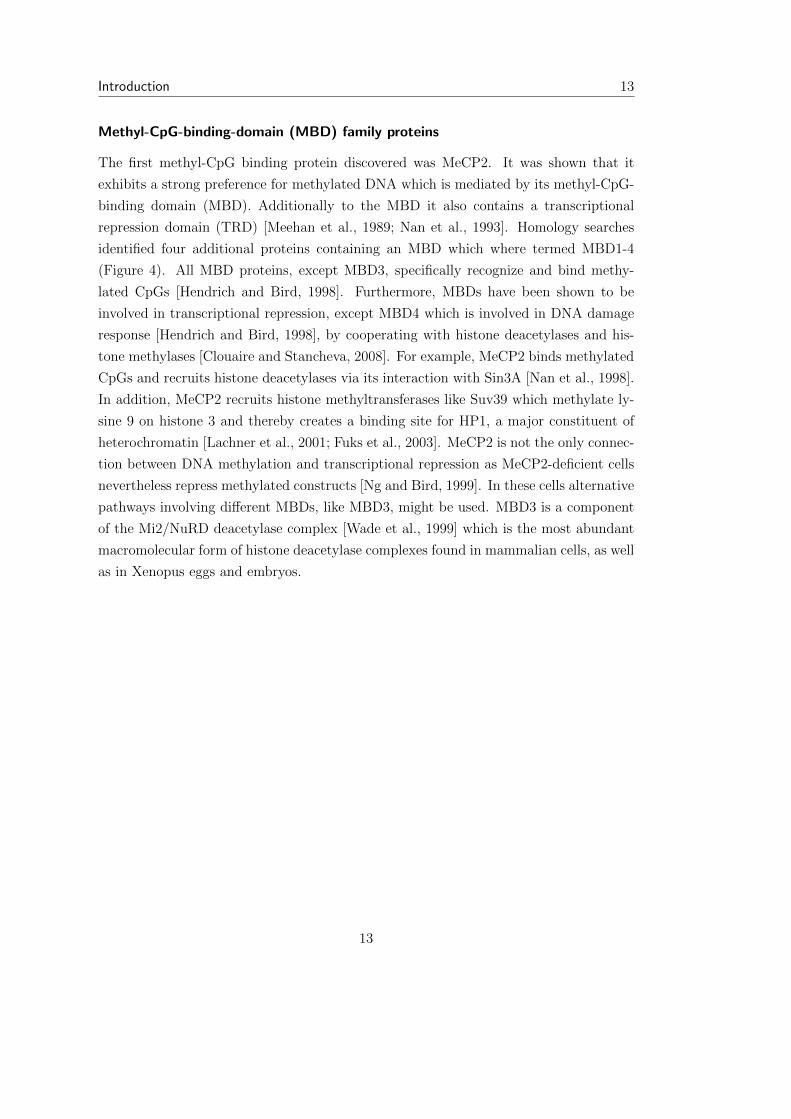
Methyl-CpG-binding-domain (MBD) family proteins
The first methyl-CpG binding protein discovered was MeCP2. It was shown that it
exhibits a strong preference for methylated DNA which is mediated by its methyl-CpG-
binding domain (MBD). Additionally to the MBD it also contains a transcriptional
repression domain (TRD) [Meehan et al., 1989; Nan et al., 1993]. Homology searches
identified four additional proteins containing an MBD which where termed MBD1-4
(Figure 4). All MBD proteins, except MBD3, specifically recognize and bind methy-
lated CpGs [Hendrich and Bird, 1998]. Furthermore, MBDs have been shown to be
involved in transcriptional repression, except MBD4 which is involved in DNA damage
response [Hendrich and Bird, 1998], by cooperating with histone deacetylases and his-
tone methylases [Clouaire and Stancheva, 2008]. For example, MeCP2 binds methylated
CpGs and recruits histone deacetylases via its interaction with Sin3A [Nan et al., 1998].
In addition, MeCP2 recruits histone methyltransferases like Suv39 which methylate ly-
sine 9 on histone 3 and thereby creates a binding site for HP1, a major constituent of
heterochromatin [Lachner et al., 2001; Fuks et al., 2003]. MeCP2 is not the only connec-
tion between DNA methylation and transcriptional repression as MeCP2-deficient cells
nevertheless repress methylated constructs [Ng and Bird, 1999]. In these cells alternative
pathways involving di↵erent MBDs, like MBD3, might be used. MBD3 is a component
of the Mi2/NuRD deacetylase complex [Wade et al., 1999] which is the most abundant
macromolecular form of histone deacetylase complexes found in mammalian cells, as well
as in Xenopus eggs and embryos.
13

14 1.3. DNA methylation
Figure 4: Schematic diagram of the MBD protein family [Rottach et al., 2009]. All family memberscontain a methyl-CpG-binding domain (MBD). MBD1, MBD2 and MeCP2 additionally harbour atranscriptional repression domain (TRD). MBD4 the only family member with catalytic activity whichis mediated by its DNA N-glycosylase domain. In addtion to its MBD and TRD domain, MBD1 alsocontains three CXXC-type zinc-finger domains (CxxC).
SRA domain proteins
It was recently discovered that Dnmt1 needs the presence of another protein, called
Np95, to stably maintain genomic methylation levels. In ESCs lacking Np95, also know
as Uhrf1, global and local DNA methylation levels are defective and almost identical to
that of dnmt1 knockout cells [Sharif et al., 2007]. Furthermore, Np95 colocalizes with
Dnmt1 in vivo at replication forks during mid-to-late-S-phase when pericentromeric het-
erochromatin is replicated [Papait et al., 2007]. Co-immunoprecipitation experiments
showed a direct interaction of Np95 and Dnmt1 [Sharif et al., 2007]. In addition, it has
been shown that Np95 can bind directly to methyl-CpG via its SRA (SET and RING
associated) domain [Unoki et al., 2004]. All these data suggest that Np95 recruits Dnmt1
to hemimethylated CpG sites at replication forks so that Dnmt1 can copy the methyla-
tion mark onto the newly synthesized strand. Cocrystallization of the SRA domain with
a hemimethylated DNA substrate showed that upon binding, the 5-mC is flipped out of
the DNA helix and positioned in a binding pocket [Hashimoto et al., 2008]. Interestingly,
a similar mechanism for DNA binding has been described for DNA methyltransferases
[Klimasauskas et al., 1994] and this base flipping is thought to be involved in the coor-
dinated transfer of the hemi-methylated CpG site from Np95 to Dnmt1.
In general SRA domain proteins fall in two distinct families. The first is characterized
by the association of the SRA domain with PHD and RING domains. The only known
mammalian homologues discovered so far are Np95 and the closely related Np97, also
14
Introduction 15
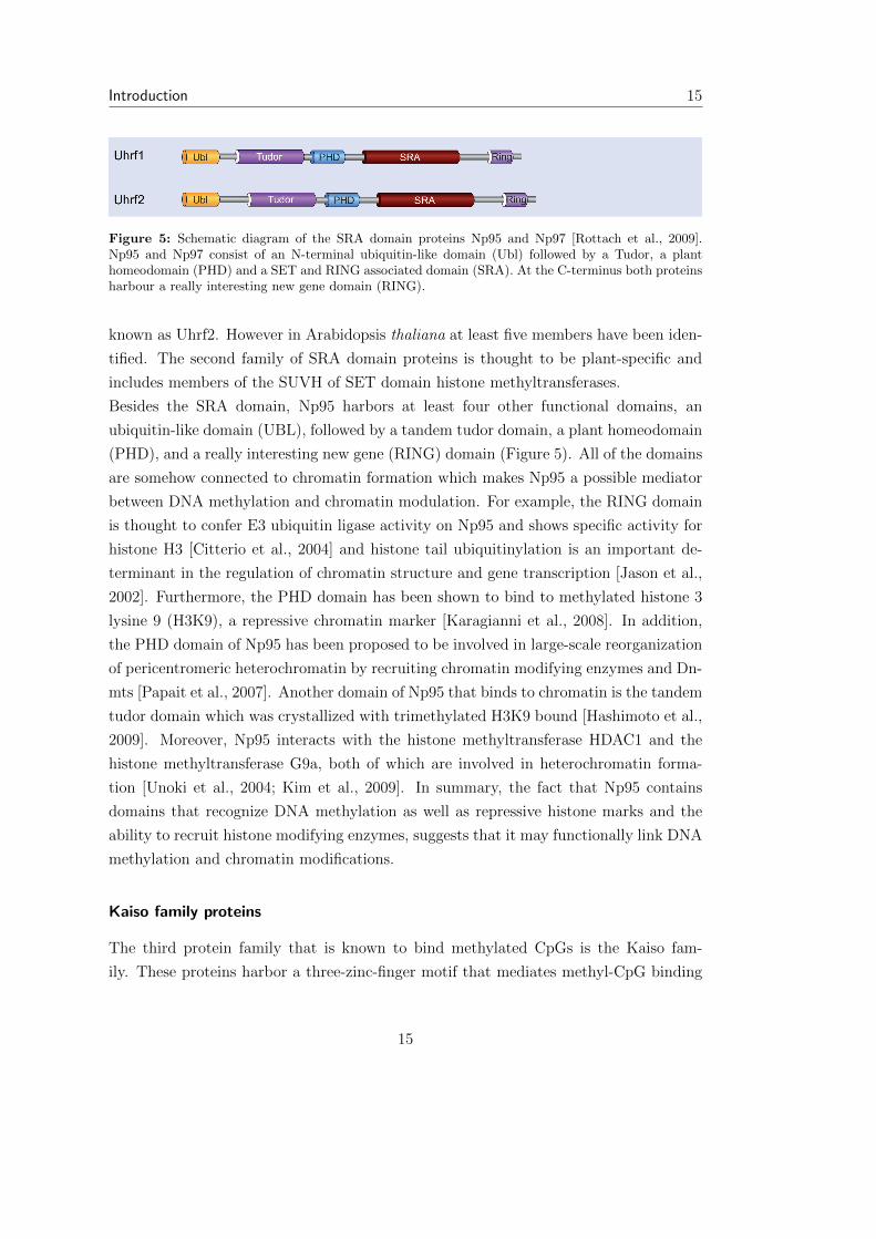
Figure 5: Schematic diagram of the SRA domain proteins Np95 and Np97 [Rottach et al., 2009].Np95 and Np97 consist of an N-terminal ubiquitin-like domain (Ubl) followed by a Tudor, a planthomeodomain (PHD) and a SET and RING associated domain (SRA). At the C-terminus both proteinsharbour a really interesting new gene domain (RING).
known as Uhrf2. However in Arabidopsis thaliana at least five members have been iden-
tified. The second family of SRA domain proteins is thought to be plant-specific and
includes members of the SUVH of SET domain histone methyltransferases.
Besides the SRA domain, Np95 harbors at least four other functional domains, an
ubiquitin-like domain (UBL), followed by a tandem tudor domain, a plant homeodomain
(PHD), and a really interesting new gene (RING) domain (Figure 5). All of the domains
are somehow connected to chromatin formation which makes Np95 a possible mediator
between DNA methylation and chromatin modulation. For example, the RING domain
is thought to confer E3 ubiquitin ligase activity on Np95 and shows specific activity for
histone H3 [Citterio et al., 2004] and histone tail ubiquitinylation is an important de-
terminant in the regulation of chromatin structure and gene transcription [Jason et al.,
2002]. Furthermore, the PHD domain has been shown to bind to methylated histone 3
lysine 9 (H3K9), a repressive chromatin marker [Karagianni et al., 2008]. In addition,
the PHD domain of Np95 has been proposed to be involved in large-scale reorganization
of pericentromeric heterochromatin by recruiting chromatin modifying enzymes and Dn-
mts [Papait et al., 2007]. Another domain of Np95 that binds to chromatin is the tandem
tudor domain which was crystallized with trimethylated H3K9 bound [Hashimoto et al.,
2009]. Moreover, Np95 interacts with the histone methyltransferase HDAC1 and the
histone methyltransferase G9a, both of which are involved in heterochromatin forma-
tion [Unoki et al., 2004; Kim et al., 2009]. In summary, the fact that Np95 contains
domains that recognize DNA methylation as well as repressive histone marks and the
ability to recruit histone modifying enzymes, suggests that it may functionally link DNA
methylation and chromatin modifications.
Kaiso family proteins
The third protein family that is known to bind methylated CpGs is the Kaiso fam-
ily. These proteins harbor a three-zinc-finger motif that mediates methyl-CpG binding
15

16 1.4. Mouse embryonic stem cells as a model of early mammalian development
Figure 6: Schematic diagram of the Kaiso family proteins [Rottach et al., 2009]. Kaiso, ZBTB4, andZBTB38 are characterized by several zinc finger motifs. Binding to methylated DNA is mediated by aC2H2 zinc finger motif (yellow). The broad complex, tramtrack, and bric brac (BTB/POZ) domain isdepicted in gray.
[Prokhortchouk et al., 2001]. The founding member kaiso and the recently identified
kaiso-like proteins ZBTB4 and ZBTB38 also contain a poxvirus and zinc finger (POZ)
domain that is involved in protein-protein interaction (Figure 6). This domain is thought
to be involved in transcriptional repression as kaiso lacking the POZ domain is not ca-
pable of silencing methylated reporters. However, transcriptional silencing was not di-
minished in the presence of the histone deacetylase inhibitor TSA as shown for MBDs
[Prokhortchouk et al., 2001]. Inconsistent with these findings, kaiso seems to be part of
the repressive N-CoR complex which contains HDAC and histone remodeling activities
[Yoon et al., 2003]. Moreover, ZBTB4 and ZBTB38 have also been shown to mediate re-
pression of transcription [Filion et al., 2006]. In summary, Kaiso family proteins might
mediate methylation dependent transcriptional repression in a way similar to that of
MBD proteins.
1.4 Mouse embryonic stem cells as a model of early
mammalian development
ESCs are pluripotent cells derived from the inner cell mass (ICM) of the blastocyst
[Evans and Kaufman, 1981]. Murine ESCs retain the full developmental potential of the
ICM as they can contribute to all tissues of the embryo and adult in vivo after rein-
troduction into mouse blastocysts [Bradley et al., 1984]. This feature makes it possible
to create knockout mice and cell lines which are an important tool in elucidating the
function of proteins in vivo. Furthermore, deletion of several genes, including Dnmts,
are lethal for somatic cells while their ESC counterparts are viable. In vitro, ESC can
16
Introduction 17
be di↵erentiated into a broad range of cell type representative for all three germ layers
of the mouse embryo [Lake et al., 2000]. Moreover, during in vitro di↵erentiation ESC
undergo developmental changes and processes similar to that seen in the ICM during
early embryonic development. These properties make mouse ESCs an optimal tool for
studying early processes in embryonic development.
1.4.1 Self-renewal and pluripotency of ESCs
Self-renewal and pluripotency are key features of ESCs. They can be expanded indefi-
nitely in culture and retain their full developmental potential without exhibiting a bias
in the generation of di↵erent somatic lineages or germline cells upon reintroduction to
the embryo. Although ESCs exhibit a characteristic pattern of epigenetic modifications
they seem not to be crucial for self-renewal and pluripotency. This is supported by the
finding that ESCs deficient for important epigenetic regulators, such as DNA methyl-
transferases or histone methyltransferases, are viable without compromising self-renewal
or genomic integrity [Tsumura et al., 2006]. However, perturbation of DNA methyla-
tion and the chromatin modifying machinery often results in increased cell death during
di↵erentiation [Jackson et al., 2004]. Nevertheless, rescue of the epigenetic machinery
by reintroduction of the missing components results in full restoration of the develop-
mental potential which suggests a role in successful lineage commitment rather than in
retaining pluripotency. In contrast, depletion of ESCs of one of the three transcriptional
organizers, Oct4, Sox2, and Nanog results unscheduled di↵erentiation into trophoblast
and hypoblast cells which cannot be rescued by reintroduction of these factors [Nichols
et al., 1998; Niwa, 2007]. These fate choices are considered abnormal as they resemble
lineages that ICM cells have already passed beyond their segregation points prior to ESC
establishment. This indicates that nave pluripotency of ESCs is critically dependent on
the action of Oct4, Sox2, and Nanog rather than on the epigenetic machinery.
1.4.2 Role of Oct4, Sox2, and Nanog in pluripotency
ESCs are in a constant struggle between di↵erentiation and self-renewal. Interestingly,
in both decisions the three transcriptional organizers Oct4, Sox2, and Nanog seem to
be involved. All three are expressed in cells of the ICM and ESCs. They appear to be
responsible for the ongoing repression of the expression and activity of lineage specifi-
cation factors and thereby in retaining pluripotency [Smith, 2005]. However, Oct4 and
Sox2 seem also to be key regulators in the extinction of pluripotency by directing the
17

18 1.4. Mouse embryonic stem cells as a model of early mammalian development
Figure 7: Self-renewal of the pluripotent ESC state requires overcoming the FGF4/Erk signal. Inhibi-tion of FGF4/Erk signalling by small molecules prevents spontaneous di↵erentiation of ESCs in culture.Leukemia inhibiting factor (LIF) stimulates STAT3 signalling which promotes ESC self-renewal andproliferation.
expression of fibroblast growth factor 4 (FGF4). FGF4 propels ESCs towards lineage
specification via the mitogen activated protein (MAP) kinase Erk1/2 pathway. Impor-
tantly the FGF4/Erk signal does not lead to the di↵erentiation of a certain lineage but
results in a general susceptibility for further lineage specific signaling. Consistent with
this, blocking of this signaling pathway leads to a general impairment of di↵erentiation
[Kunath et al., 2007; Silva and Smith, 2008](Figure 7). In summary, by promoting the
expression of FGF4, Oct4 and Sox2 synergistically drive ESCs into di↵erentiation. Con-
sequently, to maintain the naive undi↵erentiated ESCs in culture the signaling by FGF4
needs to be inhibited. This can be achieved by the addition of the cytokine leukemia
inhibiting factor (LIF). LIF stimulates the Stat3 transcription factor signaling which
acts downstream of Erk and also promotes ESC growth and viability. In addition small-
molecule inhibitors of the MAP kinase kinase (MAPKK or MEK) can be used to inhibit
Erk signaling (Figure 7).
In contrast to the homogenous expression Oct4 and Sox2, Nanog expression is sub-
jected to great fluctuations (Figure 8A). Interestingly, constitutive expression of Nanog
is su�cient to prevent ESC di↵erentiation even in the presence of active FGF4/Erk
signaling. Moreover, Nanog-deficient ESCs can remain undi↵erentiated and pluripotent
in culture, but exhibit a greatly increased tendency to di↵erentiate [Chambers et al.,
18

Introduction 19
Figure 8: (A) Immunostaining shows highly variable levels of Nanog protein (green) in Oct4 (red)positive undi↵erentiated ESCs. (B) Nanog prevents di↵erentiation of ESCs. Coincidence of low Nanogexpression and elevated FGF4/Erk signaling (pErk) result in susceptibility for further lineage specificsignals (symbolized by A,B and C expression circuits) [Silva and Smith, 2008].
2007]. This suggests that Nanog counteracts the FGF4/Erk signaling and thereby re-
tains pluripotency. Moreover, the cell-to-cell heterogeneity of expression levels creates
di↵erences in the resistance to di↵erentiation. This means that those ESCs in a culture
that express low levels of Nanog are prone to exit self-renewal and will start to di↵er-
entiate if intrinsic and/or extrinsic lineage specific signals are present above a certain
threshold. Although it seems that ESCs can repeatedly switch between high and low
levels of Nanog expression, it is clear that many cells that express little Nanog will start
to di↵erentiate.
In summary, the three transcriptional organizers Oct4, Sox2, and Nanog together re-
press lineage-associated transcriptional activity. In addition, Oct4 and Sox2 activate the
expression of FGF4 which signals via the MAP kinase Erk1/2 promoting lineage speci-
fication of ESCs. This is antagonized by Nanog via an unknown mechanism. As Nanog
expression is highly variable among ESCs, coincidence of low Nanog levels with ele-
vated FGF4/Erk signaling leads to activation of intrinsic lineage-specific transcriptional
activity resulting in di↵erentiation and loss of self-renewal (Figure 8B).
1.4.3 Embryonic stem cell di↵erentiation
Mouse ESCs are routinely cultivated in the presence of feeder layers to maintain pluripo-
tency and self-renewal. These feeder cells can also be substituted by LIF and with cells
grown on gelatinized culture dishes. When feeder cells or LIF is removed ESCs spon-
taneously di↵erentiate into derivatives of the three embryonic germ layers, mesoderm,
endoderm, and ectoderm [Keller, 2005]. In principle, there are three important methods
19
20 1.4. Mouse embryonic stem cells as a model of early mammalian development
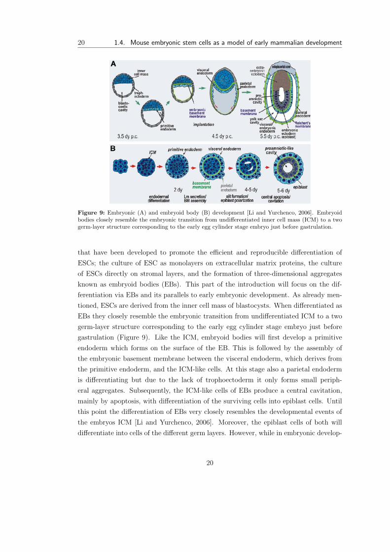
Figure 9: Embryonic (A) and embryoid body (B) development [Li and Yurchenco, 2006]. Embryoidbodies closely resemble the embryonic transition from undi↵erentiated inner cell mass (ICM) to a twogerm-layer structure corresponding to the early egg cylinder stage embryo just before gastrulation.
that have been developed to promote the e�cient and reproducible di↵erentiation of
ESCs; the culture of ESC as monolayers on extracellular matrix proteins, the culture
of ESCs directly on stromal layers, and the formation of three-dimensional aggregates
known as embryoid bodies (EBs). This part of the introduction will focus on the dif-
ferentiation via EBs and its parallels to early embryonic development. As already men-
tioned, ESCs are derived from the inner cell mass of blastocysts. When di↵erentiated as
EBs they closely resemble the embryonic transition from undi↵erentiated ICM to a two
germ-layer structure corresponding to the early egg cylinder stage embryo just before
gastrulation (Figure 9). Like the ICM, embryoid bodies will first develop a primitive
endoderm which forms on the surface of the EB. This is followed by the assembly of
the embryonic basement membrane between the visceral endoderm, which derives from
the primitive endoderm, and the ICM-like cells. At this stage also a parietal endoderm
is di↵erentiating but due to the lack of trophoectoderm it only forms small periph-
eral aggregates. Subsequently, the ICM-like cells of EBs produce a central cavitation,
mainly by apoptosis, with di↵erentiation of the surviving cells into epiblast cells. Until
this point the di↵erentiation of EBs very closely resembles the developmental events of
the embryos ICM [Li and Yurchenco, 2006]. Moreover, the epiblast cells of both will
di↵erentiate into cells of the di↵erent germ layers. However, while in embryonic develop-
20

Introduction 21
ment this follows directed and well coordinated pathways, in EBs epiblast cells exhibit a
more random di↵erentiation pattern though there are several methods that make more
directed di↵erentiation possible at this stage.
1.5 DNA hydroxymethylation
Until recently the only known covalent epigenetic modification on DNA was methylation
at position 5 of cytosine. In 2009, however, it was discovered that 5-mC is further
oxidized by the enyzme ten-eleven translocation 1 (TET1) to 5-hydroxymethylcytosine
(5-hmC) [Tahiliani et al., 2009] a base that was already detected in mammalian DNA
in 1972 but at this time mainly considered as a by-product of oxidative DNA damage
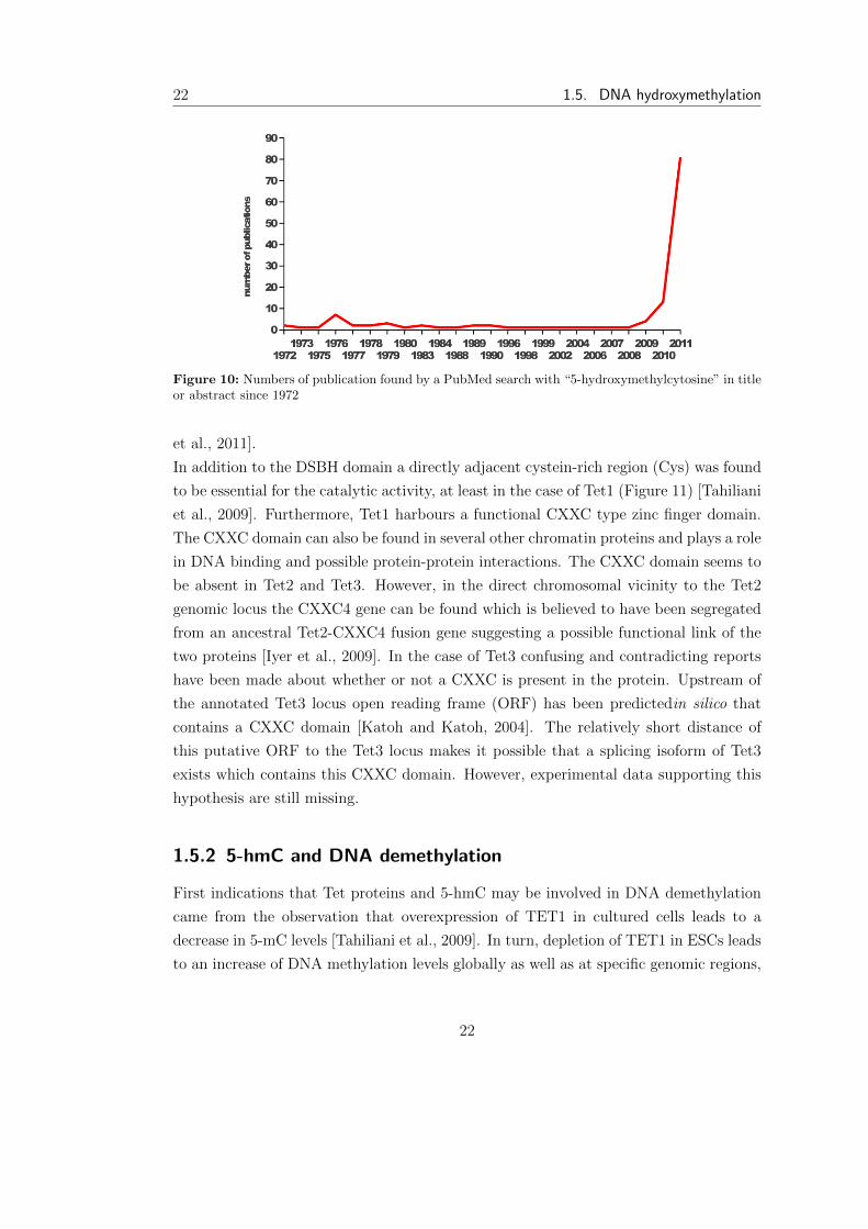
[Penn et al., 1972]. In the short time since the discovery of the oxidation from 5-mC
to 5-hmC by Tet1 an impressive amount of publications has accumulated (Figure 10).
Many possible biological functions for 5-hmC in epigenetic gene regulation have been
proposed and it has become increasingly clear that oxidation of 5-mC plays an important
role in DNA demethylation.
1.5.1 Ten-eleven-translocation protein family
TET1 was the first protein of the Ten-eleven-translocation protein family to be discov-
ered by genetic analysis of several cases of acute myeloid leukemia (AML) harbouring
a t(10;11)(q22;q23). This chromosomal rearrangement was shown to result in an N-
terminal fusion of the mixed lineage leukemia (MLL) H3K4 methyltransferase to the
C-terminus of TET1. TET1 was found to be a member of a novel and well conserved
protein family of, at this time, unknown biological function [Ono et al., 2002; Lors-
bach et al., 2003]. The function of Tet proteins was discovered by their homology to
the DNA modifying enzymes JBP1 and JBP2 in Trypanosomes. JBP1 and JBP2 be-
long to the 2-oxoglutarate- and Fe(II)-dependent dioxygenase (2OGFeDO) superfamily
and catalyse the oxidation of the methyl-group of thymine leading to the formation of
5-hydroxymethyluracil (hmU) the first step in the biosynthesis of base J (�-D-glucosyl-
hydroxymethyluracil). Like the JBP proteins, Tet proteins contain a 2OGFeDO domain
characterized by a double-stranded � helix (DSBH) fold which in the case of Tet pro-
teins has been shown to catalyse the oxidation of 5-mC to 5-hmC [Tahiliani et al.,
2009]. Moreover, it has been discovered that Tet proteins can further oxidise 5-hmC
to 5-formylcytosine (5-fC) and 5-carboxylcytosine (5-caC) [He et al., 2011; Pfa↵eneder
21
22 1.5. DNA hydroxymethylation
Figure 10: Numbers of publication found by a PubMed search with “5-hydroxymethylcytosine” in titleor abstract since 1972
et al., 2011].
In addition to the DSBH domain a directly adjacent cystein-rich region (Cys) was found
to be essential for the catalytic activity, at least in the case of Tet1 (Figure 11) [Tahiliani
et al., 2009]. Furthermore, Tet1 harbours a functional CXXC type zinc finger domain.
The CXXC domain can also be found in several other chromatin proteins and plays a role
in DNA binding and possible protein-protein interactions. The CXXC domain seems to
be absent in Tet2 and Tet3. However, in the direct chromosomal vicinity to the Tet2
genomic locus the CXXC4 gene can be found which is believed to have been segregated
from an ancestral Tet2-CXXC4 fusion gene suggesting a possible functional link of the
two proteins [Iyer et al., 2009]. In the case of Tet3 confusing and contradicting reports
have been made about whether or not a CXXC is present in the protein. Upstream of
the annotated Tet3 locus open reading frame (ORF) has been predictedin silico that
contains a CXXC domain [Katoh and Katoh, 2004]. The relatively short distance of
this putative ORF to the Tet3 locus makes it possible that a splicing isoform of Tet3
exists which contains this CXXC domain. However, experimental data supporting this
hypothesis are still missing.
1.5.2 5-hmC and DNA demethylation
First indications that Tet proteins and 5-hmC may be involved in DNA demethylation
came from the observation that overexpression of TET1 in cultured cells leads to a
decrease in 5-mC levels [Tahiliani et al., 2009]. In turn, depletion of TET1 in ESCs leads
to an increase of DNA methylation levels globally as well as at specific genomic regions,
22

Introduction 23
Figure 11: Schematic representation of the murine Ten-eleven translocation protein family. All threeTet proteins harbour a cystein-rich region (Cys) followed by the catalytic domain characterised by adouble stranded � helix (DSBH) fold. Tet1 contains a CXXC-type zinc finger domain (CXXC) in itsN-terminal part. In the direct chromosomal vicinity of Tet2 the CXXC4 gene can be found whichmight be functionally linked to Tet2. A putative ORF close upstream of Tet3 codes for a CXXC-typezinc-finger which might be spliced to Tet3.
such as LINE1 retrotransposons and transcription factor binding sites [Ficz et al., 2011;
Xu et al., 2011]. Tet1 has been shown to be important for the demethylation of brain-
derived neurotrophic factor (Bdnf) and fibroblast growth factor 1 (Fgf1) promoters in
the adult mouse brain [Guo et al., 2011]. Finally, loss of 5-mC in the male pronucleus
of zygotes correlates with an increase of 5-hmC staining and Tet3-depletion results in
failure to demethylate the paternal genome as well as promoters of pluripotency genes,
such as oct4 and nanog [Gu et al., 2011; Iqbal et al., 2011; Wossidlo et al., 2011]. The
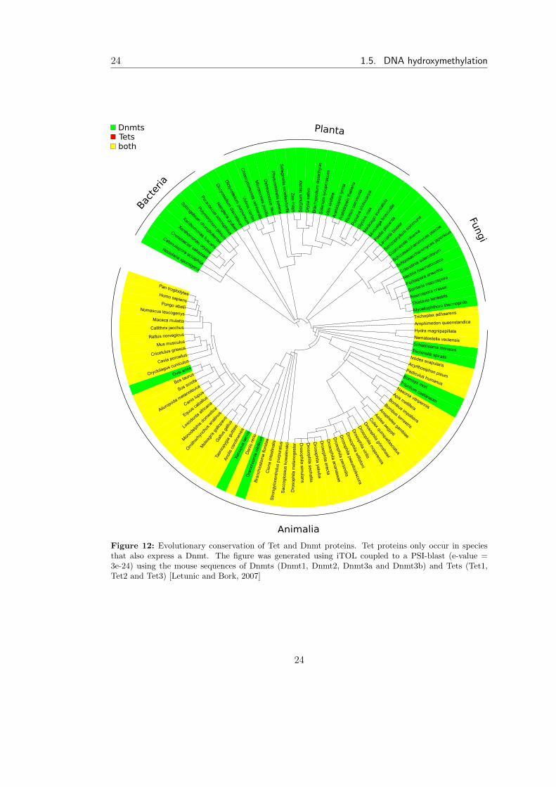
close functional link of Tet proteins to DNA methylation in animals is also evident in the
fact that Tets only occur in species that express a DNA methyltransferase (Figure 12).
However, Dnmts can be found in species that do not express Tet proteins suggesting that
other mechanisms for DNA demethylation exist. In fact, in plants, which do not have
genes coding for Tet proteins, DNA demethylation is well studied. Several mechanisms
by which 5-hmC contributes to DNA demethylation have been proposed which include
DNA repair pathways and prevention of DNA methylation maintenance.
DNA demethylation by DNA repair pathways
DNA demethylation has long been a highly debated field. The only widely accepted
mechanism, in mammals, has been passive demethylation where DNA methylation is
not maintained through replication and thereby diluted during each cell cycle. However,
it has become increasingly clear that DNA demethylation can also occur in a replication
independent context e.g. in post-mitotic tissues such as neurons. Moreover, at several
stages during development DNA methylation levels decrease with kinetics that can not
be accounted for by passive DNA demethylation alone. Active DNA demethylation is
23
24 1.5. DNA hydroxymethylation
Figure 12: Evolutionary conservation of Tet and Dnmt proteins. Tet proteins only occur in speciesthat also express a Dnmt. The figure was generated using iTOL coupled to a PSI-blast (e-value =3e-24) using the mouse sequences of Dnmts (Dnmt1, Dnmt2, Dnmt3a and Dnmt3b) and Tets (Tet1,Tet2 and Tet3) [Letunic and Bork, 2007]
24

Introduction 25
best studied in plants where a family of DNA glycosylases is responsible for the removal
of 5-mC. DNA glycosylases cleave the glycosidic bond between 5-mC and the deoxyri-
bose, creating an abasic site (AP). An AP endonuclease removes the deoxyribose at the
AP site and the gap is filled by DNA polymerase and DNA ligase. The result of this
base excision repair (BER) pathway is the replacement of the methylated cytosine by
an unmethylated cytosine [Zhu, 2009].
In mammals similar mechanisms have been proposed. In contrast to the glycosylases
in plants, known mammalian glycosylases like thymine DNA glycosylase (TDG) and
methyl-CpG-binding domain protein 4 (MBD4) only show weak activity on 5mC in
vitro. However, these enzymes have strong activities against T:G mismatches which can
be created through deamination of 5mC by cytidine deaminases [Zhu et al., 2000]. In
fact, cytidine deaminases of the apolipoprotein B mRNA editing catalytic polypeptide
(APOBEC) family have been shown to be involved in active DNA demethylation by
deaminating 5-mC.
One member of the APOBEC family, the activation-induced cytidine deaminase (AID),
has been studied in great detail over the last decade because of its critical role in gener-
ating antibody diversity in lymphocytes [Chaudhuri et al., 2007; Delker et al., 2009]. In
B-lymphocytes AID takes part in somatic hyper-mutation and class-switch recombina-
tion by deaminating cytosines to uracils which in turn are processed by error-prone BER
or mismatch repair (MMR) pathways. This mechanism results in mutations essential
for the vast diversity of antibodies present in mammals [Liu and Schatz, 2009; Maul
and Gearhart, 2010]. For a long time AID was thought to preferentially target the im-
munoglobulin locus in B -lymphocytes by an unknown mechanism. However, studies in
B-lymphocytes of mice deficient in BER and MMR revealed that AID acts extensively
on non-immunoglobulin loci and that these regions are protected by error-free repair
mechanisms. At that time these findings were interpreted as a protection mechanism
against miss targeted AID activity [Liu et al., 2008]. Only recently, AID has been im-
plicated in active DNA demethylation.
First findings suggesting a role for AID in DNA demethylation came from studies done
in zebrafish embryos. Overexpression of AID or zebrafish APOBEC deaminases and
the DNA glycosylase MBD4 led to DNA demethylation of the genome and of injected
methylated DNA [Rai et al., 2008]. Evidence for a role of AID in DNA demethyla-
tion in mammals was found in mice completely lacking AID. In the primordial germ
cells of these animals an increase in genome-wide methylation was observable. How-
ever, AID null mice are viable and fertile suggesting that other redundant pathways
25
26 1.5. DNA hydroxymethylation
may exist which can compensate for the loss of AID [Popp et al., 2010]. Studies of
nuclear reprogramming provided the first evidence that AID plays a role in active DNA
demethylation [Bhutani et al., 2010]. Fusion of mouse ESCs with human fibroblast into
non-dividing heterokaryons leads to rapid loss of DNA methylation at the promoters of
the pluripotency genes OCT4 and NANOG in the somatic genome. This process was
shown to be AID-dependent as knock-down of AID using siRNA resulted in complete
loss of pluripotency promoter demethylation and transcriptional induction. Moreover,
the AID gene is located in a cluster of pluripotency genes together with nanog and stella
and is coexpressed with these genes in oocytes, embryonic germ cells and tissues where
DNA demethylation has been shown to occur [Morgan et al., 2004; Bhutani et al., 2010].
Besides the APOBEC family, DNA methyltransferases have been proposed to play an
important role in active DNA demethylation by deaminating 5-mC. In human breast
cancer cells the de novo methyltransferases Dnmt3a and 3b can convert 5-mC to T
through deamination during the activation of oestradiol-estrogen receptor target gene
pS2 by E2. The resulting T:G mismatch is then removed by BER [Mtivier et al., 2008].
Similar observations were made during the activation of the vibronectin gene by the nu-
clear receptor chicken ovalbumin upstream promoter-transcription factor I (COUP-TFI).
Moreover, Dnmt3a was found to interact with the glycosylase TDG and could enhance
COUP-TF1-mediated activation of a methylated reporter gene [Gallais et al., 2007].
The involvement of DNA methyltransferases in setting and removing DNA methylation
raises the question how these counteracting functions are separated and controlled.
The accumulating evidence for the involvement of deamination-coupled DNA repair in
active DNA demethylation let to the identification of several DNA glycosylases involved
in this process. The family of glycosylases implicated in the deamination-coupled BER
pathway are members of the uracil DNA glycosylase (UDG) family that include TDG,
MBD4 and single-stranded-selective monofunctional uracil-DNA glycosylase 1 (SMUG1)
[Zhu et al., 2000; Cortellino et al., 2011; Guo et al., 2011]. The DNA glycosylases TDG
and SMUG1 have been shown to convert 5-hmU to cytosine suggesting that they act in
concert with Tet and AID/APOBEC proteins. Interestingly, knock-out of TDG results
in early embryonic lethality underscoring the importance of BER glycosylases during
development and DNA demethylation. TDG has been shown to directly interact with
AID by immunoprecipitation experiments [Cortellino et al., 2011; Guo et al., 2011].
Recent studies revealed that Tet proteins can further oxidize 5-hmC to 5-fC and 5-
caC. Both cytosine derivatives are present in mouse organs and cultured cells although
in a much lower abundance then 5-hmC. Interestingly, 5-caC and 5-fC are specifically
26

Introduction 27
Figure 13: Active DNA demethylation pathways in mammals. 5-mC can be directly deaminated byAID/APOBEC family or Dnmt3 proteins producing a T:G mismatch wich is repaired by TDG/MBD4glycosylases. Alternatively, 5-mC is oxidized to 5-hmC by Tet proteins and either deaminated to 5-hmU by AID/APOBEC proteins or further oxidized to 5-caC. 5-hmU and 5-caC are then recognizedby TDG/MBD4/SMUG glycosylases.
recognized by TDG and siRNA mediated depletion of TDG leads to accumulation of
5-caC [He et al., 2011; Ito et al., 2011; Maiti and Drohat, 2011].
Taken together these data suggest that in mammals, in contrast to the one-step process
in plants, active DNA demethylation may occur in a two-step process. However, the
first step of the demethylation pathway either involves deamination of 5-mC/5-hmC by
APOBEC family deaminases or further oxidation of 5-hmC by Tet proteins. In either
case the modified cytosine is then recognized by glycosylases of the UDG family and
5-mC is replaced by an unmodified cytosine via the BER/MMR pathway (Figure 13).
DNA demethylation by prevention of DNA methylation maintenance
A di↵erent mechanism by which 5-hmC may contribute to DNA demethylation is pas-
sive DNA demethylation. Hemi-modified 5-hmCpGs are not recognized by Dnmt1
and thereby DNA methylation is not maintained at theses sites [Valinluck and Sow-
ers, 2007]. Recently, in vivo evidence for this potential mechanism has been found in
pre-implantation embryos where 5-hmC is passively lost through replication [Inoue and
Zhang, 2011]. However, it is not clear whether 5-hmC mediated passive DNA demethy-
lation is a general mechanism and further studies are required to clarify this question.
27
28 1.5. DNA hydroxymethylation
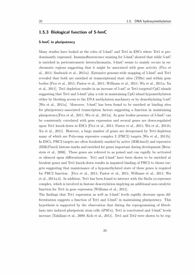
1.5.3 Biological function of 5-hmC
5-hmC in pluripotency
Many studies have looked at the roles of 5-hmC and Tet1 in ESCs where Tet1 is pre-
dominantly expressed. Immunofluorescence staining for 5-hmC showed that while 5-mC
is enriched in pericentromeric heterochromatin, 5-hmC seems to mainly occurs in eu-
chromatic regions suggesting that it might be associated with gene activity (Ficz et
al., 2011; Szulwach et al., 2011a). Extensive genome-wide mapping of 5-hmC and Tet1
revealed that both are enriched at transcriptional start sites (TSSs) and within gene
bodies [Ficz et al., 2011; Pastor et al., 2011; Williams et al., 2011; Wu et al., 2011a; Xu
et al., 2011]. Tet1 depletion results in an increase of 5-mC at Tet1-targeted CpG islands
suggesting that Tet1 and 5-hmC play a role in maintaining CpG island hypomethylation
either by blocking access to the DNA methylation machinery or by demethylating 5-mC
[Wu et al., 2011a]. Moreover, 5-hmC has been found to be enriched at binding sites
for pluripotency-associated transcription factors suggesting a function in maintaining
pluripotency[Ficz et al., 2011; Wu et al., 2011a]. In gene bodies presence of 5-hmC can
be consistently correlated with gene expression and several genes are down-regulated
upon Tet1 knock-down in ESCs [Ficz et al., 2011; Pastor et al., 2011; Wu et al., 2011b;
Xu et al., 2011]. However, a large number of genes are derepressed by Tet1-depletion
many of which are Polycomp repressive complex 2 (PRC2) targets [Wu et al., 2011b].
In ESCs, PRC2 targets are often bivalently marked by active (H3K4me3) and repressive
(H3K27me3) histone marks and enriched for genes important during development [Bern-
stein et al., 2006]. These genes are referred to as poised and can rapidly be activated
or silenced upon di↵erentiation. Tet1 and 5-hmC have been shown to be enriched at
bivalent genes and Tet1 knock-down results in impaired binding of PRC2 to theses tar-
gets suggesting that maintenance of a hypomethylated state of these genes is required
for PRC2 function. [Ficz et al., 2011; Pastor et al., 2011; Williams et al., 2011; Wu
et al., 2011a,b]. In addition, Tet1 has been found to interact with the Sin3a co-repressor
complex, which is involved in histone deacetylation implying an additional non-catalytic
function for Tet1 in gene repression [Williams et al., 2011].
The findings that Tet1 expression as well as 5-hmC levels rapidly decrease upon dif-
ferentiation suggests a function of Tet1 and 5-hmC in maintaining pluripotency. This
hypothesis is supported by the observation that during the reprogramming of fibrob-
lasts into induced pluripotent stem cells (iPSCs), Tet1 is reactivated and 5-hmC levels
increase [Tahiliani et al., 2009; Koh et al., 2011]. Tet1 and Tet2 were shown to be reg-
28
Introduction 29
ulated by pluripotency related transcription factors Oct4 and Sox2 and Tet1 depletion
in ESCs leads to down-regulation of several pluripotency related genes accompanied
by an increase in methylation of their promoters [Ficz et al., 2011; Williams et al.,
2011; Wu et al., 2011a]. Furthermore, extra-embryonic lineage markers are derepressed
in Tet1- depleted ESCs increasing the transdi↵erentiation potential of these cells into
extra-embryonic tissues upon di↵erentiation into embryoid bodies and in teratoma for-
mation [Ito et al., 2011; Koh et al., 2011]. However, both tet1 and tet2 knock-out mice
are viable and fertile, although tet1-/- mice display an overall reduced body size [Dawlaty
et al., 2011; Ko et al., 2011; Li et al., 2011b; Moran-Crusio et al., 2011]. In summary, it
is not clear to what extend Tet1 and 5-hmC contribute to pluripotency. Although Tet1
seems to regulate developmentally important genes, it is not crucial for embryonic de-
velopment. To elucidate whether 5-hmC itself is required for embryogenesis, generation
of triple Tet-knockout mice would be necessary as only a 35% reduction in 5-hmC levels
in tet1knockout ESCs is observable [Dawlaty et al., 2011].
1.6 Transcription activator-like e↵ectors
1.6.1 Biology of TAL e↵ectors
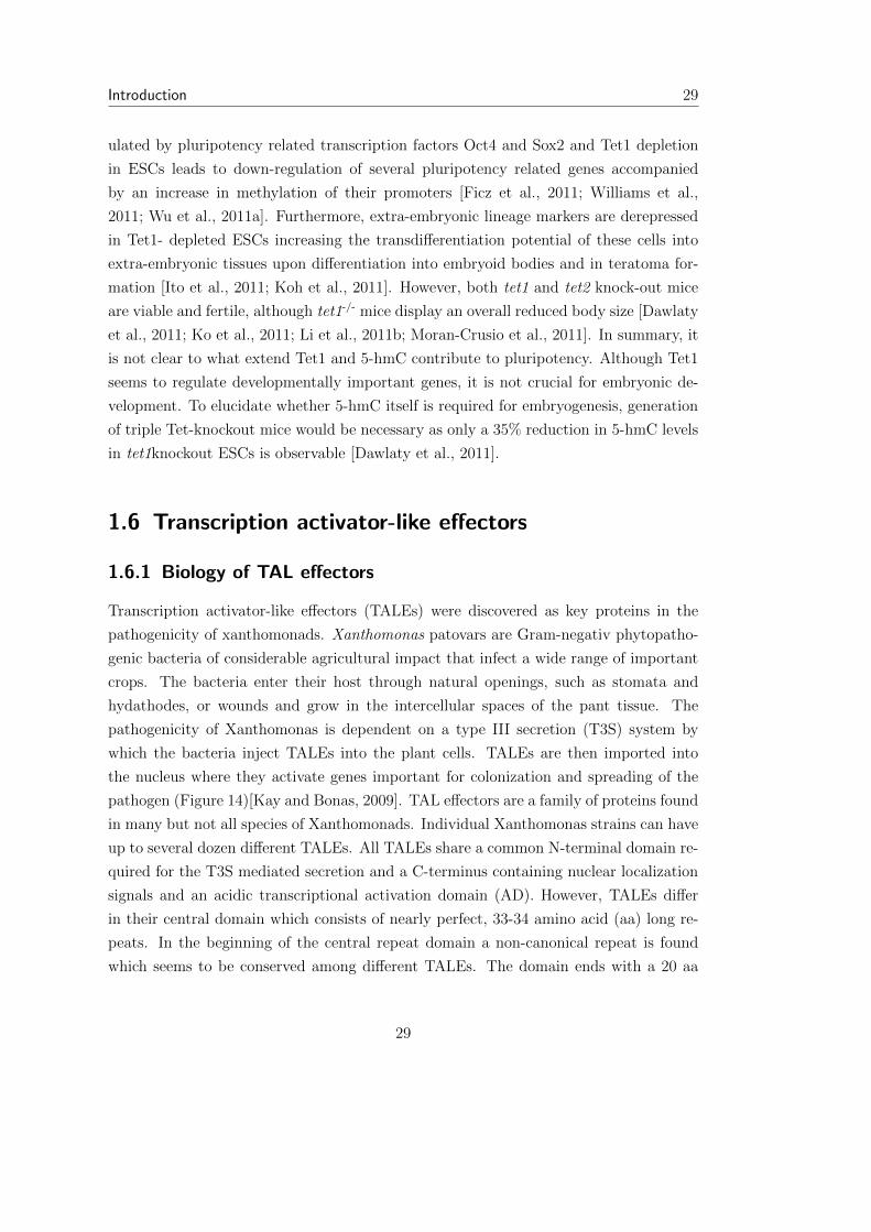
Transcription activator-like e↵ectors (TALEs) were discovered as key proteins in the
pathogenicity of xanthomonads. Xanthomonas patovars are Gram-negativ phytopatho-
genic bacteria of considerable agricultural impact that infect a wide range of important
crops. The bacteria enter their host through natural openings, such as stomata and
hydathodes, or wounds and grow in the intercellular spaces of the pant tissue. The
pathogenicity of Xanthomonas is dependent on a type III secretion (T3S) system by
which the bacteria inject TALEs into the plant cells. TALEs are then imported into
the nucleus where they activate genes important for colonization and spreading of the
pathogen (Figure 14)[Kay and Bonas, 2009]. TAL e↵ectors are a family of proteins found
in many but not all species of Xanthomonads. Individual Xanthomonas strains can have
up to several dozen di↵erent TALEs. All TALEs share a common N-terminal domain re-
quired for the T3S mediated secretion and a C-terminus containing nuclear localization
signals and an acidic transcriptional activation domain (AD). However, TALEs di↵er
in their central domain which consists of nearly perfect, 33-34 amino acid (aa) long re-
peats. In the beginning of the central repeat domain a non-canonical repeat is found
which seems to be conserved among di↵erent TALEs. The domain ends with a 20 aa
29
30 1.6. Transcription activator-like e↵ectors
Figure 14: The function of TALEs during Xanthomonas infection. TALEs are injected into the plantcell cytoplasms by Type 3 secretion (T3S). Subsequently, the protein is imported into the nucleus whereit activates the expression of genes important for pathogen survival.
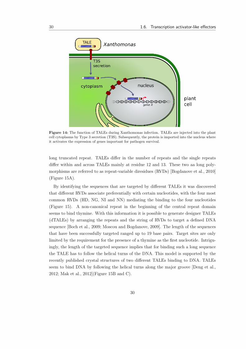
long truncated repeat. TALEs di↵er in the number of repeats and the single repeats
di↵er within and across TALEs mainly at residue 12 and 13. These two aa long poly-
morphisms are referred to as repeat-variable diresidues (RVDs) [Bogdanove et al., 2010]
(Figure 15A).
By identifying the sequences that are targeted by di↵erent TALEs it was discovered
that di↵erent RVDs associate preferentially with certain nucleotides, with the four most
common RVDs (HD, NG, NI and NN) mediating the binding to the four nucleotides
(Figure 15). A non-canonical repeat in the beginning of the central repeat domain
seems to bind thymine. With this information it is possible to generate designer TALEs
(dTALEs) by arranging the repeats and the string of RVDs to target a defined DNA
sequence [Boch et al., 2009; Moscou and Bogdanove, 2009]. The length of the sequences
that have been successfully targeted ranged up to 19 base pairs. Target sites are only
limited by the requirement for the presence of a thymine as the first nucleotide. Intrigu-
ingly, the length of the targeted sequence implies that for binding such a long sequence
the TALE has to follow the helical turns of the DNA. This model is supported by the
recently published crystal structures of two di↵erent TALEs binding to DNA. TALEs
seem to bind DNA by following the helical turns along the major groove [Deng et al.,
2012; Mak et al., 2012](Figure 15B and C).
30
Introduction 31
Figure 15: DNA recognition of TALEs. (A) Schematic representation of a TALE with its centralrepeat domain, the C-terminal NLS and activation domain (AD). Each repeat is represented by a blueellipse. The RVDs responsible for the binding to the four nucleotides are NN, HD NI and NG. (B-C)Crystal structure of the TAL e↵ector PthXo1 bound to its DNA target (PBD 3ugm). (B) Bottom view.(C) Side view.
1.6.2 Designer TAL e↵ectors as a tool for genome editing
The discovery of the TALE code has lead to a broad application of TAL e↵ectors in
biotechnology. The straightforward relationship between the type of repeat and DNA
sequence it recognizes allows a simple designing of dTALEs to target any desired DNA
sequence. Several strategies have been developed that allow cheap, reliable and quick
assemble of dTALEs with desired specificities. All stratagies are based on Golden Gate
cloning a method that relies on type IIs restriction enzymes which cleave outside their
recognition sequence and create unique 4-bp overhangs. Well planned design of the
overhangs produced by these enzymes allows ligation of multiple DNA fragments in an
ordered fashion in a single reaction [Engler et al., 2008, 2009].
The main application is the use of dTALEs for targeted editing of genomic sequences.
For this purpose dTALEs are used to target the catalytic domain of the FokI nuclease
to specific sites in the genome to introduce DNA double-strand breaks (DSBs). Because
FokI nucleases work as dimers, the TALE nucleases (TALENs) are designed as pairs that
bind opposing DNA target sites which are seperated by a spacer (Figure 16). This layout
allows the FokI monomers to dimerize only at the desired location and create a DSB.
31

32 1.6. Transcription activator-like e↵ectors
Figure 16: designer TALEs as tool for genomic targeting. dTALEs are fused to FokI monomers togenerate TALE nucleases (TALENs). TALENs are desgined as pairs binding opposing DNA targetsites which a separated by a spacer. When both TALENs bind there target sites the FokI nucleasesdimerize and generate a double-strand break (DSB) within the spacer sequence. The DSB is repaired byeither non homologous end joining or homologous recombination. The latter is used to insert selectioncassettes for further selection of clones containing mutated alleles.
The created DSBs are then repaired by one of two highly conserved pathways which
can be exploited to introduce specific DNA sequence modifications [Carroll, 2011]. In
non homologous end joining (NHEJ), the broken chromosome is repaired by imprecise
rejoining. NHEJ therefore introduces random mutation in form of small deletions or
insertions at the break site which can disrupt gene function. The second pathway that
can be induced by DSBs is homologous recombination (HR) (Figure 16). HR is an error-
free DNA repair pathway in which homologous sequences of the sister chromatid, or any
other homologous template in close proximity, are used to repair the damaged site. This
pathway can be exploited to introduce specific mutations or additional sequences into
the targeted locus.
First findings that TALENs can be used to introduce site specific mutations came from
reporter assays performed in yeast (Christian et al., 2010). Subsequent studies showed
the potential of TALENs to mediate site-directed mutagenesis in human embryonic kid-
ney cells with high e�ciency [Miller et al., 2011]. Since then TALENs have been applied
in a varitey of experimental systems. Human embryonic stem cells and induced pluripo-
tent stem cells were successfully modified at five di↵erent loci by TALEN-mediated
targeting [Hockemeyer et al., 2011]. In plants TALENs were shown to cleave a epi-
somal target in leaves of tabacco as well as to introduce NHEJ-mediated mutagenesis
of an endogenous locus in Arabidopsis thaliana [Cermak et al., 2011; Mahfouz et al.,
32

Introduction 33
2011]. In Caenorhabditis elegans and Caenorhabditis briggsae TALENs were used to
introduce mutations in germ line cells of adults which successfully produced o↵-spring
with mutations in the targeted genomic loci [Wood et al., 2011]. Somatic and hertiable
gene knock-outs have been made in zebrafish using TALENs [Huang et al., 2010; Sander
et al., 2011]. Furthermore, IgM knock-out rats were generated by embryo microinjection
of TALEN DNA or mRNA constructs [Tesson et al., 2011].
A general concern in using targeted nucleases for genome editing is the possibility of
mutagenesis at unintended sites. This is of particular importance for therapeutic appli-
cations of TALENs in humans. Although specificity is expected to be relatively high due
to the required pairing of two TALENs to generate an active nuclease, it is not clear how
many unintended mutations are introduced by TALENs. Complete genome sequencing
before and after TALEN treatment would need to be performed in order to answer this
question.
1.7 Aims of this work
DNAmethylation plays a central role in epigenetic regulation of mammalian gene expres-
sion. The overall pattern of DNA methylation is established during early development
and is essential for lineage specification and maintenance of di↵erentiated states. These
observations led to the notion that DNA methylation represents a stable epigenetic mark
which is subjected to little changes after the initial setting of cell type specific patterns.
In the last decade this idea evolved into a more dynamic view where DNA methylation
levels can rapidly change in response to internal and external signalling. However, it
remained highly debated what mechanisms are used to remove DNA methylation marks,
a process that is required in order to explain the observable DNA methylation dynam-
ics. Recently, the Tet family of enzymes were described to catalyse the oxidation of
5-methyl-cytosine (5-mC) to 5-hydroxymethyl-cytosine (5-hmC), a potential intermedi-
ate in DNA demethylation.
At first, I aimed at developing a new set methods to quantify and map 5-hmC in ge-
nomic DNA. Using these new techniques I wanted to gain first insights into the function
of 5-hmC and Tet enzymes by analysing the abundance of this modification in mouse
tissues, during di↵erentiation and in myeloid leukemia. Moreover, I tried to analyse the
functional links between 5-hmC and other epigenetic networks by studying the 5-hmC
binding characteristics of known 5-mC binding proteins as well as by analysing the func-
tion of the Tet1 CXXC domain.
33

34 1.7. Aims of this work
Furthermore, I tried to identify new epigenetic factors and networks involved in trans-
gene silencing. For this I wanted to develop an assay based on the di↵erent silencing
characteristics of two commonly used promoter systems. At last, I aimed at study-
ing the function of designer transcription activator-like e↵ectors (dTALEs) in activating
transcription of the pluripotency gene oct4.
34
2 Results
2.1 Sensitive enzymatic quantification of
5-hydroxymethylcytosine in genomic DNA
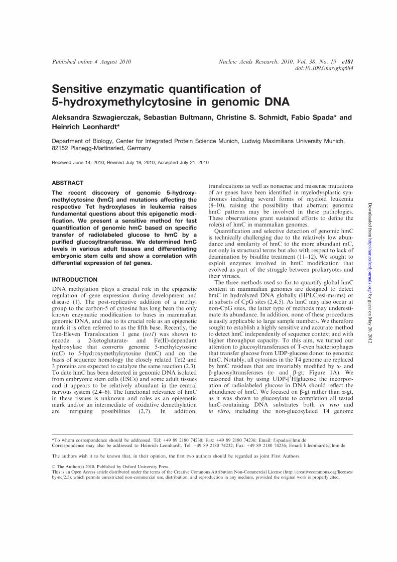
Sensitive enzymatic quantification of5-hydroxymethylcytosine in genomic DNAAleksandra Szwagierczak, Sebastian Bultmann, Christine S. Schmidt, Fabio Spada* andHeinrich Leonhardt*
Department of Biology, Center for Integrated Protein Science Munich, Ludwig Maximilians University Munich,82152 Planegg-Martinsried, Germany
Received June 14, 2010; Revised July 19, 2010; Accepted July 21, 2010
ABSTRACT
The recent discovery of genomic 5-hydroxy-methylcytosine (hmC) and mutations affecting therespective Tet hydroxylases in leukemia raisesfundamental questions about this epigenetic modi-fication. We present a sensitive method for fastquantification of genomic hmC based on specifictransfer of radiolabeled glucose to hmC by apurified glucosyltransferase. We determined hmClevels in various adult tissues and differentiatingembryonic stem cells and show a correlation withdifferential expression of tet genes.
INTRODUCTION
DNA methylation plays a crucial role in the epigeneticregulation of gene expression during development anddisease (1). The post-replicative addition of a methylgroup to the carbon-5 of cytosine has long been the onlyknown enzymatic modification to bases in mammaliangenomic DNA, and due to its crucial role as an epigeneticmark it is often referred to as the fifth base. Recently, theTen-Eleven Translocation 1 gene (tet1) was shown toencode a 2-ketoglutarate- and Fe(II)-dependanthydroxylase that converts genomic 5-methylcytosine(mC) to 5-hydroxymethylcytosine (hmC) and on thebasis of sequence homology the closely related Tet2 and3 proteins are expected to catalyze the same reaction (2,3).To date hmC has been detected in genomic DNA isolatedfrom embryonic stem cells (ESCs) and some adult tissuesand it appears to be relatively abundant in the centralnervous system (2,4–6). The functional relevance of hmCin these tissues is unknown and roles as an epigeneticmark and/or an intermediate of oxidative demethylationare intriguing possibilities (2,7). In addition,
translocations as well as nonsense and missense mutationsof tet genes have been identified in myelodysplastic syn-dromes including several forms of myeloid leukemia(8–10), raising the possibility that aberrant genomichmC patterns may be involved in these pathologies.These observations grant sustained efforts to define therole(s) of hmC in mammalian genomes.Quantification and selective detection of genomic hmC
is technically challenging due to the relatively low abun-dance and similarity of hmC to the more abundant mC,not only in structural terms but also with respect to lack ofdeamination by bisulfite treatment (11–12). We sought toexploit enzymes involved in hmC modification thatevolved as part of the struggle between prokaryotes andtheir viruses.The three methods used so far to quantify global hmC
content in mammalian genomes are designed to detecthmC in hydrolyzed DNA globally (HPLC/esi-ms/ms) orat subsets of CpG sites (2,4,5). As hmC may also occur atnon-CpG sites, the latter type of methods may underesti-mate its abundance. In addition, none of these proceduresis easily applicable to large sample numbers. We thereforesought to establish a highly sensitive and accurate methodto detect hmC independently of sequence context and withhigher throughput capacity. To this aim, we turned ourattention to glucosyltransferases of T-even bacteriophagesthat transfer glucose from UDP-glucose donor to genomichmC. Notably, all cytosines in the T4 genome are replacedby hmC residues that are invariably modified by a- andb-glucosyltransferases (a- and b-gt; Figure 1A). Wereasoned that by using UDP-[3H]glucose the incorpor-ation of radiolabeled glucose in DNA should reflect theabundance of hmC. We focused on b-gt rather than a-gt,as it was shown to glucosylate to completion all testedhmC-containing DNA substrates both in vivo andin vitro, including the non-glucosylated T4 genome
*To whom correspondence should be addressed. Tel: +49 89 2180 74230; Fax: +49 89 2180 74236; Email: [email protected] may also be addressed to Heinrich Leonhardt. Tel: +49 89 2180 74232; Fax: +49 89 2180 74236; Email: [email protected]
The authors wish it to be known that, in their opinion, the first two authors should be regarded as joint First Authors.
Published online 4 August 2010 Nucleic Acids Research, 2010, Vol. 38, No. 19 e181doi:10.1093/nar/gkq684
! The Author(s) 2010. Published by Oxford University Press.This is an Open Access article distributed under the terms of the Creative Commons Attribution Non-Commercial License (http://creativecommons.org/licenses/by-nc/2.5), which permits unrestricted non-commercial use, distribution, and reproduction in any medium, provided the original work is properly cited.
by guest on May 20, 2012
http://nar.oxfordjournals.org/D
ownloaded from
(13,14). This indicates that b-gt can glucosylate hmC in-dependently of DNA sequence and structural context andtherefore is ideally suited for this assay.
MATERIALS AND METHODS
Cell culture and differentiation of ESCs
Undifferentiated J1 and E14 ESCs were maintained ongelatin-coated dishes in Dulbecco’s modiEed Eagle’smedium containing 16% fetal bovine serum (PAALaboratories GmbH), 0.1mM b-mercaptoethanol(Invitrogen), 2mM L-glutamine, 1!MEM Non-essentialAmino Acid Solution, 100U/ml penicillin, 100 mg/mlstreptomycin (PAA Laboratories GmbH) and 1000U/mlrecombinant mouse LIF (Millipore). To induce embryoidbody (EB) formation, ESCs were resuspended in thesame medium as above but without LIF and culturedin hanging drops (600 cells/20 ml drop) for 4 days.Subsequently, EBs were cultured in bacterial culturedishes and the medium was replaced every 4 days.
DNA and RNA isolation
All tissue samples were prepared from 6-week-old 129svmice. Genomic DNA and total RNA were isolatedfrom tissue samples using the NucleoSpin Triprep Kit(Macherey-Nagel). Genomic DNA and total RNA wereisolated from ESCs and EBs using the Blood & Cellculture DNA mini kit (QIAGEN) and TRIzol reagent
(Invitrogen), respectively. To avoid genomic DNA con-tamination, isolated RNA was digested with recombinantRNase-free DNase I (Roche) and further purified with theQIAGEN RNeasy kit. Genomic DNA samples weresheared to 500–1500 bp fragments by sonication toreduce the viscosity and improve homogeneity. The con-centration of genomic DNA samples was measured byfluorometry. Fifty microliters of diluted sample weremixed with 50 ml of 2!TNE (Tris 20mM, pH 7.4; NaCl400mM and EDTA 2mM) containing 200 ng/ml ofHoechst 33258. Fluorescence was measured in aTECAN infinite M1000 plate reader (Ex: 350/10; Em:455/10). Serial dilutions (20–2000 ng/ml) of the hmC con-taining reference DNA fragment (see below) were used asstandard for quantification.
Protein expression and purification
The sequence encoding bacteriophage T4 b-gt wassynthesized at Mr. Gene GmbH (Regensburg) and clonedinto pET28b vector (Novagen). BL21(DE3) E. coli cellscarrying the expression construct were grown at 37"Cuntil A600=0.6–0.7 and induced with 1mM isopropylb-D-thiogalactopyranoside for 16 h at 20"C. Lysates wereprepared by sonication in 300mM NaCl, 50mMNa2HPO4, 10mM imidazole, 1mM b-mercaptoethanol,cleared by centrifugation and applied to nickel-nitrilotriacetic acid column (QIAGEN) pre-equilibratedwith lysis buffer. Washing and elution were performedwith lysis buffer containing 20 and 250mM imidazole,
y = 15148xR! = 0,9991
5,000
10,000
15,000
20,000
25,000
30,000
35,000
0 0,5 1 1,5 2
dpm
% of hmC
UDP
[ glucosyltransferase ]
O
OOHOH
OH
OH
UDP
O
N
NH2
ON
H O
N
NH2
ON
O
OHOH
OH
OHA
B C
250
130
100
70
55
35
kDa 1 2
Figure 1. Elements of the hmC glucosylation assay. (A) Schematic representation of the hmC glucosylation reaction catalyzed by b-gt.(B) Coomassie blue stained gel showing the purified b-gt preparation. (C) Example of calibration curve using mixtures of hmC-containing andunmodified reference fragment (equal total DNA amounts). Note the linear relationship between [3H]glucose incorporation and percentage of hmC.
e181 Nucleic Acids Research, 2010, Vol. 38, No. 19 PAGE 2 OF 5
by guest on May 20, 2012
http://nar.oxfordjournals.org/D
ownloaded from
respectively. Eluted proteins were applied to a SuperdexS-200 preparative gel filtration column (GE Healthcare)in 150mM NaCl, 20mM Tris, pH 8.0, 1mM DTT.Fractions containing the b-gt peak were pooled andapplied to a ResourceQ anion exchange column (GEHealthcare) in order to eliminate residual contaminants,resulting in pure b-gt in the flowthrough.
Preparation of reference DNA fragments
Reference DNA fragments (1139 bp) containing 0 and100% hmC were prepared by polymerase chain reaction(PCR), using dCTP and 5-hydroxymethyl-dCTP (BiolineGmbH), respectively. T4 phage DNA was used astemplate with primers: 50-TGG AGA AGG AGA ATGAAG AAT AAT-30 and 50-GTG AAG TAA GTA ATAAAT GGA TTG-30, Phusion HF DNA Polymerase(Finnzymes) and the following cycling profile: one cycleof 98"C for 2min and 35 cycles of 98"C for 10 s; 58"C for10 s; and 72"C for 30 s. Primer sequences were selectedthat do not contain cytosine residues. PCR productswere purified by gel electrophoresis followed by silicacolumn purification (Nucleospin, Macherey-Nagel).
Quantitative hmC glucosylation assay
Reactions contained 150mM NaCl, 20mM Tris, pH 8.0,25mM CaCl2, 1mM DTT, 2.8mM ‘cold’ UDP-glucose(Sigma-Aldrich), 0.86 nM UDP-[3H]glucose (glucose-6-3H; 60Ci/mmol; Hartmann Analytic GmbH), 1 mg ofDNA substrate and 36 nM recombinant b-gt in a totalvolume of 50 ml. Reactions were incubated for 20min atroom temperature and terminated by heating at 65"C for10min. Twenty microliters of each reaction were spottedin duplicate on paper filters (Whatmann) and DNA wasprecipitated by incubation in 5% TCA for 15min at roomtemperature. Filters were washed twice with 5% TCA andonce with 70% ethanol. Remaining radioactivity wasmeasured using a Liquid Scintillation Analyzer Tri-Carb2100TR (Packard) with quench indicating parameter seton tSIE/AEC (transformed spectral index of the externalstandard/automatic efficiency control) in 4ml of RotiszintEco Plus scintillation liquid (Roth GmbH) in Snaptwistvials (Simport). Samples were measured for 30min or untilthe 2! value reached 2%. The percentage of hmC per totalcytosine was calculated from the incorporation of[3H]glucose using a calibration curve measured with thereference fragment series for every experiment. The per-centage of hmC was then corrected for the difference in Cabundance between reference fragment (35%) and mousegenome (42%).
cDNA synthesis and real-time PCR
Five hundred nanograms of total RNA were used forcDNA synthesis with the High-Capacity cDNA ReverseTranscription Kit (with RNase Inhibitor; AppliedBiosystems). Equal amounts of cDNA were used forreal-time PCR with Power SYBR Green PCR MasterMix (Applied Biosystems) on a 7500 Fast Real-TimePCR System (Applied Biosystems) according to the manu-facturer’s instructions. Gene expression levels were
normalized to Gapdh and calculated using the compara-tive CT method (!!CT method).Primers for quantitative real-time PCR were designed
with the Primer Express software (Applied Biosystems)and contained the following sequences: Gapdh forward50-CAT GGC CTT CCG TGT TCC TA-30, Gapdhreverse 50-CTT CAC CAC CTT CTT GAT GTCATC-30, Tet1 forward 50-CCA GGA AGA GGC GACTAC GTT-30, Tet1 reverse 50-TTA GTG TTG TGTGAA CCT GAT TTA TTG T-30, Tet2 forward 50-ACTTCT CTG CTC ATT CCC ACA GA-30, Tet2 reverse50-TTA GCT CCG ACT TCT CGA TTG TC-30, Tet3forward 50-GAG CAC GCC AGA GAA GAT CAA-30
and Tet3 reverse 50-CAG GCT TTG CTG GGA CAATC-30.
RESULTS AND DISCUSSION
T4 b-gt was expressed in bacteria as a 6 ! His tag fusionand purified to homogeneity by sequential nickel-NTA,size exclusion and ion exchange chromatography(Figure 1B). To assess whether transfer of [3H]glucose toDNA is proportional to the hmC content within the rangepreviously reported for mammalian tissues, we prepared aseries of standard DNA substrate samples with globalhmC content ranging from 0.25 to 2% of total cytosineby mixing corresponding proportions of two preparationsof the same 1.2 kb DNA fragment, one having all cytosineresidues replaced by hmC and the other containing nohmC (Figure 1C). Using a 325-fold excess of unlabelledUDP-glucose, the incorporation of radiolabeled glucose in1 mg of total DNA substrate was strictly linear in thisrange. This standard sample series was measured inevery assay to generate a calibration curve for the calcu-lation of hmC content in genomic DNA samples. We firstmeasured genomic hmC levels in wild-type and Dnmt1, 3aand 3b triple knockout (TKO) J1 ESCs (15) (Figure 2Aand B). Due to the absence of all three major DNAmethyltransferases, genomic DNA from TKO ESCs isexpected to contain very little, if any, cytosine methyla-tion. Indeed, the measured level of genomic hmC in TKOESCs was at the detection limit (0.025%) of our assay,while genomic DNA from wild-type ESCs contained0.3% hmC relative to total cytosine. Real-time reversetranscription (RT) PCR analysis showed that Tet1–3mRNA levels are similar in wild-type and TKO ESCs,with Tet1 transcripts largely preponderant and Tet3mRNA the least abundant (more than 40-fold lowerthan Tet1). It was previously shown that differentiationof mouse ESCs by withdrawal of LIF from monolayercultures for 5 days results in a reduction of genomichmC and concomitant decrease in Tet1 transcripts (2).We followed genomic hmC and Tet1–3 transcriptdynamics during EB differentiation of two commonlyused wild-type ESC lines (Figure 2A and B andSupplementary Figure S1). In both cases, a sharpdecrease of hmC content was evident after 4 days of EBculture, but a substantial recovery was observed after add-itional 4 days of culture (Day 8). Interestingly, the tetgenes showed distinct expression dynamics during ESCs
PAGE 3 OF 5 Nucleic Acids Research, 2010, Vol. 38, No. 19 e181
by guest on May 20, 2012
http://nar.oxfordjournals.org/D
ownloaded from

differentiation. Tet1 transcripts drastically decreasedin the first 4 days and further dropped by Day 8 of EBculture. Tet2 mRNA levels also decreased in the first4 days, but were fully restored in Day 8 EBs.In contrast, Tet3 transcripts doubled at Day 4 and in-creased about 20 times by Day 8 of EB culture. Thus,the relatively high hmC content in undifferentiated ESCscorrelates with high levels of Tet1 and, to a lower extent,Tet2 transcripts, while the partial recovery of genomichmC in Day 8 EBs correlates with increased Tet2 andTet3 mRNA levels.We then analyzed several adult mouse tissues
(Figure 2C and D). As reported earlier, the highestlevels of genomic hmC were found in brain regions,although kidney also showed relatively high levels. In allcases, the hmC content was at least four times higher thanthe detection limit, while in a previous report using a dif-ferent assay the same non-neural tissues were either mar-ginally above or right at the detection limit (5). AbundanthmC in brain tissues correlated with high levels of Tet3and to a lower extent Tet2, a pattern similar to Day 8 EBs.Thus, most differentiated tissues are characterized by verylow levels of Tet1 and high levels of Tet3, while undiffer-entiated ESCs show the opposite pattern. It will be
interesting to determine whether all pluripotent cell typeshave this pattern and at which stages along the specifica-tion of the various somatic lineages the relative expressionlevels of tet genes change. Interestingly, kidney representsan exception among the adult tissues analyzed as it showsrelatively high hmC content and a prevalence of Tet2 tran-scripts. This is consistent with a cellular defect in proximalconvoluted tubules of the kidney as the only phenotypedescribed for Tet2 null mice (16). These observationssuggest that Tet proteins have redundant roles and thatthe lack of a specific Tet family member may result inphenotypic alterations only in tissues where high levelsof that Tet enzyme cannot be compensated by the otherfamily members. In this context, it should be notedthat the assay described here could also be employed tomeasure the enzymatic activity of Tet proteins and theirmutants identified in leukemia patients by using mC-containing DNA substrates.
In conclusion, we have established an accurate assay forthe quantification of genomic hmC that: (i) is more sensi-tive than previously described methods; (ii) is not subjectto sequence bias; (iii) allows simultaneous processing oflarge sample numbers; and (iv) does not requirespecialized and expensive equipment. It should be noted
CA
0
0,2
0,4
0,6
0,8
1
1,2
1,4
1,6
% o
f hm
C
0
0,05
0,1
0,15
0,2
0,25
0,3
0,35
% o
f hm
C
0
20
40
60
80
100
120
140
160
180
day 4 day 8 TKO
rela
tive
expr
essi
on l
evel
Tet1Tet2Tet3
B
undiff.embryoid bodies
undiff.wild type
kidne
yliv
er
cere
bellu
mco
rtex
hippo
campu
slun
gtes
tis
thymus
splee
n
0
5
10
15
20
25
30
35
40
rela
tive
expr
essi
on l
evel
kidne
yliv
er
cere
bellu
mco
rtex
hippo
campu
s
D
day 4 day 8 TKOundiff.
embryoid bodiesundiff.
wild type
*
Tet1Tet2Tet3
Figure 2. Quantification of genomic hmC and Tet transcripts in mouse tissues, undifferentiated ESCs and EBs. (A and C) hmC glucosylation assays.The percentage of hmC per total cytosine was calculated from the incorporation of [3H]glucose using a calibration curve from the reference fragment(see Figure 1C). Shown are average values and error bars from two (A) or one (C) biological replicates, each measured in two independent assays,with the exception of hippocampus that was measured only once. In every assay, each sample was measured in duplicate. The dashed line in(A) indicates the estimated limit of detection. (B and D) Real-time RT–PCR analysis for Tet transcript levels. Expression levels are all relative toTet1 in kidney (set to 1), so that values in b and d are directly comparable. Shown are average values and error bars from two (B) and one(D) biological replicates, each measured from two independent cDNA synthesis reactions. In every real-time PCR reaction, each sample wasmeasured in triplicate. Genomic DNA and RNA samples used in A/C and B/C, respectively, were isolated from the very same cell and tissue lysates.
e181 Nucleic Acids Research, 2010, Vol. 38, No. 19 PAGE 4 OF 5
by guest on May 20, 2012
http://nar.oxfordjournals.org/D
ownloaded from
that lower concentrations of ‘cold’ UDP-glucose shouldallow scaling down the amount of substrate DNAwithout loss of signal. This assay will be highly useful todetermine the global abundance of hmC in genomic DNA,especially in situations where limited amounts of tissue areavailable such as isolates of rare primary cell types andclinical samples.
SUPPLEMENTARY DATA
Supplementary Data are available at NAR Online.
ACKNOWLEDGEMENTS
The authors thank Daniela Meilinger, Andrea Steiner andAndreas Brachmann (Ludwig Maximilians UniversityMunich) for help with EB culture, dissection of mousetissues and scintillation counting, respectively. TKOESCs were provided by Masaki Okano (RIKEN Centerfor Developmental Biology, Kobe, Japan).
FUNDING
Deutsche Forschungsgemeinschaft (Grants DFG SPP1356, SFB 646 and 684 to H.L.); financial support bythe Elite Network of Bavaria (International DoctorateProgram NanoBioTechnology) and the InternationalMax Planck Research School for Molecular and CellularLife Sciences (IMPRS-LS to C.S.S.). S.B. is a fellow of thegraduate school Life Science Munich (LSM). Funding foropen access charge: Deutsche Forschungsgemeinschaft(DFG SPP 1356, SFB 646 and 684 to H.L.).
Conflict of interest statement. None declared.
REFERENCES
1. Rottach,A., Leonhardt,H. and Spada,F. (2009) DNAmethylation-mediated epigenetic control. J. Cell. Biochem., 108,43–51.
2. Tahiliani,M., Koh,K.P., Shen,Y., Pastor,W.A., Bandukwala,H.,Brudno,Y., Agarwal,S., Iyer,L.M., Liu,D.R., Aravind,L. et al.(2009) Conversion of 5-methylcytosine to 5-hydroxymethylcytosinein mammalian DNA by MLL partner TET1. Science, 324,930–935.
3. Iyer,L.M., Tahiliani,M., Rao,A. and Aravind,L. (2009) Predictionof novel families of enzymes involved in oxidative and othercomplex modifications of bases in nucleic acids. Cell Cycle, 8,1698–1710.
4. Feng,J., Zhou,Y., Campbell,S.L., Le,T., Li,E., Sweatt,J.D.,Silva,A.J. and Fan,G. (2010) Dnmt1 and Dnmt3a maintain DNAmethylation and regulate synaptic function in adult forebrainneurons. Nat. Neurosci., 13, 423–430.
5. Kriaucionis,S. and Heintz,N. (2009) The nuclear DNA base5-hydroxymethylcytosine is present in Purkinje neurons and thebrain. Science, 324, 929–930.
6. Penn,N.W., Suwalski,R., O’Riley,C., Bojanowski,K. and Yura,R.(1972) The presence of 5-hydroxymethylcytosine in animaldeoxyribonucleic acid. Biochem. J., 126, 781–790.
7. Loenarz,C. and Schofield,C.J. (2009) Oxygenase catalyzed5-methylcytosine hydroxylation. Chem. Biol., 16, 580–583.
8. Lorsbach,R.B., Moore,J., Mathew,S., Raimondi,S.C.,Mukatira,S.T. and Downing,J.R. (2003) TET1, a member of anovel protein family, is fused to MLL in acute myeloid leukemiacontaining the t(10;11)(q22;q23). Leukemia, 17, 637–641.
9. Ono,R., Taki,T., Taketani,T., Taniwaki,M., Kobayashi,H. andHayashi,Y. (2002) LCX, leukemia-associated protein with aCXXC domain, is fused to MLL in acute myeloid leukemia withtrilineage dysplasia having t(10;11)(q22;q23). Cancer Res., 62,4075–4080.
10. Langemeijer,S.M.C., Kuiper,R.P., Berends,M., Knops,R.,Aslanyan,M.G., Massop,M., Stevens-Linders,E., van Hoogen,P.,van Kessel,A.G., Raymakers,R.A.P. et al. (2009) Acquiredmutations in TET2 are common in myelodysplastic syndromes.Nat. Genet., 41, 838–842.
11. Hayatsu,H. and Shiragami,M. (1979) Reaction of bisulfite withthe 5-hydroxymethyl group in pyrimidines and in phage DNAs.Biochemistry, 18, 632–637.
12. Huang,Y., Pastor,W.A., Shen,Y., Tahiliani,M., Liu,D.R. andRao,A. (2010) The behaviour of 5-hydroxymethylcytosine inbisulfite sequencing. PLoS ONE, 5, e8888.
13. Georgopoulos,C.P. and Revel,H.R. (1971) Studies with glucosyltransferase mutants of the T-even bacteriophages. Virology, 44,271–285.
14. Kornberg,S.R., Zimmerman,S.B. and Kornberg,A. (1961)Glucosylation of deoxyribonucleic acid by enzymes frombacteriophage-infected Escherichia coli. J. Biol. Chem., 236,1487–1493.
15. Tsumura,A., Hayakawa,T., Kumaki,Y., Takebayashi,S.,Sakaue,M., Matsuoka,C., Shimotohno,K., Ishikawa,F., Li,E.,Ueda,H.R. et al. (2006) Maintenance of self-renewal ability ofmouse embryonic stem cells in the absence of DNAmethyltransferases Dnmt1, Dnmt3a and Dnmt3b. Genes Cells, 11,805–814.
16. Tang,H., Araki,K., Li,Z. and Yamamura,K. (2008)Characterization of Ayu17-449 gene expression and resultantkidney pathology in a knockout mouse model. Transgenic Res.,17, 599–608.
PAGE 5 OF 5 Nucleic Acids Research, 2010, Vol. 38, No. 19 e181
by guest on May 20, 2012
http://nar.oxfordjournals.org/D
ownloaded from

Sensitive enzymatic quantification of 5-hydroxymethylcytosine in genomic DNA Aleksandra Szwagierczak, Sebastian Bultmann, Christine Schmidt, Fabio Spada and Heinrich Leonhardt Ludwig Maximilians University Munich, Department of Biology, Center for Integrated Protein Science Munich (CIPSM), 82152 Planegg-Martinsried, Germany.
SUPPLEMENTARY FIGURE S1
Quantification of genomic hmC and Tet transcripts in undifferentiated E14 embryonic stem cells (ESCs) and embryoid bodies (EBs). (A) hmC glucosylation assay. The percentage of hmC over total cytosine is calculated from the incorporation of [3H]glucose using a calibration curve from the reference fragment (see Fig. 1C) and corrected for the difference in cytosine abundance between the latter (35%) and mouse genome (42%). Shown are values from one assay, where each sample was measured in duplicate. (B) Real time RT-PCR analysis for Tet transcript levels. Shown are average values and error bars from two independent cDNA synthesis reactions. In every PCR reaction each sample was measured in triplicate. Genomic DNA and RNA samples used in A and B, respectively, were isolated from the very same ESC and EB lysates.

2.2 Characterization of PvuRts1I endonuclease as a tool
to investigate genomic 5-hydroxymethylcytosine
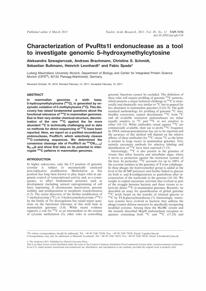
Characterization of PvuRts1I endonuclease as a toolto investigate genomic 5–hydroxymethylcytosineAleksandra Szwagierczak, Andreas Brachmann, Christine S. Schmidt,Sebastian Bultmann, Heinrich Leonhardt* and Fabio Spada*
Ludwig Maximilians University Munich, Department of Biology and Center for Integrated Protein ScienceMunich (CIPSM), 82152 Planegg-Martinsried, Germany
Received October 18, 2010; Revised February 14, 2011; Accepted February 15, 2011
ABSTRACT
In mammalian genomes a sixth base,5-hydroxymethylcytosine (hmC), is generated by en-zymatic oxidation of 5-methylcytosine (mC). This dis-covery has raised fundamental questions about thefunctional relevance of hmC in mammalian genomes.Due to their very similar chemical structure, discrim-ination of the rare hmC against the far moreabundant mC is technically challenging and to dateno methods for direct sequencing of hmC have beenreported. Here, we report on a purified recombinantendonuclease, PvuRts1I, which selectively cleaveshmC-containing sequences. We determined theconsensus cleavage site of PvuRts1I as hmCN11–12/N9–10G and show first data on its potential to inter-rogate hmC patterns in mammalian genomes.
INTRODUCTION
In higher eukaryotes, only the C5 position of genomiccytosine is subject to enzymatically catalyzedpost-replicative modification. Methylation at thisposition has long been known to play major roles in epi-genetic control of transcriptional activity and, as a conse-quence, to affect fundamental processes such asdevelopment (including natural reprogramming of cellfate), imprinting, X chromosome inactivation, genomestability and predisposition to neoplastic transformation(1,2). The recent discovery of the further modification of5–methylcytosine (mC) to 5-hydroxymethylcytosine (hmC)by the family of Tet dioxygenases has raised major ques-tions on the functional relevance of this sixth base inmammalian genomes (3,4). While recent evidencesupports a role for hmC as an intermediate in the erasureof cytosine methylation (5), other roles in controlling
genomic functions cannot be excluded. The definition ofthese roles will require profiling of genomic hmC patterns,which presents a major technical challenge as hmC is struc-turally and chemically very similar to mC but in general farless abundant in mammalian genomes (3,4,6–9). The goldstandard methodology for profiling of genomic mC sites,bisulfite conversion, cannot discriminate hmC from mCand all available restriction endonucleases are eitherequally sensitive to mC and hmC or not sensitive toeither (10–12). While antibodies raised against hmC arecommercially available, their use to probe hmC frequencyby DNA immunoprecipitation has yet to be reported andthe accuracy of this method will depend on the relativeaffinity of these antibodies for hmC versus mC as the latteris present in large excess in mammalian genomes. Veryrecently enzymatic methods for selective labeling andidentification of hmC have been reported (7,13).Interestingly, hmC is also present in the genomes of
viruses that infect bacteria and unicellular algae, whereit serves as protection against the restriction systems ofthe host. In particular, hmC accounts for up to 100% ofthe cytosine residues in the genomes of T-even coliphages.In these phages the hydroxymethyl group is added at thelevel of the dCMP precursor and further linked to glucose(in both a- and b-configurations) or gentiobiose after in-corporation of the nucleotide in the genome (14–16). Wesought to exploit enzymatic activities that evolved as partof the struggle between bacteria and these viruses to se-lectively detect hmC in mammalian genomes. Recently, wedescribed an assay for quantification of global genomichmC levels based on the transfer of tritiated glucose tohmC by T4 b-glucosyltransferase (7). Interestingly, restric-tion systems have evolved in bacteria that address thephage counter defense measures by specifically recognizingmodified cytosine. Among these the McrBC system andthe recently described MspJI endonuclease recognize se-quences containing both mC and hmC (17,18) and
*To whom correspondence should be addressed. Tel: +49 89 2180 74230; Fax: +49 89 2180 74236; Email: [email protected] may also be addressed to Heinrich Leonhardt. Tel: +49 89 2180 74232; Fax +49 89 2180 74236; Email: [email protected]
Published online 4 March 2011 Nucleic Acids Research, 2011, Vol. 39, No. 12 5149–5156doi:10.1093/nar/gkr118
! The Author(s) 2011. Published by Oxford University Press.This is an Open Access article distributed under the terms of the Creative Commons Attribution Non-Commercial License (http://creativecommons.org/licenses/by-nc/2.5), which permits unrestricted non-commercial use, distribution, and reproduction in any medium, provided the original work is properly cited.

therefore per se are not useful to discriminate thesemodified cytosines. At least two endonucleases,PvuRts1I and GmrSD, were shown to restrict DNA con-taining glucosylated hmC (19,20). However, GmrSD doesnot cleave non-glucosylated (hmC-containing) T4 DNA,has the additional disadvantages of being a heterodimerand of co-purifying with the GroEL chaperonin (19).PvuRts1I is encoded by a single gene present on the kana-mycin resistance plasmid Rts1 originally isolated fromProteus vulgaris and its restriction activity in vivo wasshown to be modulated by hmC glucosylation in acomplex fashion (20). However, as PvuRts1I was notpurified, its activity has not been characterized in vitro.Here, we show that purified recombinant PvuRts1I se-
lectively cleaves hmC-containing DNA and determine itscleavage site. In addition, we present initial data on theuse of hmC as a tool to investigate hmC patterns in mam-malian genomes.
MATERIALS AND METHODS
Cloning and purification of PvuRts1I
The sequence encoding PvuRts1I was synthesized at MrGene GmbH (Regensburg) and cloned into the pET28avector (Novagen). BL21(DE3) Escherichia coli cellscarrying the expression vector were grown in LBmedium at 37!C until A600=0.6–0.7 and induced with1mM isopropyl b-D-thiogalactopyranoside for 16 h at18!C. Lysates were prepared by sonication in 300mMNaCl, 50mM Na2HPO4 pH 8.0, 10mM imidazole, 10%glycerol and 1mM b-mercaptoethanol, cleared by centri-fugation and applied to a nickel–nitrilotriacetic acidcolumn (QIAGEN) pre-equilibrated with lysis buffer.Washing and elution were performed with lysis buffer con-taining 20 and 250mM imidazole, respectively. Elutedproteins were applied to a Superdex S-200 preparativegel filtration column (GE Healthcare) in 150mM NaCl,20mM Tris pH 8.0, 10% glycerol, 1mM DTT and peakfractions were pooled. The stability of PvuRts1I uponstorage was improved by supplementation with 10%glycerol.
Preparation of DNA substrates
In vivo a/b-glucosylated and non-glucosylated T4 phageDNA was isolated essentially as described (4). Briefly,T4 stocks were propagated on E. coli strain CR63,which was also used for the isolation of glucosylated T4DNA. To isolate non-glucosylated T4 DNA, wild-type T4phage was amplified on an ER1565 galU mutant strain.b-glucosylated T4 DNA was generated in vitro by treat-ment of non-glucosylated T4 DNA with purified T4b-glucosyltransferase (7). Genomic DNA was isolatedfrom mouse cerebellum and triple knockout (TKO) em-bryonic stem cells (ESCs) (21) as described (7).Reference DNA fragments containing exclusively hmC,
mC or unmodified cytosine residues were prepared by PCRusing 5-hydroxymethyl-dCTP (Bioline GmbH),5-methyl-dCTP (Jena Bioscience GmbH) and dCTP, re-spectively. T4 phage DNA template, Phusion HF DNA
Polymerase (Finnzymes) and primer 50-GTG AAG TAAGTA ATA AAT GGA TTG-30, which does not containcytosine residues, were used for amplification of all refer-ence DNA fragments. To generate the reference 1139 bpfragment with 100% hmC for restriction with PvuRts1I thesecond primer was 50-TGG AGA AGG AGA ATG AAGAAT AAT-30, which also does not contain cytosineresidues. To generate the 800 and 500 bp control sub-strates containing only mC and only unmodified cytosinefor restriction with PvuRTS1I the second primer was 50-GCC ATA TTG ATA ATG AAA TTA AAT GTA-30 and50-TCA GCA ATT TTA ATA TTT CCA TCT TC-30,respectively. PCR products were purified by gel electro-phoresis followed by silica column purification(Nucleospin, Macherey-Nagel). The 140 bp fragmentused to determine the orientation of the PvuRTS1Icleavage overhang was amplified with primers 50-TATACT GAA GTA CTT CAT CA-30 and 50-CTT TGCGTG ATT TAT ATG TA-30.
For the preparation of substrates with a single PvuRts1Iconsensus containing hmC or mC in symmetrical or asym-metrical configuration a 94 bp fragment was amplifiedfrom the T4 genome with primers 50-CTC GTA GACTGC GTA CCA ATC TAA CTC AGG ATA GTTGAT-30 and 50-TAT GAT AAG TAT GTA GGT TATT-30. This fragment contains a single site corresponding tothe identified PvuRts1I consensus hmCN11–12/N9–10G andwas used as a template according to the strategy depictedin Figure 3. To generate substrates with symmetriccytosine modifications or unmodified cytosine thefragment was amplified with forward primer 50-CTCGTA GAC TGC GTA CCA-30 and reverse primer 150-TAT GAT AAG TAT GTA GGT TAT T-30 in thepresence of the respective modified or unmodified dCTP.To generate substrates with asymmetric cytosine modifi-cations the same forward primer was paired with reverseprimer 2 50-TAT GAT AAG TAT GTA GGT TAT TCAA-30.
DNA restriction with PvuRts1I and identification ofcleavage and recognition site
Unless otherwise stated the reaction conditions contained150mM NaCl, 20mM Tris pH 8.0, 5mM MgCl2, 1mMDTT. One unit of PvuRTS1I was defined as amount ofenzyme required to digest 1 mg of hmC-containing T4 DNAin 15min at 22!C. For assessment of enzyme specificity,100 ng of each control fragment were digested separatelyor together with 200 ng of genomic DNA in 30 ml reactionscontaining standard buffer and 1U of purified PvuRts1Iat 22!C for 15min.
For identification of the cleavage and recognition site,the 1139 bp fully hydroxymethylated fragment amplifiedfrom the T4 genome or whole non-glucosylated T4DNA were digested under standard conditions.Fragment ends were blunted with Klenow polymerase(NEB) and cloned using the Zero Blunt" PCR CloningKit (Invitrogen). Randomly selected clones weresequenced and the data were analyzed usingWebLogo (22).
5150 Nucleic Acids Research, 2011, Vol. 39, No. 12
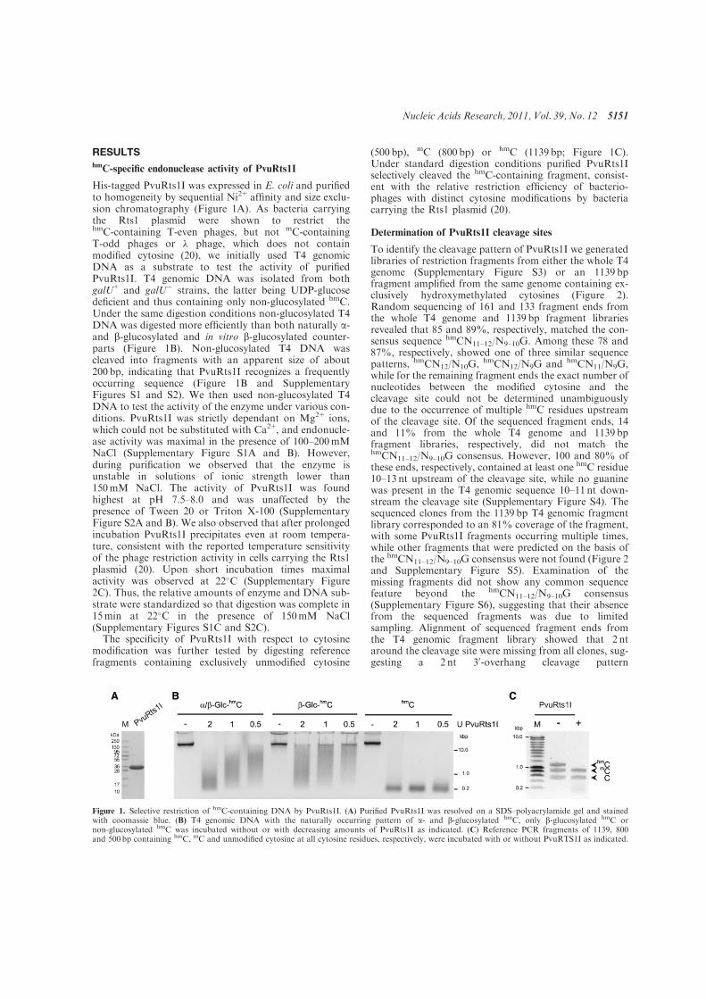
RESULTShmC-specific endonuclease activity of PvuRts1I
His-tagged PvuRts1I was expressed in E. coli and purifiedto homogeneity by sequential Ni2+ affinity and size exclu-sion chromatography (Figure 1A). As bacteria carryingthe Rts1 plasmid were shown to restrict thehmC-containing T-even phages, but not mC-containingT-odd phages or ! phage, which does not containmodified cytosine (20), we initially used T4 genomicDNA as a substrate to test the activity of purifiedPvuRts1I. T4 genomic DNA was isolated from bothgalU+ and galU" strains, the latter being UDP-glucosedeficient and thus containing only non-glucosylated hmC.Under the same digestion conditions non-glucosylated T4DNA was digested more efficiently than both naturally a-and b-glucosylated and in vitro b-glucosylated counter-parts (Figure 1B). Non-glucosylated T4 DNA wascleaved into fragments with an apparent size of about200 bp, indicating that PvuRts1I recognizes a frequentlyoccurring sequence (Figure 1B and SupplementaryFigures S1 and S2). We then used non-glucosylated T4DNA to test the activity of the enzyme under various con-ditions. PvuRts1I was strictly dependant on Mg2+ ions,which could not be substituted with Ca2+, and endonucle-ase activity was maximal in the presence of 100–200mMNaCl (Supplementary Figure S1A and B). However,during purification we observed that the enzyme isunstable in solutions of ionic strength lower than150mM NaCl. The activity of PvuRts1I was foundhighest at pH 7.5–8.0 and was unaffected by thepresence of Tween 20 or Triton X-100 (SupplementaryFigure S2A and B). We also observed that after prolongedincubation PvuRts1I precipitates even at room tempera-ture, consistent with the reported temperature sensitivityof the phage restriction activity in cells carrying the Rts1plasmid (20). Upon short incubation times maximalactivity was observed at 22!C (Supplementary Figure2C). Thus, the relative amounts of enzyme and DNA sub-strate were standardized so that digestion was complete in15min at 22!C in the presence of 150mM NaCl(Supplementary Figures S1C and S2C).
The specificity of PvuRts1I with respect to cytosinemodification was further tested by digesting referencefragments containing exclusively unmodified cytosine
(500 bp), mC (800 bp) or hmC (1139 bp; Figure 1C).Under standard digestion conditions purified PvuRts1Iselectively cleaved the hmC-containing fragment, consist-ent with the relative restriction efficiency of bacterio-phages with distinct cytosine modifications by bacteriacarrying the Rts1 plasmid (20).
Determination of PvuRts1I cleavage sites
To identify the cleavage pattern of PvuRts1I we generatedlibraries of restriction fragments from either the whole T4genome (Supplementary Figure S3) or an 1139 bpfragment amplified from the same genome containing ex-clusively hydroxymethylated cytosines (Figure 2).Random sequencing of 161 and 133 fragment ends fromthe whole T4 genome and 1139 bp fragment librariesrevealed that 85 and 89%, respectively, matched the con-sensus sequence hmCN11–12/N9–10G. Among these 78 and87%, respectively, showed one of three similar sequencepatterns, hmCN12/N10G, hmCN12/N9G and hmCN11/N9G,while for the remaining fragment ends the exact number ofnucleotides between the modified cytosine and thecleavage site could not be determined unambiguouslydue to the occurrence of multiple hmC residues upstreamof the cleavage site. Of the sequenced fragment ends, 14and 11% from the whole T4 genome and 1139 bpfragment libraries, respectively, did not match thehmCN11–12/N9–10G consensus. However, 100 and 80% ofthese ends, respectively, contained at least one hmC residue10–13 nt upstream of the cleavage site, while no guaninewas present in the T4 genomic sequence 10–11 nt down-stream the cleavage site (Supplementary Figure S4). Thesequenced clones from the 1139 bp T4 genomic fragmentlibrary corresponded to an 81% coverage of the fragment,with some PvuRts1I fragments occurring multiple times,while other fragments that were predicted on the basis ofthe hmCN11–12/N9–10G consensus were not found (Figure 2and Supplementary Figure S5). Examination of themissing fragments did not show any common sequencefeature beyond the hmCN11–12/N9–10G consensus(Supplementary Figure S6), suggesting that their absencefrom the sequenced fragments was due to limitedsampling. Alignment of sequenced fragment ends fromthe T4 genomic fragment library showed that 2 ntaround the cleavage site were missing from all clones, sug-gesting a 2 nt 30-overhang cleavage pattern
Figure 1. Selective restriction of hmC-containing DNA by PvuRts1I. (A) Purified PvuRts1I was resolved on a SDS–polyacrylamide gel and stainedwith coomassie blue. (B) T4 genomic DNA with the naturally occurring pattern of a- and b-glucosylated hmC, only b-glucosylated hmC ornon-glucosylated hmC was incubated without or with decreasing amounts of PvuRts1I as indicated. (C) Reference PCR fragments of 1139, 800and 500 bp containing hmC, mC and unmodified cytosine at all cytosine residues, respectively, were incubated with or without PvuRTS1I as indicated.
Nucleic Acids Research, 2011, Vol. 39, No. 12 5151

(Supplementary Figure S5). This was confirmed by directsequencing of the two fragments generated by digestion ofa 140 bp amplicon containing a single PvuRts1I site(Supplementary Figure S7).The results above reveal a symmetric nature of the
preferred cleavage sites and raise the issue of PvuRTs1Iactivity on sites with modified cytosine in symmetric andasymmetric configuration. To clarify this issue, we used aPCR strategy to generate DNA substrates with identicalsequence and containing a single PvuRts1I consensus sitewith hmC or mC in symmetrical and asymmetrical config-urations or no modified cytosine (Figure 3A). In thepresence of enzyme amounts that did not cleave substrateswith unmodified and mC sites, digestion of substrates withasymmetric hmC at the PvuRTs1I site was reduced withrespect to substrates with symmetric hmC, but still appre-ciable. Residual undigested substrate with symmetric hmCat the PvuRTs1I site in these reaction conditions was typ-ically observed with such short substrates, but not withlonger ones.
Digestion of mammalian genomic DNA with PvuRts1I
To investigate cleavage site preference and efficiency ofPvuRts1I digestion for mammalian genomic DNA, weinitially selected the upstream regulatory region III ofthe mouse nanog gene (23). As this region was shown tobe bound by Tet1 and to acquire CpG methylation uponknockdown of Tet1 in ESCs (5), it represents a potentialcandidate as a mammalian genomic sequence containinghmC. Real time amplification of this region from ESCgenomic DNA did not show a significant decrease ofproduct after PvuRts1I digestion (data not shown). Wethen devised a strategy to positively identify rarePvuRts1I digestion products. After PvuRts1I digestiongenomic fragments were ligated to a linker with arandom 2nt 30-overhang. Ligation products were thenamplified using nanog specific primers paired with alinker specific primer, but no amplification productcould be obtained (data not shown). This result may beexplained by an extremely seldom occurrence of hmC at
Figure 2. Cleavage site of PvuRts1I. A library of PvuRts1I restriction fragments was generated from an 1139 bp PCR fragment containing onlyhydroxymethylated cytosine residues and the sequence of 133 restriction fragment ends from randomly chosen clones was determined. (A) Graphicalmap of the fragment ends. A total of 119 analyzed fragment ends (triangles) matched the consensus sequence hmCN11–12/N9–10G, which was presentat 97 sites (thin vertical lines) in the 1139 bp PCR fragment (thick horizontal line). Fifty three fragment ends related to the sequence motif hmCN12/N10G (dark green triangles), 37 to hmCN11/N10G (bright green triangles) and 14 to hmCN11/N9G (light green triangles), while 15 fragment endsmatching the consensus sequence hmCN11–12/N9–10G could not assigned unambiguously to any of these subsets (gray triangles). Fourteen fragmentends did not match the prevalent consensus sequence (gray circles, see Supplementary Figure S3). (B) Occurrence of the three subsets of cleavage sitesand LOGO representation of the corresponding consensus sequence. The absolute height of each position reflects its overall conservation, while therelative height of nucleotide letters represents their relative frequency. The slash in the three cleavage sequence subtypes indicates the exact cleavagesite.
5152 Nucleic Acids Research, 2011, Vol. 39, No. 12
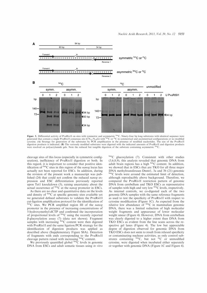
cleavage sites of this locus (especially in symmetric config-uration), inefficiency of PvuRts1I digestion or both. Inthis regard, it is important to consider that positive iden-tification of hmC sites in this region of the nanog locus hasactually not been reported for ESCs. In addition, duringthe revision of the present work a manuscript was pub-lished (24) that could not confirm the reduced nanog ex-pression and ESC differentiation previously reportedupon Tet1 knockdown (5), raising uncertainty about theactual occurrence of hmC at the nanog promoter in ESCs.
As there are no clear and quantitative data on the levelsand density of hmC at specific genomic sites available yetwe generated defined substrates to validate the PvuRst1Icut-ligation amplification protocol for the identification ofhmC sites. We PCR amplified region III of the nanogpromoter in the presence of increasing concentrations of5-hydroxymethyl-dCTP and confirmed the incorporationof proportional levels of hmC using the recently reportedb-glucosylation assay (7) (data not shown). Fragmentsamples with increasing hmC content were then digestedwith PvuRts1I and the same ligation/PCR strategy for theidentification of digestion products was applied asdescribed above (Supplementary Figure S8A). Detectionof fragments with ends corresponding to the PvuRts1Icleavage pattern raised with increasing hmC content.
We previously quantified global hmC levels in genomicDNA from ESCs and adult somatic tissues using in vitro
hmC glucosylation (7). Consistent with other studies(3,6,8,9), this analysis revealed that genomic DNA fromadult brain regions has a high hmC content. In addition,we showed that in ESCs that are TKO for all three majorDNA methyltransferases Dnmt1, 3a and 3b (21) genomichmC levels were around the estimated limit of detection,although reproducibly above background. Therefore, wecompared the PvuRts1I restriction pattern of genomicDNA from cerebellum and TKO ESCs as representativeof samples with high and very low hmC levels, respectively.As internal controls, we co-digested each of the twogenomic DNA samples with the same reference fragmentsas used to test the specificity of PvuRts1I with respect tocytosine modification (Figure 1C). As expected from therelative low abundance of hmC in mammalian genomicDNA, there was a limited reduction of high molecularweight fragments and appearance of lower molecularweight smear (Figure 4). However, DNA from cerebellumwas clearly digested to a higher extent than DNA fromTKO ESCs as evident from the line scans across the re-spective gel lanes (Figure 4). The low but appreciabledegree of digestion observed for genomic DNA fromTKO ESCs does not seem to result from relaxed specificityor contaminating nuclease activities, as only control sub-strates containing hmC, but not mC or unmodifiedcytosine, were digested when incubated either separatelyor together with genomic DNA (Figure 1C and Figure 4).
Figure 3. Differential activity of PvuRts1I on sites with symmetric and asymmetric hmC. Ninety-four bp long substrates with identical sequence weregenerated that contain a single PvuRts1I consensus site (CN12/N10G) with hmC or mC in symmetrical and asymmetrical configurations or no modifiedcytosine. (A) Strategy for generation of the substrates by PCR amplification in the presence of modified nucleotides. The size of the PvuRts1Idigestion products is indicated. (B) The variously modified substrates were digested with the indicated amounts of PvuRts1I and digestion productswere resolved on polyacrylamide gels. Note the reduced but tangible digestion of the substrate containing asymmetric hmC.
Nucleic Acids Research, 2011, Vol. 39, No. 12 5153
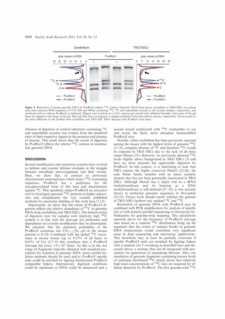
Absence of digestion of control substrates containing mCand unmodified cytosine was evident from the unalteredratio of their respective signals in the presence and absenceof enzyme. This result shows that the extent of digestionby PvuRts1I reflects the relative hmC content in mamma-lian genomic DNA.
DISCUSSION
Several modification and restriction systems have evolvedas defense and counter defense strategies in the strugglebetween unicellular microorganisms and their viruses.Here, we show that, in contrast to previouslycharacterized endonucleases which cleave hmC-containingsequences, PvuRts1I has a preference for thenon-glucosylated form of this base and discriminatesagainst mC. This specificity makes PvuRts1I an attractivetool to investigate genomic hmC patterns in higher eukary-otes and complements the very recently publishedmethods for enzymatic labeling of this sixth base (7,13).Importantly, we show that the extent of PvuRts1I di-
gestion reflects the relative abundance of hmC in genomicDNA from cerebellum and TKO ESCs. The limited extentof digestion even for samples with relatively high hmCcontent is in line with the cleavage site preference anddependence on cytosine modification that we determined.We calculate that the statistical probability of thePvuRts1I consensus site CN11–12/N9–10G in the mousegenome is 0.126. Combined with the global hmC occur-rence in mouse tissues (up to 0.13% of all bases or0.65% of Cs) (3,7–9) this translates into a PvuRts1Icleavage site every 1.9# 105 bases. As this is in the sizerange of fragments typically obtained with standard pro-cedures for isolation of genomic DNA, more careful iso-lation methods should be used and/or PvuRts1I specificends could be enriched by ligating biotinylated PvuRts1Icompatible linkers. Alternatively, digestion conditionscould be optimized or DNA could be denatured and a
second strand synthesized with hmC nucleotides to cutand reveal the likely more abundant hemimodifiedPvuRts1I sites.
Notably, while cerebellum has been previously reportedamong the tissues with the highest levels of genomic hmC(3,7,9), complete absence of mC and therefore hmC wouldbe expected in TKO ESCs due to the lack of all threemajor Dnmts (21). However, we previously detected hmClevels slightly above background in TKO ESCs (7) andhere we show minimal but appreciable digestion byPvuRts1I. In this context, it is interesting to note thatESCs express the highly conserved Dnmt2 (25,26), theonly Dnmt family member with an intact catalyticdomain that has not been genetically inactivated in TKOESCs. Although Dnmt2 has a major role as a tRNAmethyltransferase and its function as a DNAmethyltransferase is still debated (27–32), it was recentlyshown to methylate genomic sequences in Drosophila(32,33). Future work should clarify whether the genomeof TKO ESCs harbors any residual mC and hmC.
Restriction of genomic DNA with PvuRts1I may becombined with PCR amplification for analysis of specificloci or with massive parallel sequencing or microarray hy-bridization for genome-wide mapping. The calculationsreported above for the frequency of PvuRts1I cleavagesites based on a random hmC distribution bring up theargument that the extent of random breaks in genomicDNA preparations would contribute very significantnoise in deep sequencing and microarray applications.This drawback may at least be partially overcome ifspecific PvuRts1I ends are enriched by ligating linkerswith a random 2nt 30-overhang as described here and dis-cussed above, a strategy that can be integrated with pro-cedures for generation of sequencing libraries. Also, oursimulation of genomic fragments containing known levelsof randomly distributed hmC clearly shows that relativelyhigh local concentrations of hmC sites are required for ef-ficient detection by PvuRts1I. The first genome-wide hmC
TKO ESCs
+ +
PvuRts1I
- -
+ PvuRts1IPvuRts1I_
hmCmC
C
gray values (x1000)
hmCmC
C
Cerebellum
gray values (x1000)
0 5 10 15 20 25 30051015202530
+ PvuRts1IPvuRts1I_
Figure 4. Restriction of mouse genomic DNA by PvuRts1I reflects hmC content. Genomic DNA from mouse cerebellum or TKO ESCs was mixedwith three reference PCR fragments of 1139, 800 and 500 bp containing hmC, mC and unmodified cytosine at all cytosine residues, respectively, andincubated with or without PvuRts1I as indicated. Digests were resolved on a 0.8% agarose gel stained with ethidium bromide. Line scans of the gellanes are aligned to the image of the gel. Red and blue lines correspond to samples incubated with and without enzyme, respectively. Arrows point tothe main difference in the profiles form cerebellum and TKO ESC DNA digested with PvuRts1I (red lines).
5154 Nucleic Acids Research, 2011, Vol. 39, No. 12
profiles from mammalian tissues have just been reported(13). From these first data sets, it is apparent that genomichmC is not randomly distributed and that its accumulationin gene bodies is proportional to transcriptional activity.Thus, PvuRts1I may prove a valuable tool to probe hmCaccumulation at defined genomic regions. In addition, theselectivity of PvuRts1I for hmC-containing sites may con-stitute an advantage with respect to endonucleases such asMcrBC and MspJ1 as these enzymes do not discriminatebetween mC and hmC and require in vitro enzymatic hmCglucosylation to specifically protect hmC-containing sitesfrom digestion and thus distinguish them from mC sites.
In conclusion, we show that PvuRts1I is an hmC specificendonuclease and provide a biochemical characterizationof its enzymatic properties for future applications as diag-nostic tool in the analysis of hmC distribution at genomicloci in development and disease.
SUPPLEMENTARY DATA
Supplementary Data are available at NAR Online.
ACKNOWLEDGEMENTS
The authors thank Ken Kreutzer (Duke University,Duhram, NC) for providing the T4 phage stock andE. coli strains CR63 and ER1565 galU and MasakiOkano (RIKEN Center for Developmental Biology,Kobe, Japan) for the TKO ESCs.
FUNDING
Deutsche Forschungsgemeinschaft (DFG SPP 1356, SFB646 and 684 to H.L.); Life Science Munich (LSM to S.B.).Financial support by the Elite Network of Bavaria(International Doctorate Program NanoBioTechnology)and International Max Planck Research School forMolecular and Cellular Life Sciences (IMPRS-LS toC.S.S.). Funding for open access charge: DeutscheForschungsgemeinschaft.
Conflict of interest statement. None declared.
REFERENCES
1. Bird,A. (2002) DNA methylation patterns and epigenetic memory.Genes Dev., 16, 6–21.
2. Rottach,A., Leonhardt,H. and Spada,F. (2009) DNAmethylation-mediated epigenetic control. J. Cell. Biochem., 108,43–51.
3. Kriaucionis,S. and Heintz,N. (2009) The nuclear DNA base5-hydroxymethylcytosine is present in Purkinje neurons and thebrain. Science, 324, 929–930.
4. Tahiliani,M., Koh,K.P., Shen,Y., Pastor,W.A., Bandukwala,H.,Brudno,Y., Agarwal,S., Iyer,L.M., Liu,D.R., Aravind,L. et al.(2009) Conversion of 5-methylcytosine to 5-hydroxymethylcytosinein mammalian DNA by MLL Partner TET1. Science, 324,930–935.
5. Ito,S., D’Alessio,A.C., Taranova,O.V., Hong,K., Sowers,L.C. andZhang,Y. (2010) Role of Tet proteins in 5mC to 5hmCconversion, ES-cell self-renewal and inner cell mass specification.Nature, 466, 1129–1133.
6. Feng,J., Zhou,Y., Campbell,S.L., Le,T., Li,E., Sweatt,J.D.,Silva,A.J. and Fan,G. (2010) Dnmt1 and Dnmt3a maintain DNA
methylation and regulate synaptic function in adult forebrainneurons. Nat. Neurosci., 13, 423–430.
7. Szwagierczak,A., Bultmann,S., Schmidt,C.S., Spada,F. andLeonhardt,H. (2010) Sensitive enzymatic quantification of5-hydroxymethylcytosine in genomic DNA. Nucleic Acids Res.,38, e181.
8. Munzel,M., Globisch,D., Bruckl,T., Wagner,M., Welzmiller,V.,Michalakis,S., Muller,M., Biel,M. and Carell,T. (2010)Quantification of the sixth DNA base hydroxymethylcytosine inthe brain. Angew. Chem. Int. Ed. Engl., 49, 5375–5377.
9. Globisch,D., Munzel,M., Muller,M., Michalakis,S., Wagner,M.,Koch,S., Bruckl,T., Biel,M. and Carell,T. (2010) Tissuedistribution of 5-hydroxymethylcytosine and search for activedemethylation intermediates. PLoS ONE, 5, e15367.
10. Huang,Y., Pastor,W.A., Shen,Y., Tahiliani,M., Liu,D.R. andRao,A. (2010) The behaviour of 5-hydroxymethylcytosine inbisulfite sequencing. PLoS ONE, 5, e8888.
11. Jin,S.-G., Kadam,S. and Pfeifer,G.P. (2010) Examination of thespecificity of DNA methylation profiling techniques towards5-methylcytosine and 5-hydroxymethylcytosine. Nucleic AcidsRes., 38, e125.
12. Nestor,C., Ruzov,A., Meehan,R. and Dunican,D. (2010)Enzymatic approaches and bisulfite sequencing cannot distinguishbetween 5-methylcytosine and 5-hydroxymethylcytosine in DNA.Biotechniques, 48, 317–319.
13. Song,C.-X., Szulwach,K.E., Fu,Y., Dai,Q., Yi,C., Li,X., Li,Y.,Chen,C.-H., Zhang,W., Jian,X. et al. (2010) Selective chemicallabeling reveals the genome-wide distribution of5-hydroxymethylcytosine. Nat. Biotechnol., 29, 68–72.
14. Flaks,J.G. and Cohen,S.S. (1957) The enzymic synthesis of5-hydroxymethyldeoxycytidylic acid. Biochim. Biophys. Acta, 25,667–668.
15. Wiberg,J.S. and Buchanan,J.M. (1964) Studies on labiledeoxycytidylate hydroxymethylases from Escherichia coli Binfected with temperature-sensitive mutants of bacteriophage T4.Proc. Natl Acad. Sci. USA, 51, 421–428.
16. Lehman,I.R. and Pratt,E.A. (1960) On the structure of theglucosylated hydroxymethylcytosine nucleotides of coliphages T2,T4 and T6. J. Biol. Chem., 235, 3254–3259.
17. Raleigh,E.A. (1992) Organization and function of the mcrBCgenes of Escherichia coli K-12. Mol. Microbiol., 6, 1079–1086.
18. Zheng,Y., Cohen-Karni,D., Xu,D., Chin,H.G., Wilson,G.,Pradhan,S. and Roberts,R.J. (2010) A unique family of Mrr-likemodification-dependent restriction endonucleases. Nucleic AcidsRes., 38, 5527–5534.
19. Bair,C.L. and Black,L.W. (2007) A type IV modificationdependent restriction nuclease that targets glucosylatedhydroxymethyl cytosine modified DNAs. J. Mol. Biol., 366,768–778.
20. Janosi,L., Yonemitsu,H., Hong,H. and Kaji,A. (1994) Molecularcloning and expression of a novel hydroxymethylcytosine-specificrestriction enzyme (PvuRts1I) modulated by glucosylation ofDNA. J. Mol. Biol., 242, 45–61.
21. Tsumura,A., Hayakawa,T., Kumaki,Y., Takebayashi,S.,Sakaue,M., Matsuoka,C., Shimotohno,K., Ishikawa,F., Li,E.,Ueda,H.R. et al. (2006) Maintenance of self-renewal ability ofmouse embryonic stem cells in the absence of DNAmethyltransferases Dnmt1, Dnmt3a and Dnmt3b. Genes Cells, 11,805–814.
22. Crooks,G.E., Hon,G., Chandonia,J.M. and Brenner,S.E. (2004)WebLogo: a sequence logo generator. Genome Res., 14,1188–1190.
23. Hattori,N., Imao,Y., Nishino,K., Hattori,N., Ohgane,J., Yagi,S.,Tanaka,S. and Shiota,K. (2007) Epigenetic regulation of Nanoggene in embryonic stem and trophoblast stem cells. Genes Cells,12, 387–396.
24. Koh,K.P., Yabuuchi,A., Rao,S., Huang,Y., Cunniff,K.,Nardone,J., Laiho,A., Tahiliani,M., Sommer,C.A.,Mostoslavsky,G. et al. (2011) Tet1 and Tet2 regulate5-hydroxymethylcytosine production and cell lineage specificationin mouse embryonic stem cells. Cell Stem Cell, 8, 200–213.
25. Okano,M., Xie,S. and Li,E. (1998) Dnmt2 is not required forde novo and maintenance methylation of viral DNA in embryonicstem cells. Nucleic Acids Res., 26, 2536–2540.
Nucleic Acids Research, 2011, Vol. 39, No. 12 5155

26. Yoder,J.A. and Bestor,T.H. (1998) A candidate mammalian DNAmethyltransferase related to pmt1p of fission yeast. Hum. Mol.Genet., 7, 279–284.
27. Goll,M.G., Kirpekar,F., Maggert,K.A., Yoder,J.A., Hsieh,C.L.,Zhang,X., Golic,K.G., Jacobsen,S.E. and Bestor,T.H. (2006)Methylation of tRNAAsp by the DNA methyltransferasehomolog Dnmt2. Science, 311, 395–398.
28. Rai,K., Chidester,S., Zavala,C.V., Manos,E.J., James,S.R.,Karpf,A.R., Jones,D.A. and Cairns,B.R. (2007) Dnmt2 functionsin the cytoplasm to promote liver, brain and retina developmentin zebrafish. Genes Dev., 21, 261–266.
29. Schaefer,M. and Lyko,F. (2010) Lack of evidence for DNAmethylation of Invader4 retroelements in Drosophila andimplications for Dnmt2-mediated epigenetic regulation.Nat. Genet., 42, 920–921.
30. Schaefer,M. and Lyko,F. (2010) Solving the Dnmt2 enigma.Chromosoma, 119, 35–40.
31. Zemach,A., McDaniel,I.E., Silva,P. and Zilberman,D. (2010)Genome-wide evolutionary analysis of eukaryotic DNAmethylation. Science, 328, 916–919.
32. Gou,D., Rubalcava,M., Sauer,S., Mora-Bermudez,F.,Erdjument-Bromage,H., Tempst,P., Kremmer,E. and Sauer,F.(2010) SETDB1 is involved in postembryonic DNAmethylation and gene silencing in Drosophila. PLoS ONE, 5,e10581.
33. Phalke,S., Nickel,O., Walluscheck,D., Hortig,F., Onorati,M.C.and Reuter,G. (2009) Retrotransposon silencing andtelomere integrity in somatic cells of Drosophila dependson the cytosine-5 methyltransferase DNMT2. Nat. Genet., 41,696–702.
5156 Nucleic Acids Research, 2011, Vol. 39, No. 12

�
1��
Characterization of PvuRts1I endonuclease as a tool to investigate genomic
5-hydroxymethylcytosine
Aleksandra Szwagierczak, Andreas Brachmann, Christine S. Schmidt, Sebastian Bultmann, Heinrich Leonhardt and Fabio Spada Ludwig Maximilians University Munich, Department of Biology and Center for Integrated Protein Science Munich (CIPSM), 82152 Planegg-Martinsried, Germany.
SUPPLEMENTARY INFORMATION
Supplementary Figures S1-8
Supplementary Methods
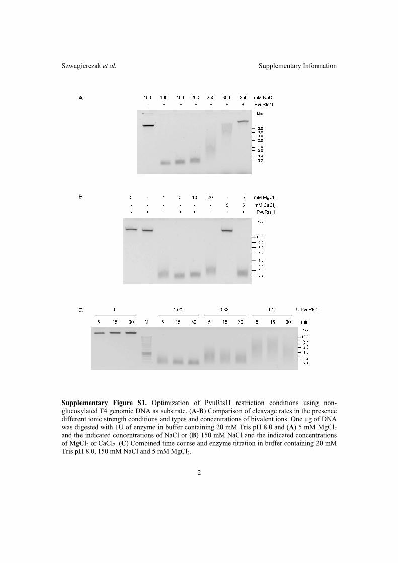
Szwagierczak et al. Supplementary Information�
2 �
Supplementary Figure S1. Optimization of PvuRts1I restriction conditions using non-glucosylated T4 genomic DNA as substrate. (A-B) Comparison of cleavage rates in the presence different ionic strength conditions and types and concentrations of bivalent ions. One µg of DNA was digested with 1U of enzyme in buffer containing 20 mM Tris pH 8.0 and (A) 5 mM MgCl2 and the indicated concentrations of NaCl or (B) 150 mM NaCl and the indicated concentrations of MgCl2 or CaCl2. (C) Combined time course and enzyme titration in buffer containing 20 mM Tris pH 8.0, 150 mM NaCl and 5 mM MgCl2.
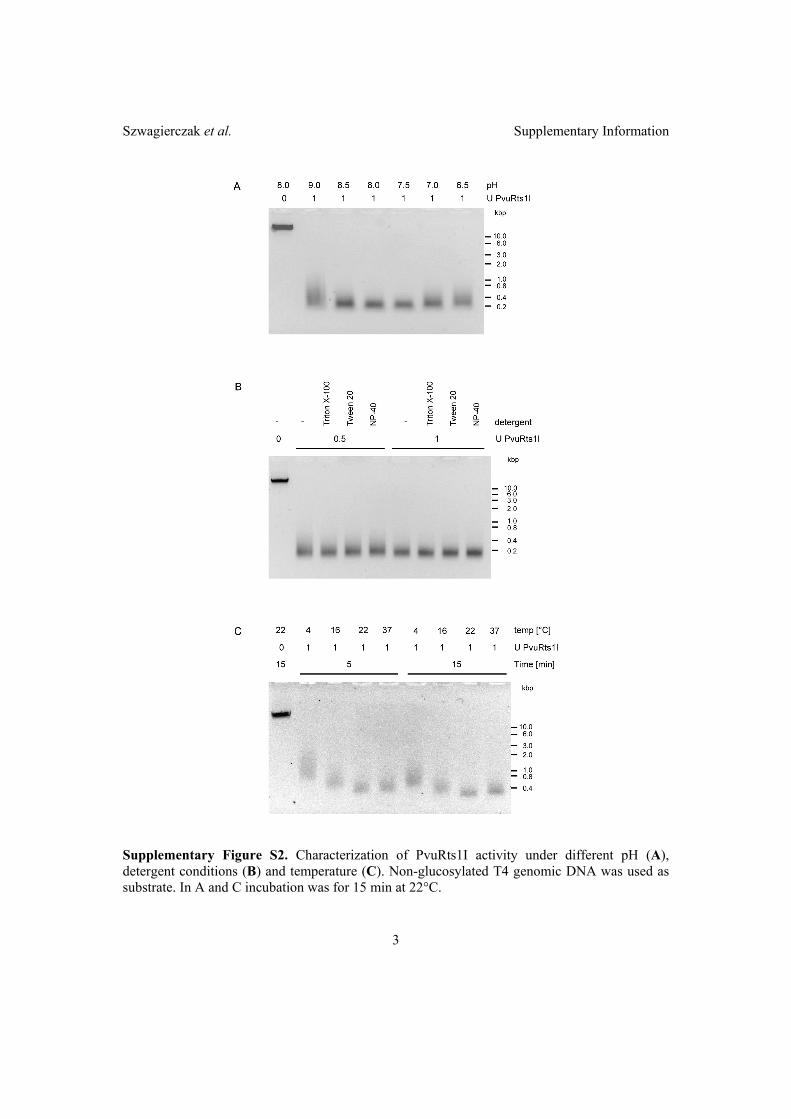
Szwagierczak et al. Supplementary Information�
3��
Supplementary Figure S2. Characterization of PvuRts1I activity under different pH (A), detergent conditions (B) and temperature (C). Non-glucosylated T4 genomic DNA was used as substrate. In A and C incubation was for 15 min at 22°C.
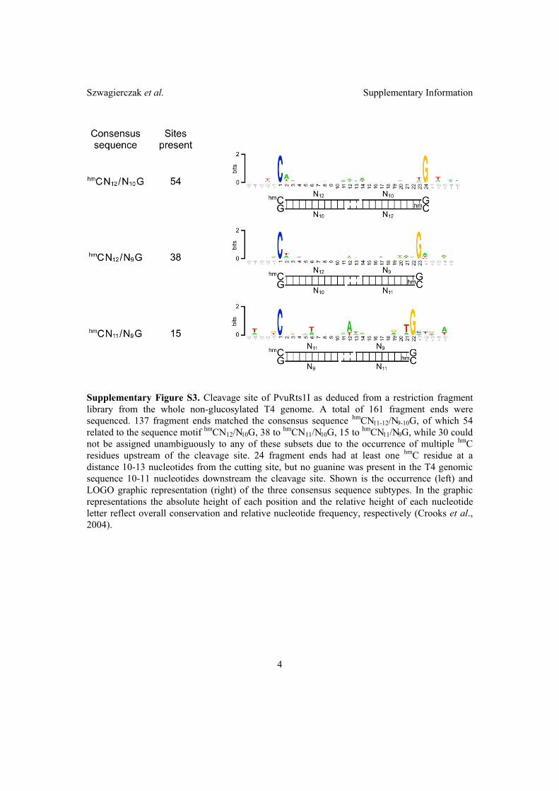
Szwagierczak et al. Supplementary Information�
4��
Supplementary Figure S3. Cleavage site of PvuRts1I as deduced from a restriction fragment library from the whole non-glucosylated T4 genome. A total of 161 fragment ends were sequenced. 137 fragment ends matched the consensus sequence hmCN11-12/N9-10G, of which 54 related to the sequence motif hmCN12/N10G, 38 to hmCN11/N10G, 15 to hmCN11/N9G, while 30 could not be assigned unambiguously to any of these subsets due to the occurrence of multiple hmC residues upstream of the cleavage site. 24 fragment ends had at least one hmC residue at a distance 10-13 nucleotides from the cutting site, but no guanine was present in the T4 genomic sequence 10-11 nucleotides downstream the cleavage site. Shown is the occurrence (left) and LOGO graphic representation (right) of the three consensus sequence subtypes. In the graphic representations the absolute height of each position and the relative height of each nucleotide letter reflect overall conservation and relative nucleotide frequency, respectively (Crooks et al., 2004).

Szwagierczak et al. Supplementary Information�
5��
Supplementary Figure S4. Sequences form the T4 genomic 1139 bp fragment cut by PvuRts1I that deviate from the predicted consensus sequence hmC N11-12 / N9-10 G. All cytosine residues are hydroxymethylated but for simplicity they are here indicated as Cs. hmC and guanine residues 11-13 nucleotides upstream of and 9-10 nucleotides downstream to the cleavage site, respectively, are highlighted in red. Residues 21-23 nucleotides downstream of a hmC are shaded in light red.
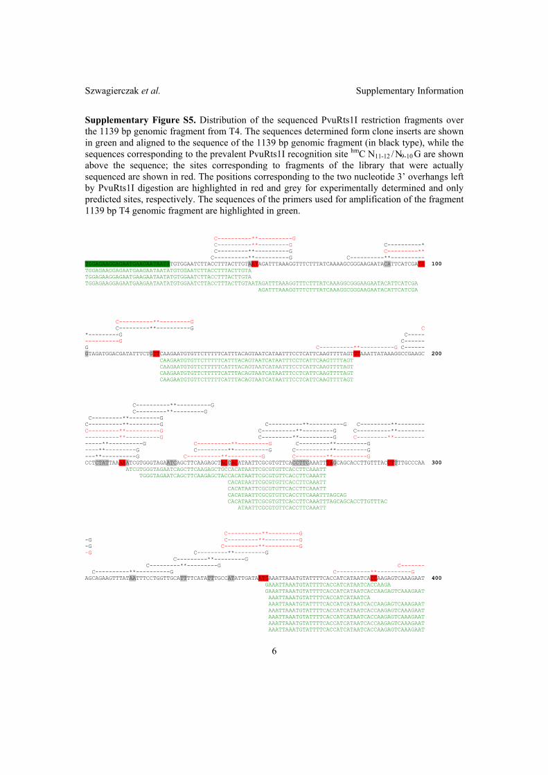
Szwagierczak et al. Supplementary Information�
6��
Supplementary Figure S5. Distribution of the sequenced PvuRts1I restriction fragments over the 1139 bp genomic fragment from T4. The sequences determined form clone inserts are shown in green and aligned to the sequence of the 1139 bp genomic fragment (in black type), while the sequences corresponding to the prevalent PvuRts1I recognition site hmC N11-12 / N9-10 G are shown above the sequence; the sites corresponding to fragments of the library that were actually sequenced are shown in red. The positions corresponding to the two nucleotide 3’ overhangs left by PvuRts1I digestion are highlighted in red and grey for experimentally determined and only predicted sites, respectively. The sequences of the primers used for amplification of the fragment 1139 bp T4 genomic fragment are highlighted in green. C----------**----------G C----------**---------G C----------* C---------**----------G C---------** C----------**----------G C----------**---------- TGGAGAAGGAGAATGAAGAATAATATGTGGAATCTTACCTTTACTTGTAATAGATTTAAAGGTTTCTTTATCAAAAGCGGGAAGAATACATTCATCGATA 100 TGGAGAAGGAGAATGAAGAATAATATGTGGAATCTTACCTTTACTTGTA TGGAGAAGGAGAATGAAGAATAATATGTGGAATCTTACCTTTACTTGTA TGGAGAAGGAGAATGAAGAATAATATGTGGAATCTTACCTTTACTTGTAATAGATTTAAAGGTTTCTTTATCAAAGGCGGGAAGAATACATTCATCGA AGATTTAAAGGTTTCTTTATCAAAGGCGGGAAGAATACATTCATCGA C----------**---------G C---------**----------G C *---------G C----- ----------G C------ G C----------**----------G C------ GTAGATGGACGATATTTCTGTTCAAGAATGTGTTCTTTTTCATTTACAGTAATCATAATTTCCTCATTCAAGTTTTAGTGTAAATTATAAAGGCCGAAGC 200 CAAGAATGTGTTCTTTTTCATTTACAGTAATCATAATTTCCTCATTCAAGTTTTAGT CAAGAATGTGTTCTTTTTCATTTACAGTAATCATAATTTCCTCATTCAAGTTTTAGT CAAGAATGTGTTCTTTTTCATTTACAGTAATCATAATTTCCTCATTCAAGTTTTAGT CAAGAATGTGTTCTTTTTCATTTACAGTAATCATAATTTCCTCATTCAAGTTTTAGT C----------**----------G C---------**---------G C---------**---------G C----------**---------G C----------**----------G C---------**-------- C---------**----------G C----------**---------G C----------**-------- ----------**----------G C---------**----------G C---------**--------- -----**----------G C----------**---------G C---------**---------G ----**---------G C---------**----------G C----------**---------G ---**----------G C----------**----------G C---------**----------G CCTCTATTAAAAATCGTGGGTAGAATCAGCTTCAAGAGCTACCACATAATTCGCGTGTTCACCTTCAAATTTAGCAGCACCTTGTTTACCTTTTGCCCAA 300 ATCGTGGGTAGAATCAGCTTCAAGAGCTGCCACATAATTCGCGTGTTCACCTTCAAATT TGGGTAGAATCAGCTTCAAGAGCTACCACATAATTCGCGTGTTCACCTTCAAATT CACATAATTCGCGTGTTCACCTTCAAATT CACATAATTCGCGTGTTCACCTTCAAATT CACATAATTCGCGTGTTCACCTTCAAATTTAGCAG CACATAATTCGCGTGTTCACCTTCAAATTTAGCAGCACCTTGTTTAC ATAATTCGCGTGTTCACCTTCAAATT C----------**---------G -G C---------**----------G -G C----------**----------G -G C---------**---------G C---------**---------G C---------**---------G C------- C----------**----------G C----------**----------G AGCAGAAGTTTATAATTTCCTGGTTGCATTTTCATATTTGCCATATTGATAATGAAATTAAATGTATTTTCACCATCATAATCACCAAGAGTCAAAGAAT 400 GAAATTAAATGTATTTTCACCATCATAATCACCAAGA� GAAATTAAATGTATTTTCACCATCATAATCACCAAGAGTCAAAGAAT� AAATTAAATGTATTTTCACCATCATAATCA� AAATTAAATGTATTTTCACCATCATAATCACCAAGAGTCAAAGAAT� AAATTAAATGTATTTTCACCATCATAATCACCAAGAGTCAAAGAAT AAATTAAATGTATTTTCACCATCATAATCACCAAGAGTCAAAGAAT AAATTAAATGTATTTTCACCATCATAATCACCAAGAGTCAAAGAAT AAATTAAATGTATTTTCACCATCATAATCACCAAGAGTCAAAGAAT
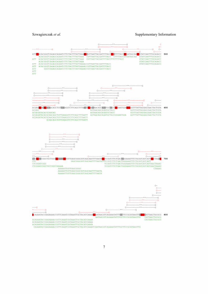
Szwagierczak et al. Supplementary Information�
7��
C----------**---------G C C---------**----------G C- ---**----------G C----------**---------G C----------**----------G C- C---------**----------G C----------**----------G C----- ATTTAACACGGGTCAGAGCAGAATCTTCTACTTTATTAAAACCGTTAATTACGATTTTACCTTCTTTTACCGTGATAGCAATTGTATCAATTTGCAGACC 500 ACACGGGTCAGAGCAGAATCTTCTACTTTATTAAA CGTTAATTACGATTTTACC CTTTTACCGTGATAGCAA GTATCAATTTGCAGACC ATT ACACGGGTCAGAGCAGAATCTTCTACTTTATTAAA CGTTAATTACGATTTTACCTTCTTTTACC GTATCAATTTGCAGACC ACACGGGTCAGAGCAGAATCTTCTACTTTATTAAA GTATCAATTTGCAGACC ATT ACACGGGTCAGAGCAGAATCTTCTACTTTATTAAAACCGTTAATTACGATTTTACC GTATCAATTTGCAGACC ATT ACACGGGTCAGAGCAGAATCTTCTACTTTATTAAAACCGTTAATTACGATTTTACC ATT GGGTCAGAGCAGAATCTTCTACTTTATTAAAACCGTTAATTACGATTTTACC ATT ATT C----------**----------G C----------**---------G C----------**----------G C---------**----------G C---------**---------G C----------**---------G C----------**---------G C---------**----------G C---------**----------G ---------**---------G C---------**---------G ---------**---------G C----------**----------G C----------**----------G C---- --------**----------G C----------**----------G C----------**---------G C------- -----**----------G C----------**----------G C---------**----------G C---------- ACGAGATACACGCAACAGCTGTTGAAGGTCTTCAGCTTTAATTTCAGTAACAGCAGATGCTACCGGGAATGGAATTGGTTTATTAGGAGCAACTACTGTA 600 ACGAG AGTAACAGCAGATGCTACC GGTTTATTAGGAGCAACTACTGTA ACGAGATACACGCAACAG AGTAACAGCAGATGCTACC GGTTTATTAGGAGCAACTACTGTA ACGAGATACACGCAACAGCTGTTGAAGGTCTTCAGCTTTAATT AGTAACAGCAGATGCTACCGGGAATGGA GGTTTATTAGGAGCAACTACTGTA ACGAGATACACGCAACAGCTGTTGAAGGTCTTCAGCTTTAATT GCAACAGCTGTTGAAGGTCTTCAGCTTTAATT C----------**----------G C----------**---------G C---------**----------G C---------**---------G C----------**---------G C---------**----------G C----------**----------G -----**---------G C---------**------ ---**----------G C----------**----------G C----------* **----------G C----------**----------G C---------** CTCGGATCGGCTGCTGGCCAAAAAATTGTTGAGCGGGCATCAGCAATTTTAATATTTCCATCTTCTGACTGGGAAATTTCTGCATCATCATTAACTAAAG 700 CTC GAGCGGGCATCAGCAATTTTAATATTTCCATCTTCTGACTGGGAAATTTCTGCATCATCATT CTCGGATCGGC TCCATCTTCTGACTGGGAAATTTCTGCATCATCATTAACTAAAG CTCGGATCGGCTGCTGGCCAAAAA TCCATCTTCTGACTGGGAAATTTCTGCATCATCATTAACTAAAG AAAAATTGTTGAGCGGGC CTAAAG AAAAATTGTTGAGCGGGCATCAGCAATTTTAATA AAAAATTGTTGAGCGGGCATCAGCAATTTTAATA C- C--- C--- C--- C------ C----------**---------G C---------**----------G C----------**---------G C---------**----------G ---G C----------**----------G *---------G C----------**----------G ----------G C----------**----------G ACAGAATACCGAGAAAACCGTTCAAATCGTAAATTGCTACATCAAAATCAATAACGTCAGAAATATTTGCTTCCGCATAAGTTGTACCATTAACTGCGCG 800 AATAACGTCAGAAATATTTGCTTCCGCATAAGTTG CATTAACTGCGCG ACAGAATACCGAGAAAACCGTTCAAATCATAAATTGCTACATCAAAA CATTAACTGCGCG ACAGAATACCGAGAAAACCGTTCAAATCATAAATTGCTACATCAAAA ACAGAATACCGAGAAAACCGTTCAAATCATAAATTGCTACATCAAAA CAGAATACCGAGAAAACCGTTCAAATCGTAAATTGCTACATCAAAATCAATAACGTCAGAAATATTTGCTTCCGCATAAGTTG

Szwagierczak et al. Supplementary Information�
8��
--------**---------G -------**----------G -------**---------G ------**----------G ---**---------G C----------**---------G C---------**----------G C----------**---------G C---------**----------G C----------**----------G C---------**---------G C----------**----------G C----------**----------G C----------**----------G AGTCATAATAAATTGACCGGATTTAAGCATAATACCAGAGTTAATAGTAGCGAAATTTTTAAGCAGAGCAGTAGTATCTTTAGACAGTTTCATGTAATTT 900 AGTCATA GCAGAGCAGTAGTATCTTTAGACAGTTTCATGTAATTT AGTCATA TCTTTAGACAGTTTCATGTAATTT TCTTTAGACAGTTTCATGTAATTT C----------**----------G C---------**---------G C----------**---------G C------- C---------**----------G C---------**----- C----------**---------G C----------**----------G C---------**----------G C---------**---------G CCTTCAATTCAAATGAGATTTAATTTTATAACTAATTTAATAAAGCAATTAACGATTAAAATCAGCCGCAATTGTTTCCGCAACAATTTGAGCAGCAACA 1000 C AAAATCAGCCGCAATTGTTTCCGCAACAATTTGAGCAGCAACA CCTTCAATTCAAATGAGATTTAATTTTATAACTAATTTAAT AAATCAGCCGCAATTGTTTC CCTTCAATTCAAATGAGATTTAATTTTATAACTAATTTAATAAAGCAATTAACG GCCGCAATTGTTTCCGCAACAATTTGAGC CAATTGTTTCCGCAACAATTTGAGC AACAATTTGAGCAGCAACA C----------**----------G C---------**---------G ---**----------G C----------**----------G C----------**----------G ----G C---------**---------G C---------**---------G C----------**---------G C---------**---------G C-------- C---------**----------G C----------**----------G C---------**--------- ATTAGACGTTCATCTGCATTACCGCAATAATCATCTTCAAGGCGTTCACCACATGAAGTCATAATAAATTTAGCACCGGCGTTTAGGGATTCTGTAGTAT 1100 ATT CTTCAAGGCGTTCACCACATGAAGTC CTGTAGTAT CTGTAGTAT CTGTAGTAT CTGTAGTAT ATT CTGTAGTAT CTGTAGTAT CTGTAGTAT CTGTAGTAT GTAT AT -**---------G G GTTTGCGCATTAGTTCAATCCATTTATTACTTACTTCAC 1139 GTTTGCGCATTAGTTCAATCCATTTATTACTTACTTCAC GTTTGCGCATTAGTTCAATCCATTTATTACTTACTTCAC GTTTGCGCATTAGTTCAATCCATTTATTACTTACTTCAC GTTTGCGCATTAGTTCAATCCATTTATTACTTACTTCAC GTTTGCGCATTAGTTCAATCCATTTATTACTTACTTCAC GTTTGCGCATTAGTTCAATCCATTTATTACTTACTTCAC GTTTGCGCATTAGTTCAATCCATTTATTACTTACTTCAC GTTTGCGCATTAGTTCAATCCATTTATTACTTACTTCAC GTTTGCGCATTAGTTCAATCCATTTATTACTTACTTCAC GTTTGCGCATTAGTTCAATCCATTTATTACTTACTTCAC

Szwagierczak et al. Supplementary Information�
9��
Supplemental Figure S6. Analysis of sequences from the T4 genomic 1139 bp fragment matching the PvuRts1I consensus cleavage site hmCN11-12/N9-10G that were not found among the sequenced fragments. In the LOGO graphic representations on the right the absolute height of each position and the relative height of each nucleotide letter reflect overall conservation and relative nucleotide frequency, respectively (Crooks et al., 2004).
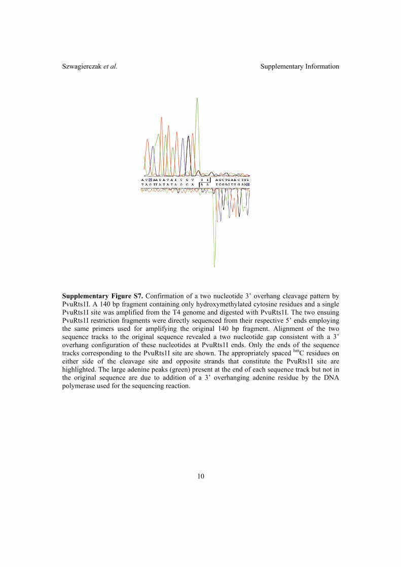
Szwagierczak et al. Supplementary Information�
10��
Supplementary Figure S7. Confirmation of a two nucleotide 3’ overhang cleavage pattern by PvuRts1I. A 140 bp fragment containing only hydroxymethylated cytosine residues and a single PvuRts1I site was amplified from the T4 genome and digested with PvuRts1I. The two ensuing PvuRts1I restriction fragments were directly sequenced from their respective 5’ ends employing the same primers used for amplifying the original 140 bp fragment. Alignment of the two sequence tracks to the original sequence revealed a two nucleotide gap consistent with a 3’ overhang configuration of these nucleotides at PvuRts1I ends. Only the ends of the sequence tracks corresponding to the PvuRts1I site are shown. The appropriately spaced hmC residues on either side of the cleavage site and opposite strands that constitute the PvuRts1I site are highlighted. The large adenine peaks (green) present at the end of each sequence track but not in the original sequence are due to addition of a 3’ overhanging adenine residue by the DNA polymerase used for the sequencing reaction.
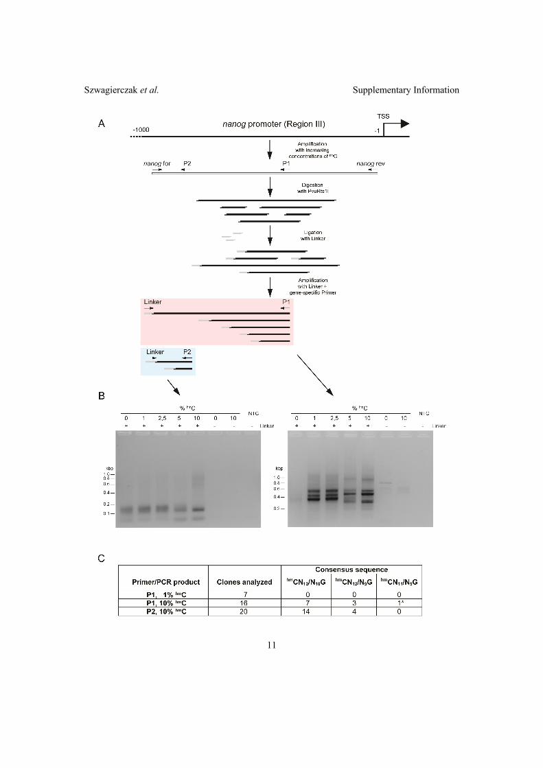
Szwagierczak et al. Supplementary Information�
11��
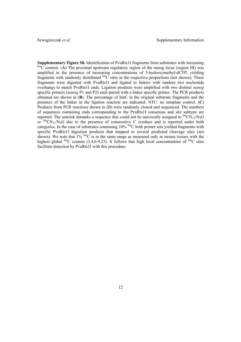
Szwagierczak et al. Supplementary Information�
12��
Supplementary Figure S8. Identification of PvuRts1I fragments from substrates with increasing hmC content. (A) The proximal upstream regulatory region of the nanog locus (region III) was amplified in the presence of increasing concentrations of 5-hydroxymethyl-dCTP, yielding fragments with randomly distributed hmC sites in the respective proportions (not shown). These fragments were digested with PvuRts1I and ligated to linkers with random two nucleotide overhangs to match PvuRts1I ends. Ligation products were amplified with two distinct nanog specific primers (nanog P1 and P2) each paired with a linker specific primer. The PCR products obtained are shown in (B). The percentage of hmC in the original substrate fragments and the presence of the linker in the ligation reaction are indicated. NTC: no template control. (C) Products from PCR reactions shown in (B) were randomly cloned and sequenced. The numbers of sequences containing ends corresponding to the PvuRts1I consensus and site subtype are reported. The asterisk demarks a sequence that could not be univocally assigned to hmCN12/N9G or hmCN11/N9G due to the presence of consecutive C residues and is reported under both categories. In the case of substrates containing 10% hmC both primer sets yielded fragments with specific PvuRts1I digestion products that mapped to several predicted cleavage sites (not shown). We note that 1% hmC is in the same range as measured only in mouse tissues with the highest global hmC content (3,4,6-9,23). It follows that high local concentrations of hmC sites facilitate detection by PvuRts1I with this procedure.

Szwagierczak et al. Supplementary Information�
13��
SUPPLEMENTARY METHODS Generation of fragments from the nanog upstream region III containing known levels of hmC, PvuRts1I digestion and identification of digestion products. Genomic DNA from JM8A3.N1 ESCs (EUCOMM, Helmholtz Center Munich, Neuherberg, Germany) was isolated using the NucleoSpin Triprep Kit (Macherey-Nagel). To prepare substrates containing different hmC levels (0%, 1%, 2,5%, 5%, 10%), genomic DNA from JM8A3.N1 cells was used as a template to amplify a 867 bp fragment from region III of the nanog promoter (Hattori et al, Genes to cell, 2007) using corresponding ratios of 5-hydroxymethyl-dCTP (Bioline GmbH) and dCTP, Phusion HF DNA Polymerase (Finnzymes) and the following primers: nanog for 5´-TCA GGA GTT TGG GAC CAG CTA-3´ and nanog rev 5´-CCC CCC TCA AGC CTC CTA-3´. After purification of the PCR fragments using the NucleoSpin Extract II kit (Macherey-Nagel), 250ng of each fragment was digested with 2U of PvuRTS1I for 15min at 22°C and the enzyme was heat inactivated at 60°C for 20 min. Twentyfive nanograms of digested fragment were ligated to a linker containing random two nucleotide 3´ overhangs, generated by annealing the following primers: For 5´-CTC GTA GAC TGC GTA CCA TG NN-3´ and Rev 5´-CA TGG TAC GCA GTC TAC CAG-3´. The ligation reaction was carried out using T4 DNA Ligase (NEB) overnight at 16°C. As a control for ligation specificity, each fragment was ligated in the absence of the linker. To selectively amplify fragments cut by PvuRTS1I, the ligated products were amplified by PCR with Phusion HF DNA Polymerase (Finnzymes) using a linker specific forward primer (For 5´-CTC GTA GAC TGC GTA CCA TG-3´) and nanog specific reverse primers (P2: 5´-GAG TCA GAC CTT GCT GCC AAA-3´ and P1: 5´-GCC GTC TAA GCA ATG GAA GAA-3´). Libraries of digested and ligated fragments containing 1 and 10% hmC were generated using the Zero Blunt® PCR Cloning Kit (Invitrogen). Randomly selected clones were sequenced and analyzed for the presence of PvuRts1I ends.

2.3 Di↵erent binding properties and function of CXXC
zinc finger domains in Dnmt1 and Tet1
Different Binding Properties and Function of CXXC ZincFinger Domains in Dnmt1 and Tet1Carina Frauer1., Andrea Rottach1., Daniela Meilinger1, Sebastian Bultmann1, Karin Fellinger1¤a, Stefan
Hasenoder1¤b, Mengxi Wang1, Weihua Qin1, Johannes Soding2, Fabio Spada1*, Heinrich Leonhardt1*
1 Department of Biology II and Center for Integrated Protein Science Munich (CIPSM), Ludwig Maximilians University Munich, Planegg, Germany, 2 Gene Center Munich,
Ludwig Maximilians University Munich, Munich, Germany.
Abstract
Several mammalian proteins involved in chromatin and DNA modification contain CXXC zinc finger domains. We comparedthe structure and function of the CXXC domains in the DNA methyltransferase Dnmt1 and the methylcytosine dioxygenaseTet1. Sequence alignment showed that both CXXC domains have a very similar framework but differ in the central tip region.Based on the known structure of a similar MLL1 domain we developed homology models and designed expression constructsfor the isolated CXXC domains of Dnmt1 and Tet1 accordingly. We show that the CXXC domain of Tet1 has no DNA bindingactivity and is dispensable for catalytic activity in vivo. In contrast, the CXXC domain of Dnmt1 selectively binds DNA substratescontaining unmethylated CpG sites. Surprisingly, a Dnmt1 mutant construct lacking the CXXC domain formed covalentcomplexes with cytosine bases both in vitro and in vivo and rescued DNA methylation patterns in dnmt12/2 embryonic stemcells (ESCs) just as efficiently as wild type Dnmt1. Interestingly, neither wild type nor DCXXC Dnmt1 re-methylated imprintedCpG sites of the H19a promoter in dnmt12/2 ESCs, arguing against a role of the CXXC domain in restraining Dnmt1methyltransferase activity on unmethylated CpG sites.
Citation: Frauer C, Rottach A, Meilinger D, Bultmann S, Fellinger K, et al. (2011) Different Binding Properties and Function of CXXC Zinc Finger Domains in Dnmt1and Tet1. PLoS ONE 6(2): e16627. doi:10.1371/journal.pone.0016627
Editor: Anton Wutz, Wellcome Trust Centre for Stem Cell Research, United Kingdom
Received September 14, 2010; Accepted January 5, 2011; Published February 2, 2011
Copyright: ! 2011 Frauer et al. This is an open-access article distributed under the terms of the Creative Commons Attribution License, which permitsunrestricted use, distribution, and reproduction in any medium, provided the original author and source are credited.
Funding: This work was supported by grants from the Deutsche Forschungsgemeinschaft (DFG, SFB646 and TR5; http://www.dfg.de/en/index.jsp), theNanosystem Initiative Munich (NIM; http://www.nano-initiative-munich.de/) and the Center for NanoScience (CeNS; http://www.cens.de/) to HL. The funders hadno role in study design, data collection and analysis, decision to publish, or preparation of the manuscript.
Competing Interests: The authors have declared that no competing interests exist.
* E-mail: [email protected] (FS); [email protected] (HL)
. These authors contributed equally to this work.
¤a Current address: Intervet International GmbH, Unterschleissheim, Germany¤b Current address: Institute of Stem Cell Research, Helmholtz Zentrum Munchen, Neuherberg, Germany
Introduction
In mammals DNA methylation is restricted to cytosine residuesand mainly involves CpG dinucleotides. CpG methylation iswidespread across mammalian genomes, including gene bodiesregardless of their transcriptional activity [1–4]. However, highlyCpG-rich regions (CpG islands) are refractory to methylation andmostly coincide with promoters of constitutively active genes. Themethylation state of other regulatory sequences with moderate tolow CpG density, including promoters and enhancers, showsdevelopmental and/or tissue-specific variations and positivelycorrelates with a transcriptionally silent state [1,3–8]. Densemethylation of repetitive sequences is also thought to maintainthese elements in a silent state and thus contribute to genomestability [9–11]. In mammals cytosine methylation is catalyzed bya family of DNA methyltransferases (Dnmts) [12]. Dnmt3a andDnmt3b establish methylation patterns during embryonic devel-opment of somatic as well as germ cell lineages and, consistently,show developmental stage and tissue specific expression patterns.In contrast, Dnmt1 is ubiquitous and generally the most abundantDNA methyltransferase in mammalian tissues, where it associateswith the replication machinery and restores symmetrical methyl-ation at hemimethylated CpG sites generated by the semi-
conservative DNA replication process [13]. Thus, Dnmt1maintains methylation patterns with high fidelity and is essentialfor embryonic development and genome integrity [9,14,15].
Dnmt1 is a large enzyme with a complex domain structure thatlikely evolved by fusion of at least three genes [16]. It comprises aregulatory N-terminal region and a C-terminal catalytic domainconnected by a linker of seven glycine-lysine repeats (Figure 1A)[17].The N-terminal part contains a PCNA binding domain (PBD), aheterochromatin targeting sequence (TS), a CXXC-type zinc fingerdomain and two Bromo-Adjacent Homology domains (BAH1 andBAH2). The C-terminal domains of mammalian Dnmts contain all tencatalytic motifs identified in bacterial DNA (cytosine-5) methyltrans-ferases [12]. Thus, prokaryotic and mammalian cytosine methyltrans-ferases are thought to adopt the same catalytic mechanism. However,the C-terminal domain of Dnmt1 is the only DNA methyltransferasedomain in Dnmts that is not catalytically active when expressedseparately. Indeed, interaction with the N-terminal part is required forallosteric activation of the enzyme [18]. Remarkably, the first 580amino acids (aa) of human DNMT1 are dispensable for bothenzymatic activity and substrate recognition, whereas deletion of thefirst 672 aa results in an inactive enzyme [19]. Interestingly, thistruncation eliminates part of the CXXC domain, suggesting aninvolvement of this domain in allosteric activation. However, addition
PLoS ONE | www.plosone.org 1 February 2011 | Volume 6 | Issue 2 | e16627

CXXC Domains of Dnmt1 and Tet1
PLoS ONE | www.plosone.org 2 February 2011 | Volume 6 | Issue 2 | e16627
of an N-terminal fragment containing the isolated CXXC domain tothe catalytic domain was not sufficient for catalytic activation [20].
CXXC-type zinc finger domains are found in several otherproteins with functions related to DNA or chromatin modification,including the histone H3 lysine 4 (H3K4) methyltransferases mixed-lineage leukaemia (MLL) proteins 1 and 4, the CpG-binding protein(CGBP, also known as Cfp1 or CXXC1), the methyl-CpG bindingdomain protein 1 (MBD1), the H3 lysine 36 (H3K36) demethylasesKDM2A and B (also known as JHD1A/FBXL11 and JHD1B/FBXL10) and the MLL1 fusion partner TET1 (Figure 1A) [21–28].The CXXC domains of some of these proteins were shown tomediate specific binding to double stranded DNA templatescontaining unmethylated CpG sites [21,22,29,30]. A region ofDnmt1 which mainly includes the CXXC domain (aa 628–753) wasalso shown to bind Zn ions and DNA [20,31,32]. However, availabledata on the selectivity of this DNA binding activity are conflicting.Whereas a fragment including aa 613–748 of mouse Dnmt1 wasshown to bind DNA with a slight preference for hemimethylatedCpG sites [20], aa 645–737 of human DNMT1 were shown toselectively bind unmethylated DNA [32]. As these studies useddifferent constructs and species, the selectivity of DNA binding bythe CXXC domain of Dnmt1 with regard to CpG methylation stateand the role of the CXXC domain in allosteric activation andsubstrate discrimination remain to be firmly established.
Notably, not all CXXC domains show DNA binding activity, asexemplified by the fact that only one out of three CXXC domainsin MBD1 binds DNA [29]. Interestingly, TET1 was recentlyshown to be a 2 oxoglutarate- and Fe(II)-dependent dioxygenaseresponsible for converting genomic 5-methylcytosine (mC) to 5-hydroxymethylcytosine (hmC) [33,34]. However, it is not knownwhether the CXXC domain of TET1 is involved in recognition ofmethylated DNA substrates.
Here we report a functional study and characterization of theDNA binding activity for the CXXC domains of mouse Dnmt1and Tet1 proteins. We generated isolated CXXC domain anddeletion constructs based on structural homology models tominimize structural alterations. We show that the CXXC domainof Dnmt1 preferentially binds DNA substrates containingunmethylated CpG sites, but does not contribute significantly tothe DNA binding properties of the full length enzyme and isdispensable for its catalytic activity in vitro and in vivo. In addition,we found that the CXXC domain of Tet1 does not bind DNA invitro and is also dispensable for catalytic activity of Tet1 in vivo.
Results
Sequence homology and structural modeling identifydistinct CXXC domain subtypes
Dnmt1 contains a zinc finger domain of the CXXC type, whichis present in several mammalian proteins including MLL1
(Figure 1A–C) and is highly conserved among Dnmt1 sequencesfrom various animal species (Figure S1 in File S1). The primarystructure of CXXC domains spans two clusters of 6 and 2 cysteineresidues separated by a stretch of variable sequence and length.Sequence alignment and homology tree construction identifiedthree distinct groups of CXXC domains (Figure 1B and C). Thesequence between the two cysteine clusters in the CXXC domainsof Dnmt1, CGBP/Cfp1, Fbxl19, Mll1, Mll2 and Kdm2 proteinsand CXXC domain 3 of Mbd1 is highly conserved and contains aKFGG motif. The two other homology groups, including theCXXC domains 1 and 2 of Mbd1 on one side and those of Tet1,Cxxc4/Idax, Cxxc5/RINF and Cxxc10 on the other side, lack theKFGG motif and diverge from the first group and from each otherin the sequence between the cysteine clusters. We generatedstructural homology models for the CXXC domains of mouseDnmt1 and Tet1 using the NMR structure of the MLL1 CXXCdomain as a template (Figure 1D and E)[35]. The CXXC domainsof these proteins adopt an extended crescent-like structure thatincorporates two Zn2+ ions each coordinated by four cysteineresidues. The peptide of the MLL1 CXXC domain predicted toinsert into the major groove of the DNA double helix (cyan inFig. 1E) is located on one face of the structure and is contiguous tothe KFGG motif [35]. The predicted structure of the Tet1 CXXCdomain lacks the short 310 helix (g1 in Figure 1E) formed byresidues PKF and partially overlapping the KFGG motif, but issimilar to the MLL1 CXXC domain in the region of the DNA-contacting peptide. However, each of the two predicted b-strandsin Tet1 carries three positive charges, whereas there is only one orno charged residue in the C-terminal strands of the CXXCdomains in MLL1 and Dnmt1. Depending on the orientation ofthe positively charged side chains, it cannot be excluded that thecharge density prevents strand pairing in the Tet1 CXXC domain.
The Dnmt1 CXXC domain binds unmethylated DNATo investigate the binding properties of the Dnmt1 CXXC
domain, we generated a GFP fusion construct including aa 652–699(GFP-CXXCDnmt1). According to our homology model the ends ofthis fragment form an antiparallel b-sheet that structurally delimitsthe domain as in MLL1. We first compared the localization andmobility of GFP-CXXCDnmt1 and GFP in mouse C2C12myoblasts. While GFP was diffusely distributed in both nucleusand cytoplasm, GFP-CXXCDnmt1 was exclusively nuclear with apunctuated pattern throughout the nucleoplasm and was enrichedin nucleoli, a pattern independent of cell cycle stage (Figure 2A andFigure S2 in File S1). Enrichment in the nucleus and nucleoli isfrequently observed with constructs containing stretches with highdensity of basic residues. After photobleaching half of the nuclearvolume we observed a slower fluorescence recovery rate for GFP-CXXCDnmt1 than for GFP (Figure 2B). To rule out a contribution ofnucleolar interactions to the slower kinetics of GFP-CXXCDnmt1,
Figure 1. Sequence and predicted structural homology of CXXC domains. (A) Schematic representation of the domain structure in Dnmt1 andTet1. The catalytic domain and the N-terminal region of Dnmt1 are connected by seven lysine-glycine repeats [(KG)7]. PBD: PCNA binding domain; TS:targeting sequence; CXXC: CXXC-type zinc finger domain; BAH1 and 2: bromo-adjacent homology domain; NLS: nuclear localization signal; Cys-rich:cysteine rich region. (B) Alignment of mammalian CXXC domains. Numbers on the right side indicate the position of the last amino acid in thecorresponding protein. The Mbd1a isoform contains three CXXC motifs (Mbd1_1-3). Absolutely conserved residues, including the eight cysteinesinvolved in zinc ion coordination are highlighted in red and the conserved KFGG motif is in red bold face. Positions with residues in red face share 70%similarity as calculated with the Risler algorithm [66]. At the top residues of MLL1 involved in b sheets b1 and b2 (black arrows), a helices a1 and a2 andstrict a turns (TTT) are indicated. All sequences are from M. musculus. Accession numbers (for GenBank unless otherwise stated): Dnmt1, NP_034196;Mll1, NP_001074518; Mll4, O08550 (SwissProt); CGBP, NP_083144; Kdm2a, NP_001001984; Kdm2b, NP_001003953; Fbxl19, NP_766336; Mbd1,NP_038622; CXXC4/Idax, NP_001004367; CXXC5, NP_598448; CXXC10 (see Materials and Methods). (C) A homology tree was generated from thealignment in (B). The three subgroups of CXXC domains identified are in different colors. Average distances between the sequences are indicated. (D–E)Homology models of the mouse Dnmt1 (D; red) and Tet1 (E; blue) CXXC domains superimposed to the CXXC domain of MLL1 (green; [35]). MLL1residues that were described to contact DNA according to chemical shift measurements [35] are cyan in (E), while cysteines involved in coordination ofthe two zinc ions are yellow. Arrows point to the KFGG motif in MLL1 and Dnmt1. The locations of a helices and b sheets are indicated as in (B).doi:10.1371/journal.pone.0016627.g001
CXXC Domains of Dnmt1 and Tet1
PLoS ONE | www.plosone.org 3 February 2011 | Volume 6 | Issue 2 | e16627
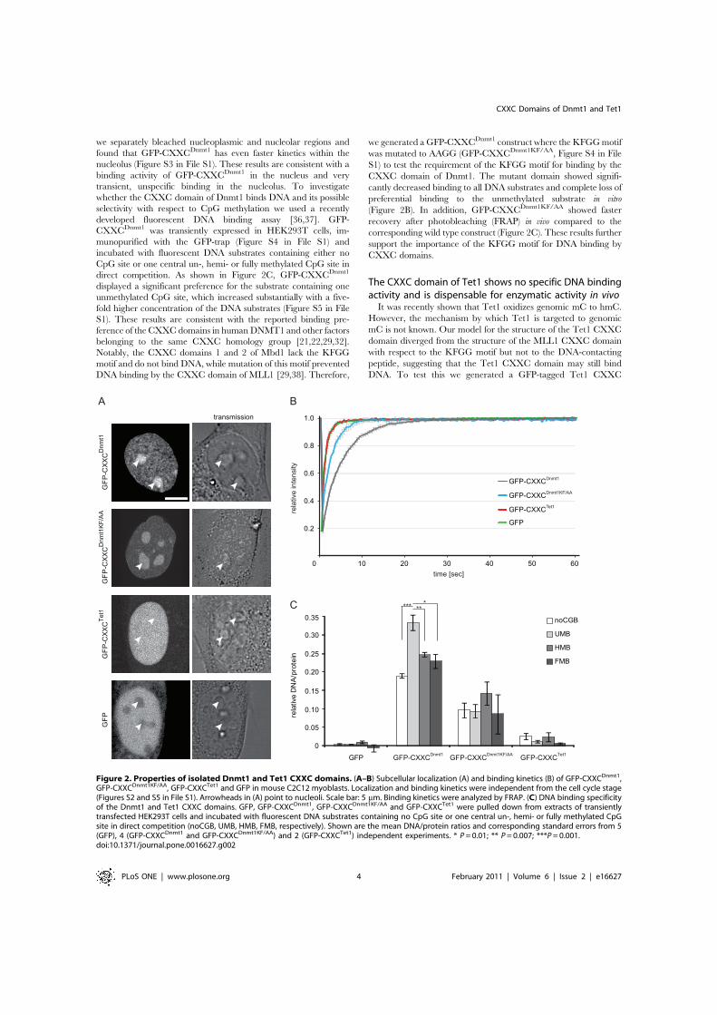
we separately bleached nucleoplasmic and nucleolar regions andfound that GFP-CXXCDnmt1 has even faster kinetics within thenucleolus (Figure S3 in File S1). These results are consistent with abinding activity of GFP-CXXCDnmt1 in the nucleus and verytransient, unspecific binding in the nucleolus. To investigatewhether the CXXC domain of Dnmt1 binds DNA and its possibleselectivity with respect to CpG methylation we used a recentlydeveloped fluorescent DNA binding assay [36,37]. GFP-CXXCDnmt1 was transiently expressed in HEK293T cells, im-munopurified with the GFP-trap (Figure S4 in File S1) andincubated with fluorescent DNA substrates containing either noCpG site or one central un-, hemi- or fully methylated CpG site indirect competition. As shown in Figure 2C, GFP-CXXCDnmt1
displayed a significant preference for the substrate containing oneunmethylated CpG site, which increased substantially with a five-fold higher concentration of the DNA substrates (Figure S5 in FileS1). These results are consistent with the reported binding pre-ference of the CXXC domains in human DNMT1 and other factorsbelonging to the same CXXC homology group [21,22,29,32].Notably, the CXXC domains 1 and 2 of Mbd1 lack the KFGGmotif and do not bind DNA, while mutation of this motif preventedDNA binding by the CXXC domain of MLL1 [29,38]. Therefore,
we generated a GFP-CXXCDnmt1 construct where the KFGG motifwas mutated to AAGG (GFP-CXXCDnmt1KF/AA, Figure S4 in FileS1) to test the requirement of the KFGG motif for binding by theCXXC domain of Dnmt1. The mutant domain showed signifi-cantly decreased binding to all DNA substrates and complete loss ofpreferential binding to the unmethylated substrate in vitro(Figure 2B). In addition, GFP-CXXCDnmt1KF/AA showed fasterrecovery after photobleaching (FRAP) in vivo compared to thecorresponding wild type construct (Figure 2C). These results furthersupport the importance of the KFGG motif for DNA binding byCXXC domains.
The CXXC domain of Tet1 shows no specific DNA bindingactivity and is dispensable for enzymatic activity in vivo
It was recently shown that Tet1 oxidizes genomic mC to hmC.However, the mechanism by which Tet1 is targeted to genomicmC is not known. Our model for the structure of the Tet1 CXXCdomain diverged from the structure of the MLL1 CXXC domainwith respect to the KFGG motif but not to the DNA-contactingpeptide, suggesting that the Tet1 CXXC domain may still bindDNA. To test this we generated a GFP-tagged Tet1 CXXC
Figure 2. Properties of isolated Dnmt1 and Tet1 CXXC domains. (A–B) Subcellular localization (A) and binding kinetics (B) of GFP-CXXCDnmt1,GFP-CXXCDnmt1KF/AA, GFP-CXXCTet1 and GFP in mouse C2C12 myoblasts. Localization and binding kinetics were independent from the cell cycle stage(Figures S2 and S5 in File S1). Arrowheads in (A) point to nucleoli. Scale bar: 5 mm. Binding kinetics were analyzed by FRAP. (C) DNA binding specificityof the Dnmt1 and Tet1 CXXC domains. GFP, GFP-CXXCDnmt1, GFP-CXXCDnmt1KF/AA and GFP-CXXCTet1 were pulled down from extracts of transientlytransfected HEK293T cells and incubated with fluorescent DNA substrates containing no CpG site or one central un-, hemi- or fully methylated CpGsite in direct competition (noCGB, UMB, HMB, FMB, respectively). Shown are the mean DNA/protein ratios and corresponding standard errors from 5(GFP), 4 (GFP-CXXCDnmt1 and GFP-CXXCDnmt1KF/AA) and 2 (GFP-CXXCTet1) independent experiments. * P = 0.01; ** P = 0.007; ***P = 0.001.doi:10.1371/journal.pone.0016627.g002
CXXC Domains of Dnmt1 and Tet1
PLoS ONE | www.plosone.org 4 February 2011 | Volume 6 | Issue 2 | e16627

construct (GFP-CXXCTet1) following the same criteria as for GFP-CXXCDnmt1 and investigated its cellular localization, in vivobinding kinetics and in vitro DNA binding activity. GFP-CXXCTet1
was prevalently nuclear with a homogeneous distribution includ-ing nucleoli that was independent of cell cycle stage (Figure 2A andFigure S6 in File S1). After photobleaching GFP-CXXCTet1
showed very fast recovery kinetics similar to GFP (Figure 2B) andits DNA binding activity in vitro was also similar to the backgroundlevels of the GFP control (Figure 2C). We conclude that theisolated CXXC domain of Tet1 has no specific DNA bindingactivity. Together with the observation that the CXXC domains 1and 2 of Mbd1 also lack the KFGG motif and do not bind DNA[29] and that mutation of this motif reduced DNA binding by theCXXC domains of both Dnmt1 (Figure 2C) and MLL1 [38], thisresult indicates that the KFGG motif is a major determinant forDNA binding by CXXC domains.
To assess whether the CXXC domain is required for catalyticactivity of Tet1 we generated a GFP-Tet1 fusion construct and acorresponding mutant lacking the CXXC domain (GFP-Tet1DCXXC). In C2C12 myoblasts GFP-Tet1 and GFP-Tet1DCXXC showed punctuated nuclear patterns that did notdepend on the cell cycle stage (Figure 3A and data not shown).The same constructs were transfected in HEK293T cells andglobal levels of genomic hmC were measured using a recentlydescribed hmC glucosylation assay [39]. Overexpression of GFP-Tet1 and GFP-Tet1DCXXC determined a similar 5-fold increase ofgenomic hmC levels relative to control samples overexpressingGFP (Figure 3B), indicating that the CXXC domain is notrequired for enzymatic activity of Tet1 in vivo.
Deletion of the CXXC domain does not affect the activityof Dnmt1 in vitro
To explore the role of the CXXC domain in Dnmt1 functionwe generated GFP-Dnmt1 fusion constructs where the CXXCdomain, as defined by our homology model, was deleted. Wereasoned that precise deletion of the entire structure delimited bythe antiparallel b-sheet (Figure 1D) would have the highestchances to preserve native folding of the rest of the protein. Weintroduced this deletion in GFP fusion constructs encoding eitherthe full length Dnmt1 or the isolated N-terminal region (GFP-Dnmt1DCXXC and GFP-NTRDCXXC, respectively; Figure 4A and
Figure S4 in File S1). We then compared DNA binding properties,catalytic activity and interaction between N-terminal region andC-terminal catalytic domain of DCXXC and corresponding wildtype constructs. Competitive DNA binding assays with the sameset of substrates as used for the experiments with GFP-CXXCDnmt1 and GFP-CXXCTet1 reported above (Figure 2C)showed that both GFP-Dnmt1 and GFP-Dnmt1DCXXC bind DNAindependently of the presence and methylation state of a CpG site(Figure 4B). As the isolated CXXC domain preferentially boundthe substrate containing an unmethylated CpG site, the result withGFP-Dnmt1 and GFP-Dnmt1DCXXC indicates that the CXXCdomain contributes negligibly to the DNA binding specificity ofthe full-length enzyme.
Several groups reported that interaction between the N-terminal region and the C-terminal catalytic domain of Dnmt1leads to allosteric activation of Dnmt1 [16,18–20,40]. To testwhether the CXXC domain is involved in this intramolecularinteraction, we co-expressed either GFP-tagged N-terminal region(GFP-NTR) or GFP-NTRDCXXC constructs with a Cherry- andHis-tagged C-terminal domain (Ch-CTD-His) in HEK293T cellsand performed co-immunoprecipitation experiments. Ch-CTD-His co-precipitated both GFP-NTR and GFP-NTRDCXXC,indicating that the CXXC domain is dispensable for theinteraction between the N-terminal region and the C-terminaldomain of Dnmt1 (Figure 4C).
To investigate whether the CXXC domain is needed forenzymatic activity or substrate recognition, we tested formation ofthe covalent complex with cytosine and transfer of the methylgroup for GFP-Dnmt1 and GFP-Dnmt1DCXXC. We first employedan assay to monitor covalent complex formation that exploits theformation of an irreversible covalent bond between the enzymeand the mechanism-based inhibitor 5-aza-2-deoxycytosine (5-aza-dC). This results in permanent trapping of the enzyme by DNAsubstrates containing 5-aza-dC, as opposed to the reversiblecomplex formed with substrates containing the natural substrate 2-deoxycytosine (dC) [36]. GFP-Dnmt1 and GFP-Dnmt1DCXXC
were incubated with fluorescent DNA substrates containing eitherdC (binding) or 5-aza-dC (trapping) at a single CpG site in directcompetition. DNA-protein complexes were then isolated by GFPpulldown and molar DNA/protein ratios were calculated fromfluorescence measurements (Figure 4D). Covalent complex
Figure 3. Cellular localization and in vivo catalytic activity of GFP-Tet1 and GFP-Tet1DCXXC. (A) Live images of C2C12 myoblasts expressingGFP-Tet1. Scale bar: 5 mm. (B) Genomic hmC content in HEK293T cells overexpressing GFP, GFP-Tet1 and GFP-Tet1DCXXC. Shown are mean values andstandard deviation of hmC percentage over total cytosine for three measurements from one transfection.doi:10.1371/journal.pone.0016627.g003
CXXC Domains of Dnmt1 and Tet1
PLoS ONE | www.plosone.org 5 February 2011 | Volume 6 | Issue 2 | e16627

formation was then estimated by comparing trapping and bindingactivities. GFP-Dnmt1 and GFP-Dnmt1DCXXC showed compara-ble covalent complex formation rates (trapping/binding ratios),which were about 15- and 12-fold higher for hemi- than un-methylated substrates, respectively (Figure 4E). Together with thedata from binding experiments (Fig. 4B), this result indicates thatthe preference of Dnmt1 for hemimethylated substrates isdetermined at the covalent complex formation step rather thanupon DNA binding. Furthermore, the CXXC domain clearly doesnot play a major role in determining either the efficiency or themethylation state-specificity of covalent complex formation.
Next, we tested whether deletion of the CXXC domain affectsthe ability of Dnmt1 to transfer [3H]methyl groups from the donorS-adenosylmethionine (SAM) to a poly(dI?dC)-poly(dI?dC) sub-strate, a standard DNA methyltransferase activity assay. Thisshowed that in vitro GFP-Dnmt1 and GFP-Dnmt1DCXXC are equallyactive methyltransferases (Figure S7 in File S1). This result is incontrast with a previous report showing that deletion of aa 647–690in human DNMT1 encompassing the CXXC domain resulted in adrastic loss of catalytic activity [32]. However, according to ourhomology model the deletion by Pradhan et al. would eliminate thepredicted N-terminal b-strand (b1 in Figure 1) preventing theformation of the antiparallel b-sheet and potentially distort the
folding of the rest of the protein. This is in contrast with our GFP-Dnmt1DCXXC mutant that was designed to retain the b-sheetstructure. To test whether this may account for the observeddiscrepancy, we generated GFP fusion constructs of wild typehuman DNMT1 and the same deletion as reported by Pradhan et al.and tested covalent complex formation with 5-aza-dC containingDNA substrates as described above. While the human wild typeconstruct showed the same preference for hemimethylated overunmethylated trapping substrates as the mouse constructs, thispreference was clearly reduced for the human CXXC deletionmutant (Figure S8 in File S1). This result is consistent with the loss ofenzymatic activity shown by Pradhan et al. for this mutant andtogether with the retention of trapping and catalytic activity by thedifferent deletion in GFP-Dnmt1DCXXC suggests that disruption ofthe antiparallel b-sheet delimiting the CXXC domain results infurther distortion and loss of activity of the enzyme.
In conclusion, we showed that, in vitro, deletion of the CXXCdomain does not affect the interaction between N-terminal regionand C-terminal domain, DNA binding, the preference forhemimethylated substrates upon covalent complex formationand the methyltransferase activity of Dnmt1. Together, thesedata strongly argue against an involvement of the CXXC domainin allosteric activation of Dnmt1.
Figure 4. DNA binding specificity, intramolecular interaction and trapping of wild-type Dnmt1 and CXXC deletion constructs invitro. (A) Schematic representation of Dnmt1 expression constructs. (B) DNA binding specificity of GFP-Dnmt1 and GFP-Dnmt1DCXXC were assayed asdescribed in Figure 2C. (C) Co-immunoprecipitation of the C-terminal domain of Dnmt1 (Ch-CTD-His) and the N-terminal region with and withoutdeletion of the CXXC domain (GFP-NTR and GFP-NTRDCXXC, respectively). GFP fusions were detected using an anti-GFP antibody, while the C-terminaldomain construct was detected using an anti-His antibody. GFP was used as negative control. I = input, B = bound. (D) Comparison of binding andtrapping activities for GFP-Dnmt1 and GFP-Dnmt1DCXXC to monitor irreversible covalent complex formation with hemimethylated substrates. (E)Relative covalent complex formation rate of GFP-Dnmt1 and GFP-Dnmt1DCXXC on substrates containing one un- (UMT) or hemi-methylated CpG site(HMT) in direct competition. The trapping ratio for GFP-Dnmt1 on unmethylated substrate was set to 1. In (D) and (E) the means and correspondingstandard deviations of triplicate samples from three independent experiments are shown. GFP was used as negative control.doi:10.1371/journal.pone.0016627.g004
CXXC Domains of Dnmt1 and Tet1
PLoS ONE | www.plosone.org 6 February 2011 | Volume 6 | Issue 2 | e16627
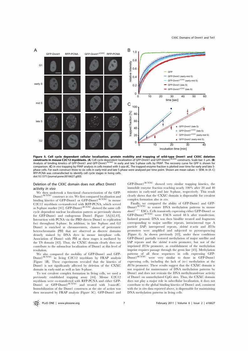
Deletion of the CXXC domain does not affect Dnmt1activity in vivo
We then undertook a functional characterization of the GFP-Dnmt1DCXXC construct in vivo. We first compared localization andbinding kinetics of GFP-Dnmt1 or GFP-Dnmt1DCXXC in mouseC2C12 myoblasts co-transfected with RFP-PCNA, which servedas S-phase marker [41]. GFP-Dnmt1DCXXC showed the same cell-cycle dependent nuclear localization pattern as previously shownfor GFP-Dnmt1 and endogenous Dnmt1 (Figure 5A)[42,43].Interaction with PCNA via the PBD directs Dnmt1 to replicationfoci throughout S-phase. In addition, in late S-phase and G2Dnmt1 is enriched at chromocenters, clusters of pericentricheterochromatin (PH) that are observed as discrete domainsdensely stained by DNA dyes in mouse interphase cells.Association of Dnmt1 with PH at these stages is mediated bythe TS domain [42]. Thus, the CXXC domain clearly does notcontribute to the subnuclear localization of Dnmt1 at this level ofresolution.
We also compared the mobility of GFP-Dnmt1 and GFP-Dnmt1DCXXC in living C2C12 myoblasts by FRAP analysis(Figure 5B). These experiments revealed that the kinetics ofDnmt1 is not significantly affected by deletion of the CXXCdomain in early-mid as well as late S-phase.
To test covalent complex formation in living cells, we used apreviously established trapping assay [44]. Mouse C2C12myoblasts were co-transfected with RFP-PCNA and either GFP-Dnmt1 or GFP-Dnmt1DCXXC and treated with 5-aza-dC.Immobilization of the Dnmt1 constructs at the site of action wasthen measured by FRAP analysis (Figure 5C). GFP-Dnmt1 and
GFP-Dnmt1DCXXC showed very similar trapping kinetics, theimmobile enzyme fraction reaching nearly 100% after 20 and 40minutes in early-mid and late S-phase, respectively. This resultclearly shows that the CXXC domain is dispensable for covalentcomplex formation also in vivo.
Finally, we compared the ability of GFP-Dnmt1 and GFP-Dnmt1DCXXC to restore DNA methylation patterns in mousednmt12/2 ESCs. Cells transiently expressing either GFP-Dnmt1 orGFP-Dnmt1DCXXC were FACS sorted 48 h after transfection.Isolated genomic DNA was then bisulfite treated and fragmentscorresponding to major satellite repeats, intracisternal type Aparticle (IAP) interspersed repeats, skeletal a-actin and H19apromoters were amplified and subjected to pyrosequencing(Figure 6). As shown previously [43], under these conditionsGFP-Dnmt1 partially restored methylation of major satellite andIAP repeats and the skeletal a-actin promoter, but not of theimprinted H19a promoter, as establishment of the methylationimprint requires passage through the germ line [45]. Methylationpatterns of all these sequences in cells expressing GFP-Dnmt1DCXXC were very similar to those in GFP-Dnmt1expressing cells, including the lack of (re-) methylation at theH19a promoter. These results suggest that the CXXC domain isnot required for maintenance of DNA methylation patterns byDnmt1 and does not restrain the DNA methyltransferase activityof Dnmt1 on unmethylated CpG sites. Thus, the CXXC domaindoes not play a major role in subcellular localization, it does notcontribute to the global binding kinetics of Dnmt1 and, consistentwith the in vitro data reported above, is dispensable for maintainingDNA methylation patterns in living cells.
Figure 5. Cell cycle dependant cellular localization, protein mobility and trapping of wild-type Dnmt1 and CXXC deletionconstructs in mouse C2C12 myoblasts. (A) Cell cycle dependent localization of GFP-Dnmt1 and GFP-Dnmt1DCXXC constructs. Scale bar: 5 mm. (B)Analysis of binding kinetics of GFP-Dnmt1 and GFP-Dnmt1DCXXC in early and late S-phase cells by FRAP. The recovery curve for GFP is shown forcomparison. (C) In vivo trapping by FRAP analysis in cells treated with 5-aza-dC. The trapped enzyme fraction is plotted over time for early and late S-phase cells. For each construct three to six cells in early-mid and late S phase were analysed per time point. Shown are mean values 6 SEM. In (A–C)RFP-PCNA was cotransfected to identify cell cycle stages in living cells.doi:10.1371/journal.pone.0016627.g005
CXXC Domains of Dnmt1 and Tet1
PLoS ONE | www.plosone.org 7 February 2011 | Volume 6 | Issue 2 | e16627

Discussion
We generated homology models based on the reported structureof the MLL1 CXXC domain to design isolated CXXC domainconstructs and CXXC domain deletion mutants for Dnmt1 andTet1 with minimal probability of structural alteration. Accordingto these models CXXC domains are delimited by an antiparallelb-sheet, a discrete structural element. Our data show that theCXXC domain of mouse Dnmt1 preferentially binds DNAsubstrates containing unmethylated CpG sites as previously shownfor CXXC domains of human DNMT1 and other mammalianproteins. We note that sequences C-terminal to the correspondingpeptide in CGBP/Cfp1 were reported to be required for DNAbinding in vitro [22] and that only a significantly larger peptidespanning the CXXC-3 domain of Mbd1a was tested for DNAbinding. However, sequences C-terminal to CXXC domains arenot conserved (Figure 1B) and our data show that they are notrequired for DNA binding by the CXXC domain of Dnmt1.Nevertheless, all the CXXC domains reported to selectively bindunmethylated CpG sites cluster in a distinct homology group andcontain the KFGG motif. The latter was shown to be crucial forDNA binding by the CXXC domain of MLL1 [38] and here weextend this observation to the CXXC domain of Dnmt1.Sequence alignment reveals two distinct CXXC domain homologygroups that lack the KFGG motif (Figure 1A). Consistent with arole of this motif in DNA binding, members of these groups suchas CXXC-1/2 of Mbd1 [29] and the CXXC domain of Tet1 (thisstudy) show no DNA binding activity. While no specific function isknown for CXXC-1/2 of Mbd1, the CXXC domain of Tet1 isclosely related to those in CXXC4/Idax and CXXC5/RINF thatwere shown to mediate protein-protein interactions [46–48]. This
suggests that the CXXC domain of Tet1, rather than mediatingDNA binding, may function as a protein-protein interactiondomain. However, our data do not rule out the possibility that theDNA binding properties of the CXXC domain within the contextof full length Tet1 may be different from those of the isolateddomain. Nevertheless, we show that the CXXC domain is notrequired for enzymatic activity of Tet1 in vivo.
Although we observed a clear DNA binding activity by theisolated CXXC domain of Dnmt1, we found that, within thecontext of the full length enzyme, this domain is dispensable foroverall DNA binding properties, preference for hemimethylatedsubstrates upon covalent complex formation, methyltransferaseactivity and allosteric activation as well as for the ability to restoremethylation of representative sequences in dnmt1 null ESCs.Consistent with our data, a recent report showed a preference ofthe CXXC domain of human DNMT1 for substrates containingunmethylated CpG sites [32]. However, the same report showedthat deletion of the CXXC domain from the human enzymeresults in a significant decrease in methyltransferase activity onhemimethylated substrates in vitro and 25% lower methylation atrDNA repeats upon overexpression in HEK293 cells, suggesting adominant negative effect of the deletion construct. Thesediscrepancies may be due to the fact that the fragment deletedby Pradhan et al. includes the N-terminal strand of the predictedantiparallel b-sheet, potentially leading to disruption of nativefolding, to species-specific differences and/or to the analysis ofnon-physiological expression levels in HEK293 cells. In ourtrapping assay the same human deletion mutant showed reducedcovalent complex formation, consistent with loss of enzymaticactivity. The report from Pradhan et al. also showed that mutationof cysteine 667 to glycine within the CXXC domain of human
Figure 6. The CXXC deletion construct of Dnmt1 restores methylation in dnmt1 null cells. Mouse dnmt12/2 ESCs transiently expressingGFP-Dnmt1 or GFP-Dnmt1DCXXC were isolated by FACS-sorting 48 h after transfection and CpG methylation levels within the indicated sequenceswere analyzed by bisulfite treatment, PCR amplification and direct pyrosequencing. Methylation levels of untransfected wild type and dnmt12/2 ESCsare shown for comparison.doi:10.1371/journal.pone.0016627.g006
CXXC Domains of Dnmt1 and Tet1
PLoS ONE | www.plosone.org 8 February 2011 | Volume 6 | Issue 2 | e16627

DNMT1 disrupts DNA binding and enzymatic activity. However,as this point mutation involves one of the zinc coordinatingresidues it is not unlikely to alter peptide folding with negativeconsequences potentially extending beyond the CXXC domainand including reduced enzymatic activity. In this respect thedominant negative effect observed upon overexpression of thismutant may be explained by the prevalent occurrence of Dnmt1as a dimer [49]. These observations, together with preservedability for covalent complex formation and catalytic activity of ourCXXC domain deletion, support the validity of our homologymodel-driven approach for functional characterization of theCXXC domain. In addition, our genetic complementationapproach constitutes a rather physiologic functional assay.However, due to the transient approach and the analysis ofgenomic methylation at only a few representative sequences, subtleor highly sequence specific effects of deletion of the CXXCdomain cannot be excluded.
It was recently shown that binding of Cfp1/CGBP andKDM2A to CpG islands through their CXXC domains leads tolocal enrichment and depletion of H3K4 and H3K36 methylation,respectively [26,30]. Analogously, Dnmt1 may bind CpG islandsthrough its CXXC domain. However, this interaction would notlead to a straightforward functional interpretation as CpG islandswith high CpG density are generally refractive to DNAmethylation and a function of Dnmt1 as a de novo DNAmethyltransferase is not well established. It could be envisagedthat binding to unmethylated CpG sites/islands by the CXXCdomain may have a negative effect on the enzymatic activity ofDnmt1 and restrain its function as a de novo DNA methyltrans-ferase. However, we show that in dnmt1 null ESCs methylation ofthe imprinted H19a promoter is not restored upon expression ofeither wild type or DCXXC Dnmt1 constructs, arguing against anegative regulatory function of the CXXC domain.
Notably, binding of unmethylated CpG sites by KFGG motif-containing CXXC domains does not exclude a role in protein-protein interaction as the CXXC domain of MLL1 was reportedto interact with both DNA and Polycomb Repressive Complex 1components HPC2/CBX4 and BMI-1 [21,50]. Therefore, it ispossible that the CXXC domain of Dnmt1 has regulatoryfunctions in specific cell types or developmental stages that mayinvolve DNA binding and/or interaction with other proteins. Thegeneration of dedicated animal models may be instrumental fortesting these possibilities.
Materials and Methods
Bioinformatic methodsAlignments were performed using the ClustalW2 software [51].
The CXXC domain homology tree (Figure 1C) was generatedfrom the alignment in Figure 1B with Jalview 2.4 by unweightedpair group method with arithmetic mean (UPGMA). Theneighbor-joining method gave the same result. Average distancesbetween the sequences were calculated using the BLOSSUM62matrix. The human CXXC10 coding sequence [52] wasdetermined by assembling ESTs AI438961, BX114363,BX492895, BU633058.1, AW207644.1 and the genomic sequenceAC073046.7. The putative translational start site is located16308 bp upstream of the annotated transcriptional start site ofTET3. A partial coding sequence of murine Cxxc10 containingthe CXXC domain was identified by aligning the humanCXXC10 protein sequence to the ORFs present inNT_039353.7 upstream of the tet3 gene from position 35663306to 35808487). A very high match was found 13266 nt upstream oftet3 at positions 35676374-35676572 of NT_039353.7. To build
homology models for the CXXC domains of Dnmt1 (aa 645–696)and Tet1 (aa 561–614), we submitted the respective sequences tothe HHpred server [53]. The best template was the CXXCdomain of MLL1 (PDB-ID: 2J2S). The 49 residues of the CXXCdomain in Dnmt1 can be aligned to this domain with 45%sequence identity and only a single amino acid gap after residue661 (Figure 1B). 3D models were calculated with the homologymodeling software MODELLER [54] (version 9.5) using thisalignment. Distance restraints were given to MODELLER toenforce a distance of 2.360.1 A between the eight sulphurs in theZn-coordinating cysteines and the Zn2+ ions. TM-align [55] wasused to superpose the model structure with the template domain.Images were generated using the PyMol Molecular GraphicsSystem (Version 1.3, Schrodinger, LLC). The quality of themodels and the underlying alignments were checked with DOPE[56] and Verify3D [57] and results for both models were found tobe comparable to the MLL1 template structure (2J2S).
Expression constructsFusion constructs were generated using enhanced green
fluorescent protein, monomeric red fluorescent protein ormonomeric cherry and are here referred to as GFP, RFP andCherry fusions, respectively. Mammalian expression constructs forGFP, mouse GFP-Dnmt1, GFP-NTR and human RFP-PCNAwere described previously [42,44,49,58]. The deletion constructGFP-Dnmt1DCXXC was obtained by replacing the sequencecoding for aa 655–696 with three alanine codons in the GFP-Dnmt1 construct as described [59]. The GFP-DNMT1DCXXC
construct was generated by subcloning the sequence coding forhuman DNMT1DCXXC from the homonymous construct byPradhan et al. [32] in the pEGFP-C2 vector (Clonetech). Togenerate GFP-Tet1 three partially overlapping fragments span-ning the Tet1 coding sequence were amplified using E14 ESCscDNA as template. The fragments were then joined by overlapextension PCR and inserted into the pCAG-GFP-IB vector [43].To generate GFP-Tet1DCXXC aa 569-621 of murine Tet1 weredeleted from GFP-Tet1 using a type IIs restriction endonucleaseapproach as described [60]. To generate GFP-CXXCDnmt1 andGFP-CXXCTet1 sequences coding for the respective CXXCdomains (aa 643-700 for Dnmt1 and 561-614 for Tet1) wereamplified by PCR using the GFP-Dnmt1 expression construct andcDNA from E14 ESCs as templates, respectively. PCR fragmentswere then inserted into the pCAG-GFP-IB vector. GFP-NTRDCXXC was obtained by replacing the BglII-XhoI fragmentof GFP-NTR with the same fragment of GFP-Dnmt1DCXXC. Ch-CTD-His was generated by replacing the GFP coding sequence ina GFP-CTD construct [49] with the Cherry coding sequence. Allconstructs were confirmed by sequencing.
Cell culture, transfection and cell sortingHEK293T cells [61] and mouse C2C12 myoblasts [62] were
cultured in DMEM supplemented with 50 mg/ml gentamicin and10% and 20% fetal calf serum, respectively. For expression offusion proteins HEK293T cells were transfected with polyethy-lenimine (Sigma). For live cell imaging, C2C12 cells were grownto 40% confluence on Lab-Tek chambers (Nunc) or m-slides(Ibidi) and transfected with TransFectin transfection reagent(BioRad) according to the manufacturer’s instructions. MouseESCs were cultured as described [63] and transfected withFuGENE HD (Roche) according to the manufacturer’s instruc-tions. ESCs were sorted with a FACS Aria II instrument (BectonDickinson). The dnmt12/2 J1 ESCs used in this study arehomozygous for the c allele [14].
CXXC Domains of Dnmt1 and Tet1
PLoS ONE | www.plosone.org 9 February 2011 | Volume 6 | Issue 2 | e16627

In vitro DNA binding and trapping assaysIn vitro DNA binding and trapping assays were performed as
described previously [36,37] with the following modifications.DNA substrates labeled with four different ATTO fluorophores(Tables S1 and S2 in File S1) were used at a final concentration of125 nM each in the pull-down assay with immobilized GFPfusions. After removal of unbound substrate, the amounts ofprotein and DNA were determined by fluorescence intensitymeasurements with a Tecan Infinite M1000 plate reader usingcalibration curves from purified GFP or DNA coupled ATTOfluorophores, respectively. The following excitation/emission 6detection bandwidth settings were used: 490/511610 nm forGFP, 550/580615 nm for ATTO550, 600/630615 nm forATTO590, 650/670610 nm for ATTO647N and 700/720610 nm for ATTO700. Cross detection of GFP and differentATTO dyes was negligible with these settings. Binding andtrapping ratios were calculated dividing the concentration ofbound DNA substrate by the concentration of GFP fusion onthe beads.
In vivo mC hydroxylation assayGenomic DNA was isolated from HEK293T cells 24 h after
transfection with the GFP-Tet1 and GFP-Tet1DCXXC constructsand global hmC levels were measured using the in vitroglucosylation assay as previously described [63], except that100 nM b-glucosyltransferase and only UDP-[3H]glucose donor(0.43 mM) were used.
Co-immunoprecipitationCo-immunoprecipitation was performed as described previously
[49,64]. Shortly, HEK293T cells were transiently co-transfectedwith expression plasmids for GFP fusions and the Ch-CTD-Hisconstruct, harvested and lysed. GFP fusions were pulled downusing the GFP-Trap [65] (Chromotek) and subjected to westernblotting using anti-GFP (Roche or Chromotek) and anti-His(Invitrogen) monoclonal antibodies.
Live cell microscopy, FRAP analysis and live cell trappingassay
Live cell imaging and FRAP experiments were performed asdescribed previously [43]. For each construct 6-15 nuclei wereaveraged and the mean values as well as the standard errors werecalculated. For presentation, we used linear contrast enhancementon entire images. The DNA methyltransferase trapping assay wasdescribed previously [44]. Briefly, transfected cells were incubatedwith 30 mM 5-aza-dC (Sigma) for the indicated periods of timebefore photobleaching experiments. FRAP analysis was performedwith a confocal laser scanning microscope (TCS SP5, Leica)
equipped with a 636/1.4 NA Plan-Apochromat oil immersionobjective. Microscope settings were as described except that asmaller region of interest (3 mm63 mm) was selected for photo-bleaching. Mean fluorescence intensities of the bleached regionwere corrected for background and for total loss of nuclearfluorescence over the time course, and normalized by the mean ofthe last 10 prebleach values.
DNA Methylation AnalysisGenomic DNA was isolated with the QIAmp DNA Mini Kit
(Qiagen) and 1.5 mg were bisulfite converted using the EZ DNAMethylation-Gold Kit (Zymo research) according to the manu-facturer’s instructions. Primer sets and PCR conditions for IAP-LTR, skeletal a-actin and H19 promoters were as described [43].Primer sequences for major satellites were AAAATGAGAAA-CATCCACTTG (forward primer) and CCATGATTTT-CAGTTTTCTT (reverse primer). For amplification we usedQiagen Hot Start Polymerase in 1x Qiagen Hot Start Polymerasebuffer supplemented with 0.2 mM dNTPs, 0.2 mM forwardprimer, 0.2 mM reverse primer, 1.3 mM betaine (Sigma) and60 mM tetramethylammonium-chloride (TMAC, Sigma). Pro-moter regions and IAP-LTR were amplified with two subsequent(nested) PCR reactions and major satellite repeats were amplifiedwith a single amplification reaction. Pyrosequencing reactionswere carried out by Varionostic GmbH (Ulm, Germany).Pyrosequencing primers are listed in Table S3 in File S1.
Supporting Information
File S1 Tables S1–S3, Figures S1–S8 and Supplementalmethods.(PDF)
Acknowledgments
The authors thank Sabine Brunner and Lucia Puchbauer for technicalassistance in the generation of the homology models. We also thankTaiping Chen and En Li (Novartis Institutes for Biomedical Research,Boston, MA) for providing dnmt1 null ESCs and Shriharsa Pradhan (NewEngland BioLabs, Ipswich, MA) for the human DNMT1DCXXC constructand constructive discussion.
Author Contributions
Conceived and designed the experiments: FS HL. Performed theexperiments: CF AR SB DM KF SH MW WQ. Analyzed the data: CFAR SB DM KF SH JS. Contributed reagents/materials/analysis tools: JS.Wrote the paper: FS HL. Generated homology models: JS.
References
1. Ball MP, Li JB, Gao Y, Lee JH, LeProust EM, et al. (2009) Targeted andgenome-scale strategies reveal gene-body methylation signatures in human cells.Nat Biotechnol 27: 361–368.
2. Suzuki MM, Bird A (2008) DNA methylation landscapes: provocative insightsfrom epigenomics. Nat Rev Genet 9: 465–476.
3. Laurent L, Wong E, Li G, Huynh T, Tsirigos A, et al. (2010) Dynamic changesin the human methylome during differentiation. Genome Res 320: 320–331.
4. Lister R, Pelizzola M, Dowen RH, Hawkins RD, Hon G, et al. (2009) HumanDNA methylomes at base resolution show widespread epigenomic differences.Nature 462: 315–322.
5. Mohn F, Weber M, Rebhan M, Roloff TC, Richter J, et al. (2008) Lineage-specific polycomb targets and de novo DNA methylation define restriction andpotential of neuronal progenitors. Mol Cell 30: 755–766.
6. Deng J, Shoemaker R, Xie B, Gore A, LeProust EM, et al. (2009) Targetedbisulfite sequencing reveals changes in DNA methylation associated with nuclearreprogramming. Nat Biotechnol 27: 353–360.
7. Schmidl C, Klug M, Boeld TJ, Andreesen R, Hoffmann P, et al. (2009) Lineage-specific DNA methylation in T cells correlates with histone methylation andenhancer activity. Genome Res 19: 1165–1174.
8. Edwards JR, O’Donnell AH, Rollins RA, Peckham HE, Lee C, et al. (2010)Chromatin and sequence features that define the fine and gross structure ofgenomic methylation patterns. Genome Res 20: 972–980.
9. Gaudet F, Hodgson JG, Eden A, Jackson-Grusby L, Dausman J, et al. (2003)Induction of tumors in mice by genomic hypomethylation. Science 300:489–492.
10. Walsh CP, Chaillet JR, Bestor TH (1998) Transcription of IAP endogenousretroviruses is constrained by cytosine methylation. Nat Genet 20: 116–117.
11. Xu G-L, Bestor TH, Bourc’his D, Hsieh C-L, Tommerup N, et al. (1999)Chromosome instability and immunodeficiency syndrome caused by mutationsin a DNA methyltransferase gene. Nature 402: 187–191.
12. Goll MG, Bestor TH (2005) Eukaryotic cytosine methyltransferases. Annu RevBiochem 74: 481–514.
CXXC Domains of Dnmt1 and Tet1
PLoS ONE | www.plosone.org 10 February 2011 | Volume 6 | Issue 2 | e16627
13. Leonhardt H, Page AW, Weier HU, Bestor TH (1992) A targeting sequencedirects DNA methyltransferase to sites of DNA replication in mammalian nuclei.Cell 71: 865–873.
14. Lei H, Oh SP, Okano M, Juttermann R, Goss KA, et al. (1996) De novo DNAcytosine methyltransferase activities in mouse embryonic stem cells. Develop-ment 122: 3195–3205.
15. Li E, Bestor TH, Jaenisch R (1992) Targeted mutation of the DNAmethyltransferase gene results in embryonic lethality. Cell 69: 915–926.
16. Margot JB, Aguirre-Arteta AM, Di Giacco BV, Pradhan S, Roberts RJ, et al.(2000) Structure and function of the mouse DNA methyltransferase gene:Dnmt1 shows a tripartite structure. J Mol Biol 297: 293–300.
17. Spada F, Rothbauer U, Zolghadr K, Schermelleh L, Leonhardt H (2006)Regulation of DNA methyltransferase 1. Adv Enzyme Regul 46: 224–234.
18. Zimmermann C, Guhl E, Graessmann A (1997) Mouse DNA methyltransferase(MTase) deletion mutants that retain the catalytic domain display neither denovo nor maintenance methylation activity in vivo. Biol Chem 378: 393–405.
19. Pradhan S, Esteve PO (2003) Allosteric activator domain of maintenance humanDNA (cytosine-5) methyltransferase and its role in methylation spreading.Biochemistry 42: 5321–5332.
20. Fatemi M, Hermann A, Pradhan S, Jeltsch A (2001) The activity of the murineDNA methyltransferase Dnmt1 is controlled by interaction of the catalyticdomain with the N-terminal part of the enzyme leading to an allostericactivation of the enzyme after binding to methylated DNA. J Mol Biol 309:1189–1199.
21. Birke M, Schreiner S, Garcia-Cuellar MP, Mahr K, Titgemeyer F, et al. (2002)The MT domain of the proto-oncoprotein MLL binds to CpG-containing DNAand discriminates against methylation. Nucl Acids Res 30: 958–965.
22. Lee JH, Voo KS, Skalnik DG (2001) Identification and characterization of theDNA binding domain of CpG-binding protein. J Biol Chem 276: 44669–44676.
23. Cross SH, Meehan RR, Nan X, Bird A (1997) A component of thetranscriptional repressor MeCP1 shares a motif with DNA methyltransferaseand HRX proteins. Nat Genet 16: 256–259.
24. Tsukada Y, Fang J, Erdjument-Bromage H, Warren ME, Borchers CH, et al.(2006) Histone demethylation by a family of JmjC domain-containing proteins.Nature 439: 811–816.
25. Frescas D, Guardavaccaro D, Bassermann F, Koyama-Nasu R, Pagano M(2007) JHDM1B/FBXL10 is a nucleolar protein that represses transcription ofribosomal RNA genes. Nature 450: 309–313.
26. Blackledge NP, Zhou JC, Tolstorukov MY, Farcas AM, Park PJ, et al. (2010)CpG Islands Recruit a Histone H3 Lysine 36 Demethylase. Mol Cell 38:179–190.
27. Lorsbach RB, Moore J, Mathew S, Raimondi SC, Mukatira ST, et al. (2003)TET1, a member of a novel protein family, is fused to MLL in acute myeloidleukemia containing the t(10;11)(q22;q23). Leukemia 17: 637–641.
28. Ono R, Taki T, Taketani T, Taniwaki M, Kobayashi H, et al. (2002) LCX,leukemia-associated protein with a CXXC domain, is fused to MLL in acutemyeloid leukemia with trilineage dysplasia having t(10;11)(q22;q23). Cancer Res62: 4075–4080.
29. Jorgensen HF, Ben-Porath I, Bird AP (2004) Mbd1 is recruited to bothmethylated and nonmethylated CpGs via distinct DNA binding domains. MolCell Biol 24: 3387–3395.
30. Thomson JP, Skene PJ, Selfridge J, Clouaire T, Guy J, et al. (2010) CpG islandsinfluence chromatin structure via the CpG-binding protein Cfp1. Nature 464:1082–1086.
31. Bestor TH (1992) Activation of mammalian DNA methyltransferase by cleavageof a Zn binding regulatory domain. EMBO J 11: 2611–2617.
32. Pradhan M, Esteve PO, Chin HG, Samaranayke M, Kim GD, et al. (2008)CXXC domain of human DNMT1 is essential for enzymatic activity.Biochemistry 47: 10000–10009.
33. Tahiliani M, Koh KP, Shen Y, Pastor WA, Bandukwala H, et al. (2009)Conversion of 5-Methylcytosine to 5-Hydroxymethylcytosine in MammalianDNA by MLL Partner TET1. Science 324: 930–935.
34. Ito S, D’Alessio AC, Taranova OV, Hong K, Sowers LC, et al. (2010) Role ofTet proteins in 5mC to 5hmC conversion, ES-cell self-renewal and inner cellmass specification. Nature 466: 1129–1133.
35. Allen MD, Grummitt CG, Hilcenko C, Min SY, Tonkin LM, et al. (2006)Solution structure of the nonmethyl-CpG-binding CXXC domain of theleukaemia-associated MLL histone methyltransferase. EMBO J 25: 4503–4512.
36. Frauer C, Leonhardt H (2009) A versatile non-radioactive assay for DNAmethyltransferase activity and DNA binding. Nucl Acids Res 37: e22.
37. Rottach A, Frauer C, Pichler G, Bonapace IM, Spada F, et al. (2010) The multi-domain protein Np95 connects DNA methylation and histone modification.Nucl Acids Res 38: 1796–1805.
38. Ayton PM, Chen EH, Cleary ML (2004) Binding to nonmethylated CpG DNAis essential for target recognition, transactivation, and myeloid transformation byan MLL oncoprotein. Mol Cell Biol 24: 10470–10478.
39. Szwagierczak A, Bultmann S, Schmidt CS, Spada F, Leonhardt H (2010)Sensitive enzymatic quantification of 5-hydroxymethylcytosine in genomicDNA. Nucleic Acids Research 38: e181.
40. Bacolla A, Pradhan S, Larson JE, Roberts RJ, Wells RD (2001) Recombinanthuman DNA (cytosine-5) methyltransferase. III. Allosteric control, reactionorder, and influence of plasmid topology and triplet repeat length onmethylation of the fragile X CGG.CCG sequence. J Biol Chem 276:18605–18613.
41. Easwaran HP, Leonhardt H, Cardoso MC (2005) Cell Cycle Markers for LiveCell Analyses. Cell Cycle 4: 453–455.
42. Easwaran HP, Schermelleh L, Leonhardt H, Cardoso MC (2004) Replication-independent chromatin loading of Dnmt1 during G2 and M phases. EMBORep 5: 1181–1186.
43. Schermelleh L, Haemmer A, Spada F, Rosing N, Meilinger D, et al. (2007)Dynamics of Dnmt1 interaction with the replication machinery and its role inpostreplicative maintenance of DNA methylation. Nucl Acids Res 35:4301–4312.
44. Schermelleh L, Spada F, Easwaran HP, Zolghadr K, Margot JB, et al. (2005)Trapped in action: direct visualization of DNA methyltransferase activity inliving cells. Nat Methods 2: 751–756.
45. Tucker KL, Beard C, Dausmann J, Jackson-Grusby L, Laird PW, et al. (1996)Germ-line passage is required for establishment of methylation and expressionpatterns of imprinted but not of nonimprinted genes. Genes Dev 10: 1008–1020.
46. Andersson T, Sodersten E, Duckworth JK, Cascante A, Fritz N, et al. (2009)CXXC5 Is a Novel BMP4-regulated Modulator of Wnt Signaling in NeuralStem Cells. J Biol Chem 284: 3672–3681.
47. Hino S-i, Kishida S, Michiue T, Fukui A, Sakamoto I, et al. (2001) Inhibition ofthe Wnt Signaling Pathway by Idax, a Novel Dvl-Binding Protein. Mol Cell Biol21: 330–342.
48. London TBC, Lee H-J, Shao Y, Zheng J (2004) Interaction between the internalmotif KTXXXI of Idax and mDvl PDZ domain. Biochem Biophys ResCommun 322: 326–332.
49. Fellinger K, Rothbauer U, Felle M, Langst G, Leonhardt H (2009) Dimerizationof DNA methyltransferase 1 is mediated by its regulatory domain. J CellBiochem 106: 521–528.
50. Xia ZB, Anderson M, Diaz MO, Zeleznik-Le NJ (2003) MLL repression domaininteracts with histone deacetylases, the polycomb group proteins HPC2 andBMI-1, and the corepressor C-terminal-binding protein. Proc Natl AcadSci U S A 100: 8342–8347.
51. Thompson JD, Higgins DG, Gibson TJ (1994) CLUSTAL W: improving thesensitivity of progressive multiple sequence alignment through sequenceweighting, position-specific gap penalties and weight matrix choice. Nucl AcidsRes 22: 4673–4680.
52. Katoh M (2004) Identification and characterization of human CXXC10 gene insilico. Int J Oncol 25: 1193–1199.
53. Soding J, Biegert A, Lupas AN (2005) The HHpred interactive server for proteinhomology detection and structure prediction. Nucl Acids Res 33: W244–248.
54. Sali A, Potterton L, Yuan F, van Vlijmen H, Karplus M (1995) Evaluation ofcomparative protein modeling by MODELLER. Proteins 23: 318–326.
55. Zhang Y, Skolnick J (2005) TM-align: a protein structure alignment algorithmbased on the TM-score. Nucl Acids Res 33: 2302–2309.
56. Shen M-y, Sali A (2006) Statistical potential for assessment and prediction ofprotein structures. Protein Sci 15: 2507–2524.
57. Eisenberg D, Luthy R, Bowie JU (1997) VERIFY3D: Assessment of proteinmodels with three-dimensional profiles. In: Carter CWJ, Sweet RM, eds.Methods Enzymol: Academic Press. pp 396–404.
58. Sporbert A, Domaing P, Leonhardt H, Cardoso MC (2005) PCNA acts as astationary loading platform for transiently interacting Okazaki fragmentmaturation proteins. Nucl Acids Res 33: 3521–3528.
59. Fellinger K, Leonhardt H, Spada F (2008) A mutagenesis strategy combiningsystematic alanine scanning with larger mutations to study protein interactions.Anal Biochem 373: 176–178.
60. Ko J-K, Ma J (2005) A rapid and efficient PCR-based mutagenesis methodapplicable to cell physiology study. Am J Physiol Cell Physiol 288: C1273–1278.
61. DuBridge RB, Tang P, Hsia HC, Leong PM, Miller JH, et al. (1987) Analysis ofmutation in human cells by using an Epstein-Barr virus shuttle system. Mol CellBiol 7: 379–387.
62. Blau HM, Pavlath GK, Hardeman EC, Chiu CP, Silberstein L, et al. (1985)Plasticity of the differentiated state. Science 230: 758–766.
63. Szwagierczak A, Bultmann S, Schmidt CS, Spada F, Leonhardt H (2010)Sensitive enzymatic quantification of 5-hydroxymethylcytosine in genomicDNA. Nucl Acids Res 38: e181.
64. Meilinger D, Fellinger K, Bultmann S, Rothbauer U, Bonapace IM, et al. (2009)Np95 interacts with de novo DNA methyltransferases, Dnmt3a and Dnmt3b,and mediates epigenetic silencing of the viral CMV promoter in embryonic stemcells. EMBO Rep 10: 1259–1264.
65. Rothbauer U, Zolghadr K, Muyldermans S, Schepers A, Cardoso MC, et al.(2007) A versatile nanotrap for biochemical and functional studies withfluorescent fusion proteins. Mol Cell Proteomics 7: 282–289.
66. Mohseni-Zadeh S, Brezellec P, Risler JL (2004) Cluster-C, an algorithm for thelarge-scale clustering of protein sequences based on the extraction of maximalcliques. Comput Biol Chem 28: 211–218.
CXXC Domains of Dnmt1 and Tet1
PLoS ONE | www.plosone.org 11 February 2011 | Volume 6 | Issue 2 | e16627

1
�
Different binding properties and function of CXXC zinc finger domains in Dnmt1 and Tet1
Carina Frauer#, Andrea Rottach#, Daniela Meilinger, Sebastian Bultmann, Karin Fellinger, Stefan Hasenöder, Mengxi Wang, Weihua Qin, Johannes Söding§, Fabio Spada* and Heinrich Leonhardt*
Department of Biology and Center for Integrated Protein Science Munich (CIPSM), Ludwig Maximilians University Munich, Planegg-Martinsried and §Gene Center Munich, Ludwig Maximilians University Munich, Munich, Germany. *Corresponding authors: FS: [email protected]; Fax +49 89 2180-74236; Tel. +49 89 2180-74230 HL: [email protected]; Fax +49 89 2180-74236; Tel. +49 89 2180-74232 #These authors contributed equally to this work
SUPPLEMENTAL INFORMATION FILE
Tables S1-3
Figures S1-8
Supplemental Methods

Frauer et al. Supplemental Information File
2
�
Table S1. Sequences of DNA oligonucleotides used for preparation of double stranded DNA
substrates. M: 5-methylcytosine.
Name Sequence
CG-up 5’- CTCAACAACTAACTACCATCCGGACCAGAAGAGTCATCATGG -3’
MG-up 5’- CTCAACAACTAACTACCATCMGGACCAGAAGAGTCATCATGG -3’
noCG-up 5’- CTCAACAACTAACTACCATCTGGACCAGAAGAGTCATCATGG -3’
Fill-In-550 5’- ATTO550-CCATGATGACTCTTCTGGTC -3’
Fill-In-590 5’- ATTO590-CCATGATGACTCTTCTGGTC -3’
Fill-In-647N 5’- ATTO647N-CCATGATGACTCTTCTGGTC -3’
Fill-In-700 5’- ATTO700-CCATGATGACTCTTCTGGTC -3’
Table S2. DNA substrates used for the in vitro DNA binding and trapping assays.
Name CpG site Label Oligo I Oligo II dCTP reaction Purpose
noCGB 700 no CpG site 700 noCG-up Fill-In-700 dCTP Binding
UMB 550
unmethylated
550
CG-up
Fill-In-550
dCTP Binding UMB 590 590 Fill-In-590
UMB 647N 647N Fill-In-647N
UMB 700 700 Fill-In-700
UMT 550 550 Fill-In-550 5-aza-dCTP Trapping
HMB 590
hemimethylated
590
MG-up
Fill-In-590 dCTP Binding
HMB 647N 647N Fill-In-647N
HMT 550 550 Fill-In-550 5-aza-dCTP Trapping
HMT 647N 647N Fill-In-647N
FMB 647N fully methylated 647N MG-up Fill-In-647N 5methyl dCTP Binding
Table S3. Primers used for pyrosequencing. Each primer is biotinylated at the 5’ end.
Name Sequence
skeletal D-actin-1 5’- AGTTGGGGATATTTTTTATA -3’
skeletal D-actin-1b 5’- TTTTGGTTAGTGTAGGAGAT -3’
skeletal D-actin-2 5’- TGGGAAGGGTAGTAATATTT -3’
H19-1 5’- ATAGTTATTGTTTATAGTTT -3’
H19-2 5’- AGGAATATGTTATATTTAT -3’
IAP LTR-1 5’- CCCTAATTAACTACAACCCA -3’
IAP LTR-2 5’- TGTAGTTAATTAGGGAGTGA -3’
Major Satellite-1 5’- AAAATGAGAAATATTTATTTG -3’
Major Satellite-2 5’- GAGAAATATATACTTTAGGA -3’
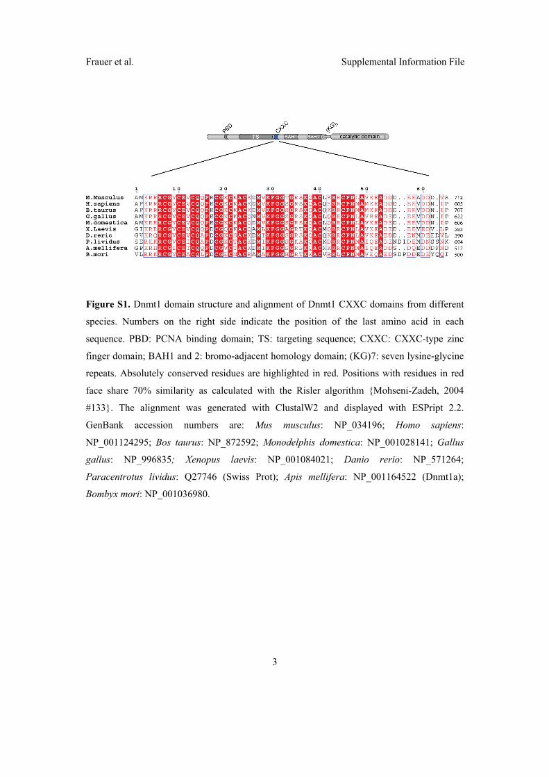
Frauer et al. Supplemental Information File
3
�
Figure S1. Dnmt1 domain structure and alignment of Dnmt1 CXXC domains from different
species. Numbers on the right side indicate the position of the last amino acid in each
sequence. PBD: PCNA binding domain; TS: targeting sequence; CXXC: CXXC-type zinc
finger domain; BAH1 and 2: bromo-adjacent homology domain; (KG)7: seven lysine-glycine
repeats. Absolutely conserved residues are highlighted in red. Positions with residues in red
face share 70% similarity as calculated with the Risler algorithm {Mohseni-Zadeh, 2004
#133}. The alignment was generated with ClustalW2 and displayed with ESPript 2.2.
GenBank accession numbers are: Mus musculus: NP_034196; Homo sapiens:
NP_001124295; Bos taurus: NP_872592; Monodelphis domestica: NP_001028141; Gallus
gallus: NP_996835; Xenopus laevis: NP_001084021; Danio rerio: NP_571264;
Paracentrotus lividus: Q27746 (Swiss Prot); Apis mellifera: NP_001164522 (Dnmt1a);
Bombyx mori: NP_001036980.
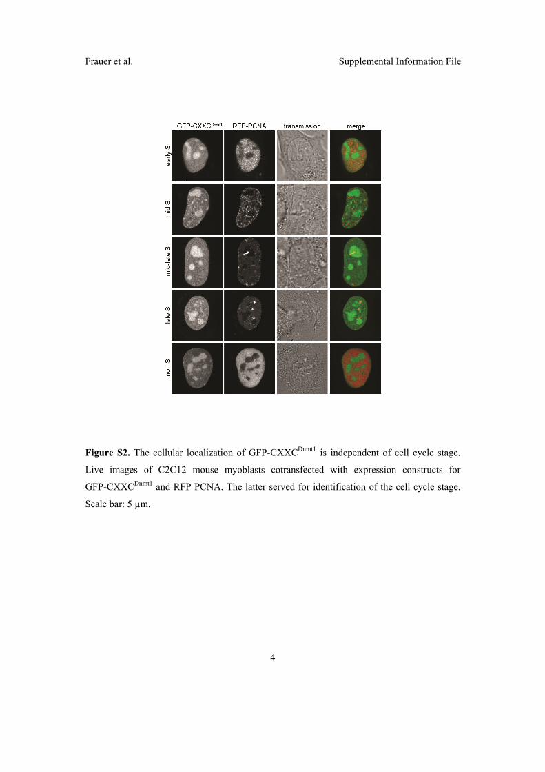
Frauer et al. Supplemental Information File
4
�
Figure S2. The cellular localization of GFP-CXXCDnmt1 is independent of cell cycle stage.
Live images of C2C12 mouse myoblasts cotransfected with expression constructs for
GFP-CXXCDnmt1 and RFP PCNA. The latter served for identification of the cell cycle stage.
Scale bar: 5 µm.

Frauer et al. Supplemental Information File
5
�
Figure S3. Differential mobility of GFP-CXXCDnmt1 in nucleoli and nucleoplasm of mouse
C2C12 myoblasts measured by FRAP analysis. Identical regions of interest over the
nucleoplasm or nucleoli (as exemplified in the inset) were bleached and recovery curves were
recorded over 30 seconds. GFP-CXXCDnmt1 kinetics is faster in nucleoli than in the nucleus,
which indicates more transient (possibly unspecific) binding in the former than in the latter.
Scale bar: 5 µm.
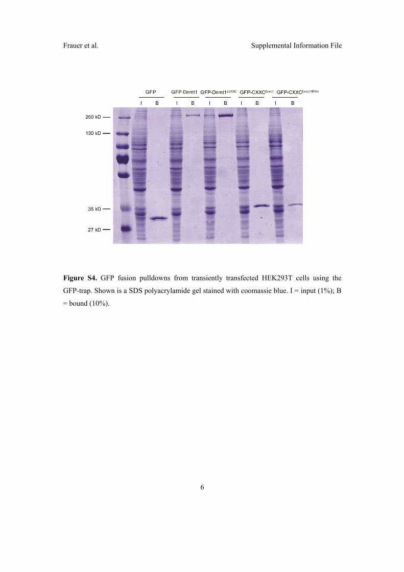
Frauer et al. Supplemental Information File
6
�
Figure S4. GFP fusion pulldowns from transiently transfected HEK293T cells using the
GFP-trap. Shown is a SDS polyacrylamide gel stained with coomassie blue. I = input (1%); B
= bound (10%).

Frauer et al. Supplemental Information File
7
�
Figure S5. The CXXC domain of Dnmt1 preferentially binds unmethylated CpG sites. GFP
and GFP-CXXCDnmt1 purified from transiently transfected HEK293T cells with the GFP trap
were challenged with fluorescent DNA substrates containing no CpG site or one central un-,
hemi- or fully methylated CpG site in direct competition (noCGB, UMB, HMB and FMB,
respectively) as in Figure 2C, except that a five-fold higher concentration (625 nM) of each
substrate was used.
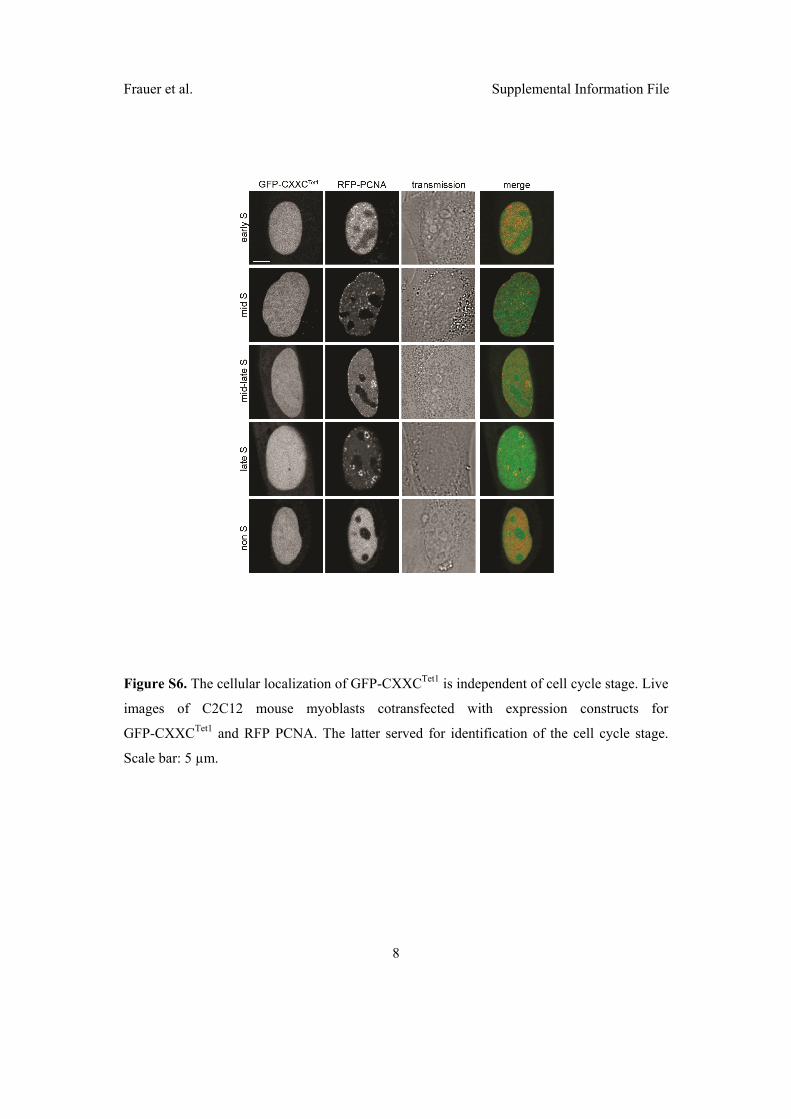
Frauer et al. Supplemental Information File
8
�
Figure S6. The cellular localization of GFP-CXXCTet1 is independent of cell cycle stage. Live
images of C2C12 mouse myoblasts cotransfected with expression constructs for
GFP-CXXCTet1 and RFP PCNA. The latter served for identification of the cell cycle stage.
Scale bar: 5 µm.

Frauer et al. Supplemental Information File
9
�
Figure S7. Radioactive methyltransferase activity assay for GFP Dnmt1 and
GFP-Dnmt1¨CXXC. The transfer of [3H]-methyl groups to poly(dI•dC)-poly(dI•dC) substrate
was measured for increasing volumes of GFP fusion proteins immunopurified from
transiently transfected HEK293T cells. Counts per minute (cpm) were normalized to the
relative protein concentration as determined by SDS-PAGE analysis. GFP was used as
negative control. Numbers above the bars indicate the volume (µl) of protein solution added.
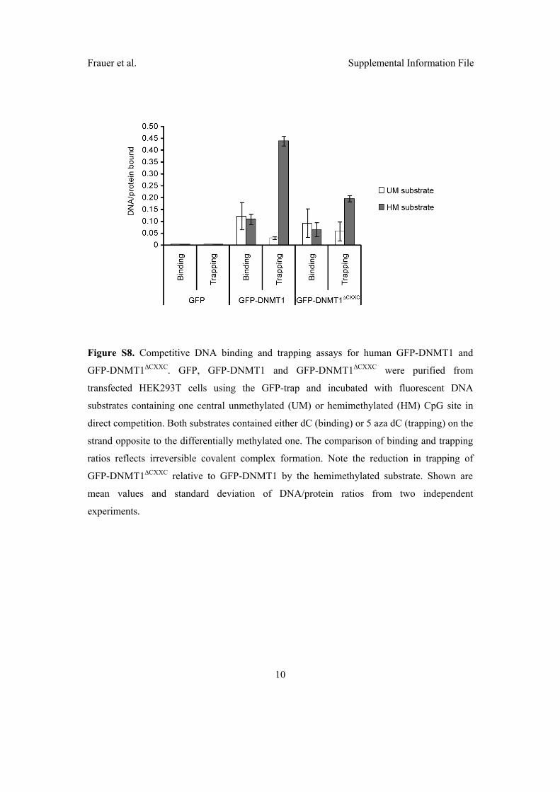
Frauer et al. Supplemental Information File
10
�
Figure S8. Competitive DNA binding and trapping assays for human GFP-DNMT1 and
GFP-DNMT1¨CXXC. GFP, GFP-DNMT1 and GFP-DNMT1¨CXXC were purified from
transfected HEK293T cells using the GFP-trap and incubated with fluorescent DNA
substrates containing one central unmethylated (UM) or hemimethylated (HM) CpG site in
direct competition. Both substrates contained either dC (binding) or 5 aza dC (trapping) on the
strand opposite to the differentially methylated one. The comparison of binding and trapping
ratios reflects irreversible covalent complex formation. Note the reduction in trapping of
GFP-DNMT1¨CXXC relative to GFP-DNMT1 by the hemimethylated substrate. Shown are
mean values and standard deviation of DNA/protein ratios from two independent
experiments.

Frauer et al. Supplemental Information File
11
�
SUPPLEMENTARY METHODS
In vitro methyltransferase activity assay
Eight milligrams of His-tagged GFP-binding protein (GBP; Chromotek) were coupled to 1ml
Ni-NTA agarose beads (Qiagen) by incubating for 2 h at 4°C in PBS and unbound protein
was washed out twice with PBS. Extracts of HEK293T cells expressing GFP or a GFP fusions
were prepared in 200 µl lysis buffer II (50 mM NaH2PO4 pH 8.0, 300 mM NaCl, 10 mM
imidazole, 0.5 % Tween-20, 2 mM MgCl2, 1 mg/ml DNaseI, 2 mM PMSF, 1X mammalian
protease inhibitor mix). After centrifugation, supernatants were diluted to 500 µl with
immunoprecipitation buffer II (50 mM NaH2PO4 pH 8.0, 300 mM NaCl, 10 mM imidazole,
0.05 % Tween-20) and precleared by incubation with 25 µl of equilibrated Ni-NTA agarose
beads for 30 min at 4°C followed by centrifugation. Precleared extracts were then incubated
with 40 µg of His-tagged GFP-trap coupled to Ni-NTA beads for 2 h at 4°C with constant
mixing. GFP or GFP fusions were pulled down by centrifugation at 540 g. After washing
twice with wash buffer II (50 mM NaH2PO4 pH 8.0, 300 mM NaCl, 20 mM imidazole, 0.05
% Tween-20), complexes were eluted with 60 µl of elution buffer (10 mM Tris pH 7.5, 100
mM KCl, 1 mM EDTA, 1 mM DTT, 250 mM imidazole) for 10 min at 25°C with constant
mixing. 10 µl aliquots of all eluates were subjected to western blot analysis using mouse or rat
monoclonal antibodies to GFP (Roche and Chromotek, respectively) and quantified by
densitometry. Indicated volumes of eluate were incubated with 1 µg of poly(dI·dC)-
poly(dI·dC) substrate (Sigma), 0.5 µg/µl of BSA and 1 µCi of S-adenosyl-[3H-
methyl]-methionine in 50 µl of trapping buffer (10 mM Tris pH 7.5, 100 mM KCl, 1 mM
EDTA, 1 mM DTT) for 60 min at 37°C. 15 µl of each sample were spotted onto blotting
paper and the DNA was precipitated with ice cold 5 % TCA. After washing twice with 5%
TCA and once with cold 70 % ethanol, paper filters were air dried and analyzed by
scintillation in 4 ml scintillation cocktail (Rotiszint® eco plus, Roth) for 5 min.

2.4 Genomic 5-hydroxymethylcytosine levels correlate
with TET2 mutations and a distinct global gene
expression pattern in secondary acute myeloid
leukemia

2 He Y, Wertheim JA, Xu L, Miller JP, Karnell FG, Choi JK et al. Thecoiled-coil domain and Tyr177 of bcr are required to induce amurine chronic myelogenous leukemia-like disease by bcr/abl.Blood 2002; 99: 2957–2968.
3 Shtivelman E, Lifshitz B, Gale RP, Canaani E. Fused transcript ofabl and bcr genes in chronic myelogenous leukaemia. Nature1985; 315: 550–554.
4 Golub TR, Barker GF, Bohlander SK, Hiebert SW, Ward DC,Bray-Ward P et al. Fusion of the TEL gene on 12p13 to the AML1gene on 21q22 in acute lymphoblastic leukemia. Proc Natl AcadSci USA 1995; 92: 4917–4921.
5 De Braekeleer E, Douet-Guilbert N, Le Bris MJ, Berthou C,Morel F, De Braekeleer M. A new partner gene fused to ABL1 in at(1;9)(q24;q34)-associated B-cell acute lymphoblastic leukemia.Leukemia 2007; 21: 2220–2221.
6 De Keersmaecker K, Graux C, Odero MD, Mentens N, Somers R,Maertens J et al. Fusion of EML1 to ABL1 in T-cell acutelymphoblastic leukemia with cryptic t(9;14)(q34;q32). Blood2005; 105: 4849–4852.
7 Graux C, Cools J, Melotte C, Quentmeier H, Ferrando A,Levine R et al. Fusion of NUP214 to ABL1 on amplified episomesin T-cell acute lymphoblastic leukemia. Nat Genet 2004; 36:1084–1089.
8 Soler G, Radford-Weiss I, Ben-Abdelali R, Mahlaoui N,Ponceau JF, Macintyre EA et al. Fusion of ZMIZ1 to ABL1 in a B-cell acute lymphoblastic leukaemia with a t(9;10)(q34;q22.3)translocation. Leukemia 2008; 22: 1278–1280.
9 Hidalgo-Curtis C, Chase A, Drachenberg M, Roberts MW, Finkel-stein JZ, Mould S et al. The t(1;9)(p34;q34) and t(8;12)(p11;q15) fusepre-mRNA processing proteins SFPQ (PSF) and CPSF6 to ABL andFGFR1. Genes Chromosomes Cancer 2008; 47: 379–385.
10 De Braekeleer E, Douet-Guilbert N, Rowe D, Bown N, Morel F,Berthou C et al. ABL1 fusion genes in hematological malignancies:a review. Eur J Haematol 2011; 86: 361–371.
11 Damen JE, Liu L, Rosten P, Humphries RK, Jefferson AB,Majerus PW et al. The 145-kDa protein induced to associate withShc by multiple cytokines is an inositol tetraphosphate andphosphatidylinositol 3,4,5-triphosphate 5-phosphatase. Proc NatlAcad Sci USA 1996; 93: 1689–1693.
12 Helgason CD, Damen JE, Rosten P, Grewal R, Sorensen P,Chappel SM et al. Targeted disruption of SHIP leads tohemopoietic perturbations, lung pathology, and a shortened lifespan. Genes Dev 1998; 12: 1610–1620.
13 Luo JM, Yoshida H, Komura S, Ohishi N, Pan L, Shigeno K et al.Possible dominant-negative mutation of the SHIP gene in acutemyeloid leukemia. Leukemia 2003; 17: 1–8.
14 Gilby DC, Goodeve AC, Winship PR, Valk PJ, Delwel R, Reilly JT.Gene structure, expression profiling and mutation analysis of thetumour suppressor SHIP1 in Caucasian acute myeloid leukaemia.Leukemia 2007; 21: 2390–2393.
15 Lo TC, Barnhill LM, Kim Y, Nakae EA, Yu AL, Diccianni MB.Inactivation of SHIP1 in T-cell acute lymphoblastic leukemia dueto mutation and extensive alternative splicing. Leuk Res 2009; 33:1562–1566.
Supplementary Information accompanies the paper on the Leukemia website (http://www.nature.com/leu)
Genomic 5-hydroxymethylcytosine levels correlate with TET2 mutations and a distinctglobal gene expression pattern in secondary acute myeloid leukemia
Leukemia (2011) 25, 1649–1652; doi:10.1038/leu.2011.134;published online 31 May 2011
The TET proteins are 2-oxoglutarate- and Fe(II)-dependentoxygenase catalyzing the conversion of 5-methylcytosine(5mC) to 5-hydroxymethylcytosine (5hmC).1 The TET1(ten–eleven translocation 1) gene was originally identified asan MLL fusion partner in rare cases of acute myeloid leukemia(AML) with a t(10;11)(q22;q23).2,3 The definite function of5hmC still remains elusive, but hydroxylation of 5mC has beensuggested to be involved in the process of DNA demethylation.This suggests a possible role of 5hmC in epigenetic generegulation. Recently, hemizygous deletions and mutations ofTET2 were found in a wide range of myeloid malignancies,including myelodysplastic syndrome (MDS), myeloproliferativedisorders such as chronic myelomonocytic leukemia (CMML)and in secondary AML (sAML).4–6 Interestingly, very recently,myeloid neoplasias harboring heterozygous TET2 mutationswere shown to have decreased levels of 5hmC.7
To explore the relationship among TET2 mutations, globalgene expression profiles (GEPs) and 5hmC levels, we measured5hmC levels in the genomic DNA in a series of 30 sAML patientsusing a novel assay method employing b-glucosyltransferasefrom bacteriophage T4.8 In addition to the TET2 mutationalstatus, we screened for IDH1/2 mutations (see SupplementaryMaterial).
All patients had developed AML after a preceding MDS,refractory anemia with excess blast or CMML phase. Theaverage age at diagnosis was 70.8 years. Eight patients had anormal karyotype (nk), eight patients had a complex aberrant
karyotype (ak) with more than three chromosomal aberrationsand the remainder of the patients had one or two chromosomalaberrations, which are typical of MDS, that is, del(5)(q)(two patients), ! 8 (five patients), "7 or del(7)(q) (six patients;see Supplementary Table 1). We sequenced the completecoding region of TET2 in all 30 patients. In all, 7 of the30 patients (23.3%) had TET2 mutations. One patient (no. 16)had single-nucleotide deletions in both alleles of TET2 atamino-acid positions 218 and 519, which caused truncationof the protein after 250 and 533 amino acids, respectively.Two patients (nos. 15 and 26) had nonsense mutationsat positions 1216 and 1274, and four patients (nos. 7, 14, 20and 30) had missense mutations (see Table 1). All the TET2mutations (except for patient no. 16) were heterozygous.We did not detect any deletions in patients with TET2 mutationsusing a commercially available fluorescence in situ hybridiza-tion probe for the TET2 locus. There was no significantassociation between TET2 mutational status and any particularchromosomal abnormality. Although there was a trend towarda higher frequency of TET2 mutations in patients with a nk incomparison with patients with an ak (50% (4 out of 8 nkpatients) versus 13.6% (3 out of 22 ak patients), w2-test:P# 0.17).
The analysis of the 5hmC levels of the patients’ DNA usingthe b-glucosyltransferase assay revealed a 5hmC contentof the DNA, ranging from 0.006 to 0.054%. This range of5hmC levels, about 9- to 14-fold difference between thelowest and highest measurements, agrees well withthe measurements reported by Ko et al.,7 although verydifferent patient groups were assayed. In contrast to the
Letters to the Editor
1649
Leukemia
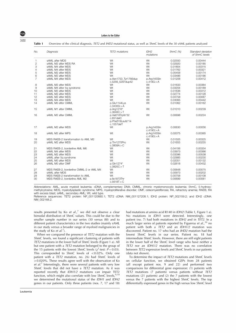
results presented by Ko et al.,7 we did not observe a clearbimodal distribution of 5hmC values. This could be due to thesmaller sample number in our series (30 versus 88) and todifferent patient characteristics in the two studies (mainly sAMLin our study versus a broader range of myeloid malignancies inthe study of Ko et al.7).
When we compared the presence of TET2 mutation with the5hmC levels, we found a significant clustering of patients withTET2 mutations in the lower half of 5hmC levels (Figure 1 a). Allbut one patient with a TET2 mutation belonged to the group ofthe 15 patients with the lowest 5hmC levels (w2-test: P# 0.03).This corresponded to 5hmC levels of o0.02%. Only onepatient with a TET2 mutation, no. 26) had 5hmC levels of40.020%. These results agree well with the observation of Koet al.7 Interestingly, there were several patients with very low5hmC levels that did not have a TET2 mutation. As it wasreported recently that IDH1/2 mutations can impair TET2function, which might also correlate with low 5hmC levels,9,10
we determined the mutational status of the IDH1 and IDH2genes in our patients. Only three patients (nos. 7, 17 and 18)
had mutations at amino acid R140 in IDH2 (Table 1, Figure 1 a).No mutations in IDH1 were detected. Interestingly, onepatient (no. 7) had both mutations in IDH2 and in TET2. In amuch larger series of patients reported by Figueroa et al.,10 nopatient with both a TET2 and an IDH1/2 mutation wasdiscovered. Patient no. 17 who had an IHD2 mutation had thelowest 5hmC levels in our series. Patient no. 18 hadintermediate 5hmC levels. However, there are still eight patientsin the lower half of the 5hmC level range who have neither aTET2 nor an IDH1/2 mutation. There was no correlationbetween TET2 expression levels and 5hmC levels in our patients(data not shown).
To determine the impact of TET2 mutations and 5hmC levelson cellular function, we obtained GEPs from 28 patients(all except patient nos. 9 and 22) and performed twocomparisons for differential gene expression: (1) patients withTET2 mutations (7 patients) versus patients without TET2mutations (21 patients) and (2) the 7 patients with the lowestversus the 7 patients with the highest 5hmC levels. The topdifferentially expressed genes in the high versus low 5hmC level
Table 1 Overview of the clinical diagnosis, TET2 and IHD2 mutational status, as well as 5hmC levels of the 30 sAML patients analyzed
No. Diagnosis TET2 mutations IDH2mutations
5hmC (%) Standard deviationof 5hmC levels
1 sAML after MDS Wt Wt 0.02550 0.004442 sAML M2 after MDS RA Wt Wt 0.02920 0.001853 sAML M4 after MDS Wt Wt 0.01804 0.003154 sAML M4 after MDS Wt Wt 0.01762 0.003215 sAML M2 after MDS Wt Wt 0.05458 0.001746 sAML M2 after MDS Wt Wt 0.03486 0.001867 sAML M4 after MDS p.Asn1753_Tyr1766dup
c.5256_5297dup42p.Arg140Glnc.419G4A
0.01208 0.00412
8 sAML M2 after MDS Wt Wt 0.01633 0.003849 sAML M4 after 5q syndrome Wt Wt 0.03204 0.00189
10 sAML M2 after MDS Wt Wt 0.01636 0.0031211 sAML M2 after MDS Wt Wt 0.02774 0.0012812 sAML M2 after MDS Wt Wt 0.04758 0.0008713 sAML M4 after MDS Wt Wt 0.03065 0.0044214 sAML M0 after CMML p.Glu1144Lys
c.3430G4AWt 0.01062 0.00162
15 sAML M1 after CMML p.Arg1216*c.3646C4T
Wt 0.01010 0.00239
16 sAML M2 after CMML p.Val218Trpfs*32c.651delCp.Phe519Leufs*14c.1557delT
Wt 0.00698 0.00224
17 sAML M2 after MDS Wt p.Arg140Glnc.419G4A
0.00630 0.00056
18 sAML M2 after MPS Wt p.Arg140Glnc.419G4A
0.02075 0.00385
19 MDS RAEB-2 transformation to AML M2 Wt Wt 0.01505 0.0032520 sAML M2 after MDS p.Thr1270Pro
c.3808A4CWt 0.01655 0.00255
21 MDS RAEB-2, borderline AML M6 Wt Wt 0.04156 0.0033422 sAML M4 after MDS Wt Wt 0.03973 0.0038623 sAML M2 after MDS Wt Wt 0.03396 0.0053824 sAML after 5q syndrome Wt Wt 0.02885 0.0023025 sAML M0 after MDS Wt Wt 0.02051 0.0013226 sAML M0 after OMF p.Gln1274*
c.3820C4TWt 0.02618 0.00432
27 MDS RAEB-2, borderline CMML-2 or AML M4 Wt Wt 0.00648 0.0007528 sAML after MDS Wt Wt 0.00973 0.0020229 MDS RAEB-2 transformation to AML Wt Wt 0.00758 0.0010830 MDS RAEB-2, borderline AML M2 p.Ile1873Thr
c.5618T4CWt 0.00699 0.00081
Abbreviations: AML, acute myeloid leukemia; cDNA, complementary DNA; CMML, chronic myelomonocytic leukemia; 5hmC, 5-hydroxy-methylcytosine; MDS, myelodysplastic syndrome; MPS, myeloproliferative disorder; OMF, osteomyelofibrosis; RA, refractory anemia; RAEB, RAwith excess blast; sAML, secondary AML; Wt, wild type.Reference sequences: TET2 protein: NP_001120680.1; TET2 cDNA: NM_001127208.1; IDH2 protein: NP_002159.2; and IDH2 cDNA:NM_002168.2.
Letters to the Editor
1650
Leukemia

Figure 1 (a) Bar graph of 5hmC (%) levels of the 30 patients of this study. Patients with a TET2 mutation or an IDH2 mutation are indicated with ared or a blue rectangle, respectively. The yellow bars indicate the samples that were used for the differential gene expression analysis in thecomparison of low versus high 5hmC levels. Note that the patients 9 and 22 did not have gene expression data of sufficient quality to be includedin this analysis. (b) Heatmaps of the 50 most significantly differentially expressed probe sets comparing samples with TET2 mutations (n#7) versusTET2 wild type (N#21; top) and samples with low (n# 7) and high (n# 7) 5hmC levels (bottom).
Letters to the Editor
1651
Leukemia
comparison had a lower P-value and had a higher degreeof deregulation than the differentially expressed genes fromthe comparison TET2 mutated versus wild type (Figure 1 b;Supplementary Figure 1 and Supplementary Tables 2 and 3).These results indicate that 5hmC levels are most likely a morerelevant measurement to define biologically distinct secondaryleukemia subtypes than the TET2 (or IDH1/2) mutational status.The fact that in some patient samples with low 5hmC levelsneither TET2 nor IDH1/2 mutations could be identified suggeststhat additional genes might be directly or indirectly involved inthe regulation of 5hmC levels. To further elucidate theregulation of 5hmC levels and their role in leukemogenesis,larger groups of sAML as well as de novo AML patients needto be studied.
Conflict of interest
The authors declare no conflict of interest.
Acknowledgements
SKB, KS and HL are supported by grants from the DeutscheForschungsgemeinschaft (SFB 684 and SPP 1463). SKB issupported by a grant from the German Ministry of Educationand Research (BMBF) National genome research network(NGFNplus; PKL-01GS0876-6) and by institutional funding fromthe Helmholtz Zentrum Munich, German Research Center forEnvironmental Health. We thank Natalia Huk for technicalassistance and Alexander Kohlmann, Torsten Haferlach andClaudia Haferlach for primary gene expression data andcytogenetic data.
Author Contributions
NK and FS designed and performed the mutation screening withthe help of AD and BK, and wrote the manuscript. SB, AS andHL designed and performed the 5hmC measurements and wrotethe manuscript. HL supervised the project. PMK and SSperformed cytogenetics and fluorescence in situ hybridizationanalysis. TH and MM analyzed the GEPs. KS designedexperiments and wrote the manuscript. SKB designed experi-ments, analyzed the data, supervised the project and wrote themanuscript.
N Konstandin1,2,4, S Bultmann3,4, A Szwagierczak3,A Dufour1, B Ksienzyk1,2, F Schneider1,2, T Herold1,
M Mulaw2, PM Kakadia1,2, S Schneider1, K Spiekermann1,2,H Leonhardt3,4 and SK Bohlander1,2,4
1Laboratory of Leukemia Diagnostics, Department ofMedicine III, University of Munich Hospital,
Munich, Germany;2Helmholtz Zentrum Munich, National Research Center for
Environmental Health, Clinical Cooperative Group‘Leukemia’, Munich, Germany and
3Department of Biology II, Center for Integrated ProteinScience, Ludwig Maximilians University Munich,
Planegg-Martinsried, GermanyE-mail: [email protected] or
[email protected] authors contributed equally to this work.
References
1 Tahiliani M, Koh KP, Shen Y, Pastor WA, Bandukwala H, Brudno Yet al. Conversion of 5-methylcytosine to 5-hydroxymethylcytosinein mammalian DNA by MLL partner TET1. Science 2009; 324:930–935.
2 Ono R, Taki T, Taketani T, Taniwaki M, Kobayashi H, Hayashi Y.LCX, leukemia-associated protein with a CXXC domain, is fused toMLL in acute myeloid leukemia with trilineage dysplasia havingt(10;11)(q22;q23). Cancer Res 2002; 62: 4075–4080.
3 Lorsbach RB, Moore J, Mathew S, Raimondi SC, Mukatira ST,Downing JR. TET1, a member of a novel protein family, is fused toMLL in acute myeloid leukemia containing the t(10;11)(q22;q23).Leukemia 2003; 17: 637–641.
4 Mohamedali AM, Smith AE, Gaken J, Lea NC, Mian SA,Westwood NB et al. Novel TET2 mutations associated withUPD4q24 in myelodysplastic syndrome. J Clin Oncol 2009; 27:4002–4006.
5 Langemeijer SM, Kuiper RP, Berends M, Knops R, Aslanyan MG,Massop M et al. Acquired mutations in TET2 are common inmyelodysplastic syndromes. Nat Genet 2009; 41: 838–842.
6 Delhommeau F, Dupont S, Della Valle V, James C, Trannoy S,Masse A et al. Mutation in TET2 in myeloid cancers. N Engl J Med2009; 360: 2289–2301.
7 Ko M, Huang Y, Jankowska AM, Pape UJ, Tahiliani M,Bandukwala HS et al. Impaired hydroxylation of 5-methylcytosinein myeloid cancers with mutant TET2. Nature 2010; 468:839–843.
8 Szwagierczak A, Bultmann S, Schmidt CS, Spada F, Leonhardt H.Sensitive enzymatic quantification of 5-hydroxymethylcytosine ingenomic DNA. Nucleic Acids Res 2010; 38: e181.
9 Xu W, Yang H, Liu Y, Yang Y, Wang P, Kim SH et al.Oncometabolite 2-hydroxyglutarate is a competitive inhibitor ofa-ketoglutarate-dependent dioxygenases. Cancer Cell 2011; 19:17–30.
10 Figueroa ME, Abdel-Wahab O, Lu C, Ward PS, Patel J, Shih Aet al. Leukemic IDH1 and IDH2 mutations result in ahypermethylation phenotype, disrupt TET2 function, andimpair hematopoietic differentiation. Cancer Cell 2010; 18:553–567.
Supplementary Information accompanies the paper on the Leukemia website (http://www.nature.com/leu)
Immunophenotype-defined sub-populations are common at diagnosis in childhoodB-cell precursor acute lymphoblastic leukemia
Leukemia (2011) 25, 1652–1657; doi:10.1038/leu.2011.136;published online 17 June 2011
Neoplasms often display significant heterogeneity in morpho-logy, gene expression (including cell surface markers), genetic
aberrations, cell proliferation kinetics and response to therapy.1
Heterogeneity in antigen marker expression is well known inacute myeloid leukemia (AML);2 however, has rarely beenstudied in acute lymphoblastic leukemia (ALL). In ALL, shifts inimmunophenotypic and genetic profiles can occur between
Letters to the Editor
1652
Leukemia
Konstandin et al. Supplemental Material Page 1
Supplemental Information:
Methods:
Patient Samples
Patient samples were obtained from the Laboratory for Leukemia Diagnostics, University of
Munich Hospital Grosshadern. The samples were received for routine cytogenetics and
molecular genetics diagnostics. The institutional review board of the University Hospital
Munich has approved the use of left over diagnostic samples for this research project in an
anonymous fashion.
Cytogenetics and FISH
Each sample was routinely karyotyped according to standard protocols (GTG banding). All
patients were screened for deletions of the TET2 locus using a fluorescence in situ
hybridization (FISH) probe covering the TET2 gene from marker RH43141 to RH69608 (XL
TET2, MetaSystems, Altlussheim, Germany).
Mutation Analysis
Sample Preparation
Cells from diagnostic bone marrow and peripheral blood samples were enriched by Ficoll
denstity gradient centrifugation and thereafter lysed by RLT buffer (Quiagen, Hilden,
Germany) and stored at -80 °C.
Genomic DNA was extracted from patient samples with secondary AML using QIAamp DNA
mini Kit (Quiagen, Hilden, Germany).
Sequencing of TET2
Sanger sequencing analysis was performed on PCR-amplified genomic DNA fragments
spanning the entire coding region of TET2 isoform a (primers are shown below).
For the PCR of TET2, 50 ng of genomic DNA was used and amplified with Taq PCR Master
Mix Kit (Quiagen, Hilden, Germany) in a 25 µl reaction using the following program: 95°C for
5 min, 35 cycles at 95°C for 30 sec, 56°C for 30 sec and 72°C for 1 min with a terminal
elongation at 72 °C for 5 min.
Purified PCR fragments were sequenced using the Big Dye Terminator v1.1 Cycle
Sequencing Kit on 3100 Avant Genetic Analyzer (Applied Biosystems, Darmstadt, Germany).
Konstandin et al. Supplemental Material Page 2
All fragments were sequenced bidirectional and sequence variations were confirmed again
by sequencing from the original DNA.
Sequencing of IDH1/2
Sanger sequencing was performed on PCR- amplified genomic DNA fragments including the
genomic region of IDH1/2 containing the mutational hotspots R132 and SNP rs 11554137 in
the IDH1 exon 4, and R140 and R172 in the IDH2 exon 4.
For IDH1 the following primers were used:
IDH1-F 5’-CATAATGTTGGCGTCAAATGTG-3’, IDH1-R 5’-ACATGCAAAATCACATTATTGCC-3’For IDH2 the following primers were used:
IDH2-F 5’-GTTCAAGCTGAAGAAGATGTGG-3’, IDH2-R 5’-TGTGGCCTTGTACTGCAGAG-3’
For the PCR of IDH1 and IDH2, 50 ng of genomic DNA was used and amplified with Taq
PCR Master Mix Kit (Quiagen, Hilden, Germany) in a 25 µl reaction using the following
program: 94°C for 5 min, 35 cycles at 94°C for 1min, 56°C for 1min and 72°C for 1 min with a
terminal elongation at 72 °C for 10 min.
Sequencing was performed as described above
Konstandin et al. Supplemental Material Page 3
TET2 primers:
cDNA sequence: uc003hxk.2Genomic sequence: hg18_chr4:106286391-106421407
NAME LEFT_PRIMER RIGHT_PRIMER PRODUCT_SIZE
Exon3_1 CAGTTTGCTATGTCTAGGTATTCCG TTTCCCCTCCTGCTCATTC 740
Exon3_2 ATGTCTCCGATTTGAGTGATAAG CCCTGGATGTTATTTTCTGC 607
Exon3_3 TGCAATGCTAAATACCTGTTCC TTCTGACATTGGTCTTGTTTTCTC 607
Exon3_4 CATCTACACATGTATGCAGCCC TGGTCTGTTTTGGAGAAGTGC 607
Exon3_5 CCAAGTTGAAATGAATCAAGGG TGTATTGTTTGAACAAGAAACCTG 613
Exon3_6 CAAAATCAAGCGAGTTCGAG ATACAGGCATGTGGCTTGC 604
Exon3_7 GAAGCAAGAACAGCAGCAAAC TCTGAAGATAAATTTGCTAATTCTGG 600
Exon4 GCCCTTAATGTGTAGTTGGGG TGCTTTGTGTGTGAAGGCTG 271
Exon5 TGCCTCTTGAATTCATTTGC GGGTAACCCAATTCTCAGGG 234
Exon6 TGCAAGTGACCCTTGTTTTG CAACCAAAGATTGGGCTTTC 342
Exon7 CAGCTGCACAGCCTATATAATG TCACTTCATCTAAGCTAATGAATTCTC 279
Exon8 GGGATTCAAAATGTAAGGGG TGCAGTGGTTTCAACAATTAAG 323
Exon9 TGTCATTCCATTTTGTTTCTGG TCTGCTCCTCAACATGAGATG 691
Exon10 ACACACACGTTTTCTTTGGG cagaacttacaaGTTGATGGGG 511
Exon11_1 CCTACATTTAAGTATCCTCACTAGCC AACTGCTGAAACCATCTCCC 676
Exon11_2 AGGTATCCAAGCCAAGACCC GGATCCAGAAAGCTCTGCTC 593
Exon11_3 GTCAGGAAAAGCAGCCATTG CTGACAGGTTGGTTGTGGTC 594
5hmC Assay
For quantification of 5-hydroxymethylcytosine (hmC) in the genomic DNA samples a recently
published method was applied (1). This method utilizes the beta –glucosyltransferase (β-gt)
from the bacteriophage T4 which specifically transfers the glucose moiety from UDP-glucose
to hmC. By measuring the incorporation of radioactively labeled glucose into genomic DNA
the hmC content of the DNA can be calculated.
The bacteriophage T4 protein β-gt was expressed and purified as described before (1). In
brief, BL21(DE3) E. coli cells carrying the expression construct were grown at 37°C until A600
= 0.6–0.7 and induced with isopropyl β-d-thiogalactopyranoside for 16 h at 20°C. Cells were
lysed and the protein was purified using a nickel-nitrilotriacetic acid column (QIAGEN)
followed by gel filtration. Fractions containing the β-gt peak were pooled and applied to a
Konstandin et al. Supplemental Material Page 4
ResourceQ anion exchange column (GE Healthcare) in order to eliminate residual
contaminants, resulting in pure β-gt in the flowthrough.
Reference PCR fragments containing a known amount of 5hmC were produced as described
before (1).
Genomic DNA samples were sheared to 500–1500_bp fragments by sonication to reduce the
viscosity and improve homogeneity.
Reactions contained 150 mM NaCl, 20 mM Tris, pH 8.0, 25 mM CaCl2, 1 mM DTT, 0.86
nM UDP-[3H]glucose (glucose-6-3H; 60 Ci/mmol; Hartmann Analytic GmbH), 500 ng of DNA
substrate and 100 nM recombinant β -gt in a total volume of 50 µl. Reactions were
incubated for 20 min at room temperature and terminated by heating at 65°C for 10 min.
Unincorporated nucleotides were removed by using the NucleoSpin® Extract II kit
(Macherey-Nagel) according to manufacturers protocol. Remaining radioactivity was
measured using a Liquid Scintillation Analyzer Tri-Carb 2100TR (Packard) with quench
indicating parameter set on tSIE/AEC (transformed spectral index of the external
standard/automatic efficiency control) in 4 ml of Rotiszint Eco Plus scintillation liquid (Roth
GmbH) in Snaptwist vials (Simport). Samples were measured for 30 min or until the 2σ
value reached 2%. The percentage of hmC per total cytosine was calculated from the
incorporation of [3H]glucose using a calibration curve measured with the reference fragment
series for every experiment. The percentage of hmC was then corrected for the difference in
C abundance between reference fragment (17,5%) and human genome (20,5%).
Gene Expression profiles
For the expression analysis, RNA from patient samples was labeled and hybridized to
Affymetrix HG-U133A and HG-U133B or HG-U133plus2 chips as previously described (2).
The HG-U133A, HG-U133B and HG-U133plus2 .CEL files were first normalized separately
using robust multi-array (RMA) normalization and then combined into one matrix and
normalized all together using the empirical Bayes (eB) method as previously described (3).
The normalized expression data were analyzed with the twilight program (4) in the R
software package (http://www.r-project.org/).
Konstandin et al. Supplemental Material Page 5
Supplemental Figure Legends:
Figure 1:
Top:
Degree of up- and down regulation of the 50 most significantly deregulatd probe sets in the
comparison TET2 mutated (n = 7) versus TET2 wild type (n =21). The genes toward the right
side are upregulated in TET2 mutated samples. The scale is linear (i.e. 2 denotes a 2 fold
higher expression in TET2 mutated samples).
Bottom:
Degree of up- and down regulation of the 50 most significanlty deregulatd probe sets in the
comparison low (n = 7) versus high (n = 7) 5hmC levels. The genes toward the right side are
upregulated in samples with low 5hmC levels. The scale is linear (i.e. 2 denotes a 2 fold
higher expression in samples with low 5hmC levels).
Supplemental Tables:
Supplemental Table 1:
Additional clinical and karyotype data of the patients.
Supplemental Table 2:
The 50 most significantly deregulated probe sets in the comparison TET2 mutated (n = 7)
versus TET2 wild type (n =21).
Supplemental Table 3:
The 50 most significantly deregulated probe sets in the comparison low (n = 7) versus high (n
= 7) 5hmC levels.
References:
1. Szwagierczak A, Bultmann S, Schmidt CS, Spada F, Leonhardt H. Sensitive enzymatic
quantification of 5-hydroxymethylcytosine in genomic DNA. Nucleic Acids Res.
2010;38:e181.

Konstandin et al. Supplemental Material Page 6
2. Metzeler KH, Hummel M, Bloomfield CD, Spiekermann K, Braess J, Sauerland MC, et al.
An 86-probe-set gene-expression signature predicts survival in cytogenetically normal
acute myeloid leukemia. Blood. 2008;112:4193-201.
3. Johnson WE, Li C, Rabinovic A. Adjusting batch effects in microarray expression data
using empirical Bayes methods. Biostatistics. 2007;8:118-27.
4. Scheid S, Spang R. twilight; a Bioconductor package for estimating the local false
discovery rate. Bioinformatics. 2005;21:2921-2.

2.5 Recognition of 5-hydroxymethylcytosine by the
Uhrf1 SRA domain
Recognition of 5-Hydroxymethylcytosine by the Uhrf1SRA DomainCarina Frauer1,4., Thomas Hoffmann2,4., Sebastian Bultmann1,4, Valentina Casa3, M. Cristina Cardoso3,
Iris Antes2,4*, Heinrich Leonhardt1,4*
1 Department of Biology II, Ludwig Maximilians University Munich, Planegg-Martinsried, Germany, 2 Department of Life Sciences, Technical University Munich, Freising-
Weihenstephan, Germany, 3 Department of Biology, Technical University Darmstadt, Darmstadt, Germany, 4 Center for Integrated Protein Science Munich (CIPSM),
Munich, Germany
Abstract
Recent discovery of 5-hydroxymethylcytosine (5hmC) in genomic DNA raises the question how this sixth base isrecognized by cellular proteins. In contrast to the methyl-CpG binding domain (MBD) of MeCP2, we found that the SRAdomain of Uhrf1, an essential factor in DNA maintenance methylation, binds 5hmC and 5-methylcytosine containingsubstrates with similar affinity. Based on the co-crystal structure, we performed molecular dynamics simulations of theSRA:DNA complex with the flipped cytosine base carrying either of these epigenetic modifications. Our data indicate thatthe SRA binding pocket can accommodate 5hmC and stabilizes the flipped base by hydrogen bond formation with thehydroxyl group.
Citation: Frauer C, Hoffmann T, Bultmann S, Casa V, Cardoso MC, et al. (2011) Recognition of 5-Hydroxymethylcytosine by the Uhrf1 SRA Domain. PLoS ONE 6(6):e21306. doi:10.1371/journal.pone.0021306
Editor: Shuang-yong Xu, New England Biolabs, Inc., United States of America
Received March 18, 2011; Accepted May 25, 2011; Published June 22, 2011
Copyright: ! 2011 Frauer et al. This is an open-access article distributed under the terms of the Creative Commons Attribution License, which permitsunrestricted use, distribution, and reproduction in any medium, provided the original author and source are credited.
Funding: This work was supported by grants from the Nanosystems Initiative Munich (NIM, http://www.nano-initiative-munich.de), the Center for NanoSciences(CeNS, http://www.cens.de) Munich, and by grants from the Deutsche Forschungsgemeinschaft (DFG, SFB TR5, http://www.dfg.de) to HL. CF gratefullyacknowledges support by the Elite Network of Bavaria (International Doctorate Program NanoBioTechnology, IDK-NBT, http://www.cens.de/doctorate-program)and the International Max Planck Research School for Molecular and Cellular Life Sciences (IMPRS-LS, http://www.imprs-ls.de). SB was supported by the GraduateSchool for Life Sciences Munich (LSM, http://www.lsm.bio.lmu.de). The funders had no role in study design, data collection and analysis, decision to publish, orpreparation of the manuscript.
Competing Interests: The authors have declared that no competing interests exist.
* E-mail: [email protected] (IA); [email protected] (HL)
. These authors contributed equally to this work.
Introduction
DNA methylation is an epigenetic modification that is wellknown to control eukaryotic gene expression [1,2]. In fact,methylation of regulatory sequences often correlates with atranscriptionally silent state. DNA methylation in mammals occursas 5-methylcytosine (5mC) within CpG dinucleotides and iscatalyzed by a family of DNA methyltransferases (Dnmts) [3].Dnmt members are distinguished by their function; while the denovo methyltransferases Dnmt3a and Dnmt3b establish methyla-tion patterns during development and cellular differentiation [4,5],the maintenance methyltransferase Dnmt1 copies these patternsduring DNA replication [6,7,8]. Although DNA methylation perse can prevent binding of transcriptional regulators [9], the mainmechanism by which transcriptional repression is achievedappears to involve 5mC binding proteins (MBPs). MBPsspecifically recognize methylation marks and consequently stabi-lize silent chromatin states by recruitment of histone modifyingenzymes and chromatin remodeling factors [10].
There are three families of MBPs known to date: the methyl-CpG binding domain (MBD) family, the Uhrf family and theKaiso protein family. In contrast to the members of the MBD andKaiso families that specifically recognize fully methylated CpGsites, Uhrf1, the best characterized member of the Uhrf family,preferentially binds hemimethylated DNA, the substrate ofmaintenance methylation [11,12,13,14]. Notably, crystal struc-
tures of the DNA binding domains of MeCP2 and Uhrf1 incomplex with DNA revealed striking differences: whereas theMeCP2 MBD recognizes methylated CpG sites based onhydration of the DNA major groove, the Uhrf1 (Set and Ringassociated) SRA domain uses a base-flipping mechanism to bindDNA containing hemimethylated CpG sites [11,12,14,15].Interestingly, Uhrf1 recently emerged as essential cofactor formaintenance methylation potentially by recruiting Dnmt1 to itstarget sites [13,16,17].
In addition to 5mC, genomic DNA has been recently shown tocontain 5-hydroxy-methylcytosine (5hmC), which results fromoxidation of 5mC catalyzed by Tet proteins [18,19,20]. This newmodification has been implicated in DNA demethylation, eitherpassively as 5hmC containing DNA is not a substrate for Dnmt1[21], or actively by so far unknown mechanisms. The centralquestions remain which proteins recognize 5hmC modified DNAand whether 5hmC has a direct role in gene regulation similar toits analog 5mC.
In this study, we characterized the 5mC/5hmC DNA bindingproperties of two representative 5mC binding protein domains, theMBD of MeCP2 and the SRA domain of Uhrf1. We found that incontrast to the MBD, the SRA domain binds hydroxymethylatedDNA substrates with similar affinity as methylated substrates. Weinvestigated the binding mode and energies of Uhrf1 to DNAsubstrates containing 5mC and 5hmC using molecular dynamicssimulations of the respective SRA:DNA complexes.
PLoS ONE | www.plosone.org 1 June 2011 | Volume 6 | Issue 6 | e21306
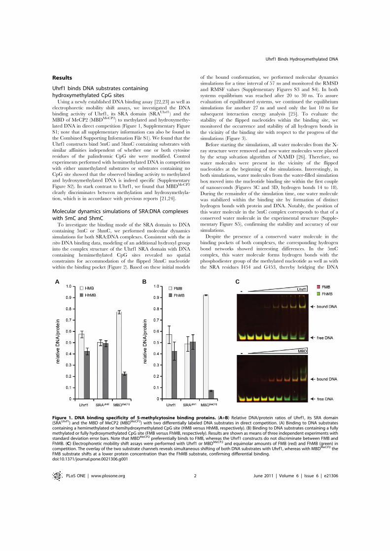
Results
Uhrf1 binds DNA substrates containinghydroxymethylated CpG sites
Using a newly established DNA binding assay [22,23] as well aselectrophoretic mobility shift assays, we investigated the DNAbinding activity of Uhrf1, its SRA domain (SRAUhrf1) and theMBD of MeCP2 (MBDMeCP2) to methylated and hydroxymethy-lated DNA in direct competition (Figure 1, Supplementary FigureS1; note that all supplementary information can also be found inthe Combined Supporting Information File S1). We found that theUhrf1 constructs bind 5mC and 5hmC containing substrates withsimilar affinities independent of whether one or both cytosineresidues of the palindromic CpG site were modified. Controlexperiments performed with hemimethylated DNA in competitionwith either unmethylated substrates or substrates containing noCpG site showed that the observed binding activity to methylatedand hydroxymethylated DNA is indeed specific (SupplementaryFigure S2). In stark contrast to Uhrf1, we found that MBDMeCP2
clearly discriminates between methylation and hydroxymethyla-tion, which is in accordance with previous reports [21,24].
Molecular dynamics simulations of SRA:DNA complexeswith 5mC and 5hmC
To investigate the binding mode of the SRA domain to DNAcontaining 5mC or 5hmC, we performed molecular dynamicssimulations for both SRA:DNA complexes. Consistent with the invitro DNA binding data, modeling of an additional hydroxyl groupinto the complex structure of the Uhrf1 SRA domain with DNAcontaining hemimethylated CpG sites revealed no spatialconstraints for accommodation of the flipped 5hmC nucleotidewithin the binding pocket (Figure 2). Based on these initial models
of the bound conformation, we performed molecular dynamicssimulations for a time interval of 57 ns and monitored the RMSDand RMSF values (Supplementary Figures S3 and S4). In bothsystems equilibrium was reached after 20 to 30 ns. To assureevaluation of equilibrated systems, we continued the equilibriumsimulations for another 27 ns and used only the last 10 ns forsubsequent interaction energy analysis [25]. To evaluate thestability of the flipped nucleotides within the binding site, wemonitored the occurrence and stability of all hydrogen bonds inthe vicinity of the binding site with respect to the progress of thesimulations (Figure 3).
Before starting the simulations, all water molecules from the X-ray structure were removed and new water molecules were placedby the setup solvation algorithm of NAMD [26]. Therefore, nowater molecules were present in the vicinity of the flippednucleotides at the beginning of the simulations. Interestingly, inboth simulations, water molecules from the water-filled simulationbox moved into the nucleotide binding site within the first coupleof nanoseconds (Figures 3C and 3D, hydrogen bonds 14 to 18).During the remainder of the simulation time, one water moleculewas stabilized within the binding site by formation of distincthydrogen bonds with protein and DNA. Notably, the position ofthis water molecule in the 5mC complex corresponds to that of aconserved water molecule in the experimental structure (Supple-mentary Figure S5), confirming the stability and accuracy of oursimulations.
Despite the presence of a conserved water molecule in thebinding pockets of both complexes, the corresponding hydrogenbond networks showed interesting differences. In the 5mCcomplex, this water molecule forms hydrogen bonds with thephosphodiester group of the methylated nucleotide as well as withthe SRA residues I454 and G453, thereby bridging the DNA
Figure 1. DNA binding specificity of 5-methylcytosine binding proteins. (A+B) Relative DNA/protein ratios of Uhrf1, its SRA domain(SRAUhrf1) and the MBD of MeCP2 (MBDMeCP2) with two differentially labeled DNA substrates in direct competition. (A) Binding to DNA substratescontaining a hemimethylated or hemihydroxymethylated CpG site (HMB versus HhMB, respectively). (B) Binding to DNA substrates containing a fullymethylated or fully hydroxymethylated CpG site (FMB versus FhMB, respectively). Results are shown as means of three independent experiments withstandard deviation error bars. Note that MBDMeCP2 preferentially binds to FMB, whereas the Uhrf1 constructs do not discriminate between FMB andFhMB. (C) Electrophoretic mobility shift assays were performed with Uhrf1 or MBDMeCP2 and equimolar amounts of FMB (red) and FhMB (green) incompetition. The overlay of the two substrate channels reveals simultaneous shifting of both DNA substrates with Uhrf1, whereas with MBDMeCP2 theFMB substrate shifts at a lower protein concentration than the FhMB substrate, confirming differential binding.doi:10.1371/journal.pone.0021306.g001
Uhrf1 Binds Hydroxymethylated DNA
PLoS ONE | www.plosone.org 2 June 2011 | Volume 6 | Issue 6 | e21306

backbone:protein interaction (Figure 3A–C, hydrogen bonds 14–16, Figure 4A). Furthermore, direct hydrogen bonds between the5mC DNA backbone and the protein are formed involvingresidues G453, S486, and R489 (hydrogen bonds 1–4).
The hydrogen bond network of the 5hmC complex is morestable compared to the 5mC complex (Figure 3D, compare with3C). Most prominently, one additional and very stable hydrogenbond is formed between the conserved water molecule and thehydroxyl group of the 5hmC nucleotide (hydrogen bond 17). Thisinteraction seems to specifically stabilize the hydrogen bondingnetwork between the DNA backbone and the binding pocketresidues G453, S486, and R489 (hydrogen bonds 1–4). Interest-ingly, these hydrogen bonds have been previously identified to beimportant for DNA binding [14] and possibly stabilize the flippedconformation of the nucleotide within the binding site. In addition,
the hydrogen bond network within the protein involving residuesV466 and G453 as well as residues T484 and D474 is stabilized inthe 5hmC complex (hydrogen bonds 11–13).
Since water dynamics and to some extent also DNA dynamicscan depend on the ion concentration parameters used in themolecular dynamics simulation, we performed a second simulationof the 5hmC complex with a higher ion concentration (Supple-mentary Figure S6). Consistent to the first simulation with 5hmC,we observed the same overall water dynamics and hydrogenbonding patterns including hydrogen bond formation between thehydroxyl group of the 5hmC nucleotide and the conserved watermolecule within the SRA structure. Notably, the stable hydrogenbonding between protein residue S486 and the DNA backbone inthe first simulation (hydrogen bonds 2a and 2b) seems to bereplaced by a stable hydrogen bond of S486 with the water
Figure 2. Structure of the Uhrf1 SRA domain in complex with hemimethylated and hemihydroxymethylated DNA. (A) Experimentalstructure of the Uhrf1 SRA domain in complex with hemimethylated DNA (PDB-ID:3fde, [14]). The protein is shown in cartoon and the DNA in licoricerepresentation. The 5mC nucleotide is highlighted in green. Note that the 5mC residue is flipped out of the DNA double helix. (B+C) Models of theSRA binding pocket with bound 5mC (B) and 5hmC (C) serving as starting points for the molecular dynamics simulations. The location of the hydroxylgroup in the 5hmC complex is highlighted by the white arrow. The view is from the top of the binding site (DNA backbone) and rotated by 90degrees compared to (A).doi:10.1371/journal.pone.0021306.g002
Uhrf1 Binds Hydroxymethylated DNA
PLoS ONE | www.plosone.org 3 June 2011 | Volume 6 | Issue 6 | e21306

Figure 3. Molecular dynamics simulations of the SRA domain in complex with 5mC and 5hmC containing DNA. (A+B) Three and two-dimensional schematic drawings summarizing the hydrogen bond networks between the nucleotides, the SRA binding pocket, and a conservedwater molecule during the simulations. The numbers in (B) correspond to the numbering in (C+D). (C+D) Hydrogen bond occurrences during themolecular dynamics simulations of the SRA domain in complex with either 5mC (C) or 5hmC containing DNA (D). Each vertical line represents a singleobserved hydrogen bond. The hydrogen bond between 5hmC and the conserved water is highlighted in red.doi:10.1371/journal.pone.0021306.g003
Uhrf1 Binds Hydroxymethylated DNA
PLoS ONE | www.plosone.org 4 June 2011 | Volume 6 | Issue 6 | e21306

molecule in the second simulation (hydrogen bond 18), indicatingtwo alternative interaction patterns for the S486 residue in the5hmC complex (Figures 4B and 4C, compare Figure 3D andSupplementary Figure S6B). In conclusion, these data suggest thatstable, water bridged hydrogen bond formation of the hydroxylgroup of the flipped 5hmC nucleotide with its surrounding occursin and stabilizes this DNA:SRA complex.
Similar interaction energies for SRA complexes with 5mCand 5hmC containing DNA
To estimate the binding affinity between the Uhrf1 SRA domainand DNA containing either 5mC or 5hmC, we calculated therespective interaction energies using the linear interaction energy(LIE) approach [25]. To exclude energy contributions due to base-flipping when comparing the interaction of the DNA with the protein(bound state) or with the solvent (unbound state), we simulated theDNA in a flipped state in both cases. We determined the differencebetween the binding energies of the two complexes (DDG =DG5mC2DG5hmC). We included either i) the whole DNA and SRAstructure (DDG = 27.94 kcal/mol) or ii) the flipped nucleotide with itsfive neighboring nucleotides and the binding pocket of the protein,defined as all residues within a distance of 15 A from the nucleotide inthe starting conformation (DDG = 26.65 kcal/mol). These valuessuggest that the slight difference in binding affinity is predominantlydue to interaction of the flipped nucleotide with the proximal proteinresidues that form the binding site. Considering the estimateduncertainty of about 3–4 kcal/mol in our calculations, these valuesindicate that both 5mC and 5hmC containing DNA substrates bindwith very similar affinity to the SRA domain of Uhrf1.
Discussion
In summary, we observed fundamentally different bindingspecificities for the DNA binding domains of representative 5mCbinding proteins. Hydroxylation of 5mC clearly interferes withDNA binding by the MBD of MeCP2 and might preventsubsequent establishment of repressive chromatin structures in acellular context, thereby changing the cellular interpretation of anepigenetic modification. Notably, MeCP2 expression is highest inbrain tissues where also 5hmC levels are highest [18,27,28]. Instark contrast, Uhrf1, a key factor in maintenance methylation,recognizes 5hmC as well as 5mC. The results of our moleculardynamics simulations provide a structural explanation forrecognition of 5hmC. Interestingly, the flipped 5hmC base notonly fits into the binding pocket of the Uhrf1 SRA domain, but is
specifically stabilized by hydrogen bond formation involving the5hmC hydroxyl group. This interaction is bridged by a conservedwater molecule present within the SRA binding pocket and seemsto stabilize the overall hydrogen bond network of the 5hmCcomplex. Also in the 5mC complex a conserved water molecule isfound in the vicinity of the flipped cytosine, which in this case,however, only interacts with the SRA domain and the backbone ofthe DNA and not with the flipped nucleotide itself.
The specific binding of Uhrf1 to 5hmC containing DNA wasclearly unexpected and puts the existing hypothesis on Uhrf1 functioninto a new perspective. Knock-out studies in mouse embryonic stemcells and embryos revealed that Uhrf1 is essential for maintenanceDNA methylation by Dnmt1 [17]. Based on the specific binding ofUhrf1 to hemimethylated CpG sites and its interaction with Dnmt1,Uhrf1 was suggested to operate by recruiting Dnmt1 to its target sites[11,12,13,14,17]. Recent studies suggested a role of hydroxymethyla-tion in passive [21] and/or active [29,30,31] DNA demethylation.The binding of Uhrf1 to hydroxymethylated DNA reported in thisstudy now raises the question how Uhrf1 contributes to change ormaintenance of methylation in vivo. In this context it should also benoted that the preferential binding of Uhrf1 to hemimethylated DNAis relatively weak, especially if compared to the intrinsic preference ofDnmt1 for methylation of these substrates [22,23]. Moreover,multiple interactions of Uhrf1 with repressive histone tail modifica-tions [23] as well as other heterochromatin associated proteins[32,33] seem to be required for the specific localization and targetingof Uhrf1 in vivo. Together, these data strongly argue for a morecomplex mechanism of Uhrf1 function in living cells and emphasizethe need for further studies to understand the pivotal role of Uhrf1 inthe establishment, maintenance and change of genome-widemethylation patterns.
Using a combination of in vitro and in silico studies, we clearlydemonstrate that Uhrf1 can bind 5hmC containing DNA. It stillremains elusive whether or in which specific context Uhrf1 binds5hmC modified DNA substrates in living cells. Uhrf1 binding to5hmC and possible functional consequences in vivo are likely todepend on additional interacting factors. Comparison of genome-wide Uhrf1 ChIP profiles with 5mC and 5hmC distribution shouldhelp to clarify the interactions and functions of Uhrf1 in vivo. Finally, itis interesting to note that Uhrf1 is the only base-flipping protein withso far unknown catalytic function on DNA. The direct interaction ofa water molecule with the hydroxyl group of 5hmC within the SRAbinding pocket might possibly point towards a role of Uhrf1 in thefurther modification of this sixth DNA base. In conclusion, our studyprovides new perspectives on the cellular interpretation and possiblefurther metabolism of this new epigenetic DNA modification.
Materials and Methods
Expression constructs, cell culture and transfectionMammalian expression constructs for enhanced green fluorescent
protein (GFP), Uhrf1 (GFP-Uhrf1), the SRA domain of Uhrf1 (GFP-SRAUhrf1) and the MBD of MeCP2 (MBDMeCP2-YFP) weredescribed previously [22,23,34]. Note that all constructs encodefusion proteins of either GFP or yellow fluorescent protein (YFP).HEK293T cells [35] were cultured in DMEM supplemented with50 mg/ml gentamicin and 10% fetal calf serum. For expression ofGFP/YFP fusion proteins, HEK293T cells were transfected with thecorresponding expression constructs using polyethylenimine (Sigma).
DNA substrate preparationFluorescently labeled DNA substrates were prepared by mixing
two HPLC-purified DNA oligonucleotides (IBA GmbH, Supple-mentary Tables S1 and S2) in equimolar amounts, denaturation
Figure 4. Hydrogen bond networks stabilizing 5mC and 5hmCwithin the SRA binding pocket. (A) SRA complex with DNAcontaining 5mC. (B+C) SRA complex with DNA containing 5hmC. In the5hmC complex, the water molecule stably interacts with the hydroxylgroup of the nucleotide, but two alternative conformations of the SRAbinding pocket exist depending on the ion concentration. In theabsence of salt, binding involves an interaction of the S486 residue withthe phosphate group of the flipped nucleotide (B), whereas in thepresence of 0.5 M NaCl, residue S486 interacts with the conservedwater molecule (C).doi:10.1371/journal.pone.0021306.g004
Uhrf1 Binds Hydroxymethylated DNA
PLoS ONE | www.plosone.org 5 June 2011 | Volume 6 | Issue 6 | e21306

for 30 sec at 92uC and slow cool-down to 25uC allowinghybridization. After purification by 15% non-denaturing PAGE,DNA substrates were resuspended in binding buffer (20 mMTrisHCl pH 7.5, 150 mM NaCl, 1 mM EDTA, 1 mM DTT).
Pull-down DNA binding assayIn vitro DNA binding assays were performed as described
previously [22,23]. In brief, GFP/YFP fusions were purified fromHEK293T extracts using the GFP-TrapH (ChromoTek GmbH)and incubated with two differentially labeled DNA substrates at afinal concentration of 200 nM DNA/50–100 nM immobilizedprotein for 45 min at room temperature in binding buffer. Afterremoval of unbound substrate, the amounts of protein and DNAwere determined by fluorescence intensity measurements with aTecan Infinite M1000 plate reader. Binding ratios were calculateddividing the concentration of bound DNA substrate by theconcentration of GFP/YFP fusion on the beads, corrected byvalues from a control experiment using DNA substrates of thesame sequence but with different fluorescent labels, andnormalized by the total amount of bound DNA.
Electrophoretic mobility shift assayFor competitive electrophoretic mobility shift assays, equimolar
amounts of two differentially labeled DNA substrates (250 nMeach) were incubated with increasing amounts of GFP/YFP fusionprotein (Supplementary Figure S1), subjected to 6% non-denaturing PAGE and analyzed with a Typhoon scanner (GEHealthcare), which allowed separate detection of DNA substratesand protein by ATTO labels and GFP tag, respectively, using thefollowing laser/filter settings: 532 nm/580 nm (ATTO550),633 nm/none (ATTO700), 488 nm/520 nm (GFP/YFP).
Molecular dynamics simulationsMolecular dynamics simulations were performed based on the
X-ray structure of the Uhrf1 SRA domain with the PDB identifier3FDE [14], using the program NAMD 2.7b1 [26] and theCHARMM22/27 force field [36,37]. Binding free energies wereestimated using the Linear Interaction Energy (LIE) model [25].
After energy minimization of 50,000 steps, one hydrogen atomof the methyl group of the protein-bound 5-methylcytosine (5mC)residue was substituted by a hydroxyl group using the tool psfgen.CHARMM22 force field parameters were available for 5mC(patch: PRES 5MC2), but not for 5-hydroxymethylcytosine(5hmC). Therefore, a new 5hmC residue was created based onthe 5mC parameters and topology. For this purpose, onehydrogen atom of the 5mC methyl group was exchanged by ahydroxyl group. The charges of the hydroxyl group weresubsequently set to charges of the hydroxyl group of a serineresidue according to the CHARMM27; the charges of the CH2
group were adjusted accordingly (Supplementary Table S3). Aftersolvation, the 5mC and 5hmC structures were further energyminimized for 50,000 steps. For each structure, two simulationswere performed, in which the charges were either neutralized or asalt concentration of 0.5 M was used.
Each simulation was performed using periodic boundary condi-tions and particle-mesh-ewald summation [38] for long range non-bonded interactions. The non-bonded cutoff was set to 14 A with aswitching/shifting distance of 12 A. A stepsize of 1 fs was chosen.The systems were heated from 0 to 200 K for 160 ps under constantvolume. Harmonic restraints (1000 kcal mol21 nm22) were appliedto all atoms of the complex. The heat up was continued withoutharmonic restraints from 200 to 300 K for 80 ps under constantpressure conditions, using a Nose-Hoover barostat [39,40] with atarget pressure of 1.01325 bar, an oscillation time scale of 100 fs, and
a damping time scale of 50 fs. The temperature was maintained byLangevin dynamics using a damping coefficient of 5/ps. Thetemperature bath was not coupled to hydrogen atoms. After theheat up procedure, the simulations were continued for 57 ns. Duringthe simulations, all bond lengths were constrained to ideal valuesusing the Shake algorithm [41,42].
For analysis of the simulation results, all hydrogen bonds formedby the flipped nucleotides and the binding site were identified andmonitored throughout the simulations and the occurrence of watermolecules in and around the binding site was monitored every5 ps. In order to estimate the difference in the binding free energyof the two nucleotides, we performed three further simulations inwhich the protein and the two DNA molecules were simulatedseparately using the conditions described above. To keep the DNAin the flipped state, we additionally applied harmonic restraints tothe whole DNA backbone (atom names: C49, P, O1P, O2P, O59,C59, C39, O39). The solvated single protein was simulated for34 ns and the separated DNA molecules were simulated for 20 ns.
To estimate the binding affinity of the two DNA molecules tothe protein, we estimated the binding free energy according to theLinear Interaction Energy (LIE) model [25]:
DGbind~aDSVvdwDNA{sTzbDSVel
DNA{sTzc !1"
DSVel=vdwDNA{sT~SV
el=vdwbound T{SV
el=vdwunboundT !2"
In this approach the binding free energy is approximated by thedifference between the interaction energies DVel and DVvdw of theligand in the protein-ligand complex (bound state) and in solution(unbound state). The ,. denotes the average values obtainedfrom the simulation trajectories. According to the linear responseapproximation the weights a and b were set to 1 and 0.5,respectively. We calculated the DNA-(protein+solvent) (boundstate) and the DNA-solvent (free state) interaction energies fromthe trajectories of the DNA/SRA and the DNA/solventsimulations, using the average energy over the last 10 ns.
Supporting Information
Figure S1 Electrophoretic mobility shift assays withmethylated and hydroxymethylated DNA substrates. In-creasing amounts of Uhrf1, its SRA domain (SRAUhrf1) or the MBDdomain of MeCP2 (MBDMeCP2) were incubated with two differen-tially ATTO-labeled DNA substrates, which contain either onecentral fully methylated or fully hydroxymethylated CpG site (FMB-ATTO700 or FhMB-ATTO550, respectively), in direct competition.Samples were subjected to 6% non-denaturing PAGE and analyzedwith a Typhoon scanner (GE Healthcare). The first, second and thirdcolumns show the scans for GFP/YFP, ATTO700 and ATTO550fluorescence, respectively. The overlay of the two ATTO channels isshown in the fourth column (FMB: red, FhMB:green).(PDF)
Figure S2 DNA binding specificity of Uhrf1. RelativeDNA/Uhrf1 ratios are shown for two differentially labeledfluorescent DNA substrates in direct competition. (A) Binding ofUhrf1 to DNA substrates containing no CpG site or one centralhemimethylated CpG site (noCGB versus HMB, respectively). (B)Binding of Uhrf1 to DNA substrates containing one central un- orhemimethylated CpG site (UMB versus HMB, respectively).Results are shown as means of three independent experimentswith standard deviation error bars. DNA substrates were prepared
Uhrf1 Binds Hydroxymethylated DNA
PLoS ONE | www.plosone.org 6 June 2011 | Volume 6 | Issue 6 | e21306
by hybridization as described in the main text, except for noCGB,which was prepared by primer extension as described previously[22]. See Supplementary Tables S1 and S2 for DNA oligonucle-otide sequences and purification grade of the used substrates.(PDF)
Figure S3 Atom-positional root-mean-square deviation ofthe protein and DNA backbone atoms during the simula-tions. The terminal DNA and protein residues were excluded fromthe calculations in the ‘‘subset’’ sets (red and black lines).(PDF)
Figure S4 Atom-positional root-mean-square fluctua-tions of the protein (A, C) and both DNA strands (B, D)during two simulation periods. Note that both structuresshow the same flexibility pattern during both simulation periodsand are overall stable during both periods. This is in agreementwith the RMSD data in Figure S3, which shows that equilibrationis reached after 30 ns of simulation time.(PDF)
Figure S5 Superposition of the equilibrated 5mC struc-ture after simulation (atom-name specific coloring) andthe crystal structure (PDB-ID:3fde [14], green). The 5mCnucleotide, the residue I454 of the SRA binding pocket and theconserved water molecule are shown. Note that the distancebetween the oxygen atoms of the conserved water molecules in thetwo structures is only 1.1 A.(PDF)
Figure S6 Molecular dynamics simulations of the Uhrf1SRA domain in complex with 5mC (A) and 5hmC (B)containing DNA in 0.5 M NaCl. Hydrogen bond occurrencesduring the simulation of the SRA:DNA complex using aconcentration of 0.5 M NaCl.(PDF)
Table S1 Sequences of DNA oligonucleotides used forpreparation of double stranded fluorescent DNA sub-strates. M: 5-methylcytosine. X: 5-hydroxymethylcytosine.(PDF)
Table S2 DNA substrates used for the DNA bindingassays.(PDF)
Table S3 Residue Topology File and parameters usedfor the 5hmC residue during the simulations.(PDF)
File S1 Combined supporting figures and tables.(PDF)
Author Contributions
Conceived and designed the experiments: CF HL IA MCC TH.Performed the experiments: CF TH. Analyzed the data: CF HL IA TH.Wrote the paper: CF HL IA. Performed initial experiments: SB VC.
References
1. Bird A (2002) DNA methylation patterns and epigenetic memory. Genes Dev16: 6–21.
2. Rottach A, Leonhardt H, Spada F (2009) DNA methylation-mediated epigeneticcontrol. J Cell Biochem 108: 43–51.
3. Goll MG, Bestor TH (2005) Eukaryotic cytosine methyltransferases. Annu RevBiochem 74: 481–514.
4. Lei H, Oh S, Okano M, Juttermann R, Goss K, et al. (1996) De novo DNAcytosine methyltransferase activities in mouse embryonic stem cells. Develop-ment 122: 3195–3205.
5. Okano M, Bell DW, Haber DA, Li E (1999) DNA methyltransferases Dnmt3aand Dnmt3b are essential for de novo methylation and mammaliandevelopment. Cell 99: 247–257.
6. Leonhardt H, Page AW, Weier HU, Bestor TH (1992) A targeting sequencedirects DNA methyltransferase to sites of DNA replication in mammalian nuclei.Cell 71: 865–873.
7. Chuang LS-H, Ian H-I, Koh T-W, Ng H-H, Xu G, et al. (1997) Human DNA-(Cytosine-5) Methyltransferase-PCNA Complex as a Target for p21WAF1.Science 277: 1996–2000.
8. Li E, Bestor TH, Jaenisch R (1992) Targeted mutation of the DNAmethyltransferase gene results in embryonic lethality. Cell 69: 915–926.
9. Becker PB, Ruppert S, Schutz G (1987) Genomic footprinting reveals cell type-specific DNA binding of ubiquitous factors. Cell 51: 435–443.
10. Sasai N, Defossez PA (2009) Many paths to one goal? The proteins thatrecognize methylated DNA in eukaryotes. Int J Dev Biol 53: 323–334.
11. Arita K, Ariyoshi M, Tochio H, Nakamura Y, Shirakawa M (2008) Recognitionof hemi-methylated DNA by the SRA protein UHRF1 by a base-flippingmechanism. Nature 455: 818–821.
12. Avvakumov GV, Walker JR, Xue S, Li Y, Duan S, et al. (2008) Structural basisfor recognition of hemi-methylated DNA by the SRA domain of humanUHRF1. Nature 455: 822–825.
13. Bostick M, Kim JK, Esteve P-O, Clark A, Pradhan S, et al. (2007) UHRF1 Playsa Role in Maintaining DNA Methylation in Mammalian Cells. Science 317:1760–1764.
14. Hashimoto H, Horton JR, Zhang X, Bostick M, Jacobsen SE, et al. (2008) TheSRA domain of UHRF1 flips 5-methylcytosine out of the DNA helix. Nature455: 826–829.
15. Ho KL, McNae IW, Schmiedeberg L, Klose RJ, Bird AP, et al. (2008) MeCP2binding to DNA depends upon hydration at methyl-CpG. Mol Cell 29:525–531.
16. Achour M, Jacq X, Ronde P, Alhosin M, Charlot C, et al. (2008) Theinteraction of the SRA domain of ICBP90 with a novel domain of DNMT1 isinvolved in the regulation of VEGF gene expression. Oncogene 27: 2187–2197.
17. Sharif J, Muto M, Takebayashi SI, Suetake I, Iwamatsu A, et al. (2007) TheSRA protein Np95 mediates epigenetic inheritance by recruiting Dnmt1 tomethylated DNA. Nature.
18. Kriaucionis S, Heintz N (2009) The nuclear DNA base 5-hydroxymethylcytosineis present in Purkinje neurons and the brain. Science 324: 929–930.
19. Tahiliani M, Koh KP, Shen Y, Pastor WA, Bandukwala H, et al. (2009)Conversion of 5-Methylcytosine to 5-Hydroxymethylcytosine in MammalianDNA by MLL Partner TET1. Science 324: 930–935.
20. Ito S, D’Alessio AC, Taranova OV, Hong K, Sowers LC, et al. (2010) Role ofTet proteins in 5mC to 5hmC conversion, ES-cell self-renewal and inner cellmass specification. Nature 466: 1129–1133.
21. Valinluck V, Sowers LC (2007) Endogenous cytosine damage products alter thesite selectivity of human DNA maintenance methyltransferase DNMT1. CancerRes 67: 946–950.
22. Frauer C, Leonhardt H (2009) A versatile non-radioactive assay for DNAmethyltransferase activity and DNA binding. Nucleic Acids Res 37: e22.
23. Rottach A, Frauer C, Pichler G, Bonapace IM, Spada F, et al. (2010) The multi-domain protein Np95 connects DNA methylation and histone modification.Nucleic Acids Res 38: 1796–1804.
24. Valinluck V, Tsai HH, Rogstad DK, Burdzy A, Bird A, et al. (2004) Oxidativedamage to methyl-CpG sequences inhibits the binding of the methyl-CpGbinding domain (MBD) of methyl-CpG binding protein 2 (MeCP2). NucleicAcids Res 32: 4100–4108.
25. Aqvist J, Luzhkov VB, Brandsdal BO (2002) Ligand binding affinities from MDsimulations. Acc Chem Res 35: 358–365.
26. Phillips JC, Braun R, Wang W, Gumbart J, Tajkhorshid E, et al. (2005) Scalablemolecular dynamics with NAMD. J Comput Chem 26: 1781–1802.
27. Globisch D, Munzel M, Muller M, Michalakis S, Wagner M, et al. (2010) Tissuedistribution of 5-hydroxymethylcytosine and search for active demethylationintermediates. PLoS One 5: e15367.
28. Szwagierczak A, Bultmann S, Schmidt CS, Spada F, Leonhardt H (2010)Sensitive enzymatic quantification of 5-hydroxymethylcytosine in genomicDNA. Nucleic Acids Res 38: e181.
29. Guo JU, Su Y, Zhong C, Ming GL, Song H (2011) Hydroxylation of 5-Methylcytosine by TET1 Promotes Active DNA Demethylation in the AdultBrain. Cell 145: 423–434.
30. Iqbal K, Jin SG, Pfeifer GP, Szabo PE (2011) Reprogramming of the paternalgenome upon fertilization involves genome-wide oxidation of 5-methylcytosine.Proc Natl Acad Sci U S A 108: 3642–3647.
31. Wossidlo M, Nakamura T, Lepikhov K, Marques CJ, Zakhartchenko V, et al.(2011) 5-Hydroxymethylcytosine in the mammalian zygote is linked withepigenetic reprogramming. Nat Commun 2: 241.
32. Kim JK, Esteve PO, Jacobsen SE, Pradhan S (2009) UHRF1 binds G9a andparticipates in p21 transcriptional regulation in mammalian cells. Nucleic AcidsRes 37: 493–505.
33. Meilinger D, Fellinger K, Bultmann S, Rothbauer U, Bonapace IM, et al. (2009)Np95 interacts with de novo DNA methyltransferases, Dnmt3a and Dnmt3b,
Uhrf1 Binds Hydroxymethylated DNA
PLoS ONE | www.plosone.org 7 June 2011 | Volume 6 | Issue 6 | e21306

and mediates epigenetic silencing of the viral CMV promoter in embryonic stemcells. EMBO Rep 10: 1259–1264.
34. Brero A, Easwaran HP, Nowak D, Grunewald I, Cremer T, et al. (2005) MethylCpG-binding proteins induce large-scale chromatin reorganization duringterminal differentiation. J Cell Biol 169: 733–743.
35. DuBridge RB, Tang P, Hsia HC, Leong PM, Miller JH, et al. (1987) Analysis ofmutation in human cells by using an Epstein-Barr virus shuttle system. Mol CellBiol 7: 379–387.
36. MacKerell AD, Jr., Banavali N, Foloppe N (2000) Development and currentstatus of the CHARMM force field for nucleic acids. Biopolymers 56: 257–265.
37. MacKerell AD, Bashford D, Bellott, Dunbrack RL, Evanseck JD, et al. (1998)All-Atom Empirical Potential for Molecular Modeling and Dynamics Studies ofProteins{. The Journal of Physical Chemistry B 102: 3586–3616.
38. Darden T, York D, Pedersen L (1993) PARTICLE MESH EWALD - ANN.LOG(N) METHOD FOR EWALD SUMS IN LARGE SYSTEMS. Journalof Chemical Physics 98: 10089–10092.
39. Martyna GJ, Tobias DJ, Klein ML (1994) CONSTANT-PRESSUREMOLECULAR-DYNAMICS ALGORITHMS. Journal of Chemical Physics101: 4177–4189.
40. Feller SE, Zhang YH, Pastor RW, Brooks BR (1995) CONSTANT-PRESSURE MOLECULAR-DYNAMICS SIMULATION - THE LANGE-VIN PISTON METHOD. Journal of Chemical Physics 103: 4613–4621.
41. Miyamoto S, Kollman PA (1992) SETTLE - AN ANALYTICAL VERSIONOF THE SHAKE AND RATTLE ALGORITHM FOR RIGID WATERMODELS. Journal of Computational Chemistry 13: 952–962.
42. Ryckaert JP, Ciccotti G, Berendsen HJC (1977) NUMERICAL-INTEGRA-TION OF CARTESIAN EQUATIONS OF MOTION OF A SYSTEMWITH CONSTRAINTS - MOLECULAR-DYNAMICS OF N-ALKANES.Journal of Computational Physics 23: 327–341.
Uhrf1 Binds Hydroxymethylated DNA
PLoS ONE | www.plosone.org 8 June 2011 | Volume 6 | Issue 6 | e21306

1
Supplementary Material
Recognition of 5-hydroxymethylcytosine by the Uhrf1 SRA domain
Carina Frauer, Thomas Hoffmann, Sebastian Bultmann, Valentina Casa, M. Cristina Cardoso,
Iris Antes and Heinrich Leonhardt
Supplementary Tables
Supplementary Table S1. Sequences of DNA oligonucleotides used for preparation of double stranded fluorescent DNA substrates. M: 5-methylcytosine. X: 5-hydroxymethylcytosine. Name Sequence CGup 5’- CTCAACAACTAACTACCATCCGGACCAGAAGAGTCATCATGG -3’ MGup 5’- CTCAACAACTAACTACCATCMGGACCAGAAGAGTCATCATGG -3’ hmCGup 5'- CTCAACAACTAACTACCATCXGGACCAGAAGAGTCATCATGG -3' noCGup 5’- CTCAACAACTAACTACCATCTGGACCAGAAGAGTCATCATGG -3’ um550 5’- ATTO550-CCATGATGACTCTTCTGGTCCGGATGGTAGTTAGTTGTTGAG -3’ um590 5’- ATTO590-CCATGATGACTCTTCTGGTCCGGATGGTAGTTAGTTGTTGAG -3’ um647N 5’- ATTO647N-CCATGATGACTCTTCTGGTCCGGATGGTAGTTAGTTGTTGAG -3’ um700 5’- ATTO700-CCATGATGACTCTTCTGGTCCGGATGGTAGTTAGTTGTTGAG -3’ mC700 5'- ATTO700-CCATGATGACTCTTCTGGTCMGGATGGTAGTTAGTTGTTGAG -3'
hmC550 5'- ATTO550-CCATGATGACTCTTCTGGTCXGGATGGTAGTTAGTTGTTGAG -3'
550-Fill-In 5’- ATTO550-CCATGATGACTCTTCTGGTC -3’
Supplementary Table S2. DNA substrates used for the DNA binding assays. Name CpG site Label Oligo I Oligo II Purification grade and use HMB550 hemimethylated ATTO550 MGup um550 hybridization of HPLC-purified oligos
gel-purification used for data in figure 2 and
supplementary figure 1
HMB700 hemimethylated ATTO700 MGup um700 HhMB700 hemihydroxymethylated ATTO700 hmCGup um700 FMB700 fully methylated ATTO700 MGup mC700 FhMB550 fully hydroxymethylated ATTO550 hmCGup hmC550 noCG550 no CpG site ATTO550 noCGup 550-Fill-In primer extension for noCG550
hybridization of HPLC-purified oligos for HMB substrates
gel-purification used for data in supplementary figure 2A
HMB550 hemimethylated ATTO550 MGup um550 HMB647N hemimethylated ATTO647N MGup um647N
UMB550 unmethylated ATTO550 CGup um550 hybridization of HPLC-purified oligos used for data in supplementary figure 2B,
n=2 UMB590 unmethylated ATTO590 CGup um590 HMB590 hemimethylated ATTO590 MGup um590 UMB647N unmethylated ATTO647N CGup um647N hybridization of PAGE-purified oligos
used for data in supplementary figure 2B, n=1
UMB700 unmethylated ATTO700 CGup um700 HMB700 hemimethylated ATTO700 MGup um700
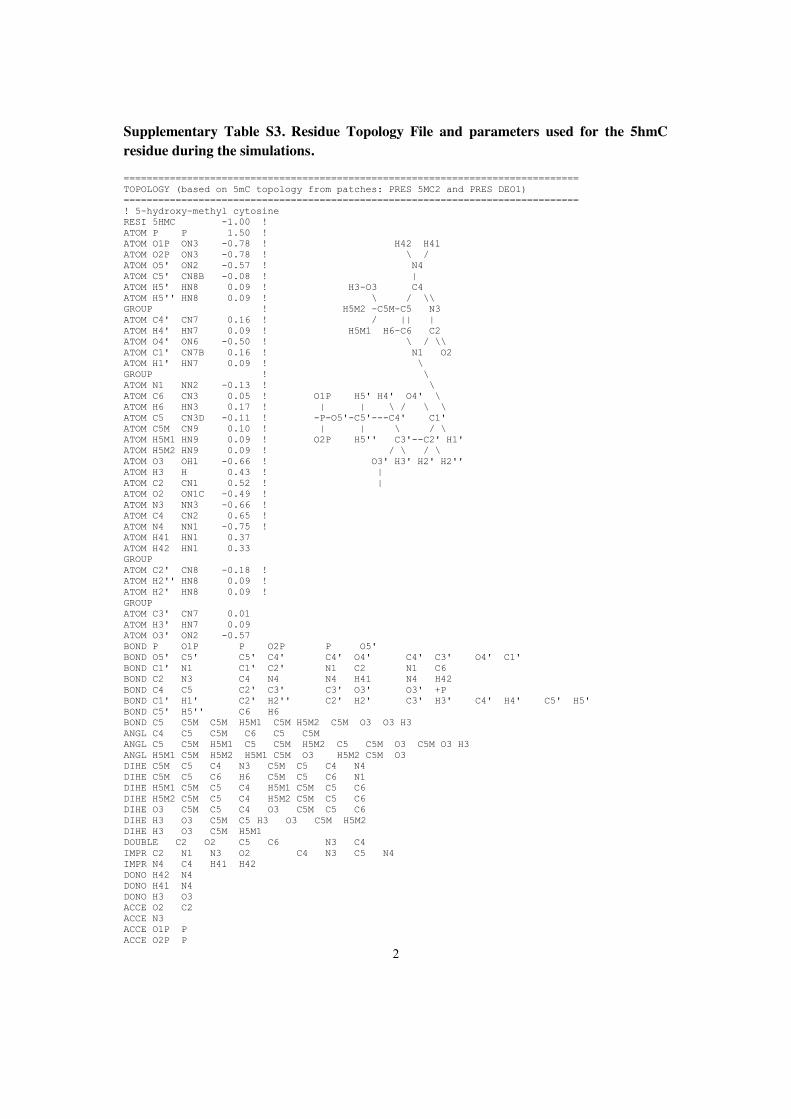
2
Supplementary Table S3. Residue Topology File and parameters used for the 5hmC residue during the simulations.
=============================================================================== TOPOLOGY (based on 5mC topology from patches: PRES 5MC2 and PRES DEO1) =============================================================================== ! 5-hydroxy-methyl cytosine RESI 5HMC -1.00 ! ATOM P P 1.50 ! ATOM O1P ON3 -0.78 ! H42 H41 ATOM O2P ON3 -0.78 ! \ / ATOM O5' ON2 -0.57 ! N4 ATOM C5' CN8B -0.08 ! | ATOM H5' HN8 0.09 ! H3-O3 C4 ATOM H5'' HN8 0.09 ! \ / \\ GROUP ! H5M2 -C5M-C5 N3 ATOM C4' CN7 0.16 ! / || | ATOM H4' HN7 0.09 ! H5M1 H6-C6 C2 ATOM O4' ON6 -0.50 ! \ / \\ ATOM C1' CN7B 0.16 ! N1 O2 ATOM H1' HN7 0.09 ! \ GROUP ! \ ATOM N1 NN2 -0.13 ! \ ATOM C6 CN3 0.05 ! O1P H5' H4' O4' \ ATOM H6 HN3 0.17 ! | | \ / \ \ ATOM C5 CN3D -0.11 ! -P-O5'-C5'---C4' C1' ATOM C5M CN9 0.10 ! | | \ / \ ATOM H5M1 HN9 0.09 ! O2P H5'' C3'--C2' H1' ATOM H5M2 HN9 0.09 ! / \ / \ ATOM O3 OH1 -0.66 ! O3' H3' H2' H2'' ATOM H3 H 0.43 ! | ATOM C2 CN1 0.52 ! | ATOM O2 ON1C -0.49 ! ATOM N3 NN3 -0.66 ! ATOM C4 CN2 0.65 ! ATOM N4 NN1 -0.75 ! ATOM H41 HN1 0.37 ATOM H42 HN1 0.33 GROUP ATOM C2' CN8 -0.18 ! ATOM H2'' HN8 0.09 ! ATOM H2' HN8 0.09 ! GROUP ATOM C3' CN7 0.01 ATOM H3' HN7 0.09 ATOM O3' ON2 -0.57 BOND P O1P P O2P P O5' BOND O5' C5' C5' C4' C4' O4' C4' C3' O4' C1' BOND C1' N1 C1' C2' N1 C2 N1 C6 BOND C2 N3 C4 N4 N4 H41 N4 H42 BOND C4 C5 C2' C3' C3' O3' O3' +P BOND C1' H1' C2' H2'' C2' H2' C3' H3' C4' H4' C5' H5' BOND C5' H5'' C6 H6 BOND C5 C5M C5M H5M1 C5M H5M2 C5M O3 O3 H3 ANGL C4 C5 C5M C6 C5 C5M ANGL C5 C5M H5M1 C5 C5M H5M2 C5 C5M O3 C5M O3 H3 ANGL H5M1 C5M H5M2 H5M1 C5M O3 H5M2 C5M O3 DIHE C5M C5 C4 N3 C5M C5 C4 N4 DIHE C5M C5 C6 H6 C5M C5 C6 N1 DIHE H5M1 C5M C5 C4 H5M1 C5M C5 C6 DIHE H5M2 C5M C5 C4 H5M2 C5M C5 C6 DIHE O3 C5M C5 C4 O3 C5M C5 C6 DIHE H3 O3 C5M C5 H3 O3 C5M H5M2 DIHE H3 O3 C5M H5M1 DOUBLE C2 O2 C5 C6 N3 C4 IMPR C2 N1 N3 O2 C4 N3 C5 N4 IMPR N4 C4 H41 H42 DONO H42 N4 DONO H41 N4 DONO H3 O3 ACCE O2 C2 ACCE N3 ACCE O1P P ACCE O2P P

3
ACCE O3' ACCE O4' ACCE O5' ACCE O3 BILD -O3' P O5' C5' 1.6001 101.45 -46.90 119.00 1.4401 !alpha BILD -O3' O5' *P O1P 1.6001 101.45 -115.82 109.74 1.4802 BILD -O3' O5' *P O2P 1.6001 101.45 115.90 109.80 1.4801 BILD P O5' C5' C4' 1.5996 119.00 -146.00 110.04 1.5160 !beta BILD O5' C5' C4' C3' 1.4401 108.83 60.00 116.10 1.5284 !gamma BILD C5' C4' C3' O3' 1.5160 116.10 140.00 115.12 1.4212 !delta BILD C4' C3' O3' +P 1.5284 111.92 155.00 119.05 1.6001 !epsilon BILD C3' O3' +P +O5' 1.4212 119.05 -95.20 101.45 1.5996 !zeta BILD O4' C3' *C4' C5' 1.4572 104.06 -120.04 116.10 1.5160 BILD C2' C4' *C3' O3' 1.5284 100.16 -124.08 115.12 1.4212 BILD C4' C3' C2' C1' 1.5284 100.16 -30.00 102.04 1.5251 BILD C3' C2' C1' N1 1.5284 101.97 147.89 113.71 1.4896 BILD O4' C1' N1 C2 1.5251 113.71 -97.2 125.59 1.3783 !chi BILD C1' C2 *N1 C6 1.4896 117.79 -180.00 120.6 1.364 BILD C2 N1 C6 C5 1.399 120.6 0.0 121.0 1.337 BILD C6 N1 C2 N3 1.364 120.6 0.0 118.9 1.356 BILD N1 N3 *C2 O2 1.399 118.9 180.0 121.9 1.237 BILD N1 C2 N3 C4 1.399 118.9 0.0 120.0 1.334 BILD C5 N3 *C4 N4 1.426 121.8 180.00 118.9 1.337 BILD N3 C4 N4 H41 1.337 117.9 0.00 118.9 1.01 BILD H41 C4 *N4 H42 1.01 118.9 180.00 120.7 1.01 BILD N1 C5 *C6 H6 0.0 0.0 180.0 0.0 0.0 BILD C1' C3' *C2' H2' 1.5284 102.04 -114.67 110.81 1.01 BILD O4' C2' *C1' H1' 0.0 0.0 -115.0 0.0 0.0 BILD C1' C3' *C2' H2'' 0.0 0.0 115.0 0.0 0.0 BILD C1' C3' *C2' H2' 0.0 0.0 -115.0 0.0 0.0 BILD C2' C4' *C3' H3' 0.0 0.0 115.0 0.0 0.0 BILD C3' O4' *C4' H4' 0.0 0.0 -115.0 0.0 0.0 BILD C4' O5' *C5' H5' 0.0 0.0 -115.0 0.0 0.0 BILD C4' O5' *C5' H5'' 0.0 0.0 115.0 0.0 0.0 BILD C6 C4 *C5 C5M 0.0 0.0 180.0 0.0 0.0 BILD C4 C5 C5M H5M1 0.0 0.0 180.0 0.0 0.0 BILD C5 H5M1 *C5M H5M2 0.0 0.0 -115.0 0.0 0.0 BILD H5M1 H5M2 *C5M O3 0.0 0.0 115.0 0.0 0.0 BILD C4 C5 C5M O3 0.0 0.0 60.0 0.0 0.0 BILD C5 C5M O3 H3 0.0 0.0 180.0 0.0 0.0 =============================================================================== =============================================================================== FORCEFIELD PARAMETERS: =============================================================================== ... BONDS ... !added for 5HMC TU_TCB TH und ISA OH1 CN9 428.000 1.4200 ! ACC. TO OH1-CT3 ... ANGLES ... !added for 5HMC TU_TCB TH und ISA OH1 CN9 CN3D 75.700 110.1000 !ACC. TO OH1-CT2-CT2 OH1 CN9 HN9 45.900 108.8900 !ACC. TO OH1-CT3-HA H OH1 CN9 57.500 106.0000 !ACC. TO H-OH1-CT2 ... DIHEDRALS ... !added for 5HMC TU_TCB TH und ISA H OH1 CN9 CN3D 1.3000 1 0.00 !ACC. TO H-OH1-CT2-CT2 H OH1 CN9 CN3D 0.3000 2 0.00 !ACC. TO H-OH1-CT2-CT2 H OH1 CN9 CN3D 0.4200 3 0.00 !ACC. TO H-OH1-CT2-CT2 CN3 CN3D CN9 OH1 0.0 3 0.0 !ACC. TO CN3-CN3D-CN9-HN9 CN2 CN3D CN9 OH1 0.35 3 0.0 !ACC. TO CN3-CN3D-CN9-HN9 HN9 CN9 OH1 H 0.1400 3 0.00 !ACC. TO X-CT2-OH1-X ... IMPROPER ...

4
Supplementary Figures
Supplementary Figure S1. Electrophoretic mobility shift assays with methylated and hydroxymethylated DNA substrates. Increasing amounts of Uhrf1, its SRA domain (SRAUhrf1) or the MBD domain of MeCP2 (MBDMeCP2) were incubated with two differentially ATTO-labeled DNA substrates, which contain either one central fully methylated or fully hydroxymethylated CpG site (FMB-ATTO700 or FhMB-ATTO550, respectively), in direct competition. Samples were subjected to 6 % non-denaturing PAGE and analyzed with a Typhoon scanner (GE Healthcare). The first, second and third columns show the scans for GFP/YFP, ATTO700 and ATTO550 fluorescence, respectively. The overlay of the two ATTO channels is shown in the fourth column (FMB: red, FhMB:green).
5
Supplementary Figure S2. DNA binding specificity of Uhrf1. Relative DNA/Uhrf1 ratios are shown for two differentially labeled fluorescent DNA substrates in direct competition. (A) Binding of Uhrf1 to DNA substrates containing no CpG site or one central hemimethylated CpG site (noCGB versus HMB, respectively). (B) Binding of Uhrf1 to DNA substrates containing one central un- or hemimethylated CpG site (UMB versus HMB, respectively). Results are shown as means of three independent experiments with standard deviation error bars. DNA substrates were prepared by hybridization as described in the main text, except for noCGB, which was prepared by primer extension as described previously [1]. See Supplementary Tables 1 and 2 for DNA oligonucleotide sequences and purification grade of the used substrates.

6
Supplementary Figure S3. Atom-positional root-mean-square deviation of the protein and DNA backbone atoms during the simulations. The terminal DNA and protein residues were excluded from the calculations in the “subset” sets (red and black lines).
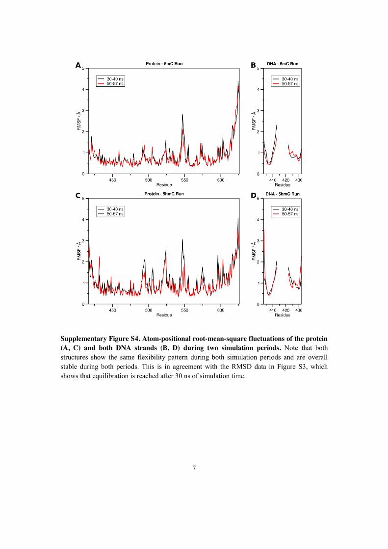
7
Supplementary Figure S4. Atom-positional root-mean-square fluctuations of the protein (A, C) and both DNA strands (B, D) during two simulation periods. Note that both structures show the same flexibility pattern during both simulation periods and are overall stable during both periods. This is in agreement with the RMSD data in Figure S3, which shows that equilibration is reached after 30 ns of simulation time.
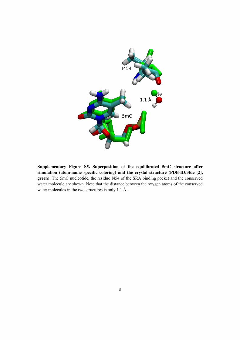
8
Supplementary Figure S5. Superposition of the equilibrated 5mC structure after simulation (atom-name specific coloring) and the crystal structure (PDB-ID:3fde [2], green). The 5mC nucleotide, the residue I454 of the SRA binding pocket and the conserved water molecule are shown. Note that the distance between the oxygen atoms of the conserved water molecules in the two structures is only 1.1 Å.
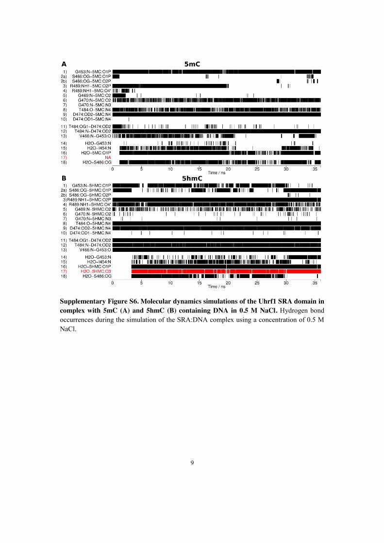
9
Supplementary Figure S6. Molecular dynamics simulations of the Uhrf1 SRA domain in complex with 5mC (A) and 5hmC (B) containing DNA in 0.5 M NaCl. Hydrogen bond occurrences during the simulation of the SRA:DNA complex using a concentration of 0.5 M NaCl.

10
Supplementary References
1. Frauer C, Leonhardt H (2009) A versatile non-radioactive assay for DNA methyltransferase activity and DNA binding. Nucleic Acids Res 37: e22.
2. Hashimoto H, Horton JR, Zhang X, Bostick M, Jacobsen SE, et al. (2008) The SRA domain of UHRF1 flips 5-methylcytosine out of the DNA helix. Nature 455: 826-829.

2.6 Np95 interacts with de novo DNA
methyltransferases, Dnmt3a and Dnmt3b, and
mediates epigenetic silencing of the viral CMV
promoter in embryonic stem cells

EMBO open
Np95 interacts with de novo DNA methyltransferases,Dnmt3a and Dnmt3b, and mediates epigenetic silencingof the viral CMV promoter in embryonic stem cellsDaniela Meilinger 1*, Karin Fellinger 1*, Sebastian Bultmann1, Ulrich Rothbauer1, Ian Marc Bonapace2,Wolfgang E.F. Klinkert3, Fabio Spada1+ & Heinrich Leonhardt1++
1Department of Biology II, Center for Integrated Protein Science Munich, Ludwig Maximilians University Munich, Planegg-
Martinsried, Germany, 2Department of Structural and Functional Biology, University of Insubria, Busto Arsizio, Italy, and3Department of Neuroimmunology, Max Planck Institute of Neurobiology, Am Klopferspitz, Martinsried, Germany
This is an open-access article distributed under the terms of the Creative Commons Attribution License, which permits distribution, andreproduction in any medium, provided the original author and source are credited. This license does not permit commercial exploitationwithout specific permission.
Recent studies have indicated that nuclear protein of 95 kDa(Np95) is essential for maintaining genomic methylation byrecruiting DNA methyltransferase (Dnmt) 1 to hemi-methylatedsites. Here, we show that Np95 interacts more strongly withregulatory domains of the de novo methyltransferases Dnmt3aand Dnmt3b. To investigate possible functions, we developed anepigenetic silencing assay using fluorescent reporters in embryo-nic stem cells (ESCs). Interestingly, silencing of the cytomegalo-virus promoter in ESCs preceded DNA methylation and wasstrictly dependent on the presence of either Np95, histone H3methyltransferase G9a or Dnmt3a and Dnmt3b. Our resultsindicate a regulatory role for Np95, Dnmt3a and Dnmt3b inmediating epigenetic silencing through histone modificationfollowed by DNA methylation.Keywords: DNA methylation; histone modification; epigenetics;silencing; Uhrf1EMBO reports (2009) 10, 1259–1264. doi:10.1038/embor.2009.201
INTRODUCTIONIn mammals, DNA methylation contributes to the establishment andmaintenance of cell-type-specific gene expression programmes,imprinting, X-chromosome inactivation and genome stability (Bird,2002). The majority of genomic methylation occurs at cytosineresidues within CpG dinucleotides and is catalysed by the DNAmethyltransferases (Dnmt) 1, 3a and 3b. Dnmt1 is responsible formaintaining genomic methylation, whereas Dnmt3a and Dnmt3bare mainly involved in de novo establishment of methylationpatterns during cellular differentiation (Leonhardt et al, 1992; Li et al,1992; Lei et al, 1996; Okano et al, 1999; Spada et al, 2007).Nuclear protein of 95 kDa (Np95; also known as Uhrf1) hasrecently been identified as an essential co-factor for maintaininggenomic methylation (Bostick et al, 2007; Sharif et al, 2007;Achour et al, 2008). dnmt1!/! and np95!/! embryonic stem cells(ESCs) and embryos have similar reduced levels of DNA methylation.In addition, Np95 interacts with Dnmt1, binds hemi-methylated CpGsites through its Set and Ring associated (SRA) domain and both Np95and Dnmt1 accumulate at replication sites (Uemura et al, 2000;Bostick et al, 2007; Papait et al, 2007; Arita et al, 2008; Avvakumovet al, 2008; Hashimoto et al, 2008). Thus, it has been proposed thatNp95 mediates maintenance of genomic methylation by recruitingDnmt1 to hemi-methylated CpG sites generated during replication.
Here, we investigated a possible involvement of Np95 inepigenetic regulation beyond its role in Dnmt1-mediated main-tenance of DNA methylation. We found that Np95 interacts withthe de novo methyltransferases, Dnmt3a and Dnmt3b, andmediates promoter silencing before DNA methylation is detected.
RESULTS AND DISCUSSIONNp95 interacts with Dnmt3a and Dnmt3bImmunoprecipitation experiments showed that different isoformsof both de novo methyltransferases Dnmt3a and Dnmt3b interact
Received 19 December 2008; revised 3 July 2009; accepted 4 August 2009;published online 2 October 2009
*These authors contributed equally to this work+Corresponding author. Tel: " 49 89 218074230; Fax: " 49 89 218074236;E-mail: [email protected]++Corresponding author. Tel: " 49 89 218074232; Fax: " 49 89 218074236;E-mail: [email protected]
1Department of Biology II, Center for Integrated Protein Science Munich (CIPSM),Ludwig Maximilians University Munich, Gro!haderner Street 2,82152 Planegg-Martinsried, Germany2Department of Structural and Functional Biology, University of Insubria,Via da Giussano 12, 21052 Busto Arsizio (VA), Italy3Department of Neuroimmunology, Max Planck Institute of Neurobiology,Am Klopferspitz 18, 82152 Martinsried, Germany
&2009 EUROPEAN MOLECULAR BIOLOGY ORGANIZATION EMBO reports VOL 10 | NO 11 | 2009
scientificreportscientific report
1259

with Np95 in wild-type (wt) ESCs, including the more abundantDnmt3a2 and Dnmt3b1 (Fig 1A). Furthermore, using a greenfluorescent protein (GFP) trap (Rothbauer et al, 2008), weco-immunoprecipitated endogenous Dnmt1 and isoforms ofDnmt3a and Dnmt3b with a GFP–Np95 fusion constructtransiently expressed in np95!/! ESCs and, vice versa, endo-genous Np95 co-immunoprecipitated with GFP–Dnmt3a orGFP–Dnmt3b1 fusions in dnmt3a and 3b double knockout(DKO) ESCs (supplementary Fig S1A,B online). In addition, weobserved co-immunoprecipitation of endogenous DNMT3b andinverted CCAAT box binding protein of 90 kDa—the humanhomologue of Np95—from human embryonic kidney 293T(HEK293T) cell extracts (supplementary Fig S1C online). Weconfirmed the interaction of Np95 with Dnmt3a/b by using arecently developed fluorescent two hybrid assay (F2H; Zolghadret al, 2008). GFP–Dnmt3 fusion constructs were used as bait bytethering them to a lac operator array present in baby hamsterkidney (BHK) cells, so that the array was visible as a distinct
nuclear spot of enriched GFP fluorescence (Fig 1B). A Cherry–Np95 fusion (prey) accumulated at this spot only when GFPfusions of full-length Dnmt3a and Dnmt3b1 or their amino-terminal regions were used as bait and not when their isolatedCarboxy-terminal catalytic domains were used. We furthermapped the interaction of Np95 with Dnmt3a/b through co-immunoprecipitation of deletion constructs and isolated domainstransiently expressed in HEK293T cells (supplementary Fig S2online). The results were consistent with those produced by F2H:the N-terminal regions of Dnmt3a and Dnmt3b1, but not theirC-terminal catalytic domains, interacted with Np95. Deletion ofthe PHD or PWWP domains of Dnmt3a and Dnmt3b did noteliminate the interaction with Np95. We then determined thedomains of Np95 involved in this interaction. We found that theSRA domain and the N-terminal 298 amino acids of Np95, whichinclude the ubiquitin-like domain, interacted with Dnmt3a andDnmt3b1, whereas the PHD domain and the C-terminal 132amino acids, including the Ring domain, did not. Furthermore,
A
B DAPI G-Dnmt3a Ch-Np95 DAPI G-Dnmt3b Ch-Np95
G-D
nmt3
a fl
G-D
nmt3
b fl
G-D
nmt3
a N
G-D
nmt3
b N
G-D
nmt3
a C
G-D
nmt3
b C
72
kDa130
95
I S B I S B
np95–/– wt
Anti-Dnmt3b
Anti-Dnmt3a
Anti-Np95IP:
kDa
G-Dnmt1
Np95-His
I B
G-Dnmt3b
I B
G-Dnmt3a
I B
GFP
I B
Low salt
High salt
95
95
Anti-His
C IP:
transf.:
Anti-GFP
ESCs:
Fig 1 | Np95 interacts with de novo methyltransferases Dnmt3a and Dnmt3b. (A) Co-immunoprecipitation of Dnmt3a and Dnmt3b with Np95 in wt and
np95!/! E14 ESCs. The Dnmt3a2 isoform is shown in the lower panel. (B) F2H shows recruitment of Cherry–Np95 (prey) at the lac operator array
(indicated by arrowheads) when GFP fusions of full-length Dnmt3a and Dnmt3b1 (G-Dnmt3a/b fl) or their amino-terminal regions (G-Dnmt3a/b N)
are used as bait and not with their isolated C-terminal catalytic domains (G-Dnmt3a/b C). Scale bars, 5mm. (C) Co-immunoprecipitation of
Np95-His with GFP-tagged Dnmt1, Dnmt3a and Dnmt3b1 (G-Dnmt) transiently co-expressed in HEK293T cells. Co-expression of GFP was used as the
control. In the upper row, immunoprecipitations carried out in the presence of 150 mM NaCl throughout the procedure are shown, whereas in the
lower row, immunoprecipitation and wash buffers were carried out using 300 and 500 mM NaCl, respectively. Two per cent of input and supernatant
relative to bound fractions were loaded in (A) and (C). B, bound; Dnmt, DNA methyltransferase; ESCs, embryonic stem cells; F2H, fluorescent
two-hybrid assay; GFP, green fluorescent protein; HEK293T, human embryonic kidney 293T; I, input; Np95, nuclear protein of 95 kDa;
S, supernatant; wt, wild type.
Np95 interacts with Dnmt3a and Dnmt3b
D. Meilinger et al
EMBO reports VOL 10 | NO 11 | 2009 &2009 EUROPEAN MOLECULAR BIOLOGY ORGANIZATION
scientificreport
1260

CAG GFP CMV mRFPCAG GFP CMV mRFP
wt
Day
2D
ay 6
np95–/–
Rel
ativ
e ex
pres
sion
of m
RFP
/GFP
0
0.2
0.4
0.6
0.8
1.0
wt
!/!!/!
dnmt1–/– TKO np95–/– DKO G9a!/! suv39–/–
Time after transfection (days)
Rel
ativ
e ex
pres
sion
of m
RFP
/GFP
0
0.2
0.4
0.6
0.8
1.0
1.2A
C
B
2 4 6 8 10
wt
np95–/–
dnmt1–/–
TKO
240220200180160140120100806040200
Fig 2 | Promoter silencing activity in wild-type and mutant ESCs. ESCs with the indicated phenotypes were transiently co-transfected with CMV
promoter-driven mRFP and CAG promoter-driven GFP reporter constructs. Between 90 and 150 images per sample were acquired either (A) every
second day after transfection from a single experiment or (B) only at days 2 and 7–10 after transfection from 3–5 independent experiments. Relative
levels of red over green fluorescence are shown with values for day 2 (first day of imaging) set to 1 (A). Wild-type J1 and E14 cells
gave similar results (data not shown). suv39!/! stands for Suv39h1/2 double null ESCs. (C) Representative images of wt and np95!/! E14 ESCs
co-transfected as in (A) and (B) (upper panels) and respective heat map intensity plots (lower panels). Scale bar, 15 mm. CAG, CMV early enhancer/
chicken b actin promoter; CMV, cytomegalovirus promoter; DKO, double knockout; Dnmt, DNA methyltransferase; ESCs, embryonic stem cells; GFP,
green fluorescent protein; mRFP, monomeric red fluorescent protein; Np95, nuclear protein of 95 kDa; TKO, triple knockout; wt, wild type.
Np95 interacts with Dnmt3a and Dnmt3b
D. Meilinger et al
&2009 EUROPEAN MOLECULAR BIOLOGY ORGANIZATION EMBO reports VOL 10 | NO 11 | 2009
scientificreport
1261

we observed co-immunoprecipitation of endogenous Np95 withGFP–Dnmt3a and GFP–Dnmt3b transiently expressed in dnmt1!/!
ESCs, indicating that Dnmt3a and Dnmt3b interact with Np95independently of Dnmt1 (supplementary Fig S1D online). Tocompare the relative association between endogenous Np95 andDnmts, we re-probed the blot in Fig 1A with a Dnmt1 antibody andobserved a substantially weaker signal for the co-immunoprecipitatedDnmt1 relative to the input than in the case of Dnmt3a2 andDnmt3b1 (supplementary Fig S3A online). To compare furtherthe stability of Np95 interactions with the Dnmts, we transientlyco-expressed Np95-His with GFP–Dnmt1, GFP–Dnmt3a orGFP–Dnmt3b1 in HEK293T cells and immunoprecipitated withthe GFP trap in the presence of different salt concentrations (Fig 1C;supplementary Fig S3B online). Interestingly, under high saltconditions, the interaction between Np95-His and GFP–Dnmt1was lost, whereas co-immunoprecipitation of GFP–Dnmt3a andGFP–Dnmt3b1 remained relatively unaffected. These data clearlyindicate that Np95 interacts more strongly with the de novomethyltransferases, Dnmt3a and Dnmt3b, than with Dnmt1.
Np95, Dnmt3a/3b and G9a mediate epigenetic silencingAs DNA methylation has a central role in epigenetic silencing, weinvestigated the requirement of DNA methyltransferases andNp95 for promoter silencing in ESCs. We found that, on transienttransfection of wt ESCs, constructs driven by the cytomegalovirus(CMV) promoter were rapidly silenced, as opposed to longer-lasting expression of constructs driven by the chimeric CMV earlyenhancer/chicken b actin (CAG) promoter (Fig 2), which isconsistent with the popularity of the CAG promoter for stabletransgene expression in ESCs and mice. We then established anepigenetic silencing assay based on this observation. ESCs wereco-transfected with two distinct plasmids, one expressing mono-meric red fluorescent protein (mRFP) under the CMV promoter,the other expressing GFP driven by the CAG promoter. mRFP andGFP expression was monitored after transfection for up to ten daysby using automated image acquisition and quantificationof fluorescent signals (supplementary Fig S4A online). The ratiobetween mRFP and GFP expression declined steadily in wt ESCs,reflecting preferential silencing of the CMV promoter (Fig 2).By contrast, DKO ESCs and ESCs lacking all three major DNAmethyltransferases (dnmt1, 3a and 3b triple knockout) showed nopreferential silencing of the CMV promoter. Surprisingly, np95!/!
ESCs were also unable to silence the CMV promoter, whereasdnmt1!/! ESCs showed only partly reduced silencing under theseconditions. Similar results were obtained on swapping GFP andmRFP reporter sequences, ruling out potential artefacts due todifferences in their coding sequences or stability of the reporterproteins (supplementary Fig S4B online). Thus, despite expressinga full complement of DNA methyltransferases, ESCs lacking Np95are as deficient in promoter silencing activity as ESCs lacking allthree major Dnmts. We next investigated whether silencing of theCMV promoter correlates with CpG methylation. Interestingly,promoter methylation was detected only ten days after transfec-tion and was lower in np95!/! than in wt ESCs, whereas none ofthe dnmt mutant ESCs showed appreciable DNA methylation(Fig 3; supplementary Fig S5A online). At the same time noobvious methylation was detected in any of the ESC lines withinthe CpG island of the CAG promoter construct (supplementaryFig S5B online). Thus, CMV promoter silencing depends on the
presence of both Np95 and de novo Dnmts, but ensues wellbefore de novo methylation of the promoter is detected. Thisprompted us to investigate the involvement of repressive histonemethylation as a possible mechanism for the observed silencing.We found that in the absence of histone H3 lysine 9 methyl-transferases (H3K9MTs), G9a or Suv39h1/2, silencing of the CMVpromoter was completely abolished or reduced, respectively,indicating that G9a and, in part, Suv39h1/2 are also required forsilencing (Fig 2B).
The results shown here indicate that Np95 interacts withDnmt3a and Dnmt3b and mediates silencing of the CMVpromoter by mechanisms that are, at least initially, independentof de novo DNA methylation. Importantly, our data also show theinvolvement of H3K9MTs, G9a and Suv39h1/2 in CMV promotersilencing. H3K9MTs were reported to associate with de novo
CMV promoter
CpG sites
Met
hyla
tion
perc
enta
ge (%
)M
ethy
latio
n pe
rcen
tage
(%)
np95 –/–
wt
CpG sites
(–243) (–138) (+1)
TATA1 7 14
50
45
40
35
30
25
20
15
10
5
01 2 3 4 5 6 7 8 9 10 11 12 13 14
50
45
40
35
30
25
20
15
10
5
01 2 3 4 5 6 7 8 9 10 11 12 13 14
Day 2 Day 6 Day 10
Day 2 Day 6 Day 10
Fig 3 | Methylation of the CMV promoter 2, 6 and 10 days after
transfection. Wild-type and np95!/! ESCs were transfected as in Fig 2
and GFP-positive cells were sequentially sorted at the indicated days after
transfection. Total DNA was isolated from sorted cells and bisulphite-
treated. A proximal part of the CMV promoter was amplified and
subjected to pyrosequencing. (A) Schematic drawing of the 14 proximal
CpG sites analysed (shown as open circles). The numbers above
correspond to the CpG sites shown in (B) and (C) and numbers in
brackets refer to the position of CpG sites with respect to the
transcription start site. (B,C) Methylation percentage at individual CpG
sites for (B) wt and (C) np95!/! ESCs as measured by pyrosequencing.
CMV, cytomegalovirus; ESCs, embryonic stem cells; GFP, green
fluorescent protein; wt, wild type.
Np95 interacts with Dnmt3a and Dnmt3b
D. Meilinger et al
EMBO reports VOL 10 | NO 11 | 2009 &2009 EUROPEAN MOLECULAR BIOLOGY ORGANIZATION
scientificreport
1262

Dnmts, and major satellite repeats were found to be hypomethy-lated in ESCs lacking either Suv39h1/2 or Dnmt3 enzymes.However, major satellite transcript levels were altered only inSuv39h1/2-deficient cells and not in Dnmt3-deficient cells (Fukset al, 2003; Lehnertz et al, 2003; Martens et al, 2005). A recentstudy showed that G9a, Dnmt1, Dnmt3a and Dnmt3b arerequired for normal methylation at long terminal repeats ofendogenous retrotransposable elements, although transcription ofthese elements was increased in Dnmt-deficient ESCs, but notG9a-deficient ESCs (Dong et al, 2008). Furthermore, recent studieshave shown that G9a interacts with Dnmt3a and Dnmt3b andmediates de novo methylation of the oct4, nanog and dnmt3lpromoters on retinoic-acid-induced differentiation of ESCs (Feld-man et al, 2006; Li et al, 2007; Epsztejn-Litman et al, 2008).However, two of these studies showed that neither G9a nor denovo Dnmts are required to silence the oct4 promoter, and G9awas also found to be dispensable for silencing the nanog anddnmt3l promoters (Feldman et al, 2006; Epsztejn-Litman et al,2008). In the third study, nanog, but not oct4, was shown to besilenced in differentiating ESCs lacking both Dnmt3a and Dnmt3b(Li et al, 2007). We found that silencing of oct4 during embryoidbody differentiation is largely independent from the presence ofNp95 as well as all three major Dnmts, and occurs in the absenceof DNA methylation (D.M., F.S., S.B., and H.L., unpublisheddata). These data, together with our findings on silencing of theCMV promoter in ESCs, indicate that Dnmts, Np95 and H3K9MTsmediate silencing through many mechanisms that do notnecessarily involve DNA methylation and might depend on thepresence of different cis elements and an intricate interplay withother epigenetic and transcription factors. Interestingly, Np95 wasrecently shown to interact with G9a (Kim et al, 2009) and here weshow that silencing of the CMV promoter in ESCs strictly dependson Np95 and on de novo Dnmts as well as G9a. Taken togetherthese observations suggest that Np95, de novo Dnmts and G9amight be involved in a common silencing pathway.
In summary, our data clearly support a crucial role of Np95 inepigenetic silencing mediated by de novo DNA and histonemethyltransferases, and make Np95 an attractive target forepigenetic reprogramming strategies.
METHODSCell culture and transfection. HEK293T cells, BHK cells and ESCswere cultured and transfected as described by Schermelleh et al(2007), except FuGENE HD (Roche, Mannheim, Germany) wasused for transfection of ESCs. The dnmt1!/! J1 ESCs used in thisstudy were homozygous for the c allele (Lei et al, 1996). BHK cellswere co-transfected on glass coverslips with GFP–Dnmt3 andCherry–Np95 constructs using Transfectin (Bio-Rad, Munich,Germany) according to the manufacturer’s instructions. Cellfixation and microscopy were carried out as described byZolghadr et al (2008).Co-immunoprecipitation. ESCs and HEK293T cell extracts wereprepared in lysis buffer (20 mM Tris–HCl (pH 7.5), 0.5 mM EDTA,2 mM phenylmethyl sulphonyl fluoride and 0.5% NP40) contain-ing 150 or 300 mM NaCl (high-salt condition) and diluted withlysis buffer without NP40. GFP trap (Rothbauer et al, 2008) and aspecific rabbit antiserum (Citterio et al, 2004) were used forimmunoprecipitation of GFP fusions and endogenous Np95,respectively. GFP trap and protein G beads (Sigma, Taufkirchen,
Germany) were washed with dilution buffer containing increasingsalt concentrations (150 and 300 mM, or 300 and 500 mM NaClfor the high-salt condition) and re-suspended in SDS–PAGEsample buffer. The following mouse monoclonal antibodieswere used for immunoblotting: anti-His (C-terminal, Invitrogen,Karlsruhe, Germany), anti-Dnmt3a (clone 64B1446, Imgenex, SanDiego, CA, USA) and anti-Dnmt3b (clone 52A1018, Abcam,Cambridge, UK). Np95 was detected with the same antiserumused for immunoprecipitation and a rabbit antiserum was used fordetection of Dnmt1 (Grohmann et al, 2005). Horseradishperoxidase-conjugated rabbit anti-mouse or goat anti-rabbitsecondary antibodies (Sigma) and ECL Plus reagent (GE Health-care, Munich, Germany) were used for detection.Silencing assay. ESCs were co-transfected with pCAG-eGFP-IBand pCMV-mRFP as described above and images from live cellswere acquired at the indicated time points with an InCell Analyser1000 (GE Healthcare) using a # 20 air objective (NA$ 0.45) andstandard filter settings for GFP and RFP. A total of 90–150 imageswere acquired for each channel, using the same exposure timethroughout the time course. Cells were passaged every second dayand images were taken 4–5 h after seeding. Images were analysedusing ImageJ v1.42a software. To calculate fluorescent reporterexpression, pictures were processed using a Gaussian bluralgorithm (radius (sigma)$ 2), and a threshold for maximal signaland minimal background coverage was adjusted and applied toeach channel (supplementary Fig S4A online). The threshold wasconverted into area selection and the total size of the selected areawas measured.DNA methylation analysis. ESCs were transfected as in thesilencing assay with pCAG-eGFP-IB and pCMV-mRFP, and GFP-positive cells were sequentially sorted with a FACSVantage orFACSAria II (Becton Dickinson, Heidelberg, Germany) at days 2, 6and 10 after transfection. After each sorting, total DNA wasisolated using the QIAmp DNA Mini kit (Qiagen, Hilden,Germany) and bisulphite treated with the EZ DNA Methylation-Gold kit (Zymo research, Orange, CA, USA). The followingprimers were used for PCR amplification: CMV-forward TGGGATTTTTTTATTTGGTAGT; CMV-reverse ATGGGAGTTTGTTTTGGTATTA; CAG-forward GGAGAGGTGAGGAGGTAGTTAATTAGAand CAG-reverse CCCCAAACCCCTCAAAACTT. Pyrosequencingwas carried out by Varionostic GmbH (Ulm, Germany).Supplementary information is available at EMBO reports online(http://www.emboreports.org).
ACKNOWLEDGEMENTSWe are grateful to the following colleagues for providing ESCs: M. Mutoand H. Koseki for wt and np95!/! E14; E. Li and T. Chen for wt,dnmt1!/! and DKO J1; Masaki Okano for TKO J1; Gunnar Schotta forSuv39h1/2 dn; Y. Shinkai and T. Jenuwein for wt and G9a!/! TT2. Wethank L. Schermelleh for help with image processing and K. Zolghadr forassistance with the InCell Analyzer. This study was supported by theNanosystems Initiative Munich and BioImaging Network Munich, and bygrants from the Deutsche Forschungsgemeinschaft to H.L.
CONFLICT OF INTERESTThe authors declare that they have no conflict of interest.
REFERENCESAchour M et al (2008) The interaction of the SRA domain of ICBP90 with
a novel domain of DNMT1 is involved in the regulation of VEGF geneexpression. Oncogene 27: 2187–2197
Np95 interacts with Dnmt3a and Dnmt3b
D. Meilinger et al
&2009 EUROPEAN MOLECULAR BIOLOGY ORGANIZATION EMBO reports VOL 10 | NO 11 | 2009
scientificreport
1263
Arita K, Ariyoshi M, Tochio H, Nakamura Y, Shirakawa M (2008) Recognitionof hemi-methylated DNA by the SRA protein UHRF1 by a base-flippingmechanism. Nature 455: 818–821
Avvakumov GV, Walker JR, Xue S, Li Y, Duan S, Bronner C, Arrowsmith CH,Dhe-Paganon S (2008) Structural basis for recognition ofhemi-methylated DNA by the SRA domain of human UHRF1. Nature455: 822–825
Bird A (2002) DNA methylation patterns and epigenetic memory. Genes Dev16: 6–21
Bostick M, Kim JK, Esteve P-O, Clark A, Pradhan S, Jacobsen SE (2007)UHRF1 plays a role in maintaining DNA methylation in mammaliancells. Science 317: 1760–1764
Citterio E, Papait R, Nicassio F, Vecchi M, Gomiero P, Mantovani R,Di Fiore PP, Bonapace IM (2004) Np95 is a histone-binding proteinendowed with ubiquitin ligase activity. Mol Cell Biol 24: 2526–2535
Dong KB et al (2008) DNA methylation in ES cells requires the lysinemethyltransferase G9a but not its catalytic activity. EMBO J 27:2691–2701
Epsztejn-Litman S et al (2008) De novo DNA methylation promoted by G9aprevents reprogramming of embryonically silenced genes. Nat Struct MolBiol 15: 1176–1183
Feldman N, Gerson A, Fang J, Li E, Zhang Y, Shinkai Y, Cedar H, Bergman Y(2006) G9a-mediated irreversible epigenetic inactivation of Oct-3/4during early embryogenesis. Nat Cell Biol 8: 188–194
Fuks F, Hurd PJ, Deplus R, Kouzarides T (2003) The DNA methyltransferasesassociate with HP1 and the SUV39H1 histone methyltransferase. NucleicAcids Res 31: 2305–2312
Grohmann M, Spada F, Schermelleh L, Alenina N, Bader M, Cardoso MC,Leonhardt H (2005) Restricted mobility of Dnmt1 in preimplantationembryos: implications for epigenetic reprogramming.BMC Dev Biol 5: 18
Hashimoto H, Horton JR, Zhang X, Bostick M, Jacobsen SE, Cheng X (2008)The SRA domain of UHRF1 flips 5-methylcytosine out of the DNA helix.Nature 455: 826–829
Kim JK, Esteve PO, Jacobsen SE, Pradhan S (2009) UHRF1 binds G9a andparticipates in p21 transcriptional regulation in mammalian cells.Nucleic Acids Res 37: 493–505
Lehnertz B, Ueda Y, Derijck AA, Braunschweig U, Perez-Burgos L, Kubicek S,Chen T, Li E, Jenuwein T, Peters AH (2003) Suv39h-mediated histone H3lysine 9 methylation directs DNA methylation to major satellite repeats atpericentric heterochromatin. Curr Biol 13: 1192–1200
Lei H, Oh S, Okano M, Juttermann R, Goss K, Jaenisch R, Li E (1996) De novoDNA cytosine methyltransferase activities in mouse embryonic stemcells. Development 122: 3195–3205
Leonhardt H, Page AW, Weier HU, Bestor TH (1992) A targeting sequencedirects DNA methyltransferase to sites of DNA replication in mammaliannuclei. Cell 71: 865–873
Li E, Bestor TH, Jaenisch R (1992) Targeted mutation of the DNAmethyltransferase gene results in embryonic lethality. Cell 69: 915–926
Li JY et al (2007) Synergistic function of DNA methyltransferases Dnmt3aand Dnmt3b in the methylation of Oct4 and Nanog. Mol Cell Biol 27:8748–8759
Martens JH, O’Sullivan RJ, Braunschweig U, Opravil S, Radolf M, Steinlein P,Jenuwein T (2005) The profile of repeat-associated histone lysinemethylation states in the mouse epigenome. EMBO J 24: 800–812
Okano M, Bell DW, Haber DA, Li E (1999) DNA methyltransferases Dnmt3aand Dnmt3b are essential for de novo methylation and mammaliandevelopment. Cell 99: 247–257
Papait R, Pistore C, Negri D, Pecoraro D, Cantarini L, Bonapace IM (2007)Np95 is implicated in pericentromeric heterochromatin replication andin major satellite silencing. Mol Biol Cell 18: 1098–1106
Rothbauer U, Zolghadr K, Muyldermans S, Schepers A, Cardoso MC,Leonhardt H (2008) A versatile nanotrap for biochemical and functionalstudies with fluorescent fusion proteins. Mol Cell Proteomics 7: 282–289
Schermelleh L, Haemmer A, Spada F, Rosing N, Meilinger D, Rothbauer U,Cristina Cardoso M, Leonhardt H (2007) Dynamics of Dnmt1 interactionwith the replication machinery and its role in postreplicativemaintenance of DNA methylation. Nucleic Acids Res 35: 4301–4312
Sharif J et al (2007) The SRA protein Np95 mediates epigenetic inheritance byrecruiting Dnmt1 to methylated DNA. Nature 450: 908–912
Spada F, Haemmer A, Kuch D, Rothbauer U, Schermelleh L, Kremmer E,Carell T, Langst G, Leonhardt H (2007) DNMT1 but not its interactionwith the replication machinery is required for maintenance of DNAmethylation in human cells. J Cell Biol 176: 565–571
Uemura T, Kubo E, Kanari Y, Ikemura T, Tatsumi K, Muto M (2000) Temporaland spatial localization of novel nuclear protein NP95 in mitotic andmeiotic cells. Cell Struct Funct 25: 149–159
Zolghadr K, Mortusewicz O, Rothbauer U, Kleinhans R, Goehler H,Wanker EE, Cardoso MC, Leonhardt H (2008) A fluorescent two-hybrid(F2H) assay for direct visualization of protein interactions in living cells.Mol Cell Proteomics 7: 2279–2287
EMBO reports is published by Nature Publishing Groupon behalf of European Molecular Biology Organization.
This article is licensed under a Creative Commons Attribution-Noncommercial-ShareAlike 3.0 License. [http://creativecommons.org/licenses/by-nc-sa/3.0/]
Np95 interacts with Dnmt3a and Dnmt3b
D. Meilinger et al
EMBO reports VOL 10 | NO 11 | 2009 &2009 EUROPEAN MOLECULAR BIOLOGY ORGANIZATION
scientificreport
1264

SUPPLEMENTARY INFORMATION Meilinger et al.
1
Supplementary Figure S1. Np95 interacts with de novo methyltransferases Dnmt3a and 3b. (A) Co-immunoprecipitation of endogenous Dnmt3a2 (left and right), Dnmt3b isoforms (left) and Dnmt1 (right) with GFP-Np95 transiently expressed in np95-/- ESCs. Left and right panels are from independent experiments where 2 and 4% of input (I) relative to bound (B) fractions was loaded, respectively. (B) Co-immunoprecipitation of endogenous Np95 with either GFP-Dnmt3a (left panel) or GFP-Dnmt3b1 (central panel) transiently expressed in DKO ESCs. Transient expression of GFP was used as control (right panel). GFP and GFP fusions were immunoprecipitated with GFP-trap as in experiments shown in Fig. 1B. 2% of input (I) relative to bound (B) fractions was loaded. (C) Co-immunoprecipitation of endogenous ICBP90/UHRF1 and DNMT3b in HEK293T cells. Antibodies to mouse proteins cross-react with the respective human homologues. 4% of input (I) relative to bound (B) fractions was loaded. (D) Co-immunoprecipitation of endogenous Np95 with either GFP-Dnmt3a or GFP-Dnmt3b1 (lower panel) transiently expressed in dnmt1-/- ESCs.
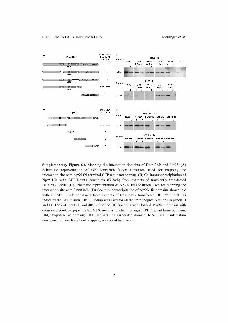
SUPPLEMENTARY INFORMATION Meilinger et al.
2
Supplementary Figure S2. Mapping the interaction domains of Dnmt3a/b and Np95. (A) Schematic representation of GFP-Dnmt3a/b fusion constructs used for mapping the interaction site with Np95 (N-terminal GFP tag is not shown). (B) Co-immunoprecipitation of Np95-His with GFP-Dnmt3 constructs (G-3a/b) from extracts of transiently transfected HEK293T cells. (C) Schematic representation of Np95-His constructs used for mapping the interaction site with Dnmt3a/b. (D) Co-immunoprecipitation of Np95-His domains shown in c with GFP-Dnmt3a/b constructs from extracts of transiently transfected HEK293T cells. G indicates the GFP fusion. The GFP-trap was used for all the immunoprecipitations in panels B and D. 0.5% of input (I) and 40% of bound (B) fractions were loaded. PWWP, domain with conserved pro-trp-trp-pro motif; NLS, nuclear localization signal; PHD, plant homeodomain; Ubl, ubiquitin-like domain; SRA, set and ring associated domain; RING, really interesting new gene domain. Results of mapping are scored by + or -.
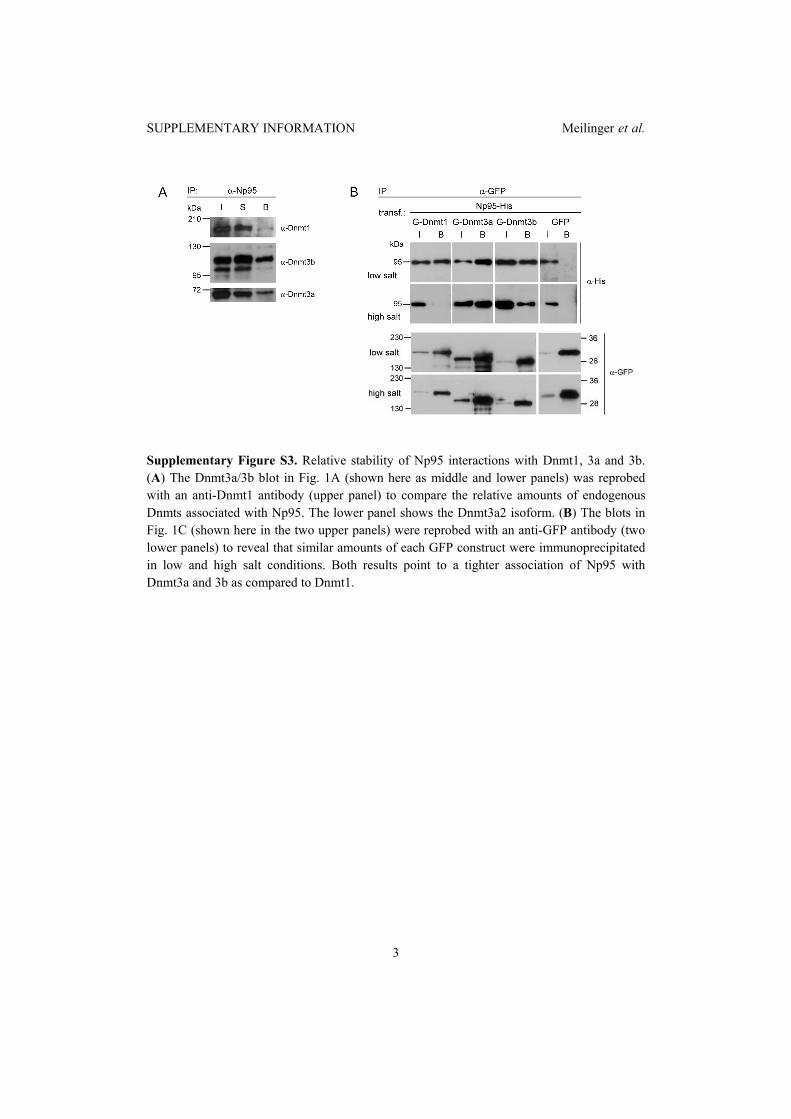
SUPPLEMENTARY INFORMATION Meilinger et al.
3
Supplementary Figure S3. Relative stability of Np95 interactions with Dnmt1, 3a and 3b. (A) The Dnmt3a/3b blot in Fig. 1A (shown here as middle and lower panels) was reprobed with an anti-Dnmt1 antibody (upper panel) to compare the relative amounts of endogenous Dnmts associated with Np95. The lower panel shows the Dnmt3a2 isoform. (B) The blots in Fig. 1C (shown here in the two upper panels) were reprobed with an anti-GFP antibody (two lower panels) to reveal that similar amounts of each GFP construct were immunoprecipitated in low and high salt conditions. Both results point to a tighter association of Np95 with Dnmt3a and 3b as compared to Dnmt1.
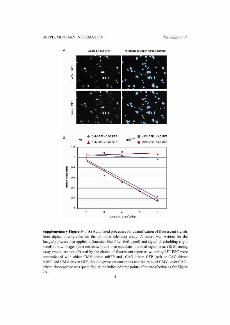
SUPPLEMENTARY INFORMATION Meilinger et al.
4
Supplementary Figure S4. (A) Automated procedure for quantification of fluorescent signals from digital micrographs for the promoter silencing assay. A macro was written for the ImageJ software that applies a Gaussian blur filter (left panel) and signal thresholding (right panel) to raw images (data not shown) and then calculates the total signal area. (B) Silencing assay results are not affected by the choice of fluorescent reporter. wt and np95-/- ESC were cotransfected with either CMV-driven mRFP and CAG-driven GFP (red) or CAG-driven mRFP and CMV-driven GFP (blue) expression constructs and the ratio of CMV- over CAG-driven fluorescence was quantified at the indicated time points after transfection as for Figure 2A.
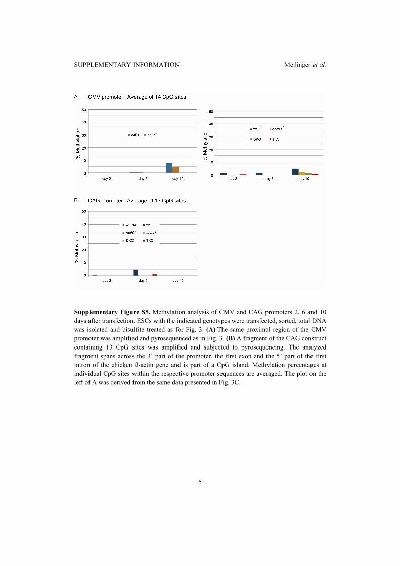
SUPPLEMENTARY INFORMATION Meilinger et al.
5
Supplementary Figure S5. Methylation analysis of CMV and CAG promoters 2, 6 and 10 days after transfection. ESCs with the indicated genotypes were transfected, sorted, total DNA was isolated and bisulfite treated as for Fig. 3. (A) The same proximal region of the CMV promoter was amplified and pyrosequenced as in Fig. 3. (B) A fragment of the CAG construct containing 13 CpG sites was amplified and subjected to pyrosequencing. The analyzed fragment spans across the 3’ part of the promoter, the first exon and the 5’ part of the first intron of the chicken ß-actin gene and is part of a CpG island. Methylation percentages at individual CpG sites within the respective promoter sequences are averaged. The plot on the left of A was derived from the same data presented in Fig. 3C.

SUPPLEMENTARY INFORMATION Meilinger et al.
6
Supplementary methods Plasmid construction. The CMV-driven enhanced GFP construct was from Clontech (pEGFP-C1). To generate the CMV-driven mRFP construct (pCMV-mRFP) the coding sequence for eGFP in pEGFP-C1 was replaced with that for mRFP from pRSETB-mRFP (Campbell et al, 2002; provided by Roger Tsien). To create CAG-driven eGFP, mRFP and mCherry expression constructs (pCAG-eGFP-IB, pCAG-mRFP-IB and pCAG-mCherry-IB, respectively) sequences coding for the respective fluorescent proteins from pEGFP, pRSETB-mRFP and pRSETB-mCherry (Shaner et al, 2004; also provided by R. Tsien) were inserted downstream to the CAG promoter in the pCAG-IRESblast vector(Chen et al, 2003). The expression construct for Np95-His was described previously (Citterio et al, 2004). To generate expression constructs for GFP-Np95, Ch-Np95, GFP-Dnmt3a and GFP-Dnmt3b1 the sequences coding for Np95, Dnmt3a or Dnmt3b1 were then transferred from the respective CMV promoter-driven constructs (Chen et al, 2003; Citterio et al, 2004) to either pCAG-eGFP-IB or pCAG-mCherry-IB downstream to sequences coding for the fluorescent protein. GFP-Dnmt3a and GFP-Dnmt3b1 deletion constructs were generated by overlap extension mutagenesis(Ho et al, 1989) to remove the following amino acids from Dnmt3a and 3b1, respectively: 278-343 and 223-287��'PWWP); 485-582 and 435-532 ('PHD). GFP fusion constructs of N-terminal regions (aa 1-629 and 1-580) and C-terminal domains (aa 630-908 and 581-859) of Dnmt3a and 3b, respectively, were generated by PCR cloning using full length constructs as templates. All constructs were characterised by sequencing and immunoblotting. Supplementary references Campbell RE, Tour O, Palmer AE, Steinbach PA, Baird GS, Zacharias DA, Tsien RY (2002) A monomeric red fluorescent protein. Proc Natl Acad Sci U S A 99(12): 7877-7882 Chen T, Ueda Y, Dodge JE, Wang Z, Li E (2003) Establishment and maintenance of genomic methylation patterns in mouse embryonic stem cells by Dnmt3a and Dnmt3b. Mol Cell Biol 23(16): 5594-5605 Citterio E, Papait R, Nicassio F, Vecchi M, Gomiero P, Mantovani R, Di Fiore PP, Bonapace IM (2004) Np95 is a histone-binding protein endowed with ubiquitin ligase activity. Mol Cell Biol 24(6): 2526-2535 Ho SN, Hunt HD, Horton RM, Pullen JK, Pease LR (1989) Site-directed mutagenesis by overlap extension using the polymerase chain reaction. Gene 77(1): 51-59 Shaner NC, Campbell RE, Steinbach PA, Giepmans BNG, Palmer AE, Tsien RY (2004) Improved monomeric red, orange and yellow fluorescent proteins derived from Discosoma sp. red fluorescent protein. Nat Biotechnol 22(12): 1567-1572

2.7 Targeted transcriptional activation of silent oct4
pluripotency gene by combining designer TALEs and
inhibition of epigenetic modifiers
Targeted transcriptional activation of silent oct4pluripotency gene by combining designer TALEsand inhibition of epigenetic modifiersSebastian Bultmann1, Robert Morbitzer2, Christine S. Schmidt1, Katharina Thanisch1,Fabio Spada1, Janett Elsaesser2, Thomas Lahaye2,* and Heinrich Leonhardt1,*
1Department of Biology, Center for Integrated Protein Science Munich (CIPSM) and 2Department of Biology,Institute of Genetics, 82152 Planegg-Martinsried, Ludwig Maximilians University Munich, Germany
Received September 22, 2011; Revised and Accepted February 13, 2012
ABSTRACT
Specific control of gene activity is a valuable tool tostudy and engineer cellular functions. Recentstudies uncovered the potential of transcriptionactivator-like effector (TALE) proteins that can betailored to activate user-defined target genes. Itremains however unclear whether and how epigen-etic modifications interfere with TALE-mediatedtranscriptional activation. We studied the activityof five designer TALEs (dTALEs) targeting the oct4pluripotency gene. In vitro assays showed that thefive dTALEs that target distinct sites in the oct4promoter had the expected DNA specificityand comparable affinities to their correspondingDNA targets. In contrast to their similar in vitroproperties, transcriptional activation of oct4 bythese distinct dTALEs varied up to 25-fold. WhiledTALEs efficiently upregulated transcription of theactive oct4 promoter in embryonic stem cells (ESCs)they failed to activate the silenced oct4 promoter inESC-derived neural stem cells (NSCs), indicatingthat as for endogenous transcription factors alsodTALE activity is limited by repressive epigeneticmechanisms. We therefore targeted the activity ofepigenetic modulators and found that chemical in-hibition of histone deacetylases by valproic acid orDNA methyltransferases by 5-aza-20-deoxycytidinefacilitated dTALE-mediated activation of theepigenetically silenced oct4 promoter in NSCs.Notably, demethylation of the oct4 promoteroccurred only if chemical inhibitors and dTALEswere applied together but not upon treatment withinhibitors or dTALEs only. These results show that
dTALEs in combination with chemical manipulationof epigenetic modifiers facilitate targeted transcrip-tional activation of epigenetically silenced targetgenes.
INTRODUCTION
The ability to specifically manipulate the expression ofendogenous genes by engineered designer transcriptionfactors has wide-ranging applications in basic andapplied biology (1–4). Availability of suitable DNA-binding scaffolds that can be tailored to bind user-definedtarget sequences has been the major limitation in the gen-eration and application of designer transcription factors.Recent studies however demonstrated that transcriptionactivator-like effector proteins (TALEs) from the plantpathogenic bacterial genus Xanthomonas contain aDNA-binding domain that can be adjusted to bind anydesired target sequence with high specificity (5–9). TheTALE DNA-binding domain is composed of tandemarranged 33–35 amino acid repeats, with each repeatbinding to one base (10,11). Base preferences of repeatsare specified by residues 12 and 13, known as the repeatvariable diresidues (RVDs), that determine preferentialpairing with A (NI), C (HD), G (NK) and T (NG) nu-cleotides, respectively. The use of this TALE code facili-tates the assembly of TALE repeat arrays that bind anydesired DNA sequence (12).A recent study investigated a large number of dTALEs
and found that most, but not all, activated the desired tar-get promoters (5). Notably, the epigenetically controlledoct4 and c-myc gene could not be upregulated by theirmatching dTALEs, suggesting that epigenetic modifica-tions affect dTALE-mediated gene activation.We systematically investigated the application of
dTALEs to the murine pluripotency gene oct4 to clarify
*To whom correspondence should be addressed. Tel: +49 89 2180 74740; Fax: +49 89 2180 74702; Email: [email protected] may also be addressed to Heinrich Leonhardt. Tel: +49 89 2180 74232; Fax: +49 89 2180 74236; Email: [email protected]
The authors wish it to be known that, in their opinion, the first two authors should be regarded as joint First Authors.
Nucleic Acids Research, 2012, 1–10doi:10.1093/nar/gks199
! The Author(s) 2012. Published by Oxford University Press.This is an Open Access article distributed under the terms of the Creative Commons Attribution Non-Commercial License (http://creativecommons.org/licenses/by-nc/3.0), which permits unrestricted non-commercial use, distribution, and reproduction in any medium, provided the original work is properly cited.
Nucleic Acids Research Advance Access published March 13, 2012
at Universitaetsbibliothek M
uenchen on March 18, 2012
http://nar.oxfordjournals.org/D
ownloaded from
how epigenetic modifications affect their performance.The inspection of five dTALEs that bind to distinctDNA sequences within the oct4 promoter revealedsimilar affinities to their DNA targets but up to 25-folddifferences in their efficiency as transcriptional activators.Further studies revealed that dTALE-mediated activationof a silent oct4 promoter in neural stem cells (NSCs) canbe drastically improved by treatment with the his-tone deacetylase (HDAC) inhibitor valproic acid (VPA)and the DNA methyltransferase inhibitor 5-aza-20-deoxycytidine (5azadC). These data suggest thatchromatin modifications that are involved intranscriptional gene silencing, hinder dTALE-mediatedgene activation and that simultaneous inhibition ofHDACs and DNA methyltransferases may overcomethis limitation of dTALE technology.
MATERIALS AND METHODS
Construction of plasmids
A Gateway cassette from pGWB5 (13) was amplified(forward primer: 50-GGGGCGATCGCACAAGTTTGTACAAAAAAGCTGAACGAG-30; reverse primer: 50- GGGGCGGCCGCAACCACTTTGTACAAGAAAGCTGAACG-30), thereby adding AsiSI and NotI restrictionsites. This fragment was cloned via AsiSI and NotI intopCAG_mCh (14) generating pCAG_mCh_GW. TheVP16AD was amplified from RSVE2F1-VP16 (15) (forward primer: 50GGGGGTCTCT
CACCATGGATCCTGCCCCCCCGACCGATGTCAGC-30; reverse primer:50-GGGGGTCTCCCTTCTACCCACCGTACTCGTCAATTCCAAGG-30), thereby adding aBamHI restriction site to the 50 end and cloned intopENTR-D- TOPO (Invitrogen) generating pENTR-D-BamHI_VP16AD. TALE repeat arrays were generatedvia multi-fragment cut-ligation using golden gate cloning(16) and ligated either into pENTR-D-TALE-!rep-BpiI-A or pENTR-D-TALE-!rep-BpiI-AC-VP16AD. Allentry clones were transferred by LR recombination(Invitrogen) into the expression vector pCAG_mCh_GW.The oct4 reporter construct (poct4-GFP) was generated
by inserting the XhoI/AvrII fragment of GOF-18 (17)which includes the basepairs !1 to !4716 upstream ofthe transcriptional start site of oct4 together with alinker oligo (50-CCTAGGTGAGCCGTCTTTCCACCAGGCCCCCGGCTCGGGGTGCGATCGCCGCCCATGG-30) into pGL-3 basic (Promega) cut with XhoI/NcoI.Subsequently, the Luciferase ORF was removed bycutting with KasI/FseI and the eGFP ORF (amplifiedwith: forward primer: 50-AAAGGCGCCAGTGAGCAAGGGCG-30; reverse primer: 50-AAAGGCCGGCCTTACTTGTACAGCTCGTCC-30) was inserted.The promoter mutants TB83, TB68, TB60 and TB31
were generated by site-directed mutagenesis using aAsiSI/AatII derived sub-cloned poct4-GFP fragment astemplate with either forward primer: 50-TCTCCCACCCCCACAGCTCTGCTCCTTTGGGGAGGGAGAGGTGAAAC-30, 50-GCTCTGCTCCTCCACCCACCCAGGGGTTGGGGAGGGAGAGGTGAAACCG-30,00 50-CCTCCACCCACCCAGGGGGCGGGGCCTTGGGGAG
GGAGAGGTGAAACCG-30 or 50-GGTCAAGGCTAGAGGGTGGGATTGGGTTGGGGAGGGAGAGGTGAAACCG-30 together with reverse primer: 50-GAAACTGAGGCGAGCGCTATCTG-30, thereby deleting TB83,TB68; TB60 and TB31 and inserting them individuallyat the position of TB31.
Immunofluorescence staining
For immunostaining, ogNSCs were grown on cover slipsand transiently transfected with the T-83VP16 constructfor Oct4 stainings or untransfected for Pax6, Nestin andOlig2 stainings. Cells were fixed with 2.0% or 3.7% for-maldehyde in phosphate-buffered salie (PBS) andpermeabilized in PBS containing 0.2% Triton X-100.The Oct4 staining was performed using a goat primaryantibody against the murine Oct4 (goat; 1:1000, SantaCruz) and a secondary anti-goat antibody coupled toAlexa Fluor 647 (1:2000, Molecular Probes). The neuralstem cell markers Pax6 (rabbit; 1:1000, Millipore), Nestin(mouse monoclonal, Rat-401; 1:10, DevelopmentalStudies Hybridoma Bank, University of Iowa) and Olig2(rabbit; 1:500, Millipore) were detected with secondaryantibodies conjugated to Alexa Fluor 488 (MolecularProbes). The antibodies were diluted in PBS containing0.02% Tween 20 and 2% bovine serum albumin (BSA).Cells were counterstained with DAPI and mounted inVectashield (Vector Laboratories). Images were acquiredwith a Zeiss Axioplan 2 fluorescence microscope equippedwith a Plan-NEOFLUAR 40"/1.3 oil objective (Zeiss).
Cell culture, transfection and fluorescence-activated cellsorting
HEK293T cells (18) were cultured in Dulbecco’s modifiedEagle’s medium (DMEM) supplemented with 50 mg/mlgentamicin and 10% fetal bovine serum (FBS).For expression of fusion proteins, HEK293T cells weretransfected with polyethylenimine (Sigma). ogESCs werecultured as described (19). ogNCSs were cultured inN2B27 medium supplemented with 20 ng/ml FGF-2and EGF. NSCs and ESCs were transfected usingLipofectamin 2000 (Invitrogen) according to the manufac-turer’s instructions and sorted with a fluorescence-activated cell sorting (FACS) Aria II instrument (BectonDickinson).
Generation of transgenic cell lines
ogESCs were generated by transfecting wt J1 ESCs (20)with the poct4-GFP reporter construct and repeatedsorting for eGFP expression. Finally, single cell sortingwas used to obtain a clonal transgenic cell line.
Derivation of NSCs from ESCs
ogESCs were differentiated into ogNSCs as previouslydescribed (21–23). In brief, 3.5" 105 cells were plated ina 25 cm2 culture flask with N2B27 medium containing1000U/ml of LIF (ESGRO, Millipore). The next daythe medium was exchanged against N2B27 without LIFto initiate differentiation into the neural lineage. After 7days cells were plated in Euromed-N (Euroclone)
2 Nucleic Acids Research, 2012
at Universitaetsbibliothek M
uenchen on March 18, 2012
http://nar.oxfordjournals.org/D
ownloaded from
supplemented with 20 ng/ml EGF and FGF2 (Peprotech).After 5 days, neurospheres were collected and plated ingelatin-coated flasks in N2B27 medium containing 20 ng/ml EGF and FGF2 to allow outgrowth of NSCs.
Treatment of ogNSCs with epigenetic inhibitors
VPA sodium salt (Sigma-Aldrich) was dissolved in PBS ata concentration of 250mM and sterile filtered. 5-aza-20-deoxycytidine (Sigma-Aldrich) was dissolved in PBS at aconcentration of 30mM. Trichostatin A (TSA; Sigma-Aldrich) was dissolved in dimethyl sulfoxide (DMSO)at a concentration of 5mM. Cells were transfected withthe T-83 construct as described above. Medium waschanged after 12 h against medium containing dilutionsof the respective inhibitor or combination thereof asindicated in Figure 4 and respective legend. Cellswere cultured for additional 36 h followed by FACS andquantitative real-time-polymerase chain reaction(qRT-PCR).
In vitro methylation and reporter gene assay
In vitro methylation of poct4-GFP was performed usingM.SssI (New England Biolabs). Forty-five units of enzymewere incubated with 45 mg of plasmid DNA in the presenceof 160 mM SAM overnight. After 3 h of incubation, freshSAM (160 mM) was added to ensure complete methyla-tion.Methylation status of the plasmid after in vitromethy-lation was tested by digestion with MspI and HpaII(Fermentas). For the reporter gene assay HEK293T cellswere plated in six-well plates and grown to 70% conflu-ence. Subsequently, cells were co-transfected with thereporter plasmid and the respective dTALE construct.Forty-eight hours after transfection cells were lysed inPBS containing 0.5% NP40 and mammalian protease in-hibitors. The lysate was cleared by centrifugation andeGFP and mCherry fluorescence was measured with aTecan Infinite M1000 plate reader.
RNA Isolation, cDNA synthesis and qRT-PCR
Isolation of RNA and reverse transcription was carriedout as described previously (19). Real-time PCR analysiswas performed on the 7500 Fast Real-Time PCR System(Applied Biosystems) at standard reaction conditionsusing either the TaqMan Gene Expression Master Mix(Applied Biosystems) or the Power SYBR GreenPCR Master Mix (Applied Biosystems). The followingTaqMan Gene expression assays were used:gapdh (Assay ID: Mm99999915_g1), oct4 (Assay ID:Mm00658129_gH) and nanog (Assay ID: Mm01617761_g1). Primer sequences for SYBR Green PCRs:gapdh (For 50catggccttccgtgttccta 30 Rev 50cttcaccaccttcttgatgtcatc 30); tet1 (for 50ccaggaagaggcgactacgtt 30 Rev 50
ttagtgttgtgtgaacctgatttattgt 30) and hnf4a (for 50
caagaggtccatggtgtttaagg 30, Rev 50 cggctcatctccgctagct30). Relative mRNA levels were normalized to gapdhand calculated with the comparative CT Method (!!CTMethod).
In vitro DNA-binding assays
In vitro DNA-binding assays were performed as describedpreviously (24,25) with the following modifications. Twodifferentially fluorescently labeled DNA substrates corres-ponding to position ! 39 to+18 (A) and !88 to !31 (B)relative to the TSS of the oct4 promoter (Figure 3) wereused in direct competition. Substrates were preparedby annealing 50 ATTO550 or ATTO647N labeledlower strand with the respective unlabeled upper strandoligonucleotide. For competition assays, 200-nMATTO647N-labeled substrate A and ATTO550-labeledsubstrate B were added and incubated at room tempera-ture (RT) for 1 h with constant mixing. Fluorescenceintensities were measured with a Tecan Infinite M1000plate reader using the following excitation/emissionwavelengths: 490±10 nm / 511±10nm for eGFP,550±15nm / 580±15nm for ATTO550 and650±10nm / 670±10nm for ATTO647N. The measure-ments were normalized using standard curves frompurified eGFP and ATTO-dye-labeled oligonucleotides.Moreover, a control set of each substrate with distinctfluorophores was used for normalization.
Fluorescence polarization measurements
DNA affinity was determined by fluorescence polarizationmeasurements. eGFP-dTALE fusion proteins werepurified as described above and eluted from theSepharose beads by addition of 250mM imidazol.Different concentrations of GFP-dTALE fusion proteinswere incubated with their specific ATTO647N-labeledsubstrates (1 nM). After incubation for 30min, at RTfluorescence polarization was measured with a TecanInfinite M1000 plate reader using 635 nm for excitationand 670±10nm for emission. The data of fluorescencepolarization over protein concentration were fitted withy # ymax$x
Kd+x using gnuplot (http://www.gnuplot.info).
DNA methylation analysis
For the analysis of DNA methylation levels at the oct4promoter genomic DNA was isolated using theNucleoSpin Triprep Kit (Macherey-Nagel). Bisulfite treat-ment was performed using the EZ DNA Methylation-GoldTM Kit (Zymo Research Corporation) according tothe manufacturer’s protocol. Subsequently, the oct4promoter sequence was amplified in a semi-nested PCRusing the primers:
F1: 50-ATGGGTTGAAATATTGGGTTTATTTA-30
F2: 50-GTAAGAATTGAGGAGTGGTTTTAG-30
R1: 50ACCCTCTAACCTTAACCTCTAAC 30
R2=R1 with 50biotin
The biotinylated PCR products of the second PCR wereanalyzed by pyrosequencing (Varionostic GmbH, Ulm,Germany). The pyrosequencing covered five CpG sitesof which the average methylation level was calculated.DNA methylation levels of major satellite repeats andH19 promoter was performed as previously described (25).
Nucleic Acids Research, 2012 3
at Universitaetsbibliothek M
uenchen on March 18, 2012
http://nar.oxfordjournals.org/D
ownloaded from

RESULTS
Design and construction of five dTALEs targeting themurine oct4 promoter
We generated five dTALEs each targeting a distinct 19-bpsequence of the murine pluripotency gene oct4 to testwhether the position of the target sequence influencesthe efficiency of dTALE-mediated promoter activation.These five dTALEs targeted sequences upstream or down-stream of the Sp1/Sp3/hormone responsive element(HRE) box (Figure 1A, Supplementary Figure S1).DNA-binding TALE repeat arrays were generatedby Golden Gate cloning as described previously (6) andtransferred to mammalian expression vectors by Gatewayrecombination (Supplementary Figure S1A). To monitortransfection efficiency and expression levels, mCherry (26)was fused to the N-terminus of the dTALEs.Furthermore, we replaced the transcriptional activationdomain (AD) of the Xanthomonas wild-type TALEprotein (wtdTALEs) with the VP16 AD from the herpessimplex virus (VP16dTALEs) and compared the activityof these two distinct dTALE architectures.
dTALEs targeting distinct sites in the oct4 promoter havesimilar affinities in vitro but differ strongly in their in vivoperformance as transcriptional activators
The activity of the different dTALEs was first analyzed ina transient reporter gene assay. HEK293T cells wereco-transfected with an oct4 promoter-driven eGFPreporter (poct4-eGFP) and a constitutively expresseddTALE construct. Expression was analyzed by fluores-cence measurement 48 h after transfection. Notably, theVP16dTALEs activated the oct4 promoter to significantlyhigher levels than the corresponding wtdTALEs (Figure1C), despite the fact that the latter were expressed atslightly higher levels (Supplementary Figure S2). Themost distal dTALE (T-83) yielded the strongest transcrip-tional activation with both, the wtdTALE andVP16dTALE architecture (Figure 1B and C). To testwhether the variable efficiency of the dTALEs withdistinct repeat arrays is caused by the location of thetarget sites within the promoter, we replaced base pairs!31 to !102 of the oct4 reporter construct whichcontain the target sites of four of the five dTALEs, witha shorter sequence containing one dTALE target site only.The resulting four reporter constructs have the respectivedTALE target site at the same position (Figure 2A).Transcriptional activation of these four mutated reporterconstructs by the corresponding VP16dTALEs wasgreatly reduced as compared to the activation level ofthe wild-type oct4 reporter. Three of the fourVP16dTALEs induced similar eGFP expression levels(Figure 2B) while T-60 exhibited a slightly stronger acti-vation in the mutated promoter. The enhanced activity ofT-60 is possibly due to the overlap of its target site withthe SP1 site in the wild-type promoter, which also resultsin a relatively higher background in cells transfected onlywith the mutated reporter construct containing the T-60binding site (Figure 2C). The dTALEs used in this studywere designed to target different sequences within the
promoter region of oct4. The distinct RVD compositionsof these dTALE repeat arrays might result in differentbinding affinity, causing the observed difference in tran-scriptional activation. We therefore determined theaffinity and specificity of our dTALEs in vitro usingfluorescently labeled DNA substrates and eGFP–dTALEfusion proteins. We found specific binding of all fivedTALEs to their respective DNA substrates (Figure 3Aand B). Dissociation constants were determined by fluor-escence polarization and all dTALEs tested yieldedaffinities for their specific substrates, with Kd values inthe low nanomolar to high picomolar range (Figure 3C).Notably, the dTALE T-83, which was the strongest tran-scriptional activator in vivo, had a comparably low affinityin vitro. Together, these data strongly suggest that theobserved variations in dTALE-mediated activation ofthe oct4 promoter are not due to inherent differences intheir binding affinity.
dTALEs activate methylated reporter plasmids
In addition to positional effects, we tested whether theepigenetic state of the promoter might influence theefficiency of dTALE-mediated transcriptional activation.We methylated the poct4-GFP plasmid in vitro(Supplementary Figure S3A) and determined its indu-cibility by dTALEs. All dTALEs induced eGFP expres-sion from the methylated oct4 promoter, yet to lowerlevels as compared to the unmethylated reporter(Figure 1D). Notably, the relative differences in theactivity of the highly potent T-83 and the other dTALEswere up to 25-fold and thus more pronounced with themethylated as compared to the unmethylated reporterconstruct (Figure 1D). These results indicate thatdTALEs can activate heavily methylated promotersequences, albeit to a reduced extent, and suggest thatthe lack of correlation between in vitro binding affinityand in vivo activity of dTALES may reflect their differentability to overcome other repressive epigenetic marks atthe target locus.
dTALEs hyperactivate endogenous oct4 expression inembryonic stem cells
To test the ability of dTALEs to activate the endogenousoct4 gene we generated mouse embryonic stem cells(ESCs) stably carrying the poct4-GFP reporter construct(ogESCs). ogESCs were tested with T-83 fused to theVP16 AD (VP16 T83), the most efficient dTALE, andcompared with mCherry control vector. TransfectedmCherry-positive cells were selected using FACS andtotal RNA was isolated followed by reverse transcriptionand qRT-PCR. FACS analysis showed that ogESCs trans-fected with the VP16 T-83 had a 3–4-fold higher meaneGFP fluorescence intensity compared to control trans-fected cells (Supplementary Figure S4A). Transcriptionof the endogenous oct4 was induced about 2-fold asdetermined by qRT-PCR (Supplementary Figure S4B).The relatively low induction rate is likely due to the highbasal level of oct4 transcription in ESCs and the negativefeedback of Oct4 on its own promoter (27).
4 Nucleic Acids Research, 2012
at Universitaetsbibliothek M
uenchen on March 18, 2012
http://nar.oxfordjournals.org/D
ownloaded from
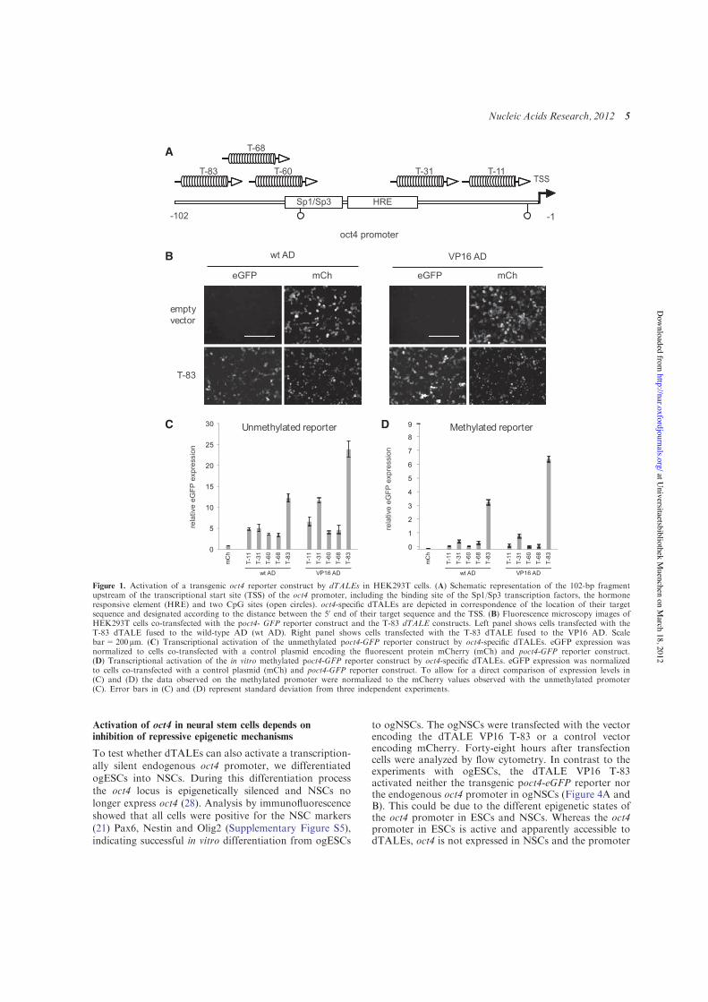
Activation of oct4 in neural stem cells depends oninhibition of repressive epigenetic mechanisms
To test whether dTALEs can also activate a transcription-ally silent endogenous oct4 promoter, we differentiatedogESCs into NSCs. During this differentiation processthe oct4 locus is epigenetically silenced and NSCs nolonger express oct4 (28). Analysis by immunofluorescenceshowed that all cells were positive for the NSC markers(21) Pax6, Nestin and Olig2 (Supplementary Figure S5),indicating successful in vitro differentiation from ogESCs
to ogNSCs. The ogNSCs were transfected with the vectorencoding the dTALE VP16 T-83 or a control vectorencoding mCherry. Forty-eight hours after transfectioncells were analyzed by flow cytometry. In contrast to theexperiments with ogESCs, the dTALE VP16 T-83activated neither the transgenic poct4-eGFP reporter northe endogenous oct4 promoter in ogNSCs (Figure 4A andB). This could be due to the different epigenetic states ofthe oct4 promoter in ESCs and NSCs. Whereas the oct4promoter in ESCs is active and apparently accessible todTALEs, oct4 is not expressed in NSCs and the promoter
A
B
C D
Figure 1. Activation of a transgenic oct4 reporter construct by dTALEs in HEK293T cells. (A) Schematic representation of the 102-bp fragmentupstream of the transcriptional start site (TSS) of the oct4 promoter, including the binding site of the Sp1/Sp3 transcription factors, the hormoneresponsive element (HRE) and two CpG sites (open circles). oct4-specific dTALEs are depicted in correspondence of the location of their targetsequence and designated according to the distance between the 50 end of their target sequence and the TSS. (B) Fluorescence microscopy images ofHEK293T cells co-transfected with the poct4- GFP reporter construct and the T-83 dTALE constructs. Left panel shows cells transfected with theT-83 dTALE fused to the wild-type AD (wt AD). Right panel shows cells transfected with the T-83 dTALE fused to the VP16 AD. Scalebar=200mm. (C) Transcriptional activation of the unmethylated poct4-GFP reporter construct by oct4-specific dTALEs. eGFP expression wasnormalized to cells co-transfected with a control plasmid encoding the fluorescent protein mCherry (mCh) and poct4-GFP reporter construct.(D) Transcriptional activation of the in vitro methylated poct4-GFP reporter construct by oct4-specific dTALEs. eGFP expression was normalizedto cells co-transfected with a control plasmid (mCh) and poct4-GFP reporter construct. To allow for a direct comparison of expression levels in(C) and (D) the data observed on the methylated promoter were normalized to the mCherry values observed with the unmethylated promoter(C). Error bars in (C) and (D) represent standard deviation from three independent experiments.
Nucleic Acids Research, 2012 5
at Universitaetsbibliothek M
uenchen on March 18, 2012
http://nar.oxfordjournals.org/D
ownloaded from
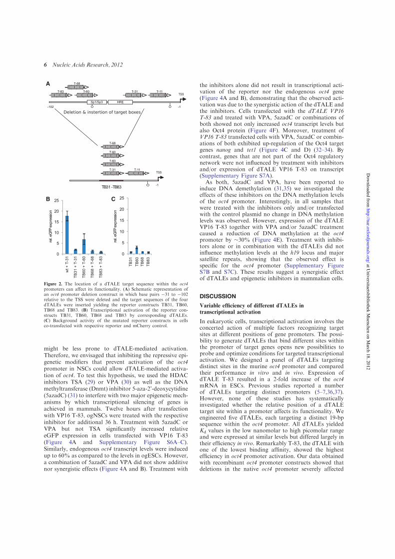
might be less prone to dTALE-mediated activation.Therefore, we envisaged that inhibiting the repressive epi-genetic modifiers that prevent activation of the oct4promoter in NSCs could allow dTALE-mediated activa-tion of oct4. To test this hypothesis, we used the HDACinhibitors TSA (29) or VPA (30) as well as the DNAmethyltransferase (Dnmt) inhibitor 5-aza-2’-deoxycytidine(5azadC) (31) to interfere with two major epigenetic mech-anisms by which transcriptional silencing of genes isachieved in mammals. Twelve hours after transfectionwith VP16 T-83, ogNSCs were treated with the respectiveinhibitor for additional 36 h. Treatment with 5azadC orVPA but not TSA significantly increased relativeeGFP expression in cells transfected with VP16 T-83(Figure 4A and Supplementary Figure S6A–C).Similarly, endogenous oct4 transcript levels were inducedup to 60% as compared to the levels in ogESCs. However,a combination of 5azadC and VPA did not show additivenor synergistic effects (Figure 4A and B). Treatment with
the inhibitors alone did not result in transcriptional acti-vation of the reporter nor the endogenous oct4 gene(Figure 4A and B), demonstrating that the observed acti-vation was due to the synergistic action of the dTALE andthe inhibitors. Cells transfected with the dTALE VP16T-83 and treated with VPA, 5azadC or combinations ofboth showed not only increased oct4 transcript levels butalso Oct4 protein (Figure 4F). Moreover, treatment ofVP16 T-83 transfected cells with VPA, 5azadC or combin-ations of both exhibited up-regulation of the Oct4 targetgenes nanog and tet1 (Figure 4C and D) (32–34). Bycontrast, genes that are not part of the Oct4 regulatorynetwork were not influenced by treatment with inhibitorsand/or expression of dTALE VP16 T-83 on transcript(Supplementary Figure S7A).
As both, 5azadC and VPA, have been reported toinduce DNA demethylation (31,35) we investigated theeffects of these inhibitors on the DNA methylation levelsof the oct4 promoter. Interestingly, in all samples thatwere treated with the inhibitors only and/or transfectedwith the control plasmid no change in DNA methylationlevels was observed. However, expression of the dTALEVP16 T-83 together with VPA and/or 5azadC treatmentcaused a reduction of DNA methylation at the oct4promoter by %30% (Figure 4E). Treatment with inhibi-tors alone or in combination with the dTALEs did notinfluence methylation levels at the h19 locus and majorsatellite repeats, showing that the observed effect isspecific for the oct4 promoter (Supplementary FigureS7B and S7C). These results suggest a synergistic effectof dTALEs and epigenetic inhibitors in mammalian cells.
DISCUSSION
Variable efficiency of different dTALEs intranscriptional activation
In eukaryotic cells, transcriptional activation involves theconcerted action of multiple factors recognizing targetsites at different positions of gene promoters. The possi-bility to generate dTALEs that bind different sites withinthe promoter of target genes opens new possibilities toprobe and optimize conditions for targeted transcriptionalactivation. We designed a panel of dTALEs targetingdistinct sites in the murine oct4 promoter and comparedtheir performance in vitro and in vivo. Expression ofdTALE T-83 resulted in a 2-fold increase of the oct4mRNA in ESCs. Previous studies reported a numberof dTALEs targeting distinct promoters (5–7,36,37).However, none of these studies has systematicallyinvestigated whether the relative position of a dTALEtarget site within a promoter affects its functionality. Weengineered five dTALEs, each targeting a distinct 19-bpsequence within the oct4 promoter. All dTALEs yieldedKd values in the low nanomolar to high picomolar rangeand were expressed at similar levels but differed largely intheir efficiency in vivo. Remarkably T-83, the dTALE withone of the lowest binding affinity, showed the highestefficiency in oct4 promoter activation. Our data obtainedwith recombinant oct4 promoter constructs showed thatdeletions in the native oct4 promoter severely affected
A
B C
Figure 2. The location of a dTALE target sequence within the oct4promoters can affect its functionality. (A) Schematic representation ofan oct4 promoter deletion construct in which base pairs !31 to !102relative to the TSS were deleted and the target sequences of the fourdTALEs were inserted yielding the reporter constructs TB31, TB60,TB68 and TB83. (B) Transcriptional activation of the reporter con-structs TB31, TB60, TB68 and TB83 by corresponding dTALEs.(C) Background activity of the mutated reporter constructs in cellsco-transfected with respective reporter and mCherry control.
6 Nucleic Acids Research, 2012
at Universitaetsbibliothek M
uenchen on March 18, 2012
http://nar.oxfordjournals.org/D
ownloaded from
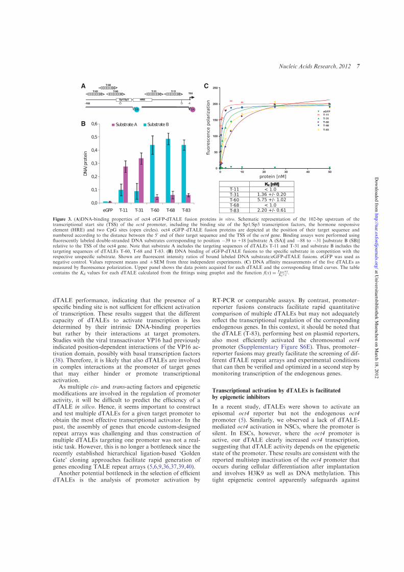
dTALE performance, indicating that the presence of aspecific binding site is not sufficient for efficient activationof transcription. These results suggest that the differentcapacity of dTALEs to activate transcription is lessdetermined by their intrinsic DNA-binding propertiesbut rather by their interactions at target promoters.Studies with the viral transactivator VP16 had previouslyindicated position-dependent interactions of the VP16 ac-tivation domain, possibly with basal transcription factors(38). Therefore, it is likely that also dTALEs are involvedin complex interactions at the promoter of target genesthat may either hinder or promote transcriptionalactivation.
As multiple cis- and trans-acting factors and epigeneticmodifications are involved in the regulation of promoteractivity, it will be difficult to predict the efficiency of adTALE in silico. Hence, it seems important to constructand test multiple dTALEs for a given target promoter toobtain the most effective transcriptional activator. In thepast, the assembly of genes that encode custom-designedrepeat arrays was challenging and thus construction ofmultiple dTALEs targeting one promoter was not a real-istic task. However, this is no longer a bottleneck since therecently established hierarchical ligation-based ‘GoldenGate’ cloning approaches facilitate rapid generation ofgenes encoding TALE repeat arrays (5,6,9,36,37,39,40).
Another potential bottleneck in the selection of efficientdTALEs is the analysis of promoter activation by
RT-PCR or comparable assays. By contrast, promoter–reporter fusions constructs facilitate rapid quantitativecomparison of multiple dTALEs but may not adequatelyreflect the transcriptional regulation of the correspondingendogenous genes. In this context, it should be noted thatthe dTALE (T-83), performing best on plasmid reporters,also most efficiently activated the chromosomal oct4promoter (Supplementary Figure S6E). Thus, promoter–reporter fusions may greatly facilitate the screening of dif-ferent dTALE repeat arrays and experimental conditionsthat can then be verified and optimized in a second step bymonitoring transcription of the endogenous genes.
Transcriptional activation by dTALEs is facilitatedby epigenetic inhibitors
In a recent study, dTALEs were shown to activate anepisomal oct4 reporter but not the endogenous oct4promoter (5). Similarly, we observed a lack of dTALE-mediated oct4 activation in NSCs, where the promoter issilent. In ESCs, however, where the oct4 promoter isactive, our dTALE clearly increased oct4 transcription,suggesting that dTALE activity depends on the epigeneticstate of the promoter. These results are consistent with thereported multistep inactivation of the oct4 promoter thatoccurs during cellular differentiation after implantationand involves H3K9 as well as DNA methylation. Thistight epigenetic control apparently safeguards against
A
B
C
Figure 3. (A)DNA-binding properties of oct4 eGFP-dTALE fusion proteins in vitro. Schematic representation of the 102-bp upstream of thetranscriptional start site (TSS) of the oct4 promoter, including the binding site of the Sp1/Sp3 transcription factors, the hormone responsiveelement (HRE) and two CpG sites (open circles). oct4 eGFP–dTALE fusion proteins are depicted at the position of their target sequence andnumbered according to the distance between the 50 end of their target sequence and the TSS of the oct4 gene. Binding assays were performed usingfluorescently labeled double-stranded DNA substrates corresponding to position !39 to+18 [substrate A (SA)] and !88 to !31 [substrate B (SB)]relative to the TSS of the oct4 gene. Note that substrate A includes the targeting sequences of dTALEs T-11 and T-31 and substrate B includes thetargeting sequences of dTALEs T-60, T-68 and T-83. (B) DNA binding of eGFP-dTALE fusions to the specific substrate in competition with therespective unspecific substrate. Shown are fluorescent intensity ratios of bound labeled DNA substrate/eGFP-dTALE fusions. eGFP was used asnegative control. Values represent means and ±SEM from three independent experiments. (C) DNA affinity measurements of the five dTALEs asmeasured by fluorescence polarization. Upper panel shows the data points acquired for each dTALE and the corresponding fitted curves. The tablecontains the Kd values for each dTALE calculated from the fittings using gnuplot and the function f x& ' # Ymax(x
Kd+x .
Nucleic Acids Research, 2012 7
at Universitaetsbibliothek M
uenchen on March 18, 2012
http://nar.oxfordjournals.org/D
ownloaded from
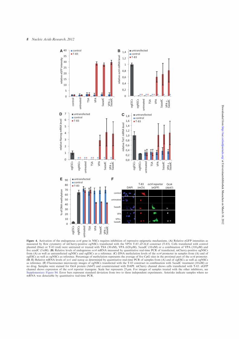
BA
F
CD
E
Figure 4. Activation of the endogenous oct4 gene in NSCs requires inhibition of repressive epigenetic mechanisms. (A) Relative eGFP intensities asmeasured by flow cytometry of mCherry-positive ogNSCs transfected with the VP16 T-83 dTALE construct (T-83). Cells transfected with controlplasmid (blue) or T-83 (red) were untreated or treated with TSA (30 nM), VPA (620 mM), 5azadC (10 nM) or a combination of VPA (310mM) andfive azadC (5 nM). (B) Relative levels of endogenous oct4 mRNA measured by quantitative real-time PCR of transfected, mCherry-positive ogNSCsfrom (A) as well as untransfected ogNSCs and ogESCs as a reference. (C) DNA methylation levels of the oct4 promoter in samples from (A) and ofogESCs as well as ogNSCs as reference. Percentage of methylation represents the average of five CpG sites in the proximal part of the oct4 promoter.(D, E) Relative mRNA levels of tet1 and nanog as determined by quantitative real-time PCR of samples from (A) and of ogESCs as well as ogNSCsas reference. (F) Fluorescence microscopy images of ogNSCs transfected with the T-83 construct in combination with 5azadC treatment (10 nM) orno drug. Samples were stained for Oct4 protein (A647) and counterstained with DAPI. mCherry channel shows cells transfected with T-83. eGFPchannel shows expression of the oct4 reporter transgene. Scale bar represents 25 mm. For images of samples treated with the other inhibitors, seeSupplementary Figure S4. Error bars represent standard deviation from two to three independent experiments. Asterisks indicate samples where nomRNA was detectable by quantitative real-time PCR.
8 Nucleic Acids Research, 2012
at Universitaetsbibliothek M
uenchen on March 18, 2012
http://nar.oxfordjournals.org/D
ownloaded from
inappropriate reactivation of the oct4 gene and thusprevents uncontrolled proliferation and cancer (41–43).
We found that chemical inhibition of repressive epigen-etic modifiers like Dnmts and HDACs enabled dTALE-mediated transcriptional activation of silent oct4 in NSCs.Interestingly, of the two HDAC inhibitors tested onlyVPA but not TSA treatment allowed efficient transcrip-tional activation of oct4 by dTALEs. A similar differencebetween the two inhibitors was previously reported forcellular reprogramming and oct4 promoter activation(44). One possible explanation for the different efficacyof the two inhibitors could be their different targetspecificities (45). Interestingly, VPA was shown to specif-ically affect the proximal region of the oct4 promoter (46)where also the T-83 dTALE binds. This might also explainwhy inhibitor treatment of cells transfected with T-31, thedTALE with the next greatest activity in the reporterassays, did not facilitate the activation of oct4 (Supple-mentary Figure S6E).
Previous studies reported that high concentrations ofVPA and/or 5azadC induce demethylation and reactiva-tion of silent genes (35,47,44,46,38). Under our experimen-tal conditions, however, these inhibitors induced DNAdemethylation of the oct4 promoter only in combinationwith dTALEs, indicating a synergistic effect. A possibleexplanation could be that binding of the dTALE interfereswith maintenance of DNA methylation and, thus, in com-bination with the epigenetic inhibitors leads to reductionof methylation levels. Such a synergistic effect would beconsistent with the recent realization that DNA methyla-tion is rather dynamic and functionally linked to otherepigenetic pathways (48). The synergy between low con-centrations of epigenetic inhibitors and dTALEs suggeststhat silent target genes could be activated withoutgenome-wide demethylation and thus avoid unwantedside effects.
In summary, we demonstrated that combining dTALEswith DNA methylation and/or HDAC inhibitors facili-tates selective activation of the endogenous oct4pluripotency gene. As in turn also Oct4 target genes arereactivated, dTALEs could be used for reprogramming ofsomatic cells to induced pluripotent stem cells (iPSCs).It remains to be investigated whether single or combin-ations of several dTALEs are more efficient than presentreprogramming strategies involving the Oct4 protein itself.However, in contrast to native transcription factors,dTALEs can be specifically directed against single genesor selected combinations of target genes and thereby allowdissection of complex transcription networks to identifykey factors in biological processes like pluripotency anddifferentiation. The combination with epigenetic inhibi-tors may, in some cases, facilitate the activation oftightly repressed genes and further expand the utility ofdTALEs in basic and applied biosciences.
SUPPLEMENTARY DATA
Supplementary Data are available at NAR Online:Supplementary Figures 1–7.
ACKNOWLEDGEMENTS
We thank Luca Gentile and Hans R. Scholer (Max PlanckInstitute for Molecular Biomedicine, Munster) forproviding the GOF-18 construct, DG Johnson (HowardHughes Medical Institute, Durham) for providing theE2F1-VP16 fusion and Kerry Tucker (Ruprecht-Karls-University,Heidelberg) for providing wt J1 ESCs.Furthermore, we thank Fabian Kohler and TobiasAnton for help with the reporter gene assays. We thankAlex Buschle for help with the immunofluorescencestaining and Carina Frauer for advice on the DNAbinding assays. CSS and KT gratefully acknowledge theInternational Max Planck Research School for Molecularand Cellular Life Sciences (IMPRS-LS). H.L., T.L., S.B.and F.S. conceived the study. H.L., T.L., F.S., S.B. andR.M. designed the experiments. S.B. performed the cellbiological experiments and DNA methylation analysis.K.T. performed the DNA binding assays. R.M. and J.E.established a collection of plasmids for assembly ofdTALE genes. RM designed and cloned the dTALEsused in this study. CSS isolated RNA samples, performedthe expression analysis and DNA methylation analysis.S.B., F.S., T.L. and H.L. wrote the manuscript.
FUNDING
The Deutsche Forschungsgemeinschaft (SPP 1356 andSFBs 646/TR5) to H.L.; grants from the 2Blades founda-tion to T.L.; The Elite Network of Bavaria (InternationalDoctorate Program NanoBioTechnology) to C.S.S.; S.B.is a fellow of the graduate school Life Science Munich(LSM). Funding for open access charge: The DeutscheForschungsgemeinschaft.
Conflict of interest statement. None declared.
REFERENCES
1. Klug,A. (2010) The discovery of zinc fingers and theirdevelopment for practical applications in gene regulation andgenome manipulation. Q. Rev. Biophys., 43, 1–21.
2. Segal,D.J. (2011) Toward controlling gene expression at will:selection and design of zinc finger domains recognizing each ofthe 5’-GNN-3’ DNA target sequences. Proc. Natl. Acad. Sci.USA, 96, 2758–2763.
3. Cathomen,T. and Joung,J.K. (2008) Zinc-finger nucleases: thenext generation emerges. Mol. Ther., 16, 1200–1207.
4. De Francesco,L. (2011) Move over ZFNs. Nat. Biotechnol., 29,681–684.
5. Zhang,F., Cong,L., Lodato,S., Kosuri,S., Church,G.M. andArlotta,P. (2011) Efficient construction of sequence-specific TALeffectors for modulating mammalian transcription. Nat.Biotechnol., 29, 149–153.
6. Morbitzer,R., Elsaesser,J., Hausner,J. and Lahaye,T. (2011)Assembly of custom TALE-type DNA binding domains bymodular cloning. Nucleic Acids Res., 39, 5790–5709.
7. Miller,J.C., Tan,S., Qiao,G., Barlow,K.A., Wang,J., Xia,D.F.,Meng,X., Paschon,D.E., Leung,E., Hinkley,S.J. et al. (2011) ATALE nuclease architecture for efficient genome editing.Nat. Biotechnol., 29, 143–148.
8. Mahfouz,M.M., Li,L., Shamimuzzaman,M., Wibowo,A., Fang,X.and Zhu,J.-K. (2011) De novo-engineered transcriptionactivator-like effector (TALE) hybrid nuclease with novel DNAbinding specificity creates double-strand breaks.Proc. Natl. Acad. Sci. USA, 108, 2623–2628.
Nucleic Acids Research, 2012 9
at Universitaetsbibliothek M
uenchen on March 18, 2012
http://nar.oxfordjournals.org/D
ownloaded from
9. Cermak,T., Doyle,E.L., Christian,M., Wang,L., Zhang,Y.,Schmidt,C., Baller,J.A., Somia,N.V., Bogdanove,A.J. andVoytas,D.F. (2011) Efficient design and assembly of customTALEN and other TAL effector-based constructs for DNAtargeting. Nucleic Acids Res., 39, e82.
10. Boch,J., Scholze,H., Schornack,S., Landgraf,A., Hahn,S., Kay,S.,Lahaye,T., Nickstadt,A. and Bonas,U. (2009) Breaking the codeof DNA binding specificity of TAL-type III effectors. Science,326, 1509–1512.
11. Moscou,M.J. and Bogdanove,A.J. (2009) A simple cipher governsDNA recognition by TAL effectors. Science, 326, 1501.
12. Bogdanove,A.J., Schornack,S. and Lahaye,T. (2010) TALeffectors: finding plant genes for disease and defense.Curr. Opin. Plant Biol., 13, 394–401.
13. Nakagawa,T., Kurose,T., Hino,T., Tanaka,K., Kawamukai,M.,Niwa,Y., Toyooka,K., Matsuoka,K., Jinbo,T. and Kimura,T.(2007) Development of series of gateway binary vectors, pGWBs,for realizing efficient construction of fusion genes for planttransformation. J. Biosci. Bioeng., 104, 34–41.
14. Niwa,H., Yamamura,K. and Miyazaki,J. (1991) Efficient selectionfor high-expression transfectants with a novel eukaryotic vector.Gene, 108, 193–199.
15. Johnson,D.G. (1994) Oncogenic capacity of the E2F1 Gene.Proc. Natl. Acad. Sci. USA, 91, 12823–12827.
16. Engler,C., Gruetzner,R., Kandzia,R. and Marillonnet,S. (2009)Golden gate shuffling: a one-pot DNA shuffling method based ontype IIs restriction enzymes. PLoS One, 4, e5553.
17. Yeom,Y., Fuhrmann,G., Ovitt,C., Brehm,A., Ohbo,K., Gross,M.,Hubner,K. and Scholer,H. (1996) Germline regulatory element ofOct-4 specific for the totipotent cycle of embryonal cells.Development, 122, 881–894.
18. DuBridge,R.B., Tang,P., Hsia,H.C., Leong,P.M., Miller,J.H. andCalos,M.P. Analysis of mutation in human cells by using anEpstein–Barr virus shuttle system. Mol. Cell. Biol., 7, 379–387.
19. Szwagierczak,A., Bultmann,S., Schmidt,C.S., Spada,F. andLeonhardt,H. (2010) Sensitive enzymatic quantification of5-hydroxymethylcytosine in genomic DNA. Nucleic Acids Res.,38, e181.
20. Li,E., Bestor,T.H. and Jaenisch,R. (1992) Targeted mutation ofthe DNA methyltransferase gene results in embryonic lethality.Cell, 69, 915–926.
21. Conti,L., Pollard,S.M., Gorba,T., Reitano,E., Toselli,M.,Biella,G., Sun,Y., Sanzone,S., Ying,Q.-L., Cattaneo,E. et al.Niche-independent symmetrical self-renewal of a mammaliantissue stem cell. PLoS Biol., 3, e283.
22. Ying,Q.-L., Stavridis,M., Griffiths,D., Li,M. and Smith,A. (2003)Conversion of embryonic stem cells into neuroectodermalprecursors in adherent monoculture. Nat. Biotechnol., 21,183–186.
23. Ying,Q.L. and Smith,A.G. (2003) Defined conditions for neuralcommitment and differentiation. Methods Enzymol., 365, 327–341.
24. Rottach,A., CFrauer,C., Pichler,G., Bonapace,I.M., Spada,F. andLeonhardt,H. (2010) The multi-domain protein Np95 connectsDNA methylation and histone modification. Nucleic Acids Res.,38, 1796–1804.
25. Frauer,C., Rottach,A., Meilinger,D., Bultmann,S., Fellinger,K.,Hasenoder,S., Wang,M., Qin,W., Soding,J., Spada,F. et al. (2011)Different binding properties and function of CXXC zinc fingerdomains in Dnmt1 and Tet1. PLoS One, 6, e16627.
26. Shaner,N.C., Campbell,R.E., Steinbach,P.A., Giepmans,B.N.G.,Palmer,A.E. and Tsien,R.Y. (2004) Improved monomeric red,orange and yellow fluorescent proteins derived from Discosomasp. red fluorescent protein. Nat. Biotechnol., 22, 1567–1572.
27. Pan,G., Li,J., Zhou,Y., Zheng,H. and Pei,D. (2006) A negativefeedback loop of transcription factors that controls stem cellpluripotency and self-renewal. FASEB J., 20, 1730–1732.
28. Kim,J.B., Zaehres,H., Wu,G., Gentile,L., Ko,K., Sebastiano,V.,Arauzo-Bravo,M.J., Ruau,D., Han,D.W., Zenke,M. et al. (2008)Pluripotent stem cells induced from adult neural stem cells byreprogramming with two factors. Nature, 454, 646–650.
29. Yoshida,M., Kijima,M., Akita,M. and Beppu,T. (1990) Potentand specific inhibition of mammalian histone deacetylase both
in vivo and in vitro by trichostatin A. J. Biol. Chem., 265,17174–17179.
30. Gottlicher,M., Minucci,S., Zhu,P., Kramer,O.H., Schimpf,A.,Giavara,S., Sleeman,J.P., Lo Coco,F., Nervi,C., Pelicci,P.G. et al.(2001) Valproic acid defines a novel class of HDAC inhibitorsinducing differentiation of transformed cells. EMBO J., 20,6969–6978.
31. Santi,D.V., Garrett,C.E. and Barr,P.J. (1983) On the mechanismof inhibition of DNA-cytosine methyltransferases by cytosineanalogs. Cell, 33, 9–10.
32. Chen,X., Fang,F., Liou,Y.-C. and Ng,H.-H. (2008) Zfp143regulates Nanog through modulation of Oct4 binding. Stem Cells,26, 2759–2767.
33. Koh,K.P., Yabuuchi,A., Rao,S., Huang,Y., Cunniff,K.,Nardone,J., Laiho,A., Tahiliani,M., Sommer,C.A.,Mostoslavsky,G. et al. (2011) Tet1 and Tet2 regulate5-hydroxymethylcytosine production and cell lineage specificationin mouse embryonic stem cells. Cell Stem Cell, 8, 200–213.
34. Rodda,D.J., Chew,J.-L., Lim,L.-H., Loh,Y.-H., Wang,B., Ng,H.-H. and Robson,P. (2005) Transcriptional regulation of nanog byOCT4 and SOX2. J. Biol. Chem., 280, 24731–24737.
35. Dong,E., Chen,Y., Gavin,D.P., Grayson,D.R. and Guidotti,A.(2010) Valproate induces DNA demethylation in nuclear extractsfrom adult mouse brain. Epigenetics, 5, 730–735.
36. Geibler,R., Scholze,H., Hahn,S., Streubel,J., Bonas,U., Behrens,S.-E. and Boch,J. (2011) Transcriptional activators of human geneswith programmable DNA-specificity. PloS One, 6, e19509.
37. Weber,E., Gruetzner,R., Werner,S., Engler,C. and Marillonnet,S.(2011) Assembly of designer TAL effectors by Golden Gatecloning. PLoS One, 6, e19722.
38. Hagmann,M., Georgiev,O. and Schaffner,W. (1997) The VP16paradox: herpes simplex virus VP16 contains a long-rangeactivation domain but within the natural multiprotein complexactivates only from promoter-proximal positions. J. Virol., 71,5952–5962.
39. Li,T., Huang,S., Zhao,X., Wright,D.A., Carpenter,S.,Spalding,M.H., Weeks,D.P. and Yang,B. (2011) Modularlyassembled designer TAL effector nucleases for targeted geneknockout and gene replacement in eukaryotes. Nucleic Acids Res.,39, 6315–6325.
40. Scholze,H. and Boch,J. (2011) TAL effectors are remote controlsfor gene activation. Curr. Opin. Microbiol., 14, 47–53.
41. Feldman,N., Gerson,A., Fang,J., Li,E., Zhang,Y., Shinkai,Y.,Cedar,H. and Bergman,Y. (2006) G9a-mediated irreversibleepigenetic inactivation of Oct-3/4 during early embryogenesis.Nat. Cell Biol., 8, 188–194.
42. Gidekel,S., Pizov,G., Bergman,Y. and Pikarsky,E. (2003) Oct-3/4is a dose-dependent oncogenic fate determinant. Cancer Cell, 4,361–370.
43. Looijenga,L.H.J., Stoop,H., de Leeuw,H.P.J.C., de GouveiaBrazao,C.A., Gillis,A.J.M., van Roozendaal,K.E.P., vanZoelen,E.J.J., Weber,R.F.A., Wolffenbuttel,K.P., van Dekken,H.et al. (2003) POU5F1 (OCT3/4) identifies cells with pluripotentpotential in human germ cell tumors. Cancer Res., 63, 2244–2250.
44. Huangfu,D., Maehr,R., Guo,W., Eijkelenboom,A., Snitow,M.,Chen,A.E. and Melton,D.A. (2008) Induction of pluripotent stemcells by defined factors is greatly improved by small-moleculecompounds. Nat. Biotechnol., 26, 795–797.
45. Kim,T.-Y., Bang,Y.-J. and Robertson,K. (2006) Histonedeacetylase inhibitors for cancer therapy. Epigenetics, 1, 14–23.
46. Teng,H.F., Kuo,Y.-L., Loo,M.R., Li,C.L., Chu,T.W., Suo,H.,Liu,H.S., Lin,K.H. and Chen,S.L. (2011) Valproic acid enhancesOct4 promoter activity in myogenic cells. J. Cell. Biochem., 110,995–1004.
47. Al-Salihi,M., Yu,M., Burnett,D.M., Alexander,A., Samlowski,W.and Fitzpatrick,F.A. (2011) The depletion of DNAmethyltransferase-1 and the epigenetic effects of5-aza-2’deoxycytidine (decitabine) are differentially regulated bycell cycle progression. Epigenetics, 6, 1021–1028.
48. Bhutani,N., Burns,D.M. and Blau,H.M. (2011) DNAdemethylation dynamics. Cell, 146, 866–872.
10 Nucleic Acids Research, 2012
at Universitaetsbibliothek M
uenchen on March 18, 2012
http://nar.oxfordjournals.org/D
ownloaded from
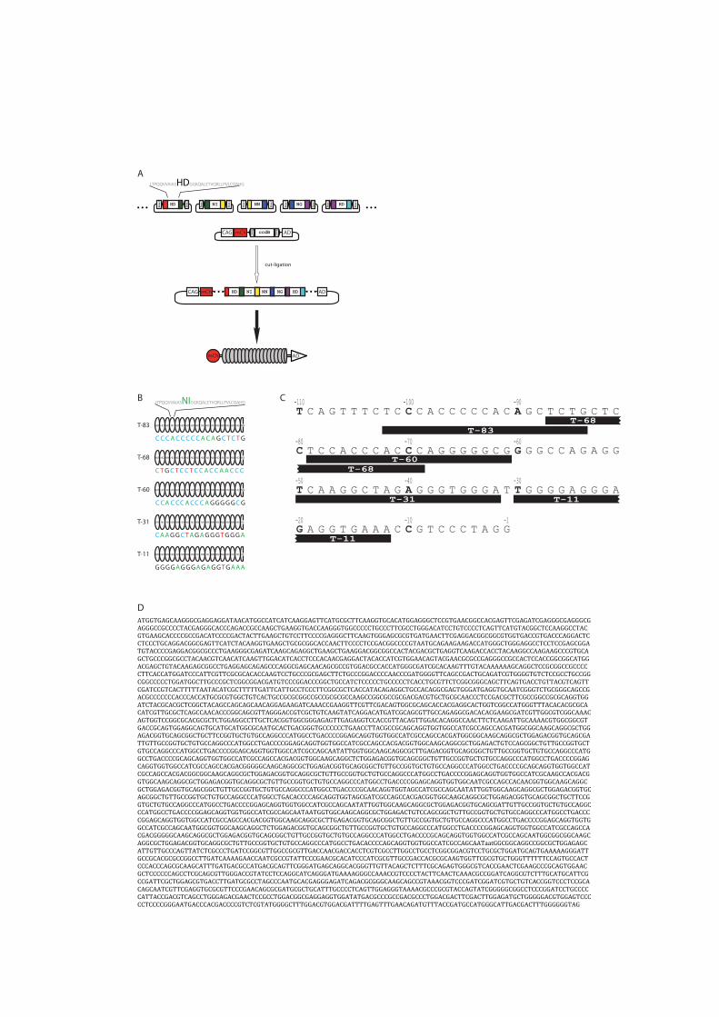
A
BLTPQQVVAIASNIGGKQALETVQRLLPVLCQAHG
LTPQQVVAIASHDGGKQALETVQRLLPVLCQAHG
BsaI
BsaI
BsaI
BsaI
BsaI
BsaI
BsaI
BsaI
BsaI
BsaI
NIHD NN NG HD
BsaI
BsaIccdBCAG mCh AD
NIHD NN NG HDCAG mCh AD
ADmCh
cut-ligation
HD HD NNHD HD NI HD HD HD NIHD HD NI HD NG HD NG NN
C C CCACCCCCA CAG TCTG
T-83
HD NN NNNI NI NN HD NG NI NINN NN NN NG NN NN NN
CA GAGGCTAGA TGG GGGA
T-31 NI
NN NINN NN NN NN NN NGNI NINN NN NN NN NI NINN
GG AGGAGGGAG TGG GAAA
T-11 NI
HD HD NNHD NI HD HD NI HD HDHD NI NN NN NN NN HD
C C AA CCCACCC GGG GGCG
T-60 NN
HD NG HDNG NN HD HD HD NG HDHD NI HD NI NI HD HD
C T AGCTCCTCC ACC ACCC
T-68 HD
T C A G T T T C T C C C A C C C C C A C A G C T C T G C T C
C T C C A C C C A C C C A G G G G G C G G G G C C A G A G G
T C A A G G C T A G A G G G T G G G A T T G G G G A G G G A
G A G G T G A A A C C G T C C C T A G G
-30-40-50
-60-70-80
-90-100-110
-10-20 -1
T-11
T-11T-31
T-60
T-68
T-11
T-68
T-83
OCT4-TALE
C
ATGGTGAGCAAGGGCGAGGAGGATAACATGGCCATCATCAAGGAGTTCATGCGCTTCAAGGTGCACATGGAGGGCTCCGTGAACGGCCACGAGTTCGAGATCGAGGGCGAGGGCGAGGGCCGCCCCTACGAGGGCACCCAGACCGCCAAGCTGAAGGTGACCAAGGGTGGCCCCCTGCCCTTCGCCTGGGACATCCTGTCCCCTCAGTTCATGTACGGCTCCAAGGCCTACGTGAAGCACCCCGCCGACATCCCCGACTACTTGAAGCTGTCCTTCCCCGAGGGCTTCAAGTGGGAGCGCGTGATGAACTTCGAGGACGGCGGCGTGGTGACCGTGACCCAGGACTCCTCCCTGCAGGACGGCGAGTTCATCTACAAGGTGAAGCTGCGCGGCACCAACTTCCCCTCCGACGGCCCCGTAATGCAGAAGAAGACCATGGGCTGGGAGGCCTCCTCCGAGCGGATGTACCCCGAGGACGGCGCCCTGAAGGGCGAGATCAAGCAGAGGCTGAAGCTGAAGGACGGCGGCCACTACGACGCTGAGGTCAAGACCACCTACAAGGCCAAGAAGCCCGTGCAGCTGCCCGGCGCCTACAACGTCAACATCAAGTTGGACATCACCTCCCACAACGAGGACTACACCATCGTGGAACAGTACGAACGCGCCGAGGGCCGCCACTCCACCGGCGGCATGGACGAGCTGTACAAGAGCGGCCTGAGGAGCAGAGCCCAGGCGAGCAACAGCGCCGTGGACGCCACCATGGGCGATCGCACAAGTTTGTACAAAAAAGCAGGCTCCGCGGCCGCCCCCTTCACCATGGATCCCATTCGTTCGCGCACACCAAGTCCTGCCCGCGAGCTTCTGCCCGGACCCCAACCCGATGGGGTTCAGCCGACTGCAGATCGTGGGGTGTCTCCGCCTGCCGGCGGCCCCCTGGATGGCTTGCCCGCTCGGCGGACGATGTCCCGGACCCGGCTGCCATCTCCCCCTGCCCCCTCACCTGCGTTCTCGGCGGGCAGCTTCAGTGACCTGTTACGTCAGTTCGATCCGTCACTTTTTAATACATCGCTTTTTGATTCATTGCCTCCCTTCGGCGCTCACCATACAGAGGCTGCCACAGGCGAGTGGGATGAGGTGCAATCGGGTCTGCGGGCAGCCGACGCCCCCCCACCCACCATGCGCGTGGCTGTCACTGCCGCGCGGCCGCCGCGCGCCAAGCCGGCGCCGCGACGACGTGCTGCGCAACCCTCCGACGCTTCGCCGGCCGCGCAGGTGGATCTACGCACGCTCGGCTACAGCCAGCAGCAACAGGAGAAGATCAAACCGAAGGTTCGTTCGACAGTGGCGCAGCACCACGAGGCACTGGTCGGCCATGGGTTTACACACGCGCACATCGTTGCGCTCAGCCAACACCCGGCAGCGTTAGGGACCGTCGCTGTCAAGTATCAGGACATGATCGCAGCGTTGCCAGAGGCGACACACGAAGCGATCGTTGGCGTCGGCAAACAGTGGTCCGGCGCACGCGCTCTGGAGGCCTTGCTCACGGTGGCGGGAGAGTTGAGAGGTCCACCGTTACAGTTGGACACAGGCCAACTTCTCAAGATTGCAAAACGTGGCGGCGTGACCGCAGTGGAGGCAGTGCATGCATGGCGCAATGCACTGACGGGTGCCCCCCTGAACCTTACGCCGCAGCAGGTGGTGGCCATCGCCAGCCACGATGGCGGCAAGCAGGCGCTGGAGACGGTGCAGCGGCTGCTTCCGGTGCTGTGCCAGGCCCATGGCCTGACCCCGGAGCAGGTGGTGGCCATCGCCAGCCACGATGGCGGCAAGCAGGCGCTGGAGACGGTGCAGCGATTGTTGCCGGTGCTGTGCCAGGCCCATGGCCTGACCCCGGAGCAGGTGGTGGCCATCGCCAGCCACGACGGTGGCAAGCAGGCGCTGGAGACTGTCCAGCGGCTGTTGCCGGTGCTGTGCCAGGCCCATGGCCTGACCCCGGAGCAGGTGGTGGCCATCGCCAGCAATATTGGTGGCAAGCAGGCGCTTGAGACGGTGCAGCGGCTGTTGCCGGTGCTGTGCCAGGCCCATGGCCTGACCCCGCAGCAGGTGGTGGCCATCGCCAGCCACGACGGTGGCAAGCAGGCTCTGGAGACGGTGCAGCGGCTGTTGCCGGTGCTGTGCCAGGCCCATGGCCTGACCCCGGAGCAGGTGGTGGCCATCGCCAGCCACGACGGGGGCAAGCAGGCGCTGGAGACGGTGCAGCGGCTGTTGCCGGTGCTGTGCCAGGCCCATGGCCTGACCCCGCAGCAGGTGGTGGCCATCGCCAGCCACGACGGCGGCAAGCAGGCGCTGGAGACGGTGCAGGCGCTGTTGCCGGTGCTGTGCCAGGCCCATGGCCTGACCCCGGAGCAGGTGGTGGCCATCGCAAGCCACGACGGTGGCAAGCAGGCGCTGGAGACGGTGCAGGCGCTGTTGCCGGTGCTGTGCCAGGCCCATGGCCTGACCCCGGAGCAGGTGGTGGCAATCGCCAGCCACAACGGTGGCAAGCAGGCGCTGGAGACGGTGCAGCGGCTGTTGCCGGTGCTGTGCCAGGCCCATGGCCTGACCCCGCAACAGGTGGTAGCCATCGCCAGCAATATTGGTGGCAAGCAGGCGCTGGAGACGGTGCAGCGGCTGTTGCCGGTGCTGTGCCAGGCCCATGGCCTGACACCCCAGCAGGTGGTAGCGATCGCCAGCCACGACGGTGGCAAGCAGGCGCTGGAGACGGTGCAGCGGCTGCTTCCGGTGCTGTGCCAGGCCCATGGCCTGACCCCGGAGCAGGTGGTGGCCATCGCCAGCAATATTGGTGGCAAGCAGGCGCTGGAGACGGTGCAGCGATTGTTGCCGGTGCTGTGCCAGGCCCATGGCCTGACCCCGGAGCAGGTGGTGGCCATCGCCAGCAATAATGGTGGCAAGCAGGCGCTGGAGACTGTCCAGCGGCTGTTGCCGGTGCTGTGCCAGGCCCATGGCCTGACCCCGGAGCAGGTGGTGGCCATCGCCAGCCACGACGGTGGCAAGCAGGCGCTTGAGACGGTGCAGCGGCTGTTGCCGGTGCTGTGCCAGGCCCATGGCCTGACCCCGGAGCAGGTGGTGGCCATCGCCAGCAATGGCGGTGGCAAGCAGGCTCTGGAGACGGTGCAGCGGCTGTTGCCGGTGCTGTGCCAGGCCCATGGCCTGACCCCGGAGCAGGTGGTGGCCATCGCCAGCCACGACGGGGGCAAGCAGGCGCTGGAGACGGTGCAGCGGCTGTTGCCGGTGCTGTGCCAGGCCCATGGCCTGACCCCGCAGCAGGTGGTGGCCATCGCCAGCAATGGCGGCGGCAAGCAGGCGCTGGAGACGGTGCAGGCGCTGTTGCCGGTGCTGTGCCAGGCCCATGGCCTGACACCCCAGCAGGTGGTGGCCATCGCCAGCAATaatGGCGGCAGGCCGGCGCTGGAGAGCATTGTTGCCCAGTTATCTCGCCCTGATCCGGCGTTGGCCGCGTTGACCAACGACCACCTCGTCGCCTTGGCCTGCCTCGGCGGACGTCCTGCGCTGGATGCAGTGAAAAAGGGATTGCCGCACGCGCCGGCCTTGATCAAAAGAACCAATCGCCGTATTCCCGAACGCACATCCCATCGCGTTGCCGACCACGCGCAAGTGGTTCGCGTGCTGGGTTTTTTCCAGTGCCACTCCCACCCAGCGCAAGCATTTGATGACGCCATGACGCAGTTCGGGATGAGCAGGCACGGGTTGTTACAGCTCTTTCGCAGAGTGGGCGTCACCGAACTCGAAGCCCGCAGTGGAACGCTCCCCCCAGCCTCGCAGCGTTGGGACCGTATCCTCCAGGCATCAGGGATGAAAAGGGCCAAACCGTCCCCTACTTCAACTCAAACGCCGGATCAGGCGTCTTTGCATGCATTCGCCGATTCGCTGGAGCGTGACCTTGATGCGCCTAGCCCAATGCACGAGGGAGATCAGACGCGGGCAAGCAGCCGTAAACGGTCCCGATCGGATCGTGCTGTCACCGGTCCCTCCGCACAGCAATCGTTCGAGGTGCGCGTTCCCGAACAGCGCGATGCGCTGCATTTGCCCCTCAGTTGGAGGGTAAAACGCCCGCGTACCAGTATCGGGGGCGGCCTCCCGGATCCTGCCCCCATTACCGACGTCAGCCTGGGAGACGAACTCCGCCTGGACGGCGAGGAGGTGGATATGACGCCCGCCGACGCCCTGGACGACTTCGACTTGGAGATGCTGGGGGACGTGGAGTCCCCCTCCCCGGGAATGACCCACGACCCCGTCTCGTATGGGGCTTTGGACGTGGACGATTTTGAGTTTGAACAGATGTTTACCGATGCCATGGGCATTGACGACTTTGGGGGGTAG
D

020040060080010001200140016001800
T-11 T-31 T-60 T-68 T-83 T-11 T-31 T-60 T-68 T-83
wtAD VP16AD
fuorescenceintensity
B
AeGFP mCh
VP16ADwtAD
eGFP mCh
T-83
control
T-11
T-31
T-60
T-68

HpaIIMspI
+-
-+
--
poct4-GFP
1.5kbp
10kbp
0.5kbp
A
C
0
200
400
600
800
1000
1200
eGFP
fuorescenceintensity
B
unmethylatedpoct4-GFP
methylatedpoct4-GFP
0
2000
4000
6000
8000
10000
12000
14000
mCh T-11 T-31 T-60 T-68 T-83 T-11 T-31 T-60 T-68 T-83
control wtAD VP16AD
0
500
1000
1500
2000
2500
3000
3500
mCh T-11 T-31 T-60 T-68 T-83 T-11 T-31 T-60 T-68 T-83
control wtAD VP16AD
eGFP
intensity
eGFP
intensity
+-
-+
--
meth.poct4-GFP

0
0,5
1
1,5
2
2,5
3
3,5
4
4,5
mCh T-83
relative
eGFP
intensity
0
0,5
1
1,5
2
2,5
3
mCh T-83
relative
oct4mRNAlev
el
A B
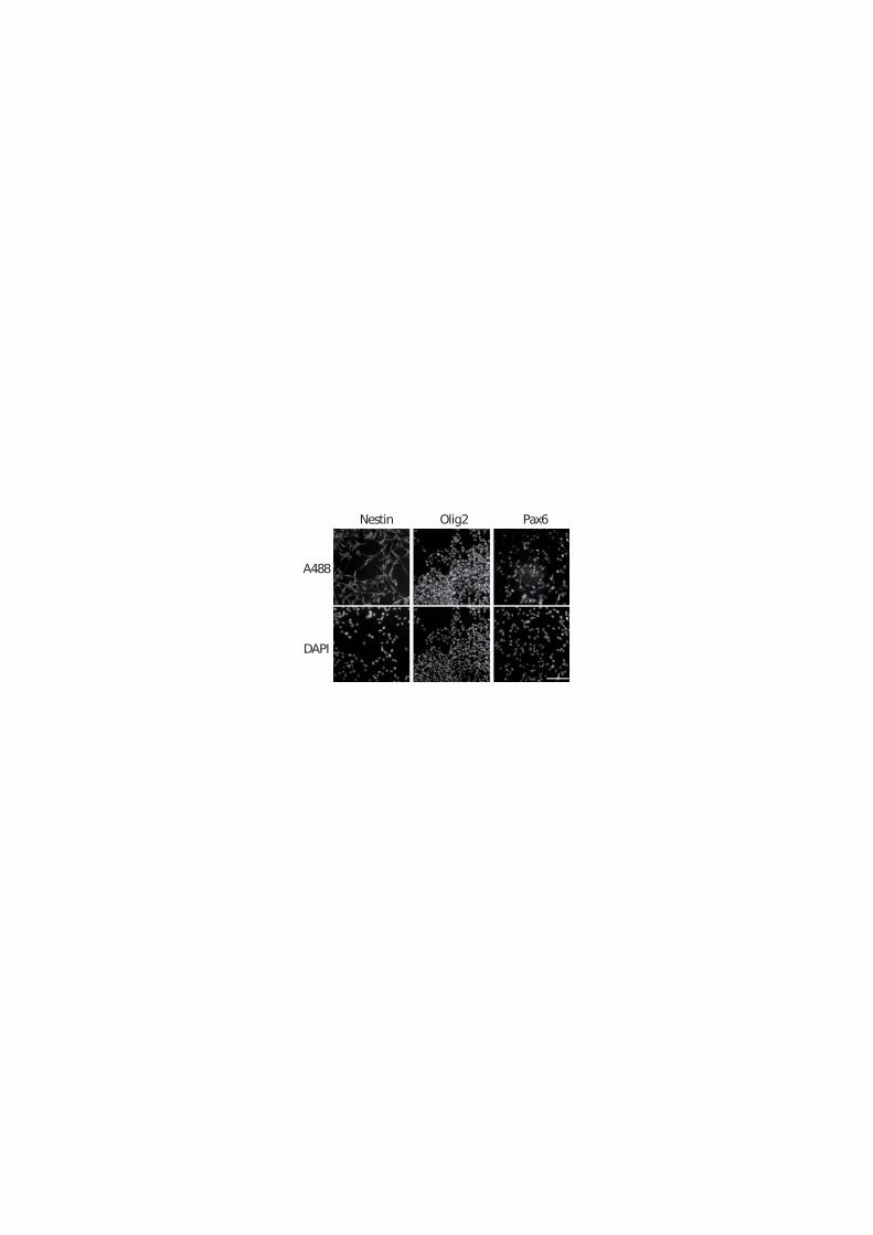
Nestin Olig2 Pax6
DAPI
A488
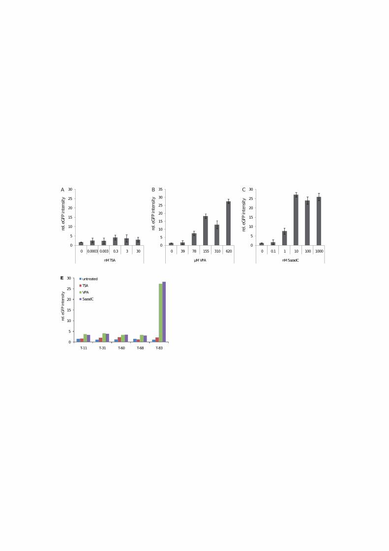
0
5
10
15
20
25
30
35
0 39 78 155 310 620
MVPA
rel.eGFPintensity
0
5
10
15
20
25
30
0 0.0003 0.003 0.3 3 30
nMTSA
rel.eGFPintensity
0
5
10
15
20
25
30
0 0.1 1 10 100 1000
nM5azadC
rel.eGFPintensity
B C
0
5
10
15
20
25
30
T-11 T-31 T-60 T-68 T-83
untreatedTSAVPA5azadC
rel.eGFPintensity
E
A
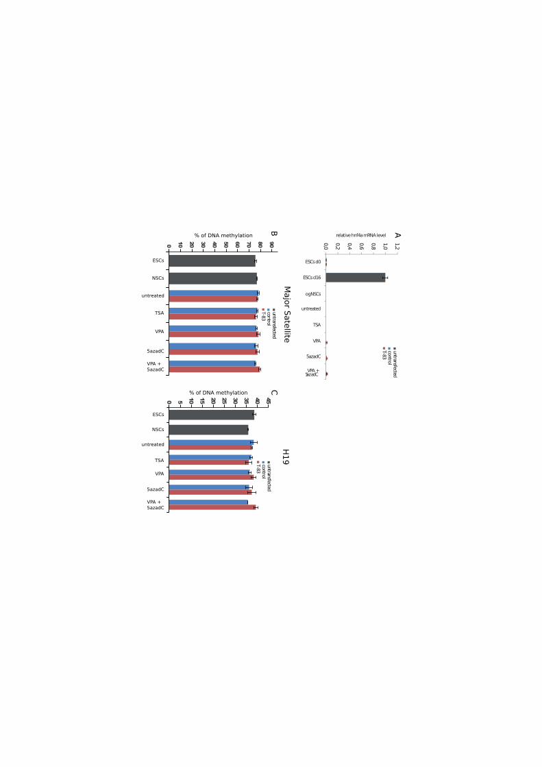
0,0 0,2 0,4 0,6 0,8 1,0 1,2
ESCsd0
untreated
TSA
VPA
5azadC
VPA+5azadC
ogNSCs
ESCsd16
relativehnf4amRNAlevel
controlT-83
untransfected
controlT-83
untransfected
ESCs
NSCs
untreated
TSA
VPA
5azadC
VPA +5azadC
% of DNA methylation
0 5 10 15 20 25 30 35 40 45
ESCs
NSCs
untreated
TSA
VPA
5azadC
VPA +5azadC
0 10 20 30 40 50 60 70 80 90
% of DNA methylation
controlT-83
untransfected
ABC
H19MajorSatellite
3 Discussion
3.1 The role and function of 5-hmC and Tet enzymes in
development and disease
3.1.1 Methods for quantification and mapping of 5-hmC
A first step towards understanding the function of 5-hmC is to analyse in what quanti-
ties 5-hmC occurs in di↵erent cell types and developmental stages. For this purpose we
developed a sensitive enzymatic method which is based on the specific transfer of a radi-
olabeled glucose to 5-hmC by �-glucosyltransferase (�-gt). This enzyme is used by the
T4 page to protect its genome, which contains exclusively hydroxymethylated cytosines,
against bacterial restriction endonucleases. To assess whether transfer of [3H]glucose by
the �-gt to DNA is proportional to the hmC content, we prepared a series of standard
DNA substrate samples by mixing corresponding proportions of two DNA fragment of
same length, one having all cytosine residues replaced by 5-hmC and the other contain-
ing no 5-hmC. With this standard we could show that the assay is linear over a range
of several orders of magnitude and extremely sensitive with a detection limit of 0.025%
5-hmC/C [Szwagierczak et al., 2010].
A crucial step for understanding the role of 5-hmC in the epigenetic landscape was to
analyse the dynamics of this modification during early embryonic development using em-
bryoid bodies (EBs). Di↵erentiation into EBs is a well established in vitro system that
recapitulates first steps of the preimplantation development [Li and Yurchenco, 2006].
Using our �-gt assay we analyzed the 5-hmC content of genomic DNA from undi↵erenti-
ated wildtype ESCs, four and eight day old EBs as well as DNA from ESCs lacking all 3
major DNA methyltransferases (Dnmt1, Dnmt3a and Dnmt3b; triple knockout (TKO)).
Interstingly, we could observe that 5-hmC levels are highly dynamic during the first
eight days of di↵erentiation. A sharp decrease in 5-hmC levels was evident after four
days of EB culture but a substantial recovery was observed after additional 4 days of
culture. Similar dynamics can be observed on the transcript levels of Tet1-3. While Tet1
148

Discussion 149
is expressed predominantly in ESCs, its transcript levels decrease drastically after four
days of EB culture. Tet3 is expressed at very low levels in ESCs but its mRNA levels
start to increase upon di↵erentiation with a dramatic increase between day four and day
eight concomitant with the recovery of the 5-hmC level. As expected, due to the lack
of DNA methylation no 5-hmC could be detected in TKO ESCs [Szwagierczak et al.,
2010]. These findings point to the fact that Tet1 is the main Tet enzyme responsible for
generation of 5-hmC in ESCs. In fact other studies could show that a knock-down of
Tet1 in ESCs results in increased 5-mC levels at certain CpG islands and deregulation of
gene expression of pluripotency-associated genes. Furthermore, Tet1-depletion in preim-
planation embryos and ESCs leads to a bias towards di↵erentation into extra-embryonic
tissues supporting a role for Tet1 in ESC maintenance and inner cell mass cell specifica-
tion [Ito et al., 2010; Ficz et al., 2011; Pastor et al., 2011; Wu et al., 2011b,a; Xu et al.,
2011]. Recent findings revealed that Tet1 is directly controlled by the pluripotency fac-
tor Oct4, integrating 5-hmC and Tet1 into the pluripotency network (Koh et al., 2011).
Moreover, a bioinformatic study which tried to identify a minimal set of pluripotency
markers found Tet1 as the best candidate using three di↵erent independent methods
[Scheubert et al., 2011].
Next we analysed the genomic 5-hmC levels and the expression of Tet enzymes in adult
mouse tissues. We found that adult tissues vary greatly in their 5-hmC content with
highest levels in the central nervous system. In contrast to ESCs, 5-hmC levels in adult
tissues correlate with high levels of Tet3 and to a lower extend Tet2, a pattern similar
to day eight EBs. Thus, most di↵erentiated tissues are characterised by very low levels
of Tet1 and high levels of Tet3, while undi↵erentiated ESCs show the opposite pattern
[Szwagierczak et al., 2010]. These findings further support the idea that Tet1 plays an
important role in pluripotency. Interestingly, kidney represents an exception among the
analysed tissues as it exhibits relatively high 5-hmC levels and Tet2 as the predominant
transcript. This is in accordance to previous findings in Tet2 null mice where the only
observable phenotype is a cellular defect in proximal convoluted tubules of the kidney
[Tang et al., 2008].
Most interesting is the finding that 5-hmC levels are highest in the central nervous
system (CNS). Cells of the CNS, in particular neurons, have to adapt to a very dy-
namic environment of inter- and intracellular contacts and signalling pathways. The
flexibility to respond to these di↵erent signals is achieved by a distinctive epigenetic
plasticity. In this context, DNA methylation dynamics have been shown to be involved
in activity-dependent gene regulation [Martinowich et al., 2003; Chen et al., 2003b; Ma
149

150 3.1. The role and function of 5-hmC and Tet enzymes in development and disease
et al., 2009], memory and learning [Miller and Sweatt, 2007; Day and Sweatt, 2010], and
repeat-associated transcript expression [Muotri et al., 2010; Skene et al., 2010]. Hydrox-
ylation of 5-mC to 5-hmC might present a mechanism by which these DNA methylation
dynamics are regulated.
To further understand the function of 5-hmC in gene regulation it is important to be
able to map its localisation in the genome. As 5-hmC is chemically and structurally very
similar to 5-mC discrimination of these two modification presents a major challenge. The
gold standard methodology for profiling of genomic 5-mC sites, bisulfite conversion, can-
not discriminate 5-hmC from 5-mC and all available restriction endonucleases are either
equally sensitive to mC and hmC or not sensitive to either [Huang et al., 2010; Jin et al.,
2010; Nestor et al., 2010].
We found reports of an endonuclease named PvuRts1I which restriction activity in vivo
was shown to be modulated by 5-hmC glucosylation in a complex fashion [Janosi et al.,
1994]. However, as PvuRts1I was not purified, its activity has not been characterized
in vitro. We could show that recombinant PvuRts1I selectively cleaves 5-hmC contain-
ing DNA and determined its cleavage site. Furthermore, we found that the extent of
PvuRts1I disgestion reflects the relative abundance of 5-hmC in genomic DNA from
cerebellum and TKO ESCs [Szwagierczak et al., 2010]. Restriction of genomic DNA
with PvuRts1I may be combined with PCR amplification for analysis of specific loci,
with massive parallel sequencing or microarray hybridisation for genome-wide mapping.
Due to its relatively complex and long recognition sequence cleavage sites occur in large
distances from another, raising the argument that the extent of random breaks in ge-
nomic DNA preparations would contribute very significant noise in deep sequencing and
microarray applications. This drawback can be overcome if Pvurts1I cut fragments are
enriched by linkers with specific 3’-overhangs.
3.1.2 Recognition of 5-hmC by the epigenetic machinery
DNA methylation is an epigenetic modification that is involved in the control of eu-
karyotic gene expression. In fact, methylation of regulatory elements correlated with a
transcriptional silent state. The mechanism by which the transcriptional repression is
achieved appears to involve 5-mC binding proteins (MBPs). MBPs specifically recognize
5-mC and consequently recruit histone modifying enzymes and chromatin remodelling
factors that establish a silent chromatin state [Sasai and Defossez, 2009].
Given the relatively well established mechanisms of how 5-mC is integrated in the epi-
genetic network, the discovery of 5-hmC in mammalian genomes immediately raised
150
Discussion 151
the question how this new modification is recognized and interpreted by the epigenetic
machinery. We therefore, characterised the 5-mC/5-hmC DNA binding properties of
two representative 5mC binding proteins, the methyl-CpG binding domain (MBD) of
MeCP2 and the SRA domain of Uhrf1. We found that in contrast to the MBD, the
SRA domain binds 5-hmC containing substrates with a similar a�nity as methylated
substrates. To better understand the binding mode and thermodynamics of Uhrf1 to
substrates containing 5-mC and 5-hmC we performed molecular dynamics simulations of
the respective SRA:DNA complexes. Surprisingly, we found that the flipped 5-hmC base
not only fits into the binding pocket of the Uhrf1 SRA domain, but is specifically sta-
bilised by hydrogen bond formation involving the 5-hmC hydroxyl group [Frauer et al.,
2011]. The specific binding of Uhrf1 to 5-hmC containing DNA was very surprising as
Uhrf1 was shown to be essential for the maintenance of DNA methylation by directing
Dnmt1 to hemimethylated CpG sites [Sharif et al., 2007]. The binding of Uhrf1 to hy-
droxymethylated DNA raises the question of how Uhrf1 contributes to the functions in
DNA demethylation or transcriptional activation proposed for 5-hmC. In this context it
should be noted that Uhrf1 is the only protein known that uses a base-flipping mecha-
nism for target recognition and has no described catalytic activity on the flipped base.
5-hmC clearly interferes with the binding by the MBD of MeCP2 and might prevent
subsequent establishment of repressive chromatin structures in a cellular context. Hy-
droxylation of 5-mC could thereby represent a mechanism of changing the cellulat inter-
pretation of an repressive epigenetic modification. Notably, MeCP2 is expressed highest
in the brain where also 5-hmC levels are highest. In fact, a recent study could show
that a functional correlation between 5-hmC levels and MeCP2 protein dosage exists
[Szulwach et al., 2011].
A key for understanding the function of 5-hmC in epigenetic gene regulation is to un-
cover the factors that are involved in its recognition and how these proteins integrate
5-hmC into the epigenetic network. While the “writers” of 5-hmC are known, infor-
mation about the “readers” is still largely missing. Uhrf1 might be just one of a large
family of 5-hmC binding proteins to be discovered and its exact function in vivo remains
to be elucidated. Recently, a member of the MBD protein family has been proposed to
be another 5-hmC binding protein. Mbd3 is part of the Mi2/NURD histone deacety-
lase complex and has been shown to bind preferentially to 5-hmC via its MBD. Mbd3
recruits the Mi2/NURD complex to 5-hmC marked genes and positively regulates their
expression by controlling promoter nucleosome occupancy [Yildirim et al., 2011].
151
152 3.1. The role and function of 5-hmC and Tet enzymes in development and disease
3.1.3 The role of the CXXC domain for Tet1 function
While the C-terminal part of Tet1, harbouring the catalytic DSBH and the cystein-rich
domain, is relatively well characterised, little is known about the N-terminal region. The
only domain described so far is a CXXC zinc-finger, a domain found in several other
proteins with functions related to DNA or chromatin modification. The CXXC domain
of some of these proteins were shown to mediate binding to double stranded DNA con-
taining unmethylated CpGs sites [Lee et al., 2001; Birke et al., 2002; Jørgensen et al.,
2004; Thomson et al., 2010]. We perfomed sequence alignments and homology tree con-
struction and identified three distinct groups of CXXC domains. The first group was
characterised by a KFGG motif between the two cystein clusters of the CXXC domain
and included Dnmt1, CGP, FBxl19, Mll1, Mll2 and Kdm2 proteins. The second and
third group, including the CXXC domains 1 and 2 of Mbd1 on one side and those of
Tet1, Cxxc4/Idax, Cxxc5/RINF and Cxxc10 on the other side, lack the KFGG motif
and diverge from the first group and from each other in the sequence between the cys-
teine clusters.
Based on structural homology models to the crystal structure of the MLL1 CXXC do-
main we generated GFP fusion constructs of the Dnmt1 and Tet1 CXXC zinc finger
(CXXCDnmt1, CXXCTet1) and tested their DNA binding properties using a fluorescent
DNA binding assay [Frauer and Leonhardt, 2009]. CXXCDnmt1 exhibited a preference for
unmethylated CpG containing substrate as expected for a KFGG containing MLL1/CGP
type CXXC domain [Lee et al., 2001; Birke et al., 2002]. Surprisingly, CXXCTet1 did
not show any DNA binding in these experiments [Frauer et al., 2011]. However, a re-
cent study showed that Tet1 binds to unmodified cytosine or 5-mC or 5-hmC containing
CpG-rich DNA through its CXXC domain [Xu et al., 2011]. The authors of this study
used a slightly longer and untagged CXXC domain. By changing the N-terminal GFP
tag to a C-terminal fusion of CXXCTet1 we were able to reproduce these findings (un-
published data). It is unclear what functional significance the strong binding of Tet1
to CpG containing DNA might have. However, binding of Tet1 to CpG rich sequences
could provide an additional layer of protection to limit the access of Dnmts.
Next, we analysed the influence of the CXXC domain on localisation and activity of
Tet1. For this purpose, we generated a GFP-Tet1 fusion lacking the CXXC domain
(Tet1CXXC). Using our �-gt assay we analysed the 5-hmC content of genomic DNA in
HEK293T cells overexpressing the wildtype or the deletion construct. The presence of
both constructs significantly increased the 5-hmC contents to similar levels suggesting
that deletion of the CXXC domain has no e↵ect on the catalytic activity under these
152

Discussion 153
conditions. In addition, wildtype Tet1 and Tet1CXXC exhibited a similar subnuclear
localisation in vivo.
The N-terminal region of Tet1 comprised almost two thirds of the protein. Homology
searches and domain prediction tools could so far not predict any similarities to other do-
mains/proteins except the CXXC domain. It is very likely that, similar to Dnmts which
also harbour a N-terminal regulatory domain, The N-terminal region of Tet1 contributes
to its localisation and integration in the epigenetic network.
3.1.4 Tet2 and 5-hmC in myeloid leukemia
TET1 was originally identified as an MLL fusion partner in rare cases of acute myeloid
leukemia (AML) with a t(10;11)(q22;q23) [Ono et al., 2002; Lorsbach et al., 2003]. Re-
cently, heterozygous deletions and mutations of TET2 were found in a wide range of
myeloid malignancies, including myelodysplastic syndrome (MDS), myeloproliferative
disorders such as chronic myelomonocytic leukemia (CMML) and in secondary AML
(sAML) [Delhommeau et al., 2009; Langemeijer et al., 2009; Mohamedali et al., 2009].
To get first insights into the relationship between TET2 mutations, global gene ex-
pression profile and 5-hmC levels, we measured the 5-hmC levels in the genomic DNA
of 30 sAML patients using our �-gt assay [Szwagierczak et al., 2010] which we opti-
mised to a detection limit of 0.005%. In addition to the TET2 mutational status, we
screened for IDH1/2 mutations. IDH1/2 catalyse the oxidative decarboxylation of isoc-
itrate to ↵-ketoglutarate (↵-KG ) and have been implicated in a variety of cancer types.
The most common mutations in IDH1/2 result in two enzymatic changes. A decreased
wildtype IDH function and gain of a new enzymatic activity to reduce ↵-KG to D-2-
hydroxyglutarate (D-2-HG). D-2-HG outcompetes ↵-KG for binding to several classes
of histone demethylases, PHD2 and TET enyzmes [Borodovsky et al., 2012]. Hence,
IDH1/2 mutations might result in altered 5-hmC levels by inhibiting TET enzyme ac-
tivity.
The analysis of the 5-hmC levels in the genomic DNA of the 30 sAML patients revealed
a 5-hmC content ranging from 0.006 to 0.054% [Konstandin et al., 2011]. Compared
to 5hmC levels in other human adult tissues genomic 5-hmC levels of blood samples
from patients with sAML were extremely low (60 fold (human lung) to 166 fold (human
brain) lower) [Kraus et al., 2012; Terragni et al., 2012]. Interestingly, low 5-hmC levels
seem to be a general feature of many cancer types and in the case of glioblastomas can
even be correlated with tumour malignancy [Kraus et al., 2012; Kudo et al., 2012; Yang
et al., 2012]. These findings suggest that loss of 5-hmC is not just a secondary e↵ect of
153

154 3.1. The role and function of 5-hmC and Tet enzymes in development and disease
global hypomethylation often observed in tumours. Reduced levels of 5-hmC could lead
to changes in gene expression patterns that directly a↵ect the expression of oncogenes
and thereby promote transformation.
In our study 7 out of 30 patients exhibited mutations in the TET2 gene and these 7
samples significantly clustered in the lower half of 5-hmC levels. Interestingly, several
patients with very low 5-hmC levels did not carry a TET2 mutation. Two of these
samples harboured an IDH2 mutation possibly explaining the low 5-hmC levels despite
the absence of a TET2 mutation. However, 8 patients did not exhibit TET2 nor IDH1/2
mutations but clustered in the lower half of 5-hmC levels [Konstandin et al., 2011]. It is
possible that these patients harbour mutations in one of the other TET genes. Another
explanation could be that the low 5-hmC levels result from mutations in genes upstream
or downstream of the TET enzymes. For example, Dnmts or their regulators could be af-
fected leading to genome-wide hypomethylation and thereby to reduced 5-hmC levels. In
addition, BER proteins that are implicated to be involved in the removal of 5-hmC could
be deregulated in these patients. Another interesting possibility is that genome-wide loss
of 5-hmC might be promoted by active/enhanced growth of cancer cells. 5-hmC might
prevent the maintenance of 5-mC by DNMT1 during replication leading to a passive
loss of 5-hmC. This hypothesis is supported by in vitro data showing a strongly reduced
activity of DNMT1 on DNA substrates containing hemi-hydroxylated (5-hmCpG/CpG)
CpG sites that could be produced in vivo by replication of 5-hmC [Valinluck and Sowers,
2007].
Next we analysed the global gene expression profiles (GEPs) from 28 patient samples.
No striking di↵erence could be seen when comparing the GEPs of patients with TET2
mutations against the GEPs of patients without TET2 mutations. However, when
we compared the GEPs of the 7 patients with the lowest versus the 7 patients with
the highest 5-hmC levels distinctive clustering of the two groups was observable [Kon-
standin et al., 2011]. Recently, similar observations were made in the context of chronic
myelomonocytic leukemia (CMML) (Prez et al., 2012). Taken together these results
indicate that 5-hmC levels are most likely more relevant to define biologically distinct
secondary leukemia subtypes than the TET2 or IDH1/2 mutational status. Interestingly,
a recent study could show that deletion of tet2 in mice leads to a dramatic reduction of
5-hmC levels in bone marrow cells and to the development of myeloid malignancies [Li
et al., 2011b]. These findings suggest that Tet2 is the predominant Tet enzyme in the
hematopoietic lineage and functions as a tumour suppressor to maintain cell homoeosta-
sis.
154
Discussion 155
3.1.5 5-hmC a stable epigenetic mark or a demethylation
intermediate?
With the discovery of 5-hmC the question emerged whether this new covalent DNA
modification represents a stable epigenetic mark by itself or an intermediate of DNA
demethylation. At first, it had been debated whether active DNA demethylation does
at all occur in mammals. But as more and more data accumulated that suggested the
active removal of 5-mC in mammalian genomes, the attention turned towards finding
the underlying mechanisms. The direct removal of the methyl group was thought to
be energetically unfavourable and soon many findings suggested that DNA repair path-
ways are involved in the process of active DNA demethylation. The critical step in
all the proposed mechanisms was the derivatisation of 5-mC by the cellular machinery.
Deamination was thought to be the likeliest mechanism by which the 5-mC could be
marked for the removal by the DNA repair machinery. However, the data that showed a
deamination of 5-mC was mainly indirect by showing the involvement of AID/APOBEC
enzymes or came from experiments performed in vitro. Therefore, it remained highly
debated whether DNA repair is involved in active DNA demethylation despite the accu-
mulating amount of data supporting this hypothesis. The discovery of the hydroxylation
of 5-mC by Tet enzymes o↵ered a simple explanation to the question of how a methy-
lated cytosine can be specifically marked for demethylation without contradicting the
existing data on deamination coupled DNA repair. In fact, shortly after the discovery
of 5-hmC is was shown that AID/APOBEC enzymes, a protein family already impli-
cated in deamination coupled repair of 5-mC, mediate active DNA demethylation by
deaminating 5-hmC to 5-hmU [Cortellino et al., 2011; Guo et al., 2011]. In addition to
linking active DNA demethylation to deamination coupled DNA repair, the presence of
5-hmC in mammalian genomes also reopens the possibility for a direct removal of the
methyl-group from 5-mC. Recent studies have shown that Tet enzymes further oxidise
5-hmC to 5-caC [He et al., 2011; Ito et al., 2011]. Although it was suggested that 5-caC
is recognised by the DNA repair machinery [He et al., 2011; Ito et al., 2011], the possibil-
ity remains that 5-caC is decarboxylated to cytosine by a so far unknown decarboxylase
presenting a pathway for DNA demethylation independent of DNA repair mechanisms.
Besides its relatively well established role in active DNA demethylation, 5-hmC might
also represent a stable epigenetic mark by itself. Especially in the nervous system 5-
hmC levels can account for up to 40% of all modified cytosines suggesting an additional
function as an epigenetic regulator [Szwagierczak et al., 2011; Szulwach et al., 2011]. In
155

156 3.2. The role of DNA methylation in transgene silencing and its mechanism
this context it is important to note that while Tet1 seems to have a dual function, the
presence of 5-hmC is generally correlated with transcriptional activity [Williams et al.,
2011; Wu et al., 2011b,a]. One explanation could be that MeCP2, a methylcytosine
binding protein especially abundant in the brain, does not bind to 5-hmC and thereby
preventing the establishment of a silent chromatin state [Frauer et al., 2011]. Another
indication that 5-hmC might be more than a DNA demethylation intermediate comes
from the recent finding that 5-hmC is bound by Mbd3. The recruitment of Mbd3 to 5-
hmC marked genes positively regulates their expression directly linking 5-hmC to active
gene expression [Yildirim et al., 2011]. Although there is first evidence that 5-hmC may
have a DNA demethylation independent function in gene regulation, further studies are
needed to identify the exact mechanisms.
3.2 The role of DNA methylation in transgene silencing
and its mechanism
Transgene expression is a widely used method in cell biology and several vector sys-
tems have been developed over the past decades for this purpose. The main di↵erences
among the vectors is the promoter which is used for transgene expression. While for
some applications transient expression for 1-2 days is su�cient, some experiments re-
quire stable long-term expression. We found that upon transient transfection of wt
ESCs constructs driven by the cytomegalovirus (CMV) promoter were rapidly silenced,
while constructs driven by the chimeric CMV early enhancer/chicken � actin (CAG)
promoter yielded long and stable expression. To understand the underlying mechanisms
of this observation we developed an epigenetic silencing assay. ESCs were cotransfected
with two distinct plasmids, one expressing monomeric red fluorescent protein (mRFP)
under the CMV promoter, the other expressing green fluorescent protein (GFP) driven
by the CAG promoter. We monitored RFP and GFP expression for up to ten days after
transfection by using automated image acquisition and quantification of fluorescent sig-
nals. While in wt ESCs CMV driven mRFP expression was rapidly silenced, dnmt3a/3b
knockout cells exhibited no silencing of the CMV promoter. Suprisingly, uhrf1-/- ESCs
were also unable to silence the CMV promoter whereas dnmt1-/- ESCs showed only
partially reduced silencing [Meilinger et al., 2009]. These findings suggest that Dnmt1
might play a minor role in this silencing process. Moreover, it implies a novel function
for Uhrf1 that is largely independent of Dnmt1. This was surprising, as Uhrf1 has been
reported to be functionally linked to Dnmt1 in maintaining DNA methylation [Sharif
156

Discussion 157
et al., 2007]. Interestingly, the silencing of the CMV promoter showed a similar depen-
dence on the presence of Uhrf1 as for the presence of Dnmt3a/Dnmt3b. In addition,
co-immunoprecipitation experiments revealed an interaction between Uhrf1 and both de
novo Dnmts. This suggests that Np95 might play a role in the de novo methylation
of the CMV promoter and that the silencing process is DNA methylation dependent.
Surprisingly, the CMV promoter exhibited nearly no DNA methylation in the first days
after transfection and reached only a very low level on the last day examined in wild type
ESCs. However, promoter methylation was less in uhrf1-/- cells than in wild type ES and
none of the dnmt deficient cell lines showed a significant amount of DNA methylation
at this time point [Meilinger et al., 2009]. Taken together, all these findings imply that
Uhrf1 is cooperating with Dnmt3a and Dnmt3b in mediating the silencing of the CMV
promoter and that this process, at least at its onset, is independent of DNA methylation.
This is in agreement with the finding that silencing precedes DNA methylation at the
promoters of certain genes [Feldman et al., 2006; Epsztejn-Litman et al., 2008].
The dependency of CMV promoter silencing on the presence of Dnmt3a/3b or Uhrf1 well
before de novo methylation can be detected could be due to the involvement of repressive
histone methylation. We therefore tested the involvement of the histone methyltrans-
ferases (HMTs) G9a and Suv39h1/2 using the epigenetic silencing assay. Interestingly,
both HMTs seem to be involved in this process although to a di↵erent extent. While in
the absence of G9a no preferential silencing of the CMV promoter was observable, cells
lacking Suv39h1/2 did only exhibit a partial silencing defect. This is likely due to the
functional di↵erences of the two HMTs. Suv39h1/2 tri-methylate lysine 9 of histone 3
(H3K9) and were shown to be involved in organization of silent chromatin domains such
as the pericentric heterochromatin [Jenuwein et al., 1998]. In contrast, G9a is crucial
for H3K9 mono- and di-methylation of euchromatin and it is essential for the transcrip-
tional silencing of several genes [Tachibana et al., 2002; Feldman et al., 2006]. This could
explain why G9a and not Suv39h1/2 is crucial for the silencing of the CMV promoter.
Taken together, the finding that DNA methylation seems to be set at a later stage in
the silencing process and the fact that HMTs are involved in the CMV promoter silenc-
ing suggests that histone modifications might be the primary mechanism by which the
silencing is initiated. This idea is supported by the finding that upon transfection into
mammalian cells, plasmids are chromatinized and both their expression and silencing is
associated with specific histone modifications [Riu et al., 2007].
157

158 3.3. Designer TALEs as a tool for targeted transcriptional activation
3.3 Designer TALEs as a tool for targeted
transcriptional activation
In eukaryotic cells, transcriptional activation involves the synergistic action of multiple
factors recognizing target sites at di↵erent positions of gene promoters. With the discov-
ery of the dTALE DNA binding specificity and its underlying code it became possible to
specifically designe TALEs that bind to user-defined target sequences. Previous studies
reported a number of dTALEs targeting distinct promoters [Geissler et al., 2011; Miller
et al., 2011; Morbitzer et al., 2011; Weber et al., 2011; Zhang et al., 2011]. However, none
of these studies has systematically investigated whether the relative position of a dTALE
target site within a promoter a↵ects its functionality. We designed five dTALEs, each
targeting a distinct 19-bp sequence within the oct4 promoter. All dTALEs yielded Kd
values in the low nanomolar to high picomolar range and were expressed at similar levels
but di↵ered largely in their e�ciency in vivo. Intriguingly, T-83, the dTALE with one of
the lowest binding a�nities, showed the highest e�ciency in oct4 promoter activation.
Our data obtained with recombinant oct4 promoter constructs showed that deletions
in the native oct4 promoter severely a↵ected dTALE performance, indicating that the
presence of a specific binding site is not su�cient for e�cient activation of transcription
[Bultmann et al., 2012]. These results suggest that the di↵erent capacity of dTALEs to
activate transcription is less determined by their intrinsic DNA-binding properties but
rather by their interactions at target promoters. Studies with the viral transactivator
VP16 had previously indicated position-dependent interactions of the VP16 activation
domain, possibly with basal transcription factors [Hagmann et al., 1997]. Therefore,
it is likely that also dTALEs are involved in complex interactions at the promoter of
target genes that may either hinder or promote transcriptional activation. As multiple
cis- and trans-acting factors and epigenetic modifications are involved in the regulation
of promoter activity, it will be di�cult to predict the e�ciency of a dTALE in silico.
Hence, it seems important to construct and test multiple dTALEs for a given target
promoter to obtain the most e↵ective transcriptional activator. Due to the modular
structure of the dTALE DNA binding domain construction of multiple TALEs in a time
and cost-e�cient manner using recently established hierarchical ligation-based Golden
Gate cloning approaches has become a feasible task Cermak et al. [2011]; Geissler et al.
[2011]; Li et al. [2011a]; Morbitzer et al. [2011]; Weber et al. [2011]; Zhang et al. [2011].
In a recent study, dTALEs were shown to activate an episomal oct4 reporter but not
the endogenous oct4 promoter [Zhang et al., 2011]. Similarly, we observed a lack of
158

Discussion 159
dTALE- mediated oct4 activation in neural stem cells (NSCs), where the promoter is
silent [Bultmann et al., 2012]. In ESCs, however, where the oct4 promoter is active, our
dTALE clearly increased oct4 transcription, suggesting that dTALE activity depends
on the epigenetic state of the promoter. These results are consistent with inactivation
of the oct4 promoter that occurs during cellular di↵erentiation after implantation and
involves H3K9 as well as DNA methylation. This tight epigenetic control apparently
safeguards against inappropriate reactivation of the oct4 gene and thus prevents uncon-
trolled proliferation and cancer [Gidekel et al., 2003; Looijenga et al., 2003; Feldman
et al., 2006]. Furthermore, the recentlys solved crystal structures of two TALEs showed
that the protein winds around the DNA along the major groove [Deng et al., 2012; Mak
et al., 2012]. Due to this conformation dTALE binding is very likely a↵ected by respres-
sive chromatin states as it has to compete with tightly packed nucleosome arrays for
DNA binding (Figure 17).
In line with this hypothesis, we found that chemical inhibition of repressive epigenetic
modifiers like Dnmts and histone deacetylases (HDACs) enabled dTALE- mediated tran-
scriptional activation of silent oct4 in NSCs. Interestingly, of the two HDAC inhibitors
tested only valproic acid (VPA) but not trichostatin A (TSA) treatment allowed e�-
cient transcriptional activation of oct4 by dTALEs. A similar di↵erence between the two
inhibitors was previously reported for cellular reprogramming and oct4 promoter activa-
tion [Huangfu et al., 2008]. One possible explanation for the di↵erent e�cacy of the two
inhibitors could be their di↵erent target specificities [Kim et al., 2006]. Interestingly,
VPA was shown to specifically a↵ect the proximal region of the oct4 promoter [Teng
et al., 2010] where also the T-83 dTALE binds. This might also explain why inhibitor
treatment of cells transfected with T-31, the dTALE with the next greatest activity in
the reporter assays, did not facilitate the activation of oct4. Previous studies reported
that high concentrations of VPA and/or 5azadC induce demethylation and reactiva-
tion of silent genes [Huangfu et al., 2008; Dong et al., 2010; Teng et al., 2010; Al-Salihi
et al., 2011]. Under our experimental conditions, however, these inhibitors induced DNA
demethylation of the oct4 promoter only in combination with dTALEs, indicating a syn-
ergistic e↵ect. A possible explanation could be that binding of the dTALE interferes
with maintenance of DNA methylation and, thus, in combination with the epigenetic in-
hibitors leads to reduction of methylation levels The synergy between low concentrations
of epigenetic inhibitors and dTALEs suggests that silent target genes could be activated
without genome-wide demethylation and thus avoid unwanted side e↵ects. In summary,
we demonstrated that combining dTALEs with DNA methylation and/or HDAC in-
159
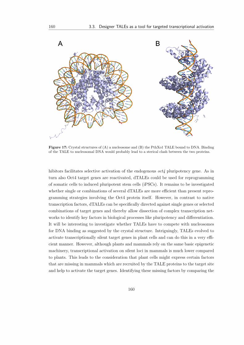
160 3.3. Designer TALEs as a tool for targeted transcriptional activation
Figure 17: Crystal structures of (A) a nucleosome and (B) the PthXo1 TALE bound to DNA. Bindingof the TALE to nucleosomal DNA would probably lead to a sterical clash between the two proteins.
hibitors facilitates selective activation of the endogenous oct4 pluripotency gene. As in
turn also Oct4 target genes are reactivated, dTALEs could be used for reprogramming
of somatic cells to induced pluripotent stem cells (iPSCs). It remains to be investigated
whether single or combinations of several dTALEs are more e�cient than present repro-
gramming strategies involving the Oct4 protein itself. However, in contrast to native
transcription factors, dTALEs can be specifically directed against single genes or selected
combinations of target genes and thereby allow dissection of complex transcription net-
works to identify key factors in biological processes like pluripotency and di↵erentiation.
It will be interesting to investigate whether TALEs have to compete with nucleosomes
for DNA binding as suggested by the crystal structure. Intriguingly, TALEs evolved to
activate transcriptionally silent target genes in plant cells and can do this in a very e�-
cient manner. However, although plants and mammals rely on the same basic epigenetic
machinery, transcriptional activation on silent loci in mammals is much lower compared
to plants. This leads to the consideration that plant cells might express certain factors
that are missing in mammals which are recruited by the TALE proteins to the target site
and help to activate the target genes. Identifying these missing factors by comparing the
160

Discussion 161
interactions of dTALEs in mammals versus those in plants, could allow the activation
of silenced genes without the need for treatment with epigenetic inihibitors.
3.3.1 FairyTALE a simple web tool for dTALE target optimisation
Since the discovery of the code underlying the DNA binding specificity of the Xan-
thomonas Transcription-activator-like e↵ectors, TALEs have been successfully used in a
variety of application and have become a popular and powerful tool in genome editing
and engineering. The modular structure of the TALE DNA binding domain allows the
construction of dTALEs that bind to any user-defined DNA target sequence. Due to the
high degree of flexibiliy in target choice, optimization of target selection is challenging.
Given that the number of possible target sites in a 1000 base pair lond double-stranded
DNA fragment is statistically 500, manual search for potential target sites is tedious
and time-consuming. The target locus needs to be screened for sequences beginning
with a thymidine and to guarantee uniqueness of the found sequences, a BLAST search
for every potential target sequence needs to be performed. Due the low specificity
of guanine recognition by the NN diresidue the number of guanines in a sequence is
preferably to be kept at a minimum [Bogdanove et al., 2010]. In addition, low complexity
sequences and repeats should be avoided. To optimize and streamline the process TALE
target site definition, we developed a web tool called fairyTALE that allows e�cient
and automated selection of the best TALE target sites in a user-defined sequence for a
variety of applications.
Our web tool allows the definition of optimal target sites for single as well as dTALE
pairs. The user chooses the length of the dTALEs along with the targeted species and
Figure 18: Work-flow of the fairyTALE target finder. Sequence input is screened for potential TALEtarget sites using the given parameters. Subsequently, potential sites are checked for possible o↵-targetsusing NCBI BLAST. Intermediate and final results are send to the user via e-mail.
161

162 3.3. Designer TALEs as a tool for targeted transcriptional activation
several other parameters concerning dTALE target site composition (e.g. low complexity
filter). The web tool will find TALE target sequences with the given parameters and
check the number of possible o↵-targets via NCBI BLAST. Subsequently, unique TALE
target sites in the input sequence are returned to the user (Figure 18). The beta version
of the fairyTALE web tool was written exclusively in pythonTM using the Biopython
module [Rossum, 2003; Cock et al., 2009] and is freely available under:
http : // bultmann . alwaysdata . net /FT web . htm
We believe that the fairyTALE web tool optimizes the target site selection process and
increases the likelyhood of successful dTALE application by reducing the amount of
possible o↵-targets.
162

4 Annex
4.1 References
References
Mazin Al-Salihi, Margaret Yu, David M Burnett, Amanda Alexander, Wolfram E Sam-
lowski, and Frank A Fitzpatrick. The depletion of DNA methyltransferase-1 and the
epigenetic e↵ects of 5-aza-2’deoxycytidine (decitabine) are di↵erentially regulated by
cell cycle progression. Epigenetics: O�cial Journal of the DNA Methylation Society,
6(8):1021–1028, August 2011. PMID: 21725200.
Madeleine P Ball, Jin Billy Li, Yuan Gao, Je-Hyuk Lee, Emily M LeProust, In-Hyun
Park, Bin Xie, George Q Daley, and George M Church. Targeted and genome-scale
strategies reveal gene-body methylation signatures in human cells. Nature Biotech-
nology, 27(4):361–368, April 2009. PMID: 19329998.
Bradley E Bernstein, Tarjei S Mikkelsen, Xiaohui Xie, Michael Kamal, Dana J Huebert,
James Cu↵, Ben Fry, Alex Meissner, Marius Wernig, Kathrin Plath, Rudolf Jaenisch,
Alexandre Wagschal, Robert Feil, Stuart L Schreiber, and Eric S Lander. A bivalent
chromatin structure marks key developmental genes in embryonic stem cells. Cell, 125
(2):315–326, April 2006. PMID: 16630819.
T H Bestor. Cloning of a mammalian DNA methyltransferase. Gene, 74(1):9–12, De-
cember 1988. PMID: 3248734.
T H Bestor and VM Ingram. Two DNAmethyltransferases from murine erythroleukemia
cells: purification, sequence specificity, and mode of interaction with DNA. Proceedings
of the National Academy of Sciences of the United States of America, 80(18):5559–
5563, September 1983. PMID: 6577443.
Nidhi Bhutani, Jennifer J Brady, Mara Damian, Alessandra Sacco, Stphane Y Corbel,
163
164 References
and Helen M Blau. Reprogramming towards pluripotency requires AID-dependent
DNA demethylation. Nature, 463(7284):1042–1047, February 2010. PMID: 20027182.
Marco Birke, Silke Schreiner, Mara-Paz Garca-Cullar, Kerstin Mahr, Fritz Titgemeyer,
and Robert K Slany. The MT domain of the proto-oncoprotein MLL binds to CpG-
containing DNA and discriminates against methylation. Nucleic Acids Research, 30
(4):958–965, February 2002. PMID: 11842107.
Jens Boch, Heidi Scholze, Sebastian Schornack, Angelika Landgraf, Simone Hahn, Sabine
Kay, Thomas Lahaye, Anja Nickstadt, and Ulla Bonas. Breaking the code of DNA
binding specificity of TAL-type III e↵ectors. Science (New York, N.Y.), 326(5959):
1509–1512, December 2009. PMID: 19933107.
Adam J Bogdanove, Sebastian Schornack, and Thomas Lahaye. TAL e↵ectors: finding
plant genes for disease and defense. Current Opinion in Plant Biology, 13(4):394–401,
August 2010.
Alexandra Borodovsky, Meghan J Seltzer, and Gregory J Riggins. Altered cancer cell
metabolism in gliomas with mutant IDH1 or IDH2. Current Opinion in Oncology, 24
(1):83–89, January 2012. PMID: 22080945.
D Bourc’his, G L Xu, C S Lin, B Bollman, and T H Bestor. Dnmt3L and the establish-
ment of maternal genomic imprints. Science (New York, N.Y.), 294(5551):2536–2539,
December 2001. PMID: 11719692.
Dborah Bourc’his and Timothy H Bestor. Meiotic catastrophe and retrotransposon
reactivation in male germ cells lacking Dnmt3L. Nature, 431(7004):96–99, September
2004. PMID: 15318244.
A Bradley, M Evans, M H Kaufman, and E Robertson. Formation of germ-line chimaeras
from embryo-derived teratocarcinoma cell lines. Nature, 309(5965):255–256, May 1984.
PMID: 6717601.
M Brandeis, D Frank, I Keshet, Z Siegfried, M Mendelsohn, A Nemes, V Temper,
A Razin, and H Cedar. Sp1 elements protect a CpG island from de novo methylation.
Nature, 371(6496):435–438, September 1994. PMID: 8090226.
Sebastian Bultmann, Robert Morbitzer, Christine S Schmidt, Katharina Thanisch, Fabio
Spada, Janett Elsaesser, Thomas Lahaye, and Heinrich Leonhardt. Targeted tran-
scriptional activation of silent oct4 pluripotency gene by combining designer TALEs
164
References 165
and inhibition of epigenetic modifiers. Nucleic Acids Research, March 2012. PMID:
22387464.
M C Cardoso and H Leonhardt. DNA methyltransferase is actively retained in the cyto-
plasm during early development. The Journal of Cell Biology, 147(1):25–32, October
1999. PMID: 10508852.
Dana Carroll. Genome engineering with Zinc-Finger nucleases. Genetics, 188(4):773
–782, 2011.
Howard Cedar and Yehudit Bergman. Linking DNA methylation and histone modifica-
tion: patterns and paradigms. Nature Reviews. Genetics, 10(5):295–304, May 2009.
PMID: 19308066.
Tomas Cermak, Erin L. Doyle, Michelle Christian, Li Wang, Yong Zhang, Clarice
Schmidt, Joshua A. Baller, Nikunj V. Somia, Adam J. Bogdanove, and Daniel F.
Voytas. E�cient design and assembly of custom TALEN and other TAL e↵ector-
based constructs for DNA targeting. Nucleic Acids Research, 39(12):e82, July 2011.
Ian Chambers, Jose Silva, Douglas Colby, Jennifer Nichols, Bianca Nijmeijer, Morag
Robertson, Jan Vrana, Ken Jones, Lars Grotewold, and Austin Smith. Nanog safe-
guards pluripotency and mediates germline development. Nature, 450(7173):1230–
1234, December 2007. PMID: 18097409.
Jayanta Chaudhuri, Uttiya Basu, Ali Zarrin, Catherine Yan, Sonia Franco, Thomas
Perlot, Bao Vuong, Jing Wang, Ryan T Phan, Abhishek Datta, John Manis, and
Frederick W Alt. Evolution of the immunoglobulin heavy chain class switch recombi-
nation mechanism. Advances in Immunology, 94:157–214, 2007. PMID: 17560275.
Frederic Chedin, Michael R Lieber, and Chih-Lin Hsieh. The DNA methyltransferase-
like protein DNMT3L stimulates de novo methylation by dnmt3a. Proceedings of the
National Academy of Sciences of the United States of America, 99(26):16916–16921,
December 2002. PMID: 12481029.
Taiping Chen, Yoshihide Ueda, Jonathan E Dodge, Zhenjuan Wang, and En Li. Es-
tablishment and maintenance of genomic methylation patterns in mouse embryonic
stem cells by dnmt3a and dnmt3b. Molecular and Cellular Biology, 23(16):5594–5605,
August 2003a. PMID: 12897133.
165
166 References
Taiping Chen, Naomi Tsujimoto, and En Li. The PWWP domain of dnmt3a and dnmt3b
is required for directing DNA methylation to the major satellite repeats at pericentric
heterochromatin. Molecular and Cellular Biology, 24(20):9048–9058, October 2004.
PMID: 15456878.
Wen G Chen, Qiang Chang, Yingxi Lin, Alexander Meissner, Anne E West, Eric C Grif-
fith, Rudolf Jaenisch, and Michael E Greenberg. Derepression of BDNF transcription
involves calcium-dependent phosphorylation of MeCP2. Science (New York, N.Y.),
302(5646):885–889, October 2003b. PMID: 14593183.
S R Cherry, D Biniszkiewicz, L van Parijs, D Baltimore, and R Jaenisch. Retroviral ex-
pression in embryonic stem cells and hematopoietic stem cells. Molecular and Cellular
Biology, 20(20):7419–7426, October 2000. PMID: 11003639.
L S Chuang, H I Ian, T W Koh, H H Ng, G Xu, and B F Li. Human DNA-(cytosine-
5) methyltransferase-PCNA complex as a target for p21WAF1. Science (New York,
N.Y.), 277(5334):1996–2000, September 1997. PMID: 9302295.
Elisabetta Citterio, Roberto Papait, Francesco Nicassio, Manuela Vecchi, Paola
Gomiero, Roberto Mantovani, Pier Paolo Di Fiore, and Ian Marc Bonapace. Np95
is a histone-binding protein endowed with ubiquitin ligase activity. Molecular and
Cellular Biology, 24(6):2526–2535, March 2004. PMID: 14993289.
T Clouaire and I Stancheva. Methyl-CpG binding proteins: specialized transcriptional
repressors or structural components of chromatin? Cellular and Molecular Life Sci-
ences: CMLS, 65(10):1509–1522, May 2008. PMID: 18322651.
Peter J A Cock, Tiago Antao, Je↵rey T Chang, Brad A Chapman, Cymon J Cox,
Andrew Dalke, Iddo Friedberg, Thomas Hamelryck, Frank Kau↵, Bartek Wilczynski,
and Michiel J L de Hoon. Biopython: freely available python tools for computational
molecular biology and bioinformatics. Bioinformatics (Oxford, England), 25(11):1422–
1423, June 2009. PMID: 19304878.
V Colot and J L Rossignol. Eukaryotic DNA methylation as an evolutionary device.
BioEssays: News and Reviews in Molecular, Cellular and Developmental Biology, 21
(5):402–411, May 1999. PMID: 10376011.
Salvatore Cortellino, Jinfei Xu, Mara Sannai, Robert Moore, Elena Caretti, Antonio
Cigliano, Madeleine Le Coz, Karthik Devarajan, Andy Wessels, Dianne Soprano,
166

References 167
Lara K Abramowitz, Marisa S Bartolomei, Florian Rambow, Maria Rosaria Bassi,
Tiziana Bruno, Maurizio Fanciulli, Catherine Renner, Andres J Klein-Szanto, Yoshi-
hiro Matsumoto, Dominique Kobi, Irwin Davidson, Christophe Alberti, Lionel Larue,
and Alfonso Bellacosa. Thymine DNA glycosylase is essential for active DNA demethy-
lation by linked deamination-base excision repair. Cell, 146(1):67–79, July 2011.
PMID: 21722948.
F H Crick. On protein synthesis. Symposia of the Society for Experimental Biology, 12:
138–163, 1958. PMID: 13580867.
John R Danzer and Lori L Wallrath. Mechanisms of HP1-mediated gene silencing in
drosophila. Development (Cambridge, England), 131(15):3571–3580, August 2004.
PMID: 15215206.
Meelad M Dawlaty, Kibibi Ganz, Benjamin E Powell, Yueh-Chiang Hu, Styliani Mark-
oulaki, Albert W Cheng, Qing Gao, Jongpil Kim, Sang-Woon Choi, David C Page,
and Rudolf Jaenisch. Tet1 is dispensable for maintaining pluripotency and its loss is
compatible with embryonic and postnatal development. Cell Stem Cell, 9(2):166–175,
August 2011. PMID: 21816367.
Jeremy J Day and J David Sweatt. DNA methylation and memory formation. Nature
Neuroscience, 13(11):1319–1323, November 2010. PMID: 20975755.
Franois Delhommeau, Sabrina Dupont, Vronique Della Valle, Chlo James, Severine
Trannoy, Aline Mass, Olivier Kosmider, Jean-Pierre Le Couedic, Fabienne Robert, An-
tonio Alberdi, Yann Lcluse, Isabelle Plo, Franois J Dreyfus, Christophe Marzac, Nicole
Casadevall, Catherine Lacombe, Serge P Romana, Philippe Dessen, Jean Soulier,
Franck Vigui, Michaela Fontenay, William Vainchenker, and Olivier A Bernard. Mu-
tation in TET2 in myeloid cancers. The New England Journal of Medicine, 360(22):
2289–2301, May 2009. PMID: 19474426.
Rebecca K Delker, Sebastian D Fugmann, and F Nina Papavasiliou. A coming-of-age
story: activation-induced cytidine deaminase turns 10. Nature Immunology, 10(11):
1147–1153, November 2009. PMID: 19841648.
Dong Deng, Chuangye Yan, Xiaojing Pan, Magdy Mahfouz, Jiawei Wang, Jian-Kang
Zhu, Yigong Shi, and Nieng Yan. Structural basis for sequence-specific recognition
of DNA by TAL e↵ectors. Science (New York, N.Y.), 335(6069):720–723, February
2012. PMID: 22223738.
167

168 References
Jacqueline Dickson, Humaira Gowher, Ruslan Strogantsev, Miklos Gaszner, Alan Hair,
Gary Felsenfeld, and Adam G West. VEZF1 elements mediate protection from DNA
methylation. PLoS Genetics, 6(1):e1000804, January 2010. PMID: 20062523.
Jonathan E Dodge, Masaki Okano, Fred Dick, Naomi Tsujimoto, Taiping Chen, Shumei
Wang, Yoshihide Ueda, Nick Dyson, and En Li. Inactivation of dnmt3b in mouse
embryonic fibroblasts results in DNA hypomethylation, chromosomal instability, and
spontaneous immortalization. The Journal of Biological Chemistry, 280(18):17986–
17991, May 2005. PMID: 15757890.
A Dong, J A Yoder, X Zhang, L Zhou, T H Bestor, and X Cheng. Structure of human
DNMT2, an enigmatic DNA methyltransferase homolog that displays denaturant-
resistant binding to DNA. Nucleic Acids Research, 29(2):439–448, January 2001.
PMID: 11139614.
Erbo Dong, Ying Chen, David P Gavin, Dennis R Grayson, and Alessandro Guidotti.
Valproate induces DNA demethylation in nuclear extracts from adult mouse brain.
Epigenetics: O�cial Journal of the DNA Methylation Society, 5(8):730–735, Decem-
ber 2010. PMID: 20716949.
J Dubochet and M Noll. Nucleosome arcs and helices. Science (New York, N.Y.), 202
(4365):280–286, October 1978. PMID: 694532.
Hariharan P Easwaran, Lothar Schermelleh, Heinrich Leonhardt, and M Cristina Car-
doso. Replication-independent chromatin loading of dnmt1 during g2 and m phases.
EMBO Reports, 5(12):1181–1186, December 2004. PMID: 15550930.
M Ehrlich, M A Gama-Sosa, L H Huang, R M Midgett, K C Kuo, R A McCune, and
C Gehrke. Amount and distribution of 5-methylcytosine in human DNA from di↵erent
types of tissues of cells. Nucleic Acids Research, 10(8):2709–2721, April 1982. PMID:
7079182.
Carola Engler, Romy Kandzia, and Sylvestre Marillonnet. A one pot, one step, precision
cloning method with high throughput capability. PloS One, 3(11):e3647, 2008. PMID:
18985154.
Carola Engler, Ramona Gruetzner, Romy Kandzia, and Sylvestre Marillonnet. Golden
gate shu✏ing: a one-pot DNA shu✏ing method based on type IIs restriction enzymes.
PloS One, 4(5):e5553, 2009. PMID: 19436741.
168
References 169
Silvina Epsztejn-Litman, Nirit Feldman, Monther Abu-Remaileh, Yoel Shufaro, Ariela
Gerson, Jun Ueda, Rachel Deplus, Franois Fuks, Yoichi Shinkai, Howard Cedar, and
Yehudit Bergman. De novo DNA methylation promoted by g9a prevents reprogram-
ming of embryonically silenced genes. Nature Structural & Molecular Biology, 15(11):
1176–1183, November 2008. PMID: 18953337.
M J Evans and M H Kaufman. Establishment in culture of pluripotential cells from
mouse embryos. Nature, 292(5819):154–156, July 1981. PMID: 7242681.
M Fatemi, A Hermann, S Pradhan, and A Jeltsch. The activity of the murine DNA
methyltransferase dnmt1 is controlled by interaction of the catalytic domain with the
n-terminal part of the enzyme leading to an allosteric activation of the enzyme after
binding to methylated DNA. Journal of Molecular Biology, 309(5):1189–1199, June
2001. PMID: 11399088.
Nirit Feldman, Ariela Gerson, Jia Fang, En Li, Yi Zhang, Yoichi Shinkai, Howard Cedar,
and Yehudit Bergman. G9a-mediated irreversible epigenetic inactivation of oct-3/4
during early embryogenesis. Nature Cell Biology, 8(2):188–194, February 2006. PMID:
16415856.
Gabriella Ficz, Miguel R Branco, Stefanie Seisenberger, Ftima Santos, Felix Krueger,
Timothy A Hore, C Joana Marques, Simon Andrews, and Wolf Reik. Dynamic regula-
tion of 5-hydroxymethylcytosine in mouse ES cells and during di↵erentiation. Nature,
473(7347):398–402, May 2011. PMID: 21460836.
Guillaume J P Filion, Svetlana Zhenilo, Sergey Salozhin, Daisuke Yamada, Egor
Prokhortchouk, and Pierre-Antoine Defossez. A family of human zinc finger pro-
teins that bind methylated DNA and repress transcription. Molecular and Cellular
Biology, 26(1):169–181, January 2006. PMID: 16354688.
J T Finch, L C Lutter, D Rhodes, R S Brown, B Rushton, M Levitt, and A Klug. Struc-
ture of nucleosome core particles of chromatin. Nature, 269(5623):29–36, September
1977. PMID: 895884.
Carina Frauer and Heinrich Leonhardt. A versatile non-radioactive assay for DNA
methyltransferase activity and DNA binding. Nucleic Acids Research, 37(3):e22,
February 2009. PMID: 19129216.
169
170 References
Carina Frauer, Thomas Ho↵mann, Sebastian Bultmann, Valentina Casa, M Cristina
Cardoso, Iris Antes, and Heinrich Leonhardt. Recognition of 5-hydroxymethylcytosine
by the uhrf1 SRA domain. PloS One, 6(6):e21306, 2011. PMID: 21731699.
F Fuks, W A Burgers, N Godin, M Kasai, and T Kouzarides. Dnmt3a binds deacetylases
and is recruited by a sequence-specific repressor to silence transcription. The EMBO
Journal, 20(10):2536–2544, May 2001. PMID: 11350943.
Francois Fuks, Paul J Hurd, Daniel Wolf, Xinsheng Nan, Adrian P Bird, and Tony
Kouzarides. The methyl-CpG-binding protein MeCP2 links DNA methylation to hi-
stone methylation. The Journal of Biological Chemistry, 278(6):4035–4040, February
2003. PMID: 12427740.
Rozenn Gallais, Florence Demay, Peter Barath, Laurence Finot, Renata Jurkowska,
Rmy Le Guvel, Frdrique Gay, Albert Jeltsch, Raphal Mtivier, and Gilles Salbert.
Deoxyribonucleic acid methyl transferases 3a and 3b associate with the nuclear orphan
receptor COUP-TFI during gene activation. Molecular Endocrinology (Baltimore,
Md.), 21(9):2085–2098, September 2007. PMID: 17579209.
F Gaudet, D Talbot, H Leonhardt, and R Jaenisch. A short DNA methyltransferase
isoform restores methylation in vivo. The Journal of Biological Chemistry, 273(49):
32725–32729, December 1998. PMID: 9830015.
Franois Gaudet, J Graeme Hodgson, Amir Eden, Laurie Jackson-Grusby, Jessica Daus-
man, Joe W Gray, Heinrich Leonhardt, and Rudolf Jaenisch. Induction of tumors
in mice by genomic hypomethylation. Science (New York, N.Y.), 300(5618):489–492,
April 2003. PMID: 12702876.
Ren Geissler, Heidi Scholze, Simone Hahn, Jana Streubel, Ulla Bonas, Sven-Erik
Behrens, and Jens Boch. Transcriptional activators of human genes with pro-
grammable DNA-specificity. PloS One, 6(5):e19509, 2011. PMID: 21625585.
Sharon Gidekel, Galina Pizov, Yehudit Bergman, and Eli Pikarsky. Oct-3/4 is a dose-
dependent oncogenic fate determinant. Cancer Cell, 4(5):361–370, November 2003.
PMID: 14667503.
Mary Grace Goll and Timothy H Bestor. Eukaryotic cytosine methyltransferases. Annual
Review of Biochemistry, 74:481–514, 2005. PMID: 15952895.
170
References 171
Mary Grace Goll, Finn Kirpekar, Keith A Maggert, Je↵rey A Yoder, Chih-Lin Hsieh,
Xiaoyu Zhang, Kent G Golic, Steven E Jacobsen, and Timothy H Bestor. Methylation
of tRNAAsp by the DNA methyltransferase homolog dnmt2. Science (New York,
N.Y.), 311(5759):395–398, January 2006. PMID: 16424344.
H Gowher, O Leismann, and A Jeltsch. DNA of drosophila melanogaster contains
5-methylcytosine. The EMBO Journal, 19(24):6918–6923, December 2000. PMID:
11118227.
Tian-Peng Gu, Fan Guo, Hui Yang, Hai-Ping Wu, Gui-Fang Xu, Wei Liu, Zhi-Guo
Xie, Linyu Shi, Xinyi He, Seung-gi Jin, Khursheed Iqbal, Yujiang Geno Shi, Zixin
Deng, Piroska E Szab, Gerd P Pfeifer, Jinsong Li, and Guo-Liang Xu. The role of
tet3 DNA dioxygenase in epigenetic reprogramming by oocytes. Nature, 477(7366):
606–610, September 2011. PMID: 21892189.
Junjie U Guo, Yijing Su, Chun Zhong, Guo-li Ming, and Hongjun Song. Hydroxylation
of 5-methylcytosine by TET1 promotes active DNA demethylation in the adult brain.
Cell, 145(3):423–434, April 2011. PMID: 21496894.
M Hagmann, O Georgiev, and W Scha↵ner. The VP16 paradox: herpes simplex virus
VP16 contains a long-range activation domain but within the natural multiprotein
complex activates only from promoter-proximal positions. Journal of Virology, 71(8):
5952–5962, August 1997. PMID: 9223485.
A T Hark, C J Schoenherr, D J Katz, R S Ingram, J M Levorse, and S M Tilghman.
CTCF mediates methylation-sensitive enhancer-blocking activity at the H19/Igf2 lo-
cus. Nature, 405(6785):486–489, May 2000. PMID: 10839547.
Hideharu Hashimoto, John R Horton, Xing Zhang, Magnolia Bostick, Steven E Jacobsen,
and Xiaodong Cheng. The SRA domain of UHRF1 flips 5-methylcytosine out of the
DNA helix. Nature, 455(7214):826–829, October 2008. PMID: 18772888.
Hideharu Hashimoto, John R Horton, Xing Zhang, and Xiaodong Cheng. UHRF1, a
modular multi-domain protein, regulates replication-coupled crosstalk between DNA
methylation and histone modifications. Epigenetics: O�cial Journal of the DNA
Methylation Society, 4(1):8–14, January 2009. PMID: 19077538.
Hideharu Hashimoto, Paula M Vertino, and Xiaodong Cheng. Molecular coupling of
DNA methylation and histone methylation. Epigenomics, 2(5):657–669, October 2010.
PMID: 21339843.
171
172 References
R David Hawkins, Gary C Hon, Leonard K Lee, Queminh Ngo, Ryan Lister, Mat-
tia Pelizzola, Lee E Edsall, Samantha Kuan, Ying Luu, Sarit Klugman, Jessica
Antosiewicz-Bourget, Zhen Ye, Celso Espinoza, Saurabh Agarwahl, Li Shen, Vic-
tor Ruotti, Wei Wang, Ron Stewart, James A Thomson, Joseph R Ecker, and Bing
Ren. Distinct epigenomic landscapes of pluripotent and lineage-committed human
cells. Cell Stem Cell, 6(5):479–491, May 2010. PMID: 20452322.
Yu-Fei He, Bin-Zhong Li, Zheng Li, Peng Liu, Yang Wang, Qingyu Tang, Jianping Ding,
Yingying Jia, Zhangcheng Chen, Lin Li, Yan Sun, Xiuxue Li, Qing Dai, Chun-Xiao
Song, Kangling Zhang, Chuan He, and Guo-Liang Xu. Tet-mediated formation of
5-carboxylcytosine and its excision by TDG in mammalian DNA. Science (New York,
N.Y.), 333(6047):1303–1307, September 2011. PMID: 21817016.
B Hendrich and A Bird. Identification and characterization of a family of mammalian
methyl-CpG binding proteins. Molecular and Cellular Biology, 18(11):6538–6547,
November 1998. PMID: 9774669.
Dirk Hockemeyer, Haoyi Wang, Samira Kiani, Christine S Lai, Qing Gao, John P Cas-
sady, Gregory J Cost, Lei Zhang, Yolanda Santiago, Je↵rey C Miller, Bryan Zeitler,
Jennifer M Cherone, Xiangdong Meng, Sarah J Hinkley, Edward J Rebar, Philip D
Gregory, Fyodor D Urnov, and Rudolf Jaenisch. Genetic engineering of human pluripo-
tent cells using TALE nucleases. Nature Biotechnology, 29(8):731–734, August 2011.
PMID: 21738127.
C L Hsieh. In vivo activity of murine de novo methyltransferases, dnmt3a and dnmt3b.
Molecular and Cellular Biology, 19(12):8211–8218, December 1999. PMID: 10567546.
D W Hsu, M J Lin, T L Lee, S C Wen, X Chen, and C K Shen. Two major forms of DNA
(cytosine-5) methyltransferase in human somatic tissues. Proceedings of the National
Academy of Sciences of the United States of America, 96(17):9751–9756, August 1999.
PMID: 10449766.
Yun Huang, William A Pastor, Yinghua Shen, Mamta Tahiliani, David R Liu, and
Anjana Rao. The behaviour of 5-hydroxymethylcytosine in bisulfite sequencing. PloS
One, 5(1):e8888, 2010. PMID: 20126651.
Danwei Huangfu, Ren Maehr, Wenjun Guo, Astrid Eijkelenboom, Melinda Snitow, Al-
ice E Chen, and Douglas A Melton. Induction of pluripotent stem cells by defined
172
References 173
factors is greatly improved by small-molecule compounds. Nature Biotechnology, 26
(7):795–797, July 2008. PMID: 18568017.
Azusa Inoue and Yi Zhang. Replication-dependent loss of 5-hydroxymethylcytosine in
mouse preimplantation embryos. Science (New York, N.Y.), 334(6053):194, October
2011. PMID: 21940858.
Khursheed Iqbal, Seung-Gi Jin, Gerd P Pfeifer, and Piroska E Szab. Reprogram-
ming of the paternal genome upon fertilization involves genome-wide oxidation of
5-methylcytosine. Proceedings of the National Academy of Sciences of the United
States of America, 108(9):3642–3647, March 2011. PMID: 21321204.
Shinsuke Ito, Ana C D’Alessio, Olena V Taranova, Kwonho Hong, Lawrence C Sowers,
and Yi Zhang. Role of tet proteins in 5mC to 5hmC conversion, ES-cell self-renewal
and inner cell mass specification. Nature, 466(7310):1129–1133, August 2010. PMID:
20639862.
Shinsuke Ito, Li Shen, Qing Dai, Susan C Wu, Leonard B Collins, James A Swen-
berg, Chuan He, and Yi Zhang. Tet proteins can convert 5-methylcytosine to 5-
formylcytosine and 5-carboxylcytosine. Science (New York, N.Y.), 333(6047):1300–
1303, September 2011. PMID: 21778364.
Lakshminarayan M Iyer, Mamta Tahiliani, Anjana Rao, and L Aravind. Prediction of
novel families of enzymes involved in oxidative and other complex modifications of
bases in nucleic acids. Cell Cycle (Georgetown, Tex.), 8(11):1698–1710, June 2009.
PMID: 19411852.
Melany Jackson, Anna Krassowska, Nick Gilbert, Timothy Chevassut, Lesley Forrester,
John Ansell, and Bernard Ramsahoye. Severe global DNA hypomethylation blocks
di↵erentiation and induces histone hyperacetylation in embryonic stem cells. Molecular
and Cellular Biology, 24(20):8862–8871, October 2004. PMID: 15456861.
L Jackson-Grusby, C Beard, R Possemato, M Tudor, D Fambrough, G Csankovszki,
J Dausman, P Lee, C Wilson, E Lander, and R Jaenisch. Loss of genomic methylation
causes p53-dependent apoptosis and epigenetic deregulation. Nature Genetics, 27(1):
31–39, January 2001. PMID: 11137995.
Isabel Jaco, Andrs Canela, Elsa Vera, and Maria A Blasco. Centromere mitotic recom-
bination in mammalian cells. The Journal of Cell Biology, 181(6):885–892, June 2008.
PMID: 18541703.
173

174 References
R H Jacobson, A G Ladurner, D S King, and R Tjian. Structure and function of
a human TAFII250 double bromodomain module. Science (New York, N.Y.), 288
(5470):1422–1425, May 2000. PMID: 10827952.
L Janosi, H Yonemitsu, H Hong, and A Kaji. Molecular cloning and expression of
a novel hydroxymethylcytosine-specific restriction enzyme (PvuRts1I) modulated by
glucosylation of DNA. Journal of Molecular Biology, 242(1):45–61, September 1994.
PMID: 8078071.
Laure J M Jason, Susan CMoore, John D Lewis, George Lindsey, and Juan Ausi. Histone
ubiquitination: a tagging tail unfolds? BioEssays: News and Reviews in Molecular,
Cellular and Developmental Biology, 24(2):166–174, February 2002. PMID: 11835281.
T Jenuwein, G Laible, R Dorn, and G Reuter. SET domain proteins modulate chromatin
domains in eu- and heterochromatin. Cellular and Molecular Life Sciences: CMLS,
54(1):80–93, January 1998. PMID: 9487389.
Da Jia, Renata Z Jurkowska, Xing Zhang, Albert Jeltsch, and Xiaodong Cheng. Struc-
ture of dnmt3a bound to Dnmt3L suggests a model for de novo DNA methylation.
Nature, 449(7159):248–251, September 2007. PMID: 17713477.
Seung-Gi Jin, Swati Kadam, and Gerd P Pfeifer. Examination of the speci-
ficity of DNA methylation profiling techniques towards 5-methylcytosine and 5-
hydroxymethylcytosine. Nucleic Acids Research, 38(11):e125, June 2010. PMID:
20371518.
Helle F Jørgensen, Ittai Ben-Porath, and Adrian P Bird. Mbd1 is recruited to both
methylated and nonmethylated CpGs via distinct DNA binding domains. Molecular
and Cellular Biology, 24(8):3387–3395, April 2004. PMID: 15060159.
T Kafri, M Ariel, M Brandeis, R Shemer, L Urven, J McCarrey, H Cedar, and A Razin.
Developmental pattern of gene-specific DNA methylation in the mouse embryo and
germ line. Genes & Development, 6(5):705–714, May 1992. PMID: 1577268.
Rohinton T Kamakaka and Sue Biggins. Histone variants: deviants? Genes & Devel-
opment, 19(3):295–310, February 2005. PMID: 15687254.
Masahiro Kaneda, Masaki Okano, Kenichiro Hata, Takashi Sado, Naomi Tsujimoto,
En Li, and Hiroyuki Sasaki. Essential role for de novo DNA methyltransferase dnmt3a
174
References 175
in paternal and maternal imprinting. Nature, 429(6994):900–903, June 2004. PMID:
15215868.
Panagiota Karagianni, Larbi Amazit, Jun Qin, and Jiemin Wong. ICBP90, a novel
methyl k9 h3 binding protein linking protein ubiquitination with heterochromatin
formation. Molecular and Cellular Biology, 28(2):705–717, January 2008. PMID:
17967883.
Masuko Katoh and Masaru Katoh. Identification and characterization of human
CXXC10 gene in silico. International Journal of Oncology, 25(4):1193–1199, Octo-
ber 2004. PMID: 15375572.
Sabine Kay and Ulla Bonas. How xanthomonas type III e↵ectors manipulate the host
plant. Current Opinion in Microbiology, 12(1):37–43, February 2009.
Ray Keller. Cell migration during gastrulation. Current Opinion in Cell Biology, 17(5):
533–541, October 2005. PMID: 16099638.
Jong Kyong Kim, Pierre-Olivier Estve, Steven E Jacobsen, and Sriharsa Pradhan.
UHRF1 binds g9a and participates in p21 transcriptional regulation in mammalian
cells. Nucleic Acids Research, 37(2):493–505, February 2009. PMID: 19056828.
Tae-You Kim, Yung-Jue Bang, and Keith D Robertson. Histone deacetylase inhibitors
for cancer therapy. Epigenetics: O�cial Journal of the DNA Methylation Society, 1
(1):14–23, March 2006. PMID: 17998811.
S Klimasauskas, S Kumar, R J Roberts, and X Cheng. HhaI methyltransferase flips its
target base out of the DNA helix. Cell, 76(2):357–369, January 1994. PMID: 8293469.
Myunggon Ko, Hozefa S Bandukwala, Jungeun An, Edward D Lamperti, Elizabeth C
Thompson, Ryan Hastie, Angeliki Tsangaratou, Klaus Rajewsky, Sergei B Koralov,
and Anjana Rao. Ten-Eleven-Translocation 2 (TET2) negatively regulates homeosta-
sis and di↵erentiation of hematopoietic stem cells in mice. Proceedings of the National
Academy of Sciences of the United States of America, 108(35):14566–14571, August
2011. PMID: 21873190.
Kian Peng Koh, Akiko Yabuuchi, Sridhar Rao, Yun Huang, Kerrianne Cunni↵, Julie
Nardone, Asta Laiho, Mamta Tahiliani, Cesar A Sommer, Gustavo Mostoslavsky, Ri-
itta Lahesmaa, Stuart H Orkin, Scott J Rodig, George Q Daley, and Anjana Rao. Tet1
175
176 References
and tet2 regulate 5-hydroxymethylcytosine production and cell lineage specification
in mouse embryonic stem cells. Cell Stem Cell, 8(2):200–213, February 2011. PMID:
21295276.
N Konstandin, S Bultmann, A Szwagierczak, A Dufour, B Ksienzyk, F Schneider,
T Herold, M Mulaw, P M Kakadia, S Schneider, K Spiekermann, H Leonhardt,
and S K Bohlander. Genomic 5-hydroxymethylcytosine levels correlate with TET2
mutations and a distinct global gene expression pattern in secondary acute myeloid
leukemia. Leukemia: O�cial Journal of the Leukemia Society of America, Leukemia
Research Fund, U.K, 25(10):1649–1652, October 2011. PMID: 21625234.
Theo F J Kraus, Daniel Globisch, Mirko Wagner, Sabina Eigenbrod, David Widmann,
Martin Munzel, Markus Muller, Toni Pfa↵eneder, Benjamin Hackner, Wolfgang Fei-
den, Ulrich Schuller, Thomas Carell, and Hans A Kretzschmar. Low values of 5-
hydroxymethylcytosine (5hmC), the ”sixth base,” are associated with anaplasia in
human brain tumors. International Journal of Cancer. Journal International Du
Cancer, January 2012. PMID: 22234893.
Yotaro Kudo, Keisuke Tateishi, Keisuke Yamamoto, Shinzo Yamamoto, Yoshinari
Asaoka, Hideaki Ijichi, Genta Nagae, Haruhiko Yoshida, Hiroyuki Aburatani, and
Kazuhiko Koike. Loss of 5-hydroxymethylcytosine is accompanied with malignant
cellular transformation. Cancer Science, February 2012. PMID: 22320381.
Markus Kuhlmann, Branimira E Borisova, Markus Kaller, Pontus Larsson, Dirk Stach,
Jianbo Na, Ludwig Eichinger, Frank Lyko, Victor Ambros, Fredrik Soderbom, Chris-
tian Hammann, and Wolfgang Nellen. Silencing of retrotransposons in dictyostelium
by DNA methylation and RNAi. Nucleic Acids Research, 33(19):6405–6417, 2005.
PMID: 16282589.
Tilo Kunath, Marc K Saba-El-Leil, Marwa Almousailleakh, Jason Wray, Sylvain Me-
loche, and Austin Smith. FGF stimulation of the erk1/2 signalling cascade triggers
transition of pluripotent embryonic stem cells from self-renewal to lineage commit-
ment. Development (Cambridge, England), 134(16):2895–2902, August 2007. PMID:
17660198.
M Lachner, D O’Carroll, S Rea, K Mechtler, and T Jenuwein. Methylation of histone
h3 lysine 9 creates a binding site for HP1 proteins. Nature, 410(6824):116–120, March
2001. PMID: 11242053.
176

References 177
J Lake, J Rathjen, J Remiszewski, and P D Rathjen. Reversible programming of pluripo-
tent cell di↵erentiation. Journal of Cell Science, 113 ( Pt 3):555–566, February 2000.
PMID: 10639341.
Saskia M C Langemeijer, Roland P Kuiper, Marieke Berends, Ruth Knops, Mariam G
Aslanyan, Marion Massop, Ellen Stevens-Linders, Patricia van Hoogen, Ad Geurts
van Kessel, Reinier A P Raymakers, Eveline J Kamping, Gregor E Verhoef, Estelle
Verburgh, Anne Hagemeijer, Peter Vandenberghe, Theo de Witte, Bert A van der
Reijden, and Joop H Jansen. Acquired mutations in TET2 are common in myelodys-
plastic syndromes. Nature Genetics, 41(7):838–842, July 2009. PMID: 19483684.
Gernot Langst and Peter B Becker. Nucleosome remodeling: one mechanism, many
phenomena? Biochimica Et Biophysica Acta, 1677(1-3):58–63, March 2004. PMID:
15020046.
F Larsen, G Gundersen, R Lopez, and H Prydz. CpG islands as gene markers in the
human genome. Genomics, 13(4):1095–1107, August 1992. PMID: 1505946.
Louise Laurent, Eleanor Wong, Guoliang Li, Tien Huynh, Aristotelis Tsirigos,
Chin Thing Ong, Hwee Meng Low, Ken Wing Kin Sung, Isidore Rigoutsos, Jeanne
Loring, and Chia-Lin Wei. Dynamic changes in the human methylome during di↵er-
entiation. Genome Research, 20(3):320–331, March 2010. PMID: 20133333.
J H Lee, K S Voo, and D G Skalnik. Identification and characterization of the DNA
binding domain of CpG-binding protein. The Journal of Biological Chemistry, 276
(48):44669–44676, November 2001. PMID: 11572867.
H Lei, S P Oh, M Okano, R Juttermann, K A Goss, R Jaenisch, and E Li. De novo DNA
cytosine methyltransferase activities in mouse embryonic stem cells. Development
(Cambridge, England), 122(10):3195–3205, October 1996. PMID: 8898232.
H Leonhardt, A W Page, H U Weier, and T H Bestor. A targeting sequence directs
DNA methyltransferase to sites of DNA replication in mammalian nuclei. Cell, 71(5):
865–873, November 1992. PMID: 1423634.
Ivica Letunic and Peer Bork. Interactive tree of life (iTOL): an online tool for phyloge-
netic tree display and annotation. Bioinformatics (Oxford, England), 23(1):127–128,
January 2007. PMID: 17050570.
177

178 References
E Li, T H Bestor, and R Jaenisch. Targeted mutation of the DNA methyltransferase
gene results in embryonic lethality. Cell, 69(6):915–926, June 1992. PMID: 1606615.
E Li, C Beard, and R Jaenisch. Role for DNA methylation in genomic imprinting.
Nature, 366(6453):362–365, November 1993. PMID: 8247133.
Jing-Yu Li, Min-Tie Pu, Ryutaro Hirasawa, Bin-Zhong Li, Yan-Nv Huang, Rong Zeng,
Nai-He Jing, Taiping Chen, En Li, Hiroyuki Sasaki, and Guo-Liang Xu. Synergistic
function of DNA methyltransferases dnmt3a and dnmt3b in the methylation of oct4
and nanog. Molecular and Cellular Biology, 27(24):8748–8759, December 2007. PMID:
17938196.
Shaohua Li and Peter D Yurchenco. Matrix assembly, cell polarization, and cell survival:
analysis of peri-implantation development with cultured embryonic stem cells. Methods
in Molecular Biology (Clifton, N.J.), 329:113–125, 2006. PMID: 16845988.
Ting Li, Sheng Huang, Xuefeng Zhao, David A. Wright, Susan Carpenter, Martin H.
Spalding, Donald P. Weeks, and Bing Yang. Modularly assembled designer TAL
e↵ector nucleases for targeted gene knockout and gene replacement in eukaryotes.
Nucleic Acids Research, 39(14):6315 –6325, 2011a.
Zhe Li, Xiaoqiang Cai, Chen-Leng Cai, Jiapeng Wang, Wenyong Zhang, Bruce E Pe-
tersen, Feng-Chun Yang, and Mingjiang Xu. Deletion of tet2 in mice leads to dysreg-
ulated hematopoietic stem cells and subsequent development of myeloid malignancies.
Blood, 118(17):4509–4518, October 2011b. PMID: 21803851.
Florian Lienert, Christiane Wirbelauer, Indrani Som, Ann Dean, Fabio Mohn, and Dirk
Schubeler. Identification of genetic elements that autonomously determine DNA
methylation states. Nature Genetics, 43(11):1091–1097, November 2011. PMID:
21964573.
Ryan Lister, Mattia Pelizzola, Robert H Dowen, R David Hawkins, Gary Hon, Julian
Tonti-Filippini, Joseph R Nery, Leonard Lee, Zhen Ye, Que-Minh Ngo, Lee Edsall,
Jessica Antosiewicz-Bourget, Ron Stewart, Victor Ruotti, A Harvey Millar, James A
Thomson, Bing Ren, and Joseph R Ecker. Human DNA methylomes at base resolution
show widespread epigenomic di↵erences. Nature, 462(7271):315–322, November 2009.
PMID: 19829295.
Man Liu and David G Schatz. Balancing AID and DNA repair during somatic hyper-
mutation. Trends in Immunology, 30(4):173–181, April 2009. PMID: 19303358.
178

References 179
Man Liu, Jamie L Duke, Daniel J Richter, Carola G Vinuesa, Christopher C Goodnow,
Steven H Kleinstein, and David G Schatz. Two levels of protection for the b cell
genome during somatic hypermutation. Nature, 451(7180):841–845, February 2008.
PMID: 18273020.
Leendert H J Looijenga, Hans Stoop, Hubert P J C de Leeuw, Carlos A de Gou-
veia Brazao, Ad J M Gillis, Kees E P van Roozendaal, Everardus J J van Zoelen,
Rob F A Weber, Katja P Wol↵enbuttel, Herman van Dekken, Friedemann Honecker,
Carsten Bokemeyer, Elizabeth J Perlman, Dominik T Schneider, Juha Kononen,
Guido Sauter, and J Wolter Oosterhuis. POU5F1 (OCT3/4) identifies cells with
pluripotent potential in human germ cell tumors. Cancer Research, 63(9):2244–2250,
May 2003. PMID: 12727846.
R B Lorsbach, J Moore, S Mathew, S C Raimondi, S T Mukatira, and J R Downing.
TET1, a member of a novel protein family, is fused to MLL in acute myeloid leukemia
containing the t(10;11)(q22;q23). Leukemia: O�cial Journal of the Leukemia Soci-
ety of America, Leukemia Research Fund, U.K, 17(3):637–641, March 2003. PMID:
12646957.
Dengke K Ma, Mi-Hyeon Jang, Junjie U Guo, Yasuji Kitabatake, Min-Lin Chang, Nat-
tapol Pow-Anpongkul, Richard A Flavell, Binfeng Lu, Guo-Li Ming, and Hongjun
Song. Neuronal activity-induced gadd45b promotes epigenetic DNA demethylation
and adult neurogenesis. Science (New York, N.Y.), 323(5917):1074–1077, February
2009. PMID: 19119186.
D Macleod, J Charlton, J Mullins, and A P Bird. Sp1 sites in the mouse aprt gene pro-
moter are required to prevent methylation of the CpG island. Genes & Development,
8(19):2282–2292, October 1994. PMID: 7958895.
Magdy M. Mahfouz, Lixin Li, Md. Shamimuzzaman, Anjar Wibowo, Xiaoyun Fang,
and Jian-Kang Zhu. De novo-engineered transcription activator-like e↵ector (TALE)
hybrid nuclease with novel DNA binding specificity creates double-strand breaks. Pro-
ceedings of the National Academy of Sciences, 108(6):2623 –2628, February 2011.
Atanu Maiti and Alexander C Drohat. Thymine DNA glycosylase can rapidly excise
5-formylcytosine and 5-carboxylcytosine: potential implications for active demethyla-
tion of CpG sites. The Journal of Biological Chemistry, 286(41):35334–35338, October
2011. PMID: 21862836.
179
180 References
Amanda Nga-Sze Mak, Philip Bradley, Raul A Cernadas, Adam J Bogdanove, and
Barry L Stoddard. The crystal structure of TAL e↵ector PthXo1 bound to its
DNA target. Science (New York, N.Y.), 335(6069):716–719, February 2012. PMID:
22223736.
Keri Martinowich, Daisuke Hattori, Hao Wu, Shaun Fouse, Fei He, Yan Hu, Guoping
Fan, and Yi E Sun. DNA methylation-related chromatin remodeling in activity-
dependent BDNF gene regulation. Science (New York, N.Y.), 302(5646):890–893,
October 2003. PMID: 14593184.
Robert W Maul and Patricia J Gearhart. AID and somatic hypermutation. Advances
in Immunology, 105:159–191, 2010. PMID: 20510733.
W Mayer, A Niveleau, J Walter, R Fundele, and T Haaf. Demethylation of the zygotic
paternal genome. Nature, 403(6769):501–502, February 2000. PMID: 10676950.
R R Meehan, J D Lewis, S McKay, E L Kleiner, and A P Bird. Identification of
a mammalian protein that binds specifically to DNA containing methylated CpGs.
Cell, 58(3):499–507, August 1989. PMID: 2758464.
Daniela Meilinger, Karin Fellinger, Sebastian Bultmann, Ulrich Rothbauer, Ian Marc
Bonapace, Wolfgang E F Klinkert, Fabio Spada, and Heinrich Leonhardt. Np95 in-
teracts with de novo DNA methyltransferases, dnmt3a and dnmt3b, and mediates
epigenetic silencing of the viral CMV promoter in embryonic stem cells. EMBO Re-
ports, 10(11):1259–1264, November 2009. PMID: 19798101.
Alexander Meissner, Tarjei S Mikkelsen, Hongcang Gu, Marius Wernig, Jacob Hanna,
Andrey Sivachenko, Xiaolan Zhang, Bradley E Bernstein, Chad Nusbaum, David B
Ja↵e, Andreas Gnirke, Rudolf Jaenisch, and Eric S Lander. Genome-scale DNAmethy-
lation maps of pluripotent and di↵erentiated cells. Nature, 454(7205):766–770, August
2008. PMID: 18600261.
Courtney A Miller and J David Sweatt. Covalent modification of DNA regulates memory
formation. Neuron, 53(6):857–869, March 2007. PMID: 17359920.
Je↵rey C Miller, Siyuan Tan, Guijuan Qiao, Kyle A Barlow, Jianbin Wang, Danny F
Xia, Xiangdong Meng, David E Paschon, Elo Leung, Sarah J Hinkley, Gladys P
Dulay, Kevin L Hua, Irina Ankoudinova, Gregory J Cost, Fyodor D Urnov, H Steve
Zhang, Michael C Holmes, Lei Zhang, Philip D Gregory, and Edward J Rebar. A
180

References 181
TALE nuclease architecture for e�cient genome editing. Nature Biotechnology, 29(2):
143–148, February 2011. PMID: 21179091.
P Miniou, M Jeanpierre, D Bourc’his, A C Coutinho Barbosa, V Blanquet, and E Viegas-
Pquignot. alpha-satellite DNA methylation in normal individuals and in ICF patients:
heterogeneous methylation of constitutive heterochromatin in adult and fetal tissues.
Human Genetics, 99(6):738–745, June 1997. PMID: 9187666.
Ahmad Miremadi, Mikkel Z Oestergaard, Paul D P Pharoah, and Carlos Caldas. Cancer
genetics of epigenetic genes. Human Molecular Genetics, 16 Spec No 1:R28–49, April
2007. PMID: 17613546.
Azim M Mohamedali, Alexander E Smith, Joop Gaken, Nicholas C Lea, Syed A Mian,
Nigel B Westwood, Corinna Strupp, Norbert Gattermann, Ulrich Germing, and Ghu-
lam J Mufti. Novel TET2 mutations associated with UPD4q24 in myelodysplastic
syndrome. Journal of Clinical Oncology: O�cial Journal of the American Society of
Clinical Oncology, 27(24):4002–4006, August 2009. PMID: 19528370.
Kelly Moran-Crusio, Linsey Reavie, Alan Shih, Omar Abdel-Wahab, Delphine Ndiaye-
Lobry, Camille Lobry, Maria E Figueroa, Aparna Vasanthakumar, Jay Patel, Xinyang
Zhao, Fabiana Perna, Suveg Pandey, Jozef Madzo, Chunxiao Song, Qing Dai, Chuan
He, Sherif Ibrahim, Miloslav Beran, Jiri Zavadil, Stephen D Nimer, Ari Melnick,
Lucy A Godley, Iannis Aifantis, and Ross L Levine. Tet2 loss leads to increased
hematopoietic stem cell self-renewal and myeloid transformation. Cancer Cell, 20(1):
11–24, July 2011. PMID: 21723200.
Robert Morbitzer, Janett Elsaesser, Jens Hausner, and Thomas Lahaye. Assembly of
custom TALE-type DNA binding domains by modular cloning. Nucleic Acids Re-
search, 39(13):5790–5799, July 2011. PMID: 21421566.
Hugh D Morgan, Wendy Dean, Heather A Coker, Wolf Reik, and Svend K Petersen-
Mahrt. Activation-induced cytidine deaminase deaminates 5-methylcytosine in DNA
and is expressed in pluripotent tissues: implications for epigenetic reprogramming.
The Journal of Biological Chemistry, 279(50):52353–52360, December 2004. PMID:
15448152.
Matthew J Moscou and Adam J Bogdanove. A simple cipher governs DNA recognition
by TAL e↵ectors. Science (New York, N.Y.), 326(5959):1501, December 2009. PMID:
19933106.
181

182 References
Alysson R Muotri, Maria C N Marchetto, Nicole G Coufal, Ruth Oefner, Gene Yeo,
Kinichi Nakashima, and Fred H Gage. L1 retrotransposition in neurons is modulated
by MeCP2. Nature, 468(7322):443–446, November 2010. PMID: 21085180.
Raphal Mtivier, Rozenn Gallais, Christophe Ti↵oche, Christine Le Pron, Renata Z
Jurkowska, Richard P Carmouche, David Ibberson, Peter Barath, Florence Demay,
George Reid, Vladimir Benes, Albert Jeltsch, Frank Gannon, and Gilles Salbert. Cycli-
cal DNA methylation of a transcriptionally active promoter. Nature, 452(7183):45–50,
March 2008. PMID: 18322525.
X Nan, R R Meehan, and A Bird. Dissection of the methyl-CpG binding domain from
the chromosomal protein MeCP2. Nucleic Acids Research, 21(21):4886–4892, October
1993. PMID: 8177735.
X Nan, H H Ng, C A Johnson, C D Laherty, B M Turner, R N Eisenman, and A Bird.
Transcriptional repression by the methyl-CpG-binding protein MeCP2 involves a his-
tone deacetylase complex. Nature, 393(6683):386–389, May 1998. PMID: 9620804.
Colm Nestor, Alexey Ruzov, Richard Meehan, and Donncha Dunican. Enzymatic ap-
proaches and bisulfite sequencing cannot distinguish between 5-methylcytosine and
5-hydroxymethylcytosine in DNA. BioTechniques, 48(4):317–319, April 2010. PMID:
20569209.
H H Ng and A Bird. DNA methylation and chromatin modification. Current Opinion
in Genetics & Development, 9(2):158–163, April 1999. PMID: 10322130.
J Nichols, B Zevnik, K Anastassiadis, H Niwa, D Klewe-Nebenius, I Chambers,
H Scholer, and A Smith. Formation of pluripotent stem cells in the mammalian
embryo depends on the POU transcription factor oct4. Cell, 95(3):379–391, October
1998. PMID: 9814708.
R H Nicolas and G H Goodwin. Molecular cloning of polybromo, a nuclear protein
containing multiple domains including five bromodomains, a truncated HMG-box,
and two repeats of a novel domain. Gene, 175(1-2):233–240, October 1996. PMID:
8917104.
Hitoshi Niwa. Open conformation chromatin and pluripotency. Genes & Development,
21(21):2671–2676, November 2007. PMID: 17974911.
182
References 183
D P Norris, D Patel, G F Kay, G D Penny, N Brockdor↵, S A Sheardown, and S Ras-
tan. Evidence that random and imprinted xist expression is controlled by preemptive
methylation. Cell, 77(1):41–51, April 1994. PMID: 8156596.
M Okano, S Xie, and E Li. Cloning and characterization of a family of novel mammalian
DNA (cytosine-5) methyltransferases. Nature Genetics, 19(3):219–220, July 1998a.
PMID: 9662389.
M Okano, S Xie, and E Li. Dnmt2 is not required for de novo and maintenance methyla-
tion of viral DNA in embryonic stem cells. Nucleic Acids Research, 26(11):2536–2540,
June 1998b. PMID: 9592134.
M Okano, D W Bell, D A Haber, and E Li. DNA methyltransferases dnmt3a and
dnmt3b are essential for de novo methylation and mammalian development. Cell, 99
(3):247–257, October 1999. PMID: 10555141.
Ryoichi Ono, Tomohiko Taki, Takeshi Taketani, Masafumi Taniwaki, Hajime Kobayashi,
and Yasuhide Hayashi. LCX, leukemia-associated protein with a CXXC do-
main, is fused to MLL in acute myeloid leukemia with trilineage dysplasia having
t(10;11)(q22;q23). Cancer Research, 62(14):4075–4080, July 2002. PMID: 12124344.
Roberto Papait, Christian Pistore, Diego Negri, Daniela Pecoraro, Lisa Cantarini, and
Ian Marc Bonapace. Np95 is implicated in pericentromeric heterochromatin replication
and in major satellite silencing. Molecular Biology of the Cell, 18(3):1098–1106, March
2007. PMID: 17182844.
William A Pastor, Utz J Pape, Yun Huang, Hope R Henderson, Ryan Lister, Myunggon
Ko, Erin M McLoughlin, Yevgeny Brudno, Sahasransu Mahapatra, Philipp Kapranov,
Mamta Tahiliani, George Q Daley, X Shirley Liu, Joseph R Ecker, Patrice M Milos,
Suneet Agarwal, and Anjana Rao. Genome-wide mapping of 5-hydroxymethylcytosine
in embryonic stem cells. Nature, 473(7347):394–397, May 2011. PMID: 21552279.
N W Penn, R Suwalski, C O’Riley, K Bojanowski, and R Yura. The presence of 5-
hydroxymethylcytosine in animal deoxyribonucleic acid. The Biochemical Journal,
126(4):781–790, February 1972. PMID: 4538516.
Toni Pfa↵eneder, Benjamin Hackner, Matthias Truss, Martin Munzel, Markus Muller,
Christian A Deiml, Christian Hagemeier, and Thomas Carell. The discovery of 5-
formylcytosine in embryonic stem cell DNA. Angewandte Chemie (International Ed.
in English), 50(31):7008–7012, July 2011. PMID: 21721093.
183

184 References
Sameer Phalke, Olaf Nickel, Diana Walluscheck, Frank Hortig, Maria Cristina Onorati,
and Gunter Reuter. Retrotransposon silencing and telomere integrity in somatic cells
of drosophila depends on the cytosine-5 methyltransferase DNMT2. Nature Genetics,
41(6):696–702, June 2009. PMID: 19412177.
Garwin Pichler, Patricia Wolf, Christine S Schmidt, Daniela Meilinger, Katrin Schneider,
Carina Frauer, Karin Fellinger, Andrea Rottach, and Heinrich Leonhardt. Coopera-
tive DNA and histone binding by uhrf2 links the two major repressive epigenetic path-
ways. Journal of Cellular Biochemistry, 112(9):2585–2593, September 2011. PMID:
21598301.
Christian Popp, Wendy Dean, Suhua Feng, Shawn J Cokus, Simon Andrews, Matteo
Pellegrini, Steven E Jacobsen, and Wolf Reik. Genome-wide erasure of DNA methyla-
tion in mouse primordial germ cells is a↵ected by AID deficiency. Nature, 463(7284):
1101–1105, February 2010. PMID: 20098412.
S Pradhan, D Talbot, M Sha, J Benner, L Hornstra, E Li, R Jaenisch, and R J Roberts.
Baculovirus-mediated expression and characterization of the full-length murine DNA
methyltransferase. Nucleic Acids Research, 25(22):4666–4673, November 1997. PMID:
9358180.
A Prokhortchouk, B Hendrich, H Jrgensen, A Ruzov, M Wilm, G Georgiev, A Bird, and
E Prokhortchouk. The p120 catenin partner kaiso is a DNA methylation-dependent
transcriptional repressor. Genes & Development, 15(13):1613–1618, July 2001. PMID:
11445535.
Kunal Rai, Stephanie Chidester, Chad V Zavala, Elizabeth J Manos, Smitha R James,
Adam R Karpf, David A Jones, and Bradley R Cairns. Dnmt2 functions in the
cytoplasm to promote liver, brain, and retina development in zebrafish. Genes &
Development, 21(3):261–266, February 2007. PMID: 17289917.
Kunal Rai, Ian J Huggins, Smitha R James, Adam R Karpf, David A Jones, and
Bradley R Cairns. DNA demethylation in zebrafish involves the coupling of a deam-
inase, a glycosylase, and gadd45. Cell, 135(7):1201–1212, December 2008. PMID:
19109892.
Efren Riu, Zhi-Ying Chen, Hui Xu, Chen-Yi He, and Mark A Kay. Histone modifi-
cations are associated with the persistence or silencing of vector-mediated transgene
184

References 185
expression in vivo. Molecular Therapy: The Journal of the American Society of Gene
Therapy, 15(7):1348–1355, July 2007. PMID: 17457320.
Guido Van Rossum. The Python Language Reference Manual. Network Theory Limited,
September 2003. ISBN 9780954161781.
Andrea Rottach, Heinrich Leonhardt, and Fabio Spada. DNA methylation-mediated
epigenetic control. Journal of Cellular Biochemistry, 108(1):43–51, September 2009.
PMID: 19565567.
Andrea Rottach, Carina Frauer, Garwin Pichler, Ian Marc Bonapace, Fabio Spada, and
Heinrich Leonhardt. The multi-domain protein np95 connects DNA methylation and
histone modification. Nucleic Acids Research, 38(6):1796–1804, April 2010. PMID:
20026581.
N Rougier, D Bourc’his, D M Gomes, A Niveleau, M Plachot, A Pldi, and E Viegas-
Pquignot. Chromosome methylation patterns during mammalian preimplantation de-
velopment. Genes & Development, 12(14):2108–2113, July 1998. PMID: 9679055.
M R Rountree, K E Bachman, and S B Baylin. DNMT1 binds HDAC2 and a new
co-repressor, DMAP1, to form a complex at replication foci. Nature Genetics, 25(3):
269–277, July 2000. PMID: 10888872.
Je↵ry D Sander, Lindsay Cade, Cyd Khayter, Deepak Reyon, Randall T Peterson,
J Keith Joung, and Jing-Ruey J Yeh. Targeted gene disruption in somatic zebrafish
cells using engineered TALENs. Nature Biotechnology, 29(8):697–698, August 2011.
PMID: 21822241.
J P Sanford, H J Clark, V M Chapman, and J Rossant. Di↵erences in DNA methylation
during oogenesis and spermatogenesis and their persistence during early embryogen-
esis in the mouse. Genes & Development, 1(10):1039–1046, December 1987. PMID:
3428592.
Ftima Santos, Brian Hendrich, Wolf Reik, and Wendy Dean. Dynamic reprogramming of
DNA methylation in the early mouse embryo. Developmental Biology, 241(1):172–182,
January 2002. PMID: 11784103.
Nobuhiro Sasai and Pierre-Antoine Defossez. Many paths to one goal? the proteins that
recognize methylated DNA in eukaryotes. The International Journal of Developmental
Biology, 53(2-3):323–334, 2009. PMID: 19412889.
185
186 References
Lothar Schermelleh, Fabio Spada, Hariharan P Easwaran, Kourosh Zolghadr, Jean B
Margot, M Cristina Cardoso, and Heinrich Leonhardt. Trapped in action: direct
visualization of DNA methyltransferase activity in living cells. Nature Methods, 2
(10):751–756, October 2005. PMID: 16179921.
Lothar Schermelleh, Andrea Haemmer, Fabio Spada, Nicole Rosing, Daniela Meilinger,
Ulrich Rothbauer, M Cristina Cardoso, and Heinrich Leonhardt. Dynamics of dnmt1
interaction with the replication machinery and its role in postreplicative mainte-
nance of DNA methylation. Nucleic Acids Research, 35(13):4301–4312, 2007. PMID:
17576694.
Lena Scheubert, Rainer Schmidt, Dirk Repsilber, Mitja Lustrek, and Georg Fuellen.
Learning biomarkers of pluripotent stem cells in mouse. DNA Research: An Inter-
national Journal for Rapid Publication of Reports on Genes and Genomes, 18(4):
233–251, 2011. PMID: 21791477.
Jafar Sharif, Masahiro Muto, Shin-ichiro Takebayashi, Isao Suetake, Akihiro Iwamatsu,
Takaho A Endo, Jun Shinga, Yoko Mizutani-Koseki, Tetsuro Toyoda, Kunihiro Oka-
mura, Shoji Tajima, Kohzoh Mitsuya, Masaki Okano, and Haruhiko Koseki. The
SRA protein np95 mediates epigenetic inheritance by recruiting dnmt1 to methylated
DNA. Nature, 450(7171):908–912, December 2007. PMID: 17994007.
Jose Silva and Austin Smith. Capturing pluripotency. Cell, 132(4):532–536, February
2008. PMID: 18295569.
Peter J Skene, Robert S Illingworth, Shaun Webb, Alastair R W Kerr, Keith D James,
Daniel J Turner, Rob Andrews, and Adrian P Bird. Neuronal MeCP2 is expressed at
near histone-octamer levels and globally alters the chromatin state. Molecular Cell,
37(4):457–468, February 2010. PMID: 20188665.
Austin Smith. The battlefield of pluripotency. Cell, 123(5):757–760, December 2005.
PMID: 16325569.
Fabio Spada, Andrea Haemmer, David Kuch, Ulrich Rothbauer, Lothar Schermelleh,
Elisabeth Kremmer, Thomas Carell, Gernot Langst, and Heinrich Leonhardt. DNMT1
but not its interaction with the replication machinery is required for maintenance
of DNA methylation in human cells. The Journal of Cell Biology, 176(5):565–571,
February 2007. PMID: 17312023.
186
Copyright © 2022 FDOKUMEN