Identification of disease-causing mutations in autosomal dominant retinitis pigmentosa (adRP) using...
10
Identification of Disease-Causing Mutations in Autosomal Dominant Retinitis Pigmentosa (adRP) Using Next-Generation DNA Sequencing Sara J. Bowne, 1,2 Lori S. Sullivan, 1,2 Daniel C. Koboldt, 2,3 Li Ding, 3 Robert Fulton, 3 Rachel M. Abbott, 3 Erica J. Sodergren, 3 David G. Birch, 4 Dianna H. Wheaton, 4 John R. Heckenlively, 5 Qin Liu, 6 Eric A. Pierce, 6 George M. Weinstock, 3 and Stephen P. Daiger 1 PURPOSE. To determine whether massively parallel next-gener- ation DNA sequencing offers rapid and efficient detection of disease-causing mutations in patients with monogenic inher- ited diseases. Retinitis pigmentosa (RP) is a challenging appli- cation for this technology because it is a monogenic disease in individuals and families but is highly heterogeneous in patient populations. RP has multiple patterns of inheritance, with mutations in many genes for each inheritance pattern and numerous, distinct, disease-causing mutations at each locus; further, many RP genes have not been identified yet. METHODS. Next-generation sequencing was used to identify mutations in pairs of affected individuals from 21 families with autosomal dominant RP, selected from a cohort of families without mutations in “common” RP genes. One thousand am- plicons targeting 249,267 unique bases of 46 candidate genes were sequenced with the 454GS FLX Titanium (Roche Diag- nostics, Indianapolis, IN) and GAIIx (Illumina/Solexa, San Di- ego, CA) platforms. RESULTS. An average sequence depth of 70 and 125 was obtained for the 454GS FLX and GAIIx platforms, respectively. More than 9000 sequence variants were identified and ana- lyzed, to assess the likelihood of pathogenicity. One hundred twelve of these were selected as likely candidates and tested for segregation with traditional di-deoxy capillary electropho- resis sequencing of additional family members and control subjects. Five disease-causing mutations (24%) were identified in the 21 families. CONCLUSION. This project demonstrates that next-generation sequencing is an effective approach for detecting novel, rare mutations causing heterogeneous monogenic disorders such as RP. With the addition of this technology, disease-causing mu- tations can now be identified in 65% of autosomal dominant RP cases. (Invest Ophthalmol Vis Sci. 2011;52:494 –503) DOI: 10.1167/iovs.10-6180 M assively parallel next-generation sequencing has revolu- tionized the speed and cost associated with generating large quantities of sequence data, making it a promising tech- nology for detecting disease-causing mutations associated with monogenic diseases. 1–5 The low-cost, high-throughput attri- butes of next-generation sequencing make it particularly attrac- tive for use in highly heterogeneous monogenic diseases such as retinitis pigmentosa (RP) where the number of potential disease-causing genes and mutations is high, and many are still unknown. RP is a multifaceted Mendelian form of inherited photore- ceptor degeneration that is monogenic in most individuals and families, but extremely heterogeneous in patient populations. RP affects approximately 1 in 4000 individuals in the United States, Europe, and Japan. This translates into approximately 1.5 million people affected with RP worldwide. 6 –10 Data from the Beijing Eye Study suggest that the prevalence of RP in China may be even higher (approximately 1 in 1000). 11 Studies in Japan, Denmark, and Kuwait show that RP is among the leading cause of blindness or visual impairment worldwide, accounting for 25% to 29% of cases in the working-age group (21– 60 years). 12–15 RP can be inherited in an autosomal dominant (adRP), autosomal recessive (arRP), or X-linked (XlRP) manner with rare mitochondrial and digenic forms also reported. 16,17 Sev- eral syndromic disorders, such as Bardet-Biedl and Usher syn- drome, have RP associated with them. 8,17 To further compli- cate matters, there are several other forms of inherited retinal degeneration—Leber’s congenital amaurosis and cone–rod dys- trophy to name a few—which have overlapping phenotypes, and to some extent, overlapping genes and mutations. 16,18,19 Significant progress has been made in determining the mo- lecular causes of RP, but much remains to be done. To date, research has identified 20 adRP, 35 arRP, and 6 XlRP loci, several overlapping with each other (RetNet, http://www. sph.uth.tmc.edu/RetNet; provided in the public domain by the University of Texas Houston Health Science Center, Houston, TX). Most of the genes have been identified for these loci, (17, 18, and 2 respectively), but mutations still cannot be found in From the 1 Human Genetics Center, The University of Texas Health Science Center, Houston, Texas; the 3 Genome Sequencing Center, Washington University, St. Louis, Missouri; the 4 Retina Foun- dation of the Southwest, Dallas, Texas; the 5 Kellogg Eye Center, Uni- versity of Michigan, Ann Arbor, Michigan; and the 6 F. M. Kirby Center for Molecular Ophthalmology, University of Pennsylvania School of Medicine, Philadelphia, Pennsylvania. 2 These authors contributed equally to the work presented here and should therefore be regarded as equivalent authors. Supported by the Foundation Fighting Blindness, National Insti- tutes of Health Grants EY007142 and EY12910, and U54 HG003079 to the Center for Large Scale Genome Sequencing and Analysis. Submitted for publication July 8, 2010; revised August 26, 2010; accepted August 30, 2010. Disclosure: S.J. Bowne, None; L.S. Sullivan, None; D.C. Koboldt, None; L. Ding, None; R. Fulton, None; R.M. Abbott, None; E.J. Sodergren, None; D.G. Birch, None; D.H. Wheaton, None; J.R. Heckenlively, None; Q. Liu, None; E.A. Pierce, None; G.M. Weinstock, None; S.P. Daiger, None Corresponding author: Stephen P. Daiger, Human Genetics Cen- ter, The University of Texas Health Science Center, Houston, TX 77030; [email protected]. Biochemistry and Molecular Biology Investigative Ophthalmology & Visual Science, January 2011, Vol. 52, No. 1 494 Copyright 2011 The Association for Research in Vision and Ophthalmology, Inc.
-
Upload
retinafoundation -
Category
Documents
-
view
1 -
download
0
Transcript of Identification of disease-causing mutations in autosomal dominant retinitis pigmentosa (adRP) using...

Identification of Disease-Causing Mutations inAutosomal Dominant Retinitis Pigmentosa (adRP) UsingNext-Generation DNA Sequencing
Sara J. Bowne,1,2 Lori S. Sullivan,1,2 Daniel C. Koboldt,2,3 Li Ding,3 Robert Fulton,3
Rachel M. Abbott,3 Erica J. Sodergren,3 David G. Birch,4 Dianna H. Wheaton,4
John R. Heckenlively,5 Qin Liu,6 Eric A. Pierce,6 George M. Weinstock,3 andStephen P. Daiger1
PURPOSE. To determine whether massively parallel next-gener-ation DNA sequencing offers rapid and efficient detection ofdisease-causing mutations in patients with monogenic inher-ited diseases. Retinitis pigmentosa (RP) is a challenging appli-cation for this technology because it is a monogenic disease inindividuals and families but is highly heterogeneous in patientpopulations. RP has multiple patterns of inheritance, withmutations in many genes for each inheritance pattern andnumerous, distinct, disease-causing mutations at each locus;further, many RP genes have not been identified yet.
METHODS. Next-generation sequencing was used to identifymutations in pairs of affected individuals from 21 families withautosomal dominant RP, selected from a cohort of familieswithout mutations in “common” RP genes. One thousand am-plicons targeting 249,267 unique bases of 46 candidate geneswere sequenced with the 454GS FLX Titanium (Roche Diag-nostics, Indianapolis, IN) and GAIIx (Illumina/Solexa, San Di-ego, CA) platforms.
RESULTS. An average sequence depth of 70� and 125� wasobtained for the 454GS FLX and GAIIx platforms, respectively.More than 9000 sequence variants were identified and ana-lyzed, to assess the likelihood of pathogenicity. One hundredtwelve of these were selected as likely candidates and testedfor segregation with traditional di-deoxy capillary electropho-resis sequencing of additional family members and control
subjects. Five disease-causing mutations (24%) were identifiedin the 21 families.
CONCLUSION. This project demonstrates that next-generationsequencing is an effective approach for detecting novel, raremutations causing heterogeneous monogenic disorders such asRP. With the addition of this technology, disease-causing mu-tations can now be identified in 65% of autosomal dominant RPcases. (Invest Ophthalmol Vis Sci. 2011;52:494–503) DOI:10.1167/iovs.10-6180
Massively parallel next-generation sequencing has revolu-tionized the speed and cost associated with generating
large quantities of sequence data, making it a promising tech-nology for detecting disease-causing mutations associated withmonogenic diseases.1–5 The low-cost, high-throughput attri-butes of next-generation sequencing make it particularly attrac-tive for use in highly heterogeneous monogenic diseases suchas retinitis pigmentosa (RP) where the number of potentialdisease-causing genes and mutations is high, and many are stillunknown.
RP is a multifaceted Mendelian form of inherited photore-ceptor degeneration that is monogenic in most individuals andfamilies, but extremely heterogeneous in patient populations.RP affects approximately 1 in 4000 individuals in the UnitedStates, Europe, and Japan. This translates into approximately1.5 million people affected with RP worldwide.6–10 Data fromthe Beijing Eye Study suggest that the prevalence of RP inChina may be even higher (approximately 1 in 1000).11 Studiesin Japan, Denmark, and Kuwait show that RP is among theleading cause of blindness or visual impairment worldwide,accounting for 25% to 29% of cases in the working-age group(21–60 years).12–15
RP can be inherited in an autosomal dominant (adRP),autosomal recessive (arRP), or X-linked (XlRP) manner withrare mitochondrial and digenic forms also reported.16,17 Sev-eral syndromic disorders, such as Bardet-Biedl and Usher syn-drome, have RP associated with them.8,17 To further compli-cate matters, there are several other forms of inherited retinaldegeneration—Leber’s congenital amaurosis and cone–rod dys-trophy to name a few—which have overlapping phenotypes,and to some extent, overlapping genes and mutations.16,18,19
Significant progress has been made in determining the mo-lecular causes of RP, but much remains to be done. To date,research has identified 20 adRP, 35 arRP, and 6 XlRP loci,several overlapping with each other (RetNet, http://www.sph.uth.tmc.edu/RetNet; provided in the public domain by theUniversity of Texas Houston Health Science Center, Houston,TX). Most of the genes have been identified for these loci, (17,18, and 2 respectively), but mutations still cannot be found in
From the 1Human Genetics Center, The University of TexasHealth Science Center, Houston, Texas; the 3Genome SequencingCenter, Washington University, St. Louis, Missouri; the 4Retina Foun-dation of the Southwest, Dallas, Texas; the 5Kellogg Eye Center, Uni-versity of Michigan, Ann Arbor, Michigan; and the 6F. M. Kirby Centerfor Molecular Ophthalmology, University of Pennsylvania School ofMedicine, Philadelphia, Pennsylvania.
2These authors contributed equally to the work presented hereand should therefore be regarded as equivalent authors.
Supported by the Foundation Fighting Blindness, National Insti-tutes of Health Grants EY007142 and EY12910, and U54 HG003079 tothe Center for Large Scale Genome Sequencing and Analysis.
Submitted for publication July 8, 2010; revised August 26, 2010;accepted August 30, 2010.
Disclosure: S.J. Bowne, None; L.S. Sullivan, None; D.C. Koboldt,None; L. Ding, None; R. Fulton, None; R.M. Abbott, None; E.J.Sodergren, None; D.G. Birch, None; D.H. Wheaton, None; J.R.Heckenlively, None; Q. Liu, None; E.A. Pierce, None; G.M. Weinstock,None; S.P. Daiger, None
Corresponding author: Stephen P. Daiger, Human Genetics Cen-ter, The University of Texas Health Science Center, Houston, TX77030; [email protected].
Biochemistry and Molecular Biology
Investigative Ophthalmology & Visual Science, January 2011, Vol. 52, No. 1494 Copyright 2011 The Association for Research in Vision and Ophthalmology, Inc.
a large fraction of individuals with RP, indicating that many ofthe causative genes and mutations have not been identified yet.For example, after testing for mutations in the known adRPgenes, we can identify mutations in only approximately 60% ofa clearly defined cohort of patients with adRP.17,20–24 In pop-ulations other than those of Western European origin, themutation detection rate is even lower.25–29
To determine whether massively parallel next-generationsequencing is an option for adRP mutation identification anddiscovery, we sequenced pairs of affected individuals from 21autosomal dominant families without mutations in knownadRP genes, by using a combination of two platforms: 454GSFLX Titanium (Roche Diagnostics, Indianapolis, IN); and GAIIx(Illumina/Solexa, San Diego, CA). One thousand ampliconscorresponding to the coding sequences and intron–exon junc-tions of 46 candidate genes were sequenced as part of this pilotproject. Variants were assessed for potential pathogenicityusing bioinformatic annotation, dbSNP, and manual review.One hundred twelve of the most likely variants were vali-dated and subjected to additional segregation and popula-tion analysis using conventional di-deoxy sequencing. Fivedefinitive mutations were identified in the 21 families prov-ing that massively parallel next-generation sequencing is aneffective approach for determining the genes and mutationsassociated with RP.
MATERIALS AND METHODS
Samples
A subset of 21 families was selected from a cohort of 230 adRP familiesthat has been described previously (Bowne SJ, et al. IOVS 2007;48:
ARVO E-Abstract 2334).20,21,23,24 Families without mutations were se-lected from the adRP cohort based on pedigree analysis and theavailability of DNA (Table 1). Pairs of affected individuals with thelowest kinship coefficient within the family were selected from eachpedigree. Six additional positive control samples were selected fromour diagnostic laboratory for analysis (Table 2).
The research adhered to the tenets of the Declaration of Helsinki.Informed consent was obtained from each of the individuals tested.This study was approved by the Committee for the Protection ofHuman Subjects of the University of Texas Health Science Center atHouston and by the respective human subjects’ review boards at eachof the participating institutions.
Population Samples
Lymphoblast DNAs from four human population control collections(CEPH, Han people of Los Angeles, Mexican-American community ofLos Angeles, and African American) were obtained from the CoriellInstitute for Medical Research (Camden, NJ) or from the Centred’Etude du Polymophisme (Paris, France).
Target Selection and PCR Assay Design
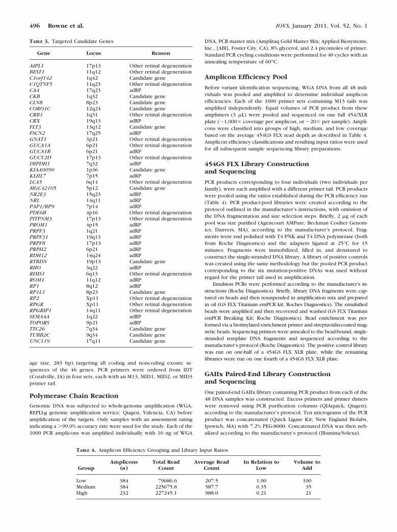
Genes targeted for sequencing were (1) known causes of autosomaldominant RP, (2) known causes of other forms of retinal degener-ation with overlapping phenotypes, or (3) potential disease-causingcandidate genes selected from sensory cilium proteome studies,EyeSAGE data, and other retinal expression and protein interactionstudies (Table 3) (Liu O, et al. IOVS 2006;47:ARVO E-Abstract3725).30 –33
In-house amplimer design algorithms were used in conjunctionwith Primer3 primer-selection software (http://sourceforge.net/projects/primer3/develop)34 to design 1000 PCR amplicons (aver-
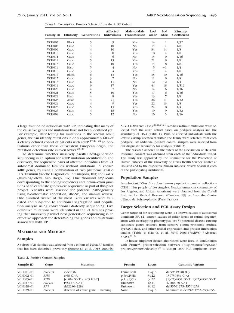
TABLE 1. Twenty-One Families Selected from the AdRP Cohort
Family ID Ethnicity GenerationsAffected
IndividualsMale-to-MaleTransmission
Lodad:ar
Lodad:XI
KinshipCoefficient
VCH007 Black 5 9 Yes 14 1 1/32VCH008 Cauc 4 10 No 14 �1 1/8VCH009 Cauc 4 10 Yes 34 14 1/8VCH010 Cauc 4 8 Yes 8 4 1/8VCH011 Cauc 4 6 No 15 0 1/16VCH012 Cauc 5 15 Yes 21 8 1/8VCH013 Cauc 4 10 Yes 14 8 1/8VCH014 Hisp 5 4 No 5 �1 1/4VCH015 Cauc 3 4 Yes 7 4 1/8VCH016 Black 6 13 Yes 35 10 1/16VCH017 Cauc 3 7 No 11 0 1/4VCH018 Cauc 6 11 No 12 �2 1/4VCH019 Cauc 6 17 Yes 44 18 1/512VCH020 Cauc 4 7 No 14 6 1/16VCH021 Cauc 5 10 Yes 17 6 1/16VCH022 Hisp 4 8 Yes 13 7 1/8VCH023 Asian 4 21 Yes 26 11 1/32VCH024 Cauc 4 9 Yes 22 13 1/8VCH025 Cauc 5 13 Yes 24 8 1/4VCH026 Cauc 4 18 Yes 19 9 1/32VCH004 Cauc 5 12 No 16 5 1/16
TABLE 2. Positive Control Samples
Sample ID Gene Mutation Protein Locus Genomic Variant
VCH001–01 PRPF31 c.del636 Frame shift 19q13 del59319048 (G)VCH002–01 RHO c.68 C�A p.Pro23His 3q22 130730334 C�AVCH005–01 RHO [c.404 G�T; c.405 G�T] p.Arg135Leu 3q22 [130732451 G�T; 130732452 G�T]VCH027–01 PRPH2 IVS2�3 A�T Unknown 6p21 42780078 A�TVCH028–01 RP1 del2280–2284 Unknown 8q12 del55701275–55701279VCH029–01 PRPF31 deletion of entire gene � flanking None 19q13 Minimum is del59283753–59328550
IOVS, January 2011, Vol. 52, No. 1 AdRP Next-Generation Sequencing 495
age size, 283 bp) targeting all coding and noncoding exonic se-quences of the 46 genes. PCR primers were ordered from IDT(Coralville, IA) in four sets, each with an M13, MID1, MID2, or MID3primer tail.
Polymerase Chain Reaction
Genomic DNA was subjected to whole-genome amplification (WGA;REPLI-g genome amplification service; Qiagen, Valencia, CA) beforeamplification of the targets. Only samples with an assessment ratingindicating a �99.0% accuracy rate were used for the study. Each of the1000 PCR amplicons was amplified individually with 10 ng of WGA
DNA, PCR master mix (Amplitaq Gold Master Mix; Applied Biosystems,Inc., [ABI], Foster City, CA), 8% glycerol, and 2.4 picomoles of primer.Standard PCR cycling conditions were performed for 40 cycles with anannealing temperature of 60°C.
Amplicon Efficiency Pool
Before variant identification sequencing, WGA DNA from all 48 indi-viduals was pooled and amplified to determine individual ampliconefficiencies. Each of the 1000 primer sets containing M13 tails wasamplified independently. Equal volumes of PCR product from theseamplimers (3 �L) were pooled and sequenced on one full 454/XLRplate (�1,000� coverage per amplicon, or �20� per sample). Ampli-cons were classified into groups of high, medium, and low coveragebased on the average 454GS FLX read depth as described in Table 4.Amplicon efficiency classifications and resulting input ratios were usedfor all subsequent sample sequencing library preparations.
454GS FLX Library Constructionand Sequencing
PCR products corresponding to four individuals (two individuals perfamily), were each amplified with a different primer tail. PCR productswere pooled using the ratios established during the PCR efficiency run(Table 4). PCR product-pool libraries were created according to theprotocol outlined in the manufacturer’s instructions, with omission ofthe DNA fragmentation and size selection steps. Briefly, 2 �g of eachpool was size purified (Agencourt AMPure; Beckman Coulter Genom-ics, Danvers, MA), according to the manufacturer’s protocol. Frag-ments were end polished with T4 PNK and T4 DNA polymerase (bothfrom Roche Diagnostics) and the adapters ligated at 25°C for 15minutes. Fragments were immobilized, filled in, and denatured toconstruct the single-stranded DNA library. A library of positive controlswas created using the same methodology but the pooled PCR productcorresponding to the six mutation-positive DNAs was used withoutregard for the primer tail used in amplification.
Emulsion PCRs were performed according to the manufacturer’s in-structions (Roche Diagnostics). Briefly, library DNA fragments were cap-tured on beads and then resuspended in amplification mix and preparedin oil (GS FLX Titanium emPCR kit; Roches Diagnostics). The emulsifiedbeads were amplified and then recovered and washed (GS FLX TitaniumemPCR Breaking Kit; Roche Diagnostics). Bead enrichment was per-formed via a biotinylated enrichment primer and streptavidin-coated mag-netic beads. Sequencing primers were annealed to the bead-bound, single-stranded template DNA fragments and sequenced according to themanufacturer’s protocol (Roche Diagnostics). The positive control librarywas run on one-half of a 454GS FLX XLR plate, while the remaininglibraries were run on one fourth of a 454GS FLX XLR plate.
GAIIx Paired-End Library Constructionand Sequencing
One paired-end GAIIx library containing PCR product from each of the48 DNA samples was constructed. Excess primers and primer dimerswere removed using PCR purification columns (QIAquick; Qiagen),according to the manufacturer’s protocol. Ten micrograms of the PCRproduct was concatenated (Quick Ligase Kit; New England Biolabs,Ipswich, MA) with 7.2% PEG-8000. Concatenated DNA was then neb-ulized according to the manufacturer’s protocol (Illumina/Solexa).
TABLE 4. Amplicon Efficiency Grouping and Library Input Ratios
GroupAmplicons
(n)Total Read
CountAverage Read
CountIn Relation to
LowVolume to
Add
Low 384 79686.6 207.5 1.00 100Medium 384 225675.8 587.7 0.35 35High 232 227245.1 988.0 0.21 21
TABLE 3. Targeted Candidate Genes
Gene Locus Reason
AIPL1 17p13 Other retinal degenerationBEST1 11q12 Other retinal degenerationC1orf142 1q42 Candidate geneC1QTNF5 11q23 Other retinal degenerationCA4 17q23 adRPCKB 1q32 Candidate geneCLN8 8p23 Candidate geneCORO1C 12q24 Candidate geneCRB1 1q31 Other retinal degenerationCRX 19q13 adRPFLT3 13q12 Candidate geneFSCN2 17q25 adRPGNAT1 3p21 Other retinal degenerationGUCA1A 6p21 Other retinal degenerationGUCA1B 6p21 adRPGUCY2D 17p13 Other retinal degenerationIMPDH1 7q32 adRPKIAA0090 1p36 Candidate geneKLHL7 7p15 adRPLCA5 6q14 Other retinal degenerationMGC42105 5p12 Candidate geneNR2E3 15q23 adRPNRL 14q11 adRPPAP1/RP9 7p14 adRPPDE6B 4p16 Other retinal degenerationPITPNM3 17p13 Other retinal degenerationPROM1 4p15 adRPPRPF3 1q21 adRPPRPF31 19q13 adRPPRPF8 17p13 adRPPRPH2 6p21 adRPRDH12 14q24 adRPRTBDN 19p13 Candidate geneRHO 3q22 adRPRIMS1 6q13 Other retinal degenerationROM1 11q12 adRPRP1 8q12 adRPRP1L1 8p23 Candidate geneRP2 Xp11 Other retinal degenerationRPGR Xp11 Other retinal degenerationRPGRIP1 14q11 Other retinal degenerationSEMA4A 1q22 adRPTOPORS 9p21 adRPTTC26 7q34 Candidate geneTUBB2C 9q34 Candidate geneUNC119 17q11 Candidate gene
496 Bowne et al. IOVS, January 2011, Vol. 52, No. 1

Sheared DNA was end repaired (DNA Terminator End Repair kit;Lucigen, Middleton, WI), purified (QIAquick columns; Qiagen), andverified on 1.2% gels (FlashGels; Lonza, Basel, Switzerland). An aden-osine was added to the 3� end of concatenated products (KlenowFragment (3�35� exo�; New England Biolabs) at 37°C for 30 minutes.Adenosine-tailed DNA was column purified (MinElute; Qiagen) andligated to an adapter-oligo mix at room temperature for 15 minutes.Ligated reactions were purified with the purification columns followedby gel purification using 1.2% agarose and extraction (MinElute GelExtraction Kit; Qiagen). PCR enrichment was performed using DNApolymerase master mix (Phusion; New England Biolabs). Reactionswere denatured at 98°C for 30 seconds followed by five cycles of 98°Cfor 10 seconds, 65°C for 30 seconds, and 72°C for 30 seconds with afinal extension at 72°C for 5-minute PCR reactions were purified on theminicolumns, and the 300- to 400-bp library fragment was isolated bygel purification.
Cluster Generation Kit ver. 2 (Illumina/Solexa) and the manufac-turer’s protocol were used to generate paired ends, which were thensequenced on the GAIIx (Illumina/Solexa) platform (SBS SequencingKit, ver. 3; Illumina/Solexa) according to the manufacturer’s protocol.
454GS FLX Alignments and Variant Detection
Sequencing data files (SFF format) were converted to FASTA formatusing sffinfo. Individual samples were separated using cross_match tomatch M13, MID1, MID2, and MID3 primer sequences with the first 20bases of every read. Manifests of read names for each primer tail werecomposed, and sequences were extracted from the master SFF file foreach individual.
Read sequences were extracted from individual SFF files usingsffinfo and then aligned to the Hs36 reference sequence using BLAT(ver. 32 � 1) (http://genome.ucsc.edu/cgi-bin/hgBlat/ provided in thepublic domain by the University of California Santa Cruz).35 Alignmentswith �90% identity, a score of �25, or mapping to multiple locationsin the genome (with the same score), were discarded. SNPs and indelswere detected in the BLAT alignments using VarScan.36 A minimum of10� coverage and at least 25% of reads supporting the variant allelewas required for variant calling. Substitutions at sites with base quality�15 were discarded. Artifacts from regions homologous to MID primertails were avoided by discarding variants called within 10 bp of thebeginning or end of the read.
GAIIx Alignments and Variant Detection
GAIIx reads were aligned to the Hs36 reference sequence using Bowtie(ver. 0.9.8, http://bowtie-bio.sourceforge.net).37 Reads were requiredto have a single best alignment (�m 1) with no more than twohigh-quality mismatches to the reference sequence. Bowtie alignmentswere parsed to identify substitutions at positions with base quality of15 or higher. The variant allele with the greatest read support wascalled for all positions at which variants were detected.
Indel Validation with Illumina/Solexa Data
To detect small indels, GAIIx reads were aligned to the Hs36 referencesequence (Novoalign, ver. 2.03.12; Novoalign, Selangor, Malaysia).VarScan36 was used to call indels based on unique read alignments.Indels with ambiguous positions or flanked by homopolymers wereremoved. Validation of 454GS FLX indels required that GAIIx indelcalls be the same type, size (within 1 bp), and position (�5 bp).
Di-deoxy Sequencing of PotentialDisease-Causing Variants
The 1000 amplicon PCR primers with M13 tails were used for alladditional analyses of potential disease-causing variants. Genomic DNAwas amplified using Taq master mix (AmpliTaq Gold 360 Master Mix;ABI) and standard amplification conditions. PCR product was treatedwith exonuclease I and shrimp alkaline phosphatase (ExoSapIt; USB,Cleveland, OH) before sequencing. Clean PCR product was sequenced
as described previously with dye termination chemistry (BigDye Ter-minator ver. 1.1; ABI) and M13 primers.38 Sequence reactions weretreated with a reaction cleanup kit (BigDye XTerminator; ABI), accord-ing to the manufacturer’s protocol, and run on one of two automatedcapillary sequencers (3100 Avant or 3730XL; ABI). Sequence analysiswas then performed with one of three commercial software programs(Sequencing Analysis, Variant Reporter, or SeqScape; ABI).
RESULTS
Sample
The sample selected for this project included a subset of familymembers from our previously described AdRP Cohort (BowneSJ, et al. IOVS 2007;48:ARVO E-Abstract 2334;).21,23,24,38,39
Families in the adRP cohort, based on pedigree analysis,have a high likelihood of having the autosomal dominantform of RP. Analysis of pedigrees for the likelihood ofdominance versus X-linked or recessive inheritance didshow some families with ad:Xl likelihood odds ratios of lessthan 0, indicating that the disease in a few families could becaused by mutations in an X-linked gene with clinical ex-pression in carrier females (Table 1).
A proband from each family was tested previously for mu-tations in the complete coding regions of CA4, CRX, FSCN2,IMPDH1, NRL, PRPF31, RDS, RHO, ROM1, RP9, and TOPORS,and in mutation hot spots of RP1, PRPF3, PRPF8, NR2E3, andSNRNP200. Likely disease-causing mutations were identified in141 of the 230 families. Only families without previously iden-tified mutations were considered for this study.
Twenty-one families were selected for this project based onpedigree analysis and availability of family member DNAs. Twoaffected individuals from each family were selected to have thelowest kinship coefficient possible—that is, the most distant,available affected relatives with a common ancestor carrying aputative adRP mutation. This increased the probability that anyshared variant identified in this project would be associatedwith disease, not just identical by chance. The demographicand pedigree characteristics of the families and the kinshipcoefficient of the two selected family members are shown inTable 1.
Six individuals with known mutations were also selected touse as positive controls in this study (Table 2). These individ-uals had a variety of mutation types in several different genes,thereby testing the identification rate of next-generation se-quencing for different classes of DNA variants.
DNA Targets
Each of the 46 genes selected for this project was (1) a knowncause of adRP, (2) a known cause of other forms of retinaldegeneration with phenotypes overlapping adRP, or (3) a po-tential disease-causing candidate gene selected from sensorycilium proteome studies, EyeSAGE data, and other studies ofretinal expression and protein interaction (Liu Q, et al. IOVS2006;47:ARVO E-Abstract 3725).30–33 The genes selected, theirchromosomal location, and the associated diseases, if any, arelisted in Table 3.
PCR primers corresponding to 1000 amplicons were de-signed to amplify all the coding and noncoding exonic se-quences for each of the 46 genes selected. PCR primers weremanufactured in sets of four with each set containing the samegenome-specific sequence and one of four different tail se-quences (M13, MD1, MD2, and MD3). These four tail se-quences allowed PCR product from four individuals to becombined after amplification, while retaining the ability todistinguish the four individuals on sequence assembly.
IOVS, January 2011, Vol. 52, No. 1 AdRP Next-Generation Sequencing 497
Target Amplification and Library Construction
Genomic DNA from each of the 48 individuals tested in thisstudy was amplified by WGA before target amplification. A454GS FLX amplicon efficiency test was performed on a poolof the DNAs to optimize product pooling such that each am-plimer represented in the sequencing library was relativelyequivalent (Table 4).
Eleven patient pool libraries were constructed for analysison the 454GS FLX sequencing system. Each of these poolscorresponded to two sets of affected family pairs that had beenamplified individually with the four different, tailed targetprimers. The six positive control samples were pooled to formone library that was not sorted by MD tail.
One concatenated, paired-end library was constructed foranalysis (PE sequencer; Illumina/Solexa). Concatenation andshearing of the PCR products before library construction en-abled access to those regions that, due to the short read lengthof GAIIx sequencing, would otherwise not be sequenced, andalso gives more random distribution of all positions in a par-ticular sequence. This feature introduces less quality bias basedon position of the variation within a given sequence. Thepaired-end library pooled all 42 unknown affected individualsand the six positive controls. Since this library was not sort-able, it was used only for variant confirmation, not individualvariant identification.
454GS FLX Analyses of Positive Controls
Sequence reads corresponding to the pooled positive controllibrary were aligned to Hs36 and analyzed for the presence of
each known mutation. Five of the six mutations were detectedat a read frequency of 6% to 13%, which is in accordance withthe predicted detection rate of 8% (Table 5). The sixth variant,a large 47-kb deletion of PRPF31 and several flanking genespresent in VCH029, was not detected using this technology, asexpected.
Sequence Alignment and SNP Variant Detection
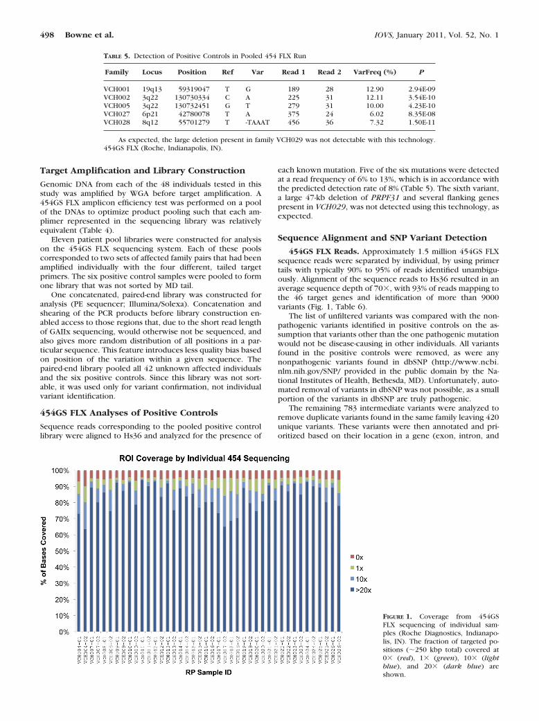
454GS FLX Reads. Approximately 1.5 million 454GS FLXsequence reads were separated by individual, by using primertails with typically 90% to 95% of reads identified unambigu-ously. Alignment of the sequence reads to Hs36 resulted in anaverage sequence depth of 70�, with 93% of reads mapping tothe 46 target genes and identification of more than 9000variants (Fig. 1, Table 6).
The list of unfiltered variants was compared with the non-pathogenic variants identified in positive controls on the as-sumption that variants other than the one pathogenic mutationwould not be disease-causing in other individuals. All variantsfound in the positive controls were removed, as were anynonpathogenic variants found in dbSNP (http://www.ncbi.nlm.nih.gov/SNP/ provided in the public domain by the Na-tional Institutes of Health, Bethesda, MD). Unfortunately, auto-mated removal of variants in dbSNP was not possible, as a smallportion of the variants in dbSNP are truly pathogenic.
The remaining 783 intermediate variants were analyzed toremove duplicate variants found in the same family leaving 420unique variants. These variants were then annotated and pri-oritized based on their location in a gene (exon, intron, and
TABLE 5. Detection of Positive Controls in Pooled 454 FLX Run
Family Locus Position Ref Var Read 1 Read 2 VarFreq (%) P
VCH001 19q13 59319047 T G 189 28 12.90 2.94E-09VCH002 3q22 130730334 C A 225 31 12.11 3.54E-10VCH005 3q22 130732451 G T 279 31 10.00 4.23E-10VCH027 6p21 42780078 T A 375 24 6.02 8.35E-08VCH028 8q12 55701279 T -TAAAT 456 36 7.32 1.50E-11
As expected, the large deletion present in family VCH029 was not detectable with this technology.454GS FLX (Roche, Indianapolis, IN).
FIGURE 1. Coverage from 454GSFLX sequencing of individual sam-ples (Roche Diagnostics, Indianapo-lis, IN). The fraction of targeted po-sitions (�250 kbp total) covered at0� (red), 1� (green), 10� (lightblue), and 20� (dark blue) areshown.
498 Bowne et al. IOVS, January 2011, Vol. 52, No. 1

splice-site) and on the predicted transcript or protein alterationand by manual assessment of the potential of the affected geneto cause RP. The resulting 112 variants were classified aspotentially pathogenic, thereby warranting additional analysis(Fig. 2).
GAIIx Reads. The 66.7 million 36-bp reads from thepooled GAIIx library (Illumina/Solexa) were aligned to Hs36with an average sequence depth of 125� (Table 6). Variantswere called and compared with the list of the 454GS FLXvariants. To confirm a 454GS FLX variant required GAIIxread coverage of at least 100� and at least two variant-supporting reads. These data were confirmatory but notused in the initial identification of the 112 potential patho-genic variants.
Evaluation of Potential Pathogenic Variants
The 112 potential pathogenic variants were subjected to a seriesof analyses to determine whether they were true variants, if theysegregated with disease in the family in which they were identi-fied, and whether they were present in unaffected controls.
Fluorescent di-deoxy capillary sequencing was used to de-termine whether the variants identified by next-generationsequencing were actually genomic variants. Genomic DNAsfrom the original affected family pair and from two additionalfamily members (when available) were tested with the corre-sponding M13 tailed primers for the original 1000 amplimeramplifications. Traditional Sanger sequencing showed that 55of the 112 potential pathogenic variants were artifacts of454GS FLX sequencing. An additional four of the potentialpathogenic variants did not amplify within the genomic re-gions specified for the variant and so were also assumed to beartifacts. With this strategy, 55 of the potential pathogenicvariants were confirmed to be present in the identified individ-uals.
Once a variant was determined to be real, segregationanalysis was used to assess its likelihood of it being disease-causing. If initial analyses showed correct segregation in thefirst set of four family members tested, then all availablefamily DNAs were tested. Forty-three of the 53 confirmedvariants did not segregate with disease in the family andhence were determined to be benign. Ten of the variantssegregated with disease in all available family members(Table 7).
Three of the 10 segregating variants, KLHL7 p.A153V,RPGR p.G65D, and PRPF31 c.946 –1, were identified andcharacterized in parallel laboratory testing and determinedto be pathogenic.40,41 An additional variant in RPGR,p.G738*, was identified among the 10 segregating variants.Although not previously reported, RPGR p.G738*, like manyother reported RPGR mutations, produces a premature ter-mination codon in ORF15 and hence is most likely patho-
genic. One additional segregating variant in GUCY2D,p.R838C, has also been reported to cause cone–rod dystro-phy.42 No further testing was performed for these five dis-ease-causing mutations (Fig. 3).
Ethnically matched control population DNAs were testedfor the possible presence of the remaining five variants ofunknown pathogenicity. Three of the variants, PRPF8 c.1–51G�A, PITPNM3 p.R703W, and TTC26 c.896�73G�T werefound in control DNAs and hence are benign. The two remain-ing variants, PROM1 c.1302�3C�T and MRFP c.641�9G�A,were not found in the controls. The number of immediatelyavailable family member DNA samples was low (three and two,respectively) for the PROM1 and MRFP variants. Subsequentcollection and testing of three additional VCH008 family mem-bers demonstrated that the PROM1 c.1302�3C�T variantdoes not segregate with disease. Collection and testing of three
FIGURE 2. Flow chart of variant analysis.
TABLE 6. Sequence Data Generated on Next-Generation Platforms
Regions for targeted sequencingCandidate genes 46PCR amplicons 1,000Total positions targeted (unique) 249,267
Individual 454 data*Number of samples 48Total reads (�230-bp XLR) 1.46 millionAvg. sequence depth per sample 70x
Pooled GAIIx data†Number of samples pooled 48Total reads (36-bp frag) 66.68 millionAvg. sequence depth per sample 125x
* Roche Diagnostics, Indianapolis, IN.† Illumina/Solexa, San Diego, CA.
IOVS, January 2011, Vol. 52, No. 1 AdRP Next-Generation Sequencing 499

additional VCH025 family members also demonstrated that theMRFP variant does not segregate with disease. These datademonstrate that both the PROM1 and MRFP variants arebenign.
Indels
Analysis of the individual reads from 454GS FLX sequencingidentified 77 small, high-confidence indels ranging in size from
1 to 3 bp. Indels with ambiguous positions or flanked byhomopolymers were removed and the remaining comparedwith GAIIx indel data. A total of 10 indels were identified withthe GAIIx data (Table 8).
Indels were evaluated using the same fluorescent capillarysequencing strategy described above for the possibly patho-genic SNP variants. Traditional sequencing failed to confirmthe presence of nine of the indels. The 10th indel, a 3-bp
TABLE 7. Ten Potential Disease-Causing Variants
Family Gene Chr Position Ref VarFunctional
Class Nucleotide ProteinReferencesequence
Frequency inControls (%)
VCH010 KLHL7 7 23146928 C T Missense c.458C�T p.A153V NM_001031710 0.0VCH017 RPGR X 38030984 G T Nonsense c.2212G�T p.G738* NM_001034853 0.0VCH018 RPGR X 38067103 G A Missense c.194G�A p.G65D NM_001034853 0.0VCH020 PRPF31 19 59323259 G C Splice-site c.946–1 Unknown NM_015629 0.0VCH012 GUCY2D 17 7858743 C T Missense c.2512C�T p.R838C NM_000180 0.0VCH024 PRPF8 17 1534891 G A 5’UTR c.1–51G�A Unknown NM_006445 2.0VCH013 PITPNM3 17 6308263 G A Missense c.2108C�T p.R703W NM_031220 0.7VCH011 TTC26 7 138502254 G T Intronic c.896�73G�T Unknown NM_024926 2.0VCH008 PROM1 4 15619667 C T Splice-site c.1302�3C�T Unknown NM_006017 0.0VCH025 MFRP 11 118721331 G A Splice-site c.641�9G�A Unknown NM_031433 0.0
*
* *
* *
* *
*
* *
*
?
?
*
* *
**
DNA* * * *
* * *
* *
A. B.
C.
D. E.
FIGURE 3. Five families with identifiedpathogenic mutations. (A) VCH010. Thep.A153V mutation in KLHL7 was pres-ent in all three affected family memberstested. (B) VCH012. All five tested af-fected members of this family had theR838C mutation in GUCY2D (C)VCH017. Four affected members of thisfamily were either heterozygous orhemizygous for the RPGR G738X muta-tion which was not present in the oneunaffected family member tested. (D)VCH018. THE RPGR G65D mutationwas present in seven affected membersor female carriers in this family and ab-sent from the one unaffected spousetested. (E) VCH037. The c.946-1 splicesite mutation in PRPF31 segregated withdisease in the three family memberstested. *Individuals tested in this study.
500 Bowne et al. IOVS, January 2011, Vol. 52, No. 1

deletion in ORF15 of RPGR was not assessed, since RPGR islocated on the X-chromosome and the family exhibited male-to-male transmission of RP. Furthermore, 3-bp deletions inORF15 are common and usually benign.43–46
DISCUSSION
An excess of 9000 variants was identified in the 21 familiesanalyzed in this project. Most of these variants were classifiedas benign on the basis of their presence in controls withdefinitive disease-causing mutations distinct from the novelvariants. This massive reduction in variants requiring follow-upanalyses, over 8000, stresses the importance of running controlindividuals and multiple family members, if possible, when
using next-generation sequencing to detect mutations in fami-lies with inherited diseases.
Additional laboratory analyses were performed for 112 pos-sibly pathogenic variants found by 454GS FLX sequencing. Thepresence of 53 (47%) of these variants was confirmed bytraditional sequencing, whereas the remaining 59 (53%) vari-ants were found to be false positives. A large fraction of thefalse-positive variants occurred in a polynucleotide runs, whichis a known limitation of 454 sequencing methodology. Thisresult suggests that additional stringency should be used whenidentifying variants in polynucleotide runs to reduce the num-ber of false positives.
The pooled GAIIx sequencing data were not used in theinitial phases of the project, but were compared to the list of
TABLE 8. Indels present in 454 FLX and GAIIx Sequence Reads
Family Gene Chr Position Type Size (bp)
VCH021 BEST1 11 61482241 Deletion 1VCH022VCH026
VCH026 ROM1 11 52138421 Deletion 1
VCH015 GUCY2D 17 7847637 Insertion 1
VCH014 PROM1 4 15591264 Insertion 1
VCH021 PROM1 4 15604786 Deletion 1VCH022VCH025VCH026
VCH22 LCA5 6 80259054 Deletion 1VCH25VCH26
VCH019 RIMS 6 73159087 Deletion 1
VCH013 CRX 19 53034641 Deletion 1
VCH022 RP1 8 55701373 Deletion 1
VCH016 RPGR X 38030864–38030866 Deletion 3
454GS FLX (Roche, Indianapolis, IN); GAIIx (Illumina/Solexa, San Diego, CA).
FIGURE 4. Prevalence of mutationsin genes causing dominant RP. Patho-genic mutations have been identifiedin 148 of the 230 adRP cohort fami-lies including the five families re-ported in this study. The mutationremains to be identified in 82 (35%)of the families.
IOVS, January 2011, Vol. 52, No. 1 AdRP Next-Generation Sequencing 501
112 variants to determine whether cross-platform comparisonsmight also reduce the number of false-positive variants. Whencompared with the pooled GAIIx data, 85% of the confirmedvariants were present, but, 55% of the false positives were alsoseen in the GAIIx reads. This finding suggests that cross-plat-form comparisons may be useful for prioritizing variants forsubsequent follow-up, but should not be used as an exclusiverequirement for variant identification.
Next-generation sequencing of the 1000 amplicons corre-sponding to 46 candidate genes resulted in identification of fivepathogenic mutations in the 21 families tested (Table 7, Fig. 3).As expected, three of these mutations are in genes reported tobe associated with either autosomal dominant RP or autosomaldominant cone–rod dystrophy (Birch DG, et al. IOVS 2006;47:ARVO E-Abstract 1037).21,40–42 Somewhat surprising was theidentification of two mutations in RPGR. It has been known forsome time that mutations in RPGR cause X- linked RP, but thehigh frequency of symptomatic female carriers is just begin-ning to be appreciated.43,45,47
Identification of five additional mutations brings the known-mutation frequency of our AdRP cohort up to 64% (Fig. 4).That is, we can identify the disease-causing mutations in 148 ofthe 230 adRP cohort families.
This project demonstrates that next-generation sequencingcan be an effective tool for determining the pathogenic muta-tion in inherited disease families with highly heterogeneouscauses. The large number of those variants proven to be arti-facts identified in the limited region of the genome testedduring this project raises concerns that the use of next-gener-ation sequencing for larger genomic regions, such as a com-plete exome or genome, will be daunting. Coupled with thewide genetic variation known to exist in humans, this projectmakes it evident that, without the ability to perform segrega-tion analysis, it will be extremely difficult to distinguish rarepathogenic variants from rare benign variants.
Acknowledgments
The authors thank Gita Dangol, Kaylie Webb, and Joe Ray for technicalassistance.
References
1. McKernan KJ, Peckham HE, Costa GL, et al. Sequence and struc-tural variation in a human genome uncovered by short-read, mas-sively parallel ligation sequencing using two-base encoding. Ge-nome Res. 2009;19:1527–1541.
2. Albert TJ, Molla MN, Muzny DM, et al. Direct selection of humangenomic loci by microarray hybridization. Nat Methods. 2007;4:903–905.
3. Okou DT, Steinberg KM, Middle C, Cutler DJ, Albert TJ, Zwick ME.Microarray-based genomic selection for high-throughput rese-quencing. Nat Methods. 2007;4:907–909.
4. Wheeler DA, Srinivasan M, Egholm M, et al. The complete genomeof an individual by massively parallel DNA sequencing. Nature.2008;452:872–876.
5. Ng SB, Turner EH, Robertson PD, et al. Targeted capture andmassively parallel sequencing of 12 human exomes. Nature. 2009;461:272–276.
6. Haim M. Epidemiology of retinitis pigmentosa in Denmark. ActaOphthalmol Scand Suppl. 2002;233:1–34.
7. Heckenlively JR. Retinitis Pigmentosa. Philadelphia: J. B. Lippincott;1988.
8. Heckenlively JR, Daiger SP. Hereditary Retinal and ChoroidalDegenerations. 5th ed. London: Churchill Livingston Elsevier;2007.
9. Bunker CH, Berson EL, Bromley WC, Hayes RP, Roderick TH.Prevalence of retinitis pigmentosa in Maine. Am J Ophthalmol.1984;97:357–365.
10. Grondahl J. Estimation of prognosis and prevalence of retinitispigmentosa and Usher syndrome in Norway. Clin Genet. 1987;31:255–264.
11. Xu L, Hu L, Ma K, Li J, Jonas JB. Prevalence of retinitis pigmentosain urban and rural adult Chinese: The Beijing Eye Study. Eur JOphthalmol. 2006;16:865–866.
12. Buch H, Vinding T, La Cour M, Appleyard M, Jensen GB, NielsenNV. Prevalence and causes of visual impairment and blindnessamong 9980 Scandinavian adults: the Copenhagen City Eye Study.Ophthalmology. 2004;111:53–61.
13. Al-Merjan JI, Pandova MG, Al-Ghanim M, Al-Wayel A, Al-Mutairi S.Registered blindness and low vision in Kuwait. Ophthalmic Epi-demiol. 2005;12:251–257.
14. Hayakawa M, Fujiki K, Kanai A, et al. Multicenter genetic study ofretinitis pigmentosa in Japan, II: prevalence of autosomal recessiveretinitis pigmentosa. Jpn J Ophthalmol. 1997;41:7–11.
15. Hayakawa M, Fujiki K, Kanai A, et al. Multicenter genetic study ofretinitis pigmentosa in Japan, I: genetic heterogeneity in typicalretinitis pigmentosa. Jpn J Ophthalmol. 1997;41:1–6.
16. Daiger SP. Identifying retinal disease genes: how far have wecome, how far do we have to go? Novartis Found Symp. 2004;255:17–27; discussion 27–36, 177–178.
17. Daiger SP, Bowne SJ, Sullivan LS. Perspective on genes and muta-tions causing retinitis pigmentosa. Arch Ophthalmol. 2007;125:151–158.
18. den Hollander AI, Roepman R, Koenekoop RK, Cremers FP. Lebercongenital amaurosis: genes, proteins and disease mechanisms.Prog Retin Eye Res. 2008;27:391–419.
19. Rivolta C, Sharon D, DeAngelis MM, Dryja TP. Retinitis pigmentosaand allied diseases: numerous diseases, genes, and inheritancepatterns. Hum Mol Genet. 2002;11:1219–1227.
20. Sullivan LS, Bowne SJ, Seaman CR, et al. Genomic rearrangementsof the PRPF31 gene account for 2.5% of autosomal dominantretinitis pigmentosa. Invest Ophthalmol Vis Sci. 2006;47:4579–4588.
21. Sullivan LS, Bowne SJ, Birch DG, et al. Prevalence of disease-causing mutations in families with autosomal dominant retinitispigmentosa (adRP): a screen of known genes in 200 families.Invest Ophthalmol Vis Sci. 2006;47:3052–3064.
22. RetNet. The Retinal Information Network, http://www.sph.uth.tmc.edu/RetNet/. Stephen P. Daiger, PhD, Administrator, TheUniv. of Texas Health Science Center at Houston; 1996–present.
23. Bowne SJ, Sullivan LS, Gire AI, et al. Mutations in the TOPORS genecause 1% of autosomal dominant retinitis pigmentosa (adRP). MolVis. 2007;14:922–927.
24. Gire A, Sullivan LS, Bowne SJ, et al. The Gly56Arg mutation inNR2E3 accounts for 1–2% of autosomal dominant retinitis pigmen-tosa. Mol Vis. 2007;13:1970–1975.
25. Ziviello C, Simonelli F, Testa F, et al. Molecular genetics of auto-somal dominant retinitis pigmentosa (ADRP): a comprehensivestudy of 43 Italian families. J Med Genet. 2005;42:e47.
26. Sato H, Wada Y, Itabashi T, Nakamura M, Kawamura M, Tamai M.Mutations in the pre-mRNA splicing gene, PRPF31, in Japanesefamilies with autosomal dominant retinitis pigmentosa. Am J Oph-thalmol. 2005;140:537–540.
27. Wada Y, Sandberg MA, McGee TL, Stillberger MA, Berson EL, DryjaTP. Screen of the IMPDH1 gene among patients with dominantretinitis pigmentosa and clinical features associated with the mostcommon mutation, Asp226Asn. Invest Ophthalmol Vis Sci. 2005;46:1735–1741.
28. Wada Y, Tamai M. Molecular genetic analysis for Japanese patientswith autosomal dominant retinitis pigmentosa. Nippon GankaGakkai Zasshi. 2003;107:687–694.
29. Zhang XL, Liu M, Meng XH, et al. Mutational analysis of therhodopsin gene in Chinese ADRP families by conformation sensi-tive gel electrophoresis. Life Sci. 2006;78:1494–1498.
30. Lord-Grignon J, Tetreault N, Mears AJ, Swaroop A, Bernier G.Characterization of new transcripts enriched in the mouse retinaand identification of candidate retinal disease genes. Invest Oph-thalmol Vis Sci. 2004;45:3313–3319.
31. Bowes Rickman C, Ebright JN, Zavodni ZJ, et al. Defining thehuman macula transcriptome and candidate retinal disease genesusing EyeSAGE. Invest Ophthalmol Vis Sci. 2006;47:2305–2316.
502 Bowne et al. IOVS, January 2011, Vol. 52, No. 1
32. Wistow G, Bernstein SL, Wyatt MK, et al. Expressed sequence taganalysis of human retina for the NEIBank Project: retbindin, anabundant, novel retinal cDNA and alternative splicing of otherretina-preferred gene transcripts. Mol Vis. 2002;8:196–204.
33. Mitton KP, Swain PK, Khanna H, Dowd M, Apel IJ, Swaroop A.Interaction of retinal bZIP transcription factor NRL with Flt3-interacting zinc-finger protein Fiz1: possible role of Fiz1 as atranscriptional repressor. Hum Mol Genet. 2003;12:365–3673.
34. Steve Rozen HJS. Primer3. Code available at http://sourceforge.net/projects/primer3/develop. 1996.
35. Kent WJ. BLAT: the BLAST-like alignment tool. Genome Res. 2002;12:656–664.
36. Koboldt DC, Chen K, Wylie T, et al. VarScan: variant detection inmassively parallel sequencing of individual and pooled samples.Bioinformatics. 2009;25:2283–2285.
37. Langmead B, Trapnell C, Pop M, Salzberg SL. Ultrafast and memory-efficient alignment of short DNA sequences to the human genome.Genome Biol. 2009;10:R25.
38. Bowne SJ, Sullivan LS, Mortimer SE, et al. Spectrum and frequencyof mutations in IMPDH1 associated with autosomal dominant ret-initis pigmentosa and Leber congenital amaurosis. Invest Ophthal-mol Vis Sci. 2006;47:34–42.
39. Zhao C, Bellur DL, Lu S, et al. Autosomal-dominant retinitis pig-mentosa caused by a mutation in SNRNP200, a gene required forunwinding of U4/U6 snRNAs. Am J Hum Genet. 2009;85:617–627.
40. Friedman JS, Ray JW, Waseem N, et al. Mutations in a BTB-Kelchprotein, KLHL7, cause autosomal-dominant retinitis pigmentosa.Am J Hum Genet. 2009;84:792–800.
41. Vithana EN, Abu-Safieh L, Allen MJ, et al. A human homolog ofyeast pre-mRNA splicing gene, PRP31, underlies autosomal domi-nant retinitis pigmentosa on chromosome 19q13.4 (RP11). MolCell. 2001;8:375–381.
42. Kelsell RE, Gregory-Evans K, Payne AM, et al. Mutations in theretinal guanylate cyclase (RETGC-1) gene in dominant cone-roddystrophy. Hum Mol Genet. 1998;7:1179–1184.
43. Pelletier V, Jambou M, Delphin N, et al. Comprehensive survey ofmutations in RP2 and RPGR in patients affected with distinctretinal dystrophies: genotype-phenotype correlations and impacton genetic counseling. Hum Mutat. 2007;28:81–91.
44. Sharon D, Sandberg MA, Rabe VW, Stillberger M, Dryja TP, BersonEL. RP2 and RPGR mutations and clinical correlations in patientswith X-linked retinitis pigmentosa. Am J Hum Genet. 2003;73:1131–1146.
45. Vervoort R, Lennon A, Bird AC, et al. Mutational hot spot within anew RPGR exon in X-linked retinitis pigmentosa. Nat Genet. 2000;25:462–466.
46. Shu X, McDowall E, Brown AF, Wright AF. The human retinitispigmentosa GTPase regulator gene variant database. Hum Mutat.2008;29:605–608.
47. Rozet JM, Perrault I, Gigarel N, et al. Dominant X linked retinitispigmentosa is frequently accounted for by truncating mutations inexon ORF15 of the RPGR gene. J Med Genet. 2002;39:284–285.
IOVS, January 2011, Vol. 52, No. 1 AdRP Next-Generation Sequencing 503