On the heredity of retinitis pigmentosa
13
British Journal of Ophthalmology, 1982, 66, 405-416 On the heredity of retinitis pigmentosa MARCELLE JAY From Moorfields Eye Hospital, City Road, London EC] V 2PD SUMMARY The aims of this study are: (1) to determine the frequencies of the various genetic forms of retinitis pigmentosa; and (2) to perform segregation analysis on autosomal dominant, autosomal recessive, and X-linked families. The families studied consisted of 2 series of patients at Moorfields Eye Hospital: (1) 426 families seen in the Genetic Clinic; and (2) 289 families seen in the Electrodiagnostic Department. Comparison between the 2 series identified biases of ascertain- ment, and it was estimated that the combined series included 53% of simplex cases and a minimum of 15% of X-linked families. Segregation analysis of the Genetic Clinic series showed good agreement with expectation in autosomal dominant and X-linked families, but indicated that no more than 70% of all simplex cases were autosomal recessive. The rest of the simplex cases were mildly affected and may represent fresh autosomal dominant mutations, autosomal dominant transmission with reduced penetrance, the heterozygous state of X-linked disease in some of the females, and phenocopies. Many years ago a well dressed man who looked in excellent health, came to my desk in the out-patient room, at Moorfields, and in a somewhat excited manner declared 'I am going blind Sir! you can do nothing for me I know! It is in the family, and has been for centuries, and at the present time I know more than thirty who are either blind or on the way to it. When once it begins it always goes on; still I should be glad if you would look at my eyes.' I did so and found, as I expected, that he was the subject of retinitis pigmentosa.' The early authors classified the different genetic forms of retinitis pigmentosa using much the same system as did Nettleship2 in defining heredity in his series. Three main categories were recognised: (1) continuous, with direct inheritance in an unbroken line; (2) discontinuous, with parents normal but affected aunts, uncles, cousins, or grandparents; (3) collateral, where the parents were normal but sibs were affected. A fourth group consisted of cases inherited from both parents, when the heredity was termed double or reinforced. In modem terminology the first group corresponds to autosomal dominant transmission; the second might be autosomal recessive, X-linked, or autosomal dominant with reduced penetrance depending on the pedigree; and the third group probable autosomal recessive. It is very difficult, however, to analyse Correspondence to Dr Marcelle Jay. Part of a thesis accepted by the University of London for the degree of PhD. either the Nettleship2 or Bell3 series of pedigrees, as insufficient information is available. Since 1925 the genetics of retinitis pigmentosa have been studied extensively in Europe,"'0 and as a result the autosomal recessive mode of transmission has been considered to be the most common.' 1-20 The significance of X-linked transmission in retinitis pigmentosa was probably recognised intuitively by Nettleship (1845-1913), and there are numerous examples of this mode of transmission in the early literature.2130 The most significant findings, particularly in the recognition of the carrier state in the female heterozygote, were by Falls and Cotterman23 and by Bird.3' There have been several studies since 1961 comparing the proportions of the different genetic forms of retinitis pigmentosa in various parts of the world: Switzerland45 Belgium,832 Israel,33 Japan,30343 the USSR,36 the United Kingdom,"33 and the USA.3' Some of these studies were based on clinical and hospital records, and others were obtained from various samples of well-defined populations. The proportions of the different genetic forms vary from one series to another, and they reflect the ethnic and other socioeconomic factors which influence the prevalence of any genetically determined disease in a given population (Table 1). Consanguinity rates vary: in Switzerland and in Israel they are relatively high, whereas in Japan30 they 405 group.bmj.com on September 4, 2016 - Published by http://bjo.bmj.com/ Downloaded from
-
Upload
independent -
Category
Documents
-
view
1 -
download
0
Transcript of On the heredity of retinitis pigmentosa
British Journal ofOphthalmology, 1982, 66, 405-416
On the heredity of retinitis pigmentosaMARCELLE JAY
From Moorfields Eye Hospital, City Road, London EC]V 2PD
SUMMARY The aims of this study are: (1) to determine the frequencies of the various genetic formsof retinitis pigmentosa; and (2) to perform segregation analysis on autosomal dominant, autosomalrecessive, and X-linked families. The families studied consisted of 2 series of patients at MoorfieldsEye Hospital: (1) 426 families seen in the Genetic Clinic; and (2) 289 families seen in theElectrodiagnostic Department. Comparison between the 2 series identified biases of ascertain-ment, and it was estimated that the combined series included 53% of simplex cases and a minimumof 15% of X-linked families. Segregation analysis of the Genetic Clinic series showed goodagreement with expectation in autosomal dominant and X-linked families, but indicated that nomore than 70% of all simplex cases were autosomal recessive. The rest of the simplex cases weremildly affected and may represent fresh autosomal dominant mutations, autosomal dominanttransmission with reduced penetrance, the heterozygous state of X-linked disease in some of thefemales, and phenocopies.
Many years ago a well dressed man who looked in excellenthealth, came to my desk in the out-patient room, atMoorfields, and in a somewhat excited manner declared 'Iam going blind Sir! you can do nothing for me I know! It is inthe family, and has been for centuries, and at the presenttime I know more than thirty who are either blind or on theway to it. When once it begins it always goes on; still I shouldbe glad ifyou would look at my eyes.' I did so and found, as Iexpected, that he was the subject of retinitis pigmentosa.'
The early authors classified the different geneticforms of retinitis pigmentosa using much the samesystem as did Nettleship2 in defining heredity in hisseries. Three main categories were recognised: (1)continuous, with direct inheritance in an unbrokenline; (2) discontinuous, with parents normal butaffected aunts, uncles, cousins, or grandparents; (3)collateral, where the parents were normal but sibswere affected. A fourth group consisted of casesinherited from both parents, when the heredity wastermed double or reinforced.
In modem terminology the first group correspondsto autosomal dominant transmission; the secondmight be autosomal recessive, X-linked, or autosomaldominant with reduced penetrance depending on thepedigree; and the third group probable autosomalrecessive. It is very difficult, however, to analyse
Correspondence to Dr Marcelle Jay.Part of a thesis accepted by the University of London for the degreeof PhD.
either the Nettleship2 or Bell3 series of pedigrees, asinsufficient information is available.
Since 1925 the genetics of retinitis pigmentosa havebeen studied extensively in Europe,"'0 and as a resultthe autosomal recessive mode of transmission hasbeen considered to be the most common.'1-20 Thesignificance of X-linked transmission in retinitispigmentosa was probably recognised intuitively byNettleship (1845-1913), and there are numerousexamples of this mode of transmission in the earlyliterature.2130 The most significant findings,particularly in the recognition of the carrier state inthe female heterozygote, were by Falls andCotterman23 and by Bird.3'There have been several studies since 1961
comparing the proportions of the different geneticforms of retinitis pigmentosa in various parts of theworld: Switzerland45 Belgium,832 Israel,33Japan,30343 the USSR,36 the United Kingdom,"33and the USA.3' Some of these studies were basedon clinical and hospital records, and others wereobtained from various samples of well-definedpopulations. The proportions of the different geneticforms vary from one series to another, and theyreflect the ethnic and other socioeconomic factorswhich influence the prevalence of any geneticallydetermined disease in a given population (Table 1).
Consanguinity rates vary: in Switzerland and inIsrael they are relatively high, whereas in Japan30 they
405
group.bmj.com on September 4, 2016 - Published by http://bjo.bmj.com/Downloaded from

Table 1 Frequency ofdifferent geneticforms
AD AR XL Sp No. ofcases
Ammannetal.4 9% 90% 1% 153Boughman39 10% 81 1% 8-9% 608Fishman4"* 26% 19% 16% 38% 173Franpois8 19-5% 37% 4-5% 39% ?FranpoisandVerriest' 19% 35% 4 1% 41-3% 153Imaizumi47 2-1% 40-1% 43-2% 1091Jay9 39% 15% 25% 21% 77Jay3" 38-7% 17% 23-6% 20-7% 106Jay38 39% 15% 25% 21% 300Motohashi" 83% 519Panteleeva36 12-7% 27-9% 1-1% ? 678Pearlmanetal.4' 5-9% 13-2% 7-4% 73-5% 68Tanabe30: group A 54-6% 1650
group B 64-7% 839
*The remaining 1% was of undetermined genetic type. AD=autosomal dominant. AR=autosomal recessive. XL=X-linked. Sp=sporadic.
are low and similar to those in the UK42 and in theUSA.39 The sex ratio, from which calculations of an
expected proportion of X-linked cases can be made,is not always indicative of the true distribution ofX-linked disease. In the USSR, for example, therewas a significant excess of female probands (415females to 263 males). Such an excess of femaleprobands is a bias of ascertainment which mightsurprise an ophthalmologist, but is a common
experience of clinical geneticists.The aims of the present study are to determine the
frequencies of the different genetic forms of retinitispigmentosa, to perform segregation analysis wherepossible, and to analyse certain factors which are
relevant to genetic counsellors faced with a patient inwhom the diagnosis of retinitis pigmentosa has beenestablished.
Patients and methods
The patients in this study were drawn from 2 differentsources and form separate groups.
(1) 426 families with retinitis pigmentosa were seen
in the Genetic Clinic of Moorfields Eye Hospital inthe period from July 1969 to August 1979 (and thisseries will be referred to as the GC series). Patients inthese families were referred mainly from generalclinics within the hospital, though a number were
referred from other hospitals or from generalpractitioners. They do not, however, represent every
case of retinitis pigmentosa seen within the hospitalduring this period. Their referral was usually forassessment, for genetic counselling, or stimulated bythe various research projects being carried out in theGenetic Clinic, and patients in this series were seen byone of 3 clinicians.
(2) The Electrodiagnostic Department of
Moorfields Eye Hospital has been in existence since1964, and approximately 8500 patients were referredto the department up to August 1979. Their sources
of referral are from general clinics within the hospitaland from consultants at other hospitals. A searchthrough these records yielded 289 families notpreviously known to the Genetic Clinic, which form a
group called the ED series. A high proportion ofthese patients were referred to the department fordiagnostic purposes.
The GC series and the ED series probablyrepresent the majority of patients with retinitispigmentosa who have been seen at Moorfields EyeHospital, though this combined series does notrepresent complete ascertainment.
METHOD OF RECORDING THE DATA
Every family was recorded on special Genetic Clinicnotes, which included a full family history, a record ofthe pedigree, and clinical information. The datarecorded include name, address, date of birth,hospital and series numbers, nationality, race,religion, pedigree (with space for dates of birth ofrelatives), and consanguinity. The occupation was
sometimes recorded. Individual members of familieswere entered into a computerised index with thefollowing attributes concerning each entry: Surname,maiden name, initials, series number, sex, year ofbirth, mode of transmission, clinical status, area ofongin.
CRITERIA FOR MODE OF TRANSMISSION
The mode of transmission was divided into a numberof categories which could be either grouped togetheror considered separately for various analyses. Thecriteria for autosomal dominant transmission were:similar clinical manifestations in males and females; 2
A uthor
Marcelle Jay406
group.bmj.com on September 4, 2016 - Published by http://bjo.bmj.com/Downloaded from
On the heredity of retinitis pigmentosa
or more generations of direct transmission; andpreferably, but not necessarily, father-to-sontransmission.For the purpose of this study the criteria for
autosomal recessive transmission were eitherevidence of parental consanguinity, or the affectedindividual had parents who were believed to behomozygous or heterozygous for autosomal recessiveretinitis pigmentosa on the basis of the family history.Families satisfying these criteria were labelled'probably autosomal recessive.'Other families were recognised as being possibly
autosomal recessive, and these were either multiplex(2 or more affected sibs) or simplex (one affectedmember in a sibship). The multiplex were dividedinto 2 groups: (a) male only affected, which could beautosomal recessive or X-linked and termed 'malemultiplex'; and (b) male and female affected or onlyfemales affected, termed 'mixed multiplex.' All thesimplex cases (without parental consanguinity) werealso recognised as being possibly autosomal recessivebut were further subdivided according to clinicalcriteria of age and severity of the disease43 into thefollowing categories: clinically autosomal dominant(mildly affected); clinically autosomal recessive(severely affected females); clinically autosomalrecessive or X-linked (severely affected males);uncategorised (intermediate in severity).
X-linked transmission in the GC series was evidentfrom the pedigree and from the examination ofheterozygotes, but evidence of an affected malematernal relative and an apparently unaffectedmother was taken to be sufficient to assume X-linkedinheritance in the ED series. A further categorylabelled 'transmission unknown' was assigned tothose few families where, although there was morethan one affected member, the mode of transmissioncould not be deduced from the pedigree.The attribute of clinical status was divided into:
affected, obligate heterozygote, possible hetero-
zygote unaffected, unknown, and possibly affected.The category 'unaffected' has been kept exclusivelyfor parents of simplex cases and of all multiplex cases,other than those considered to be probably autosomalrecessive. The category 'unknown' usually refers tothe members of early generations in the autosomaldominant or X-linked families, and the 'possiblyaffected' is an at-risk category used for children ofaffected subjects with autosomal dominant or X-linked inheritance.The code for area of origin was based on a variant
of the system used by the regional health authoritiesfor the various counties.The index has been used primarily for identifying
families and comparing surnames in pedigrees. Thereis in addition another file of the common andduplicated surnames occurring in the main index. Thedata in the computer are revised at regular intervalsand updated whenever additional informationbecomes available.
Results and discussion
GENETIC CLINIC SERIES
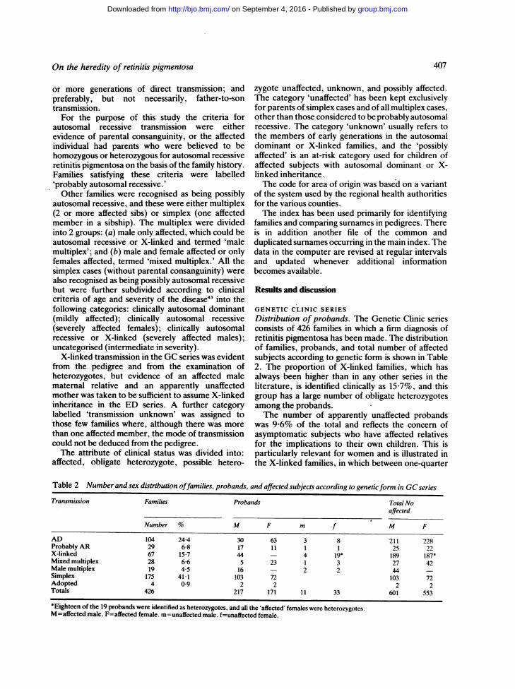
Distribution of probands. The Genetic Clinic seriesconsists of 426 families in which a firm diagnosis ofretinitis pigmentosa has been made. The distributionof families, probands, and total number of affectedsubjects according to genetic form is shown in Table2. The proportion of X-linked families, which hasalways been higher than in any other series in theliterature, is identified clinically as 15-7%, and thisgroup has a large number of obligate heterozygotesamong the probands.The number of apparently unaffected probands
was 9-6% of the total and reflects the concern ofasymptomatic subjects who have affected relativesfor the implications to their own children. This isparticularly relevant for women and is illustrated inthe X-linked families, in which between one-quarter
Table 2 Number and sex distribution offamilies, probands, and affected subjects according to geneticform in GC series
Transmission Families Probands Total Noaffected
Number % M F m f M F
AD 104 24-4 30 63 3 8 211 228Probably AR 29 6-8 17 11 1 1 25 22X-linked 67 15-7 44 - 4 19* 189 187*Mixed multiplex 28 6 6 5 23 1 3 27 42Male multiplex 19 4-5 16 - 2 2 44 -
Simplex 175 41-1 103 72 103 72Adopted 4 0-9 2 2 2 2Totals 426 217 171 11 33 601 553
*Eighteen of the 19 probands were identified as heterozygotes, and all the 'affected' females were heterozygotes.M=affected male. F=affected female. m=unaffected male. f=unaffected female.
407
group.bmj.com on September 4, 2016 - Published by http://bjo.bmj.com/Downloaded from

Marcelle Jay
Table 3 Sex distribution ofsimplex cases in the GCseriesaccording to clinical severity
Males Females Totals
Clinically AR (severely affected 31 15 46Clinically AD (mildly affected) 27 23 50Unclassified(intermediate in severity) 45 34 79Totals 103 72 175
and one-third of these families were ascertainedthrough the females, and where 18 out of 19 womenwere discovered to be heterozygotes. The largenumber of female probands in the X-linked families isundoubtedly related to the demand for geneticcounselling in severe disease. It is also possibly aconsequence of the well-publicised research interestof the clinicians at Moorfields Eye Hospital.The proportion of simplex cases is 41 1%, and their
division according to clinical form is shown in Table 3.There are 9 male and 8 female simplex cases amongthe probands in the autosomal recessive group, and ifall simplex cases with or without parental con-sanguinity are considered as a group there are 112male and 80 female simplex cases. The excess ofmales in this group is statistically significant (x2=5 33,p<002). Similarly, if all simplex and multiplex cases,with or without consanguinity are grouped together,there is a total of 172 affected males and 113 affectedfemales. The deviation from the expected sex distri-bution is highly significant (x2= 12-21, p<O01).There is a significant excess of females among the
probands in the autosomal dominant families (63females to 30 males), but the excess of females amongall affected members of the same group is no longersignificant (228 females to 211 males), indicating a
bias of ascertainment in this group. This may be dueto various factors such as the economic implicationsof occupation which may weigh more heavily on menthan on women. Women may be more prone thanmen to seek specialist advice on prognosis andpossible treatment, and they appear more likely toseek genetic counselling.
Comparison of frequency of genetic forms. Thefrequency of the various genetic forms differs whencalculated as a proportion of the families on the onehand, and as a proportion of the total number ofaffected individuals in the pedigree on the other. Theproportion of autosomal dominant disease is muchhigher (38%) among affected individuals than amongfamilies, where it is 24-4%. Similarly, the X-linkedproportion is 15-7% among families and 32-6%among affected individuals. This clearly illustrates thebias which is introduced when a series is based onaffected individuals with a positive family history.
The frequency of autosomal dominant retinitispigmentosa among families (24-4%) is much lowerthan the 39% found in previous studies.93738 Thismay be the result of a different division of the series orof the relatively higher proportion of X-linked andsimplex cases in the present series. The proportion ofautosomal dominant disease in the present study iscomparable to 2 Sther studies: Frangois8 found19-5%, and Fishman' found 26%.The frequency of autosomal recessive retinitis
pigmentosa is 6-4%, which does not take into accountall the simplex and multiplex cases which may berecessive after removal of the X-linked proportion,and this topic will be discussed in the light of theresults of segregation analysis.The estimation of the frequency of genetic forms
among families shows that the proportion of simplexcases is slightly higher than in a previous study basedon 305 families.43 This may be due to moregeneralised referral from various sources or from agreater demand by patients for assessment andcounselling.
Estimation of X-linked form in simplex andmultiplex sibships. A method suggested by Fraser45was used to estimate the X-linked proportion in thesegroups, based on the comparison of sibships withaffected members of one sex only. With autosomalrecessive inheritance there should be as many sibshipsof males only affected as of females only affected. Thedefined autosomal recessive sibships are removedfrom the analysis, and the excess of sibships with onlymales affected can be directly attributable to X-linkedinheritance. Furthermore a sibship with, for example,3 affected children may be distributed in 4 differentcombinations: 3 males, 3 females, 2 males and 1female, or 1 male and 2 females. If nk is the number ofsibships other than male only affected with k affectedmembers, then the number of sibships with only maleaffected is:
nk
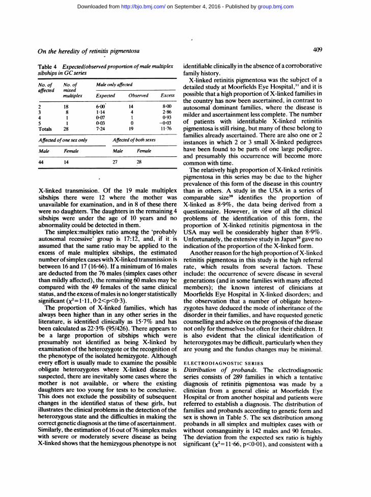
This calculated number of sibships is compared withthe observed number of sibships with only malesaffected, and the excess corresponds to the number ofsibships which may be attributable to X-linkedtransmission. This excess may also be converted to asimplex number of X-linked cases by multiplying it bythe simplex:multiplex ratio obtained from theautosomal recessive group. These methods will givean approximation of the proportion of X-linkedcases, and they have been applied to all multiplexfamilies in the GC series (Table 4). The resultsshowed that 11 -76 out of the 19 male multiplexsibships in this series could be attributable to
408
group.bmj.com on September 4, 2016 - Published by http://bjo.bmj.com/Downloaded from
On the heredity of retinitis pigmentosa
Table 4 Expected/observed proportion ofmale multiplexsibships in GC series
No. of No. of Male only affectedaffected mixed
multiplex Expected Observed Excess
2 18 6-00 14 8003 8 1*14 4 2-864 1 0-07 1 0935 1 003 0 -003Totals 28 7-24 19 11-76
Affected ofone sex only Affected ofboth sexes
Male Female Male Female
44 14 27 28
X-linked transmission. Of the 19 male multiplexsibships there were 12 where the mother was
unavailable for examination, and in 8 of these therewere no daughters. The daughters in the remaining 4sibships were under the age of 10 years and no
abnormality could be detected in them.The simplex:multiplex ratio among the 'probably
autosomal recessive' group is 17:12, and, if it isassumed that the same ratio may be applied to theexcess of male multiplex sibships, the estimatednumber of simplex cases with X-linked transmission isbetween 16 and 17 (16-66). If a minimum of 16 malesare deducted from the 76 males (simplex cases otherthan mildly affected), the remaining 60 males may becompared with the 49 females of the same clinicalstatus, and the excess ofmales is no longer statisticallysignificant (X2= 1- I l, 0-2<p<0-3).The proportion of X-linked families, which has
always been higher than in any other series in theliterature, is identified clinically as 15-7% and hasbeen calculated as 22-3% (95/426). There appears tobe a large proportion of sibships which werepresumably not identified as being X-linked byexamination of the heterozygote or the recognition ofthe phenotype of the isolated hemizygote. Althoughevery effort is usually made to examine the possibleobligate heterozygotes where X-linked disease issuspected, there are inevitably some cases where themother is not available, or where the existingdaughters are too young for tests to be conclusive.This does not exclude the possibility of subsequentchanges in the identified status of these girls, butillustrates the clinical problems in the detection of theheterozygous state and the difficulties in making thecorrect genetic diagnosis at the time of ascertainment.Similarly, the estimation of 16 out of76 simplex maleswith severe or moderately severe disease as beingX-linked shows that the hemizygous phenotype is not
identifiable clinically in the absence of a corroborativefamily history.
X-linked retinitis pigmentosa was the subject of adetailed study at Moorfields Eye Hospital,3" and it ispossible that a high proportion of X-linked families inthe country has now been ascertained, in contrast toautosomal dominant families, where the disease ismilder and ascertainment less complete. The numberof patients with identifiable X-linked retinitispigmentosa is still rising, but many of these belong tofamilies already ascertained. There are also one or 2instances in which 2 or 3 small X-linked pedigreeshave been found to be parts of one large pedigree,and presumably this occurrence will become morecommon with time.The relatively high proportion of X-linked retinitis
pigmentosa in this series may be due to the higherprevalence of this form of the disease in this countrythan in others. A study in the USA in a series ofcomparable size39 identifies the proportion ofX-linked as 8-9%, the data being derived from aquestionnaire. However, in view of all the clinicalproblems of the identification of this form, theproportion of X-linked retinitis pigmentosa in theUSA may well be considerably higher than 8-9%.Unfortunately, the extensive study in Japan30 gave noindication of the proportion of the X-linked form.
Another reason for the high proportion of X-linkedretinitis pigmentosa in this study is the high referralrate, which results from several factors. Theseinclude: the occurrence of severe disease in severalgenerations (and in some families with many affectedmembers); the known interest of clinicians atMoorfields Eye Hospital in X-linked disorders; andthe observation that a number of obligate hetero-zygotes have deduced the mode of inheritance of thedisorder in their families, and have requested geneticcounselling and advice on the prognosis of the diseasenot only for themselves but often for their children. Itis also evident that the clinical identification ofheterozygotes may be difficult, particularly when theyare young and the fundus changes may be minimal.
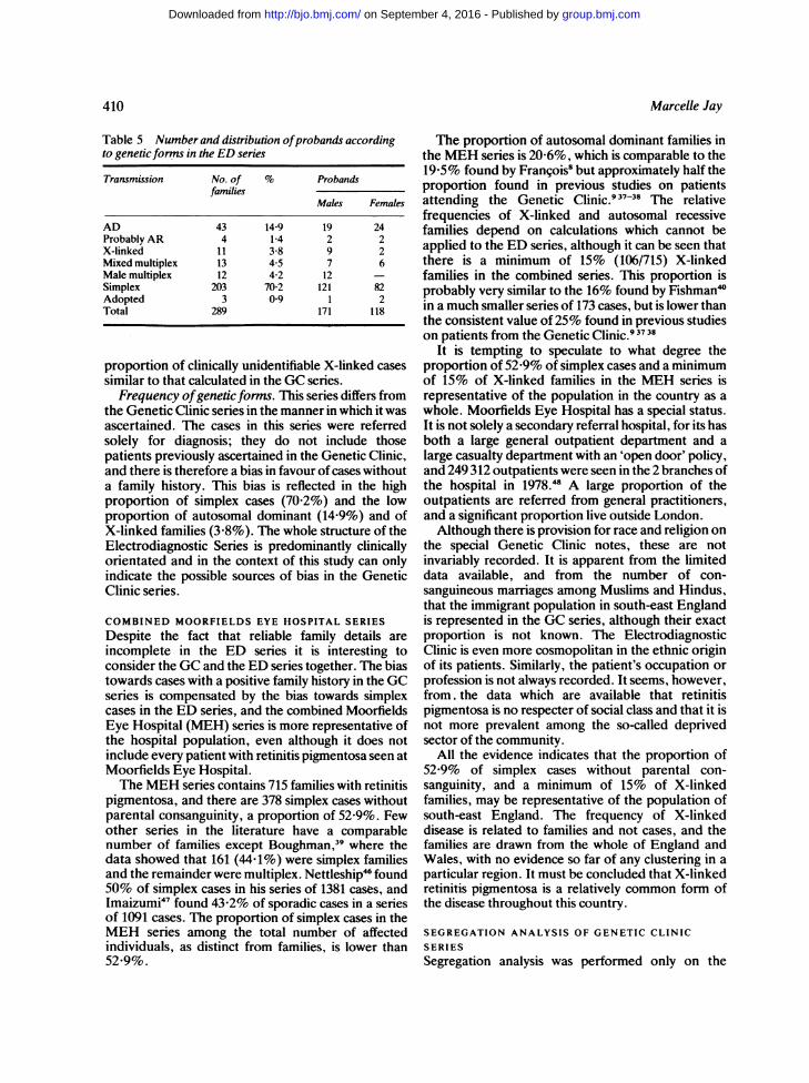
ELECTRODIAGNOSTIC SERIESDistribution of probands. The electrodiagnosticseries consists of 289 families in which a tentativediagnosis of retinitis pigmentosa was made by aclinician from a general clinic at Moorfields EyeHospital or from another hospital and patients werereferred to establish a diagnosis. The distribution offamilies and probands according to genetic form andsex is shown in Table 5. The sex distribution amongprobands in all simplex and multiplex cases with orwithout consanguinity is 142 males and 90 females.The deviation from the expected sex ratio is highlysignificant (x2= 11-66, p<O-0l), and consistent with a
409
group.bmj.com on September 4, 2016 - Published by http://bjo.bmj.com/Downloaded from
Marcelle Jay
Table 5 Number and distribution ofprobands accordingto geneticforms in the ED series
Transmission No. of % Probandsfamilies
Males Females
AD 43 14-9 19 24Probably AR 4 1-4 2 2X-linked 11 3-8 9 2Mixed multiplex 13 4-5 7 6Male multiplex 12 4-2 12 -
Simplex 203 70-2 121 82Adopted 3 09 1 2Total 289 171 118
proportion of clinically unidentifiable X-linked cases
similar to that calculated in the GC series.Frequency ofgeneticforms. This series differs from
the Genetic Clinic series in the manner in which it wasascertained. The cases in this series were referredsolely for diagnosis; they do not include thosepatients previously ascertained in the Genetic Clinic,and there is therefore a bias in favour of cases withouta family history. This bias is reflected in the highproportion of simplex cases (70 2%) and the lowproportion of autosomal dominant (14-9%) and ofX-linked families (3-8%). The whole structure of theElectrodiagnostic Series is predominantly clinicallyorientated and in the context of this study can onlyindicate the possible sources of bias in the GeneticClinic series.
COMBINED MOORFIELDS EYE HOSPITAL SERIES
Despite the fact that reliable family details areincomplete in the ED series it is interesting toconsider the GC and the ED series together. The biastowards cases with a positive family history in the GCseries is compensated by the bias towards simplexcases in the ED series, and the combined MoorfieldsEye Hospital (MEH) series is more representative ofthe hospital population, even although it does notinclude every patient with retinitis pigmentosa seen atMoorfields Eye Hospital.The MEH series contains 715 families with retinitis
pigmentosa, and there are 378 simplex cases withoutparental consanguinity, a proportion of 52-9%. Fewother series in the literature have a comparablenumber of families except Boughman,39 where thedata showed that 161 (44-1%) were simplex familiesand the remainder were multiplex. Nettleship46 found50% of simplex cases in his series of 1381 cases, andImaizumi47 found 43-2% of sporadic cases in a seriesof 1091 cases. The proportion of simplex cases in theMEH series among the total number of affectedindividuals, as distinct from families, is lower than52 9%.
The proportion of autosomal dominant families inthe MEH series is 20-6%, which is comparable to the19 5% found by Francois' but approximately half theproportion found in previous studies on patientsattending the Genetic Clinic.9 37-38 The relativefrequencies of X-linked and autosomal recessivefamilies depend on calculations which cannot beapplied to the ED series, although it can be seen thatthere is a minimum of 15% (106/715) X-linkedfamilies in the combined series. This proportion isprobably very similar to the 16% found by Fishman'in a much smaller series of 173 cases, but is lower thanthe consistent value of25% found in previous studieson patients from the Genetic Clinic.93738
It is tempting to speculate to what degree theproportion of 52-9% of simplex cases and a minimumof 15% of X-linked families in the MEH series isrepresentative of the population in the country as awhole. Moorfields Eye Hospital has a special status.It is not solely a secondary referral hospital, for its hasboth a large general outpatient department and alarge casualty department with an 'open door' policy,and 249 312 outpatients were seen in the 2 branches ofthe hospital in 1978.48 A large proportion of theoutpatients are referred from general practitioners,and a significant proportion live outside London.Although there is provision for race and religion on
the special Genetic Clinic notes, these are notinvariably recorded. It is apparent from the limiteddata available, and from the number of con-sanguineous marriages among Muslims and Hindus,that the immigrant population in south-east Englandis represented in the GC series, although their exactproportion is not known. The ElectrodiagnosticClinic is even more cosmopolitan in the ethnic originof its patients. Similarly, the patient's occupation orprofession is not always recorded. It seems, however,from. the data which are available that retinitispigmentosa is no respecter of social class and that it isnot more prevalent among the so-called deprivedsector of the community.
All the evidence indicates that the proportion of52-9% of simplex cases without parental con-sanguinity, and a minimum of 15% of X-linkedfamilies, may be representative of the population ofsouth-east England. The frequency of X-linkeddisease is related to families and not cases, and thefamilies are drawn from the whole of England andWales, with no evidence so far of any clustering in aparticular region. It must be concluded that X-linkedretinitis pigmentosa is a relatively common form ofthe disease throughout this country.
SEGREGATION ANALYSIS OF GENETIC CLINICSERIES
Segregation analysis was performed only on the
410
group.bmj.com on September 4, 2016 - Published by http://bjo.bmj.com/Downloaded from
On the heredity of retinitis pigmentosa
Genetic Clinic series, where the family structure wasknown in all but a few cases.
Possible autosomal recessive families. All knownautosomal dominant, X-linked, and adopted familieswere removed from the group for analysis, and theremainder consisted of: the probably autosomalrecessive group; all the multiplex cases; and all thesimplex cases. (Male-only multiplex sibships wereincluded, since even those which may be X-linked donot affect the segregation ratio.)
Various methods of segregation analysis areavailable.4"5' A number of these depend oncomplete ascertainment and cannot therefore beapplied to this series. The method of choice is thatdescribed by Crow52 and is an adaptation of earliermethods." S This method is similar to the Weinberg'proband' method but adapted for use in multipleincomplete ascertainment. A first approximation ofthe segregation ratio po is estimated by obtaining theratio of affected sibs to the total number of sibs ofprobands for the pooled families.
If a=number of probandss=sibship sizer=number of affected
thenP=
a(r-1)l:a (s- 1)
A parameter (ir), known as the probability ofascertainment is calculated from the equation
la (a-1)Xa (r- 1)
The corrections suggested by Crow52 consist of acorrection factor C defined by
IIC=1 +ir+por(s-3)
which is applied to calculate a more accurate value ofthe segregation ratio p where
Ca (r- 1)ECa (s- 1)
The effect ofC is to reduce the contribution from thelarger sized families, so that p is greater than po.The variance Vp is given by
p (l-p)
:Ca (S-I1)
Crow52 also suggested calculating the 2 limiting valuesof p, corresponding to truncate selection where i= 1,
and single selection where ir=O. The value of p fortruncate selection is the upper limiting value, and thevalue of p for single selection is the lower limitingvalue of the segregation ratio.
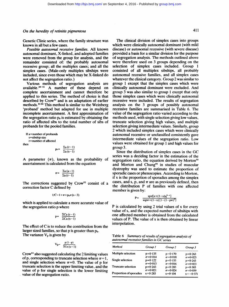
The clinical division of simplex cases into groupswhich were clinically autosomal dominant (with milddisease) or autosomal recessive (with severe disease)provided a basis for a similar division for the purposeof segregation analysis. The methods outlined abovewere therefore used on 3 groups depending on theselection of simplex cases included. Group 1consisted of all multiplex sibships, all probablyautosomal recessive families, and all simplex caseswhatever the clinical category. Group 2 was similar togroup 1 except that the simplex cases which wereclinically autosomal dominant were excluded. Andgroup 3 was also similar to group 1 except that onlythose simplex cases which were clinically autosomalrecessive were included. The results of segregationanalysis on the 3 groups of possibly autosomalrecessive families are summarised in Table 6. Thevalue of the segregation ratio varied according to themethods used, with single selection giving low values,truncate selection giving high values, and multipleselection giving intermediate values. Similarly, group2 which included simplex cases which were clinicallyautosomal recessive or unclassified consistently gaveintermediate values of the segregation ratio. Lowvalues were obtained for group 1 and high values forgroup 3.
Since the distribution of simplex cases in the GCseries was a deciding factor in the estimation of thesegregation ratio, the equation derived by Morton55and Morton and Chung56 in studies of musculardystrophy was used to estimate the proportion ofsporadic cases or phenocopies. According to Morton,if x is the proportion of sporadics among the simplexcases, and s, p, and vr are as previously defined, thenthe distribution P of families with one affectedmember is given by:
p_ sp[x+(I-x)qs-']xspvr+(1-x) [1-(1-pr)s]
P is calculated by using 2 trial values of x for everyvalue of s, and the expected number of sibships withone affected member is obtained from the calculatedvalues of P. The value of x is then obtained by linearinterpolation.
Table 6 Summary ofresults ofsegregation analysis ofautosomal recessive families in GC series
Method Group I Group 2 Group 3
Multiple selection p=0d135 p=0170 p=0-263o=0-014 r=0-018 a=0(025
Single selection p=0-122 p=0-155 p=0-243or=0 013 o=0 016 o-=0023
Truncate selection p=0-243 p=0-283 p=0-383(o=0 021 o-=0024 o=0-039
Proportion of sporadics x=0 285 x=0 104 x=-0-371
411
group.bmj.com on September 4, 2016 - Published by http://bjo.bmj.com/Downloaded from
Marcelle Jay
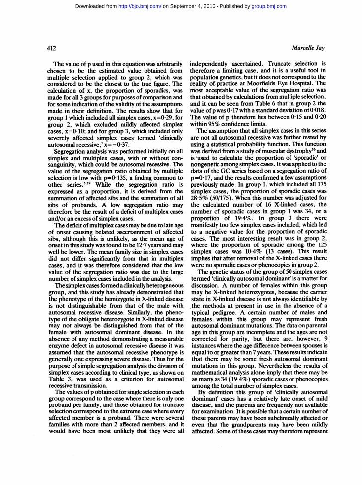
The value of p used in this equation was arbitrarilychosen to be the estimated value obtained frommultiple selection applied to group 2, which was
considered to be the closest to the true figure. Thecalculation of x, the proportion of sporadics, was
made for all 3 groups for purposes of comparison andfor some indication of the validity of the assumptionsmade in their definition. The results show that forgroup 1 which included all simplex cases, x=0 29; forgroup 2, which excluded mildly affected simplexcases, x=0 10; and for group 3, which included onlyseverely affected simplex cases termed 'clinicallyautosomal recessive,' x= -0-37.
Segregation analysis was performed initially on allsimplex and multiplex cases, with or without con-
sanguinity, which could be autosomal recessive. Thevalue of the segregation ratio obtained by multipleselection is low with p=0-135, a finding common toother series.539 While the segregation ratio isexpressed as a proportion, it is derived from thesummation of affected sibs and the summation of allsibs of probands. A low segregation ratio maytherefore be the result of a deficit of multiplex cases
and/or an excess of simplex cases.The deficit ofmultiplex cases may be due to late age
of onset causing belated ascertainment of affectedsibs, although this is unlikely, as the mean age ofonset in this study was found to be 12-7 years and maywell be lower. The mean family size in simplex cases
did not differ significantly from that in multiplexcases, and it was therefore considered that the lowvalue of the segregation ratio was due to the largenumber of simplex cases included in the analysis.Thesimplex cases formed a clinically heterogeneous
group, and this study has already demonstrated thatthe phenotype of the hemizygote in X-linked diseaseis not distinguishable from that of the male withautosomal recessive disease. Similarly, the pheno-type of the obligate heterozygote in X-linked diseasemay not always be distinguished from that of thefemale with autosomal dominant disease. In theabsence of any method demonstrating a measurableenzyme defect in autosomal recessive disease it wasassumed that the autosomal recessive phenotype isgenerally one expressing severe disease. Thus for thepurpose of simple segregation analysis the division ofsimplex cases according to clinical type, as shown on
Table 3, was used as a criterion for autosomalrecessive transmission.The values ofp obtained for single selection in each
group correspond to the case where there is only oneproband per family, and those obtained for truncateselection correspond to the extreme case where every
affected member is a proband. There were severalfamilies with more than 2 affected members, and itwould have been most unlikely that they were all
independently ascertained. Truncate selection istherefore a limiting case, and it is a useful tool inpopulation genetics, but it does not correspond to thereality of practice at Moorfields Eye Hospital. Themost acceptable value of the segregation ratio wasthat obtained by calculations from multiple selection,and it can be seen from Table 6 that in group 2 thevalue ofp was 0-17 with a standard deviation of0-018.The value of p therefore lies between 0-15 and 0-20within 95% confidence limits.The assumption that all simplex cases in this series
are not all autosomal recessive was further tested byusing a statistical probability function. This functionwas derived from a study of muscular dystrophy56 andis used to calculate the proportion of 'sporadic' ornongenetic among simplex cases. It was applied to thedata of the GC series based on a segregation ratio ofp=0-17, and the results confirmed a few assumptionspreviously made. In group 1, which included all 175simplex cases, the proportion of sporadic cases was28-5% (50/175). When this number was adjusted forthe calculated number of 16 X-linked cases, thenumber of sporadic cases in group 1 was 34, or aproportion of 19-4%. In group 3 there weremanifestly too few simplex cases included, which ledto a negative value for the proportion of sporadiccases. The most interesting result was in group 2,where the proportion of sporadic among the 125simplex cases was 10-4% (13 cases). This resultimplies that after removal of the X-linked cases therewere no sporadic cases or phenocopies in group 2.The genetic status of the group of 50 simplex cases
termed 'clinically autosomal dominant' is a matter fordiscussion. A number of females within this groupmay be X-linked heterozygotes, because the carrierstate in X-linked disease is not always identifiable by.the methods at present in use in the absence of atypical pedigree. A certain number of males andfemales within this group may represent freshautosomal dominant mutations. The data on parentalage in this group are incomplete and the ages are notcorrected for parity, but there are, however, 9instances where the age difference between spouses isequal to or greater than 7 years. These results indicatethat there may be some fresh autosomal dominantmutations in this group. Nevertheless the results ofmathematical analysis alone imply that there may beas many as 34 (19-4%) sporadic cases or phenocopiesamong the total number of simplex cases.By definition this group of 'clinically autosomal
dominant' cases has a relatively late onset of milddisease, and the parents are frequently not availablefor examination. It is possible that a certain number ofthese parents may have been subclinically affected oreven that the grandparents may have been mildlyaffected. Some of these cases may therefore represent
412
group.bmj.com on September 4, 2016 - Published by http://bjo.bmj.com/Downloaded from
On the heredity of retinitis pigmentosa
a group of autosomal dominant retinitis pignentosawith or without reduced penetrance, with a smallnumber of fresh autosomal dominant mutations. It isalso possible that a number of these mildly affectedsimplex cases are clinically unidentifiablephenocopies.The statistical analysis has shown that out of 175
simplex cases it may be assumed that 109 areautosomal recessive and 16 are X-linked males. Theremaining 50 are mildly affected, and some of thesemay be fresh autosomal dominant mutations. Theremainder may be X-linked heterozygotes,autosomal dominant cases whose parents haveescaped detection, or possibly clinicallyunidentifiable manifesting heterozygotes, orphenocopies. While it is possible that a few of thesemildly affected simplex cases are autosomal recessive,the results of segregation analysis indicate that thisgroup, if it exists, is small.Autosomal dominant families. The method used
was that given by Emery.49 The number of normaland affected offspring of all affected by normalmatings is compared with the expected number and a
X2 test is applied to the deviation from the expected1:1 ratio. The segregation ratio is the number ofaffected offspring to total number of offspring, andthe penetrance is the ratio of the calculated totheoretical value of the segregation ratio. Thesegregation ratio for the autosomal dominant familieswas found to be p=0453, indicating a penetrance of90X6%. There were in fact 12 pedigrees out of a totalof 104 (11.5%) showing reduced penetrance.
X-linkedfamilies. The method used was that givenby Emery" for X-linked recessive inheritance, sinceno pedigree in the GC series fulfilled the criteria forX-linked dominant transmission. The method usedwas similar to that used for autosomal dominantinheritance, except that the segregation ratio in thiscase was the number of affected sons to the totalnumber of sons. There was the added condition thatonly the male offspring of obligate heterozygoteswere considered. This condition would haverestricted the number of infornative sibships to such.a degree that it was decided to consider the wholepedigree in the calculations and not merely theproband's generation.The segregation ratio for X-linked families was
p=047, which is in good agreement with thetheoretical value. The number of informative familieswas small, as only the sons of obligate heterozygoteswere included in the analysis.
IMPLICATIONS FOR GENETIC COUNSELLINGThe Genetic Clinic series remains the basis of thisstudy owing to the unique combination of genetic andclinical information it provides. Every single
proband, and numerous relatives of probands, wereseen by one or 2 of only 3 clinicians, resulting in aremarkable homogeneity in clinical approach. Themain conclusions drawn from the results of thevarious analyses should be considered in the light ofcorroborative evidence from the Electrodiagnosticseries.
Frequency of genetic forms. The frequency of thedifferent genetic forms in the GC series was identifiedinitially by considering the phenotype and the familyhistory. The estimation of the unidentified X-linkedproportion in the series has shown that the phenotypeis not necessarily distinguishable from the autosomalrecessive phenotype in the hemizygote and from theautosomal dominant phenotype in the heterozygote.
In the group of conditions such as retinitis pig-mentosa, where there is genetic heterogeneity andthe occurrence of phenocopies, it cannot bepresumed that all cases which are not demonstrablyautosomal dominant or X-linked are necessarilyautosomal recessive. Segregation analysis hasdemonstrated that autosomal recessive transmissionmay be assumed in a group defined by a number ofcriteria. It has been shown that autosomal recessiveretinitis pigmentosa usually produces early andsevere visual loss. Autosomal recessive disease can beassumed if one or more sib is severely affected, par-ticularly if there is parental consanguinity. A propor-tion of severely affected male simplex and multiplexcases are X-linked, and in these instances the het-erozygotes usually show evidence of being mildlyaffected, in contrast to the heterozygotes for auto-somal recessive disease, who at present cannot bedistinguished from normal individuals.
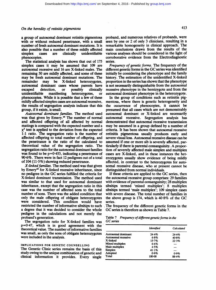
If these criteria are applied to the GC series, thenthe autosomal recessive group comprises: 29 familieswith evidence of parental consanguinity; 28 multiplexsibships termed 'mixed multiplex'; 8 multiplexsibships termed 'male multiplex'; 109 simplex caseswith severe disease. The total number of families inthe above group is 174, which is 40-8% of the GCseries.The frequency of the different genetic forms in theGC series is therefore as shown in Table 7.
Table 7 Frequency ofdifferentgeneticforms in theGC series
Identified Calculated
Autosomal dominant 24 4% 24-4%Autosomal recessive 6-8% 40-8%X-linked 15 7% 22-3%Mixed multiplex 6-6% -
Male multiplex 4.5%Simplex 41-1% -
Adopted 09% 0-9%Total 100-00 88-4%
413
group.bmj.com on September 4, 2016 - Published by http://bjo.bmj.com/Downloaded from
Marcelle Jay
The remaining 11-6% in the calculated total consistsof 50 simplex cases with mild disease whose geneticstatus is uncertain but most of whom are notautosomal recessive.
Genetic counselling. The genetic counselling ofautosomal dominant and identifiable X-linkedfamilies is straightforward. The counselling ofpresumably autosomal recessive families depends ona number of factors and differs in multiplex andsimplex cases.
(1) Multiplex cases. Autosomal recessive trans-mission may be justifiably assumed in sibships wherefemales only are affected, or where males and femalesare equally severely affected. In sibships consistingonly of affected males, and in the absence of parentalconsanguinity, X-linked transmission should alwaysbe suspected and every effort made to exclude it byexamining obligate or possible heterozygotes.
(2) Simplex cases. The genetic counselling ofsimplex cases, which account for half the ascertainedfamilies, still remains a problem and depends uponvarious factors such as age, sex, and clinical state. Thediagnosis of clinical form is age-dependent and, if thedisease is mild, often cannot be made until the patientis in the fourth decade. It can often be made in thesecond decade when the disease is severe. This can bean important factor in a situation where parents ofone affected child are concerned about the risk tofurther children. It is usually possible to diagnosesevere disease when the proband is of childbearingage and wishes to know the risk of transmitting thedisease.
Males without parental consanguinity and withsevere or moderately severe disease form 74%(76/103) of the total, and, of these, 21% (36/76) areestimated to be X-linked, the remainder beingautosomal recessive (79%). When the disease issevere or moderately severe, the ratio of autosomalrecessive to X-linked is 4 to 1. The risk of transmittingautosomal recessive disease is very small, as itdepends upon the frequency of heterozygotes in thegeneral population. In X-linked disease sons areunaffected but all daughters are obligateheterozygotes. Males with severe or moderatelysevere disease, whose genetic status is uncertain,have negliglible chance of producing affectedchildren (provided they have not married a relative),but have a 1:10 chance of producing a daughter who isa heterozygote for X-linked disease.Males with mild disease form the remaining 26% of
the total, and the problem of their genetic status isnot entirely resolved. They may represent freshautosomal dominant mutations, autosomal domi-nant transmission with reduced penetrance, orunidentified phenocopies. Taking into account the90-6% penetrance in autosomal dominant retinitis
pigmentosa and the small incidence of phenocopies,this group of males with mild disease haveapproximately a 45% chance of producing affectedchildren who will also have the mild form of thedisease.
Similarly, females without parental consanguinityand with severe or moderately severe diseasecomprise 68% (49/72) of the total and may becounselled on the basis that they have autosomalrecessive disease. The remaining 32% of the total arefemales with mild disease, a number ofwhom may beexpected to be obligate heterozygotes for X-linkeddisease and the others fresh autosomal dominantmutations, autosomal dominant transmission withreduced penetrance, or unidentified phenocopies. Incontrast to males in whom X-linked transmission issuspected but cannot be confirmed due to the lack ofavailability of obligate heterozygotes, a precisediagnosis can usually be established in the females bythe time they are of childbearing age. It is usuallypossible to distinguish clinically between females whoare obligate heterozygotes for X-linked disease andthose who have autosomal dominant disease. Theformer have a 50% chance of their sons having severedisease, while the latter have a 45% chance of theirchildren having mild disease.Comment. The methods of analysis which were
used are those currently available, and they are basedon some assumptions and conditions which are notalways realised in clinical practice. The systematicrecording of ascertainments, referrals, and similardata is a prerequisite of a certain type of statisticalanalysis. It is an approach which is admirably suited tothe computerised methods of the present day, butwhich must serve and not dominate the clinician inthe practice of medicine.
It is perhaps apt to end with the observation thatafter 20 years of collecting families with retinitispigmentosa Nettleship2 found that half his cases hadno family history and no parental consanguinity. Thefindings in this study are remarkably similar, but it ishoped that the conclusions which are tentativelysubmitted will be of some assistance to the clinician ingiving genetic advice and to those who continue theresearch into this disease.
Conclusions
(1) The various phenotypes in retinitis pigmentosacannot always be easily recognised in the absence of acorroborative family history.
(2) The X-linked form of retinitis pigmentosa is acommon form of the disease in England and Wales. Itwas identified clinically as 15-7% and calculated as22-3% in the Genetic Clinic series, and it wasestimated that a minimum of 15% of families in the
414
group.bmj.com on September 4, 2016 - Published by http://bjo.bmj.com/Downloaded from
415On the heredity of retinitis pigmentosa
combined Genetic Clinic and Electrodiagnostic serieshad X-linked disease.
(3) Simplex cases without parental consanguinityconstitute 41-1% of families in the Genetic Clinicseries and approximately 50% of the combinedGenetic Clinic and Electrodiagnostic Clinic series.The results of segregation analysis and clinicalheterogeneity indicate that no more than 70% of allsimplex cases in theGCseries are autosomal recessive.
(4) The criteria for autosomal recessive trans-mission in retinitis pigmentosa should include severe
disease.(5) The clinical assessment of simplex cases is
essential for determining their genetic status. Therewere 192 simplex cases with or without parental con-
sanguinity in the GC series, and, of these, 50 (26%)were mildly affected with disease resemblingautosomal dominant disease in clinical form andprogression. It is considered that this is not whollyaccounted for by a mild phenotype in autosomalrecessive disease, but more probably evidence offresh autosomal dominant mutations, autosomaldominant transmission with reduced penetrance, theheterozygous state of X-linked disease in some of thefemales, and phenocopies.
(6) The results in the 2 series reflect a bias ofascertainment inherent in each.
(7) The excess of female probands in the autosomaldominant families in the Genetic Clinic series mayindicate a greater concern for the genetic implicationsof the disease in women than in men.
I thank Mr Barrie Jay and Professor Alan Bird for allowing me accessto the notes of the Genetic Clinic and for their clinical assessment ofcases in this study. I am grateful to Professor G. B. Arden and MrJ. H. Kelsey for allowing me access to the notes of the Electro-diagnostic Department at Moorfields Eye Hospital. I am also gratefulto Mrs Vanessa Hanna for secretarial help.
This research was supported in part by MRC grant NoG80/0986171N and by a grant from the National Retinitis PigmentosaFoundation, Inc.
References
I Hutchinson J. On retinitis pigmentosa and allied affections, as
illustrating the laws of heredity. Ophthabnic Rev 1882; 1: 2-7.2 Nettleship E. On some hereditary diseases of the eye. TransOphthalmol Soc UK 1909; 29: 57-198.
3 Bell J. Retinitis pigmentosa and allied diseases. In: Pearson K,ed. Treasury ofHuman Inheritance. Cambridge: Cambridge Uni-versity Press, 1922: 2 (1).
4 Ammann F, Klein D, Bohringer HR. Resultats preliminaires sur
la frequence et la distribution geographique des degenerescencestapeto-retiniennes en Suisse (etude de cinq cantons). J GenetHum 1961; 10:99-127.
5 Ammann F, Klein D, Franceschetti A. Genetic and epidemio-logical investigations on pigmentary degeneration of the retinaand allied disorders in Switzerland. J Neurol Sci 1965; 2: 183-96.
6 Biro I. Ueber den Zusammenhang von Degeneratio pigmentosaretinae und Storungen des Gehors. Ophthalmologica 1944; 107:149-57.
7 Francois J. Degenerescence pigmentaire de la retine a hereditedominante. Bull Soc Belge Ophtalmol 1935; 70: 79-86.
8 Frangois J. Chorioretinal heredodegeneration. Proc R Soc Med1961;54: 1109-18.
9 Jay B. Hereditary aspects of pigmentary retinopathy. TransOphthalmol Soc UK 1972; 92:173-8.
10 Wibaut F. Studien uber Retinitis pigmentosa. Klin MonatsblAugenheilkd 1931; 87: 298-307.
11 Botermans CHG. Primary pigmentary retinal degeneration andits association with neurological diseases. In: Vinken PJ, BruynGW, eds. Handbook of Clinical Neurology: Neuroretinaldegenerations. New York: American Elsevier, 1972; 13: 148-378.
12 Deutman AF. Rod-cone dystrophy: Primary, hereditary, pig-mentary retinopathy, retinitis pigmentosa. In: Krill AE.Hereditary Retinal and Choroidal Diseases. Hagerstown: Harperand Row, 1977; 2: 479-576.
13 Duke-Elder S, Dobree JH. System of Ophthalmology. Diseasesof the Retina. London: Henry Kimpton, 1967; 10: 577-628.
14 Franceschetti A, Francois J, Babel J. Les heredo-degenerescenceschorio-ritiniennes. Paris: Masson, 1963; 1: 203-369.
15 Frangois J. Heredity in Ophthalmology. St Louis: Mosby, 1961:441-53.
16 Frangois J. Counseling in hereditary eye disorders. In: BellowJG, ed. Contemporary Ophthalmology. Baltimore: Williams andWilkins, 1972: 459-71.
17 Krill AE. Retinitis pigmentosa: a review. Sight Sav Rev 1972; 42:20-8.
18 Merin S, Auerbach E. Retinitis pigmentosa. Surv Ophthalmol1976; 20: 303-46.
19 Scheie HG, Albert DM. Adler's Textbook of Ophthalmology.Philadelphia: Saunders, 1969: 130-3.
20 Sorsby A. Ophthalmic Genetics. London: Butterworths, 1970:134.
21 Hussels I. Une famille atteinte de retinopathie pigmentaire liheau sexe, de maladie de Parkinson et d'autres troubles neuro-psychiatriques. J Genet Hum 1967; 16: 106-55.
22 Diem M. Retinitis punctata albescens et pigmentosa. KlinMonatsbl Augenheilkd 1914; 53: 371-9.
23 Falls HF, Cotterman C. Choroidal degeneration. A sex-linkedform in which heterozygous women exhibit a tapetal-like retinalreflex. Arch Ophthalmol 1948; 40: 683-703.
24 Gasalla ML. L'heredite dans une famille atteinte de retinitepigmentaire. Bull Mem Soc Fr Ophtalmol 1931; 44: 169-73.
25 Gonin J. Nouvelles observations de scotome annulaire dans ladegenerescence pigmentaire de la retine. Ann Oculist (Paris)1902; 128: 90-107.
26 Hoare GW. Choroido-retinal dystrophy. Br J Ophthalmol 1965;49:449-59.
27 McKenzie DS. The inheritance of retinitis pigmentosa in onefamily. Trans Ophthalmol Soc NZ 1951; 5: 79-82.
28 McQuarrie MD. Two pedigrees of hereditary blindness in man. JGenet 1935; 30: 147-53.
29 Ricci A, Ammann F, Franceschetti A. Reflet tapetoide reversible(phenomene de Mizuo inverse) chex des conductrices deretinopathie pigmentaire recessive liee au sexe. Bull Mem Soc FrOphtalmol 1963; 76: 31-5.
30 Tanabe U. Genetic study of retinal pigmentary dystrophy(retinitis pigmentosa) in Japan. Jinrui Idengaku Zasshi 1972; 16:119-54.
31 Bird AC. X-linked retinitis pigmentosa. Br J Ophthalmol 1975;59:177-99.
32 Francois J. Chorioretinal degeneration or retinitis pigmentosa ofintermediate sex-linked heredity. Doc Ophthalmol 1962; 16:111-27.
33 Faber J. Retinitis pigmentosa in Israel. Statistical-clinical survey.MD Thesis, Jerusalem: Hebrew University, Vision ResearchLaboratory, 1970.
34 Imaizumi K. Retinitis pigmentosa: genetic carriers and earlycases. S Afr Arch Ophthalmol 1974; 2: 257-69.
group.bmj.com on September 4, 2016 - Published by http://bjo.bmj.com/Downloaded from

Marcelle Jay
35 Motohashi A. The prevalence of pigment degeneration of theretina. Jpn J Clin Ophthalmol 1968; 22: 27-35.
36 Panteleeva OA. On the hereditary tapeto-retinal degenerations.Vestn Oftalmol 1969; 1: 53-6.
37 Jay B. Recent advances in ophthalmic genetics: geneticcounselling. BrJ Ophthalmol 1974; S8: 427-37.
38 Jay B. Retinitis pigmentosa in childhood-diagnosis, counselingand prevention. Metab Ophthalmol 1978; 2: 221-2.
39 Boughman JA. Population genetic studies of retinitis pigmentosa.PhD Thesis, Indiana University, 1978.
40 Fishman GA. Retinitis pigmentosa. Genetic percentages. ArchOphthalmol 1978; 96: 822-6.
41 Pearlman JT, Flood TP, Seiff SR. Retinitis pigmentosa withoutpigment. Am J Ophthalmol 1976; 81: 417-9.
42 Bell J. A determination of the consanguinity rate in the generalhospital population of England and Wales. Ann Eugen 1940; 10:370-91.
43 Jay B, Bird A, Jay M. The incidence of the different genetic formsof retinitis pigmentosa. Doc Ophthalmol Proc Series 1978; 17:313-8.
44 Francois J, Verriest G. Etude biometrique de la r6tinopathiepigmentaire. Ann Oculist (Paris) 1962; 195: 937-51.
45 Fraser GR. Sex-linked recessive congenital deafness and excessof males in profound childhood deafness. Ann Hum Genet 1965;29:171-96.
46 Nettleship E. On retinitis pigmentosa and allied diseases. R LondOphthal Hosp Rep 1907-8; 17: 1-56, 151-66, 333-426.
47 Imaizumi K. Statistical investigation on retinitis pigmentosa. JpnJ Clin Ophthalmol 1971; 25: 293-304.
48 Moorfields Eye Hospital Biennial Report April 1977 to March1979; 41.
49 Emery AEH. Methodology in Medical Genetics. An Introductionto Statistical Methods. Edinburgh, London, and New York:Churchill Livingstone, 1976; 34-50.
50 Li CC. Human Genetics, Principles and Methods. New York:McGraw-Hill, 1961: 58-78.
51 Mayo 0. Fundamental and population genetics. In: Fraser G,Mayo, eds. Textbook of Human Genetics. Oxford: Blackwell,1975:47.
52 Crow JF. Problems of ascertainment in the analysis of familydata. In: Neel JV, Shaw, M, Schull W, eds. Genetics and theEpidemiology of Chronic Diseases. Department of Health,Education and Welfare, Publication 1163, 1965: 23-44.
53 Bailey NTJ. The estimation of the frequencies of recessives withincomplete multiple selection. Ann Eugen 1951; 16: 215-22.
54 Haldane JBS. The estimation of the frequencies of recessiveconditions in man. Ann Eugen 1938; 8: 255-62.
55 Morton NE. Genetic tests under incomplete ascertainment. AmJHum Genet 1959; 11: 1-16.
56 Morton NE, Chung CS. Formal genetics of muscular dystrophy.Am J Hum Genet 1959; 11: 360-79.
416
group.bmj.com on September 4, 2016 - Published by http://bjo.bmj.com/Downloaded from
On the heredity of retinitis pigmentosa.
M. Jay
doi: 10.1136/bjo.66.7.4051982 66: 405-416 Br J Ophthalmol
http://bjo.bmj.com/content/66/7/405Updated information and services can be found at:
These include:
serviceEmail alerting
Sign up in the box at the top right corner of the online article. Receive free email alerts when new articles cite this article.
Notes
http://group.bmj.com/group/rights-licensing/permissionsTo request permissions go to:
http://journals.bmj.com/cgi/reprintformTo order reprints go to:
http://group.bmj.com/subscribe/To subscribe to BMJ go to:
group.bmj.com on September 4, 2016 - Published by http://bjo.bmj.com/Downloaded from