Risedronate metal complexes potentially active against Chagas disease
TRYPANOSOMA CRUZI DNA IN CARDIAC LESIONS OF ARGENTINEAN PATIENTSWITH END-STAGE CHRONIC CHAGAS HEART DISEASE
ALEJANDRO G. SCHIJMAN, CARLOS A. VIGLIANO, RODOLFO J VIOTTI, JUAN M. BURGOS, SILVIA BRANDARIZ,BRUNO E. LOCOCO, MARIA I. LEZE, HECTOR A. ARMENTI, AND MARIANO J. LEVIN
Laboratorio de Biologıa Molecular de la Enfermedad de Chagas, INGEBI, Consejo Nacional de Ciencia y Tecnologia, Departmentof Fisiology, Celular and Molecular Biology, F.CEyN, University of Buenos Aires, Buenos Aires, Argentina; Department of
Cardiology, Eva Peron Hospital, San Martin, Buenos Aires, Argentina; Division of Pathology, Instituto de Cardiologıa y CirugıaCardiovascular, Fundacion Favaloro, Buenos Aires, Argentina
Abstract. The extent of inflammation, fibrosis, and progression of chronic Chagas heart disease (cChHD) wasassociated with persistence of parasite DNA in cardiac lesions of necropsies or explants from Argentinean cChHDpatients. A Trypanosoma cruzi−based polymerase chain reaction showed a positive result in 1) 15% of cardiac sectionswith less than 10 mononuclear inflammatory cells/high-power field (440×) (MNC/HPF), 89% with 10–19 MNC/HPF, and100% with more than 20 MNC/HPF (P < 0.0001); 2) 33% with less than 10% fibrosis, 79% with 10–19% fibrosis, and100% with more than 20% fibrosis (P < 0.01); 3) 25% of specimens from patients classified in Kuschnir groups 0 andI, 70% in group II and 90% in group III (P < .001); and 4) 45% and 90% of the specimens from cChHD patients withoutor with heart failure, respectively (P < 0.01). These findings stress the role of the parasite in pathogenesis and diseaseprogression of cChHD.
INTRODUCTION
Chagas disease is the main cause of chronic myocarditis inLatin American countries, where the disease is endemic, af-fecting approximately 20 million people.1 Fifty thousand cha-gasic patients die every year, mainly due to a chronic cardio-myopathy that is clinically evident only 10–30 years after theinitial infection.2
Chronic Chagas heart disease (cChHD) evolves graduallyfrom an asymptomatic period with radiographic or electro-cardiographic manifestations known as the indeterminatephase, and progresses slowly to a dilated cardiomyopathymanifested by arrhythmias, left ventricular dysfunction, em-bolic episodes, and autonomic dysfunction.2,3 The apical an-eurysm is found to be a distinctive pathologic marker in morethan 50% of symptomatic patients.4
In general, the cause of death is heart failure or throm-boembolic phenomena. Anatomically, the heart of a patientwith cChHD is characterized by myocardial damage associ-ated with mononuclear inflammatory foci scattered through-out the organ.2,4 The coexistence of areas of myocytic degen-eration, inflammatory infiltration, and fibrosis suggests achronically evolving process. The lesions are progressive, self-perpetuating, and cumulative.
Since the intracellular forms of the parasite (Trypanosomacruzi) that cause this disease are rarely found at the sites ofthe heart lesions, the hypothesis of an autoimmune processhas been proposed as a leading mechanism of cChHD patho-genesis.5,6 However, polymerase chain reaction (PCR) ampli-fication and immunohistochemical techniques showed thepresence of parasite DNA and proteins close to the inflam-matory foci in lesions found in cardiac and digestive tissuefrom patients infected in endemic areas in Brazil and Ven-ezuela.6–11
In this study, we applied the PCR technique to analyze theproportion of sections containing parasite DNA in left ven-tricular free walls or cardiac apexes collected from heartnecropsies or explants in cChHD patients from Argentinawho died at different stages of heart disease progression. Thecombination of clinical, electrocardiographic, histologic andPCR-based analysis enabled us to demonstrate a significant
association between the detection of parasite DNA with in-flammation, fibrosis, and clinical progression of cChHD.
MATERIALS AND METHODS
Study groups. Hearts from three groups of patients col-lected at necropsy and from transplant recipients were stud-ied. Group A included hearts from six cChHD patients with-out symptoms of heart failure (HF). Group B included heartsfrom 10 patients with severe cChHD (New York Heart As-sociation functional classes III and IV) who had died or weretreated with heart transplants. Group C (control group) in-cluded hearts from six patients with non-chagasic cardiomy-opathy (DCM) with HF (New York Heart Association func-tional classes III and IV) at the time of death or heart trans-plantation. Chronic ChHD and DCM patients were followed-up by the Service of Chagas Disease of the Department ofCardiology, Hospital Eva Peron (San Martın, Buenos Aires,Argentina). The patients had undergone heart transplanta-tion at the Instituto de Cardiologıa y Cirugıa Cardiovascular,Fundacion Favaloro in Buenos Aires. The diagnosis of Cha-gas disease relied on positive results in at least two of threeconventional serologic assays: complement fixation, indirecthemagglutination, and indirect immunofluorescence. Thisstudy was reviewed and approved by the Biomedical EthicsCommittee of the Instituto de Cardiologıa y Cirugıa Cardio-vascular de la Fundacion Favaloro and that of the Eva PeronHospital. Patients undergoing heart transplants gave in-formed consent to have their explanted hearts analyzed. Theother heart specimens included in the study were obtainedfrom necropsy specimens that were retrospectively analyzed.
On admission, cChHD patients were clinically classifiedaccording to the criteria of Kuschnir into four groups: 0 (posi-tive serology only), I (abnormal electrocardiogram [ECG], II(radiologic heart enlargement), and III (overt signs of heartfailure).12 At the time of death or heart transplantation thepatients were reclassified based on the progression of thecardiomyopathy. Such progression was determined accordingto 1) development of new electrocardiographic changes(NEC): right bundle branch block (RBBB), left anterior fas-cicular block (LAFB), RBBB plus LAFB, left bundle branch
Am. J. Trop. Med. Hyg., 70(2), 2004, pp. 210–220Copyright © 2004 by The American Society of Tropical Medicine and Hygiene
210

block (LBBB), complete atrial ventricular block (CAVB),atrial fibrillation (AF), atrial flutter (AA), atrial tachycardia,and sustained ventricular tachycardia (SVT); and 2) deterio-ration of the clinical group.
The cChHD patients included in this study did not receivetrypanocidal therapy. Patients undergoing heart transplanta-tion did not receive trypanocidal prophylaxis. Their clinicaland ECG conditions are shown in Table 1.
Collection of histologic heart sections. Transmural sectionsat the apex, atrium and the whole circumference of the thirdcut were collected in all hearts, according to the procedures ofEdwards and others.13 Samples were fixed in 10% bufferedformaldehyde and embedded in paraffin. After evaluation ofall histologic heart tissue sections, we selected two from eachheart, one with the lowest degree of inflammation (section a,Table 2) and the other one with the highest degree (section b,
Table 2). From each selected histologic section, a 5 �m−thickslice was stained with hematoxylin and eosin and Masson’strichrome. Twenty high-power fields (HPFs) were examinedat 400× magnification: five of the subepicardial myocardium,five of the outer mesocardium, five of the inner mesocardium,and five of the subendocardium.
The number of mononuclear cells (MNC) and the percent-age of fibrosis were determined on each HPF using an eyepiece mounted grid with a surface of 0.05 mm2. The degree ofmyocarditis was defined for each heart according to the Dal-las criteria.14 For statistical purposes, sections were classifiedin three groups, those with low inflammation and less than 10MNC/HPF, those with moderate inflammation and between10 and 19 MNC/HPF, and those with high inflammation withat least 20 MNC/HPF. The percentage of fibrosis was calcu-lated following the grading system of Kunkel and others.15
TABLE 1Clinical data of patients with chronic Chagas disease and dilated cardiomyopathy*
Group PatientAge
(years) Sex
AdmissionClinical outcome
NECLVDD(mm)
HF(months)
TF(days) FCG
Cause of death or hearttransplantationECG CGA
A 1 55 M Normal 0 No ND (CTR 0,49) No 3 0 Intracerebral hemorrhage2 52 M Normal 0 No ND (CTR 0,50) No 13 0 PO (chagasic megacolon)
sepsis3 65 F RBBB VPC Lown 4
SVEI LAHB 52 No 1,401 I PO (carcinoma of the ex-
trahepatic bile ducts)4 46 M RBBB CAVB LAE
RVHII PP ND (CTR 0,54) No 1,434 II PO (renal cell carcinoma)
5 40 F LAHB T wavechanges VPCLown 2
II No 66 No 2,298 II Eclampsia
6 71 M RBBB LAHB II No ND (CTR 0,57) No 36 II Cardiac arrest during anes-thesia induction (benignprostatic hyperplasia)
B 7 48 M RBBB LAHB VPCLown 2
I VT VF PP 63 13 3,000 III Sudden cardiac death
8 79 F VPC Lown 3 LAE Twave changes
II AF 72 15 1,770 III Pulmonary embolism
9 54 F SB AVB1° VPC Twave changes VT
II PP 65 1 1,771 III HF
10 41 F AVB1° RBBBLAHB
III CAVB VT VF 74 24 63 III HF/acute pancreatitis
11 58 M EIA (lateral) RAELAE RVH
III AF VT 91 21 414 III HF
12 43 M EIA (anterior) VPCLown 2 SVE
III VT 78 10 289 III HF
13 65 F SB (40x�) RBBBLAHB
III PP 67 25 759 III HF
14 27 M AVB1° LAHB EIA(anterior)
III RBBB CAVB 76 14 414 III HF
15 43 M VT III PP 69 36 819 III HF/heart transplantation16 48 F RBBB LAHB VPC
Lown 3 SSSIII AF VT VF PP 72 19 387 III HF/heart transplantation
C A 49 M LIBB AF – No 80 36 890 – HFB 61 M LBBB LVH LAE
RVH– No 75 18 489 – HF
C 63 F AF LBBB – No 78 53 542 – HF/heart transplantationD 41 M LVH RAE LAE
EIA (inferior)– AF 60 16 56 – HF
E 45 M LBBB LVH RAELAE
– No 89 28 519 – HF/heart transplantation
F 26 F LAHB VPC Lown 2RAE LAE
– No 80 3 64 – HF/heart transplantation
* ECG � electrocardiogram; CGA � chagasic clinical group at admission according to Kuschnir classification9; NEC � new electrocardiographic changes; LVDD � left ventricular diastolicdiameter; HF � heart failure; TF � time of follow-up; FCG � final chagasic clinical group; ND � not done; CTR � cardiothoracic ratio; PO � postoperative; RBBB � right bundle branchblock; VPC � ventricular premature contractions; SVE � supraventricular extrasystolia; LAHB � left anterior hemiblock; CAVB � complete atrioventricular block; LAE � left auricularenlargement; RVH � right ventricular hypertrophy; PP � permanent pacemaker; VT � ventricular tachycardia; VF � ventricular fibrillation; AF � atrial fibrillation; SB � sinus bradycardia;AVB1° � first degree atrioventricular block; EIA � electric inactivation area; SSS � sick sinus syndrome; LBBB � left bundle branch block; LVH � left ventricular hypertrophy; RAE �right auricular enlargement.
PARASITE PERSISTANCE IN CHRONIC CHAGAS HEART DISEASE 211

The histologic sections were classified in three categories,those with mild fibrosis (less than 10% ), those with moderatefibrosis (between 10% and 20%), and those with severe fi-brosis (more than 20%).12
To compare the number of MNC/HPF and the volume frac-tion of the fibrosis between patient groups, we analyzed onesection of the left ventricular free wall region from each heart.In cases where two sections of the left ventricular free wallwere previously selected, we analyzed the one with the high-est degree of inflammation.
Polymerase chain reaction. Two to five 10 �m−thick serialtissue specimens consecutive to the section stained for histo-logic analysis were transferred into sterile polypropylene mi-crotubes (Eppendorf AF, Hamburg, Germany) for isolationof DNA. To avoid cross-contamination between samples,sterile disposable instruments and gloves were used duringhandling. The DNA was extracted using the QiAmp tissue kit(Qiagen, Valencia, CA) following the manufacturer’s recom-mendations for paraffin-embedded tissues, as previously re-ported.16 Because the tissues were obtained from archivalspecimens, some of them from necropsies collected more than
10 years ago, the integrity of the purified DNA was firstlyverified by the amplification of a human �-globin gene frag-ment, as reported previously.17 Accordingly, we searched forT. cruzi DNA only in those samples that were found to bepositive by a �-globin gene-based PCR.
A hot-start PCR procedure targeted to the 330-basepairminicircle fragment of the T. cruzi kinetoplast genome wasperformed in 50-�L reactions using PCR tubes containingwax beads (Molecular BioProducts, Inc., San Diego, CA).The 12-�L lower mixture carried 2 �L of 25 mM MgCl2, 5 �Lof 2.5 mM of each deoxynucleotide triphosphate (Promega,Madison, WI), 1.5 �L of 50 �� of primers 121 (5�-AAATAATGTACGGG(T/G)GAGATGCATGA-3�) and122 (5�-GGTTCGATTGGGGTT GGTGTAATATA-3�),and 1.2 �L of 10× Taq buffer (Gibco-BRL, Gaithersburg,MD). The 33-�L upper mixture contained 4 �L of 25 mMMgCl2, 3.3 �L of 10× Taq buffer, 1.25 units of Taq DNApolymerase (Gibco-BRL), and 5 �L of specimen DNA. Am-plification was performed in a MJR PTC-100 thermocycler(MJ Research, Inc., Waltham, MA) under the conditions pre-viously reported.18 The PCR products were analyzed by 3%
TABLE 2Pathologic and Trypanosoma cruzi (T.c.) DNA findings in autopsied or explanted hearts in CChD and DCM patients*
Group PatientsFCG
(Kuschnir)9
Heartweight
(g)Myocarditis(Dallas)11
Fibrosis(Kunkel)12
Histologicsections
20 HPFs (400×)
Inflammation(MNC/HPF) Fibrosis (%)
Polymerasechain reaction
Median (25–75p) Median (25–75p)Human�-globin
T.c.kdna Score
A 1 0 310 No Normal a LV 5 (4–6) 3 (2–4) 2/4 0/2 0(CChD without HF) b Apex 8 (5–10) 2 (1–3) 2/4 0/2 0
2 0 325 Borderline Normal a Apex 5 (4–5) 3 (2–5) 2/4 0/2 0b LV 11 (8–13) 6 (4–9) 2/4 2/2 1
3 I 320 Active Moderate a LV 9 (7–13) 12 (9–21) 2/5 0/2 0b LV 22 (12–40) 18 (11–27) 2/5 1/2 1
4 II 600 Borderline Moderate a LV 9 (6–10) 11 (7–33) 2/4 0/2 0b LV 11 (9–13) 18 (9–33) 2/3 2/2 1
5 II 550 Active Moderate a RA 10 (8–15) 10 (6–19) 2/2 1/2 1b LV 11 (8–22) 13 (10–19) 2/2 2/2 1
6 II 700 Active Severe b LV 16 (11–40) 19 (12–36) 2/3 2/2 1a RV 10 (8–12) 30 (19–35) 0/5 ND ND
B 7 III 660 Active Severe a Apex 19 (13–28) 28 (22–52) 3/3 3/3 1(CChD with HF) b LV 28 (19–60) 42 (33–54) 3/3 3/3 1
8 III 380 Borderline Severe a RV 5 (4–7) 14 (4–18) 0/5 ND NDb LV 8 (5–15) 30 (23–36) 2/3 2/2 1
9 III 430 Active Severe a LV 57 (19–98) 21 (15–26) 3/5 3/3 1b LV 81 (23–150) 19 (12–25) 2/4 2/2 1
10 III 380 Active Moderate a RV 4 (3–6) 11 (6–16) 2/3 0/2 0b LV 27 (6–68) 15 (9–31) 3/3 3/3 1
11 III 660 Active Severe a LV 10 (8–18) 28 (20–39) 3/3 3/3 1b VS 27 (16–83) 32 (24–40) 3/3 3/3 1
12 III 625 Active Moderate a LV 17 (7–52) 14 (10–33) 2/4 2/2 1b LA 26 (17–47) 13 (8–18) 2/5 2/2 1
13 III 500 Active Moderate a RV 33 (13–55) 14 (8–22) 1/3 1/1 1b LV 34 (16–60) 13 (11–24) 2/3 1/2 1
14 III 450 Active Severe a LV 31 (23–65) 29 (19–41) 4/4 4/4 1b LV 38 (29–88) 31 (24–39) 4/4 4/4 1
15 III 400 Active Severe a Apex 20 (12–35) 23 (20–33) 2/2 2/2 1b LV 53 (29–94) 22 (17–25) 2/2 2/2 1
16 III 420 Active Moderate a LA 11 (8–14) 9 (6–12) 2/2 0/2 0b LV 62 (30–95) 15 (9–20) 3/3 3/3 1
C A – 875 No Mild LV 7 (5–9) 9 (6–12) 2/2 0/2 0(DCM with HF) B – 550 No Moderate LV 5 (3–7) 16 (10–20) 2/3 0/2 0
C – 480 Borderline Mild LV 11 (8–15) 7 (5–11) 2/2 0/2 0D – 550 No Mild LV 7 (4–12) 10 (7–19) 2/4 0/2 0E – 755 Borderline Moderate LV 14 (10–17) 11 (6–14) 2/2 0/2 0F – 315 No Moderate LV 6 (4–11) 12 (6–18) 2/2 0/2 0
* CChD � chronic Chagas disease; DCM � dilated cardiomyopathy; FCG � final clinical group; MNC � mononuclear cells; HPF � high-power field; 25–75p � 25th–75th percentiles; kDNA� kinetoplast DNA; HF � heart failure; LV � left ventricle; RA � right atrium; RV � right ventricle; ND � not done; VS � ventricular septum; LA � left atrium.
SCHIJMAN AND OTHERS212
agarose gel electrophoresis followed by Southern hybridiza-tion, as previously reported.18 The results obtained by thekinetoplast DNA (kDNA)−PCR on the serial tissue speci-mens consecutive to the histologic heart section were scoredas 1 if at least one of them amplified T. cruzi kDNA or as 0if all the tested specimens were PCR negative (Table 2).
In addition, a nested PCR procedure targeted to the 435-basepair short interspersed repetitive element (SIRE)19 ofthe nuclear genome of T. cruzi was assayed in the heart sec-tions obtained from two group B patients (cases 9 and 12,Table 1). The first round of the SIRE PCR was carried out ina 50-�L volume reaction containing 15 pm of primers SIs(5�-GGAGAGCTGGCTAACTTAAT-3�) and SIa (5�-GGGGTCCTCCAACCACAAGAC-3�) using the followingcycling conditions: one cycle at 94°C for five minutes and58°C for two minutes, followed by 35 cycles at 72°C for oneminute, 94°C for one minute, and 58°C for one minute, and afinal step at 72°C for seven minutes. The second round ofSIRE PCR was carried out in a 50-�L volume reaction with15 pm of primers SIns (5�-GTATGAATCTTTTGGGAA-GAAC-3�) and SIna (5�-TTACTTACGAAGTGGCA-GACT-3�) using the following cycling conditions: one cycle at94°C for five minutes and 55°C for two minutes, followed by35 cycles at 72°C for one minute, 94°C for one minute, and55°C for one minute, and a final step at 72°C for 10 minutes.The SIRE PCR products were visualized by UV light after elec-trophoresis in ethidium bromide−stained 3% agarose gels.
Negative controls for each PCR consisted of specimenpreparation reagents without DNA. Positive specimen con-trols consisted of 10-�m serial brain tissue specimens ob-tained from a necropsy of a patient with acquired immuno-deficiency syndrome who died of chagasic meningoencepha-litis. Purification of DNA from tissue specimens, assemblingof reagent mixtures, cycling, gel electrophoresis, and hybrid-ization procedures were carried out in different laboratoryworking areas. The PCR was carried out blinded to the clini-cal and/or the histologic findings.
Statistical analysis. Continuous variables were analyzed byone-way analysis of variance (ANOVA) and unpaired t-testswhen appropriate. Categorical variables were compared bythe chi-square test and Fisher’s exact test. The number ofinflammatory cells and the area of fibrosis in the histologicheart sections were expressed as median values. The agree-ment between inflammation and fibrosis was studied usingthe Spearman correlation coefficient. The Kruskal-Wallisone-way ANOVA was used to test the differences betweengroups. The Mann-Whitney U test was used to evaluate theagreement between the median value of inflammatory cellcounts and the median percentage of fibrosis with the PCRscores in each heart tissue block. P values < 0.05 were con-sidered statistically significant. The pathologic and clinicaldata were analyzed using the SPSS® 6.1 statistical analysissoftware for Windows (SPSS, Inc., Chicago, IL).
RESULTS
Clinical features of the study groups. Group A was com-posed of six cChHD patients, four males and two females,with a mean ± SD age of 54.83 ± 11.58 years, without evidenceof HF at the moment of death. The mean ± SD time offollow-up was 864.2 ± 981.9 days (Table 1).
Group B were composed of 10 cChHD patients, five males
and five females, with a mean ± SD age of 50.60 ± 14.37 years,with evidence of HF as cause of death or indication for hearttransplantation. The mean ± SD time of follow-up was 968.6± 925.8 days and the mean ± duration of the clinical symptomsof heart failure was 17.80 ± 9.55 months (Table 1).
Group C was composed of six patients with DCM, fourmales and two females, with a mean ± SD age of 47.50 ± 13.68years, with HF at the moment of death or heart transplanta-tion. The mean ± SD time of follow-up was 426.67 ± 319.2days and the mean ± SD duration of the clinical symptoms ofheart failure was 25.67 ± 17.47 months (Table 1). The meanages, time of follow-up, and period with heart failure weresimilar among groups B and C (Table 1).
During monitoring of the cChHD patients, new electrocar-diographic changes (NECs) were detected in two of six casesin group A and in all patients in group B (P � 0.008). Con-versely, an NEC occurred only in one of six cases in controlgroup C, a significant difference when compared with theNECs found in group B (P � 0.001). No deterioration of theclinical group was detected in group A; however, a clinicaldeterioration occurred in 3 of 10 patients in group B (P notsignificant). The causes of death or heart transplantation forall patients are shown in Table 1.
Histopathologic findings. The mean ± SD heart weight was467.5 ± 170.0 grams, 490.5 ± 114.7 grams, and 587.5 ± 200.0grams in groups A, B, and C respectively (P not significant)(Table 2). We observed borderline myocarditis in three of 16hearts and active myocarditis in 12 of 16 hearts from cChHDpatients. In contrast, only two of six DCM hearts presentedborderline myocarditis and none had active myocarditis(Table 2). The morphometric data for inflammation and fi-brosis are shown in Table 2. There was a positive correlationbetween the inflammation and fibrosis measured in the his-tologic heart sections of patients with Chagas disease (Spear-man correlation coefficient � 0.388, P < 0.001).
A comparison between the median intensity of heart tissueinflammation and the percentage of fibrosis, as measured inthe left ventricular free wall sections of the hearts fromgroups A, B, and C is shown in Figure 1. The median numberof MNC/HPF in left ventricular wall specimens in group A(11, range � 11−16) was significantly lower than in group B(31, range � 17–53) (P � 0.039) (Figure 1A). The percentageof fibrosis in the same left ventricular wall sections was 15.5%(range � 6–18%) in group A and 20.5% (range � 15–30%)in Group B (P not significant) (Figure 1B). The comparisonof inflammation between sections from groups B and Cshowed significant differences; we observed a median value of31 (range � 17−53) MNC/HPF in group B compared with 7(range � 6–11) MNC/HPF in group C (P � 0.005) (Figure1A). The percentage of fibrosis in group C was 10.5% (range� 9–12%), which was significantly lower than that measuredin group B (P � 0.005) (Figure 1B). No significant differencesin inflammation and fibrosis were observed between groupsA and C (Figure 1A and B).
The intensity of inflammation and fibrosis was also evalu-ated in the four layers of the myocardium (Figure 1C and D).In the subepicardium, group B showed higher MNC/HPFcounts and a higher percentage of fibrosis than groups A andC (P < 0.001). In the outer mesocardium, group B specimensshowed higher counts of MNC/HPF than those in groups A orC (P < 0.001) and a higher percentage of fibrosis than thosein groups A or C (P < 0.001). In the inner mesocardium,
PARASITE PERSISTANCE IN CHRONIC CHAGAS HEART DISEASE 213

group B specimens showed higher counts of MNC/HPF thanthose in groups A or C (P < 0.001) and a higher percentage offibrosis than those in groups A (P < 0.01) or C (P < 0.001).Interestingly, sections from the outer and inner mesocardiumof group A patients contained a higher number of MNC/HPFthan group C (P < 0.001 and P < 0.01, respectively), but therewere no differences in the percentage of fibrosis. In the sub-endocardium, group B specimens showed slightly more in-flammation than group C (P � 0.045), but there were nodifferences when compared with group A (P not significant)(Figure 1C). Group B samples showed more fibrosis thanthose from groups A (P < 0.01) or C (P < 0.01), but there wereno differences between groups A and C (Figure 1D). Asshown in Figure 1D, in cChHD, the fibrosis was higher in themesocardial layers, whereas in DCM the subendocardiumwas more involved.
The appearance of NECs in all patients (groups A, B, andC) correlated with inflammatory infiltration in the LV wallsamples (Spearman correlation coefficient � 0.578, P < 0.01),as well as with the percentage of fibrosis (� � 0.620, P <0.005).
Detection of T. cruzi DNA by PCR. We tested for thepresence of T. cruzi DNA in serial tissue specimens consecu-tive to the section stained for morphometric analysis fromeach selected heart tissue block (sections a and b, Table 2).One hundred nineteen specimens were processed for PCRanalysis; 82 were positive for amplification of the human�-globin gene (Table 2) and were further analyzed by thekDNA-based PCR. Parasite kDNA amplicons were obtainedfrom 53 (75.7%) of 70 tissue specimens tested (Table 2) from15 of the 16 hearts from patients with Chagas disease ana-lyzed in this study. The only heart sections for which no para-site DNA could be amplified were those from case 1, a patientwith positive serologic test results for Chagas disease butwithout clinical or histologic signs of myocarditis (Table 2 andFigure 2A and lane 1). None of the 12 heart specimens fromhearts of DCM patients were positive for T. cruzi kDNA(Table 2).
The PCR results observed after agarose gel electrophoresis(I) and hybridization with a T. cruzi kinetoplast radioactiveprobe (II) are shown in Figure 2. Nucleic acids were obtainedfrom preparations of tissue specimens consecutive to the
FIGURE 1. Comparison of the median number of mononuclear cells per high-power field (MNC/HPF) (A) and the volume fraction of fibrosis(B) in 20 HPFs examined at 400× magnification in left ventricle (LV) free wall sections of hearts from patients with chronic Chagas disease (groupsA and B) and dilated cardiomyopathy patients (group C) stained with hematoxylin and eosin and Masson’s trichrome. Median MNC/HPF (C)and percentage of fibrosis (D) were measured in five HPFs of the subepicardial myocardium (cross-hatched bars), five of the outer mesocardium(vertically striped bars), five of the inner mesocardium (diagonally striped bars), and five of the subendocardium (brick-patterned bars). Errorbars show the 25% and 75% percentiles.
SCHIJMAN AND OTHERS214

stained sections shown in Figure 2. Two cases from group Aare shown in Figure 2A, B, and C. Figure 2, lane 1 shows theonly example in which no parasite DNA could be amplifiedfrom heart sections of case 1 (Tables 1 and 2 and Figure 2A),a patient who died in the indeterminate phase of Chagas dis-ease, without any evidence of myocarditis. Figure 2, lanes 2
and 3 show that for patient 3 (Table 1) parasite DNA wasamplified only from the tissue section with the highest degreeof inflammation (Figure 2C and Table 2, histologic section 3b).
Four PCR results from patients in group B are shown inFigure 2D−H. Patient 7 was a 48-year-old man admitted intoclinical group I who had clinical deterioration and was reas-signed to Kuschnir’s group III (Table 1). The histologic analy-sis of the heart tissue sections from the left ventricular freewall (Figure 2D and Table 2, histologic section 7b) showedevidence of active myocarditis; parasite kDNA was obtainedfrom all the tested serial tissue specimens (Figure 2, lane 4).Case 9 was a 54-year-old woman who was admitted into clini-cal group II, progressed to clinical group III, and died after arapid onset of symptoms of HF. The histologic sections fromher heart showed active myocarditis (Table 2, histologic sec-tion 9b and Figure 2E) with evidence of T. cruzi kDNA (Fig-ure 2, lane 5). Patient 14 died of heart failure at age 27 (Figure2F). In the left ventricular free wall from his heart necropsy,all tested serial samples were PCR positive (Table 2, histo-logic sections 14a and 14b and Figure 2, lane 6). Interestingly,amastigote nests were detected in another histologic section,adjacent to the ones analyzed by the PCR. Case 16 was a46-year-old woman who underwent a heart transplant. Herexplanted heart showed regions with very low counts of in-flammatory cells (Figure 2G and H); however, parasite DNAwas found in specimens consecutive to the histologic sectionwith myocarditis (Figure 2H, lane 8). In Group C, parasitekDNA was not detected in any DCM control specimen, asshown in Figure 2I, lane 9 (case C in Table 2). The result ofthe kDNA PCR was negative for DNA samples extractedfrom archival specimens of brain, lung, kidney, and liver col-lected at necropsies from cases 3, 6, 14, and 16.
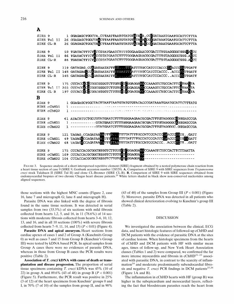
Six tissue sections positive for T. cruzi kDNA from patients8, 9, 10, 11, 12, and 14 were further tested for the presence ofthe nuclear, multicopy SIRE.19 A nested PCR procedure re-sulted in the amplification of a 230-basepair SIRE fragmentfrom two heart samples (cases 9 and 12, Table 2). The am-plicon from case 9 was sequenced and aligned with those fromparasite strains CL-Brenner and Tulahuen II,19 as well as withSIRE sequences amplified from two endomyocardial biopsyspecimens (Figure 3).20
Association of inflammation and fibrosis with T. cruziDNA in heart lesions. The degree of inflammation and fibro-sis in the histologic sections was associated with the detec-tion of T. cruzi kDNA by PCR (Figure 4). Heart sectionswere classified into three categories according to the MNCcounts and the percentage of fibrosis (see Materials andMethods). Accordingly, the PCR result was positive in 1)serial tissue samples derived from only one (15%) of sevenheart sections with low inflammation collected from hearts1−4, 8, and 10 (Table 2), 2) eight (89%) of nine sections withmoderate inflammation (Figure 2, lane 4 and micrograph Dfrom case 7), collected from hearts 2, 4−7, 11, 12, and 16(Table 2), and 3) all 14 (100%) sections with high inflamma-tion (Figure 2, lane 3 and micrograph C; lane 6 and micro-graph F), collected from hearts 3, 7, and 9−16 (Table 2) andFigure 4) (P < 0.0001).
The association of inflammation with detection of T. cruziDNA was also observed when histologic sections with differ-ent degrees of inflammation from the same heart were com-pared. Accordingly, in cases 2 and 3 in group A and cases 10and 16 in group B, parasite DNA was amplified only from
FIGURE 2. Agarose gel electrophoresis (I) and Southern hybrid-ization (II) showing kinetoplast DNA (kDNA)−based polymerasechain reaction (PCR) results obtained by amplification of DNA fromheart tissue samples. Lanes 1−9 of the gel correspond to the micro-graphs A−I. The arrows indicate the 330-basepair kDNA amplicon. −� negative kDNA PCR control; + � positive T. cruzi kDNA PCRcontrol, lane M � 100-basepair molecular weight marker. A−I, Mi-croscopic examination of heart tissue sections corresponding to theones tested by the PCR. A, Case 1a, left ventricle free wall (Masson’strichrome stained, magnification × 200). B, Case 3a, anterolateral leftventricle (hematoxylin and eosin stained, magnification × 200). C,Case 3b, lateral left ventricle, focal myocarditis (hematoxylin andeosin stained, magnification × 400). D, Case 7b, posterolateral leftventricle, active, diffuse myocarditis (hematoxylin and eosin stained,magnification × 400). E, Case 9b, anterolateral left ventricle, activemyocarditis with severe fibrosis (Masson’s trichrome stained, magni-fication × 200). F, Case 14b, lateral left ventricle, intense pericarditis,and diffuse myocarditis (hematoxylin and eosin stained, magnifica-tion × 200). G, Case 16a, left auricular wall (hematoxylin and eosinstained, magnification × 200). H, Case 16b, lateral left ventricular wall(Masson’s trichrome stained, magnification × 200). I, Dilated cardio-myopathy case E, lateral left ventricle wall (hematoxylin and eosinstained, magnification × 400).
PARASITE PERSISTANCE IN CHRONIC CHAGAS HEART DISEASE 215
those sections with the highest MNC counts (Figure 2, case16, lane 7 and micrograph G; lane 8 and micrograph H).
Parasite DNA was also linked with the degree of fibrosisfound in the same tissue sections. It was detected in serialsamples from two (33.3%) of six sections with mild fibrosiscollected from hearts 1,2, 5, and 16, in 11 (78.6%) of 14 sec-tions with moderate fibrosis collected from hearts 3–6, 10, 12,13, and 16, and in all 10 sections (100%) with severe fibrosiscollected from hearts 7−9, 11, 14, and 15) (P < 0.01) (Figure 4).
Parasite DNA and apical aneurysm. Heart sections fromcardiac apexes of cases 1 and 2 of Group A (Kuschnirs Group0) as well as cases 7 and 15 from Group B (Kuschnir’s GroupIII) were tested by kDNA based PCR. In apical samples fromGroup A cases there were no evidences of parasite DNA,whereas in those from Group B cases the PCR results werepositive (Table 2).
Association of T. cruzi kDNA with cause of death or trans-plantation and disease progression. The proportion of serialtissue specimens containing T. cruzi kDNA was 45% (10 of22) in group A and 89.6% (43 of 48) in group B (P < 0.001)(Figure 5). Furthermore, the PCR result was positive in 25%(3 of 12) of the heart specimens from Kuschnir’ groups 0 andI, in 70% (7 of 10) of the samples from group II, and in 90%
(43 of 48) of the samples from Group III (P < 0.001) (Figure5). Moreover, parasite DNA was detected in all patients whoshowed clinical deterioration evolving to Kuschnir’s group III(Table 2).
DISCUSSION
We investigated the association between the clinical, ECGdata, and heart histologic features of followed-up cChHD andDCM patients with the evidence of parasitic DNA at the sitesof cardiac lesions. When histologic specimens from the heartsof cChHD and DCM patients with HF with similar meanages, times of follow-up, and New York Heart Associationclasses (Tables 1 and 2) were compared, we confirmed the farmore intense myocarditis and fibrosis in cChHD21–23 associ-ated with parasitic DNA, in contrast to the scarcity of inflam-mation24 and moderate predominantly subendocardial fibro-sis and negative T. cruzi PCR findings in DCM patients25,26
(Figure 1A and B).The inflammation in cChHD hearts with HF (group B) was
higher in the subepicardium and mesocardial layers, reflect-ing the fact that bloodstream parasites reach the heart from
FIGURE 3. Sequence analysis of a short interspersed repetitive element (SIRE) fragment obtained by a nested polymerase chain reaction froma heart tissue section of case 9 (SIRE 9, Genbank accession number 128529). A, Comparison of SIRE 9 with SIRE sequences from Trypanosomacruzi stock Tulahuen II (SIRE Tul II) and clone CL-Brenner (SIRE CL-B). B, Comparison of SIRE 9 with SIRE sequences obtained fromendomyocardial biopsies of two chronic Chagas heart disease patients.20 White letters shaded in black show non-conserved nucleotides amongaligned sequences.
SCHIJMAN AND OTHERS216

the coronary vessels, contacting the epicardium (Figure 1C).The distribution of fibrosis in the myocardial layers ofcChHD with HF followed the pattern of inflammation, whichwas clearly not the case for DCM. Indeed, heart sections fromcChHD patients showed more fibrosis in the mesocardium,whereas in DCM patients the subendocardium seemed to bemore involved (Figure 1D), probably as a consequence of thedisturbance of oxygen supply and demand.25–27
The appearance of NECs during follow-up is a strikingcharacteristic of cChHD.2 The progression of ECG abnor-malities was reported to be approximately 20−25% in longi-tudinal population studies of individuals chronically infectedwith T. cruzi for seven or more years.28–30 In this study, NECswere detected in 100% of cases in group B, in 30% of casesfrom group A, and in only 17% in group C. Remarkably, ingroup B the ECG evolution was associated with heart failure
and death, with heart tissue necropsies showing diffuse andfibrosing myocarditis, which are typical signs of end-stagecChHD that all linked to the detection of parasite DNA.
The paucity of parasite nests in hearts of cChHD patients ledseveral investigators to discard a role for the parasite in itspathogenesis.6,17,31 In certain series of necropsies, amastigotenests were identified in only 25−36% of the hearts.21,32,33 How-ever, immunohistochemical analysis using monoclonal antibod-ies to T. cruzi allowed detection of T. cruzi antigens in 71% ofthe cardiac regions with moderate or severe myocarditis and inonly 16.6% with mild or absent myocarditis.7 Moreover, a sig-nificant correlation between the presence of parasite antigensand increased CD8 T cell counts has been also reported.34
The PCR has shown the presence of nuclear and kineto-plastid sequences from the parasite in biopsy or necropsyspecimens from hearts and digestive tissues of patients with
FIGURE 4. Association of Trypanosoma cruzi kinetoplast DNA−based polymerase chain reaction (PCR) results with the extent of inflam-mation (top) and the percentage of fibrosis (bottom). n � number of histologic sections tested; MNC/HPF � mononuclear cells per high-powerfield.
PARASITE PERSISTANCE IN CHRONIC CHAGAS HEART DISEASE 217

chronic Chagas disease from endemic regions of Brazil andVenezuela.8–11 In the present study, amastigote nests weredetected in only one heart tissue block (case 14 in group B),whereas the PCR amplified the highly repetitive kinetoplastidminicircle sequence in 15 of 16 heart necropsy specimens(Table 2). For the first time, the middle repetitive nuclearelement known as SIRE19 has been amplified from an eight-year-old heart preparation and sequenced (Figure 3). Thecharacterization of T. cruzi nuclear DNA sequences fromthese archival necropsy specimens, as well as those from heartbiopsy specimens,20 provide a starting point to determine thegenomic structure of the parasite populations associated withthe human tissue lesions.
Our study suggests that there is a strong association betweenthe histologic evidence of heart tissue damage and T. cruzi am-plicons. Indeed, there was a significant correlation between the
number of tissue sections positive by PCR and the intensity ofheart inflammation, as well as fibrosis at the corresponding car-diac lesions (Figure 4). The proportion of PCR-positive sectionsincreased in patients who died or received a heart transplant dueto HF (group B) compared with those from patients who haddied of other causes (group A) (Figure 5). This finding suggeststhat the transition from a chronic asymptomatic period of thedisease, represented by infected individuals with the clinical pro-file of group A patients, to manifest cChHD, the clinical profileof group B patients, is directly associated with the detection ofparasite DNA in the affected tissues. Moreover, amplification ofspecific parasite DNA sequences was linked to clinical deterio-ration (Figure 5).
The absence of intact parasites at the sites of inflammationsuggests that microorganisms invading the myocardium arerapidly destroyed, probably immunologically, whereas DNA
Figure 5. Association of Trypanosoma cruzi kinetoplast DNA−based polymerase chain reaction (PCR) results with cause of death or hearttransplantation (top) and final clinical group (bottom) in chronic Chagas heart disease. n � number of serial tissue samples from the histologicsections tested. 0, I, II, and III � chronic Chagas heart disease groups classified following the criteria of Kuschnir.
SCHIJMAN AND OTHERS218

and antigens may be still detected at the sites of injury. Basedon experimental evidence showing that parasite DNA did notpersist for more than two days in murine-infected tissues,35
PCR findings appear to indicate intact or recently destroyedparasites, rather than mere residual DNA.
The positive correlation between parasite DNA in tissuelesions and inflammation, fibrosis, and disease progressionand severity suggests that during the chronic infection theparasite contacts the heart at different times and locations,invoking an immunologic response at these sites. As a directconsequence, parasites are lysed, the surrounding tissue isdamaged, and replacement fibrosis ensues. The accumulationof these multifocal lesions, mainly in the left ventricular wall,leads to cardiac dysfunction. Moreover, heart damage may bealso enhanced by the humoral immune response of the pa-tient against intracellular parasite antigens, such as the T.cruzi ribosomal P proteins, which generates arrhythmogenicantibodies.36,37
In conclusion, these results suggest not only a relevant roleof the parasite in the pathogenesis and disease progression ofcChHD, but also a need to develop novel anti-parasitic drugs,with sufficient efficacy to clear intracellular parasites, thuspreventing the progression of heart disease.38,39
Received August 1, 2003. Accepted for publication October 22, 2003.
Acknowledgments: We are grateful to Dr. Ruben Laguens (Divisionde Anatomıa Patologica, Instituto de Cardiologıa y Cirugıa Cardio-vascular, Fundacion Favaloro, Buenos Aires, Argentina) and Dr. Fe-lipe Kierszenbaum (Department of Microbiology, Michigan StateUniversity, East Lansing, MI) for their helpful comments on themanuscript, and to Sonia Lafon and Julia Ferrando for their skillfultechnical assistance.
Financial support: This work was supported by grants from the WorldHealth Organization-Tropical Diseases Research projects, the Re-gional Technical Cooperation project RLA 6-0-26 and CoordinatedResearch Project of the International Atomic Energy Agency, Con-sejo Nacional de Ciencia y Tecnologia (PEI 107/97), the University ofBuenos Aires, FONCyT BID 802/OC-AR PICT 01421 and PI CT02030, the Alberto Roemmers and Bunge and Born Foundations, andRamon Carrillo-Arturo Oñativia of the Secretary of Health of Ar-gentina. The work of Matiano J. Levin was partially supported by anInternational Research Scholar grant from the Howard HughesMedical Institute (Chevy Chase, MD).
Authors’ addresses: Alejandro G. Schijman, Juan M. Burgos, SilviaBrandariz, and Mariano J. Levin, Vuelta de Obligado 2490, SecondFloor, (CP 1428), Buenos Aires, Argentina, Telephone: 54-11-4783-2871, Fax: 54-11-4786-8578, E-mail: [email protected]. Carlos A.Vigliano, Division Patologıa. Instituto de Cardiologıa y Cirugıa Car-diovascular, Fundacion Favaloro, Belgrano 1746 (CP 1093), BuenosAires, Argentina, Telephone and fax : 54-11-4378-1315 and Depar-tamento de Cardiologıa, Hospital Eva Peron, Ruta 8 y Diego Pombo,San Martın, Provincia de Buenos Aires, Buenos Aires, Argentina005411-3781315. Rodofo J. Viotti, Bruno E. Lococo, Marıa I. Leze,and Hector A. Armenti, Departamento de Cardiologıa, Hospital EvaPeron, Ruta 8 y Diego Pombo, San Martın, Provincia de BuenosAires, Buenos Aires, Argentina 005411-3781315.
REFERENCES
1. Moncayo A, 1999. Progress towards the interruption of transmis-sion of Chagas disease in the southern countries. Medicina (BAires) 59 (suppl 2): 120–124.
2. Rosenbaum MB, 1964. Chagasic myocardiomyopathy. Prog Car-diovasc Dis 7: 199–225.
3. Storino R, Milei J, 1994. Evolucion natural y estudios longitudi-nales. Storino R, Milei J, eds. Enfermedad de Chagas. BuenosAires: Mosby Doyma Argentina, 593–604.
4. Brener Z, 1987. Pathogenesis and immunopathology of chronicChagas’ disease. Mem Inst Oswaldo Cruz 82 (suppl): 215–219.
5. Tarleton RL, Zhang L, 1999. Chagas disease etiology: autoim-munity or parasite persistence? Parasitol Today 15: 94–99.
6. Levin MJ, 1996. In chronic Chagas heart disease, don’t forget theparasite. Parasitol Today 12: 415–416.
7. Bellotti G, Bocchi EA, de Moraes AV, Higuchi ML, Barbero-Marcial M, Sosa E, Esteves-Filho A, Kalil R, Weiss R, JateneA, Pileggi F, 1996. In vivo detection of Trypanosoma cruziantigens in hearts of patients with chronic Chagas’ heart dis-ease. Am Heart J 131: 301–307.
8. Jones EM, Colley DG, Tostes S, Reis Lopes E, Vnencak-JonesCL, McCurley TL, 1993. Amplification of a Trypanosomacruzi DNA sequence from inflammatory lesions in human cha-gasic cardiomyopathy. Am J Trop Med Hyg 48: 348–357.
9. Vago AR, Andrade LO, Leite AA, d’Avila Reis D, Macedo AM,Adad SJ, Tostes S Jr, Moreira MC, Filho GB, Pena SD, 2000.Genetic characterization of Trypanosoma cruzi directly fromtissues of patients with chronic Chagas disease: differentialdistribution of genetic types into diverse organs. Am J Pathol156: 1805–1809.
10. Olivares-Villagomez D, McCurley TL, Vnencak-Jones CL, Cor-rea-Oliveira R, Colley DG, Carter CE, 1998. Polymerase chainreaction amplification of three different Trypanosoma cruziDNA sequences from human chagasic cardiac tissue. Am JTrop Med Hyg 59: 563–570.
11. Anez N, Carrasco H, Parada H, Crisante G, Rojas A, FuenmayorC, Gonzalez N, Percoco G, Borges R, Guevara P, Ramirez JL,1999. Myocardial parasite persistence in chronic chagasic pa-tients. Am J Trop Med Hyg 60: 726–732.
12. Kuschnir E, Sgammini H, Castro R, Evequoz C, Ledesma R,Brunetto J, 1985. Valoracion de la funcion cardiaca por an-giografıa radioisotopica en pacientes con cardiopatıa chagasicacronica. Arq Bras Cardiol 45: 249–256.
13. Edwards WD, Tajik AJ, Seward JB, 1981. Standardized nomen-clature and anatomic basis for regional tomographic analysis ofthe heart. Mayo Clin Proc 56: 479–497.
14. Aretz HT, Billingham ME, Edwards WD, Factor SM, Fallon JT,Fenoglio JJ Jr, Olsen EG, Schoen FJ, 1987. Myocarditis. Ahistopathologic definition and classification. Am J CardiovascPathol 1: 3–14.
15. Kunkel B, Llapp H, Kober G, Kaltenbach M, 1978. Light micro-scopic evaluation of myocardial biopsies. Kaltenbach M, Loo-gen F, Olsen, EGJ, eds. Cardiomyopathy and Myocardial Bi-opsy. Berlin: Springer Verlag, 62–70.
16. Schijman AG, Vigliano C, Burgos JM, Favaloro R, Perrone S,Laguens R, Levin M, 2000. Early diagnosis of recurrence ofTrypanosoma cruzi infection by polymerase chain reaction af-ter heart transplantation of a chronic Chagas’ heart diseasepatient. J Heart Lung Transplant 19: 1114–1117.
17. Brandariz S.; Schijman A.; Vigliano C.; Arteman P., Viotti R.,Armenti H.; Beldjord C, Levin MJ, 1995. Detection of Trypa-nosoma cruzi DNA from archival heart tissue of a chronicChagas’ disease reference case. Lancet 346: 1370–1371.
18. Schijman AG, Altcheh J, Burgos JM, Biancardi M, Bisio M,Levin M, Freilij H, 2003. Aetiological treatment of congenitalChagas’ disease diagnosed and monitored by the polymerasechain reaction. J Antimicrob Chemother 52: 441–449.
19. Vazquez M, Lorenzi H, Schijman AG, Ben-Dov C, Levin MJ,1999. Analysis of the distribution of SIRE in the nuclear ge-nome of Trypanosoma cruzi. Gene 239: 207–216.
20. Elias F, Vigliano C, Laguens R, Levin MJ, Berek C, 2003. Analy-sis of the presence of Trypanosoma cruzi in the heart tissue ofthree patients with chronic Chagas’ heart disease. Am J TropMed Hyg 68: 242–247.
21. Andrade ZA, Andrade SG, 1999. Patologia. Brener Z, AndradeZA, eds. Trypanosoma cruzi e Doenca de Chagas. Rio de Ja-neiro: Guanabara Koogan, 199–248.
22. Machado CR, Camargos ER, Guerra LB, Moreira MC, 2000.Cardiac autonomic denervation in congestive heart failure:comparison of Chagas’ heart disease with other dilated cardio-myopathy. Hum Pathol 31: 3–10.
23. Higuchi ML, Fukasawa S, De Brito T, Parzianello LC, Bellotti G,Ramirez JAF, 1999. Different microcirculatory and interstitialmatrix patterns in idiopathic dilated cardiomyopathy and Cha-
PARASITE PERSISTANCE IN CHRONIC CHAGAS HEART DISEASE 219

gas’ disease: a three dimensional confocal microscopy study.Heart 82: 279–285.
24. Kühl U, Noutsias M, Seeberg B, Schultheiss H-P, 1996. Immu-nohistological evidence for a chronic intramyocardial inflam-matory process in dilated cardiomyopathy. Heart 75: 295–300.
25. Unverferth DV, Baker PB, Swift SE, Chaffee R, Fetters JK,Uretsky BF, Thompson ME, Leier CV, 1986. Extent of myo-cardial fibrosis and cellular hypertrophy in dilated cardiomy-opathy. Am J Cardiol 57: 816–820.
26. Beltrami CA, Finato N, Rocco M, Feruglio GA, Puricelli C,Cigola E, Sonnenblick EH, Olivetti G, Anversa P, 1995. Thecellular basis of dilated cardiomyopathy in humans. J Mol CellCardiol 27: 291–305.
27. Raso P, Tafuri WL, 1971. Alteracoes do pericardio na fasecronica da tripanossomıase humana e nas fases aguda e cronicada molestia experimental. Rev Soc Bras Med Trop Sao Paulo5: 135–153.
28. Maguire JH, Hoff R, Sherlock I, Guimaraes AC, Sleigh AC,Ramos NB, Mott KE, Weller TH, 1987. Cardiac morbidity andmortality due to Chagas’ disease: prospective electrocardio-graphic study of a Brazilian community. Circulation 75: 1140–1145.
29. Laranja F, Dias E, Nobrega G, Miranda A, 1956. Chagas’ disease:a clinical epidemiologic and pathologic study. Circulation 14:1035–1060.
30. Andrade ZA, 1985. Bases morfologicas das arritmias na miocard-ite chagasica. Cancado JR, Chuster M, eds. Cardiopatia Cha-gasica. Belo Horizonte, Brazil: Fundacao Carlos Chagas 79–90.
31. Kierszenbaum F, 1999. Chagas’ disease and the autoimmunityhypothesis. Clin Microbiol Rev 12: 210–233.
32. Köberle F, 1958. Cardiopatia chagasica. O Hosp 53: 311–346.33. Mott KE, Hagstrom JWC, 1965. The pathologic lesions of the
cardiac autonomic nervous system in chronic Chagas’ myocar-ditis. Circulation 31: 273–286.
34. Higuchi ML, Reis MM, Aiello VD, Benvenuti LA, Gutierrez PS,Bellotti G, Pileggi F, 1997. Association of an increase in CD8+T cells with the presence of Trypanosoma cruzi antigens inchronic, human, chagasic myocarditis. Am J Trop Med Hyg 56:485–489.
35. Zhang L, Tarleton RL, 1999. Parasite persistence correlates withdisease severity and localization in chronic Chagas’ disease.J Infect Dis 180: 480–486.
36. Masuda MO, Levin M, de Oliveira SF, dos Santos Costa PC,Bergami PL, dos Santos Almeida NA, Pedrosa RC, Ferrari I,Hoebeke J, Campos de Carvalho AC. 1998. Functionally activecardiac antibodies in chronic Chagas’ disease are specificallyblocked by Trypanosoma cruzi antigens. FASEB J 12: 1551–1558.
37. Chiale PA, Ferrari I, Mahler E, Vallazza MA, Elizari MV,Rosenbaum MB, Levin MJ, 2001. Differential profile and bio-chemical effects of antiautonomic membrane receptro anti-bodies in ventricular arrhythmias and sinus node dysfunction.Circulation 103: 1765–1771.
38. Viotti R, Vigliano C, Armenti H, Segura E, 1994. Treatment ofchronic Chagas’ disease with benznidazole: clinical and sero-logic evolution of patients with long-term follow-up. Am HeartJ 127: 151–162.
39. Urbina JA, 2001. Specific treatment of Chagas disease: currentstatus and new developments. Curr Opin Infect Dis 14: 733–741.
SCHIJMAN AND OTHERS220
Copyright © 2022 FDOKUMEN