3-Hydroxy Kynurenine Treatment Controls T. cruzi Replication and the Inflammatory Pathology...
13
3-Hydroxy Kynurenine Treatment Controls T. cruzi Replication and the Inflammatory Pathology Preventing the Clinical Symptoms of Chronic Chagas Disease Carolina P. Knubel 1 , Fernando F. Martı´nez 1 , Eva V. Acosta Rodrı ´guez 1 , Andre ´ s Altamirano 1 , He ´ ctor W. Rivarola 2 , Cintia Diaz Luja ´n 3 , Ricardo E. Fretes 3 , Laura Cervi 1 , Claudia C. Motra ´n 1 * 1 Departamento de Bioquı ´mica Clı ´nica, Facultad de Ciencias Quı ´micas, Centro de Investigaciones en Bioquı ´mica Clı ´nica e Inmunologı ´a (CIBICI-CONICET), Universidad Nacional de Co ´ rdoba, Ciudad Universitaria, Co ´ rdoba, Argentina, 2 Ca ´tedra de Fı ´sica Biome ´dica, Facultad de Ciencias Me ´dicas, Universidad Nacional de Co ´ rdoba, Santa Rosa, Co ´ rdoba, Argentina, 3 Facultad de Medicina, Instituto de Biologı ´a Celular, Universidad Nacional de Co ´ rdoba, Co ´ rdoba, Argentina Abstract Background: 3-Hydroxy Kynurenine (3-HK) administration during the acute phase of Trypanosoma. cruzi infection decreases the parasitemia of lethally infected mice and improves their survival. However, due to the fact that the treatment with 3-HK is unable to eradicate the parasite, together with the known proapoptotic and immunoregulatory properties of 3-HK and their downstream catabolites, it is possible that the 3-HK treatment is effective during the acute phase of the infection by controlling the parasite replication, but at the same time suppressed the protective T cell response before pathogen clearance worsening the chronic phase of the infection. Therefore, in the present study, we investigated the effect of 3-HK treatment on the development of chronic Chagas’ disease. Principal Findings: In the present study, we treated mice infected with T. cruzi with 3-HK at day five post infection during 5 consecutive days and investigated the effect of this treatment on the development of chronic Chagas disease. Cardiac functional (electrocardiogram) and histopathological studies were done at 60 dpi. 3-HK treatment markedly reduced the incidence and the severity of the electrocardiogram alterations and the inflammatory infiltrates and fibrosis in heart and skeletal muscle. 3-HK treatment modulated the immune response at the acute phase of the infection impairing the Th1- and Th2-type specific response and inducing TGF-b-secreting cells promoting the emergence of regulatory T cells and long-term specific IFN-c secreting cells. 3-HK in vitro induced regulatory phenotype in T cells from T. cruzi acutely infected mice. Conclusions: Our results show that the early 3-HK treatment was effective in reducing the cardiac lesions as well as altering the pattern of the immune response in experimental Chagas’ disease. Thus, we propose 3-HK as a novel therapeutic treatment able to control both the parasite replication and the inflammatory response. Citation: Knubel CP, Martı ´nez FF, Acosta Rodrı ´guez EV, Altamirano A, Rivarola HW, et al. (2011) 3-Hydroxy Kynurenine Treatment Controls T. cruzi Replication and the Inflammatory Pathology Preventing the Clinical Symptoms of Chronic Chagas Disease. PLoS ONE 6(10): e26550. doi:10.1371/journal.pone.0026550 Editor: Guillermo H. Giambartolomei, National Council of Sciences (CONICET), Argentina Received July 23, 2011; Accepted September 28, 2011; Published October 19, 2011 Copyright: ß 2011 Knubel et al. This is an open-access article distributed under the terms of the Creative Commons Attribution License, which permits unrestricted use, distribution, and reproduction in any medium, provided the original author and source are credited. Funding: This work was supported by grants from Consejo Nacional de Investigaciones Cientı ´ficas y Tecnicas from Argentina (CONICET), Agencia Nacional de Promocio ´ n Cientı ´fica y Te ´cnica (PICT 25511), Ministerio de Ciencia y Tecnologı ´a de la Provincia de Co ´ rdoba and Secretarı ´a de Ciencia y Te ´cnica, Universidad Nacional de Co ´ rdoba (grants to CCM). The work of CPK and FFM was supported by CONICET fellowships (Argentina). The funding bodies had no role in the study design, data collection and analysis, decision to publish, or preparation of the manuscript. Competing Interests: The authors have declared that no competing interests exist. * E-mail: [email protected] Introduction Chagas disease, caused by the obligate intracellular hemofla- gellate protozoan parasite Trypanosoma cruzi, is an endemic disorder affecting 17 million people and remains an important public health problem in Latin America [1]. The acute phase of the disease is characterized by a large parasite replication with the trypomastigotes (Tps) present in the blood of infected people several years after the primary infection, and the infection can be transmitted by infected blood transfusion and organ transplant from donors originating from areas of Latin America where the disease is endemic. This situation has transformed Chagas disease into a worldwide concern that could have severe consequences for human health over the long term [2]. In the mammalian host, the parasite’s biological cycle includes the nondividing, blood-circulating Tps, which infect the nucleated cells and also the replicating intracellular amastigotes (Am) that reside in the cytoplasm of the infected cell [3]. The human pathology is extremely diverse and depends on the parasite biology as well as its relationship with the host [4]. The chronic phase of the infection frequently involves long-lasting inflammatory lesions and immune system disorders, with progressive pathology in the heart, esophagus, or colon. Although chagasic megaesophagus and megacolon produce these typical clinical conditions in 5% to 10% of patients, Chagas cardiomyopathy is by far the most serious form of the disease [5]. Chronic Chagas heart disease is a slowly evolving inflammatory cardiomyopathy that may lead to severe cardiac dilatation, PLoS ONE | www.plosone.org 1 October 2011 | Volume 6 | Issue 10 | e26550
Transcript of 3-Hydroxy Kynurenine Treatment Controls T. cruzi Replication and the Inflammatory Pathology...
3-Hydroxy Kynurenine Treatment Controls T. cruziReplication and the Inflammatory Pathology Preventingthe Clinical Symptoms of Chronic Chagas DiseaseCarolina P. Knubel1, Fernando F. Martınez1, Eva V. Acosta Rodrıguez1, Andres Altamirano1, Hector W.
Rivarola2, Cintia Diaz Lujan3, Ricardo E. Fretes3, Laura Cervi1, Claudia C. Motran1*
1 Departamento de Bioquımica Clınica, Facultad de Ciencias Quımicas, Centro de Investigaciones en Bioquımica Clınica e Inmunologıa (CIBICI-CONICET), Universidad
Nacional de Cordoba, Ciudad Universitaria, Cordoba, Argentina, 2 Catedra de Fısica Biomedica, Facultad de Ciencias Medicas, Universidad Nacional de Cordoba, Santa
Rosa, Cordoba, Argentina, 3 Facultad de Medicina, Instituto de Biologıa Celular, Universidad Nacional de Cordoba, Cordoba, Argentina
Abstract
Background: 3-Hydroxy Kynurenine (3-HK) administration during the acute phase of Trypanosoma. cruzi infection decreasesthe parasitemia of lethally infected mice and improves their survival. However, due to the fact that the treatment with 3-HKis unable to eradicate the parasite, together with the known proapoptotic and immunoregulatory properties of 3-HK andtheir downstream catabolites, it is possible that the 3-HK treatment is effective during the acute phase of the infection bycontrolling the parasite replication, but at the same time suppressed the protective T cell response before pathogenclearance worsening the chronic phase of the infection. Therefore, in the present study, we investigated the effect of 3-HKtreatment on the development of chronic Chagas’ disease.
Principal Findings: In the present study, we treated mice infected with T. cruzi with 3-HK at day five post infection during5 consecutive days and investigated the effect of this treatment on the development of chronic Chagas disease. Cardiacfunctional (electrocardiogram) and histopathological studies were done at 60 dpi. 3-HK treatment markedly reduced theincidence and the severity of the electrocardiogram alterations and the inflammatory infiltrates and fibrosis in heart andskeletal muscle. 3-HK treatment modulated the immune response at the acute phase of the infection impairing the Th1-and Th2-type specific response and inducing TGF-b-secreting cells promoting the emergence of regulatory T cells andlong-term specific IFN-c secreting cells. 3-HK in vitro induced regulatory phenotype in T cells from T. cruzi acutely infectedmice.
Conclusions: Our results show that the early 3-HK treatment was effective in reducing the cardiac lesions as well as alteringthe pattern of the immune response in experimental Chagas’ disease. Thus, we propose 3-HK as a novel therapeutictreatment able to control both the parasite replication and the inflammatory response.
Citation: Knubel CP, Martınez FF, Acosta Rodrıguez EV, Altamirano A, Rivarola HW, et al. (2011) 3-Hydroxy Kynurenine Treatment Controls T. cruzi Replication andthe Inflammatory Pathology Preventing the Clinical Symptoms of Chronic Chagas Disease. PLoS ONE 6(10): e26550. doi:10.1371/journal.pone.0026550
Editor: Guillermo H. Giambartolomei, National Council of Sciences (CONICET), Argentina
Received July 23, 2011; Accepted September 28, 2011; Published October 19, 2011
Copyright: � 2011 Knubel et al. This is an open-access article distributed under the terms of the Creative Commons Attribution License, which permitsunrestricted use, distribution, and reproduction in any medium, provided the original author and source are credited.
Funding: This work was supported by grants from Consejo Nacional de Investigaciones Cientıficas y Tecnicas from Argentina (CONICET), Agencia Nacional dePromocion Cientıfica y Tecnica (PICT 25511), Ministerio de Ciencia y Tecnologıa de la Provincia de Cordoba and Secretarıa de Ciencia y Tecnica, UniversidadNacional de Cordoba (grants to CCM). The work of CPK and FFM was supported by CONICET fellowships (Argentina). The funding bodies had no role in the studydesign, data collection and analysis, decision to publish, or preparation of the manuscript.
Competing Interests: The authors have declared that no competing interests exist.
* E-mail: [email protected]
Introduction
Chagas disease, caused by the obligate intracellular hemofla-
gellate protozoan parasite Trypanosoma cruzi, is an endemic disorder
affecting 17 million people and remains an important public
health problem in Latin America [1]. The acute phase of the
disease is characterized by a large parasite replication with the
trypomastigotes (Tps) present in the blood of infected people
several years after the primary infection, and the infection can be
transmitted by infected blood transfusion and organ transplant
from donors originating from areas of Latin America where the
disease is endemic. This situation has transformed Chagas disease
into a worldwide concern that could have severe consequences for
human health over the long term [2].
In the mammalian host, the parasite’s biological cycle includes
the nondividing, blood-circulating Tps, which infect the nucleated
cells and also the replicating intracellular amastigotes (Am) that
reside in the cytoplasm of the infected cell [3]. The human
pathology is extremely diverse and depends on the parasite biology
as well as its relationship with the host [4]. The chronic phase of
the infection frequently involves long-lasting inflammatory lesions
and immune system disorders, with progressive pathology in the
heart, esophagus, or colon. Although chagasic megaesophagus and
megacolon produce these typical clinical conditions in 5% to 10%
of patients, Chagas cardiomyopathy is by far the most serious form
of the disease [5].
Chronic Chagas heart disease is a slowly evolving inflammatory
cardiomyopathy that may lead to severe cardiac dilatation,
PLoS ONE | www.plosone.org 1 October 2011 | Volume 6 | Issue 10 | e26550

congestive heart failure and death [6]. The most typical functional
heart abnormalities revealed by electrocardiogram (ECG) are
intraventricular conduction disturbances (IVCD), sinus bradycar-
dia and arrhythmia, with histological changes including the
degeneration of the cardiomyocytes coexisting with fibrosis and
mononuclear cell infiltration [7]. The presence of a cardiac
inflammatory infiltrate in the apparent absence of parasites
suggests that an autoimmune component could be involved in
the pathogenesis of the disease [8]. In Chagas disease, the
unresolved infection and the inappropriately balanced inflamma-
tion could be two important factors that contribute to chronic
diseases and to initiate an autoimmune response.
Indoleamine 2,3 dioxigenase (IDO) is an intracellular enzyme
which is constitutively expressed in several human and mouse
cells. Being present in innate immune cells, such as macrophages
and dendritic cells, IDO catalyzes the initial rate-limiting step of
the tryptophan (Trp) catabolism, leading to the production of
L-kynurenine (Kyn), 3-hydroxykynurenine (3-HK), 3-hydroxyan-
thranilic acid (3-HAA) and quinolinic acid among others
(collectively known as ‘‘kynurenines’’) [9]. The IDO gene
promoter contains multiple response elements to proinflamma-
tory mediators, demonstrating the strong correlation between
inflammation and induced IDO expression [10]. Thus, it has
been proposed that IDO activity and ‘‘kynurenines’’ help to tame
exaggerated inflammatory responses through the inhibition of a T
cell proliferation, promotion of T cell anergy or death and the
generation of regulatory T (Treg) cells that drive peripheral
tolerance [11–14]. Moreover, because of its ability to inhibit the
proliferation of facultative intracellular pathogens, it is assumed
that IDO forms part of the innate host defence against infections
[15,16]. We have demonstrated that IDO activity is up-regulated
after T. cruzi infection in mice, with the blocking of IDO activity
in vivo impairing mice resistance to infection and exacerbating the
tissue and blood parasite load and the infection associated
pathology [17]. In addition, in contrast to the observed for others
intracellular pathogens which are sensitive to Trp starvation, we
have previously demonstrated that T. cruzi Am and Tps are
sensitive to the Kyn downstream metabolite 3-HK, and the
therapeutic administration of 3-HK (1 mg/kg/day, intraperito-
neally) during the acute phase of the infection decreased the
parasitemia and improved the survival of lethally infected mice
[17], suggesting that the pharmacologic intervention of IDO
pathway could be used as a novel antitrypanosomatid therapeutic
strategy. Due to the fact we have demonstrated that treatment
with 3-HK is unable to eradicate the parasite during the acute
phase of the infection, together with the known proapoptotic and
immunoregulatory properties of 3-HK and their downstream
catabolites, it is possible that although 3-HK treatment may be
effective during the acute phase of the infection by controlling the
parasite replication, at the same time it suppresses the protective
T cell response before pathogen clearance, thus worsening the
chronic phase of the infection. On the other hand, another
possible effect of 3-HK treatment (highly desirable) could be the
restriction of pathogen growth together with the prompt
activation of immunoregulatory mechanisms able to control the
Chagas disease’s characteristic pathogenic inflammation. In the
present study, mice infected with a non-lethal Tps dose able to
develop the chronic phase of Chagas disease were treated
therapeutically with 3-HK during 5 consecutive days and the
effect of this treatment on the development of chronic Chagas
disease was investigated. Our results show that the early 3-HK
treatment was effective in reducing the cardiac lesions as well as
altering the pattern of the immune response in experimental
Chagas’ disease. Thus, we propose 3-HK as a novel therapeutic
treatment able to control both the parasite replication and the
inflammatory response.
Results
3-HK treatment of acutely T cruzi–infected BALB/c micemarkedly reduces the severity of chronic Chagas disease
To investigate the role of 3-HK treatment in determining the
outcome of chronic Chagas disease, we infected BALB/c mice
with 500 Tps of T. cruzi and 5 days post infection (dpi) the mice
were treated daily with different 3-HK doses or PBS (control) for 5
consecutive days (dpi 5–10). The Tps dose was selected in view of
the fact that almost all 500-Tps-infected mice were able to develop
the acute infection and progress to the chronic phase. Mice
infected with T. cruzi and treated with 1 mg/kg/day of 3-HK (3-
HK mice) showed lower levels of parasitemia than control mice,
but as was observed when a letal T. cruzi dose was used [17], no
sterilizing effect was observed (Figure 1A). At the peak of
parasitemia of control mice (day 16), 3-HK mice presented a
significant reduction in circulating parasites, (4.0760.916106 vs.
7.9562.446106 parasites/ml, p,0.002) (Figure 1A, C). A histo-
logical analysis of hearts and skeletal muscle from 3-HK and
control mice on 16 dpi revealed the typical histopathological
alterations of acute chagasic inflammation, with the presence of
nests of T. cruzi Am and foci of lymphomononuclear inflammatory
infiltrates (not shown). As was described previously [18], we found
more tissue parasitism in skeletal muscle than in cardiac tissue,
with 3-HK mice showing significant between-group differences in
the skeletal muscle parasite nest number (Figure 1B). In
agreement, 3-HK mice showed a significant reduction in tissue
parasitism evaluated by real time PCR to detect T. cruzi DNA
(Figure 1C). On the other hand, the treatment with 3-HK did not
adversely affect the normal histology in uninfected mice (NI) (not
shown). These results demonstrated that 3-HK treatment of mice
infected with T. cruzi was able to control the parasite load in blood
and target tissues.
To evaluate whether higher 3-HK doses could be effective in
eradicating the blood parasites, doses of 5, 10, 50, 100 and
500 mg/kg/day were assayed. As shown in Figure 1D, all
administered doses were able to decrease the parasitemic peak
by 40 to 65% compared to parasite peaks in the control group.
However no significant differences were observed between the
assayed doses. For this, we chose the dose of 1 mg/kg/day for this
study. Also, we observed that the animals did not show any toxic
symptoms during the time of the study, for any of the administered
doses.
Sixty dpi (chronic phase), the cardiac electrophysiology was
studied by ECG, which revealed a significant decrease in the
incidence and the severity of the alterations in 3-HK mice
compared with controls (Table 1). Definite ECG abnormalities
were found in 75% of the control mice, such as arrhythmias and
intraventricular conduction disturbances (IVCD) from mild to
severe and heart block, whereas only 20% of the 3-HK mice
showed some mild IVCD (p,0.01) (Table 1). To evaluate the
efficacy of early 3-HK treatment in preventing the development of
chronic lesions in T. cruzi infected mice, a comparative histological
analysis of the inflammation and fibrosis in cardiac tissue and
skeletal muscle of infected 3-HK treated and non-treated mice at
60 dpi was performed. Figure 2A and B shows that the treatment
with 3-HK led to a reduction in the number of inflammatory foci
containing 15 or more cells in skeletal muscle and cardiac tissue.
Additionally, 3-HK-treated mice showed less epicardial fibrosis
(collagen deposition) and a lack of dystrophic calcification
compared with that in non-treated mice (Figure 2A). Moreover,
3-HK Treatment Prevents Chagasic Cardiopathy
PLoS ONE | www.plosone.org 2 October 2011 | Volume 6 | Issue 10 | e26550

we also confirmed the acute phase findings showing that, even
though not clear the infection, 3-HK treatment is effective in
reducing the levels of parasite load in the target tissues (Figure 2C).
These results indicate that 3-HK treatment plays a critical role
in the parasite clearance and control of the inflammatory
associated pathology by decreasing the parasite load in blood
and tissues and preventing chronic tissue alterations.
3-HK treatment modulates the parasite specific immuneresponse during the acute phase of the infection andpromotes the development of long-term IFN-c secretingcells
In our next set of experiments, we investigated whether the 3-
HK treatment conditions the development of any particular
cellular or humoral immune response able to contribute to the
parasite clearance and/or the control of the inflammatory
associated pathology.
We analysed the T cell compartment and the cytokine response
against parasite antigens at different times pi. No significant
differences in the absolute number of total spleen cells were
observed between 3-HK and control mice (not shown). However,
a significant reduction in the absolute number of CD4+ T cells in
spleen from 3-HK mice compared with control mice was observed
at 30 and 60 dpi, with an increase in the number of CD8+ T cells
being significant at 60 dpi and a significant increase in the CD8+/
CD4+ ratio at 30 and 60 dpi (Figure S1).
Then, we quantify the early cytokine response in plasma and
culture supernatants of spleen mononuclear cells (SMC) cultured
with parasite antigens at the peak of parasitemia (16 dpi). To carry
this out, SMC from non-infected mice (NI) or 16 dpi mice, treated
or not with 3-HK, were cultured with or without an extract of T.
cruzi (F105) [19]. After 72 h, culture supernatants were collected
and analyzed for IFN-c, TNF, IL-1b, IL-12, IL-6, IL-4, IL-5,
TGF-b and IL-10. As shown in Figure 3, SMC from control mice
cultured with F105 produced significantly higher levels of Th1-
type cytokine IFN-c and of Th2-type cytokine IL-5 than SMC
from NI mice. However, no differences between infected and NI
mice were observed for TNF, IL-1b, IL-12, IL-6, IL-4, TGF-b or
IL-10 cytokines (Figure 3A and not shown). In addition, SMC
from 3-HK mice secreted significantly lower levels of INF-c and
IL-5 than SMC from control mice, with the levels of IL-5 being
comparable to those secreted by SMC from NI mice (Figure 3A).
Finally, SMC from 3-HK mice were able to secrete more than
four times the amount of TGF-b produced by SMC from control
Figure 1. 3-HK treatment controls the parasite load in blood and target tissues. Parasitemia, histological analysis, relative amount ofparasite DNA and ED50 were determined in 500-Tps infected mice treated with 1 mg/kg/day of 3-HK (3-HK) or vehicle (control) from dpi 5 to 10 bythe i.p. route. (A) Quantitation of parasitemia (Tps/ml blood). Results are means 6 SD of 6–8 mice/group. (B) Quantitation of the number of nests ofamastigotes at 3 levels of heart and skeletal muscle from control (n = 3) and 3-HK (n = 4) groups at 16 dpi, employing Axio-Vision 3.0.6 software. (C)Relative amount of T. cruzi satellite DNA in the skeletal muscle, heart and spleen from 20-day T. cruzi infected 3-HK and control mice. Murine GAPDHwas used for normalization. Data are shown as mean 6 SD of triplicates, n = 4 mice per group. (D) Parasitemia (Tps/ml blood) at 16 dpi was recordedin 500-Tps infected mice treated with different 3-HK doses (1, 5, 10, 50, 100 and 500 mg/kg/day) or vehicle. The mean value determined for a groupof 6–8 mice was used to calculate the percentage of inhibition in parasitemia respect to the vehicle control group. The percentage of inhibition wascalculated as [(C-3-HK)/C]6100 where C is the parasitemia in the control group and 3-HK is the parasitemia in the treated group. Results are means 6SD of 6–8 mice/group. One representative of two experiments is shown.doi:10.1371/journal.pone.0026550.g001
3-HK Treatment Prevents Chagasic Cardiopathy
PLoS ONE | www.plosone.org 3 October 2011 | Volume 6 | Issue 10 | e26550

Figure 2. 3-HK treatment impairs chronic Chagas’ disease-associated inflammatory pathology. Heart and skeletal muscle sectionsobtained at dpi 60 from 500-Tps-infected mice, treated (3-HK) or not (C) with 3-HK, were stained with H/E and Masson trichrome staining. (A)Representative histological sections of heart (left panels) and skeletal muscle (right panel) from control (upper panels) and 3-HK mice (lower panels).Left and right panels showing focal mononuclear cell infiltrates stained with H/E. Middle panel showing collagen deposition stained with Massontrichrome stain (X100). (B) Quantitation of foci of cellular in flammatory infiltrates at 3 levels of heart and skeletal muscle from control (n = 4) and 3-HK(n = 4) groups, employing Axio-Vision 3.0.6 software. (C) Relative amount of T. cruzi satellite DNA in the skeletal muscle, heart and spleen from 60-dayT. cruzi infected 3-HK and control mice. Murine GAPDH was used for normalization. Data are shown as mean 6 SD of triplicates, n = 4 mice per group.doi:10.1371/journal.pone.0026550.g002
Table 1. Electrocardiographic results in 3-HK treated, Control (infected and untreated) and Normal non-infected (NI) mice at 60days post-infection.
Animal group Heart rate (beats/min) PQ interval (s) QT interval (s) Axis (grade) Mice showing abnormality (%)
3-HK (n = 10) 273.8 (12.8) 0.01–0.04 0.01–0.04 65 (2.2) 20
Control (n = 8) 321.8 (14.3) 0.01–0.02 0.01–0.08 66.2 (2.5) 75*
NI (n = 10) 240 (28.6) 0.02–0.02 0.02–0.06 80 (4.9) 0
Numbers in parentheses are standard errors.Prolonged QT: intraventricular blockade.*p ,0.01 vs 3-HK or NI using ANOVA and Fisher exact test.doi:10.1371/journal.pone.0026550.t001
3-HK Treatment Prevents Chagasic Cardiopathy
PLoS ONE | www.plosone.org 4 October 2011 | Volume 6 | Issue 10 | e26550
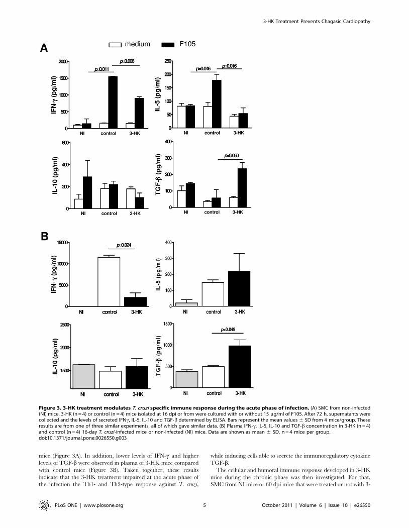
mice (Figure 3A). In addition, lower levels of IFN-c and higher
levels of TGF-b were observed in plasma of 3-HK mice compared
with control mice (Figure 3B). Taken together, these results
indicate that the 3-HK treatment impaired at the acute phase of
the infection the Th1- and Th2-type response against T. cruzi,
while inducing cells able to secrete the immunoregulatory cytokine
TGF-b.
The cellular and humoral immune response developed in 3-HK
mice during the chronic phase was then investigated. For that,
SMC from NI mice or 60 dpi mice that were treated or not with 3-
Figure 3. 3-HK treatment modulates T. cruzi specific immune response during the acute phase of infection. (A) SMC from non-infected(NI) mice, 3-HK (n = 4) or control (n = 4) mice isolated at 16 dpi or from were cultured with or without 15 mg/ml of F105. After 72 h, supernatants werecollected and the levels of secreted IFNc, IL-5, IL-10 and TGF-b determined by ELISA. Bars represent the mean values 6 SD from 4 mice/group. Theseresults are from one of three similar experiments, all of which gave similar data. (B) Plasma IFN-c, IL-5, IL-10 and TGF-b concentration in 3-HK (n = 4)and control (n = 4) 16-day T. cruzi-infected mice or non-infected (NI) mice. Data are shown as mean 6 SD, n = 4 mice per group.doi:10.1371/journal.pone.0026550.g003
3-HK Treatment Prevents Chagasic Cardiopathy
PLoS ONE | www.plosone.org 5 October 2011 | Volume 6 | Issue 10 | e26550

HK were cultured with F105. After 72 h, culture supernatants
were collected and analyzed for Th1-type cytokines (IFN-c and
TNF), Th2-type cytokines (IL4 and IL5) and the regulatory
cytokines IL-10 and TGF-b. SMC from 3-HK mice showed a long
term Th1-type response with secretion of IFN-c but not the Th2-
type cytokines IL-4 or IL-5 (Figure 4A). Moreover, SMC from 3-
Figure 4. 3-HK treatment induces a long-term Th1-type cellular and humoral immune response against T. cruzi. (A) SMC from 3-HK(n = 4) or control (n = 4) mice isolated at 60 dpi or from non-infected (NI) mice (n = 4) were cultured with or without 15 mg/ml of F105. After 72 h,supernatant were collected and the levels of secreted IFNc, TNF, IL-4, IL-5, IL-10 and TGF-b determined by ELISA. Bars represent the mean values 6 SDfrom 4 mice/group. These results are from one of three similar experiments, all of which gave similar data. (B) Plasma IFN-c, TNF, IL-4, IL-5, IL-10 andTGF-b concentration in 3-HK (n = 4) and control (n = 4) 60-day T. cruzi-infected mice or non-infected (NI) mice. Data are shown as mean 6 SD, n = 4mice per group. (C) Sera from 3-HK or control mice at 60 dpi were assayed for detection of IgG1 and IgG2a isotypes of antibodies against F105, R13and H13 by ELISA. Titrations were performed in duplicate, and the end point was expressed as the serum dilution with an optical density twice ashigh as those of the corresponding NI sera.doi:10.1371/journal.pone.0026550.g004
3-HK Treatment Prevents Chagasic Cardiopathy
PLoS ONE | www.plosone.org 6 October 2011 | Volume 6 | Issue 10 | e26550
HK mice showed a tendency to produce higher levels of the
regulatory cytokines IL-10 and TGF-b than control mice
(Figure 4A). In contrast, no differences between control and NI
mice were observed for these cytokines, suggesting that at 60 dpi
the cells able to respond to F105 antigen are absent in control mice
(Figure 4A). In agreement, higher levels of the Th1-type cytokines
IFN-c and TNF and of the regulatory cytokines IL-10 and TGF-bwere detected in sera from 3-HK mice compared with control
mice (Figure 4B). These data show that 3-HK treatment was able
to generate long-term T. cruzi-specific cells capable of secreting
Th1-type and regulatory cytokines.
Then, we evaluated the effect of 3-HK treatment on the specific
and autoreactive antibody response. To test antibodies against the
parasite, F105 and the synthetic peptide R13 were used as antigens
in ELISA assays. The R13 peptide sequence that corresponds to
the C-terminal region of T. cruzi ribosomal P1 and P2 proteins is
almost identical to the mammalian P1/P2 sequence, H13, except
for one non-conservative amino acid substitution of an S by an E
residue [20]. Since the incidence and levels of anti-R13 and anti-
H13 antibodies are associated with the electrophysiological and
histological alterations present in the heart of Chagas disease
patients and T. cruzi infected or immunized mice [21–25], we used
the synthetic peptide H13 to test the autorreactive antibody
response. The quantification of IgG1 and IgG2a antibody isotypes
against T. cruzi derived antigens (F105 and R13) and the
autoantigen H13 revealed that, in agreement with the cytokine
response against the parasite antigens, 3-HK mice showed a 4-fold
increase in the levels of IgG2a and a 2-fold decrease in the levels of
IgG1 antibodies against F105 compared with control mice,
suggesting that 3-HK treatment skews the Th2-type antibody
response (prevalent in BALB/c mice [26,27]), towards a Th1-type
antibody response (Figure 4C). In addition, the antibody response
profile against R13 did not show significant differences between 3-
HK and control mice. Interestingly, 3-HK treated mice showed
more than a 2-fold decrease in the levels of IgG1 antibodies
against the auto antigen H13 than control mice, while no
significant differences were observed in the levels of IgG2a
antibodies.
Summing up, these results suggest that the ability of 3-HK
treatment to induce a healing response in T. cruzi-infected Balb/c
mice was associated not only with a reduction in parasite numbers
but also with a switch in the dominant Th2-type immune response
observed in these animals.
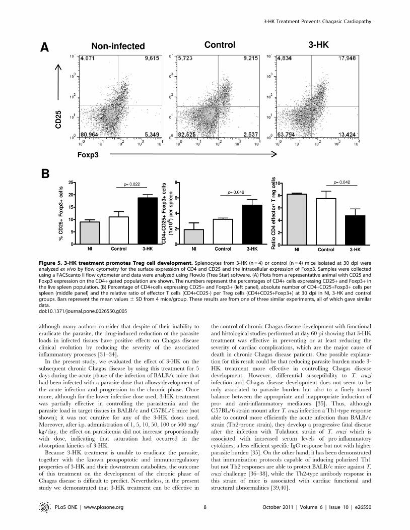
3-HK treatment promotes Treg cell developmentRegulatory T cells (Treg) are important players in maintaining
immune homeostasis and controlling excessive immune responses
[28] which have been implicated in the suppression of Th1-
mediated pathology or Th2 responses in a variety of settings [29].
Natural Treg cells that emerge from the thymus with self-
specificity limit activation and expansion of autoreactive T cells in
the periphery, whereas inducible Treg cells derive from conven-
tional T cells that are antigen stimulated in the presence of
mediators such as TGF-b and IL-10 [30]. The fact that SMC from
acutely infected-3-HK mice produced little or no IFN-c or IL-5
(Th1- and Th2-type cytokines respectively) but generated
substantial amounts of TGF-b, together with increasing evidence
of the involvement of IDO+ DC, IDO pathway catabolites and
TGF-b in the induction of Treg cells, prompted us to investigate if
3-HK administration could result in Treg induction. We
compared by flow cytometry the presence of CD4+CD25+Foxp3+cells in spleen from 3-HK and control mice at different times pi
(15, 30 and 60 dpi), and found that, similar to that observed in NI,
,10% of CD4+ cells of the spleen from 30 day infected control
mice were CD25+ Foxp3+ (Figure 5A, B), whereas after 3-HK
treatment, the percentage of CD4+ cells in the spleen that were
CD25+ Foxp3+ increased to , 18%. This reflects a considerable
expansion of the Treg population, because the absolute number of
spleen cells was similar in both groups of mice (not shown).
Accordingly, the ratio of effector CD4 T cells (CD4+CD25-Foxp3-
cells) per regulatory T cells in the 3-HK group was 4.7 to 1
compared to 7.5 to 1 for control group (Figure 5B). We also
detected the expansion of CD4+CD25+Foxp3+ at 15 dpi in one
out of three experiments performed with four mice per group.
Furthermore, we were unable to detect significant expansion of
either CD4+CD25+ Foxp3+ cells at 60 dpi or CD4+CD25+CTLA-4+ or GITR+ cells at 15, 30 or 60 dpi (not shown).
3-HK induces Treg phenotype in T cells from T. cruziacutely infected mice
To investigate whether 3-HK was able to induce in vitro the Treg
phenotype, we decided to mimic in vitro the 3-HK administration
in vivo in T. cruzi infected mice. To carry this out, SMC from 7 day-
infected mice (a time point where the IDO activity has been
induced by the infection [17]) were stimulated with F105 or anti-
CD3 (to mimic the specific or polyclonal stimulation induced
during the infection respectively) in the presence or absence of
20 mM of 3-HK for 1 week. In addition, we compared the effects
of 3-HK on SMC from NI mice. The FACS analysis revealed that
SMC from T. cruzi-infected mice, but not from NI mice, expanded
the CD4+CD25+Foxp3+ cells when they were stimulated with T.
cruzi antigens or anti-CD3 in the presence of 3-HK (Figure 6A). In
addition, as we observed at 16 dpi during in vivo 3-HK treatment,
SMC from infected mice produced significantly lower levels of the
Th1-type cytokines IFN-c and TNF and also of the Th2-type
cytokine IL-4 when they were stimulated with parasite specific
antigen (F105) or polyclonally (anti-CD3) in the presence of 3-HK
(Figure 6B). Moreover, 3-HK was able to suppress the antigen
specific but not anti-CD3-induced secretion of IL-10 of SMC from
infected mice. In addition, 3-HK was able to increase the secretion
of TGF-b in cultures stimulated with anti-CD3 (Figure 6B).
Finally, 3-HK was able to slightly suppress the secretion of IFN-cof SMC from normal mice stimulated with anti-CD3 (Figure 6B).
Discussion
Chagas disease is a tropical parasitic disease that is currently on
the increase interest in non-endemic geographical areas such as
the United States and Europe, mainly due to the population
movement of infected people [2]. The fact that the parasite’s
biological cycle in mammals includes the blood-circulating Tps
and the replicating intracellular Am presents an extra challenge for
those trying to develop new drugs for this disease because anti-T.
cruzi drugs need to be able to cross the plasma membrane in order
to be effective, in contrast with other trypanosome parasites, such
as Trypanosoma brucei, which remain in the blood and so are
potentially more accessible to drugs.
At present, Chagas’ disease desperately needs more treatment
options. Benznidazole and Nifurtimox, the only existing current
options, were introduced more than forty years ago and are not
ideal, given that their side effects can be severe and can lead to
resistance. Recently, we reported that 3-HK treatment of BALB/c
mice lethally infected with T. cruzi is able to control the
parasitemia and to improve the survival, with this effect being
associated to the direct toxic effect of this compound on T. cruzi
Am and Tps [17]. However, under the experimental conditions
employed in those experiments, 3-HK treatment was only partially
efficient in controlling the parasitemia and was not curative [17],
3-HK Treatment Prevents Chagasic Cardiopathy
PLoS ONE | www.plosone.org 7 October 2011 | Volume 6 | Issue 10 | e26550
although many authors consider that despite of their inability to
eradicate the parasite, the drug-induced reduction of the parasite
loads in infected tissues have positive effects on Chagas disease
clinical evolution by reducing the severity of the associated
inflammatory processes [31–34].
In the present study, we evaluated the effect of 3-HK on the
subsequent chronic Chagas disease by using this treatment for 5
days during the acute phase of the infection of BALB/c mice that
had been infected with a parasite dose that allows development of
the acute infection and progression to the chronic phase. Once
more, although for the lower infective dose used, 3-HK treatment
was partially effective in controlling the parasitemia and the
parasite load in target tissues in BALB/c and C57BL/6 mice (not
shown); it was not curative for any of the 3-HK doses used.
Moreover, after i.p. administration of 1, 5, 10, 50, 100 or 500 mg/
kg/day, the effect on parasitemia did not increase proportionally
with dose, indicating that saturation had occurred in the
absorption kinetics of 3-HK.
Because 3-HK treatment is unable to eradicate the parasite,
together with the known proapoptotic and immunoregulatory
properties of 3-HK and their downstream catabolites, the outcome
of this treatment on the development of the chronic phase of
Chagas disease is difficult to predict. Nevertheless, in the present
study we demonstrated that 3-HK treatment can be effective in
the control of chronic Chagas disease development with functional
and histological studies performed at day 60 pi showing that 3-HK
treatment was effective in preventing or at least reducing the
severity of cardiac complications, which are the major cause of
death in chronic Chagas disease patients. One possible explana-
tion for this result could be that reducing parasite burden made 3-
HK treatment more effective in controlling Chagas disease
development. However, differential susceptibility to T. cruzi
infection and Chagas disease development does not seem to be
only associated to parasite burden but also to a finely tuned
balance between the appropriate and inappropriate induction of
pro- and anti-inflammatory mediators [35]. Thus, although
C57BL/6 strain mount after T. cruzi infection a Th1-type response
able to control more efficiently the acute infection than BALB/c
strain (Th2-prone strain), they develop a progressive fatal disease
after the infection with Tulahuen strain of T. cruzi which is
associated with increased serum levels of pro-inflammatory
cytokines, a less efficient specific IgG response but not with higher
parasite burden [35]. On the other hand, it has been demonstrated
that immunization protocols capable of inducing polarized Th1
but not Th2 responses are able to protect BALB/c mice against T.
cruzi challenge [36–38], while the Th2-type antibody response in
this strain of mice is associated with cardiac functional and
structural abnormalities [39,40].
Figure 5. 3-HK treatment promotes Treg cell development. Splenocytes from 3-HK (n = 4) or control (n = 4) mice isolated at 30 dpi wereanalyzed ex vivo by flow cytometry for the surface expression of CD4 and CD25 and the intracellular expression of Foxp3. Samples were collectedusing a FACScanto II flow cytometer and data were analyzed using FlowJo (Tree Star) software. (A) Plots from a representative animal with CD25 andFoxp3 expression on the CD4+ gated population are shown. The numbers represent the percentages of CD4+ cells expressing CD25+ and Foxp3+ inthe live spleen population. (B) Percentage of CD4+cells expressing CD25+ and Foxp3+ (left panel), absolute number of CD4+CD25+Foxp3+ cells perspleen (middle panel) and the relative ratio of effector T cells (CD4+CD25-) per Treg cells (CD4+CD25+Foxp3+) at 30 dpi in NI, 3-HK and controlgroups. Bars represent the mean values 6 SD from 4 mice/group. These results are from one of three similar experiments, all of which gave similardata.doi:10.1371/journal.pone.0026550.g005
3-HK Treatment Prevents Chagasic Cardiopathy
PLoS ONE | www.plosone.org 8 October 2011 | Volume 6 | Issue 10 | e26550
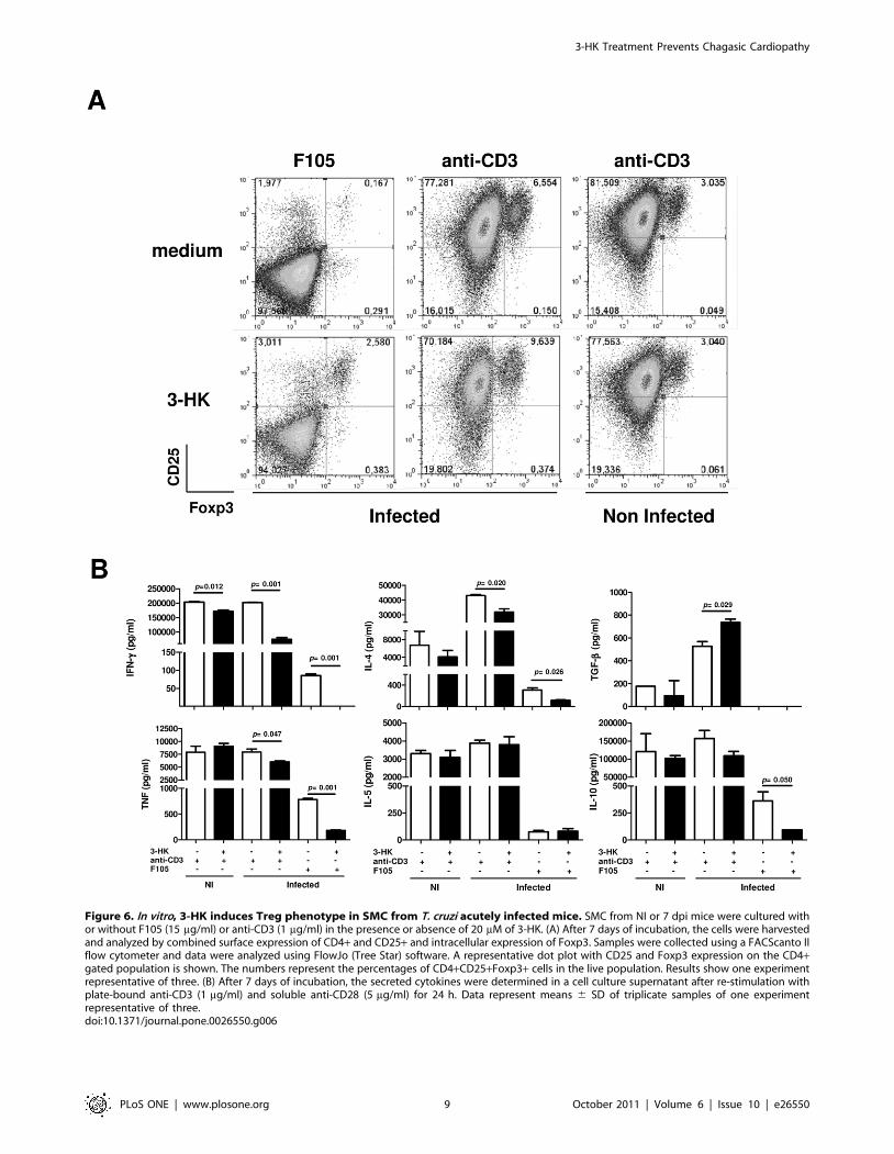
Figure 6. In vitro, 3-HK induces Treg phenotype in SMC from T. cruzi acutely infected mice. SMC from NI or 7 dpi mice were cultured withor without F105 (15 mg/ml) or anti-CD3 (1 mg/ml) in the presence or absence of 20 mM of 3-HK. (A) After 7 days of incubation, the cells were harvestedand analyzed by combined surface expression of CD4+ and CD25+ and intracellular expression of Foxp3. Samples were collected using a FACScanto IIflow cytometer and data were analyzed using FlowJo (Tree Star) software. A representative dot plot with CD25 and Foxp3 expression on the CD4+gated population is shown. The numbers represent the percentages of CD4+CD25+Foxp3+ cells in the live population. Results show one experimentrepresentative of three. (B) After 7 days of incubation, the secreted cytokines were determined in a cell culture supernatant after re-stimulation withplate-bound anti-CD3 (1 mg/ml) and soluble anti-CD28 (5 mg/ml) for 24 h. Data represent means 6 SD of triplicate samples of one experimentrepresentative of three.doi:10.1371/journal.pone.0026550.g006
3-HK Treatment Prevents Chagasic Cardiopathy
PLoS ONE | www.plosone.org 9 October 2011 | Volume 6 | Issue 10 | e26550
When the effect of 3-HK treatment on the development of any
particular cellular or humoral immune response able to contribute
to the parasite clearance and/or the control of the inflammatory
associated pathology was studied, 3-HK treatment impaired, during
the acute phase of the infection, the parasite specific Th2-type
immune response promoting the development of TGF-b secreting
cells. These results are in agreement with those demonstrating that
the Th2-type antibody response in this strain of mice is associated
with cardiac functional and structural abnormalities [39,40].
Certainly, 3-HK mice showed a shift from Th2-type to the Th1-
type humoral response against T. cruzi antigens and autoantigens in
concordance with a lower incidence and severity of the electro-
physiological abnormalities found in these mice [21–25].
In addition, 3-HK mice showed, at the chronic phase of the
infection, an increased CD8+/CD4+ cell ratio and a population of
T. cruzi-specific cells capable of secreting Th1-type cytokines. The
development of a stable, T. cruzi–specific, CD8+ T cell population
with the characteristics of memory cells showing effector function
and providing protective immunity upon rechallenge has been
reported after Beznidazole-induced cure or after heterologous
plasmid DNA prime- human adenovirus boost vaccination of
C57BL/6 mice [41,42]. Though, additional studies must be
performed in order to well characterize the Th1-type cytokines
producing cell population that emerge after 3-HK treatment. On
the other hand, SMC from chronically infected 3-HK mice also
showed a tendency to produce higher levels of the regulatory
cytokines IL-10 and TGF-b than control mice when stimulated
with parasite antigens, with similar cytokine profiles observed in
plasma from chronically infected 3-HK mice, suggesting that the
emerging Th1-type response could be controlled by these two
regulatory cytokines in 3-HK mice.
Fallarino et al. [43] have reported that when CD4+ T cells are
stimulated in vitro for 7 days in low Trp medium in the presence of
Trp catabolites (or IDO+ DC) they produce little or no IFN-c or IL-
4. However, they produce TGF-b which is necessary for the
induction of the Treg phenotype in T cells. Both these conditions,
Trp catabolism and Trp catabolites, are present when cultures of
SMC from T. cruzi infected mice (but not from NI mice) are in vitro
activated in the presence of 3-HK (because of the known infection-
induced up-regulated IDO activity [17]). These conditions are also
present when 3-HK is administered in the acute phase of in vivo
infection. In agreement, 3-HK treatment was able to promote TGF-
b producing cells and Treg development which was observed in the
spleen at day 30 pi and in vitro by stimulating SMC from T. cruzi
infected mice. Several studies have painted a picture of TGF-b as
being a largely immunosuppressive cytokine able to act on naive T
cells by suppressing the proliferation [44], inhibiting Th1 cell
polarization [45] and promoting the generation of inducible Treg
cells [46,47]. However it has been recently described that TGF-benhances effector Th1 cell activation and promote its self regulation
via IL-10 [48]. In agreement, Guinazu et al. [27] demonstrated an
association between high levels of IFN-c accompanied with high
levels of TGF-b and the absence of an autoimmune response in B6
mice (Th1-prone) immunized with a T. cruzi antigen. Thus,
although it has been shown that TGF-b production during the
acute phase of T. cruzi infection contributes to the intracellular
parasite replication [49,50], it is possible that because 3-HK directly
controls the intracellular parasite replication, the early production of
this immunomodulatory cytokine could be responsible for the Treg
development and the Th1 and Th2 response suppression
contributing to the early control of the inflammatory response.
The role of Treg cells during T. cruzi infection is complex, with
divergent effects observed depending on the strain of mice
(C57BL/6 vs BALB/c) and parasites. Thus, the relative amount
of Treg not change significantly during the course of T. cruzi
infection of C57BL/6 mice, with the depletion of these cells not
appearing to play a major role in regulating effector responses
during the acute or chronic phase of the infection [51,52]. On the
contrary, Mariano, et al. have reported the role of natural Treg
cells in controlling the parasitism as well as the cardiac
inflammation and regulatory cytokines production in BALB/c
mice infected with Y strain of T. cruzi [53]. Therefore, these
findings are in agreement with our study in which the 3-HK
induced development of Treg was associated with the control of
the inflammatory associated pathology in BALB/c mice.
Recently, it was demonstrated that the Aryl hydrocarbon
receptor (Ahr), a ligand-activated transcription factor that
mediates dioxin toxicity, is required to induce IDO in DC [54].
Moreover, Kyn, and to a lesser extent 3-HK, can activate this
receptor present on naıve T cells with this activation leading to
Ahr-dependent Treg generation, which has been demonstrated to
be potentiated by TGF-b through Ahr up-regulation [55]. It is also
plausible that the early TGF-b production could lead to Ahr up-
regulation in DC and T cells, which might potentiate the effect of
3-HK (and other Trp catabolites produced during T. cruzi
infection) on Treg and IDO induction. Experiments to investigate
whether T. cruzi infection is able to modulate the Ahr expression
on DC and T cells, and the role of Ahr expression on T. cruzi-
induced IDO up-regulation and Treg generation are underway.
Taken together, the results presented here indicate that 3-HK
treatment for T. cruzi acute infection ameliorates the clinical
outcome of chronic Chagas disease through the control of the
parasite replication and also by inducing immunomodulatory
effects, thus creating a balance between the control and clearance
of the infectious organism on the one hand and the prevention of
the immune-mediated pathology on the other hand. Therefore,
the successful treatment of T. cruzi infection may not require the
perhaps unachievable goal of sterilizing treatment, but it might be
equally important to provide an efficient control of the parasite
burden and the immune response in order to avoid progression
from T. cruzi infection to Chagas disease.
Finally, it is important to highlight that a putative T. cruzi
kynureninase enzyme has been identified [56], which could be
involved in avoiding the parasite own destruction by the host 3-
HK, considering that as today there is no available knowledge on
an alternative pathway for Trp catabolism or the existence of the
Kyn pathway in this parasite. These facts suggest that the presence
of this enzyme is one of the mechanisms by which T. cruzi subverts
the host’s immune systems and therefore make the use of this
compound highly attractive in combined therapies for the purpose
of achieving a parasitological cure.
Materials and Methods
Mice and parasitesFemale BALB/c mice, 6–8 wk old, were obtained from
Comision Nacional de Energıa Atomica (CNEA; Buenos Aires,
Argentina) and maintained according to the National Research
Council’s guide for the care and use of laboratory animals. The
Tulahuen strain of T. cruzi was used, which was maintained by
weekly intraperitoneal (ip) inoculations in mice. The studies were
approved by the Institutional Animal care and Use Committee of
the Faculty of Chemical Sciences, National University of Cordoba
(Approval ID Res 459/09).
3-HK treatment and parasite loadMice were infected with 500 Tps from T. cruzi and treated with
3-HK as described previously [57]. 3-HK was resuspended in 0.1
3-HK Treatment Prevents Chagasic Cardiopathy
PLoS ONE | www.plosone.org 10 October 2011 | Volume 6 | Issue 10 | e26550
M PBS, with this vehicle also being employed as control. The
levels of parasitemia were monitored in blood collected at different
times post infection (pi) as previously described [17]. Five days post
infection (dpi), mice were daily treated with 3-hydroxy-dl-
kynurenine (3-HK) from Sigma-Aldrich (St. Louis, MO, USA) at
1, 5, 10, 50, 100 or 500 mg/kg/d for 5 consecutive days (dpi: 5–
10) by the i.p. route. 3-HK was resuspended in 0.1 M PBS, with
this vehicle also being employed as control. The levels of
parasitemia were monitored in blood collected at different times
post infection (pi) from the tails. For that, erythrocytes were lysed
in a 0.87% ammonium chloride buffer, and viable Tps were
counted in a Neubauer chamber. For determination of tissue
parasitism, genomic DNA was purified from spleen, skeletal
muscle and heart from 20 days infected mice using TRIzol reagent
and following manufacturers instruction. Satellite DNA from T.
cruzi (GenBank AY520036) was quantified by real time PCR using
specific Custom Taqman Gene Expression Assay (Applied
Biosystem) using the primer and probe sequences described by
Piron et al [58]. A sample containing 200 ng of genomic DNA was
amplified and considered positive for T. cruzi when the threshold
cycle (CT) for the T. cruzi target was ,45. Abundance of satellite
DNA from T. cruzi was normalized to GAPDH abundance.
(Taqman Rodent GAPDH Control Reagent, Applied Biosystem)
and expressed as arbitrary units.
Electrocardiographic studiesDouble-blind electrocardiograms (ECG) were performed at 60
days pi with a conventional ECG recorder (Fukuda) run at
50 mm/s [59,60]. The peripheral leads DI, DII, DIII, aVR, aVF,
and aVL were utilized. As controls for the ECG studies, normal
non-infected mice were used. The ECG registers were read taking
into account the electrocardiographic pattern in normal and in T.
cruzi infected mice previously described [61,62]. All alterations
suggestive of bundle branch blocks were recorded under the name
of intraventricular blockade as suggested by Sadigursky & Andrade
[61]. We also used their criteria to characterize the intraventric-
ular conduction abnormalities and auricle–ventricle blocks.
Histological studiesThe heart and the skeletal muscle from the quadriceps were
removed at different times pi, fixed in buffered 10% formalin
(pH 7.0), and embedded in paraffin. Organs were cut with a
microtome to obtain 5-mm slices, collecting 1 section every 100 mm
of the tissue until 300 mm of each organ was sectioned, thus
obtaining 3 of each organ belonging to 3 levels, which were then
stained with hema-toxilyn/eosin (H/E). At least 25 fields from
each section were checked for parasites and histopathology under
an X40 objective of a photonic microscope (Axioskop; Carl Zeiss
Vision, Oberkochen, Germany) in a blind study. Twelve random
digital images per organ were taken at the 3 levels (4/level) with a
Hitachi color camera (Hitachi, Tokyo, Japan) being employed to
measure the number of amastigotes, the number of nests of the
amastigotes, and the number of inflammatory infiltrates. Mea-
surements were performed by the quantitative image analysis
system Axiovision 3.0.6 (Carl Zeiss) as described previously [17].
In addition, 5-mm sections of tissues were prepared for Masson
trichrome staining [63], which highlights type I collagen
deposition, to assess fibrosis.
AntigensT. cruzi antigens (F105), were prepared from epimastigote
(Tulahuen strain) harvested from cultures in monophasic medium
[64]. The epimastigote homogenate was centrifuged at 105,000 X
g as described previously [19], and the supernatant was used for
ELISA and in vitro cultures. The synthetic peptide R13
(EEEDDDMGFGLFD) corresponding to the C-terminal domain
of T. cruzi P1 and P2 ribosomal P proteins [65] and the synthetic
peptide H13 (EESDDDMGFGLFD) corresponding to the C-
terminal domain of mammalian P1 and P2 proteins [66], were
synthesized and linked to BSA using glutaraldehyde by the
Neosystem (Strasbourg, France).
In vitro culturesSpleen mononuclear cells (SMC) from normal (NI), infected
non-treated (control) or infected 3-HK-treated (3-HK) mice were
isolated at the indicated times pi. For cytokine determinations,
SMC were cultured with or without F105 (15 mg/ml) at
46106 cells/ml in complete RPMI medium in 24-well plates for
72 h. Supernatants were collected and the secreted cytokines
measured.
To determine the effect of 3-HK on Treg induction in vitro,
SMC from NI or 7 dpi mice were cultured with or without F105
(15 mg/ml) or anti-CD3 (1 mg/ml, BD Pharmingen) in presence or
absence of 20 mM of 3-HK at 26106 cells/ml of complete RPMI
medium in 24-well plates. After culturing for 72 h, cultures
stimulated with anti-CD3 were expanded with complete RPMI
supplemented with 10 ng/ml of mouse rIL-2 (R&D Systems,
Minneapolis, MN). One week after primary stimulation the
presence of Treg cells was determined by FACS and the secreted
cytokines were measured in culture supernatant, after re-
stimulation with plate bound anti-CD3 (1 mg/ml, BD PharMin-
gen) and soluble anti-CD28 (5 mg/ml, BD Pharmingen) for 24 h at
26106 cells/ml of complete RPMI medium in 48-well plates.
Cytokine measurementCytokines were measured in cell culture supernatants by
capture ELISA using antibodies and protocols suggested by the
manufacturer (eBiociences). The detection limit of the ELISA
determinations was 125 pg/ml for IFN-c, 62 pg/ml for IL-6 and
TGF-b; 31 pg/ml for IL-10, IL-4 and IL-5; and 15 pg/ml for
TNF.
Flow CytometryTreg cells phenotyping was carried out by staining for CD4
(PerCP-Cy5 anti–mouse CD4, RM4-5, BD PharMingen) and
CD25 (APC anti–mouse CD25, PC61.5, eBiosciences, San
Diego, CA, USA), followed by intracellular staining for Foxp3
(PE-labeled anti-Foxp3, FJK-16 s, eBiosciences) using BD
PharMingen reagents according to the manufacturer’s protocol.
Isotype controls for each antibody (BD PharMingen) were also
included. Samples were collected using a FACScanto II flow
cytometer (BD Bioscience) and analyzed using FlowJo (Tree Star)
software. Treg cells were identified by the phenotype of CD4+CD25+Foxp3+.
Antibody assaysThe quantification of IgG1 and IgG2a antibody isotypes against
R13, H13 and F105 was carried out as described [24]. Titrations
were performed in duplicate and the end point was expressed as
the serum dilution, with the optical density reading being twice
that of the corresponding NI.
Statistical analysisThe data are expressed as means 6 SD. Each experiment was
repeated at least twice. Statistical analysis was performed using the
Student’s t-test and ECG data were analyzed using ANOVA and
Fisher exact test.
3-HK Treatment Prevents Chagasic Cardiopathy
PLoS ONE | www.plosone.org 11 October 2011 | Volume 6 | Issue 10 | e26550

Supporting Information
Figure S1 Comparison of the total numbers of CD4+,CD8+ and CD8+/CD4+ cells ratio in the spleens of 3-HKand control mice at 16, 30 and 60 dpi. Results are means 6
SD of 4 mice/group. One representative of two experiments is
shown.
(DOC)
Acknowledgments
We thank native speaker Dr. Paul Hobson, who revised the manuscript.
E.V.A.-R., L.C. and C.C.M. are Carrer Investigators of Consejo Nacional
de Investigaciones Cientıficas y Tecnicas from Argentina.
Author Contributions
Conceived and designed the experiments: CPK CCM EVAR LC.
Performed the experiments: CPK EVAR REF CDL HWR AA FFM.
Analyzed the data: CPK EVAR FFM CDL REF AA CDL HWR CCM
LC. Contributed reagents/materials/analysis tools: EVAR CCM REF
HWR. Wrote the paper: CPK CCM EVAR LC.
References
1. Schmunis G, Yadon Z (2010) Chagas disease: a Latin American health problem
becoming a world health problem. Acta Trop 115: 14–21.
2. Guerri-Guttenberg RA, Grana DR, Ambrosio G, Milei J (2008) Chagascardiomyopathy: Europe is not spared! Eur Heart J 29: 2587–2591.
3. Burleigh BA, Andrews NW (1995) The mechanisms of Trypanosoma cruziinvasion of mammalian cells. Annu Rev Microbiol 49: 175–200.
4. Kierszenbaum F (2005) Where do we stand on the autoimmunity hypothesis of
Chagas disease? Trends Parasitol 21: 513–516.
5. Prata A (2001) Clinical and epidemiological aspects of Chagas disease. LancetInfect Dis 1: 92–100.
6. Rosenbaum MB (1964) CHAGASIC MYOCARDIOPATHY. Prog CardiovascDis 7: 199–225.
7. Elizari M (1999) [Chagasic myocardiopathy: historical perspective]. Medicina (B
Aires) 59 Suppl 2: 25–40.
8. Bilate AM, Cunha-Neto E (2008) Chagas disease cardiomyopathy: currentconcepts of an old disease. Rev Inst Med Trop Sao Paulo 50: 67–74.
9. Mellor A (2005) Indoleamine 2,3 dioxygenase and regulation of T cell immunity.
Biochem Biophys Res Commun 338: 20–24.
10. Hassanain HH, Chon SY, Gupta SL (1993) Differential regulation of human
indoleamine 2,3-dioxygenase gene expression by interferons-gamma and -alpha.Analysis of the regulatory region of the gene and identification of an interferon-
gamma-inducible DNA-binding factor. J Biol Chem 268: 5077–5084.
11. Munn DH, Sharma MD, Baban B, Harding HP, Zhang Y, et al. (2005) GCN2
kinase in T cells mediates proliferative arrest and anergy induction in response toindoleamine 2,3-dioxygenase. Immunity 22: 633–642.
12. Terness P, Bauer TM, Rose L, Dufter C, Watzlik A, et al. (2002) Inhibition of
allogeneic T cell proliferation by indoleamine 2,3-dioxygenase-expressingdendritic cells: mediation of suppression by tryptophan metabolites. J Exp
Med 196: 447–457.
13. Fallarino F, Grohmann U, Vacca C, Bianchi R, Orabona C, et al. (2002) T cell
apoptosis by tryptophan catabolism. Cell Death Differ 9: 1069–1077.
14. Mellor A, Munn D (2004) IDO expression by dendritic cells: tolerance andtryptophan catabolism. Nat Rev Immunol 4: 762–774.
15. Pfefferkorn ER (1984) Interferon gamma Blocks the Growth of Toxoplasma
gondii in Human Fibroblasts by Inducing the Host Cells to Degrade
Tryptophan. PNAS 81: 908–912.
16. Byrne GI, Lehmann LK, Landry GJ (1986) Induction of tryptophan catabolismis the mechanism for gamma-interferon-mediated inhibition of intracellular
Chlamydia psittaci replication in T24 cells. Infect Immun 53: 347–351.
17. Knubel CP, Martinez FF, Fretes RE, Lujan CD, Theumer MG, et al. (2010)Indoleamine 2,3-dioxigenase (IDO) is critical for host resistance against
Trypanosoma cruzi. FASEB J 24: 2689–2701.
18. Zhang L, Tarleton RL (1999) Parasite persistence correlates with disease severity
and localization in chronic Chagas’ disease. J Infect Dis 180: 480–486.
19. Gea S, Gruppi A, Cerban F, Pistoresi Palencia MC, Vottero-Cima E (1992)Immune response in mice immunized with acidic antigenic fractions from
Trypanosoma cruzi cytosol. Rev Inst Med Trop Sao Paulo 34: 389–394.
20. Krowczynska AM, Coutts M, Makrides S, Brawerman G (1989) The mouse
homologue of the human acidic ribosomal phosphoprotein PO: a highlyconserved polypeptide that is under translational control. Nucleic Acids Res 17:
6408.
21. Levin MJ, Levitus G, Kerner N, Lafon S, Schijman A, et al. (1990)Autoantibodies in Chagas’ heart disease: possible markers of severe Chagas’
heart complaint. Mem Inst Oswaldo Cruz 85: 539–543.
22. Kaplan D, Ferrari I, Bergami PL, Mahler E, Levitus G, et al. (1997) Antibodies
to ribosomal P proteins of Trypanosoma cruzi in Chagas disease possessfunctional autoreactivity with heart tissue and differ from anti-P autoantibodies
in lupus. PNAS 94: 10301–10306.
23. Motran CC, Cerban FM, Rivarola W, Iosa D, Vottero de Cima E (1998)
Trypanosoma cruzi: immune response and functional heart damage induced inmice by the main linear B-cell epitope of parasite ribosomal P proteins. Exp
Parasitol 88: 223–230.
24. Motran CC, Cerban FM, Rivarola HW, Vottero de Cima E (1999)Characterization of autoantibodies generated in mice by immunization with
the C-terminal region of Trypanosoma cruzi ribosomal P1 and P2 proteins. Clin
Immunol 91: 17–24.
25. Motran CC, Fretes RE, Cerban FM, Rivarola HW, Vottero de Cima E (2000)
Immunization with the C-terminal region of Trypanosoma cruzi ribosomal P1
and P2 proteins induces long-term duration cross-reactive antibodies with heart
functional and structural alterations in young and aged mice. Clin Immunol 97:
89–94.
26. Hoft DF, Lynch RG, Kirchhoff LV (1993) Kinetic analysis of antigen-specific
immune responses in resistant and susceptible mice during infection with
Trypanosoma cruzi. J Immunol 151: 7038–7047.
27. Guinazu N, Pellegrini A, Giordanengo L, Aoki MP, Rivarola HW, et al. (2004)
Immune response to a major Trypanosoma cruzi antigen, cruzipain, is
differentially modulated in C57BL/6 and BALB/c mice. Microbes Infect 6:
1250–1258.
28. Sakaguchi S, Powrie F (2007) Emerging Challenges in Regulatory T Cell
Function and Biology. Science 317: 627–629.
29. Moore KW, de Waal Malefyt R, Coffman RL, O’Garra A (2001) Interleukin-10
and the interleukin-10 receptor. Annu Rev Immunol 19: 683–765.
30. Curotto de Lafaille MA, Lafaille JJ (2009) Natural and adaptive foxp3+regulatory T cells: more of the same or a division of labor? Immunity 30:
626–635.
31. Tarleton RL (2001) Parasite persistence in the aetiology of Chagas disease.
Int J Parasitol 31: 550–554.
32. Viotti R, Vigliano C, Lococo B, Bertocchi G, Petti M, et al. (2006) Long-Term
Cardiac Outcomes of Treating Chronic Chagas Disease with Benznidazole
versus No Treatment: A Nonrandomized Trial. Ann Intern Med 144: 724–734.
33. Tarleton R, Reithinger R, Urbina J, Kitron U, Gurtler R (2007) The challenges
of Chagas Disease-- grim outlook or glimmer of hope. PLoS Med 4: e332.
34. Viotti R, Vigliano C (2007) Etiological treatment of chronic Chagas disease:
neglected ’evidence’ by evidence-based medicine. Expert Rev Anti Infect Ther
5: 717–726.
35. Roggero E, Perez A, Tamae-Kakazu M, Piazzon I, Nepomnaschy I, et al. (2002)
Differential susceptibility to acute Trypanosoma cruzi infection in BALB/c and
C57BL/6 mice is not associated with a distinct parasite load but cytokine
abnormalities. Clin Exp Immunol 128: 421–428.
36. Hoft DF, Schnapp AR, Eickhoff CS, Roodman ST (2000) Involvement of
CD4(+) Th1 cells in systemic immunity protective against primary and
secondary challenges with Trypanosoma cruzi. Infect Immun 68: 197–204.
37. Cazorla SI, Becker PD, Frank FM, Ebensen T, Sartori MJ, et al. (2008) Oral
Vaccination with Salmonella enterica as a Cruzipain-DNA Delivery System
Confers Protective Immunity against Trypanosoma cruzi. Infect Immun 76:
324–333.
38. Millar AE, Wleklinski-Lee M, Kahn SJ (1999) The Surface Protein Superfamily
of Trypanosoma cruzi Stimulates a Polarized Th1 Response That Becomes
Anergic. J Immunol 162: 6092–6099.
39. Giordanengo L, Fretes R, Diaz H, Cano R, Bacile A, et al. (2000) Cruzipain
induces autoimmune response against skeletal muscle and tissue damage in mice.
Muscle Nerve 23: 1407–1413.
40. Giordanengo L, Maldonado C, Rivarola HW, Iosa D, Girones N, et al. (2000)
Induction of antibodies reactive to cardiac myosin and development of heart
alterations in cruzipain-immunized mice and their offspring. Eur J Immunol 30:
3181–3189.
41. Bustamante JM, Bixby LM, Tarleton RL (2008) Drug-induced cure drives
conversion to a stable and protective CD8+ T central memory response in
chronic Chagas disease. Nat Med 14: 542–550.
42. Rigato PO, de Alencar BC, de Vasconcelos JRC, Dominguez MR, Araujo AF,
et al. (2011) Heterologous Plasmid DNA Prime-Recombinant Human
Adenovirus 5 Boost Vaccination Generates a Stable Pool of Protective Long-
Lived CD8+ T Effector Memory Cells Specific for a Human Parasite,
Trypanosoma cruzi. Infect Immun 79: 2120–2130.
43. Fallarino F, Grohmann U, You S, McGrath BC, Cavener DR, et al. (2006) The
combined effects of tryptophan starvation and tryptophan catabolites down-
regulate T cell receptor zeta-chain and induce a regulatory phenotype in naive T
cells. J Immunol 176: 6752–6761.
44. Kehrl J, Wakefield L, Roberts A, Jakowlew S, Alvarez-Mon M, et al. (1986)
Production of transforming growth factor beta by human T lymphocytes and its
potential role in the regulation of T cell growth. J Exp Med 163: 1037–1050.
3-HK Treatment Prevents Chagasic Cardiopathy
PLoS ONE | www.plosone.org 12 October 2011 | Volume 6 | Issue 10 | e26550
45. Gorelik L, Constant S, Flavell RA (2002) Mechanism of Transforming Growth
Factor {beta}-induced Inhibition of T Helper Type 1 Differentiation. J ExpMed 195: 1499–1505.
46. Zheng SG, Gray JD, Ohtsuka K, Yamagiwa S, Horwitz DA (2002) Generation
Ex Vivo of TGF-{beta}-Producing Regulatory T Cells from CD4+CD25-Precursors. J Immunol 169: 4183–4189.
47. Chen W, Jin W, Hardegen N, Lei K-j, Li L, et al. (2003) Conversion ofPeripheral CD4+CD25- Naive T Cells to CD4+CD25+ Regulatory T Cells by
TGF-{beta} Induction of Transcription Factor Foxp3. J Exp Med 198:
1875–1886.48. Huss DJ, Winger RC, Peng H, Yang Y, Racke MK, et al. (2010) TGF-{beta}
Enhances Effector Th1 Cell Activation but Promotes Self-Regulation via IL-10.J Immunol 184: 5628–5636.
49. Silva J, Twardzik D, Reed S (1991) Regulation of Trypanosoma cruzi infectionsin vitro and in vivo by transforming growth factor beta (TGF-beta). J Exp Med
174: 539–545.
50. Waghabi MC, Keramidas M, Calvet CM, Meuser M, Soeiro MdNC, et al.(2007) SB-431542, a Transforming Growth Factor {beta} Inhibitor, Impairs
Trypanosoma cruzi Infection in Cardiomyocytes and Parasite Cycle Comple-tion. Antimicrob Agents Chemother 51: 2905–2910.
51. Kotner J, Tarleton R (2007) Endogenous CD4(+) CD25(+) regulatory T cells
have a limited role in the control of Trypanosoma cruzi infection in mice. InfectImmun 75: 861–869.
52. Sales-Junior PA, Golgher D, Oliveira RV, Vieira V, Arantes RME, et al. (2008)The regulatory CD4+CD25+ T cells have a limited role on pathogenesis of
infection with Trypanosoma cruzi. Microbes and Infection 10: 680–688.53. Mariano FS, Gutierrez FR, Pavanelli WR, Milanezi CM, Cavassani KA, et al.
(2008) The involvement of CD4+CD25+ T cells in the acute phase of
Trypanosoma cruzi infection. Microbes Infect 10: 825–833.54. Nguyen NT, Kimura A, Nakahama T, Chinen I, Masuda K, et al. (2010) Aryl
hydrocarbon receptor negatively regulates dendritic cell immunogenicity via akynurenine-dependent mechanism. PNAS 107: 19961–19966.
55. Mezrich JD, Fechner JH, Zhang X, Johnson BP, Burlingham WJ, et al. (2010)
An Interaction between Kynurenine and the Aryl Hydrocarbon Receptor CanGenerate Regulatory T Cells. J Immunol 185: 3190–3198.
56. Ecco G, Vernal J, Razzera G, Tavares C, Serpa VI, et al. (2009) Initial
characterization of a recombinant kynureninase from Trypanosoma cruzi
identified from an EST database. Gene 448: 1–6.
57. Zuniga E, Motran C, Montes CL, Diaz FL, Bocco JL, et al. (2000) Trypanosoma
cruzi-induced immunosuppression: B cells undergo spontaneous apoptosis and
lipopolysaccharide (LPS) arrests their proliferation during acute infection. Clin
Exp Immunol 119: 507–515.
58. Piron M, Fisa R, Casamitjana N, Lopez-Chejade P, Puig L, et al. (2007)
Development of a real-time PCR assay for Trypanosoma cruzi detection in
blood samples. Acta Trop 103: 195–200.
59. Andrade SG, Sadigursky M (1987) The conduction system of the heart in mice
chronically infected with Trypanosoma cruzi: histopathological lesions and
electrocardiographic correlations. Mem Inst Oswaldo Cruz 82: 59–66.
60. Laguens RP, Meckert PC, Gelpi RJ (1981) Chronic Chagas disease in the
mouse. I. Electrocardiographic and morphological patterns of the cardiopathy.
Medicina (B Aires) 41: 35–39.
61. Sadigursky M, Andrade S (1986) Electrocardiographic changes in experimental
chronic murine Chagas’ disease. Braz J Med Biol Res 19: 379–388.
62. Wehrens XHT, Kirchhoff S, Doevendans PA (2000) Mouse electrocardiogra-
phy: An interval of thirty years. Cardiovasc Res 45: 231–237.
63. Szapiel SV, Elson NA, Fulmer JD, Hunninghake GW, Crystal RG (1979)
Bleomycin-induced interstitial pulmonary disease in the nude, athymic mouse.
Am Rev Respir Dis 120: 893–899.
64. Camargo E (1964) Growth and differentiation in Trypanosoma cruzi. I. Origin
of metacyclic trypanosomes in liquid media. Rev Inst Med Trop Sao Paulo 6:
93–100.
65. Vazquez MP, Schijman AG, Levin MJ (1992) Nucleotide sequence of a cDNA
encoding a Trypanosoma cruzi acidic ribosomal P1 type protein. Nucleic Acids
Res 20: 2599.
66. Rich BE, Steitz JA (1987) Human acidic ribosomal phosphoproteins P0, P1, and
P2: analysis of cDNA clones, in vitro synthesis, and assembly. Mol Cell Biol 7:
4065–4074.
3-HK Treatment Prevents Chagasic Cardiopathy
PLoS ONE | www.plosone.org 13 October 2011 | Volume 6 | Issue 10 | e26550