Amifostine Metabolite WR-1065 Disrupts Homologous Recombination in Mammalian Cells
Upload
independentCategory
view
0download
0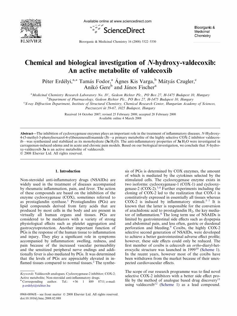
Available online at www.sciencedirect.com
Bioorganic & Medicinal Chemistry 16 (2008) 5322–5330
Chemical and biological investigation of N-hydroxy-valdecoxib:An active metabolite of valdecoxib
Peter Erdelyi,a,* Tamas Fodor,a Agnes Kis Varga,b Matyas Czugler,c
Aniko Gereb and Janos Fischera
aMedicinal Chemistry Research Laboratory No. IV., Gedeon Richter Plc., PO Box 27, H-1475 Budapest 10, HungarybDepartment of Pharmacology, Gedeon Richter Plc., PO Box 27, H-1475 Budapest 10, Hungary
cX-ray Diffraction Department, Institute of Structural Chemistry, Chemical Research Center, Hungarian Academy of Sciences,
Pusztaszeri ut 59-67, 1025 Budapest, Hungary
Received 14 October 2007; revised 25 February 2008; accepted 28 February 2008
Available online 4 March 2008
Abstract—The inhibition of cyclooxygenase enzymes plays an important role in the treatment of inflammatory diseases. N-Hydroxy-4-(5-methyl-3-phenylisoxazol-4-yl)benzenesulfonamide (3)—a primary metabolite of the highly selective COX-2 inhibitor valdecox-ib—was synthesized and stabilized as its monohydrate (3aÆH2O). The anti-inflammatory properties of 3aÆH2O were investigated incarrageenan-induced edema and in acute and chronic pain models. Based on our biological investigation, we conclude that N-hydro-xy-valdecoxib 3a is an active metabolite of valdecoxib.� 2008 Elsevier Ltd. All rights reserved.
1. Introduction
Non-steroidal anti-inflammatory drugs (NSAIDs) arewidely used in the treatment of diseases accompaniedby rheumatic inflammation, pain, and fever. The actionof these compounds are based on the inhibition of theenzyme cyclooxygenase (COX), sometimes referred toas prostaglandin synthase.1 Prostaglandins (PGs) arelipid compounds derived from fatty acids that areproduced by most cells in the body and are present invirtually all human organs and tissues. PGs areconsidered to be mediators with a variety of strongphysiological effects such as platelet aggregation andgastrocytoprotection. Another important function ofPGs is the response of the human tissue to inflammationand injury. They play a significant role in symptomsaccompanied by inflammation: swelling, redness, andpain because of the increased vascular permeabilityand the sensitized peripheral nerve endings and addi-tionally fever is also mediated by PGs. It was determinedthat the levels of PGs are appreciably elevated in in-flamed tissues compared to normal tissues.2 The synthe-
0968-0896/$ - see front matter � 2008 Elsevier Ltd. All rights reserved.
doi:10.1016/j.bmc.2008.02.088
Keywords: Valdecoxib analogues; Cyclooxygenase-2 inhibitor; COX-2;
Active metabolite; Non-steroidal anti-inflammatory drugs.* Corresponding author. Tel.: +36 1 889 8711; e-mail:
sis of PGs is determined by COX enzymes, the amountof which is mediated by the cytokines selected by thestimulated cells. The cyclooxygenase enzyme exists intwo isoforms: cyclooxygenase-1 (COX-1) and cyclooxy-genase-2 (COX-2).3,4 Further experiments including thecloning of COX-2 led to the realization that COX-1 isconstitutively expressed in essentially all tissues whereasCOX-2 is induced by inflammatory stimuli.5–7 It isknown that the latter is responsible for the conversionof arachidonic acid to prostaglandin H2, the key media-tor of inflammation.8 The long term use of NSAIDs islimited by gastrointestinal side effects such as dyspepsiaand abdominal pain, and less often, gastric or duodenalperforation and bleeding.9 Coxibs, the highly COX-2selective second generation of NSAIDs, were developedto achieve a better gastrointestinal adverse effect profile;however, these side effects could only be reduced. Thefirst member of coxibs is celecoxib an ortho-diaryl-het-erocyclic structure was launched in 199910 (Scheme 1).In the recent years, however most of the coxibs havebeen withdrawn from the market because of their unex-pected cardiovascular effects.
The scope of our research programme was to find novelselective COX-2 inhibitors with a better side effect pro-file by the method of analogue based drug discovery11
using valdecoxib12 (Scheme 1) as a lead compound.
Scheme 1. Celecoxib and valdecoxib.
P. Erdelyi et al. / Bioorg. Med. Chem. 16 (2008) 5322–5330 5323
The close analogues of celecoxib and rofecoxib have al-ready been reported in this journal by others.13,14
2. Chemistry
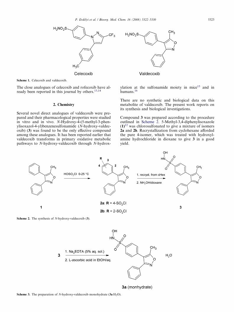
Several novel direct analogues of valdecoxib were pre-pared and their pharmacological properties were studiedin vitro and in vivo. N-Hydroxy-4-(5-methyl-3-phen-ylisoxazol-4-yl)benzenesulfonamide (N-hydroxy-valdec-oxib) (3) was found to be the only effective compoundamong these analogues. It has been reported earlier thatvaldecoxib transforms in primary oxidative metabolicpathways to N-hydroxy-valdecoxib through N-hydrox-
Scheme 3. The preparation of N-hydroxy-valdecoxib monohydrate (3aÆH2O
Scheme 2. The synthesis of N-hydroxy-valdecoxib (3).
ylation at the sulfonamide moiety in mice15 and inhumans.16
There are no synthetic and biological data on thismetabolite of valdecoxib. The present work reports onits synthesis and biological investigations.
Compound 3 was prepared according to the procedureoutlined in Scheme 2. 5-Methyl-3,4-diphenylisoxazole(1)12 was chlorosulfonated to give a mixture of isomers2a and 2b. Recrystallization from cyclohexane affordedthe pure 4-isomer, which was treated with hydroxyl-amine hydrochloride in dioxane to give 3 in a goodyield.
).
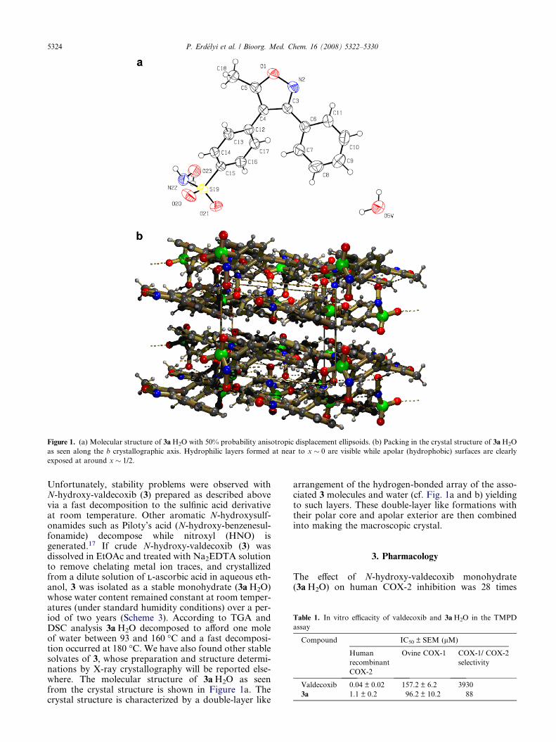
Figure 1. (a) Molecular structure of 3aÆH2O with 50% probability anisotropic displacement ellipsoids. (b) Packing in the crystal structure of 3aÆH2O
as seen along the b crystallographic axis. Hydrophilic layers formed at near to x � 0 are visible while apolar (hydrophobic) surfaces are clearly
exposed at around x � 1/2.
Table 1. In vitro efficacity of valdecoxib and 3aÆH2O in the TMPD
assay
Compound IC50 ± SEM (lM)
Human
recombinant
COX-2
Ovine COX-1 COX-1/ COX-2
selectivity
Valdecoxib 0.04 ± 0.02 157.2 ± 6.2 3930
3a 1.1 ± 0.2 96.2 ± 10.2 88
5324 P. Erdelyi et al. / Bioorg. Med. Chem. 16 (2008) 5322–5330
Unfortunately, stability problems were observed withN-hydroxy-valdecoxib (3) prepared as described abovevia a fast decomposition to the sulfinic acid derivativeat room temperature. Other aromatic N-hydroxysulf-onamides such as Piloty’s acid (N-hydroxy-benzenesul-fonamide) decompose while nitroxyl (HNO) isgenerated.17 If crude N-hydroxy-valdecoxib (3) wasdissolved in EtOAc and treated with Na2EDTA solutionto remove chelating metal ion traces, and crystallizedfrom a dilute solution of LL-ascorbic acid in aqueous eth-anol, 3 was isolated as a stable monohydrate (3aÆH2O)whose water content remained constant at room temper-atures (under standard humidity conditions) over a per-iod of two years (Scheme 3). According to TGA andDSC analysis 3aÆH2O decomposed to afford one moleof water between 93 and 160 �C and a fast decomposi-tion occurred at 180 �C. We have also found other stablesolvates of 3, whose preparation and structure determi-nations by X-ray crystallography will be reported else-where. The molecular structure of 3aÆH2O as seenfrom the crystal structure is shown in Figure 1a. Thecrystal structure is characterized by a double-layer like
arrangement of the hydrogen-bonded array of the asso-ciated 3 molecules and water (cf. Fig. 1a and b) yieldingto such layers. These double-layer like formations withtheir polar core and apolar exterior are then combinedinto making the macroscopic crystal.
3. Pharmacology
The effect of N-hydroxy-valdecoxib monohydrate(3aÆH2O) on human COX-2 inhibition was 28 times

Figure 2. Inhibition effect on ovine COX-1 enzyme. Figure 4. Effect of the test compounds in the LPS evoked PGE2
production assay.
Figure 5. Effect of valdecoxib in paw edema.
P. Erdelyi et al. / Bioorg. Med. Chem. 16 (2008) 5322–5330 5325
lower than that of valdecoxib, therefore, the COX-1/COX-2 selectivity of 3aÆH2O was much lower than thatof valdecoxib (Table 1 and Figs. 2 and 3). Compound3aÆH2O inhibited the LPS-induced PGE2 elevation sig-nificantly weaker than valdecoxib (Fig. 4).
Compound 3aÆH2O had a dose dependent significantedema inhibition effect in the carrageenan-induced pawedema assay. It had an anti-edema effect at a dose of0.3 mg/kg po whereas valdecoxib showed no inhibitionat the same dose (Figs. 5 and 6).
The analgetic and anti-inflammatory activity was deter-mined in rats in acute and chronic experiments. The car-rageenan provoked mechanical hyperalgesia assayindicated that both compounds—valdecoxib and3aÆH2O—had considerable analgesic activity (Fig. 7).Compound 3aÆH2O showed a significant analgesic activ-ity for 3 h after treatment and it has a longer duration ofaction as compared with valdecoxib.
In the subchronic pain model, valdecoxib reached theefficacy of 3aÆH2O only at a higher dose level (10 mg/kg vs 3 mg/kg), (Fig. 8). Valdecoxib and 3aÆH2O have
Figure 3. Inhibition effect on human COX-2 enzyme.
Figure 6. Effect of 3aÆH2O in paw edema.
strong analgesic effect in the carrageenan-inducedmechanical allodynia assay (Figs. 9 and 10).
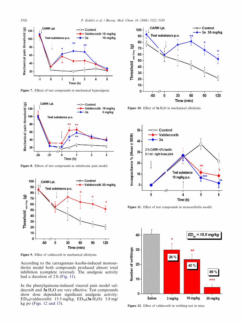
Figure 7. Effects of test compounds in mechanical hyperalgesia.
Figure 8. Effects of test compounds in subchronic pain model.
Figure 9. Effect of valdecoxib in mechanical allodynia.
Figure 10. Effect of 3aÆH2O in mechanical allodynia.
Figure 11. Effect of test compounds in monoarthritis model.
Figure 12. Effect of valdecoxib in writhing test in mice.
5326 P. Erdelyi et al. / Bioorg. Med. Chem. 16 (2008) 5322–5330
According to the carrageenan–kaolin-induced monoar-thritis model both compounds produced almost totalinhibition (complete reversal). The analgesic activityhad a duration of 2 h (Fig. 11).
In the phenylquinone-induced visceral pain model val-decoxib and 3aÆH2O are very effective. Test compoundsshow dose dependent significant analgesic activity:ED50(valdecoxib): 15.5 mg/kg; ED50(3aÆH2O): 3.8 mg/kg po (Figs. 12 and 13).

Figure 13. Effect of 3aÆH2O in writhing test in mice.
Figure 14. Cardiac effect of valdecoxib and 3aÆH2O on isolated rabbit
heart.
P. Erdelyi et al. / Bioorg. Med. Chem. 16 (2008) 5322–5330 5327
Compound 3aÆH2O significantly increases coronaryblood flow dose dependently without altering otherparameters (QTS, HR, LPV) in rabbits. Valdecoxibhas no effect on coronary blood flow (Fig. 14). The nitricoxide (NO) donor properties of 3aÆH2O could explainthis phenomenon.18 Neither valdecoxib nor its analogue3aÆH2O modified the resting blood pressure of the con-scious animals as compared with the solvent-treatedgroup and the values were 84.5 ± 3.54, 82.4 ± 7.96,and 86.45 ± 6.38 mmHg, respectively.
4. Conclusions
Direct analogues of valdecoxib have been prepared toobtain a drug analogue with better properties. N-Hydro-xy-valdecoxib (3a) proved to be an active compound. Itis a known metabolite of valdecoxib in human and inmice. The synthesis and the stabilization of 3a are de-scribed. The unsolvated N-hydroxy-valdecoxib 3 provedto be unstable. It could be stabilized by recrystallization
of compound 3 from a water–ethanol mixture and elim-inating the traces of metal ions and free radicals with thehelp of EDTA and LL-ascorbic acid to afford a stablemonohydrate 3aÆH2O. This monohydrate had a weakin vitro COX-2 activity as compared to valdecoxib.Based on in vivo analgesic and anti-inflammatory tests3aÆH2O is more potent and has a longer duration of ac-tion than the reference compound and significantly in-creases the coronary blood flow in isolated rabbitheart. The nitric oxide (NO) donor properties of 3aÆH2Ocould play a role in its mechanism of action.
5. Methods
In vitro and in vivo testing were performed as given inthe literature cited.19–21 Only modifications are de-scribed in this article.
5.1. Spectrophotometric assays with human recombinantCOX-2 and bovine COX-1
Enzymatic activities of the human recombinant COX-2and bovine COX-1 were measured using a spectropho-tometric assay based on the oxidation of TMPD duringthe reduction of PGG2 to PGH2. Valdecoxib and3aÆH2O inhibited human recombinant COX-2 in a con-centration dependent manner. Inhibition data of valdec-oxib and 3aÆH2O are shown in Table 1 and Figures 2and 3.
5.2. LPS evoked PGE2 production in human whole blood
The production of PGE2 by LPS-challenged humanwhole blood has been used to evaluate the efficacy ofCOX-2 inhibitors in vitro. PGE2 production was mea-sured in heparinized whole blood samples, incubatedwith LPS for 24 h, as a reflection of the inducibleCOX activity of monocyte COX-2 (Fig. 4).
5.3. Carrageenan-induced paw edema
The edema was induced in male Wistar rats (130–150 g)by intraplantar injection of carrageenan (CARR; 50 llof 1% solution) into the right hind paw 1 h after the oraladministration of a drug (Figs. 5 and 6). The irritant in-duced a rapid swelling of the right paw being maximal at3 h and lasting for 6 h. Right hind paw volume was mea-sured with a plethysmometer (Ugo Basile, type 7150) be-fore and 1, 2, 3, 4, 5, and 6 h after the CARR injection.The increase of paw volume is calculated as a percentagecompared with the volume measured immediately be-fore injection of the irritant for each animal. Effectivelytreated animals show much less edema. The difference ofaverage values between treated animals and vehicle con-trol groups is calculated for each time interval and sta-tistically evaluated.22
5.4. Carrageenan-induced mechanical hyperalgesia
CARR-induced hyperalgesia is one of the most fre-quently applied model of inflammatory pain. The mac-romolecule induces local inflammation resulting in
5328 P. Erdelyi et al. / Bioorg. Med. Chem. 16 (2008) 5322–5330
persisting hyperalgesia and allodynia. This assay moni-tors the capacity of the test drugs to reverse hyperalgesia(i.e., decreased threshold to painful stimuli) produced bycarrageenan (CARR)-induced inflammation. Male albi-no rats (Wistar strain—TOXI-COOP Ltd, Hungary–weighing 140–190 g) were used (n = 6/group). Themechanical pain threshold of the inflamed hind pawwas measured according to the Randall-Selitto methodwith an ‘analgesimeter’ (Ugo Basile). The hind pawwas compressed with a progressively increasing pres-sure. The nociceptive threshold is defined as the force,in grams when the animal first vocalized or made a vig-orous attempt to remove the paw. Data are presented asthe absolute threshold values (means ± SEM) in grams.The percentage reversal of mechanical hyperalgesia wascalculated from mean threshold values of a given treat-ment group as follows:
% reversal ¼ T xh � T 0h
T lh � T 0h
� 100;
wherein Txh and T0h are the post- and pre-dose thresh-old and Tlh is the baseline threshold.
• Acute experiments: On the day of the experiment thebaseline threshold was determined first. After base-line measurements the animals received CARR(100 ll of 2% solution) into the plantar surface ofright hind paw. The injected hind paw became maxi-mally inflamed by 2.5–3 h after injection, character-ized by edema and decreased mechanical painthreshold. Test compounds were administered orallyimmediately after 1 h reading. Changes in the thresh-old were checked at 1, 2, and 3 h following drugadministration (Fig. 7).
• Subchronic experiments: After baseline measurementsthe animals received CARR (150 ll mL of 2% solu-tion) ip of right hind paw. The effects of analgesicdrugs were studied 24 h after induction of inflamma-tion (in subchronic phase). Test compound wasadministered orally immediately after 24 h reading(Fig. 8).
5.5. Carrageenan-induced mechanical allodynia in rats
The Electronic von Frey Anesthesiometer was used forthe determination of pain sensitivity. The test is per-formed by applying pressure to the paw through thehand-held force transducer with a sensitive cottoncoated rigid tip of the probe. After baseline measure-ments the animals received CARR (100 ll of 2% solu-tion) into the plantar surface of right hind paw (Figs.9 and 10).
5.6. Carrageenan–Kaolin-induced monoarthritis in rats
An Incapacitance Tester (ICT) was used to measure thechange in weight load on the hind paws before and afterinduction of monoarthritis, as a measure of functionaldisability caused by arthritic inflammation and pain.Male WISTAR rats (TOXI-COOP Ltd, Hungary—weighing 120–190 g) were injected with mixture of 2%carrageenan and 2% kaolin (CarrKao) into the joint
cavity of the right knee (in 0.1 mL sterile saline) underether anesthesia (n = 8–10/group). Control rats were in-jected with vehicle.
In therapeutic type experiment drugs were given 4 hafter CarrKao injection. Time of measurements: pre-injection of CarrKao, immediately before drug treat-ment, and 1 and 2 h after the treatment. Change inweight load in the injected and contra-lateral hind limbwas measured by ICT device in grams.
The incapacitance was calculated as follows:
% incapacitance ¼ mll � mrl
mll þ mrl
� 100;
wherein mll and mrl are the left and right limb weightload (g).
The efficacy of treatment was characterized by percent-age reversal of IC% for various time points. Intraarticu-lar administration of CarrKao induced a significantreduction of weight load on the affected leg, with maxi-mum effect at 3–5 h after the injection (Fig. 11).22
5.7. Visceral pain model in mice
The phenylquinone (PQ) abdominal contraction assaywas used to study the effects of increasing doses of testcompounds in a chemo-nociceptive assay.
COX-2 Expression can be induced by lipopolysaccha-ride (LPS) pre-treatment with 24 h before the study.Inducible COX-2 can be upregulated in acute inflamma-tory processes. Male NMRI mice (23–30 g, TOXI-COOP Ltd, Hungary) were orally administered with testcompound or vehicle 30 min before the challenge withthe intraperitoneal injection of 0.02% PQ. Immediatelyafter ip administration of the irritant the animals wereplaced individually in boxes (12 · 12 · 12 cm) and thenumber of abdominal contractions (writhing) werecounted for 5–20 min post-injection. Each dose groupsconsisted of eight animals. The number of these abdom-inal writhings (constriction of abdomen, turning oftrunk, and extension of hind legs) was counted duringa 20 min session, beginning 5 min after the injection ofPQ. The analgesic activity of the drugs at each dosewas determined as a percentage inhibition comparedto the degree of writhing in the vehicle controls. Theanti-nociceptive response was analyzed by analysis ofvariance (ANOVA) followed by Tukey’s t-test to assessthe significance (Figs. 12 and 13).
5.8. Cardiac effect on isolated rabbit heart
New Zealand white rabbits hearts were perfused via theaorta with oxygenated, thermostated Krebs solution.The test compounds were dissolved in the perfusionsolution to obtain the requested concentrations. The ba-sic value of coronary blood flow was determined. After-wards measured amounts of compounds were added tothe perfusion liquid and the perfusion was recordedfor 30 min at every 10 min. After 30 min perfusion per-formed without compound the measurement was
P. Erdelyi et al. / Bioorg. Med. Chem. 16 (2008) 5322–5330 5329
repeated with the medium and high amount of com-pounds (Fig. 14).
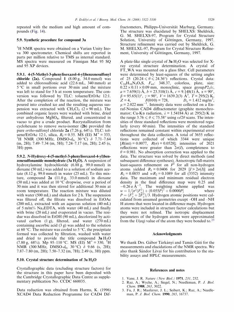
5.9. Synthetic procedure for compound 3a
1H NMR spectra were obtained on a Varian Unity Ino-va 300 spectrometer. Chemical shifts are reported inparts per million relative to TMS as internal standard.MS spectra were measured on Finnigan Mat 95 SQand 95 XP devices.
5.9.1. 4-(5-Methyl-3-phenylisoxazol-4-yl)benzenesulfonylchloride (2a). Compound 1 (8.00 g, 34.0 mmol) wasadded to chlorosulfonic acid (22.6 mL, 340 mmol) at5 �C in small portions over 30 min and the mixturewas left to stand for 5 h at room temperature. The con-version was followed by TLC: toluene/EtOAc 12:1.After the completion of the reaction, the mixture waspoured into crushed ice and the resulting aqueous sus-pension was extracted with CH2Cl2 (2 · 90 mL). Thecombined organic layers were washed with brine, driedover anhydrous MgSO4, filtered, and concentrated invacuo to give a crude product. Recrystallization fromcyclohexane to remove meta-isomer (2b) provided thepure ortho-sulfonyl chloride 2a (7.26 g, 64%). TLC: tol-uene/EtOAc 12:1, silica, Rf = 0.55. MS (EI) M+ = 333,1H NMR (300 MHz, DMSO-d6, 30 �C) d 7.71–7.64(m, 2H); 7.49–7.34 (m, 5H); 7.24–7.17 (m, 2H); 2.45 (s,3H) ppm.
5.9.2. N-Hydroxy-4-(5-methyl-3-phenylisoxazol-4-yl)ben-zenesulfonamide monohydrate (3aÆH2O). A suspension ofhydroxylamine hydrochloride (6.88 g, 99.0 mmol) indioxane (50 mL) was added to a solution of sodium ace-tate (8.12 g, 99.0 mmol) in water (25 mL). To this mix-ture, compound 2a (11.0 g, 33.0 mmol) in dioxane(50 mL) was added at 10 �C dropwise, over a period of30 min and it was then stirred for additional 30 min atroom temperature. The reaction mixture was dilutedwith water (500 mL) and shaken for 2 h. The suspensionwas filtered off, the filtrate was dissolved in EtOAc(200 mL), extracted with an aqueous solution (40 mL)of 5 m/m% Na2EDTA, with water (40 mL) and finallywith brine (20 mL) and evaporated in vacuo. The resi-due was dissolved in EtOH (90 mL), decolorized by acti-vated carbon (1 g), filtered, and water (270 mL)containing ascorbic acid (3 g) was added to the solutionat 60 �C. The mixture was cooled to 5 �C, the precipitateformed was collected by filtration, washed with waterand dried to provide the title compound 3aÆH2O(7.80 g, 68%). Mp 95–110 �C. MS (EI) M+ = 330, 1HNMR (300 MHz, DMSO-d6, 30 �C) d 9.66 (s, 2H);7.87–7.80 (m, 2H); 7.50–7.32 (m, 7H); 2.49 (s, 3H) ppm.
5.10. Crystal structure determination of 3aÆH2O
Crystallographic data (excluding structure factors) forthe structure in this paper have been deposited withthe Cambridge Crystallographic Data Centre as supple-mentary publication No. CCDC 660035.
Data reduction was obtained from Harms, K. (1996)XCAD4 Data Reduction Programme for CAD4 Dif-
fractometers, Philipps-Universitat Marburg, Germany.The structure was elucidated by SHELXS: Sheldrick,G. M. SHELXS-97, Program for Crystal StructureSolution, University of Gottingen, Germany, 1997.Structure refinement was carried out by Sheldrick, G.M. SHELXL-97, Program for Crystal Structure Refine-ment, University of Gottingen, Germany, 1997.
A plate-like single crystal of 3aÆH2O was selected for X-ray crystal structure determination. A crystal ofFT_WA was mounted on a glass fiber. Cell parameterswere determined by least-squares of the setting anglesof 25 (20.24 6 h 6 24.36�) reflections. Crystal data:C16H16N2O5S, Fwt: 348.37, colorless, plate, size:0.22 · 0.11 · 0.09 mm, monoclinic, space groupP21/c,a = 7.659(1) A, b = 23.510(1) A, c = 9.148(1) A, a = 90�,b = 95.65(1)�, c = 90�, V = 1639.2(3) A3, T = 295(2) K,Z = 4, F(000) = 728, Dx = 1.412 mg/m3,l = 2.022 mm�1. Intensity data were collected on a En-raf-Nonius CAD4 diffractometer (graphite monochro-mator; Cu-Ka radiation, k = 1.54184 A) at 295(2) K inthe range 3.76 6 h 6 75.58� using x/2h scans. The inten-sities of three standard reflections were monitored regu-larly (every 60 min). The intensities of the standardreflections remained constant within experimental errorthroughout the data collection. A total of 3655 reflec-tions were collected of which 3344 were unique[R(int) = 0.0077, R(r) = 0.0528]; intensities of 2021reflections were greater than 2r(I), completeness toh = 0.981. No absorption correction was applied to thedata. The structure was solved by direct methods (andsubsequent difference syntheses). Anisotropic full-matrixleast-squares refinement on F2 for all non-hydrogenatoms yielded R1 = 0.0406 for 2029 [I > 2r(I)] andR1 = 0.0855 and wR2 = 0.1089 for all (3352) intensitydata. The maximum and minimum residual electrondensity in the final difference map were 0.25 and�0.26 e A�3. The weighting scheme applied wasw ¼ 1=½r2ðF 2
oÞ þ ð0:055P Þ2 þ 0:0000P � whereP ¼ ðF 2
o þ 2F 2cÞ=3. Hydrogen atomic positions were cal-
culated from assumed geometries except –OH and –NHH atoms that were located in difference maps. Hydrogenatoms were included in structure factor calculations butthey were not refined. The isotropic displacementparameters of the hydrogen atoms were approximatedfrom the U(eq) value of the atom they were bonded to.23
Acknowledgments
We thank Drs. Gabor Tarkanyi and Tamas Gati for themeasurements and elucidations of the NMR spectra. Wealso thank Sandor Levai for his contribution to the sta-bility assays and HPLC measurements.
References and notes
1. Vane, J. R. Nature (New Biol.) 1971, 231, 232.2. Raz, A.; Wyche, A.; Siegel, N.; Needleman, P. J. Biol.
Chem. 1988, 263, 3022.3. Fu, J. R.; Masferrer, J. L.; Seibert, K.; Raz, A.; Needle-
man, P. J. Biol. Chem. 1990, 265, 16737.
5330 P. Erdelyi et al. / Bioorg. Med. Chem. 16 (2008) 5322–5330
4. Battistini, B.; Botting, B.; Bakhle, Y. S.. Drug NewsPerspect. 1994, 7, 501.
5. Xie, W.; Chipman, J. G.; Robertson, D. L.; Erikson, R.L.; Simmons, D. L. Proc. Natl. Acad. Sci. U.S.A. 1991, 88,2692.
6. Kujubu, D. A.; Fletcher, B. S.; Varnum, B. C.; Lim, R.W.; Herschman, H. R. J. Biol. Chem. 1991, 266, 12866.
7. O’Banion, M. K.; Sadowski, H. B.; Winn, V.; Young, D.A. A. J.. Biol. Chem. 1991, 266, 23261.
8. Vane, J. R.; Botting, R. M. . Adv. Prostaglandin, Throm-boxane, Leukot. Res. 1995, 23, 41.
9. Allison, M. C.; Howatson, A. G.; Torrance, C. J.; Lee, F.D.; Russell, R. I. G. N. Engl. J. Med. 1984, 310, 563.
10. Penning, T. D.; Talley, J. J.; Bertenshaw, S. R.; Carter, J.S.; Collins, P. W.; Docter, S.; Graneto, M. J.; Lee, L. F.;Malecha, J. W.; Miyashiro, J. M.; Rogers, R. S.; Rogier,D. J.; Yu, S. S.; Anderson, G. D.; Burton, E. G.; Cogburn,J. N.; Gregory, S. A.; Koboldt, C. M.; Perkins, W. E.;Seibert, K.; Veenhuizen, A. W.; Zhang, Y. Y.; Isakson, P.C. J. Med. Chem. 1997, 40, 1347.
11. Analogue-Based Drug Discovery; Fischer, J., Ganellin, C.R., Eds.; Wiley-VHC Verlag GmbH & Co. KGaA, 2006.
12. Talley, J. J.; Brown, D. L.; Carter, J. S.; Graneto, M. J.;Koboldt, C. M.; Masferrer, J. L.; Perkins, W. E.; Rogers,R. S.; Shaffer, A. F.; Zhang, Y. Y.; Zweifel, B. S.; Seibert,K. J. Med. Chem. 2000, 43, 775.
13. Uddin, Md. J.; Rao, P. N. P.; Knaus, E. E. Bioorg. Med.Chem. 2003, 11, 5273.
14. Zarghi, A.; Rao, P. N. P.; Knaus, E. E. Bioorg. Med.Chem. 2007, 15, 1056.
15. Zhang, J. Y.; Yuan, J. J.; Wang, Y. F.; Bible, R. H.;Breau, A. P. Drug Metab. Dispos. 2003, 31, 491.
16. Yuan, J. J.; Yang, D. C.; Zhang, J. Y.; Bible, R.; Karim,A.; Findlay, J. W. A. Drug Metab. Dispos. 2002, 30, 1013.
17. Bonner, F. T.; Ko, Y. Inorg. Chem. 1992, 31, 2514.18. Zamora, R.; Grzesiok, A.; Weber, H.; Feelisch, M.
Biochem. J. 1995, 312, 333.19. Gierse, K.; Hauser, S. D.; Creely, D. P.; Koboldt, C. M.;
Rangwala, S. H.; Isakson, P. C.; Siebert, K. Biochem. J.1995, 305, 479.
20. Drug Discovery and Evaluation Pharmacological Assays;Vogel, H. G., Vogel, W. H., Eds.; Springer-Verlag: Berlin,Heidelberg, 1997.
21. Atkinson, D. C. Arch. Int. Pharmacodyn. Ther. 1971, 193,391.
22. Statistics: Data were analyzed by two-way ANOVAfollowed by Tukey’s (HSD) post hoc test, comparingdrug-treated with vehicle-treated animals. Significance wasconsidered at p < 0.05 level (*p < 0.05; **p < 0.01;***p < 0.001).
23. Neutral atomic scattering factors and anomalous scat-tering factors are taken from: International Tables for X-ray Crystallography; Wilson, A. J. C., Ed.; KluwerAcademic Publishers: Dordrecht, 1992; Vol. C,, Tables6.1.1.4, pp 500–502, 4.2.6.8, pp 219–222 and 4.2.4.2, pp193–199.
Copyright © 2022 FDOKUMEN