Secondary Metabolite Production and Terpenoid ... - MDPI
17
Article Secondary Metabolite Production and Terpenoid Biosynthesis in Endophytic Fungi Cladosporium cladosporioides Isolated from Wild Cymbopogon martinii (Roxb.) Wats Hemalatha Jayaram 1 , Vinutha Marigowda 2 and Kunigal Jagadishchandra Thara Saraswathi 1, * Citation: Jayaram, H.; Marigowda, V.; Thara Saraswathi, K.J. Secondary Metabolite Production and Terpenoid Biosynthesis in Endophytic Fungi Cladosporium cladosporioides Isolated from Wild Cymbopogon martinii (Roxb.) Wats. Microbiol. Res. 2021, 12, 812–828. https://doi.org/10.3390/ microbiolres12040059 Academic Editor: Elisa Bona Received: 7 October 2021 Accepted: 26 October 2021 Published: 1 November 2021 Publisher’s Note: MDPI stays neutral with regard to jurisdictional claims in published maps and institutional affil- iations. Copyright: © 2021 by the authors. Licensee MDPI, Basel, Switzerland. This article is an open access article distributed under the terms and conditions of the Creative Commons Attribution (CC BY) license (https:// creativecommons.org/licenses/by/ 4.0/). 1 Department of Microbiology and Biotechnology, Jnana Bharathi Campus, Bangalore University, Bengaluru 560056, Karnataka, India; [email protected] 2 Department of Biotechnology and Genetics, Ramaiah College of Arts, Science and Commerce, Bengaluru 560054, Karnataka, India; [email protected] * Correspondence: [email protected]; Tel.: +98-80-601-063 Abstract: Endophytic fungi Cladosporium cladosporioides (F1-MH810309) and Cladosporium tenuissi- mum (F2-MN715834) from the leaf of wild Cymbopogon martinii (MT90507) were isolated and selected based on the persistent occurrence during different seasons of the year. They were identified based on the morphological features and molecular characterization (ITS sequence), and later deposited at NCBI. Phytochemical studies on F1, F2 and host extracts showed the presence of alkaloids, flavonoids, phenols, terpenoids and tannins. The GC-MS of F1 extract (control) under the axenic condition re- vealed compounds like hexadecane, heptadecane,2,4-Ditert-butylphenol, E-14 hexadecenal, geraniol, geranyl acetate and cubenol similar to the host. The GC-MS of F2 extract (control) revealed metabo- lites that were unique. Further, both F1 and F2 were cultured in the supplementation of different concentrations (5%, 10%, 15% and 20%) of the host plant extract (an-axenic condition). The GC- MS of F1 extracts (test) exhibited good growth and showed the gradual increased production of terpenoid compounds whereas the F2 (test) did not show any growth. These compounds such as hyrdoxymenthol, nor-borneol, cedralacetate, α-cyclogeraniol, campesterol, β-cyclogeraniol, linalool oxide,2,3-boranediol, citronellyltiglate and 2,3-pinanediol were produced in a minor quantity and were known as biotransformed forms of the precursor compounds present in the host extract. In comparison, only F1 was able to produce terpenoids similar to the host species both in axenic and an-axenic conditions. Hence from the current study, the endophytic fungus F1 isolated from wild C. martinii for the first time can serve as a better resource for the bioprospection of an important terpenoid and its metabolites. Keywords: Cymbopogon martinii; endophytic fungi; Cladosporium cladosporioides; Cladosporium tenuissi- mum; terpenoids; biotransformation 1. Introduction Cymbopogon martinii (Roxb.) commonly called “Rosha” grass belongs to the Rusae series and serves as an important species of Cymbopogon, yielding essential oil rich in geraniol [1,2]. The species is found widely distributed in south India and exists in two distinct forms referred to as “motia and sofia” varieties producing quality essential oil [3,4]. The essential oil of C. martinii possesses significant traditional medicinal prop- erties and finds extensive usage in the treatment of rheumatism, arthritis, lumbago and spasms past from ancient times [5]. Furthermore, because of its potential activity it is used in preventing chronic diseases like cancer, diabetes and coronary heart diseases as well as showing antimicrobial, antifungal, antiviral, anthelmintic, antioxidant and cyto- toxicity [6,7]. These compounds are produced against biotic and abiotic stress and help in defense mechanisms, communication and attracting pollinators [8]. The essential oil compounds of C. martinii have been extensively used in aromatic industries in the prepa- Microbiol. Res. 2021, 12, 812–828. https://doi.org/10.3390/microbiolres12040059 https://www.mdpi.com/journal/microbiolres
-
Upload
khangminh22 -
Category
Documents
-
view
0 -
download
0
Transcript of Secondary Metabolite Production and Terpenoid ... - MDPI

Article
Secondary Metabolite Production and Terpenoid Biosynthesisin Endophytic Fungi Cladosporium cladosporioides Isolatedfrom Wild Cymbopogon martinii (Roxb.) Wats
Hemalatha Jayaram 1, Vinutha Marigowda 2 and Kunigal Jagadishchandra Thara Saraswathi 1,*
�����������������
Citation: Jayaram, H.; Marigowda,
V.; Thara Saraswathi, K.J. Secondary
Metabolite Production and Terpenoid
Biosynthesis in Endophytic Fungi
Cladosporium cladosporioides Isolated
from Wild Cymbopogon martinii
(Roxb.) Wats. Microbiol. Res. 2021, 12,
812–828. https://doi.org/10.3390/
microbiolres12040059
Academic Editor: Elisa Bona
Received: 7 October 2021
Accepted: 26 October 2021
Published: 1 November 2021
Publisher’s Note: MDPI stays neutral
with regard to jurisdictional claims in
published maps and institutional affil-
iations.
Copyright: © 2021 by the authors.
Licensee MDPI, Basel, Switzerland.
This article is an open access article
distributed under the terms and
conditions of the Creative Commons
Attribution (CC BY) license (https://
creativecommons.org/licenses/by/
4.0/).
1 Department of Microbiology and Biotechnology, Jnana Bharathi Campus, Bangalore University,Bengaluru 560056, Karnataka, India; [email protected]
2 Department of Biotechnology and Genetics, Ramaiah College of Arts, Science and Commerce,Bengaluru 560054, Karnataka, India; [email protected]
* Correspondence: [email protected]; Tel.: +98-80-601-063
Abstract: Endophytic fungi Cladosporium cladosporioides (F1-MH810309) and Cladosporium tenuissi-mum (F2-MN715834) from the leaf of wild Cymbopogon martinii (MT90507) were isolated and selectedbased on the persistent occurrence during different seasons of the year. They were identified basedon the morphological features and molecular characterization (ITS sequence), and later deposited atNCBI. Phytochemical studies on F1, F2 and host extracts showed the presence of alkaloids, flavonoids,phenols, terpenoids and tannins. The GC-MS of F1 extract (control) under the axenic condition re-vealed compounds like hexadecane, heptadecane,2,4-Ditert-butylphenol, E-14 hexadecenal, geraniol,geranyl acetate and cubenol similar to the host. The GC-MS of F2 extract (control) revealed metabo-lites that were unique. Further, both F1 and F2 were cultured in the supplementation of differentconcentrations (5%, 10%, 15% and 20%) of the host plant extract (an-axenic condition). The GC-MS of F1 extracts (test) exhibited good growth and showed the gradual increased production ofterpenoid compounds whereas the F2 (test) did not show any growth. These compounds such ashyrdoxymenthol, nor-borneol, cedralacetate, α-cyclogeraniol, campesterol, β-cyclogeraniol, linalooloxide,2,3-boranediol, citronellyltiglate and 2,3-pinanediol were produced in a minor quantity andwere known as biotransformed forms of the precursor compounds present in the host extract. Incomparison, only F1 was able to produce terpenoids similar to the host species both in axenic andan-axenic conditions. Hence from the current study, the endophytic fungus F1 isolated from wildC. martinii for the first time can serve as a better resource for the bioprospection of an importantterpenoid and its metabolites.
Keywords: Cymbopogon martinii; endophytic fungi; Cladosporium cladosporioides; Cladosporium tenuissi-mum; terpenoids; biotransformation
1. Introduction
Cymbopogon martinii (Roxb.) commonly called “Rosha” grass belongs to the Rusaeseries and serves as an important species of Cymbopogon, yielding essential oil richin geraniol [1,2]. The species is found widely distributed in south India and exists intwo distinct forms referred to as “motia and sofia” varieties producing quality essentialoil [3,4]. The essential oil of C. martinii possesses significant traditional medicinal prop-erties and finds extensive usage in the treatment of rheumatism, arthritis, lumbago andspasms past from ancient times [5]. Furthermore, because of its potential activity it isused in preventing chronic diseases like cancer, diabetes and coronary heart diseases aswell as showing antimicrobial, antifungal, antiviral, anthelmintic, antioxidant and cyto-toxicity [6,7]. These compounds are produced against biotic and abiotic stress and helpin defense mechanisms, communication and attracting pollinators [8]. The essential oilcompounds of C. martinii have been extensively used in aromatic industries in the prepa-
Microbiol. Res. 2021, 12, 812–828. https://doi.org/10.3390/microbiolres12040059 https://www.mdpi.com/journal/microbiolres

Microbiol. Res. 2021, 12 813
ration of perfumes, soaps, cosmetics, food and beverage industries. These essential oilcompounds show high biodegradability, volatility and negligible toxicity and are replacingthe synthetic metabolic compounds in aromatic industries [9]. Even the extracts of Cymbo-pogons have been known to possess biological activities such as antimicrobial, antioxidant,anti-hypercholesterolaemic potential, and anti-inflammatory and antimutagenic activi-ties [10–12] and are commonly used in food preservation, pharmaceuticals and naturalmedicines [13].
Wild plants species growing in typical agro-geo climatic conditions offer unique nichesand lodge a variety of fungi within them [14]. These large groups of endophytic fungiare ubiquitously found asymptomatically associated with the plant tissues during theirlife cycle without negatively affecting the host [15–17] and are variable depending oncomponents, plant genotype, growth stage, physiological status, tissue type and otherenvironmental conditions [18]. They help the host plant adopt to various biotic and abi-otic stresses and produce secondary metabolites including alkaloids, flavonoids, phenols,phenylpropanoids, quinines, indole derivatives, steroids, amines and terpenoids main-taining specific bioactivities [15,19,20]. The endophytic fungi associated with the host areidentified using traditional taxonomic means and molecular tools like flanking internaltranscribed spacers (ITS). They are also known to be a repository of secondary metabo-lites [17,21] possessing various biological activities, which are used in pharmaceuticalapplications [22,23].
The association of endophytic fungi with aromatic and medicinal plants is consideredto be the prominent source for producing volatile organic or terpenoid compounds [24,25]and they show potential bioactivity and are hence used in pharmaceutical industries [26,27].The biosynthetic potential of endophytes in producing terpenoid compounds similar tothe host plant has gained a lot of commercial importance. Some of the known terpenoidcompounds such as Taxol, HupA, 2-Carene, 5,8-diol, bisabolene and cadinene are producedfrom endophytic fungi along with other secondary metabolites [28,29]. A more intriguingand inexplicable issue with many endophytes that has to be critically evaluated is theirability to produce host metabolites, which can be harnessed on a large scale for potentialuse in diverse areas [19]. In the present study, the endophytic fungi isolated from wild Cym-bopogon martinii were studied for the biogenesis of common terpenoid compounds betweenthem and the biotransformation of plant metabolites into commercially useful terpenoids.
2. Materials and Methods2.1. Collection and Authentication of Plant
The wild genotype of Cymbopogon martinii was collected from Devarayana Durgahills, Tumkur, Karnataka. The plant was identified and authenticated from RegionalAyurveda Research Institute for Metabolic Disorders (RARIMD), Ministry of AYUSH,Bengaluru, under the accession no. SMPU/RARIMD/BNG/2019-20/352/RRCBI-1052.The herbarium (AC-25/2021) and seeds were deposited at ICAR- National Bureau of PlantGenetic Resources (NBPGR), Pusa, New Delhi and the DNA barcoding sequence of theplant was deposited in GenBank NCBI under accession no. MT90507.
2.2. Plant Extraction Method
The powdered leaf sample (50 g) of the plant was mixed with 150 mL ethyl acetatesolvent and kept in a shaker at 120 rpm for 48 h. The extract was filtered through Whatmanfilter paper grade no. 1. The filtrate obtained was dried at 40 ◦C using a rotary vacuumevaporator. The residue was dissolved in 1 mL of ethyl acetate and the extract was usedfor the GC-MS analysis. Furthermore, the powdered leaf (50 g) of the plant was used as thehost plant extract for supplementing in the broth culture of endophytic fungi by subjectingit to 48 h of shaking in distilled water and filtering it through Whatman filter paper gradeno. 1.

Microbiol. Res. 2021, 12 814
2.3. Isolation and Characterization of Endophytic Fungi
The leaf samples of wild C. martinii were collected and rinsed in distilled water, blotdried and cut into small pieces (10–15 mm length). The leaves were surface sterilized byimmersing in Tween-20 for 5 min, followed by two times wash with distilled water; 70%ethanol (3 min), two times wash with distilled water; 4% sodium hypochlorite solution(3 min) and washed thoroughly 4–5 times with distilled water to remove traces of surfac-tants [30]. The edges of the leaves were trimmed and placed onto the petri plates containingpotato dextrose agar (PDA) media and incubated at room temperature for 8–9 days. Theendophytic fungi emerging after fourteen days in the culture media were transferred tofresh PDA to obtain the pure culture. The morphological characters of the endophyticfungi were studied based on the macroscopic and microscopic observations.
2.4. Characterization of Endophytic Fungi
The genomic DNA extraction from endophytic fungi was carried out by followingthe [31]. Extracted DNA was subjected to PCR amplification using forward (ITS1-F CTTG-GTCATTTAGAGGAAGTAA) and reverse (ITS4-R TCCGTAGGTGAACCTGCGG) primers.The PCR products were run on agarose gel (0.8%) and the bands were purified usingthe Nucleo-pore PCR Clean-up Gel Extraction kit (Genetix Biotech Asia Pvt. Ltd., Delhi,India). The amplified DNA was subjected to sequencing using the 3130XL genetic an-alyzer (Applied Biosystems, Waltham, MA, USA). The DNA sequences of endophyticfungi were aligned using Clustal W and Bio Edit tool version 7.0 and the phylogenetic treewas constructed. The DNA sequences of endophytic fungi were deposited in the NCBIGen Bank.
2.5. Extraction of Secondary Metabolites
The endophytic fungi were mass cultured using 250 mL of potato dextrose broth (PDB)without supplementing the host plant extract (control—axenic culture) and incubated atroom temperature for 21 days under static conditions. The broth culture was filteredthrough Whatman filter paper grade no. 1 and the filtrate obtained was subjected toextraction using ethyl acetate as the solvent system (1:2). The mixture was subjected for20 min and was kept aside until two immiscible layers were formed. The ethyl acetatelayer was collected using a separating funnel. The extract was concentrated under reducedpressure and evaporated to dryness at 40 ◦C using the rotary vacuum evaporator, furthermixed with 1 mL of ethyl acetate before storing at 4 ◦C.
The endophytic fungi supplemented with host plant extract (test—an-axenic culture)(5%, 10%, 15% and 20%), were mass cultured using 250 mL of PDB medium under staticconditions and incubated at room temperature for 21 days. The broth culture was filteredthrough Whatman filter paper grade no. 1 and the filtrate obtained was subjected toextraction using ethyl acetate as the solvent system (1:2). The mixture was subjected tomixing for 20 min and was kept aside until two immiscible layers were formed. The extractwas concentrated under reduced pressure and evaporated to dryness at 40 ◦C using therotary vacuum evaporator and mixed with 1 mL of ethyl acetate before storing at 4 ◦C. Theexperiments were conducted in three replicates.
2.6. Phytochemical Analysis
The plant and fungal extracts (F1 and F2) were analyzed for the presence of phyto-chemical compounds using standard methods [32,33] and screened for the presence ofsecondary metabolites.
Alkaloids (Wagner’s reagent): About 1 mL plant extract was treated with 4–5 drops ofWagner’s reagent and observed for the formation of a reddish-brown precipitate. Wagner’sreagent is a mixture of 1.27 g of iodine and 2 g potassium iodide in 100 mL of water [34].
Tannins: The fungal crude extract was treated with alcoholic FeCl3 reagent. A bluish-black color, which disappears on the addition of a drop of dilute H2SO4 was followed bythe formation of yellowish-brown precipitate [35].

Microbiol. Res. 2021, 12 815
Saponins (foam test): About 6 mL of water was added to 2 mL of extract in a test tube.The mixture was shaken vigorously and observed for the formation of persistent foamconfirming saponins’ presence [36].
Terpenoids (Salkowski’s test): About 1 mL of chloroform was added to 2 mL of extract.A reddish-brown precipitate is formed immediately on the addition of a few drops ofconcentrated HCl [37].
Quinones: About 2 mL of the fungal extract was treated with concentrated HCl. Theformation of yellow precipitate or coloration indicated the presence of quinones [38].
Phenols: About 2 mL of fungal extract was taken and mixed with aqueous 5% FeCl3and observed for deep blue or black color formation [39].
Flavonoids: About 0.5 mL of crude extract was taken in a test to which 5–10 drops ofdiluted HCl and a small piece of zinc were added and the solution was boiled for a fewminutes. Reddish-pink or brown color precipitate indicates the presence of flavonoids [40].
2.7. Gas Chromatography and Mass Spectroscopy-(GC-MS)
The GC-MS analysis of the extracts of endophytic fungi and the host plant was carriedout at Kerala Forest Research Institute (KFRI), Kerala. The analysis was carried out onShimadzu GC-MS gas chromatogram QP2010S using two capillary columns (0.25 mmID),one coated with dimethylsiloxane and the other with Carbowax 30M. Nitrogen was used asa carrier gas at 65.2 kpa inlet pressure and organized the temperature between 26 to 28 ◦Cfor 6 ◦C/min under the dimethylsiloxane column. The split ratio used was 1:50. Heliumwas used as a carrier gas (1 mL/min) along with the ion source temperature of 20 ◦C. Theidentification of the compounds was made by analogizing the relative retention indices ofthe peaks with literature data 3 ± 10 and peak development co-injection with authenticoriginal samples. Mass spectral data were compared against NIST05 (National Institute ofStandards and Technology, Washington, DC, USA), WILEY 8 and FFNSCI.3 (Flavour andFragrance Natural and Synthetic Compounds) libraries for the identification of bioactivecompounds.
3. Results3.1. Isolation and Identification of Endophytic Fungi
A total of eight isolates of endophytic fungi such as Alternaria species, Aspergillusspecies, Fusarium species, Trichoderma species, Curvularia species and Cladosporiumspecies were isolated from the inoculants at different seasons of the year. Of the eightisolates, two endophytic fungi such as Cladosporium cladosporioides (F1) and Cladosporiumtenuissimum (F2) were isolated consistently at different seasons of the year and henceselected for further studies. The morphological studies and molecular characterizationconfirmed that the fungi were the Cladosporium species belonging to the Dothideomycetesgroup of Cladosporiaceae family. Endophytic fungi F1 produced olivaceous colonies withdiffused aerial mycelia, woolly-mat finished texture, in reverse of its olive-black color.Conidiophores were straight solitary, unbranched terminal or lateral mycelia bearing8–9 conidia in chains. Conidia were limoniform, ovoid, light brown and aseptate. WhereasF2 produced velvety, olive gray with white margins, reversed with its reddish-white color;conidiophores were straight, solitary, unbranched, terminal mycelia bearing 3–4 conidia inchains. Conidia were limoniform, ovoid, aseptate and stained blue. The results of the ITSsequences from the genomic DNA were submitted to NCBI Gen Bank with accession nos.F1-MH810309 and F2-MN715834, respectively (Figures 1 and 2). The phylogenetic tree forthe same was constructed using the Clustal W tool (Figure 3).

Microbiol. Res. 2021, 12 816
Microbiol. Res. 2021, 12, FOR PEER REVIEW 5
nos. F1-MH810309 and F2-MN715834, respectively (Figures 1 and 2). The phylogenetic tree for the same was constructed using the Clustal W tool (Figure 3).
Figure 1. Wild genotype of Cymbopogon martinii (MT905074).
(a) (b)
Figure 2. Morphological identification of endophytic fungi (F1 and F2) isolated from wild C. martinii (leaf). (a): Endophytic fungus F1 (Cladosporium cladosporioides-MH810309) showing gray colored mat bearing cladospores stained with lactophenol cotton blue; (b): Endophytic fungus F2 (Cladosporium tenuissimum-MN71583) showing gray-white colored mat bearing cladospores stained with lactophenol cotton blue; the scale of the Micrograph: 40× magnification.
Figure 3. Dendrogram showing phylogenetic relationship between endophytic fungi (F1 and F2) on the basis of ITS sequencing. Phylogenies were inferred by neighbor-joining analysis and trees gen-erated using MEGA 6.0 software. Numbers at branch points indicate bootstrap values. The scale bar represents estimated differences in nucleotide sequence.
3.2. Phytochemical Screening of Endophytic Fungi The phytochemical analysis of fungal extracts (F1 and F2) revealed the presence of
alkaloids, flavonoids, phenols, terpenoids and tannins (Table 1).
Figure 1. Wild genotype of Cymbopogon martinii (MT905074).
Microbiol. Res. 2021, 12, FOR PEER REVIEW 5
nos. F1-MH810309 and F2-MN715834, respectively (Figures 1 and 2). The phylogenetic tree for the same was constructed using the Clustal W tool (Figure 3).
Figure 1. Wild genotype of Cymbopogon martinii (MT905074).
(a) (b)
Figure 2. Morphological identification of endophytic fungi (F1 and F2) isolated from wild C. martinii (leaf). (a): Endophytic fungus F1 (Cladosporium cladosporioides-MH810309) showing gray colored mat bearing cladospores stained with lactophenol cotton blue; (b): Endophytic fungus F2 (Cladosporium tenuissimum-MN71583) showing gray-white colored mat bearing cladospores stained with lactophenol cotton blue; the scale of the Micrograph: 40× magnification.
Figure 3. Dendrogram showing phylogenetic relationship between endophytic fungi (F1 and F2) on the basis of ITS sequencing. Phylogenies were inferred by neighbor-joining analysis and trees gen-erated using MEGA 6.0 software. Numbers at branch points indicate bootstrap values. The scale bar represents estimated differences in nucleotide sequence.
3.2. Phytochemical Screening of Endophytic Fungi The phytochemical analysis of fungal extracts (F1 and F2) revealed the presence of
alkaloids, flavonoids, phenols, terpenoids and tannins (Table 1).
Figure 2. Morphological identification of endophytic fungi (F1 and F2) isolated from wild C. martinii (leaf). (a): Endophyticfungus F1 (Cladosporium cladosporioides-MH810309) showing gray colored mat bearing cladospores stained with lac-tophenol cotton blue; (b): Endophytic fungus F2 (Cladosporium tenuissimum-MN71583) showing gray-white colored matbearing cladospores stained with lactophenol cotton blue; the scale of the Micrograph: 40× magnification.
Microbiol. Res. 2021, 12, FOR PEER REVIEW 5
nos. F1-MH810309 and F2-MN715834, respectively (Figures 1 and 2). The phylogenetic tree for the same was constructed using the Clustal W tool (Figure 3).
Figure 1. Wild genotype of Cymbopogon martinii (MT905074).
(a) (b)
Figure 2. Morphological identification of endophytic fungi (F1 and F2) isolated from wild C. martinii (leaf). (a): Endophytic fungus F1 (Cladosporium cladosporioides-MH810309) showing gray colored mat bearing cladospores stained with lactophenol cotton blue; (b): Endophytic fungus F2 (Cladosporium tenuissimum-MN71583) showing gray-white colored mat bearing cladospores stained with lactophenol cotton blue; the scale of the Micrograph: 40× magnification.
Figure 3. Dendrogram showing phylogenetic relationship between endophytic fungi (F1 and F2) on the basis of ITS sequencing. Phylogenies were inferred by neighbor-joining analysis and trees gen-erated using MEGA 6.0 software. Numbers at branch points indicate bootstrap values. The scale bar represents estimated differences in nucleotide sequence.
3.2. Phytochemical Screening of Endophytic Fungi The phytochemical analysis of fungal extracts (F1 and F2) revealed the presence of
alkaloids, flavonoids, phenols, terpenoids and tannins (Table 1).
Figure 3. Dendrogram showing phylogenetic relationship between endophytic fungi (F1 and F2)on the basis of ITS sequencing. Phylogenies were inferred by neighbor-joining analysis and treesgenerated using MEGA 6.0 software. Numbers at branch points indicate bootstrap values. The scalebar represents estimated differences in nucleotide sequence.
3.2. Phytochemical Screening of Endophytic Fungi
The phytochemical analysis of fungal extracts (F1 and F2) revealed the presence ofalkaloids, flavonoids, phenols, terpenoids and tannins (Table 1).
Microbiol. Res. 2021, 12 817
Table 1. Phytochemical analysis of the extracts from endophytic fungi and host plant.
Sl No. TestHost Plant Extract
(C. martinii)
PDB (Control)(without Supplementation of Plant Extract)
PDB (Test)(with Supplementation
of Plant Extract)
Cladosporiumcladosporioides
(F1)
Cladosporiumtenuissimum
(F2)
Cladosporiumcladosporioides
(F1)
1 Alkaloid +ve +ve +ve +ve
2 Flavonoid +ve +ve +ve +ve
3 Phenol +ve +ve +ve +ve
4 Tannin +ve +ve −ve +ve
5 Terpenoid +ve +ve +ve +ve
6 Quinine +ve −ve −ve −ve
7 Essential oil +ve +ve −ve +ve
(+) Presence, (−) Absence.
3.3. Analysis of Secondary Metabolites (GC-MS)
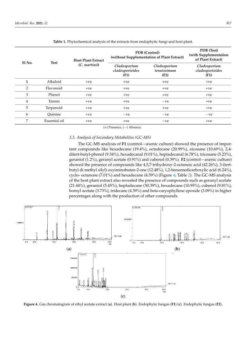
The GC-MS analysis of F1 (control—axenic culture) showed the presence of impor-tant compounds like hexadecane (19.4%), octadecane (20.99%), eicosane (10.69%), 2,4-ditert-butyl-phenol (9.34%), hexadecanal (9.01%), heptadecanal (6.78%), tricosane (5.23%),geraniol (1.2%), geranyl acetate (0.91%) and cubenol (0.39%). F2 (control—axenic culture)showed the presence of compounds like 4,5,7-trihydroxy-2-octanoic acid (42.26%), 3-(tert-butyl di methyl silyl) oxyiminobutan-2-one (12.48%), 1,2-benzenedicarboxylic acid (6.24%),cyclo- octanone (7.01%) and hexadecane (4.59%) (Figure 4; Table 2). The GC-MS analysisof the host plant extract also revealed the presence of compounds such as geranyl acetate(21.44%), geraniol (5.45%), heptadecane (30.39%), hexadecane (10.95%), cubenol (9.81%),bornyl acetate (3.73%), tridecane (4.39%) and beta-caryophyllene epoxide (3.09%) in higherpercentages along with the production of other compounds.
Microbiol. Res. 2021, 12, FOR PEER REVIEW 7
(a) (b)
(c)
Figure 4. Gas chromatogram of ethyl acetate extract (a). Host plant (b). Endophytic fungus (F1) (c). Endophytic fungus (F2).

Microbiol. Res. 2021, 12 818
Table 2. GC-MS analysis of the extracts of endophytic fungi (F1a and F2) without supplementation of host plant extract.
Sl No. Compound
Area%
Extract of PlantC. martinii
Extract of Endophytes
F1a F2
1 Tetradecane 2.08 0.97 2.12
2 Dodecane 2.88 - -
3 1S-Endo-bornyl acetate 3.73 - -
4 Tridecane 4.39 - -
5 Geraniol 5.45 1.2 -
6 Farnesane 0.34 - -
7 Geranyl acetate 21.44 0.91 -
8 Heptadecane 30.39 3.17 -
9 (-)-Beta-caryophyllene 0.63 - -
10 Decane, 2,3,5,8-tetramethyl- 0.44 - -
11 Hexadecane 10.95 19.4 4.59
12 Gamma-cadinene 0.63 -
13 2-(3-Isopropenyl-4-methyl-4-vinylcyclohexyl)-2-propanol 0.50 - -
14 (-)-Beta-caryophyllene epoxide 3.09 - -
15 Tridecane 2.07 - -
16 Cubenol 9.81 0.39 -
17 (-)-Guaiol 0.83 - -
18 Agarospirol 1.79 - -
19 Beta-eudesmol 0.94 - -
20 Globulol 3.07 - -
21 2,4-Ditert-butylphenol - 9.34 -
22 E-14-Hexadecanal - 9.01 -
23 Octadecane - 20.99 -
24 7,9-Di-tert-butyl-1-oxaspiro (4,5) deca-6,9-diene-2,8-dione - 2.10 1.40
25 Heneicosane, 4-cyclohexyl- - 2.20 -
26 E-15-heptadecenal - 6.78 -
27 Eicosane - 10.69 -
28 Decane 4-cycloheyl-, 4-cyclohexyl- - 1.25 -
29 Tricosane - 5.23 -
30 1-Heneicosanol - 1.96 -
31 Tetracosane - 2.28 -
32 1,2-Benzenedicarboxylic acid - 3.62 6.24
33 Cyclooctanone - - 7.01
34 Cycloheptanone - - 2.66
35 3-Methyl-2-butenoic acid, pentadecyl ester - - 2.61
36 Valeric acid, 2-pentadecyl ester - - 1.21
37 3-(tert-Butyl dimethylsilyl) oxyiminobutan-2-one - - 12.48
38 Pentadecane - - 3.12
39 4,5,7-Trihydroxy-2-octenoic acid - - 42.26
40 Erythro-cis (1,4), trans (1,4)-4,4-dihyrdoxybicyclooctyl - - 9.04
41 Nonadecane - - 1.37
Test organism F1 (an-axenic culture) supplemented with different concentrations ofthe host plant extract (5%, 10%, 15% and 20%) produced a higher number of secondary

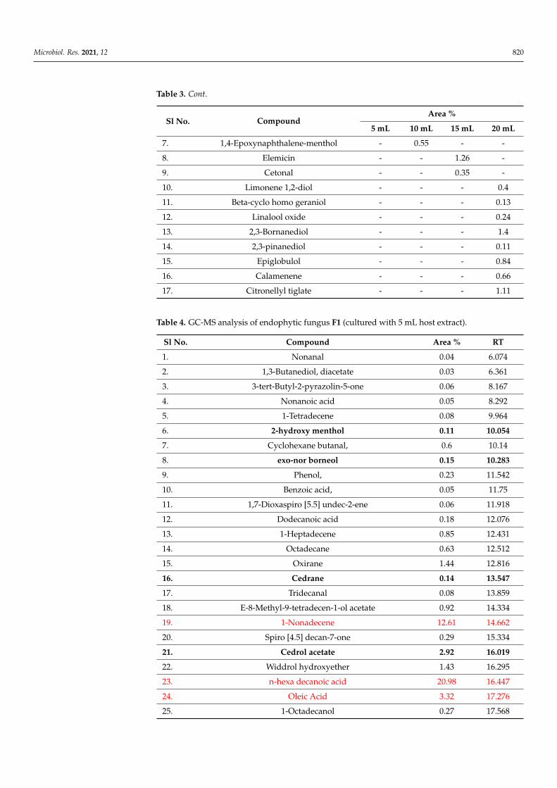
Microbiol. Res. 2021, 12 819
metabolites when compared to control (Figure 5; Table 3). With supplementation of 5 mL(5%) of the host plant extract, the fungi produced compounds such as exo-nor borneol(0.15%), cedrol acetate (2.92%), hydroxy menthol (0.11%), nonadecene (12.6%), hexade-cenoic acid (20.98%), 1,4-epoxynaphthalene-1(2H)-menthol (1.63%) and more (Table 4);with 10 mL (10%) supplementation it produced 2-hydroxymentol (0.14%), oxo-borneol(0.28%), alpha cyclo geraniol (0.4%), cetonal (0.36%), phthalic acid (14.64%), hexadecenoicacid (7.45%), campesterol (0.41%), 1,4-epoxynaphthalene-1(2H)-menthol (0.55%), gamma-sitosterol (2.22%) and more (Table 5); under 15 mL (15%) it produced 2-hydroxymenthol(0.15%), elemicin (1.26%), cedrane (1.06%), cetonal (0.35%), decanoic acid (11.9%), ph-thalic acid (21.69%), dibutyl phthalate (11.7%) along with other compounds (Table 6); and20 mL (20%) supplementation produced limonene (0.4%), beta-cyclo homo geraniol (0.13%),linalool oxide (0.24%), 2,3-bornanediol (1.4%), nor borneol (0.38%), 2,3-pinanediol (0.1%),epiglobulol (0.84%), di isobutyl phthalate (7.9%), hexadecenoic acid (8.03%), phthalic acid(9.23%) and citronellyl tiglate (1.11%) along with many other compounds (Table 7).
Microbiol. Res. 2021, 12, FOR PEER REVIEW 8
(0.15%), cedrol acetate (2.92%), hydroxy menthol (0.11%), nonadecene (12.6%), hexade-cenoic acid (20.98%), 1,4-epoxynaphthalene-1(2H)-menthol (1.63%) and more (Table 4); with 10 mL (10%) supplementation it produced 2-hydroxymentol (0.14%), oxo-borneol (0.28%), alpha cyclo geraniol (0.4%), cetonal (0.36%), phthalic acid (14.64%), hexadecenoic acid (7.45%), campesterol (0.41%), 1,4-epoxynaphthalene-1(2H)-menthol (0.55%), gamma-sitosterol (2.22%) and more (Table 5); under 15 mL (15%) it produced 2-hydroxymenthol (0.15%), elemicin (1.26%), cedrane (1.06%), cetonal (0.35%), decanoic acid (11.9%), phthalic acid (21.69%), dibutyl phthalate (11.7%) along with other compounds (Table 6); and 20 mL (20%) supplementation produced limonene (0.4%), beta-cyclo homo geraniol (0.13%), lin-alool oxide (0.24%), 2,3-bornanediol (1.4%), nor borneol (0.38%), 2,3-pinanediol (0.1%), ep-iglobulol (0.84%), di isobutyl phthalate (7.9%), hexadecenoic acid (8.03%), phthalic acid (9.23%) and citronellyl tiglate (1.11%) along with many other compounds (Table 7).
Figure 5. Biosynthesis of monoterpenoids in Cladosporium cladosporioides (F1-MH810309) with host plant by cross-talk. The green color represents precursor compounds from the plant extract. The blue color represents compounds produced by F1 in support of the plant extract. Orange color in-dicates the enzymes involved: GPP geraniol diphosphate, LS linalool synthase, OXD oxidase, GI geraniol isomerase, UN unclear reaction, ADH alcohol dehydrogenase, ORD oxidoreductase, GES geraniol synthase, BS bornyl synthase, BPH bornyl pyrophosphate hydrolase, AOXD asymmetric oxidase, BEDH borneol dehydrogenase, CH camphor hydroxylase, HCeDH hydroxy camphor de-hydrogenase, LO limonene-3-monooxygenase, IPEDH iso-piperitenol dehydrogenase, IPRed iso-piperitone reductase, IS isomerase, PRed pulegone reductase and MEDH menthol dehydrogenase, UN unclear reaction.
Table 3. GC-MS analysis of secondary metabolites of endophytic fungi F1 (Cladosporium cladospori-oides isolated from wild C. martnii with supplementation of host plant extract at different concentra-tions).
Sl No. Compound Area %
5 mL 10 mL 15 mL 20 mL 1. 2-Hydroxy menthol 0.11 0.14 0.15 -
2. Nor-borneol 0.15 0.28 - 0.38 3. Cedrane 0.14 - 1.06 - 4. Cedral acetate 2.92 - - - 5. Alpha cyclo geraniol - 0.4 - - 6. Campesterol - 0.41 - - 7. 1,4-Epoxynaphthalene-menthol - 0.55 - - 8. Elemicin - - 1.26 - 9. Cetonal - - 0.35 -
Figure 5. Biosynthesis of monoterpenoids in Cladosporium cladosporioides (F1-MH810309) with hostplant by cross-talk. The green color represents precursor compounds from the plant extract. Theblue color represents compounds produced by F1 in support of the plant extract. Orange colorindicates the enzymes involved: GPP geraniol diphosphate, LS linalool synthase, OXD oxidase,GI geraniol isomerase, UN unclear reaction, ADH alcohol dehydrogenase, ORD oxidoreductase,GES geraniol synthase, BS bornyl synthase, BPH bornyl pyrophosphate hydrolase, AOXD asymmet-ric oxidase, BEDH borneol dehydrogenase, CH camphor hydroxylase, HCeDH hydroxy camphordehydrogenase, LO limonene-3-monooxygenase, IPEDH iso-piperitenol dehydrogenase, IPRed iso-piperitone reductase, IS isomerase, PRed pulegone reductase and MEDH menthol dehydrogenase,UN unclear reaction.
Table 3. GC-MS analysis of secondary metabolites of endophytic fungi F1 (Cladosporium cladosporioidesisolated from wild C. martnii with supplementation of host plant extract at different concentrations).
Sl No. CompoundArea %
5 mL 10 mL 15 mL 20 mL
1. 2-Hydroxy menthol 0.11 0.14 0.15 -
2. Nor-borneol 0.15 0.28 - 0.38
3. Cedrane 0.14 - 1.06 -
4. Cedral acetate 2.92 - - -
5. Alpha cyclo geraniol - 0.4 - -
6. Campesterol - 0.41 - -
Microbiol. Res. 2021, 12 820
Table 3. Cont.
Sl No. CompoundArea %
5 mL 10 mL 15 mL 20 mL
7. 1,4-Epoxynaphthalene-menthol - 0.55 - -
8. Elemicin - - 1.26 -
9. Cetonal - - 0.35 -
10. Limonene 1,2-diol - - - 0.4
11. Beta-cyclo homo geraniol - - - 0.13
12. Linalool oxide - - - 0.24
13. 2,3-Bornanediol - - - 1.4
14. 2,3-pinanediol - - - 0.11
15. Epiglobulol - - - 0.84
16. Calamenene - - - 0.66
17. Citronellyl tiglate - - - 1.11
Table 4. GC-MS analysis of endophytic fungus F1 (cultured with 5 mL host extract).
Sl No. Compound Area % RT
1. Nonanal 0.04 6.074
2. 1,3-Butanediol, diacetate 0.03 6.361
3. 3-tert-Butyl-2-pyrazolin-5-one 0.06 8.167
4. Nonanoic acid 0.05 8.292
5. 1-Tetradecene 0.08 9.964
6. 2-hydroxy menthol 0.11 10.054
7. Cyclohexane butanal, 0.6 10.14
8. exo-nor borneol 0.15 10.283
9. Phenol, 0.23 11.542
10. Benzoic acid, 0.05 11.75
11. 1,7-Dioxaspiro [5.5] undec-2-ene 0.06 11.918
12. Dodecanoic acid 0.18 12.076
13. 1-Heptadecene 0.85 12.431
14. Octadecane 0.63 12.512
15. Oxirane 1.44 12.816
16. Cedrane 0.14 13.547
17. Tridecanal 0.08 13.859
18. E-8-Methyl-9-tetradecen-1-ol acetate 0.92 14.334
19. 1-Nonadecene 12.61 14.662
20. Spiro [4.5] decan-7-one 0.29 15.334
21. Cedrol acetate 2.92 16.019
22. Widdrol hydroxyether 1.43 16.295
23. n-hexa decanoic acid 20.98 16.447
24. Oleic Acid 3.32 17.276
25. 1-Octadecanol 0.27 17.568

Microbiol. Res. 2021, 12 821
Table 4. Cont.
Sl No. Compound Area % RT
26. Octadecanoic acid 7.9 18.307
27. Decanoic acid, 0.37 19.100
28. 1-Tetracosanol 1.38 20.234
29. Octadecanal 0.3 20.553
30. 2-palmitoyl glycerol 1.72 21.228
31. 1,2-Benzenedicarboxylic acid, mono (2-ethylhexyl 1.32 21.553
32. 1-Triacontanol 1.86 21.805
33. 10,12,14-Nonacosatriynoic acid 0.72 22.263
34. Abietic acid 1.3 22.385
35. Retinoic acid 0.43 22.544
36. Glyceryl mono acetate 3.99 22.629
37. Stearin 0.97 22.787
38. Squalene 0.93 23.568
39. Corticosterone 21-acetate 0.63 23.796
40. Trans-beta-lonone 0.58 23.934
41. Dehydroergosterol 3,5-dinitrobenzoate 0.92 24.848
42. Cholest-5-en-3-ol (3. beta) 0.24 26.134
43. Cycloeucalenol 0.36 26.73
44. Stigmasta-4,7,22-trien-3. alpha-ol 0.88 30.15
45. Hexa decanoic acid 1.42 30.562
46. Rhodopin 0.83 31.201
47. 1,4-Epoxynaphthalene-1(2H)-methanol 1.63 32.554
48. Ergosta-8,24(28)-dien-3-ol, 14-methyl-, (3. beta,5. alpha) 1.79 33.576
Red color indicates the compounds with high percentages. Bold indicates similar compounds present in theessential oil of the host plant. RT: retention time.
Table 5. GC-MS analysis of endophytic fungus F1 (cultured with 10 mL host extract).
Sl No. Compound Area % R Time
1. 1-Tetradecene 0.1 9.951
2. 2-hydroxymenthol 0.14 10.041
3. 2-hydroxybutyl acrylate 0.45 10.129
4. Oxo-borneol 0.28 10.271
5. 1-Heptadecene 0.93 12.418
6. Alpha-cyclo geraniol 0.4 14.319
7. 1-Nonadecene 2.12 14.649
8. 2,11-Dodecadiene 0.4 14.906
9. Cetonal 0.36 15.318
10. diisobutyl phthalate 19.28 15.547
11. l-(+)-Ascorbic acid 2,6-dihexadecanoate 1.12 16.297
12. n-Hexadecanoic acid 7.45 16.389
13. Dibutyl phthalate 12.3 16.494
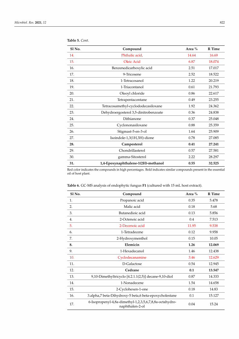
Microbiol. Res. 2021, 12 822
Table 5. Cont.
Sl No. Compound Area % R Time
14. Phthalic acid, 14.64 16.69
15. Oleic Acid 6.87 18.074
16. Benzenedicarboxylic acid 2.51 17.017
17. 9-Tricosene 2.52 18.522
18. 1-Tetracosanol 1.22 20.219
19. 1-Triacontanol 0.61 21.793
20. Oleoyl chloride 0.86 22.617
21. Tetrapentacontane 0.49 23.255
22. Tetracosamethyl-cyclododecasiloxane 1.92 24.362
23. Dehydroergosterol 3,5-dinitrobenzoate 0.36 24.838
24. Dithianone 0.37 25.048
25. Cyclononasiloxane 0.88 25.359
26. Stigmast-5-en-3-ol 1.64 25.909
27. Isoindole-1,3(1H,3H)-dione 0.78 27.085
28. Campesterol 0.41 27.241
29. Chondrillasterol 0.57 27.581
30. gamma-Sitosterol 2.22 28.297
31. 1,4-Epoxynaphthalene-1(2H)-methanol 0.55 32.525
Red color indicates the compounds in high percentages. Bold indicates similar compounds present in the essentialoil of host plant.
Table 6. GC-MS analysis of endophytic fungus F1 (cultured with 15 mL host extract).
Sl No. Compound Area % R Time
1. Propanoic acid 0.35 5.478
2. Malic acid 0.18 5.68
3. Butanedioic acid 0.13 5.856
4. 2-Octenoic acid 0.4 7.513
5. 2-Decenoic acid 11.95 9.538
6. 1-Tetradecene 0.12 9.958
7. 2-Hydroxymenthol 0.15 10.05
8. Elemicin 1.26 12.069
9. 1-Hexadecanol 1.46 12.438
10. Cyclodecanamine 3.46 12.629
11. D-Galactose 0.54 12.945
12. Cedrane 0.1 13.547
13. 9,10-Dimethyltricyclo [4.2.1.1(2,5)] decane-9,10-diol 0.87 14.333
14. 1-Nonadecene 1.54 14.658
15. 2-Cyclohexen-1-one 0.18 14.83
16. 3.alpha,7 beta-Dihydroxy-5 beta,6 beta-epoxycholestane 0.1 15.127
17. 6-Isopropenyl-4,8a-dimethyl-1,2,3,5,6,7,8,8a-octahydro-naphthalen-2-ol 0.04 15.24

Microbiol. Res. 2021, 12 823
Table 6. Cont.
Sl No. Compound Area % R Time
18. Cetonal 0.35 15.327
19. Di isobutyl phthalate 7.35 15.556
20. Phthalic acid 21.69 16.236
21. n-Hexadecanoic acid 9.16 16.417
22. Dibutyl phthalate 11.78 16.505
23. Oleic acid 7.73 17.261
24. Acetophenone 0.49 18.384
25. Pentaerythrityl tetrachloride 0.36 18.815
26. 4-Bromobutanoic acid 0.29 19.057
27. Pentaerythrityl tetrachloride 0.91 19.826
28. 1-Tetracosanol 1.02 20.226
29. Carbamazepine 0.33 20.651
30. 1-Triacontanol 0.55 21.795
31. glyceryl mono oleate 2.36 22.621
32. 1-Hexacosanol 0.35 23.255
33. 1-Nonadecene 2.01 18.53
Red color indicates the compounds in high percentages. Bold indicates similar compounds present in the essentialoil of host plant.
Table 7. GC-MS analysis of endophytic fungus F1 (cultured with 20 mL host extract).
Sl No. Compound Area % R Time
1. Glycerine 0.11 4.264
2. 2-Nonadecanone 0.13 7.756
3. Limonene glycol or P-menth-8-ene 0.4 9.191
4. Beta-cyclohomo geraniol 0.13 9.487
5. Linalool oxide 0.24 9.56
6. 2,3-Bornanediol 1.4 10.046
7. Cyclohexanebutanal 1.15 10.146
8. 2-Norborneol 0.38 10.278
9. Alpha-campholene aldehyde 0.18 10.418
10. Lomustine 0.13 10.637
11. Cyclohexanol 0.14 10.754
12. 10-Methyl-8-tetradecen-1-ol acetate 0.18 11.021
13. 4-Nonanone 0.22 11.245
14. Benzoic acid, 4-ethoxy-, ethyl ester 0.11 11.742
15. 1,7-Dioxaspiro [5.5] undec-2-ene 0.58 11.908
16. 1-Heptadecene 1.12 12.427
17. 6-Acetyl-4,4,7-trimethylbicyclo [4.1.0] heptan-2-one 0.42 12.735
18. 2,3-pinanediol 0.1 13.03
19. Bicyclo(3.1.1)heptane-2,3-diol, 2,6,6-trimethyl- 0.67 13.544
20. 1H-Benzocyclohepten-7-ol,2,3,4,4a,5,6,7,8-octahydro-1,1,4a,7-tetramethyl- 0.23 14.18

Microbiol. Res. 2021, 12 824
Table 7. Cont.
Sl No. Compound Area % R Time
21. 1-Cyclohexanone, 2-methyl-2-(3-methyl-2-oxobutyl) 7.3 14.337
22. 1-Nonadecene 3.41 14.662
23. 2,5,9-Tetradecatriene, 3,12-diethyl- 0.23 14.839
24. 2-Dodecen-1-yl (-) succinic anhydride 1.57 14.998
25. Epiglobulol 0.84 15.131
26. 6-Isopropenyl-4,8a-dimethyl-1,2,3,5,6,7,8,8a-octahydro-naphthalen-2-ol 1.28 15.246
27. Spiro [4.5] decan-7-one, 1,8-dimethyl-8,9-epoxy-4-isopropyl- 0.42 15.326
28. Diisobutyl phthalate 7.91 15.556
29. 3-Methyl-5-(1,4,4-trimethylcyclohex-2-enyl) pentan-1-ol 1.72 15.666
30. Calamenene 0.66 15.780
31. 2,6-Bis-(acetamido)-pyridine 0.93 16.115
32. Phthalic acid, isobutyl 2-pentyl ester 1.21 16.231
33. Widdrol hydroxyether 3.35 16.302
34. n-Hexadecanoic acid 8.03 16.429
35. Dibutyl phthalate 4.65 16.504
36. 1,2-Benzenedicarboxylic acid, butyl decyl ester 1.53 16.579
37. Phthalic acid, isobutyl octadecyl ester 9.23 16.709
38. 2,4,7,14-Tetramethyl-4-vinyl-tricyclo [5.4.3.0(1,8)]t 1.38 16.945
39. 1,2-Benzenedicarboxylic acid, butyl 8-methylnonyl ester 0.79 17.024
40. 2-naphthalene butanoic acid 5.94 17.161
41. 2-Methyl-5-(2,6,6-trimethyl-cyclohex-1-enyl)-pentane-2,3-diol 0.35 17.336
42. Phthalic acid, isobutyl 2-pentyl ester 0.56 17.416
43. 2-[4-methyl-6-(2,6,6-trimethylcyclohex-1-enyl)hexa-1,3,5-trienyl]cy 1.11 17.516
44. Citronellyl tiglate 1.11 17.761
45. Oleic acid 5.41 18.110
46. Octadecanoic acid 1.68 18.288
47. Benzo[b]dihydropyran, 6-hydroxy-4,4,5,7,8-pentamethyl- 1.6 18.390
48. 1-Tricosanol 2.78 18.529
49. Beta-sitosterol 0.17 18.867
50. 1-Tetracosanol 1.67 20.226
51. Oxirane, hexadecyl 0.15 20.546
52. Hexadecanoic acid, 0.41 21.223
53. 1,2-Benzenedicarboxylic acid, 3.68 21.548
54. 1-Triacontanol 1.06 21.795
55. 10,12,14-Nonacosatriynoic acid 0.24 22.259
56. Glyceryl monooleate 1.73 22.620
57. 1-Hexacosanol 0.77 23.252
58. 2,6,10,14,18,22-Tetracosahexaene, 2,6,10,15,19,23-hexamethyl- 0.21 23.559

Microbiol. Res. 2021, 12 825
Table 7. Cont.
Sl No. Compound Area % R Time
59. Dehydroergosterol 3,5-dinitrobenzoate 0.44 24.836
60. 9,19-Cycloergost-24(28)-en-3-ol, 4,14-dimethyl-, 0.18 26.716
61. Ergosta-5,8,22-trien-3-ol, (3 beta,22E) 0.54 27.008
62. Anthraergostatetraenol 0.13 27.239
63. Stigmasta-7,25-dien-3-ol, (3 beta, 5 alpha) 0.11 27.831
64. 17-Pentatriacontene 0.14 28.588
65. Rhodopin 0.96 31.174
66. 1,4-Epoxynaphthalene-1(2H)-methanol,4,5,7-tris(1,1-dimethylethyl)-3 0.72 32.520
67. Cholesta-8,14-dien-3-ol, 4,4-dimethyl- 0.43 33.532
Red color indicates the compounds in high percentages. Bold indicates similar compounds present in the essentialoil of host plant.
4. Discussion
During the present study, six endophytic fungal species like Alternaria, Aspergillus,Fusarium, Trichoderma, Curvularia and Cladosporium were isolated from wild Cymbopogonmartinii (MT90507). Furthermore, the Cladosporium species were studied (C. cladosporioidesand C. tenuissimum) based on their consistent growth throughout the year. The C. cladospo-rioides is also reported as endophytic fungi in the genus Cymbopogon such as C. citratus,C. flexuosus and C. caesius [41,42] expressing tremendous ecological adaptability [43].
The phytochemical analysis of C. cladosporioides (F1) and C. tenuissimum (F2) showedthe presence of secondary compounds belonging to alkaloids, flavonoids, phenols, ter-penoids and tannins which are similar to those of the host plant (wild C. martinii). TheGC-MS analysis of ethyl acetate extracts of endophytic fungi F1 and F2 (control-withoutadding host plant extract) revealed the presence of compounds like 1-octen-3-ol, 3-methyl-1-butanol, hexanol, trans-2-octanol and hexadecanol in similarity with the host plant extract.These compounds are known to possess antimicrobial, anti-inflammatory and antioxidantactivities [44]. Sharing of the above mentioned similar secondary metabolite compoundsbetween endophytic fungi and the host plant has been reported earlier [9,45]. Likewise,common compounds were shared between endophytic fungi F1 (control) and the hostplant such as octadecane, eicosane, hexadecane, heptadecane and 2,4-ditert-butyl phe-nol coming under alkanes producing in higher percentages along with the productionof geranyl acetate, cubenol and geraniol belonging to monoterpenoids. The productionof such an alkane group of compounds has been reported in endophytic fungi includingalso C. cladosporioides and C. tenuissimum and these are known to possess potential bio-logical activities [46–48]. Under in vitro conditions, the endophytic fungus F1 having apre-existing terpenoid pathway could produce compounds similar to that of the host plant.The transcriptome studies made on endophytic fungus C. cladosporioides expressed thepresence of a pathway for terpenoid biosynthesis [49–51].
The endophytic fungus F1 (test) produced a large number of plant derived secondarymetabolites belonging to hydrocarbons, hydrocarbons esters and monoterpenoid. There arereports on C. cladosporioides as endophyte producing compounds belonging to a monoter-penoid group [26,52–55]. Studies made on C. cladosporioides isolated from Taxus brevifolia,T. baccata, H. serrata and Tobacco species expressed the production of monoterpenoid com-pounds similar to that of host plants [28,29,56,57]. Supplementation of the host plant extractat different concentrations (5–20%) to broth culture resulted in variation in monoterpenoidcompound production in the endophytic fungi (Table 3). The F1 (test) might utilize theprecursor components from the host plant extract and convert them into biotransformedmonoterpenoid compounds (Figure 5), and similar studies have been made earlier (VijayLakshmi et al., 2019, 56]. These monoterpenoid compounds were produced in low percent-
Microbiol. Res. 2021, 12 826
ages due to the poor catalytic activity of plant derived enzymes in the endophytic system(Sun et al., 2019). The monoterpenoid compounds and their precursors are known toexpress antioxidant, anticancer and antimicrobial activities [7,58,59]. The present study re-vealed that the endophytic fungus F11 (C. cladosporioides) and the host plant share commonMevalonate (MVA) and Phenylpropanoid (PP) pathways as also documented in earlierstudies [7,50].
The endophytes show a strong relationship and long-term association with the hostplant and thus mimic the plant for secondary metabolite production [60]. The relation-ship shared between endophyte and plant could lead to coevolution and adaptation [61],which results in horizontal gene transfer, establishes cross-talk between gene clusters andhelps in the biosynthesis of common terpenoid compounds [43,51,62]. There are severalreports on revealing the capability of C. cladosporioides for adaptation within the host plantenvironment thus producing secondary metabolites similar to the host plant [9,21,43,63,64].The in vitro cultured endophytic fungus F2 (C. tenisimum) could not produce secondarymetabolites similar to F1 (C. cladosporioides) or the host plant as the metabolites were pro-duced for their basic survival activity within the host system and thus were not involvedin cross-talk with the plant [65].
In the current study, the endophytic fungus F1 has expressed high adoptability anda stronger relationship with the wild genotype of C. martinii and has been successful inproducing secondary metabolites similar to the host plant. Further, the mechanism ofthe molecular interactions happening during secondary metabolite production betweenendophytic fungi and the host plant needs elaborate studies using ‘omic’ approaches. Theinteraction of the endophytic fungi with the host plant explains the vital activities taken upby the sessile natured plant.
Author Contributions: H.J. wrote the original manuscript. V.M. contributed for tables, figures andmanuscript editing. K.J.T.S. revised the whole manuscript. All authors have read and agreed to thepublished version of the manuscript.
Funding: This research did not receive any specific grant from any funding agencies in public,commercial, or not-for profit sectors.
Institutional Review Board Statement: Not applicable.
Informed Consent Statement: Not applicable.
Data Availability Statement: The datasets generated during and/or analyzed during the currentstudy are available from the corresponding author on reasonable request.
Conflicts of Interest: The authors declare that they have no conflict of interest.
References1. Bor, N.L. The genus Cymbopogon Spreng. India, Burma and Ceylon, Part. I. J. Bombay Nat. Hist. Soc. 1953, 51, 890–916.2. Promila, P. A review on the medicinal and aromatic plant Cymbopogon martini (Roxb.) Watson (Palmarosa). Int. J. Chem. Stud.
2018, 6, 1311–1315.3. Ashwini Murthy, A.; Thara Saraswathi, K.T. Chemical profiling of leaf extract and essential oil in wild Cymbopogon martinii (Roxb.)
collected from Devarayanadurga hills (Tumkur, Karnataka). Int. J. Sci. Res. Rev. 2019, 8, 1–9.4. Smitha, G.R.; Dhaduk, H.L. A new chemotype of palmarosa [Cymbopogon martini (Roxb.) W. Watson] identified from ‘The Aravali
Range’ of Rajasthan, India. Med. Plants 2018, 10, 203–209. [CrossRef]5. Grenera. Nutrients: Lemon Grass Oil. Retrieved from Essential Oil. 2008.6. Farh, M.E.; Jeon, J. Roles of fungal volatiles from perspective of distinct lifestyles in filamentous fungi. Plant Pathol. J. 2020, 36,
193–203. [CrossRef] [PubMed]7. Chen, Y.; Hu, B.; Xing, J.; Li, C. Endophytes: The novel sources for plant terpenoid biosynthesis. Appl. Microbiol. Biotechnol. 2021,
105, 4501–4513. [CrossRef] [PubMed]8. Pagare, S.; Bhatia, M.; Tripathi, N.; Pagare, S.; Bansal, Y.K. Secondary metabolites of plants and their role: Overview. Curr. Trends
Biotechnol. Pharm. 2015, 9, 293–304.9. Roy, S.; Banerjee, D. Volatile Organic Compounds from Endophytic fungi. In Recent Advance in White Biotechnology through Fungi,
Fungal Biology; Springer: Cham, Germany, 2019; pp. 149–176.
Microbiol. Res. 2021, 12 827
10. Singh, A.; Singh, D.K.; Kharwar, R.N.; White, J.F.; Gond, S.K. Fungal endophytes as efficient sources of plant-derived bioactivecompounds and their prospective applications in natural product drug discovery: Insights, avenues and challenges. Microorgan-isms 2021, 19, 197. [CrossRef] [PubMed]
11. Shah, G.; Shri, R.; Panchal, V.; Sharma, N.; Singh, B.; Mann, A.S. Scientific basis for the therapeutic use of Cymbopogon citratus,Staf (lemon grass). J. Adv. Pharm. Technol. Res. 2011, 2, 3–8. [CrossRef]
12. Singh, R.P.; Sharad, S.; Kapur, S. Free radicals and oxidative stress in neurodegenerative diseases: Relevance of dietary antioxidants.J. Indian Acad. Clin. Med. 2004, 5, 218–225.
13. Balakrishnan, B.; Paramasivam, S.; Arulkumar, A. Evaluation of the lemongrass plant (Cymbopogon citratus) extracted indifferent solvents for antioxidant and antibacterial activity against human pathogens. Asian Pac. J. Trop. Dis. 2014, 4, 134–139.[CrossRef]
14. Rodriguez, R.J.; Henson, J.; Van Volkenburgh, E.; Hoy, M.; Wright, L.; Beckwith, F.; Kim, Y.O.; Redman, R.S. Stress tolerance inplants via habitat-adapted symbiosis. ISME J. 2008, 2, 404. [CrossRef]
15. Ali, S.; Duan, J.; Charles, T.C.; Glick, B.R. A bioinformatics approach to the determination of genes involved in endophyticbehavior in Burkholderia spp. J. Theor. Biol. 2014, 343, 193–198. [CrossRef] [PubMed]
16. Gouda, S.; Kerry, R.G.; Das, G.; Paramithiotis, S.; Shin, H.S.; Patra, J.K. Revitalization of plant growth-promoting rhizobacteria forsustainable development in agriculture. Microbiol. Res. 2018, 206, 131–140. [CrossRef] [PubMed]
17. Chowdappa, S.; Jagannath, S.; Konappa, N.; Udayashankar, A.C.; Jogaiah, S. Udayashankar and Sudisha Jogaiah. Detection andCharacterization of antibacterial siderophores secreted by endophytic fungi from Cymbidium aloifolium. Biomolecules 2020, 10,1412. [CrossRef]
18. Ganjewala, D.; Gupta, A.K. Lemongrass (Cymbopogon flexuosus. Steud.) Wats essential oil: Overview and biological activities.Recent Prog. Med. Plants 2013, 37, 235–271.
19. Khare, E.; Mishra, J.; Arora, N.K. Multifaceted interactions between endophytes and plant: Developments and prospects. Front.Microbiol. 2018, 9, 2732. [CrossRef] [PubMed]
20. Potshang Bam, M.; Devi, S.I.; Sahoo, D.; Strobel, G.A. Functional characterization of endophytic fungal community associatedwith Oryza sativa L. and Zea mays L. Front. Microbiol. 2017, 8, 325. [CrossRef]
21. Morath, S.U.; Hung, R.; Bennett, J.W. Fungal volatile organic compounds: A review with emphasison their biotechnologicalpotential. Fungal Biol. Rev. 2012, 30, 73–83. [CrossRef]
22. Strobel, G.; Daisy, B. Bioprospecting for microbial endophytes and their natural products. Microbiol. Mol. Biol. Rev. 2003, 67,491–502. [CrossRef]
23. Gouda, S.; Das, G.; Sen, S.K.; Shin, H.-S.; Patra, J.K. Endophytes: A treasure house of bioactive compounds of medicinalimportance. Front. Microbiol. 2016, 7, 1538. [CrossRef] [PubMed]
24. Lutfia, A.; Munir, E.; Yurnaliza, Y.; Basyuni, M. Chemical analysis and anticancer activity of sesterterpenoid from an endophyticfungus Hypomontagnella monticulosa Zg15SU and its host Zingiber griffithii Baker. Heliyon 2021, 7, e06292. [CrossRef]
25. Kusari, S.; Zühlke, S.; Spiteller, M. An endophytic fungus from Camptotheca acuminata that produces camptothecin and analogues.J. Nat. Prod. 2009, 72, 2–7. [CrossRef] [PubMed]
26. Chen, M.; Yang, L.; Li, Q.; Shen, Y.; Shao, A.; Lin, S.; Huang, L. Volatile metabolites analysis and molecular identification ofendophytic fungi bn12 from Cinnamomum camphora var. borneol. China J. Chin. Mater. Med. 2011, 36, 3217–3221. [CrossRef]
27. Stierle, A.; Stierle, D.; Stierle, S. Bioactive compounds from four endophytic Penicillium sp. of a northwest pacific yew tree. Nat.Prod. Chem. 2000, 24, 933–977.
28. Zhu, D.; Wang, J.; Zeng, Q.; Zhang, Z.; Yan, R. A novel endophytic Huperzine A-producing fungus Shiraia sp. Slf14, isolatedfrom Huperzia serrata. J. Appl. Microbiol. 2010, 109, 1469–1478. [CrossRef]
29. Kasaei, A.; Mobini-Dehkordi, M.; Mahjoubi, F.; Saffar, B. Isolation of Taxol-producing endophytic fungi from Iranian yew throughnovel molecular approach and their effects on human breast cancer cell line. Curr. Microbiol. 2017, 74, 702–709. [CrossRef]
30. Pimentel, I.C.; Glienke-Blanco, C.; Gabardo, J.; Stuart, R.M.; Azevedo, J.L. Identification and colonization of endophytic fungifrom soyabean (Glycine max (L.) Merril) under different environmental conditions. Braz. Arch. Biol. Technol. 1998, 49, 705–711.[CrossRef]
31. Landum, M.C.; do Rosário Félix, M.; Alho, J.; Garcia, R.; Cabrita, M.J.; Rei, F. Antagonistic activity of fungi of Olea europaea L.against Colletotrichum acutatum. Microbiol. Res. 2016, 183, 100–108. [CrossRef] [PubMed]
32. Sofowora, A. Medicinal Plants and Traditional Medicine in Africa, 2nd ed.; Scientific Research; Spectrum Books Limited: Ibadan,Nigeria, 1993; pp. 1–153.
33. Harborne, A.J. Phytochemical Methods—A Guide to Modern Techniques of Plant Analysis; Springer Science & Business Media:Dordrecht, The Netherlands, 1973. [CrossRef]
34. Danmalam, U.H.; Hanwa, U.A.; Abdurrazak, A.; Hassan Maidoki, A.L.; Ambi, A.A. Phytochemical analysis of some crotalariaspecies growing in Samaru-zaria, Nigeria for the presence of pyrrolizidine alkaloids. Trends Sci. Technol. J. 2017, 2, 1035–1037.
35. Desire, M.H.; Bernard, F.; Forsah, M.R.; Assang, C.T.; Denis, O.N. Enzymes and qualitative phytochemical screening of endophyticfungi isolated from Lantana camara Linn. Leaves. J. Appl. Biol. Biotechnol. 2014, 2, 1–6.
36. Escribano, J.; Cabanes, J.; Jiménez-Atiénzar, M.; Ibañez-Tremolada, M.; Gómez-Pando, L.R.; García-Carmona, F.; Gandía-Herrero,F. Characterization of betalains, saponins and antioxidant power in differently colored quinoa (Chenopodium quinoa) varieties.Food Chem. 2017, 234, 285–294. [CrossRef]

Microbiol. Res. 2021, 12 828
37. Malik, S.K.; Ahmed, M.; Khan, F. Identification of novel anticancer terpenoids from Prosopis juliflora (Sw) DC (Leguminosae) pods.Trop. J. Pharm. Res. 2018, 17, 661–668. [CrossRef]
38. Eghbaliferiz, S.; Emami, S.A.; Tayarani-Najaran, Z.; Iranshahi, M.; Shakeri, A.; Hohmann, J.; Asili, J. Cytotoxic diterpene quinonesfrom Salvia tebesana Bunge. Fitoterapia 2018, 128, 97–101. [CrossRef]
39. Unuigbe, C.; Enahoro, J.; Erharuyi, O.; Okeri, H.A. Phytochemical analysis and antioxidant evaluation of lemon grass (Cymbopogoncitratus DC.) Stapf leaves. J. Appl. Sci. Environ. Manag. 2019, 23, 223–228. [CrossRef]
40. Sarkar, M. Investigation of In-Vitro Antioxidant Potential, Brine Shrimp Lethality and Thrombolytic Activity in Ficus Mollis VahlLeaves along with Phytochemical Screening. Bachelor’s Thesis, BRAC University, Dhaka, Bangladesh, 2018.
41. Deshmukh, S.K.; Kolet, M.J.; Verekar, S.A. Distribution of endophytic fungi in lemon grass (Cymbopogon citratus (DC). Stapf.). J.Cell Tissue Res. 2010, 10, 2263–2267.
42. Krishnamurthy, Y.L.; Hemalatha, T.V. Isolation of endophytic fungi from some grasses. J. Mycol. Plant Pathol. 2003, 33, 305–306.43. Salvatore, M.M.; Andolfi, A.; Nicoletti, R. The genus Cladosporium: A rich source of diverse and bioactive natural compounds.
Molecules 2021, 26, 3959. [CrossRef]44. Adeleke, B.S.; Babalola, O.O. Pharmacological potential of fungal endophytes associated with medicinal plants: A review. J. Fungi
2021, 7, 147. [CrossRef] [PubMed]45. Zhao, J.; Zhou, L.; Wang, J.; Shan, T.; Zhong, L.; Liu, X.; Gao, X. Endophytic fungi for producing bioactive compounds originally
from their host plants. Curr. Res. Technol. Educ. Trop. Appl. Microbiol. Microbial. Biotechnol. 2010, 1, 567–576.46. Sharma, D.; Paramanik, A.; Agarwal, P.K. Evaluation of bioactive secondary metabolites from endophytic fungus Pestalotiopsis
neglecta BAB-5510 isolated from leaves of Cupressus torulosa D. Don. 3 Biotech 2016, 6, 210. [CrossRef]47. Kanjana, M.; Kanimozhi, G.; Udayakumar, R.; Panneerselvam, A. GC-MS analysis of bioactive compounds of endophytic fungi
Chaetomium globosum, Cladosporium tenuissimum and Penicillium janthinellum. J. Biomed. Pharm. Sci. 2019, 2, 123.48. Kaur, N.; Arora, D.S.; Kalia, N.; Kaur, M. Bioactive potential of endophytic fungus Chaetomium globosum and GC-MS analysis of
its responsible components. Sci. Rep. 2020, 10, 18792. [CrossRef]49. Li, J.Y.; Sidhu, R.S.; Ford, E.; Long, D.; Hess, W.; Strobel, G. The induction of taxol production in the endophytic fungus–Periconia
sp. From Torreya grandifolia. J. Ind. Microbiol. Biotechnol. 1998, 20, 259–264. [CrossRef]50. Miao, L.Y.; Mo, X.C.; Xi, X.Y.; Zhou, L.; De, G.; Ke, Y.S.; Liu, P.; Song, F.J.; Jin, W.W.; Zhang, P. Transcriptome analysis of a
taxol-producing endhphytic fungus Cladosporium cladosporioides MD2. AMB Express 2018, 8, 41. [CrossRef]51. Zhang, Q.; Chen, X.; Xu, C.; Zhao, H.; Zhang, X.; Zeng, G.; Qian, Y.; Liu, R.; Guo, N.; Mi, W.; et al. Horizontal gene transfer
allowed the emergence of broad host range entomopathogens. Proc. Natl. Acad. Sci. USA 2019, 116, 7982–7989. [CrossRef]52. Paul, D.; Park, K.S. Identification of Volatile produced by Cladosporium cladosporioides CL-1, a fungal biocontrol agent that
promotes plant growth. Sensors 2013, 13, 13969–13977. [CrossRef]53. Gao, J.; Xu, A.; Tang, X. Isolation, identification and volatile compound analysis of an aroma-producing endophytic yeast from
romaine lettuce. Food Sci. 2011, 23, 33.54. Nithya, K.; Muthumary, J. Secondary metabolite from Phomopsis sp. isolated from Plumeria acutifolia Poiret. Recent Res. Sci.
Technol. 2010, 2, 99–103.55. Suwannarach, N.; Kumla, J.; Bussaban, B.; Nuangmek, W.; Matsui, K.; Lumyong, S. Biofumigation with the endophytic fungus
Nodulisporium spp. CMU-UPE34 to control postharvest decay of citrus fruit. Crop Prot. 2013, 45, 63–70. [CrossRef]56. De Souza, J.J.; Vieira, I.J.C.; Rodrigues-Filho, E.; Braz-Filho, R. Terpenoids form endophytic fungi. Molecules 2011, 16, 10604–10618.
[CrossRef] [PubMed]57. Wani, M.C.; Taylor, H.L.; Wall, M.E.; Cogoon, P.; Mcphail, A.T. Plant as antitumor agents. VI. Isolation and structure of taxol, a
novel antileukemic and antitumor agent from Taxus brevifolia. J. Am. Chem. Soc. 1971, 93, 2325–2327. [CrossRef] [PubMed]58. Kusari, S.; Pandey, S.P.; Spiteller, M. Untapped mutualistic paradigms linking host plant and endophytic fungal production of
similar bioactive secondary metabolites. Phytochemistry 2013, 91, 81–87. [CrossRef] [PubMed]59. Newman, D.J. Are microbial endophytes the ‘actual’ producers of bioactive antitumor agents? Trends Cancer 2018, 4, 662–670.
[CrossRef]60. Germaine, K.; Liu, X.; Cabellos, G.; Hogan, J.; Ryan, D.; Dowling, D.N. Bacterial endophyte-enhanced phyto-remediation of the
organochlorine herbicide 2,4 dichlorophenoxyacetic acid. FEMS Microbiol. Ecol. 2006, 57, 302–310. [CrossRef]61. Kirby, J.; Keasling, J.D. Biosynthesis of plant isoprenoids: Perspectives for microbial engineering. Annu. Rev. Plant Biol. 2009, 60,
335–355. [CrossRef]62. Tiwari, P.; Bae, H. Horizontal gene transfer and endophytes: An implication for the acquisition of novel traits. Plants 2020, 9, 305.
[CrossRef]63. Bennett, J.W.; Inamadar, A.A. Are some fungal volatile organic compounds (VOCs) mycotoxins? Toxins 2015, 7, 3785–3804.
[CrossRef]64. Bensh, K.; Groenewald, J.Z.; Starink-Willemse, M.; Andersen, B.; Sumerell, B.A.; Shin, H.D. Species and ecological diversity
within the Cladosporium cladosporioides complex (Davidiellacear, Capnodiales). Stud. Mycol. 2010, 67, 1–94. [CrossRef]65. Gunatilaka, A.A.L. Natural products from plant-associated microorganisms: Distribution, structural diversity, bioactivity, and
implications of their occurrence. J. Nat. Prod. 2006, 69, 509–526. [CrossRef]