A new sesquiterpenoid metabolite from Psilocybe samuiensis
12
1 Title Two new sesquiterpenoids from culture broth of Psilocybe samuiensis Authors Pornpakakul, S., a * Suwancharoen, S., a Petsom, A., a Roengsumran, S., a Muangsin, N., a Chaichit, N., b Piapukiew, J., c Sihanonth, P., d Allen, J. W. e * Corresponding author. Tel.: +66-2-2187637, +66-81-5620555; fax: +66-2- 2187598; e-mail: [email protected]. a Department of Chemistry, Faculty of Science, Chulalongkorn University, Thailand, Phyathai Road, Bangkok 10330 Thailand b Department of Physics, Faculty of Science and Technology, Thammasart University, Thailand, Pathumthani 12121 Thailand c Department of Botany, Faculty of Science, Chulalongkorn University, Thailand, Phyathai Road, Bangkok 10330 Thailand d Department of Microbiology, Faculty of Science, Chulalongkorn University, Thailand, Phyathai Road, Bangkok 10330 Thailand e P.O. Box 45164 Seattle, Washington 98105, U.S.A
Transcript of A new sesquiterpenoid metabolite from Psilocybe samuiensis

1
Title
Two new sesquiterpenoids from culture broth of Psilocybe samuiensis
Authors
Pornpakakul, S.,a* Suwancharoen, S.,a Petsom, A.,a Roengsumran, S.,a
Muangsin, N.,a Chaichit, N.,b Piapukiew, J.,c Sihanonth, P.,d Allen, J. W.e
*Corresponding author. Tel.: +66-2-2187637, +66-81-5620555; fax: +66-2-
2187598; e-mail: [email protected].
aDepartment of Chemistry, Faculty of Science, Chulalongkorn University,
Thailand, Phyathai Road, Bangkok 10330 Thailand
bDepartment of Physics, Faculty of Science and Technology, Thammasart
University, Thailand, Pathumthani 12121 Thailand
cDepartment of Botany, Faculty of Science, Chulalongkorn University,
Thailand, Phyathai Road, Bangkok 10330 Thailand
dDepartment of Microbiology, Faculty of Science, Chulalongkorn
University, Thailand, Phyathai Road, Bangkok 10330 Thailand
eP.O. Box 45164 Seattle, Washington 98105, U.S.A

2
Abstract
Two new ent-2,3-secoaromadendrane-type sesquiterpenoids,
psilosamuiensin A and psilosamuiensin B, were isolated from the broth of
Psilocybe samuiensis. Their structures were established by spectroscopic
data and the configurations of psilosamuiensin A were confirmed by single
crystal X-ray crystallographic analysis. Both compounds were inactive
when tested for antimicrobial activity against Bacillus subtilis ATTC 6633,
Staphyllococcus aureus ATTC 25923, Escherichia coli ATTC 25922,
Pseudomonas aeruginosa ATTC 27853 and Candida albicans ATTC 10231
using disk diffusion assay at 200 µg/disc and for cytotoxic activity against
HEP-G2 (hepatoma), SW620 (colon), Chago (lung), KATO-3 (gastric) and
BT474 using MTT colorimetric method at concentration of 100 µg/ml.
Keywords
Psilocybe samuiensis; magic mushroom; hallucinogenic mushroom,
secondary metabolite; sesquiterpene; secoaromadendrane.
ขอคิดเห็น[G1]: 100 หรือ 10
3
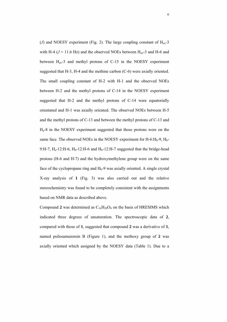
1. Introduction
Magic mushroom or hallucinogenic mushroom is the name most commonly
given to mushrooms that grow naturally and have hallucinogenic properties
(Seivewright and Lagundoye, 2000). After the psychedelic effect of some
species of genus Psilocybe was first reported by Wasson in 1957 (Wasson,
1957), Hofman and co-workers then isolated two important hallucinogenic
compounds, psilocybin as the main psychotropic compound, and psilocin
from Psilocybe mexicana (Hofmann et al., 1958; Hofmann et al., 1959).
Later Leung and Pual found two more 4-phosphoryloxytryptamine
derivatives, baeocystin and norbaeocystin, from Psilocybe baeocystis
(Leung and Pual, 1968) and Koike and co-workers found psilocybin and
psilocin together with ergosterol, ergosterol peroxide and α,α-trehalose
from dried fruiting bodies of Psilocybe argentipes (Koike et al., 1981). Due
to psychedelic effect of these mushrooms, they were investigated
worldwide. It found that they belong to the genus Panaeolus, Psilocybe,
Conocybe, Gymnopilus, Inocybe and Pluteus and mainly belong to the
genus Panaeolus and Psilocybe (Stamets, 1996) and the psilocybin and
psilocin content in various species was highly variable depended on species
(Beug and Bigwood 1981; Beug and Bigwood 1982; Marcano et al. 1994;
Gartz et al. 1994; Musshoff et al. 2000) and mainly contained in cap of the
mushroom (Tsujikawa 2003).

4
In 1991 Allen reported the exploration of the hallucinogenic mushrooms in
Thailand (Allen, 1991). He found that the mushrooms included the
following species: P. cubensis, P. subcubensis, Copelandia cyanescens
(Berkeley et Broome) Singer, and a new bluing Psilocybe species, Psilocybe
samuiensis Guzman, Bandala and Allen (Allen and Merlin 1992; Gartz et
al., 1994; Guzmán et al., 1993.). The alkaloid content of both naturally
occurring and in vitro cultivated fruit bodies of P. samuiensis was analyzed
by HPLC and the results revealed that high concentrations of psilocybin and
psilocin and small amounts of baeocystin were detected (Allen, 1992;
Guzmán et al., 1993; Gartz et al., 1994).
Recently we studied the metabolites of P. samuiensis cultured in malt
extract broth (MEB) and we found two new ent-2,3-secoaromadendrane-
type sesquiterpenoids, named psilosamuiensin A 1 and psilosamuiensin B 2
(Figure 1), from the ethyl acetate extract of culture broth. Herein we
described the isolation and structural elucidation of those compounds and
their antimicrobial activity and for cytotoxic activity.
2. Results and Discussion
On the basis of 1H NMR analysis, the hexane, ethyl acetate and methanol
extracts of Psilocybe samuiensis mycelia were mainly fatty acid. The culture
broth of Psilocybe samuiensis was concentrated and then extracted with
hexane, ethyl acetate and methanol, respectively. The hexane extract of the

5
culture broth was obtained only in a small amount. The ethyl acetate extract
of the culture broth was isolated by column chromatography in stepwise
fashion. Compound 1 was isolated as a major metabolite (67 mg/litre of
broth). The molecular formula of 1 was established as C15H26O4 on the basis
of HRESIMS which indicated three degrees of unsaturation. IR spectrum
was consistent with the presence of hydroxyl functional group (3425-3352
cm-1). 1H-NMR spectrum of 1 showed three signals of methyl groups at δH
0.83, 1.13 and 1.34 and five signals of the protons attached to carbons
bearing oxygen atom at δH 3.27, 3.55, 3.42, 3.87 and 5.30. The 13C-NMR
and HSQC experiments identified 15 carbon signals consisting of three
methyl carbons at δC 11.4, 14.5 and 31.6, four methylene carbons at δC 18.4,
38.6, 61.8 and 73.4, six methine carbons at δC 21.5, 24.2, 30.4, 35.6, 56.6
and 92.9 and two quaternary sp3-carbons at δC 26.5 and 73.3. Additionally
the carbon signals at δC 92.9, 73.4, 73.4 and 61.8 indicated that these
carbons attached with oxygen atom and the carbon signal at δC 92.9 was an
anomeric carbon. Since no sp2- or sp-carbon showed in the 13C-NMR data
and the three unsaturations were accounted for, it was implied that 1 should
contain three rings. A series of COSY and HSQC experiments established
partial connectivities as showed in Fig. 2 in bold. The assignment of 1 was
made from HMBC and NOESY experimental data (Table 1) and the crucial
HMBC and NOESY correlations were shown in Figure 2. The relative
stereochemistry of 1 was determined by a combination of coupling constant
6
(J) and NOESY experiment (Fig. 2). The large coupling constant of Hax-3
with H-4 (J = 11.6 Hz) and the observed NOEs between Hax-3 and H-6 and
between Hax-3 and methyl protons of C-15 in the NOESY experiment
suggested that H-3, H-4 and the methine carbon (C-6) were axially oriented.
The small coupling constant of H-2 with H-1 and the observed NOEs
between H-2 and the methyl protons of C-14 in the NOESY experiment
suggested that H-2 and the methyl protons of C-14 were equatorially
orientated and H-1 was axially oriented. The observed NOEs between H-5
and the methyl protons of C-13 and between the methyl protons of C-13 and
Ha-8 in the NOESY experiment suggested that those protons were on the
same face. The observed NOEs in the NOESY experiment for H-6:Hb-9, Hb-
9:H-7, Ha-12:H-6, Hb-12:H-6 and Hb-12:H-7 suggested that the bridge-head
protons (H-6 and H-7) and the hydroxymethylene group were on the same
face of the cyclopropane ring and Hb-9 was axially oriented. A single crystal
X-ray analysis of 1 (Fig. 3) was also carried out and the relative
stereochemistry was found to be completely consistent with the assignments
based on NMR data as described above.
Compound 2 was determined as C16H28O4 on the basis of HRESIMS which
indicated three degrees of unsaturation. The spectroscopic data of 2,
compared with those of 1, suggested that compound 2 was a derivative of 1,
named psilosamuiensin B (Figure 1), and the methoxy group of 2 was
axially oriented which assigned by the NOESY data (Table 1). Due to a

7
presence of the hemiacetal, compound 1 may undergo the reaction with
methanol during isolation to provide 2. To prove this doubt compound 1
was treated with excess methanol in the presence of a catalytic amount of p-
toluenesulfonic acid and without p-toluenesulfonic acid and the reactions
were monitored by TLC. It found that the reaction under acid condition was
complete within 3 hours while the reaction under non-acid condition was
undergone very slowly. After evaporation of the solvent and purification by
silica gel column chromatography, 1H and 13C NMR spectra indicated that
the product from the reaction was mainly compound 2. This result suggested
that compound 2 may be arisen during the process of extraction and
isolation and crystallization of 1.
Furthermore, compound 1 and 2 were tested for antimicrobial activity
against Bacillus subtilis ATTC 6633, Staphyllococcus aureus ATTC 25923,
Escherichia coli ATTC 25922, Pseudomonas aeruginosa ATTC 27853 and
Candida albicans ATTC 10231 using disk diffusion assay and for cytotoxic
activity against HEP-G2 (hepatoma), SW620 (colon), Chago (lung), KATO-
3 (gastric) and BT474 using MTT [3-(4,5-dimethylthiazol-2-yl)-2,5-
diphenyltetrazolium bromide] colorimetric method. Both were inactive
against those five standard microorganisms at 200 µg/disc and those five
cell lines at concentration of 100 µg/ml.
ขอคิดเห็น[G2]: 100 หรือ 10

8
3. Experimental
3.1 General experimental procedures
1H and 13C NMR spectra were recorded on a Varian Mercury +400 MHz
NMR spectrometer (1H at 400 MHz and 13C at 100 MHz). Chloroform-d
(CDCl3) was used in NMR experiments and chemical shifts (δ) were
referenced the signals of residual solvents at δ 7.26 ppm (1H) and 77.0 ppm
(13C). HRESIMS spectra were recorded on Micromass LCT (LC/MS).
Optical rotations were measured on a Perkin Elmer 341 polarimeter, using a
sodium lamp at wavelength 589 nm. FT-IR spectra were recorded on a
Perkin-Elmer Model 1760X Fourier Transform Infrared Spectrophotometer.
Melting points were examined using a Fisher-John melting point apparatus.
All solvents used for column chromatography were commercial grade and
were distilled prior to use. TLC were carried out on precoated silica gel 60
(Merck's TLC aluminium sheet, silica gel 60 F254 Art. 1.05554.0001) and
spots were detected under UV (254 and 365 nm) before and after spraying
with a vanillin/sulphuric acid solution followed by heating the plate.
Isolations were carried out using column chromatography (CC) [silica gel
60 (Merck Art. 1.09385.9025, 0.040-0.063 mm)]. Malt extract for culture of
the fungus was purchased from Himedia.

9
3.2 Psilocybe samuiensis
Spores print dry specimens of Psilocybe samuiensis from Koh Samui, Surat
Thani Province, Thailand were received from John W. Allen in July 2004.
The spores print was used for cultivation.
3.3 Cultivation, extraction and isolation
The spores of Psilocybe samuiensis from spore print were streaked
in Petri dishes containing Potato Dextrose Agar (PDA). The Petri dishes
were incubated at room temperature (25-30oC) and examined for fungal
mycelium from spores. Outgrowing mycelia were purified and transferred
into others Petri dishes containing PDA. Stock culture of P. samuiensis was
grown on MEA at room temperature (25-30oC) for 2 weeks. The agar was
then cut with a flamed 8 mm diameter cork borer. Five pieces of agar
cultures were inoculated into 250 ml Erlenmeyer flasks containing 100 ml
of MEB (x 110), and then statically cultured at ambient temperature for 11
weeks. The culture was filtered through filter paper (Whatman No. 93) and
the broth was then concentrated under reduced pressure at 35oC into 500 ml.
The concentrated broth was extracted with 200 ml of hexane (x 5), with 200
ml of ethyl acetate (x 5) and with 200 ml of methanol (x 5), respectively.
The hexane extract of broth was obtained in a small amount. The combined
ethyl acetate layers were dried over anhydrous sodium sulphate and then
evaporated under reduced pressure at 35oC to yield the ethyl acetate extract

10
(5.88 g) as yellowish brown viscous liquid. The ethyl acetate extract (4.97
g) was chromatographed on silica gel column eluted with CH2Cl2-MeOH
gradient in a stepwise fashion (3% MeOH to 100% MeOH). Fractions with
similar components were combined together according to the TLC profile.
Compound 1 was obtained from elution of 3% methanol in
dichloromethane. The solvent was removed by rotary evaporation and the
residue was obtained as white solid mixed with yellow viscous liquid (960
mg). The mixture was purified by crystallization from dichloromethane-
methanol to give compound 1 as colorless crystals. The filtrate was further
purified by silica gel column chromatography (Merck's silica gel 60 Art.
1.09385.9025), and crystallization from dichloromethane-methanol to give
compound 1 as colorless crystals. Both crystals of 1 were combined and
total amount of 1 was 736 mg (67 mg/litre of broth).
After evaporation of the filtrate from purification of compound 1, a yellow
viscous liquid (179 mg) was obtained. The residue was purified by TLC to
give compound 2 as colorless oil (19 mg).
3.3.1 Psilosamuiensin A 1
Colorless crystals; m.p. 91-92 oC; [α]D20 -48 (MeOH, c 0.25); νmax
(KBr) 3425-3352, 2960, 2927, 2872, 1140, 1102, 1043 and 1013 cm-1; δH
(CDCl3, 400 MHz) 0.83 (3H, d, J = 7.2 Hz, H-15), 0.91 (1H, m, H-7), 0.93
(1H, m, H-6), 1.13 (3H, s, H-13), 1.34 (3H, s, H-14), 1.53 (1H, m, Ha-8),

11
1.55 (1H, dd, J = 6.8, 14.4 Hz, Ha-9), 1.69 (1H, m, Hb-8), 1.94 (1H, br s, H-
1), 2.01 (1H, m, H-4), 2.09 (1H, m, H-5), 2.11 (1H, dd, J = 12.8, 14 Hz, Hb-
9), 3.27 (1H, d, J = 10.8 Hz, Hb-12), 3.42 (1H, dd, J = 11.6, 4.4 Hz, Heq-3),
3.55 (1H, d, J = 10.4, Ha-12), 3.87 (1H, t, J = 11.6 Hz, Hax-3), and 5.30 (1H,
br s, H-2) ppm; δC (CDCl3, 100 MHz) 11.4 (C-13), 14.5 (C-15), 18.4 (C-8),
21.5 (C-6), 24.2 (C-7), 26.5 (C-11), 30.4 (C-5), 31.6 (C-14), 35.6 (C-4),
38.6 (C-9), 56.6 (C-1), 61.8 (C-3), 73.3 (C-10), 73.4 (C-12), and 92.9 (C-2)
ppm; ESI-TOF MS m/z 293.1732 [M+Na]+; C15H26O4Na calc 293.1729.
3.3.2 Psilosamuiensin B 2
[α]D20 -51 (CHCl3, c 0.07,); νmax (film) 3397, 2967, 2931, 2878, 1129, 1103
and 1045 cm-1; δH (CDCl3, 400 MHz) 0.77 (3H, d, J = 6.8 Hz, H-15), 0.82
(1H, m, H-7), 0.84 (1H, m, H-6), 1.08 (3H, s, H-13), 1.24 (3H, s, H-14),
1.47 (1H, m, Ha-8), 1.47 (1H, m, Ha-9), 1.64 (1H, m, Hb-8), 1.89 (1H, br s,
H-1), 1.96 (1H, m, H-4), 2.00 (1H, m, H-5), 2.03 (1H, m, Hb-9), 3.21 (1H, d,
J=11.2 Hz, Hb-12), 3.32 (1H, dd, J=11.6, 4.8 Hz, Heq-3), 3.34 (3H, s, OMe),
3.51 (1H, d, J=11.2 Hz, Ha-12), 3.64 (1H, t, J=11.6 Hz, Hax-3) and 4.70 (1H,
d, J=2.4 Hz, H-2) ppm; δC (CDCl3, 100 MHz) 11.5 (C-13), 14.4 (C-15), 18.5
(C-8), 21.9 (C-6), 24.3 (C-7), 26.3 (C-11), 30.8 (C-5), 31.7 (C-14), 35.6 (C-
4), 38.6 (C-9), 55.3 (OMe), 56.6 (C-1), 61.8 (C-3), 73.4 (C-10), 73.9 (C-12)
and 99.6 (C-2) ppm; ESI-TOF MS m/z 307.1890 [M+Na]+; C16H28O4Na
calc 307.1885.
12
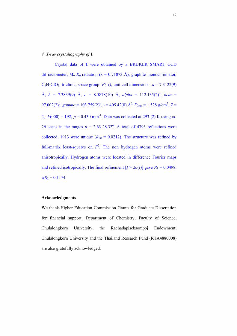
4. X-ray crystallography of 1
Crystal data of 1 were obtained by a BRUKER SMART CCD
diffractometer, Mo Kα radiation (λ = 0.71073 Å), graphite monochromator,
C8H7ClO3, triclinic, space group P(-1), unit cell dimensions a = 7.3122(9)
Å, b = 7.3839(9) Å, c = 8.5878(10) Å, alpha = 112.135(2)o, beta =
97.002(2)o, gamma = 103.759(2)o, V = 405.42(8) Å3, Dcalc = 1.528 g/cm3, Z =
2, F(000) = 192, µ = 0.430 mm-1. Data was collected at 293 (2) K using ω-
2θ scans in the ranges θ = 2.63-28.32o. A total of 4793 reflections were
collected, 1913 were unique (Rint = 0.0212). The structure was refined by
full-matrix least-squares on F2. The non hydrogen atoms were refined
anisotropically. Hydrogen atoms were located in difference Fourier maps
and refined isotropically. The final refinement [I > 2σ(I)] gave R1 = 0.0498,
wR2 = 0.1174.
Acknowledgments
We thank Higher Education Commission Grants for Graduate Dissertation
for financial support. Department of Chemistry, Faculty of Science,
Chulalongkorn University, the Rachadapiseksompoj Endowment,
Chulalongkorn University and the Thailand Research Fund (RTA4880008)
are also gratefully acknowledged.