Immunomodulation by the estrogen metabolite 2-methoxyestradiol
Upload
independentCategory
view
2download
0
A Novel Function for Kojic Acid, a Secondary Metabolitefrom Aspergillus Fungi, as Antileishmanial AgentAna Paula D. Rodrigues1,4,5, Luis Henrique S. Farias1,4, Antonio Sergio C. Carvalho2, Alberdan S. Santos2,
Jose Luiz M. do Nascimento3, Edilene O. Silva1,4*
1 Universidade Federal do Para, Instituto de Ciencias Biologicas, Laboratorio de Biologia Estrutural, Belem, Para, Brazil, 2 Universidade Federal do Para, Instituto de Ciencias
Exatas e Naturais, Laboratorio de Investigacao Sistematica em Biotecnologia e Biodiversidade Molecular do Instituto de Ciencias Exatas e Naturais, Belem, Para, Brazil,
3 Universidade Federal do Para, Instituto de Ciencias Biologicas, Laboratorio de Neuroquımica Molecular e Celular, Belem, Para, Brazil, 4 Instituto Nacional de Ciencia e
Tecnologia em Biologia Estrutural e Bioimagem, Universidade Federal do Rio de Janeiro, Rio de Janeiro, Rio de Janeiro, Brazil, 5 Laboratorio de Microscopia Eletronica,
Instituto Evandro Chagas, Secretaria de Vigilancia em Saude do Ministerio da Saude, Belem, Para, Brazil
Abstract
Kojic acid (KA) is a fungal metabolite used as a topical treatment skin-whitening cosmetic agent for melasma in humans;however its potential as an anti-leishmanial agent is unknown. Chemotherapy is one of the most effective treatments forLeishmaniasis. However, the drugs available are expensive, invasive, require long-term treatment and have severe sideeffects. Thus, the development of new effective leishmanicidal agents is a necessity. In this study we investigated the anti-leishmanial effect of KA on L. amazonensis, following in vitro and in vivo infections. KA (50 mg/mL) was found to decrease thegrowth by 62% (IC50 34 mg/mL) and 79% (IC50 27.84 mg/mL) of promastigotes and amastigotes in vitro, respectively.Ultrastructural analysis of KA-treated amastigotes showed the presence of vesicles bodies into the flagellar pocket, and anintense intracellular vacuolization and swelling of the mitochondrion. During the in vitro interaction of parasites and thehost cell, KA reverses the superoxide anions (O2
-) inhibitory mechanism promoted by parasite. In addition, 4 weeks after KA-topical formulation treatment of infected animals, a healing process was observed with a high production of collagen fibersand a decrease in parasite burden. Thus, these results demonstrated the great potential of KA as an anti-leishmanialcompound.
Citation: Rodrigues APD, Farias LHS, Carvalho ASC, Santos AS, do Nascimento JLM, et al. (2014) A Novel Function for Kojic Acid, a Secondary Metabolite fromAspergillus Fungi, as Antileishmanial Agent. PLoS ONE 9(3): e91259. doi:10.1371/journal.pone.0091259
Editor: David M. Ojcius, University of California Merced, United States of America
Received October 18, 2013; Accepted February 8, 2014; Published March 12, 2014
Copyright: � 2014 Rodrigues et al. This is an open-access article distributed under the terms of the Creative Commons Attribution License, which permitsunrestricted use, distribution, and reproduction in any medium, provided the original author and source are credited.
Funding: The work was supported by Programa Nacional de Cooperacao Academica (PROCAD - grant number 620179/2008-2 and Novas Fronteiras - grantnumber 630/2010), Instituto Nacional de Biologia Estrutural e Bioimagem (CNPq - grant number 573767/2008-4), Conselho Nacional de DesenvolvimentoCientıfico e Tecnologico (Universal - grant number 482922/2010-9), Fundacao de Amparo a Pesquisa do Estado do Para, Fundacao Carlos Chagas Filho de Amparoa Pesquisa do Estado do Rio de Janeiro, Fundacao de Amparo e Desenvolvimento da Pesquisa and Universidade Federal do Para/PROPESP. The funders had norole in study design, data collection and analysis, decision to publish, or preparation of the manuscript.
Competing Interests: The authors hereby declare three patents with the following application numbers: US20110178169 Class: 514460 filed 14 Aug 2009,Issued 21 Jul 2011: Use of 5- Hidroxy-2-hydroxymethyl-y-pyrone (HMP) as a leishmanicidal agent by Alberdan Silva Santos et al. - US patent; WO2010/017613 filed14 Aug 2009, issued 18 Feb 2010: Use of 5-Hidroxy-2-hydroxymethyl-y-pyrone (HMP) as a leishmanicidal agent by Alberdan Silva Santos et al. – World IntellectualProperty Organization; PI0817954-9 filed 14 Aug 2008, issued 11 Jan 2011: Uso do 5-hidroxi-2-hidroximetil-c-pirona como agente de ativacao do macrofago nocombate da leishmaniose cutanea by Alberdan Silva Santos et al. – Brazilian patent. Thus, the authors declare that these patents, relating to material pertinent tothis article, do not alter their adherence to all the PLOS ONE policies on sharing data and materials.
* E-mail: [email protected]
Introduction
Leishmaniasis is a disease caused by protozoan parasites of the
genus Leishmania. Parasites are transmitted by sandflies and can
infect cells of the mononuclear phagocyte lineage in vertebrate
hosts. Leishmaniasis is endemic in 98 countries and affects 12
million people in the entire world available in the WHO
homepage (http://www.who.int/tdr/diseases/leish/diseaseinfo.
htm). The disease is a public health problem in Brazil, particularly
in the Amazon region, due to the presence of seven different
enzootic species of Leishmania, involving hosts and different sand fly
vectors that are commonly found in this region [1]. Leishmaniasis
severity varies, extending from mucosal and cutaneous to visceral
and diffuse cutaneous infections [2]. Diffuse cutaneous leishman-
iasis or anergic diffuse cutaneous leishmaniasis (ADCL) is caused
by Leishmania (Leishmania) amazonensis, which causes cellular
immune response depression, results in parasite-rich lesions and
is characterized by non-ulcerated lesions.
Chemotherapy is one of the most effective treatments for this
disease. The first line of treatment recommended by the WHO
consists of the use of pentavalent antimonials, amphotericin B and
pentamidines, which have demonstrated treatment failure and
parasite resistance. In addition, these treatments are expensive,
invasive and have severe side effects. Treatment for ADCL is not
effective for some patients due to the anergic response profile [3].
Alternative treatments are available, such as miltefosine, which is
effective against visceral leishmaniasis in India, but is teratogenic
and few studies have shown effects on tegumentary leishmaniasis
[4]. New substances, isolated from plants and microorganisms,
have demonstrated leishmanicidal action and most act by
promoting host cell activation to combat leishmania parasites
[5,6].
PLOS ONE | www.plosone.org 1 March 2014 | Volume 9 | Issue 3 | e91259
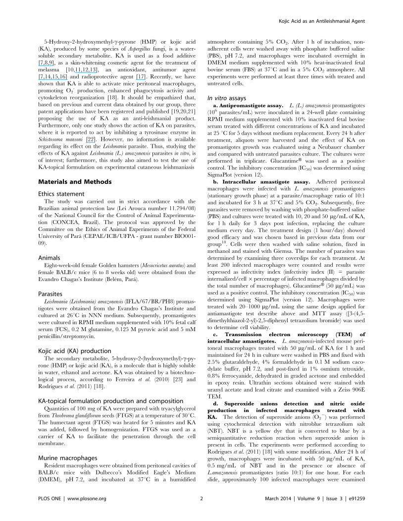
5-Hydroxy-2-hydroxymethyl-c-pyrone (HMP) or kojic acid
(KA), produced by some species of Aspergillus fungi, is a water-
soluble secondary metabolite. KA is used as a food additive
[7,8,9], as a skin-whitening cosmetic agent for the treatment of
melasma [10,11,12,13], an antioxidant, antitumor agent
[7,14,15,16] and radioprotective agent [17]. Recently, we have
shown that KA is able to activate mice peritoneal macrophages,
promoting O2- production, enhanced phagocytosis activity and
cytoskeleton reorganization [18]. It should be empathized that,
based on previous and current data obtained by our group, three
patent applications have been registered and published [19,20,21]
proposing the use of KA as an anti-leishmanial product.
Furthermore, only one study shows the action of KA on parasites,
where it is reported to act by inhibiting a tyrosinase enzyme in
Schistosoma mansoni [22]. However, no information is available
regarding its effect on the Leishmania parasite. Thus, studying the
effects of KA against Leishmania (L.) amazonensis parasites in vitro, is
of interest; furthermore, this study also aimed to test the use of
KA-topical formulation on experimental cutaneous leishmaniasis
Materials and Methods
Ethics statementThe study was carried out in strict accordance with the
Brazilian animal protection law (Lei Arouca number 11.794/08)
of the National Council for the Control of Animal Experimenta-
tion (CONCEA, Brazil). The protocol was approved by the
Committee on the Ethics of Animal Experiments of the Federal
University of Para (CEPAE/ICB/UFPA - grant number BIO001-
09).
AnimalsEight-week-old female Golden hamsters (Mesocricetus auratus) and
female BALB/c mice (6 to 8 weeks old) were obtained from the
Evandro Chagas’s Institute (Belem, Para).
ParasitesLeishmania (Leishmania) amazonensis (IFLA/67/BR/PH8) promas-
tigotes were obtained from the Evandro Chagas’s Institute and
cultured at 26uC in NNN medium. Subsequently, promastigotes
were cultured in RPMI medium supplemented with 10% fetal calf
serum (FCS), 0.2 M glutamine, 0.125 M pyruvic acid and 5 mM
penicillin/streptomycin.
Kojic acid (KA) productionThe secondary metabolite, 5-hydroxy-2-(hydroxymethyl)-c-py-
rone (HMP) or kojic acid (KA), is a molecule that is highly soluble
in water, ethanol and acetone. KA was obtained by a biotechno-
logical process, according to Ferreira et al. (2010) [23] and
Rodrigues et al. (2011) [18].
KA-topical formulation production and compositionQuantities of 100 mg of KA were prepared with tryacylglycerol
from Theobroma glandiflorum seeds (FTGS) at a temperature of 30uC.
The humectant agent (FTGS) was heated for 5 minutes and KA
was added, followed by homogenization. FTGS was used as a
carrier of KA to facilitate the penetration through the cell
membrane.
Murine macrophagesResident macrophages were obtained from peritoneal cavities of
BALB/c mice with Dulbecco’s Modified Eagle’s Medium
(DMEM), pH 7.2, and incubated at 37uC in a humidified
atmosphere containing 5% CO2. After 1 h of incubation, non-
adherent cells were washed away with phosphate buffered saline
(PBS), pH 7.2, and macrophages were incubated overnight in
DMEM medium supplemented with 10% heat-inactivated fetal
bovine serum (FBS) at 37uC and in a 5% CO2 atmosphere. All
experiments were performed at least three times with treated and
untreated cells.
In vitro assaysa. Antipromastigote assay. L. (L.) amazonensis promastigotes
(106 parasites/mL) were inoculated in a 24-well plate containing
RPMI medium supplemented with 10% inactivated fetal bovine
serum treated with different concentrations of KA and incubated
at 25 uC for 5 days without medium replacement. Every 24 h after
treatment, aliquots were harvested and the effect of KA on
promastigotes growth was evaluated using a Neubauer chamber
and compared with untreated parasites culture. The cultures were
performed in triplicate. GlucantimeH was used as a positive
control. The inhibitory concentration (IC50) was determined using
SigmaPlot (version 12).
b. Intracellular amastigote assay. Adhered peritoneal
macrophages were infected with L. amazonensis promastigotes
(stationary growth phase) at a parasite/macrophage ratio of 10:1
and incubated for 3 h at 37uC and 5% CO2. Subsequently, free
parasites were removed by washing with phosphate-buffered saline
(PBS) and cultures were treated with 10, 20 and 50 mg/mL of KA,
for 1 h daily for 3 days post infection, replacing the culture
medium every day. The treatment design (1 hour/day) showed
good efficacy and was chosen based in previous data from our
group18. Cells were then washed with saline solution, fixed in
methanol and stained with Giemsa. The number of parasites was
determined by examining three coverslips for each treatment. At
least 200 infected macrophages were counted and results were
expressed as infectivity index (infectivity index (II) = parasite
internalized/cell6percentage of infected macrophages divided by
the total number of macrophages). GlucantimeH (50 mg/mL) was
used as a positive control. The inhibitory concentration (IC50) was
determined using SigmaPlot (version 12). Macrophages were
treated with 20–1000 mg/mL using the same design applied for
antiamastigote test describe above and MTT assay ([3-(4,5-
dimethylthiazol-2-yl)-2,5-diphenyl tetrazolium bromide) was used
to determine cell viability.
c. Transmission electron microscopy (TEM) of
intracellular amastigotes. L. amazonensis-infected mouse peri-
toneal macrophages treated with 50 mg/mL of KA for 1 h and
maintained for 24 h in culture were washed in PBS and fixed with
2.5% glutaraldehyde, 4% formaldehyde in 0.1 M sodium caco-
dylate buffer, pH 7.2, and post-fixed in 1% osmium tetroxide,
0.8% ferrocyanide, dehydrated in graded acetone and embedded
in epoxy resin. Ultrathin sections obtained were stained with
uranyl acetate and lead citrate and examined with a Zeiss 906E
TEM.
d. Superoxide anions detection and nitric oxide
production in infected macrophages treated with
KA. The detection of superoxide anions (O22) was performed
using cytochemical detection with nitroblue tetrazolium salt
(NBT). NBT is a yellow dye that is converted to blue by a
semiquantitative reduction reaction when superoxide anion is
present in cells. The experiments were performed according to
Rodrigues et al. (2011) [18] with some modification. After 24 h of
growth, macrophages were incubated with 50 mg/mL of KA,
0.5 mg/mL of NBT and in the presence or absence of
L.amazonensis promastigotes (ratio 10:1) for one hour. For each
slide, approximately 100 infected macrophages were examined
Kojic Acid as an Antileishmanial Agent
PLOS ONE | www.plosone.org 2 March 2014 | Volume 9 | Issue 3 | e91259

and counted. Cells were differentiated as infected macrophages
that presented O22 reaction and macrophages infected that not
presented O22 reaction (SO2). Results were presented as number
of infected cells showed formazan deposits. The supernadant of
macrophages infected and treated were used for nitrite detection
by griess reaction.
In vivo assaysa. Antileishmanial experiment. Animals (eight-week-old
female Golden hamsters) were infected with 106 of L. amazonensis
promastigotes/mL during the stationary growth phase with a
maximum volume of 0.2 mL on both hind paws. Animals were
separated in 3 groups: untreated (n = 5); KA-treated 100 mg/kg/
day (n = 5) and KA vehicle (n = 5). KA topical formulation
treatment was initiated after 5 weeks of infection. The KA
formulation and vehicle was applied topically to all lesions once
daily for 4 weeks. Control groups were also maintained in parallel.
During the treatment period, the lesion size was measured weekly
using a caliper. Width and height of both hind paws were used to
calculate the lesion area (mm2). After the treatment, animals were
euthanized and tissues from lesions were processed for histopath-
ological analysis, scanning and transmission electron microscopy
analysis.
b. Histophatological analysis. Tissue biopsies from infect-
ed and treated animals were fixed and embedded in paraffin.
Sections of 5–6 mm were stained with haematoxilin and eosin
(H&E) for histopathological analysis. Morphometric analysis was
employed to quantify tissue parasitism as described by Rocha-
Vieira et al. (2003) [24]. Images from tissues were captured with a
Zeiss Axiophot microscope connected to a video camera.
Quantitative morphometric analysis was performed using ImageJ
software and digitalized images obtained. The parasite number
was measured in 10 non-contiguous fields from both infected
footpads of each animal (magnification 630 x).
c. Transmission and scanning electron
microscopy. Tissue sections from infected and treated animals
were processed as described above for TEM and examined with a
LEO 906E TEM. For SEM, tissue was processed according to
Haggis et al. (1977) [25]. Samples were fixed and post-fixed as
described above for TEM, dehydrated in graded ethanol, frozen
using liquid nitrogen, fractured and, after thawing, critical-point
dried. Samples were mounted, coated with gold and examined
with a LEO 1450VP SEM.
Statistical analysisAll experiments were performed in triplicate. The mean and
standard deviations of at least three experiments were determined.
Statistical analyses of the differences between mean values in the
experimental groups were performed using ANOVA, the
Student’s t-test (employing the GraphPad Prism 5.0 program).
All p-values ,0.05 were as considered statistically significant.
Results
KA has antileishmanial activity, in vitroThe activity on L. amazonensis promastigotes forms was
monitored for 120 hours. KA promoted a dose-dependent
reduction of 62% in parasite proliferation, when treated with
50 mg/mL for 120 h (IC50 34 mg/mL - Figure 1A). KA was 3-fold
more effective than glucantimeH reference drug (IC50 122.4 mg/
mL-Figure 1B), usually used for cutaneous leishmaniasis treatment
in Amazon region. Leishmanicidal activity on intracellular
parasites was evaluated after 72 h of treatment with KA. A
growth inhibition of 79% (IC50 27.9 mg/mL) after 72 h of
treatment (Figure 1C). GlucantimeH promoted a reduction of
59% after 72 h of treatment with 50 mg/mL (IC50 77.4 mg/mL).
MTT assay showed no cytotoxic effect on macrophage treated at
20–1000 mg/ml of KA when compared with control (Supplemen-
tary Figure S1).
KA promotes ultrastructural alterations in amastigoteforms of L. amazonensis
TEM was used to analyze alterations in the parasite organelles
and as tool to elucidate the mechanisms of drug action. Infected
cells showed that KA induced a decreased amastigote number
Figure 1. KA activity against L. (L.) amazonensis in vitro. (A)Growth curve of L. (L.) amazonensis promastigotes treated with differentKA concentrations (B) Growth curve of L. (L.) amazonensis promasti-gotes treated with glucantimeH (GLU). Results are from threeexperiments performed in triplicate. *p,0.05; ***p,0.001; comparedwith control (CTL). (C) Effect of KA on intracellular amastigote survival ofL. (L.) amazonensis. Macrophages infected and treated with KA and GLU(50 mg/mL) (***p,0.001 compared with CTL).doi:10.1371/journal.pone.0091259.g001
Kojic Acid as an Antileishmanial Agent
PLOS ONE | www.plosone.org 3 March 2014 | Volume 9 | Issue 3 | e91259

(Figure 2B–C), with a large amount of membranous structures
inside the parasitophorous vacuoles (Figure 3B-arrowheads).
Furthermore, different and significant morphological alterations,
such as some vesicles bodies into the flagellar pocket (Figure 2D),
intense intracellular vacuolization (Figure 2F-asterisks), presence of
many lipid-like bodies (Figure 2E-asterisks), and swelling of the
mitochondrion (Figure 2F-arrowheads) were seen in the intracel-
lular parasites.
Infected macrophage treated with KA producedsuperoxide anions (O2
2), but not nitric oxideFor superoxide detection, infected and treated macrophages
were analyzed with a cytochemical assay using NBT. Leishmania-
infected macrophages treated for 1 h with 50 mg/ml showed
formazan deposits distributed in the cellular cytoplasm of infected
cells, showing intense superoxide production (Figure 3D), in
comparison with the infected-cells without KA-treatment
(Figure 3C). The presence of formazan deposits enhanced (63%)
when infected macrophage were treated 50 mg/ml KA as
compared with infected macrophages without treatment. On the
other hand, nitric oxide (NO) production was not observed in
infected macrophages that were treated with 50 mg/ml of KA
(data not shown).
KA- topical formulation (ointment) promoted amastigotedestruction in vivo
After the end of the treatment, tissue samples were collected.
Lesion size was measure weekly during the treatment period.
Surprisingly, there was a low, but significant, reduction in lesion
size in the end of 4 weeks of treatment (Figure 4A), associated with
a reduced number of amastigote forms (Figure 4B). Topical
treatment with KA ointment decreased the parasite number at the
lesion site by 92.1%. Interestingly, the vehicle group presented a
discrete reduction in lesion size, but no decrease in amastigote
number (Figure 4B). In control group, lesion size enhanced and a
large number of amastigotes were detected (Figure 4A and 4B).
KA-topical formulation promoted collagen productionIn control group was observed a large number of amastigotes
dispersed for all tissue (Figure 5A–B). Vehicle group presented
many amastigotes forms inside of host cell vacuoles (Figure 5C–D).
On the other hand, KA-treated group presented lower number of
amastigotes and collagen fibers in the infection site, confirmed by
picrosirius red stain (Figure 5E–F). These fibers demonstrated an
organized and parallel distribution in treated animals and seem to
fill the spaces between host cell vacuoles (See supplementary
movies S1 and S2). Ultrastructural analysis by TEM and SEM of
treated group were performed. Analysis by TEM of treated group
showed a predominant parallel distribution of collagen fibers
(Figure 6B) when compared to control that presented amastigotes
forms inside parasitophorous vacuoles (Figure 6A). As well as,
analysis by SEM of control group demonstrated higher number of
amastigotes inside vacuoles (Figure 6C-arrows). In contrast, KA-
treated group presented empty vacuoles and a large number of
collagen fibers in almost all recovered tissue (Figure 6D-arrow-
heads).
Discussion
It is well known that chemotherapy is the only effective
treatment for Leishmaniasis infections, however, the antileishma-
nial drugs available are, in general, toxic expensive and require
long-term treatment; furthermore, most of them can only be given
to patients parenterally. These side effects and disadvantages
demonstrate the necessity to identify new, effective and safe
compounds for the treatment of this disease [4,26].
In the present study, we showed that KA acted as an
antileishmanial agent without affecting the host cell. KA treatment
decreased the growth of L. amazonensis promastigotes by 62% at
50 mg/mL (IC50: 34 mg/mL). Moreover, KA was more effective
against intracellular amastigotes, with a growth inhibition of 79%
for 50 mg/mL of KA after 72 h of treatment (IC50: 27.84 mg/mL).
Figure 2. Ultrastructural effects of KA on intracellular amasti-gotes of Leishmania (L.) amazonensis. (A) General view of untreatedinfected macrophages showing a typical morphology. Note parasites(stars) within the parasitophorous vacuoles. (B) General view ofinfected macrophages treated for 1 h and cultivated for 24 h, showinglarge parasitophorous vacuole. Note reduced number of amastigotes,parasite and flagellar fragments (arrowheads). (C) Infected andtreated macrophages presented vacuoles with damaged parasites(stars) or without amastigote forms. (D) Higher magnification of (C);parasites inside PV with alterations in the flagellar membrane (thinarrows) and vesicles inside the flagellar pocket (asterisks). (E)Intracellular amastigotes with membrane profiles in the flagellar pocket(thin arrows) and in the parasite cytoplasm (arrows); intenseformation of lipid-like bodies (asterisks) in the cytoplasm ofamastigotes forms. (F) Intracellular parasites presented concentricmembrane (arrow), kinetoplast swelling (arrow heads) and lipid-likebodies (asterisks). N, nucleus; FP, flagellar pocket; K, kinetoplast; F,flagellum; M, mitochondria; PV parasitophorous vacuole. Bars: (A–C)5 mm; (D–F) 2 mm.doi:10.1371/journal.pone.0091259.g002
Kojic Acid as an Antileishmanial Agent
PLOS ONE | www.plosone.org 4 March 2014 | Volume 9 | Issue 3 | e91259

Recently, we reported that KA could modulate macrophage
activation through cytoskeleton rearrangement, increase cell
surface exposure, and enhance the phagocytic process and
superoxide anions (O22) production [18]. The antiamastigote
effect observed in the present report showed that KA promotes a
stimulatory effect in macrophages, killing the parasites. Macro-
phages infected and treated for just 1 h were able to produce O22,
even in the presence of L. amazonensis. It is known that L.
amazonensis can evade the macrophage microbicidal action,
inhibiting reactive oxygen species (ROS) and NO production
[27,28] and KA was able to reverse this inhibitory process. The
mechanism by which KA exerts its antileishmanial effects in vitro
seems to involve the activation of macrophages and amastigotes
killing by O2-production rather than the action of NO. In
agreement with our results, Valadares et al. (2011) [29] demon-
strated that the Agaricus blazei Murill mushroom presents effects
against intracellular amastigotes with a reduction of 84.4% by a
mechanisms that are NO-independent.
We also analyzed the effects of KA on parasite ultrastructure.
Identification of morphological changes can help elucidate the
mechanisms of drug action [30]. In intracellular amastigotes,
significant morphological alterations were observed, such as
intense intracellular vacuolization, disruptions in flagellar mem-
brane and the presence of many vesicles bodies in the flagellar
pocket. Different natural compounds with anti-leishmanial activity
induce alterations in this particular region of the parasite,
suggesting alterations in the endocytic/exocytic pathway [6,31].
Other characteristic features seen in intracellular amastigotes
Figure 3. Superoxide radicals (O22) detection in the infected macrophages treated with 50 mg/mL KA for 1 h. (A) Macrophages without
infection and untreated. (B) Positive control, interaction with Zymozan (arrows). (C) Macrophage infected with L. (L.) amazonensis (arrows) showedsuperoxide production inhibition. (D) Macrophages infected with L. amazonensis and treated with KA reverted the inhibitory effect. Bars: 10 mm. (E)Number of infected macrophages that presented formazan deposits. MO+L.a.: macrophages infected with L. amazonensis; MO+L.a.+KA: macrophagesinfected with L. amazonensis and treated with 50 mg/mL KA; O2
+: infected macrophages with formazan deposits; O2: infected macrophages withoutformazan deposites. (***p,0.001).doi:10.1371/journal.pone.0091259.g003
Figure 4. Effect of KA topical formulation on experimental infection with L. amazonensis. (A) Development of lesions in L. (L.) amazonensis-infected animals treated with KA. The treatment started after 5 weeks post-infection and continued for 4 weeks. Data represent the averagemeasurements of 5 animals for each group. (B) Parasite load in the lesion sites. Amastigotes were quantified after interruption of treatment and themean number of parasites evaluated in 10 fields reported; (**p,0.001). Results are expressed as the mean number of cells evaluated in 10 fields;(*p,0.05; ***p,0.0001).doi:10.1371/journal.pone.0091259.g004
Kojic Acid as an Antileishmanial Agent
PLOS ONE | www.plosone.org 5 March 2014 | Volume 9 | Issue 3 | e91259

treated with KA, for 72 h was the presence of myelin-like figures
and the over-accumulation of lipid-like bodies in the cytoplasm.
Studies have shown that myelin-like figures are correlated to the
autophagic process during drug action [32]. This process is
associated with the cell response to starvation or stresses [33] and
is dependent on ROS production [34]. With respect to lipid body
accumulation in the parasites, close association has been reported
with increased lipid body production, ROS production and
autophagy [34]. KA seems to induce lipid droplet formation
though ROS activity in the host cell and consequently lead to
autophagic cell death. However, further studies are necessary to
clarify these questions.
Due to the leishmanicidal effect of KA upon intracellular
amastigotes in vitro, we examined the effect in vivo with a KA-
topical formulation in the animal model of cutaneous leishman-
iasis. Several new topical treatments have been made available to
treat this disease, but well-known drugs, such as imiquimod and
paromomycin, are the only ones that have reached the clinical trial
testing phase as a topical formulation (ointment, cream, gel and
solution) [35,36]. Nevertheless, the results obtained are still a
controversial interpretation of results, complicated by nonstandard
definitions of the disease and its cure.
In this study, infected animals treated with KA-topical
formulation promoted an initiation of healing process, due to
the production of numerous collagen fibers at the infection site, as
well as a decrease in parasite burden, after the end of treatment.
Wound healing is a process characterized by inflammation, cell
proliferation and tissue remodeling where neutrophils, macro-
phages and lymphocytes arrive first, followed by fibroblasts [37].
Leishmania produces proteases that are capable of destroying matrix
proteins, and disrupting dermal barriers to propagate infection
[38]. However, our present data also show that KA promoted the
production of a large number of type I collagen fibers over the
type III fibers in the infected animals. The presence of type III
fibers is suggested to be related to the success of parasitism, since
these fibers provide support to inflammatory cells, such as
vacuolated and parasitized histiocytes [38]. It has been reported
that, in murine models, the presence of collagen fibers is
prominent in areas where new epithelium is produced, and where
parasites have been eliminated and the inflammatory process
controlled [39]. Furthermore, it has been demonstrated that KA
enhances the wound-healing process following topical application
[40].
Interestingly, our data show a discrete decrease in the lesion
size, in association with the presence of numerous, aligned and
parallel collagen fibers, and the absence or a low-level of parasites.
Thus, not only could lesion size determine the long-term outcome
of tissue damage and the repair process, but this could also
influence the killing of parasites and parasite environment
modification [41,42]. Besides, the small decrease in lesion size in
KA-treated animals compared to control animals can be explained
by the fact that the intensive collagen fibers production can be
related to the beginning of the healing process, where there has not
been enough time to occurred tissue remodeling, the final stage of
wound healing [43].
Another interesting observation is that animals that received
only the treatment with vehicle (triacylglycerols obtained from the
fruit seed of Theobroma grandiflorum) showed a similar decrease in
lesion size when compared to in KA treated animals. However,
when histological analysis of lesion was performed, the vehicle
group showed a lot of amastigotes when compared to the treated
Figure 5. Skin lesion section of infected animals, untreated andtreated with KA topical formulation for one month. Histopatho-logycal analysis of lesion site of untreated animal (A–B) showed tissuedamage (A) and numerous amastigote forms (arrows) dispersed for alltissue (B); Vehicle group (C–D) showed an intense vacuolization for alltissue (C) and a higher number of amastigotes (arrows) inside theparasitophorous vacuoles (D); KA-treated animals (E–F) presentedreduced number of vacuoles in the macrophages and reduced numberof amastigotes (E–F); H&E stain. Bars: (A, C, E) 20 mm; (B, D, F) 10 mm.doi:10.1371/journal.pone.0091259.g005
Figure 6. Ultrastructural analysis of skin lesions from infectedanimals untreated and treated with KA-topical formulation.Transmission electron microscopy of control (A) and KA-treated animals(B). Note the presence of amastigotes in the parasitophorous vacuolesand few collagen fibers in the control group and absence of intracellularamastigotes with organized fibers in the treated group. Scanningelectron microscopy of untreated lesion showed the presence of a largenumber of amastigotes (arrows) inside the parasitophorous vacuoles(C) and lesion from treated animals showed an intense production offibrous material suggestive of collagen fibers (D-arrowheads). P,parasite; HCN, host cell nuclei; PV, parasitophorous vacuole. Bars: (A–B)3 mm, (C–D) 20 mm.doi:10.1371/journal.pone.0091259.g006
Kojic Acid as an Antileishmanial Agent
PLOS ONE | www.plosone.org 6 March 2014 | Volume 9 | Issue 3 | e91259

group. The vehicle from Theobroma grandiflorum seeds seems to help
the healing process, but not the parasite death; however, further
studies should be conducted to determine its healing potential.
The present findings indicate that KA seems to act indirectly
against the parasite and promotes, initially, the activation of
macrophages, leading to a O2-production that is able to kill the L.
amazonensis parasite and help to control the infection. Thus, KA
could be useful for the selective treatment of cutaneous leishman-
iasis and may hold great potential as an anti-leishmanial agent. To
our knowledge, this report demonstrates, for the first time, the
action of KA on Leishmania amazonensis in vitro and in vivo. This study
forms part of a continual search for new bioactive products,
obtained from biotechnological processes that can act effectively
against neglected diseases, such as leishmaniasis.
Supporting Information
Figure S1 MTT assay to determine macrophage viabil-ity. Formazan result solution was read at plate reader at 550 nm
and absorbance represents cells viability. No differences were
found at 20–1000 mg/ml of KA when compared with the control.
OD: optical density; CTL: control.
(TIF)
Movie S1 Z-stack confocal images of control groupstained with picrosirius red for collagen detection. A
total of 13 images were collected and the distance between planes
is 0.98 mm. (630x).
(MPG)
Movie S2 Z-stack confocal images KA-treated groupstained with picrosirius red for collagen detection. A
total of 8 images were collected and the distance between planes is
0.98 mm. (630x).
(MPG)
Acknowledgments
We thank Raimundo Nonato Barbosa Pires and Antonio F.P. Martins for
their technical assistance and animal care.
Author Contributions
Conceived and designed the experiments: APDR LHSF EOS. Performed
the experiments: APDR. Analyzed the data: APDR LHSF EOS JLMN.
Contributed reagents/materials/analysis tools: ASCC ASS JLMN. Wrote
the paper: APDR LHSF EOS JLMN.
References
1. Lainson R (2010) The Neotropical Leishmania species: a brief historical review
of their discovery, ecology and taxonomy. Rev Pan-Amaz Saude 1: 13–32.
2. Silveira FT, Lainson R, Corbett CEP (2004) Clinical and immunopathological
spectrum of American cutaneous leishmaniasis with special reference to the
disease in Amazonian Brazil: a review. Mem Inst Oswaldo Cruz 99: 239–251.
3. Silveira FT (2009) Diffuse cutaneous leishmaniasis (DCL) in the Amazon region,
Brazil: clinical and epidemiological aspects. Gaz Med Bahia 79: 25–29.
4. Croft SL, Sundar S, Fairlamb AH (2006) Drug resistance in Leishmaniasis. Clin
Microbiol Rev 19: 111–126.
5. Polonio T, Efferth T (2008) Leishmaniasis: Drug resistance and natural products
(Review). Int J Mol Med: 277–286.
6. Guimaraes LRC, Rodrigues APD, Marinho PSB, Muller AH, Guilhon GM, et
al. (2010) Activity of the julocrotine, a glutarimide alkaloid from Croton pullei var.
glabrior, on Leishmania (L.) amazonensis. Parasitol Res 107: 1075–1081.
7. Burdock GA, Soni MG, Carabin IG (2001) Evaluation of health aspects of kojic
acid in food. Regul Toxicol Pharmacol 33: 80–101.
8. Blumenthal CZ (2004) Production of toxic metabolites in Aspergillus niger,
Aspergillus oryzae, and Trichoderma reesei: justification of mycotoxin testing in food
grade enzyme preparations derived from the three fungi. Regul Toxicol
Pharmacol 39: 214–228.
9. Bentley R (2006) From miso, sake and shoyu to cosmetics: a century of science
for kojic acid. Nat Prod Rep 23: 1046–1062.
10. Lim JT (1999) Treatment of melasma using kojic acid in a gel containing
hydroquinone and glycolic acid. Dermatol surg 25: 282–284.
11. Nohynek GJ, Kirkland D, Marzin D, Toutain H, Leclerc-Ribaud C, et al. (2004)
An assessment of the genotoxicity and human health risk of topical use of kojic
acid [5-hydroxy-2-(hydroxymethyl)-4H-pyran-4-one]. Food Chem Toxicol 42:
93–105.
12. Lin CH, Wu HL, Huang YL (2007) Combining high-performance liquid
chromatography with on-line microdialysis sampling for the simultaneous
determination of ascorbyl glucoside, kojic acid, and niacinamide in bleaching
cosmetics. Anal Chim Acta 581: 102–107.
13. Ha YM, Chung SW, Song S, Lee H, Suh H, et al. (2007) 4-(6-Hydroxy-2-
naphthyl)-1,3-bezendiol: a potent, new tyrosinase inhibitor. Biol Pharm Bull 30:
1711–1715.
14. Gomes AJ, Lunardi CN, Gonzalez S, Tedesco AC (2001) The antioxidant action
of Polypodium leucotomos extract and kojic acid: reactions with reactive oxygen
species. Braz J Med Biol Res 34: 1487–1494.
15. Tamura T, Mitsumori K, Totsuka Y, Wakabayashi K, Kido R, et al. (2006)
Absence of in vivo genotoxic potential and tumor initiation activity of kojic acid
in the rat thyroid. Toxicology 222: 213–224.
16. Moto M, Mori T, Okamura M, Kashida Y, Mitsumori K (2006) Absence of liver
tumor-initiating activity of kojic acid in mice. Arch Toxicol 80: 299–304.
17. Emami S, Hosseinimehr SJ, Taghdisi SM, Akhlaghpoor S (2007) Kojic acid and
its manganese and zinc complexes as potential radioprotective agents. Bioorg
Med Chem Lett 17: 45–48.
18. Rodrigues APD, Carvalho ASC, Santos AS, Alves CN, Do Nascimento JLM, et
al. (2011) Kojic acid, a secondary metabolite from Aspergillus sp., acts as an
inducer of macrophage activation. Cell Biol Int 35: 335–343.
19. Santos AS, Silva EO, Nascimento JLM, Alves CN, Carvalho ASC, et al. (2010)
Use of 5-hydroxy-2-hydroxymethyl-c-pyrone as a macrophage activation agentto combat cutaneous leishmaniasis. European patent WO2010017613.
20. Santos AS, Silva EO, Nascimento JLM, Alves CN, Carvalho ASC, et al. (2011)
Uso do 5-hidroxi-2-hidroximetil-c-pirona como agente de ativacao do macro-fago no combate da Leishmaniose Cutanea. Brazilian patent PI0817954-9.
21. Santos AS, Silva EO, Nascimento JLM, Alves CN, Carvalho ASC, et al. (2011)Use of 5-hydroxy-2-hydroxymethyl-c-pyrone as a macrophage activation agent
to combat cutaneous leishmaniasis. U.S. patent US20110178169A1.
22. Fitzpatrick JM, Hirai Y, Hirai H, Hoffmann KF (2007) Schistosome eggproduction is dependent upon the activities of two developmentally regulated
tyrosinases. FASEB J 21: 823–835.
23. Ferreira NR, Sarquis MIM, Alves CN, Santos AS (2010) Biotransformation ofsucrose into 5-hydroxy-2-hydroxymethyl-c-pirone by Aspergillus flavus. Annals
of the Brazilian Academy of Sciences 82: 569–576.
24. Rocha-Vieira E, Ferreira E, Vianna P, et al. (2003) Histopathological outcomeof Leishmania major-infected BALB/c mice is improved by oral treatment with
N-acetyl-l-cysteine. Immunology 108: 401–408.
25. Haggis GH, Phipps-Todd B (1977) Freeze-fracture for scanning electronmicroscopy. J Microsc 111: 193–201.
26. Croft SL, Seifert K, Yardley V (2006) Current scenario of drug development for
leishmaniasis. Indian J Med Res 123: 399–410.
27. Olivier M, Gregory DJ, Forget G (2005) Subversion mechanisms by which
leishmania parasites can escape the host immune response: a signaling point of
view. Clin Microbiol Rev 18: 293–305.
28. Mukbel RM, Patten C, Gibson K, Ghosh M, Petersen C, et al. (2007)
Macrophage killing of Leishmania amazonensis amastigotes requires both nitricoxide and superoxide. The Am J Trop Med Hyg 76: 669–675.
29. Valadares DG, Duarte MC, Oliveira JS, Chavez-Fumagalli MA, Martins VT,et
al. (2011) Leishmanicidal activity of the Agaricus blazei Murill in differentLeishmania species. Parasitol Int 60: 357–363. doi:10.1016/j.parint.2011.06.001.
30. Adade CM, Souto-Padron T (2010) Contributions of ultrastructural studies to
the cell biology of trypanosmatids: targets for anti-parasitic drugs. OpenParasitol J 4: 178–187.
31. Vendrametto MC, Santos AO, Nakamura CV, Dias Filho BP, Cortez DA, et al.
(2010) Evaluation of antileishmanial activity of eupomatenoid-5, a compoundisolated from leaves of Piper regnellii var. pallescens. Parasitol Int 59: 154–158.
32. Rodrigues JCF, Rodriguez C, Urbina JA (2002) Ultrastructural and biochemical
alterations induced by promastigote and amastigote forms of Leishmania
amazonensis. Curr Pharm Des 46: 487–499.
33. Kondo Y, Kanzawa T, Sawaya R, Kondo S (2005) The role of autophagy in
cancer development and response to therapy. Nat Rev Cancer 5:726–734.
34. Djavaheri-Mergny M, Amelotti M, Mathieu J, Besancon F, Bauvy C, et al.
(2006) NF-KB activation represses tumor necrosis factor-alpha-inducedautophagy. J Biol Chem 281: 30373–30382.
35. Alavi-Naini R, Fazaeli A, O’Dempsey T (2012) Topical treatment modalities for
old world cutaneous leishmaniasis: a review. Prague Med Rep 113: 105–118.
36. Croft SL, Olliaro P (2011) Leishmaniasis chemotherapy—challenges andopportunities. Clin Microbiol Infect 17: 1478–1483.
37. Miller M, Nanchahal J (2005) Advances in the modulation of cutaneous wound
healing and scarring. BioDrugs 19: 363–381.
Kojic Acid as an Antileishmanial Agent
PLOS ONE | www.plosone.org 7 March 2014 | Volume 9 | Issue 3 | e91259
38. Abreu-Silva AL, Calabrese KS, Mortara RA, Tedesco RC, Cardoso FO, et al.
(2004) Extracellular matrix alterations in experimental murine Leishmania (L.)
amazonensis infection. Parasitology; 128: 385–390.
39. Baldwin T, Sakthianandeswaren A, Curtis JM, Kumar B, Smyth GK, et al.
(2007) Wound healing response is a major contributor to the severity ofcutaneous leishmaniasis in the ear model of infection. Parasite Immunol 29:
501–513.40. Mohammadpour M, Behjati M, Sadeghi A (2012) Wound healing by topical
application of antioxidant iron chelators: kojic acid and deferiprone. Int Wound J
doi:10.1111/j.1742–481X.2012.00971.x.
41. Sakthianandeswaren A, Elso CM, Simpson K, Curtis JM, Kumar B, et al. (2005)
The wound repair response controls outcome to cutaneous leishmaniasis. Proc
Natl Acad Sci U S A 102: 15551–15556.
42. Lecoeur H, Buffet P, Morizot G, Goyard S, Guigon G, et al. (2007)
Optimization of topical therapy for Leishmania major localized cutaneous
leishmaniasis using a reliable C57BL/6 model. PLoS Negl Trop Dis 1: e34.
43. Nurden A T (2011) Platelets, inflammation and tissue regeneration. Thromb
Haemost 105(suppl.6): S13–33.
Kojic Acid as an Antileishmanial Agent
PLOS ONE | www.plosone.org 8 March 2014 | Volume 9 | Issue 3 | e91259
Copyright © 2022 FDOKUMEN