MICROPROPAGATION AND SECONDARY METABOLITE ...
354
1 MICROPROPAGATION AND SECONDARY METABOLITE STUDIES IN TAXUS SPP. AND WITHANIA SOMNIFERA (L.) DUNAL. A THESIS SUBMITTED TO THE UNIVERSITY OF PUNE FOR THE DEGREE OF DOCTOR OF PHILOSOPHY IN BIOTECHNOLOGY BY ANJALI ABHAY KULKARNI PLANT TISSUE CULTURE DIVISION NATIONAL CHEMICAL LABORATORY PUNE 411 008. SEPTEMBER 2000
-
Upload
khangminh22 -
Category
Documents
-
view
0 -
download
0
Transcript of MICROPROPAGATION AND SECONDARY METABOLITE ...
1
MICROPROPAGATION AND SECONDARY METABOLITE STUDIES IN
TAXUS SPP. AND WITHANIA SOMNIFERA (L.) DUNAL.
A THESIS SUBMITTED TO THE UNIVERSITY OF PUNE FOR THE DEGREE
OF DOCTOR OF PHILOSOPHY IN BIOTECHNOLOGY
BY
ANJALI ABHAY KULKARNI
PLANT TISSUE CULTURE DIVISION
NATIONAL CHEMICAL LABORATORY
PUNE 411 008.
SEPTEMBER 2000

2
CONTENTS Page No.
Acknowledgements iv-v
Certificate v i
Key To Abbreviations vii-viii
Synopsis ix-xii
CHAPTER 1 : General Introduction 1-35
Preamble 1-2
PART A: General Introduction to Plant Tissue
and Cell Culture Techniques.
3-7
PART B: Secondary Metabolite Production in
vitro.
8-35
CHAPTER 2 : Materials And Methods (General) 36-42
CHAPTER 3 : Control of Contamination and Phenolic Browning
in vitro.
43-94
Identification and Control of Bacterial and Fungal Genera Associated with in vitro Cultures of Taxus baccata ssp. wallichiana Zucc. Pilg. and Withania somnifera (L.) Dun. as well as Control of Phenolic Oxidation.
3.1 Introduction 43-52
3.2 Materials and Methods 53-62
3.3 Results and Discussion 63-92
Subsection
1
Bacterial Contamination 63-79
Subsection
2
Fungal Contamination and Phenolic
Oxidation/Browning
80-92
3.4 Conclusions 93-94
Contd…..

3
Contents Contd.
CHAPTER 4 : Studies on Callus Induction from Various Explants
of Taxus spp. and Analysis of Taxane Content of in
vitro and in vivo Tissues.
95-149
4.1 Introduction 95-112
4.2 Materials and Methods 113-123
4.3 Results and Discussion 124-147
4.4 Conclusions 148-149
CHAPTER 5 : In vitro Studies on Micropropagation from Mature
and Juvenile Explants of Taxus spp.
150-204
PART A: Micropropagation Studies Using
Mature Explants of Taxus spp.
150-186
5.1 Introduction 150-159
5.2 Materials and Methods 160-162
5.3 Results and Discussion 163-183
5.4 Conclusions 184-186
PART B: In vitro Studies with Embryo Explants
of Taxus spp.
187-204
5.5 Introduction 187-190
5.6 Materials and Methods 191-193
5.7 Results and Discussion 194-203
5.8 Conclusions 204
Contd…..
4
Contents Contd.
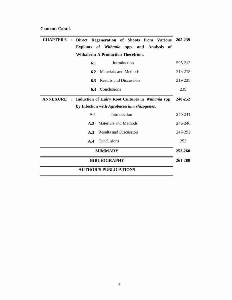
CHAPTER 6 : Direct Regeneration of Shoots from Various
Explants of Withania spp. and Analysis of
Withaferin-A Production Therefrom.
205-239
6.1 Introduction 205-212
6.2 Materials and Methods 213-218
6.3 Results and Discussion 219-238
6.4 Conclusions 239
ANNEXURE : Induction of Hairy Root Cultures in Withania spp.
by Infection with Agrobacterium rhizogenes.
240-252
A.1 Introduction 240-241
A.2 Materials and Methods 242-246
A.3 Results and Discussion 247-252
A.4 Conclusions 252
SUMMARY 253-260
BIBLIOGRAPHY 261-280
AUTHOR’S PUBLICATIONS

5
ACKNOWLEDGEMENTS
I take this opportunity to convey my heartfelt thanks to my research guide Dr. K. V.
Krishnamurthy for suggesting the topic of my thesis. It was a great chance to come across a lot of
difficulties during the tenure of my work and to successfully overcome them. I am also thankful to
him for his continuous guidance and advice. I am highly indebted to scores of my earlier teachers
because of whom I decided to take up research. A special mention to Mr. S. B. Bhagwat, Dr. M. R.
Pendse, Dr. S. S. Bhargava, Dr. P. K. Chitnis, Dr. R. J. Thengane, Mrs. K. R. Gadgil, Mrs. T. Bapat
and Mrs. V. Kelkar.
A large number of people from different institutions have been helpful to me during the
course of the present work. I am thankful to Dr. Ombir Singh of CRC, Shimla, without whose
unstinted and generous help in collecting plant material of Taxus spp., this thesis would have
remained incomplete. I am also grateful to Mr. Tembhurne and Mr. Negi of CRC, Shimla for help in
the forest during collection of plant material of Taxus spp. Dr. P. S. Ahuja of IHBT, Palampur, Mr. U.
C. Shah, Almora and Dr. V. K. Mehta of CIMAP (Regional Center), Jammu are also acknowledged
for helping in plant and seed material collection of Taxus spp. Mr. A. Sane who supplied seeds of
cultivated variety WS-20 is also gratefully acknowledged.
Dr. A. Pande of ARI, Pune and Dr. M. G. Watve of MES, AG college, Pune are duly
acknowledged for their guidance in identifying bacterial and fungal contaminants observed in vitro.
I am indebted to Dr. S. S. Bhrgava, University of Pune, Dr. S. M. Joshi, NCL and Dr. S.
Ponnaratnam, NCL who allowed me to use HPLC systems in their departments for my work. I must
also acknowledge Dr. Atta -Ur-Rahman, Pakistan and Dr. P. Uma Devi, India for sending me
standards of Withaferin-A.
I am thankful to Dr. A. P. Kulkarni, Pune for developing a specific software for identification
of bacterial contaminants encountered in vitro and also for helping me in statistical analysis of my
data. Also as my father-in-law, he has been very supportive and encouraging.
I will always remember the friendship as well as a number of stimulating discussions with
Drs. Swati Kelkar, Vandana Mhaske, Mohini Joshi, D. Vijaykumar, T. K. Dattaroy and Sunita
Mahajan. I am also thankful to Mr. M. L. Mohan and Dr. D. V. Lakshmi for their help and hospitality.
Drs. A. P. Sagare, K. Suhasini and Ms. Seema Deshpande created a friendly lab atmosphere and
my thanks are due to them. I would also like to thank Mr. A. K. Banerjee for help in photography.

6
The scientific and non-scientific staff at the Plant Tissue Culture Division, NCL, Pune were
instrumental towards completion of my thesis and are acknowledged.
The debt of my parents, Baba and Mummy, my sister, Vaishu, my mother-in-law, Aai, my
brother and sister-in-law, Anand and Aaditi and all my grandparents can never be cleared. I am
thankful for their continual support. I am deeply grateful to my husband, Abhay, without whose
guidance, patience and perseverance, the thesis would not have been completed. As a token of my
appreciation I, herewith, dedicate this thesis to him.
Finally, I thank Dr. Paul Ratnasamy, Director, NCL, Pune for permitting to submit my thesis
to University of Pune and CSIR, Government of India for the research fellowship awarded.
7
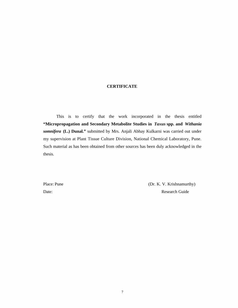
CERTIFICATE
This is to certify that the work incorporated in the thesis entitled
“Micropropagation and Secondary Metabolite Studies in Taxus spp. and Withania
somnifera (L.) Dunal.” submitted by Mrs. Anjali Abhay Kulkarni was carried out under
my supervision at Plant Tissue Culture Division, National Chemical Laboratory, Pune.
Such material as has been obtained from other sources has been duly acknowledged in the
thesis.
Place: Pune (Dr. K. V. Krishnamurthy)
Date: Research Guide
8
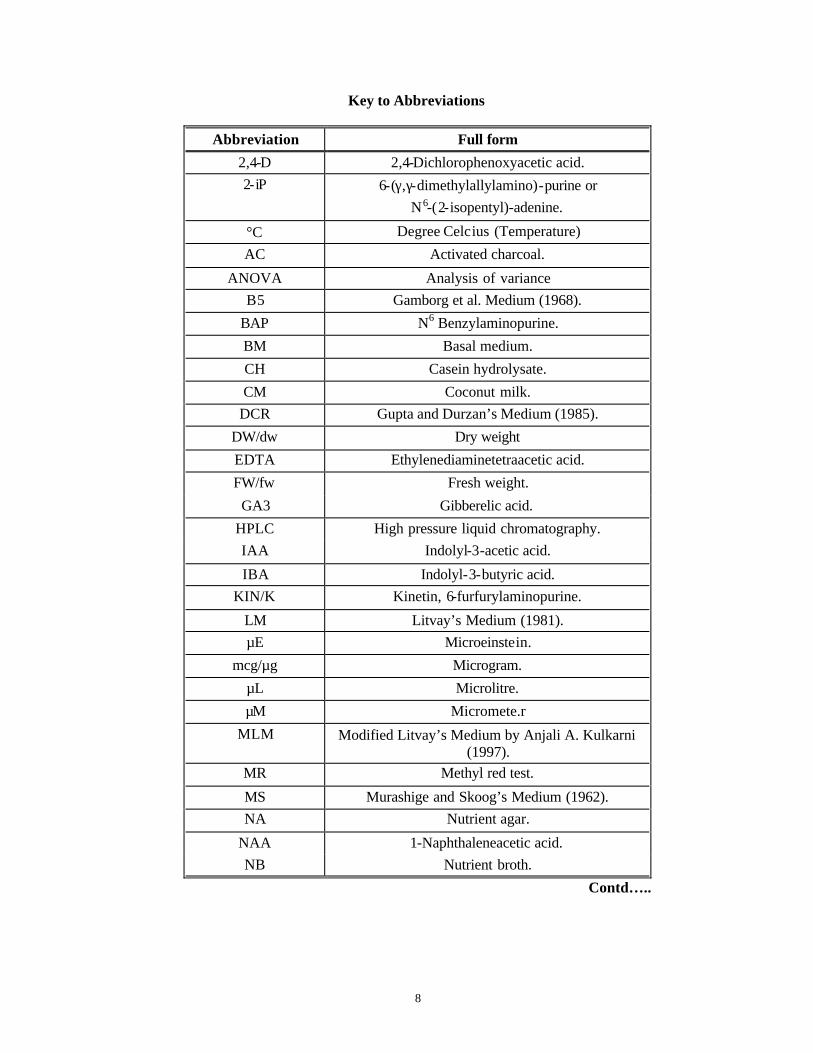
Key to Abbreviations
Abbreviation Full form
2,4-D 2,4-Dichlorophenoxyacetic acid. 2-iP 6-(γ,γ-dimethylallylamino)-purine or
N6-(2-isopentyl)-adenine.
°C Degree Celcius (Temperature)
AC Activated charcoal.
ANOVA Analysis of variance B5 Gamborg et al. Medium (1968).
BAP N6 Benzylaminopurine.
BM Basal medium.
CH Casein hydrolysate.
CM Coconut milk. DCR Gupta and Durzan’s Medium (1985).
DW/dw Dry weight
EDTA Ethylenediaminetetraacetic acid.
FW/fw Fresh weight.
GA3 Gibberelic acid.
HPLC High pressure liquid chromatography. IAA Indolyl-3-acetic acid.
IBA Indolyl-3-butyric acid. KIN/K Kinetin, 6-furfurylaminopurine.
LM Litvay’s Medium (1981). µE Microeinstein.
mcg/µg Microgram.
µL Microlitre.
µM Micromete.r
MLM Modified Litvay’s Medium by Anjali A. Kulkarni (1997).
MR Methyl red test.
MS Murashige and Skoog’s Medium (1962). NA Nutrient agar.
NAA 1-Naphthaleneacetic acid. NB Nutrient broth.
Contd…..
9
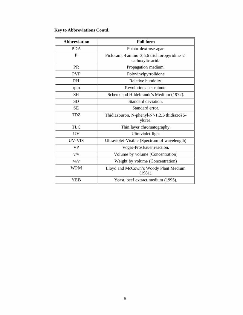
Key to Abbreviations Contd.
Abbreviation Full form
PDA Potato-dextrose-agar. P Picloram, 4-amino-3,5,6-trichloropyridine-2-
carboxylic acid. PR Propagation medium.
PVP Polyvinylpyrrolidone
RH Relative humidity.
rpm Revolutions per minute
SH Schenk and Hildebrandt’s Medium (1972).
SD Standard deviation. SE Standard error.
TDZ Thidiazouron, N-phenyl-N'-1,2,3-thidiazol-5-ylurea.
TLC Thin layer chromatography. UV Ultraviolet light
UV-VIS Ultraviolet-Visible (Spectrum of wavelength)
VP Voges-Proskauer reaction.
v/v Volume by volume (Concentration)
w/v Weight by volume (Concentration)
WPM Lloyd and McCown’s Woody Plant Medium (1981).
YEB Yeast, beef extract medium (1995).

10
SYNOPSIS
Since the dawn of civilization upto the twenty-first century, plants have been the
most important sources of drugs for the mankind. One of the most exciting discoveries
during this long journey has been the isolation of a variety of chemicals from plants,
which have potent anti-cancer activities. Cancer is a major killer disease all over the
world and more than six million new cases are reported every year.
“Taxol” , a novel diterpenoid, isolated first from the bark of Taxus brevifolia Nutt.
(Taxaceae) has been the most promising anti-cancer agent isolated in recent years. It is
also reported from all the known spp. of Taxus including the Himalayan Yew: Taxus
baccata ssp. wallichiana Zucc. Pilg. These plants are also rich in a variety of other related
molecules namely "Taxanes" with different degrees of cytotoxicity and anti-tumor
activities.
The plants have very low content of taxol (0.04-0.1% on dry weight basis) in
mature organs. These trees are very slow growing and their seeds have a long dormancy
period of two years. In addition, they are becoming endangered due to overexploitation.
Hence the tissue culture work was started in the Himalayan Yew with the aim to explore
the possibility of standardizing a micropropagation protocol because of its extreme slow
growth and non-availability of the planting material as well as to assess taxane
production in vitro .
“Withaferin -A”, a withasteroid/steroidal lactone, first isolated from Withania
somnifera (L.) Dunal has anti-tumor activity in a number of systems. The plant belonging
to Solanaceae is used extensively in Ayurvedic system of medicine. It also has a number
of tropane alkaloids effective against arthritis, rheumatism, fevers, fatigue etc. Plant tissue
culture work was initiated with the aim to micropropagate selected superior chemotypes
from various explants and to assess for the possible presence of Withaferin -A in tissue
culture raised plants, callus cultures and hairy root cultures.
The present thesis is divided into six chapters and an annexure followed by
summary and a list of references.
11
CHAPTER 1 GENERAL INTRODUCTION.
This chapter deals with an overview of developments in plant tissue culture, both in
angiosperms and gymnosperms with special reference to medicinal plants. In vitro studies
carried out in Taxus spp. and Withania spp. so far have also been described.
CHAPTER 2 MATERIALS AND METHODS (GENERAL).
Compositions of various plant tissue culture and bacteriological media used has been
included in this chapter as also the various techniques used during the course of this work.
CHAPTER 3 CONTAMINATION CONTROL.
This chapter deals with bacterial and fungal contaminants and phenolic oxidation
observed during the course of this work and measures undertaken to control and
overcome these problems.
§ % data for contaminants observed in Taxus spp. and Withania spp.
§ Identification of contaminant bacteria based on morphological and biochemical
characters.
§ Comparison with reported data using a specially developed software program for
identifying the contaminating bacteria upto the genus level.
§ Identification of fungi based on vegetative and reproductive structures.
§ Antibiogramme for bacteria and determination of MIC and MBC of effective
antibiotics.
§ Determination of effective anti-fungal agents.
§ Development of a pre-treatment solution with both anti-fungal and ant i-bacterial
agents to overcome contamination problem in Taxus spp. and data for reduction in
contamination with this solution.
§ Steps undertaken to reduce contamination in Withania spp. cultures.
12
CHAPTER 4 STUDIES ON CALLUS INDUCTION FROM VARIOUS EXPLANTS
OF TAXUS SPP. AND ANALYSIS OF TAXANE CONTENT OF IN
VITRO AND IN VIVO TISSUES.
Here callus induction is reported from different explants on different basal media
supplemented with various combinations of plant growth regulators. Callus induction is
confirmed by histology. The taxane contents (Taxol and 10-DAB, 10-deacetyl baccatin-
III) are analyzed by TLC and HPLC in different callus lines and are compared against the
contents of the explants of the parent trees (in vivo tissues) determined by TLC and
HPLC.
CHAPTER 5 IN VITRO STUDIES ON MICROPROPAGATION FROM MATURE
AND JUVENILE EXPLANTS OF TAXUS SPP.
§ The present chapter describes precocious germination of embryos on various basal
media and the effect of light and darkness, culture vessel, cold treatment,
developmental stage, locations and growth regulators.
§ The chapter also describes accelerated, precocious primary sprouting from axillary
and apical buds of mature explants as influenced by different basal media and growth
regulator combinations, light conditions etc. It also describes growth of separated
sprouts on various subculture media.
CHAPTER 6 DIRECT REGENERATION OF SHOOTS FROM VARIOUS
EXPLANTS OF WITHANIA SPP. AND ANALYSIS OF
WITHAFERIN-A PRODUCTION THEREFROM.
§ This chapter summarizes the results of direct regeneration of shoots from various
explants such as leaves, nodes, internodes, hypocotyls and embryos. Various
parameter studies affecting the regeneration of plants from each of the explant are
also reported. Direct regeneration of shoots is confirmed by histology.
§ The present chapter also describes TLC and HPLC analysis of seedlings, multiple
shoot and hairy root cultures of Withania somnifera .
ANNEXURE INDUCTION OF HAIRY ROOT CULTURES IN WITHANIA BY
INFECTION WITH AGROBACTERIUM RHIZOGENES.

13
This annexure describes the preliminary results obtained in induction of hairy root
cultures with different strains of Agrobacterium rhizogenes and their growth studies. The
analysis of hairy roots for withanolide production is described in the previous chapter.
SUMMARY.
This part of the thesis summarizes the findings of the present investigation and its future
implications.
BIBLIOGRAPHY
This part of the thesis lists all the references together.
KEY REFERENCES.
1. Wani M. C., Taylor H. L., Wall M. E., Coggon P., McPhail A. T. (1971) Plant
antitumor agents .VI. The isolation and structure of taxol, a novel antileukemic and
antitumor agent from Taxus brevifolia. J. Am. Chem. Soc. 93: 2325-2327.
2. Heinstein P. F., Chang C.-j. (1994) Taxol. Ann. Rev. Plant Physiol. Plant Mol. Biol.
45: 663-674.
3. Ray A. B., Gupta M. (1994) Withasteroids, a growing group of naturally occurring
steroidal lactones. Progress in the Chemistry of Organic Natural Products Vol. 63 (Ed.
Hrz W., Kirby G. W., Moore R. E., Steglich W., and Tamm Ch.) Springer -Verlag
Wien, New York.
4. Roja G., Heble M. R. and Sipahimalani A. T. (1991) Tissue cultures of Withania
somnifera: morphogenesis and withanolide synthesis. Phytother. Res. 5: 185-187.

14
CCHHAAPPTTEERR 11
General Introduction.
15
In recent years a lot of attention has been devoted to novel molecules derived from
natural sources that exhibit a range of clinical and pharmacological activities. This has led
to a flurry of research activities in phytochemicals, synthesized by various plants,
growing in diverse habitats and displaying a range of habits.
In our laboratory a number of plants producing a number of anti-cancer and anti-
tumor metabolites were identified and detailed investigations were undertaken with
respect to morphogenesis, in vitro culture and production of secondary metabolites and
their chemical elucidation.
The present thesis describes the work on morphogenesis, genetic transformation
and production of anti-cancer compounds in vitro of two plants namely, Taxus baccata
ssp. wallichiana Zucc. Pilg. and Withania somnifera (L.) Dunal.
Taxus baccata ssp. wallichiana Zucc. Pilg. is a woody and endangered Himalayan
Gymnosperm producing the most important anti-cancer diterpenoid Taxol and a range of
other clinically important and structurally related Taxanes (Baloglu and Kingston, 1999
and references therein). Withania somnifera (L.) Dun. is a herbaceous Solanaceous plant
producing anti-tumor Withanolides and a range of Tropane Alkaloids having anti-
arthritic, anti-rheumatic and anti-depression properties (Malhotra et al., 1961; Uma Devi
et al., 1992, 1993, 1995).
Because of their diverse habits, habitats and taxonomic differences both these
plants responded differently in vitro. Hence the present thesis has beeen organized as
follows for better clarity, reading and understanding:
1. Chapter 1 : General Introduction describes History of Plant Tissue Culture,
Morphogenesis and Regeneration in vitro (PART A) and Secondary Metabolite
Production by in vitro Cultures (PART B). It also describes the overall objectives of
the present thesis in brief.
2. Chapter 2: Describes the General Methodology of plant tissue culture techniques
followed for both the plant species. The specific techniques, media and conditions
used for each of the plant species are described in their respective chapters.
Preamble
16
3. Chapter 3 : Outlines the strategy employed for Control of Contamination and
Phenolic Browning. Since the problems of bacterial and fungal contamination and
phenolic browning are common for both the plant species, the procedure followed to
evolve a control strategy for contamination and phenolic browning, applicable for the
explants of both Taxus spp. and Withania spp. is written as a “Common” chapter.
4. Chapter 4: Deals with the work done on Taxus spp. including an Introduction,
Literature Survey related to undifferentiated in vitro cultures of yews and Taxol
Production therefrom and Objectives in detail. It also includes Callus Formation,
Nutrient Media Optimization, Suspension Cultures and Analysis of Taxane
Production in in vitro Cultures of Taxus baccata ssp. wallichiana Zucc. Pilg.
5. Chapter 5 : Includes an Introduction, Literature Survey related to precocious
germination and regeneration studies in in vitro cultures of yews and Objectives in
detail. It also details Micropropagation Studies on Taxus baccata ssp. wallichiana
Zucc. Pilg. with Mature and Juvenile Explants. Results obtained with Precocious
Germination and Endosperm Culture have also been described here. The results
obtained have also been extrapolated to plant material collected from different
locations in the Himalayas.
6. Chapter 6: Outlines the work done on Withania somnifera (L.) Dun. including
Literature Survey and Objectives pertinent to the species. It also includes Direct Shoot
Regeneration from Various Explants of Withania somnifera (L.) Dun. and Analysis of
Withanolide Production therefrom.
7. Annexure : Describes the preliminary results obtained on Hairy Root Production from
various explants of Withania somnifera (L.) Dun. Culture of Hairy and Seedling
Roots in Liquid Culture and Analysis of Withanolide Production therefrom forms the
contents of this chapter.
8. Bibliography.
9. Summary: Describes concisely the results obtained during the course of the present
work.
17
In the last forty years the flow of biological discovery has swelled from a trickle
into a torrent, driven by a number of new methodologies developed in Plant Tissue
Culture, Recombinant DNA Technology, Monoclonal Antibodies and Microchemical
Instrumentation (Opportunities in Biology, 1989). Great advances are seen in health care
with development of new therapeutic drugs and improved methods of diagnosis. This is
true especially for ailments such as cancer, AIDS, Alzheimer's.
Biological research has been transformed from a collection of single discipline
endeavors into an interactive science with bridges between a number of traditional
disciplines. This synergism has made biology the “sunrise field” of the new millenium.
The whole gamut of new discoveries in biology and allied sciences can be
grouped together under a single umbrella term of “Biotechnology” . It can be defined as a
manipulation of biological systems (organisms or biological processes) via modern
technology to solve practical problems in agriculture, environment, medicine and
indus try. It includes clonal propagation via tissue culture, protoplast fusion, gene
cloning, DNA recombination, genetic engineering, mutant induction, in vitro genetic
selection, molecular marker aided genome analysis and gene mapping (Huang et al.,
1993). Enhanced production of pharmacologically important secondary metabolites in
vitro can also be included here.
Higher plants are the ultimate and renewable solar-powered biochemical factories
manufacturing both primary and secondary metabolites from air, water and minerals. This
has led man to manipulate and modify the plants so as to suit his growing requirements of
food, industrial raw material, drugs, pesticides, flavors and fragrances. Since the earliest
primitive farming, man has been continuously exploiting plants towards his own needs,
by carrying out experiments for plant improvement (Pauls, 1995). But a large number of
techniques developed over the last 150 years have actually laid the foundations of the
modern science in agriculture. Since the last fifty years, development in agriculture has
been greatly aided, in general, by the development of “Biotechnology” as mentioned
earlier and “Plant Tissue Culture” and “Genetic Engineering” in particular (Brar and
Khush, 1994). These techniques can contribute to various crop-improvement stages like
PART A. GENERAL INTRODUCTION TO PLANT TISSUE
AND CELL CULTURE TECHNIQUES.
18
germplasm manipulations, genotype selection, stabilization, variety testing, variety
increase, proprietary protection and crop production (Pauls, 1995).
Since plants are immovable, environment has a very strong impact on every
aspect of plant form and function. As a consequence, plant development shows a high
degree of plasticity. Environmental stimuli are used as cues to rapidly modify and
influence developmental programming to provide flexibility in response throughout the
growth cycle of plant. The entire technology of “Plant Tissue Culture” is based on this
ability of plant cells to be influenced by their surroundings and differentiate to give rise to
a range of organs dependent on the culture conditions (Bowles and Leyser, 1994). “Plant
Tissue Culture” is the maintenance and propagation of plant parts, as small as a
single cell, in axenic culture under controlled environmental conditions (Pauls, 1995).
The theoretical basis of tissue culture, in general, was established as early as 1838-1839
in Schlieden and Schwann’s Cell Theory. Haberlandt (1902) first attempted to prove this
theory experimentally using monocotyledonous plants and though his attempt was
unsuccessful, he elaborated the concept of Totipotency that refers to the potential of an
individual cell to regenerate a whole plant. This period was followed by many
unsuccessful attempts at plant tissue culture. But Kotte (1922) and Robbins (1922a, b)
independently achieved in vitro culture of pea and maize roots in synthetic media under
sterile conditions. Although these root cultures could not be maintained indefinitely, they
proved that it was easier to maintain cultures having meristematic cells. White (Tomato
roots: 1934a, Spontaneous tumor callus in tobacco: 1939a), Gautheret (Carrot tissue:
1939) and Nobecourt (Carrot tissue: 1939) first achieved the prolonged and indefinite
growth of plant tissues in vitro . White also observed that root meristems are free of
viruses during the course of these studies (White 1934b). Gautheret (1942, 1955)
described the phenomenon of callus habituation where, as the cultures age they can grow
in nutrient media free of plant growth regulators, especially auxins.
During the period between 1951-1961, successful Callus Cultures were raised
starting from normal or tumor tissues from a number of plants belonging to Dicots,
Monocots and Gymnosperms (Gautheret, 1985 and references therein).
The demonstration of hormonal regulation of growth, differentiation and
organ formation in plants (Skoog and Miller, 1957), the regeneration of plantlets from
callus cultures (Reinert, 1958) and plantlet regeneration from cell suspension cultures
19
(Steward et al., 1958) paved the way for the real exploitation of this knowledge towards
the benefit of mankind.
The formation of full plants of tobacco from isolated cells in microculture (Vasil
and Hildebrandt, 1965) and formation of a somatic embryo from a single plated cell of
carrot (Backs -Hussemann and Reinert, 1970) finally led to the unequivocal demonstration
of totipotency of single and isolated plant cells more than 60 years after Haberlandt’s
theory of totipotency. The concept of totipotency also holds true for haploid pollen cells
which was proven by the demonstration of formation of somatic embryos from
microspores of Datura: ‘Androgenesis’ (Guha and Maheshwari, 1964). Due to the
voluminous amount of work done on these aspects in vitro on a broad range of plants,
detailed review of literature is beyond the scope of the present thesis.
‘Regeneration’ is defined as the ability of single protoplasts, single cells or
tissues to develop complete plants. When the organogenetically formed shoots are
rooted, it can be termed as regeneration. The regenerative response in vitro occurs in
two different patterns viz. ‘Somatic embryogenesis’ and ‘Organogenesis’. The former
leads to the production of a bipolar structure containing a root/shoot axis with a closed
independent vascular system while the latter leads to the production of unipolar structures
such as a root or a shoot primordium, whose vascular system is often connected with the
parent plant (Thorpe, 1993). These responses are due to the two properties of the plant
cells namely, ‘Autonomy’ and ‘Totipotency’.
In tissue cultures, the meristematic region at the initiation of organogenesis
originates either from the reproduction of previously existing meristems or is produced de
novo from other component cells in the tissue (Endress, 1994). External manipulations
such as selection of explant (type of or gan, physiological and ontogenetical age of the
explant, season for obtaining explant, size of explants, position of explant on mother
plant, state of mother plant), changes in chemical components like media, plant growth
regulators and other additives like amino acids, coconut milk (CM), casein hydrolysate
(CH), oligosaccharides and various physical factors (physical form of the medium, pH of
the medium, humidity, light, temperature) induce explant cells in the right physiological
state to respond and unde rgo organized development. These structural changes are results
of preceding biochemical and biophysical events that ultimately reflect selective gene
activity (Thorpe, 1980).
20
Organogenesis in vitro can follow either of the two paths:
1. Indirect organogenesis: Primary explant is induced to form callus prior to undergoing
de novo organogenesis. The resultant plants may show variations due to the
involvement of the callus phase.
2. Direct organogenesis : No intervening callus phase is involved and the regenerants
may be identical to the parent plant.
The key feature of de novo organogenesis is the formation of a 'meristemoid'
arising from vacuolated parenchyma cells. The cells of the meristemoid are small,
isodiametric, thin-walled with prominent nuclei and with extensive network of
plasmodesmata. This is the first step of ‘Cellular Differentiation’. It is followed by
‘Acquisition of Competence’, ‘Cellular Determination’ and finally ‘Cellular
Differentiation’ giving rise to organs (Christianson and Warnick, 1985). The whole
process is high-energy intensive where accumulated starch and carbohydrates from the
media are used (Thorpe, 1980).
Refined tissue culture procedures have made it possible to introduce foreign DNA
and cloned genes into cultured cells, protoplasts and plant organs from diverse biological
systems and to regenerate transgenic plants in more than 50 plant species, across
Gymnosperms, Dicots and Monocots, with altered genetic properties. The first
Agrobacterium tumefaciens transformed transgenic plant expressing engineered foreign
genes was Solanaceous Nicotiana tabacum (Horsch et al., 1984) and the successive plant
generations have also expressed the introduced gene. The first transgenic conifer was
Larix decidua obtained via Agrobacterium rhizogenes mediated transformation (Huang et
al., 1993).
Agrobacterium rhizogenes mediated transformation was also used for the
production of Hairy Roots that can synthesize a number of secondary metabolites of the
parent plant at a level comparable to or more than in planta . First report of such a result
appeared for Hyoscyamus muticus (Flores and Filner, 1985). Hairy roots have also been
shown to produce novel secondary metabolites, which are not produced by the parent
plant.
21
Most of the current interest in medicinal plants has stemmed from the fact that in
the recent past, a number of promising drugs have been developed from these plants
based on the traditional knowledge and the new insights gained by modern methods of
research. Hence cataloguing of medicinal plants and their uses, constant review of their
natural status (endangered or otherwise) and exploitation of their natural biodiversity
have proved to be important tools in their detailed and varied investigations. Use of Plant
Tissue and Cell Culture can be an invaluable aid in the constant search for new drugs
from as yet unexplored or underexplored plants.
22
Introduction
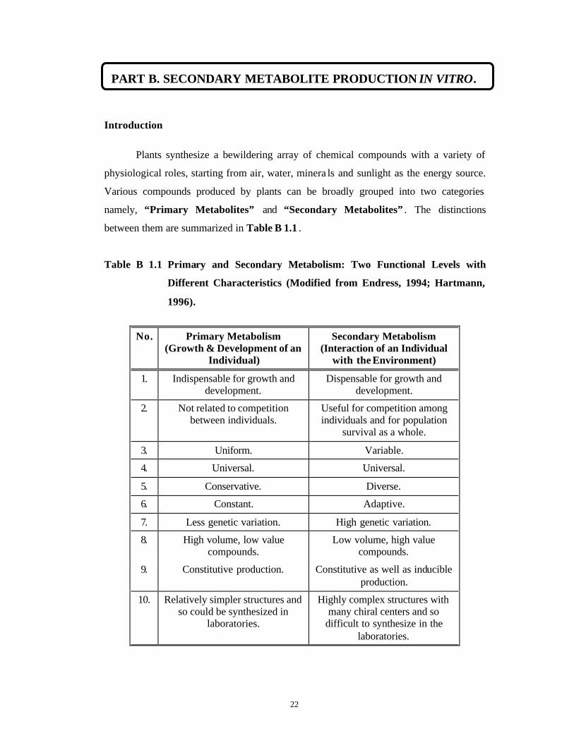
Plants synthesize a bewildering array of chemical compounds with a variety of
physiological roles, starting from air, water, minera ls and sunlight as the energy source.
Various compounds produced by plants can be broadly grouped into two categories
namely, “Primary Metabolites” and “Secondary Metabolites” . The distinctions
between them are summarized in Table B 1.1 .
Table B 1.1 Primary and Secondary Metabolism: Two Functional Levels with
Different Characteristics (Modified from Endress, 1994; Hartmann,
1996).
No. Primary Metabolism (Growth & Development of an
Individual)
Secondary Metabolism (Interaction of an Individual
with the Environment)
1. Indispensable for growth and development.
Dispensable for growth and development.
2. Not related to competition between individuals.
Useful for competition among individuals and for population
survival as a whole.
3. Uniform. Variable.
4. Universal. Universal.
5. Conservative. Diverse.
6. Constant. Adaptive.
7. Less genetic variation. High genetic variation.
8. High volume, low value compounds.
Low volume, high value compounds.
9. Constitutive production. Constitutive as well as inducible production.
10. Relatively simpler structures and so could be synthesized in
laboratories.
Highly complex structures with many chiral centers and so
difficult to synthesize in the laboratories.
PART B. SECONDARY METABOLITE PRODUCTION IN VITRO.
23
The secondary metabolites are also referred to as “Natural Products”. It is
believed that more than 100,000 different structures of secondary metabolites maybe
synthesized by organisms, to a tune of 109 tons per year (Bowles and Leyser, 1994). Out
of these, more than 80% are found in plants (Harborne, 1993). They are used either as
medicines/pharmaceuticals, foods, neutraceuticals (foods as well as medicines used for
preventive and curative treatments), flavors, colors, spices or fragrances by humans while
in plants they constitute a chemical response to pollinators and distributors, to
competitors and herbivores, to symbionts and pathogens and to stress. They are also
believed to detoxify substances accumulated in the primary metabolism and to provide
chemical signals to coordinate metabolism of multicellular organisms. They are believed
to coordinate activities of different individuals of the same species (Endress, 1994). Many
of the social, political and economic changes throughout the history of the mankind have
been driven by the desire to gain or exploit economically important plants like spices,
rubber, coffee, tea, tobacco, cocoa and opium poppy producing bioactive molecules.
Although secondary metabolism was first recognized in 1873 (Sachs, 1873), its
function was elucidated only in 1888 (Stahl, 1888). But upto the 1950s, secondary
metabolites were regarded as end products of deluxe metabolism and relegated to the rank
of ‘waste products’. It was only during the 1960s, that their eco-relations were discovered
(Kurz and Constabel, 1998). More recently, biotechnologies have become indispensable
in efforts to experimentally widen our knowledge of the ontogeny, metabolism, function
and production of secondary metabolites.
Chemical diversity in plants is not incidental but is brought about by specifically
organized and controlled biosynthetic pathways, well integrated into the metabolism of
plants. This is also the expression of plant genome under developmental control. Most of
the secondary metabolites are formed via a few biogenetic routes, leading to one or a few
key metabolites, from which numerous derivatives are formed by simple enzymatic
transformations. The biosynthesis usually occurs in an organ in a tissue -specific manner
and is often temporally restricted during the development. It is often accompanied by
specific translocation and storage behavior (Hartmann, 1996; Kurz and Constabel, 1998).

24
The enormous diversity of secondary metabolites found in nature results from
three main features such as a high degree of chemical freedom, strictly regulated
metabolic pathways and enzyme optimization (Hartmann, 1996).
As commodities, secondary metabolites are increasingly in great demand. This
demand is increasingly becoming acute because of their non-availability, owing to
adverse climatic conditions, pests, political instability in cropping areas, misuse and
overexploitation. Many medicinally important plants are already threatened in their
natural habitats. E.g. Lithospermum erythrorhizon, Hydrastis canadensis, Cephaelis
ipecacuanha , Rauwolfia serpentina , Podophyllum peltatum, Artemisia genipi, Taxus spp.
There is a growing need to preserve these plants either in vivo or in vitro because of the
uniqueness of each species. and their extinction may lead not so much in the loss of an
individual secondary metabolite, but rather, in the specific bouquet of all the metabolites
presented by each unique species (Kurz and Constabel, 1998). Up till now, 47 major
drugs have been produced from the tropical forests but it is believed that around 328
potential, major drugs and 125,000 flowering trees of potential pharmacological
importance may be still hidden in the unexplored depths of these forests. Every year the
market for herbs-based drugs is increasing at the rate of 12-15%
(http://www.phytochemistry.freeserve.co.uk).
Plant tissue and cell culture technologies in this context are seen as a savior in
channelizing the resources of the nature for the benefit of the mankind by conservation of
elite, endangered plants and eco-friendly production of drugs and drug intermediates.
Cryopreservation of germplasm would help in maintaining the genetic diversity of the
endangered population and improved cell and tissue culture technologies would help in
producing the active compounds in vitro with better productivities without cutting down
the natural resources. Recombinant DNA technologies would also supplement plant cell
technologies towards this end.
Large -scale production of phytochemicals with plant cell and tissue culture
technologies has the following advantages (Modified from Zafar et al., 1992):
1) Independence from environmental factors like climate, pests, geographical and
seasonal constraints.
2) Defined production systems as and when required.

25
3) More consistent product quality and yield.
4) Potential for fast growth rates.
5) Continuous and homogeneous supply of plant material in a uniform physiological
state.
6) Reduction in land use for ‘cash crops’.
7) Freedom from political constraints.
8) Process isolation from related metabolic pathways leading to higher yields.
9) Magnification and accentuation of chemical reactions under growth control.
10) Use of improvement strategies for better yields and cost-benefit ratios by growth
medium and microenvironment manipulations.
11) Use of recombinant DNA technologies for yield improvement.
12) Use of genetic transformation with Agrobacterium tumefaciens and Agrobacterium
rhizogenes to achieve high product yields in the absence of growth regulators but
with proper organ differentiation.
13) Production of novel compounds in vitro , which are absent in the in vivo parent plant
material.
14) Possibility of having continuous production of metabolites that are normally
transiently made in the life cycle of the parent plant (e.g. flower colors).
15) Possibility of having an active synthetic tissue for a metabolite normally
accumulated in a non-active tissue like bark.
16) Understanding of complex metabolic pathways involved in the synthesis of
secondary products and identification of the key enzymes.
17) Possibility of metabolic engineering at the cellular level for better productivities.
18) Ability for large-scale cultivation of cells, organs and even entire plants in
bioreactors for easier and higher product recoveries.
Despite a number of advantages listed above, plant cell and tissue culture
technologies also suffer from some drawbacks (Zafar et al., 1992):
1. Since plant cells are much larger in size, they have a bigger surface area and hence
lower metabolic activity resulting in slower growth rates as compared to smaller sized
microbial cells.
2. Plant cells tend to grow together as cell clumps rather than single cells posing some
problems for their cultivation in bioreactors.
26
3. The yield of secondary metabolites in vitro cannot be predicted beforehand. Many-a-
times the compounds of interest are not produced in vitro or if produced are present in
extremely low quantities.
4. High genetic and epigenetic instability of cell cultures can lead to loss of
phytochemical production capacities of cells.
5. Differentiation-related products are not produced in unorganized tissues that are easier
to manipulate in vitro .
6. Culture conditions may trigger new pathways producing novel but useless products.
7. Empirical methods developed for cell cultures of a particular plant cannot be
extrapolated to a wide range of plants and culture systems.
There are four main in vitro approaches for production of secondary metabolites:
1. Callus Cultures
Callus cultures consist of an undifferentiated, proliferating mass of cells usually
arising on wounds of differentiated tissues and cells. These can be initiated either from
the source tissue synthesizing the secondary compound of interest or from other tissues
like embryo. Generally the plants which accumulate relatively high yields of specific
secondary metabolites, give rise to tissue cultures producing high levels of secondary
metabolites and vice versa. This is because the capacity for the biosynthesis of secondary
metabolites is genetically determined (Lindsey and Yeoman, 1985). Cell division usually
occurs in parenchymatous cells by dedifferentiation. During this process adult cells
temporarily revert to juvenile state (Rejuvenation) and hence show intense growth and
division activity. The degree of dedifferentiation is different in different cells derived
from the same explant (Lindsey and Yeoman, 1985 and references therein). Norma lly
juvenile and hence physiologically the most active tissues give better callus formation.
The exogenous plant growth regulator requirement (Type, concentration, auxin to
cytokinin ratio) for callus formation depends upon the genotype and endogenous hormone
content of the tissues (Pierik, 1987).
During callus formation there are two distinct phases namely, the “wound
response” and the “growth response”. The former phase is characterized by a rapid
increase in metabolic activity when the explant is cultured on the growth medium but
does not lead to callus formation. The latter phase results in cell division and is dependent
27
upon an exogenous supply of auxin. The cell structure also undergoes changes such as
increased numbers of mitochondria and polyribosomes, disappearance of storage reserves
like starch etc. Thus both the “wound response” and the “growth response” are
characterized by distinct periods of m-RNA activity and the levels of t-RNA and r-RNA
are regulated independently (Lindsey and Yeoman, 1985 and references therein).
Due to the presence of diffusion gradients of nutrients, oxygen and other factors,
the growth of callus is not always uniform and rapid and hence has less possibility for
scale-up. But the chemical gradients, cell-to-cell contact and small degrees of
differentiation seem to favor secondary metabolite production of some classes (Lindsey
and Yeoman, 1985). Callus induction requires an environment where some of the cells
can divide and proliferate. The strongest induction factor is the growth/nutrient medium
supplemented with plant growth regulators (Gibson et al., 1995). With prolonged culture,
some of the callus cells may undergo some degree of differentiation and may change the
spectrum of secondary metabolite synthesis. For many years, callus cultures have been
employed in the study of secondary metabolism in vitro. In many of the callus cultures,
conditions for callus induction and growth (Growth Medium) are not conducive for
secondary metabolite production and the tissues need to be transferred to a new medium
with a different composition (Production Medium). Production methods tend to be
contingent upon defining conditions that allow maximum product accumulation rate.
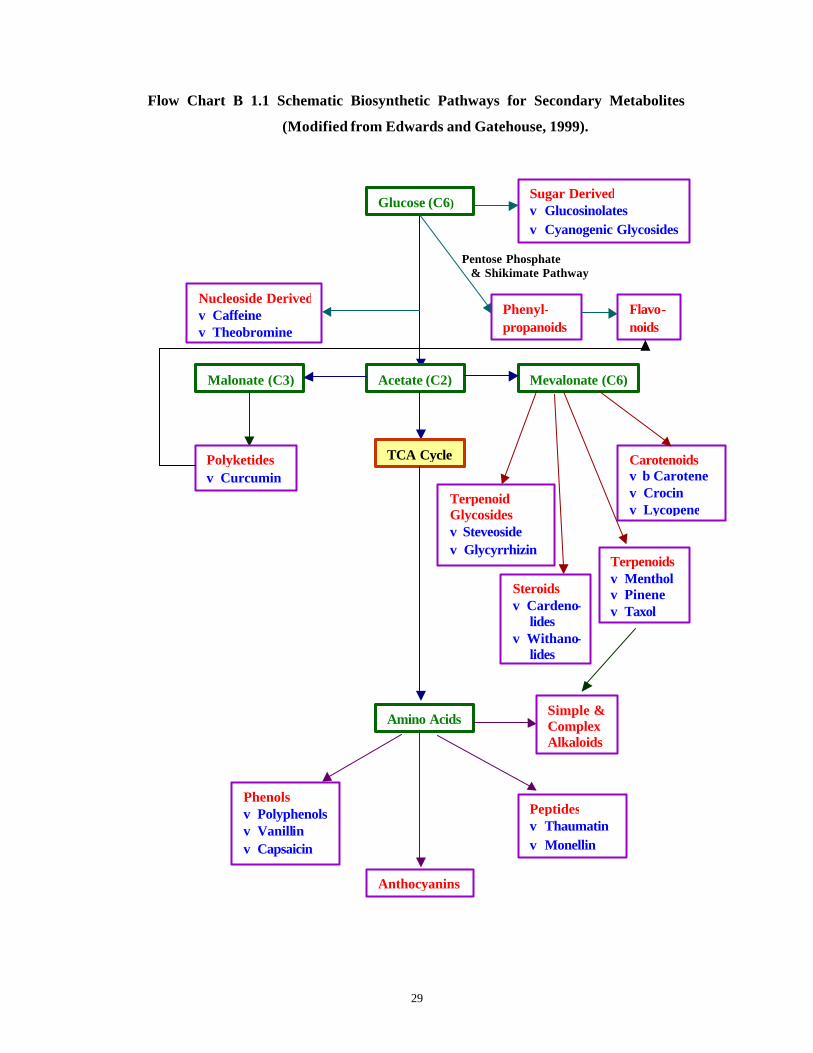
Table B 1.2 lists different classes of secondary metabolites produced by callus
and cell suspension cultures in vitro while Flow Chart B 1.1 depicts the mode of
formation of these compounds.

28
Table B 1.2 Groups of Natural Products So Far Isolated from Callus and Cell
Suspension Cultures of Higher Plants (Stöckigt et al., 1995).
I Phenyl-propanoids
II Alkaloids III Terpenoids IV Quinones V. Steroids
1. Antho-cyanins
1. Acridines 1. Carotenoids 1. Anthra-quinones
1. Steroidal lactones
2. Coumarins 2. Betalaines 2. Monoterpenes 2. Benzo-quinones
2. Steroidal glycos ides
3. Flavonoids 3. Quinolizidines 3. Sesquiterpenes 3. Naphtho-quinones
4. Hydroxy-cinnamoyl derivatives
4. Furo-quinolines
4. Diterpenes
5. Isoflavonoids 5. Harringtonines 5. Triterpenes
6. Lignans 6. Isoquinolines
7. Phenalinones 7. Indoles
8. Proantho-cyanidins
8. Purines
9. Stilbenes 9. Pyridines
10. Tannins 10. Tropane Alkaloids
11. Indole Alkaloids
12. Isoquinoline Alkaloids
29
Flow Chart B 1.1 Schematic Biosynthetic Pathways for Secondary Metabolites
(Modified from Edwards and Gatehouse, 1999).
Pentose Phosphate
& Shikimate Pathway
Glucose (C6)
Acetate (C2)
Sugar Derived ϖ Glucosinolates ϖ Cyanogenic Glycosides
TCA Cycle
Mevalonate (C6) Malonate (C3)
Amino Acids
Anthocyanins
Polyketides ϖ Curcumin
Carotenoids ϖ β Carotene ϖ Crocin ϖ Lycopene
Terpenoids ϖ Menthol ϖ Pinene ϖ Taxol
Terpenoid Glycosides ϖ Steveoside ϖ Glycyrrhizin
Peptides ϖ Thaumatin ϖ Monellin
Phenols ϖ Polyphenols ϖ Vanillin ϖ Capsaicin
Nucleoside Derived ϖ Caffeine ϖ Theobromine
Simple & Complex Alkaloids
Phenyl-propanoids
Flavo-noids
Steroids ϖ Cardeno-
lides ϖ Withano-
lides
30
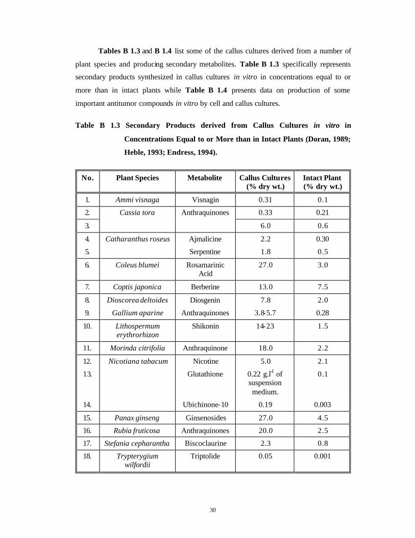
Tables B 1.3 and B 1.4 list some of the callus cultures derived from a number of
plant species and producing secondary metabolites. Table B 1.3 specifically represents
secondary products synthesized in callus cultures in vitro in concentrations equal to or
more than in intact plants while Table B 1.4 presents data on production of some
important antitumor compounds in vitro by cell and callus cultures.
Table B 1.3 Secondary Products derived from Callus Cultures in vitro in
Concentrations Equal to or More than in Intact Plants (Doran, 1989;
Heble, 1993; Endress, 1994).
No. Plant Species Metabolite Callus Cultures (% dry wt.)
Intact Plant (% dry wt.)
1. Ammi visnaga Visnagin 0.31 0.1
2. 0.33 0.21
3.
Cassia tora Anthraquinones
6.0 0.6
4. Ajmalicine 2.2 0.30
5.
Catharanthus roseus
Serpentine 1.8 0.5
6. Coleus blumei Rosamarinic Acid
27.0 3.0
7. Coptis japonica Berberine 13.0 7.5
8. Dioscorea deltoides Diosgenin 7.8 2.0
9. Gallium aparine Anthraquinones 3.8-5.7 0.28
10. Lithospermum erythrorhizon
Shikonin 14-23 1.5
11. Morinda citrifolia Anthraquinone 18.0 2.2
12. Nicotine 5.0 2.1
13. Glutathione 0.22 g.l-1 of suspension medium.
0.1
14.
Nicotiana tabacum
Ubichinone-10 0.19 0.003
15. Panax ginseng Ginsenosides 27.0 4.5
16. Rubia fruticosa Anthraquinones 20.0 2.5
17. Stefania cepharantha Biscoclaurine 2.3 0.8
18. Trypterygium wilfordii
Triptolide 0.05 0.001

31
Table B 1.4 Antitumor Compounds Produced from Cell and Callus Cultures of
Higher Plants (Modified from Misawa and Nakanishi, 1988).
No. Plant Species Metabolite Cultured Cells Yield
(% dry weight)
Reference
1. Baccharis megapotamica
Baccharine - -
2. Brucea antidysenterica
Bruceantin 5.8 x 10⁻² Misawa et al. (1983)
3. Camptotheca acuminata and Nothapodytes
foetida
Camptothecin 2.5 x 10⁻⁴
4. Catharanthus roseus
Vinblastine and Vincristine
5.0 x 10⁻⁴
Misawa et al. (1974)
5. Cephalotaxus harringtonia
Harringtonine, Homo-
harringtonine
5.5 x 10⁻⁸ Misawa et al. (1985)
6. Maytenus bucchnanii
Maytansine 5.0 x 10⁻⁷ Misawa et al. (1985)
7. Ochrosia elliptica
Ellipticine 2.7 x 10⁻⁴ Kouadio et al. (1985)
8. Podophyllum peltatum and Podophyllum hexandrum
Podophyllotoxin 7.1 x 10⁻¹ Kadkade (1982)
9. All Taxus spp. Taxol See Chapter 4, Table 4.3 for the details.*
10. Trypterygium wilfordii
Tripdiolide 1.0 x 10⁻² Misawa et al. (1983)
11. Withania somnifera
Withanolide I, Withanolide G
1.3-17.2, 1.0-31.7 Roja et al. (1991)*
* New data added based on the literature survey carried out during the course of the present work.
32
2. Suspension Cultures
When callus is suspended in a liquid growth medium, the cells disperse producing
cell suspension cultures that can have faster and uniform growth rates coupled with
secondary metabolite production. With their relatively faster growth, ease of manipulation
and comparative homogeneity, suspension cultures are the most widely employed
systems in the studies of secondary metabolism (Parr, 1989). They represent various
degrees of cellular aggregation and generally, the more aggregated, slower growing
cultures display the highest productivities (Lindsey and Yeoman, 1985). They can be
‘induced’ by media manipulations and elicitors to produce compounds of interest. They
are also amenable for growth in fermentors. But these cultures show genetic and
biochemical heterogeneity.
Street (1977) has described the growth and development of a cell culture as
comprising a succession of physiological states, each characterized by distinctive
structural and biochemical features.
Normally plant cells tend to grow in aggregates of 2-20 cells and can rarely grow
as fine cell suspensions. Because of this aggregation, cells in the center tend to be
morphologically, physiologically and chemically distinct from the cells at the periphery
(Gibson et al., 1995). Cell aggregation can be reduced by choice of an appropriate culture
medium having suitable osmotic agents such as sorbitol and low concentrations of cell
wall degrading enzymes (King et al., 1973). Such a finely dispersed culture grows in a
generalized form of sigmoidal curve (Street, 1977) and goes through a number of phases
such as Lag Phase (absence of cell division or very low rate of cell division, increase in
protein synthesis and increase in the levels of energy-generating systems of cells
depending upon the nutrient medium and plant growth regulator composition, low levels
of secondary metabolites), Log Phase (a short period of exponential cell division
followed by a steady rate of cell division, increase in average cell size, increase in nucleic
acid contents, increase in primary metabolism which can be made to divert into secondary
metabolism by certain media and culture environment manipulations in some species) and
Stationary Phase (period of decelerating cell divisions due to exhaustion of an essential
nutrient, cessation of cell divisions and hence absence of growth, decrease in respiration,
RNA synthesis, protein synthesis). If at the stationary phase, the nutrient medium
composition is changed, differentiation may occur which may in turn lead to increase in
33
secondary metabolite production (Observed in many Solanaceous plants). In some plant
species like Acer pseudoplatanus, King (1980) observed that phenols continued to
accumulate even with declining cell divisions and lowered cell aggregations. So he
suggested that the changing growth rate, rather than organization per se, affected the
expression of secondary metabolism. In such suspension cultures, cultural conditions
undergo changes continuously leading to uncoupling of growth and secondary metabolite
accumulation. This uncoupling could be modified by nutrient medium composition (King
et al., 1973). They further suggested that the gross changes occurring in the metabolic
activity and cell composition resulted due to independence of the control mechanism for
cell division and for the synthesis of individual metabolites.
The origin of rapidly dividing and friable cell suspension cultures appears to be
the result of a random, critical event occurring in the initial phase of explant exposure to
culture medium. This event is probably more a function of the species and the nature of
the explant than of the experimental conditions (King, 1980). Such friable cell lines
exhibit following characters:
♦ High friability.
♦ Homogeneous cell type.
♦ Cells more or less isodiametric.
♦ Cells highly cytoplasmic.
♦ Cells with distinct nuclei.
♦ Cells with many starch grains.
♦ Cultures with limited cytodifferentiation. Presence of a few tracheids or pigmentation.
♦ Cell doubling times of 24-72 hrs.
♦ No apparent regeneration capacity.
♦ Auxotrophic for several common metabolites.
♦ Easily converted to hormone autotrophy (Habituation).
♦ Growth promoted by high CO2 levels.
♦ Susceptibility to ethylene.
♦ Abnormal ploidy levels, often aneuploidy.
34
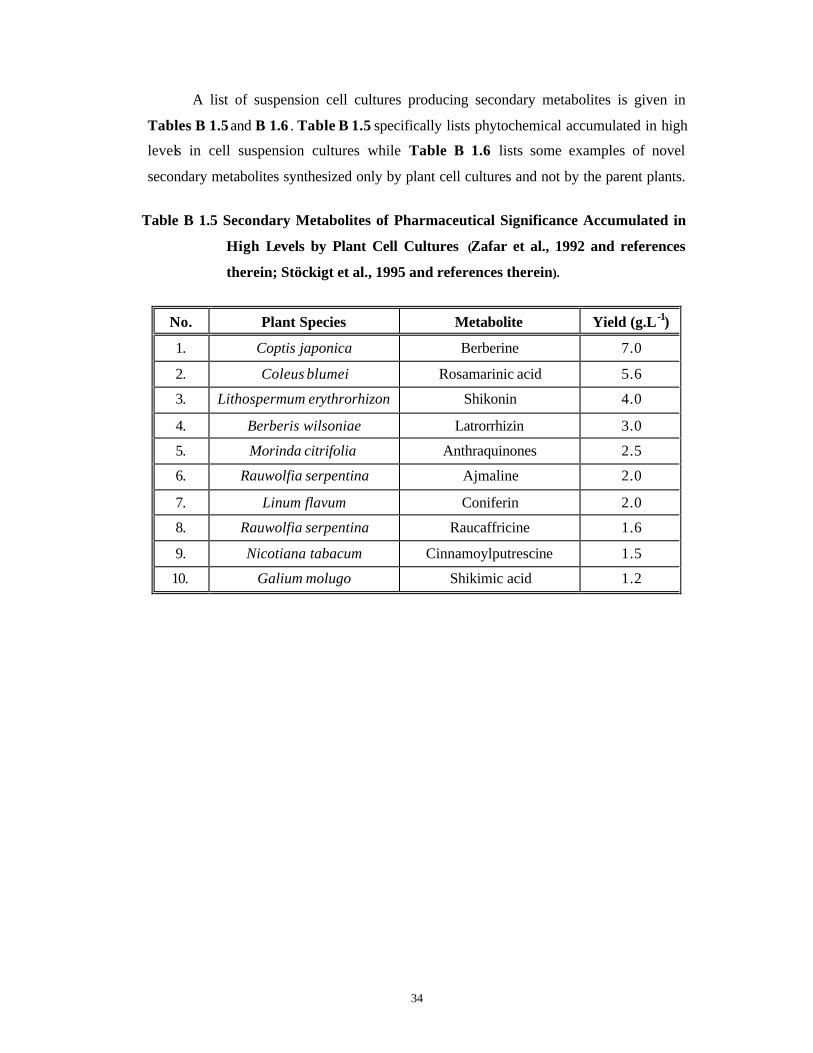
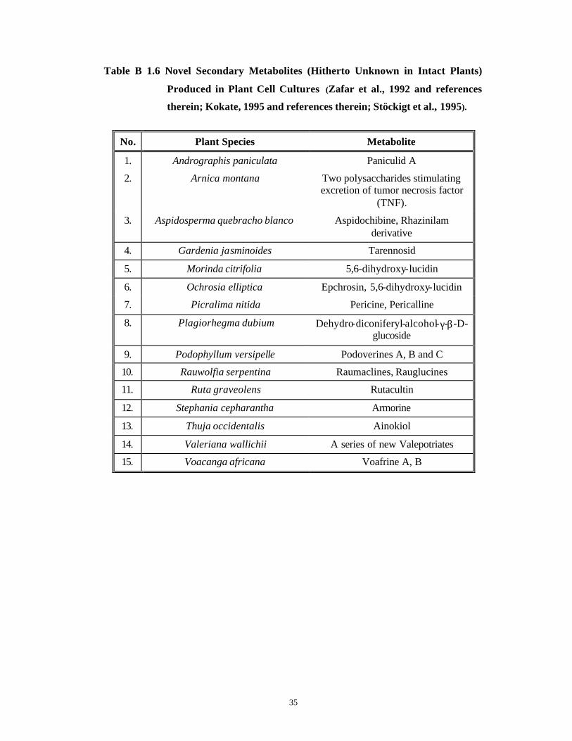
A list of suspension cell cultures producing secondary metabolites is given in
Tables B 1.5 and B 1.6 . Table B 1.5 specifically lists phytochemical accumulated in high
levels in cell suspension cultures while Table B 1.6 lists some examples of novel
secondary metabolites synthesized only by plant cell cultures and not by the parent plants.
Table B 1.5 Secondary Metabolites of Pharmaceutical Significance Accumulated in
High Levels by Plant Cell Cultures (Zafar et al., 1992 and references
therein; Stöckigt et al., 1995 and references therein).
No. Plant Species Metabolite Yield (g.L-1)
1. Coptis japonica Berberine 7.0
2. Coleus blumei Rosamarinic acid 5.6
3. Lithospermum erythrorhizon Shikonin 4.0
4. Berberis wilsoniae Latrorrhizin 3.0
5. Morinda citrifolia Anthraquinones 2.5
6. Rauwolfia serpentina Ajmaline 2.0
7. Linum flavum Coniferin 2.0
8. Rauwolfia serpentina Raucaffricine 1.6
9. Nicotiana tabacum Cinnamoylputrescine 1.5
10. Galium molugo Shikimic acid 1.2
35
Table B 1.6 Novel Secondary Metabolites (Hitherto Unknown in Intact Plants)
Produced in Plant Cell Cultures (Zafar et al., 1992 and references
therein; Kokate, 1995 and references therein; Stöckigt et al., 1995).
No. Plant Species Metabolite
1. Andrographis paniculata Paniculid A
2. Arnica montana Two polysaccharides stimulating excretion of tumor necrosis factor
(TNF).
3. Aspidosperma quebracho blanco Aspidochibine, Rhazinilam derivative
4. Gardenia jasminoides Tarennosid
5. Morinda citrifolia 5,6-dihydroxy-lucidin
6. Ochrosia elliptica Epchrosin, 5,6-dihydroxy-lucidin
7. Picralima nitida Pericine, Pericalline
8. Plagiorhegma dubium Dehydro-diconiferyl-alcohol-γ-β-D-glucoside
9. Podophyllum versipelle Podoverines A, B and C
10. Rauwolfia serpentina Raumaclines, Rauglucines
11. Ruta graveolens Rutacultin
12. Stephania cepharantha Armorine
13. Thuja occidentalis Ainokiol
14. Valeriana wallichii A series of new Valepotriates
15. Voacanga africana Voafrine A, B
36
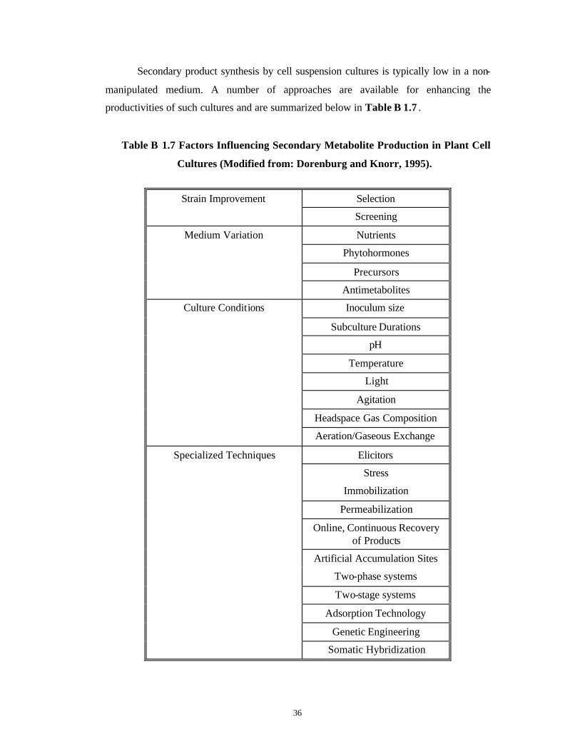
Secondary product synthesis by cell suspension cultures is typically low in a non-
manipulated medium. A number of approaches are available for enhancing the
productivities of such cultures and are summarized below in Table B 1.7 .
Table B 1.7 Factors Influencing Secondary Metabolite Production in Plant Cell
Cultures (Modified from: Dorenburg and Knorr, 1995).
Selection Strain Improvement
Screening
Nutrients
Phytohormones
Precursors
Medium Variation
Antimetabolites
Inoculum size
Subculture Durations
pH
Temperature
Light
Agitation
Headspace Gas Composition
Culture Conditions
Aeration/Gaseous Exchange
Elicitors
Stress
Immobilization
Permeabilization
Online, Continuous Recovery of Products
Artificial Accumulation Sites
Two-phase systems
Two-stage systems
Adsorption Technology
Genetic Engineering
Specialized Techniques
Somatic Hybridization

37
3. Immobilized Cell Cultures
When free cells are entrapped in various matrices like alginate, foam etc. or when
they are allowed to grow into porous supports like reticulated, polyurethane foam, they
give rise to immobilized cell cultures (Parr, 1989). These systems combine the advantages
of the afore-mentioned two systems and are increasingly being preferred, since the cell-
to-cell contact is better and the synthesized product can be easily removed without cell
destruction. Some of these techniques applied for in vitro cultures of Taxus spp. are
described in Chapter 4, Table 4.3.
4. Differentiated Cultures
Secondary metabolites are also synthesized or stored in organized structures such
as roots, shoots, stigmas, embryos, transformed roots (By use of Agrobacterium
rhizogenes) and transformed shoots (By use of shooty mutant of Agrobacterium
tumefaciens). The root cultures can easily be exploited in bioreactors (Parr, 1989). These
cultures show biochemical and genetic stability and usually exhibit the full biosynthetic
capacity associated with the organ in planta.
A list of differentiated cultures producing secondary metabolites is given in Table
B 1.8 for multiple shoot cultures and in Tables B 1.9 and B 1.10 for hairy root cultures.
A. Multiple Shoot Cultures
The synthetic capacity of dedifferentiated tissue often differs substantially from
that of fully differentiated tissues, both qualitatively and quantitatively. The differing
synthetic capacities are usually a direct result of differences in enzyme profiles which
mirror the organ-specific expression of biosynthetic genes (Endress, 1994). The
differentiated cultures often show biochemical (Flores and Filner, 1985) and genetic (Aird
et al., 1988) stability and hence offer a predictable and high-productivity system which
does not require extensive optimization. For example, the accumulation of isoprenoids
usually is dependent upon plastid differentiation, as many of the enzymes in the pathway
are plastid -related. In other cases, formation of glandular hairs or other storage organs is
necessary for metabolite production to proceed. For example, vindoline, a major alkaloid
of Catahranthus roseus is scarcely produced in suspension cell cultures but is produced in

38
multiple shoot cultures derived from the same (Hirata et al., 1987). Callus cultures of
Taraxacum officinale synthesize and accumulate α- and β-amyrins while differentiated
tissues synthesize and accumulate taraxasterol and lupeol because these differentiated
tissues develop laticifers where these compounds are stored (Akashi et al., 1994).
Secondary product formation is an expression of a particular state of cell
differentiation, which can be influenced by a number of signals. In some cases, initiation
of morphological differentiation represents such a triggering signal. Different classes of
secondary compounds require different degrees of cell or tissue differentiation. Formation
of physical and biochemical gradients due to cellular organization is also an important
contributing factor.
Table B 1.8 represents some of the work done in various plant species on multiple
shoot cultures that produce secondary compounds in amounts either equivalent to or
greater than those present in the parent plant.
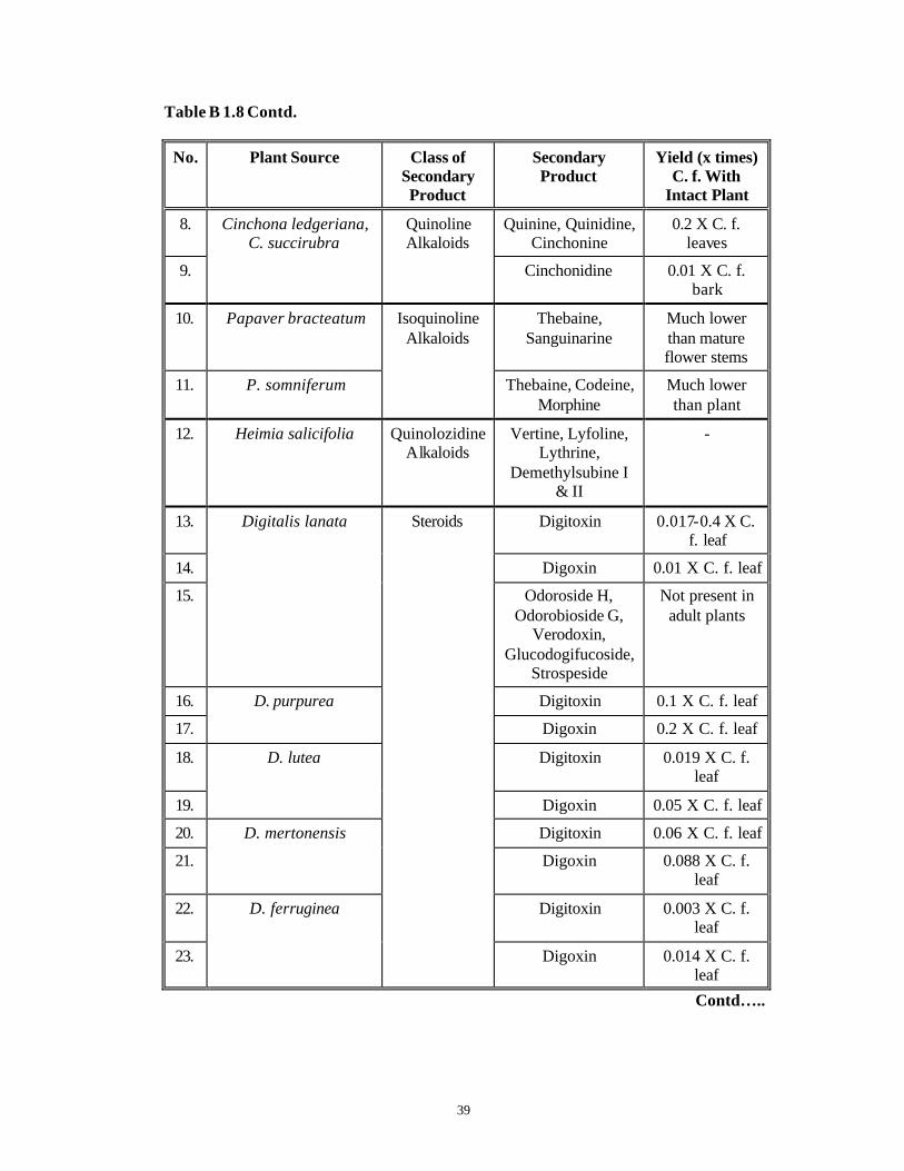
Table B 1.8 Secondary Metabolite Production in Multiple Shoot Cultures of Various
Plant species (Charlwood et al., 1990).
No. Plant Source Class of Secondary Product
Secondary Product
Yield (x times) C.f. With
Intact Plant
1. Atropa belladonna Hyoscyamine, Scopolamine
0.07 X C. f. plant shoot
2. Datura innoxia
Tropane Alkaloids
Hyoscyamine, Scopolamine
0.07 X C. f. leaves
3. Ajmalicine, Cataranthine,
Vindoline
1-8 X C. f. plant
4. 3',4' -anhydrovinblastine
0.3 X C. f. leaves
5. Vinblastine Lower than leaves
6.
Catharanthus roseus Indole Alkaloids
Catahranthine 5 X parent plant
7. Rauwolfia serpentina Indole Alkaloids
Ajmalidine, Ajmaline, Yohimbine
1.4 X C. f. young shoot
Contd…..
39
Table B 1.8 Contd.
No. Plant Source Class of Secondary Product
Secondary Product
Yield (x times) C. f. With
Intact Plant
8. Quinine, Quinidine, Cinchonine
0.2 X C. f. leaves
9.
Cinchona ledgeriana, C. succirubra
Quinoline Alkaloids
Cinchonidine 0.01 X C. f. bark
10. Papaver bracteatum Thebaine, Sanguinarine
Much lower than mature flower stems
11. P. somniferum
Isoquinoline Alkaloids
Thebaine, Codeine, Morphine
Much lower than plant
12. Heimia salicifolia Quinolozidine Alkaloids
Vertine, Lyfoline, Lythrine,
Demethylsubine I & II
-
13. Digitoxin 0.017-0.4 X C. f. leaf
14. Digoxin 0.01 X C. f. leaf
15.
Digitalis lanata
Odoroside H, Odorobioside G,
Verodoxin, Glucodogifucoside,
Strospeside
Not present in adult plants
16. Digitoxin 0.1 X C. f. leaf
17.
D. purpurea
Digoxin 0.2 X C. f. leaf
18. Digitoxin 0.019 X C. f. leaf
19.
D. lutea
Digoxin 0.05 X C. f. leaf
20. Digitoxin 0.06 X C. f. leaf
21.
D. mertonensis
Digoxin 0.088 X C. f. leaf
22. Digitoxin 0.003 X C. f. leaf
23.
D. ferruginea
Steroids
Digoxin 0.014 X C. f. leaf
Contd…..

40
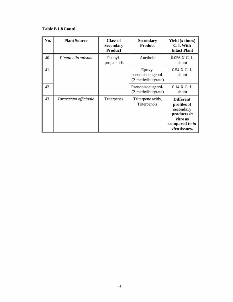
Table B 1.8 Contd.
No. Plant Source Class of Secondary Product
Secondary Product
Yield (x times) C. f. With
Intact Plant
24. Digitoxin 0.012 X C. f. leaf
25.
D. ambigua
Digoxin 0.066 X C. f. leaf
26. Dioscorea composita Diosgenin 0.7 X C. f. tuber
27. Sarsaapogenin, Smilagenin
0.3 X C. f. mature rhizome
28. Markogenin, Samogenin
0.66 X C. f. mature rhizome
29.
Yucca schidigera
Steroids
Gitogenin, Negitogenin
0.54 X C. f. mature rhizome
30. Chrysanthemum cinerariaefolium
Pyrethrine, Cinerin, Jasmolin
-
31. Pelargonium fragrans Pinene, Sabinine, Farnesene,
Carvone, Cadinine
0.5 X C. f. leaf
32. P. graveolens Geraniol, Citronellol,
Citronellyl formate
-
33. P. tomentosum
Isoprenoids
Menthone, Isomenthone
0.5 X C. f. leaf
34. Solanum nigrum 0.35 X C. f. leaf
35. S. laciniatum
Solasodine
10-20 X C. f. In vitro shoots of
other spp.
36. Withania somnifera
Steroidal Alkaloids
Withanolides -
37. Citrus paradisi Naringin, 0.18 X C. f. seedling leaf
38. Chrysosplenium americanum
O-methylated flavonol
glucosides
0.1 x C. f. first leaf
39. Foeniculum vulgare
Phenyl-propanoids
Anethole -
Contd…..
41
Table B 1.8 Contd.
No. Plant Source Class of Secondary Product
Secondary Product
Yield (x times) C. f. With
Intact Plant
40. Anethole 0.056 X C. f. shoot
41. Epoxy-pseudoisoeugenol-(2-methylbutyrate)
0.54 X C. f. shoot
42.
Pimpinella anisum Phenyl-propanoids
Pseudoisoeugenol-(2-methylbutyrate)
0.14 X C. f. shoot
43. Taraxacum officinale Triterpenes Triterpene acids, Triterpenols
Different profiles of secondary
products in vitro as
compared to in vivo tissues.

42
B. Hairy Root Cultures
Ever since the discoveries of Mendel, geneticists have been interested in the
prospect of directed genetic change. Conventional plant breeding was the only source till
the early part of 20th century to achieve this aim. But after the discovery of Smith and
Townsend (1907), that a Gram negative soil bacterium, Agrobacterium tumefaciens,
causes a tumorous plant disease known as ‘crown gall’, the options have widened. Later
on, Agrobacterium rhizogenes was shown to cause ‘hairy root phenotype’ in infected
plants (White et al., 1982). These changes were mediated through transfer and subsequent
integration into the host cell genome of a fragment of DNA known as T-DNA, from the
bacteria. The expression of genes on this fragment in the plant cell environment led to the
development of typical transformed phenotypes (Weising and Kahl, 1996 and references
therein; Sheng and Citovsky, 1996).
Agrobacterium rhizogenes and Plant Interaction
Agrobacterium spp. are non-sporing, aerobic, Gram negative rods (0.6-1.0 µM x
1.5-3.0 µM) belonging to family Rhizobiaceae. They may occur as single cells or in pairs,
are motile by 1-6 peritrichous flagella and secrete copious extracellular polysaccharide
slime (Holt et al., 1994).
Based on their phytopathogenic characters (Gelvin et al., 1988-1990), agrobacteria
are grouped into three classes namely:
I. Agrobacterium tumefaciens, causing crown gall disease.
II. Agrobacterium rhizogenes, causing hairy root disease.
III. Agrobacterium radiobacter, avirulent spp.
Based on their growth patterns (Gelvin et al., 1988-1990), agrobacteria can be
classified into two groups :
1) Biotype 1, where the strains can grow upto 37°C, can grow on typical E. coli -type
media and can produce ketolactose from lactose. Many A. tumefaciens strains and few
A. rhizogenes strains fall in this category.
2) Biotype 2, where the strains can grow only at 28-29°C and not at 37°C, cannot grow
on typical E. coli-type media but require special media and cannot produce
43
ketolactose from lactose. These strains can utilize erythritol as a carbon source. Many
A. rhizogenes strains fall in this category.
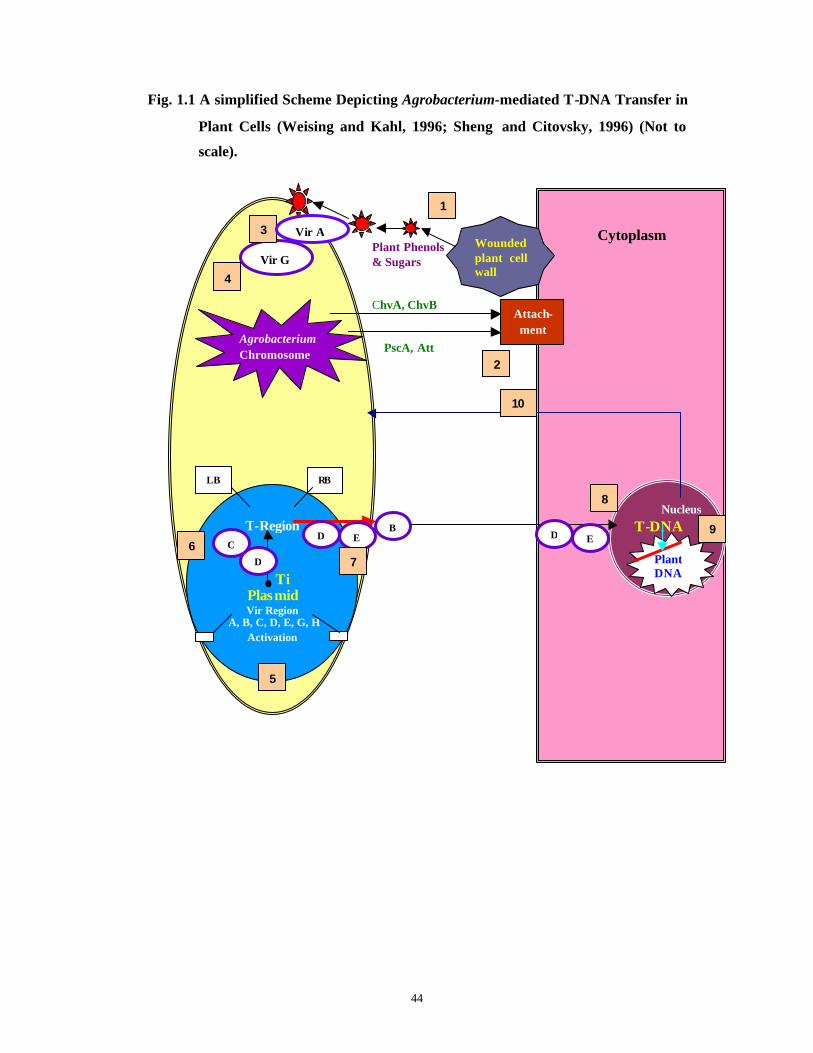
Formation of crown gall or hairy roots is a direct expression of genes in the T-
DNA of the Ti (Tumor-inducing) or Ri (Root-inducing) plasmids (Size: 200 kb) that are
transferred to the host plant cell from agrobacteria and become stably integrated into its
genome. Hairy root causing Ri plasmids are classified according to the opines (Agropine-,
Mannopine- and Cucumopine-type) synthesized by them. The T-DNA, despite its
prokaryotic origin, has eukaryotic transcriptional and translational control sequences that
can be recognized by the host cell machinery. The whole process of Agrobacteruim-
mediated T-DNA transfer in plant cells is depicted in Fig. 1.1 . The figure summarizes a
number of interactions involved in T-DNA transfer to plant cells. The distinct events are
numbered consecutively as described in detail by Weising and Kahl (1996) and Sheng
and Citovsky (1996).
The natural host range of A. rhizogenes includes most of the Dicots and some
Gymnosperms. Monocots are generally not susceptible for Agrobacterium rhizogenes
mediated transformation. However the constraint regards Monocots has been overcome in
vitro for many economically important Monocots (Godwin et al., 1992). The main block
for transformation in Monocots apparently occurs at the very first step of induction of vir
genes by phenols that are absent at the wound site in Monocots. Amongst the Dicots,
plants belonging to the family Solanaceae are highly susceptible for infections by all the
strains of Agrobacterium.
Hairy roots are characterized by their rapid, highly branching growth on hormone-
free media, plagiotropic root development, morphological and chromosomal stability,
productivity of secondary metabolites and spontaneous and frequent regeneration into
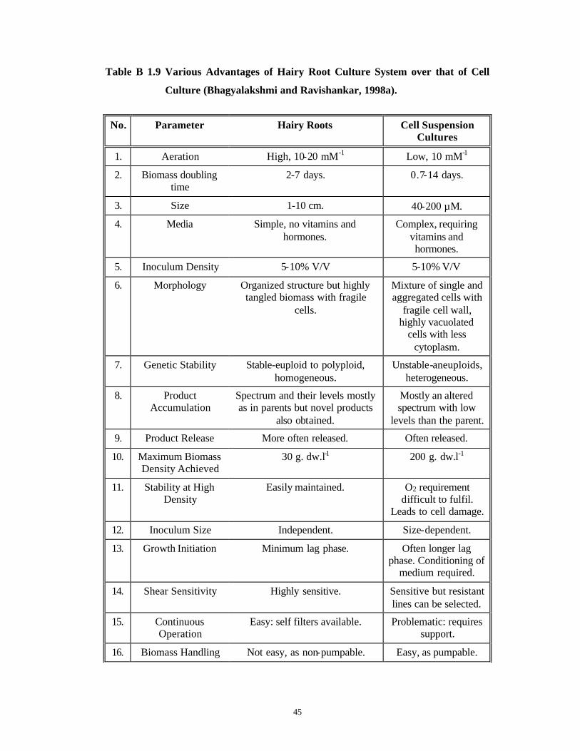
entire plants (Christey, 1997; Tanaka, 1997). Because of these reasons they are favored
over suspension cell cultures for secondary metabolite production, as shown in Table B
1.9 .
44
Fig. 1.1 A simplified Scheme Depicting Agrobacterium-mediated T-DNA Transfer in
Plant Cells (Weising and Kahl, 1996; Sheng and Citovsky, 1996) (Not to
scale).
Plant Phenols
& Sugars ChvA, ChvB PscA, Att
Agrobacterium Chromosome
Cytoplasm
NucleusT-DNA
Wounded plant cell wall
Vir G
Attach-ment
T-Region
Ti
Plasmid Vir Region
A, B, C, D, E, G, H Activation
LB RB
B D E
1
2
3
4
5
D
C 6 7
D E
8
Plant DNA
9
10
Vir A
45
Table B 1.9 Various Advantages of Hairy Root Culture System over that of Cell
Culture (Bhagyalakshmi and Ravishankar, 1998a).
No. Parameter Hairy Roots Cell Suspension Cultures
1. Aeration High, 10-20 mM-1 Low, 10 mM-1
2. Biomass doubling time
2-7 days. 0.7-14 days.
3. Size 1-10 cm. 40-200 µM.
4. Media Simple, no vitamins and hormones.
Complex, requiring vitamins and hormones.
5. Inoculum Density 5-10% V/V 5-10% V/V
6. Morphology Organized structure but highly tangled biomass with fragile
cells.
Mixture of single and aggregated cells with
fragile cell wall, highly vacuolated
cells with less cytoplasm.
7. Genetic Stability Stable-euploid to polyploid, homogeneous.
Unstable-aneuploids, heterogeneous.
8. Product Accumulation
Spectrum and their levels mostly as in parents but novel products
also obtained.
Mostly an altered spectrum with low
levels than the parent.
9. Product Release More often released. Often released.
10. Maximum Biomass Density Achieved
30 g. dw.l-1 200 g. dw.l-1
11. Stability at High Density
Easily maintained. O2 requirement difficult to fulfil.
Leads to cell damage.
12. Inoculum Size Independent. Size-dependent.
13. Growth Initiation Minimum lag phase. Often longer lag phase. Conditioning of
medium required.
14. Shear Sensitivity Highly sensitive. Sensitive but resistant lines can be selected.
15. Continuous Operation
Easy: self filters available. Problematic: requires support.
16. Biomass Handling Not easy, as non-pumpable. Easy, as pumpable.
46
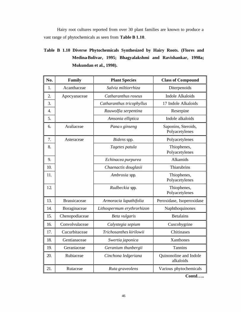
Hairy root cultures reported from over 30 plant families are known to produce a
vast range of phytochemicals as seen from Table B 1.10.
Table B 1.10 Diverse Phytochemicals Synthesized by Hairy Roots. (Flores and
Medina-Bolivar, 1995; Bhagyalakshmi and Ravishankar, 1998a;
Mukundan et al., 1998).
No. Family Plant Species Class of Compound
1. Acanthaceae Salvia miltiorrhiza Diterpenoids
2. Catharanthus roseus Indole Alkaloids
3. Catharanthus tricophyllus 17 Indole Alkaloids
4. Rauwolfia serpentina Reserpine
5.
Apocyanaceae
Amsonia elliptica Indole alkaloids
6. Araliaceae Panax ginseng Saponins, Steroids, Polyacetylenes
7. Bidens spp. Polyacetylenes
8. Tagetes patula Thiophenes, Polyacetylenes
9. Echinacea purpurea Alkamids
10. Chaenactis douglasii Thiarubrins
11. Ambrosia spp. Thiophenes, Polyacetylenes
12.
Asteraceae
Rudbeckia spp. Thiophenes, Polyacetylenes
13. Brassicaceae Armoracia lapathifolia Peroxidase, Isoperoxidase
14. Boraginaceae Lithospermum erythrorhizon Naphthoquinones
15. Chenopodiaceae Beta vulgaris Betalains
16. Convolvulaceae Calystegia sepium Cuscohygrine
17. Cucurbitaceae Trichosanthes kirilowii Chitinases
18. Gentianaceae Swertia japonica Xanthones
19. Geraniaceae Geranium thunbergii Tannins
20. Rubiaceae Cinchona ledgeriana Quinonoline and Indole alkaloids
21. Rutaceae Ruta graveolens Various phytochemicals
Contd…..
47
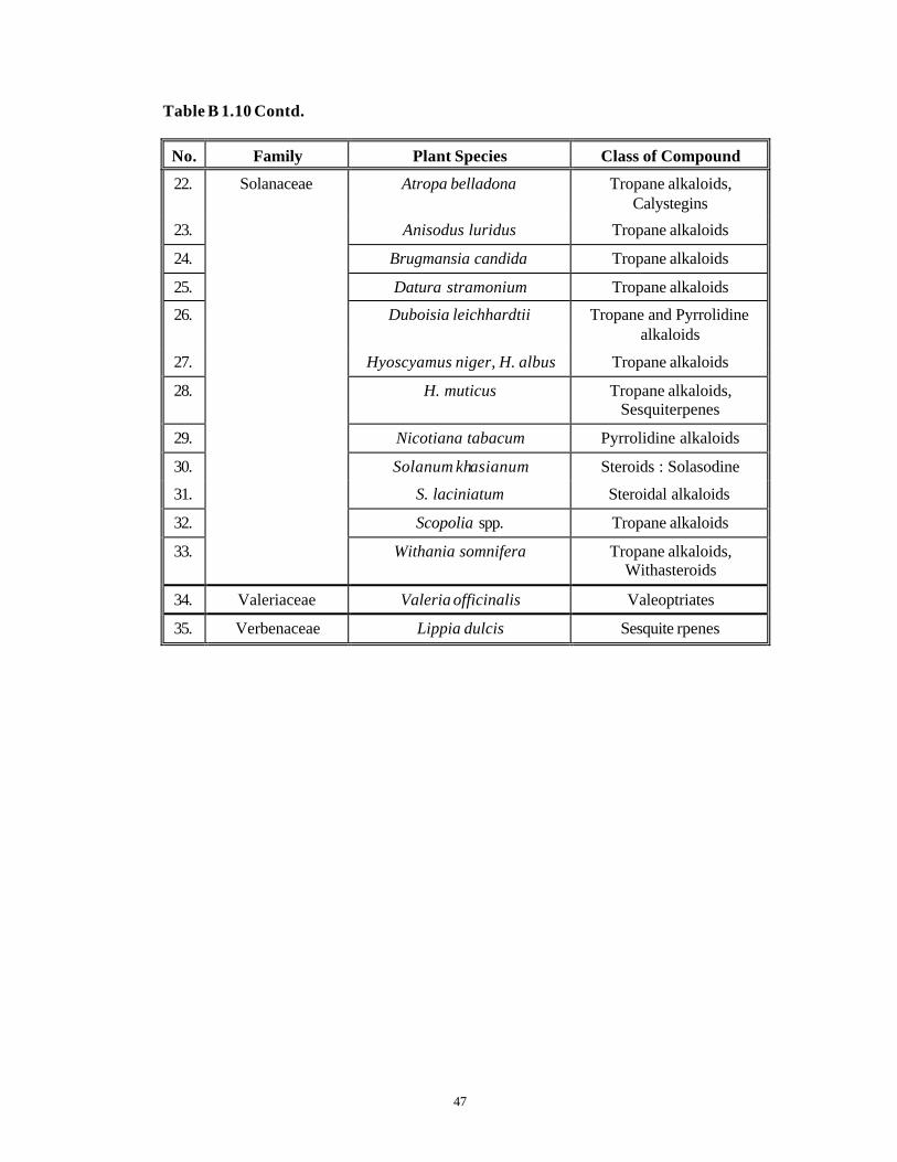
Table B 1.10 Contd.
No. Family Plant Species Class of Compound
22. Atropa belladona Tropane alkaloids, Calystegins
23. Anisodus luridus Tropane alkaloids
24. Brugmansia candida Tropane alkaloids
25. Datura stramonium Tropane alkaloids
26. Duboisia leichhardtii Tropane and Pyrrolidine alkaloids
27. Hyoscyamus niger, H. albus Tropane alkaloids
28. H. muticus Tropane alkaloids, Sesquiterpenes
29. Nicotiana tabacum Pyrrolidine alkaloids
30. Solanum khasianum Steroids : Solasodine
31. S. laciniatum Steroidal alkaloids
32. Scopolia spp. Tropane alkaloids
33.
Solanaceae
Withania somnifera Tropane alkaloids, Withasteroids
34. Valeriaceae Valeria officinalis Valeoptriates
35. Verbenaceae Lippia dulcis Sesquite rpenes
48
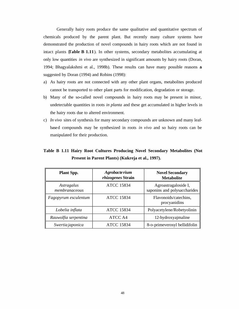
Generally hairy roots produce the same qualitative and quantitative spectrum of
chemicals produced by the parent plant. But recently many culture systems have
demonstrated the production of novel compounds in hairy roots which are not found in
intact plants (Table B 1.11). In other systems, secondary metabolites accumulating at
only low quantities in vivo are synthesized in significant amounts by hairy roots (Doran,
1994; Bhagyalakshmi et al., 1998b). These results can have many possible reasons as
suggested by Doran (1994) and Robins (1998):
a) As hairy roots are not connected with any other plant organs, metabolites produced
cannot be transported to other plant parts for modification, degradation or storage.
b) Many of the so-called novel compounds in hairy roots may be present in minor,
undetectable quantities in roots in planta and these get accumulated in higher levels in
the hairy roots due to altered environment.
c) In vivo sites of synthesis for many secondary compounds are unknown and many leaf-
based compounds may be synthesized in roots in vivo and so hairy roots can be
manipulated for their production.
Table B 1.11 Hairy Root Cultures Producing Novel Secondary Metabolites (Not
Present in Parent Plants) (Kukreja et al., 1997).
Plant Spp. Agrobacterium rhizogenes Strain
Novel Secondary Metabolite
Astragalus membranaceous
ATCC 15834 Agroastragaloside I, saponins and polysaccharides
Fagopyrum esculentum ATCC 15834 Flavonoids/catechins, procyanidins
Lobelia inflata ATCC 15834 Polyacetylene/Robetyolinin
Rauwolfia serpentina ATCC A4 12-hydroxyajmaline
Swertia japonica ATCC 15834 8-o-primeverosyl bellidifolin

49
From the foregone Introduction, it is obvious that there is an urgent need to
catalogue biodiversity, to identify and to characterize the chemistry of medicinal plants,
as they are the storehouses of “ futuristic medicines”. To continue such efforts which
have been made by earlier researchers in obtaining significant results in the in vitro
establishment and studies of a number of medicinal plants, we in the present thesis
attempted to work with two plants namely, Taxus baccata ssp. wallichiana Zucc. Pilg.
(Himalayan Yew) and Withania somnifera (L.) Dun. (Ashwagandha) that are known to
produce antitumor secondary metabolites. The present thesis deals with morphogenesis,
micropropagation and secondary metabolite studies of these two plants in vitro. Part
I of the present thesis deals with Taxus baccata ssp. wallichiana Zucc. Pilg. (Himalayan
Yew) while Part II deals with Withania somnifera (L.) Dun. (Ashwagandha). The overall
objectives and the justification for undertaking the study with these two plants, in this
work presented as a thesis here, are discussed in detail in respective chapters in Part I and
Part II.

50
CCHHAAPPTTEERR 22
Materials and Methods (General).

51
During the course of work presented in this thesis, different techniques related to
plant tissue and cell culture, analytical TLC and HPLC, microbial culture (for growing
and maintaining the contaminating bacteria and fungi isolated from in vitro cultures and
also wild strains of Agrobacterium rhizogenes used in genetic transformation
experiments), biochemical characterization and long and short term maintenance of the
microbes were used.
The present chapter describes the general and common methods of plant tissue
and cell culture used for both Taxus and Withania spp. Specific methodologies and
techniques related to plant tissue and cell cultures, analysis of production of secondary
metabolites in vitro as well as microbial culture, characterization and identification are
described for both Taxus and Withania spp. in their respective chapters.
2.1 Glassware
Test tubes (2.5 x 15 cm), petri dishes (55 mm and 85 mm diameter), Erlenmeyer
flasks and beakers (100, 250, 500, 1000 and 2000 ml capacity), pipettes (1, 2, 5 and 10 ml
capacity) and measuring cylinders of all the capacities (10 ml-2000 ml) were purchased
from “Borosil” India. In addition, glass, screw capped bottles (6 x 15 cm) purchased from
“Ballarpur Industries”, India were used for all tissue culture related experiments.
For the analytical work, Erlenmeyer flasks (250 ml, 500 ml and 1000 ml
capacity), graduated and Pasteur pipettes, funnels, measuring cylinders, glass beakers,
glass capillaries, glass sample vials were from “Borosil” India. Round bottom flasks of
varying capacities were purchased from “Duran”, Germany. 50 µl constant volume glass
microsyringes needed for HPLC injections were purchased from ‘Exmire’ (“Ito Corp.”,
Japan).

52
2.1.1 Preparation of glassware
Glassware was cleaned by boiling initially in a saturated solution of sodium
bicarbonate for one hour and subsequent washing in tap water. It was then immersed in
30% nitric acid solution for 30 min followed by a thorough wash with tap water. The
glassware thus washed was then rinsed twice with glass distilled water and allowed to dry
in an oven at 200oC for two hours.
Test tubes and flasks needed for tissue culture work were plugged with absorbent
cotton (“Seasons Healthcare Ltd.”, Andhra Pradesh, India). Such plugged flasks, screw
capped bottles and petri dishes were wrapped in brown paper prior to sterilization.
Autoclaving was carried out at 121oC and at a constant pressure of 1.1 kg.cm-2 for 1 h.
2.2 Plasticware
Sterile disposable plastic dishes (55 mm and 85 mm diameter) and disposable
filter sterilization units having filters of pore size 22 µm were obtained from “Laxbro”
(India). Microtips (20-1000µL) and Eppendorf tubes (1-2 ml) were obtained from “Sigma
Chemical Co.” (USA) as well as from “Tarsons” (India). The syringes and disposable
small filter units used for filtering the callus sample extracts before HPLC, were
purchased from “Millipore Corporation”, (USA).
2.3 Chemicals
The chemicals used during the course of the study were of analytical grade.
Inorganic salts were obtained from “Qualigens” and “S. D. fine chemicals”, (India). All
vitamins and growth regulators were obtained from “Sigma Chemical Co.”, (USA). Agar-
agar and sucrose were obtained from “Qualigens” and “S. D. fine chemicals”, (India).
Phytagel was purchased from “Sigma Chemical Co.”, (USA). Casein acid hydrolysate,
peptone, yeast extract and beef extract were purchased from “HiMedia Laboratories”,
(India).
Coconut milk (liquid endosperm) was obtained from green tender coconuts,
brought from a local market. It was firs t filtered through rough filter paper and collected
in 500 ml flasks (300 ml/flask). It was then autoclaved at 121oC and at a pressure of 1.1
53
kg.cm-2 for 20 min, and then refiltered through Whatman No. 1 filter paper. The filtrate
was stored at -20oC as 300 ml aliquots in a freezer until required.
All the solvents used for the analytical work in HPLC and TLC were of
“Lichrosolv” grade from “Merck”, (India). Silica gels of various properties were
purchased either loose or in the form of pre -coated plates from “Merck”, (India).
2.4 Preparation of Plant Tissue Culture Media
Glass distilled water was used for the preparation of culture media. After addition
of all constituents of media, the pH was adjusted to 5.8 using 0.1 N KOH or 0.1 N HCl.
When activated charcoal (“Sarabhai Chemicals”, India) was added to the medium, pH
was adjusted to 6.0 before autoclaving the medium. Gelling agent (agar-agar or phytagel)
was added as per requirement and the medium was steamed to melt the gelling agent. It
was then dispensed into test tubes (20 ml per tube) or Erlenmeyer flasks (100 ml per 250
ml flask) or screw capped bottles (50 ml per bottle) and was autoclaved at 121oC at a
pressure of 1.1 kg.cm -2 for 20 min. When no gelling agent was added, each tube
contained liquid medium with a filter paper bridge of Whatman No. 1 filter paper. Heat
labile constituents like antibiotics were filter-sterilized by passing through a Millipore
membrane (0.22 µm pore size) (“Millipore Corporation”, USA) and added aseptically to
the autoclaved medium just before gelling. All the plant growth regulators used during the
course of the present work were added before autoclaving the medium. As per the
requirements, the medium was also poured in sterile plastic dishes (12 ml per 55 mm
dish, 20 ml per 85 mm dish) in a laminar flow hood.
The composition of various media used for culturing of explants of both the plant
spp. (Taxus spp. and Withania spp.) are given in Table 2.1 and a comparison of the
concentrations of cations and anions in each of the media are indicated in Table 2.2.
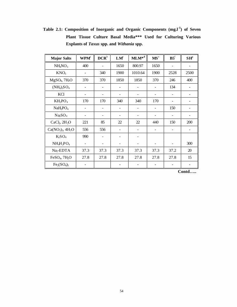
54
Table 2.1: Composition of Inorganic and Organic Components (mg.l-1) of Seven
Plant Tissue Culture Basal Media*** Used for Culturing Various
Explants of Taxus spp. and Withania spp.
Major Salts WPMa DCRb LMc MLM*d MSe B5f SHg
NH4NO3 400 - 1650 800.97 1650 - -
KNO3 - 340 1900 1010.64 1900 2528 2500
MgSO4, 7H2O 370 370 1850 1850 370 246 400
(NH4)2SO4 - - - - - 134 -
KCl - - - - - - -
KH2PO4 170 170 340 340 170 - -
NaH2PO4 - - - - - 150 -
Na2SO4 - - - - - - -
CaCl2, 2H 2O 221 85 22 22 440 150 200
Ca(NO3)2, 4H2O 556 556 - - - - -
K2SO4 990 - - -
NH4H2PO4 - - - - - - 300
Na2-EDTA 37.3 37.3 37.3 37.3 37.3 37.2 20
FeSO4, 7H2O 27.8 27.8 27.8 27.8 27.8 27.8 15
Fe2(SO4)3 - - - - - -
Contd…..
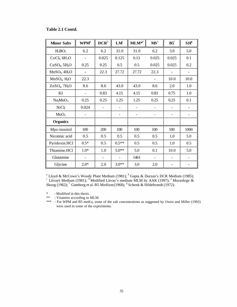
55
Table 2.1 Contd.
Minor Salts WPMa DCRb LMc MLM*d MSe B5f SHg
H3BO3 6.2 6.2 31.0 31.0 6.2 3.0 5.0
CoCl2, 6H 2O - 0.025 0.125 0.13 0.025 0.025 0.1
CuSO4, 5H2O 0.25 0.25 0.5 0.5 0.025 0.025 0.2
MnSO4, 4H2O - 22.3 27.72 27.72 22.3 - -
MnSO4, H2O 22.3 - 10.0 10.0
ZnSO4, 7H2O 8.6 8.6 43.0 43.0 8.6 2.0 1.0
KI - 0.83 4.15 4.15 0.83 0.75 1.0
Na2MoO4 0.25 0.25 1.25 1.25 0.25 0.25 0.1
NiCl2 0.024 - - - - - -
MoO3 - - - - - -
Organics
Myo-inositol 100 200 100 100 100 100 1000
Nicotinic acid 0.5 0.5 0.5 0.5 0.5 1.0 5.0
Pyridoxin.HCl 0.5* 0.5 0.5** 0.5 0.5 1.0 0.5
Thiamine.HCl 1.0* 1.0 5.0** 5.0 0.1 10.0 5.0
Glutamine - - - 1461 - - -
Glycine 2.0* 2.0 3.0** 3.0 2.0 - -
a Lloyd & McCown’s Woody Plant Medium (1981); b Gupta & Durzan’s DCR Medium (1985); c Litvay's Medium (1981); d Modified Litvay’s medium MLM by AAK (1997); e Murashige & Skoog (1962); f Gamborg et al. B5 Medium (1968); g Schenk & Hildebrandt (1972).
* : Modified in this thesis. ** : Vitamins according to MLM. *** : For WPM and B5 medi a, some of the salt concentrations as suggested by Owen and Miller (1992)
were used in some of the experiments.

56
Table 2.2 A Comparison of the Inorganic Nutrient Ion Concentration of Seven Plant
Tissue Culture Basal Media** Used for Culturing Various Explants of
Taxus spp. and Withania spp. (Adapted from McCown and Sellmer,
1987).
Macroelements = mM, Microelements = µM.
Inorganic
Ions WPM DCR LM MLM* MS B5 SH
NH4+ 4.94 5.0 20.6 10 20.61 2.02 2.6
K+ 12.61 4.62 22.52 10 20.04 24.7 24.7
Ca++ 3.0 2.69 0.15 0.15 2.99 1.02 1.4
Mg++ 1.5 1.5 7.5 7.5 1.5 1.01 1.6
Mn++ 0.132 0.132 0.124 0.132 0.132 0.059 0.059
Zn++ 0.03 0.03 0.15 0.03 0.029 0.007 0.014
Na+ 0.224 0.102 0.01 0.224 0.224 1.1 0.1
Fe++ 0.1 0.1 0.05 0.05 0.1 0.05 0.027
NO3- 9.64 10.48 39.4 20 39.4 24.7 24.7
SO42- 7.44 1.76 7.5 7.5 1.76 2.04 1.7
PO43- 1.25 1.25 2.5 2.5 1.25 1.1 2.6
BO33- 0.1 0.1 0.501 0.1 0.1 0.049 0.082
Cl- 1.31 1.16 0.3 1.31 6.0 2.04 2.7
Fe-EDTA3- 0.11 0.11 0.11 0.11 0.11 0.11
Co++ - 0.105 0.6 - 0.105 0.083 0.332
Cu++ 0.1 0.1 2.0 0.1 0.1 0.1 0.8
MoO42- 1.03 1.61 5.17 1.03 1.03 1.03 0.412
I- - 5.0 25 - 5.0 4.52 6.0116
Ni++ - 0.193 - - - - -
Total Ionic Strength of
Media.
43.516 36.042 144.185 60.736 100.480 65.738 69.838
* : Modified in this thesis. ** : For WPM and B5 media, some of the salt concentrations as suggested by Owen and Miller (1992)
were used in some of the experiments.
57
2.5 Growth and Maintenance of Plant Tissue and Cell Cultures
The stationary tissue cultures (callus and shoot cultures) belonging to both the
plants on solid media were subcultured at regular intervals of 3-4 weeks. Observations
were taken at the same time. The cell suspension cultures of Taxus baccata ssp.
wallichiana Zucc. Pilg., were subcultured after every 4 days into the fresh liquid media
(10% v/v) and the relevant observations of Packed Cell Volume (PCV), fresh and dry
weights and the secondary metabolite contents were determined at regular intervals of 0
days, 9 days and 18 days. Liquid cultures of seedling and hairy roots of Withania
somnifera (L.) Dun. were subcultured after every 6 days. Various details of such
methodologies are described in relevant chapters of this thesis.
2.6 Statistical Analysis
“Randomized Block” was the experimental design used in this study. Depending
upon the size and the availability of the explants, each experiment consisted of either 5,
10 or 50 replicates (mentioned in each of the chapters more specifically) and each
experiment was repeated at least three times. In addition, the experiments were repeated
over successive years. Standard Deviations (S. D.) and Standard Errors of Means (S. E.)
were calculated in each experiment. Analysis of Variance (ANOVA) was carried out and
the post-hoc significance of differences was calculated based on Student's t-test
(Wardlaw, 1985).
58
CCHHAAPPTTEERR 33
Control of Contamination and Phenolic
Browning in vitro.
< Identification and Control of Bacterial and
Fungal Genera Associated with in vitro Cultures
of Taxus baccata ssp. wallichiana Zucc. Pilg.
and Withania somnifera (L.) Dun. as well as
Control of Phenolic Browning.

59
3.1. Introduction:
Plant tissue culture techniques are extensively used for mass production of elite
plants as well as to study the basic aspects of primary and secondary metabolism,
morphogenesis and genetic engineering etc. (George and Sherrington, 1984). But even
after three decades of research and development in plant tissue culture, microbial
contamination by yeasts, fungi, bacteria, viruses, mites and thrips is still the major
problem that has hampered the establishment of truly aseptic plants and their successful
micropropagation (Herman, 1990; Leifert et al., 1994). The influence of bacteria on shoot
growth can range from total inhibition to no apparent effect (McCown, 1986). The
contaminating bacteria and fungi may be endophytic or epiphytic, may be pathogenic or
saprophytic (Debergh and Maene, 1981, 1984).
Another type of hazard for plant tissue and cell cultures is caused by ‘latent’
bacteria and viruses that do not produce any symptoms on the plant or any visible growth
on the medium for long periods of time in vitro . They reduce the multiplication rates and
productivity of tissue cultures and may become ‘virulent’ at a later stage due to changes
in incubation temperatures, growth medium composition and removal of antibiotics from
the medium (Leifert and Waites, 1994).
Increasing attention is being paid to ascertain sources of contamination, to develop
procedures for their elimination by avoidance, rigorous manipulation of the nutritional
and environmental conditions and by specific antibiotic treatments (DeFossard and
DeFossard, 1988; Debergh and Read, 1991).
The earliest plant tissues cultured were cambii from woody Angiosperms and
Gymnosperms (Gautheret, 1934). However the rate of progress in this field was relatively
slow during the last 4 decades owing to a large number of bacterial and fungal
contaminants associated with mature explants of Gymnosperms, due to problems of
maturation (Gupta and Durzan, 1985), and also due to autotoxic phenolic exudations
produced by them (Young et al., 1984).
60
3.1.1 Nature and occurrence of contaminants
The contaminants could be of endophytic or epiphytic origin (Debergh and
Maene, 1984; Hennerty et al., 1988). An “endophyte” is defined as a fungus or a
bacterium that causes inapparent, asymptomatic but parasitic infections within the tissues
of the plant. These may be ‘obligate endophytes’ or ‘facultative endophytes’. The latter
are subdivided into ‘constitutive’ and ‘inducible’ types. Inducible endophytes are most
frequently encountered during in vitro culture, since in nature, they are epiphytes of the
parent plant (Gunson and Spencer-Phillips, 1994). An “epiphyte” is defined as a fungus
or a bacterium associated with external plant surfaces that survive surface sterilization
procedures (Leifert et al., 1989; Wilson and Power, 1989; Gunson and Spencer-Phillips,
1994). Other types of microbes could be air-borne and/or human associated (Leifert et al.,
1989) and can be introduced via poor aseptic techniques, insufficient sterilization of
instruments (Boxus and Terzi, 1988), failure of equipment (Trick and Lingens, 1985;
Giles and Morgan, 1987) and laboratory environment with a high microbial load (Cassels,
1991). These various types of contaminants are summarized in Table 3.1 .

61
Table 3.1 Contaminants from Various Sources (Adapted from: Leifert and
Woodward, 1998).
Contaminant Likely Source of Contamination
Comments
Bacteria
Gram negative rods. Inefficient disinfection of explants.
Often Pseudomonas spp. and Enterobacteriaceae.
Gram positive rods. Inefficient laboratory procedures.
Often Bacillus and Clostridium spp.
Gram positive cocci (e.g. Staphylococcus
epidermidis).
Poor aseptic techniques in subculturing of plants and
pouring of media.
Staphylococcus spp. are considered as obligate
human inhabitants.
Inefficient sterilization of media.
Often Bacillus subtilis and B. pumilus.
Bacillus spp.
Inefficient sterilization of instruments used for
subculture.
Bacillus circulans can survive in 70% alcohol.
Fungi/Yeasts
General increase Mite or thrip infestation of growth-rooms.
Especially when plant -specific growth rooms are
affected.
Penicillium spp., Candida spp.
During pouring of media. Especially when plant -specific growth rooms are
affected.
Fusarium poe Growth room infestation with Sideroptes graminis mites.
Fungus with white mycelium and a pink base.
Grey, black and green moulds (Botrytis,
Aspergillus, Alternaria, Penicillium spp.)
Pink yeasts (Rhodotorula spp.)
High laboratory air contamination, Faulty laminar
airflow cabinets.
Common air contaminants within buildings.
Black moulds Poor hygiene in growth rooms, plant and media cold stores.
Moulds are common in damp areas of the buildings. Spores are air-transmitted.
Cladosporium spp. Insufficient protection of laboratory against outside air.
Cladosporium spores are common in outside air.
62
The contamination problem is acute especially with explants of mature tissue
origin. It is a well-known fact that the incidence of contamination is closely linked with
the state of the mother plant, which is denoted as “Stage 0” (Debergh and Read, 1991).
When mother plants are of a great age and growing under natural environmental
conditions, the incidences of contamination increase from “Stage 1”, i.e. surface
sterilization, onwards (Debergh and Read, 1991).
In spite of the controlled laboratory conditions, the external environmental
conditions also affect the contamination rates in tropical countries since the parent plants
support a rich microflora with them at “Stage 0” itself (Enjarlic et al., 1988; Watt et al.,
1996). In addition, sudden seasonal outbreaks (just before the monsoon) of bacterial and
fungal contamination are observed involving hitherto sterile cultures of a number of plant
species. Such sudden outbreaks of fungal contamination are often associated with mites
and thrips, the arthropods inhabiting culture rooms and acting as vectors for fungal spores
(Debergh and Maene, 1984; Leifert et al., 1991a, 1994; Danby et al., 1994). The origin of
sudden outbreaks of bacteria is not known. It has been suggested that PPFMs (pink
pigmented facultative methylotrophic bacteria) like Methylobacterium spp. are the
responsible agents. These bacteria are associated with 70 plant species across all the
families in vivo (Holland and Polacco, 1994 and references therein).
3.1.2 Harmful effects of contaminants
In most laboratories, losses due to contamination average between 3-15% of
plants at every subculture (Leifert et al., 1994), rendering commercial micropropagation
less economical. It also leads to severe production losses, low progeny performance and
rejections of entire shipments of plants due to quarantine regulations (Cassells, 1991). In
scientific experimental setup, to offset the effects of contamination, sample size needs to
be very large and same experiments have to be repeated many times. In addition, the
continued presence of microbial contaminants alters the morphological nature and
physiological response of cultures and makes it difficult to interpret the results obtained.
There are many contaminating bacteria which cause diseases only in vitro, and on all the
varieties of plants (“Vitropaths”, Herman, 1990). Considering the high frequency of
mutations and variations in bacteria, such organisms can become pathogenic and cause
epidemics ex vitro . They may also block the vascular tissues.
63
Latent bacterial contamination during the proliferation phase adversely affects
rooting due to changes in growth regulators and water potential of the media (Leifert et
al., 1994) and also kills young trees after weaning (Boxus and Terzi, 1988). Many
Bacillus spp. increase pH of the medium, inhibiting proton pumps involved in plant
nutrient uptake and cause precipitation of many nutrients from the medium (Leifert et al.,
1994). Cultures of apricot with latent bacterial contaminants showed high CO2 and low
O2 concentration in the vessel headspace, thereby decreasing the proliferation and
photosynthetic rates (Marino et al., 1996).
3.1.3 Beneficial effects of contaminants
Bacterial and fungal contaminants are also known to exhibit some beneficial
effects in vitro. Nowak (1996) has coined the terms “Bacterization” and “Biotization”
referring respectively to the addition of bacteria or other microbes to cultures or to in
vitro cultured plants to get an enhanced biological effect. He isolated a strain of
Pseudomonas from surface-disinfected onion roots which stimulated growth of potatoes
in vitro, induced root branching, root hair formation, stem elongation, lignin deposition,
better acclimatization and better resistance to fungal pathogens ex vitro . Same results
were also obtained for a variety of other plants.
A Pseudomonas spp. isolated during in vitro culture of Oregano was reported to
control vitrification and to improve acclimatization ex vitro (Shetty et al., 1995). Two
strains of Pseudomonas maltofila increased the regeneration potential of recalcitrant calli
of Soybean under co-cultivation (Herman, 1995). Culture extracts of Trichoderma viride
(Strain A) and Bacillus subtilis (Strain B) showed antifungal activity while extracts from
Pseudomonas fluorescens (Strain X) had the highest antibacterial activity (Hussain et al.,
1994). When these were added to cultures of tobacco, they reduced the growth of
accidental contaminants. A selected strain of a bacterial endophyte, Bacillus subtilis from
a healthy Chestnut tree, showed broad-spectrum anti-fungal activity against a number of
phytopathogenic fungi like Cryphonectria parasitica, Alternaria radicina etc. (Wilhe lm
et al., personal communication). A new strain of Bacillus circulans was found to promote
somatic embryogenesis in Pelargonium when combined with TDZ (Murthy et al., 1999).
A number of bacterial endophytes isolated from Taxus spp. produce Taxol and
related Taxanes (Page et al., 1999). It is believed that due to close associations of such
endophytes with hosts, some genetic exchange takes place and the endophytes develop

64
the ability to synthesize particular metabolites. Similarly a number of fungal genera
isolated from Taxus spp. produce Taxol and related Taxanes (Stierle et al., 1995; Strobel
et al., 1996).
In another example (Czembor and Strobel, 1997), indigenous isolates of Fusarium
avenaceum were shown to inhibit seed germination of spotted knapweed (Centaurea
maculosa), without adversely affecting seed germination of wheat or oat. Thus this can be
an effective bio -control measure. Phytotoxic metabolites from culture filtrates of
Alternaria solani stimulated adventitious shoot induction in potato tubers when co-
cultivated. Jasmonic acid and two other potato tuber-inducing principles were isolated
from culture filtrates of fungus Lasiodiplodia theobromae which can be beneficially used
for in vitro cultures of potato (Herman, 1995).
A recent hypothesis suggests that microbes may actually be essential to all
cytokinin-autotrophic plant tissue cultures as well as to all plants in vivo as cytokinins are
synthesized by these microbes like Methylobacterium spp. (Holland, 1997).
3.1.4 Control of contaminants
The use of anti-microbial agents (anti-bacterial as well as anti-fungal) to control
contamination is the preferred method (Thurston et al., 1979; Falkiner, 1988; Wilson and
Power, 1989; Kneifel and Leonhardt, 1992; Watt et al., 1996). However their
indisc riminate use may lead to phytotoxicity problems (Phillips et al., 1981; Pollock et
al., 1983) and development of resistant strains (Leifert et al., 1991b). Hence
characterization of contaminating microbes (bacteria and fungi) to generic and species
level can yield important information about the source of contamination, the relative
importance of different sources, pathogenicity, if any, of the isolated genera (Danby et al.,
1994) and more specifically the development of versatile antibacterial and antifungal
therapies (Falkiner, 1988; Leifert et al., 1989).
65
3.1.5 Phenolic browning/oxidation in vitro
The presence of phenolic compounds causing death of explants has been another
important problem of tissue cultures of woody perennials (Compton and Preece, 1986) in
addition to various bacterial and fungal contaminants as discussed above. Some of these
exudates appear as a reaction to injury and/or infection. In tissue culture they appear after
tissue excision and are many times aggravated by growth media constituents (Seneviratne
and Wijesekara, 1996). Tissue blackening occurs due to action of copper-containing
oxidase enzymes: polyphenoloxidases like tyrosinases, which are released or synthesized
in oxidative conditions after tissue wounding and they oxidize o-diphenols released due to
cellular wounding to o-quinones (Scalbert et al., 1988; Marks and Simpson, 1990 and
references therein). The onset of tissue browning has been found to be associated with
changes in protein pattern, amino acid content, ethylene production and the occurrence of
saccharose and accumulation of starch (Lindfors et al., 1990). These changes eventually
lead to growth inhibition or death of explants. Other types of phenolic exudates appear at
the end of incubation period and are apparently products of dying cells (Seneviratne and
Wijesekara, 1996). The phenolic exudation is aided by light and is autocatalytic.
A number of methodologies have been employed to overcome the phenolic oxidation.
Some of them are as follows (Pierik, 1987):
• Addition of activated charcoal (0.2-3.0% w/v) to the medium.
• Addition of polymeric polyvinylpyrrolidone (PVP) or polyvinylpolypyrrolidone
(PVPP) to the medium. These absorb phenols through hydrogen bonding.
• Additions of anti-oxidants/reducing agents like citric and ascorbic acids, thiourea
glutathione and L-cysteine in the medium or before surface sterilization. These reduce
the redox potential of explants and stop the oxidation reactions (Marks and Simpson,
1990).
• Addition of diethyl-dithiocarbonate (DIECA) (2g.l-1) in the rinses after surface
sterilization and as droplets at the time of micro grafting.
• Addition of amino acids like glutamine, arginine and asparagine to the media.
• Frequent subcultures onto fresh media.
• Use of liquid medium for easier and quicker dilution of toxic products.
• Reduction of wounded tissues to decrease exudation.
• Reduction of salt concentration in the growth media.
66
• Omission of plant growth regulators as they aid in medium darkening.
• Soaking of explants in water before culturing to reduce browning.
• Incubation of shoot bases in darkness during culture period.
• Immediate inoculation of explants after removing them from the parent trees.
• Sealing ends of explants with paraffin wax.
During the course of the present work, the problem of phenolic oxidation was
particularly encountered on culturing stem bits and needle explants of Taxus spp. since
they were derived from mature trees (80-120 year old). Due to oxidation of externally
released polyphenols, the explants (stem bits, needles) as well as the nutrient medium
became brown and the explants did not respond in vitro and a strategy to control phenolic
oxidation had therefore become a necessity if not warranted.
The problem of phenolic oxidation was relatively less in cultures of Withania spp.
because it is a herbaceous plant and the explants were derived from in vitro grown
seedlings and were juvenile.
Bacterial and fungal contamination of cultures, especially initiated with mature
explants of Taxus spp. for establishment of callus (Chapter 4 of this thesis) and shoot
cultures (Chapter 5 of this thesis) under aseptic conditions, was yet another major
problem and in the initial experiments all the cultures (100%) were contaminated. In case
of cultures of Withania spp., the internodal explants (Chapter 6 of this thesis) showed
presence of typical yellow bacteria hindering the regeneration response. These bacteria
were however absent in all the other cultures raised with explants such as nodes, leaves,
hypocotyls, embryos and seeds. To our knowledge, there are neither detailed reports
describing the contamination in tissue cultures of these two plants nor methods to control
contamination observed especially for in vitro cultures of Taxus explants in literature.
Probably contamination was not a problem in these studies reported earlier for these two
species or serious attempts have not been made to control the observed contamination.
However, some authors like Gibson et al. (1993) have mentioned presence of
contaminants in tissue cultures of Taxus brevifolia and have used broad-spectrum anti-
fungal and anti-bacterial agents to control them. However neither the methods adapted to
control contamination nor the percentages of contamination were reported. On the other
hand, a large number of endophytic bacteria and fungi have been isolated from trees of
67
Taxus spp. growing under natural habitats and many have been shown to produce the
Taxane class of anti-cancer agents independent of the parent plants (Stierle et al., 1995;
Strobel et al., 1996). However, the association of these fungi and bacteria and their effect
on in vitro cultures is not known. On culturing of various explants, especially of Taxus
spp. in various nutrient media in our studies, all the cultures got contaminated and use of
broad spectrum antifungal and antibacterial agents to control them, as reported by Gibson
et al. (1993), did not yield significant positive response and results. Therefore an attempt
has been made to systematically identify and characterize the bacterial and fungal
associates and contaminants of Taxus spp. and Withania spp. and to develop a
contamination control strategy in order to obtain sterile cultures of Taxus spp. and
Withania spp. Furthermore, since the explants of Taxus spp. in particular, were derived
from mature trees, they exuded phenolics hampering the in vitro response. Hence it had
become necessary to formulate a strategy to overcome not only the problem of phenolic
oxidation but also the high degree of fungal and bacterial contamination associated with
cultures of Taxus spp. and Withania spp. under in vitro conditions.
The objectives of the work in the present chapter have therefore been:
1. To elucidate the nature of the contaminants associated with in vitro cultures of Taxus
spp. and Withania spp., which are varied in their habits and are collected from diverse
habitats.
2. To identify the contaminants upto the generic and species level.
3. To devise a suitable and very specific control strategy to overcome both bacterial and
fungal contaminants encountered in both the species based on the know-how
generated.
4. To develop a common protocol applicable not only to control phenolic browning but
also contamination associated with cultures of Taxus spp. and Withania spp. so as,
5. To establish sterile and healthy cultures in both the species for further in vitro studies
as an acid test for the effectiveness of the developed protocol for control of
contamination and phenolic browning.
While the bacterial contaminants and associates observed commonly in Taxus and
Withania spp. have been described under Subsection 1 of the present chapter, Subsection
2 of this chapter deals with the fungal contaminants and phenolic oxidation/browning.
68
3.2. Materials and Methods
3.2.1 Collection of plant material
While the twigs of Taxus spp. were collected from different locations in the
Himalayas as described in detail in Chapter 4, Section 4.2.1, the seeds of the cultivated
variety WS-20 of Withania spp. were collected as described in detail in Chapter 6,
Section 6.2.1 .
3.2.2 Surface sterilization and culture of explants
3.2.2.1 Taxus baccata ssp. wallichiana Zucc. Pilg.
Stem bits with axillary buds and trimmed needles (3-5 cm long), stem bits without
axillary buds, needles, and seeds were first given a liquid soap treatment with 1% (v/v)
Teepol (“Bennett & Colman”, India) for five minutes with constant agitation. After
washing off the soap solution, one minute long treatment with 95% (v/v) ethyl alcohol
followed by ten minute duration treatment with 0.1% (w/v) mercuric chloride (HgCl2)
under vacuum (equivalent to 20" Hg) was given. HgCl2 was washed off with six washes
of sterile distilled water in a laminar airflow cabinet. This methodology of surface
sterilization of explants shall be hereinafter referred to as the “Routine Surface
Sterilization Protocol” . The surface sterilized explants were then inoculated on nutrient
media and incubated under conditions as described in Section 3.2.3.1 . The percentage of
contamination either due to bacteria or fungi or by both was scored.
3.2.2.2 Withania somnifera (L.) Dun.
Seeds of Withania spp. used for raising in vitro seedlings were rinsed in a mixture
of 1% (v/v) Teepol and 70% (v/v) ethyl alcohol for 15 seconds followed by treatment
with 3% sodium hypochlorite (v/v) for five minutes. Glass distilled water was used for all
the washing steps. The seeds were then washed with six washes of sterile distilled water
in a laminar airflow cabinet. This methodology of surface sterilization of explants shall be
hereinafter referred to as the “Routine Surface Sterilization Protocol” . These seeds
were used for raising seedlings. The internode explants were derived from such two-
month old in vitro grown seedlings as described in Chapter 6, Sections 6.2.2 and 6.2.3.
Explants were inoculated on nutrient media and incubated under conditions as described
69
in Section 3.2.3.2 . The percentage of contamination either due to bacteria or fungi or by
both was scored.
3.2.3 Media and culture conditions
3.2.3.1 Taxus baccata ssp. wallichiana Zucc. Pilg.
The stem and needle explants of Taxus spp. were inoculated in plastic dishes (55
mm diameter) containing full strength woody plant medium (WPM) (Lloyd and
McCown, 1981) with 2% sucrose, 0.3% phytagel supplemented individually with 2, 4, 6,
8 or 10 mg.l-1 2,4-D for callus induction. All the details of media composition and media
preparation were as described earlier in Chapter 2, Section 2.4. The cultures were
incubated in total darkness at 25 + 2°C. In addition, stem explants with axillary buds were
inoculated in culture tubes containing WPM with its major salts reduced to half strength
(1/2 WPM) without plant growth regulators but with 2% sucrose, 0.8% agar for enhanced
axillary budding. All the details of media composition and media preparation were as
described earlier in Chapter 2, Section 2.4. The cultures were incubated under 16 hour
photoperiod provided by cool white fluorescent tubes of light intensity 4.8-19 µE.m-2s-1 at
25 + 2°C. The embryos and endosperm halves dissected out from seeds were cultured in
plastic dishes (55 mm diameter) on full strength WPM medium without plant growth
regulators and with 2% sucrose, 0.3% phytagel. The cultures were incubated in total
darkness at 25 + 2°C.
3.2.3.2 Withania somnifera (L.) Dun.
All the internode explants of Withania spp. were inoculated in culture tubes
containing full strength MS medium (Murashige and Skoog, 1962) supplemented with
2% sucrose, 0.8% agar and 2, 4, 6, 8 and 10 mg.l-1 2,4-D independently for callus
induction. All the details of media composition and media preparation were as described
earlier in Chapter 2, Section 2.4 . The cultures were incubated under 24 hour photoperiod
provided by cool white fluorescent tubes of light intensity 38 µE.m-2s-1 at 25 + 2°C.

70
3.2.4 Isolation of bacterial contaminants
The bacteria appeared either from the cut end of the needles and stem bits of
Taxus spp. during the first week of culture or after one month from the developing calli
while the contaminating bacteria appeared within 15 days from the internodes of
Withania spp. causing them to become spongy and unresponsive for regeneration. The
isolated bacteria were grown and stored as follows.
3.2.5 Growth, storage, identification and control of contaminating bacteria
3.2.5.1 Growth, purification and storage
The contaminant bacteria were removed with a sterile wire loop from cultures of
Taxus spp. and Withania spp. and grown either in 5 ml of nutrient broth (NB) prepared
according to the manufacturer’s instructions (“Difco Laboratories”, USA) or on plates of
nutrient agar (NA) [NB fortified with 2% bacto-agar (“Difco Laboratories”, USA)]. Glass
distilled water was used in media preparations and pH was adjusted to 7.0 before
autoclaving, using 0.1 N KOH or 0.1 N HCl. Autoclaving was done at 121oC at a pressure
of 1.1 kg.cm-2 for 20 min.
Bacteria in NB were grown in dark for 24-48 hrs at 200 rpm in an incubator
shaker (“Steelmate”, India) at 26 + 2oC. The bacterial suspensions were then streaked
onto sterile plates of nutrient agar (NA) to isolate single colonies and to check for the
purity of the various isolates. Wherever the colonies were morphologically dissimilar,
purification was carried out with the serial dilution method of Bradbury (Bradbury, 1988).
Short-term storage of pure bacterial cultures was achieved by maintaining them in the
form of single colonies on plates of NA kept at 4oC. Long term storage was achieved by
making glycerol stocks of each of the pure strains and storing them at a temperature of –
70oC (Maniatis et al. , 1982).
The isolated, pure bacteria were used for their identification through
morphological and biochemical characters.
71
3.2.5.2 Identification through morphological and biochemical characters
Bacterial cultures initiated from single colonies and in the log phase of growth
(Usually grown for 24-48 hours at 26 + 2ºC, in dark and at 200 rpm on a rotary
shaker/incubator) (O. D. = 1 at wavelength of 600-650 nm) (Senior et al., 1995) were
used for all the experiments of morphological and biochemical characterization.
For morphological characterization, cultures in log phase of growth were streaked
and grown for 24 hrs on NA plates (at 26 + 2ºC, in dark, in an incubator) and were
visually examined for colony characters (size, shape, margin, elevation, colour and
opacity).
A number of biochemical tests were performed (See Tables 3.4 and 3.5) with
bacterial cultures in the log phase of their growth, for their identification, as described in
Bradbury (1988), the Bergey's Manual of Determinative Bacteriology (Holt et al., 1994)
and Principles of Biochemical Tests in Diagnostic Microbiology (Blazewic and Ederer,
1975). Since all these tests are routinely carried out in Microbiological practice for
identification of various microflora, their details are not given here. The results obtained
through morphological and biochemical tests of characterization were tabulated as
‘Observed Values’ (Tables 3.4 and 3.5). These ‘Observed Values’ were computerized
and used in the identification of bacterial contaminants through a software program
specially developed for this purpose.
3.2.5.3 Identification of the bacterial isolates based on a software program
The theoretical values as given in tables 4.1, 5.1 and 5.2 from Bergey’s Manual
(Holt et al., 1994), were computerized and regarded as the ‘Master Values’. The
‘Observed Values’ were also computerized and a program in Visual Basic was prepared
to count the number of matches (‘Similarity Indices’) between the Observed and the
Master values. The results obtained were processed for calculating the percentages and
for determining the maximum percentages for each strain. The strains from the respective
tables and with the highest two percentages of matches were considered to be the
identities of the contaminant bacteria. After specific identification, antibiogramme assays
were carried out to identify the effective antibiotics and their bactericidal concentrations
for each of the identified bacteria.
72
3.2.5.4 Antibiogramme assay
Susceptibility of the bacterial isolates to different antibiotics was assayed by using
commercially available filter discs, Biodisc-12 (“Pathoteq Laboratories”, India),
impregnated with known concentrations of twelve different antibiotics (See Table 3.2),
by the top agar method (Blazewic and Ederer, 1975). The efficiency of each individual
antibiotic agent against each isolate was deduced by measuring the diameter of the
clearing zone around each filter disc after incubation of the cultures at 26 + 2ºC for 24
hours in the dark, in order to determine the minimum inhibitory concentration (MIC) and
minimum bactericidal concentration (MBC) of the effective antibiotic.
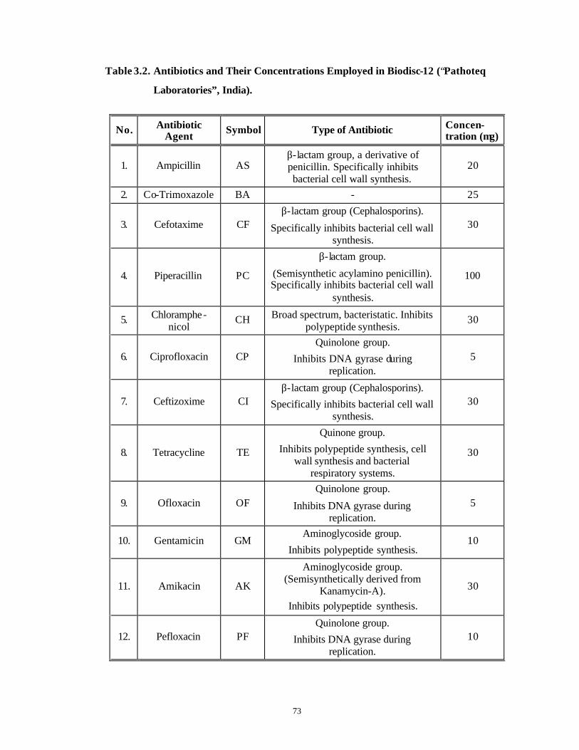
73
Table 3.2. Antibiotics and Their Concentrations Employed in Biodisc-12 (“Pathoteq
Laboratories”, India).
No. Antibiotic Agent Symbol Type of Antibiotic Concen-
tration (µg)
1. Ampicillin AS β-lactam group, a derivative of penicillin. Specifically inhibits bacterial cell wall synthesis.
20
2. Co-Trimoxazole BA - 25
3. Cefotaxime CF β-lactam group (Cephalosporins).
Specifically inhibits bacterial cell wall synthesis.
30
4. Piperacillin PC
β-lactam group.
(Semisynthetic acylamino penicillin). Specifically inhibits bacterial cell wall
synthesis.
100
5. Chloramphe-nicol
CH Broad spectrum, bacteristatic. Inhibits polypeptide synthesis.
30
6. Ciprofloxacin CP Quinolone group.
Inhibits DNA gyrase during replication.
5
7. Ceftizoxime CI β-lactam group (Cephalosporins).
Specifically inhibits bacterial cell wall synthesis.
30
8. Tetracycline TE
Quinone group.
Inhibits polypeptide synthesis, cell wall synthesis and bacterial
respiratory systems.
30
9. Ofloxacin OF Quinolone group.
Inhibits DNA gyrase during replication.
5
10. Gentamicin GM Aminoglycoside group.
Inhibits polypeptide synthesis. 10
11. Amikacin AK
Aminoglycoside group. (Semisynthetically derived from
Kanamycin-A). Inhibits polypeptide synthesis.
30
12. Pefloxacin PF Quinolone group.
Inhibits DNA gyrase during replication.
10
74
3.2.5.5 Determination of MIC (Minimum Inhibitory Concentration) and MBC
(Minimum Bactericidal Concentration) by liquid assay
Liquid bioassays were carried out for each of the seven effective antibiotics
identified by the Bio-discs on solid media, to determine the efficacy of each individual
antibiotic at three different concentrations. For each bacterial isolate, three tubes of NB
were prepared for each antibiotic agent. First tube contained NB supplemented with a
sterile solution of an antibiotic at the concentration used in the biodisc. In the second
tube, a sterile solution of the same antibiotic one unit concentration below the
concentration used in Biodisc-12 was added to NB. In the third tube a sterile solution of
the antibiotic one unit concentration above the concentration used in the Biodisc-12 was
aseptically added to NB (Table 3.9 ). The presence or absence of growth of each bacterial
isolate in these amended media was determined visually by the turbidity of the medium
after growth for 24 - 48 hours at 26 + 2ºC at 200 rpm on an incubator-shaker in the dark.
Bacteria cultured in the amended media were streaked on plates of plain NA to
confirm the results of this liquid bioassay. Wherever the growth of the bacterium was
observed on these plates, the original antibiotic concentration was designated as Minimal
Inhibitory Concentration (MIC) and wherever the bacteria were unable to resume their
growth, that antibiotic concentration was designated as Minimal Bactericidal
Concentration (MBC). Wherever the growth of a bacterium was not hampered by an
antibiotic at all, it was considered to be resistant to that particular antibiotic.
The type of antibiotic and its effective concentration derived, based on the above
methodology to identify bacterial contaminants, was used along with antifungal agents in
a strategy of pre-treatment as discussed under Section 3.3.4.
75
Ξ The identification of useful antifungal agents and their effective concentrations
against the identified fungal contaminants observed in cultures of Taxus spp. and
Withania spp. are described in the following sections.
3.2.6 Isolation and storage of fungal contaminants
3.2.6.1 Taxus baccata ssp. wallichiana Zucc. Pilg.
With the “Routine Surface Sterilization Protocol” (C. f. Section 3.2.2), almost
all the cultures of Taxus spp. became contaminated with fungi during the first two weeks
(Fig. 3.1 C, D). The isolated fungal contaminants were purified by the standard hyphal tip
and serial dilution methods (Alexopoulos and Benecke, 1962; Ainsworth and Sussman,
1966-1968) and grown on slants of potato dextrose agar (PDA) medium prepared
according to the manufacturer’s instructions (“Difco Laboratories”, USA). Pure fungal
cultures were maintained on slants of PDA kept at 4oC (Alexopoulos and Beneke, 1962).
3.2.6.2 Withania somnifera (L.) Dun.
The fungal contaminants observed in internode explants of Withania spp. were
purified, grown and stored as described above in Section 3.2.6.1 .
The isolated fungi from both the plants, in pure form, were further used for their
identification.
3.2.7 Identification of fungal contaminants
The identification was carried out based on macroscopic and microscopic
morphological characters of vegetative and reproductive structures (Ainsworth and
Sussman, 1966-1968). Once definitive identification of fungal genera was known, various
antifungal agents could be tried to control fungal contamination in cultures of Taxus spp.
and Withania spp.
76
3.2.8 Use of antimicrobial agents to control bacterial and fungal contaminants
A number of anti-microbial agents were dissolved in glass distilled water and used
at following concentrations: Bavistin (“BASF”, India) at 1% (w/v), Aureofungin
(“Hindustan Antibiotics Ltd.”, India) at 0.5% (w/v), 8- Hydroxyquinoline, Hemisulphate
salt (8-HQS) (“Sigma Chemical Co.”, USA) at 0.1% and 0.25% (w/v), Augmentin
(“Time Carp Pharma Labs Pvt. Ltd.”, India) at 500 mg.l-1, Streptopenicillin (“Hindustan
Antibiotics Ltd.”, India) at 500 mg.l-1, Ofloxacin (Ofloxacin infusion, Tarivid® i. v.,
“Hoechst Marion Roussel Ltd.”, India) at 25 mgl-1, Pefloxacin (Pefloxacin infusion,
Pelox*-400, “Wockhardt Ltd.”, India) at 25 mgl-1 and Ciprofloxacin (Ciprofloxacin
injection IP, Ciplox, “Cipla Ltd.”, India) at 25 mgl-1.
These anti-microbial agents individually or in combination were not incorporated
in the tissue culture media but were used in the “Modified Surface Sterilization”
procedure which was developed as a strategy to contain the bacterial and fungal
contamination as well as phenolic browning based on the results obtained in this chapter.
For details see Section 3.3.4 .
The percentage of fungal contamination was scored for “Routine Surface
Sterilized” explants without pre-treatment and compared against the “Modified Surface
Sterilization” procedure involving a pre-treatment strategy (Flow Chart 3.3 ) containing
antifungal agents individually and in combination with antibacterial agents as well as
antioxidants. For details refer to Sections 3.3.4, 3.3.5 and 3.3.6 .
3.2.9 Control of phenolic browning/oxidation
A number of strategies were used to control phenolic browning. The most basic
strategy was the use of anti-oxidation agents such as Ascorbic acid and Citric acid
(“HiMedia Laboratories”, India) and soluble PVP, Mol. Wt. 3,60,000 (“Sigma Chemical
Co.”, USA) at 2% (w/v) strength in the pre-treatment solution (For details refer to
Sections 3.3.5 and 3.3.6).The pre-treated explants were used either for axillary sprouting
or for callus initiation experiments. The other methods used to control phenolic browning
included use of 1/2 WPM (Strength of major salts reduced to 1/2) without plant growth
regulators in liquid form (absence of gelling agents) or in solid form (presence of gelling
agents) for inoculating the stem bit explants intended for enhanced axillary budding.
77
Further the media were modified in some cases by addition of 0.5% (w/v) activated
charcoal (AC). The control of phenolic browning in cultures is described in detail in
Section 3.3.5 .
3.2.10 Statistical analysis
All the morphological and biochemical tests were carried out in triplicate for each
of the bacterial and fungal isolates and the sets of experiments were repeated twice. The
experiments on control measures for the contaminants were carried out in triplicate over
three successive years and the Standard Errors of the Means (SE) and/or Analysis of
Variance (ANOVA) were calculated wherever applicable. Post-hoc comparisons were
performed with t-test (Wardlaw, 1985).
Each treatment in each of the experiments carried out to control phenolic
browning/oxidation consisted of 50 replicates and each experiment was repeated thrice
All the experiments were repeated over three successive years. Standard Errors of the
Means (SE) and Analysis of Variance (ANOVA) were calculated wherever applicable.
Post-hoc comparisons were performed with t-test (Wardlaw, 1985).
78
3.3 Results and Discussion
When the explants of Taxus spp. and Withania spp. were inoculated after using
the “Routine Surface Sterilization Protocol” , high degree of either bacterial or fungal
or bacterial as well as fungal contamination was observed.
Subsection 1: Bacterial Contamination
Plant tissue cultures are known to host a large number of microbes due to the
favorable conditions present in the plant tissue culture media (high amount of sugars and
organic compounds) and the microenvironment. In the earlier years, most of the efforts
directed at overcoming these contaminants involved a treatment of the infected explants
with a variety of antibiotics without identifying the contaminants (Young et al., 1984;
Falkiner, 1988; Kneifel and Leonhardt, 1992). A number of researchers in the recent
years have concentrated on identifying these contaminants up to the genus and species
level for the development of very specific strategies to overcome these contaminants
(Leifert et al., 1989; Leifert et al., 1991a, b; Buckley et al., 1995; Reed et al., 1995;
Fellner et al., 1996) and also to inhibit the indiscriminate use of effective antibiotics,
which may give rise to new and more virulent resistant strains. In addition, a large
number of antibiotics are known to be highly phytotoxic to plant tissue cultures (Thurston
et al., 1979; Pollock et al., 1983).
The methodology followed for identification of the contaminant bacteria in this
thesis is depicted concisely in Flow Chart 3.1.
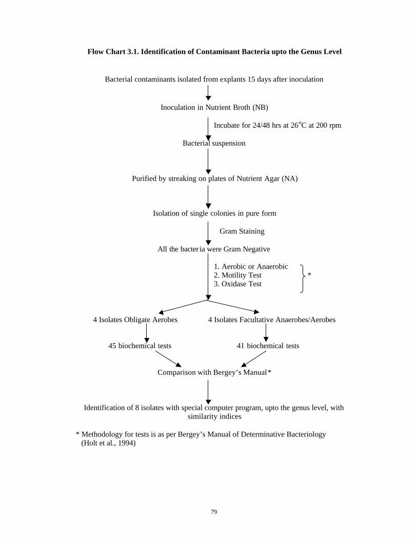
79
Flow Chart 3.1. Identification of Contaminant Bacteria upto the Genus Level
Bacterial contaminants isolated from explants 15 days after inoculation
Inoculation in Nutrient Broth (NB) Incubate for 24/48 hrs at 26oC at 200 rpm
Bacterial suspension
Purified by streaking on plates of Nutrient Agar (NA)
Isolation of single colonies in pure form
Gram Staining
All the bacter ia were Gram Negative
1. Aerobic or Anaerobic 2. Motility Test * 3. Oxidase Test
4 Isolates Obligate Aerobes 4 Isolates Facultative Anaerobes/Aerobes
45 biochemical tests 41 biochemical tests
Comparison with Bergey’s Manual *
Identification of 8 isolates with special computer program, upto the genus level, with similarity indices
* Methodology for tests is as per Bergey’s Manual of Determinative Bacteriology
(Holt et al., 1994)
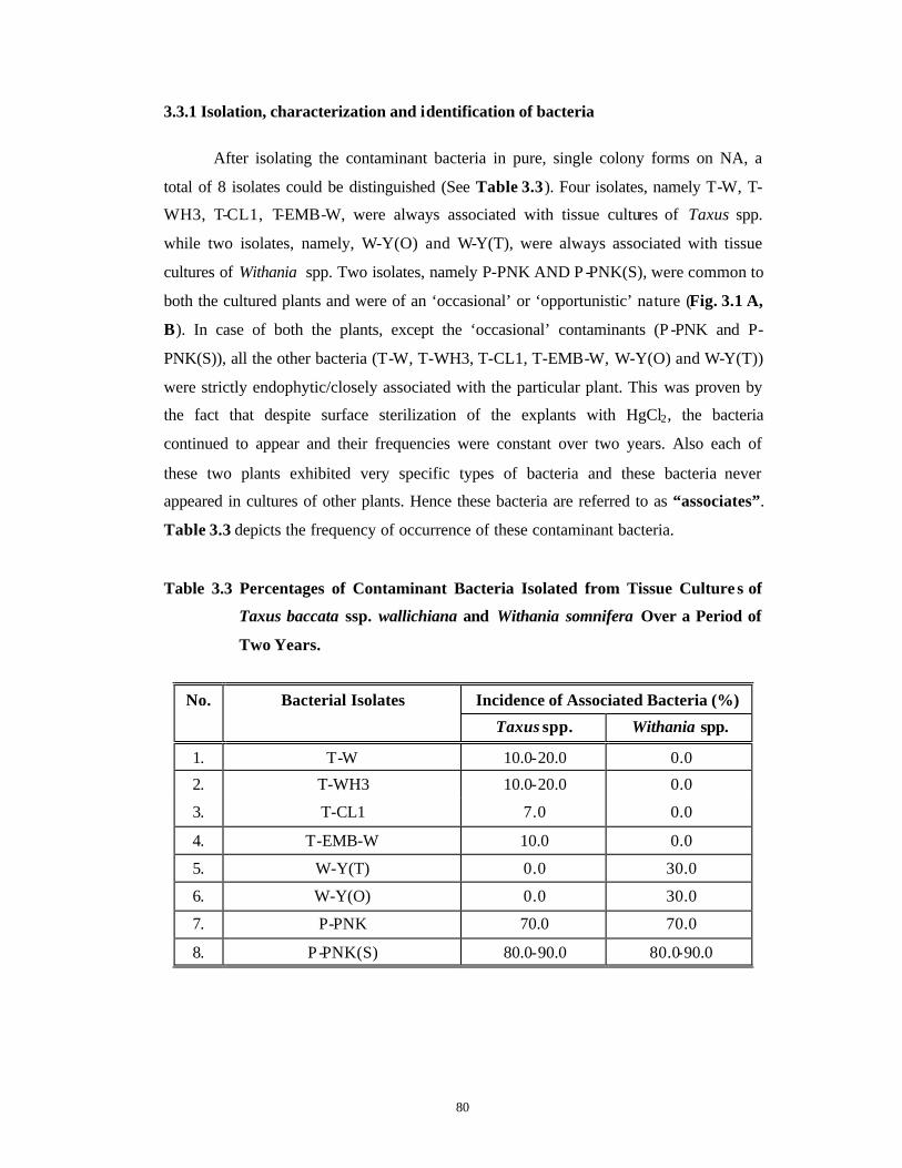
80
3.3.1 Isolation, characterization and identification of bacteria
After isolating the contaminant bacteria in pure, single colony forms on NA, a
total of 8 isolates could be distinguished (See Table 3.3). Four isolates, namely T-W, T-
WH3, T-CL1, T-EMB-W, were always associated with tissue cultures of Taxus spp.
while two isolates, namely, W-Y(O) and W-Y(T), were always associated with tissue
cultures of Withania spp. Two isolates, namely P-PNK AND P -PNK(S), were common to
both the cultured plants and were of an ‘occasional’ or ‘opportunistic’ nature (Fig. 3.1 A,
B). In case of both the plants, except the ‘occasional’ contaminants (P -PNK and P-
PNK(S)), all the other bacteria (T-W, T-WH3, T-CL1, T-EMB-W, W-Y(O) and W-Y(T))
were strictly endophytic/closely associated with the particular plant. This was proven by
the fact that despite surface sterilization of the explants with HgCl2, the bacteria
continued to appear and their frequencies were constant over two years. Also each of
these two plants exhibited very specific types of bacteria and these bacteria never
appeared in cultures of other plants. Hence these bacteria are referred to as “associates”.
Table 3.3 depicts the frequency of occurrence of these contaminant bacteria.
Table 3.3 Percentages of Contaminant Bacteria Isolated from Tissue Culture s of
Taxus baccata ssp. wallichiana and Withania somnifera Over a Period of
Two Years.
Incidence of Associated Bacteria (%) No. Bacterial Isolates
Taxus spp. Withania spp.
1. T-W 10.0-20.0 0.0
2. T-WH3 10.0-20.0 0.0
3. T-CL1 7.0 0.0
4. T-EMB-W 10.0 0.0
5. W-Y(T) 0.0 30.0
6. W-Y(O) 0.0 30.0
7. P-PNK 70.0 70.0
8. P-PNK(S) 80.0-90.0 80.0-90.0
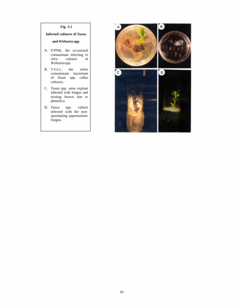
81
Fig. 3.1
Infected cultures of Taxus
and Withania spp.
A. P-PNK, the occasional contaminant infecting in vitro cultures of Withania spp.
B. T -CL1, the white contaminant bacterium of Taxus spp. callus cultures.
C. Taxus spp. stem explant infected with fungus and turning brown due to phenolics.
D. Taxus spp. cultureinfected with the non-sporulating opportunistic fungus.

82
It is clear from Table 3.3 that out of the eight bacteria isolated; P-PNK and P-PNK(S)
were common to both the plants and were found to be opportunistic and occasional. They
were pink and intense pink pigmented contaminants respectively, appearing in distinct
outbreaks that were unrelated to the monsoon season. In between these sudden outbreaks,
all the cultures remained sterile. The intense pink bacteria (P-PNK(S)) were more
predominant in the cultures and because of their fast growth rate, the cultures were killed
within two days.
Four isolates (T-W, T-WH3, T-CL1 and T-EMB-W) were always associated with
tissue cultures of Taxus baccata ssp. wallichiana. All of them were white in colour. One
isolate, T-EMB-W was associated with embryos of this plant and was slow growing. But
once this contaminant appeared in the cultures (10%), further growth of the embryos was
arrested. The isolate T-CL1 was present in callus cultures (7%) while T-W and T-WH3
were associated with newly inoculated needles (10-20%) and appeared within one week
of inoculation. As compared to the pigmented opportunistic contaminants, these bacteria
were slow growing in plant tissue culture media but eventually succeeded in killing the
explant or the callus.
Two types of yellow bacteria, W-Y(O) and W-Y(T) were isolated (30%) only
from the cultured internodes of Withania somnifera . They oozed out from the explant,
making it spongy and unresponsive. They were fast growing and killed the explants
within three days after their appearance.
Following the isolation of contaminant bacteria in pure, single colony form,
morphological and biochemical characterizations were performed which are described
in following subsections.
3.3.1.1 Morphological characterization
All the bacterial types had round and smooth walled colonies and only the yellow
colored colonies exhibited fluorescence. The white and pink colored colonies did not
have any fluorescence.
All the contaminants were Gram stained using the double stain method and were
found to be either Gram-negative rods or coccobacilli (Figs. 3.2 and 3.3). A distinct cycle

83
Fig. 3.2
Figs. A. – D. Gram staining of ‘associated’ contaminant bacteria from in vitro cultures of Taxus spp. (Magnification: 100x).
A. T-W, with long, rod shaped cells.
B. T-CL1, with both rods and cocci.
C. T-EMB-W, with slender rod shaped cells.
D. T-WH3, with short, rod shaped cells.
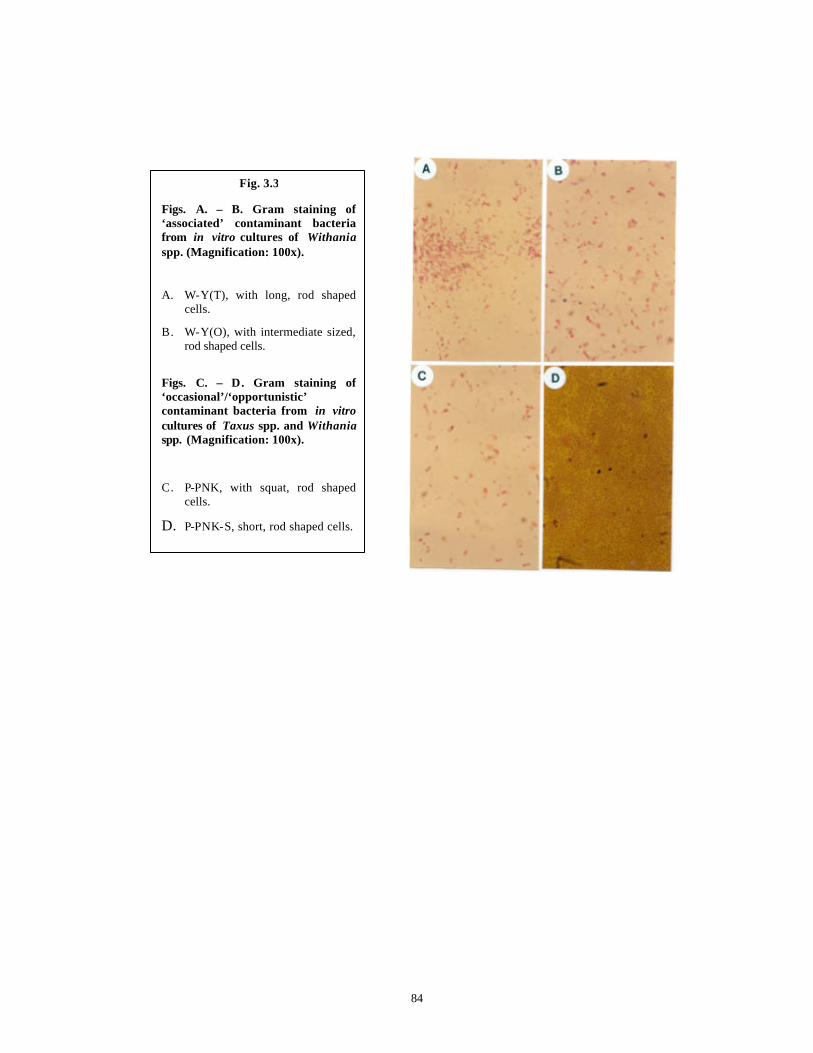
84
Fig. 3.3
Figs. A. – B. Gram staining of ‘associated’ contaminant bacteria from in vitro cultures of Withaniaspp. (Magnification: 100x).
A. W-Y(T), with long, rod shaped cells.
B. W-Y(O), with intermediate sized, rod shaped cells.
Figs. C. – D. Gram staining of ‘occasional’/‘opportunistic’ contaminant bacteria from in vitrocultures of Taxus spp. and Withaniaspp. (Magnification: 100x).
C. P-PNK, with squat, rod shaped cells.
D. P-PNK-S, short, rod shaped cells.
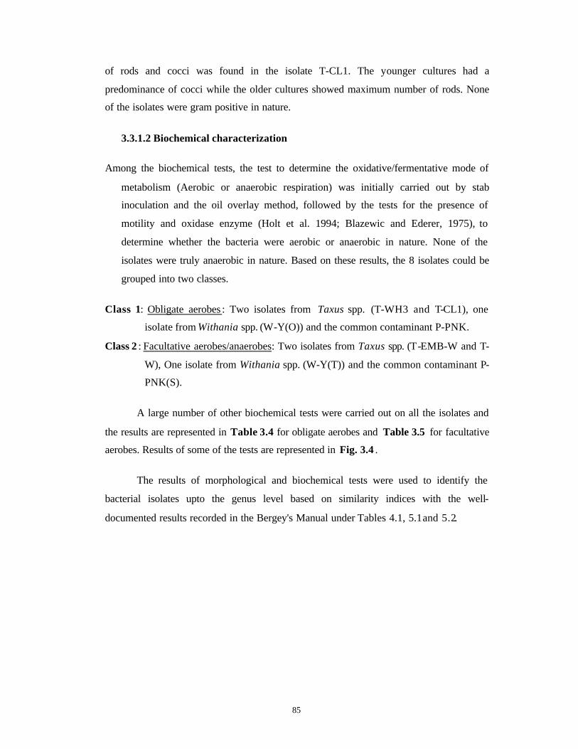
85
of rods and cocci was found in the isolate T-CL1. The younger cultures had a
predominance of cocci while the older cultures showed maximum number of rods. None
of the isolates were gram positive in nature.
3.3.1.2 Biochemical characterization
Among the biochemical tests, the test to determine the oxidative/fermentative mode of
metabolism (Aerobic or anaerobic respiration) was initially carried out by stab
inoculation and the oil overlay method, followed by the tests for the presence of
motility and oxidase enzyme (Holt et al. 1994; Blazewic and Ederer, 1975), to
determine whether the bacteria were aerobic or anaerobic in nature. None of the
isolates were truly anaerobic in nature. Based on these results, the 8 isolates could be
grouped into two classes.
Class 1: Obligate aerobes : Two isolates from Taxus spp. (T-WH3 and T-CL1), one
isolate from Withania spp. (W-Y(O)) and the common contaminant P-PNK.
Class 2 : Facultative aerobes/anaerobes: Two isolates from Taxus spp. (T-EMB-W and T-
W), One isolate from Withania spp. (W-Y(T)) and the common contaminant P-
PNK(S).
A large number of other biochemical tests were carried out on all the isolates and
the results are represented in Table 3.4 for obligate aerobes and Table 3.5 for facultative
aerobes. Results of some of the tests are represented in Fig. 3.4 .
The results of morphological and biochemical tests were used to identify the
bacterial isolates upto the genus level based on similarity indices with the well-
documented results recorded in the Bergey's Manual under Tables 4.1, 5.1 and 5.2.
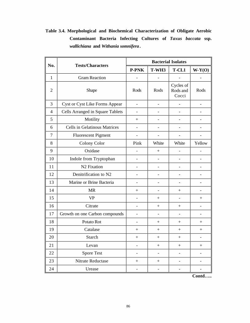
86
Table 3.4. Morphological and Biochemical Characterization of Obligate Aerobic
Contaminant Bacteria Infecting Cultures of Taxus baccata ssp.
wallichiana and Withania somnifera .
Bacterial Isolates No. Tests/Characters
P-PNK T-WH3 T-CL1 W-Y(O)
1 Gram Reaction - - - -
2 Shape Rods Rods Cycles of Rods and
Cocci Rods
3 Cyst or Cyst Like Forms Appear - - - -
4 Cells Arranged in Square Tablets - - - -
5 Motility + - - -
6 Cells in Gelatinous Matrices - - - -
7 Fluorescent Pigment - - - -
8 Colony Color Pink White White Yellow
9 Oxidase - + - -
10 Indole from Tryptophan - - - -
11 N2 Fixation - - - -
12 Denitrification to N2 - - - -
13 Marine or Brine Bacteria - - - -
14 MR + - + -
15 VP - + - +
16 Citrate - + + -
17 Growth on one Carbon compounds - - - -
18 Potato Rot - + + +
19 Catalase + + + +
20 Starch + + + -
21 Levan - + + +
22 Spore Test - - - -
23 Nitrate Reductase + + - -
24 Urease - - - -
Contd…..

87
Table 3.4 Contd.
Bacterial Isolates No. Tests/Characters
P-PNK T-WH3 T-CL1 W-Y(O)
25 Growth at 5°C* - - - -
26 Growth at 27°C + + + +
27 Growth at 37°C + + + +
28 Growth at 60°C** - - - -
29 Growth in 20 % NaCl - - - ND
30 Anaerobic Growth (stab method)
- - - -
31 Anaerobic Growth (oil overlay method) - - - -
32 Growth in Ashby's Medium
(Without N2) V - - -
33 Gas Production in Glucose Medium - - - -
34 Acid Production in Glucose Medium
- + - +
35 Growth in Glucose Medium - + + +
36 Acid Production in Mannose Medium - - + ND
37 Growth in Mannose Medium - + + +
38 Acid production in Lactose Medium + - + -
39 Growth in Lactose Medium + + + +
40 Acid production in Rhamnose Medium - - + -
41 Growth in Rhamnose Medium + + + +
42 Acid production in Sucrose Medium + - - -
43 Growth in Sucrose Medium + + + +
44 Acid production in Mannitol Medium - - - -
45 Growth in Mannitol Medium + + + +
* : Growth occurs after transfer to 27 °C in the strains, which do not grow at 5°C . ** : Growth does not occur even after transfer to 27°C. + : Positive test result. - : Negative test result.
ND : Not determined. V : Variable results.

88
Table 3.5. Morphological and Biochemical characterization of Facultative
Anaerobic/Aerobic Contaminant Bacteria Infecting Cultures of Taxus
baccata ssp. wallichiana and Withania somnifera.
Bacterial Isolates No. Tests / Characters P-
PNK(S) T-EMB -
W T-W W-Y(T)
1 Gram Reaction - - - -
2 Cell Shape Rods Cocco-bacilli Rods Rods
3 Motility + + + -
4 Cysts or Cyst -Like Forms Present - - - -
5 Cells Arranged in Square Tablets - - - -
6 Oxidase + - + +
7 Fluorescent pigment - - - -
8 Colony Color Intense Pink White White Yellow
9 Indole from Tryptophan - - - -
10 MR - + - +
11 VP + + - -
12 Citrate + + + +
13 Growth on one Carbon compounds - + - -
14 Potato Rot - + + +
15 Catalase + + + +
16 Starch + + + -
17 Levan + + + -
18 Spore Test - - - -
19 Nitrate Reductase + + - +
20 Urease ND - - +
21 Growth at 5°C* - + + -
22 Growth at 27°C + + + +
23 Growth at 37°C + + + +
24 Growth at 60°C** - - - -
Contd…..
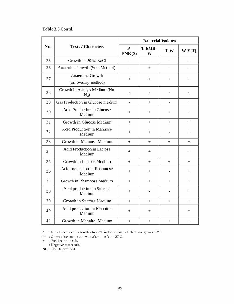
89
Table 3.5 Contd.
Bacterial Isolates No. Tests / Characters P-
PNK(S) T-EMB -
W T-W W-Y(T)
25 Growth in 20 % NaCl - - - -
26 Anaerobic Growth (Stab Method) - + - -
27 Anaerobic Growth
(oil overlay method) + + + +
28 Growth in Ashby's Medium (No N2)
- - - -
29 Gas Production in Glucose me dium - + - +
30 Acid Production in Glucose Medium + + + +
31 Growth in Glucose Medium + + + +
32 Acid Production in Mannose Medium + + - +
33 Growth in Mannose Medium + + + +
34 Acid Production in Lactose Medium + + - -
35 Growth in Lactose Medium + + + +
36 Acid production in Rhamnose Medium + + - +
37 Growth in Rhamnose Medium + + + +
38 Acid production in Sucrose Medium + - - +
39 Growth in Sucrose Medium + + + +
40 Acid production in Mannitol Medium + + - +
41 Growth in Mannitol Medium + + + +
* : Growth occurs after transfer to 27°C in the strains, which do not grow at 5°C. ** : Growth does not occur even after transfer to 27°C. + : Positive test result. - : Negative test result. ND : Not Determined.

90
Fig. 3.4
Figs. A – D A representation of biochemical tests performed for characterization of contaminant bacteria from in vitro cultures of Taxus spp. and Withania spp.
A. Presence (reddish brown coloration) or absence (absence of coloration) of nitrate reductase in contaminant bacteria from Withania spp.: W-Y(O), W-Y(T) and common contaminant: P-PNK(S). The leftmost bottle is control without bacteria.
B. Presence (reddish brown coloration) or absence (absence of coloration) of nitrate reductase in contaminant bacteria from Taxus spp.: T -EMB-W, T-CL1, T-WH3, T -W. The leftmost bottle is control without bacteria.
C. Presence or absence of utilization of citrate by contaminant bacteria from Withania spp.: W-Y(T) and W-Y(O) and two common contaminants: P-PNK and P-PNK(S). The leftmost bottle is control without bacteria Blue coloration indicates conversion of citrate in the medium by the bacterial enzymes.
D. Presence or absence of utilization of citrate by contaminant bacteria from Taxus spp.: T-W, T-EMB-W, T -WH3 and T-CL1 and two common contaminants: P-PNK and P-PNK(S). Blue to bluish-green colorat ion indicates conversion of citrate in the medium by bacterial enzymes. The leftmost bottle in the upper-hand corner is control without bacteria
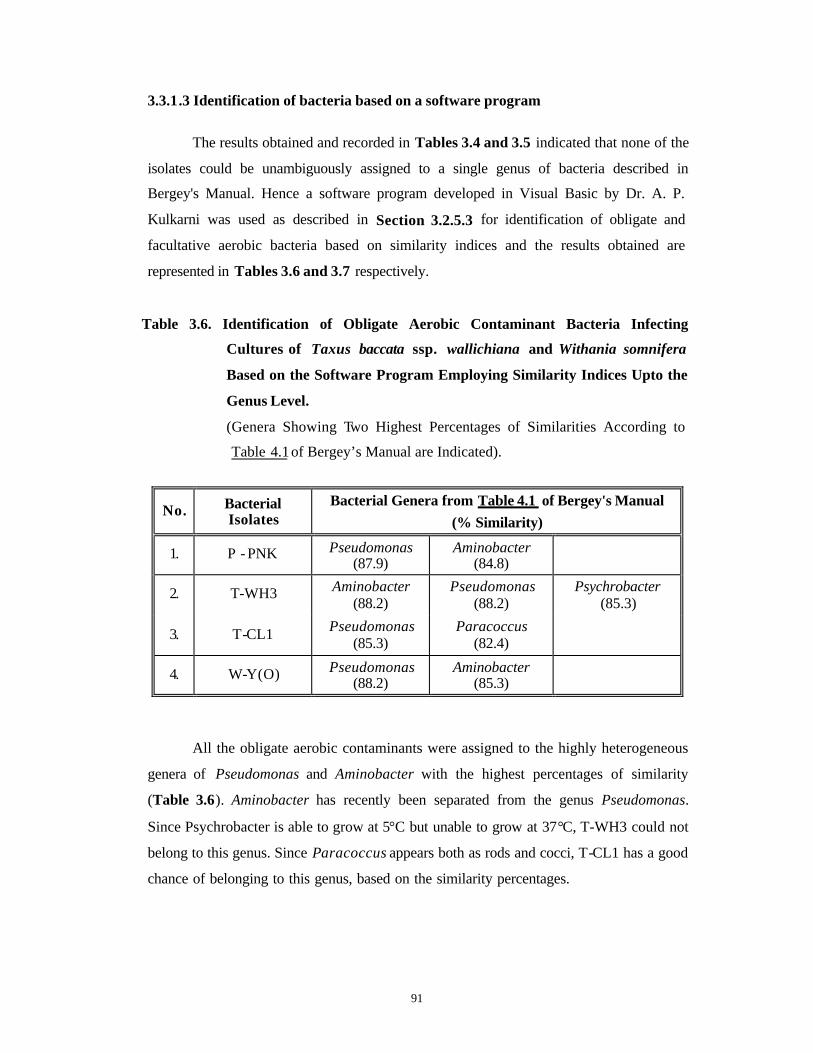
91
3.3.1.3 Identification of bacteria based on a software program
The results obtained and recorded in Tables 3.4 and 3.5 indicated that none of the
isolates could be unambiguously assigned to a single genus of bacteria described in
Bergey's Manual. Hence a software program developed in Visual Basic by Dr. A. P.
Kulkarni was used as described in Section 3.2.5.3 for identification of obligate and
facultative aerobic bacteria based on similarity indices and the results obtained are
represented in Tables 3.6 and 3.7 respectively.
Table 3.6. Identification of Obligate Aerobic Contaminant Bacteria Infecting
Cultures of Taxus baccata ssp. wallichiana and Withania somnifera
Based on the Software Program Employing Similarity Indices Upto the
Genus Level.
(Genera Showing Two Highest Percentages of Similarities According to
Table 4.1 of Bergey’s Manual are Indicated).
No. Bacterial Isolates
Bacterial Genera from Table 4.1 of Bergey's Manual (% Similarity)
1. P - PNK Pseudomonas (87.9)
Aminobacter (84.8)
2. T-WH3 Aminobacter (88.2)
Pseudomonas (88.2)
Psychrobacter (85.3)
3. T-CL1 Pseudomonas (85.3)
Paracoccus (82.4)
4. W-Y(O) Pseudomonas (88.2)
Aminobacter (85.3)
All the obligate aerobic contaminants were assigned to the highly heterogeneous
genera of Pseudomonas and Aminobacter with the highest percentages of similarity
(Table 3.6). Aminobacter has recently been separated from the genus Pseudomonas.
Since Psychrobacter is able to grow at 5°C but unable to grow at 37°C, T-WH3 could not
belong to this genus. Since Paracoccus appears both as rods and cocci, T-CL1 has a good
chance of belonging to this genus, based on the similarity percentages.
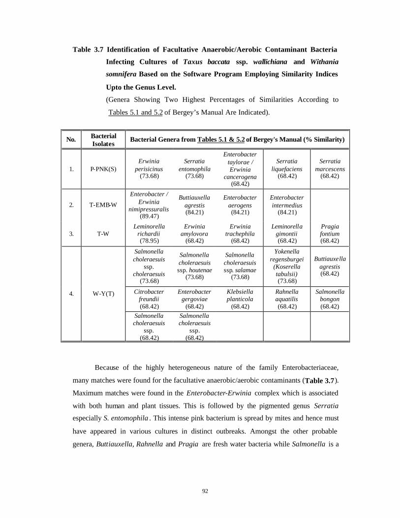
92
Table 3.7 Identification of Facultative Anaerobic/Aerobic Contaminant Bacteria
Infecting Cultures of Taxus baccata ssp. wallichiana and Withania
somnifera Based on the Software Program Employing Similarity Indices
Upto the Genus Level.
(Genera Showing Two Highest Percentages of Similarities According to
Tables 5.1 and 5.2 of Bergey’s Manual Are Indicated).
No. Bacterial Isolates
Bacterial Genera from Tables 5.1 & 5.2 of Bergey's Manual (% Similarity)
1. P-PNK(S) Erwinia
perisicinus (73.68)
Serratia entomophila
(73.68)
Enterobacter taylorae / Erwinia
cancerogena (68.42)
Serratia liquefaciens
(68.42)
Serratia marcescens
(68.42)
2. T-EMB-W
Enterobacter / Erwinia
nimipressuralis (89.47)
Buttiauxella agrestis (84.21)
Enterobacter aerogens (84.21)
Enterobacter intermedius
(84.21)
3. T-W Leminorella
richardii (78.95)
Erwinia amylovora
(68.42)
Erwinia trachephila
(68.42)
Leminorella gimontii (68.42)
Pragia fontium (68.42)
Salmonella choleraesuis
ssp. choleraesuis
(73.68)
Salmonella choleraesuis ssp. houtenae
(73.68)
Salmonella choleraesuis ssp. salamae
(73.68)
Yokenella regensburgei
(Koserella tabulsii) (73.68)
Buttiauxella agrestis (68.42)
Citrobacter freundii (68.42)
Enterobacter gergoviae
(68.42)
Klebsiella planticola
(68.42)
Rahnella aquatilis (68.42)
Salmonella bongon (68.42)
4. W-Y(T)
Salmonella choleraesuis
ssp. (68.42)
Salmonella choleraesuis
ssp. (68.42)
Because of the highly heterogeneous nature of the family Enterobacteriaceae,
many matches were found for the facultative anaerobic/aerobic contaminants (Table 3.7).
Maximum matches were found in the Enterobacter-Erwinia complex which is associated
with both human and plant tissues. This is followed by the pigmented genus Serratia
especially S. entomophila . This intense pink bacterium is spread by mites and hence must
have appeared in various cultures in distinct outbreaks. Amongst the other probable
genera, Buttiauxella, Rahnella and Pragia are fresh water bacteria while Salmonella is a

93
very big and heterogeneous assemblage associated with humans and animals. Leminorella
is also a human associated form.
It is clear from these results that further definitive ass ignation of bacteria to the species
level can only be done after either fatty acid or DNA sequence analysis. The various
databases available over the world for bacteria are mainly concerned with medically
important bacteria and not much information is available for plant related organisms.
There is a need to build up such databases urgently.
As described by Leifert et al. (1994), the gram negative Pseudomonads infected
more than 50% of stage 1 and stage 2 cultures while Erwinia spp. infected 10% of such
cultures. This is because these genera are associated with aerial surfaces of plants under
field conditions (Last and Deighton, 1965). Since none of the contaminants were gram
positive, the possibility of their introduction from the laboratory air is excluded. Since
none of the contaminants formed endospores, Bacillus spp. that can spread from
inefficient autoclaving and insufficient sterilization of instruments used for inoculations
and subcultures were absent.
From this, we can reasonably conclude that mos t of the bacteria found in vitro
were associated with plants either exogenously or endogenously, except those belonging
to genera Serratia , Salmonella and Leminorella .
Effective control measures against contaminant bacteria using antibiogramme assays
were carried out subsequently, after bacterial identification upto the generic level.
3.3.2 Control measures
Antibiogramme assays, both in solid and liquid media, were carried out (Flow
Chart 3.2 ) and the results of, which are presented in Tables 3.8 and 3.9 . These assays
helped in the identification of effective antibiotics and their minimum concentration
required to control the identified bacteria effectively. The results obtained are described
herewith.

94
Flow Chart 3.2. Identification of Effective Antibiotics and Their Optimum
Concentration for Control of Each of the Contaminant Bacteria
Each of the six identified 100 µl bacterial Placed sterile biodisc bacteria was grown in NB suspension spread (Pathoteq Laboratories) (24-48 h) at 26°C & 200 rpm. over plates of NA containing 12 antibiotics (Top Agar Method) & incubated overnight at 26°C. Diameter of clearing Zones (cm) measured. Optimized the type of antibiotic for each bacterial genus.
(First two with the biggest clearing zones were chosen).
Added 3 Conc. of the identified type of antibiotic to NB: Liquid Bioassay to determine effective concentration of the chosen antibiotic.
(168 combinations)
Incubated overnight at 26°C and 200 rpm.
Streaked on plates of NA to determine Bactericidal, Bacteristatic and Resistant
Concentrations.
Identified type & antibiotic conc. to be used in pre-treatment of explants to control contaminants.
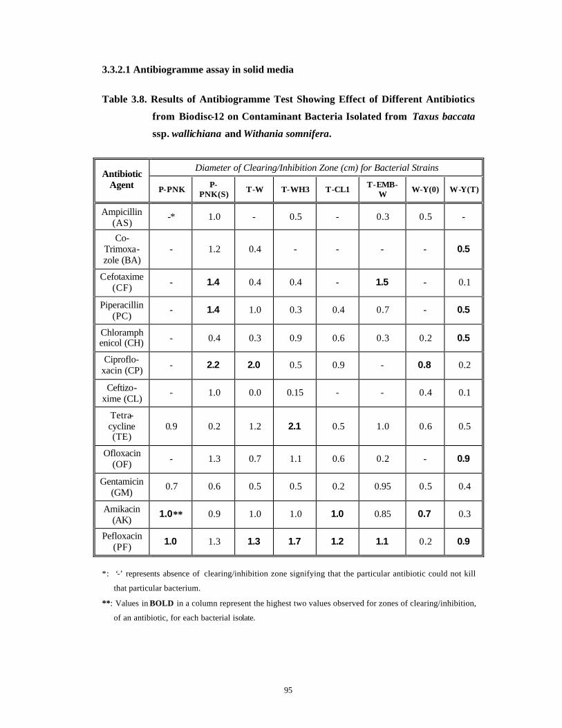
95
3.3.2.1 Antibiogramme assay in solid media
Table 3.8. Results of Antibiogramme Test Showing Effect of Different Antibiotics
from Biodisc-12 on Contaminant Bacteria Isolated from Taxus baccata
ssp. wallichiana and Withania somnifera.
Diameter of Clearing/Inhibition Zone (cm) for Bacterial Strains
Antibiotic Agent P-PNK P-
PNK(S) T-W T-WH3 T-CL1 T-EMB-W W-Y(0) W-Y(T)
Ampicillin (AS)
-* 1.0 - 0.5 - 0.3 0.5 -
Co-Trimoxa-zole (BA)
- 1.2 0.4 - - - - 0.5
Cefotaxime (CF) - 1.4 0.4 0.4 - 1.5 - 0.1
Piperacillin (PC)
- 1.4 1.0 0.3 0.4 0.7 - 0.5
Chloramphenicol (CH) - 0.4 0.3 0.9 0.6 0.3 0.2 0.5
Ciproflo-xacin (CP) - 2.2 2.0 0.5 0.9 - 0.8 0.2
Ceftizo-xime (CL)
- 1.0 0.0 0.15 - - 0.4 0.1
Tetra-cycline (TE)
0.9 0.2 1.2 2.1 0.5 1.0 0.6 0.5
Ofloxacin (OF) - 1.3 0.7 1.1 0.6 0.2 - 0.9
Gentamicin (GM)
0.7 0.6 0.5 0.5 0.2 0.95 0.5 0.4
Amikacin (AK)
1.0** 0.9 1.0 1.0 1.0 0.85 0.7 0.3
Pefloxacin (PF) 1.0 1.3 1.3 1.7 1.2 1.1 0.2 0.9
*: ‘-’ represents absence of clearing/inhibition zone signifying that the particular antibiotic could not kill
that particular bacterium.
**: Values in BOLD in a column represent the highest two values observed for zones of clearing/inhibition,
of an antibiotic, for each bacterial isolate.

96
In the solid antibiogramme assay (Fig. 3.5 ), antibiotic BA was the least effective
(activity against 3 isolates) with very small zones of inhibition. AS could control the
growth of 4 isolates while CF and CL could control the growth of 5 isolates. PC, CP, OF
inhibited growth of 6 isolates while CH could inhibit growth of 7 isolates. TE, GM, AK,
PF were broad-spectrum antibiotics controlling growth of all the eight isolates (Table
3.8 ).
Among the bacteria, T-W and W-Y(T) were controlled by all the antibiotics tested
except AS. T-CL1, T-WH3, T-EMB-W, W-Y(O) and P-PNK were resistant to BA.
Growth of T-CL1 and W-Y(O) was not hampered by four antibiotics each. P-PNK was
not sensitive to 8 antibiotics out of 12 antibiotics tested, whereas P-PNK(S) was
sus ceptible to all the antibiotics.
3.3.2.2 Antibiogramme assay in liquid media
To evaluate the efficiency of a single antibiotic agent with respect to the effective
concentration and to confirm the preliminary results of the antibiogramme assay,
antibiotic sensitivity tests were carried out in NB. Since the results of antibiogramme
assay in solid media are affected by the diffusibilty of the antibiotics, which are in turn
dependent upon the structure and molecular weights of the antibiotics, sensitivity of the
organism, concentration of the drug on the disc and the medium constituents, the
experiment was repeated in liquid media. The concentrations used in the Biodisc-12 were
further manipulated for this test. For the liquid assay only seven most effective antibiotics
were used on the basis of the results of inhibition zone diameters obtained in the solid
antibiogramme assay. These were AK, TE, GM, PF, PC, CP and OF. Each antibiotic was
aseptically added to tubes of NB at three different concentrations (Table 3.9) and these
tubes were then inoculated individually with a loopful of bacteria. In all 168 combinations
were tried. In many cases, the antibiotic concentrations used in the Biodisc-12 were found
to be bacteristatic and not bactericidal (Table 3.9).
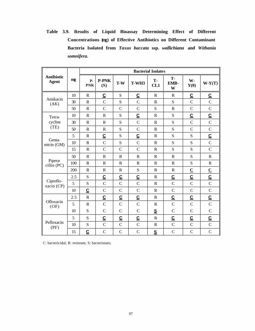
97
Table 3.9. Results of Liquid Bioassay Determining Effect of Different
Concentrations (µg) of Effective Antibiotics on Different Contaminant
Bacteria Isolated from Taxus baccata ssp. wallichiana and Withania
somnifera.
Bacterial Isolates
Antibiotic Agent µg P-
PNK P-PNK
(S) T-W T-WH3 T-CL1
T-EMB-
W
W-Y(0) W-Y(T)
10 R C S C R R C C 30 R C S C R S C C
Amikacin (AK)
50 R C C C S R C C
10 R R S C R S C C
30 R R S C R S C C Tetra-cycline (TE) 50 R R S C R S C C
5 R C S C R S S C
10 R C S C R S S C Genta-
micin (GM) 15 R C C C R S S C
50 R R R R R R S R 100 R R R R R R S R Pipera-
cillin (PC) 200 R R R S R R C C
2.5 S C C C R C C C 5 S C C C R C C C Ciproflo-
xacin (CP) 10 C C C C R C C C
2.5 R C C C R C C C
5 R C C C R C C C Ofloxacin (OF)
10 S C C C S C C C
5 S C C C R C C C 10 S C C C R C C C Pefloxacin
(PF) 15 C C C C S C C C
C: bactericidal, R: resistant, S: bacteristatic.
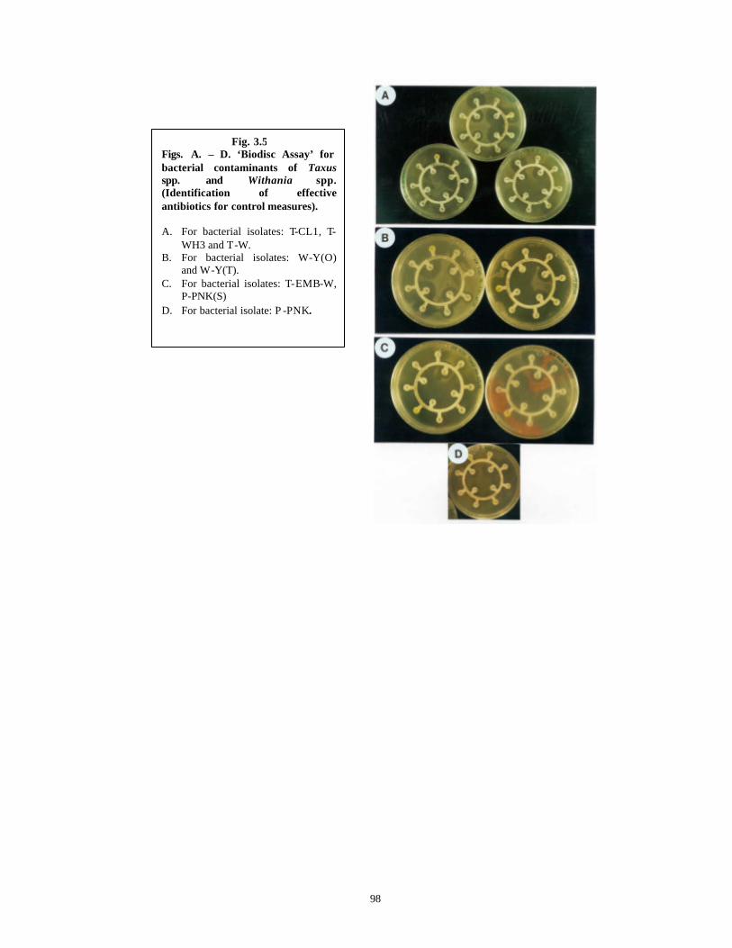
98
Fig. 3.5 Figs. A. – D. ‘Biodisc Assay’ for bacterial contaminants of Taxus spp. and Withania spp. (Identification of effective antibiotics for control measures).
A. For bacterial isolates: T-CL1, T-
WH3 and T-W. B. For bacterial isolates: W-Y(O)
and W-Y(T). C. For bacterial isolates: T-EMB-W,
P-PNK(S) D. For bacterial isolate: P -PNK.
99
From Table 3.9 it is clear that the two common contaminants (P-PNK and P-
PNK(S)) were controlled by the Quinolone group of antibiotics (CP, OF and PF). In case
of contaminants from Taxus spp. and Withania spp., all of the isolates had at least one
antibiotic giving a bactericidal response and hence could be used to control them. Only T-
CL1 was found to be resistant to all the antibiotics in liquid bioassay. It gave a
bacteristatic response for 10 µg of ofloxacin and 15 µg of pefloxacin. None of the
antibiotics proved to be bactericidal at the given concentration. It is reasonable to believe
that higher concentrations of the same antibiotics will prove to be bactericidal.
In general, it is thus clear that for all the 8 isolates, the Quinolone group of
antibiotics comprising of CP, OF and PF are very effective in terms of their bactericidal
activities at 2.5, 2.5 and 5 µg respectively and can be used in the strategy to be adopted to
control various contaminant bacteria.
Using the standardized strategy of isolation of contaminant bacteria from Tauxs
spp. and Withania spp. in vitro cultures , followed by their characterization, identification
and antibiogramme assay, the minimum bactericidal concentration (MBC) of a specific
antibiotic against each of the contaminant bacteria was determined. The determined MBC
was used (A) for preventing the bacterial contamination in cultures of Taxus spp. and
Withania spp. and (B) for combining with anti-fungal agents in a strategy to control
fungal and bacterial contamination as described under Section 3.3.4 .
100
Subsection 2: Fungal Contamination and Pheno lic Oxidation/Browning
Even though Taxus spp. plants in vivo are known to produce Taxanes with
antifungal properties (Young et al., 1992; Wagner and Flores, 1994; Elmer et al., 1994),
in vitro cultures showed many fungal contaminants (Fig. 3.1 C, D). Two types of
contaminants were identified: ‘Constant’ (endophytes or plant associates) and
‘Occasional’ or ‘Opportunistic’ (appearing suddenly, mostly dependant on
environmental conditions like monsoon). Based on the identified fungal genera very
specific anti-fungal strategies have been developed in order to circumvent the possibility
of development of resistant strains. In addition, this may help in pinpointing the source of
contamination and to check for the pathogenicity, if any, of the isolated genera (Danby et
al., 1994). In case of Taxus spp., it would also help to determine whether any of the
isolated genera are Taxol/Taxane producers.
3.3.3 Isolation and identification of fungal contaminants
3.3.3.1 Constant/endophytic contaminants
The plant tissue cultures especially of woody plants show a steady relatively
unchanged rate of contamination that is caused by the ‘constant’ contaminants. These
constant contaminants are associated with the plants and introduced through the explants
(Stage 0) or due to insufficient surface sterilization as in many cases the sterilant can not
reach all the crevices that are usually found on mature explants (Stage 1) (Leifert and
Waites, 1994). The endophytic nature of several bacteria and fungi sharing different
ecological associations with the plants supports this assumption (Knogge, 1996; Alfano
and Collmer, 1996). In case of Taxus spp., there are various reports in literature enlisting
different endophytic bacterial and fungal genera associated with this plant (Stierle et al.,
1995; Strobel et al., 1996). Incidentally many of these genera have been identified in
tissue cultures of the Himalayan Taxus spp. during the course of the present work, based
on macroscopic and microscopic morphological characters of vegetative and reproductive
structures and are listed below.
1. Alternaria spp. Nees (1816) Anamorphic Pleosporaceae, Dothidiales, Ascomycota
(Hawksworth et al., 1995). Found in 20% of cultures (Fig. 3.6 A).
101
In natural environments, 50 species of the genus are widesprea d and include many
plant pathogens. Remaining species are cosmopolitan saprobes.
2. Phoma spp. Sacc. (1880), Anamorphic Pleosporaceae, Dothidiales, Ascomycota
(Hawksworth et al., 1995). Found in 60% of cultures. This is reported to be an
endophyte of Taxus spp (Yang et al., 1994) with the ability to produce Taxol. (Fig.
3.6 B ).
In natural environments, it is widespread and is saprobic or necrotrophic on
leaves and stems. It is also reported to be quite common in vitro across various genera
(Danby et al., 1994).
3. Pestalotiopsis spp. Steyaert (1949), Anamorphic Amphisphaeriaceae, Xylariales,
Ascomycota (Hawksworth et al., 1995). Found in 15% of cultures. This is reported to
be an endophyte of Taxus spp (Strobel et al., 1996) with the ability to produce Taxol.
(Fig. 3.6 C).
In natural environments, it is widespread and is saprobic or necrotrophic on leaves
and stems. It is also a known parasite of bark and wood and hence is closely associated
with the trees.
4. Phyllosticta spp. Pers (1818), Anamorphic Mycosphaerellaceae, Dothidiales,
Ascomycota (Hawksworth et al., 1995). Found in 10% of cultures (Fig. 3.6 D).
In natural environments, it is a key spp. on conifers, growing as biotrophic or
necrotrophic or saprobic on the plants. Its teleomorph Guignardia is highly parasitic
causing leaf spot and leaf blight diseases.
5. Fusarium spp. Link (1809), Anamorphic Hypocreaceae, Hypocreales, Ascomycota
(Hawksworth et al., 1995). Found in 20% of cultures (Fig. 3.6 E, F ).
In natural environments, the genus is widespread and includes saprobic,
necrotrophic and parasitic spp. Its another anamorph Acremonium is reported to be
endophytic in Taxus (Strobel et al. 1997). Fusarium is also reported to be quite
common in vitro across various genera (Danby et al., 1994).
6. Chaetomiu m spp. Kunze (1817), Chaetomiaceae, Sordariales, Ascomycota
(Hawksworth et al., 1995). Appeared in 10% of cultures and quickly sporulated
forming dark brown ascospores (Fig. 3.6 G, H).

102
Fig. 3.6
Figs. A. – H. Contaminating fungi of Taxus spp. and Witha nia spp. Magnification given in the brackets.
A. Alternaria spp. Nees. (1816) (40x)
B. Phoma spp. Sacc. (1880) (40x)
C. Spore mass of Pestalotiopsisspp. Steyaert (1949) (100x)
D. Phyllosticta spp. Pers. (1818) (10x)
E. Fusarium spp. Link. (1809) (40x)
F. Fusarium spp. Link. (1809): Conidia on phyllides (100x)
G. Chaetomium spp. Kunze (1817) (10x)
H. Chaetomium spp. Kunze (1817) (40x)
103
In contrast to the various endophytic and constant fungal contaminants in Taxus
spp., no constant/endophytic contaminants were found in cultures of Withania spp. in
vitro.
3.3.3.2 Occasional/opportunistic contaminants
The ‘Occasional’ fungal contaminants, usually of very specific genera, are often
associated with mites and thrips, the arthropods that inhabit the culture rooms and act as
vectors of fungal contaminants (Debergh and Maene, 1981; Blake, 1988; Leifert et al. ,
1991a, 1994). Their occurrence is also corelated with the spore load in the environment
that is very high during the monsoon season in the tropics. Following fungal genera were
identified during the course of the present work. Fusarium oxysporum was the only
fungus observed in cultures of Withania spp. in vitro at a frequency of 70-80% whereas
the following fungi were found to appear in Taxus spp. cultures:
1. Fusarium oxysporum Anamorphic Hypocreaceae, Hypocreales, Ascomycota
(Hawksworth et al., 1995). Most frequently observed in the monsoon season (May-
August) and infected cultures at a frequency of 80-100% irrespective of species,
developmental stage or explant. Leifert et al., (1994) have reported that sudden fungal
contamination by an allied spp., Fusarium poe was often associated with mites which
actually feed on this fungus.
2. Chaetomium bostrichodes Zopf., Chaetomiaceae, Sordariales, Ascomycota
(Hawksworth et al., 1995). Found to infect 20-30% cultures in the monsoon season.
Slow -growing nature of this fungus restricted the losses.
3. Occasionally, an unidentified black mycelium (never formed repr oductive structures
for definitive identification) caused extensive losses of the cultures to the tune of 70-
80%.
4. Neurospora spp. Shear and Dodge (1927), Teleomorphic
Sordariaceae/Neurosporaceae, Sordariales, Ascomycota (Hawksworth et al., 1995).
Appeared irrespective of the genera, at a frequency ranging from 60-70%. This fungus
is a widespread saprobe on rotting wood and soil.
The identification of fungal genera occurring in cultures of Taxus spp. and
Withania spp. as ‘constant’ and ‘occasional/opportunistic’ contaminants has led us to
devise a “Contamination Control Strategy” to control fungal contaminants with the
104
help of antifungal and antimicrobial agents and in combination with antibacterial agents,
especially of the quinolone group, specific for the identified bacteria, to control both
fungal as well as bacterial contamination by way of a pre-treatment of explants before
their surface sterilization and subsequent culture in their respective optimized media. For
details see Section 3.3.4.
3.3.4 Control of contamination – the strategy
3.3.4.1 Taxus baccata ssp. wallichiana Zucc. Pilg.
Diverse chemicals have been employed to combat both bacterial and fungal
contamination. These include the use of routinely used surface sterilization agents like
alcohol, commercial bleach, mercuric chloride, hydrogen peroxide (Grayson, 1982) as
well as new agents like diethylpyrocarbonate (Macek et al., 1994), 8-hydroxy-quinolinol-
sulfate (Laimer da Camara Machado et al. , 1991) and sulfite pads (Shure et al., 1994). In
addition, use of antifungal agents such as benomyl, chlorothalonil, Amphotericin-B (Watt
et al. , 1996) as well as benlate, captan, ethirimol and many others (Thurston et al., 1979)
has also been reported. Similarly use of antibacterial agents in the medium to control
various contaminating bacteria in tissue culture is well known (Falkiner, 1988; Leifert et
al., 1991; Meyer et al., 1992). However, the combined use of both antifungal and
antibacterial agents to control both bacteria and fungi is the preferred method (Thurston et
al., 1979; Wilson and Power, 1989; Cassells, 1991; Watt et al., 1996). But their
indiscriminate use may lead to phytotoxicity problems (Phillips et al., 1981; Pollock et
al., 1983) and development of resistant strains (Leifert et al., 1991b). Hence
characterization of contaminating microbes (bacteria and fungi) to generic and species
level can lead to more specific development of versatile antibacterial and antifungal
therapies (Falkiner, 1988; Leifert et al., 1989). As the bacterial and fungal contaminants
occurring in cultures of Taxus spp. and Withania spp. have clearly been identified upto
genus and species level in this chapter, a strategy to treat the explants before “Routine
Surface Sterilization” and culture of explants, in a pre-treatment solution consisting of
antibacterial and antifungal agents, has been envisaged so as to overcome the
phytotoxicity problems and development of resistant strains of bacteria and fungi.
A mixture of anti-fungal and antibacterial agents was used in an aqueous pre-
treatment solution to treat the various mature explants of Taxus spp. The various anti-
105
fungal agents were not added to the media because of their well-known phytotoxicity and
also because of their growth-regulator like effects in vitro (Shields et al., 1984; Watt et
al., 1996). Table 3.10 tabulates the results obtained with pre-treatment solutions of
different compositions to minimize the contamination percentage in cultures of Taxus
spp. In pilot experiments, the pre -treatment period was varied from 4 hours upto 36 hours.
Since no statistically significant differences were found in the pre-treatment time duration
upto 16 hours and the pre-treatments lasting longer than 16 hours showed a definite
phytotoxic effect in the form of browning of explants, overnight pre -treatment of explants
(16 hours) with an optimized pre-treatment solution was preferred to minimize the
contamination in cultures of Taxus spp. (Table 3.10).
It is clear from Table 3.10 that the presence of bavistin (1%) in the pre-treatment
solution caused significant reduction in contamination percentage. When it was
supplemented with aureofungin or 8-hydroxy-quinoline hemisulphate (8-HQS) and
augmentin and streptopenicillinin, further statistically significant reduction in the
contamination percentage was achieved. Since a mixture of anti-microbial agents is being
used, the possibility of development of resistant strains to a particular anti-microbial
agent is minimized, if not eliminated. Bavistin and 8-HQS together were found to be
better as compared to Bavistin and Aureofungin. Since pre-treatment no. 3 with 0.1% 8-
HQS gave statistically comparable results with pre-treatment no. 3a with 0.25% 8-HQS,
the pre-treatment no. 3a with lower percentage of 8-HQS was chosen in the attempts to
further reduce the contamination with 500 mg.l-1 each of augmentin and streptopenicillin.
In this composition, the contamination percentage was further reduced to 14.33%, which
is significantly lower than all the other treatments.
The efficient control of contamination may be due to presence and action of
following anti-microbial agents such as:
♦ Bavistin (50% Carbendazim) is a systemic fungicide belonging to the benzimidazole
group of fungicides. It is the most wide spectrum systemic fungicide deve loped so far
with the maximum activity against Ascomycota, Phycomycota and Fungi Imperfecti.
It contains methyl-3-benzimidazole carbamate (MBC) with fungicidal activity (Singh,
1990). It interferes with DNA production and post-DNA synthesis aspects of fungal
cell replication. It protects both the medium and the plant tissues (Thurston et al.,
1979) from fungal contamination.
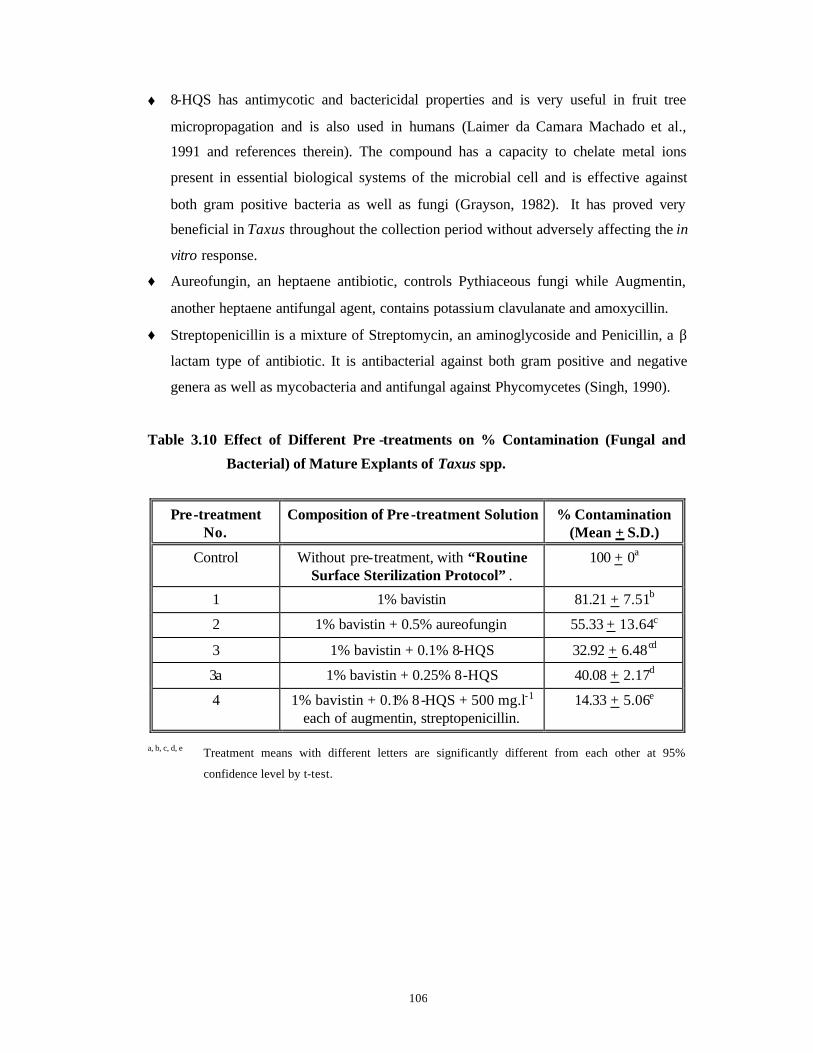
106
♦ 8-HQS has antimycotic and bactericidal properties and is very useful in fruit tree
micropropagation and is also used in humans (Laimer da Camara Machado et al.,
1991 and references therein). The compound has a capacity to chelate metal ions
present in essential biological systems of the microbial cell and is effective against
both gram positive bacteria as well as fungi (Grayson, 1982). It has proved very
beneficial in Taxus throughout the collection period without adversely affecting the in
vitro response.
♦ Aureofungin, an heptaene antibiotic, controls Pythiaceous fungi while Augmentin,
another heptaene antifungal agent, contains potassium clavulanate and amoxycillin.
♦ Streptopenicillin is a mixture of Streptomycin, an aminoglycoside and Penicillin, a β
lactam type of antibiotic. It is antibacterial against both gram positive and negative
genera as well as mycobacteria and antifungal against Phycomycetes (Singh, 1990).
Table 3.10 Effect of Different Pre -treatments on % Contamination (Fungal and
Bacterial) of Mature Explants of Taxus spp.
Pre-treatment No.
Composition of Pre -treatment Solution % Contamination (Mean + S.D.)
Control Without pre-treatment, with “Routine Surface Sterilization Protocol” .
100 + 0a
1 1% bavistin 81.21 + 7.51b
2 1% bavistin + 0.5% aureofungin 55.33 + 13.64c
3 1% bavistin + 0.1% 8-HQS 32.92 + 6.48cd
3a 1% bavistin + 0.25% 8-HQS 40.08 + 2.17d
4 1% bavistin + 0.1% 8-HQS + 500 mg.l-1 each of augmentin, streptopenicillin.
14.33 + 5.06e
a, b, c, d, e Treatment means with different letters are significantly different from each other at 95%
confidence level by t-test.

107
Use of the Quinolone group of antibiotics in the pre -treatment solution for Taxus
spp. Along with Antimicrobial Agents
Preliminary experiments showed that when the effective antibiotics, ofloxacin,
pefloxacin and ciprofloxacin, which were very specific for various identified bacteria
(Section 3.3.2.2, Table 3.9 ), were used in the pre-treatment solution without the anti-
fungal agents, all the cultures of Taxus spp. were overcome by the faster growing fungi
and the relatively slower growing bacteria did not appear. All the cultures were lost due to
fungal contamination (Table 3.11). Hence it was decided to use the identified effective
antibiotics at their bactericidal concentrations in the presence of antifungal agents. Since
the antibiotics to be used in the pre-treatment solution were for a shorter duration (as
compared to incorporation in the medium and continuous exposure of the explants to the
antibiotics), the antibiotics at ten times the effective bactericidal concentration determined
by liquid bioassays (Table 3.9 ) were used. To the optimized composition of pre-treatment
solution containing various antimicrobial agents (See Table 3.10, Treatment No. 4),
antibiotics such as 25 mg.l-1 each of ofloxacin, pefloxacin and ciprofloxacin were added,
based on the earlier results of optimization of MBC with solid and liquid Antibiogramme
assays (See Sections 3.3.2.1 and 3.3.2.2 of the present chapter for the details) to eliminate
bacterial contaminants of Taxus spp. cultures in vitro. Ofloxacin, Pefloxacin and
Ciprofloxacin are Quinolone antibiotics and inhibit the action of DNA gyrases (C. f.
Table 3.2).
The best pre-treatment solution (No. 6 in Table 3.11 ) comprising 1% bavistin,
0.1% 8-HQS, 500 mg.l-1 each of augmentin and streptopenicillin and 25 mg.l-1 each of
ofloxacin, pefloxacin and ciprofloxacin has proved to be very beneficial to control both
bacterial and fungal contamination of Taxus explants cultured throughout the collection
period of shoots (May-October) without adversely affecting the in vitro response. The
contamination percentage with this pr e-treatment solution, applied for a period of 16
hours followed by surface sterilization of mature explants of Taxus spp., could be
successfully reduced to 10%. This is significantly noteworthy compared to contamination
percentage obtained with and without pre-treatment employing various mixtures of anti-
fungal and anti-bacterial agents (Tables 3.10 and 3.11).
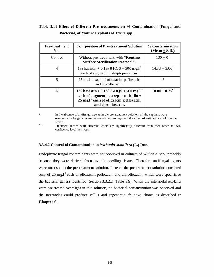
108
Table 3.11 Effect of Different Pre -treatments on % Contamination (Fungal and
Bacterial) of Mature Explants of Taxus spp.
Pre-treatment No.
Composition of Pre -treatment Solution % Contamination (Mean + S.D.)
Control Without pre-treatment, with “Routine Surface Sterilization Protocol” .
100 + 0a
4 1% bavistin + 0.1% 8-HQS + 500 mg.l-1 each of augmentin, streptopenicillin.
14.33 + 5.06b
5 25 mg.l-1 each of ofloxacin, pefloxacin and ciprofloxacin.
-*
6 1% bavistin + 0.1% 8-HQS + 500 mg.l -1 each of augmentin, streptopenicillin + 25 mg.l-1 each of ofloxacin, pefloxacin
and ciprofloxacin.
10.00 + 0.25c
* In the absence of antifungal agents in the pre-treatment solution, all the explants were
overcome by fungal contamination within two days and the effect of antibiotics could not be scored.
a, b, c Treatment means with different letters are significantly different from each other at 95% confidence level by t-test.
3.3.4.2 Control of Contamination in Withania somnifera (L.) Dun. Endophytic fungal contaminants were not observed in cultures of Withania spp., probably
because they were derived from juvenile seedling tissues. Therefore antifungal agents
were not used in the pre-treatment solution. Instead, the pre-treatment solution consisted
only of 25 mg.l-1 each of ofloxacin, pefloxacin and ciprofloxacin, which were specific to
the bacterial genera identified (Section 3.3.2.2, Table 3.9). When the internodal explants
were pre-treated overnight in this solution, no bacterial contamination was observed and
the internodes could produce callus and regenerate de novo shoots as described in
Chapter 6.

109
3.3.5 Control of phenolic browning/oxidation – the strategy
The effect of different chemical treatments on the percentage of explants of Taxus
spp. browning due to phenolic oxidation is presented in Table 3.12. All explants of Taxus
spp. died due to phenolic browning in the Control experiments when the explants were
not pre-treated with anti-oxidizing agents (Treatment No. 1 in Table 3.12 ). When the
explants were pre-treated overnight with various anti-oxidizing agents such as ascorbic
and citric acid, soluble PVP, each at 2%, the percentage incidence of phenolic oxidation
decreased significantly to 52% (Treatment No. 2 in Table 3.12). However the percentage
of explants browning due to phenolic oxidation increased significantly to 63.75% when
full strength liquid WPM was used for their culture, despite the use of pre-treatment
(Treatment No. 3 in Table 3.12).On the contrary, the percentage of explant browning
decreased significantly to 40% (Treatment No. 4 in Table 3.12) when WPM was used at
half strength of major salts (McCown and Sellmer, 1987). When AC at 0.5% was
incorporated in the nutrient medium (solid or liquid), the explants browning due to
phenolic oxidation was not observed (Treatment No. 5 in Table 3.12). AC probably
nullified the effect of phenolic oxidation on explants by adsorbing the polyphenols
released into the medium and accounted for healthier and faster growth of the shoots of
Taxus spp. (3 cm in 15 days). These results are in tune with the earlier observations
reported for Conifers (Thorpe et al., 1991). Thus the problem of phenolic oxidation in
Taxus cultures has been overcome completely by a treatment of explants with anti-
oxidizing agents for 16 hours at 26 + 2°C with constant shaking at 200 rpm in dark. This
strategy of pre-treatment of explants with anti-oxidizing agents has been further
combined to include the anti-bacterial and anti-fungal agents to control both the bacterial
and fungal contamination as well as phenolic browning observed with mature explants of
Taxus spp. in culture. For further details see Section 3.3.6 .
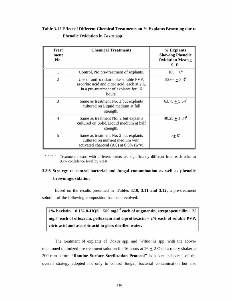
110
Table 3.12 Effect of Different Chemical Treatments on % Explants Browning due to
Phenolic Oxidation in Taxus spp.
Treatment No.
Chemical Treatments % Explants Showing Phenolic Oxidation Mean +
S. E.
1. Control, No pre-treatment of explants. 100 + 0a
2. Use of anti-oxidants like soluble PVP, ascorbic acid and citric acid, each at 2%,
in a pre-treatment of explants for 16 hours.
52.66 + 3.3b
3. Same as treatment No. 2 but explants cultured on Liquid medium at full
strength.
63.75 + 5.54c
4. Same as treatment No. 2 but explants cultured on Solid/Liquid medium at half
strength.
40.25 + 1.84d
5. Same as treatment No. 2 but explants cultured on nutrient medium with
activated charcoal (AC) at 0.5% (w/v).
0 + 0e
a, b, c, d, e Treatment means with different letters are significantly different from each other at
95% confidence level by t-test. 3.3.6 Strategy to control bacterial and fungal contamination as well as phenolic
browning/oxidation
Based on the results presented in Tables 3.10, 3.11 and 3.12 , a pre-treatment
solution of the following composition has been evolved:
1% bavistin + 0.1% 8-HQS + 500 mg.l-1 each of augmentin, streptopenicillin + 25
mg.l-1 each of ofloxacin, pefloxacin and ciprofloxacin + 2% each of soluble PVP,
citric acid and ascorbic acid in glass distilled water.
The treatment of explants of Taxus spp. and Withania spp. with the above-
mentioned optimized pre-treatment solution for 16 hours at 26 + 2ºC on a rotary shaker at
200 rpm before “Routine Surface Sterilization Protocol” is a part and parcel of the
overall strategy adopted not only to control fungal, bacterial contamination but also

111
phenolic oxidation of explants and is referred hereinafter as the “Modified Surface
Sterilization Protocol” .
A comparison of “Routine Surface Sterilization Protocol” and “Modified
Surface Sterilization Protocol” is presented here in Flow Chart 3.3.

112
Flow Chart 3.3 Routine and Modified Surface Sterilization Protocols for Stem and
Needle Explants of Taxus spp. and Internodal Explants of Withania
spp.
* Explants: Stem bits and needles of Taxus spp. and internodal explants of Withania spp.
** Pre-treatment solution for Taxus explants: 1% Bavistin + 0.25% 8-HQS + 500 mg.l-1 each of
streptopenicillin and augmentin + 25 mg.l- 1 each of ofloxacin, pefloxacin and ciprofloxacin +
2% each of soluble PVP, citric acid and ascorbic acid for 16 hours at 26 + 2ºC on a rotary
shaker at 200 rpm.
Pre-treatment solution for Withania explants: 25 mg.l- 1 each of ofloxacin, pefloxacin and
ciprofloxacin for 16 hours at 26 + 2ºC on a rotary shaker at 200 rpm.
Rinsing of explants* in liquid soap
(5 min.)
Washe
s with glass
distilled
6 Rinses in sterile glass
distilled water
Inoculation of explants
on optimized media
Rinses in glass distilled water (3 times each)
Optimized pre -treatment
solution** for 16 hours in glass distilled water
95% ethyl alcohol (1 min.)
0.1% HgCl2 (10 min. with
vacuum)
Rinses in glass distilled water (3 times each)
ROUTINE PROTOCOL
MODIFIED PROTOCOL
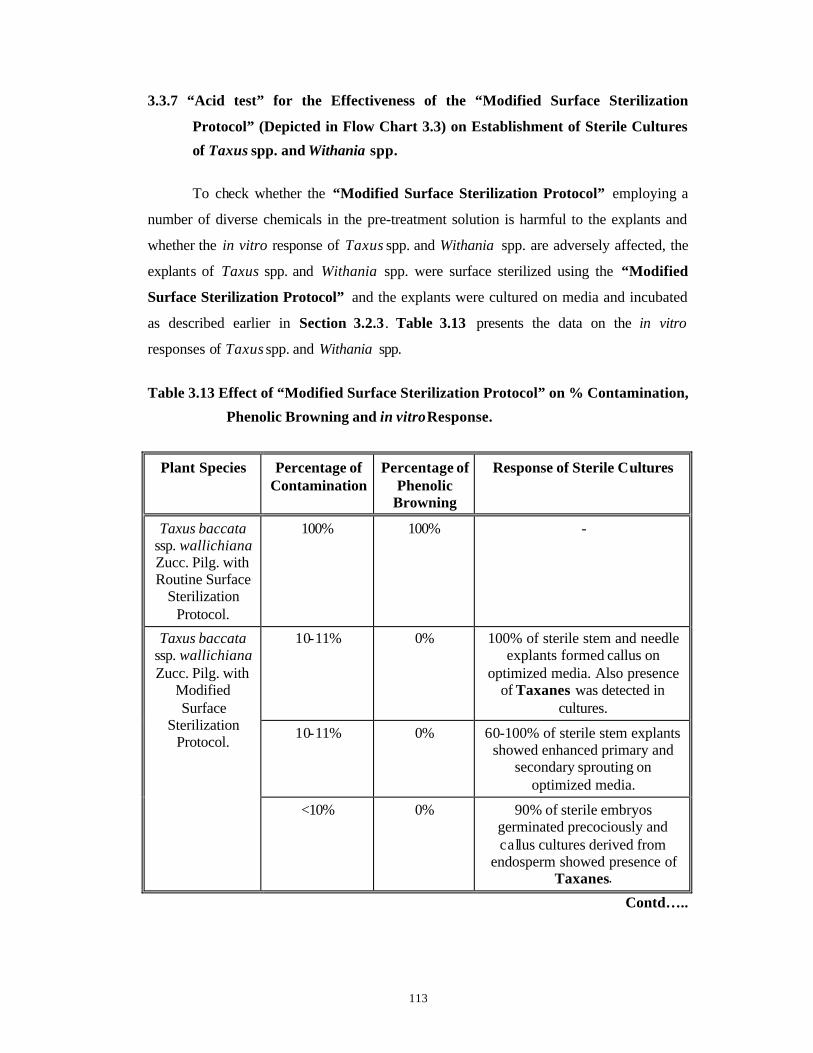
113
3.3.7 “Acid test” for the Effectiveness of the “Modified Surface Sterilization
Protocol” (Depicted in Flow Chart 3.3) on Establishment of Sterile Cultures
of Taxus spp. and Withania spp.
To check whether the “Modified Surface Sterilization Protocol” employing a
number of diverse chemicals in the pre-treatment solution is harmful to the explants and
whether the in vitro response of Taxus spp. and Withania spp. are adversely affected, the
explants of Taxus spp. and Withania spp. were surface sterilized using the “Modified
Surface Sterilization Protocol” and the explants were cultured on media and incubated
as described earlier in Section 3.2.3. Table 3.13 presents the data on the in vitro
responses of Taxus spp. and Withania spp.
Table 3.13 Effect of “Modified Surface Sterilization Protocol” on % Contamination,
Phenolic Browning and in vitro Response.
Plant Species Percentage of Contamination
Percentage of Phenolic
Browning
Response of Sterile Cultures
Taxus baccata ssp. wallichiana Zucc. Pilg. with Routine Surface
Sterilization Protocol.
100% 100% -
10-11% 0% 100% of sterile stem and needle explants formed callus on
optimized media. Also presence of Taxanes was detected in
cultures.
10-11% 0% 60-100% of sterile stem explants showed enhanced primary and
secondary sprouting on optimized media.
Taxus baccata ssp. wallichiana Zucc. Pilg. with
Modified Surface
Sterilization Protocol.
<10% 0% 90% of sterile embryos germinated precociously and callus cultures derived from
endosperm showed presence of Taxanes.
Contd…..
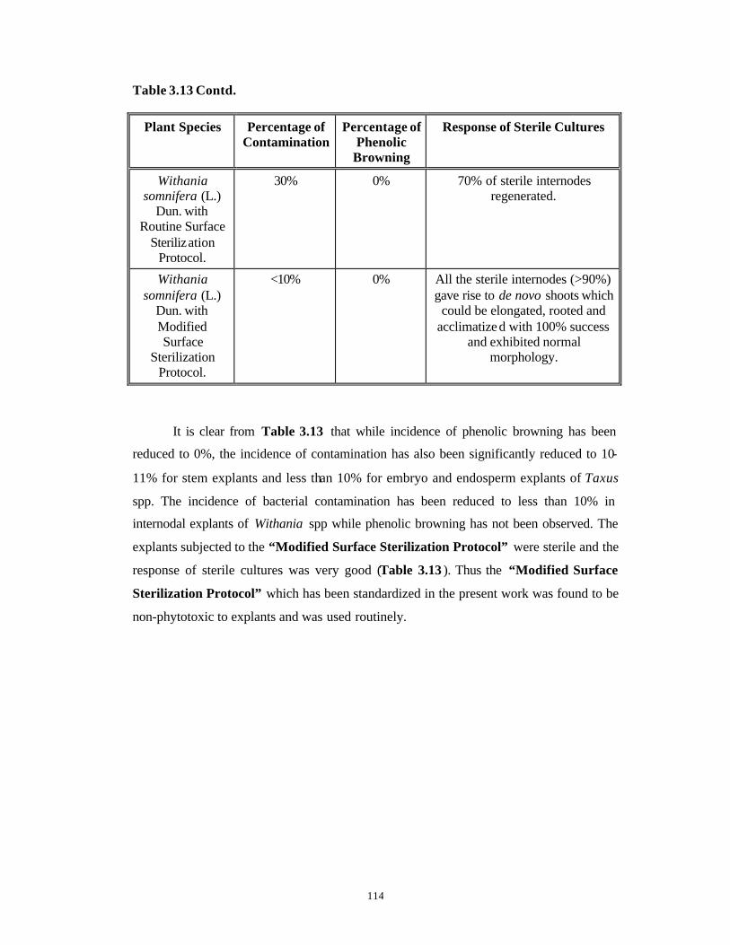
114
Table 3.13 Contd.
Plant Species Percentage of Contamination
Percentage of Phenolic
Browning
Response of Sterile Cultures
Withania somnifera (L.)
Dun. with Routine Surface
Sterilization Protocol.
30% 0% 70% of sterile internodes regenerated.
Withania somnifera (L.)
Dun. with Modified Surface
Sterilization Protocol.
<10% 0% All the sterile internodes (>90%) gave rise to de novo shoots which
could be elongated, rooted and acclimatize d with 100% success
and exhibited normal morphology.
It is clear from Table 3.13 that while incidence of phenolic browning has been
reduced to 0%, the incidence of contamination has also been significantly reduced to 10-
11% for stem explants and less than 10% for embryo and endosperm explants of Taxus
spp. The incidence of bacterial contamination has been reduced to less than 10% in
internodal explants of Withania spp while phenolic browning has not been observed. The
explants subjected to the “Modified Surface Sterilization Protocol” were sterile and the
response of sterile cultures was very good (Table 3.13 ). Thus the “Modified Surface
Sterilization Protocol” which has been standardized in the present work was found to be
non-phytotoxic to explants and was used routinely.
115
3.4 Conclusions
1) The prevalent bacterial contaminants of Taxus spp. and Withania spp.
belong to the genera such as Pseudomonas, Enterobacter-Erwinia
complex and Serratia spp.
2) The fungal contaminants of Taxus spp. and Withania spp. belong to the
class Ascomycota.
3) The bacterial and fungal contamination (endophytic as well as
occasional/opportunistic) associated with mature explants of Taxus spp.
and juvenile explants of Withania spp. can be effectively controlled with
the help of specific anti-microbial agents in a pre-treatment solution
such that no phytotoxicity problems could be encountered.
4) To control the bacterial contamination observed with internodal explants
of Withania spp., a 16 hour long pre-treatment of internodes with 25
mg.l-1 each of ofloxacin, pefloxacin and ciprofloxacin is adequate
without adversely affecting the regeneration capacity of the internodes.
Phytotoxic nature of the optimized pre-treatment on the explants was
not observed.
5) It is necessary to employ a 16 hour long pre-treatment in an aqueous
solution containing 1% bavistin + 0.1% 8-HQS + 500 mg.l -1 each of
augmentin, streptopenicillin + 25 mg.l-1 each of ofloxacin, pefloxacin and
ciprofloxacin to overcome the fungal and the bacterial contamination
associated with mature explants of Taxus spp., to obtain 90% sterile
cultures.
6) The phenolic browning of stem and needle explants of Taxus spp.
derived from mature trees can be controlled by incorporation of anti-
oxidants in the pre-treatment solution along with the antimicrobial
agents, use of half strength nutrient medium and use of activated
charcoal at 0.5% in the solid medium.
7) A pre-treatment solution containing 1% bavistin + 0.1% 8-HQS + 500
mg.l-1 each of augmentin and streptopenicillin + 25 mg.l-1 each of
ofloxacin, pefloxacin and ciprofloxacin + 2% each of soluble PVP, Citric
acid and Ascorbic acid is used for 16 hours to completely overcome

116
phenolic browning of mature explants of Taxus spp. and to obtain 90%
sterile cultures free from bacterial and fungal contaminants.
8) All the chemicals used for overcoming the problems of contamination
and phenolic browning were not phytotoxic as all the treated explants
had high success rate of in vitro response and the regenerants were
normal in morphology.

117
CHAPTER 4
Studies on Callus Induction from Various
Explants of Taxus spp. and Analysis of Taxane
Content of In Vitro and In Vivo Tissues.

118
4.1 Introduction
Research interest in Taxus spp. has become more prominent and pertinent in
recent years because it has been realized that plants belonging to Taxus spp. (Commonly
referred to as the Yews) contain a diverse group of phytochemicals showing a range of
biological activities (Parmar et al., 1999). Prominent among these are Taxane class of
diterpenoid compounds especially Taxol, 10 -DAB and Baccatin-III with antitumor and
anticancer activity in vivo as well as in vitro. This interest is amply evident by the fact
that many multinationals are joining the rat race, spending billions of dollars to develop
the product (Taxol) either by extraction from bark of trees of Taxus spp. or by the method
of semi-synthesis from naturally occurring precursors such as 10-DAB and Baccatin-III.
The phylogenetic position of the yew family, Taxaceae (Order: Coniferales,
Class: Gymnosperms ) has been very controversial (Sahni, 1920; Chaw et al., 1993). The
family includes five genera and twenty species characterized by presence of a single
ovule, not organized in seed cones, as opposed to other conifers. The name Taxus is
derived from its Greek name toxus, which is made from two words: toxon meaning bow
and toxicon meaning poison. Most authors consider Taxus to contain eight species: Taxus
baccata L., Taxus brevifolia Nutt., Taxus canadensis Marshall, Taxus chinensis (Pilger)
Rehder, Taxu s cuspidata Sieb. et Zucc., Taxus floridana Chapm., Taxus globosa Schltdl.,
Taxus wallichiana Zucc. Furthermore, there are two hybrids: Taxus x media Rehder is a
cross between T. baccata and T. cuspidata while Taxus x hunnewelliana Rehder is a cross
between T. cuspidata and T. canadensis (Van Rozendaal et al., 1999).
Today there is an increased tendency to consider various Taxus species as
intraspecific and geographic variants of a single species T. baccata, descending from
Paleotaxus rediviva, a fossil tree abundant in the Triassic, 200 million years ago; since all
the species have identical chromosome numbers of 2n=24 and they readily undergo
hybridization with each other (Hartzell, 1991).

119
4.1.1 Distribution Range and Botany of the Himalayan Yew (Saxena and Sarbhai,
1971-72; Dubey, 1997; Chatterjee and Dey, 1997;
http://willow.ncfes.umn.edu/silvics_manual/Volume_1/)
The Himalayan region in India extends for about 2500 km and occupies 18% of
the total area of this country. It is divided into different provinces physiographically and
biogeographically. The forest cover in the entire area has dwindled to about 21% (Bisht et
al., 1998 and references therein). This is viewed with concern because the Himalayas are
one of the chief centers of biodiversity and contain an immense wealth of flora and fauna,
with many endemic genera, which has not been properly documented. Tissue culture can
serve as an important tool in saving many of these endangered plant genera.
The Himalayan Yew, Taxus baccata ssp. wallichiana Zucc. Pilg., also referred to
as Taxus wallichiana Zucc. as well as Taxus baccata L. in some publications, is one such
endangered plant in the Himalayas, occurring at elevations of 1,800 m-3,000 m in the
evergreen and coniferous forests. Even now, there is no unanimity amongst the botanists
about these names. In the present thesis, the name Taxus baccata ssp. wallichiana
Zucc. Pilg. (Dr. S. Bhattacharya, personal communication) has been used
throughout. Similarly the name Taxol is used instead of Paclitaxe l, due to its better
familiarity.
In the earlier days, the wood of these plants was used for making bows, bedsteads
and other wooden articles of daily use. The bark was used for procuring a red dye,
important in religious ceremonies and for making tea. In addition, the trees were also used
for medicinal purposes (Hartzell, 1991).
The plants grow in well-drained soils and can tolerate deep shade and moist
habitats. Usually they form the understory component in a broad-leaved (Quercus spp.,
Rhododendron spp., Ace r spp.) or Conifer (Abies spp., Cupressus spp.) forests. But the
trees rarely grow in clusters or as pure strands (Dubey, 1997; Chatterjee and Dey, 1997).
The trees have a wide range of adaptability and can grow even under exposed conditions
after overstory removal, by undergoing a number of morphological and anatomical
changes like increase in the quantity of photosynthetic tissue, establishment of multi-
layered branch arrangement etc. Also the strobilus production is shown to be positively
120
associated with overstory openness with respect to light and microenvironment rather
than with branch mass or branch bifurcations (DiFazio et al., 1997).
The Himalayan Yew is an evergreen, slow-growing and long-living tree or shrub.
Whether the differences in size and form are genetic traits or the results of environment
and stand history is not known. The root system is deep and wide spreading. The roots
display a diarch structure and the tracheids display spiral thickenings.
It has wide spreading horizontal br anches (for catching maximum, available light)
forming a canopy, unlike other conifers. The branches have only unlimited growth. There
are no short shoots as in Pinus spp. The stems show a number of anatomical peculiarities
like absence of resin canals, small cortex and pith as compared to the vascular cylinder,
xylem tracheids with opposite bordered-pits and spiral thickenings, absence of xylem
parenchyma in the wood, uniseriate medullary rays, conjoint, collateral, endarch and open
vascular bundles and abundant secondary growth.
Unlike Pinus, the needles are not dimorphic. They are 2.5-3.8 cm long, linear,
flattened, distichous, alternate, acute, narrowed in a short petiole decurrent along the twig,
dark green and shining above and pale yellowish-brown or rusty-red below. The needles
are shed in May-June. Anatomically the needles show a dorsiventral structure where the
mesophyll is well differentiated into an upper pallisade and a lower spongy layer. There is
a single vascular bundle signifying the mid-rib. A number of xerophytic structures like
sunken stomata restricted to the lower epidermis, a thick cuticle above the upper
epidermis and a continuous upper epidermis and a discontinuous lower epidermis are
present.
The Himalayan Yew is dioecious where ma le and female trees are identical but
distinct. Male strobili appear from March to May in the axils of leaves of the previous
year, on the lower side of the branches, in subglobose and solitary catkins. They are
stalked, pale yellow with a dozen, spirally arranged, basal bract scales. 6-12 filamentous
stamens/microsporophylls are present at the top, which bear 5-9 anthers/microsporangia.
The sperms in Taxus spp. have no cell membrane or cell wall and hence are not cells.
Instead the sperms are nuclei with associated cytoplasm and organelles from the
spermatogenous cell (Anderson and Owens, 1999).
121
There is no female cone in Taxus spp. It is very much reduced and is represented
by a single erect ovule surrounded by four imbricate bracts. These solitary structures arise
in the axils of the leaves of the previous year on the underside of the branches. Absence
of seed cones, presence of a rudimentary pollen chamber, absence of a ventral canal cell
in the archegonia, a short free nuclear stage (16-32 free nuclei) and a short period of
gametophyte and embryo development can be taken as the advanced evolutionary
features of Taxus spp. (Brukhin and Bozhkov, 1996). Wind pollination and fertilization
occur in the same year and fertilization occurs two months after pollination. The fruits
ripen from August/September to November of the same year. The fruit comprises of a
single, olive-green seed, 8 mm long, almost completely enclosed by a juicy red aril
developing from the enlarged disc-like base of the ovule. The embryo ha s two cotyledons.
The seeds are shed in the same year, approximately 12 months after ovule initiation. The
seeds have a dormancy requirement of two years before natural germination can occur.
4.1.2 Vegetative Propagation of Taxus spp.
Normally, vegetative propagation is achieved through cuttings. The percentages of
rooting varied between cultivars and species depending on temperature, misting, use of
IBA. 63-100% rooting was observed with many species and cultivars of Taxus spp.
(Eccher, 1988). Chee (1995a) reported that stem cuttings of T. cuspidata treated with an
aqueous solution of IBA (0.2%) + NAA (0.1%) + Thiamine (0.08%) produced roots at a
frequency of 73.5%. However the success rate was not very high for the Himalayan Yew
(20-30%) and the cuttings took at least three months to root. Success of grafting and
layering was also not very encouraging (Chatterjee and Dey, 1997).
4.1.3 Development of Taxol and Related Class of Taxane Molecules from Taxus spp.
Plants belonging to Taxus spp. have been investigated since 1856, mostly for the
poisonous alkaloids present in various plant parts. But the intensive study of the plants of
this genus started in the early 1960s, when the National Cancer Institute (NCI) at USA
began an intensive screening program for plant compounds with anti-cancer activities.
Taxol was found to be one of the most promising anticancer agents, among the 120,000
compounds screened. After 1969 (elucidation of structure of Taxol), these studies
diversified into biochemical, biotechnological and clinical arenas. Important milestones in
this long and illustrious journey are tabulated in Table 4.1.
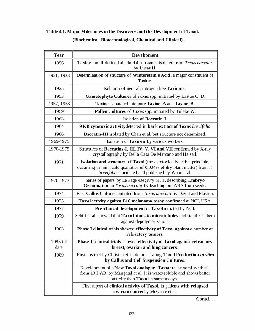
122
Table 4.1. Major Milestones in the Discovery and the Development of Taxol.
(Biochemical, Biotechnological, Chemical and Clinical).
Year Development
1856 Taxine , an ill-defined alkaloidal substance isolated from Taxus baccata by Lucas H.
1921, 1923 Determination of structure of Winterstein’s Acid, a major constituent of Taxine .
1925 Isolation of neutral, nitrogen-free Taxinine .
1953 Gametophyte Cultures of Taxus spp. initiated by LaRue C. D.
1957, 1958 Taxine separated into pure Taxine -A and Taxine -B .
1959 Pollen Cultures of Taxus spp. initiated by Tuleke W.
1963 Isolation of Baccatin-I.
1964 9 KB cytotoxic activity detected in bark extract of Taxus brevifolia
1966 Baccatin-III isolated by Chan et al. but structure not determined.
1969-1975 Isolation of Taxusin by various workers.
1970-1975 Structures of Baccatins -I, III, IV, V, VI and VII confirmed by X-ray crystallography by Della Casa De Marcano and Halsall.
1971 Isolation and structure of Taxol (the cytotoxically active principle, occurring in miniscule quantities of 0.004% of dry plant matter) from T.
brevifolia elucidated and published by Wani et al.
1970-1973 Series of papers by Le Page -Degivry M. T. describing Embryo Germination in Taxus baccata by leaching out ABA from seeds.
1974 First Callus Culture initiated from Taxus baccata by David and Plastira.
1975 Taxol activity against B16 melanoma assay confirmed at NCI, USA.
1977 Pre-clinical development of Taxol initiated by NCI.
1979 Schiff et al. showed that Taxol binds to microtubules and stabilizes them against depolymerization.
1983 Phase I clinical trials showed effectivity of Taxol against a number of refractory tumors.
1985-till date
Phase II clinical trials showed effectivity of Taxol against refractory breast, ovarian and lung cancers.
First abstract by Christen et al. demonstrating Taxol Production in vitro by Callus and Cell Suspension Cultures.
Development of a New Taxol analogue : Taxotere by semi-synthesis from 10 DAB, by Mangatal et al. It is water-soluble and shows better
activity than Taxol in some assays.
1989
First report of clinical activity of Taxol, in patients with relapsed ovarian cancer by McGuire et al.
Contd…..
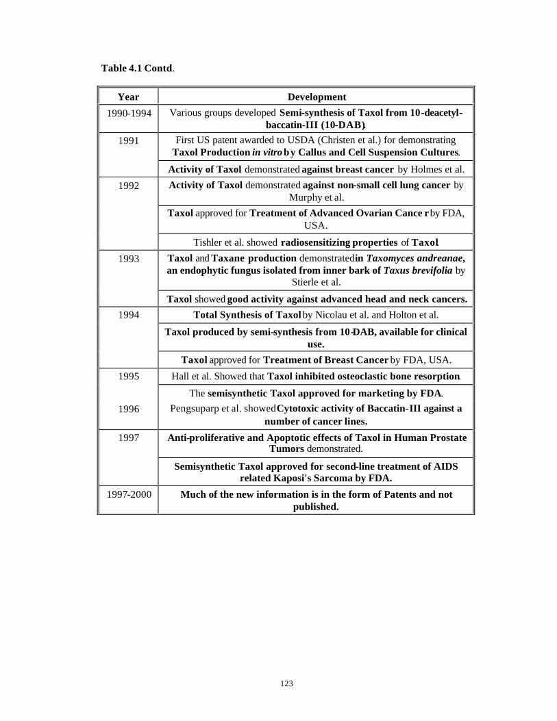
123
Table 4.1 Contd.
Year Development
1990-1994 Various groups developed Semi-synthesis of Taxol from 10-deacetyl-baccatin-III (10-DAB).
First US patent awarded to USDA (Christen et al.) for demonstrating Taxol Production in vitro by Callus and Cell Suspension Cultures.
1991
Activity of Taxol demonstrated against breast cancer by Holmes et al.
Activity of Taxol demonstrated against non-small cell lung cancer by Murphy et al.
Taxol approved for Treatment of Advanced Ovarian Cance r by FDA, USA.
1992
Tishler et al. showed radiosensitizing properties of Taxol.
Taxol and Taxane production demonstrated in Taxomyces andreanae, an endophytic fungus isolated from inner bark of Taxus brevifolia by
Stierle et al.
1993
Taxol showed good activity against advanced head and neck cancers.
Total Synthesis of Taxol by Nicolau et al. and Holton et al.
Taxol produced by semi-synthesis from 10-DAB, available for clinical use.
1994
Taxol approved for Treatment of Breast Cancer by FDA, USA.
Hall et al. Showed that Taxol inhibited osteoclastic bone resorption. 1995
The semisynthetic Taxol approved for marketing by FDA.
1996 Pengsuparp et al. showed Cytotoxic activity of Baccatin-III against a number of cancer lines.
Anti-proliferative and Apoptotic effects of Taxol in Human Prostate Tumors demonstrated.
1997
Semisynthetic Taxol approved for second-line treatment of AIDS related Kaposi's Sarcoma by FDA.
1997-2000 Much of the new information is in the form of Patents and not published.
124
4.1.4 Taxane Class of Molecules
The chemical constituents of different Taxus spp. belong to alkaloids, diterpenoids
with Taxane skeleton, lignans, biflavonoids, steroids, sugar derivatives, and diterpenoids
with tropane skeletons. These various compounds exhib it diverse functions such as
anticancer, antitumor and antileukemic Taxanes, moulting hormones, toxic alkaloids,
antineoplastic and antifungl diterpenes and so on (Young et al., 1992; Parmar et al., 1999
and references therein). Because of this, plants of this genus have been intensively studied
in the recent years.
Upto 1999, over 350 taxoids have been isolated and characterized. The basic
structure for most of the taxoids consists of a pentamethyl tricyclopentadecane skeleton,
which is often called as the Normal Taxane Skeleton (Fig. 4.1). In addition, a number of
Taxanes with rearrangements in this basic skeleton are also known (See the review by
Baloglu and Kingston, 1999 for details). Out of all these Taxanes, clinically and/or
chemically the most important Taxanes are: Taxol/Paclitaxel, 10-deacetylTaxol,
Cephalomannine , Baccatin-III , 10-deacetylbaccatin-III (10-DAB ) and Taxol C (C. f.
Table 4.1 ). There are a number of Taxol-like derivatives derived from Baccatin -III,
esterified at C-13 with phenylisoserines having different N-acyl and N-alkyl groups,
which are collectively known as ‘Taxols’. Five major structural types of Taxols: Taxols
A-D and Taxuspinanane are known till date. Overall 28 natural Taxols have been
reported from different Taxus spp. (Das et al., 1999 and references therein). These
compounds possess an unusual oxetane ring at C-5, C-20 and a phenylisoserine side chain
at C-13, both of which are necessary for the anticancer and other activities of Taxol. Fig.
4.2 depicts the structures of some of the important taxoids.
The contents of Taxanes in plants of Taxus spp. are known to vary based on
geographical locations, environmental and agricultural influences, harvesting and storage
conditions (Vidensek et al., 1990; Kelsey and Vance, 1992; Wheeler et al., 1992; ElSohly
et al., 1994). The average yield of Taxol (the most important and efficient anti-cancer
agent identified till date) in bark of Taxus brevifolia ranges between 0.014-0.017%
(Vidensek et al., 1990; Patel, 1998) and 16,000 lb. bark is needed to produce 1 kg purified
Taxol (Patel, 1998 and references therein). Other species of Taxus are reported to contain
Taxol in the range of 0.001-0.0832 mg.g-1 DW (Arteca and Wickremesinhe, 1993).
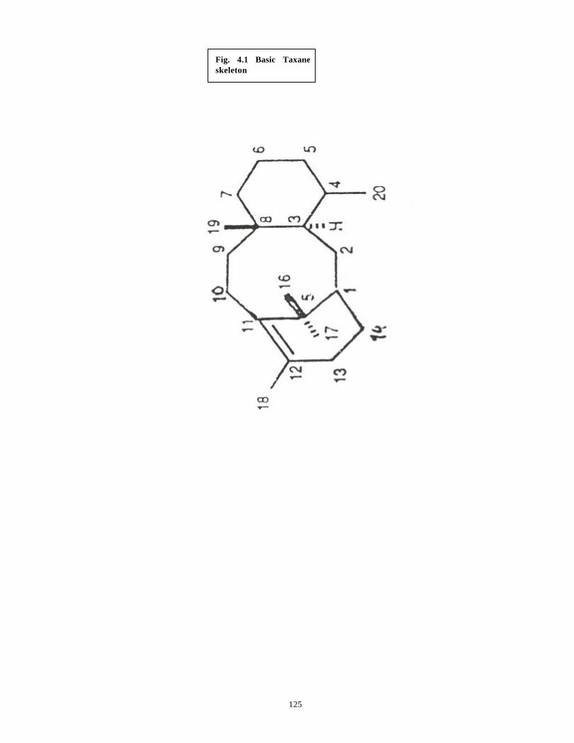
125
Fig. 4.1 Basic Taxane skeleton
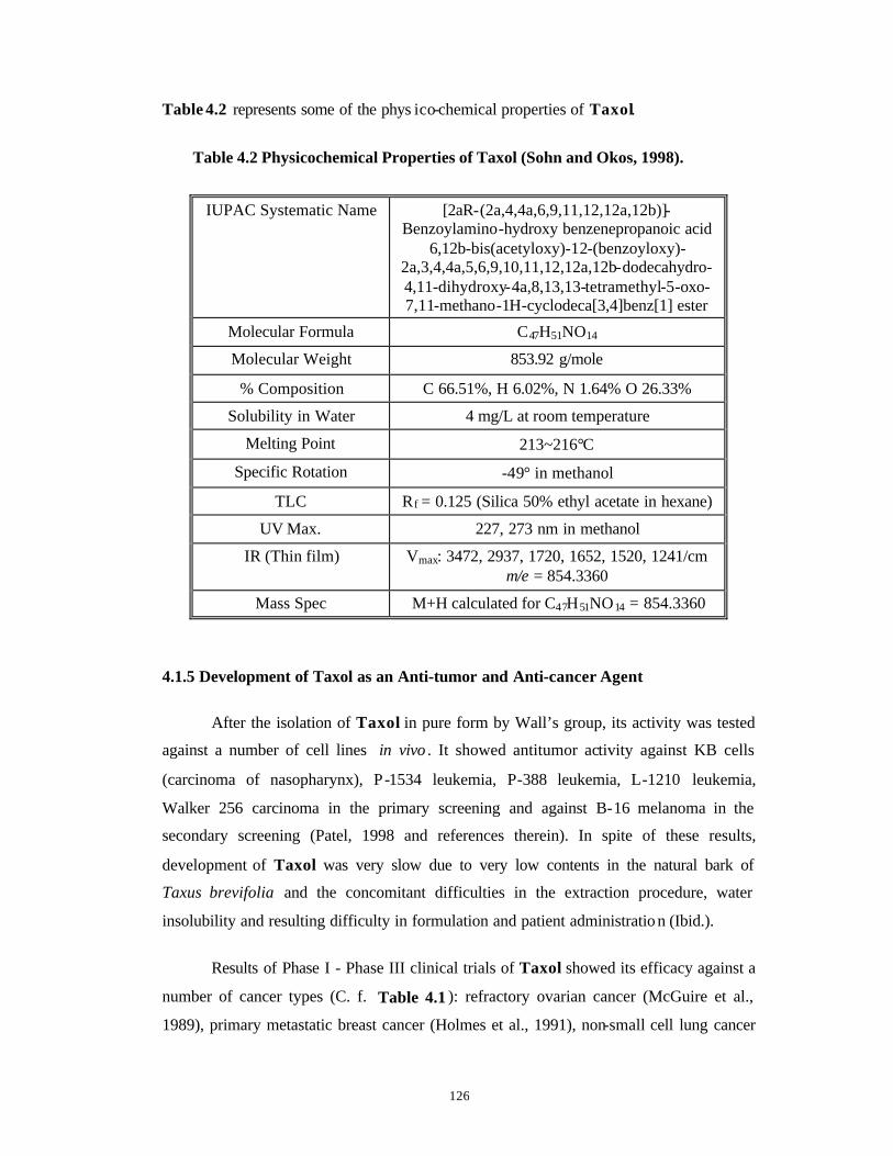
126
Table 4.2 represents some of the phys ico-chemical properties of Taxol.
Table 4.2 Physicochemical Properties of Taxol (Sohn and Okos, 1998).
IUPAC Systematic Name [2aR-(2a,4,4a,6,9,11,12,12a,12b)]-Benzoylamino-hydroxy benzenepropanoic acid
6,12b-bis(acetyloxy)-12-(benzoyloxy)-2a,3,4,4a,5,6,9,10,11,12,12a,12b-dodecahydro-4,11-dihydroxy-4a,8,13,13-tetramethyl-5-oxo-7,11-methano-1H-cyclodeca[3,4]benz[1] ester
Molecular Formula C47H51NO14
Molecular Weight 853.92 g/mole
% Composition C 66.51%, H 6.02%, N 1.64% O 26.33%
Solubility in Water 4 mg/L at room temperature
Melting Point 213~216°C
Specific Rotation -49° in methanol
TLC Rf = 0.125 (Silica 50% ethyl acetate in hexane)
UV Max. 227, 273 nm in methanol
IR (Thin film) Vmax: 3472, 2937, 1720, 1652, 1520, 1241/cm m/e = 854.3360
Mass Spec M+H calculated for C47H51NO14 = 854.3360
4.1.5 Development of Taxol as an Anti-tumor and Anti-cancer Agent
After the isolation of Taxol in pure form by Wall’s group, its activity was tested
against a number of cell lines in vivo . It showed antitumor activity against KB cells
(carcinoma of nasopharynx), P-1534 leukemia, P-388 leukemia, L-1210 leukemia,
Walker 256 carcinoma in the primary screening and against B-16 melanoma in the
secondary screening (Patel, 1998 and references therein). In spite of these results,
development of Taxol was very slow due to very low contents in the natural bark of
Taxus brevifolia and the concomitant difficulties in the extraction procedure, water
insolubility and resulting difficulty in formulation and patient administratio n (Ibid.).
Results of Phase I - Phase III clinical trials of Taxol showed its efficacy against a
number of cancer types (C. f. Table 4.1 ): refractory ovarian cancer (McGuire et al.,
1989), primary metastatic breast cancer (Holmes et al., 1991), non-small cell lung cancer

127
(Murphy et al., 1992) etc. Taxol is also combined with a number of other drugs such as
Doxorubicin (O'Shaughnessy et al., 1994) and Navelbine/Vinorelbine (Chang and
Garrow, 1995) to increase its efficacy against various types of cancers.
Taxol is shown to inhibit growth of parasites such as Plasmodium falciparum,
Trypanosoma and Physarum (Schrével et al., 1994 and references therein). Taxol inhibits
osteoclastic bone resorption (Hall et al., 1995) and acts as a radiation sensitizer (Tishler et
al., 1992).
4.1.6 Taxol-Tubulin Interactions and Mode of Action of Taxol
Taxol binds to and stabilizes both the 2-dimensional zinc sheets of tubulin as well
as the 3-dimensional microtubule against low temperature and alkalinity-induced
depolymerization and polymerization can proceed even in the absence of guanidine
triphosphate (GTP) and microtubule associated proteins (MAPs). Taxol promotes both
the nucleation and elongation phases of polymerization and can reduce the critical
concentration of tubulin to almost zero (Patel, 1998 and reference therein). Taxol binds
into a pocket in the second globular domain of β tubulin, facing the central hole in the
microtubule. Here it makes close contact with the shorter S9-S10 loop, the core helix and
the M loop between S7 and H9 (Fig. 4.3, adapted from Amos and Löwe, 1999).
Mutations in this loop reduce the toxicity of Taxol to cancer cells.
4.1.7 Mode of Action of Taxol
Taxol stabilizes the microtubules against depolymerization and hence cells are
arrested at the G2-M phase of cell division and cell division cannot proceed.
Because of the high efficacy of Taxol, it is believed that mitotic inhibition can not
be the sole process responsible for its clinical activity and tubulin can not be its only
target. Following alternatives have been suggested:
1. Taxol also binds to a protein: Bcl-2, which normally blocks the process of cell death
or apoptosis. When Taxol binds to Bcl-2, apoptosis is allowed to proceed (Fang et al.,
1998).
2. Another mechanism suggested for Taxol-induced apoptosis is a phosphorylation
pathway possibly involving the p34cdc2 kinase (Donaldson et al., 1994).
128
3. Taxol is believed to induce cytokines via signal transduction pathways and has been
shown to transcriptionally activate expression of a number of genes. The C-1 to C-4
positions on the Taxol molecule appear to be important (Watson et al., 1998 and
references therein).
4. Taxol is also shown to activate other regulatory activities such as induction of MAP
kinases, induction of protein-tyrosine phosphorylation, tumor necrosis factor (TNF)
and interleukin (IL-1) etc. (Watson et al., 1998 and references therein).
5. Taxol is a potent platelet antiaggregant (Canizares et al., 1997).
4.1.8 Rationale for Use of Plant Tissue and Cell Cultures of Taxus spp. for
Production of Taxane Class of Molecules
It has become imperative to find alternative sources for Taxol production due to
the dwindling natural resources of Taxus spp. because of their overexploitation for
Taxane extraction from bark, stem and needles. Naturally, the plants belonging to Taxus
spp. never form the major component of any forest (Dr. O. Singh, Personal
communication) and due to overexploitation; these plants have attained an endangered
status.
1) Normally, synthetic chemistry proves to be an important tool in such endeavors as the
molecules can be synthesized totally in the laboratory. But in case of Taxol, it has
proved to be an uphill task for synthetic chemists. The two total syntheses reported so
far (Nicolau et al., 1994; Holton et al. , 1994) employ more than 35 steps and hence
cannot be used for economical production of Taxol.
2) Some methods of semi-synthesis of Taxol from 10 DAB -III or Baccatin-III (Taxol
precursors found in large quantities in T. baccata and T. wallichiana) by attachment
of side chain also have been proposed (Denis et al., 1988; Lavelle et al., 1993). These
methods may prove to be viable alternatives for Taxol production.
3) Stierle et al. (1993) reported the production of Taxol (25-50 ng/L) by an endophytic
fungus Taxomyces andreanae isolated from an identified tree of T. brevifolia. It
appears unlikely that strain improvement by mutagenesis or genetic engineering will
result in spectacular yield enhancement necessary for bioreactor/fermentor scale-up
(Patel, 1998).
4) Sinc e vegetative propagation of Taxus spp. by use of cuttings has not yielded
significant results (Section 4.1.2), especially for the Himalayan Yews, use of plant
129
tissue culture methods seems to be the best alternative amongst various approaches as
outlined below.
Plant tissue culture methods have proved to be an important alternative wherein a
number of Taxus spp. have shown production of Taxane class of compounds in vitro (C.
f. Table 4.3 ). Such cultures can be scaled up for large-scale production of Taxanes.
Cultures producing Taxol can be used for direct scale-up. Even cultures producing Taxol
precursors like 10 DAB -III and Baccatin III can be scaled up for their subsequent use in
semi-synthetic production of Taxol. Also these Taxol precursors by themselves have
antitumor/cytotoxic properties like Taxol and hence can be used independently
(Pengsuparp et al., 1996). Natural, field-grown populations of Taxus spp. plants show
considerable seasonal, environmental and epigenetic variation in Taxane contents
(Kelsey and Vance, 1992; Wheeler et al., 1992). Using defined conditions in a cell culture
system, Taxane production can be closely controlled to supply consistent product quality.
Cell cultures could eliminate the need to use the limited natural resources of Taxus spp.,
thus preserving the largely unexplored genetic diversity within the genera for selecting
high yielding variants for cell culture process (Gibson et al., 1995). The cell cultures can
also be used for selection of elite variants and for elicitation, immobilization and
permeabilization studies as well as for genetic manipulations. They can also be used for
elucidating the biosynthetic pathway of Taxol so that very specific manipulations for
improving Taxol yields could be carried out. In addition, the rate limiting steps can be
identified and can be modified by genetic manipulations (Wildung and Croteau, 1996;
Hefner et al., 1996; Pennington et al., 1998). Table 4.3 presents various studies carried
out so far in the establishment of undifferentiated callus and cell cultures of Taxus spp.
and production of Taxol therefrom. The table also lists genetic transformation studies
with Agrobacterium tumefaciens and A. rhizogenes.
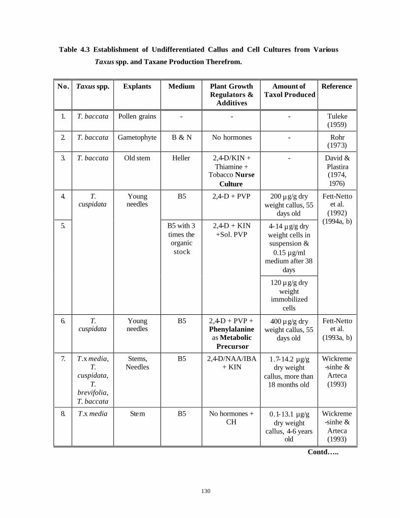
130
Table 4.3 Establishment of Undifferentiated Callus and Cell Cultures from Various
Taxus spp. and Taxane Production Therefrom.
No. Taxus spp. Explants Medium Plant Growth Regulators &
Additives
Amount of Taxol Produced
Reference
1. T. baccata Pollen grains - - - Tuleke (1959)
2. T. baccata Gametophyte B & N No hormones - Rohr (1973)
3. T. baccata Old stem Heller 2,4-D/KIN + Thiamine +
Tobacco Nurse Culture
- David & Plastira (1974, 1976)
4. B5 2,4-D + PVP 200 µg/g dry weight callus, 55
days old
4-14 µg/g dry weight cells in suspension &
0.15 µg/ml medium after 38
days
5.
T. cuspidata
Young needles
B5 with 3 times the organic stock
2,4-D + KIN +Sol. PVP
120 µg/g dry weight
immobilized cells
Fett-Netto et al.
(1992) (1994a, b)
6. T. cuspidata
Young needles
B5 2,4-D + PVP + Phenylalanine as Metabolic
Precursor
400 µg/g dry weight callus, 55
days old
Fett-Netto et al.
(1993a, b)
7. T.x media, T.
cuspidata, T.
brevifolia, T. baccata
Stems, Needles
B5 2,4-D/NAA/IBA + KIN
1.7-14.2 µg/g dry weight
callus, more than 18 months old
Wickreme-sinhe & Arteca (1993)
8. T.x media Stem B5 No hormones + CH
0.1-13.1 µg/g dry weight
callus, 4-6 years old
Wickreme-sinhe & Arteca (1993)
Contd…..
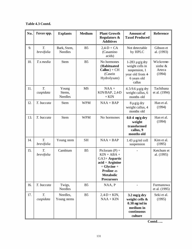
131
Table 4.3 Contd.
No. Taxus spp. Explants Medium Plant Growth Regulators &
Additives
Amount of Taxol Produced
Reference
9. T. brevifolia
Bark, Stem, Needles
B5 2,4-D + CA (Casamino
acids)
Not detectable by HPLC
Gibson et al. (1993)
10. T.x media Stem B5 No hormones (Habituated Callus ) + CH
(Casein Hydrolysate)
1-283 µg/g dry weight cells in suspension, 1
year old from 4-6 years old
callus
Wickreme-sinhe & Arteca (1994)
11. T. cuspidata
Young Stems,
Needles
MS NAA + KIN/BAP, 2,4-D
+ KIN
4.5-9.6 µg/g dry weight callus, 6
months old
Tachibana et al. (1994)
12. T. baccata Stem WPM NAA + BAP 8 µg/g dry weight callus, 4
months old
Han et al. (1994)
13. T. baccata Stem WPM No hormones 0.8 -4 µg/g dry weight
transformed callus, 9
months old
Han et al. (1994)
14. T. brevifolia
Young stem SH NAA + BAP 1.43 µg/ml cell suspension
Kim et al. (1995)
15. T. brevifolia
Cambium B5 Picloram (P) + KIN + ABA +
GA3 + Aspartic acid + Arginine
+ Glycine + Proline as Metabolic Precursors
- Ketchum et al. (1995)
16. T. baccata Twigs, Needles
B5 NAA, P - Furmanowa et al. (1995)
17. T. cuspidata
Needles, Young stems
B5 2,4-D + KIN, NAA + KIN
3.2 mg/g dry weight cells & 0.38 µg/ml in medium in continuous
culture
Seki et al. (1995)
Contd…..
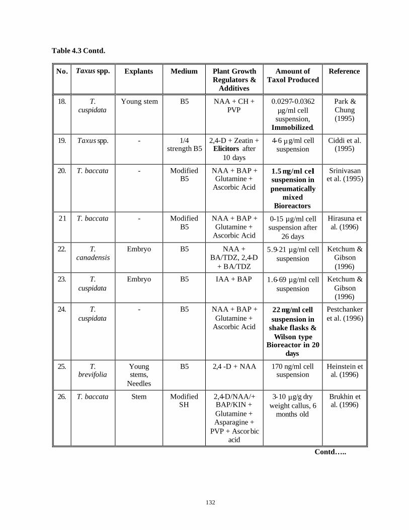
132
Table 4.3 Contd.
No. Taxus spp. Explants Medium Plant Growth Regulators &
Additives
Amount of Taxol Produced
Reference
18. T. cuspidata
Young stem B5 NAA + CH + PVP
0.0297-0.0362 µg/ml cell
suspension, Immobilized.
Park & Chung (1995)
19. Taxus spp. - 1/4 strength B5
2,4-D + Zeatin + Elicitors after
10 days
4-6 µg/ml cell suspension
Ciddi et al. (1995)
20. T. baccata - Modified B5
NAA + BAP + Glutamine +
Ascorbic Acid
1.5 µg/ml cell suspension in pneumatically
mixed Bioreactors
Srinivasan et al. (1995)
21 T. baccata - Modified B5
NAA + BAP + Glutamine +
Ascorbic Acid
0-15 µg/ml cell suspension after
26 days
Hirasuna et al. (1996)
22. T. canadensis
Embryo B5 NAA + BA/TDZ, 2,4-D
+ BA/TDZ
5.9-21 µg/ml cell suspension
Ketchum & Gibson (1996)
23. T. cuspidata
Embryo B5 IAA + BAP 1.6-69 µg/ml cell suspension
Ketchum & Gibson (1996)
24. T. cuspidata
- B5 NAA + BAP + Glutamine +
Ascorbic Acid
22 µg/ml cell suspension in
shake flasks & Wilson type
Bioreactor in 20 days
Pestchanker et al. (1996)
25. T. brevifolia
Young stems,
Needles
B5 2,4 -D + NAA 170 ng/ml cell suspension
Heinstein et al. (1996)
26. T. baccata Stem Modified SH
2,4-D/NAA/+ BAP/KIN + Glutamine + Asparagine +
PVP + Ascorbic acid
3-10 µg/g dry weight callus, 6
months old
Brukhin et al. (1996)
Contd…..
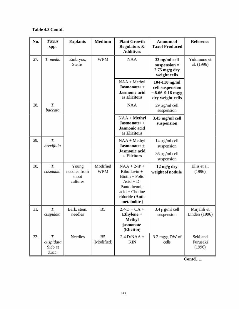
133
Table 4.3 Contd.
No. Taxus spp.
Explants Medium Plant Growth Regulators &
Additives
Amount of Taxol Produced
Reference
NAA 33 µg/ml cell suspension = 2.75 mg/g dry weight cells
27. T. media
NAA + Methyl Jasmonate/ + Jasmonic acid
as Elicitors
104-110 µg/ml cell suspension
= 8.66 -9.16 mg/g dry weight cells
NAA 29 µg/ml cell suspension
28. T. baccata
NAA + Methyl Jasmonate/ + Jasmonic acid
as Elicitors
3.45 mg/ml cell suspension
14 µg/ml cell suspension
29. T. brevifolia
Embryos, Stems
WPM
NAA + Methyl Jasmonate/ + Jasmonic acid
as Elicitors 36 µg/ml cell suspension
Yukimune et al. (1996)
30. T. cuspidata
Young needles from
shoot cultures
Modified WPM
NAA + 2-iP + Riboflavin + Biotin + Folic
Acid + D-Pantothennic
acid + Choline chloride (Anti-
metabolite )
12 µg/g dry weight of nodule
Ellis et al. (1996)
31. T. cuspidata
Bark, stem, needles
B5 2,4-D + CA + Ethylene +
Methyl jasmonate (Elicitor)
3.4 µg/ml cell suspension
Mirjalili & Linden (1996)
32. T. cuspidata
Sieb et Zucc.
Needles B5 (Modified)
2,4-D/NAA + KIN
3.2 mg/g DW of cells
Seki and Furusaki (1996)
Contd…..

134
Table 4.3 Contd.
No. Taxus spp. Explants Medium Plant Growth Regulators &
Additives
Amount of Taxol Produced
Reference
CA + NAA 0.2 µg-excreted/ml cell suspension
33. T. cuspidata
Sieb et Zucc.
Needles B5 (Modified)
CA + NAA + KIN
0.3 mg/g dry weight cells per
day.
Seki et al. (1997)
34. T. chinensis
- MS 2,4-D + NAA + BAP + Ascorbic
acid
13-32.7 µg/g dry weight cells in suspension &
0.045-0.091 µg/ml extracellular
medium
Wang et al. (1997)
Plant growth regulators absent, No adsorbent
0.15 mg/L in medium
35. T. cuspidata
Cell culture from
habituated callus line
Modified SH
Plant growth regulators
absent, with Adsorbent
0.23 mg/L in medium + adsorbent
Kwon et al. (1998)
36. T. baccata - B5 NAA 40 µg/g dry weight cells in suspension
Laskaris et al. (1999)
NAA + BAP/TDZ, 2,4-D
+ BAP/TDZ
0.29-1.4 µg/ml cell suspension
37. T. canadensis
Embryos B5
NAA + BAP/TDZ, 2,4-D
+ BAP/TDZ + Methyl
Jasmonate (Elicitor)
2.89-16.4 µg/ml cell suspension
Ketchum et al. (1999)
38. T. cuspidata
Young stems
Ericksson's E6
2,4-D + KIN + BAP
51 µg/g DW in cells and 8 µg/L of medium in a 6.2 L
Bioreactor
Vanek et al. (1999)
Contd…..

135
Table 4.3 Contd.
No. Taxus spp. Explants Medium Plant Growth Regulators &
Additives
Amount of Taxol Produced
Reference
2,4-D + KIN + GA3
5.2 µg/g DW in cells
39. T. baccata Young stems
Modified B5
2,4-D + KIN + GA3 + VSO4
(Elicitor)
13.1 µg/g DW in cells and excretion
in medium increased
Cusidó et al. (1999)
14 mg/g DW of cells with < 30 g.l-1
sucrose
40. T. chinensis
- MS 2,4-D + NAA + BAP
274.4 mg/L with sucrose feeding in fed-batch cultures
Wang et al. (2000)
In view of the above literature survey, it is quite evident that alternative
approaches should be explored for production of Taxol and its related Taxanes through
callus and cell suspension culture technology so that the dependence on extraction of
Taxanes from bark, stems and needles of Taxus spp. could be minimized, if not totally
eliminated. The development of such methods has become increasingly more relevant and
important in view of the dwindling natural resources of Taxus spp. in their natural
habitats and because of increase in demands of Taxol and related Taxanes in the anti-
cancer therapy. The present chapter is therefore devoted to the work on callus induction,
growth and Taxane production in vitro from tissues of the Himalayan Yew.
136
The specific objectives were as follows:
1. When this work was started, very few reports were available on tissue cultures of
Taxus spp. and production of Taxanes therefrom. Therefore, the work was initiated
right from optimization of the basal nutrient media so as to achieve better callus
growth and high Taxol and/or 10-DAB production in one and the same medium. The
results obtained by use of the standardized methodology could prove useful in their
scale-up for production of desired Taxanes.
2. Identification of hyper-producer cell lines of Taxanes.
3. Analysis of growth and Taxane production from both diploid and haploid callus lines
of the Himalayan Yew.
4. Extrapolation of the optimized protocol to develop callus cultures from different
collection locations in the Himalayas.
5. Preliminary work on establishment of cell suspension cultures from callus cultures of
Taxus spp. and to study for the possible increased production of Taxanes under such
conditions of liquid culture.
137
4.2 Material and Methods
4.2.1 Collection of plant material
Taxus baccata ssp. wallichiana Zucc. Pilg. is an endangered Himalayan
Gymnosperm. The collections of plant material were done at different locations above
2000 m in the Himalayas like Darjeeling (West. Bengal, W. B.), Narkanda (Near Shimla),
Khajjiyar (Himachal Pradesh, H. P.) and Almora (Uttar Pradesh, U. P.) (Fig. 4.4) during
the months of March to October. (It was not possible to collect the material during
remaining part of the year because of very heavy snow -fall in this area and the resultant
inaccessibility to these areas by road). The plants were at least 80-130 years old, fully
mature (all the female plants were producing seeds) and open to the vagaries of the nature
(Fig. 4.5 A). The branches were cut from the lower regions of the plant and their cut ends
were sealed with ‘Parafilm-M’ (“American National Can”, USA). These were then
packed in cool packs and transported to the laboratory. In the laboratory, the branches
along with needles and seeds (when present), (Fig. 4.5 B) were stored in dark in a walk-in
cold room at 4-8oC until processed, within a fortnight.
4.2.2 Surface sterilization of explants
Stem bit and needle explants of Taxus spp. were surface sterilized according to
“Modified Surface Sterilization Protocol” described in Chapter 3, Flow Chart 3.3.
Endosperm explants were taken out by cutting open the seeds that were surface sterilized
according to “Modified Surface Sterilization Protocol” described in Chapter 3, Flow
Chart 3.3 (For details refer to Chapter 5, Section 5.5.2).
4.2.3 Inoculation of explants
4.2.3.1 Initiation of callus cultures
Stem bit explants (5-7 mm long), needle explants (3-6 mm long) and endosperm
halves taken out from seeds were cultured in a laminar airflow cabinet. Explants were cut
to desired size (as above) on sterile filter papers using autoclaved forceps and surgical
blade holders and these were transferred to 55 mm diameter plastic dishes containing
various nutrient media and plant growth regulator combinations for initiation of callus
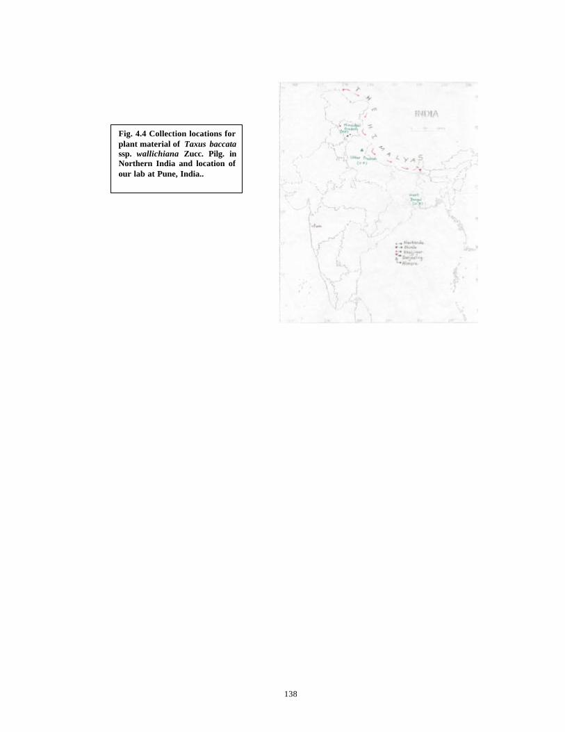
138
Fig. 4.4 Collection locations for plant material of Taxus baccatassp. wallichiana Zucc. Pilg. in Northern India and location of our lab at Pune, India..
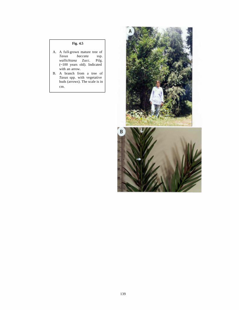
139
Fig. 4.5 A. A full-grown mature tree of
Taxus baccata ssp. wallichiana Zucc. Pilg. (~100 years old). Indicated with an arrow.
B. A branch from a tree of Taxus spp. with vegetative buds (arrows). The scale is in cm.
140
cultures. The forceps and blade holders fitted with sterile surgical blades (No. 11 and 23,
“Surgeon”, India) were dipped in ethyl alcohol and flamed after each transfer.
4.2.3.2 Initiation of suspension cultures
For initiating cell suspension cultures in Taxus spp., 200 mg fresh weight (FW) of
callus was placed in 20 ml of optimized liquid medium in each of the 100 ml capacity
Erlenmeyer flasks on a magnetic shaker (“Infors AG”, Germany) at 100 rpm for varying
periods of time before Taxane analysis.
4.2.4 Media and culture conditions
A number of basal media were used for callus induction experiments such as
Schenk and Hidebrandt’s SH medium (1972) with major salts reduced to 1/2 strength (1/2
SH), Gamborg’s B5 medium (1968) at full strength, Lloyd and McCown’s Woody Plant
Medium: WPM (1981) at full strength and Gupta and Durzan’s DCR medium (1985) at
full strength. Their compositions are described in Chapter 2, Table 2.1 . The media were
modified by addition of different concentrations of various plant growth regulators such
as Indole-3-acetic acid (IAA), Indole-3-butyric acid (IBA), α-naphthalene-acetic acid
(NAA), Picloram (P), 2,4-dichlorophenoxyacetic acid (2,4-D), N6 benzylaminopurine
(BAP), Kinetin (KIN) and N 6-(2-isopentyl)-adenine (2-iP) individually or in permutations
and combinations. All these details regarding concentrations and combinations of plant
growth regulators used are listed in Tables 4.4 and 4.5.
All the media contained 2% sucrose and 0.3% phytagel. All the plant growth
regulators were added before autoclaving of the media at 121ºC and at a pressure of 1.1
kg.cm-2. pH of the media was adjusted to 5.8 before autoclaving as described in detail in
Chapter 2, Section 2.4 .
For initiating suspension cultures from Taxane hyper-producing calli, only an
optimized medium B5 supplemented individually with P at 2, 4, 6, 8 or 10 mg.l-1 was
used. The media were prepared as described above and contained 2% sucrose without any
gelling agent.
All callus and suspension cultures were incubated in the dark at a temperature of
25 + 2oC.

141
4.2.5 Observations and subculture schedules
Callus cultures were observed routinely and subcultures were carried out at
intervals of 21-35 days depending upon the growth rate, phenolic browning (if any) and
nature of the developing callus tissues. At the time of subcultures, fresh weight
determinations of promising cell lines were done only under sterile conditions. For this,
the callus derived from a single needle explant was dried between sheets of sterile filter
paper till no moisture was released from the callus. The callus mass was then weighed on
a pre-weighed sterile piece of aluminium foil kept on a micro-balance (“Anamed”, India),
placed inside the laminar airflow cabinet. Dry weights were determined only at the time
of extraction of callus tissues for Taxane content determination by TLC and HPLC. Here
the callus derived from a single needle explant was dried at 60°C in an oven till constant
weight was obtained. Usually this took 16-24 hours. The spent solid medium from
various nutrient media and plant growth regulator combinations was saved after
subculture of the calli to fresh media. These spent media were analyzed independently for
presence of Taxanes by HPLC.
Suspension cultures for preliminary studies were raised from randomly selected
callus lines (Also see Table 4.11) in three replicate flasks at day 0. These were
subcultured in fresh media of identical compositions after every 4 days. Routine growth
determinations in suspension cell cultures were carried out by centrifuging 8 ml of culture
broth in a graduated, conical centrifuge tube at 5000 rpm for 5 min. to determine Packed
Cell Volumes (PCV).
Out of the three replicate flasks of suspension cultures, at day 9, one flask was
harvested for each of the media and cell line combinations (See Table 4.11 ) and cells and
media were extracted independently and analyzed by HPLC for Taxane contents
(Sections 4.2.7.4, 4.2.7.5 and 4.2.8.2). At the same time, fresh and dry weight
measurements of cells in suspension cultures were also carried out. For this, the cells
were separated from the liquid media by centrifuging them at 5000 rpm for 5 min. The
liquid medium was pipetted out and the cell pellet was dried between sheets of filter
paper till no moisture was released from the cell pellet. The fresh weight of pellet was
then determined on the analytical balance. Dry weight determinations were carried out
after drying the cells at 60°C in an oven till constant weight was obtained. Usually this

142
took 16-24 hours. The liquid medium separated from the cells was used for direct
extraction of Taxanes. Then at day 18, the contents of one flask were harvested for each
of the media and cell line combinations. The fresh and dry weights of cells were
determined as described above. The dried cell mass and media were extracted
independently and analyzed by HPLC for Taxane contents (Sections 4.2.7.4, 4.2.7.5 and
4.2.8.2). The above procedure was repeated with the other remaining two replicate flasks
and ana lysis was done by HPLC (Section 4.2.8.2 ) on separate days to check for the
reproducibility of the experiments.
4.2.6 Extraction of in vitro grown callus tissues for analysis of Taxanes by TLC
Various callus lines were extracted and screened by TLC as a primary screen to
identify Taxane producing, fast growing callus lines. Promising cell lines of various ages
were then screened by HPLC. Only 50 mg (DW) of callus tissue was used for extraction
as TLC was used only as a qualitative screen.
4.2.6.1 Extraction of callus tissues
Extraction of callus tissues was carried out by the method of Vidensek et al.
(1990) modified during the course of the present work. All the solvents used were of
HPLC grade and the steps involved in the extraction procedure were as f ollows:
1. Calli of different age groups growing on various basal media and plant growth
regulator combinations were selected for extraction (C. f. Table 4.5).
2. 50 mg dry weight (DW) of each type of calli was taken for analysis.
3. The dried tissues were ground in a pre-cooled mortar and pestle with liquid nitrogen
and extracted overnight in methanol (10 times w/v) on a rotary shaker at 25°C and
100 rpm.
4. The procedure was repeated three times and the methanolic extracts were pooled
together.
5. The extracts were filtered through Whatman No.1 filter paper and dried in a Centrivap
Concentrator (“Labconco”, USA).
6. The residue was redissolved in chloroform : water (1:1 v/v) and then centrifuged at
7000-8000 rpm, at 16°C to separate the layers of chloroform and water.

143
7. The lower chloroform layer was collected and the upper layer of water was discarded.
The layer at the interphase was re-extracted with chloroform and again the chloroform
layer was collected.
8. The chloroform fractions from both the separations were combined and dried in the
Centrivap Concentrator as described earlier.
9. The residue was redissolved in 1 ml methanol and used for spotting on TLC plates.
4.2.7 Extraction of in vivo as well as in vitro grown tissues and cells and spent
medium for analysis of Taxanes by HPLC
4.2.7.1 Extraction of in vivo tissues (Bark and Needles)
For extraction of in vivo tissues, bark shavings and needles from all the identified
trees of the Shimla region were pooled together and the extraction for determination of
the content of Taxanes was carried out by the method of Vidensek et al. (1990) modified
during the course of the present work. All the solvents used were of HPLC grade and the
steps involved in the extraction procedure were as follows:
1. Bark shavings and needles were dried in an oven at 60°C, till constant weight was
obtained.
2. The dried tissues were ground in a mortar and pestle and the powder was sieved
through a metal sieve of pore size 0.8 mm.
3. 10 g of bark and needle powders were extracted with 10 volumes of methanol (100
ml) on a rotary shaker at 100 rpm and 25°C for 16 hours.
4. The extraction procedure using methanol was repeated thrice and the methanolic
extracts were pooled together for bark and needle tissues respectively.
5. The methanolic extracts were filtered through Whatman No.1 filter paper and dried on
a Büchi Rotavapor-R (“Laboratoriums-Technik AG”, Switzerland) at 40°C, at a
vacuum equivalent to 25” Hg.
6. The residue was redissolved in chloroform : water (1:1 v/v) and then centrifuged at
7000-8000 rpm at 16°C in a centrifuge (“Remi instruments”, India) to separate the
layers of chloroform and water.
7. The lower chloroform layer was collected and upper layer of water was discarded.
The layer at the interphase was re-extracted with chloroform and again the chloroform
fraction was collected.
144
8. The chloroform fractions from both the separations were combined and dried on a
Büchi Rotavapor -R at 35°C, at a vacuum equivalent to 25” Hg.
9. The residue was redissolved in 2 ml of methanol and was filtered through 0.2 or 0.4
µM nylon filters before analysis by HPLC.
10. 20 µL of extract suspended in methanol was analyzed by HPLC.
4.2.7.2 Extraction of callus tissues
Extraction of callus tissues for determination of the content of Taxanes was
carried out by the method of Vidensek et al. (1990) modified during the course of the
present work. All the solvents used were of HPLC grade and the steps involved in the
extraction procedure were as follows:
1. Calli of different age groups growing on various basal media and plant growth
regulator combinations were selected for extraction.
2. 200-400 mg dry weight of each type of calli was taken for analysis. (The tissues were
dried in an oven at 60°C, till constant weight was obtained).
3. The dried tissues were ground in a pre-cooled mortar and pestle with liquid nitrogen
and extracted overnight in methanol (10 times w/v) on a rotary shaker at 25°C and
100 rpm.
4. The procedure was repeated three times and the methanolic extracts were pooled
together.
5. The extracts were filtered through Whatman No.1 filter paper and dried in the
Centrivap Concentrator as described earlier.
6. The residue was redissolved in chloroform : water (1:1 v/v) and then centrifuged at
7000-8000 rpm, at 16°C to separate the layers of chloroform and water.
7. The lower chloroform layer was c ollected and the upper layer of water was discarded.
The layer at the interphase was re-extracted with chloroform and again the chloroform
layer was collected.
8. The chloroform fractions from both the separations were combined and dried in the
Centrivap Concentrator at conditions as described earlier.
9. The residue was redissolved in 1 ml of methanol and was filtered through 0.2-0.4 µM
nylon filters before analysis by HPLC.
145
10. 20 µL of extract suspended in methanol, from each callus line, was analyzed by
HPLC.
4.2.7.3 Extraction of spent solid medium (with phytagel) from callus cultures
All the solvents used during extraction were of HPLC grade and the extraction
procedure involved the following steps modified from Fett-neto et al. (1992).
1. Solid nutrient media of identical compositions and combinations, used for growing
callus cultures derived from the same parent tree, were pooled together and their fresh
weights were determined as described earlier.
2. These media were then extracted with 10 volumes of methanol (10 times w/v) on a
rotary shaker at 100 rpm and 25°C for 16 hours.
3. The procedure was repeated three times and the methanolic extracts were pooled
together.
4. The extracts were filtered through Whatman No.1 filter paper and dried on a Büchi
Rotavapor-R at 40°C and a vacuum equivalent to 25” Hg.
5. The residue was directly dissolved in 2 ml methanol and filtered through 0.2-0.4 µM
nylon filters before analysis by HPLC.
6. 20 µL of extract suspended in methanol, from each type of medium was analyzed by
HPLC.
4.2.7.4 Extraction of cells grown in suspension cultures
Extraction of cells grown in suspension cultures for determination of the content
of Taxanes was carried out by the method of Vidensek et al. (1990) modified during the
course of the present work. All the solvents used were of HPLC grade and the steps
involved in the extraction procedure were as follows:
1. Cells growing in suspension cultures were separated from the liquid nutrient medium
on day 9 and day 18, from separate but identical replicate flasks, by centrifuging them
at 7000-8000 rpm, at 16°C.
2. The cells were blotted dry on filter papers and dried in oven at 60°C, till constant
weight was obtained.
3. All the other steps followed were identical to the ones described above in Section
146
4.2.7.2 for extraction of callus tissues.
4.2.7.5 Extraction of spent medium (without cells) from suspension cultures
All the solvents used during extraction were of HPLC grade and the extraction
procedure involved the following steps modified from Fett-neto et al. (1992).
1. Cells growing in suspension cultures were separated from the liquid nutrient medium
on day 9 and day 18, from separate but identical replicate flasks, by centrifuging them
at 7000-8000 rpm, at 16°C.
2. The spent medium was extracted overnight with equal volume s (v/v) of chloroform on
a rotary shaker at 100 rpm and 25°C.
3. These extractions were repeated for three days and all the chloroform extracts were
combined.
4. Since the medium contained a lot of water, it formed two layers with chloroform
automatically. The two layers were separated by centrifuging them at 7000-8000 rpm,
at 16°C.
5. The chloroform fraction was dried in the Centrivap Concentrator as described earlier
6. The residue was resuspended in 1 ml of methanol, filtered through 0.2-0.4 µM nylon
filters before analysis by HPLC.
7. 20 µL of extract from each type of medium was analyzed by HPLC.
4.2.8 Analytical techniques
4.2.8.1 Thin Layer Chromatography (TLC)
TLC was carried out by using handmade glass plates of size 10 cm x 15 cm x 0.2
mm coated with either Silica Gel G or Silica Gel F254 (“Merck”, India). In addition, pre-
coated plates of size 10 cm x 15 cm x 0.2 mm, with aluminum backing (“Merck”, India)
and coated with either Silica Gel G or Silica Gel F254 were used. Routine procedures for
analytical TLC were followed as given in Fried and Sherma (1994). The actual procedure
followed is described here in brief. Starting line was marked at a distance of 1.5 cm from
the bottom of the plate and the finishing line was marked at a distance of 2 cm from the
top of the plate. Spots of standard and sample solutions in methanol were applied with the
help of microcapillaries. Each spot contained 50 µL of the standard solution of a strength
147
0.5 mg/ml (2.5 µg of standard chemical applied effectively) and 50 µL of the sample
solution from callus extracts. Chromatogram development was done in vertical glass
chambers saturated with chloroform : methanol (7:1 v/v) using the method of Strobel et
al. (1992), upto the finishing line marked on the TLC plate. At this time, the plate was
removed from the development chamber and the mobile phase was allowed to dry. The
developed plates with the normal Silica Gel G were sprayed with H2SO4 and kept in an
oven at 100°C for 5 min for visualization of spots of both standard and sample while the
Silica Gel F254 plates were observed under a short wavelength UV torch of 312 nm
(“Vilber Lonrmat”, Cedex, France) for detection of UV quenching Taxanes in the sample
and their comparison with the standards. R f was calculated as per the formula (Fried and
Sherma, 1994):
Rf = Distance travelled by the applied solute mixture front (cm)
Distance travelled by the solvent (mobile phase) front (cm)
4.2.8.2 High Performance Liquid Chromatography (HPLC)
All routine procedures for analytical HPLC were followed (Melander and
Horvath, 1980; Scott, 1996). The actual procedure followed for HPLC analysis was
modified from the protocol given by ElSohly et al. (1994).
HPLC was carried out on a Merck-H itachi instrument with a series D-7000
interface with HSM Manager, L-7100 pump, L-7420 diode array detector, a rheodyne
injector with a 20 µL injection loop. RP-18 column (“Merck”, India) of dimensions 250 x
4.6 mm with particle size of 5µM was used. The column was operated in the reverse-
phase mode. The data were recorded at a wavelength of 225 nm on a computer and the
chromatograms and data reports were printed on HP Deskjet 200 printer (“Hewlett
Packard”, India). Photographs of selected runs were recorded on color films. External
standard method of quant itation was used.
All the solvents used were of HPLC (Lichrosolv) grade (“Merck”, India). The in-
house purified Milli-Q water was used (“Millipore Corporation”, USA). All the solvents
and samples were filtered through 0.22 or 0.45 µm pore size filters (“Millipore
Corporation”, USA). Degassing of solvents was done under vacuum (equivalent to 20-

148
25” Hg). The mobile phase consisted of a mixture of Methanol (65%) and Water (35%)
under isocratic conditions for analysis of Taxanes. The flow rate was 1.0 ml/min. and the
run was continued for 30 minutes for complete resolution and detection of all the
Taxanes. All the samples (extracts of callus tissues, cells from suspension cultures and
solid as well as liquid spent media) were prepared and filtered as described in the
preceding section 4.2.7 and 20 µL was used for injection. The standard solutions of Taxol
and 10-DAB were prepared in HPLC grade methanol at a strength of 0.5 mg/ml. These
were also filtered before HPLC analysis and 20 µL was used for injection. Injection
volume of 20 µL was kept constant during the experimentation. Spiking of the samples
was accomplished by addition of authentic standard such that 2.5 µg of the standard was
present in the spiked injection. Thus 10 µL from standard solution of a strength of 0.5
mg/ml was added during each injection.
Since there was no change in back-pressure after many injections, it was not
necessary to develop any column regeneration procedures as has been described earlier by
many authors (Kopycki et al., 1994 and references therein). The column was flushed after
every injection by mobile phase for 10 min. to re-establish the initial conditions.
Plotting of graphs and calculations of amounts of Taxanes in the tissues by the
external standard method of quantification were done using routine protocols as described
in the “Instruction Manual for HSM Manager” as well as using the formula given by
Scott (1996):
Cp(s) = ap(s) X Cp(st)
ap(st)
where, Cp(s) is the concentration of solute in the mixture.
ap(s) is the area of the peak for the solute (p) in the sample chromatogram.
ap(st) is the area of peak for solute (p) in the reference chromatogram.
Cp(st) is the concentration of the standard in the reference solution.
The linearity of detector response for standards of Taxol and 10-DAB was
observed by injecting 20 µL from solutions of standards ranging in concentration from
0.625 µg-10 µg/ml (ElSohly et al., 1995) and plotting the standard curve (Fig. 4.11 B, C).
Identification of Taxol and 10-DAB in callus and cell samples was confirmed by

149
retention time, co-chromatography with the standard and peak purity by wavelength (Fett-
neto et al., 1993a, b).
4.2.9 Statistical analysis
In experiments to optimize nutrient media for callus growth, each medium and
plant growth regulator combination (experimental unit) consisted of 10 plastic dishes
(replicate), each dish containing 15 needle pieces derived from a single parent tree. The
experiments were repeated over four successive years for each of the trees. Fresh and dry
weight determinations were performed with five callus pieces from a single plastic dish
and the means with standard errors of means were taken for comparisons (Wardlaw,
1985).
Cell suspension culture experiments were carried out in optimized media and only
three replicates were used for each experiment and each experiment was repeated thrice.
The rate of cell suspensions growth was monitored by PCV at each subculture, for all the
three replicate flasks and the mean PCV was used to determine the growth of cell
suspensions. Fresh and dry weight determinations were performed with cell pellets
derived on centrifugation of cells from three replicate flasks and the means were taken for
comparisons.
The bark and needle tissues of the parental trees and the cultures derived from
them were used for in vivo and in vitro tissue extractions for Taxane contents in duplicate
and the whole procedure was repeated at least thrice.
TLC spottings and HPLC injections for each of the callus and cell lines and
standards were done at least in duplicate and the injections were repeated on different
days to check for the repeatability of the experimental procedure. Day to day variations, if
any, were also checked with the standard solutions. The variations in peak areas from the
respective experiments were tabulated and the mean and standard errors of means were
calculated (Wardlaw, 1985).
Wherever the standard errors of means are less than 2% of the average mean
values, they are not mentioned individually in the tables and only the mean values are
represented (Ray and Jha, 1999).
150
4.3 Results and Discussion
4.3.1 Callus initiation and growth studies in stem and needle explants of Taxus spp.
During the initial experiments, all the cultures were lost due to heavy bacterial and
fungal contamination and phenolic browning as the explants were derived from mature
trees. In order to get sterile cultures, the explants (stem bits and needles as well as entire
seeds with endosperm) were surface sterilized by the “Modified Surface Sterilization
Protocol” presented in Chapter 3, Flow Chart 3.3 . By use of this protocol, more than
90% sterile cultures were consistently obtained (Table 3.13).
In the present study, callus cultures were initiated on a large number of
combinations of nutrient media and plant growth regulator combinations, from stem bit
and needle explants of Taxus spp. These are referred to as ‘Diploid Callus Cultures’
hereinafter. In addition, endosperm halves derived from seeds were also used for callus
initiation, the details of which are described in Chapter 5, Section 5.6.3 . These are
referred to as ‘Haploid Callus Cultures’ hereinafter. All ‘Diploid Callus Cultures’
derived from different parent trees were used in all the experiments to optimize nutrient
media which support better callus growth and all the cultures were maintained
independently. For details of deriving callus cultures from endosperm refer to Chapter 5,
Part B.
The main criterion for optimization of nutrient media was to select those media
with low ionic strength so as to reduce further phenolic browning and to obtain good
callus growth. On this basis, SH medium was used at 1/2 strength of major salts (1/2 SH)
since the ionic strength of SH medium is quite high at 69.9 mM (C. f. Chapter 2, Table
2.2 ). In addition, low salt media such as B5, WPM and DCR at full strengths of major
salts were also used (C. f. Chapter 2, Table 2.2). In the pilot experiments, these media
were initially appended with 5 concentrations of 2,4-D (2, 4, 6, 8, 10 mg.l-1) since it is the
most commonly used auxin for callus induction. Table 4.4 presents the results obtained in
this experiment.
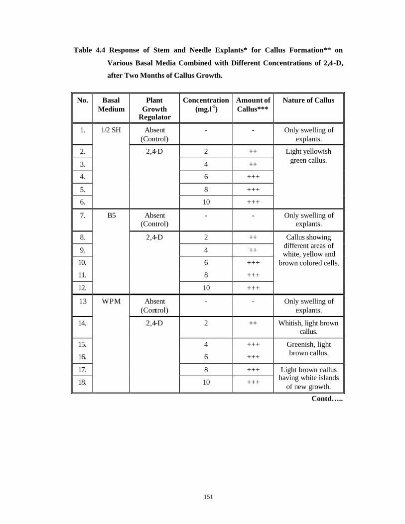
151
Table 4.4 Response of Stem and Needle Explants* for Callus Formation** on
Various Basal Media Combined with Different Concentrations of 2,4-D,
after Two Months of Callus Growth.
No. Basal Medium
Plant Growth
Regulator
Concentration (mg.l-1)
Amount of Callus***
Nature of Callus
1. Absent (Control)
- - Only swelling of explants.
2. 2 ++
3. 4 ++
4. 6 +++
5. 8 +++
6.
1/2 SH
2,4-D
10 +++
Light yellowish green callus.
7. Absent (Control)
- - Only swelling of explants.
8. 2 ++
9. 4 ++
10. 6 +++
11. 8 +++
12.
B5
2,4-D
10 +++
Callus showing different areas of white, yellow and
brown colored cells.
13 Absent (Control)
- - Only swelling of explants.
14. 2 ++ Whitish, light brown callus.
15. 4 +++
16. 6 +++
Greenish, light brown callus.
17. 8 +++
18.
WPM
2,4-D
10 +++
Light brown callus having white islands
of new growth.
Contd…..
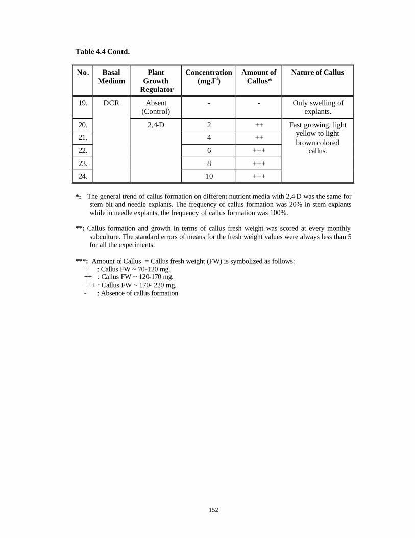
152
Table 4.4 Contd.
No. Basal Medium
Plant Growth
Regulator
Concentration (mg.l-1)
Amount of Callus*
Nature of Callus
19. Absent (Control)
- - Only swelling of explants.
20. 2 ++
21. 4 ++
22. 6 +++
23. 8 +++
24.
DCR
2,4-D
10 +++
Fast growing, light yellow to light brown colored
callus.
*: The general trend of callus formation on different nutrient media with 2,4-D was the same for
stem bit and needle explants. The frequency of callus formation was 20% in stem explants while in needle explants, the frequency of callus formation was 100%.
**: Callus formation and growth in terms of callus fresh weight was scored at every monthly
subculture. The standard errors of means for the fresh weight values were always less than 5 for all the experiments.
***: Amount of Callus = Callus fresh weight (FW) is symbolized as follows:
+ : Callus FW ~ 70-120 mg. ++ : Callus FW ~ 120-170 mg. +++ : Callus FW ~ 170- 220 mg. - : Absence of callus formation.
153
Callus induction occurred in 20-40 days on all the above-mentioned nutrient
media with 2,4-D from both stem bit and needle explants. The stem bit explants were
extremely poor in terms of callus induction in all the media combinations. Hardly 20% of
stem bit explants produced calli within 20-40 days and out of these only 10% survived in
subsequent subcultures. On the other hand, needle explants formed callus with 100%
frequency within 20-40 days and most of these cultures (>95%) could be subcultured
routinely.
It is clear from Table 4.4 that all basal media with 6, 8 and 10 mg.l-1 2,4-D, gave
good callus growth (FW in the range of 175-225 mg) irrespective of the source of explant
(stem bits or needles). The calli were subcultured on fresh, identical media at monthly
intervals after callus initiation. The differences in calli obtained on various basal media
and 2,4-D combinations became apparent at this stage. Calli obtained on 1/2 SH, WPM
and DCR media supplemented with 2,4-D could not be maintained beyond their fourth
monthly subculture (140-180 days after callus initiation) and the callus survival
deteriorated gradually from one passage to another. This was more pronounced in callus
cultures obtained from stem bit explants. No new growth of cells was apparent on these
calli and subsequently these calli became brown due to phenolics, became necrotic and
died. The reason for non-survival of callus obtained from both the stem bit and needle
explants beyond four subcultures on these media is unclear. Hence these media were not
tried in further experiments with different plant growth regulators. On the contrary, B5
medium was found to be the best in terms of callus growth as well as callus survival over
the monthly subcultures. Calli growing in B5 medium with 2,4-D and obtained from
needle explants alone are still being maintained in our laboratory over the last 4 years. B5
medium was therefore chosen for further optimization of plant growth regulators. Preece
(1995) observed that when the nutrient salts are optimized for in vitro tissues of a plant,
the nutrient level and their balance in the medium makes tissues to be under less stress
and in vitro performance gets dramatically improved. This is evident in our observation
that callus cultures of Taxus spp. in B5 medium do not exude any phenolics.
Large scale studies on callus induction and growth, with the ultimate objective of
their use in Taxane production, were therefore carried out with needle bit explants and all
the subsequent data refers to needle derived calli.
154
B5 medium was further modified with individual additions of auxins like 2,4-D,
IAA, IBA, NAA and P at various concentrations and combinations of IAA + IBA, IBA +
2-iP, 2,4-D + KIN and NAA + KIN (See Table 4.5) for callus induction from needle
explants. Good callus growth and callus fresh weight (FW) in the range of 120-225 mg
was obtained on B5 medium supplemented individually with 2,4-D or NAA, depending
on its concentration. Callus growth and fresh weight of callus were at their best in B5
medium supplemented individually with 2, 4, 6, 8, 10 mg.l-1 P. At the end of six months,
the calli consistently weighed more than 250 mg (FW) and were very fast growing. B5
medium supplemented individually with IAA or IBA gave poor callus response. Hence
initially IBA was combined with 2-iP to see if a combination of an auxin and a cytokinin
could lead to good callus initiation and growth. But the results were not encouraging
(Table 4.5 ). Hence IBA and 2-iP were not used in further experiments, in combination
with other auxins/cytokinins. Instead, IAA and IBA were used together in concentrations
ranging 0.5-10 mg.l-1. These auxins in many combinations supported good callus growth
and the fresh weights of calli ranged between 175-225 mg (Table 4.5). Growth
stimulation at relatively high exogenous auxin concentrations has been reported earlier
for other Gymnosperms like Cephalotaxus harringtonia (Delfel and Rothfus, 1977). KIN
at low concentration of 0.2 mg.l-1 alone was tried with 2,4-D and NAA independently as
KIN is known to exert an inhibitory effect on callus growth of Taxus spp. at higher
concentrations (Fett-Neto et al., 1993a). However no significant improvement in growth
of calli was obtained in these combinations (Table 4.5). Figures 4.6-4.9 represent the
results obtained on callus induction and growth from needle explants on B5 medium and
different plant growth regulator combinations.
In none of the cultures, presence of distinctly organized structures such as roots or
shoots has been observed. However, 5% of callus cultures developed from needle
explants on B5 medium supplemented with 6 mg.l-1 2,4-D and 0.2 mg.l-1 KIN showed
presence of root-like outgrowths (Fig. 4.7 C). But these did not develop further.
The calli were friable, white to light yellow to light yellow -green to light brown in
color on all the growth media and plant growth regulator combinations tried (Table 4.5).
None of the calli were hard, nodular or green in color.
Portions of calli showing active growth and friability were separated from the rest
of the explant and used for subculturing of calli in their respective media combinations.
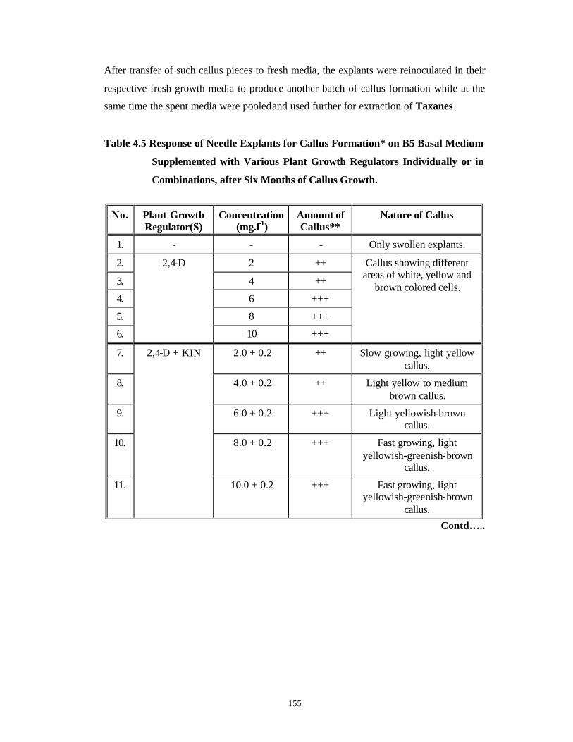
155
After transfer of such callus pieces to fresh media, the explants were reinoculated in their
respective fresh growth media to produce another batch of callus formation while at the
same time the spent media were pooled and used further for extraction of Taxanes.
Table 4.5 Response of Needle Explants for Callus Formation* on B5 Basal Medium
Supplemented with Various Plant Growth Regulators Individually or in
Combinations, after Six Months of Callus Growth.
No. Plant Growth Regulator(S)
Concentration (mg.l-1)
Amount of Callus**
Nature of Callus
1. - - - Only swollen explants.
2. 2 ++
3. 4 ++
4. 6 +++
5. 8 +++
6.
2,4-D
10 +++
Callus showing different areas of white, yellow and
brown colored cells.
7. 2.0 + 0.2 ++ Slow growing, light yellow callus.
8. 4.0 + 0.2 ++ Light yellow to medium brown callus.
9. 6.0 + 0.2 +++ Light yellowish-brown callus.
10. 8.0 + 0.2 +++ Fast growing, light yellowish-greenish-brown
callus.
11.
2,4-D + KIN
10.0 + 0.2 +++ Fast growing, light yellowish-greenish-brown
callus.
Contd…..
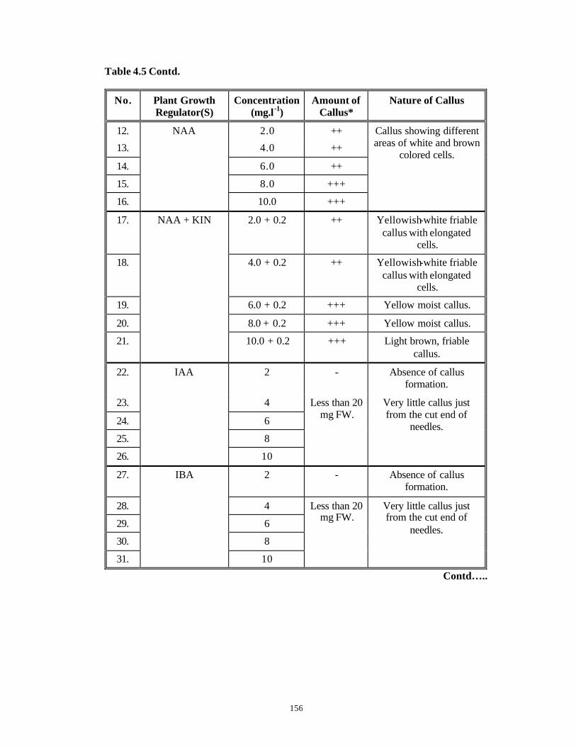
156
Table 4.5 Contd.
No. Plant Growth Regulator(S)
Concentration (mg.l-1)
Amount of Callus*
Nature of Callus
12. 2.0 ++
13. 4.0 ++
14. 6.0 ++
15. 8.0 +++
16.
NAA
10.0 +++
Callus showing different areas of white and brown
colored cells.
17. 2.0 + 0.2 ++ Yellowish-white friable callus with elongated
cells.
18. 4.0 + 0.2 ++ Yellowish-white friable callus with elongated
cells.
19. 6.0 + 0.2 +++ Yellow moist callus.
20.
NAA + KIN
8.0 + 0.2 +++ Yellow moist callus.
21. 10.0 + 0.2 +++ Light brown, friable callus.
22. 2 - Absence of callus formation.
23. 4
24. 6
25. 8
26.
IAA
10
Less than 20 mg FW.
Very little callus just from the cut end of
needles.
27. 2 - Absence of callus formation.
28. 4
29. 6
30. 8
31.
IBA
10
Less than 20 mg FW.
Very little callus just from the cut end of
needles.
Contd…..
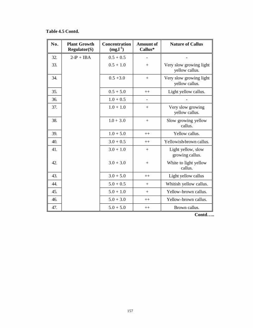
157
Table 4.5 Contd.
No. Plant Growth Regulator(S)
Concentration (mg.l-1)
Amount of Callus*
Nature of Callus
32. 0.5 + 0.5 - -
33. 0.5 + 1.0 + Very slow growing light yellow callus.
34. 0.5 +3.0 + Very slow growing light yellow callus.
35. 0.5 + 5.0 ++ Light yellow callus.
36. 1.0 + 0.5 - -
37. 1.0 + 1.0 + Very slow growing yellow callus.
38. 1.0 + 3.0 + Slow growing yellow callus.
39. 1.0 + 5.0 ++ Yellow callus.
40. 3.0 + 0.5 ++ Yellowish-brown callus.
41. 3.0 + 1.0 + Light yellow, slow growing callus.
42. 3.0 + 3.0 + White to light yellow callus.
43. 3.0 + 5.0 ++ Light yellow callus
44. 5.0 + 0.5 + Whitish yellow callus.
45. 5.0 + 1.0 + Yellow-brown callus.
46. 5.0 + 3.0 ++ Yellow-brown callus.
47.
2-iP + IBA
5.0 + 5.0 ++ Brown callus.
Contd…..
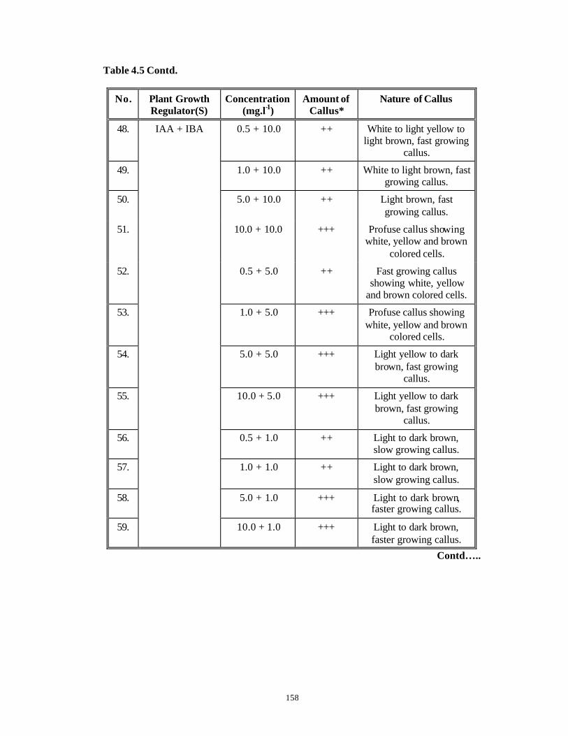
158
Table 4.5 Contd.
No. Plant Growth Regulator(S)
Concentration (mg.l-1)
Amount of Callus*
Nature of Callus
48. 0.5 + 10.0 ++ White to light yellow to light brown, fast growing
callus.
49. 1.0 + 10.0 ++ White to light brown, fast growing callus.
50. 5.0 + 10.0 ++ Light brown, fast growing callus.
51. 10.0 + 10.0 +++ Profuse callus showing white, yellow and brown
colored cells.
52. 0.5 + 5.0 ++ Fast growing callus showing white, yellow
and brown colored cells.
53. 1.0 + 5.0 +++ Profuse callus showing white, yellow and brown
colored cells.
54. 5.0 + 5.0 +++ Light yellow to dark brown, fast growing
callus.
55. 10.0 + 5.0 +++ Light yellow to dark brown, fast growing
callus.
56. 0.5 + 1.0 ++ Light to dark brown, slow growing callus.
57. 1.0 + 1.0 ++ Light to dark brown, slow growing callus.
58. 5.0 + 1.0 +++ Light to dark brown, faster growing callus.
59.
IAA + IBA
10.0 + 1.0 +++ Light to dark brown, faster growing callus.
Contd…..

159
Table 4.5 Contd.
No. Plant Growth Regulator(S)
Concentration (mg.l-1)
Amount of Callus*
Nature of Callus
60. 0.5 + 0.5 + Brown callus.
61. 1.0 + 0.5 + Brown callus.
62. 5.0 + 0.5 ++ Whitish yellow callus.
63.
IAA + IBA
10.0 + 0.5 ++ Light yellow to light brown callus.
64. 2.0 +++
65. 4.0 +++
66. 6.0 +++
67. 8.0 ++++
68.
P
10.0 ++++
Fast growing, light yellow to light brown colored
callus.
* : Callus formation and growth in terms of callus fresh weight was scored at every monthly
subculture. The standard errors of means for fresh weights were always less than 5 for all the experiments.
**: Amount of Callus = Callus mean fresh weight (FW) is symbolized as follows :
+ : Callus FW ~ 70-120 mg. ++ : Callus FW ~ 120-175 mg. +++ : Callus FW ~ 175- 225 mg. ++++ : Callus FW ~ 225-275 mg and more. - : Absence of callus formation.
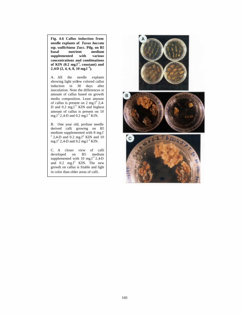
160
Fig. 4.6 Callus induction from needle explants of Taxus baccatassp. wallichiana Zucc. Pilg. on B5 basal nutrient medium supplemented with various concentrations and combinations of KIN (0.2 mg.l-1, constant) and 2,4-D (2, 4, 6, 8, 10 mg.l -1). A. All the needle explants showing light yellow colored callus induction in 30 days after inoculation. Note the differences in amount of callus based on growth media composition. Least amount of callus is present on 2 mg.l-1 2,4-D and 0.2 mg.l-1 KIN and highest amount of callus is present on 10 mg.l-1 2,4-D and 0.2 mg.l -1 KIN. B. One year old, profuse needle derived calli growing on B5 medium supplemented with 8 mg.l-1 2,4-D and 0.2 mg.l-1 KIN and 10 mg.l-1 2,4-D and 0.2 mg.l -1 KIN. C. A closer view of calli developed on B5 medium supplemented with 10 mg.l-1 2,4-D and 0.2 mg.l-1 KIN. The new growth on callus is friable and light in color than older areas of calli.

161
Fig. 4.7 Callus proliferation from needle explants of Taxus baccata ssp. wallichiana Zucc. Pilg. on B5 basal nutrient medium supplemented with various concentrations and combinations of KIN (0.2 mg.l -
1, constant) and 2,4-D (2, 4, 6, 8, 10 mg.l-1). A. & B. Profuse callus
formation and proliferation in 60 days on B5 basal nutrient medium supplemented with various concentrations and combinations of KIN (0.2 mg.l-1, constant) and 2,4-D (2, 4, 6, 8, 10 mg.l-1). Note callus induction from cut edges of needle explants and eventual proliferation all over the needle explant. The callus is light yellow, friable and relatively fast growing.
C. A root like outgrowth from
callus grown on B5 basal nutrient medium supplemented with KIN at 0.2 mg.l-1 and 2,4-D at 6 mg.l-1.
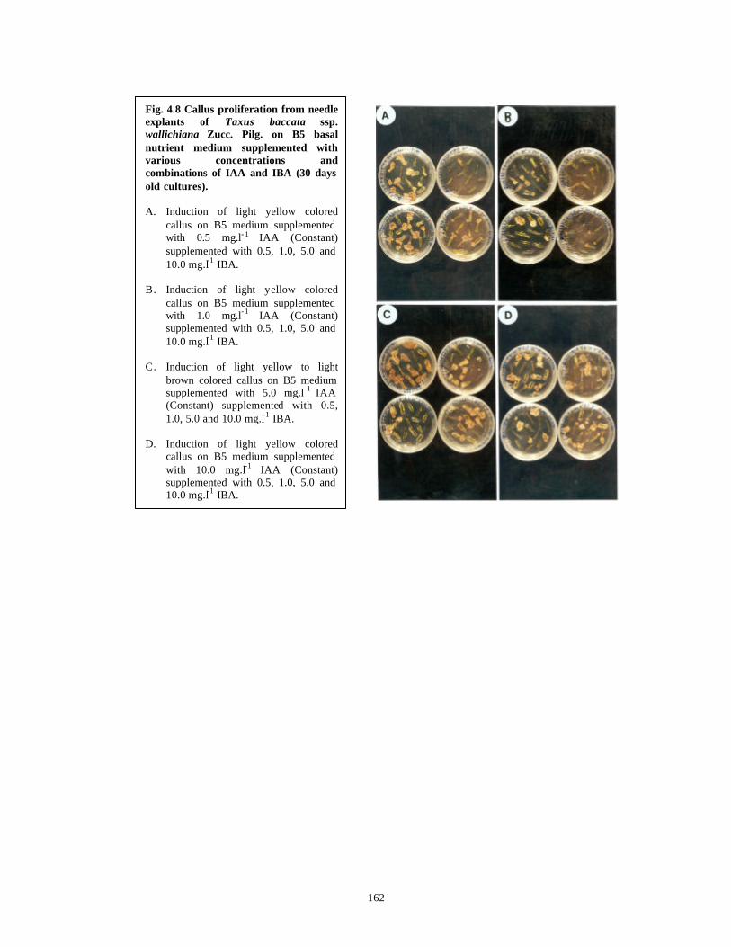
162
Fig. 4.8 Callus proliferation from needle explants of Taxus baccata ssp. wallichiana Zucc. Pilg. on B5 basal nutrient medium supplemented with various concentrations and combinations of IAA and IBA (30 days old cultures). A. Induction of light yellow colored
callus on B5 medium supplemented with 0.5 mg.l- 1 IAA (Constant) supplemented with 0.5, 1.0, 5.0 and 10.0 mg.l-1 IBA.
B. Induction of light yellow colored
callus on B5 medium supplemented with 1.0 mg.l- 1 IAA (Constant) supplemented with 0.5, 1.0, 5.0 and 10.0 mg.l-1 IBA.
C. Induction of light yellow to light
brown colored callus on B5 medium supplemented with 5.0 mg.l-1 IAA (Constant) supplemented with 0.5, 1.0, 5.0 and 10.0 mg.l-1 IBA.
D. Induction of light yellow colored
callus on B5 medium supplemented with 10.0 mg.l-1 IAA (Constant) supplemented with 0.5, 1.0, 5.0 and 10.0 mg.l-1 IBA.
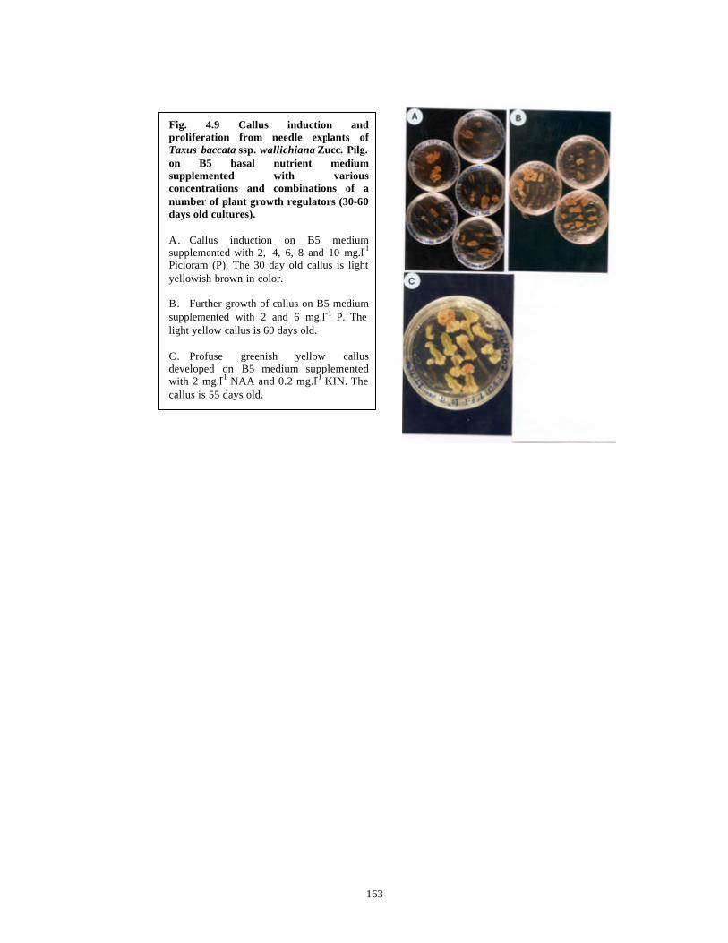
163
Fig. 4.9 Callus induction and proliferation from needle explants of Taxus baccata ssp. wallichiana Zucc. Pilg. on B5 basal nutrient medium supplemented with various concentrations and combinations of a number of plant growth regulators (30-60 days old cultures). A. Callus induction on B5 medium supplemented with 2, 4, 6, 8 and 10 mg.l-1
Picloram (P). The 30 day old callus is light yellowish brown in color. B. Further growth of callus on B5 medium supplemented with 2 and 6 mg.l-1 P. The light yellow callus is 60 days old. C. Profuse greenish yellow callus developed on B5 medium supplemented with 2 mg.l-1 NAA and 0.2 mg.l-1 KIN. The callus is 55 days old.

164
A graphical representation of growth of 15 callus lines, starting from initiation of
callus on optimized media (B5 medium supplemented with 2, 4, 6, 8 and 10 mg.l-1 P,
referred to in the figure as BP2, BP4, BP6, BP8 and BP10) over a period of three months,
in three monthly transfers is presented in Fig. 4.10 . Callus initiated in B5 medium
supplemented with 2 mg.l-1 P had a high initial FW of 67 mg but it did not increase
significantly during the next two transfers of one month duration each. Calli initiated on
B5 medium supplemented with 4, 6 and 8 mg.l-1 P did not have initial high FW and the
growth rate was not very fast in the next two monthly transfers except for calli growing
on B5 medium supplemented with 10 mg.l-1 P. The callus lines on B5 medium
supplemented with 10 mg.l-1 P (Line Nos. 11, 12, 13, 14 and 15) had an initial FW
ranging from 11-35 mg that increased to 21-49 mg in the third month of culture. Such a
comparatively faster growth rate of callus in the first three months with high
concentrations of P is similar to observations reported earlier (Gibson et al., 1995 and
references therein). The rate of callus growth increased further during the next three
months of culture and the final weights obtained at the end of six months are mentioned
in Table 4.5 .
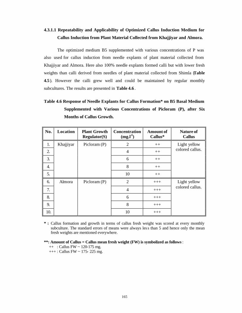
165
4.3.1.1 Repeatability and Applicability of Optimized Callus Induction Medium for
Callus Induction from Plant Material Collected from Khajjiyar and Almora.
The optimized medium B5 supplemented with various concentrations of P was
also used for callus induction from needle explants of plant material collected from
Khajjiyar and Almora. Here also 100% needle explants formed calli but with lower fresh
weights than calli derived from needles of plant material collected from Shimla (Table
4.5 ). However the calli grew well and could be maintained by regular monthly
subcultures. The results are presented in Table 4.6 .
Table 4.6 Response of Needle Explants for Callus Formation* on B5 Basal Medium
Supplemented with Various Concentrations of Picloram (P), after Six
Months of Callus Growth.
No. Location Plant Growth Regulator(S)
Concentration (mg.l-1)
Amount of Callus*
Nature of Callus
1. 2 ++
2. 4 ++
3. 6 ++
4. 8 ++
5.
Khajjiyar Picloram (P)
10 ++
Light yellow colored callus.
6. 2 +++
7. 4 +++
8. 6 +++
9. 8 +++
10.
Almora Picloram (P)
10 +++
Light yellow colored callus.
* : Callus formation and growth in terms of callus fresh weight was scored at every monthly
subculture. The standard errors of means were always les s than 5 and hence only the mean fresh weights are mentioned everywhere.
**: Amount of Callus = Callus mean fresh weight (FW) is symbolized as follows :
++ : Callus FW ~ 120-175 mg. +++ : Callus FW ~ 175- 225 mg.

166
It is clear from Table 4.6 that needles collected from Almora showed better callus
formation in all the concentrations of P than needles collected from Khajjiyar during six
months of callus growth. In addition, the fresh weights of calli remained more or less
constant in the range of 175-225 mg. Unfortunately, due to paucity of time, Taxane
analysis of these cultures could not be carried out but they are being maintained in the
laboratory.
4.3.2 Extraction and Analysis of Taxanes in in vivo and in vitro tissues of Taxus spp
Methanol was chosen for primary extraction of Taxanes because it has been
shown to give high yields of polar compounds like 10-DAB and related structures as
compared to extractions with solvents like chloroform (Theodoridis et al., 1998). After
primary extraction, liquid-liquid extraction was performed using chloroform and water
which has been shown to recover 95-100% of Taxol and Baccatin-III and 70-90% of 10-
DAB-III (Cass et al., 1997).
Since the Taxanes display a wide range of polarity and molecular size, their
separation upto baseline in HPLC needs to be properly adjusted. A number of reports
have appeared in literature using a variety of columns and solvent systems for HPLC
analysis of Taxanes and it is not possible and pertinent to review all of those with respect
to present work. The present system of reverse phase HPLC analysis using methanol and
water was chosen because both the Taxanes of interest namely Taxol and 10-DAB were
baseline separated and the run time required was only 30 min. Methanol has a high
solvent strength parameter ε º = 0.95 and hence increased the solubility of hydrophobic
Taxanes (Kopycki et al., 1994).
The HPLC run of standard solutions of Taxol and 10-DAB was performed under
identical conditions to those employed for analysis of samples. Fig. 4.11 A shows the
standard chromatogram for 10-DAB (d) and Taxol (t) while Fig. 4.11 B and C show the
linearity of UV-VIS detector response for concentrations of standards ranging from 0.625
5.0 µg.
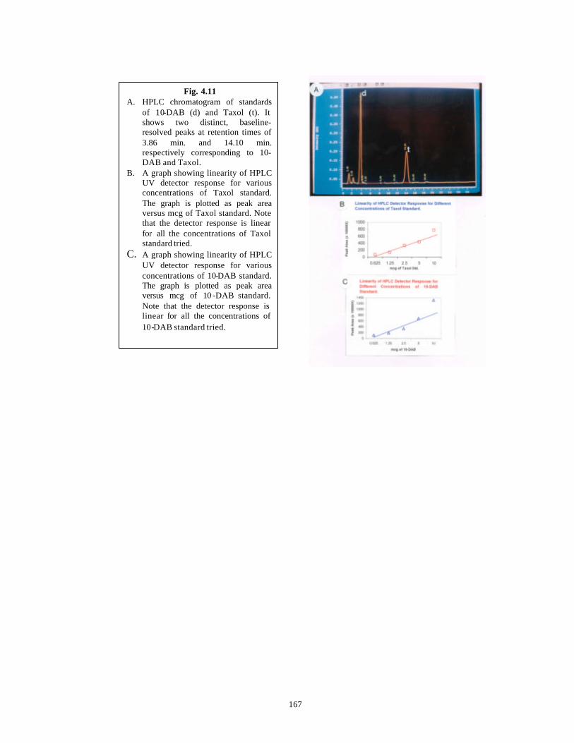
167
Fig. 4.11 A. HPLC chromatogram of standards
of 10-DAB (d) and Taxol (t). It shows two distinct, baseline-resolved peaks at retention times of 3.86 min. and 14.10 min. respectively corresponding to 10-DAB and Taxol.
B. A graph showing linearity of HPLC UV detector response for various concentrations of Taxol standard. The graph is plotted as peak area versus mcg of Taxol standard. Note that the detector response is linear for all the concentrations of Taxol standard tried.
C. A graph showing linearity of HPLC UV detector response for various concentrations of 10-DAB standard. The graph is plotted as peak area versus mcg of 10 -DAB standard. Note that the detector response is linear for all the concentrations of 10-DAB standard tried.
168
4.3.2.1 Extraction and Analysis of Taxan es in in vivo tissues of Taxus spp.
The Taxane content of pooled bark and needle samples from all the six chosen
trees from Shimla, are as presented in Table 4.6. As reported by earlier authors (Denis et
al., 1988), the bark contained higher amounts of 10-DAB than Taxol and Taxol content
was significantly higher than that of needles. Surprisingly the needles did not contain any
10-DAB and had very small amount of Taxol. Chattopadhyay et al. (1994) reported that
out of Taxus spp. plant material collected from Assam, Arunachal Pradesh, Uttar Pradesh
and Himachal Pradesh, only the needles of the material collected from Himachal Pradesh
had Taxol and 10-DAB while plant material from other locations contained only 10-
DAB. However, the authors have not reported quantitative amounts of the Taxanes in
their publication and thus limits comparison of their data with the quantitative data
obtained here.
Chattopadhyay et al. (1997) also reported that the heartwood of the Himalayan
Yew, T. wallichiana showed a number of unique Taxanes besides Taxol and 10-DAB,
and is similar to the results obtained in the present study. This can be seen from the
chromatogram in Fig. 4.12 A wherein a number of peaks besides Taxol and 10-DAB
could be seen (green curve in the chromatogram) but were not identified as standards
were not available.
Table 4.6 Amounts of Taxanes in in vivo tissues of Taxus spp.
Amount of Taxanes (µg.g -1 DW)
Mean + S. E.
Tissues
Taxol 10-DAB
Needles 000.0941 + 0.08 -
Bark 134.0600 + 3.20 1495.0 + 24.7 -: Absence of 10-DAB.
4.3.2.2 Extraction and analysis of Taxanes in selected callus lines of Taxus spp.
For extractions, methanol was the preferred solvent due to reasons outlined above
and additional extractions with hexane or other apolar solvents was not necessary to
remove lipophilic compounds (Theodoridis et al., 1998). This was proven during the
169
HPLC analysis because there was no significant increase in back-pressure of the column
and the chromatograms sometimes showed only an initial broad peak of polar impurities
which did not interfere with the Taxane analysis. For any successful analysis of plant
secondary metabolism, it is necessary to have a separation technique providing efficient
separation of metabolites as well as good selectivity and sensitivity of detection and
capacity to provide important structural information on-line. TLC and HPLC are the most
commonly used analytical techniques. These techniques were used for callus cultures
raised on defined media to identify the Taxane hyper-producer lines which could be
further used for preliminary studies in raising cell suspension cultures.
4.3.2.2.1 Use of TLC in Taxane analysis
In the present work, TLC was used as a preliminary screen to check for the
presence of Taxol in callus lines raised on B5 media supplemented with various plant
growth regulators. In all, 58 diploid callus lines and 10 haploid callus lines were screened
by TLC. Out of these, 20 diploid callus cultures and 5 haploid callus cultures showed
spots having identical Rf value of 0.67 to the standard of Taxol. Only these promising
callus lines derived from needles (diploid) and endosperm (haploid) were extracted on a
larger scale (C. f. Section 4.2.7.1 ) and used for HPLC analysis (C. f. Table 4.8). In
addition, one Taxane non-producer callus (based on negative TLC results) growing on
B5 medium supplemented with 10 mg.l-1 P (Callus Line No. 5 in Table 4.8) and chosen
at random was also analyzed by HPLC. The spots of Taxol were visualized by spraying
the TLC plate with H2SO4 and then heating it. This is a general method followed for
visualization of secondary metabolites. Visualization under UV light also showed the
fluorescing spots with Rf of 0.67. But some additional spots in the vicinity were also
visible on the plates, which might represent other Taxanes with similar structure to
Taxol.
4.3.2.2.2 Use of HPLC in Taxane analysis
HPLC is the most appropriate technique for an efficient separation of crude plant
extracts and can be coupled with different spectroscopic detection methods (Hostettmann
and Wolfender, 1997). In the present endeavor, HPLC coupled with UV photodiode array
detection was used for the UV spectra of natural compounds providing useful information
about the type of constituents and their structures (Hostettmann a nd Wolfender, 1997).
170
The TLC positive needle -derived diploid calli (Callus Line Nos. 1-13 of different
ages), a TLC negative diploid Callus Line No. 5 and the endosperm-derived haploid
Callus Line Nos. 1-5 (C. f. Tables 4.8 and 4.9 ), growing on B5 or DCR media
respectively, supplemented with various plant growth regulators were used further for
quantitative Taxane analysis by HPLC. The quantitative amounts of Taxanes in all these
different callus lines analyzed by HPLC are presented in Tables 4.8 and 4.9 . Figs. 4.12-
4.14 represent the chromatograms of various callus lines.
While carrying out the HPLC analysis of cell and callus cultures of Taxus spp.,
everyday at least two independent injections of the samples were performed and an
average of the peak areas was calculated. The standard error of the mean (S. E.) obtained
was always less than 6.0. The standard and sample injections were repeated on successive
days and again the S. E. for the peak areas of 10-DAB and Taxol were respectively 6.86
and 3.89. S. E. for peak areas of samples were between 3 and 10.
HPLC analysis of TLC positive Needle -derived calli for Taxane production
The TLC positive, needle-derived callus cultures (except Callus Line No. 5 which
is TLC negative) growing on B5 medium supplemented with P had relatively faster
growth (Callus Line Nos. 1-4 of different ages, Table 4.8 ) compared to callus cultures
growing on B5 medium supplemented with 2,4-D + KIN (Callus Line Nos. 10-13, Table
4.8 ) and NAA + KIN (Callus Line Nos. 6-9, Table 4.8). The TLC positive, endosperm-
derived callus cultures (Table 4.9) on DCR medium supplemented with P grew faster as
compared to those on B5 medium supplemented with P (Table 5.14 ). The details
pertaining to needle and endosperm derived callus culture initiation, growth and nature of
callus are described in Tables 4.5 and 5.14 . By comparison of Tables 4.5, 5.14 and 4.8 it
is clear that higher Taxane contents were detected in relatively faster growing callus
cultures. This will become evident in the following discussion.
It is clear from Table 4.8 that B5 medium supplemented with 0.2 mg.l-1 KIN
along with 2,4-D concentrations of 2, 4, 6 and 8 (Referred to as BD2K0.2-BD8K0.2,
Callus Line Nos. 10-13) and B5 medium supplemented with 0.2 mg.l-1 KIN along with
NAA concentrations of 2, 4, 6 and 8 (Referred to as BN2K0.2-BN8K0.2, Callus Line
Nos. 6-9) did not produce detectable levels of Taxanes in needle-derived calli. This was
true for three year old calli of Callus Line Nos. 6-9 and 10-13.
171
On the other hand, B5 medium supplemented with various concentrations of P
(Referred to as BP, Callus Line Nos. 1-4 of different ages) showed appreciable amounts
of Taxanes. In addition, there is a general trend in increase of Taxol and 10-DAB in all
callus lines maintained at different concentrations of P with the age of the callus line from
3 months to 2 years with minor deviations in the trend. For example, the amount of 10-
DAB in 3 month old callus growing on medium BP8 was 138.10 µg/g DW of callus. This
amount increased to 2585 µg 10-DAB /g DW of callus in 1 year old callus and then
decreased to 2282 µg 10-DAB /g DW of callus in the second year (Table 4.8). In contrast,
the amount of Taxol was 76.0 µg/g DW of callus in 3 month old callus growing on
medium BP6. This amount decreased to 60.4 µg Taxol/g DW of callus in 1 year old
callus and then increased to 540.0 µg Taxol/g DW of callus in the second year (Table
4.8 ). This pattern of Taxane content observed with our cultures is in contrast to earlier
reports where P induced good callus growth but inhibited Taxane production (Shuler,
1994 and references therein; Gibson et al., 1995 and references therein). Also in contrast
to earlier reports (Shuler, 1994 and references therein), both faster growth of callus and
high Taxane production was achieved here in the same medium.
2 year old callus growing on BP6 medium (B5 medium with 6 mg.l-1 P) produced
the maximum level of Taxol (540 µg/g DW of callus) which is ~ 6000 times more than in
the in vivo needles. Concomitantly it had intermediate contents of 10-DAB at 1836 µg/g
DW of callus. Also another callus of the same age growing on BP4 medium (B5 medium
with 4 mg.l-1 P) had 517 µg Taxol/g DW of callus. Yet another callus of the same age
growing on BP8 medium (B5 medium with 8 mg.l-1 P) had 344 µg Taxol/g DW of callus
and 10-DAB content at 2282 µg/g DW of callus. Surprisingly, callus of the same age
growing on BP10 medium (B5 medium with 10 mg.l-1 P) had no detectable levels of
Taxol and 10-DAB . A relatively fast rate of growth of this callus compared to other
callus lines might be the reason for absence of Taxanes in the callus (Also see Fig. 4.10 ).
1 year old callus growing on BP8 medium produced a maximum level of 10-DAB
(2585 µg/g DW of callus). This is surprising in the light of the fact that 10-DAB is absent
in in vivo needles. However Taxol was absent in this callus line although Taxol was
detected in needles in vivo.
172
The pattern of 10-DAB and Taxol accumulation reported here in callus cultures in
vitro is the actual picture present in the parent tree(s) or is a manifestation of the culture
conditions needs further study and elucidation.
This is the first report of production of Taxol and 10-DAB in in vitro cultures
of the Himalayan Yew. An earlier report by Banerjee et al. (1996) reported presence of
only minor taxoids in the in vitro cultures of the Himalayan Yew.
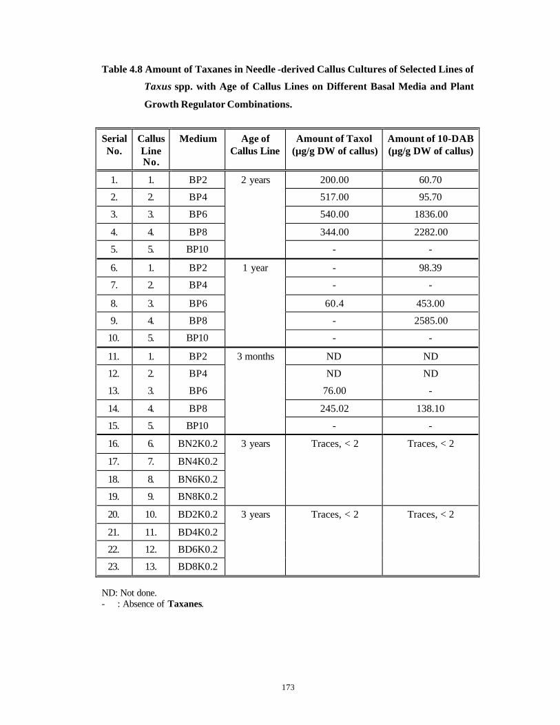
173
Table 4.8 Amount of Taxanes in Needle -derived Callus Cultures of Selected Lines of
Taxus spp. with Age of Callus Lines on Different Basal Media and Plant
Growth Regulator Combinations.
Serial No.
Callus Line No.
Medium Age of Callus Line
Amount of Taxol (µg/g DW of callus)
Amount of 10-DAB (µg/g DW of callus)
1. 1. BP2 200.00 60.70
2. 2. BP4 517.00 95.70
3. 3. BP6 540.00 1836.00
4. 4. BP8 344.00 2282.00
5. 5. BP10
2 years
- -
6. 1. BP2 - 98.39
7. 2. BP4 - -
8. 3. BP6 60.4 453.00
9. 4. BP8 - 2585.00
10. 5. BP10
1 year
- -
11. 1. BP2 ND ND
12. 2. BP4 ND ND
13. 3. BP6 76.00 -
14. 4. BP8 245.02 138.10
15. 5. BP10
3 months
- -
16. 6. BN2K0.2
17. 7. BN4K0.2
18. 8. BN6K0.2
19. 9. BN8K0.2
3 years Traces, < 2 Traces, < 2
20. 10. BD2K0.2
21. 11. BD4K0.2
22. 12. BD6K0.2
23. 13. BD8K0.2
3 years Traces, < 2 Traces, < 2
ND: Not done. - : Absence of Taxanes.
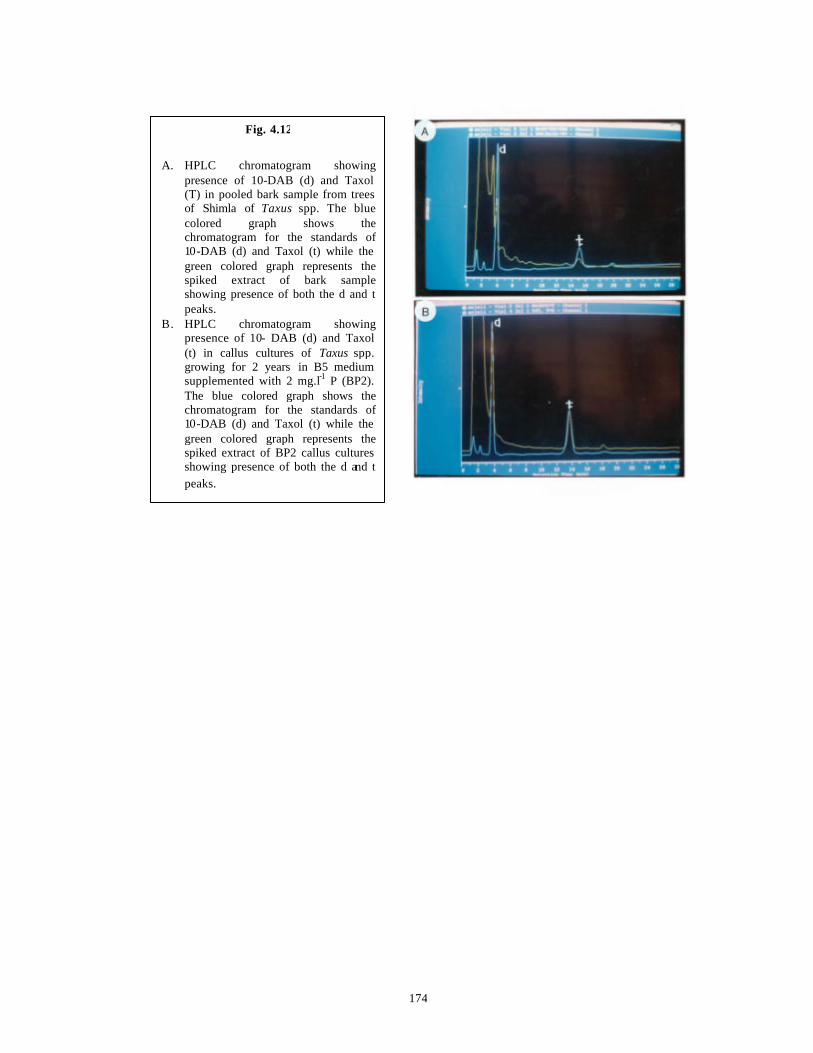
174
Fig. 4.12
A. HPLC chromatogram showing
presence of 10-DAB (d) and Taxol (T) in pooled bark sample from trees of Shimla of Taxus spp. The blue colored graph shows the chromatogram for the standards of 10-DAB (d) and Taxol (t) while the green colored graph represents the spiked extract of bark sample showing presence of both the d and t peaks.
B. HPLC chromatogram showing presence of 10- DAB (d) and Taxol (t) in callus cultures of Taxus spp. growing for 2 years in B5 medium supplemented with 2 mg.l-1 P (BP2). The blue colored graph shows the chromatogram for the standards of 10-DAB (d) and Taxol (t) while the green colored graph represents the spiked extract of BP2 callus cultures showing presence of both the d and t peaks.
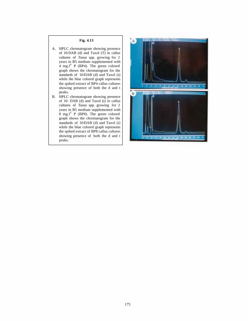
175
Fig. 4.13
A. HPLC chromatogram showing presence of 10-DAB (d) and Taxol (T) in callus cultures of Taxus spp. growing for 2 years in B5 medium supplemented with 4 mg.l-1 P (BP4). The green colored graph shows the chromatogram for the standards of 10-DAB (d) and Taxol (t) while the blue colored graph represents the spiked extract of BP4 callus cultures showing presence of both the d and t peaks.
B. HPLC chromatogram showing presence of 10- DAB (d) and Taxol (t) in callus cultures of Taxus spp. growing for 2 years in B5 medium supplemented with 8 mg.l-1 P (BP8). The green colored graph shows the chromatogram for the standards of 10-DAB (d) and Taxol (t) while the blue colored graph represents the spiked extract of BP8 callus cultures showing presence of both the d and t peaks.
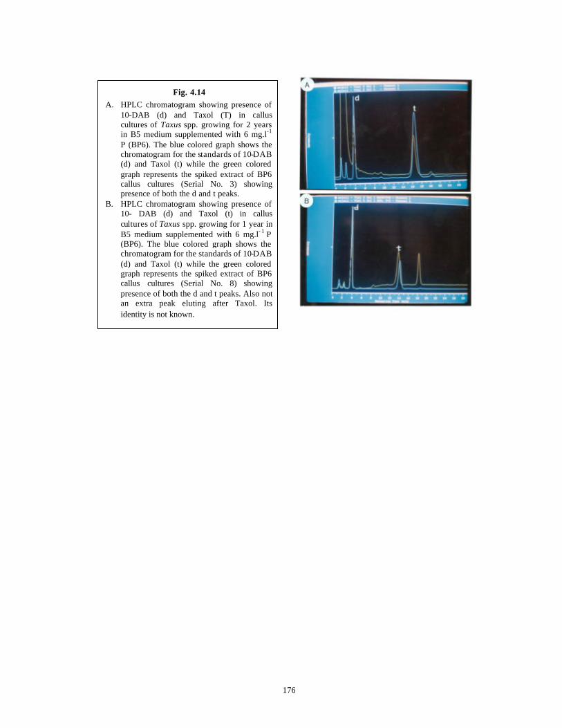
176
Fig. 4.14 A. HPLC chromatogram showing presence of
10-DAB (d) and Taxol (T) in callus cultures of Taxus spp. growing for 2 years in B5 medium supplemented with 6 mg.l-1
P (BP6). The blue colored graph shows the chromatogram for the standards of 10-DAB (d) and Taxol (t) while the green colored graph represents the spiked extract of BP6 callus cultures (Serial No. 3) showing presence of both the d and t peaks.
B. HPLC chromatogram showing presence of 10- DAB (d) and Taxol (t) in callus cultures of Taxus spp. growing for 1 year in B5 medium supplemented with 6 mg.l- 1 P (BP6). The blue colored graph shows the chromatogram for the standards of 10-DAB (d) and Taxol (t) while the green colored graph represents the spiked extract of BP6 callus cultures (Serial No. 8) showing presence of both the d and t peaks. Also not an extra peak eluting after Taxol. Its identity is not known.
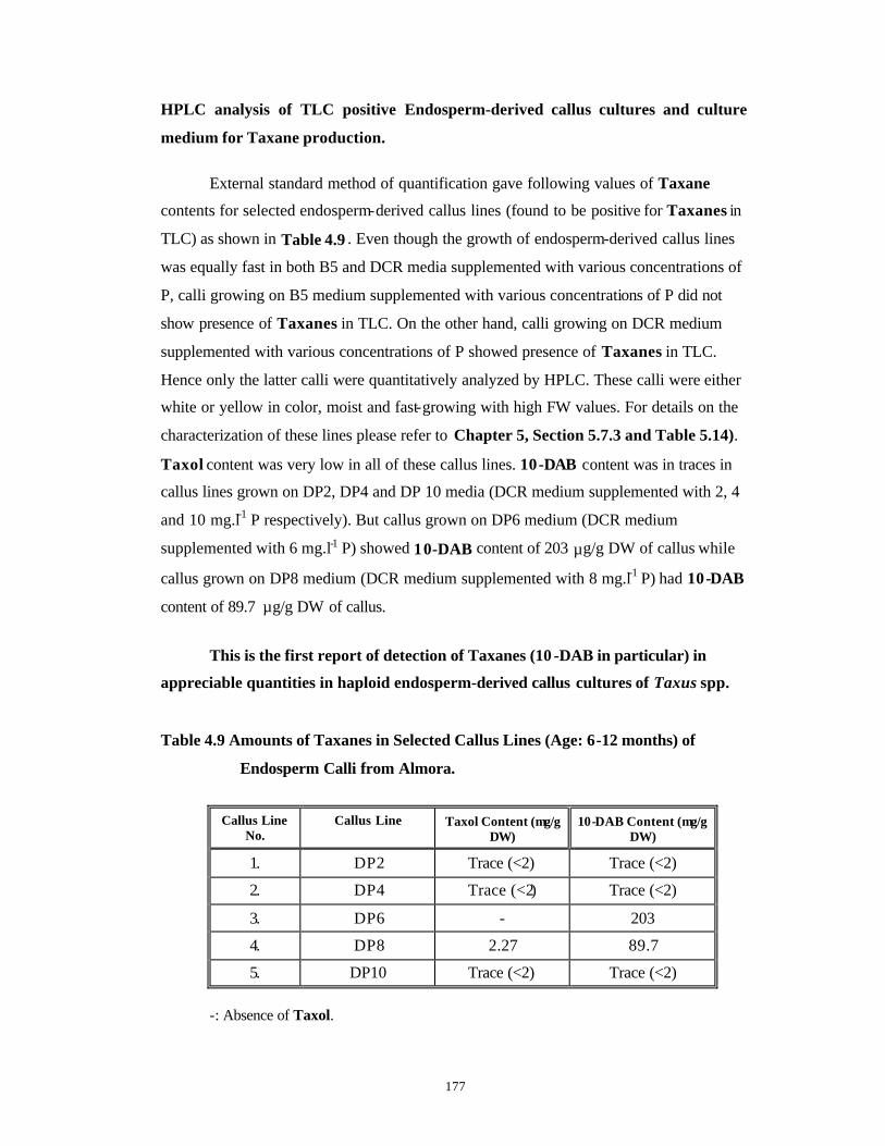
177
HPLC analysis of TLC positive Endosperm-derived callus cultures and culture
medium for Taxane production.
External standard method of quantification gave following values of Taxane
contents for selected endosperm-derived callus lines (found to be positive for Taxanes in
TLC) as shown in Table 4.9 . Even though the growth of endosperm-derived callus lines
was equally fast in both B5 and DCR media supplemented with various concentrations of
P, calli growing on B5 medium supplemented with various concentrations of P did not
show presence of Taxanes in TLC. On the other hand, calli growing on DCR medium
supplemented with various concentrations of P showed presence of Taxanes in TLC.
Hence only the latter calli were quantitatively analyzed by HPLC. These calli were either
white or yellow in color, moist and fast-growing with high FW values. For details on the
characterization of these lines please refer to Chapter 5, Section 5.7.3 and Table 5.14).
Taxol content was very low in all of these callus lines. 10-DAB content was in traces in
callus lines grown on DP2, DP4 and DP 10 media (DCR medium supplemented with 2, 4
and 10 mg.l-1 P respectively). But callus grown on DP6 medium (DCR medium
supplemented with 6 mg.l-1 P) showed 10-DAB content of 203 µg/g DW of callus while
callus grown on DP8 medium (DCR medium supplemented with 8 mg.l-1 P) had 10-DAB
content of 89.7 µg/g DW of callus.
This is the first report of detection of Taxanes (10 -DAB in particular) in
appreciable quantities in haploid endosperm-derived callus cultures of Taxus spp.
Table 4.9 Amounts of Taxanes in Selected Callus Lines (Age: 6-12 months) of
Endosperm Calli from Almora.
Callus Line No.
Callus Line Taxol Content (µg/g DW)
10-DAB Content (µg/g DW)
1. DP2 Trace (<2) Trace (<2)
2. DP4 Trace (<2) Trace (<2)
3. DP6 - 203
4. DP8 2.27 89.7
5. DP10 Trace (<2) Trace (<2)
-: Absence of Taxol.
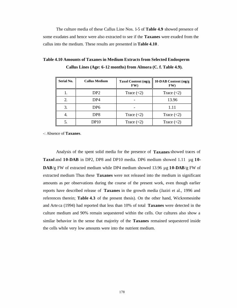
178
The culture media of these Callus Line Nos. 1-5 of Table 4.9 showed presence of
some exudates and hence were also extracted to see if the Taxanes were exuded from the
callus into the medium. These results are presented in Table 4.10 .
Table 4.10 Amounts of Taxanes in Medium Extracts from Selected Endosperm
Callus Lines (Age: 6-12 months) from Almora (C. f. Table 4.9).
Serial No. Callus Medium Taxol Content (µg/g
FW) 10-DAB Content (µg/g
FW)
1. DP2 Trace (<2) Trace (<2)
2. DP4 - 13.96
3. DP6 - 1.11
4. DP8 Trace (<2) Trace (<2)
5. DP10 Trace (<2) Trace (<2) -: Absence of Taxanes.
Analysis of the spent solid media for the presence of Taxanes showed traces of
Taxol and 10-DAB in DP2, DP8 and DP10 media. DP6 medium showed 1.11 µg 10-
DAB/g FW of extracted medium while DP4 medium showed 13.96 µg 10-DAB /g FW of
extracted medium Thus these Taxanes were not released into the medium in significant
amounts as per observations during the course of the present work, even though earlier
reports have described release of Taxanes in the growth media (Jaziri et al., 1996 and
references therein; Table 4.3 of the present thesis). On the other hand, Wickremesinhe
and Arteca (1994) had reported that less than 10% of total Taxanes were detected in the
culture medium and 90% remain sequestered within the cells. Our cultures also show a
similar behavior in the sense that majority of the Taxanes remained sequestered inside
the cells while very low amounts were into the nutrient medium.
179
4.3.3 Initiation and growth determinations in suspension cell cultures
In the course of the present work, only preliminary studies with cell suspension
cultures were carried out to see if some of the callus lines obtained during the course of
this work are amenable for liquid culture and to see if the Taxane contents could be
increased by cell suspension culture.
Cell suspension cultures were initiated as described earlier in Section 4.2.3.2 only
on B5 medium supplemented with 2-10 mg.l-1 P (Optimized medium for best growth and
Taxane content) by random selection of calli (Serial Nos. 8, 9, 14 and a non-producer
callus with Serial No. 5 of Table 4.8). Fig. 4.15 shows microscopic view of cell
suspension grown in BP6 and BP8 media (Cell Suspension Line Nos. 1 and 2).
Dry weight (DW) analyses of cell cultures were done at the time of extraction for
HPLC analysis. Fig. 4.16 is a representative graph showing changes in DW with time in
selected cell suspension cultures. It is clear from the graph that except for the cell
suspension grown in BP10 medium, all the other three cell suspensions showed the
typical growth curve of a liquid cell suspension culture. The lag phase of growth was over
by day 9 when the cells entered the log phase of growth and even at day 18 this growth
was not declining towards stationary phase. On the contrary, cell suspension grown in
BP10 medium exhibited a relatively flat pattern of growth.
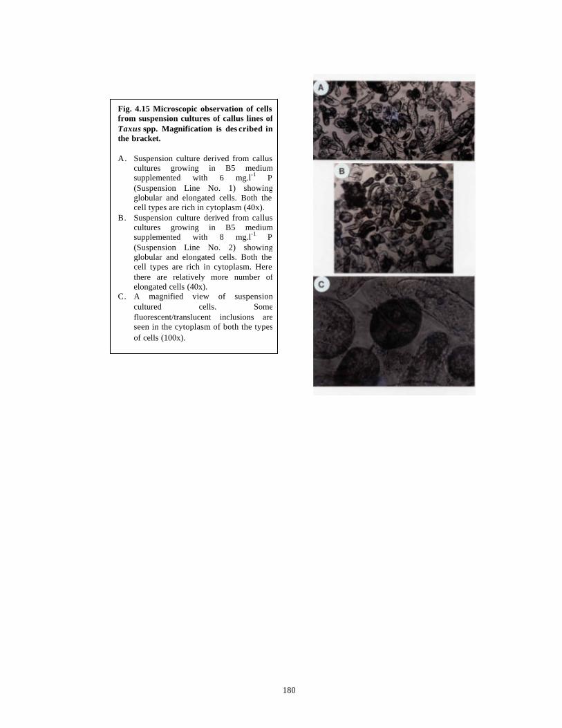
180
Fig. 4.15 Microscopic observation of cells from suspension cultures of callus lines of Taxus spp. Magnification is des cribed in the bracket. A. Suspension culture derived from callus
cultures growing in B5 medium supplemented with 6 mg.l-1 P (Suspension Line No. 1) showing globular and elongated cells. Both the cell types are rich in cytoplasm (40x).
B. Suspension culture derived from callus cultures growing in B5 medium supplemented with 8 mg.l-1 P (Suspension Line No. 2) showing globular and elongated cells. Both the cell types are rich in cytoplasm. Here there are relatively more number of elongated cells (40x).
C. A magnified view of suspension cultured cells. Some fluorescent/translucent inclusions are seen in the cytoplasm of both the types of cells (100x).
181
4.3.4 Analysis of Taxanes in selected cell suspension cultures of Taxus spp.
Table 4.11 depicts the results of HPLC obtained with cell suspension cultures
derived from some of the randomly selected callus lines. The suspension cultures initiated
from these callus lines are numbered as in Table 4.11. A dramatic increase (almost 10
times) in Taxol content was evident in Cell Suspension Line No. 1 from day 0 to day 9
and then its total disappearance at day 18. 10-DAB content decreased steadily from day 0
to day 18 in this line. Fig. 4.17 B represents the chromatogram for BP6 cell suspension
culture (Cell Suspension Line No. 1) derived from Callus Line with Serial No. 8 in Table
4.8 while Fig. 4.17 A represents the chromatogram of the standard solutions of 10-DAB
and Taxol. Cell Suspension Line No. 2 showed an abrupt decrease in Taxol and 10-DAB
contents from day 0 to day 9 itself in liquid medium. Cell Suspension Line No. 3 initially
had undetectable levels of Taxol but contained very high levels of 10-DAB . Over the
culture period of 18 days, 10-DAB content decreased rapidly and steadily while Taxol
contents increased concomitantly. In contrast, the most surprising observation was that
the Taxane non-producer Cell Suspension Line No. 4 (TLC and HPLC results were
negative for parent callus culture), exhibited presence of both Taxol and 10-DAB in
appreciable quantities in suspension culture and they were still on the upward spiral
even at day 18. More attention is required in continuing these cell suspension lines in
culture beyond 18 days to elucidate the pattern of Taxane production versus the age of
cell suspension culture.
The spent liquid media analyzed by HPLC showed extremely low amounts of
Taxanes (< 2 µg/ml liquid medium) (Table 4.11 ). This proves that all the Taxanes are
sequestered within the cells. One possible reason is that since Taxol is a lipophilic
compound, it is not getting excreted in the aqueous extracellular medium. Russin et al.
(1995) have shown by immunohistochemistry that Taxol is localized in cell walls of
xylem, phloem and vascular cambium and hence cannot be easily released into the
medium.
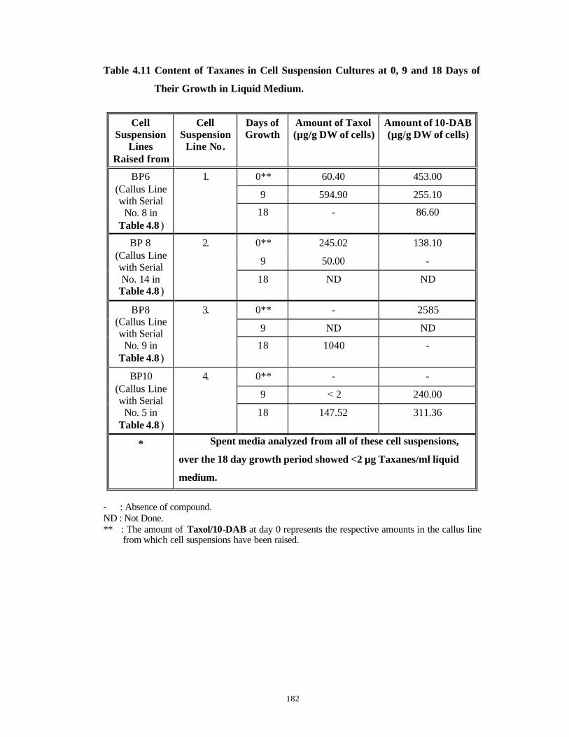
182
Table 4.11 Content of Taxanes in Cell Suspension Cultures at 0, 9 and 18 Days of
Their Growth in Liquid Medium.
Cell Suspension
Lines Raised from
Cell Suspension
Line No.
Days of Growth
Amount of Taxol (µg/g DW of cells)
Amount of 10-DAB (µg/g DW of cells)
0** 60.40 453.00
9 594.90 255.10
BP6 (Callus Line with Serial
No. 8 in Table 4.8 )
1.
18 - 86.60
0** 245.02 138.10
9 50.00 -
BP 8 (Callus Line with Serial No. 14 in
Table 4.8 )
2.
18 ND ND
0** - 2585
9 ND ND
BP8 (Callus Line with Serial
No. 9 in Table 4.8 )
3.
18 1040 -
0** - -
9 < 2 240.00
BP10 (Callus Line with Serial
No. 5 in Table 4.8 )
4.
18 147.52 311.36
* Spent media analyzed from all of these cell suspensions,
over the 18 day growth period showed <2 µg Taxanes/ml liquid
medium.
- : Absence of compound. ND : Not Done. ** : The amount of Taxol/10-DAB at day 0 represents the respective amounts in the callus line
from which cell suspensions have been raised.
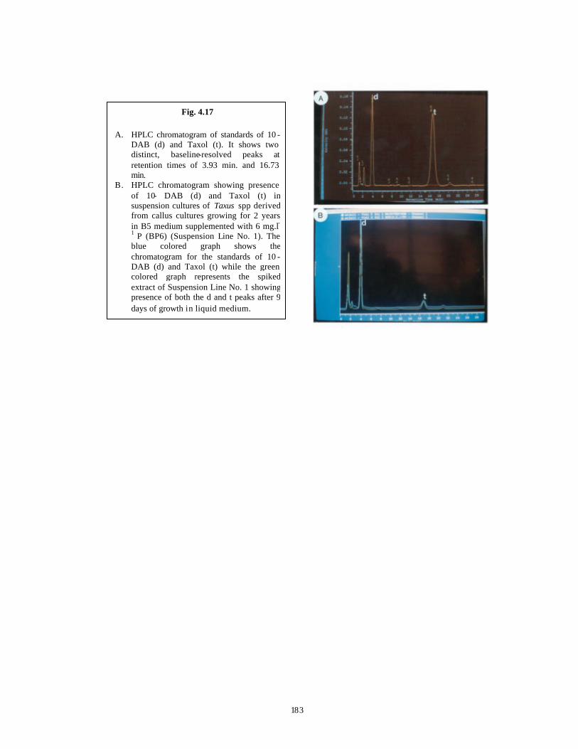
183
Fig. 4.17 A. HPLC chromatogram of standards of 10 -
DAB (d) and Taxol (t). It shows two distinct, baseline-resolved peaks at retention times of 3.93 min. and 16.73 min.
B. HPLC chromatogram showing presence of 10- DAB (d) and Taxol (t) in suspension cultures of Taxus spp derived from callus cultures growing for 2 years in B5 medium supplemented with 6 mg.l-1 P (BP6) (Suspension Line No. 1). The blue colored graph shows the chromatogram for the standards of 10 -DAB (d) and Taxol (t) while the green colored graph represents the spiked extract of Suspension Line No. 1 showing presence of both the d and t peaks after 9 days of growth in liquid medium.
184
4.4 Conclusions
1) Callus cultures were raised from stem bits and needle (diploid calli)
explants of mature trees of Taxus baccata ssp. wallichiana Zucc. Pilg.
and from endosperm in seeds (haploid calli)
2) Callus initiation was achieved on 1/2 SH, B5, DCR and WPM media each
supplemented individually with 2, 4, 6, 8 and 10 mg.l-1 2,4-D from both
stem bits and needle explants.
3) The stem bit explants formed callus with 20% efficiency as opposed to
100% for needle explants.
4) B5 medium with a number of growth regulators was the best medium for
survival and good growth of needle -derived calli over monthly
subcultures for more than 4 years.
5) Needle explants gave rise to callus with fast growth rate and high FW on
B5 medium supplemented individually with high concentrations (6, 8,
10 mg.l-1) of auxins like 2,4 -D, NAA and P. The best callus growth rate
and high FW was obtained in calli grown on P containing media.
6) B5 medium supplemented individually with 2, 4, 6, 8 and 10 mg.l-1 of
auxins like IAA and IBA individually, produced insignificant amounts
of callus from needles.
7) Callus induction from needle explants and the subsequent callus growth
were very good on B5 medium supplemented with auxins like IAA and
IBA together in various concentrations.
8) Callus growth of needle-derived calli did not improve on B5 medium with
2-iP combined with IBA and KIN combined with 2,4 -D and NAA.
9) Taxane production was not detected in needle -derived calli cultured on
B5 medium supplemented with various combinations of 2,4-D + KIN
and NAA + KIN. Taxane production did not improve with age of such
calli.
10) B5 medium supplemented with Picloram was found to be the best
medium for good growth of needle -derived callus cultures as well as
Taxane production therefrom.
11) Taxane contents increased with increasing age of callus tissues in
culture on the optimized B5 medium with P. The highest Taxol content

185
was 540 µg/g DW of callus in 2 year old needle -derived diploid callus
line with Serial No. 3 growing on B5 medium supplemented with 6 mg.l-
1 P.
12) Needle-derived Callus Line with Serial No. 9 on B5 medium
supplemented with 8 mg.l-1 P had the highest 10-DAB content of 2585
µg/g DW of callus.
13) Higher P concentrations above 4 mg.l-1 are conducive for both Taxol and
10-DAB production.
14) Endosperm-derived callus growing on DCR medium supplemented with
8 mg.l-1 P produced 203 µg 10-DAB/g DW of callus.
15) Taxanes were not detected in the spent solid or liquid nutrient media
used for growing either haploid or diploid callus cultures.
16) Taxane contents increased many-fold in three out of the four cell
suspension lines analyzed.
17) Taxol and 10-DAB were detected in Taxane non-producer Callus Line
with Serial No. 5 on raising a cell suspension from such a callus. The
contents of both these compounds increased with the increase in cell
suspension culture period from 0 days to 18 days.

186
CCHHAAPPTTEERR 55
In vitro Studies on Micropropagation from Mature
and Juvenile Explants of Taxus spp.
187
5.1 Introduction
The goal of ‘Clonal Pro pagation’ or ‘Micropropagation’ is to obtain a large
number of genetically identical, physiologically uniform and developmentally normal
plantlets, preferably with a high photosynthetic or photoautotrophic potential to survive
the harsh ex-vitro conditions, in a reduced time period and at a lowered cost (Jeong et al.,
1995). Although true-to-type plantlets are the desired goal of clonal propagation, due to
involvement of tissue culture component, many times, somaclonal variants are obtained.
These can be used for selection of new useful variants, which can then be propagated on a
large scale by micropropagation. E.g. stress and disease tolerant variants.
There are many advantages of clonal propagation (Vasil and Vasil, 1980) like:
a. Uniform genetic background.
b. Production of uniformly superior seeds.
c. Improved progeny evaluation of breeding experiments.
d. Improved vigor and quality.
e. Elimination of pathogens.
f. An improved aesthetic value due to richness and fullness.
g. Facilitation of international transport and exchange of germplasm without quarantine.
h. Fast release of newly developed cultivar.
There are three methods of ‘Clonal Propagation or Micropropagation’ (Vasil and
Vasil, 1980):
(1) Enhanced Axillary Bud Breaking – It is the slowest method as compared to the other
two methods but is widely used in commercial set-ups due to assured genetic
uniformity of newly derived plants.
(2) Production of Adventitious Buds/Organogenesis – This involves de novo formation of
meristematic loci on the explant, leading to development of root or shoot meristems,
depending upon the balance of plant growth regulators in the growth medium and a
number of other physical and physiological factors. Although a large number of
plants can be regenerated by this procedure, it does not have a wider applicability
PART A: MICROPROPAGATION STUDIES USING MATURE
EXPLANTS OF TAXUS SPP.
188
since many of the economically important plant species do not undergo organogenesis
in vitro .
(3) Somatic Embryogenesis – These are the asexual, bipolar embryos produced from
somatic cells of the explant due to hormonal stimuli. This method of pr opagation
provides the fastest route for production of a large number of clonal plants since the
somatic embryos contain both the shoot and the root poles. But till date very few
species have demonstrated their potential for direct somatic embryogenesis.
Especially in Gymnosperms, few successes have been achieved only from zygotic
embryos.
The method of enhanced axillary bud breaking produces the least number of
plantlets as the number of shoots produced is controlled by the number of axillary buds
cultured. But this is the most widely used method of micropropagation because:
I. It produces true -to-type plantlets with a uniform genetic background (Brown and
Thorpe, 1995).
II. The method is relatively simpler with a reasonably good rate of propagation
(Pierik, 1987).
III. The growth of the resulting plants is very good either due to rejuvenation and/or
the lack of internal infections (Pierik, 1987).
This method of enhanced axillary bud breaking is widely used for angiospermic
hardwoods (Bonga and Von Aderkas, 1992) but rarely used for conifers (Thorpe et al.,
1991). Very few successes have been achieved in conifers starting from mature material
and even in successful cases, rarely more than three shoots have been obtained from a
single explant and rooting has been problematic as has been described in the following
examples: 30 year old Larix spp. (Bonga and Pond, 1991), 19 year old Larix decidua
(Diner, 1995) 4-16 year old Psuedotsuga spp. (Pullman and Timmis, 1992), 90 year old
Sequoia sempervirens (Bon et al., 1994), 30 year old Juniperus brevifolia (Moreira da
Silva and Debergh, 1996) and 20 year old Pinus spp. (Nadgauda et al., 1993). However It
is necessary to use explants from elite, mature trees in order to obtain the maximum
genetic gains in terms of desired characters like wood/pulp making quality, production of
high value secondary metabolites, reforestation potential etc.
189
But such mature explants are rarely responsive in culture due to a number of
factors such as (Modified from Pierik, 1987):
(1) Chronological, physiological and ontogenetical aging of mature explants
(Fortanier and Jonkers, 1976); where a number of morphological and physiological
attributes are affected unidirectionally and many times irreversibly. Hence for such
explants to respond, a reversion in their maturation-related characters that is
“Rejuvenation” needs to occur in vitro at the meristematic regions. The induction of
rejuvenation is extremely difficult in woody spp.
(2) In general, woody plants have relatively weak regenerative capacity and lower
multiplication rates.
(3) Because of strong dormancy observed in woody plants, buds do not open and stem
elongation fails to take place.
(4) “Topophysis” (The phenomenon whereby the position of the explant in the plant
affects its in vitro growth and response after isolation and culture due to gradients of
regeneration) plays a greater role in woody plants.
(5) Sterilization of mature explants, especially those grown under field conditions, is
difficult to achieve successfully.
(6) The explants derived from field grown plants exhibit considerable variations in vitro,
due to the different growth conditions and annual fluctuations in the climate in vivo.
(7) The adult trees generally have a great degree of genetic variation as compared to
agricultural and horticultural plants and hence the results obtained are highly variable
and many times difficult to repeat (Pierik, 1987).
In addition, explants of mature origin have high rates of contamination and
phenolic oxidation as described earlier in Chapter 3 in detail.
The success of micropropagation also depends on following factors in vitro such
as:
• Biological Factors (Pierik, 1987) such as genotype, age, health and physiology of the
parent plant, environmental conditions pertaining to parent plants, position, size and
method of inoculation of the explant.
190
• Physical Factors (Pierik, 1987; Jeong et al., 1995) such as light including day length,
spectral composition and irradiance, temperature, water availability, gaseous
components such as oxygen, carbon dioxide, ethylene and other hydrocarbons,
electric currents, medium composition and volume.
5.1.1 Vegetative propagation of Taxus spp.
Normally, vegetative propagation is achieved through cuttings. In case of Taxus
spp., the percentage of rooting varied between cultivars and species depending on the
conditions of temperature, misting and use of IBA. 63-100% rooting was observed with
many species and cultivars (Eccher, 1988). Chee (1995) reported that stem cuttings of T.
cuspidata treated with an aqueous solution of IBA (0.2%) + NAA (0.1%) + Thiamine
(0.08%) produced roots at a frequency of 73.5%.
But the success rate was not very high for the Himalayan Yew (20-30%) and the
cuttings took at least three months to root. Success of grafting and layering was also not
very encouraging (Chatterjee and Dey, 1997). A variety of reasons have been attributed
for this low rate of success in rooting (Mitchell, 1997 and references therein):
§ Great age of parent trees (more than 100 years) and the associated low growth vigor.
§ Variation in rooting among parent trees from different geographical locations due to
variations in a number of ecological and environmental factors.
§ Variation in rooting percentage between different species and cultivars as well as
between male and female trees.
§ The yews tend to grow in stressful environments (too cold, too exposed) which might
reduce the shoot vigor, finally adversely affecting rooting success.
The roots of mature trees of Yews are associated with ectomycorrhizal mats
(ECM) and vesicular arbuscular mycorrhiza (VAM) in their natural habitats. ECM and
VAM might play important roles in rooting and establishment of cuttings derived from
mature trees by suppressing root pathogens and increasing the available nutrients
(Griffiths et al., 1995).
191
5.1.2 In vitro propagation of Taxus spp.
Even though success with vegetative propagation has not been significant in
Taxus spp., not much work has been done with respect to micropropagation studies in
vitro in Taxus spp. There are no full-length papers on micropropagation from mature
explants of Taxus spp. in the literature. Only two abstracts report formation of 2-4 shoots
in Taxus spp. (Amos and McCown, 1981; Barnes, 1983) without much information on the
media used and the culture conditions. Also there are no reports on micropropagation in
the Himalayan Yew. Precocious germination from seeds of Taxus spp., de novo
adventitious bud development and somatic embryogenesis are some of the studies carried
out in Taxus spp. so far and are presented in Table 5.1. In addition, much of the
information has been in the form of patents thus restricting its use and availability in the
public domain.
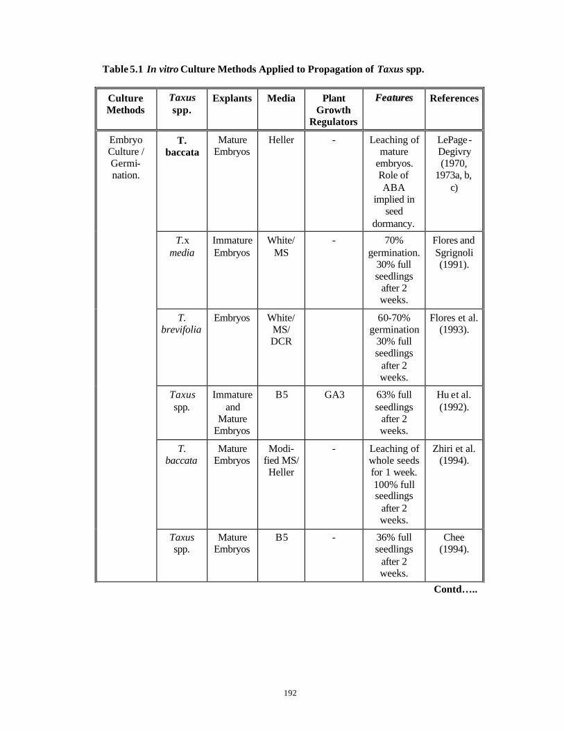
192
Table 5.1 In vitro Culture Methods Applied to Propagation of Taxus spp.
Culture Methods
Taxus spp.
Explants Media Plant Growth
Regulators
FFeeaattuurreess References
T. baccata
Mature Embryos
Heller - Leaching of mature
embryos. Role of ABA
implied in seed
dormancy.
LePage-Degivry (1970,
1973a, b, c)
T.x media
Immature Embryos
White/ MS
- 70% germination.
30% full seedlings
after 2 weeks.
Flores and Sgrignoli (1991).
T. brevifolia
Embryos White/ MS/ DCR
60-70% germination
30% full seedlings
after 2 weeks.
Flores et al. (1993).
Taxus spp.
Immature and
Mature Embryos
B5 GA3 63% full seedlings
after 2 weeks.
Hu et al. (1992).
T. baccata
Mature Embryos
Modi-fied MS/ Heller
- Leaching of whole seeds for 1 week. 100% full seedlings
after 2 weeks.
Zhiri et al. (1994).
Embryo Culture / Germi-nation.
Taxus spp.
Mature Embryos
B5 - 36% full seedlings
after 2 weeks.
Chee (1994).
Contd…..

193
Table 5.1 Contd.
Culture Methods
Taxus spp.
Explants Media Plant Growth
Regulators
FFeeaattuurreess References
T. mairei, T.
baccata, T.
canade-nsis, T.
media, T. cuspidata
Mature and
Immature embryos
1/2 MS + PVP
GA3 15% germination
from immature embryos and 90%
germination from
mature embryos.
Chang and Yang
(1996).
Embryo Culture / Germi-nation.
T. chinensis
Mature embryos
B5, New medium with AC,
20% sugar
- 97% germination
Chien et al. (1998).
Taxus spp.
Shoot tips
WPM BAP 2-4 shoots per explant
Amos and McCown (1981).
T. floridana
Shoot tips
WPM BAP 2-4 shoots per explant
Barnes (1983).
T. brevifolia
Embryos 1/2 B5 BAP Multiple shoots, 58%
rooting, plantlets.
Chee (1995b).
Micro-propagation
T. mairei Shoot tips and
stem from 6 to
18 months
old seedlings
MS BAP Both orthotropic
and plagiotropic
shoots obtained.
60% rooting.
Chang et al. (1998).
Contd…..
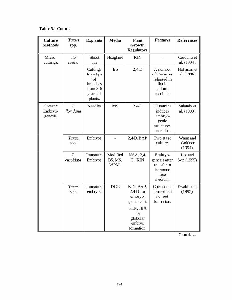
194
Table 5.1 Contd.
Culture Methods
Taxus spp.
Explants Media Plant Growth
Regulators
FFeeaattuurreess References
Shoot tips
Hoagland KIN - Cerdeira et al. (1994).
Micro-cuttings.
T.x media
Cuttings from tips
of branches from 3-6 year old plants.
B5 2,4-D A number of Taxanes released in
liquid culture
medium.
Hoffman et al. (1996)
T. floridana
Needles MS 2,4-D Glutamine induces embryo-
genic structures on callus.
Salandy et al. (1993).
Taxus spp.
Embryos - 2,4-D/BAP Two stage culture.
Wann and Goldner (1994).
T. cuspidata
Immature Embryos
Modified B5, MS, WPM.
NAA, 2,4-D, KIN
Embryo-genesis after
transfer to hormone
free medium.
Lee and Son (1995).
Somatic Embryo-genesis.
Taxus spp.
Immature embryos
DCR KIN, BAP, 2,4-D for embryo-
genic calli.
KIN, IBA for
globular embryo
formation.
Cotyledons formed but
no root formation.
Ewald et al. (1995).
Contd…..

195
Table 5.1 Contd.
Culture Methods
Taxus spp.
Explants Media Plant Growth
Regulators
FFeeaattuurreess References
Somatic Embryo-genesis.
T. brevifolia
Immature Embryos
WPM (MCM)
2,4-D, BAP, NAA
for embryo-
genic callus.
BAP, KIN, NAA for embryoid formation.
5% emblings
produced on hormone free 1/2 WPM
(MCM).
Chee (1996).
Nodule Culture
Taxus spp.
Juvenile needles from in
vitro shoot
cultures.
Modified WPM
NAA, 2-iP Cohesive cellular units of nodules
with differentia -ted internal structures.
Ellis et al. (1996).
From the literature review presented in Table 5.1, it is clear that it is extremely
important to develop a micropropagation protocol for the Himalayan Yew as it is
becoming endangered in its natural habitat due to merciless felling of the trees or
unauthorized stripping of the bark for Taxol extraction, leading t o slow death of the trees.
New stocks of plants are required to replenish the natural resources. Even though the
juvenile shoots produced in vitro are not desirable for planting, they provide an
opportunity in the elucidation of production of Taxane class of compounds.
Micropropagation by use of mature explants is desirable as the advantages far outweigh
the disadvantages faced when using mature explants in vitro .
196
In view of the aforesaid, the objectives of the present work were: 1. Optimization of nutrient media, plant growth regulators and culture conditions for
achieving enhanced axillary budding from stem explants of mature trees of Taxus
baccata ssp. wallichiana Zucc. Pilg. collected from Shimla.
2. Establishment of primary sprouts and subsequent initiation as well as establishment of
secondary sprouts.
3. Extrapolation of the standardized protocol for plant material of Taxus spp. collected
from other locations in the Himalayas.
4. Preliminary studies on precocious embryo germination, seedling establishment and
raising of callus cultures from endosperm halves of seeds of the Himalayan Yew.
5. Analysis of Taxane contents in callus cultures derived from endosperm of the
Himalayan Yew.
The results achieved on the objectives are presented in this chapter as two
parts:
PART A: Deals with the attempts on development of a widely applicable
micropropagation protocol for Taxus spp. from mature explants,
collected from different locations in the Himalayas.
PART B: Describes the results of in vitro studies with embryo and endosperm
explants of Taxus spp.

197
5.2 Materials and Methods
5.2.1 Collection of plant material
Collection of twigs of Taxus baccata ssp wallichiana Zucc. Pilg. from various
locations in the Himalayas and the preparation of explants for in vitro studies were as
described in detail in Chapter 4, Section 4.2.1.
5.2.2 Pre -treatment and surface sterilization of explants
Stem bit explants were surface sterilized according to the “Modified Surface
Sterilization Protocol” described in Chapter 3, Flow Chart. 3.3.
5.2.3 Media and culture conditions
A number of basal media either at full strength or at half strength (with major salts
reduced to half strengths) were used for axillary bud break. In the pilot experiments, a
number of basal media at full strength suc h as: Murashige and Skoog’s MS medium
(1962) (High salt medium), Gamborg’s B5 medium (1968) (Medium salt medium), Lloyd
and McCown’s Woody Plant Medium, WPM (1981) (Low salt medium) without addition
of plant growth regulators were used. The composition details were as described in
Chapter 2, Table 2.1. WPM was selected for further study based on the sprouting
obtained and shoot survival and was supplemented individually with various
concentrations of TDZ (0.01, 0.05, 0.1, 0.5, 1.0 mg.l-1) and BAP (0.1, 2.0, 5.0 mg.l-1).
To further optimize the results obtained, the following media at half strength of
major salts were used: WPM (Based on pilot experiment data), Gupta and Durzan’s DCR
medium (1985), Litvay’s Medium, LM (1981) and Modified Litvay’s Medium, MLM
(Kulkarni, 1997) which was modified to contain 10 mM each of KNO3, NH4NO3 and
glutamine. The composition details were as described in Chapter 2, Table 2.1. For each
experiment, these half strength media without any plant growth regulator served as
control treatments (1/2 WPM-H, 1/2 DCR-H, 1/2 LM-H and 1/2 MLM-H). 1/2 WPM and
1/2 DCR media were further modified by individual additions of a number of cytokinins
like BAP, KIN, TDZ and 2-iP at concentrations of 0.01, 0.05, 0.1 and 0.5 mg.l-1. In one
set of experiments, IBA and BAP were used together. All the media contained 2%
sucrose and 0.85% agar and 0.5% activated charcoal (AC) in case of solid media. Liquid
198
media had only 2% sucrose (No agar and AC) and support to the explants was provided
with Whatman No. 1 filter paper bridges. Media used for establishment of primary and
secondary sprouts were appended with 10% coconut milk (CM) and 1g.l-1 of casein
hydrolysate (CH). All other details regarding media preparation, inoculation and culture
of explants and routine tissue culture practices were as described earlier in Chapter 2,
Sections 2.4, 2.5 and Chapter 4, Section 4.2.3 .
Stem bit explants with axillary buds and trimmed needles were inoculated
independently in each culture tube and incubated either in the dark or at light intensities
of 4.6 or 19 µEm-2s -1 provided by cool white fluorescent tubelights (“Phillips”, India) for
16 hours at a temperature of 25 + 2oC.
5.2.4 Elongation and establishment of primary sprouts
Initiation of primary sprouts from axillary buds of the stem bit explants was
observed in 25-35 days on the optimal media (C. f. Tables 5.5, 5.5 A). Elongation of
these occurred on the same media to a length of 0.5-1.0 cm during the second monthly
transfer. In the third monthly transfer, the elongated sprouts were transferred to the same
basal media without plant growth regulators where the sprouts grew into well-developed
shoots of a length upto 3 cm. These shoots were then detached from the mother explant
and cultured individually either on the same basal medium without plant growth
regulators or on the same basal medium supplemented with 10% CM and 1 g.l-1 CH for
independent establishment of primary shoots (C. f. Table 5.9 ). The cultures were
incubated under the conditions described under Section 5.2.3, Media and culture
conditions .
5.2.5 Secondary sprouting
The elongated primary shoots were cultured either on 1/2 WPM or 1/2 DCR basal
media supplemented individually with low concentrations of KIN or BAP (0.01, 0.05 and
0.1 mg.l-1), 10% CM and 1 g.l-1 CH, for induction of secondary sprouting.

199
5.2.6 Statistical analysis
Each culture tube with one shoot explant was considered as one replicate. Each
treatment in each set of experiments consisted of 50 such replicates and each experiment
was repeated three or more times over successive years. The treatments were analyzed by
one-way ANOVA and the individual treatment means were compared by Student’s t-test
(Wardlaw, 1985).
200
5.3 Results and Discussion
5.3.1 Primary sprouting from mature shoot explants
5.3.1.1 Plant material of Taxus spp. collected from Shimla
Initially stem bit explants were cultured on full strength MS, B5 and WPM basal
media without plant growth regulators (compositions described earlier in Chapter 2,
Table 2.1). MS medium has a high total ionic strength of salts at 100.48 mM while B5
medium has a total ionic strength of salts at 65.738 mM (C. f. Chapter 2, Table 2.2). All
the explants (100%) inoculated in these media exuded heavy amount of phenolics due to
high ionic strengths of these media and were dead within three days. Hence these media
were not used for further studies. Only the explants inoculated in full strength WPM
without plant growth regulators, with a total ionic strength of 43.516 mM (C. f. Chapter
2, Table 2.2 ), produced less phenolics and responded to axillary shoot development at a
frequency of 42.94% (Table 5.2 ). This medium was therefore chosen for further
experiments with cytokinins. Full strength WPM was modified by individual addition of
BAP and TDZ at a number of concentrations (Table 5.2). BAP and TDZ were chosen for
these initial experiments because they had been reported to be useful in Conifer/Woody
plants micropropagation (Fiola et al., 1990 and references therein; Thorpe et al., 1991).
Table 5.2 and Fig. 5.1 describe the results obtained.

201
Table 5.2 Effect of Different Plant Growth Regulator Concentrations on %
Sprouting from Mature Explants Cultured on Full Strength WPM.
(Plant material collected from Shimla during 1995).
No. Plant Growth Regulator Concentration (mg.l-1)
% Sprouting Mean + S. E.
1. Nil (Control) - 42.94 + 1.32a
2. 0.01 41.05 + 1.05a
3. 0.05 25.00 + 0.00a
4. 0.1 10.55 + 0.55b
5. 0.5 80.00 + 20.00c
6.
TDZ
1.0 8.83 + 2.17d
7. 0.1 31.67 + 1.67a
8. 2.0 37.5 + 2.5a
9.
BAP
5.0 58.3 + 8.3b
a, b, c, d : Values followed by different letters are significantly different from each other at 95%
confidence level by t-test.
Although the maximum initiation of primary sprouting was observed with higher
concentrations of TDZ (80% at 0.5 mg.l-1) and BAP (58.3% at 5.0 mg.l-1), further growth
and development of primary shoots was hampered and the shoots failed to elongate even
upto a length of 1 cm in subsequent two transfers. On the contrary, primary sprouts
developed on lower concentrations of BAP (0.1 and 2.0 mg.l-1) and TDZ (0.01 and 0.05
mg.l-1) could elongate to a length of 2 cm but released phenolic exudates in limited
quantities into the nutrient medium. The release of phenolics into the nutrient medium
even at low concentrations of BAP (Fig. 5.1 B) and on basal medium without plant
growth regulators (Fig. 5.1 C) is quite evident. This led to browning of shoots and
yellowing of leaves and the shoots were not able to survive beyond 2 months. It has been
suggested that an improper ba lance of nutrient salts leads to darkening of media and/or
explants (Preece, 1995).

202
Fig. 5.1 Plant material of Taxus baccata ssp. wallichiana Zucc. Pilg. collected from Shimla, 1995. Figs. A – C Response of vegetative buds in vitro. A. Elongation of apical bud on
full strength WPM without plant growth regulators.
B. Simultaneous sprouting of two axillary buds (arrows) on full strength WPM with 0.1 mg.l-1 BAP. Note the browning of the medium due to phenolics.
C. Separation of these two sprouts from the mother plant and their independent growth and establishment on full strength WPM without plant growth regulators. Note the yellowing of new leaves and phenolic exudates in the medium.
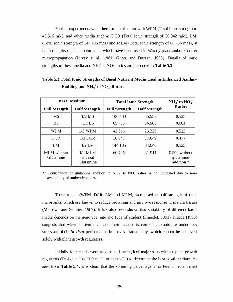
203
Further experiments were therefore carried out with WPM (Total ionic strength of
43.516 mM) and other media such as DCR (Total ionic strength of 36.042 mM), LM
(Total ionic strength of 144.185 mM) and MLM (Total ionic strength of 60.736 mM), at
half strengths of their major salts, which have been used in Woody plant and/or Conifer
micropropagation (Litvay et al., 1981; Gupta and Durzan, 1985). Details of ionic
strengths of these media and NH4+ to NO3
- ratios are presented in Table 5.3 .
Table 5.3 Total Ionic Strengths of Basal Nutrient Media Used in Enhanced Axillary
Budding and NH4+ to NO3
- Ratios.
Basal Medium Total Ionic Strength
Full Strength Half Strength Full Strength Half Strength
NH4+ to NO3
- Ratios
MS 1/2 MS 100.480 55.937 0.523
B5 1/2 B5 65.738 36.903 0.081
WPM 1/2 WPM 43.516 23.326 0.512
DCR 1/2 DCR 36.042 17.649 0.477
LM 1/2 LM 144.185 84.046 0.523
MLM without Glutamine
1/2 MLM without
Glutamine
60.736 31.911 0.500 without glutamine addition.*
*: Contribution of glutamine addition to NH4
+ to NO3- ratios is not indicated due to non-
availability of authentic values.
These media (WPM, DCR, LM and MLM) were used at half strength of their
major salts, which are known to reduce browning and improve response in mature tissues
(McCown and Sellmer, 1987). It has also been shown that suitability of different basal
media depends on the genotype, age and type of explant (Franclet, 1991). Preece (1995)
suggests that when nutrient level and their balance is correct, explants are under less
stress and their in vitro performance improves dramatically, which cannot be achieved
solely with plant growth regulators.
Initially four media were used at half strength of major salts without plant growth
regulators (Designated as “1/2 medium name-H”) to determine the best basal medium. As
seen from Table 5.4, it is clear, that the sprouting percentage in different media varied

204
significantly. Except 1/2 MLM-H, all the other three media showed high incidence of
sprouting. Since only 6.46% sprouting was obtained in 1/2 MLM-H, this medium was not
used in further experiments with cytokinins. In MLM, the NH4+ to NO3
- ratio was 0.5 and
in addition, organic nitrogen was present in the medium in the form of 10 mM glutamine.
Apparently the modification of reducing the major salt concentration to half in MLM is
not quite enough for Taxus spp. for supporting growth and development of axillary buds.
It also appears that organic nitrogen is not required for growth of Taxus spp., as sprouting
percentage in 1/2 MLM-H is much lower than that observed for 1/2 LM-H. Even though
sprouting was the highest (78.95%) on 1/2 LM-H medium, the newly induced shoots
failed to elongate further and their apices became necrotic within 15 days. This medium
was therefore not used for further experiments with cytokinins. The total ionic strength of
LM is 144.185 mM and for 1/2 LM the total ionic strength is approximately 84.046. This
further confirmed the observations of pilot experiments of the non-suitability of high
ionic strength media for culture of Taxus spp. The shoot necrosis of primary shoots,
observed in 1/2 LM might therefore be a manifestation of this effect. In addition, LM has
a NH4+ to NO3
- ratio of 0.523 that is identical with MS medium, a high salt medium. The
other two media namely 1/2 WPM-H and 1/2 DCR-H showed 52.94% and 50% sprouting
(statistically similar) and in these media, the primary shoots neither became necrotic nor
showed phenolic exudations. This may be due to very low total ionic strengths of
approximately 23.326 mM and 17.649 mM respectively of these media. These values are
less than half of 1/2 MS (55.937 mM), 1/2 LM (84.046 mM) and 1/2 MLM (31.911 mM).
Also the NH4+ to NO3
- ratios for WPM and DCR are 0.512 and 0.477 respectively, lesser
than 1/2 LM (0.522), 1/2 MS (0.522) and 1/2 MLM (0.50). The amount of chlorides is
also very less in WPM and DCR, thus eliminating the chances of chloride poisoning of
the plants. Hence 1/2 WPM and 1/2 DCR media were used in all the subsequent
experiments.

205
Table 5.4 Effect of Different Basal Media on % Sprouting from Mature Explants
(Plant material collected from Shimla during 1995-1996).
Medium % Sprouting
Mean + S.E.
1/2 WPM - H 52.94 + 5.32a
1/2 DCR - H 50.00 + 0.00a
1/2 LM - H 78.95 + 5.27b
1/2 MLM - H 6.46 + 0.21c a, b, c : Treatment means with different letters are significantly different from each other at 95%
confidence level by t-test.
1/2 WPM and 1/2 DCR media were further used to test the effects of different
cytokinins individually as the conifers are known to induce sprouting of buds in response
to exogenously applied cytokinins (Thorpe et al., 1991).
1/2 WPM was chosen for the initial experiments. As a means to avoid any
possibility of leaching of phenolics further, it was decided to use this medium in liquid
form. 1/2 WPM without plant growth regulators gave 52.94% sprouting. It was then
combined with TDZ as TDZ was known to induce sprouting of buds or development of
de no vo shoot buds in woody tissues (Fiola et al., 1990 and references therein).
1/2 WPM with 0.1 mg.l-1 TDZ gave low percentage of sprouting (13.75%, Table
5.5 ) while no sprouting was observed in 1/2 WPM with 0.5 mg.l-1 TDZ, as has been the
case in many other conifers (Rong, 1989; Morte et al., 1992). Although the initiation of
sprouting was high at 27.2% and 42.86% with 0.01 and 0.05 mg.l-1 TDZ respectively, the
newly sprouted buds were highly swollen and failed to elongate beyond 0.5 cm (Fig. 5.2).
Since the quality of newly initiated primary shoots was not very good in liquid medium
with TDZ, liquid medium was not used subsequently. 1/2 WPM solidified with agar and
supplemented with AC was therefore used in further experiments with BAP and KIN.
Higher percentage of sprout induction and better shoot quality (ability of shoots to
elongate, orthotropism) were observed in 1/2 WPM with BAP and KIN (Table 5.5 and
Figs. 5.3 and 5.4). 1/2 WPM supplemented with 0.05 mg.l-1 BAP gave 83.33% sprouting
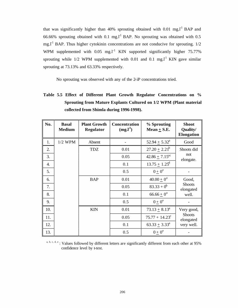
206
that was significantly higher than 40% sprouting obtained with 0.01 mg.l-1 BAP and
66.66% sprouting obtained with 0.1 mg.l-1 BAP. No sprouting was obtained with 0.5
mg.l-1 BAP. Thus higher cytokinin concentrations are not conducive for sprouting. 1/2
WPM supplemented with 0.05 mg.l-1 KIN supported significantly higher 75.77%
sprouting while 1/2 WPM supplemented with 0.01 and 0.1 mg.l-1 KIN gave similar
sprouting at 73.13% and 63.33% respectively.
No sprouting was observed with any of the 2-iP concentrations tried.
Table 5.5 Effect of Different Plant Growth Regulator Concentrations on %
Sprouting from Mature Explants Cultured on 1/2 WPM (Plant material
collected from Shimla during 1996-1998).
No. Basal Medium
Plant Growth Regulator
Concentration (mg.l-1)
% Sprouting Mean + S.E.
Shoot Quality/
Elongation
1. Absent - 52.94 + 5.32a Good
2. 0.01 27.20 + 2.21b
3. 0.05 42.86 + 7.15ac
4. 0.1 13.75 + 1.25d
Shoots did not
elongate.
5.
TDZ
0.5 0 + 0e -
6. 0.01 40.00 + 0a
7. 0.05 83.33 + 0b
8. 0.1 66.66 + 0a
Good, Shoots
elongated well.
9.
BAP
0.5 0 + 0e -
10. 0.01 73.13 + 8.13a
11. 0.05 75.77 + 14.23c
12. 0.1 63.33 + 3.33a
Very good, Shoots
elongated very well.
13.
1/2 WPM
KIN
0.5 0 + 0e -
a, b, c, d, e : Values followed by different letters are significantly different from each other at 95% confidence level by t-test.

207
Fig. 5.2 Plant material collected from Shimla, 1996. Figs. A – D Response of vegetative buds in vitro on 1/2 strength WPM supplemented with various concentrations of TDZ. A. Response of primary sprouting and
shoot bud induction on 1/2 strength WPM supplemented with various concentrations of TDZ (From left to right: Control (without plant growth regulator), 0.01 mg.l-1 TDZ, 0.05 mg.l-1 TDZ, 0.1 mg.l-1 TDZ and 0.5 mg.l-1 TDZ). Note the heavy callus and stunting effect of TDZ on shoot bud elongation.
B. Simultaneous development of an apical bud (a) and three axillary buds (arrows) on 1/2 WPM with 0.05 mg.l-1 TDZ.
C. Simultaneous development of an apical bud and three axillary buds on 1/2 WPM with 0.05 mg.l- 1 TDZ.
D. Two swollen apical buds on 1/2 WPM with 0.1 mg.l-1 TDZ.

208
Fig. 5.3 Plant material collected during 1998-1999.
Figs. A - E Initiation of primary sprouting on 1/2 strength WPM basal medium without plant growth regulators (1/2 WPM – H) (Control treatment) and supplemented with various concentrations of KIN. A. Sprouting of an apical bud on 1/2 WPM
– H. B. Sprouting of an axillary bud on 1/2
WPM – H. C. Sprouting of four axillary buds (arrows)
simultaneously on 1/2 WPM – H. D. Sprouting of an apical bud on 1/2 WPM
+ 0.01 mg.l-1 KIN. E. Initiation of sprouting of two axillary
buds simultaneously on 1/2 WPM + 0.01 mg.l-1 KIN.
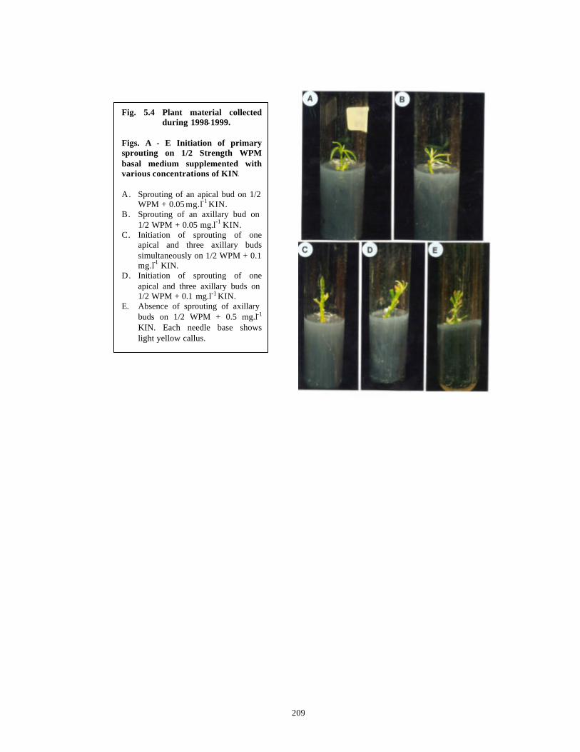
209
Fig. 5.4 Plant material collected during 1998-1999.
Figs. A - E Initiation of primary sprouting on 1/2 Strength WPM basal medium supplemented with various concentrations of KIN. A. Sprouting of an apical bud on 1/2
WPM + 0.05 mg.l-1 KIN. B. Sprouting of an axillary bud on
1/2 WPM + 0.05 mg.l-1 KIN. C. Initiation of sprouting of one
apical and three axillary buds simultaneously on 1/2 WPM + 0.1 mg.l-1 KIN.
D. Initiation of sprouting of one apical and three axillary buds on 1/2 WPM + 0.1 mg.l-1 KIN.
E. Absence of sprouting of axillary buds on 1/2 WPM + 0.5 mg.l-1
KIN. Each needle base shows light yellow callus.
210
The results optimized with 1/2 WPM and plant growth regulator combinations
were further extrapolated to 1/2 DCR medium in the next set of experiments. TDZ was
not tried with 1/2 DCR medium as, although good percentage of sprouting was obtained
in 1/2 WPM supplemented with TDZ, the shoots initiated failed to elongate beyond 0.5
cm. (See Fig. 5.2 ). The negative sprouting response obtained with higher concentratio ns
of BAP and KIN (0.5 mg.l-1) in 1/2 WPM medium, led us to use only lower
concentrations of these cytokinins with 1/2 DCR medium (Table 5.5 A). While 100%
sprouting was obtained on 1/2 DCR supplemented with 0.05 and 0.1 mg.l-1 BAP, 1/2
DCR supplemented with KIN supported only 46.43-58.89% sprouting (statistically
similar) (Table 5.5 A and Fig. 5.5). 1/2 DCR medium without plant growth regulators
but supplemented with 10% CM gave 100% sprouting, which decreased to 53% and 42%
respectively by supplementation with 0.01 and 0.05 mg.l-1 BAP. Addition of even such
low concentrations of BAP to a medium containing 10% CM might have increased the
overall cytokinin availability and its effect on the explants, leading to deterioration in the
percentage of sprouting on 1/2 DCR medium containing 10% CM and 0.01 or 0.05 mg.l-1
BAP. This once again proved that higher concentrations of cytokinins are not conducive
for axillary bud break and sprouting of mature Taxus explants.
No sprouting was observed with any of the 2-iP concentrations tried.
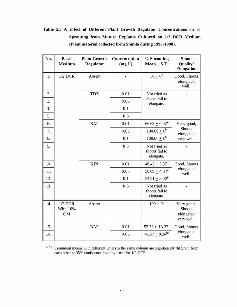
211
Table 5.5 A Effect of Different Plant Growth Regulator Concentrations on %
Sprouting from Mature Explants Cultured on 1/2 DCR Medium
(Plant material collected from Shimla during 1996 -1998).
No. Basal Medium
Plant Growth Regulator
Concentration (mg.l-1)
% Sprouting Mean + S.E.
Shoot Quality/
Elongation
1. Absent - 50 + 0a Good, Shoots elongated
well.
2. 0.01
3. 0.05
4. 0.1
5.
TDZ
0.5
Not tried as shoots fail to
elongate.
-
6. 0.01 66.63 + 0.02a
7. 0.05 100.00 + 0b
8. 0.1 100.00 + 0b
Very good, Shoots
elongated very well.
9.
BAP
0.5 Not tried as shoots fail to
elongate.
-
10. 0.01 46.43 + 3.57a
11. 0.05 58.89 + 4.84a
12. 0.1 54.21 + 3.00a
Good, Shoots elongated
well.
13.
1/2 DCR
KIN
0.5 Not tried as shoots fail to
elongate.
-
14. Absent - 100 + 0a Very good, Shoots
elongated very well.
15. 0.01 53.33 + 13.33b
16.
1/2 DCR With 10%
CM
BAP
0.05 41.67 + 8.34bc
Good, Shoots elongated
well.
a, b, c: Treatment means with different letters in the same column are significantly different from
each other at 95% confidence level by t-test for 1/2 DCR.
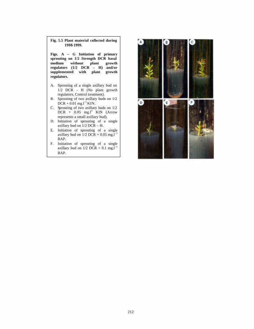
212
Fig. 5.5 Plant material collected during 1998-1999.
Figs. A – G Initiation of primary sprouting on 1/2 Strength DCR basal medium without plant growth regulators (1/2 DCR – H) and/or supplemented with plant growth regulators. A. Sprouting of a single axillary bud on
1/2 DCR – H (No plant growth regulators, Control treatment).
B. Sprouting of two axillary buds on 1/2 DCR + 0.01 mg.l- 1 KIN.
C. Sprouting of two axillary buds on 1/2 DCR + 0.05 mg.l-1 KIN (Arrow represents a small axillary bud).
D. Initiation of sprouting of a single axillary bud on 1/2 DCR – H.
E. Initiation of sprouting of a single axillary bud on 1/2 DCR + 0.05 mg.l - 1
BAP. F. Initiation of sprouting of a single
axillary bud on 1/2 DCR + 0.1 mg.l - 1
BAP.
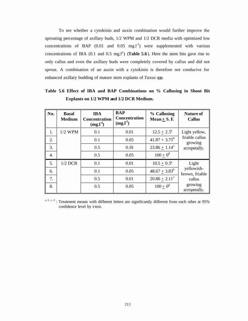
213
To see whether a cytokinin and auxin combination would further improve the
sprouting percentage of axillary buds, 1/2 WPM and 1/2 DCR media with optimized low
concentrations of BAP (0.01 and 0.05 mg.l-1) were supplemented with various
concentrations of IBA (0.1 and 0.5 mg.l-1) (Table 5.6 ). Here the stem bits gave rise to
only callus and even the axillary buds were completely covered by callus and did not
sprout. A combination of an auxin with a cytokinin is therefore not conducive for
enhanced axillary budding of mature stem explants of Taxus spp.
Table 5.6 Effect of IBA and BAP Combinations on % Callusing in Shoot Bit
Explants on 1/2 WPM and 1/2 DCR Medium.
No. Basal Medium
IBA Concentration
(mg.l-1)
BAP Concentration (mg.l-1)
% Callusing Mean + S. E
Nature of Callus
1. 0.1 0.01 12.5 + 2.5a
2. 0.1 0.05 41.87 + 3.75b
3. 0.5 0.01 23.86 + 1.14c
4.
1/2 WPM
0.5 0.05 100 + 0d
Light yellow, friable callus
growing acropetally.
5. 0.1 0.01 10.5 + 0.5a
6. 0.1 0.05 48.67 + 3.83b
7. 0.5 0.01 20.86 + 2.11c
8.
1/2 DCR
0.5 0.05 100 + 0d
Light yellowish-
brown, friable callus
growing acropetally.
a, b, c, d : Treatment means with different letters are significantly different from each other at 95%
confidence level by t-test.
214
From the foregoing results obtained on sprouting of mature stem explants of
Taxus spp. collected from Shimla and presented in Tables 5.2, 5.4, 5.5, 5.5 A and 5.6,
one may conclude that:
a) Basal media with low total ionic salt strengths and lower ratios of ammonium
nitrogen to nitrate nitrogen such as 1/2 WPM and 1/2 DCR are conducive for
enhanced axillary budding of mature stem explants of Taxus spp.
b) Organic nitrogen in the form of an amino acid like glutamine does not enhance
axillary sprouting in Taxus spp.
c) Although the ionic strength of 1/2 DCR medium is much lower than 1/2 WPM,
(17.649 mM and 23.326 mM respectively), 1/2 DCR-H does not give statistically
significant higher sprouting percentage than 1/2 WPM-H (Tables 5.5 and 5.5 A).
d) 1/2 WPM supplemented with lower concentrations of BAP (0.05, 0.1 mg.l-1) gave
higher sprouting percentage as compared to 1/2 WPM supplemented with lower
concentrations of KIN (0.05, 0. 1 mg.l-1). The results are statistically significant
(Table 5.5).
e) 1/2 WPM supplemented with lower concentrations of KIN (0.05, 0.1 mg.l-1) gave
higher sprouting percentage as compared to 1/2 DCR supplemented with lower
concentrations of KIN (0.05, 0.1 mg.l-1). The results are statistically significant
(Tables 5.5 and 5.5 A).
f) 1/2 WPM with 0.05 mg.l-1 BAP with 83.33% sprouting and 1/2 WPM with 0.05 mg.l-
1 KIN with 75.77% sprouting are the optimal media (Table 5.5).
g) 1/2 DCR supplemented with lower concentrations of BAP (0.05, 0.1 mg.l-1) gave
higher sprouting percentage as compared to 1/2 DCR supplemented with lower
concentrations of KIN (0.05, 0.1 mg.l-1). The results are statistically significant
(Table 5.5 A).
h) 1/2 DCR supplemented with lower concentrations of BAP (0.05, 0.1 mg.l-1) gave
higher sprouting percentage as compared to 1/2 WPM supplemented with lower
concentrations of BAP (0.05, 0.1 mg.l-1). The results are statistically significant
(Tables 5.5 and 5.5 A).
i) 1/2 DCR supplemented with lower concentrations of BAP (0.05, 0.1 mg.l-1) gave
higher sprouting percentage as compared to 1/2 WPM supplemented with lower
concentrations of KIN (0.05, 0.1 mg.l-1). The results are statistically significant
(Tables 5.5 and 5.5 A).
215
j) 1/2 DCR without plant growth regulators (1/2 DCR-H) gave 50% sprouting while 1/2
DCR supplemented with lower concentrations of BAP (0.05, 0.1 mg.l-1) gave
identical sprouting percentage (100%) and is similar to 1/2 DCR supplemented with
10% CM. Thus apparently the absence of low concentrations of BAP in 1/2 DCR
medium can be compensated by addition of CM. This cytokinin -like activity of CM is
further proved by the fact that when CM is supplemented with BAP, sprouting
percentage decreases, apparently due to higher additive cytokinin activity by CM and
BAP together (Table 5.5 A).
k) 1/2 DCR with 0.05 mg.l-1 BAP or 1/2 DCR with only 10% CM but without additional
cytokinins are the best media for primary sprouting of stem explants of Taxus spp.
with 100% response (Table 5.5 A).
l) BAP is a better cytokinin with 1/2 DCR to achieve high sprouting (100%) and good
shoot quality as compared to KIN (Table 5.5 A).
m) BAP is a better cytokinin with 1/2 WPM to achieve high sprouting but shoot
quality and elongation capacity are not as good as shoot quality and elongation
ability obtained with KIN (Table 5.5).
n) Hence KIN is a better cytokinin with 1/2 WPM even though sprouting is
relatively low as compared to BAP but shoot quality and elongation ability are
superior as compared to BAP (Table 5.5).
o) Higher cytokinin concentrations are not conducive for sprouting (Tables 5.5 and 5.5
A).
p) TDZ is not conducive for sprouting due to inability of the primary sprouts to elongate
(Table 5.2).
q) Combination of an auxin with a cytokinin does not give enhanced sprouting but leads
to callus formation (Table 5.6).
The responding explants developed either two or three primary sprouts in all the
media combinations of 1/2 WPM and 1/2 DCR tried (with and without cytokinins) (Fig.
5.6 C-E) and even when the apical bud was present, its dominance was overcome by
simultaneous sprouting of axillary buds (Fig. 5.8 D-F).

216
5.3.1.2 Repeatability and applicability of the protocol for primary sprouting to
Taxus plant material collected from different locations (Trees of different
ages, with variable macro-and micro-climatic conditions).
The protocol standardized for primary sprouting of mature shoot explants
collected from Shimla was further extrapolated to plant material collected from different
locations such as Khajjiyar and Almora. The aim was to che ck for the applicability of the
standardized protocol with the plant material collected from Shimla to the plant materials
collected from such diverse locations as Khajjiyar and Almora. This is important because
it is desirable to have a uniform protocol so that it will have a wider applicability in
propagation of plants, irrespective of parameters such as the age of parent plants, different
collection locations, differences in natural habitats and different growth patterns due to
differences in macro- and micro-climatic conditions at each of these locations.
The protocol standardized with plant material collected from Shimla with respect
to basal media and plant growth regulator combinations (1/2 WPM with KIN at 0.01,
0.05, 0.1 and 0.5 mg.l-1 and 1/2 DCR with BAP at 0.01, 0.05, 0.1 and 0.5 mg.l-1) (C. f.
Section 5.3.1, Tables 5.5, 5.5 A) were only tried for these experiments. The results
obtained on culture of stem bit explants obtained from different locations, on 1/2 WPM-H
and 1/2 WPM supplemented with optimized KIN concentrations as well as 1/2 DCR-H
and 1/2 DCR supplemented with optimized BAP concentrations are presented in Table
5.7 . Plant material collected from Khajjiyar gave identical percentage of sprouting (50%)
in 1/2 WPM without plant growth regulators and 1/2 WPM supplemented with various
KIN concentrations. But percentage of sprouting was significantly superior in 1/2 DCR
medium without plant growth regulators (90%). The highest percentage of sprouting
(68.57%) was obtained in 1/2 WPM supplemented with 0.5 mg.l-1 KIN in plant material
collected from Almora and it was statistically similar to 50% sprouting obtained with 1/2
DCR-H. All the other treatments gave statistically identical results.
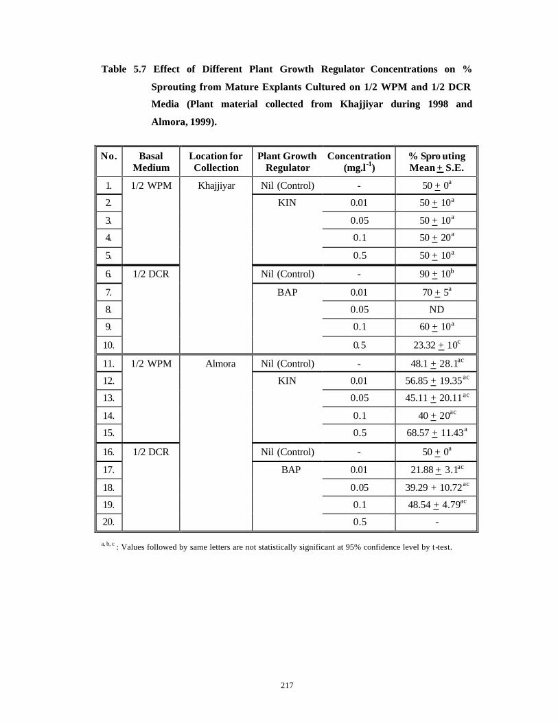
217
Table 5.7 Effect of Different Plant Growth Regulator Concentrations on %
Sprouting from Mature Explants Cultured on 1/2 WPM and 1/2 DCR
Media (Plant material collected from Khajjiyar during 1998 and
Almora, 1999).
No. Basal Medium
Location for Collection
Plant Growth Regulator
Concentration (mg.l-1)
% Spro uting Mean + S.E.
1. Nil (Control) - 50 + 0a
2. 0.01 50 + 10a
3. 0.05 50 + 10a
4. 0.1 50 + 20a
5.
1/2 WPM
KIN
0.5 50 + 10a
6. Nil (Control) - 90 + 10b
7. 0.01 70 + 5a
8. 0.05 ND
9. 0.1 60 + 10a
10.
1/2 DCR
Khajjiyar
BAP
0.5 23.32 + 10c
11. Nil (Control) - 48.1 + 28.1ac
12. 0.01 56.85 + 19.35ac
13. 0.05 45.11 + 20.11ac
14. 0.1 40 + 20ac
15.
1/2 WPM
KIN
0.5 68.57 + 11.43a
16. Nil (Control) - 50 + 0a
17. 0.01 21.88 + 3.1ac
18. 0.05 39.29 + 10.72ac
19. 0.1 48.54 + 4.79ac
20.
1/2 DCR
Almora
BAP
0.5 - a, b, c : Values followed by same letters are not statistically significant at 95% confidence level by t-test.
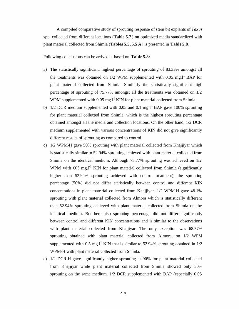
218
A compiled comparative study of sprouting response of stem bit explants of Taxus
spp. collected from different locations (Table 5.7 ) on optimized media standardized with
plant material collected from Shimla (Tables 5.5, 5.5 A ) is presented in Table 5.8 .
Following conclusions can be arrived at based on Table 5.8 :
a) The statistically significant, highest percentage of sprouting of 83.33% amongst all
the treatments was obtained on 1/2 WPM supplemented with 0.05 mg.l-1 BAP for
plant material collected from Shimla. Similarly the statistically significant high
percentage of sprouting of 75.77% amongst all the treatments was obtained on 1/2
WPM supplemented with 0.05 mg.l-1 KIN for plant material collected from Shimla.
b) 1/2 DCR medium supplemented with 0.05 and 0.1 mg.l-1 BAP gave 100% sprouting
for plant material collected from Shimla, which is the highest sprouting percentage
obtained amongst all the media and collection locations. On the other hand, 1/2 DCR
medium supplemented with various concentrations of KIN did not give significantly
different results of sprouting as compared to control.
c) 1/2 WPM-H gave 50% sprouting with plant material collected from Khajjiyar which
is statistically similar to 52.94% sprouting achieved with plant material collected from
Shimla on the identical medium. Although 75.77% sprouting was achieved on 1/2
WPM with 0.05 mg.l-1 KIN for plant material collected from Shimla (significantly
higher than 52.94% sprouting achieved with control treatment), the sprouting
percentage (50%) did not differ statistically between control and different KIN
concentrations in plant mate rial collected from Khajjiyar. 1/2 WPM-H gave 48.1%
sprouting with plant material collected from Almora which is statistically different
than 52.94% sprouting achieved with plant material collected from Shimla on the
identical medium. But here also sprouting percentage did not differ significantly
between control and different KIN concentrations and is similar to the observations
with plant material collected from Khajjiyar. The only exception was 68.57%
sprouting obtained with plant material collected from Almora, on 1/2 WPM
supplemented with 0.5 mg.l-1 KIN that is similar to 52.94% sprouting obtained in 1/2
WPM-H with plant material collected from Shimla.
d) 1/2 DCR-H gave significantly higher sprouting at 90% for plant material collected
from Khajjiyar while plant material collected from Shimla showed only 50%
sprouting on the same medium. 1/2 DCR supplemented with BAP (especially 0.05
219
and 0.1 mg.l-1) gave higher sprouting response for plant material collected from
Shimla as compared to plant material collected from Khajjiyar. This response of plant
material collected from Shimla was comparable to the response obtained on 1/2 DCR-
H with plant material collected from Khajjiyar. 1/2 DCR-H gave identical sprouting at
50% for plant material collected from Almora as well as Shimla. 1/2 DCR
supplemented with BAP, at all the tested concentrations, gave significantly higher
sprouting response for plant material collected from Shimla as compared to plant
material collected from Almora.
e) Thus although the general trends such as suitability of low salt media and low
cytokinin concentrations for sprouting are identical for all plant materials collected
from different locations such as Shimla, Khajjiyar and Almora, the best sprouting
response is observed with plant material collected from Shimla on 1/2 DCR medium
supplemented with 0.05 and 0.1 mg.l-1 BAP. Only plant material collected from
Khajjiyar gave 90% sprouting on 1/2 DCR-H which is significantly higher than the
results obtained with plant material collected from Shimla on the same medium.

220
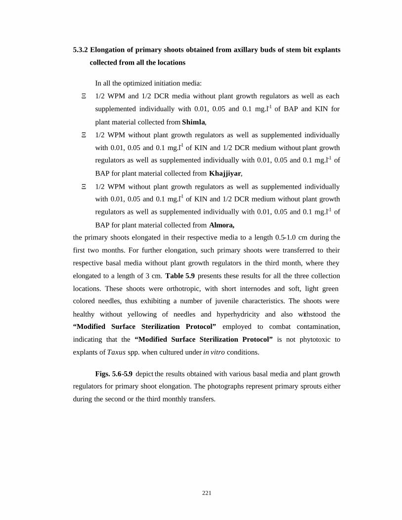
221
5.3.2 Elongation of primary shoots obtained from axillary buds of stem bit explants
collected from all the locations
In all the optimized initiation media:
Ξ 1/2 WPM and 1/2 DCR media without plant growth regulators as well as each
supplemented individually with 0.01, 0.05 and 0.1 mg.l-1 of BAP and KIN for
plant material collected from Shimla,
Ξ 1/2 WPM without plant growth regulators as well as supplemented individually
with 0.01, 0.05 and 0.1 mg.l-1 of KIN and 1/2 DCR medium without plant growth
regulators as well as supplemented individually with 0.01, 0.05 and 0.1 mg.l-1 of
BAP for plant material collected from Khajjiyar,
Ξ 1/2 WPM without plant growth regulators as well as supplemented individually
with 0.01, 0.05 and 0.1 mg.l-1 of KIN and 1/2 DCR medium without plant growth
regulators as well as supplemented individually with 0.01, 0.05 and 0.1 mg.l-1 of
BAP for plant material collected from Almora,
the primary shoots elongated in their respective media to a length 0.5-1.0 cm during the
first two months. For further elongation, such primary shoots were transferred to their
respective basal media without plant growth regulators in the third month, where they
elongated to a length of 3 cm. Table 5.9 presents these results for all the three collection
locations. These shoots were orthotropic, with short internodes and soft, light green
colored needles, thus exhibiting a number of juvenile characteristics. The shoots were
healthy without yellowing of needles and hyperhydricity and also withstood the
“Modified Surface Sterilization Protocol” employed to combat contamination,
indicating that the “Modified Surface Sterilization Protocol” is not phytotoxic to
explants of Taxus spp. when cultured under in vitro conditions.
Figs. 5.6-5.9 depict the results obtained with various basal media and plant growth
regulators for primary shoot elongation. The photographs represent primary sprouts either
during the second or the third monthly transfers.
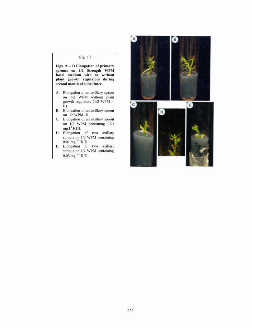
222
Fig. 5.6 Figs. A – D Elongation of primary sprouts on 1/2 Strength WPM basal medium with or without plant growth regulators during second month of subculture. A. Elongation of an axillary sprout
on 1/2 WPM without plant growth regulators (1/2 WPM –H).
B. Elongation of an axillary sprout on 1/2 WPM –H.
C. Elongation of an axillary sprout on 1/2 WPM containing 0.01 mg.l-1 KIN.
D. Elongation of two axillary sprouts on 1/2 WPM containing 0.01 mg.l-1 KIN.
E. Elongation of two axillary sprouts on 1/2 WPM containing 0.05 mg.l-1 KIN.
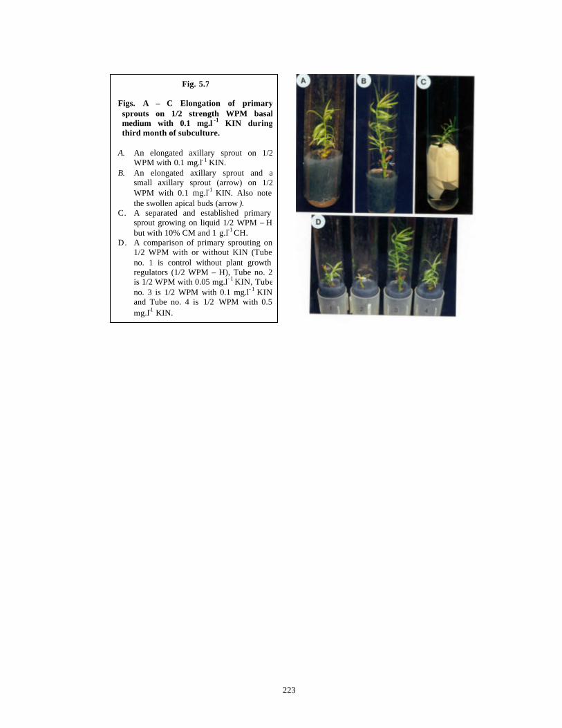
223
Fig. 5.7 Figs. A – C Elongation of primary sprouts on 1/2 strength WPM basal medium with 0.1 mg.l -1 KIN during third month of subculture.
A. An elongated axillary sprout on 1/2
WPM with 0.1 mg.l- 1 KIN. B. An elongated axillary sprout and a
small axillary sprout (arrow) on 1/2 WPM with 0.1 mg.l-1 KIN. Also note the swollen apical buds (arrow ).
C. A separated and established primary sprout growing on liquid 1/2 WPM – H but with 10% CM and 1 g.l-1 CH.
D. A comparison of primary sprouting on 1/2 WPM with or without KIN (Tube no. 1 is control without plant growth regulators (1/2 WPM – H), Tube no. 2 is 1/2 WPM with 0.05 mg.l- 1 KIN, Tube no. 3 is 1/2 WPM with 0.1 mg.l- 1 KIN and Tube no. 4 is 1/2 WPM with 0.5 mg.l-1 KIN.
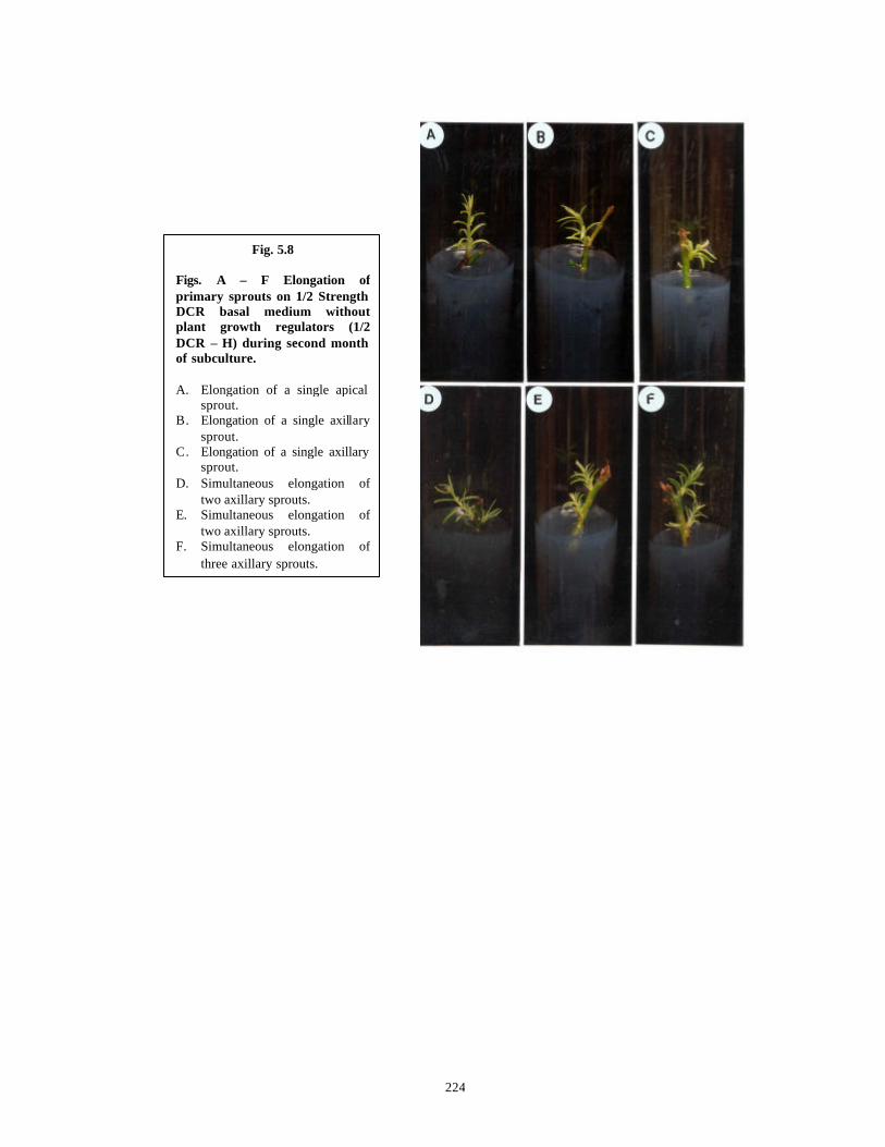
224
Fig. 5.8 Figs. A – F Elongation of primary sprouts on 1/2 Strength DCR basal medium without plant growth regulators (1/2 DCR – H) during second month of subculture. A. Elongation of a single apical
sprout. B. Elongation of a single axillary
sprout. C. Elongation of a single axillary
sprout. D. Simultaneous elongation of
two axillary sprouts. E. Simultaneous elongation of
two axillary sprouts. F. Simultaneous elongation of
three axillary sprouts.
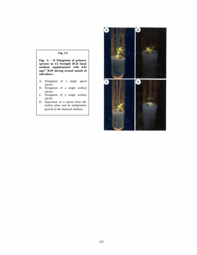
225
Fig. 5.9 Figs. A – D Elongation of primary sprouts on 1/2 Strength DCR basal medium supplemented with 0.05 mg.l-1 BAP during second month of subculture. A. Elongation of a single apical
sprout. B. Elongation of a single axillary
sprout. C. Elongation of a single axillary
sprout. D. Separation of a sprout from the
mother plant and its independent growth in the identical medium.
226
5.3.3 Establishment of shoots obtained from axillary buds of stem bit explants
collected from all the locations
The elongated primary shoots of a length of 3 cm, growing on 1/2 WPM-H and
1/2 DCR-H media, were cut at the end of third month from the parent explants and
transferred to identical media and to media combinations used for growth of primary
shoots during the first two monthly transfers (See Table 5.9, Columns D, E and F).
While the primary shoots failed to establish on their transfer from 1/2 WPM-H and 1/2
DCR-H media to the same respective media, the primary shoots established very well and
showed sustained growth on their respective basal media with or without plant growth
regulator combinations, but each supplemented further with 10% CM and 1g.l-1 CH.
(Table 5.9 ). 20% of the cytokinin activity of coconut milk has been attributed to a water-
soluble compound: G3A2-ZR, 14-O-{3-O-[β-D-galactopyranosyl-(1→2)-α-D-
galactopyranosyl-(1→3)-α-L-arabinofuranosyl]-4-O-(α -L-arabinofuranosyl)-β-D-
galactopyranosyl}-trans-zeatin riboside. Its cytokinin activity might be elicited after its
hydrolysis to zeatin in cells or it might be preferentially taken up due to its water
solubility (Kobayashi et al., 1995). It has also been reported that reduced nitrogen added
as a mixture of amino acids e.g. CH, is extremely growth stimulating (Gupta, 1982). In
the basal media without CM and CH, but with plant growth regulators added individually,
the shoot tips became necrotic and then died.
Figs. 5.7 C, 5.9 D and 5.10 A depict the results obtained with various basal
media, plant growth regulators with CM + CH in shoot establishment The photographs
represent established primary sprouts during their fourth month of growth.
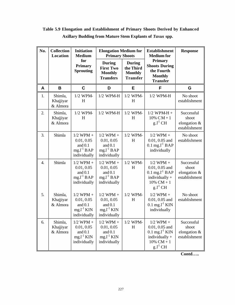
227
Table 5.9 Elongation and Establishment of Primary Shoots Derived by Enhanced
Axillary Budding from Mature Stem Explants of Taxus spp.
Elongation Medium for Primary Shoots
No. Collection Location
Initiation Medium
for Primary
Sprouting
During First Two Monthly
Transfers
During the Third Monthly Transfer
Establishment Medium for
Primary Shoots During
the Fourth Monthly Transfer
Response
A B C D E F G
1. Shimla, Khajjiyar & Almora
1/2 WPM-H
1/2 WPM-H 1/2 WPM-H
1/2 WPM-H No shoot establishment
2. Shimla, Khajjiyar & Almora
1/2 WPM-H
1/2 WPM-H 1/2 WPM-H
1/2 WPM-H + 10% C M + 1
g.l-1 CH
Successful shoot
elongation & establishment
3. Shimla 1/2 WPM + 0.01, 0.05 and 0.1
mg.l-1 BAP individually
1/2 WPM + 0.01, 0.05
and 0.1 mg.l-1 BAP individually
1/2 WPM-H
1/2 WPM + 0.01, 0.05 and
0.1 mg.l-1 BAP individually
No shoot establishment
4. Shimla 1/2 WPM + 0.01, 0.05 and 0.1
mg.l-1 BAP individually
1/2 WPM + 0.01, 0.05
and 0.1 mg.l-1 BAP individually
1/2 WPM-H
1/2 WPM + 0.01, 0.05 and
0.1 mg.l-1 BAP individually + 10% CM + 1
g.l-1 CH
Successful shoot
elongation & establishment
5. Shimla, Khajjiyar & Almora
1/2 WPM + 0.01, 0.05 and 0.1
mg.l-1 KIN individually
1/2 WPM + 0.01, 0.05
and 0.1 mg.l-1 KIN individually
1/2 WPM-H
1/2 WPM + 0.01, 0.05 and 0.1 mg.l-1 KIN
individually
No shoot establishment
6. Shimla, Khajjiyar & Almora
1/2 WPM + 0.01, 0.05 and 0.1
mg.l-1 KIN individually
1/2 WPM + 0.01, 0.05
and 0.1 mg.l-1 KIN individually
1/2 WPM-H
1/2 WPM + 0.01, 0.05 and 0.1 mg.l-1 KIN individually + 10% CM + 1
g.l-1 CH
Successful shoot
elongation & establishment
Contd…..
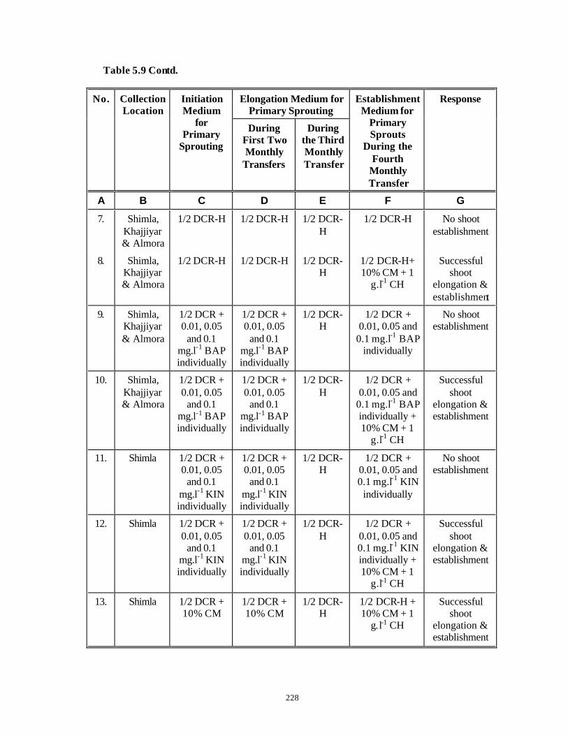
228
Table 5.9 Contd.
Elongation Medium for Primary Sprouting
No. Collection Location
Initiation Medium
for Primary
Sprouting
During First Two Monthly
Transfers
During the Third Monthly Transfer
Establishment Medium for
Primary Sprouts
During the Fourth
Monthly Transfer
Response
A B C D E F G
7. Shimla, Khajjiyar & Almora
1/2 DCR-H 1/2 DCR-H 1/2 DCR-H
1/2 DCR-H No shoot establishment
8. Shimla, Khajjiyar & Almora
1/2 DCR-H 1/2 DCR-H 1/2 DCR-H
1/2 DCR-H+ 10% CM + 1
g.l-1 CH
Successful shoot
elongation & establishment
9. Shimla, Khajjiyar & Almora
1/2 DCR + 0.01, 0.05
and 0.1 mg.l-1 BAP individually
1/2 DCR + 0.01, 0.05
and 0.1 mg.l-1 BAP individually
1/2 DCR-H
1/2 DCR + 0.01, 0.05 and
0.1 mg.l-1 BAP individually
No shoot establishment
10. Shimla, Khajjiyar & Almora
1/2 DCR + 0.01, 0.05
and 0.1 mg.l-1 BAP individually
1/2 DCR + 0.01, 0.05
and 0.1 mg.l-1 BAP individually
1/2 DCR-H
1/2 DCR + 0.01, 0.05 and
0.1 mg.l-1 BAP individually + 10% CM + 1
g.l-1 CH
Successful shoot
elongation & establishment
11. Shimla 1/2 DCR + 0.01, 0.05
and 0.1 mg.l-1 KIN individually
1/2 DCR + 0.01, 0.05
and 0.1 mg.l-1 KIN individually
1/2 DCR-H
1/2 DCR + 0.01, 0.05 and 0.1 mg.l-1 KIN
individually
No shoot establishment
12. Shimla 1/2 DCR + 0.01, 0.05
and 0.1 mg.l-1 KIN individually
1/2 DCR + 0.01, 0.05
and 0.1 mg.l-1 KIN individually
1/2 DCR-H
1/2 DCR + 0.01, 0.05 and 0.1 mg.l-1 KIN individually + 10% CM + 1
g.l-1 CH
Successful shoot
elongation & establishment
13. Shimla 1/2 DCR + 10% CM
1/2 DCR + 10% CM
1/2 DCR-H
1/2 DCR-H + 10% CM + 1
g.l-1 CH
Successful shoot
elongation & establishment
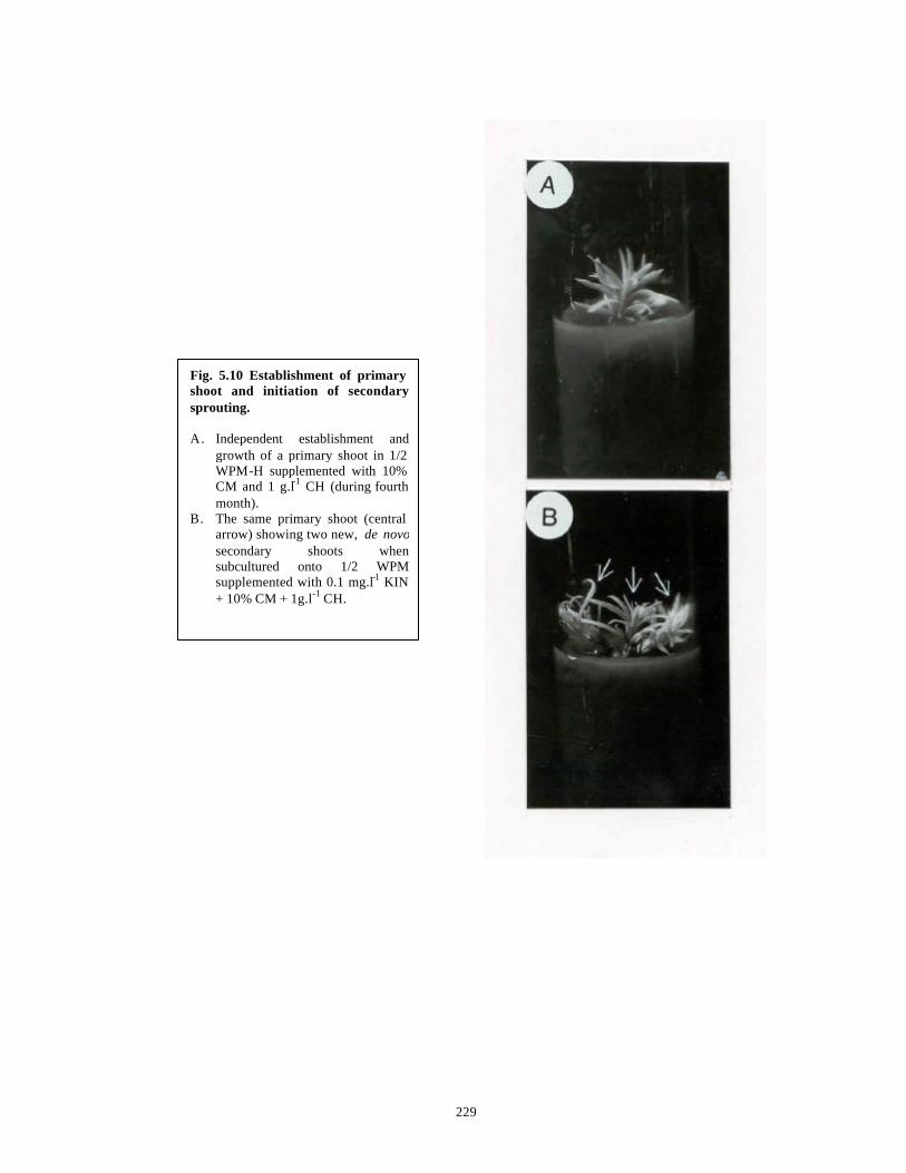
229
Fig. 5.10 Establishment of primary shoot and initiation of secondary sprouting. A. Independent establishment and
growth of a primary shoot in 1/2 WPM-H supplemented with 10% CM and 1 g.l-1 CH (during fourth month).
B. The same primary shoot (central arrow) showing two new, de novosecondary shoots when subcultured onto 1/2 WPM supplemented with 0.1 mg.l-1 KIN + 10% CM + 1g.l-1 CH.
230
5.3.4 Second order sprouting
The established primary sprouts on various media as described in Table 5.9 were
transferred on respective identical media in the fifth month. Second order sprouting (2
new, secondary shoots per each shoot) at a frequency of 25% was observed only on 1/2
WPM supplemented with 0.1 mg.l-1 KIN, 10% CM and 1 g.l-1 CH. None of the other
media used for transfers (C. f. Table 5.9) induced second order sprouting from the buds
formed de novo on primary sprouts in vitro . Fig. 5.10 B shows second order sprouting.
The remaining cultures of primary shoots that did not develop secondary sprouts
did not undergo any further development except for elongation to a length of 7-8 cm in 8
monthly transfers.
During the entire course of this experimentation, 500-600 primary shoots were
obtained on various optimal nutrient media, out of which 300 primary shoots could be
established and out of them 25 showed development of primary sprouts.
5.3.5 Rooting
Rooting of elongated primary or secondary sprouts has not been achieved.
Preliminary experiments were carried out by inoculating the primary sprouts in 1/4 WPM
and 1/4 DCR (Major salts of both the media reduced to 1/4 strength) without plant growth
regulators and/or supplemented with different concentrations and combinations of IAA,
IBA and NAA. But in none of the media rooting was achieved. The great age of the
mother trees and the possible recalcitrance of the explants might be reasons for this.
Rooting of tissue cultured conifers is considered problematic (Mohammed and Vidaver,
1988). It has also been reported that with repeated trnasfers the degree of rejuvenation
and hence the responsiveness of such explants increases (Franclet et al., 1987). The
cultures maintained in the laboratory have undergone only 8 transfers and the response
may increase with passage of time. Further studies need to be undertaken on this aspect.
231
5.4 Conclusions
1) A protocol has been standardized for enhanced axillary budding of
mature stem bit explants of Taxus baccata ssp. wallichiana Zucc. Pilg.
Primary and secondary shoots have been obtained at a high frequency
within six months.
2) Basal media with low total ionic salt strengths and lower ratios of
ammonium nitrogen to nitrate nitrogen such as 1/2 WPM and 1/2 DCR
are conducive for enhanced axillary budding of mature explants of
Taxus spp.
3) Organic nitrogen in the form of an amino acid like glutamine does not
enhance axillary sprouting in Taxus spp.
4) 1/2 WPM supplemented with 0.05 mg.l-1 BAP gave 83.33% sprouting
while 1/2 WPM supplemented with 0.05 mg.l -1 KIN gave 75.77%
sprouting in plant material collected from Shimla. The former
percentage of sprouting was statistically higher than the latter.
5) 1/2 DCR medium supple mented with lower concentrations of BAP (0.05,
0.1 mg.l-1) gave 100% sprouting percentage as compared to 58.89%
and 54.21% sprouting obtained on 1/2 DCR medium supplemented with
lower concentrations of KIN (0.05, 0.1 mg.l -1) with plant material
collected from Shimla. The results are statistically significant.
6) 1/2 DCR medium without plant growth regulators (1/2 DCR-H) gave 50%
sprouting while 1/2 DCR supplemented with lower concentrations of
BAP (0.05, 0.1 mg.l-1) gave identical sprouting percentage (100%) that
is similar to sprouting percentage obtained with 1/2 DCR
supplemented with 10% CM. Thus apparently the absence of low
concentrations of BAP in 1/2 DCR medium can be compensated by
addition of CM.
7) BAP is a better cytokinin with 1/2 WPM to achieve high sprouting but
shoot quality and elongation capacity is not as good as shoot quality
and elongation ability obtained with KIN. So KIN is the cytokinin of
choice with 1/2 WPM.
8) Higher cytokinin concentrations are not conducive for sprouting.

232
9) TDZ is not conducive for sprouting due to inability of the primary
sprouts to elongate.
10) Combination of an auxin with a cytokinin is not conducive for enhanced
axillary budding of mature stem explants of Taxus spp.
11) The standardized protocol of primary sprouting can be extrapolated
successfully to plant material collected from Khajjiyar and Almora,
despite the plants being of different ages and growing under different
macro- and micro-environmental conditions than the plants at Shimla.
12) 1/2 WPM-H gave 50% sprouting with plant material collected from
Khajjiyar, statistically similar to 52.94% sprouting achieved with plant
material collected from Shimla on an identical medium. Although
75.77% sprouting was achieved on 1/2 WPM with 0.05 mg.l-1 KIN with
plant material collected from Shimla, sprouting percentage did not
differ statistically with KIN concentrations with plant material collected
from Khajjiyar.
13) 1/2 DCR-H gave significantly higher sprouting at 90% with plant material
collected from Khajjiyar while plant material collected from Shimla had
only 50% sprouting on the same medium. 1/2 DCR supplemented with
BAP gave higher sprouting response with Shimla material as
compared to Khajjiyar material.
14) 1/2 WPM-H gave 48% sprouting with plant material collected from
Almora, statistically different than 52.94% sprouting achieved with
plant material collected from Shimla on an identical medium. A big
standard error of mean was obtained with Almora material hinting at
underlying variabilities of the plant material. Although 75.77%
sprouting was achieved on 1/2 WPM with 0.05 mg.l-1 KIN with plant
material collected from Shimla, sprouting percentage did not differ
statistically with KIN concentrations with plant material collected from
Almora.
15) 1/2 DCR-H gave identical sprouting at 50% with plant material collected
from Almora as well as Shimla. 1/2 DCR medium supplemented with
BAP gave significantly higher sprouting response with Shimla material
as compared to Almora material.
233
16) Thus although the general trends such as suitability of low salt media
and low cytokinin concentrations for sprouting are identical in plant
material collected from Shimla, Khajjiyar and Almora, the best
sprouting response is observed with plant material collected from
Shimla. Only plant material collected from Khajjiyar gave 90%
sprouting on 1/2 DCR-H which is significantly higher than the results
of sprouting obtained with plant material collected from Shimla.
17) Elongation of primary sprouts into primary shoots of length 3 cm was
possible on respective basal media without plant growth regulators in
the cultures established from plants from all the locations in three
months time.
18) Independent establishment of primary shoots was possible on
respective basal media supplemented with 10% CM and 1 g.l-1 CH in
the fourth month.
19) Secondary sprouting of buds developed in vitro on primary shoots was
achieved on 1/2 WPM supplemented with 0.1 mg.l-1 KIN, 10% CM and 1
g.l-1 CH at a frequency of 25% during the fifth month.
20) “Modified Surface Sterilization Protocol” developed was not phytotoxic
to the explants as primary and secondary sprouts were healthy and
gave good in vitro response.
234
5.5 Introduction 5.5.1 Dormancy
Seed dormancy is defined as the absence of germination of an intact, viable
seed under germination favoring conditions within a specified time lapse (Hilhorst,
1995). It is controlled both at the metabolic and the genomic levels. It is an adaptive trait
that optimizes the distribution of germination over a period of time in a population of
seeds (Bewley, 1997). Usually two types of dormancy are recognized, based on timing
rather than the exogenous or endogenous dormancy-controlling factors (Hilhorst, 1995):
1. Primary dormancy: Related to seed development and maturation. Such seeds when
dispersed from parent plant are in a dormant state.
2. Secondary dormancy: Occurs after seed dispersal and is subjected to annual dormancy
cycles in the seed bank and is reversible.
Both developmental arrest of growth and primary dormancy have been associated
with the presence of in situ ABA and sensitivity to ABA during seed development
(Hilhorst, 1995; Bewley, 1997). After harvesting and cleaning, most seeds require drying
to an appropriate seed moisture content prior to cold storage, to stabilize the dormant
state. Based on this, the seeds are grouped in two classes (Chao and Lin, 1996):
I. Orthodox Seeds – Tolerant of desiccation. Their longevity increases with
decreasing storage temperature and moisture contents.
II. Recalcitrant Seeds – Desiccation sensitive. High moisture content is required for
maintaining seed viability.
In general, dry storage at elevated temperatures (‘After-ripening’) is very
effective in breaking primary dormancy as ABA is degraded at higher temperatures. ‘Pre -
chilling’ is another way to remove ABA by leakage. Endogenous ABA contents are also
lowered by red light irradiation and exogenous or endogenous GAs. Thus the “Hormone
Balance Theory” still appears to be valid, in that ABA action during development or as a
remnant in the mature seed may be antagonized by GA which is synthesized upon
PART B: IN VITRO STUDIES WITH EMBRYO EXPLANTS OF
TAXUS SPP.

235
germination. This process is further aided by low temperatures. The role of GA is
probably restricted to the induction of endosperm-weakening enzymes (Bewley, 1997).
5.5.2 Germination
By definition, germination incorporates those events that commence with the
uptake of water by the quiescent dry seed and terminate with the elongation of the
embryo axis (Bewley, 1997). Visible germination occurs after penetration of the
structures surrounding the embryo by the radicle.
Uptake of water by a mature dry seed is triphasic where a rapid initial uptake
(Phase I) is followed by a plateau phase (Phase II) and a further increase in water uptake
after germination, when the embryonic axes elongate (Phase III) (Bewley and Black,
1994). Upon imbibition, metabolic activity is rapidly resumed with turnover or
replacement of necessary components. At this stage, the seeds are perceptible for external
stimuli like light, chilling, alternating temperatures, chemical and hormonal treatments.
The stimuli may act at the level of transcription. Most of the receptors for these stimuli
are believed to be membrane-bound. The primary events in the release from dormancy are
the reception of the stimulus by the embryo and the immediate signal transduction chain
that leads to the secondary events involving metabolic and hormonal changes (Bewley,
1997).
5.5.3 Dormancy and Germination of Taxus spp. Seeds
Plants belonging to Taxus spp. commonly produce many more ovules than seeds.
But ovule attrition occurs due to: inadequate pollination, resource limitation, genetic load,
loss of seeds to predators and pathogens and physical damage from abiotic factors like
frost. Sine these are understory plants, light availability due to overstory canopy becomes
critical. Successful sexual reproduction occurs only beneath overstory gaps where
sufficient light levels are available (DiFazio et al., 1998). Taxus spp. plants are dioecious
and the numbers of male plants are limited, affecting the success of pollination.
Pollination is also affected by wind, weather conditions, microclimate, genetic load and
maternal effects (DiFazio et al., 1998). The mature seeds of Taxus are enclosed in an
edible red aril and hence undergo heavy predation. Seed production in the Himalayan
Yew shows a periodical effect. During some years, more seeds are produced while at
236
other times; very low seed production is observed (Dr. O. Singh, Personal
Communication). All these factors contribute towards very low production of functional
seeds, which can be collected in a growing season.
Storage of Taxus spp. in germplasm banks ex situ may help to preserve the
threatened diversity of this important genus. The high level of dormancy, the small size of
embryo and inadequate supply of seeds have hampered the efforts to gain knowledge
about storage characteristics of Taxus seeds and also the large scale use of seeds and
embryos for in vitro experiments to exploit the characters of this juvenile tissue. All these
facts have limited the number of experiments which could be carried out on embryos
during the given period of time of the present thesis.
The development of Taxus seeds is asynchronous and a single female tree shows
seeds in all stages of developments. Fully mature seeds of Taxus spp. survive almost
complete desiccation and if adequately dried and stored at 14% relative humidity (RH),
they survive sub-freezing temperatures without any lethal injuries, for a long period of
time Here longevity is projected to increase with reductions in temperatures This is
further aided by extremely high lipid contents (71%) of the seeds. This suggests that these
seeds exhibit orthodox rather than recalcitrant or intermediate storage characteristics.
Immature embryos can be partially dried to avoid desiccation damage and then stored at
5°-6°C. For short -term storage, Taxus seeds can be placed under prolonged stratification
period where catabolism is limited and dormancy is eventually broken. Here seed
viability can be maintained for a few years (Walters-Vertucci et al., 1996).
Naturally, Taxus seeds have a dormancy requirement of at least two long freezing
seasons before germination can occur. Vernalization at low temperatures can help to
overcome this dormancy (Hartzell, 1991). Germination ex vitro has been reported to
require alternating temperatures and cold stratification for 8.5 months to achieve 50%
germination (Chang and Yang, 1996).
Embryo culture in vitro is the sterile isolation and growth of an immature or
mature embryo, with the aim of obtaining a complete, viable plant (Pierik, 1987). It
can be a useful tool to overcome seed dormancy and stratification problems as well as to
abbreviate the breeding cycle. In case of endangered spp. like Taxus it can serve the
additional function of providing the planting material for replenishing the dwindling
237
natural populations of trees while maintaining the natural genetic variation present in the
species. It can also serve as a physiological tool in identifying the various factors
involved in dormancy and germination of these plants. The success of embryo culture
depends on a number of factors such as genotype, developmental stage of embryo at
isolation, growth conditions of mother plant, composition of nutrient media, oxygen
availability, light and temperature (Pierik, 1987).
A number of studies have been carried out in embryo culture of Taxus spp. as
presented earlier in Table 5.1 . But till now there are no reports on precocious germination
of embryos of the Himalayan Yew. The present chapter describes studies carried out to
overcome the natural dormancy of the Yew seeds by removing the embryos from the
seeds and by culturing them on various nutrient media under a range of incubation
conditions.
The objectives of the present work were as follows:
1. To standardize a protocol for high frequency precocious germination and seedling
development from excised embryos of immature and mature seeds of Taxus spp.
2. To extrapolate the protocol developed to seeds collected from different locations.
3. To initiate and maintain fast-growing haploid callus cultures derived from endosperm
of the seeds and
4. To analyze for the possible production of Taxanes therefrom.
238
5.6 Material and Methods
5.6.1 Collection and storage of seeds
The seeds were collected during the months of September and October from the
locations mentioned earlier in Chapter 4, Section 4.2.1 . At the time of collection the
seeds were in various developmental stages (Fig. 5.11 A). The immature seeds without
any aril development were stored at 4°C in zip-locked polythene bags until use. Wherever
aril development was observed (Fig. 5.11 B ), care was taken to remove the aril. Such
seeds were then immersed in water and only the filled seeds (those sinking to the bottom
in water in a glass beaker) were stored at 4°C in zip-locked polythene bags until use, after
drying them on filter papers at room temperature for one day.
5.6.2 Excision and inoculation of embryos and endosperm
After the entire seeds were surface sterilized according to the “Modified Surface
Sterilization Protocol” (Chapter 3, Flow Chart 3.3 ), they were imbibed overnight in
sterile distilled water on an orbital shaker (“Steelmate”, India) at 90 rpm at 25 + 2°C.
On the next day, the imbibed seeds were rinsed with sterile water and the embryos
were excised by cutting the seeds longitudinally. The embryos were rinsed in sterile water
to remove adhering tissues of endosperm and inoculated in various media.
In immature seeds, the endosperm was either liquid or amorphous and hence
could not be inoculated while in mature seeds the endosperm was relatively compact and
solid and could be easily removed and inoculated for in vitro experiments.
5.6.3 Media and culture conditions
5.6.3.1 Precocious embryo germination and seedling development
The following media without plant growth regulators were used for embryo
germination experiments and the details of their compositions were as given in Chapter
2, Table 2.1; ⟨DCR medium, B5 medium, B5 medium with SH vitamins (B5SH
medium), DCR medium with SH vitamins (DCRSH medium), WPM medium and PR
medium with half strength major salts of B5 + full strength minor salts of B5 + full

239
strength vitamins of SH medium (Chee, 1995). All these media contained 3% sucrose and
0.3% phytagel and the details of media preparation were as described in Chapter 2,
Section 2.4.
For the establishment and further growth of the seedlings media identical to
germination media but supplemented with 10% CM were used.
Two excised embryos were inoculated per each culture tube or five embryos were
inoculated per each dish of 55 mm diameter. For further growth of the seedlings they
were transferred to culture tubes individually one month after germination. The cultures
were incubated either in total darkness or in light (19-38 µEm-2s-1) with 16 h photoperiod,
at 25 + 2°C.
5.6.3.2 Adventitious bud induction
Following media were used for experiments on adventitious bud induction: B5SH
or DCRSH at pH 5.8, supplemented individually with either 1.5 or 2.5 or 3.5 mg.l-1 BAP.
These media without plant growth regulators served as controls. All these media
contained 3% sucrose and 0.3% phytagel. Subcultures were done on their respective
induction media without plant growth regulators.
Two excised embryos were inoculated per each culture tube and the cultures were
incubated in light (19-38 µEm-2s -1) with 16 h photoperiod, at 25 + 2°C.
5.6.3.3 For endosperm culture
Six endosperm halves were inoculated in each of the 55 mm sterile plastic dishes
containing 10 ml of DCR or B5 medium with 2% (w/v) sucrose, 0.3 % (w/v) phytagel and
supplemented with 2, 4, 6, 8 or 10 mg.l-1 of Picloram (P). Some of the subculture media
contained 0.5% AC to prevent phenolic browning.
Endosperm cultures were always incubated in total darkness at 25 + 2°C.
240
5.6.4 Growth analysis of endosperm-derived callus cultures
Growth analyses of callus cultures derived from endosperm, by means of fresh
and dry weights, were performed as described earlier in Chapter 4, Section 4.2.5.
5.6.5 TLC and HPLC analysis of endosperm -derived callus cultures and media
containing them
All the procedures related to extraction of callus tissues, their primary screening
by TLC and quantitation of Taxane contents in promising callus lines by HPLC and the
results obtained were described in detail in Chapter 4, Sections 4.2.6 and 4.3.2.
5.6.6 Statistical analysis
In optimization experiments of nutrient media, each medium and plant growth
regulator combination (experimental unit) consisted of 5 plastic dishes (replicate), each
dish containing either 5 embryos or endosperm halves. The experiments were repeated
over three successive years for each of the trees. The treatments were analyzed by one-
way ANOVA and the individual treatment means were compared by Student’s t-test
(Wardlaw, 1985).

241
5.7 Results and Discussion
5.7.1 Precocious germination and seedling development
Initial experiments on precocious embryo germination were performed with seeds
collected from the Khajjiyar region of Himachal Pradesh. The seeds were either light
green in color with liquid endosperm or dark green in color with solid endosperm. In the
former stage of development, embryo was extremely small and could not be dissected out
easily. But in the latter developmental stage, the embryo was white or light yellow, 2 mm
long, torpedo shaped with a distinct suspensor (Fig. 5.11 C) and could be easily separated
from the solid endosperm. The two halves of endosperm also could be inoculated
separately.
Embryos excised from the seeds and inoculated immediately after collection on
DCR medium without plant growth regulators failed to germinate. Hence the seeds were
stored at 4°C for one month and then the excised embryos were inoculated on the same
medium. This gave high values of germination on DCR medium (Table 5.11). The pre-
chilling probably helped in the inactivation of ABA in the embryos, which has been
implicated in the dormancy of embryos of Taxus spp. (Le Page -Degivry, 1968, 1973a, b).
It has also been suggested that at the time of seed-shedding, the embryos are not fully
developed (Has been confirmed in the present study by the torpedo shape of the excised
embryos) and hence need to undergo two years growth (‘After-ripening’) before they
could germinate (Wareing and Phillips, 1990; Hartzell, 1991). Thus Taxus spp. exhibit
‘Primary Dormancy’ .
In the present case, the pre-chilled embryos were inoculated in DCR medium
without plant growth regulators at pH 5.8 based on the preliminary experiments where
DCR medium was proven to be superior to achieve precocious germination in Taxus
embryos by Flores and Sgrignoli (1991). The embryos were either inoculated in culture
tubes with cotton plugs or in plastic dishes sealed hermetically to determine the effect of
culture vessel on the percentage of germination. The cultures were incubated either in
darkness or in light to test the effect of light on germination. The obtained results are
presented in Table 5.10 . It is clear from this table that the percentage of precocious
germination (Precocious germination was scored as swelling of embryo followed by
radicle emergence and greening of cotyledons ) is not significantly different amongst
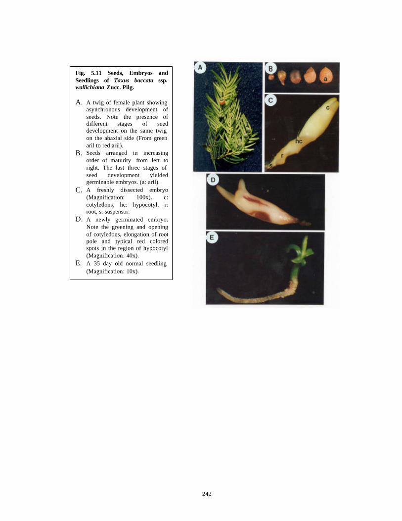
242
Fig. 5.11 Seeds, Embryos and Seedlings of Taxus baccata ssp.wallichiana Zucc. Pilg. A. A twig of female plant showing
asynchronous development of seeds. Note the presence of different stages of seed development on the same twig on the abaxial side (From green aril to red aril).
B. Seeds arranged in increasing order of maturity from left to right. The last three stages of seed development yielded germinable embryos. (a: aril).
C. A freshly dissected embryo (Magnification: 100x). c: cotyledons, hc: hypocotyl, r: root, s: suspensor.
D. A newly germinated embryo. Note the greening and opening of cotyledons, elongation of root pole and typical red colored spots in the region of hypocotyl (Magnification: 40x).
E. A 35 day old normal seedling (Magnification: 10x).
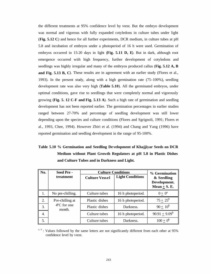
243
the different treatments at 95% confidence level by t-test. But the embryo development
was normal and vigorous with fully expanded cotyledons in culture tubes under light
(Fig. 5.12 C) and hence for all further experiments, DCR medium, in culture tubes at pH
5.8 and incubation of embryos under a photoperiod of 16 h were used. Germination of
embryos occurred in 15-20 days in light (Fig. 5.11 D, E). But in dark, although root
emergence occurred with high frequency, further development of cotyledons and
seedlings was highly irregular and many of the embryos produced callus (Fig. 5.12 A, B
and Fig. 5.13 B, C). These results are in agreement with an earlier study (Flores et al.,
1993). In the present study, along with a high germination rate (75-100%), seedling
development rate was also very high (Table 5.10). All the germinated embryos, under
optimal conditions, gave rise to seedlings that were completely normal and vigorously
growing (Fig. 5. 12 C-F and Fig. 5.13 A). Such a high rate of germination and seedling
development has not been reported earlier. The germination percentages in earlier studies
ranged between 27-70% and percentage of seedling development was still lower
depending upon the species and culture conditions (Flores and Sgrignoli, 1991; Flores et
al., 1993, Chee, 1994). However Zhiri et al. (1994) and Chang and Yang (1996) have
reported germination and seedling development in the range of 95-100%.
Table 5.10 % Germination and Seedling De velopment of Khajjiyar Seeds on DCR
Medium without Plant Growth Regulators at pH 5.8 in Plastic Dishes
and Culture Tubes and in Darkness and Light.
Culture Conditions No. Seed Pre -
treatment Culture Vessel Light Conditions % Germination
& Seedling Development. Mean + S. E.
1. No pre-chilling. Culture tubes 16 h photoperiod. 0 + 0a
2. Plastic dishes 16 h photoperiod. 75 + 25b
3. Plastic dishes Darkness. 90 + 10b
4. Culture tubes 16 h photoperiod. 90.91 + 9.09b
5.
Pre-chilling at 4°C for one
month.
Culture tubes Darkness. 100 + 0b
a, b : Values followed by the same letters are not significantly different from each other at 95%
confidence level by t-test.
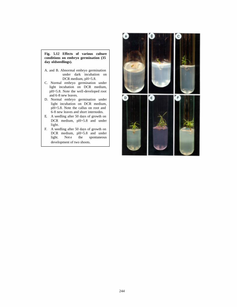
244
Fig. 5.12 Effects of various culture conditions on embryo germination (35 day old seedlings). A. and B. Abnormal embryo germination
under dark incubation on DCR medium, pH=5.8.
C. Normal embryo germination under light incubation on DCR medium, pH=5.8. Note the well-developed root and 6-8 new leaves.
D. Normal embryo germination under light incubation on DCR medium, pH=5.8. Note the callus on root and 6-8 new leaves and short internodes.
E. A seedling after 50 days of growth on DCR medium, pH=5.8 and under light.
F. A seedling after 50 days of growth on DCR medium, pH=5.8 and under light. Not e the spontaneous development of two shoots.

245
Fig. 5.13 Effects of various media and culture conditions on embryo germination (35 day old seedlings) except Fig. A. A. A 60 day old seedling growing on
liquid DCR medium with pH= 5.8 and under light.
B. Spontaneous callus formation on embryo incubated in dark on DCR medium with pH=5.8.
C. Spontaneous callus formation on embryo incubated in dark on DCR medium with pH=5.8.
D. Embryo germination on PR medium under light.
E. Embryo germination on WPM under light. Note the callus at root pole.
F. Embryo germination on B5 medium under light. Note the normal development of seedling.
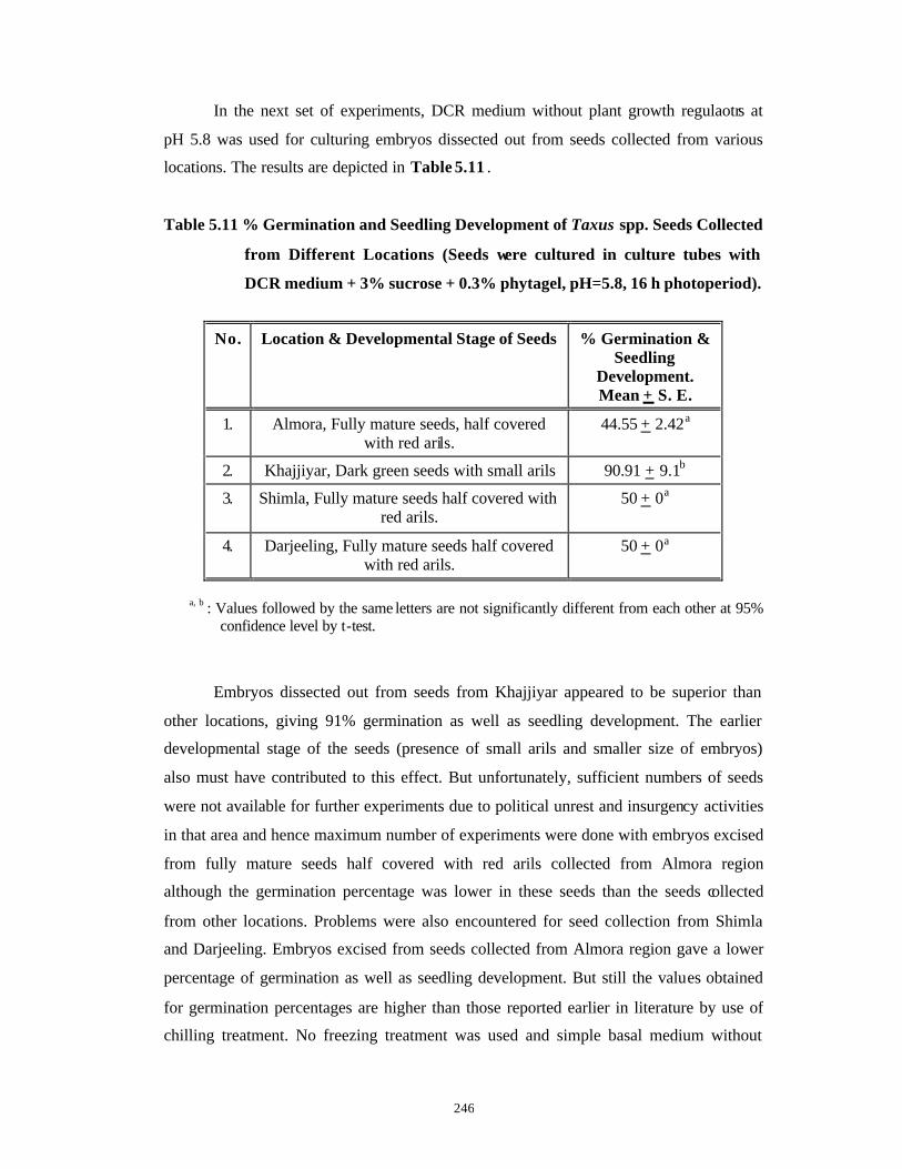
246
In the next set of experiments, DCR medium without plant growth regulaotrs at
pH 5.8 was used for culturing embryos dissected out from seeds collected from various
locations. The results are depicted in Table 5.11 .
Table 5.11 % Germination and Seedling Development of Taxus spp. Seeds Collected
from Different Locations (Seeds were cultured in culture tubes with
DCR medium + 3% sucrose + 0.3% phytagel, pH=5.8, 16 h photoperiod).
No. Location & Developmental Stage of Seeds % Germination & Seedling
Development. Mean + S. E.
1. Almora, Fully mature seeds, half covered with red arils.
44.55 + 2.42a
2. Khajjiyar, Dark green seeds with small arils 90.91 + 9.1b
3. Shimla, Fully mature seeds half covered with red arils.
50 + 0a
4. Darjeeling, Fully mature seeds half covered with red arils.
50 + 0a
a, b : Values followed by the same letters are not significantly different from each other at 95%
confidence level by t-test.
Embryos dissected out from seeds from Khajjiyar appeared to be superior than
other locations, giving 91% germination as well as seedling development. The earlier
developmental stage of the seeds (presence of small arils and smaller size of embryos)
also must have contributed to this effect. But unfortunately, sufficient numbers of seeds
were not available for further experiments due to political unrest and insurgency activities
in that area and hence maximum number of experiments were done with embryos excised
from fully mature seeds half covered with red arils collected from Almora region
although the germination percentage was lower in these seeds than the seeds collected
from other locations. Problems were also encountered for seed collection from Shimla
and Darjeeling. Embryos excised from seeds collected from Almora region gave a lower
percentage of germination as well as seedling development. But still the values obtained
for germination percentages are higher than those reported earlier in literature by use of
chilling treatment. No freezing treatment was used and simple basal medium without
247
plant growth regulators and organic additives were used in the present case as opposed to
the literature reports (c. f. Table 5.1).
Flores and Sgrignoli (1991) could not achieve germination from embryos excised
from mature seeds. In a later publication, Flores et al. (1993) reported upto 70%
germination from embryos excised from fully mature seeds after a prolonged freezing
treatment and the seedlings obtained were even lower in number. Chee (1994) obtained
58-64% germination from embryos excised from mature seeds of various Taxus spp. on
B5 medium with a seedling development percentage between 11-17%. However 100%
germination and seedling development was reported after one week long leaching of
mature embryos in running water by Zhiri et al. (1994). Leaching in running water
probably leached out the ABA from the seeds, allow ing germination to be completed on
modified MS medium supplemented with CH, yeast extract, ascorbic acid and AC. 95%
germination and seedling development were also reported by Chang and Yang (1996) by
use of 1/2 MS medium with PVP. Here also PVP probably helped in binding the
inhibitors leached out from the embryos during culture period.
Different basal media without plant growth regulators were tried in the next ser of
experiments with embryos excised from mature seeds from Almora to enhance the
percentage of embryo germination and the results are presented in Table 5.12 .
B5 medium was found to be the best medium with 91% germination followed by
WPM and DCR at 55% and 46% respectively. The differences were statistically
significant. Although WPM gave hig h percentage of germination, the root pole of
embryos showed callus development (Fig. 5.13 E) which later on spread over the entire
seedling and eventually the seedling died. Hence this medium was not used for further
experiments. For further experiments on adventitious bud induction, B5 and DCR basal
media were therefore used (Fig. 5.13 D-F).

248
Table 5.12 % Germination and Seedling Development of Embryos Excised from
Seeds of Taxus spp. (Almora) on Different Basal Media Compositions
without Plant Growth Regulators (Media supplemented with 3%
sucrose, 0.3% phytagel, pH=5.8 were used in culture tubes and were
incubated at 25 + 2°C and 16 h photoperiod).
No. Basal Medium % Germination & Seedling Development.
Mean + S. E.
1. DCR 45.55 + 2.42a
2. B5 91.66 + 8.33b
3. B5SH 25.00 + 0.00c
4. DCRSH 30.00 + 20.00ac
5. PR 37.5 + 12.5ac
6. WPM 55.00 + 5.00a
a, b, c : Values followed by the same letters are not significantly different from each other at 95% confidence level by t-test.
The germinated seedlings were transferred to their respective germination media
(solid or liquid) supplemented with 10% CM for further growth for two months. In this
medium, the seedlings developed 15-20 leaves and a well developed root system (Fig.
5.13 A). Unfortunately, the seedlings could not be acclimatized and transferred to pots
under the hot conditions of Pune but 100 full-grown seedlings were obtained on the
optimal media such as B5 and DCR without plant growth regulators.
5.7.2 Adventitious bud induction experiments
Chee (1996) reported formation of adventitious buds on embryo explants of T.
brevifolia in modified B5 medium supplemented with various concentrations of BAP.
Similar experiments were carried out in our laboratory with embryos from Almora seeds.
Since DCR and B5 media supported 45.55% and 91.66% precocious germination and
seedling development respectively in embryos excised from Almora seeds, B5 and DCR
basal media were modified by supplementation with SH vitamins, 3% sucrose 0.3%
phytagel with various BAP concentrations. However, in none of the experiments,
adventitious bud production was observed. The embryos either germinated normally or
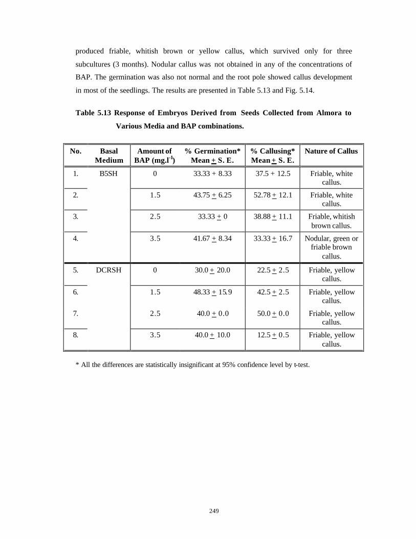
249
produced friable, whitish brown or yellow callus, which survived only for three
subcultures (3 months). Nodular callus was not obtained in any of the concentrations of
BAP. The germination was also not normal and the root pole showed callus development
in most of the seedlings. The results are presented in Table 5.13 and Fig. 5.14.
Table 5.13 Response of Embryos Derived from Seeds Collected from Almora to
Various Media and BAP combinations.
No. Basal Medium
Amount of BAP (mg.l-1)
% Germination* Mean + S. E.
% Callusing* Mean + S. E.
Nature of Callus
1. 0 33.33 + 8.33 37.5 + 12.5 Friable, white callus.
2. 1.5 43.75 + 6.25 52.78 + 12.1 Friable, white callus.
3. 2.5 33.33 + 0 38.88 + 11.1 Friable, whitish brown callus.
4.
B5SH
3.5 41.67 + 8.34 33.33 + 16.7 Nodular, green or friable brown
callus.
5. 0 30.0 + 20.0 22.5 + 2.5 Friable, yellow callus.
6. 1.5 48.33 + 15.9 42.5 + 2.5 Friable, yellow callus.
7. 2.5 40.0 + 0.0 50.0 + 0.0 Friable, yellow callus.
8.
DCRSH
3.5 40.0 + 10.0 12.5 + 0.5 Friable, yellow callus.
* All the differences are statistically insignificant at 95% confidence level by t-test.
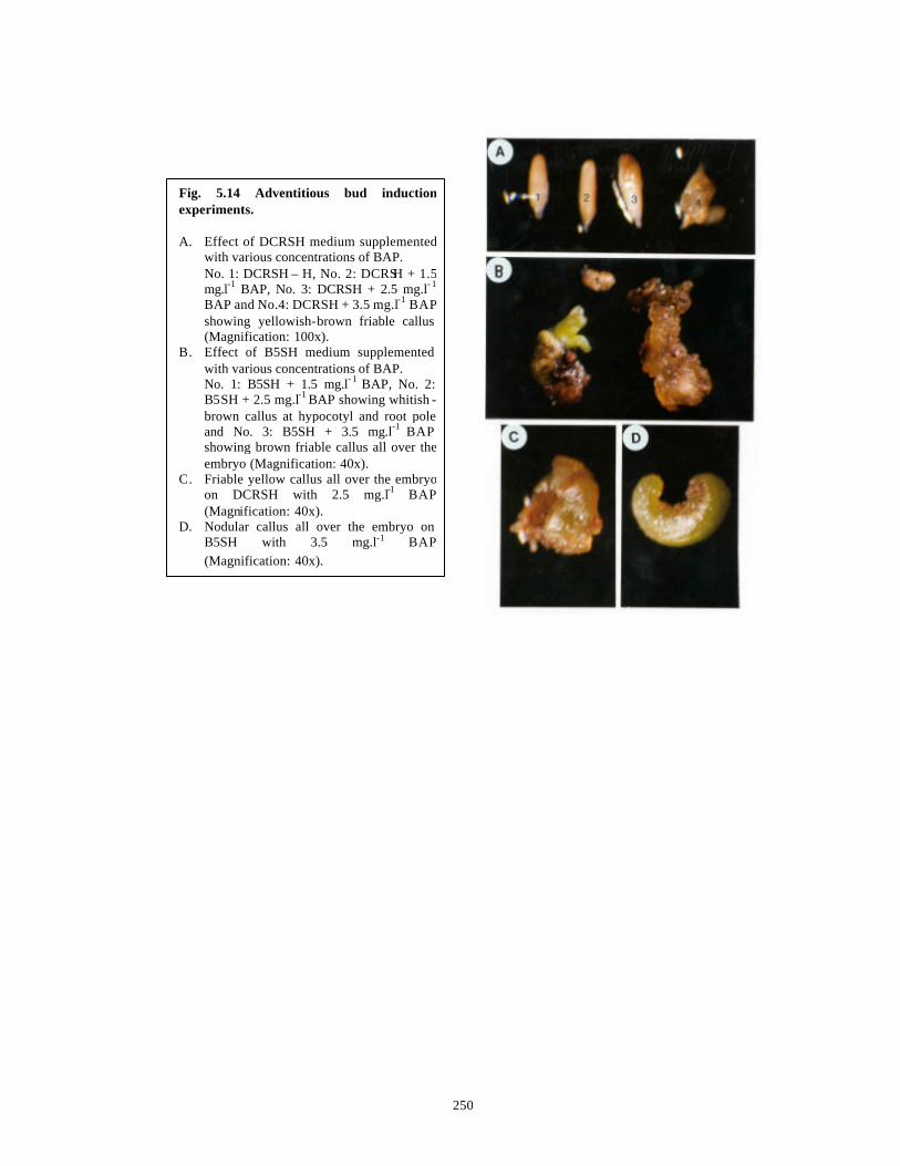
250
Fig. 5.14 Adventitious bud induction experiments. A. Effect of DCRSH medium supplemented
with various concentrations of BAP. No. 1: DCRSH – H, No. 2: DCRSH + 1.5 mg.l-1 BAP, No. 3: DCRSH + 2.5 mg.l- 1
BAP and No.4: DCRSH + 3.5 mg.l-1 BAP showing yellowish-brown friable callus (Magnification: 100x).
B. Effect of B5SH medium supplemented with various concentrations of BAP. No. 1: B5SH + 1.5 mg.l- 1 BAP, No. 2: B5SH + 2.5 mg.l-1 BAP showing whitish -brown callus at hypocotyl and root pole and No. 3: B5SH + 3.5 mg.l-1 BAP showing brown friable callus all over the embryo (Magnification: 40x).
C. Friable yellow callus all over the embryo on DCRSH with 2.5 mg.l-1 BAP (Magnification: 40x).
D. Nodular callus all over the embryo on B5SH with 3.5 mg.l-1 BAP (Magnification: 40x).
251
5.7 .3 Endosperm Culture
In case of Gymnosperms, no ‘triple fusion’ is observed during fertilization and
the haploid female gametophyte (a pre-fertilization product) itself takes over the role of
nutrition played by polyploidic Angiospermic endosperm formed by ‘triple fusion’ during
fertilization. Hence Gymnospermic endosperm is haploid. Culture of endosperm was
attempted in order to raise haploid callus lines and to check for the presence of Taxane
class of compounds in these lines.
A number of callus lines were raised from endosperm halves derived from seeds
from Almora on various media and growth regulator combinations that have proved to be
superior for embryo culture and callus cultures of stems and needles. As the auxin P was
found to be superior for obtaining good diploid callus growth (Chapter 4, Section 4.3.1)
and Taxane content therefrom (Chapter 4, Section 4.3.2), it was combined with B5 DCR
media (Optimum for culture of embryos) (C. f. Chapter 5, Section 5.7.1 ) for culturing
endosperm explants. All the inoculated endosperm halves produced varying amounts of
callus depending on the medium and the percentage response was 100%. However, the
callus morphology and growth rates varied in different media combinations. The results
obtained are presented in Table 5.14 and Figs. 5.15 and 5.16.
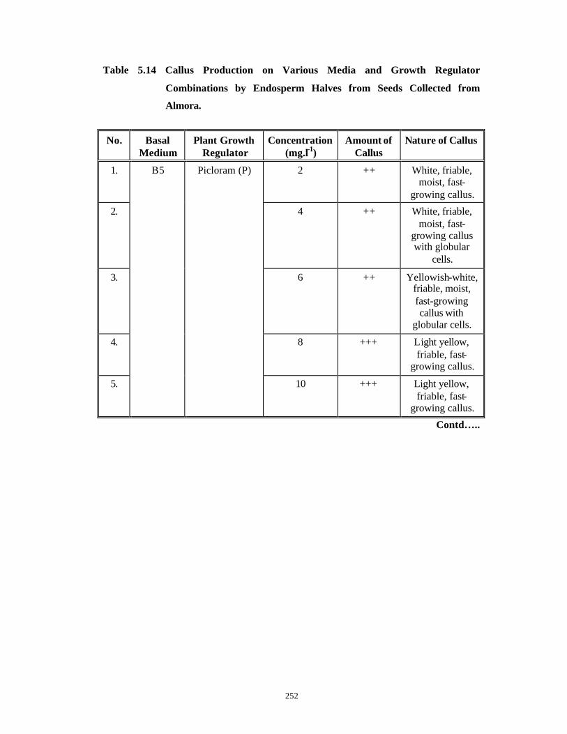
252
Table 5.14 Callus Production on Various Media and Growth Regulator
Combinations by Endosperm Halves from Seeds Collected from
Almora.
No. Basal Medium
Plant Growth Regulator
Concentration (mg.l-1)
Amount of Callus
Nature of Callus
1. 2 ++ White, friable, moist, fast-
growing callus.
2. 4 ++ White, friable, moist, fast-
growing callus with globular
cells.
3. 6 ++ Yellowish-white, friable, moist, fast-growing callus with
globular cells.
4. 8 +++ Light yellow, friable, fast-
growing callus.
5.
B5 Picloram (P)
10 +++ Light yellow, friable, fast-
growing callus.
Contd…..
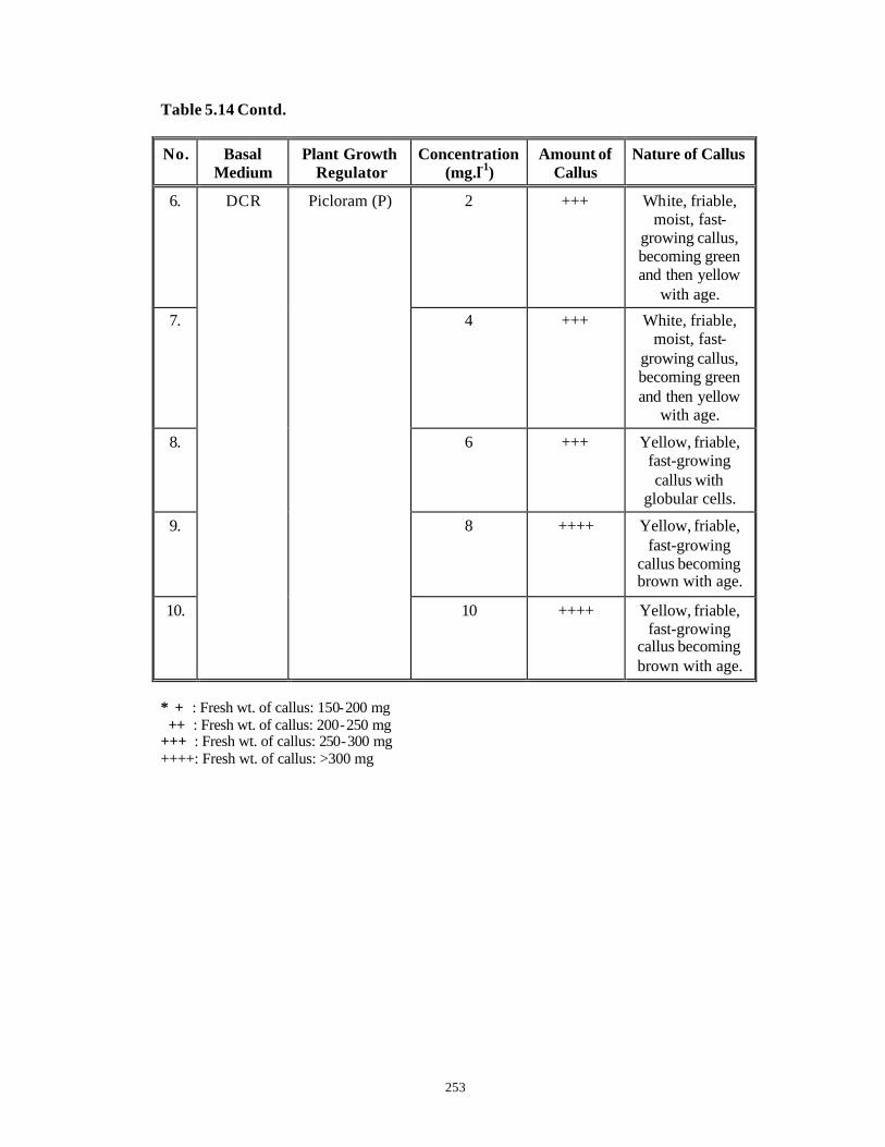
253
Table 5.14 Contd.
No. Basal Medium
Plant Growth Regulator
Concentration (mg.l-1)
Amount of Callus
Nature of Callus
6. 2 +++ White, friable, moist, fast-
growing callus, becoming green and then yellow
with age.
7. 4 +++ White, friable, moist, fast-
growing callus, becoming green and then yellow
with age.
8. 6 +++ Yellow, friable, fast-growing callus with
globular cells.
9. 8 ++++ Yellow, friable, fast-growing
callus becoming brown with age.
10.
DCR Picloram (P)
10 ++++ Yellow, friable, fast-growing
callus becoming brown with age.
* + : Fresh wt. of callus: 150-200 mg ++ : Fresh wt. of callus: 200-250 mg +++ : Fresh wt. of callus: 250-300 mg ++++: Fresh wt. of callus: >300 mg
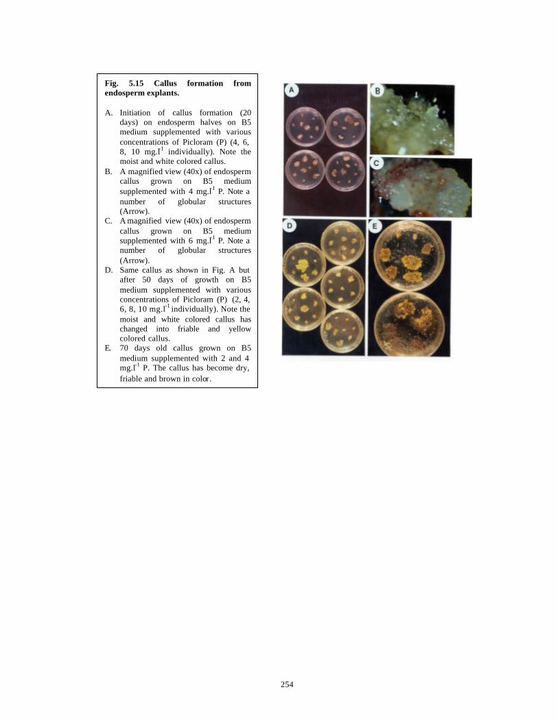
254
Fig. 5.15 Callus formation from endosperm explants. A. Initiation of callus formation (20
days) on endosperm halves on B5 medium supplemented with various concentrations of Picloram (P) (4, 6, 8, 10 mg.l-1 individually). Note the moist and white colored callus.
B. A magnified view (40x) of endosperm callus grown on B5 medium supplemented with 4 mg.l-1 P. Note a number of globular structures (Arrow).
C. A magnified view (40x) of endosperm callus grown on B5 medium supplemented with 6 mg.l-1 P. Note a number of globular structures (Arrow).
D. Same callus as shown in Fig. A but after 50 days of growth on B5 medium supplemented with various concentrations of Picloram (P) (2, 4, 6, 8, 10 mg.l-1 individually). Note the moist and white colored callus has changed into friable and yellow colored callus.
E. 70 days old callus grown on B5 medium supplemented with 2 and 4 mg.l-1 P. The callus has become dry, friable and brown in color.
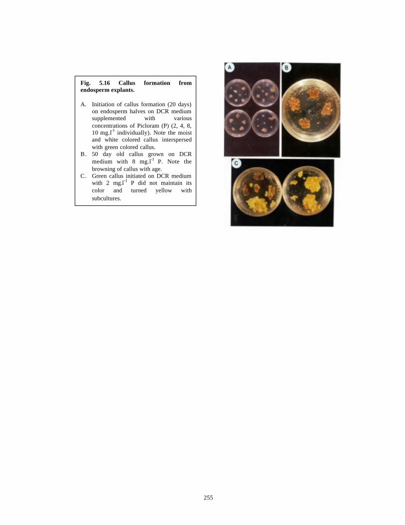
255
Fig. 5.16 Callus formation from endosperm explants. A. Initiation of callus formation (20 days)
on endosperm halves on DCR medium supplemented with various concentrations of Picloram (P) (2, 4, 8, 10 mg.l-1 individually). Note the moist and white colored callus interspersed with green colored callus.
B. 50 day old callus grown on DCR medium with 8 mg.l-1 P. Note the browning of callus with age.
C. Green callus initiated on DCR medium with 2 mg.l-1 P did not maintain its color and turned yellow with subcultures.

256
Earlier workers reported the inability to induce callus from female
gametophytes/endosperms of various Taxus spp. and the callus induced in some instances
was reported to be very slow-growing (Tuleke, 1959; David and Plastira, 1974, 1976).
Our observations on the other hand demonstrate the production of fast growing haploid
calli and to our knowledge this is the first report of success in development of
haploid endosperm cultures from Taxus spp.
B5 medium supplemented with 8 and 10 mg.l-1 of P supported fast growing,
friable callus with FW of callus 250-300 mg. DCR medium supplemented with 8 and 10
mg.l-1 of P also supported faster growing, friable callus with FW of callus more than 300
mg. The se calli were analyzed by TLC for presence of Taxanes.
5.7.4 Analysis of Endosperm Cultures (Callus and Culture Medium) for Taxane
Production.
Out of 10 endosperm-derived callus lines screened by TLC, considerable amounts
of Taxanes were detected in five callus lines. All the five Taxane positive callus lines
were found to grow on DCR medium supplemented with varying concentrations of P.
These were then further analyzed quantitatively by HPLC. Similarly the solid nutrient
media of these callus lines were also analyzed by HPLC for presence of Taxanes. The
details are presented in Chapter 4, Section 4.3 and Tables 4.9 and 4.10.

257
5.8 Conclusions
1) Precocious embryo germination and seedling development has been
achieved at a frequency of 90% from immature seeds collected from
Khajjiyar on DCR basal medium at pH 5.8, in light and without plant
growth regulators.
2) Precocious embryo germination and seedling development was found to
vary with developmental age of seeds (as judged by aril development
and embryo size) and the location of collection.
3) Precocious embryo germination and seedling development has been
achieved at a frequency of 91% from mature seeds collected from
Almora on B5 basal medium without plant growth regulators.
4) Fast growing, haploid callus lines have been established from solid
endosperm of seeds collected from Almora on the optimal B5 and DCR
media supplemented with various concentrations of Picloram (P) and
higher concentrations of P were conducive for faster growth of calli.
5) Considerable amount of Taxane production has been reported here for
the first time from endosperm-derived callus lines (For details please
refer to Chapter 4, Section 4.3.2 and Table 4.9.
258
CHAPTER 6
Direct Regeneration Of Shoots from Various
Explants of Withania spp. and Analysis of
Withaferin-A Production Therefrom.
259
6.1 Introduction
6.1.1 Botany of Withania somnifera (L.) Dunal (Sivarajan and Balachandran, 1994).
The Indian sub-continent has two species of Withania namely, W. somnifera and
W. coagulance. W. coagulance is found in the North-Western India and neighboring
regions. W. somnifera is an erect, grayish, tomentose, undershrub (30-75 cm high) with
long tuberous roots and is distributed throughout the drier sub-tropical regions of India.
Leaves are alternate or sub-opposite, broadly ovate, sub-acute, entire, 5-10 x 2.5-7 cm.
Flowers are small, greenish, axillary, solitary or in few -flowered cymes and bisexual.
Calyx is gamosepalous with 5 lobes, accrescent and inflated in a fruit. Corolla is
campanulate, greenish-yellow with 5 lobes. 5 included stamens are present. Ovary is
ovoid/globose, glabrous, many ovuled. Style is filiform and stigma is 2-lobed. Fruit is a
globose berry, orange-red when ripe and enclosed in the enlarged calyx. Seeds are many,
discoid, yellow and reniform (Fig. 6.1).
The chromosome number is 2n = 48.
6.1.2 Clinical/Pharmacological Studies in Withania Somnifera (L.) Dun.
Withania somnifera (L.) Dun. (Ashwagandha) is an important Solanaceous
medicinal plant. Overall the drug from Withania spp. is believed to impart long life,
youthful vigor and good intellectual powers (Sivarajan and Balachandran, 1994) In
Ayurveda, roots of Withania spp. are prescribed for gynecological disorders, bronchitis,
arthritis, rheumatism, inflammation, fevers, skin diseases etc. The clinical activity of roots
is due to a number of alkaloids like Somniferine, Withasomnine etc. The total alkaloidal
content of roots of Indian chemotype varies between 0.13-0.31% (Majumdar, 1955;
Malhotra et al., 1961; Anonymous, 1976; Asthana and Raina, 1989; Nigam and
Kandalkar, 1995). The leaves contain a number of withanolides like Withaferin -A and
exhibit anti-bacterial (Against a number of Gram-positive bacteria), anti-fungal and anti-
tumor properties (Kurup, 1956; Kupchan et al., 1965; Uma Devi et al., 1992; Uma Devi et
al., 1993).
Withaferin-A acts as a tumor inhibitor due to its ability to arrest dividing cells at
metaphase. It has shown growth inhibitory activity against a number of cancerous cell
lines such as KB, Sarcoma 180 as well as Ehrlich ascites carcinoma and mammary

260
Fig. 6.1 The habit and morphology of Withania somnifera (L.) Dun. (Sivarajan and Balachandran, 1994). 1. A twig. 2. A flower. 3. A flower. 4. A fruit enclosed in the
accrescent calyx.

261
carcinoma (Asthana and Raina, 1989; Uma Devi et al., 1992). It also exerts
radiosensitizing effects on the tumors and carcinomas (Uma Devi et al., 1993; Uma Devi
et al., 1995). Besides Withaferin-A, Withanolide -E and Withanolide-D also exhibit
anti-tumor activities. These compounds act as mitotic poisons, arresting the division of
tumor cells at metaphase and also inhibit RNA and protein synthesis (Budhiraja and
Sudhir, 1987). The essential requirements in a withanolide for antitumor activity are
considered to be epoxide and enone functions in the AB rings and an unsaturated lactone
in the side chain (Ray and Gupta, 1994).
Withaferin-A and Withaferin -E also have immunosuppressive as well as
immunostimulating properties. Withaferin E has insect anti-feedant properties
(Budhiraja and Sudhir, 1987 and references therein). Antifungal activity also has been
attributed to Withanolide -I and Withanolide-K from W. coagulance (Choudhary et al.,
1995).
6.1.3 Alkaloids in Withania somnifera (L.) Dun.
A wide variation (0.13-0.31%) in alkaloid contents of Withania somnifera roots
has been found. Most of this variation is genotypic. Following alkaloids have been
described in this plant (Table 6.1). Out of these, Withanine constitutes 38% of the total
alkaloids (Nigam and Kandalkar, 1995).
Table 6.1 Alkaloids in Withania somnifera (L.) Dun.
Ser. No. Alkaloid Name Ser. No. Alkaloid Name
1. Nicotine. 5. Withanine
2. Somniferine 6. Withananine.
3. Somniferinine 7. Withananinine
4. Somnine 8. Pseudo-withanine
262
6.1.4 Withasteroids in Withania somnifera (L.) Dun.
Withasteroids are a group of naturally occurring C-28 steroidal lactones built on
an intact or rearranged ergostane framework where C-22 and C-26 are appropriately
oxidized to form a δ -lactone ring. They have a nine -carbon side chain with a six
membered ring lactone. This basic structure is referred to as the “Withanolide Skeleton”
(Fig. 6.2 A). Withaferin-A, the first withanolide to be studied in detail, was first isolated
from Withania somnifera (L.) Dun. (Kurup, 1965) and its structure was first determined
by Lavie et al. (1965). See Fig. 6.2 B. Afterwards many compounds (around 50) with
similar structural features were isolated from leaves of a number of Solanaceous plants
(Budhiraja and Sudhir, 1987). These are poly-oxygenated compounds and the parent
plants are believed to have an enzyme system capable of oxidizing all carbon atoms in the
steroid nucleus.
Withanolides are one of the nine and the most abundant groups of the broad
parental group of Withasteroids. Withanolides can be grouped into two classes:
(1) With normal 17β- oriented chain. (132 compounds known)
(2) With the unusual 17α- oriented chain. (36 compounds known)
Withasteroids are mainly synthesized by some members of Solanaceae as well as
by some members of Taccaceae, Leguminosae and even by some marine organisms.
Lockley et al. (1974) have shown by incorporation of radioactive substrates in Withania
somnifera (L.) Dun. that 24-methylene-cholesterol rather than its double bond isomer, is
the true precursor of withanolides.
Plants that are morphologically indistinguishable but differ in their chemical
constituents are termed as “Chemotypes” . Although there is evidence that there are
more than one chemotype of Withania spp. in India (Chakraborty et al., 1974), the exact
number of chemotypes has not yet been studied systematically. Abraham et al. (1968)
have identified two chemotypes in Indian varieties of Withania somnifera . The major
constituents of the Indian Chemotypes are Withanone and 20-H Withanolides, many of
which contain a 5α-hydroxy-6α,7α -epoxy grouping. The ability of different chemotypes
to produce withanolides with different nuclear substitution patterns is genetically
controlled and the hybrids derived after crossing of different parents yield withanolides
either with new nuclear substitutions depending on the genotype of the parents or entirely
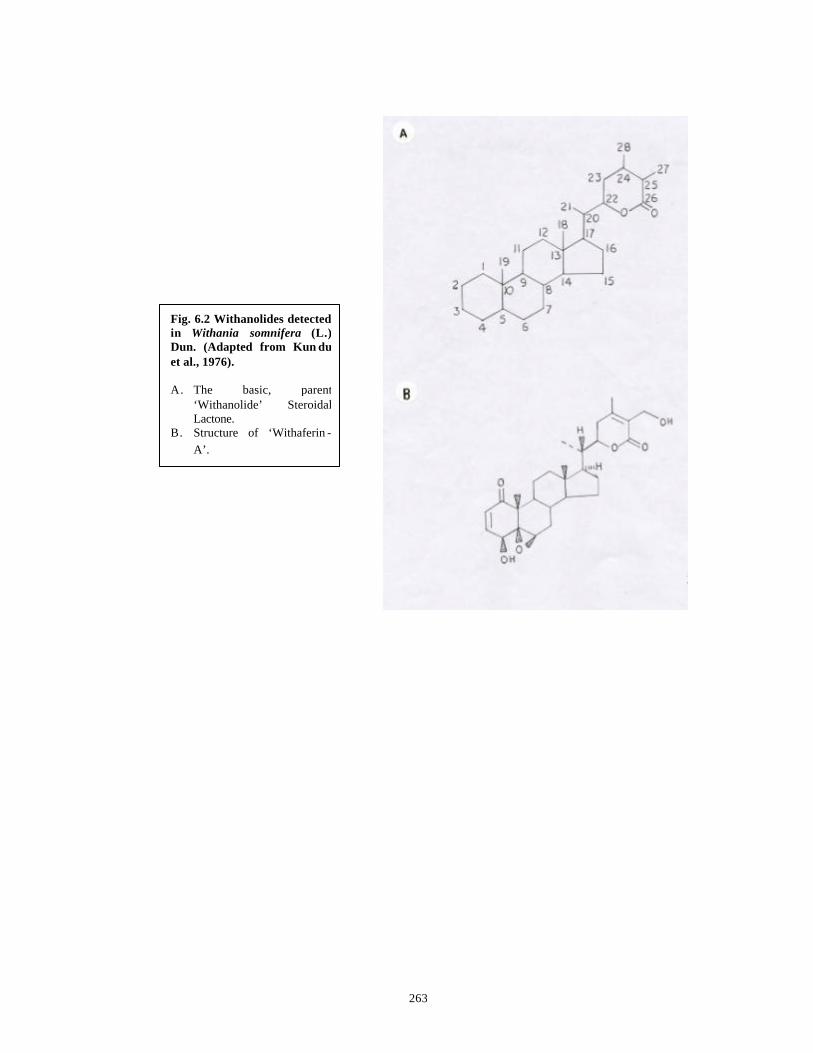
263
Fig. 6.2 Withanolides detected in Withania somnifera (L.) Dun. (Adapted from Kun du et al., 1976). A. The basic, parent
‘Withanolide’ Steroidal Lactone.
B. Structure of ‘Withaferin -A’.
264
new withanolides not found in the parents (Kirson et al., 1977; Eastwood et al., 1980;
Nittala and Lavie, 1981; Bessalle et al., 1987). Thus it appears that it might be possible to
develop hybrids with the ability to yield tailor-made withasteroids to meet specific needs.
Withaferin-A is isolated conjointly with Dihydrowithaferin-A (Kundu et al.,
1976) from leaves of Withania somnifera . It also contains 27-desoxyWithaferin -A and
27-desoxy-14α-hydroxyWithaferin-A. The yield of Withaferin -A is 0.18% on air-dry
basis in Indian plants while South African varieties contain 0.86% of the compound
(Asthana and Raina, 1989). Gupta et al. (1996) have reported Withaferin-A content of
1.6% in leaves of Withania somnifera by TLC densitometry. Besides these, the three
chemotypes of Indian Withania somnifera contain a large variety of different
withanolides.
6.1.5 In Vitro Studies in Withania Somnifera (L.) Dun.
The natural wild populations of Withania somnifera contain a tongue-paralyzing
constituent in their roots and hence cannot be used for ayurvedic treatments. However the
improved, cultivated varieties do not have such an active ingredient. Also they have
different morphology and therapeutic actions although both contain the same alkaloids
(Kaul, 1957). However since the cultivated plants show many different chemotypes
(Kirson et al., 1971; Glotter et al., 1973), production of a large number of plants of
superior selected chemotypes without any seasonal constraints is desirable. The natural
populations of Withania spp. are not easily amenable for vegetative propagation and seed
raised plants need 4-6 years to reach maturity, when they can be harvested for their
natural, active components. These difficulties can be overcome by the use of in vitro
technologies. Direct regeneration of shoots without any intermediate callus phase can
yield clonal plants for large-scale propagation. Genetic transformation using
Agrobacterium rhizogenes for hairy root formation for production of novel metabolites
(Flores and Medina-Bolivar, 1995) is an attractive possibility.
Table 6.2 summarizes the in vitro studies carried out in Withania spp. so far.
265
Table 6.2 Establishment of Differentiated/Un-differentiated and Transformed
Cultures from Withania somnifera (L.) Dun.
Regeneration Studies
No. Explants Medium Plant Growth
Regulators &
Additives
Culture Response
Amount of Withasteroids
Produced
Reference
1. Anthers with uni-nucleate microspores
MS BAP Andro-genesis→ Haploid Callus
- Vishnoi et al. (1979)
2. Germinating seeds and shoot tips
MS BAP, 2,4-D & IBA
4-10 shoots in
15% seeds & 120
shoots in 80% shoot
tips
- Sen & Sharma (1991)
3. Axillary meristems
MS BAP & CM, KIN & 2,4-
D
Callus & multiple shoots
No active compound in
callus.
Withanolide-E: 67 µg/g dw; Withanolide-
D: Traces; Withanone: 90-486 µg/g
dw; Withanolide-I:
13-172 µg/g dw;
Withanolide-G: 19-192 µg/g dw in multiple
shoots.
Roja et al. (1991)
Contd…..
266
Table 6.2 Contd.
No. Explants Medium Plant Growth
Regulators & Additives
Culture Response
Amount of Withasteroids
Produced
Reference
4. In vitro leaves
MS IAA & BAP 16 de novo multiple shoots in 57% leaf
bits
- Kulkarni et al.
(1996a)
5. Seeds, in vitro leaves
MS IAA & BAP, BAP, TDZ
2-13 shoots from seeds,
60% leaves→ multiple shoots
- Kulkarni et al.
(1996b)
6. Nodes, Internodes, Hypocotyls
MS BAP 2-5 shoots per node, more than 25 shoots
per internode, 12 shoots
per hypocotyl
- Kulkarni et al.
(1999)
7. Leaves, Shoots,
Hypocotyls and Roots
MS Callus on 2,4-D &
KIN. Shoot regeneration
on BAP
Plantlet regeneration from callus
- Rani & Grover (1999)
Contd…..
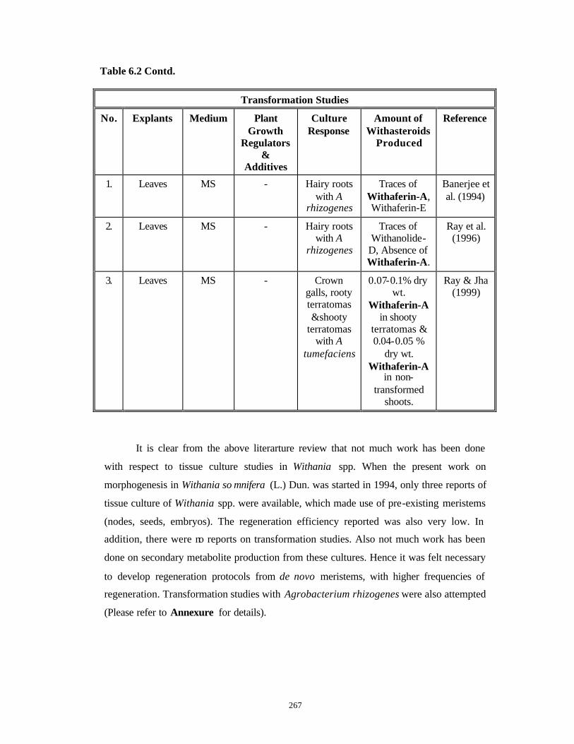
267
Table 6.2 Contd.
Transformation Studies
No. Explants Medium Plant Growth
Regulators &
Additives
Culture Response
Amount of Withasteroids
Produced
Reference
1. Leaves MS - Hairy roots with A
rhizogenes
Traces of Withaferin-A, Withaferin-E
Banerjee et al. (1994)
2. Leaves MS - Hairy roots with A
rhizogenes
Traces of Withanolide-
D, Absence of Withaferin-A.
Ray et al. (1996)
3. Leaves MS - Crown galls, rooty terratomas &shooty
terratomas with A
tumefaciens
0.07-0.1% dry wt.
Withaferin-A in shooty
terratomas & 0.04-0.05 %
dry wt. Withaferin-A
in non-transformed
shoots.
Ray & Jha (1999)
It is clear from the above literarture review that not much work has been done
with respect to tissue culture studies in Withania spp. When the present work on
morphogenesis in Withania so mnifera (L.) Dun. was started in 1994, only three reports of
tissue culture of Withania spp. were available, which made use of pre-existing meristems
(nodes, seeds, embryos). The regeneration efficiency reported was also very low. In
addition, there were no reports on transformation studies. Also not much work has been
done on secondary metabolite production from these cultures. Hence it was felt necessary
to develop regeneration protocols from de novo meristems, with higher frequencies of
regeneration. Transformation studies with Agrobacterium rhizogenes were also attempted
(Please refer to Annexure for details).
268
The objectives of the present work in Withania somnifera (L.) Dun. were
as follows:
1. Use of pre-existing meristems limits the number of explants that could be used in
micropropagation studies. Hence efforts were made to explore the possibility of
regeneration of shoots from different explants such as nodes, internodes, hypocotyls,
leaves and embryos by de novo formation of shoot buds in vitro.
2. Acclimatization of all the regenerants, derived from different explants and their
transfer to green house.
3. Assessment of differentiated and undifferentiated cultures for Withanolide
production and to check for possible correlation between tissue organization and
secondary metabolite production, if any.
4. Preliminary experiments on standardization of raising liquid cultures from normal
roots and analysis of cultures for Withanolide formation, especially Withaferin-A.
5. Preliminary experiments on standardization of raising liquid cultures from hairy roots
obtained with various explants of Withania somnifera by infection with
Agrobacterium rhizogenes and analysis of cultures for Withanolide formation,
especially Withaferin-A .
The preliminary experiments on raising liquid cultures from normal and hairy
roots (Please see Annexure for details) were carried out as pilot experiments to study
for the feasibility of their scale -up in large scale either in shake flasks or bioreactors
for production of Withaferin - A. However scaling up of cultures, if any, based on the
obtained results is not in the scope of studies aimed in this thesis.
269
6.2 Materials and Methods
6.2.1 Collection of plant material
The seeds of the cultivated variety: 'WS-20' of Withania somnifera were collected
from the nurseries in and around Pune (Fig. 6.3 A). These were maintained at room
temperature since they have a long viability period of three years.
6.2.2 Seed germination and seedling development
Seeds were used to raise in vitro seedlings of Withania somnifera (L.) Dun. Seeds
were surface sterilized as per the “Routine Surface Sterilization Protocol” indicated in
Chapter 3, Section 3.2.2.2. Five seeds were inoculated per each glass culture tube
containing 20 ml of nutrient medium and these were maintained at a light intensity of 38
µEm-2s-1 provided by cool white fluorescent tubelights (“Phillips”, India) for 24 hours
(Continuous light) at a temperature of 25 + 2oC. Germination occurred in 15 days on MS
basal medium (Murashige and Skoog, 1962) without plant growth regulators but
supplemented with 2% sucrose and 0.7% agar. This medium is hereinafter referred as
Basal Medium (BM). The medium composition and its method of preparation were as
described earlier in Chapter 2, Section 2.4 and Table 2.1 . The germinated seedlings
were also incubated under conditions described above.
6.2.3 Preparation and incubation of explants for shoot bud induction
One month after seed germination, small intact seedlings were transferred to
screw capped bottles containing 50 ml BM to achieve further growth of seedlings for
another month. These two-month old seedlings of height 8-10 cm (Fig. 6.3 B) were the
source of explants of leaves, nodes, internodes and hypocotyls. After inoculation, all these
explants were maintaine d at a light intensity of 38 µEm-2s-1 provided by cool white
fluorescent tubelights (“Phillips”, India) for 24 hours (Continuous light) at a temperature
of 25 + 2oC.
Very young or very mature leaves from the seedlings were discarded while a leaf
of an optimum size (length: 15-30 mm and width: 20-25 mm at the broadest) was cut into
3-5 pieces of size 1 cm x 1 cm with a portion of the midrib. To ensure that all the leaf
pieces had a portion of the midrib, each leaf was cut length-wise through the midrib.
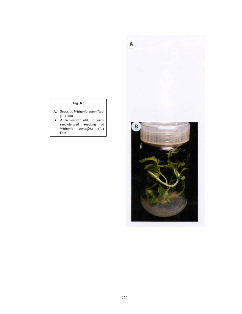
270
Fig. 6.3 A. Seeds of Withania somnifera
(L.) Dun. B. A two-month old, in vitro
seed-derived seedling of Withania somnifera (L.) Dun.
271
These two halves were then cut in transverse and were inoculated, either in the
culture tubes (20 ml nutrient medium) or in sterile plastic dishes with 55 mm diameter (12
ml nutrient medium) with either the adaxial or abaxial surface in contact with BM
fortified with various concentrations and combinations of IAA (0.5, 1.0, 1.5, 2.0 mg.l-1)
and BAP (1.0, 2.0, 3.0 mg.l-1) or NAA (0.5, 1.0, 1.5, 2.0 mg.l-1) and BAP (1.0, 2.0, 3.0
mg.l-1).
Three nodes (3-5 mm long and 2-4 mm wide) and three internodes (3-5 mm long
and 2-4 mm wide) were inoculated independently per tube containing BM supplemented
with BAP (0.1, 0.2, 0.3, 0.4, 0.5, 1.0, 5.0 mg.l-1) and TDZ (0.05, 0.1, 0.2, 0.3 mg.l-1)
individually. For each concentration and combination (if any) of plant growth regulators,
ten tubes of each type of explant were inoculated. Since the internodal explants showed
presence of yellow colored associate bacterial contaminants, they were treated with
“Modified Surface Sterilization Protocol” described earlier in Chapter 3, Flow Chart
3.3 and then used for culture.
Five hypocotyls (3-5 mm long and 2-4 mm wide) were inoculated in each bottle
containing BM with BAP (0.1, 0.2, 0.3, 0.4, 0.5 mg.l-1) and for each concentration of
BAP, five bottles were inoculated. Respons e of hypocotyl explants on higher
concentrations of BAP (1.0, 5.0 mg.l-1) and on any of the TDZ concentrations (0.05, 0.1,
0.2, 0.3 mg.l-1) was not determined because from a full grown two month old seedling
only three hypocotyl explants could be derived.
The embryos (4 mm long) were dissected out by cutting open the imbibed and
swollen seeds and inoculated in sterile disposable plastic dishes containing 12 ml BM in
each dish supplemented with either BAP (0.1, 0.2, 0.3, 0.4, 0.5,1.0, 5.0 mg.l-1) or TDZ
(0.05, 0.1, 0.2, 0.3 mg.l-1) individually. Each dish contained eight embryos and for each
BAP and TDZ concentration, five dishes were inoculated.
All the plant growth regulators used in above experiments were added to nutrient
media before autoclaving as described earlier in Chapter 2, Section 2.4.

272
6.2.4 Elongation and rooting of multiple shoots
Multiple telescopic shoots formed on BM supplemented with various IAA and
BAP combinations from leaf explants and on BAP concentrations from node, internode
and hypocotyl explants were separated and transferred to BM with 0.01 mg.l-1 BAP for
elongation as well as rooting (One step protocol). Multiple shoots formed on TDZ
containing media from node and embryo explants could not be elongated on the above
medium but they could be elongated as well as rooted on MS basal medium with 1/2
strength major salts (1/2 BM) without plant growth regulators (One step protocol).
6.2.5 Acclimatization
Twenty plantlets, 6 cm long (5 cm long shoot portion and 1 cm long root portion),
regenerated from each type of explant with well formed roots were carefully removed
from the culture vessels and washed by gently shaking in a beaker with glass distilled
water to remove the gelling agent and medium sticking to the surface. The plantlets were
then dipped in 1% (w/v) aqueous solution of the systemic fungicide Bavistin (“BASF”,
India) for 10-15 min and then washed with a fine spray of water. Treated plantlets were
transferred to autoclaved clay pots filled with a mixture of sand : soil or soil : 'Soilritemix'
(“KEL Perlite”, India) in 1:1 proportion. These were kept in moist chambers prepared by
covering plastic tubs with glass sheets to prevent the rapid loss of humidity in tropical
environments. They were kept in the hardening room at 27 + 1oC for two weeks. The
hardened plants were sprayed with a sterile solution of major salts of MS medium at 1/10
strength, twice a week, for a period of two weeks. Such hardened plants were transferred
to a green house with ambient temperature (28-30oC), diffuse sunlight and 70% humidity
maintained by overhead sprinklers.
6.2.6 Statistical analysis
Each tube or bottle or plastic dish with three or five or eight explants respectively
was taken as one replicate. Each experiment consisted of either ten (in case of tubes) or
five (in case of bottles and plastic dishes) such replicates for each treatment and each
experiment was repeated at least twice. Shoot buds longer than 0.5 mm with distinctly
visible apical meristems were only counted in each treatment. These were then compared
273
by one way ANOVA and the individual treatment means were compared by Student’s t -
test (Wardlaw 1985).
6.2.7 Histology
Explants at different stages of differentiation were fixed in formalin : acetic acid :
distilled ethyl alcohol (1:1:20) for 48-72 h depending upon the thickness of the explants.
The fixative was decanted and the explants were dehydrated in tertiary butanol series
(Sharma and Sharma, 1980). Dehydrated explants were then embedded in paraffin wax
(“E Merck”, India, melting point 58-60oC). Serial sections of thickness 10 µM were cut
using a rotory microtome (“Reichert Jung 2050, Supercut”, Germany). The sections were
heat-fixed to the slides and passed through xylene-alcohol series (Sharma and Sharma,
1980). Staining was done with Heidenhein’s haematoxylin (Heidenhein, 1892; see Purvis
et al., 1964) with or without counterstaining with 1% Eosin. Slides were mounted in DPX
mountant (Purvis et al., 1964).
6.2.8 Extraction of in vitro grown tissues for analysis of Withafe rin-A by HPLC
Following steps were carried out (Modified from Roja et al., 1991):
1. Two month old seedlings, multiple shoots derived from nodes on the optimal medium
(MS with 0.5 mg.l-1 BAP) and callus formed on the same medium at the basal cut end
of the explant were selected for extraction.
2. 200 mg dry weight (DW) of each type of tissue was taken for analysis. (The tissues
were dried in an oven at 60°C, till constant weight was obtained).
3. The tissues were ground in a pre -cooled mortar and pestle with liquid nitrogen and
extracted overnight in methanol (5 times w/v) on a rotary shaker at 26°C and 100 rpm.
4. The procedure was repeated three times and the methanolic extracts were pooled
together.
5. The extracts were filtered through Whatman No.1 filter paper and diluted with
deionised water (1 methanol : 4 water).
6. The resultant solution was extracted with 3 volumes of chloroform.
7. Chloroform layer was separated from other layers through a separating funnel.
8. The chloroform extract was dried in Eppendorf tubes in a Centrivap Concentrator
(“Labconco”, USA).
274
9. The residue was redissolved in 1 ml methanol, filtered through a 40 µM nylon filter
and 20 µL was used for HPLC analysis.
10. The standard of Withaferin-A, kindly provided by Dr. M. I. Choudhary from HEJ
Research Institute, Pakistan was also dissolved in methanol at a strength of 0.5 mg.l-1
and 20 µL was used for HPLC analysis.
6.2.9 Analytical HPLC and calculation of Withaferin -A in tissue extracts
All routine procedures of analytical HPLC were followed (Melander and Horvath,
1980; Scott, 1996). However the actual procedure used in HPLC analysis is a modified
procedure of the protocol of Roja et al. (1991).
HPLC was carried out on a Merck-H itachi instrument with a series D-7000
interface with HSM Manager, L-7100 pump, L-7420 diode array detector, a rheodyne
injector with a 20 µL injection loop. RP-18 (“Merck”, India) column of the dimension
250 x 4.6 mm with particle size of 5µm was used. The column was operated in the
reverse -phase mode. The data were recorded at 254 nm (Roja et al., 1991) on a computer
and the chromatogram data reports were printed on HP Deskjet 200 printer (“Hewlett
Packard”, India). Photographs of selected runs were recorded with color films. External
standard method of quantitation was used.
All the solvents used were of HPLC (Lichrosolv) grade (“Merck”, India). The in-
house purified Milli-Q water was used (“Millipore Corporation”, USA). All the solvents
and samples were filtered through suitable 0.22 µM pore size filters (“Millipore
Corporation”, USA). Degassing of solvents was done under vacuum (equivalent to 20-
25” Hg). The mobile phase used for the analysis of Withaferin-A consisted of Methanol
(57%) and Water (43%). The flow rate for the isocratic HPLC run (57% methanol and
43% water) was kept at 1.0 ml/min. and the run was continued for 40 min for complete
resolution and detection of the withanolides. All the samples were filtered as described in
the preceding sections and 20 µL was injected. The standard solution of Withaferin-A
was prepared in HPLC grade methanol at the strength of 0.5 mg/ml. It was also filtered
before HPLC analysis and 20 µL was injected. Injection volume of 20 µL was kept
constant during the experimentation. Spiking of the samples was accomplished by
275
addition of 10 µL of authentic standard such that approximately 2.5 µg of the standard
was present in the spiked injection.
After every injection (either of standard or sample) the HPLC system was allowed
to re-equilibriate with the mobile phase till the baseline again became horizontal (10
min.). Since there was no significant back-pressure development, it was not necessary to
develop complicated regeneration procedures for the column.
Plotting of graphs and calculations of amounts of Withaferin-A in the tissues by
the external standard method of quantification were done using routine protocols as
described in the “Instruction Manual for HSM Manager” as well as using the formula
given by Scott (1996):
Cp(s) = ap(s) X C p(st)
ap(st)
where, Cp(s) is the concentration of solute in the mixture.
ap(s) is the area of the peak for the solute (p) in the sample chromatogram.
ap(st) is the area of peak for solute (p) in the reference chromatogram.
Cp(st) is the concentration of the standard in the reference solution.
The linearity of detector response for standard of Withaferin-A was observed by
injecting solutions of standards ranging in concentration from 1.25-10 µg/ml (ElSohly et
al., 1995) and plotting the standard curve (Fig. 6.10 ). Identification of Withaferin-A in
callus and tissue samples was confirmed by retention time, co-chromatogrphy with the
standard and peak purity by wavelength (Fett-neto et al., 1993a, b).
HPLC injections of callus and tissue samples extract as well as the standards were
done at least in duplicates and the injections were repeated on different days to check for
the repeatability of the experimental procedure. Day to day variations were also checked
with the standard solutions.

276
6.3 Results and Discussion
The commercial cultivation of Withania spp. is associated with two major
problems namely, the plant to plant variation in alkaloid and withanolide yield and quality
as well as the long gestation period (4-6 years) between planting and harvesting.
Agronomically the plants are propagated through seeds since the plants do not have the
natural ability for vegetative propagation and the variability generated by sexual
recombination is very high. To overcome these constraints, biotechnological tools like
plant tissue culture can be gainfully employed where cultures can be started from
chemically superior mother plants and on-line harvesting of secondary metabolites can be
done from such differentiated cultures.
Although Skoog and Miller (1957) have shown that organ formation results from
quantitative interactions between auxins and cytokinins, many systems show organ
formation in the presence of a single growth regulator like cytokinin depending upon the
amount and balance of endogenous growth regulators (Buising et al., 1994). Here direct
formation of shoots without an intervening callus phase from a variety of explants is
reported in the presence of an auxin plus cytokinin and also in the presence of a single
cytokinin.
For all the experiments, MS was chosen as the nutrient medium because earlier
reports described its usefulness in Withania somnifera for in vitro experiments (Roja et
al., 1991; Sen and Sharma, 1991). Leaf explants were inoculated on BM supplemented
with different combinations of IAA and BAP as well as NAA and BAP while node,
internode, hypocotyl and embryo explants were inoculated on BM supplemented with
various concentrations of either BAP or TDZ individually. The threshold concentrations
of these growth regulators required for optimum organogenetic induction differed with
different explant types (Tables 6.4, 6.5, 6.8 and 6.9).
6.3.1 Regeneration of plants from leaf explants
6.3.1.1 Size and source of explants
When leaf pieces from very young (smaller than 10 mm) or very old (senescent)
leaves were used as explants, callusing was observed in all the treatments mentioned in
Tables 6.4, 6.5 and 6.6 and there was no shoot bud formation in any of the treatments.

277
But when explants were chosen from leaves of an intermediate size (length: 15-30 mm
and breadth: 20-25 mm), direct regeneration of shoots was observed (Tables 6.4 and
6.5 ). Hence for further experimentation, leaves of of an intermediate size (length: 15-30
mm and breadth: 20-25 mm) were used.
6.3.1.2 Effect of BAP, IAA and NAA
When leaf pieces were prepared (as described earlier in Section 6.2.3) from leaves
of optimum size and were inoculated on BM supplemented with only BAP (0.1-5.0 mg.l-
1) or only NAA (0.1-5.0 mg.l-1), or only IAA (0.1-5.0 mg.l-1), all the explants formed
callus irrespective of the plant growth regulator (Fig. 6.4 D-F). On BM alone, the
explants did not form callus but eventually became necrotic (Table 6.3 ). Callus formed
on BM supplemented with NAA was dry and friable in nature but it never gave rise to
roots. Callus formed on BM supplemented with IAA was moist on BM with 0.1 mg.l-1
IAA while higher IAA concentrations gave rise to dry and friable callus from cut ends of
leaf explant and formed roots from midribs and lateral veins of the leaf explant. Callus
formed on BM supplemented with BAP was moist and green. At 5.0 mg.l-1 BAP, green,
nodular callus was produced but it never gave rise to shoot buds or shoots. All the three
growth regulators on BM supplemented with 5.0 mg.l-1 concentration formed 1.5-2.5 g
FW of callus.
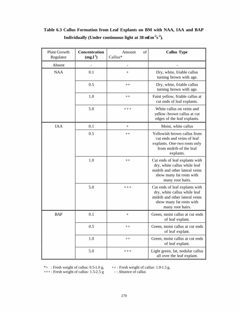
278
Table 6.3 Callus Formation from Leaf Explants on BM with NAA, IAA and BAP
Individually (Under continuous light at 38 µEm-2s-2).
Plant Growth Regulator
Concentration (mg.l-1)
Amount of Callus*
Callus Type
Absent - - -
0.1 + Dry, white, friable callus turning brown with age.
0.5 ++ Dry, white, friable callus turning brown with age.
1.0 ++ Faint yellow, friable callus at cut ends of leaf explants.
NAA
5.0 +++ White callus on veins and yellow-brown callus at cut edges of the leaf explants.
0.1 + Moist, white callus
0.5 ++ Yellowish brown callus from cut ends and veins of leaf
explants. One-two roots only from midrib of the leaf
explants.
1.0 ++ Cut ends of leaf explants with dry, white callus while leaf
midrib and other lateral veins show many fat roots with
many root hairs.
IAA
5.0 +++ Cut ends of leaf explants with dry, white callus while leaf
midrib and other lateral veins show many fat roots with
many root hairs.
0.1 + Green, moist callus at cut ends of leaf explant.
0.5 ++ Green, moist callus at cut ends of leaf explant.
1.0 ++ Green, moist callus at cut ends of leaf explant.
BAP
5.0 +++ Light green, fat, nodular callus all over the leaf explant.
*+ : Fresh weight of callus: 0.5-1.0 g, ++ : Fresh weight of callus: 1.0-1.5 g, +++ : Fresh weight of callus: 1.5-2.5 g - : Absence of callus
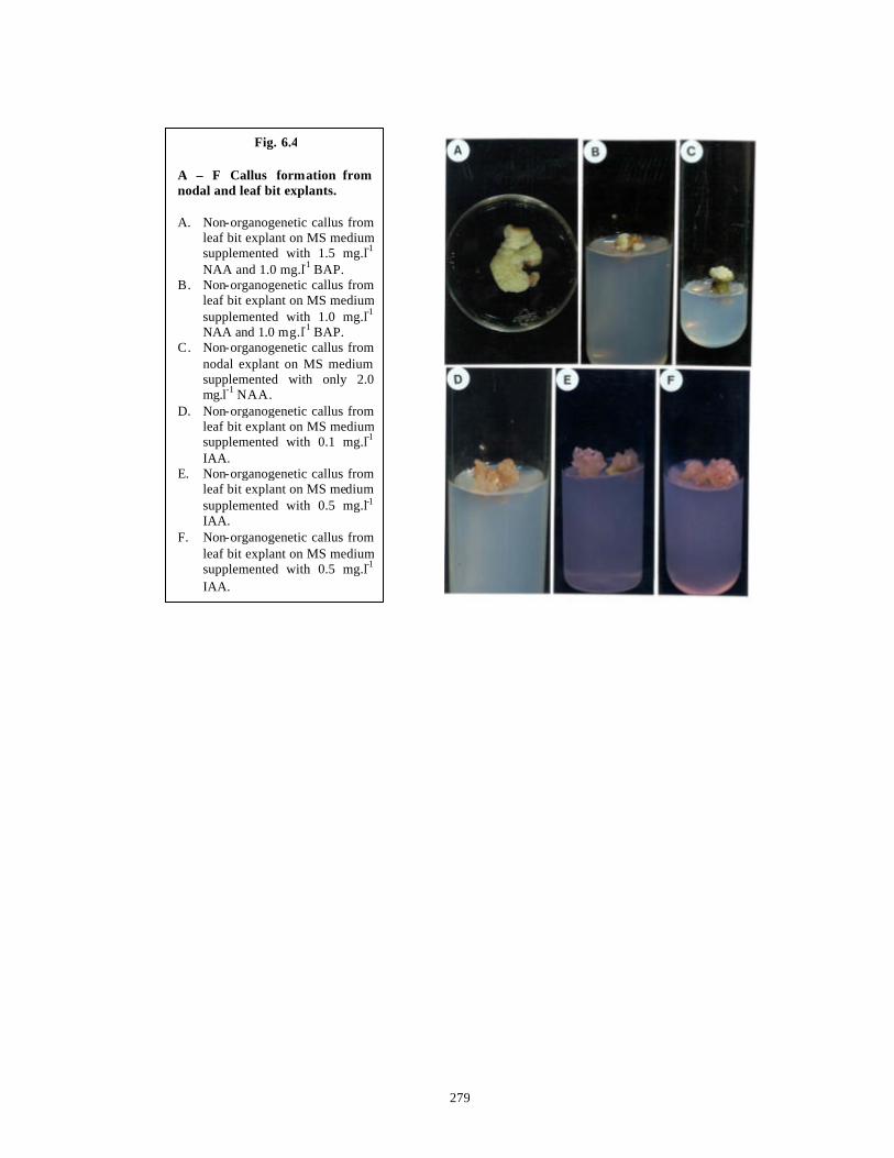
279
Fig. 6.4 A – F Callus formation from nodal and leaf bit explants. A. Non-organogenetic callus from
leaf bit explant on MS medium supplemented with 1.5 mg.l-1
NAA and 1.0 mg.l-1 BAP. B. Non-organogenetic callus from
leaf bit explant on MS medium supplemented with 1.0 mg.l-1
NAA and 1.0 mg.l-1 BAP. C. Non-organogenetic callus from
nodal explant on MS medium supplemented with only 2.0 mg.l-1 NAA.
D. Non-organogenetic callus from leaf bit explant on MS medium supplemented with 0.1 mg.l-1
IAA. E. Non-organogenetic callus from
leaf bit explant on MS medium supplemented with 0.5 mg.l-1
IAA. F. Non-organogenetic callus from
leaf bit explant on MS medium supplemented with 0.5 mg.l-1
IAA.
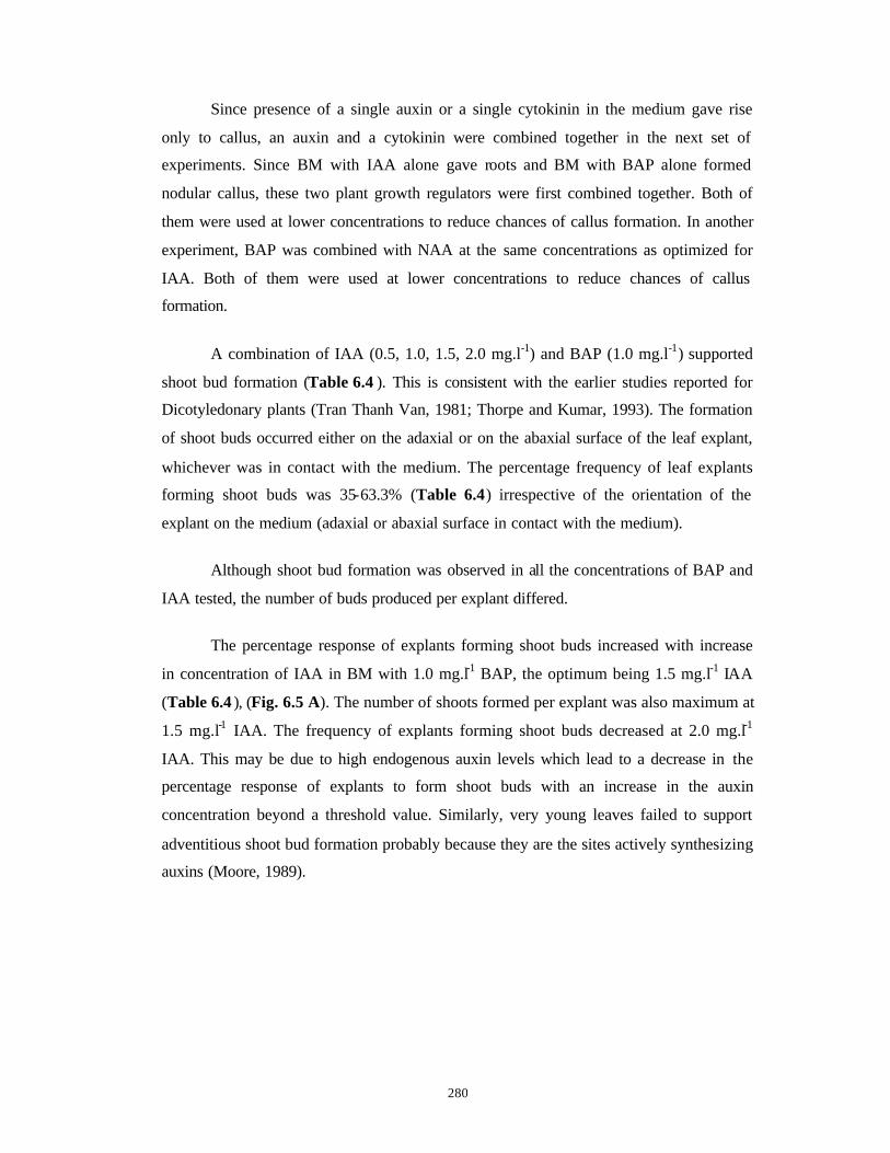
280
Since presence of a single auxin or a single cytokinin in the medium gave rise
only to callus, an auxin and a cytokinin were combined together in the next set of
experiments. Since BM with IAA alone gave roots and BM with BAP alone formed
nodular callus, these two plant growth regulators were first combined together. Both of
them were used at lower concentrations to reduce chances of callus formation. In another
experiment, BAP was combined with NAA at the same concentrations as optimized for
IAA. Both of them were used at lower concentrations to reduce chances of callus
formation.
A combination of IAA (0.5, 1.0, 1.5, 2.0 mg.l-1) and BAP (1.0 mg.l-1) supported
shoot bud formation (Table 6.4 ). This is consistent with the earlier studies reported for
Dicotyledonary plants (Tran Thanh Van, 1981; Thorpe and Kumar, 1993). The formation
of shoot buds occurred either on the adaxial or on the abaxial surface of the leaf explant,
whichever was in contact with the medium. The percentage frequency of leaf explants
forming shoot buds was 35-63.3% (Table 6.4) irrespective of the orientation of the
explant on the medium (adaxial or abaxial surface in contact with the medium).
Although shoot bud formation was observed in all the concentrations of BAP and
IAA tested, the number of buds produced per explant differed.
The percentage response of explants forming shoot buds increased with increase
in concentration of IAA in BM with 1.0 mg.l-1 BAP, the optimum being 1.5 mg.l-1 IAA
(Table 6.4 ), (Fig. 6.5 A). The number of shoots formed per explant was also maximum at
1.5 mg.l-1 IAA. The frequency of explants forming shoot buds decreased at 2.0 mg.l-1
IAA. This may be due to high endogenous auxin levels which lead to a decrease in the
percentage response of explants to form shoot buds with an increase in the auxin
concentration beyond a threshold value. Similarly, very young leaves failed to support
adventitious shoot bud formation probably because they are the sites actively synthesizing
auxins (Moore, 1989).
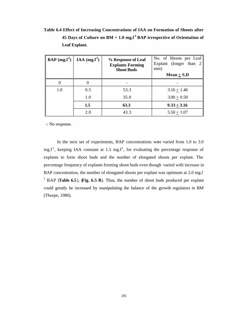
281
Table 6.4 Effect of Increasing Concentrations of IAA on Formation of Shoots after
45 Days of Culture on BM + 1.0 mg.l-1 BAP irrespective of Orientation of
Leaf Explant.
BAP (mg.l-1) IAA (mg.l-1) % Response of Leaf Explants Forming
Shoot Buds
No. of Shoots per Leaf Explant (longer than 2 mm)
Mean + S.D
0 0 - -
0.5 53.3 3.16 + 1.46
1.0 35.0 3.00 + 0.50
1.5 63.3 9.33 + 3.16
1.0
2.0 43.3 5.50 + 1.07 -: No response.
In the next set of experiments, BAP concentrations were varied from 1.0 to 3.0
mg.l-1, keeping IAA constant at 1.5 mg.l-1, for evaluating the percentage response of
explants to form shoot buds and the number of elongated shoots per explant. The
percentage frequency of explants forming shoot buds even though varied with increase in
BAP concentration, the number of elongated shoots per explant was optimum at 2.0 mg.l-
1 BAP (Table 6.5 ), (Fig. 6.5 B). Thus, the number of shoot buds produced per explant
could greatly be increased by manipulating the balance of the growth regulators in BM
(Thorpe, 1980).

282
Table 6.5 Effect of Increasing BAP Concentrations on Adventitious Shoot
Production from Leaf Explants After 45 Days of Culture on BM + 1.5
mg.l-1 IAA.
IAA (mg.l-1) BAP (mg.l-1) % Response of Leaf Explants Forming Shoot
Buds
No. of Shoots per Explant (longer than
2 mm)
Mean + S.D
0 0 - -
1.0 63.30 9.33 + 3.16
2.0 56.65 16.00 + 2.82
1.5
3.0 70.0 13.37 + 1.66
-: No response.
Sixteen shoots with visibly distinct apical meristems could be produced from a
single leaf piece in the optimal medium (Table 6.5; Fig. 6.5 C, D). Besides, there were a
number of smaller, continuously proliferating shoot buds. With repeated subculture on the
same medium at intervals of 3 weeks, each leaf piece can produce 400 shoots within a
year. Since each leaf has been explanted into 3-5 pieces of size 1 cm2, on an average,
1600 plantlets can be produced from one leaf within a year after elongation and rooting.
The leaf explants, on the contrary, on BM with BAP (1.0, 2.0, 3.0 mg. l-1) and
NAA (0.5, 1.0, 1.5, 2.0 mg.l-1) formed only non-organogenetic callus (Table 6.6) (Fig.
6.4 A, B) or callus with thick white roots, suggesting the preferential nature of auxin to
induce shoot buds in combination with BAP.

283
Fig. 6.5 A – F Direct shoot regeneration from leaf bit explants. A. Initiation of shoot buds on MS
medium supplemented with 1.5 mg.l-1 IAA and 1.0 mg.l - 1 BAP.
B. Initiation of shoot buds on MS medium supplemented with 1.5 mg.l-1 IAA and 2.0 mg.l - 1 BAP.
C. Elongation of shoot buds on MS medium supplemented with 1.5 mg.l-1 IAA and 2.0 mg.l - 1 BAP.
D. Elongation of shoot buds on MS medium supplemented with 1.5 mg.l-1 IAA and 2.0 mg.l - 1 BAP.
E. Further elongation of shoots on MS medium with 0.01 mg.l-1
BAP. F. Rooting of shoots on MS medium
with 0.01 mg.l-1 BAP.
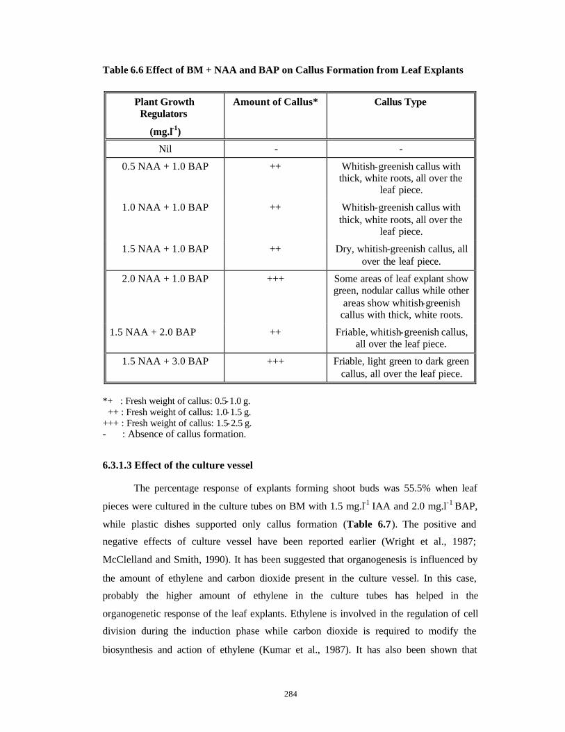
284
Table 6.6 Effect of BM + NAA and BAP on Callus Formation from Leaf Explants
Plant Growth Regulators
(mg.l-1)
Amount of Callus* Callus Type
Nil - -
0.5 NAA + 1.0 BAP ++ Whitish-greenish callus with thick, white roots, all over the
leaf piece.
1.0 NAA + 1.0 BAP ++ Whitish-greenish callus with thick, white roots, all over the
leaf piece.
1.5 NAA + 1.0 BAP ++ Dry, whitish-greenish callus, all over the leaf piece.
2.0 NAA + 1.0 BAP +++ Some areas of leaf explant show green, nodular callus while other
areas show whitish-greenish callus with thick, white roots.
1.5 NAA + 2.0 BAP ++ Friable, whitish-greenish callus, all over the leaf piece.
1.5 NAA + 3.0 BAP +++ Friable, light green to dark green callus, all over the leaf piece.
*+ : Fresh weight of callus: 0.5-1.0 g. ++ : Fresh weight of callus: 1.0-1.5 g. +++ : Fresh weight of callus: 1.5-2.5 g. - : Absence of callus formation.
6.3.1.3 Effect of the culture vessel The percentage response of explants forming shoot buds was 55.5% when leaf
pieces were cultured in the culture tubes on BM with 1.5 mg.l-1 IAA and 2.0 mg.l-1 BAP,
while plastic dishes supported only callus formation (Table 6.7). The positive and
negative effects of culture vessel have been reported earlier (Wright et al., 1987;
McClelland and Smith, 1990). It has been suggested that organogenesis is influenced by
the amount of ethylene and carbon dioxide present in the culture vessel. In this case,
probably the higher amount of ethylene in the culture tubes has helped in the
organogenetic response of the leaf explants. Ethylene is involved in the regulation of cell
division during the induction phase while carbon dioxide is required to modify the
biosynthesis and action of ethylene (Kumar et al., 1987). It has also been shown that

285
culture vessels with closures allowing free gas exchange (cotton plugs in this case) are
more conducive for regeneration than hermetically sealed culture vessels (plastic dishes
sealed with parafilm in this case) (De Proft et al., 1985; Van Aartrijk et al., 1985).
Table 6.7 Regenerative Response of Leaf Explants on BM with 1.5 mg.l-1 IAA and
2.0 mg.l-1 BAP in Different Culture Vessels.
Culture Vessel Replicates % Response of Explants Forming
Shoot Buds
Results
Plastic Dishes (Diameter=55 mm)
60 0 Only green and white callus formation.
Glass Culture Tubes (2.5 x 15 cm)
60 55.5 Shoot buds and shoot formation.
6.3.1.4 Elongation and Rooting of Shoots
All the shoot buds produced on the leaf explants in all the combinations of IAA
and BAP were able to elongate to a length of 1 cm on the induction medium itself. Such 1
cm tall shoots were separated individually from the leaf explants and transferred to BM
with 0.01 mg.l-1 BAP for elongation and rooting. 10 shoots derived from leaf explants on
each of the six IAA + BAP combinations (Tables 6.4 and 6.5 ) were transferred to the
elongation and rooting medium (Table 6.10). Elongation as well as rooting of shoots
occurred at a frequency of 100% (One step protocol), (Fig. 6.5 E, F ) on one and the same
medium simultaneously during the first month itself. (Also see Table 6.10). Thus starting
from shoot bud induction, plantlets can be obtained in two months.
286
6.3.2 Regeneration of plants from node, internode and hypocotyl explants on BM
supplemented with only BAP.
The node, internode and hypocotyl explants were prepared and inoculated on
various BM and plant growth regulator combinations as described earlier in Section
6.2.3. Since percentage response of leaf explants in forming shoot buds was practically
zero in plastic dishes (Table 6.7 ), these were not used in further experiments with nodes,
internodes and hypocotyls. BAP and TDZ are the preferred auxins for induction of
multiple shoots from pre-existing meristems and earlier authors had also reported positive
effect of BAP for in vitro cultures of Withania spp. (Vishnoi et al., 1979; Sen and
Sharma, 1991; Roja et al., 1991). Hence in the present case, BM was supplemented with
either BAP or TDZ individually.
6.3.2.1 Regeneration of plants from nodes
On BM without plant growth regulators, only a single shoot from the pre-existing
axillary bud of the node elongated (Fig. 6.6 A ).
On BM supplemented with lower concentrations of BAP (0.1, 0.2, 0.3, 0.4 mg.l-1),
a few multiple shoots were produced only from the pre-existing axillary buds (Table 6.8),
(Fig. 6.6 B). On BM supplemented with 0.5 and 5.0 mg.l-1 BAP, multiple shoots were
produced from the pre -existing meristems of axillary buds (PEM), as well as from the
portions of nodes below the axillary buds and embedded inside the medium. Ten to
twelve direct adventitious shoot buds were induced from these de novo meristems (DNM)
(Table 6.8 ), (Fig. 6.6 C). The number of shoots produced from both PEM and DNM with
0.5, 1.0 and 5.0 mg.l-1 BAP were not significantly different from each other as determined
by ANOVA. A total of 16 shoot buds derived from both PEM and DNM of each node at
0.5 mg.l-1 BAP and a total of 14 shoot buds derived from both PEM and DNM of each
node at 5.0 mg.l-1 BAP were elongated and rooted after one passage on BM supplemented
with 0.01 mg.l-1 BAP (Table 6.10). In 2% of nodes inoculated in BM supplemented with
0.5 mg.l-1 BAP, the cut end gave rise to green colored callus. This was separated from the
explants and subcultured separately in identical media for eventual use in extraction and
HPLC analysis of Withaferin-A.
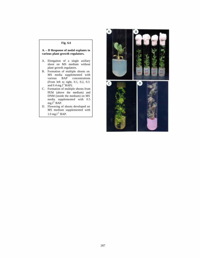
287
Fig. 6.6 A. – D Response of nodal explants to various plant growth regulators. A. Elongation of a single axillary
shoot on MS medium without plant growth regulators.
B. Formation of multiple shoots on MS media supplemented with various BAP concentrations (From left to right, 0.1, 0.2, 0.3 and 0.4 mg.l-1 BAP).
C. Formation of multiple shoots from PEM (above the medium) and DNM (inside the medium) on MS media supplemented with 0.5 mg.l-1 BAP.
D. Flowering of shoots developed on MS medium supplemented with 1.0 mg.l-1 BAP.
288
De novo shoot bud formation was not observed on BM with 1.0 mg l-1 BAP, the
reason for which is unclear. However, all the multiple shoots produced from the pre-
existing axillary buds flowered (Fig. 6.6 D).
The axillary buds on both the PEM and DNM types of newly induced shoots on
0.5 and 5.0 mg.l-1 BAP (Table 6.8 ) sprouted simultaneously overcoming the natural
apical dominance and effectively increasing the overall number of shoots which could be
separated, elongated and rooted with 100% efficiency. But only the newly induced shoots
and not the precociously sprouting axillary buds were taken into account for counting the
number of shoots produced in these BAP concentrations.
Unlike the leaf explants, once the first round of shoots was cut from the nodal
explant, it was not possible to get another round of shoot induction from the same nodal
explant.
6.3.2.2 Regeneration of plants from internodes
On lower concentrations of BAP (0.1-0.5 mg.l-1), internodes did not respond to
form shoots. Very high numbers (25-40) of de novo shoot buds were induced from
internodes only at higher BAP concentrations (1.0, 5.0 mg.l-1 BAP) (Table 6.8), (Fig. 6.7
A, B). Since the shoot buds were crowded, their exact number could not be counted even
under magnification. The complete surface of the internodes was covered with well-
developed shoot buds with distinct apical meristems. These shoot buds could be easily
elongated and rooted as mentioned earlier in Section 6.2.4 (Fig. 6.7 D).
6.3.2.3 Regeneration of plants from hypocotyls
Twelve shoots per hypocotyl explant piece were induced on BM supplemented
with 0.5 mg.l-1 BAP (Table 6.8 ) and the explants continued to form shoots even when
they were transferred to a medium without plant growth regulators (Fig. 6.8 A, C ). This
effect was more pronounced in screw capped bottles that were large and had more amount
of head space volume (Fig. 6.8 B ). During each monthly subculture on BM without plant
growth regulators, twelve shoots could be recovered upto the fourth subculture resulting
in 48 shoots from a single hypocotyl explant piece during a four month period and 144
shoots in one year. Since one hypocotyl gives rise to 3 pieces of hypocotyl explant, a
single hypocotyl would produce 432 shoots in an year. BM supplemented with lower
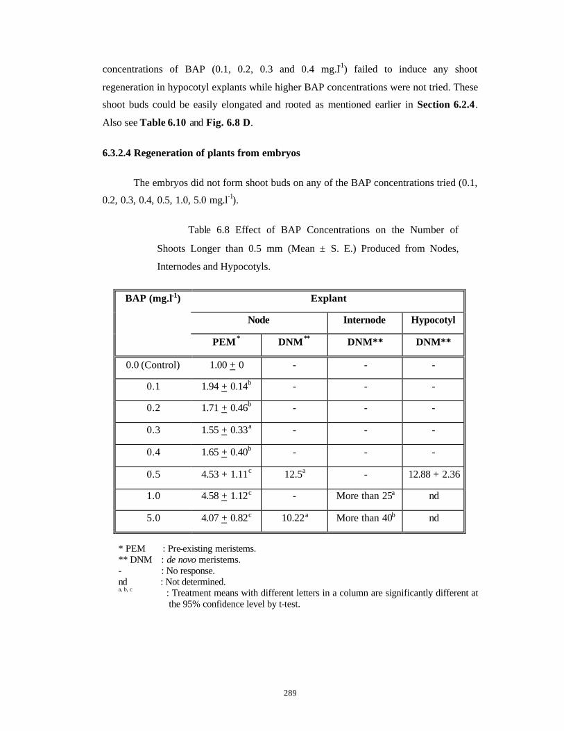
289
concentrations of BAP (0.1, 0.2, 0.3 and 0.4 mg.l-1) failed to induce any shoot
regeneration in hypocotyl explants while higher BAP concentrations were not tried. These
shoot buds could be easily elongated and rooted as mentioned earlier in Section 6.2.4.
Also see Table 6.10 and Fig. 6.8 D.
6.3.2.4 Regeneration of plants from embryos
The embryos did not form shoot buds on any of the BAP concentrations tried (0.1,
0.2, 0.3, 0.4, 0.5, 1.0, 5.0 mg.l-1).
Table 6.8 Effect of BAP Concentrations on the Number of
Shoots Longer than 0.5 mm (Mean ± S. E.) Produced from Nodes,
Internodes and Hypocotyls.
Explant
Node Internode Hypocotyl
BAP (mg.l-1)
PEM* DNM** DNM** DNM**
0.0 (Control) 1.00 + 0 - - -
0.1 1.94 + 0.14b - - -
0.2 1.71 + 0.46b - - -
0.3 1.55 + 0.33a - - -
0.4 1.65 + 0.40b - - -
0.5 4.53 + 1.11c 12.5a - 12.88 + 2.36
1.0 4.58 + 1.12c - More than 25a nd
5.0 4.07 + 0.82c 10.22a More than 40b nd
* PEM : Pre-existing meristems. ** DNM : de novo meristems. - : No response. nd : Not determined. a, b, c : Treatment means with different letters in a column are significantly different at
the 95% confidence level by t-test.
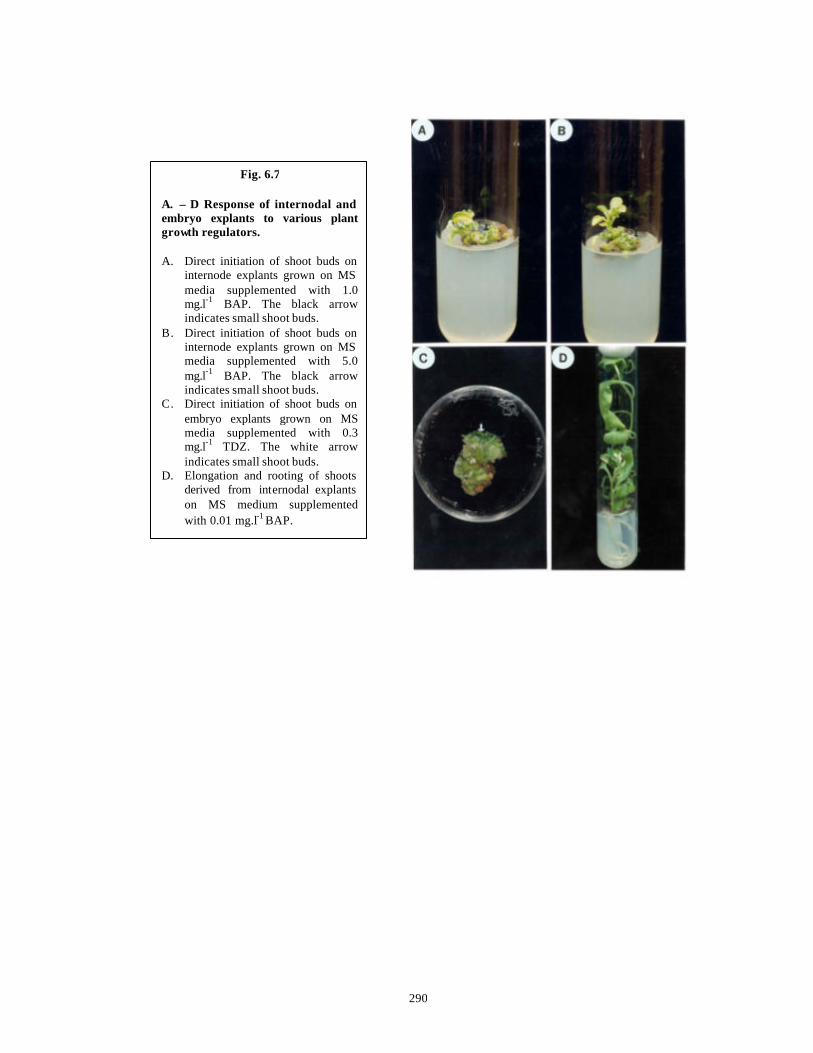
290
Fig. 6.7 A. – D Response of internodal and embryo explants to various plant growth regulators. A. Direct initiation of shoot buds on
internode explants grown on MS media supplemented with 1.0 mg.l-1 BAP. The black arrow indicates small shoot buds.
B. Direct initiation of shoot buds on internode explants grown on MS media supplemented with 5.0 mg.l-1 BAP. The black arrow indicates small shoot buds.
C. Direct initiation of shoot buds on embryo explants grown on MS media supplemented with 0.3 mg.l-1 TDZ. The white arrow indicates small shoot buds.
D. Elongation and rooting of shoots derived from internodal explants on MS medium supplemented with 0.01 mg.l-1 BAP.
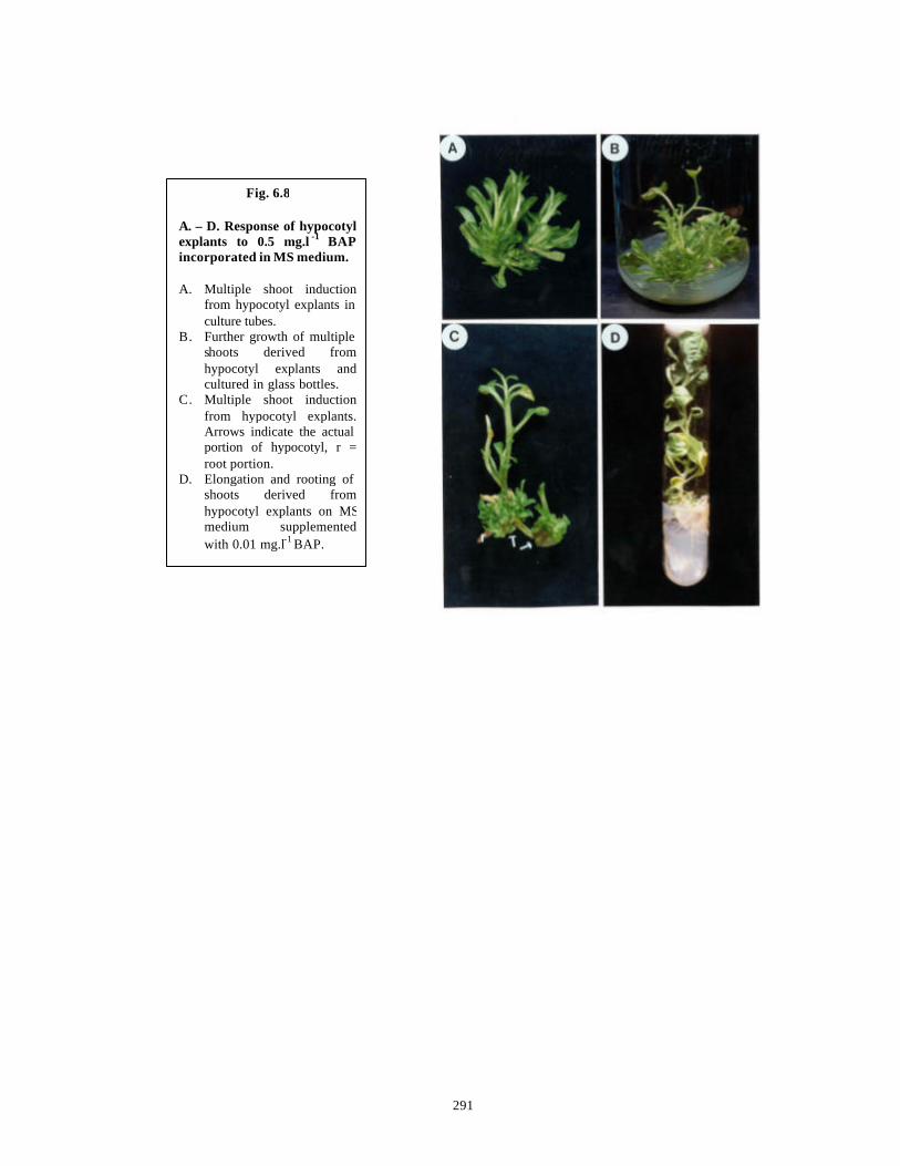
291
Fig. 6.8 A. – D. Response of hypocotyl explants to 0.5 mg.l -1 BAP incorporated in MS medium. A. Multiple shoot induction
from hypocotyl explants in culture tubes.
B. Further growth of multiple shoots derived from hypocotyl explants and cultured in glass bottles.
C. Multiple shoot induction from hypocotyl explants. Arrows indicate the actual portion of hypocotyl, r = root portion.
D. Elongation and rooting of shoots derived from hypocotyl explants on MSmedium supplemented with 0.01 mg.l-1 BAP.
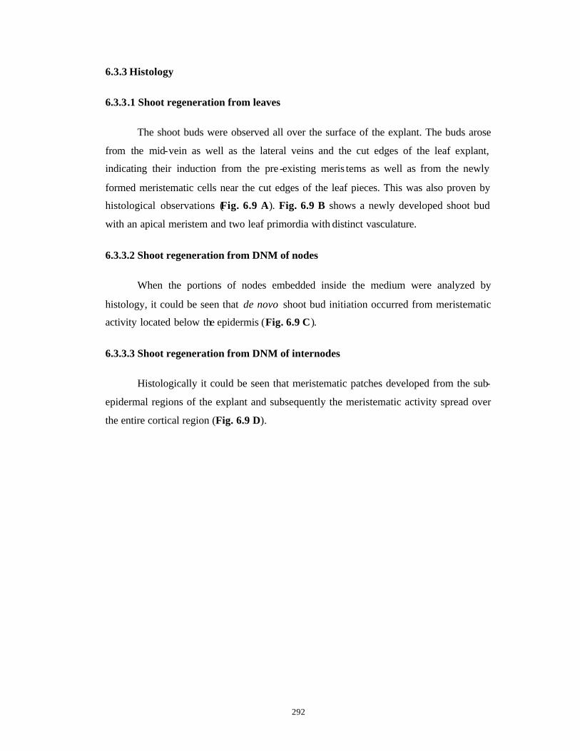
292
6.3.3 Histology
6.3.3.1 Shoot regeneration from leaves
The shoot buds were observed all over the surface of the explant. The buds arose
from the mid-vein as well as the lateral veins and the cut edges of the leaf explant,
indicating their induction from the pre -existing meris tems as well as from the newly
formed meristematic cells near the cut edges of the leaf pieces. This was also proven by
histological observations (Fig. 6.9 A). Fig. 6.9 B shows a newly developed shoot bud
with an apical meristem and two leaf primordia with distinct vasculature.
6.3.3.2 Shoot regeneration from DNM of nodes
When the portions of nodes embedded inside the medium were analyzed by
histology, it could be seen that de novo shoot bud initiation occurred from meristematic
activity located below the epidermis (Fig. 6.9 C).
6.3.3.3 Shoot regeneration from DNM of internodes
Histologically it could be seen that meristematic patches developed from the sub-
epidermal regions of the explant and subsequently the meristematic activity spread over
the entire cortical region (Fig. 6.9 D).
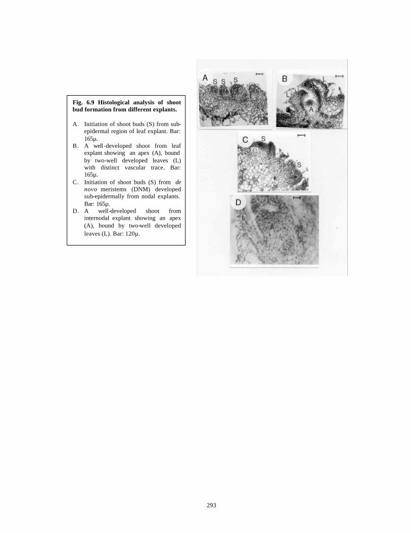
293
Fig. 6.9 Histological analysis of shoot bud formation from different explants. A. Initiation of shoot buds (S) from sub-
epidermal region of leaf explant. Bar: 165µ.
B. A well -developed shoot from leaf explant showing an apex (A), bound by two-well developed leaves (L) with distinct vascular trace. Bar: 165µ.
C. Initiation of shoot buds (S) from de novo meristems (DNM) developed sub-epidermally from nodal explants. Bar: 165µ.
D. A well-developed shoot from internodal explant showing an apex (A), bound by two-well developed leaves (L). Bar: 120µ.

294
6.3.4 Regeneration of plants from node, internode and embryo explants on BM
supplemented with only TDZ.
The node, internode and hypocotyl explants were prepared and inoculated on
various BM and plant growth regulator combinations as described earlier in Section
6.2.3, in culture tubes only as plastic dishes were not used for the reasons described
earlier in Section 6.3.2. Only the small embryonal explants were inoculated in plastic
dishes
6.3.4.1 Regeneration of plants from nodes
One or two shoots with a stunted pattern of development were induced from the
pre-existing meristem on BM supplemented with 0.05 and 0.1 mg.l-1 TDZ. Rest of the
explant was covered with callus (Table 6.9 ). Maximum number of 24 shoots per node
were obtained from the pre-existing meristems (PEM) of axillary buds on BM
supplemented with 0.2 mg.l-1 TDZ. Unlike BAP, no precocious sprouting of axillary buds
was observed and the shoots were shorter in length. In addition a distinct ring of de novo
meristem (DNM) derived adventitious shoot buds (ten to twelve) formed below and
surrounding the pre-existing axillary bud on media with 0.2 and 0.3 mg.l-1 TDZ (Table
6.9 ), (Fig. 6.10 A). The number of shoots first increased with an increase in TDZ
concentrations and then decreased (Table 6.9). This method will prove to be of a great
advantage in large -scale clonal propagation of selected elite plants.
The shoots derived from PEM as well as DNM could be elongated and rooted
only after four passages on BM with 1/2 strength of major salts (1/2 BM) without BAP
(Table 6.10) (Fig. 6.10 B ). The high carry-over effect of TDZ has been well documented
in the literature (Meyer and Van Staden, 1988; Lu, 1993).
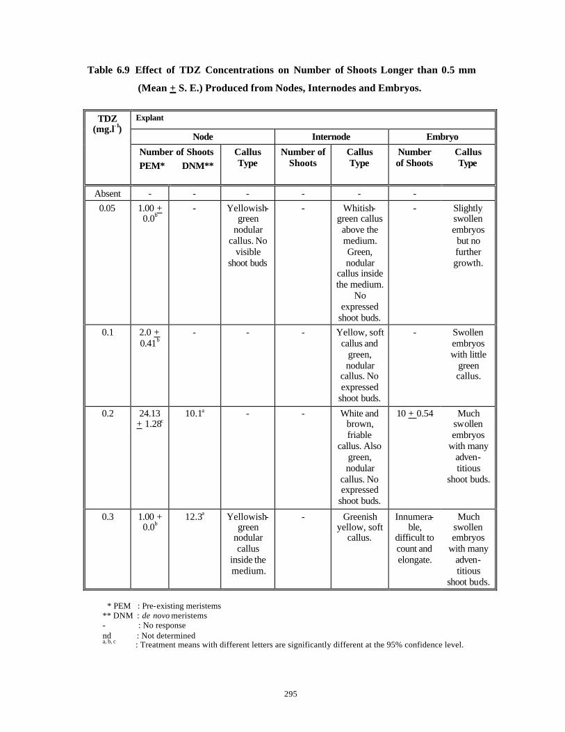
295
Table 6.9 Effect of TDZ Concentrations on Number of Shoots Longer than 0.5 mm
(Mean + S. E.) Produced from Nodes, Internodes and Embryos.
Explant
Node Internode Embryo
TDZ (mg.l-1)
Number of Shoots PEM* DNM**
Callus Type
Number of Shoots
Callus Type
Number of Shoots
Callus Type
Absent - - - - - - 0.05
1.00 + 0.0b
- Yellowish-green
nodular callus. No
visible shoot buds
- Whitish-green callus above the medium. Green, nodular
callus inside the medium.
No expressed
shoot buds.
- Slightly swollen embryos but no further growth.
0.1
2.0 + 0.41b
- - - Yellow, soft callus and
green, nodular
callus. No expressed
shoot buds.
- Swollen embryos with little
green callus.
0.2
24.13 + 1.28c
10.1a - - White and brown, friable
callus. Also green, nodular
callus. No expressed
shoot buds.
10 + 0.54 Much swollen embryos
with many adven-titious
shoot buds.
0.3
1.00 + 0.0b
12.3a Yellowish-green
nodular callus
inside the medium.
- Greenish yellow, soft
callus.
Innumera-ble,
difficult to count and elongate.
Much swollen embryos
with many adven-titious
shoot buds.
* PEM : Pre-existing meristems ** DNM : de novo meristems - : No response nd : Not determined a, b, c : Treatment means with different letters are significantly different at the 95% confidence level.

296
Fig. 6.10 A. A ring of de novo shoot buds
formed on nodes grown on MS medium supplemented with 0.2 mg.l-1 TDZ.
B. Simultaneous elongation and rooting of shoots derived from nods growing on 0.2 mg.l- 1 TDZ supplemented MS medium, on 1/2 MS medium without plant growth regulators.
C. A multiple shoot culture derived from nodes grown on MS medium supplemented with 0.5 mg.l-1 BAP and used for extraction of Withanolides for HPLC analysis.
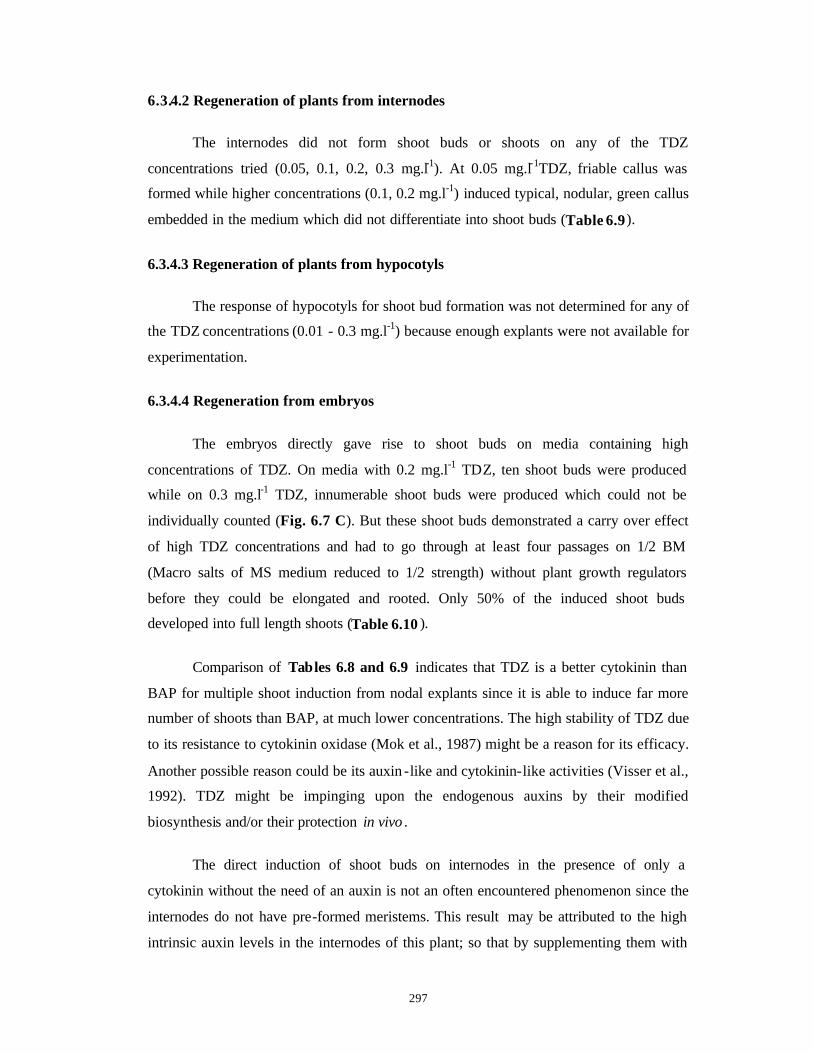
297
6.3.4.2 Regeneration of plants from internodes
The internodes did not form shoot buds or shoots on any of the TDZ
concentrations tried (0.05, 0.1, 0.2, 0.3 mg.l-1). At 0.05 mg.l-1TDZ, friable callus was
formed while higher concentrations (0.1, 0.2 mg.l-1) induced typical, nodular, green callus
embedded in the medium which did not differentiate into shoot buds (Table 6.9).
6.3.4.3 Regeneration of plants from hypocotyls
The response of hypocotyls for shoot bud formation was not determined for any of
the TDZ concentrations (0.01 - 0.3 mg.l-1) because enough explants were not available for
experimentation.
6.3.4.4 Regeneration from embryos
The embryos directly gave rise to shoot buds on media containing high
concentrations of TDZ. On media with 0.2 mg.l-1 TDZ, ten shoot buds were produced
while on 0.3 mg.l-1 TDZ, innumerable shoot buds were produced which could not be
individually counted (Fig. 6.7 C). But these shoot buds demonstrated a carry over effect
of high TDZ concentrations and had to go through at least four passages on 1/2 BM
(Macro salts of MS medium reduced to 1/2 strength) without plant growth regulators
before they could be elongated and rooted. Only 50% of the induced shoot buds
developed into full length shoots (Table 6.10 ).
Comparison of Tables 6.8 and 6.9 indicates that TDZ is a better cytokinin than
BAP for multiple shoot induction from nodal explants since it is able to induce far more
number of shoots than BAP, at much lower concentrations. The high stability of TDZ due
to its resistance to cytokinin oxidase (Mok et al., 1987) might be a reason for its efficacy.
Another possible reason could be its auxin -like and cytokinin-like activities (Visser et al.,
1992). TDZ might be impinging upon the endogenous auxins by their modified
biosynthesis and/or their protection in vivo .
The direct induction of shoot buds on internodes in the presence of only a
cytokinin without the need of an auxin is not an often encountered phenomenon since the
internodes do not have pre-formed meristems. This result may be attributed to the high
intrinsic auxin levels in the internodes of this plant; so that by supplementing them with
298
high concentrations of BAP it is possible to balance both the growth regulators so that the
explant becomes competent for organogenesis (Christianson and Warnick, 1985). TDZ
might not be able to induce organogenesis in internodes because of its auxin as well as
cytokinin like activities which might be disrupting the delicate balance necessary for
shoot bud formation.
In the present study, since shoot regeneration from node, internode, hypocotyl and
embryo explants could be achieved on MS medium supplemented with either BAP or
TDZ, further experiments by supplementation of these cytokinins with auxins were not
carried out.
6.3.5 Acclimat ization
Twenty regenerants were randomly selected from each type of explant during
subculture numbers 1, 3, 6, 9, 12 (subcultures of 3-4 weeks duration during the year) and
hardened at different periods of time taking care that they were derived from different
explants of different parent plants while the remaining sterile shoots were used for further
regeneration studies and initial experiments in plant transformation. Except for the
embryo-derived plantlets, regenerants from all the other explants (leaf bits, nodes,
internodes, hypocotyls) could be hardened with 100% success rate (Table 6.10 ). All the
regenerants exhibited normal morphology with respect to leaf shape and size and stem
morphology.

299
Table 6.10 Elongation, Rooting and Acclimatization of Shoots Derived from Various
Explants.
Explants Plant Growth
Regulator in Induction Medium
Elongation and Rooting Medium
% Elongation and Rooting
Number of Passages*
% Acclimatization
Leaves IAA + BAP BM + 0.01 BAP 100 One 100
On low BAP - One
100 BAP BM + 0.01 BAP 100
On high BAP - Two
100
Node
TDZ 1/2 BM 100 Four 100
BAP BM + 0.01 BAP 100 Two 100 Internode
TDZ - - - -
BAP BM + 0.01 BAP 100 One 100 Hypocotyl
TDZ nd nd nd nd
BAP - - - - Embryo
TDZ 1/2 BM 50 Four 0 - : No response nd : Not determined * : Passage duration is one month.
300
6.3.6 HPLC analysis of Withaferin-A standard
To detect the presence of Withaferin-A, normal, in vitro seedlings (Control),
multiple shoot cultures induced on MS medium supplemented with 0.5 mg.l-1 BAP and
the callus formed on the same medium in 2% nodal explants (C. f. Section 6.3.2.1) were
extracted and analyzed by HPLC. Fig. 6.11 A shows the chromatogram of standard (A
kind gift from Dr. M. I. Choudhary of HEJ Res. Inst., Pakistan) of Withaferin-A (10 µg
injected in 20 µL) obtained under conditions described earlier in Section 6.2.9. It can be
seen that there are two well defined and baseline resolved peaks (12 min. and 15 min.) in
the standard itself. Hence the tissue ext racts were compared with both of these peaks. A
standard curve plotted for various concentrations of Withaferin-A (1.25-10 µg per 20
µL) is shown in Figs. 6.11 B, C. It shows that the detector response is linear for the
concentrations of less than 10 µg.
6.3.7 Extraction and HPLC analysis of in vitro tissues
For extractions methanol was the preferred solvent as described above. No
additional extractions with hexane or other apolar solvents was necessary to remove
lipophilic compounds as the tissues were very juvenile (Theodoridis et al., 1998). This
was proven during the HPLC analysis because there was no significant increase in back-
pressure of the column and the chromatograms sometimes showed only an initial broad
peak of polar impurities which did not interfere with the Withaferin -A analysis.
HPLC is the most appropriate technique for an efficient separation of crude plant
extracts and can be coupled with different spectroscopic detection methods (Hostettmann
and Wolfender, 1997). In the present endeavor, HPLC coupled with UV photodiode array
detection was used. The UV spectra of natural compounds give useful information about
the type of constituents and their structures (Hostettmann and Wolfender, 1997).
While carrying out the HPLC analysis of tissue cultures of Withania spp.,
everyday at least two independent injections of the samples were performed and an
average of the peak areas was calculated. The standard error of the mean (S. E.) obtained
was always less than 8 over the analysis period. The sample injections from the same
sample preparation were repeated on consecutive days and again the S. E. for the peak
areas was less than 6.
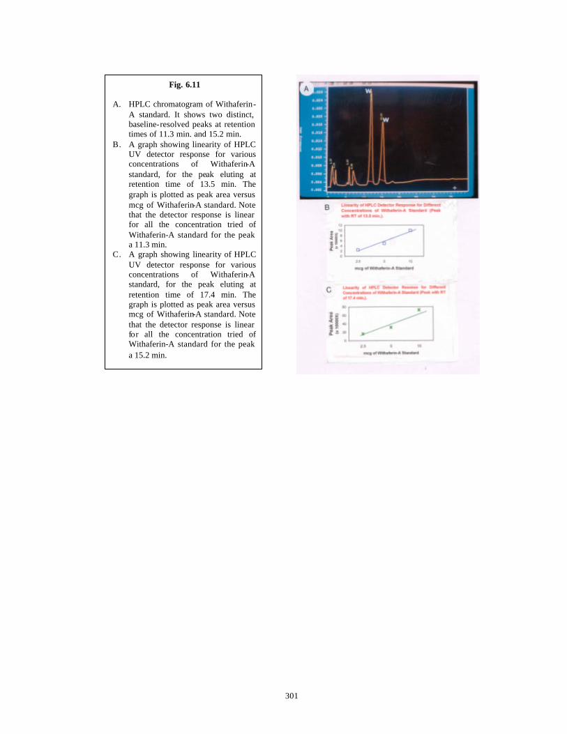
301
Fig. 6.11 A. HPLC chromatogram of Withaferin-
A standard. It shows two distinct, baseline-resolved peaks at retention times of 11.3 min. and 15.2 min.
B. A graph showing linearity of HPLC UV detector response for various concentrations of Withaferin-A standard, for the peak eluting at retention time of 13.5 min. The graph is plotted as peak area versus mcg of Withaferin-A standard. Note that the detector response is linear for all the concentration tried of Withaferin-A standard for the peak a 11.3 min.
C. A graph showing linearity of HPLC UV detector response for various concentrations of Withaferin-A standard, for the peak eluting at retention time of 17.4 min. The graph is plotted as peak area versus mcg of Withaferin-A standard. Note that the detector response is linear for all the concentration tried of Withaferin-A standard for the peak a 15.2 min.
302
Shoots of two-month old seedlings (source of explants for all the experiments) had
Withaferin-A (Table 6.11 ) (Fig. 6.12 A). The multiple shoot cultures derived from
nodes grown on agar solidified MS medium with 0.5 mg.l-1 BAP were also able to
synthesize Withaferin-A (Fig. 6.12 B ). The amount was at least ten times more than that
present in the seedling shoot s (Table 6.11). However callus cultures derived from the
same nodal explants on identical medium did not have Withaferin -A (Table 6.11). This
corroborates the earlier observations made by Roja et al. (1991) that tissue organization
into organs is necessary for Withanolide production. Roja et al. (1991) reported
production of Withanolide -I in the range of 13-172 mg/g dw of shoots, Withanolide -G
in the range of 10-317 mg/g dw of shoots, Withanone in the range of 90-486 mg/g dw of
shoots and traces of Withanolide -D except Withaferin-A (the major anti-tumor
compound).
But in the present study, Withaferin-A production has been observed and it is
produced in the range of 3.903-4.000 mg/g dw of shoots of in vitro grown seedlings and
in the range of 55.1667-51.1833 mg/g dw of in vitro shoots, even though Withaferin-A
in leaves of in vivo plants has been reported to be in the range of 0.16% DW (Gupta et al.,
1996) or 0.2-0.3% dw (Abraham et al., 1968). The values reported here are far higher
than the values reported by others and in addition, are even higher than those reported for
Agrobacterium tumefaciens transformed tissues (Ray and Jha, 1999). The range of
Withaferin-A produced in shooty terratomas was in the range of 0.7-1.0 mg/g dw of
shoots while the untransformed shoots contained 0.4-0.5 mg Withaferin-A/g dw of
shoots. Since the chemotype of Withania spp. and the in vitro shoots raised from this
chemotype used in the present study are far superior in the production of Withaferin-A,
the knowhow generated in this study could be further used for scale up studies in a
bioreactor for large scale production of Withaferin-A , even without resorting to hairy
root cultures.
Although in case of Withanolides, no particular specialized structures have been
identified for accumulation, the biosynthetic capacity may be localized in specific cell
types in the leaves and a regulatory hierarchy may be involved in which morphology is a
dominant factor (Robins, 199 ). Leaves are believed to be the sites of withanolide
synthesis since the steroids are derived from mevalonic acid (Ray and Gupta, 1994) and
probably the withanolides are transported across the entire shoot system. Similar
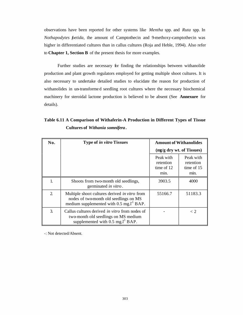
303
observations have been reported for other systems like Mentha spp. and Ruta spp. In
Nothapodytes foetida, the amount of Camptothecin and 9-methoxy-camptothecin was
higher in differentiated cultures than in callus cultures (Roja and Heble, 1994). Also refer
to Chapter 1, Section B of the present thesis for more examples.
Further studies are necessary for finding the relationships between withanolide
production and plant growth regulators employed for getting multiple shoot cultures. It is
also necessary to undertake detailed studies to elucidate the reason for production of
withanolides in un-transformed seedling root cultures where the necessary biochemical
machinery for steroidal lactone production is believed to be absent (See Annexure for
details).
Table 6.11 A Comparison of Withaferin-A Production in Different Types of Tissue
Cultures of Withania somnifera .
Amount of Withanolides
(µg/g dry wt. of Tissues)
No. Type of in vitro Tissues
Peak with retention
time of 12 min.
Peak with retention time of 15
min.
1. Shoots from two-month old seedlings, germinated in vitro .
3903.5 4000
2. Multiple shoot cultures derived in vitro from nodes of two-month old seedlings on MS
medium supplemented with 0.5 mg.l-1 BAP.
55166.7 51183.3
3. Callus cultures derived in vitro from nodes of two-month old seedlings on MS medium
supplemented with 0.5 mg.l-1 BAP.
- < 2
-: Not detected/Absent.
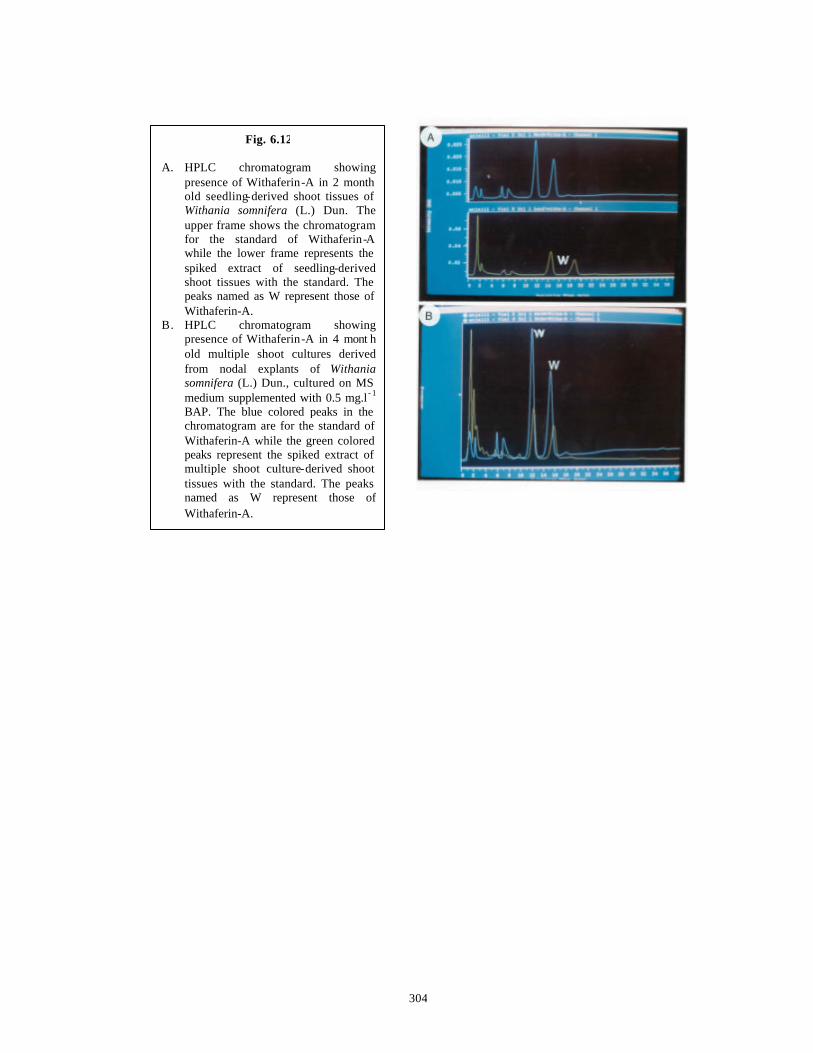
304
Fig. 6.12 A. HPLC chromatogram showing
presence of Withaferin-A in 2 month old seedling-derived shoot tissues of Withania somnifera (L.) Dun. The upper frame shows the chromatogram for the standard of Withaferin-A while the lower frame represents the spiked extract of seedling-derived shoot tissues with the standard. The peaks named as W represent those of Withaferin-A.
B. HPLC chromatogram showing presence of Withaferin-A in 4 mont h old multiple shoot cultures derived from nodal explants of Withania somnifera (L.) Dun., cultured on MS medium supplemented with 0.5 mg.l - 1
BAP. The blue colored peaks in the chromatogram are for the standard of Withaferin-A while the green colored peaks represent the spiked extract of multiple shoot culture-derived shoot tissues with the standard. The peaks named as W represent those of Withaferin-A.
305
6.4 Conclusions
1) Direct regeneration of plants has been achieved from a number of
explants like leaves, nodes, internodes, hypocotyls and embryos.
Regenerated shoots have been obtained in 70% of leaf explants on an
optimal medium while all the remaining explants gave 100% response
for shoot formation on media standardized during the course of the
present work.
2) By the protocol standardized in this study, 1600 and 432 plantlets per
year per explant could be produced from leaves and hypocotyl explants
respectively.
3) De novo regeneration of 24 shoots from a single node explant could be
obtained with TDZ. TDZ was found to be the best plant growth regulator
among the tested plant growth regulators.
4) All the regenerants had a high success rate of acclimatization and
hardening.
5) High amounts of Withaferin-A were detected in both the seedlings and in
vitro regenerated shoots from nodes. In vitro grown seedlings had 10
times more Withaferin-A than the reported values in literature for in vivo
tissues of Withania somnifera (L.) Dun. In vitro regenerated shoots
growing on MS medium supplemented with 0.5 mg.l-1 BAP had 100 times
more Withaferin-A than in vivo tissues and to our knowledge such high
amounts of Withaferin-A reported here are for the first time in literature.
The work presented in this chapter has been published in two international papers:
a) Kulkarni A. A., Thengane S. R. and Krishnamurthy K. V. (1996) Direct in vitro
regeneration of leaf explants of Withania somnifera (L.) Dunal. Plant Science
119: 163 -168.
b) Kulkarni A. A., Thengane S. R. and Krishnamurthy K. V. (2000) Direct Shoot
Regeneration from Node, Internode, Hypocotyl and Embryo Explants of
Withania somnifera (L.) Dun. Accepted for publication in Plant Cell Tissue and
Organ Culture.

306
AANNNNEEXXUURREE
Induction of Hairy Root Cultures in Withania
spp. by Infection with Agrobacterium
rhizogenes.
307
A.1 Introduction
Withania somnifera (L.) Dunal is widely prescribed in the Ayurveda because of
the pharmacological properties due to a number of Tropane Alkaloids present in the
roots of the plant (Anonymous, 1976). It is called as ‘Indian Ginseng’ and shows
comparable properties to ‘Chinese Ginseng’ (Panax ginseng ) (Ray and Gupta, 1994).
Lavie’s group (1965) elucidated the structure of Withaferin-A (A Steroidal Lactone) in
leaves of this plant, which is mostly valued for its anti-cancerous properties. The anti-
tumor activity of this compound was further demonstrated by many researchers (Kupchan
et al., 1969; Uma Devi et al., 1992). The yields of Withaferin-A in intact plants of
Withania spp. (Israeli Chemotype) are 0.2-0.3% of DW of leaves (Abraham et al., 1968).
Gupta et al. (1996) have done quantitative analysis of Indian chemotypes of Withania
somnifera by TLC densitometry and observed that Withaferin-A was totally absent in
roots, stems, seeds and persistent calyx of fruits of intact plants except in leaves (1.6%).
Surprisingly the alkaloids were detected in all the above mentioned plant parts and leaves
had the highest content. This is in contrast to the general belief that the Tropane
Alkaloids are restricted to the roots of Withania spp.
Up till now, attempts have been made to analyze callus, shoot and transformed
tissue cultures of Withania somnifera for the presence of antitumor Withanolides (Table
6.2 ). These studies have indicated that Withanolide production is related to
morphogenesis of cultures and the regenerated shoots have more content of Withanolides
than the unorganized callus cultures (Roja et al., 1991; Results presented in Chapter 6 of
the present thesis).
As discussed earlier in Chapter 1, hairy roots from many species have been
shown to synthesize a number of novel compounds absent in the parent plant. In addition
many of the compounds present in the parent plant are synthesized in higher quantities by
the hairy roots. Hairy root cultures were induced in Withania somnifera (Banerjee et al.,
1994) by Agrobacterium rhizogenes strains A4, LBA 9402 and LBA 9360 in an attempt
to further increase the quantities of Withaferin-A , normally present in shoots of intact
plants. Its presence was detected in the hairy roots as well as in the growth medium. But
quantitative estimation of Withaferin-A , the baseline separation of its peak and data on
peak purity, co-chromatography with standard of Withaferin-A were not reported and

308
thus the data is not comprehensive and conclusive. In another report, Ray et al. (1996)
reported presence of Withanolide -D and absence of Withaferin-A in hairy roots of
Withania somn ifera . In a recent publication, even though Ray and Jha (1999) have
reported formation of galls, rooty and shooty terratomas and hairy roots from leaves of
Withania somnifera with a number of Agrobacterium tumefaciens strains, presence of
Withaferin-A in hairy roots and rooty terratomas was not detected. On the other hand
shooty terratomas contained both these compounds.
In view of the above, the present study made a preliminary attempt to genetically
transform leaf and nodal explants of Withania somnifera with different strains of
Agrobacterium rhizogenes in order:
1. To induce hairy roots on different explants.
2. To check for possible production of Withanolides by these hairy roots since their site
of synthesis is leaves and not the roots.
3. To estimate the contents of Withanolides, especially Withaferin-A, quantitatively in
the in vivo and in vitro generated hairy roots. The results obtained are further
compared with Withaferin-A contents of the untransformed seedling roots and shoots.
309
A.2 Materials and Methods
A.2.1 Strains and growth of Agrobacterium rhizogenes
Five wild strains of Agrobacterium rhizogenes were procured from “American
Type Culture Collection”, USA in the form of freeze dried cultures. These were ATCC
15834, ATCC 11325, ATCC 13332, ATCC 13333 and ATCC 25818. These were
rehydrated and revived according to the supplier’s instructions in Medium 3 (Table A.1).
The wild strain A4 was a kind gift from Dr. M. R. Heble (“BARC”, Mumbai, India). All
these strains were grown routinely from single colonies in liquid YEB medium (Dietze et
al., 1995) and in liquid AB medium to reduce the clumping during growth (White and
Nester, 1980). The compositions of the media used are described in Table A.1. Glass
distilled water was used for media preparations and pH was adjusted at 7.0 using 0.1 N
KOH or 0.1 N HCl. Autoclaving was done at 121oC and at a pressure of 1.1 kg.cm-2 for
20 min. When needed, 2% bacto-agar (“Difco Laboratories”, USA) was added to the
respective media and 20 ml of this medium were poured in each of the plastic dishes (85
mm diameter, “Laxbro”, India).
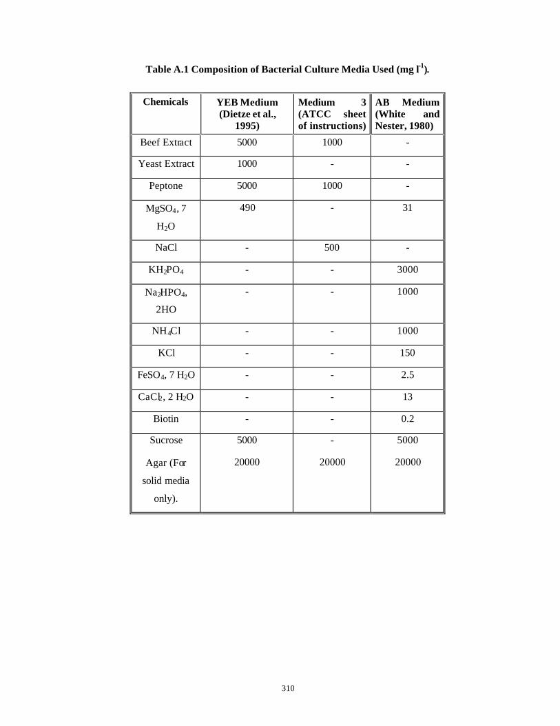
310
Table A.1 Composition of Bacterial Culture Media Used (mg l-1).
Chemicals YEB Medium
(Dietze et al., 1995)
Medium 3 (ATCC sheet of instructions)
AB Medium (White and Nester, 1980)
Beef Extract 5000 1000 -
Yeast Extract 1000 - -
Peptone 5000 1000 -
MgSO4, 7
H2O
490 - 31
NaCl - 500 -
KH2PO4 - - 3000
Na2HPO4,
2HO
- - 1000
NH4Cl - - 1000
KCl - - 150
FeSO4, 7 H2O - - 2.5
CaCl2, 2 H2O - - 13
Biotin - - 0.2
Sucrose 5000 - 5000
Agar (For
solid media
only).
20000 20000 20000
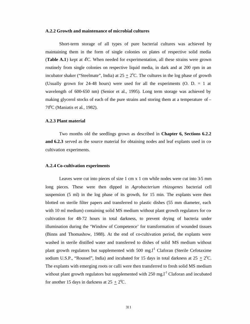
311
A.2.2 Growth and maintenance of microbial cultures
Short-term storage of all types of pure bacterial cultures was achieved by
maintaining them in the form of single colonies on plates of respective solid media
(Table A.1) kept at 4oC. When needed for experimentation, all these strains were grown
routinely from single colonies on respective liquid media, in dark and at 200 rpm in an
incubator shaker (“Steelmate”, India) at 25 + 2oC. The cultures in the log phase of growth
(Usually grown for 24-48 hours) were used for all the experiments (O. D. = 1 at
wavelength of 600-650 nm) (Senior et al., 1995). Long term storage was achieved by
making glycerol stocks of each of the pure strains and storing them at a temperature of –
70oC (Maniatis et al. , 1982).
A.2.3 Plant material
Two months old the seedlings grown as described in Chapter 6, Sections 6.2.2
and 6.2.3 served as the source material for obtaining nodes and leaf explants used in co-
cultivation experiments.
A.2.4 Co-cultivation experiments
Leaves were cut into pieces of size 1 cm x 1 cm while nodes were cut into 3-5 mm
long pieces. These were then dipped in Agrobacterium rhizogenes bacterial cell
suspension (5 ml) in the log phase of its growth, for 15 min. The explants were then
blotted on sterile filter papers and transferred to plastic dishes (55 mm diameter, each
with 10 ml medium) containing solid MS medium without plant growth regulators for co-
cultivation for 48-72 hours in total darkness, to prevent drying of bacteria under
illumination during the ‘Window of Competence’ for transformation of wounded tissues
(Binns and Thomashow, 1988). At the end of co-cultivation period, the explants were
washed in sterile distilled water and transferred to dishes of solid MS medium without
plant growth regulators but supplemented with 500 mg.l-1 Claforan (Sterile Cefotaxime
sodium U.S.P., “Roussel”, India) and incubated for 15 days in total darkness at 25 + 2oC.
The explants with emerging roots or calli were then transferred to fresh solid MS medium
without plant growth regulators but supplemented with 250 mg.l-1 Claforan and incubated
for another 15 days in darkness at 25 + 2oC.
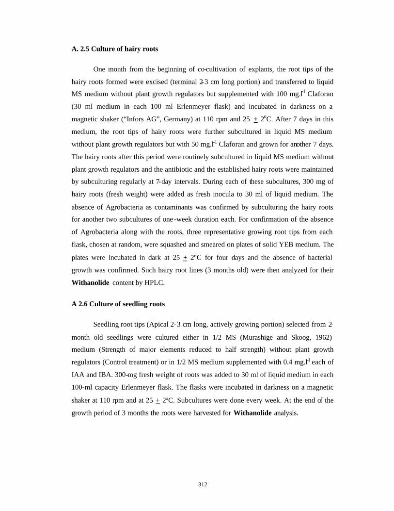
312
A. 2.5 Culture of hairy roots
One month from the beginning of co-cultivation of explants, the root tips of the
hairy roots formed were excised (terminal 2-3 cm long portion) and transferred to liquid
MS medium without plant growth regulators but supplemented with 100 mg.l-1 Claforan
(30 ml medium in each 100 ml Erlenmeyer flask) and incubated in darkness on a
magnetic shaker (“Infors AG”, Germany) at 110 rpm and 25 + 2oC. After 7 days in this
medium, the root tips of hairy roots were further subcultured in liquid MS medium
without plant growth regulators but with 50 mg.l-1 Claforan and grown for another 7 days.
The hairy roots after this period were routinely subcultured in liquid MS medium without
plant growth regulators and the antibiotic and the established hairy roots were maintained
by subculturing regularly at 7-day intervals. During each of these subcultures, 300 mg of
hairy roots (fresh weight) were added as fresh inocula to 30 ml of liquid medium. The
absence of Agrobacteria as contaminants was confirmed by subculturing the hairy roots
for another two subcultures of one -week duration each. For confirmation of the absence
of Agrobacteria along with the roots, three representative growing root tips from each
flask, chosen at random, were squashed and smeared on plates of solid YEB medium. The
plates were incubated in dark at 25 + 2°C for four days and the absence of bacterial
growth was confirmed. Such hairy root lines (3 months old) were then analyzed for their
Withanolide content by HPLC.
A 2.6 Culture of seedling roots
Seedling root tips (Apical 2-3 cm long, actively growing portion) selected from 2-
month old seedlings were cultured either in 1/2 MS (Murashige and Skoog, 1962)
medium (Strength of major elements reduced to half strength) without plant growth
regulators (Control treatment) or in 1/2 MS medium supplemented with 0.4 mg.l-1 each of
IAA and IBA. 300-mg fresh weight of roots was added to 30 ml of liquid medium in each
100-ml capacity Erlenmeyer flask. The flasks were incubated in darkness on a magnetic
shaker at 110 rpm and at 25 + 2°C. Subcultures were done every week. At the end of the
growth period of 3 months the roots were harvested for Withanolide analysis.

313
A 2.7 Withanolide analysis by HPLC
The tissue extraction, HPLC analysis and calculations of Withaferin-A contents
were done as described earlier in Chapter 6, Sections 6.2.8 and 6.2.9 .
A 2.8 Statistical analysis
Each plastic dish with five node or leaf bit explants was taken as one replicate for
hairy root induction experiments. Each experiment involving each strain of
Agrobacterium rhizogenes co-cultivated with either of the explants, consisted of five such
replicates and each experiment was repeated thrice. Hairy roots longer than 0.5 mm with
distinctly visible apical meristems were only counted in each treatment. The number of
hairy roots induced were then compared by one way ANOVA and the post-hoc
comparisons were made by Student’s ‘t – test’ (Wardlaw 1985). The hairy roots derived
from either nodal or leaf bit explants with each of the successful A. rhizogenes strains
were cultured in three replicate Erlenmeyer flasks of 100 ml capacity in liquid medium.
For analysis of withanolides, one of these replicate flasks of hairy roots was harvested on
a particular day for extraction and subsequent analysis by HPLC. To check for the
reproducibility of the extraction and HPLC procedure, another of the two remaining
replicate flasks was extracted and analyzed by HPLC on a different day. Dry weight
measurements were carried out at the time of these extractions. For this the hairy roots
from a flask were blotted dry on a filter paper and kept in an oven at 60ºC for 16-24 hours
to achieve constant dry weight.
The seedling roots were also cultured in triplicate in Erlenmeyer flasks of 100-ml
capacity, for each of the media combinations. All the other procedures followed were the
same as those for the hairy roots.

314
A.3 Results and Discussion
A.3.1 Development and establishment of hairy roots
The hairy roots emerged within 15 days from both nodal and leaf bit explants with
Agrobacterium rhizogenes strains: A4, ATCC 15834, ATCC 15332 and ATCC 25818.
Various morphologies of hairy roots induced are shown in Figs. A.1 - A.3. When the
explants were cultured on MS medium without plant growth regulators and without
Agrobacterium co-cultivation, no roots were produced (Fig. A.2 A, Fig. A.3 A). It has
been observed that the success of transformation depends on following factors (Godwin et
al., 1992):
⟨ Bacterial strains and their ability to attach to plant cells.
⟨ Host genotype and explant physiology with respect to presence of competent cells,
release of wound-induced substances/signal molecules.
⟨ Co-cultivation conditions.
As seen from Table A.2 , it is clear that the transformation ability of different
Agrobacterium rhizogenes strains is different. The best results in terms of percentage of
hairy root formation and the number of hairy roots produced were observed with strains
A4 and ATCC 15834. The hairy roots produced by these strains were thick, long with
many root hairs and were fast growing (Fig. A.1 A, B and Fig. A.2 B, C and Fig. A.3 E)
while the hairy roots produced with strain ATCC 25818 were thin, long, with a few root
hairs and slow growing (Fig. A.3 B). No hairy roots were produced after infection with
strain ATCC 15832 (Fig. A.1 C and Fig. A.3 D). Little amount of white/brown, fr iable,
rooty callus was produced after infection with strain ATCC 11325 (Fig. A.3 C with
leaves as explants). Similar differences in hairy root formation have been reported earlier
for Withania somnifera with Agrobacterium rhizogenes (Banerjee et al., 1994) and
Agrobacterium tumefaciens (Ray and Jha, 1999). Such differences might be explained by
the higher capacity of Agropine strains to induce hairy roots due to presence of both TL
and TR DNA as described earlier in Chapter 1. In addition, these different root
morphologies observed among clones of the same cultivar may highlight the randomness
of processing and integration of Ri plasmid T-DNA in plant DNA (Bhadra et al., 1993).
Leaf explants infected with bacterial strains ATCC 11325 and ATCC 15832 were
unable to form hairy roots but gave rise to callus. On the other hand, no response was
315
observed in nodal explants. This again proves the interdependence and interaction
between bacterial strains and the explant types. The Mannopine strains with only TL
DNA are clearly not able to induce hairy roots in Withania somnifera.
Both the earlier reports of hairy root formation in Withania somnifera had used
leaves as explants. Here for the first time success with nodal explants for formation of
hairy roots has been described.
It was also observed that the hairy roots derived from A. rhizogenes strains A4 and
ATCC 15834 could adapt to liquid cultures without undergoing any callusing but the
hairy roots derived from strain ATCC 25818 were not amenable for liquid culture and
soon died. These were not therefore analyzed by HPLC for Withanolide content,
especially Withaferin-A content. Bhadra et al. (1993) had also observed that not all the
hairy root lines developed on solid media in Catharanthus roseus were amenable for
liquid culture. In general, thin, regularly branched root clones are suitable for liquid
cultures and for subsequent scale -up in bioreactors. Hence proper selection of plant
genotype and its combination with A. rhizogenes strain needs to be optimized in every
case.
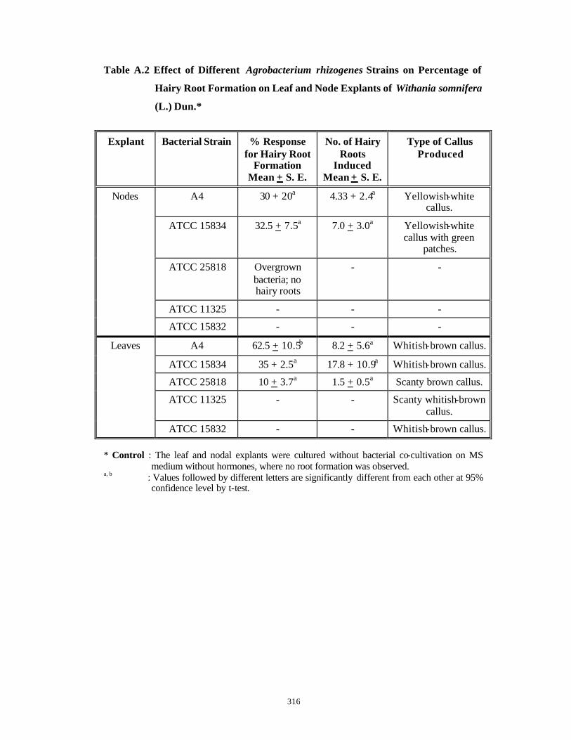
316
Table A.2 Effect of Different Agrobacterium rhizogenes Strains on Percentage of
Hairy Root Formation on Leaf and Node Explants of Withania somnifera
(L.) Dun.*
Explant Bacterial Strain % Response for Hairy Root
Formation Mean + S. E.
No. of Hairy Roots
Induced Mean + S. E.
Type of Callus Produced
A4 30 + 20a 4.33 + 2.4a Yellowish-white callus.
ATCC 15834 32.5 + 7.5a 7.0 + 3.0a Yellowish-white callus with green
patches.
ATCC 25818 Overgrown bacteria; no hairy roots
- -
ATCC 11325 - - -
Nodes
ATCC 15832 - - -
A4 62.5 + 10.5b 8.2 + 5.6a Whitish-brown callus.
ATCC 15834 35 + 2.5a 17.8 + 10.9a Whitish-brown callus.
ATCC 25818 10 + 3.7a 1.5 + 0.5a Scanty brown callus.
ATCC 11325 - - Scanty whitish-brown callus.
Leaves
ATCC 15832 - - Whitish-brown callus. * Control : The leaf and nodal explants were cultured without bacterial co-cultivation on MS
medium without hormones, where no root formation was observed. a, b : Values followed by different letters are significantly different from each other at 95%
confidence level by t-test.
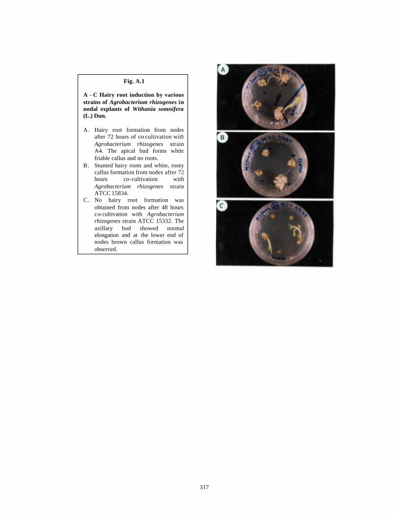
317
Fig. A.1 A - C Hairy root induction by various strains of Agrobacterium rhizogenes in nodal explants of Withania somnifera(L.) Dun. A. Hairy root formation from nodes
after 72 hours of co-cultivation with Agrobacterium rhizogenes strain A4. The apical bud forms white friable callus and no roots.
B. Stunted hairy roots and white, rooty callus formation from nodes after 72 hours co-cultivation with Agrobacterium rhizogenes strain ATCC 15834.
C. No hairy root formation was obtained from nodes after 48 hours co-cultivation with Agrobacterium rhizogenes strain ATCC 15332. The axillary bud showed normal elongation and at the lower end of nodes brown callus formation was observed.
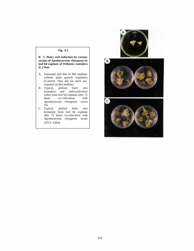
318
Fig. A.2 B - C Hairy root induction by various strains of Agrobacterium rhizogenes in leaf bit explants of Withania somnifera(L.) Dun. A. Untreated leaf bits in MS medium
without plant growth regulators (Control). They did not show any response on this medium.
B. Typical, profuse hairy root formation and yellowish-white callus from leaf bit explants after 72 hours co -cultivation with Agrobacterium rhizogenes strain A4.
C. Typical, profuse hairy root formation from leaf bit explants after 72 hours co-cultivation with Agrobacterium rhizogenes strain ATCC 15834.
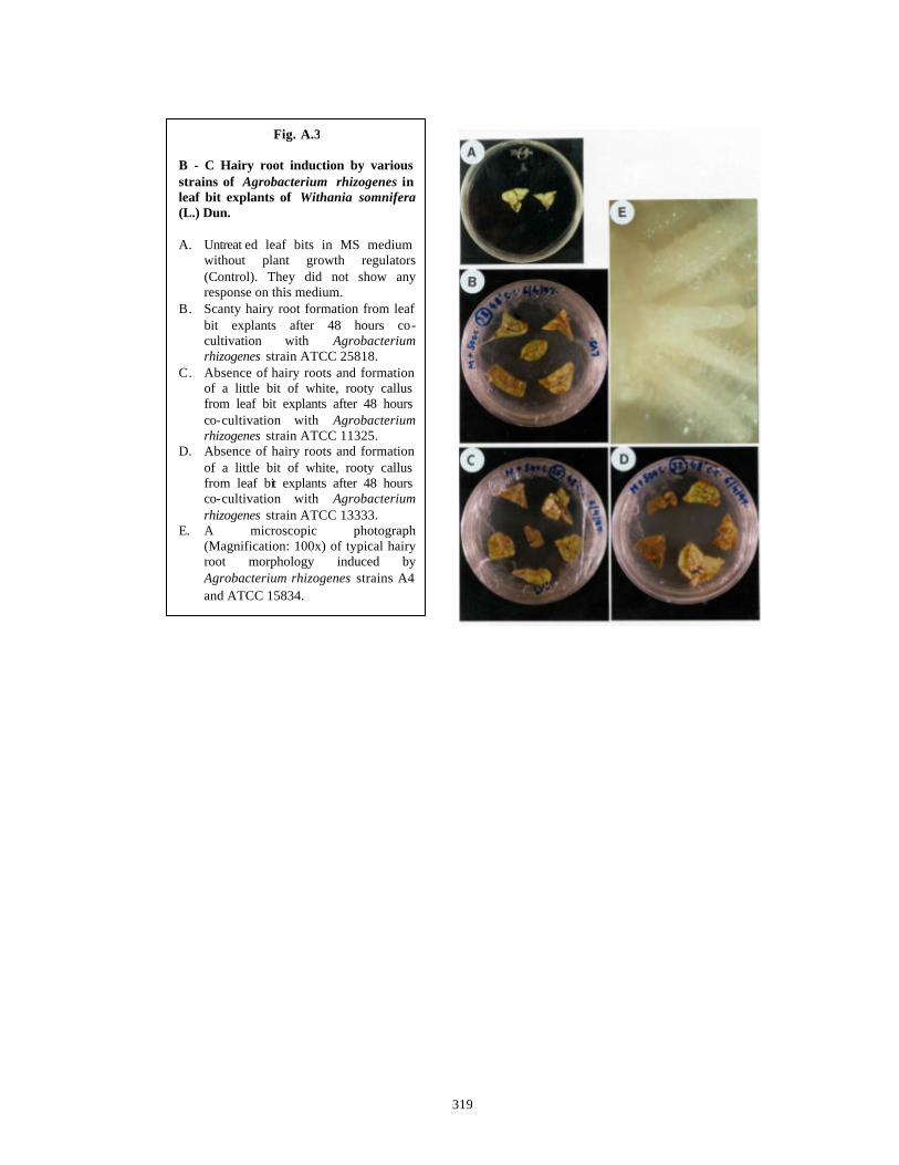
319
Fig. A.3 B - C Hairy root induction by various strains of Agrobacterium rhizogenes in leaf bit explants of Withania somnifera(L.) Dun. A. Untreat ed leaf bits in MS medium
without plant growth regulators (Control). They did not show any response on this medium.
B. Scanty hairy root formation from leaf bit explants after 48 hours co-cultivation with Agrobacterium rhizogenes strain ATCC 25818.
C. Absence of hairy roots and formation of a little bit of white, rooty callus from leaf bit explants after 48 hours co-cultivation with Agrobacterium rhizogenes strain ATCC 11325.
D. Absence of hairy roots and formation of a little bit of white, rooty callus from leaf bit explants after 48 hours co-cultivation with Agrobacterium rhizogenes strain ATCC 13333.
E. A microscopic photograph (Magnification: 100x) of typical hairy root morphology induced by Agrobacterium rhizogenes strains A4 and ATCC 15834.
320
A.3.2 Development and establishment of liquid cultures of seedling roots
The seedling root cultures of Withania somnifera were easy to establish and
maintain. In medium without plant growth regulators, the roots were thin, long, white and
with few laterals. In medium supplemented with IAA and IBA, the roots were thick, light
brown and very fast growing with many laterals. At the end of three-month growth
period, the fresh weight of roots in this medium reached to 6 g. In all, 10 different
seedling root lines were established starting from different parent seedling. These were
consecutively numbered as R1 to R10. All of these lines were analyzed by HPLC for their
withanolide contents
A.3.3 Withanolide analysis of seedling and hairy roots by HPLC
By following the HPLC protocol described earlier in Chapter 6, the following
results on Withaferin-A analysis with seedling and hairy roots have been obtained and
are presented in Table A.3.
Since the standard of Withaferin-A demonstrated two peaks of equal intensity
during HPLC, both these were used for comparison with root extracts. The hairy roots
chosen for HPLC analysis were derived from leaf explants as a larger number of hairy
roots could be harvested for analysis (See Table A.2 ).

321
Table A.3 A Comparison of Withaferin -A Production in Seedling and Hairy Root
Cultures of Withania somnifera.
No. Type of in vitro Tissues (All the seedling derived and hairy root lines were three months old at the time of extraction and
analysis).
Amount of Withanolides: Withaferin-A.
(µg/g dry wt. of Tissues)
Peak with retention time
of 12 min.
Peak with retention time
of 15 min.
1 Seedling roots (Control) derived from two-month old seedlings and grown in 1/2 MS
without plant growth regulators.
- -
2. R2 line of seedling roots grown in 1/2 MS with 0.4 mg.l-1 each of IAA and IBA.
676.8 239.25
3. R10 line of seedling roots grown in 1/2 MS with 0.4 mg.l-1 each of IAA and IBA.
< 2 -
4. R1, R3-R9 lines of seedling roots grown in 1/2 MS with 0.4 mg.l-1 each of IAA and IBA.
- < 2
5. Randomly selected hairy root line from leaves after infection with Agrobacterium rhizogenes
strain A4.
- -
6. Randomly selected hairy root line from leaves after infection with Agrobacterium rhizogenes
strain ATCC 15834.
< 2 < 2
-: Absence of compound.
Fig. A.4 represents the chromatogram of Withaferin-A standard and the graphs
of linearity of detector response for a range of Withaferin -A standards. Fig. A.5 is the
chromatogram for HPLC analysis of seedling roots showing presence of Withaferin-A.
Surprisingly, the R2 line of roots of the two-month old seedlings grown in liquid
MS medium supplemented with IAA and IBA were found to contain withanolides while
those grow n in media without plant growth regulators did not produce any withanolides
(Table A.3). Although the amount of withanolides produced is quite less than the shoot
cultures (C. f. Table 6.11), the observation is unique and has not been reported earlier.
The reason for biosynthesis of withanolides in the roots is unclear at present. Detailed
studies need to be undertaken to elucidate the reason for production of withanolides in
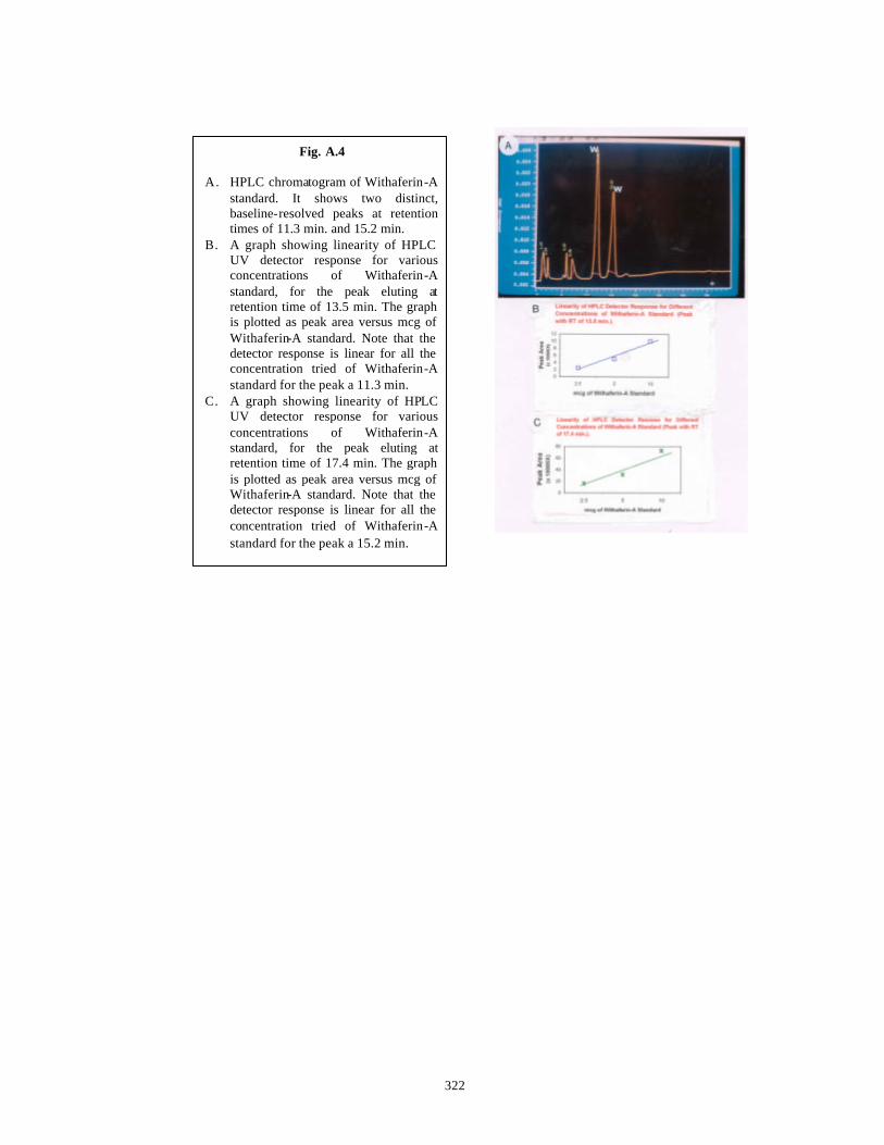
322
Fig. A.4 A. HPLC chromatogram of Withaferin-A
standard. It shows two distinct, baseline-resolved peaks at retention times of 11.3 min. and 15.2 min.
B. A graph showing linearity of HPLC UV detector response for various concentrations of Withaferin-A standard, for the peak eluting at retention time of 13.5 min. The graph is plotted as peak area versus mcg of Withaferin-A standard. Note that the detector response is linear for all the concentration tried of Withaferin-A standard for the peak a 11.3 min.
C. A graph showing linearity of HPLC UV detector response for various concentrations of Withaferin-A standard, for the peak eluting at retention time of 17.4 min. The graph is plotted as peak area versus mcg of Withaferin-A standard. Note that the detector response is linear for all the concentration tried of Withaferin-A standard for the peak a 15.2 min.
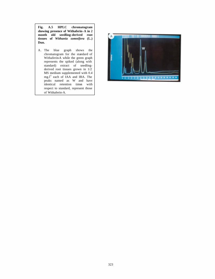
323
Fig. A.5 HPLC chromatogram showing presence of Withaferin-A in 2 month old seedling-derived root tissues of Withania somnifera (L.) Dun. A. The blue graph shows the
chromatogram for the standard of Withaferin-A while the green graph represents the spiked (along with standard) extract of seedling-derived root tissues grown in 1/2 MS medium supplemented with 0.4 mg.l-1 each of IAA and IBA. The peaks named as W and have identical retention times with respect to standard, represent those of Withaferin-A.

324
untransformed seedling root cultures, where the necessary biochem ical machinery for
steroidal lactone production like chloroplasts and the enzymes for steroidal biosynthesis
are believed to be absent. Since the roots were grown separately from the parent plants in
shake flasks, the possibility of synthesis of steroidal lactones in leaves and their
subsequent transport to roots is negated.
In contrast, the three month old hairy root lines derived after infection of in vitro
grown, two-month old leaves by Agrobacterium rhizogenes showed only traces of
Withaferin-A. This is in contrast to earlier report (Banerjee et al., 1994) but concurs with
a later report (Ray et al., 1996). Since we did not have a standard of Withanolide -D, its
presence could not be detected.
A.4 Conclusions
Even though the work carried out on hairy root induction and Withanolides
formation therefrom in this thesis is preliminary, the results obtained are significant and
opens up the possibility for large -scale investigations and extrapolations of the know-how
generated here for successful use and production of Withaferin-A from in vitro raised
seedling roots of Withania somnifera (L.) Dun. The salient observations on the work done
in this chapter could be summarized as:
1) Hairy roots can be successfully induced on leaf and nodal explants of in
vitro grown seedlings of Withania somnifera by Agrobacterium
rhizogenes strains A4, ATCC 15834 and ATCC 25818.
2) Preliminary data obtained points out the ability of hairy root induction
depending on the strain of Agrobacterium rhizogenes used for co-
cultivation.
3) Withaferin -A was absent in hairy roots after 3 months in liquid culture.
4) Seedling roots grown for 3 months in liquid culture have significant
amounts of Withaferin-A. This has important biosynthetic implications
and need to be studied further.

325
SUMMARY

326
Application of plant tissue and cell culture methods has immense potential in the
large-scale propagation and conservation of the unexplored biodiversity of plants all over
the world and especially in a huge country like India. This is especially true for plants
with medicinal properties. A number of plants have recently been studied with an
approach combining traditional medicinal knowledge with new research tools leading to
elucidation of a number of phytochemicals of high potency. The present thesis details our
efforts in characterization of two Indian Medicinal plants namely, Taxus baccata ssp.
wallichiana Zucc. Pilg. and Withania somnifera (L.) Dunal. Plants of Taxus spp.
produce a number anticancer diterpenoids of the Taxane class and hence are becoming
endangered in their natural habitats due to over-exploitation of natural resources for the
extraction of these compounds. Plants of Withania spp. also produce a range of antitumor
steroidal lactones such as Withaferin -A. Natural populations of Withania spp. show high
chemical diversity and hence need to be characterized for identification of superior
chemotypes and their exploitation in vitro for better yields of active compounds. These
plants were therefore studied with respect to aspects such as clonal propagation and
secondary metabolite production in vitro either in undifferentiated or differentiated
cultures.
During the course of the preliminary experiments, it became apparent that
bacterial and fungal contamination as well as phenolic browning hindered the successful
establishment of aseptic cultures. A series of experiments were carried out in order to
identify the bacterial and fungal contaminants and to devise specific control measures
with antimicrobial agents. These were then combined with antioxidants in a successful
‘Pre-treatment Strategy’ and a non-phytotoxic ‘Modified Surface Sterilization
Protocol’ to achieve 90% sterile cultures in vitro both from mature explants of Taxus spp.
and juvenile explants of Withania spp. The highlights of this work are:
1) The prevalent bacterial contaminants in both Taxus spp. and Withania spp. belong to
the genera such as Pseudomonas, Enterobacter-Erwinia complex and Serratia spp.
2) The fungal contaminants in both Taxus spp. and Withania spp. belong to the class
Ascomycota.
3) Both bacterial and fungal contamination (endophytic as well as
occasional/opportunistic) associated with mature explants of Taxus spp. and juvenile

327
explants of Withania spp. can be controlled by use of specific anti-microbial agents in
a pre-treatment solution such that no phytotoxicity problems are encountered.
4) To control the bacterial contamination in internodal explants of Withania spp., a 16
hour long pre-treatment in a sterile aqueous solution containing 25 mg.l-1 each of
ofloxacin, pefloxacin and ciprofloxacin is sufficient without adversely affecting the
regeneration capacity of the internodes. This proves the non-phytotoxic nature of the
optimized pre-treatment.
5) It is necessary to employ a 16 hour long pre-treatment in an aqueous solution
containing 1% bavistin + 0.1% 8-HQS + 500 mg.l-1 each of augmentin,
streptopenicillin + 25 mg.l-1 each of ofloxacin, pefloxacin and ciprofloxacin to
overcome the fungal and the bacterial contamination observed in mature explants of
Taxus spp. and to get 90% sterile cultures.
6) The phenolic oxidation observed in stem and needle explants of Taxus spp. derived
from mature trees can be reduced by incorporation of anti-oxidants in the pre-
treatment solution along with the antimicrobial agents, use of half strength nutrient
medium and use of activated charcoal at 0.5% in the solid medium.
7) A pre-treatment solution containing 1% bavistin + 0.1% 8-HQS + 500 mg.l-1 each of
augmentin and streptopenicillin + 25 mg.l-1 each of ofloxacin, pefloxacin and
ciprofloxac in + 2% each of soluble PVP, Citric acid and Ascorbic acid is used for 16
hours to completely overcome phenolic browning in mature explants of Taxus spp.
and to get 90% sterile cultures free from bacterial and fungal contaminants.
8) All the chemicals used for overcoming the problems of contamination and phenolic
browning did not lead to any phytotoxicity. All the treated explants showed high
success rate of in vitro response and the regenerants were normal in morphology.
Mature explants (derived from 80-130 year old trees) of Taxus baccata ssp.
wallichiana Zucc. Pilg. were used to initiate cultures in vitro, as they are known to yield
true-to-parent plants and also for the reason that procurement of seeds due to the
dioecious nature of this plant has become a limiting factor in the number of experiments
that could be carried out with juvenile material. Callus cultures were established from
stem bit and needle explants after optimization of nutrient media, plant growth regulators
and culture conditions. These callus cultures were fast growing and could be routinely
subcultured over the years. A number of callus cultures were analyzed by TLC and HPLC
for the presence of anticancer diterpenoids Taxol and 10-DAB. Taxol and 10-DAB

328
hyper-producer callus lines were identified that combined growth and product yield in the
same nutrient medium. Preliminary studies on suspension cultures indicated a general
trend of increase in Taxane contents during liquid culture. The highlights of this work are
as follows:
1) Callus cultures were raised from stem bits, needles (diploid calli) and endosperm
(haploid calli) of mature trees of Taxus baccata ssp. wallichiana Zucc. Pilg.
2) Callus initiation was achieved on 1/2 SH, B5, DCR and WPM media each
supplemented individually with 2, 4, 6, 8 and 10 mg.l-1 2,4-D from both stem bits and
needle explants.
3) The stem bit explants formed callus with 20% efficiency as opposed to 100% for
needle explants. Needle explants were therefore used for further studies.
4) B5 medium with a number of growth regulators was the best medium for survival and
good growth of needle -derived calli over monthly subcultures for more than 4 years.
5) Needle explants gave rise to callus with fast growth rate and high FW on B5 medium
supplemented individually with high concentrations (6, 8, 10 mg.l-1) of auxins like
2,4-D, NAA and P. The best callus growth rate and high FW was obtained in calli
grown on P containing media. Callus induction from needle explants and subsequent
callus growth on B5 medium supplemented with auxins lik e IAA and IBA together in
various concentrations was very good.
6) Taxane production was not detected in needle-derived calli cultured on B5 medium
supplemented with various combinations of 2,4-D + KIN and NAA + KIN.
7) B5 medium supplemented with Picloram was the optimized medium for good growth
as well as Taxane production for needle-derived callus cultures.
8) Taxane contents increased with increasing age of callus tissues in culture on
optimized B5 medium with P. The highest Taxol content was 540 µg/g DW of callus
in needle -derived diploid, 2 year old callus line growing on B5 medium supplemented
with 6 mg.l-1 P.
9) Needle -derived, 1 year old callus line in B5 medium supplemented with 8 mg.l-1 P
had the highest 10-DAB content of 2585 µg/g DW of callus.
10) Thus in terms of both Taxol and 10-DAB production, higher P concentrations above 4
mg.l-1 are conducive.
11) Endosperm-derived, 6 month old callus growing on DCR medium supplemented with
8 mg.l-1 P produced 203 µg 10-DAB/g DW of callus.
329
12) Taxanes were not detected in the spent solid or liquid nutrient media used for
growing either haploid or diploid callus cultures.
13) Taxane contents increased many-fold in three Cell Suspension Line Nos. 1, 3 and 5
except for the Cell Suspension Line No. 2.
14) Taxol and 10-DAB presence was detected even in Taxane non-producer 2 year old
callus grown as cell suspension in B5 medium supplemented with 10 mg.l-1 P. The
contents of both these compounds increased with the increase in liquid culture period
from 0 days to 18 days.
Efforts were made to standardize a micropropagation protocol with enhanced
axillary budding using mature explants of the Himalayan Taxus spp. Such a protocol will
provide the planting material to replenish the dwindling natural stocks of this plant. The
present protocol is applicable to plant material of different age and collected from
different locations. In addition, precocious embryo germination and seedling development
was achieved at a high frequency from seeds collected at different locations in the
Himalayas. Fast growin g and 10-DAB producing callus lines were established from
haploid endosperm explants of Taxus spp. for the first time. The highlights of this work
are as follows:
1) A protocol has been standardized for enhanced axillary budding of mature stem bit
explants of Taxus baccata ssp. wallichiana Zucc. Pilg. Primary and secondary shoots
have been obtained at a high frequency within six months. This protocol is applicable
to plant material collected from trees of different age groups, growing in different
locations in the Himalayas.
2) Basal media with low total ionic salt strengths and lower ratios of ammonium
nitrogen to nitrate nitrogen such as 1/2 WPM and 1/2 DCR are conducive for
enhanced axillary budding of mature explants of Taxus spp.
3) Organic nitrogen in the form of an amino acid like glutamine does not enhance
axillary sprouting in Taxus spp.
4) 1/2 WPM supplemented with 0.05 mg.l-1 BAP gave 83.33% sprouting while 1/2
WPM supplemented with 0.05 mg.l-1 KIN gave 75.77% sprouting in plant material
collected from Shimla. The former percentage of sprouting was significantly higher
than the latter.

330
5) 1/2 DCR medium supplemented with lower concentrations of BAP (0.05, 0.1 mg.l-1)
gave 100% sprouting percentage as compared to 58.89% and 54.21% sprouting
obtained on 1/2 DCR medium supplemented with lower concentrations of KIN (0.05,
0.1 mg.l-1) in plant material collected from Shimla. The results are statistically
significant.
6) 1/2 DCR medium without plant growth regulators (1/2 DCR-H) gave 50% sprouting
while 1/2 DCR supplemented with lower concentrations of BAP (0.05, 0.1 mg.l-1)
gave identical sprouting percentage (100%) that is similar to sprouting percentage
obtained with 1/2 DCR supplemented with 10% CM. Thus apparently the absence of
low concentrations of BAP in 1/2 DCR me dium can be compensated by addition of
CM.
7) BAP is a better cytokinin with 1/2 WPM to achieve high sprouting but shoot quality
and elongation capacity is not as good as shoot quality and elongation ability obtained
with KIN. So KIN is the cytokinin of choice with 1/2 WPM.
8) Higher cytokinin concentrations combined with any of the basal media are not
conducive for sprouting.
9) TDZ is not conducive for sprouting due to the inability of the primary sprouts to
elongate.
10) Combination of an auxin with a cytokinin is not conducive for enhanced axillary
budding of mature stem explants of Taxus spp.
11) 1/2 WPM-H gave 50% sprouting with plant material collected from Khajjiyar;
statistically similar to 52.94% sprouting achieved with plant material collected from
Shimla on the identical medium. Although 75.77% sprouting was achieved on 1/2
WPM with 0.05 mg.l-1 KIN with plant material collected from Shimla, sprouting
percentage did not differ statistically with KIN concentrations for plant material
collected from Khajjiyar.
12) 1/2 DCR-H gave significantly higher sprouting at 90% with plant material collected
from Khajjiyar while plant material collected from Shimla had only 50% sprouting on
the same medium. 1/2 DCR supplemented with BAP gave higher sprouting response
with Shimla material as compared to Khajjiyar material.
13) 1/2 WPM-H gave 48% sprouting with plant material collected from Almora;
statistically different than 52.94% sprouting achieved with plant material collected
from Shimla on the identical medium. A big standard error of mean was obtained with
Almora material hinting at underlying variabilities of the plant material. Although

331
75.77% sprouting was achieved on 1/2 WPM with 0.05 mg.l-1 KIN for plant material
collected from Shimla, sprouting percentage did not differ statis tically with KIN
concentrations with plant material collected from Almora.
14) 1/2 DCR-H gave identical sprouting at 50% with plant material collected from
Almora as well as Shimla. 1/2 DCR medium supplemented with BAP gave
significantly higher sprouting response with Shimla material as compared to Almora
material.
15) Thus although the general trends such as suitability of low salt media and low
cytokinin concentrations for sprouting are identical in plant material collected from
Shimla, Khajjiyar and Almora, the best sprouting response is observed with plant
material collected from Shimla. Only plant material collected from Khajjiyar gave
90% sprouting on 1/2 DCR-H that is significantly higher than the results of sprouting
obtained with plant material collected from Shimla.
16) Elongation of primary sprouts into shoots of length 3 cm was possible on respective
basal media without plant growth regulators, in three months time, for the cultures
established from plants of all the locations.
17) Establishment of primary shoots was possible on respective basal media
supplemented with 10% CM and 1 g.l-1 CH in the fourth month.
18) Secondary sprouting of buds developed in vivo on primary shoots was achieved on
1/2 WPM supplemented with 0.1 mg.l-1 KIN, 10% CM and 1 g.l-1 CH at a frequency
of 25% during the fifth month.
19) Precocious embryo germination and seedling development has been achieved at a
frequency of 90% from immature seeds collected from Khajjiyar on DCR basal
medium at pH 5.8, in light and without plant growth regulators.
20) Precocious embryo germination and seedling development was found to vary with
developmental age of seeds (as judged by aril development and embryo size) and the
location of collection.
21) Precocious embryo germination and seedling development has been achieved at a
frequency of 91% from mature seeds collected from Almora on B5 basal medium
without plant growth regulators. The optimum medium was found to vary depending
on the location of collection and developmental stage (as judged by aril development
and embryo size) of the seeds.
22) Fast growing, haploid callus lines have been established from solid endosperm of
seeds collected from Almora on the optimal DCR medium supplemented with various
332
concentrations of Picloram (P) and higher concentrations of P were conducive for
faster growth of calli.
Morphogenetic studies in Withania spp. were carried out in order to standardize a
high efficiency propagation protocol to be applicable to large scale propagation of
selected chemotypes without any seasonal constraints, to che ck for the presence of
Withaferin-A in in vitro raised cultures and to identify the origin of production of
Withaferin-A in organized or unorganized cultures. The highlights of this work are:
1) Direct regeneration of plants has been achieved from a number of explants like leaves,
nodes, internodes, hypocotyls and embryos. 70% of leaf explants regenerated shoots
on an optimal medium while all the remaining explants showed 100% shoot formation
response on media standardized during the course of the present work.
2) By the protocol standardized in this study, 1600 and 432 plantlets per year per explant
could be produced from leaves and hypocotyl explants respectively.
3) De novo regeneration of 24 shoots from a single node explant could be obtained with
TDZ. TDZ was found to be the best plant growth regulator among the tested plant
growth regulators.
4) All the regenerants showed a high success rate on acclimatization and hardening.
5) Both the seedlings and in vitro regenerated shoots from nodes contained high amounts
of Withaferin -A . In vitro grown seedlings had 10 times more Withaferin -A than that
reported in literature for in vivo tissues of Withania somnifera (L.) Dun. In vitro
regenerated shoots growing on MS medium supplemented with 0.5 mg.l-1 BAP had
100 times more Withaferin-A than the in vivo tissues and to our knowledge such high
amounts of Withaferin-A reported here are the first in literature. Presence of
Withaferin-A was not detected in callus tissues suggesting that production of
Withaferin-A is related to tissue organization.
Preliminary studies on hairy root induction with Agrobacterium rhizogenes in
Withania spp. were carried out basically to check for the presence of Withaferin-A in the
hairy root cultures. The highlights of this work are presented below:
1) Hairy roots can be successfully induced on leaf and nodal explants of in vitro grown
seedlings of Withania somnifera with Agrobacterium rhizogenes strains A4, ATCC
15834 and ATCC 25818.

333
2) Preliminary data obtained points out the ability of hairy root induction depending on
the strain of Agrobacterium rhizogenes used for co-cultivation.
3) Withaferin-A was absent in hairy roots after 3 months in liquid culture.
4) Seedling roots grown for 3 months in liquid culture have significant amounts of
Withaferin-A. This has important biosynthetic implications and need to be studied
further.

334
BBIIBBLLIIOOGGRRAAPPHHYY
335
Abraham A., Kirson I., Glotter E. and Lavie D. (1968) A chemotaxonomic study of Withania somnifera (L.) Dun. Phytochem. 7: 957-962.
Ainsworth G. C. and Sussman A. S. (1966-1968) The fungi. An advanced treatise. Vol. I, II and III. Academic Press, New York.
Aird E. L. H., Hamill J. D., Robins R. J. and Rhodes M. J. C. (1988) Chromosome stability in transformed hairy root cultures and the properties of variant lines of Nicotiana rustica hairy roots. In: Manipulating Secondary Metabolism in Culture. (Eds. Robins R. J. and Rhodes M. J. C.) Cambridge University Press, U. K. pp. 137-144.
Akashi. T., Furuno T., Takahashi T. and Ayabe S-I (1994) Biosynthesis of triterpenoids in cultured cells and regenerated and wild plant organs of Taraxacum officinale. Phytochem. 36: 303-308.
Alexopoulos C. J. and Beneke E. S. (1962) Laboratory manual for introductory mycology. Burgess Publishing Co., Minneapolis.
Alfano J. R. and Collmer A. (1996) Bacterial pathogens in plants: Life up against the wall. The Plant Cell 8: 1683-1698.
Amos L. A. and Löwe J. (1999) How Taxol® stabilizes microtubule structure. Chem. Biol. 6: R65-R69.
Amos R. R. and McCown B. H. (1981) Micropropagation of members of the coniferae. HortSci. 16: 453.
Anderson E. D. and Owens J. N. (1999) Megagametophyte development, fertilization and cytoplasmic inheritance in Taxus brevifolia . Int. J. Plant Sci. 160: 459-469.
Anonymous (1976) Withania . In: The Wealth of India, (Raw Materials) CSIR, New Delhi. Vol. X: 580-585.
Arteca R. N. and Wickremesinhe E. (1993) Cultured Taxus tissues as a source of Taxol, related Taxanesand other novel anti-tumor/anti-viral compounds. Int. Patent No. WO 93/23555.
Asthana R. and Raina M. K. (1989) Pharmacology of Withania somnifera (L.) Dun. Indian Drugs 26: 199-205.
Backs-Hussemann D. and Reinert J. (1970) Embryobildung durch isolierte Einzelzellen
aus Gewebe Kulturen von Daucus carota , Protoplasma 70: 49-60. Baloglu E. and Kingston D. G. I. (1999) The Taxane diterpenoids. J. Nat. Prod. 62: 1448-
1472. Banerjee S., Naqvi A. A., Mandal S. and Ahuja P. S. (1994) Transformation of Withania
somnifera (L.) Dunal by Agrobacterium rhizogenes: Infectivity and phytochemical studies. Phytother. Res. 8: 452-455.
Banerjee S., Upadhyay N., Kukreja A. K., Ahuja P. S., Kumar S., Saha G. C., Sharma R. P. and Chattopadhyay S. K. (1996) Taxanesfrom in vitro cultures of the Himalayan Yew Taxus wallichiana. Planta Med. 62: 333-335.
Barnes L. (1983) Micropropagation of endangered native conifers, Torreya taxifolia and Taxus floridana. HortSci. 18: 617.
Bessalle R. Lavie D. and Frolow F. (1987) Withanolide Y, a new withanolide from a hybrid of Withania somnifera . Phytochem. 26: 1797-1800.
Bewley J. D. and Black M. (1994) Seeds: Physiology of development and germination. Plenum Press, New York.
Bewley J. D. (1997) Seed germination and dormancy. The Plant Cell 9: 1055-1066. Bhadra R., Vani S. and Shanks J. V. (1993) Production of indole alkaloids by selected
hairy root lines of Catharanthus roseus. Biotechnol. Bioeng. 41: 581-592.
336
Bhagyalakshmi N. and Ravishankar G. A. (1998a) Natural compounds from cultured hairy roots. In: Role of Biotechnology in Medicinal and Aromatic Plants. Vol 1 (Eds. Khan I. A. and Khanum A.) Ukaaz Publications, Hyderabad. pp. 166-182.
Bhagyalakshmi N., Venkataraman L. V. and Ravishankar G. A. (1998b) Bioprocess parameters for scale-up of hairy root culures. In: Plant Tissue Culture and Molecular Biology. Applications and Prospects. (Ed. Srivastava P. S.) Narosa Publishing House, New Delhi. pp. 302-331.
Binns A. N. and Thomashow M. F. (1988) Cell biology of Agrobacterium infection and transformation of plants. Ann. Rev. Microbiol. 42: 575-606.
Bisht M. S., Vyas P., Bag N. and Palni L. M. S. (1998) Micropropagation of some plants of Indian Himalayan region. In: Plant Tissue Culture and Molecular Biology. Applications and Prospects. (Ed. Srivastava P. S.) Narosa Publishing House, New Delhi. pp. 126-170.
Blake J. (1988) Mites and thrips as bacterial and fungal vectors between plant tissue cultures. Acta Hort. 225: 163-166.
Blazewic D. J. and Ederer G. M. (1975) (Eds.) Principles of Biochemical Tests in Diagnostic Microbiology. John Wiley and Sons Inc., USA.
Bon M. C., Riccardi F. and Monteuuis O. (1994) Influence of phase change within a 90-year old Seqouia sempervirens on its in vitro organogenetic capacity and protein patterns. Trees 8: 283-287.
Bonga J. M. and Pond S. E. (1991) Adventitious shoot formation in cultures of 30-year old Larix decidua, L. leptolepsis and L. larcinia trees. Plant Cell Tissue Organ Cult. 26: 45-51.
Bonga J. M. and Von Aderkas (1992) In vitro culture of trees. Kluwer Academic Publishers, Netherlands.
Bowles D. and Leyser O. (1994) ‘TheBig Green Book’ Plant Biotechnology – Centre for exploitation of science and technology (CEST).
Boxus P. and Terzi J. M. (1988) Control of accidental contaminations during mass propagation. Acta Hort. 225: 189-190.
Bradbury J. F. (1988) Identification of cultivable bacteria from plants and plant tissue cultures by use of simple classical methods. Acta Hort. 225: 27-35.
Brar D. S. and Khush G. S. (1994) Cell and Tissue Culture for Plant improvement. In: Mechanisms of Plant Growth and Improved Productivity. Modern Approaches. (Ed. Basra A. S.), Marcel Dekker, Inc., New York, Basel, Hong Kong. pp. 229-278.
Brown D. C. W. and Thorpe T. A. (1995) Crop improvement through tissue culture. World J. Microbiol. Biotechnol. 11: 409-415.
Brukhin B. V. and Bozhkov P. V. (1996) Female gametophyte development and embryogenesis in Taxus baccata L. Acta Soc. Bot. Pol. 65: 135-139.
Brukhin V. B., Moleva I. R., Filonova L H., Grakhov V. P., Blume Ya. B. and Bozhkov P. V. (1996) Proliferative activity of callus cultures of Taxus baccata L. in relation to anticancer diterpenoid Taxol biosynthsis. Biotechnol. Lett. 18: 1309-1314.
Buckley P. M., DeWilde T. N. and Reed B. M. (1995) Characterization and identification of bacteria isolated from micropropagated mint plants. In Vitro Cell Dev. Biol. 31: 58-64.
Budhiraja R. D. and Sudhir S. (1987) Review of biological activity of withanolides. J. Sci. Ind. Res. 46: 488-491.
Buising C. M., Shoemaker R. C. and Benbow R. M. (1994) Early events of multiple bud formation and shoot development in soybean embryonic axes treated with the cytokinin, 6-benzylaminopurine. Am. J. Bot. 81: 1435-1448.
337
Canizares C., Vivar N. and Herdoiza M. (1997) The effects of Taxol, a potent platelet antiaggregant, may be due to its microtubular stabilization activity. Platelets 8: 61-63.
Cass B. J., Scott D. S. and Legge R. L. (1997) Practical considerations in the harvesting and isolation of Taxanesfrom plant biomass. Curr. Top. Phytochem. 1: 169-177.
Cassells A. C. (1991) Problems in tissue culture: Culture contamination. In: Micropropagation. Technology and Application. (Eds. Debergh P. C. and Zimmerman R. H.) Kluwer Academic Publishers, Dordrecht. pp. 31-44.
Cerdeira R. M., McChesney J. D. and Burandt C. Jr. (1994) Microcuttings of Taxus x media cv. Hicksii. In vitro Cell Dev. Biol. 30: 77.
Chakraborti S. K., De B. K. and Bandopadhyay T. (1974) Variations in the antitumor constituents of Withania somnifera. Experientia 30: 852-853.
Chan W. R., Halsall T. G., Hornby G. M., Oxford A. W., Sabel W., Bjåmer K., Ferguson G. and Robertson J. M. (1966) Taxa-4(16), 11-diene -5α,9α,10β,13α -tetraol, a new Taxanederivative from the heartwood of Yew (T. baccata L.): X-ray analysis of a p-bromobenzoate derivative. Chem. Commun. 21: 923-925.
Chang A. Y. and Garrow G. C. (1995) Pilot study of vinorelbine (navelbine) and paclitaxel (Taxol) in patients with refractory breast cancer and lung cancer. Sem. Oncol. 22: 66-71.
Chang S. H. and Yang J. C. (1996) Enhancement of plant formation from embryo cultures of Taxus mairei using suitable culture medium and PVP. Bot. Bull. Acad. Sin. 37: 35-40.
Chang S. H., Ho C. K. and Tsai J. Y. (1998) Micropropagation of Taxus mairei seedlings at different ages and recoverability of their plagiotropic shoots. Taiwan J. For. Sci. 13: 29-39.
Chao C. -C. and Lin T. -P. (1996) Content of adenylate nucleotides and energy charge in the early stage of germination of orthodox and recalcitrant seeds. Bot. Bull. Acad. Sin. 37: 229-237.
Charlwood B. V., Charlwood K. A. and Molina-Torres J. (1990) Accumulation of secondary compounds by organized plant cultures. In: Secondary products from plant tissue culture. (Eds. Charlwood B. V. and Rhodes M. J. C.) Clarendon Press, Oxford. pp. 167-200.
Chatterjee S. and Dey S. (1997) A preliminary survey of the status of Taxus baccata in Tawang distirct of Arunachal Pradesh. Ind. Forester 8: 746-754.
Chattopadhyay S. K., Tripathi V. K., Thakur R. S., Sharma R. P. and Jain S. P. (1994) Isolation of Taxol, 10-deacetylbaccatin III and (-)-betuligenol from Taxus baccata Linn. Ind. J. Chem. 33B: 409-411.
Chattopadhyay S. K., Kulshrestha M., Saha G. C., Sharma R. P., Jain S. P. and Kumar S. (1997) The taxoids and the phenolic constituents of the heartwood of the Himalayan yew Taxus wallichiana. J. Med. Arom. Plant Sci. 19: 17-21.
Chaw S-M, Long H., Wang B-S, Zharkikh A. and Li W-H (1993) The phylogenetic position of Taxaceae based on 18S r-RNA sequences. J. Mol. Evol. 37: 624-630.
Chee P. (1994) In vitro culture of zygotic embryos of Taxus spp. HortScience 29: 695-697.
Chee P. (1995a) Stimulation of adventitious rooting of Taxus species by thiamine. Plant Cell Rep. 14: 753-757.
Chee P. (1995b) Organogenesis in Taxus brevifolia tissue cultures. Plant Cell Rep. 14: 560-565.
Chee P. (1996) Plant regeneration from somatic embryos of Taxus brevifolia. Plant Cell Rep. 16: 184-187.

338
Chien C-T, Kuo-Huang L-L and Lin T-P (1998) Changes in ultrastructure and abscisic acid level, and response to applied gibberellins in Taxus mairei seeds treated with warm and cold stratification. Ann. Bot. 81: 41-47.
Choudhary M. I., Shahwar D-E, Parween Z., Jabbar A., Ali I. and Rahman A-U (1995) Antifungal steroidal lactones from Withania coagulance. Phytochem. 40: 1243-1246.
Christen A. A., Bland J. and Gibson D. M. (1989) Cell culture as a means to produce Taxol. Proc. Am. Assoc. Cancer Res. 30: A2252.
Christen A. A., Gibson D. M. and Bland J. (1991) Production of Taxol or Taxol-like compounds in cell culture. US Patent No. 5,109,504.
Christey M. C. (1997) Transgenci crop plants using Agrobacterium rhizogenes -mediated transformation. In: Hairy Roots: Cult ure and Applications. (Ed. Doran P. M.) Harwood Acdemic Publishers, Amsterdam. pp. 99-111.
Christianson M. L. and Warnick D. A. (1985) Temporal requirement for phytohormone balance in the control of organogenesis in vitro . Dev. Biol. 112: 494-497.
Ciddi V., Srinivasan V. and Shuler M. L. (1995) Elicitation of Taxus spp. cell cultures for production of Taxol. Biotechnol. Lett. 17: 1343-1346.
Collins-Pavao M., Chin C. K. and Pedersen H. (1996) Taxol partitioning in two-phase plant cell cultures of Taxus brevifolia. J. Biotechnol. 49: 95-100.
Compton M. E. and Preece J. E. (1986) Exudation and explant establishment. Intl. Asso. Plant Tissue Cult. Newslett. 50: 9-18.
Cusidó R. M., Palazón J., Navia -Osorio A., Mallol A., Bonfill M., Morales C. and Piñol M. T. (1999) Production of Taxol® and baccatin-III by a selected Taxus baccata callus line and its derived cell suspension culture. Plant Sci. 146: 101-107.
Czembor E. and Strobel G. A. (1997) Limitations of exotic and indigenous isolates of Fusarium avenaceum for the biological control of spotted knapweed - Centaurea maculosa . World J. Microbiol. Biotechnol. 13: 119-123.
Danby S, Berger F., Howitt D. J., Wilson A. R., Dawaon S. and Leifert C. (1994) Fungal
contaminants of Primula, Coffea, Musa and Iris tissue cultures. In: Physiology, Growth and Development of Plants in Culture. (Eds. Lumsden P. J., Nicholas J. R. and Davies W. J.) Kluwer Academic Publishers, The Netherlands. pp. 397-404.
Das B., Kashinatham A. and Anjani G. (1999) Natural Taxols. Ind. J. Chem. 38B: 1018-1024.
David A. and Plastira V. (1974) Histopathologie végétale. Réalisation de cultures de tissus d'If (Taxus baccata L.) à patir de fragments de pousses âgés d'un an; obtention d'une culture indéfinie. C. R. Acad. Sci. Paris 279: 1757-1759.
David A. and Plastira V. (1976) Histophysiologie végétale. Comportement en culture in vitro de cellules isolées du deux gymnospermes: Taxus baccata L. et Pinus pilaster Sol. C. R. Acad. Sci. Paris 282: 1159-1162.
De Proft M. P., Maene L. J. and Debergh P. C. (1985) Carbon dioxide and ethylene evolution in the culture atmosphere of Magnolia cultured in vitro . Physiol. Plant. 65: 375-379.
Debergh P. and Maene L. (1981) A scheme for commercial propagation of ornamental plants by tissue culture. Sci. Hort. 14: 335-345.
Debergh P. and Maene L. (1984) Pathological and physiological problems related to the in vitro culture of plants. Parasitica 40: 69-75.
Debergh P. C. and Read P. E. (1991) Micropropagation In: Micropropagation, Technology and Application. (Eds. Debergh P. C. and Zimmerman R. H.) Kluwer Academic Publishers, The Netherlands. pp. 1-13.

339
DeFossard R. A. and DeFossard H. (1988) Coping with microbial contaminants and other matters in a small commercial micropropagation laboratory. Acta Hort. 225: 167-176.
Delfel N. E. and Rothfus J. A. (1977) Antitumor alkaloids in callus cultures of Cephalotaxus harringtonia . Phytochem. 16: 1595-1598.
Della Casa De Marcano D. P. and Halsall T. G. (1970a) The structure of the diterpenoid Baccatin-I, the 4β,20-epoxide of 2α,5α,7β ,9α,10β,13α -Hexa-acetoxytaxa-4(20),11-diene. J. Chem. Soc. Chem. Commun. 21: 1381-1382.
Della Casa De Marcano D. P. and Halsall T. G. (1970b) Crystallographic structure determination of the diterpenoid baccatin V, a naturally occurring oxetan with a Taxaneskeleton. J. Chem. Soc. Chem. Commun. 21: 1382-1383.
Della Casa De Marcano D. P. and Halsall T. G. (1975) Structures of some Taxanediterpenoids. Baccatins-III, -IV, -VI, and –VII and 1-Dehydroxybaccatin-IV, possesing an oxetan ring. J. Chem. Soc. Chem. Commun. 9: 365-366.
Denis J. N., Greene A. E., Guénard D., Guéritte-Voegelein F., Mangatal L. and Potier P (1988) A highly efficient practical approach to natural Taxol. J. Am. Chem. Soc. 110: 5917-5919.
Dietze J., Blau A. and Willmitzer L. (1995) Agrobacterium-mediated transformation of potato (Solanum tuberosum). In: Gene transfer to plants. (Eds. Potrykus I. and Spangenberg G.) Springer Verlag, Berlin, Heidelberg. pp. 24-29.
DiFazio S. P., Vance N. C. and Wilson M. V. (1997) Strobilus production and growth of Pacific Yew under a range of overstory conditions in W. Oregon. Can. J. For. Res. 27: 986-993.
DiFazio S. P., Wilson M. V. and Vance N. C. (1998) Factors limiting seed production of Taxus brevifolia (Taxaceae) in W. Oregon. Am. J. Bot. 85: 910-918.
Diner A. M. (1995) Adventitious micropropagation of mature Larix decidua using dormant versus flushed axillary vegetative buds. New Forests 9: 61-65.
Donaldson K. L., Goolsby G., Kiener P. A. and Wahl A. F. (1994) Activation of p34cdc2 coincident with Taxol-induced apoptosis. Cell Growth and Differentiation 5: 1041-1050.
Doran P. (1989) Prospects for Production of Plant Chemicals from Genetically Transformed Roots. Aust. J. Biotechnol. 3: 270-277.
Doran P. M. (1994) Production of chemicals using genetically transformed plant organs. Ann. NY Acad. Sci. 745: 426-441.
Dorenburg H. and Knorr D. (1995) Strategies for the improvemnet of secondary metabolite production in plant cell cultures. Enz. Microb. Technol. 17: 674-684.
Dubey K. P. (1997) Himalayan yew (Taxus baccata ) conservation: A vegetative approach. Ind. Forester 12: 1150-1154.
Eastwood F. W., Kirson I., Lavie D and Abraham A. (1980) New withanolides from a cross of a South African chemotype by Chemotype II (Israel) in Withania somnifera. Phytochem. 19: 1503-1507.
Eccher T. (1988) Response of cuttings of 16 Taxus cultivars to rooting treatments. Acta Hort. 227: 251-253.
Edwards R. and Gatehouse J. A. (1999) Secondary metabolism. In: Plant biochemistry and molecular biology. 2nd Ed. (Eds. Lea P. J. and Leegood R. C.) John Wiley and Sons, New York. pp. 193-218.
Ellis D. D., Zeldin E. L., Brodhagen M., Russin W. A. and McCown B. H. (1996) Taxol production in nodule cultures of Taxus. J. Nat. Prod. 59: 246-250.
Elmer W. H., Mattina M. J. I. and MacEachern G. J. (1994) Sensitivity of plant pathogenic fungi to taxane extracts from ornamental yews. Phytopathol. 84: 1179-1185.
340
ElSohly H. N., Croom E. M., El-Kashoury E., ElSohly M. A. and McChesney M. D. (1994) Taxol content of stored fresh and dried Taxus clippings. J. Nat. Prod. 57: 1025-1028.
ElSohly H. N., Croom E. M. Jr., Kopycki W. J., Joshi A. S., ElSohly M. A. and McChesney J. D. (1995) Concentrations of Taxol and related Taxanesin the needles of different Taxus cultivars. Phytochem. Anal. 6: 149-156.
Endress R. ( 1994) Plant Cell Biotechnology. Springer Verlag, Berlin, Heidelberg. Enjarlic M. P., Carron F. and Lardet L. (1988). Contamination of primary cultures in
tropical areas: The case of Hevea brasiliensis. Acta Hort. 225: 57-65. Ewald D., Weckwerth W., Naujoks G. and Zocher R. (1995) Formation of embryo-like
structures in tissue cultures of different yew species. J. Plant Physiol. 147: 139-143. Falkiner F. R. (1988). Strategy for the selection of antibiotics for use against common
bacterial pathogens and endophytes of plants. Acta Hort. 225: 53-56. Fang G. F., Chang B. S., Kim C. N., Perkins C., Thompson C. B., and Bhalla K. N.
(1998) “Loop” domain is necessary for Taxol-induced mobility shift and phosphorylation of Bcl-2 as well as for inhibiting Taxol-induced cytosolic accumulation of cytochrome c and apoptosis. Cancer Res. 89: 3202-3208.
Fellner M., Kneifel W., Gregortis D. and Leonhardt W. (1996) Identification and antibiotic sensitivity of microbial contaminants from callus cultures of garlic Allium sativum L. and Allium longicuspis Regel. Plant Sci. 113: 193-201.
Fett-Neto A. G., DiCosmo F., Reynolds W. F. and Sakata K. (1992) Cell culture of Taxus as a source of the antineoplastic drug Taxol and related taxanes. Bio/Technol. 10: 1572-1575.
Fett-Neto A. G., Melanson S. J., Sakata K. and DiCosmo F. (1993a) Improved growth and Taxol yield in developing calli of Taxus cuspidata by medium composition modification. Bio/Technol. 11: 731-734.
Fett-Neto A. G., Melanson S. J., Pennington J. J. and DiCosmo F. (1993b) Effect of precursor feeding on Taxol side chain bisynthesis in callus cultures of Taxus cuspidata . Plant Physiol. 102: 97.
Fett-Neto A. G., Melanson S. J., Nicholson S. A., Pennington J. J. and DiCosmo F. (1994a) Improved Taxol yield by aromatic carboxylic acid and amino acid feeding to cell cultures of Taxus cuspidata . Biotechnol. Bioeng. 44: 967-971.
Fett-Neto A. G., Zhang W. Y. and DiCosmo F. (1994b) Kinetics of Taxol production, growth and nutrient uptake in cell suspension cultures of Taxus cuspidata. Biotechnol. Bioeng. 44: 205-210.
Fiola J. A., Hassan M. A., Swartz H. J., Bors R. H. and McNicols R. (1990) Effect of thidiazouron, light fluence rate and kanamycin on in vitro shoot organogenesis from excised Rubus cotyledons and leaves. Plant Cell Tissue Organ Cult. 20: 223-228.
Flores H. E. and Filner P. (1985) Metabolic relationships of putrescine, GABA and alkaloids in cell and root cultures of Solanaceae. In: Primary and Secondary Metabolism of Plant Cell Cultures. (Eds. Neumann K-H, Barz W. and Reinhard E.) Springer Verlag, Berlin. pp. 174-186.
Flores H. E. and Sgrignoli P. J. (1991) In vitro culture and precocious germination of Taxus embryos. In vitro Cell Dev. Biol. 27P: 139-142.
Flores T., Wagner L. J. and Flores H. E. (1993) Embryo culture and taxane production in Taxus spp. In vitro Cell Dev. Biol. 29P: 160-165.
Flores H. E. and Medina-Bolivar F. (1995) Root culture and plant natural products: “unearthing” the hidden half of plant metabolism. Plant Tissue Cult. Biotechnol. 1: 59-74.
341
Fortanier E. J. and Jonkers H. (1976) Juvenility and maturity of plants as influenced by their ontogenetical and physiological ageing. Acta Hort. 56: 37-43.
Franclet A., Boulay M., Bekkaoui F., Fouret Y., Verschoore-Martouzet B. and Walker N. (1987) Rejuvenation In: Cell and tissue culture in forestry. Vol.1 (Eds. Bonga J. M. and Durzan D. J.) Martinus Nijhoff Publishers, Netherlands. pp. 232-248.
Franclet A. (1991) Biotechnology in ‘rejuvenation’: hope for the micropropagation of difficult woody plants. Acta Hort. 289: 273-282.
Fried B. and Sherma J. (1994) Thin layer chromatography. Techniques and applications. Chromatographic Science Series. Vol. 66. Marcel and Dekker Inc., New York
Furmanowa M., Glowniak K., Zobel A., Guzewska J., Zgorka G., Rapczewska L. and Jozefczyk A. (1995) Taxol in Taxus baccata L. var. elegantissima organs and in tissue culture. Med. Fac. Landbouww. Univ. Gent 60: 2115-2118.
Gamborg O. L., Miller R. A. and Ojima K (1968) Nutrient requirements of suspension
cultures of soybean root cells. Expt. Cell Res. 50: 151-158. Gautheret R. J. (1934) Culture du tissu cambial. C. R. Acad. Sci. (Paris) 198: 2195-2196. Gautheret R. J. (1939) Sur la possibilite de realiser la culture indefinie des tissus de
tubercules de carotte. C. R. Hebd. Seances Acad. Sc. 208: 118-120. Gautheret R. J. (1942) Hetero-auxines et cultures de tissus vegetaux. Bull. Soc. Chim.
Biol. 24: 13-41. Gautheret R. J. (1955) Sur la variabilite des proprietes physiologiques des cultures de
tissus vegetaux. Rev. Gen. Bot. 62: 5-112. Gautheret R. J. (1985) History of plant tissue and cell culture: a personal account. In: Cell
Culture and Somatic Cell Genetics of Plants. Vol. 2 (Ed. Vasil I. K.) Academic Press Inc., Florida. pp. 1-59.
Gelvin S. B., Schilperoort R. A. and Verma D. P. S. (1988-1990) (Eds.) Plant Molecular Biology Manual and Update 2nd Ed. Kluwer Academic Publishers, Dordrecht.
George F. F. and Sherrington P. D. (1984) Plant propagation by tissue culture. Exegetics Ltd., Eversley, Basigstoke, UK.
Gibson D. M., Ketchum R. E. B., Vance N. C. and Christen A. A. (1993) Initiation and growth of cell lines of Taxus brevifolia (Pacific Yew). Plant Cell Rep. 12: 479-482.
Gibson D. M., Ketchum R. E. B., Hirasuna T. J. and Shuler M. L. (1995) Potential of plant cell culture for Taxaneproduction. In: Taxol® Science and Applications. (Ed. Suffness M.) CRC Press, Boca Raton, New York, London, Tokyo. pp. 71-95.
Giles, K. L. and Morgan W. M. (1987) Industrial scale micropropagation. Trends Biotechnol. 5: 35-39.
Glotter E., Kirson I., Abraham A. and Lavie D. (1973) Constituents of Withania somnifera Dun. XIII. The withanolides of chemotype III. Tetrahedron 29: 1353-1364.
Godwin I. D., Ford-Lloyd B. V. and Newbury H. J. (1992) In vitro approaches to extending the host-range of Agrobacterium for plant transformation. Aust. J. Bot. 40: 751-763.
Grayson M. (1982) (Ed.) In: Antibiotics, Chemotherapeutics and Antibacterial Agents for Disease Control Encyclopedia Reprint Series. John Wiley & Sons Inc., New York.
Griffiths R. P., ChaDWick A. C., Robatzek M., Schauer K. and Schaffroth K. A. (1995) Association of ectomycorrhizal mats with Pacific yew and other understory trees in coniferous forests. Plant and Soil 173: 343-347.
Guha S. and Maheshwari S. C. (1964) In vitro production of embryos from anthers of Datura . Nature 204: 497.
Gunson E. and Spencer-Phillips P. T. N. (1994) Latent bacterial infections: epiphytes and endophytes as contaminants of micropropagated plants. In: Physiology, growth and

342
development of plants in culture. (Eds. Lumsden P. J., Nicholas J. R. and Davies W. J.) Kluwer Academic Publishers, The Netherlands. pp. 363-378.
Gupta A. P., Verma R. K., Misra H. O. and Gupta M. M. (1996) Quantitative determination of withaferin-A in different plant parts of Withania somnifera by TLC densitometry. J. Med. Arom. Plant Sci. 18: 788-790.
Gupta P. K. (1982) Biochemical studies on plant tissue culture: Studies with Eucalyptus citriodora. Ph.D. Thesis submitted to the University of Pune.
Gupta P. K. and Durzan D. J. (1985) Shoot multiplication from mature trees of Douglas-fir (Pseudotsuga menziessi) and sugar pine (Pinus lambertiana). Plant Cell Rep. 4: 177-179.
Haberlandt G. (1902) Kulturversuche mit isolierten Pflanzenzellen. Sitzungsber. Acad. Wiss. Wien., Math. -Naturwiss. Kl., Abt. 1. 111: 69-92.
Hall T. J., Jeker H. and Schaueblin M. (1995) Taxol inhibits osteoclastic bone resorption. Calcif. Tissue Int. 57: 463-465.
Han K. H., Fleming P., Walker K., Loper M., Chilton W. S., Mocek U., Gordon M. P. and Floss H. G. (1994) Genetic transformation of mature Taxus: an approach to genetically control the in vitro production of the anticancer drug, Taxol. Plant Science 95: 187-196.
Harborne J. B. (1993) Introduction to ecological biochemistry. 4th ed. Academic Press, London.
Hartmann T. (1996) Diversity and variability of plant secondary metabolism: A mechanistic view. Entomologia. Experimentalis et Applicata 80: 177-188.
Hartzell H. Jr. (1991) The Yew Tree: A thousand whispers. Biography of a species. Hulogosi, Eugene, Oregon.
Hawksworth D. L., Kirk P. M., Sutton B. C. and Pegler D. N. (1995) Ainsworth and Bisby’s Dictionary of the Fungi. Intl. Mycol. Inst., CAB international, USA.
Heble M. R. (1993) High value chemicals by tissue culture. Chem. Ind. Digest 3rd Quarter: 113-118 and references therein.
Hefner J., Rubenstein S. M., Ketchum R. E. B., Gibson D. M., Williams R. M. and Croteau R. (1996) Cytochrome P450-catalyzed hydroxylation of taxa-4(5),11(12)-diene to taxa-4(20),11(12)-dien-5-alpha-ol: The first oxygenation step in Taxol biosynthesis. Chem. Biol. 3: 479-489.
Heinstein P., Zhou J-y, Wang M., Liu Y-C, Chen X-y, Chen D., Hoke S. H. II, Cooks R. G. and Chang C-j (1996) Taxol and Taxane formation in plant cell culture. J. Chem. Soc. Perkin Trans. 1: 845-851.
Hennerty M. J., Upton M. E., Furlong P. A. et al. (1988) Microbial contamination of in vitro cultures of apple rootstocks M26 and M9. Acta Hort. 225: 129-137.
Herman E. B. (1990) Non-axenic plant tissue culture: possibilities and opportunities. Acta Hort. 280: 112-117.
Herman E. B. (1995) (Ed.) Rece nt advances in plant tissue culture. III Regeneration and micropropagation: Techniques, systems and media. 1991-1995. Agritech Consultants Inc., USA.
Hilhorst H. W. M. (1995) A critical update on seed dormancy. I. Primary Dormancy. Seed Sci. Res. 5: 61-73.
Hirasuna T. J., Pestchanker L. J., Srinivasan V. and Shuler M. L. (1996) Taxol production in suspension cultures of Taxus baccata . Plant Cell Tissue Organ Cult. 44: 95-102.
Hirata K., Yamanaka A., Kurano N., Miyamoto K. and Miura Y. (1987) Production of indole alkaloids in multiple shoot culture of Catharanthus roseus (L). G. Don Agric. Biol. Chem. 51: 1311-1317.
343
Hoffman A. M., Voelker C. C. J., Franzen A. T., Shiotani K. S. and Sandhu J. S. (1996) Taxanesexported from Taxus x Media Hicksii cuttings into liquid medium over time. Phytochem. 43: 95-98.
Holland M. A. and Polacco J. C. (1994) PPFMs and other covert contaminants: Is there more to plant physiology than just plant? Ann. Rev. Plant Physiol. Plant Mol. Biol. 45: 197-209.
Holland M. A. (1997) Occam’s razor applied to hormonology. Plant Physiol. 115: 865-868.
Holmes F. A., Walters R. S., Theriault R. L. et al. (1991) Phase II trial of Taxol: an active drug in metastatic breast cancer. J. Natl. Cancer Inst. 83: 1797-1805.
Holt J. G., Kreig N. R., Sneath P. H. A., Stanley J. T. and Williams S. T. (1994) (Eds.) Bergey,s manual of determinative bacteriology (9th Edition), Williams and Wilkins Co., Baltimore.
Holton R. A., Somoza A. C., Kim H. B., Liang F., Biediger R. J., Boatman P. D., Shindo M., Smith C. C., and Kim S. C. (1994) First total synthesis of Taxol. 1. Functionalization of the B ring. J. Am. Chem. Soc. 116: 1597-1600.
Horsch R. B., Fraley R. T., Rogers S. G., Sanders P. R., Lloyd A. and Hoffmann N. Inheritance of functional foreign genes in plants. Science 223: 496-498.
Hostettmann K. and Wolfender J-L (1997) The search for biologically active secondary metabolites. Pestic. Sci. 51: 471-482.
Hu C-Y, Wang L. and Wu B. (1992) In vitro culture of immature Taxus embryos. HortSci. 27: 698.
Huang Y., Karnosky D. F. and Tauer C. G. (1993) Applications of biotechnology and molecular genetics to tree improvement. J. Arboricult. 19: 84-98.
Hussain S., Lane S. D. and Price D. N. (1994) A preliminary evaluation of the use of microbial culture filtrates for the control of contaminants in plant tissue culture systems. Plant Cell Tissue Organ Cult. 36: 45-51.
Jaziri M. Zhiri A., Guo Y -W, Dupont J-P, Shimomura K., Hamada H., Vanhaelen M. and
Homès J. (1996) Taxus spp. cell, tissue and organ culture as alternative sources for taxoids production: a literature survey. Plant Cell Tissue Organ Cult. 46: 59-75.
Jeong B. R., Fujiwara K. and Kozai T. (1995) Environmental control and photoautotrophic micropropagation. In: Horticultural Reviews Vol.17 (Ed. Janick J.) John Wiley and Sons Inc., New York. pp. 125-172.
Kadkade P. G. (1982) Growth and podophyllotoxin production in callus tissues of
Podophyllum peltatum. Plant Sci. Lett. 25: 107-175. Kaul K. N. (1957) On the origin, distribution and cultivation of Ashwagandha, the so
called Withania somnifera Dunal, of Indian literature on materia medica. Pharmaceut. and Drug Committee, Symp. on the Utilization of the Indian Medicinal Plants, Lucknow, 12 - 14 Oct. pp. 7-8.
Kelsey R. C. and Vance N. C. (1992) Taxol and cephalomannine concentrations in the foliage and bark of shade-grown and sun-exposed Taxus brevifolia trees. J. Nat. Prod. 55: 912-917.
Ketchum R. E. B., Gibson D. M. and Gallo L. G. (1995) Media optimization for maximum biomass production in cell cultures of Pacific Yew. Plant Cell Tissue Organ Cult. 42: 185-193.
Ketchum R. E. B. and Gibson D. M. (1996) Paclitaxel production in suspension cell cultures of Taxus. Plant Cell Tissue Organ Cult. 46: 9-16.
344
Ketchum R. E. B., Gibson D. M., Croteau R. B. and Shuler M. L. (1999) The kinetics of taxoid accumulation in cell suspension cultures of Taxus following elicitation with methyl jasmonate. Biotechnol. Bioeng. 62: 97-105.
Kim J. H., Yun J. H., Hwang Y. S., Byun S. Y. and Kim D. I. (1995) Production of Taxol and related Taxa nesin Taxus brevifolia cell cultures: effect of sugar. Biotech. Lett. 17: 101-106.
King P. J., Mansfield K. J. and Street H. E. (1973) Control of growth and cell division in plant cell suspension cultures. Can. J. Bot. 51: 1807-1823.
King P. J. (1980) Cell proliferation and growth in suspension cultures. Int. Rev. Cytol. Suppl. 11A: 25-53.
Kirson I., Glotter E., Lavie D., and Abraham A. (1971) Constituents of Withania somnifera Dun. XII. The withanolides of an Indian chemotype. J. Chem. Soc. (C), 2032-2044.
Kirson I., Abraham A. and Lavie D. (1977) Chemical analysis of hybrids of Withania somnifera L. (Dun.) Chemotypes III (Israel) by Indian I (Delhi) Israel J. Chem. 16: 20.
Kneifel W. and Leonhardt W. (1992) Testing of different antibiotics against Gram-positive and Gram-negative bacteria isolated from plant tissue culture. Plant Cell Tissue Organ Cult. 29: 139-144.
Knogge W. (1996) Molecular basis of specificity in host/fungus interactions. Eur. J. Plant Pathol. 102: 807-816.
Kobayashi H., Morisaki N., Tago Y., Hashimoto Y., Iwasaki S., Kawachi E., Nagata R. and Shudo K. (1995) Identification of a major cytokinin in coconut milk. Experientia 51: 1081-1084.
Kokate C. K. (1995) Plant tissue culture as a source of biomedicinals. In: Advances in Horticulture Vol. 11 - Medicianl and Aromatic Plants (Eds. Chadha K. L. and Gupta R.) Malhotra Publishing House, New Delhi. pp. 223-247 and references therein.
Kopycki W. J., ElSohly H. N. and McChesney J. D. (1994) HPLC determinationof Taxol and related compounds in Taxu s plant extracts. J. Liq. Chromat. 17: 2569-2591.
Kotte W. (1922) Kulturversuche mit isolierten Wurzelspitzen. Beitr. Allg. Bot. 2: 413-434.
Kouadio K. Creche J., Chenieux J. C., Rideau M. and Viel C. (1985) Alkaloid production by Ochrosia elliptica cell suspension cultures-cryostatic ellipticine, 9-methoxyellipticine, elliptinine, isoreserpine and reserpine production. J. Plant Physiol. 118: 277-283.
Kukreja A. K., Zehra M., Sultana A. and Banerjee S. (1997) Hairy roots of medicinal plants: Potential and prospects. In: Biotechnological applications of plant tissue and cell culture. (Eds. Ravishankar G. A. and Venkataraman L. V.) Oxford and IBH Publishing Co. Pvt. Ltd. New Delhi, Calcutta. pp. 74-90.
Kulkarni A. A., Thengane S. R. and Krishnamurthy K. V. (1996a) Direct in vitro regeneration of leaf explants of Withania somnifera (L.) Dunal. Plant Science 119: 163-168.
Kulkarni A. A., Thengane S. R. and Krishnamurthy K. V. (1996b) Organogenesis in Withania somnifera (L.) Dunal. In: Biotechnological Applications of Plant Tissue and Cell Culture (Ed. Ravishankar G. A.) Oxford, IBH, India. pp. 158-162.
Kulkarni A. A. (1997) Modified composition for MLM (Nutrient medium). Kulkarni A. A., Thengane S. R. and Krishnamurthy K. V. (1999) De novo regeneration of
various explants of Withania somnifera (L.) Dunal. In: Plant Tissue Culture and Biotechnology, Emerging Trends (Ed. Kavi Kishor P. B.) Universities Press, India. pp. 180-186.
345
Kumar P. P., Reid D. M. and Thorpe T. A. (1987) The role of ethylene and carbon dioxide in differentiation of shoot buds in excised cotyledons of Pinus radiata in vitro . Physiol. Plant. 69: 244-252.
Kundu A. B., Mukherjee A. and Dey A. K. (1976) Recent developments in the chemistry of withanolides. J. Sci. Ind. Res. 35: 616-626.
Kupchan S. M., Doskotch R. W., Bollinger P., McPhail A. T., Sim G. A. and Saenz R. J. A. (1965) The isolation and structure elucidation of a novel steroidal tumor inhibitor from Acnistus arborescens. J. Am. Chem. Soc. 87: 5805.
Kurup P. A. (1956) Antibiotic principle of the leaves of Withania somnifera. Curr. Sci. 25: 57-58.
Kurz W. G. W. and Constabel F. (1998) Production of secondary metabolites. In: Agricultural Biotechnology (Ed. Altman A.) Marcel Dekker Inc., New York. pp. 183-224.
Kwon I. C., Yoo Y. J., Lee J. H. and Hyun J. O. (1998) Enhancement of Taxol production by in situ recovery of product. Process Biochem. 33: 701-707.
Laimer da Camara Machado M., da Camara Machado A., Hanzer V., Kalthoff B., Weiβ
H., Mattanowich D., Regner F. and Katinger H. (1991) A new, efficient method using 8-hydroxy-quinolinol-sulphate for the initiation and establishment of tissue cultures of apple from adult material. Plant Cell Tissue Organ Cult. 27: 155-160.
LaRue C. D. (1953) Studies on growth and regeneration in gametophytes and sporophytes of Gymnosperms. In: Abnormal and pathological plant growth. Report of symposium held August 3-5, 1953. Brookhaven National Laboratory. Upton, New York. pp. 187-208.
Laskaris G., De Jong C. F., Jaziri M., Van Der Heijden R., Theodoridis G. and Verpoorte R. (1999) Geranylgeranyl diphosphate synthase activity and Taxaneproduction in Taxus baccata cells. Phytochem. 50: 939-946.
Last F. T. and Deighton F. C. (1965) The nonparasitic microflora of living leaves. Trans. Br. Mycol. Soc. 48: 83-99.
Lavelle F., Guéritte-Voegelein F., Guénard D. (1993) Le taxotère: des aiguilles d’if à la clinique. Bull. Cancer 80: 326-338.
Lavie D., Glotter E. and Shvo Y (1965) Constituents of Withania somnifera Dun. Part IV The structure of Withaferin-A. J. Am. Chem. Soc.: 7517-7531.
Le Page-Degivry M. T. (1968) Mise en evidence d’une dormance associée à une immaturité de l’embryons chez Taxus baccata. C. R. Acad. Sci. 266: 482-484.
Le Page-Degivry M. T. (1970) Physiologie Végétale. Acide abscissique et dormance chez les embryons de Taxus baccata L. C. R. Acad. Sci. Paris 271: 482-484.
Le Page-Degivry M. T. and Garello G. (1973a) La dormance embryonnaire chez Taxus baccata Inflence de la composition du milieu liquide sur l’induction de la germination. Physiol. Plant. 29: 204-207.
Le Page-Degivry M. T. (1973b) Influence de l’acid abscissique sur le développement des embryons de Taxus baccata L. cultivés in vitro . Zeitschrift fur Pflanzenphysiol. 70: 406-413.
Le Page-Degivry M. T. (1973c) Physiologie Végétale. Intervention d’un inhibiteur lié dans la dormance embryonnaire de Taxus baccata L. C. R. Acad. Sci. Paris 77: 177-180.
Lee B. S. and Son S. H. (1995) A method for producing Taxol and Taxanesfrom embryo cultures of Taxus spp. WO Patent No. 95/02063.
Leifert C., Waites W. M. and Nicholas J. R. (1989) Bacterial contaminants in micropropagated plant cultures. J. Appl. Bacteriol. 67: 353-361.
346
Leifert C., Waites W. M. and Nicholas J. R. (1991a) Contaminants of plant tissue and cell cultures. World. J. Micrbiol. Biotechnol. 7: 452-469.
Leifert C., Camotta H., Wright S. M., Waites B., Cheyne V. A. and Waites W. M. (1991b) Elimination of Lactobacillus plantarum, Corynebacterium spp., Staphylococcus saprophyticus and Pseudomonas paucimobilis from micropropagated Hemerocallis, Choisya and Delphinium cultures using antibiotics. J. Appl. Bacteriol. 71: 307-330.
Leifert C., Morris C. E. and Waites W. M. (1994) Ecology of microbial saprophytes and pathogens in tissue culture and field grown plants: reasons for contamination problem in vitro. Crit. Rev. Plant Sci. 13: 139-183.
Leifert C. and W. M. Waites (1994) Dealing with microbial contaminants in plant tissue and cell culture: hazard analysis and critical control points. In: Physiology, Growth and Development of Plants in Culture. (Eds. Lumsden P. J., Nicholas J. R. and Davies W. J.) Kluwer Academic Publishers, The Netherlands. pp. 363-378.
Leifert C. and Woodward S. (1998) Laboratory contamination management: The requirement for microbiological quality assurance. Plant Cell Tissue Organ Cult. 52: 83-88.
Lindfors A., Kuusela H., Hohtola A. and Kupila-Ahvenniemi S. (1990) Molecular correlates of tissue browning and deterioration in Scots pine calli. Biol. Plant. 32: 171-180.
Lindsey K. and Yeoman M. M. (1985) Dynamics of plant cell cultures. In: Cell culture and somatic cell genetics of plants. (Editor-in-Chief Vasil I. K.) Vol. 2, Academic Press, London, New York. pp. 61-101.
Litvay J. D, Johnson M. A., Verma D., Einsphar D. and Weyrauch W. (1981) Conifer suspension culture medium development using analytical data from developing seeds. Inst. Pap. Chem. Tech. Paper 115: 1-17.
Lloyd G. and McCown B. H. (1981) Commercially feasible micropropagation of mountain laurel Kalmia latifolia by use of shoot tip culture. Proc. Inter. Plant Prop. Soc. 30: 421-437.
Lockley W. J. S., Roberts D. P., Rees H. H. and Goodwin T. W. (1974) 24-Methylcholesta-5, 24(25) -dien-3β–ol, a new sterol from Withania somnifera. Tetrahed. Lett. 43: 3773-3776.
Lu C-Y. (1993) The use of thidiazouron in tissue culture. In vitro Cell Dev. Biol. 29P: 92-96.
Lucas H. (1856) Ueber ein den blattern von Taxus baccata L. enthaltenes alkaloid (das taxin). Arch. Pharm. 85: 145-149.
Macek T., Kral J., Vanek T., Blazek J. and Mackova M. (1994) Chemical sterilization of
nutrient media for plant cell cultures using diethylpyrocarbonate. Biotechnol. Tech. 8: 885-888.
Majumdar D. N. (1955) Withania somnifera Dunal. Part II: Alkaloidal constituents and their chemical characterization. Ind. J. Pharm. 17: 158-161.
Malhotra C. L., Das P. K., Dhalla N. S. and Prasad K. (1961) Studies on Withania ashwagandha Kaul. Part III: The effect of total alkaloids on the cardiovascular system and respiration. Ind J. Med. Res. 49: 448-460.
Mangatal L., Adeline M. T., Guenard D. Gueritte -Voegelein F. and Potier P. (1989) Application of the vicinal oxyamination reaction with asymmetric induction to the hemisynthesis of Taxol and analogues. Tetrahed. 45: 4177-4190.
Maniatis T., Fritsch E. F. and Sambrook J. (1982) (Eds.) Molecular cloning, a laboratory manual. Cold Spring Harbor Lab., USA.
347
Marino G., Altan A. D. and Biavati B. (1996) The effect of bacterial contamination on the growth and gas evolution of in vitro cultured apricot shoots. In Vitro Cell Dev. Biol. 32P: 51-56.
Marks T. R. and Simpson S. E. (1990) Reduced phenolic oxidation at culture initiation in vitro following the exposure of field-grown stock plants to darkness or low levels of irradiance. J. Hort. Sci. 65: 103-111.
McClelland M. T. and Smith M. A. L. (1990) Vessel type, closure and explant orie ntation influence in vitro performance of five woody spp. HortSci. 25: 797-800.
McCown B. H. (1986) Woody ornamentals, shade trees and conifers. In: Tissue culture as a plant production system for horticultural crops. (Eds. Zimmermann R. H., Griesbach R. J., Hammerschlag F. A. et al.) Martinus Nijhoff Publishers, Dordrecht. pp. 333-342.
McCown B. H. and Sellmer J. C. (1987) General media and vessels suitable for woody plant culture. In: Cell and tissue culture in forestry. Vol.1. (Eds. Bonga J. M. and Durzan D. J.) Martinus Nijhoff Publishers, Netherlands. pp. 4-16.
McGuire W. P., Rowinsky E. K., Rosenshein N. B., Grumbine P. C., Ettinger D. S., Armstrong D. K. and Donehower R. C. (1989) Taxol: a unique antineoplastic agent with significant activity in advanced epithelial neoplsmas. Ann. Int. Med. 111: 273-279.
Melander W. R. and Horvath C. (1980) Reversed phase chromatography. In: High-performance liquid chromatography. Advances and perspectives. Vol. 2 (Ed. Horvath C.) Academic Press Inc., New York.
Meyer H. J. and Van Staden J. (1988) In vitro multiplication of Ixia flexuosa. HortSci. 23: 1070-1071.
Meyer H. J., Van Staden J. and Allen S. (1992) The use of antibiotics to control systemic bacteria in in vitro cultures of Piper nigrum cv. Kutching. S. Afr. J. Bot. 58: 500-504.
Minore D., Weatherly H. G. and Cartmill M. (1996) Seeds, seedlings and growth of Pacific Yew (Taxus brevifolia ). Northwest Sci. 70: 223-229.
Mirjalili N. and Linden J. C. (1996) Methyl jasmonate induced production of Taxol in suspension cultures of Taxus cuspidata: Ethylene interaction and induction models. Biotechnol. Prog. 12: 110-118.
Misawa M., Sakato K., Tanaka H., Hayashi M. and Samejima H. (1974) Production of physiologically active substances by plant cell suspension cultures. In: Tissue culture and plant science. (Ed. Street H. E.) Academic Press, London, New York. pp. 405-432.
Misawa M., Hayashi M. and Takayama S. (1983) Production of antineoplastic agents by plant tissue culture. Planta Med. 49: 115-119.
Misawa M., Hayashi M. and Takayama S. (1985) Accumulation of antineoplastic agents by plant tissue culture. In: Primary and secondary metabolism of plant cell cultures. (Eds. Neumann K. H., Barz W. and Reinhard E.) Springer, Berlin, Heidelberg, New York, Tokyo. pp. 235-246.
Misawa M. and Nakanishi T. M. (1988) Antitumor compounds: Production by plant cell cultures. In: Biotechnology in Agriculture and Forestry. Vol. 4, Medicinal and Aromatic Plants I (Ed. Bajaj Y P S). Springer Verlag, Berlin, Heidelberg. pp.: 191-208.
Mitchell A. K. (1997) Propagation and growth of Pacific Yew (Taxus brevifolia Nutt.) cuttings. Northwest Sci. 71: 56-62.
Mohammed G. H. and Vidaver W. E. (1988) Root production and plantlet development in tissue-cultured conifers. Plant Cell Tissue Organ Cult. 29: 137-160.
348
Mok M. C., Mok D. W. S., Armstrong D. J. et al., (1987) Biological and biochemical effects of cytokinin active phenylurea derivatives in tissue culture systems. HortSci. 22: 1194-1197.
Moore T. C. (1989) Auxins In: Biochemistry and physiology of plant hormones. Second Ed. Springer Verlag, Berlin. pp. 28-93.
Moreira da Silva M. H. and Debergh P. (1996) Clonal and topophysis effect on axillary shoot development on Juniperus brevifolia (Seub.) Antoine. Med. Fac. Landbouww. Univ. Gent. 60: 145-149.
Morte M. A., Honrubia M. and Piqueras A. (1992) Micropropagation of Tetraclinis articulata (Vahl) Masters (Cupressaceae). Plant Cell Tissue Organ Cult. 28: 231-233.
Mukundan U., Rai A., Dawda H., Ratnaparakhi S. and Bhide V. (1998) Secondary metabolites in Agrobacterium rhizogenes mediated transformed root cultures. In: Plant Tissue Culture and Molecular Biology. Applications and Prospects. (Ed. Srivastava P. S.) Narosa Publishing House, New Delhi. pp. 302-331.
Murashige T. and Skoog F. (1962) A revised medium for rapid growth and bioassays with tobacco tissue cultures. Physiol. Plant. 15: 473-497.
Murashige T. (1974) Plant propagation through tissue cultures. Ann. Rev. Plant Physiol. 25: 135-166.
Murphy W. K., Winn R. J., Fossella F. V. et al. (1992) A phase II study of Taxol (NSC 125973) in patients with non-small cell lung cancer. Proc. Am. Soc. Clin. Oncol. 11: 294 (Abstr. 985).
Murthy B. N. S., Vettakkorumakankav N. N., KrishnaRaj S., Odumeru J. and Saxena P. K. (1999) Characterization of somatic embryogenesis in Pelargonium x hortorum mediated by a bacterium. Plant Cell Rep. 18: 607-613.
Nadgauda R. S., Nagarwala N. N., Parasharami V. A. and Mascarenhas A. F. (1993) Bud
break and multiple shoot formation from tissues of mature trees of Pinus carribea and Pinus kesiya. In Vitro Cell Dev. Biol. 29P: 131-134.
Nicolaou K. C., Yang Z., Liu J. J., Ueno H., Nantermet P. G., Guy R. K. Clalborne C. F. Renaud J., Couladouros E. A., Paulvannan K. and Sorenson E. J. (1994) Total synthesis of Taxol. Nature 367: 630-634.
Nigam K. B. and Kandalkar V. S. (1995) Ashwagandha In: Adv. in Hort. Vol. 11-Medicinal and Aromatic Plants. (Eds. Chadha K L and Gupta R.) Malhotra Publishing House, New Delhi, India. pp. 337-344.
Nittala S. S. and Lavie D. (1981) Chemistry and genetics of withanolides in Withania somnifera hybrids. Phytochem. 20: 2741-2748.
Nobecourt P. (1939) Sur la perennite et l’augmentation de volume des cultures de tissus vegetaux. C. R. Seances Soc. Biol. Ses Fil. 130: 1270-1271.
Nowak J. (1996) Beneficial effects of bacteria and fungi on plant tissue cultures, Bacterization. Agricell Rep. 27: 26-27.
Opportunities in Biology (1989) by Committee on research opportunities in Biology, Board on Biology, Commission on Life Sciences, National Research Council. National Academic Press, Washington D. C.
O'Shaughnessy J. A., Fisherman J. S. and Cowan K. H. (1994) Combination paclitaxel (Taxol) and doxorubicin therapy for metastatic breast cancer. Sem. Oncol. 21: 19-23.
Owen H. R. and Miller A. R. (1992) An examination and correction of plant tissue culture basal medium formulations. Plant Cell Tissue Organ Cult. 28: 147-150.
Page M. et al. (1999) Production of taxane and paclitaxel compounds by culture of Sphingomonas spp./ Bacillus/ Pantoea / Curtobacterium spp. from Taxus canadensis/ brevifolia/ baccata / hunnewelliana/ cuspidata. WO Patent No. 99/32651.
349
Park J-H and Chung I-S (1995) Effect of nitrogen, phosphate and cell immobilization on Taxol production from cell cultures of Taxus cuspidata . Agric. Chem. Biotechnol. 4: 308-312.
Parmar V. S., Jha A., Bisht K. S., Taneja P. et al. (1999) Constituents of the yew trees. Phytochem. 50: 1267-1304.
Parr A. J. (1989) The production of secondary metabolites by plant cell cultures. J. Biotechnol. 10: 1-26.
Patel R. N. (1998) Tour de paclitaxel: Biocatalysis for semisynthesis. Ann. Rev. Microbiol. 98: 361-195.
Pauls K. P. (1995) Plant biotechnology for crop improvement. Biotechnol. Adv. 13: 673-693.
Pengsuparp T., Kingston D. G. I., Neidigh K. A., Cordell G. A. and Pezutto J. M. (1996) Evaluation of the cytotoxic mechanism mediated by baccatin III, the synthetic precursor of Taxol. Chemico-Biol. Interact. 101: 103-114.
Pennington J. J., Fett-Neto A. G., Nicholson S. A., Kingston D. G. I. and DiCosmo F. (1998) Acetyl CoA: 10-deacetylbaccatin –III-10-O-acetyltransferase activity in leaves and cell suspension cultures of Taxus cuspidata . Phytochem. 49: 2261-2266.
Pestchanker L. J., Roberts S. C. and Shuler M. L. (1996) Kinetics of Taxol production and nutrient use in suspension cultures of Taxus cuspidata in shake flasks and a Wilson-type bioreactor. Enz. Microb. Technol. 19: 256-260.
Phillips R., Arnott S. N. and Kaplan S. E. (1981) Antibiotics in plant tissue culture: rifampicin effectively controls bacterial contaminants without affecting the growth of short-term explant cultures of Helianthus tuberosus. Plant Sci. Lett. 21: 235-240.
Pierik R. L. M. (1987) In vitro culture of higher plants. Martinus Nijhoff Publishers, Dordrecht, The Netherlands.
Pollock K., Barfield D. G. and Shields R. (1983). The toxicity of antibiotics to plant cell cultures. Plant Cell Rep. 2: 36-39.
Preece J. E. (1995) Can nutrient salts partially substitute for plant growth regulators. Plant Tissue Cult. Biotechnol. 1: 26-37.
Pullman G. S. and Timmis R. (1991) Establishment of juvenile -like shoot cultures and plantlets from 4-16-year-old Douglas-fir (Pseudotsuga menziesii (Mirb.) Franco) trees. Plant Cell Tissue Organ Cult. 29: 187-198.
Purvis M. J., Collier D. C. and Walls P. (1964) Laboratory techniques in Botany. Butterworth and Co. Ltd., London. pp. 85-137.
Rani G. and Grover I. S. (1999) In vitro callus induction and regeneration studies in
Withania somnifera. Plant Cell Tissue Org. Cult. 57: 23-27. Ray A. B. and Gupta M. (1994) Withasteroids, a growing group of naturally occurring
steroidal lactones. In: Progress in the Chemistry of Organic Natural Products. Vol.63 (Eds. Herz W., Kirby G. W., Moore R. E., Steglich W. and Tamm Ch.) Springer Verlag Wien. New York. pp. 1-106.
Ray S., Ghosh B., Sen S. and Jha S. (1996) Withanolide production by root cultures of Withania somnifera transformed with Agrobacterium rhizogenes. Planta Med. 62: 571-573.
Ray S. and Jha S. (1999) Withanolide synthesis in cultures of Withania somnifera transformed with Agrobacterium tumefaciens. Pla nt Science 166: 1-7.
Reed B. M., Buckley P. M. and DeWilde T. N. (1995) Detection and eradication of endophytic bacteria from micropropagated mint plants. In Vitro Cell Dev. Biol. 31: 53-57.
350
Reinert J. (1958) Uber die kontrolle der morphogenese und die induktion von adventivembryonen an gewebekulturen aus karotten. Planta 53: 318-333.
Robbins W. J. (1922a) Cultivation of excised root tips and stem tips under sterile conditions. Bot. Gaz. 73: 376-390.
Robbins W. J. (1922b) Effect of autolysed yeast and peptone on growth of corn root tips in the dark. Bot. Gaz. 74: 59-79.
Roberts S. C. and Shuler M. L. (1997) Large -scale plant cell culture. Curr. Opin. Biotechnol. 8: 154-159.
Robins R. J. (1998) The application of root cultures to problems of biological chemistry. Nat. Prod. Rep.: 549-570.
Rohr R. (1973) Production de cals par les gametophytes males de Taxus baccata L. cultives sur un milieu artificiel. Etude en microscopie photonique et electronique. Caryologia 25: 177-189.
Roja G., Heble M. R. and Sipahimalani A. T. (1991) Tissue cultures of Withania somnifera: morphogenesis and withanolide synthesis. Phytother. Res. 5: 185-187.
Roja G. and Heble M. R. (1994) The quinoline alkaloid camptothecin and 9-methoxycamptothecin from tissue cultures and mature trees of Nothapodytes foetida. Phytochem. 36: 65-66.
Rong H. H. (1989) Adventitious root and shoot regeneration in cultured explants of mature black spruce. Plant Sci. 62: 129-135.
Russin W. R., Ellis D. D., Gottwald J. R., Zeldin E. L., Brodhagen M. and Evert R. F. (1995) Immunocytochemical localization of Taxol in Taxus cuspidata . Int. J. Plant Sci. 156: 668-678.
Ryu D. D. Y., Lee S. O. and Romani R. J. (1990) Determination of growth rate for plant cell cultures: Comparative studies. Biotechnol. Bioeng. 35: 305-311.
Sachs J. (1873) Lehrbuch der botanik. W. Engelmann, Leipzig. Sahni B. (1920) On certain archaic features in the seed of Taxus baccata , with remarks on
the antiquity of Taxineae. Ann. Bot. 34: 117-133. Salandy A., Grafton L., Uddin M. R. and Shafi M. I. (1993) Establishing an embryogenic
cell suspension culture system in Florida Yew (Taxus floridana). In vitro Cell Dev. Biol. 29: 75A.
Saxena A. K. and Sarbhai R. P. (1971-72) A textbook of botany. Vol. II Embryophyta (Bryophyta, Pteridophyta and Gymnosperms) Ratan Prakashan Mandir, Agra.
Scalbert A., Monties B. and Favre J. -M. (1988) Polyphenols of Quercus robur adult tree and in vitro grown calli and shoots. Phytochem. 27: 3483-3488.
Schenk R. U. and Hilderbrandt A. C. (1972) Medium and techniques for induction and growth of monocotyledonous and dicotyledonous plant cell cultures. Can. J. Bot. 50: 199-204.
Schiff P. B., Fant J. and Horwitz S. B. (1979) Promotion of microtubule assembly in vitro by Taxol. Nature 277: 665-667.
Schrével J., Sinou V., Grellier P., Frappier F., Guénard D. and Potier P.(1994) Interactions between docetaxel (Taxotere) and Plasmodium falciparum-infected erythrocytes. Proc. Natl. Acad. Sci. USA 91: 8472-8476.
Scott R. P. W. (1996) Chromatographic detectors: Design, function and operation. Chromatographic Science Series. Vol. 70. Marcel Dekker Inc. New York, Basel, Hong Kong.
Seki M., Nakajima M. and Furusaki S. (1995) Continuous production of Taxol by cell cultures of Taxus cuspidata . J. Chem. Eng. 28: 488-490.
351
Seki M. and Furusaki S. (1996) An immobilized cell system for Taxol production. Chemtech 3: 41-45.
Seki M., Ohzora C., Takeda M. and Furusaki S. (1997) Taxol (paclitaxel) production using free and immobilized cells of Taxus cuspidata . Biotechnol. Bioeng. 53: 214-219.
Sen J. and Sharma A. K. (1991) Micropropagation of Withania somnifera from germinating seeds and shoot tips. Plant Cell Tissue Org. Cult. 26: 71-73.
Seneviratne P. and Wijesekara G. A. S. (1996) The problem of phenolic exudates in in vitro cultures of mature Hevea brasiliensis. J. Plantation Crops 24: 54-62.
Senior I., Holford P., Cooley R. N. and Newbury H. J. (1995) Transformation of Antirrhinum majus using Agrobacterium rhizogenes. J. Expt. Bot. 46: 1233-1239.
Sharma A. K. and Sharma A. (1980) Chromosome Techniques: Theory and practice. Butterworhs, London.
Sheng J. and Citovsky V. (1996) Agrobacterium-plant cell DNA transport: Have virulence proteins, will travel. The Plant Cell 8: 1699-1710.
Shetty K., Curtis O. F., Levin R. E., Witkowsky R. and Ang W. (1995) Prevention of vitrification associated with in vitro shoot culture of Oregano (Origanum vulgare) by Pseudomonas spp. J. Plant Physiol. 147: 447-451.
Shields R., Robinson S. J. and Anslow P. A. (1984) Use of fungicides in plant tissue culture. Plant Cell Rep. 3: 33-36.
Shuler M. L. (1994) Biochemical engineering VIII – Bioreactor engineering as an enabling technology to tap biodiversity – The case of Taxol. Ann. New York Acad. Sci. 745: 455-461.
Shure K. B., Henick-Kling Th. and Acree T. E. (1994) Inhibition of fungal infection using sulphite pads prior to initiation of callus from Vitis labruscana cv. concord. Vitis 33: 177-178.
Singh R. S. (1990) Plant Diseases (6th Edn.), Oxford & IBH Publishing Co. Pvt. Ltd., New Delhi, pp.573.
Sivarajan V. V. and Balachandran I. (1994) Ayurvedic drugs and their plant sources. Oxford and IBH Publishing Co. Pvt. Ltd., New Delhi, Bombay, Calcutta.
Skoog F. and Miller C. O. (1957) Chemical regulation of growth and organ formation. Symp. Soc. Exp. Biol. 11: 118-131.
Smith E. F. and Townsend C. O. (1907) A plant -tumor of bacterial origin. Science 25: 671-673.
Sohn H. and Okos M. R. (1998) Paclitaxel (Taxol): From nutt to drug. J. Microbiol. Biotechnol. 8: 427-440.
Srinivasan V., Pestchanker L., Moser S., Hirasuna T. J., Taticek R. A. and Shuler M. L. (1995) Taxol production in bioreactors: Kinetics of biomass accumulation, nutrient uptake, and Taxol production by cell suspensions of Taxus baccata . Biotechnol. Bioeng. 47: 666-676.
Stahl E. (1888) Pflanzen und schnecken. Biologische studien über die Schutzmittel der pflanzen gegen schneckenfraβ. Jenaer zeitschrift für medizin und naturwissenschaften. 22: 557-684.
Steward F. C., Mapes M. O. and Mears K. (1958) Growth and organized development of cultured cells. II Organization in cultures grown freely from suspended cells. Am. J. Bot. 45: 705-709.
Stierle A., Strobel G. and Stierle D. (1993) Taxol and Taxaneproduction by Taxomyces andreanae, an endophytic fungus of pacific yew. Science 260: 214-216.

352
Stierle G. A., Strobel G., Stierle D., Grothaus P. and Bignami G. (1995) The search for a taxol producing microorganism among the endophytic fungus of the Pacific Yew, Taxus brevifolia . J. Nat. Prod. 58: 1315-1324.
Stöckigt J., Obitz P., Falkenhagen H., Lutterbach R. and Endreβ S. (1995) Natural products and enzymes from plant cell cultures. Plant Cell Tissue Organ Cult. 43: 97-109.
Street H. E. (1977) Differentiation in cell and tissue cultures - regulation at the molecular level. In: Regulation of developmental processes in plants. (Eds. Schutte H. R. and Gross D.) VEB Gustav Fischer Verlag, Jena. pp. 192-218.
Strobel G. A., Stierle A. and Van Kujik F. J. G. M. (1992) Factors influencing the in vitro production of radiolabeled Taxol by Pacific yew, Taxus brevifolia . Plant Sci. 84: 65-74.
Strobel G. A., Yang J., Sears R., Kramer R., Sidhu R. S. and Hess W. M. (1996) Taxol from Pestalotiopsis microspora , an endophytic fungus of Taxus wallachiana. Microbiol. 142: 435-440.
Strobel G. A., Torczynski R. and Bollon A. (1997) Acremonium sp.-a leucinostatin A producing endophyte of European yew (Taxus baccata ). Plant Sci. 128: 97-108.
Tachibana S., Watanabe E., Itoh K. and Oki T. (1994) Formation of Taxol in Taxus cuspidata Sieb et Zucc. var. nana Rehder callus cultures. Mokuzai Gokkaishi 40: 1254-1258.
Tanaka N. (1997) Strategies for the production of secondary metabolites by pRi-transformed regenerants. Plant Tissue Cult. Biotechnol. 3: 128-137.
Theodoridis G., De Jong C. F., Laskaris G. and Verpoorte R. (1998) Applications of SPE for the HPLC analysis of Taxanesfrom Taxus cell cultures. Chromatographia 47: 25-34.
Thorpe T. A. (1980) Organogenesis in vitro: structural, physiological and biochemical aspects. Int. Rev. Cyt., Suppl. 11A: 71-111.
Thorpe T. A., Harry I. S. and Kumar P. P. (1991) Application of micropropagation to forestry. In: Micropropagation, Technology and Application. (Eds. Debergh P. C. and Zimmerman R. H.) Kluwer Academic Publishers, The Netherlands. pp. 311-336.
Thorpe T. A. (1993) In vitro organogenesis and somatic embryogenesis: physiological and biochemical aspects. In: Morphogenesis in plants. (Eds. Roubelakis K. A. and Tran Thanh Van K. M.) Plenum Press, New York. pp. 19-38.
Thorpe T. A. and Kumar P. (1993) Cellular control of morphogenesis in micropropagation of woody plants. In: Micropropagation of woody plants. (Ed. Ahuja M. R.). Kluwer Acad. Publishers, Netherlands. pp. 11-29.
Thurston K. C., Spencer S. J. and Arditti J. (1979) Phytotoxicity of fungicides and bactericides in orchid culture media. Am. J. Bot. 66: 825-835.
Tishler R. B., Schiff P. B., Geard C. R. and Hall E. J. (1992) Taxol: a novel radiation sensitizer. Int. J. Radiat. Oncol. Biol. Phys. 22: 613-617.
Tran Thanh Van K. M. (1981) Control of morphogenesis in in vitro cultures. Ann. Rev. Plant Physiol. 32: 291-311.
Trick I. and Lingens F. (1985) Aerobic spore forming bacteria as detrimental infectants in plant tissue culture. Appl. Microbiol. Biotechnol. 21: 245-249.
Tuleke W. (1959) The pollen cultures of C. D. Larue: A tissue from the pollen of Taxus. Bull. Torrey B ot. Club 86: 283-289.
Uma Devi P., Sharada A. C., Solomon F. E. and Kamath M. S. (1992) In vivo growth inhibitory effect of Withania somnifera (Ashwagandha) on a transplantable mouse tumor sarcoma 180. Ind. J. Expt. Biol. 30: 169-172.

353
Uma Devi P., Sharada A. C. and Solomon F. E. (1993) Anti-tumor and radiosensitizing effects of Withania somnifera (Ashwagandha) on a transplantable mouse tumor sarcoma 180. Ind. J. Expt. Biol. 31: 607-611.
Uma Devi P., Sharada A. C. and Solomon F. E. (1995) In vivo growth inhibitory and radiosensitizing effects of withaferin A on mouse Ehrlich ascites carcinoma. Cancer Lett. 95: 189-193.
Van Aartrijk J., Bloom-Barnhoorn G. J. and Bruinsma J. (1985) Adventitious bud formation from bud-scale explants of Lilium speciosum Thunb. in vitro: effects of aminoethoxyvinylglycine, 1-aminocyclopropane -1-carboxylic acid and ethylene. J. Plant Physiol. 117: 401-410.
Van Rozendaal E. L. M., Kurstjens S. J. L., Van Beek T. A. and Van Berg R. G. (1999) Chemotaxonomy of Taxus. Phytochem. 52: 427-433.
Vanek T., Malá J., Šaman D. and Šilhavá I. (1999) Production of Taxanesin a bioreactor by Taxus baccata cells. Planta Med. 65: 275-277.
Vasil I. K. and Hildebrandt A. C. (1965) Differentiation of tobacco plants from single isolated cells in microcultures. Science 150: 889-890.
Vasil I. K. and Vasil V. (1980) Clonal propagation. Int. Rev. Cytol. 11A: 145-173. Vidensek N., Lim P., Campbell A. and Carlson C. (1990) Taxol content in bark, wood,
root, leaf, twig and seedling from several Taxus spp. J. Nat. Prod. 53: 1609-1615. Vishnoi A., Babbar S. B. and Gupta S. C. (1979) Induction of androgenesis in anther
cultures of Withania somnifera. Z. Pflanzelphysiol. 94: 169-171. Visser C., Qureshi J. A., Gill R. and Saxena P. K. (1992) Morphoregulatory role of
thidiazouron. Plant Physiol. 99: 1704-1707. Wagner L. J. and Flores H. E. (1994) Effect of taxol and related compounds on growth of
plant pathogenic fungi. Phytopathol. 84: 1173-1178. Walters-Vertucci C., Crane J. and Vance N. C. (1996) Physiological aspects of T.
brevifolia seeds in relation to seed storage characteristics. Physiol. Plant. 98: 1-12. Wang H-Q, Zhong J-J and Yu J-T (1997) Enhanced production of taxol in suspension
cultures of Taxus chinensis by controlling inoculum size. Biotehnol. Lett. 4: 353-355. Wang H-Q, Yu J-T and Zhong J-J (2000) Significant improvement of Taxaneproduction
in suspension cultures of Taxus chinensis by sucrose feeding strategy. Process Biochem. 35: 479-483.
Wani M. C., Taylor H. L., Wall M. E., Coggon P. and McPhail A. T. (1971) Plant Antitumor agents. VI. The isolation and structure of taxol, a novel antilekemic and antitumor agent from Taxus brevifolia. J. Am. Chem. Soc. 93: 2325-2327.
Wann S. R. and Goldner W. R. (1994) Induction of somatic embryogenesis in Taxus, and the production of taxane-ring containing alkaloids therefrom. WO Patent No. 93/19585.
Wardlaw A. C. (1985) Practical statistics for experimental biologists. John Wiley and Sons, New York.
Wareing P. F. and Phillips I. D. J. (1990) Growth and differentiation in plants. 3rd Ed. Pergamon Press, Oxford.
Watson J. M., Kingston D. G. I., Chordia M. D., Chaudhary A. G., Rinehart C. A. and Watt M. P., Gauntlet B. A. and Blakeway F. C. (1996) Effect of anti-fungal agents on in
vitro cultures of Eucalyptus grandis. Suid-Afrikaanse Bosboutydskrif-nr. 175: 23-27. Weising K. and Kahl G. (1996) Natural genetic engineering of plant cells: the molecular
biology of crown gall and hairy root disease. World J. Microbiol. Biotechnol. 12: 327-351.
Wheeler N. C., Jech K., Masters S., Brobst S. W., Alvarado A. B., Hoover A. J. and Snader K. M. (1992) Effects of genetic, epigenetic and environmental factors on

354
Taxol content in Taxus brevifolia and related species. J. Nat. Prod. 55: 432-440. White F. F. and Nester E. W. (1980) Hairy root plasmid encodes virulence traits in
Agrobacterium rhizogenes. J. Bacteriol. 141: 1134-1141. White F., Ghidossi G., Gordon M. and Nester E. (1982) Tumor induction by
Agrobacterium rhizogenes involves transfer of plasmid DNA to the plant genome. Proc. Nat. Acad. Sci. USA 79: 3193-3197.
White P. R. (1934a) Potentially unlimited growth of excised tomato root tips in a liquid medium. Plant Physiol. 9: 585-600.
White P. R. (1934b) Multiplication of the viruses of tobacco and Acuba mosaics in growing excised tomato root tips. Phytopathol. 24: 1003-1011.
White P. R. (1939) Potentially unlimited growth of excised plant callus in an artificial nutrient. Am. J. Bot. 26: 59-64.
Wickremensinhe E. R. M. and Arteca R. N. (1993) Taxus callus cultures: initation, growth optimization, characterization and Taxol production. Plant Cell Tissue Organ Cult. 35: 181-193.
Wickremensinhe E. R. M. and Arteca R. N. (1994) Taxus cell suspension cultures: optimizing growth and production of Taxol. J. Plant Physiol. 44: 183-188.
Wildung M. R. and Croteau R. (1996) A cDNA clone for taxadiene synthetase, the diterpene cyclase that catalyzes the committed step of Taxol biosynthesis. J. Biol. Chem. 271: 9201-9204.
Wilson Z. A. and Power J. B. (1989) Elimination of systemic contamination in explant and protoplast cultures of rubber (Hevea brasiliensis Nuell Arg.). Plant Cell Rep. 7: 622-625.
Wright M. S., Ward D. V., Hinchee M. A., Carnes M. G. and Kaufman R. J. (1987) Regeneration of soybean (Glycine max L. Merr.) from cultured primary leaf tissue. Plant Cell Rep. 6: 83-89.
Yang X., Strobel G., Stierle A., Hess W. M., Lee J. and Clardy J. (1994) A fungal endophyte-tree relationship: Phoma sp. in Taxus wallachiana . Plant Sci. 102: 1-9.
Young D. H., Michelotti E. L., Swindell C. S. and Krauss N. E. (1992) Antifungal properties of taxol and various analogues. Experientia 48: 882-885.
Young P. M., Hutchins A. S. and Canfield M. C. (1984) Use of antibiotics to control bacteria in shoot cultures of woody plants. Plant Sci. Lett. 34: 203-209.
Yukimune Y., Tabata H., Higashi Y. and Hara Y. (1996) Methyl jasmonate-induced overproduction of paclitaxel and baccatin-III in Taxus cell suspension cultures. Nat. Biotechnol. 14: 1129-1132.
Zafar R., Aeri V. and Datta A. (1992) Application of plant tissue and cell culture for production of secondary metabolites. Fitoterapia, LXIII: 33-43.
Zhiri A., Jaziri M., Homès J., Vanhaelen M. and Shimomura K. (1994) Factors affecting the in vitro rapid germination of Taxus embryos and the evaluation of taxol content in the plantlets. Plant Cell Tissue Organ Cult. 39: 261-263.