
Pisar, Mazura Md (2014) The role of kynurenine metabolism in ...
248
Glasgow Theses Service http://theses.gla.ac.uk/ [email protected] Pisar, Mazura Md (2014) The role of kynurenine metabolism in the development of the central nervous system. PhD thesis. http://theses.gla.ac.uk/5550/ Copyright and moral rights for this thesis are retained by the author A copy can be downloaded for personal non-commercial research or study, without prior permission or charge This thesis cannot be reproduced or quoted extensively from without first obtaining permission in writing from the Author The content must not be changed in any way or sold commercially in any format or medium without the formal permission of the Author When referring to this work, full bibliographic details including the author, title, awarding institution and date of the thesis must be given
-
Upload
khangminh22 -
Category
Documents
-
view
0 -
download
0
Transcript of Pisar, Mazura Md (2014) The role of kynurenine metabolism in ...

Glasgow Theses Service http://theses.gla.ac.uk/
Pisar, Mazura Md (2014) The role of kynurenine metabolism in the development of the central nervous system. PhD thesis. http://theses.gla.ac.uk/5550/ Copyright and moral rights for this thesis are retained by the author A copy can be downloaded for personal non-commercial research or study, without prior permission or charge This thesis cannot be reproduced or quoted extensively from without first obtaining permission in writing from the Author The content must not be changed in any way or sold commercially in any format or medium without the formal permission of the Author When referring to this work, full bibliographic details including the author, title, awarding institution and date of the thesis must be given
The Role of Kynurenine Metabolism in the
Development of the Central Nervous System
Mazura Md Pisar (M.Sc)
Thesis submitted in fulfilment for the degree of Doctor of Philosophy
Institute of Neuroscience & Psychology
College of Medical, Veterinary & Life Sciences
University of Glasgow
Glasgow, Scotland
September 2014
2
Abstract
Prenatal exposure to maternal infection has been thought as a major risk factor
for neurodevelopmental brain damage and thus contributes to the
pathophysiology of neurodegenerative diseases including schizophrenia and
autism. The mechanisms of aberrant neurodevelopmental processes on the
offspring, in which primary cerebral insults occur during early brain
development, are not fully understood. In the present investigation, maternal
infection was modelled in timed-pregnant rats at embryonic day (E) 14, 16 and
18 by administering intraperitoneal injections of polyriboinosinic-
polyribocytidilic acid,poly(I:C), a viral mimetic double stranded RNA complex
which activates Toll-Like-Receptor-3 (TLR-3). The aim was to examine the
impact of maternal inflammatory response on the regulation of expression of
neurodevelopmental proteins that play important roles in many
neurodevelopment aspects, including maintenance of synaptic plasticity,
intracellular signalling and neurogenesis which may be relevant in cognitive and
behavioural functions. An examination of embryo brains 5 h after maternal
poly(I:C) showed significant differences in expression of the NMDA receptor NR2
subunits. The expression of NR2A subunits was reduced, whereas infection
induced during pregnancy enhanced NR2B subunit expression. Expression levels
of both subunits at postnatal day 21 (P21) were not affected by prenatal
poly(I:C) exposure. In utero viral challenge led to significant changes among
neurogenesis factor only at P21. In the fetal brain, acute poly(I:C) exposure had
no effect on the expression of SHH, PCNA and also SOX2 proteins. However,
when poly(I:C) was administered during mid and late gestation in the rodent
model, long term effects of prenatal viral challenge on survival and maintenance
of cell in the brain as indicated by the expression of SOX2 and SHH was clearly
demonstrable. Expression of SOX2 level was increased,while SHH was
significantly decreased, suggesting possible increase in the number of cells and
changes in the rate of differentiation, respectively. The results demonstrate
that poly(I:C) challenge in pregnant dams results in selective molecular changes
in the brain, with transient alteration in the levels of NMDA receptor subunit
NR2A and NR2B in the foetal brain, and also affecting molecules associated with
cell genesis processes at later stages of developmental age of offspring.
3 On the other hand, recent pharmacological interest in kynurenines with respect
to CNS diseases has mainly focussed on two neuroactive molecules: quinolinic
acid (QUIN) and kynurenic acid (KYNA). Manipulation of the kynurenine pathway
and its neuroactive metabolites has been associated with N-methyl-D-aspartate
(NMDA) receptor neurotoxicity and dysfunction which linked to the development
of various neurological disorders. An early developmental event has been
proposed to precipitate alterations in the NMDA receptor function. In this
respect, early development during the gestational period of rats is most suitable
for investigating the modulating effect of kynurenine pathway inhibition by
compound Ro61-8048 (3,4-dimethoxy-N-[4-(3-nitrophenyl)thiazol-2-
yl]benzenesulphomide) an inhibitor of kynurenine-3-monooxygenase (KMO) in
shifting the balance towards the production of neuroprotective, kynurenic acid.
Western blots were generated to indicate the expression of a range of proteins
relevant to different aspects of CNS development including neuritogenesis, axon
guidance, maintenance of synaptic plasticity, intracellular signalling and cell
proliferation and migration. Within 5 h of Ro61-8048, there was a significant
decrease in NR2A expression and increased NR2B in the embryo brains, with
subsequent changes in SHH and NFB at 24 h post treatment. The litters were
left undisturbed until weaning on P21 and other groups were allowed to develop
to P60, at which time they euthanized and the brains removed for analysis. At
P21, western blot analysis revealed significantly increased protein expression of
the NR2A and NR2B subunits and postsynaptic density protein (PSD95). Among
several neurodevelopmental proteins, the expression of NFB and proliferating
cell nuclear antigen (PCNA) was increased, while reduced level of SHH was
detected. We demonstrate here persisting changes in NR2A expression, with
reduced level in the hippocampus while a raised level was noted in the cortex
suggesting prenatal modulation of kynurenine pathway causes long lasting
modifications of NMDA receptor composition and function. It is important to note
that kynurenine pathway inhibition can generate a consistent set of long term
changes in the SHH in which the levels of this protein remained repressed in
some regional areas of the brain including hippocampus, cerebellum and cortex.
We show that there are some common pathways that are affected by kynurenine
pathway inhibition, and this early modulation tends to disrupt critical molecular
processes that are known to be actively occurring at each specific
developmental time. Overall, given these selective and differing developmental
4 profile, an early life modulation of the kynurenine pathway might be expected
to cause a sufficient disturbance of biological processes that are actively
occurring at the time of exposure and also able to leave a series of molecular
changes that persist into adulthood. This disruption is likely to influence the
resulting physiology of the adolescent and adult brain and subsequently can lead
to impairments in social behaviour. It is hoped that this study provides a broad
analysis of the long term molecular effects of developmental kynurenine
metabolism, and that it allows for a viable opportunity of potential therapeutic
targets for disease intervention.
5
Table of Contents
Abstract ...................................................................................... 2
List of Tables ................................................................................ 9
List of Figures .............................................................................. 10
Acknowledgement ......................................................................... 14
Author‟s Declaration ...................................................................... 16
List of abbreviations ...................................................................... 17
1 Research background................................................................. 22
1.1 Maternal infection ............................................................... 22
1.1.1 Infectious agents and models of maternal infection ................... 24
1.1.2 Immune response to infection in the central nervous system ........ 26
1.1.3 Behavioral changes and neurochemical alterations .................... 28
1.2 The Kynurenine Pathway (KP) .................................................. 31
1.2.1 Neuroactive metabolites ................................................... 32
1.2.2 Modulation of kynurenine pathway metabolism ........................ 36
1.3 Neurodevelopmental Markers .................................................. 40
1.3.1 NMDA receptor subunit NR2A and NR2B ................................. 40
1.3.2 Post synaptic density 95 (PSD95) ......................................... 44
1.3.3 Eprin type-A receptor 4 (EphA4) .......................................... 45
1.3.4 Sonic hedgehog (SHH) ...................................................... 46
1.3.5 Proliferating cell nuclear antigen (PCNA) ............................... 48
1.3.6 Sex determining region of Y-chromosome related-HMG box 2 (SOX2) .
................................................................................ 49
1.3.7 Doublecortin (DCX) ......................................................... 51
1.3.8 Neuronal nuclear antigen (NeuN) ......................................... 53
1.3.9 Nuclear factor kappa-light-chain enhancer of activated B cells
(NFB) ........................................................................ 54
1.3.10 Cyclooxygenase 2 (COX2) .................................................. 56
1.4 Research aims .................................................................... 58
6 2 Research materials & methodology ................................................ 60
2.1 Prenatal animal models set up ................................................. 60
2.1.1 Prenatal immune challenge ............................................... 60
2.1.2 Prenatal modulation of kynurenine pathway ........................... 62
2.2 General materials ................................................................ 66
2.2.1 Solutions and reagents ..................................................... 66
2.2.2 Drugs .......................................................................... 67
2.3 Western blot (WB) analysis ..................................................... 68
2.3.1 Sample preparation ......................................................... 68
2.3.2 Protein concentration ...................................................... 69
2.3.3 Protein separation .......................................................... 70
2.3.4 Protein transfer and blocking ............................................. 71
2.3.5 Western blotting: antibodies incubation ................................ 71
2.3.6 Detection method: Chemiluminescence ................................. 73
2.3.7 Image analysis and membrane quantification .......................... 74
2.3.8 Data analysis ................................................................. 74
2.4 Immunocytohemistry (ICC) procedures ....................................... 75
2.4.1 Animals and operative procedures ....................................... 75
2.4.2 Tissue processing ............................................................ 76
2.4.3 Antibody incubation and staining procedure ............................ 77
2.4.4 Confocal microscopy and image processing ............................. 78
2.4.5 Data analysis ................................................................. 80
2.5 Golgi staining ..................................................................... 80
2.5.1 Animals and operative procedures ....................................... 80
2.5.2 Tissue processing and staining procedure ............................... 81
2.5.3 Morphological and data analysis .......................................... 82
3 Effect of maternal immune activation on neurodevelopmental markers
expression in foetal and postnatal day 21 brain ................................. 84
3.1 Introduction ...................................................................... 84
3.2 Research aims .................................................................... 86
3.3 Experimental approaches ....................................................... 86
3.3.1 Animals and drug preparation ............................................. 87
3.3.2 Western blot analysis ....................................................... 90
3.3.3 Antibodies .................................................................... 91
7
3.3.4 Data analysis and statistics ................................................ 92
3.4 Results ............................................................................. 93
3.4.1 Cytokine expression ........................................................ 93
3.4.2 Protein expression .......................................................... 94
3.5 Discussion ........................................................................ 106
3.5.1 Maternal immune activation .............................................. 106
3.5.2 Protein expression ......................................................... 109
3.6 Conclusion ....................................................................... 115
4 The effect of prenatal inhibition of the kynurenine metabolism pathway on
the expression of proteins in the rat brains ..................................... 117
4.1 Introduction ..................................................................... 117
4.2 Research aims ................................................................... 118
4.3 Experimental approaches ...................................................... 118
4.3.1 Animal treatment regimens .............................................. 119
4.3.2 Western blot analysis ...................................................... 120
4.3.3 Data analysis and statistics ............................................... 121
4.4 Results ............................................................................ 121
4.4.1 Effects of prenatal modulation of kynurenine pathway on embryonic
brains following prenatal exposure to Ro61-8048 ..................... 122
4.4.2 Effects of prenatal Ro61-8048 in P21 neonates ........................ 135
4.4.3 Effects of prenatal Ro61-8048 in P60 offpsrings ....................... 140
4.5 Discussion ........................................................................ 161
4.5.1 Protein expression of NMDA receptor subunits and associated protein
............................................................................... 162
4.5.2 Differential expression of developmental molecules following
prenatal Ro61-8048 administration ...................................... 165
4.5.3 Expression profiles of inflammatory associated proteins ............ 170
4.6 Conclusion ....................................................................... 173
5 Molecular effects in the adult rat brain following prenatal inhibition of the
kynurenine pathway ................................................................. 174
5.1 Introduction ..................................................................... 174
5.2 Research aims ................................................................... 176
5.3 Experimental approaches ...................................................... 176
8
5.3.1 Prenatal treatment and operative procedures ........................ 176
5.3.2 Tissue processing and immunocytochemistry .......................... 177
5.3.3 Confocal microscopy and data analysis ................................. 178
5.4 Results ............................................................................ 179
5.4.1 DCX-expressing cells in hippocampus ................................... 179
5.4.2 Immunoreactivity of NeuN in the hippocampal formation at P60 .. 183
5.4.3 Immunocytochemical analysis of SHH distribution in the adult rat
hippocampus ................................................................ 187
5.4.4 Cerebellar NeuN-positive cells after prenatal Ro61-8048 ............ 190
5.5 Discussion ........................................................................ 192
5.5.1 Protein localisation in the hippocampus ................................ 193
5.5.2 Protein localisation in the cerebellum .................................. 198
5.6 Conclusion ....................................................................... 201
6 Prenatal modulation of the kynurenine pathway alters the morphology of
neurons in the hippocampus of the adult rat brain. ............................ 203
6.1 Introduction ..................................................................... 203
6.2 Research aims ................................................................... 204
6.3 Experimental approaches ...................................................... 204
6.3.1 Prenatal treatment ........................................................ 204
6.3.2 Golgi-impregnation method .............................................. 205
6.3.3 Morphometric analysis ..................................................... 205
6.4 Results ............................................................................ 206
6.4.1 Morphology of granule cells in the dentate gyrus ..................... 206
6.5 Discussion ........................................................................ 213
7 General discussion ................................................................... 217
7.1 Neurochemical alterations in rats offspring following maternal
inflammation during pregnancy ............................................... 217
7.2 Regulation of kynurenine pathway metabolism in the brain after prenatal
inhibition with Ro61-8048 ..................................................... 219
7.3 Future directions of study ..................................................... 223
List of References ........................................................................ 226
List of Publications ....................................................................... 247

9
List of Tables
Table 2-1 Protein assay solutions and reagents ..................................... 66
Table 2-2 General solutions used for gel electrophoresis and western blotting 66
Table 2-3 General solutions used for immunocytochemistry ..................... 67
Table 2-4 Drugs solutions .............................................................. 67
Table 2-5 Primary antibodies used for western blotting .......................... 73
Table 2-6 Secondary antibodies used for western blotting ....................... 73
10
List of Figures
Figure 1-1 Reaction sequence in the kynurenine pathway for tryptophan
degradation ............................................................... 36
Figure 1-2 Schematic diagram of NMDA receptor with its binding sites ...... 43
Figure 2-1 Schematic diagram of experimental set up for prenatal immune
challenge .................................................................. 62
Figure 2-2 Schematic diagram of prenatal modulation of kynurenine pathway
............................................................................. 65
Figure 3-1 Summary of diagrammatic representative of the experimental
approach .................................................................. 89
Figure 3-2 Effect of prenatal administration of poly(I:C) on maternal plasma
levels of MCP-1 ........................................................... 94
Figure 3-3 Expression of NMDA receptor proteins in the embryo brains at 5h
post-administration. ..................................................... 96
Figure 3-4 Expression of PSD95 and EphA4 proteins in the embryo brains at 5h
post administration. ..................................................... 97
Figure 3-5 Expression of neurodevelopmental proteins in the embryo brains
at 5h post-administration. .............................................. 98
Figure 3-6 Expression of inflammatory proteins in the embryo brain at 5h
post-administration. ..................................................... 99
Figure 3-7 NMDA receptor protein levels in the P21 offsprings after the
maternal injection of poly(I:C). ...................................... 102
Figure 3-8 PSD95 and EphA4 protein levels in the P21 offsprings after the
maternal injection of poly(I:C). ...................................... 103
Figure 3-9 Neurodevelopmental protein levels in the P21 offsprings after the
maternal injection of poly(I:C). ...................................... 104
Figure 3-10 Expression of inflammtory proteins in the P21 offsprings after the
maternal injection of poly(I:C)........................................ 105
Figure 4-1 Expression of NMDA receptor subunits NR2A and NR2B proteins in
the embryo brains after prenatal Ro61-8048. ...................... 125
Figure 4-2 Expression of PSD95 and EphA4 proteins in the embryo brains after
prenatal Ro61-8048. .................................................... 126
11 Figure 4-3 Expression of neurodevelopmental proteins in the embryo brains
after prenatal Ro61-8048. ............................................. 127
Figure 4-4 Expression of inflammatory proteins in the embryo brains after
prenatal Ro61-8048. .................................................... 128
Figure 4-5 Expression of NMDA receptor subunits NR2A and NR2B proteins in
the embryo brains 24 hour after prenatal Ro61-8048 ............. 131
Figure 4-6 Expression of PSD95 and EphA4 proteins in the embryo brains 24
hour after prenatal Ro61-8048. ...................................... 132
Figure 4-7 Expression of neurodevelopmental proteins in the embryo brains
24 hour after prenatal Ro61-8048. ................................... 133
Figure 4-8 Expression of inflammatory proteins in the embryo brains 24 hour
after prenatal Ro61-8048. ............................................. 134
Figure 4-9 Expression of NMDA-receptor proteins in the brains of P21
offspring after prenatal Ro61-8048................................... 136
Figure 4-10 Expression of PSD95 and EphA4 in the brains of P21 offspring after
prenatal Ro61-8048. .................................................... 137
Figure 4-11 Expression of neurodevelopmental proteins in the brains of P21
offspring after prenatal Ro61-8048. .................................. 138
Figure 4-12 Expression of inflammatory proteins in the brains of P21 offspring
after prenatal Ro61-8048. ............................................. 139
Figure 4-13 Expression of NMDA receptor subunits in the hippocampus of
control and Ro61-8048-treated animals. . ........................... 142
Figure 4-14 Expression of PSD95 and EphA4 in the hippocampus of control and
Ro61-8048-treated animals. ........................................... 143
Figure 4-15 Expression of neurodevelopmental proteins in the hippocampus of
control and Ro61-8048-treated animals ............................. 144
Figure 4-16 Expression of inflammatory proteins in the hippocampus of
control and Ro61-8048-treated animals. ............................ 145
Figure 4-17 Expression of NMDA receptor subunits in the cerebellum of control
and Ro61-8048-treated animals. ...................................... 147
Figure 4-18 Expression of PSD95 and EphA4 in the cerebellum of control and
Ro61-8048-treated animals. ........................................... 148
Figure 4-19 Expression of neurodevelopmental proteins in the cerebellum .. 149
Figure 4-20 Expression of inflammatory proteins in the cerebellum of control
and Ro61-8048-treated animals. ...................................... 150
12 Figure 4-21 Protein expression of NMDA receptor subunits in P60 cortex after
prenatal Ro61-8048. .................................................... 152
Figure 4-22 Protein expression of PSD95 and EphA4 in P60 cortex after
prenatal Ro61-8048. .................................................... 153
Figure 4-23 Protein expression of neurodevelopmental proteins in P60 cortex
after prenatal Ro61-8048. ............................................. 154
Figure 4-24 Protein expression of inflammatory protein in P60 cortex after
prenatal Ro61-8048. .................................................... 155
Figure 4-25 Expression of NMDA receptor subunit proteins in the midbrain of
control and Ro61-8048-treated animals. ............................ 157
Figure 4-26 Expression of PSD95 and EphA4 receptor proteins in the midbrain
of control and Ro61-8048-treated animals. ......................... 158
Figure 4-27 Expression of neurodevelopmental proteins in the midbrain of
control and Ro61-8048-treated animals. ............................ 159
Figure 4-28 Expression of inflammatory proteins in the midbrain of control and
Ro61-8048-treated animals. ........................................... 160
Figure 5-1 Timeline for prenatal treatment of Ro61-8048 for
immunocytochemical experiments. .................................. 177
Figure 5-2 Coronal section through the rat brain indicating the position and
extend of the hippocampal formation. .............................. 181
Figure 5-3 Arrangement of newly generated neuron (DCX+ve
immunoreactivity)in the dentate gyrus of P60 rat. ................ 182
Figure 5-4 Prenatal Ro61-8048 exposure increases the number of NeuN
immunoreactive neurons in the dentate gyrus of P60 rats. ...... 185
Figure 5-5 Immunocytochemical localization of NeuN in hippocampal coronal
sections of CA1 and CA3 regions ...................................... 186
Figure 5-6 Representative pictures of SHH immunoreactivity in the dentate
gyrus of P60 rat. ........................................................ 188
Figure 5-7 Immunocytochemistry for SHH in the CA1 and CA3 region of the
hippocampus. ............................................................ 189
Figure 5-8 Immunocytochemistry for NeuN in the IGL region of the
cerebellum. .............................................................. 191
13 Figure 6-1 Morphology of dentate granule cells in the hippocampus at P60 209
Figure 6-2 Reconstructions of Golgi-impregnated granule cells from control
and Ro61-8048-treated rats ........................................... 210
Figure 6-3 Morphological measurements of dentate granule neurons
following prenatal modulation of the kynurenine pathway ...... 211
Figure 6-4 Quantitative assessment of dendritic branching complexity ........ 212

14
Acknowledgement
Bismillahirrahmanirrahim,
In the name of Allah, The Most Merciful, The Most Beneficent.
Thank God Allah s.w.t. for the wisdom and perseverance that He has bestowed
upon me during this arduous journey and indeed, throughout my life.
It would not have been possible to write this thesis without the help and support
of the kind people around me, to only some of whom it is possible to give
particular mention here. First, I would like to express my gratitude to my
supervisor, Prof Trevor W. Stone for the continuous support of my Ph.D study
and for his patience, motivation, enthusiasm and immense knowledge. His
guidance helped me in all the time of research and writing of the thesis. My
sincere thank also goes to Dr Caroline Forrest for her guidance, invaluable advice
as well as essentially teaching everything I know about the techniques and
analysis. In my daily work, I have been blessed with a friendly and cheerful
group of fellow students and staff. In the various laboratories I have been aided
in running the experiments and equipments by a number of people, especially in
the Spinal Cord lab, Dr Daly‟s lab and animal house. I thank my fellow lab
mates, Khalil and Caleb, for the times we were working together, for stimulating
discussions and for all the fun we have had in the last 4 years.
Above all, to my beloved husband, Faizal Kamarudin, who is so infinitely
precious to me, your support, sacrifice, and great patience at all times, as
always for which my mere expression of thanks likewise does not suffice. To my
incredibly wonderful and loving children: Dinie, Amril and Dina, who are the
light of my life. May your lives always be full of joy and blessings and happy
times. My deep and sincere gratitude goes to my parents and families for their
continuous and unparallel love, help and unequivocal support throughout the
study. I am forever thankful to my friends in Glasgow, especially my Tadarus
group, for their friendship and support and for the wonderful times we shared as
well as for the necessary distractions from my research and made my stay in
Glasgow memorable. This journey would not have been possible if not for all of
them and I dedicate this milestone to them.

15 Finally, I would like to acknowledge the financial support of Ministry of Science,
Innovative and Techology (MOSTI), Goverment of Malaysia in the award of
scholarship that provided the necessary financial support for the study. I am
thankful to Forest Research Institute Malaysia (FRIM) for granting me a study
leave and for providing me the necessary administration support throughout my
study.
For any errors or inadequacies that may remain in this work, of course the
responsibility is entirely my own.
“And if all the trees on earth were pens and the ocean were ink, with seven
oceans behind it, to add to its supply. Yet would not the words of Allah be
exhausted. For Allah is exalted in power Full of Wisdom”
Luqman [31:27]

16
Author’s Declaration
I declare that the work presented in this thesis is my own work, with exception
of part of the data, ELISA and HPLC analysis which was performed by Dr Forrest.
This thesis has not been submitted in any previous application for any other
degree in the University of Glasgow or any other institutions.
.................................
MAZURA MD PISAR
17
List of abbreviations
ALS Amyotrophic lateral sclerosis
AMPA 2-amino-3-(3-hydroxy-5-methyl-isoxazol-4-yl)propionic acid
ANOVA Analysis of variance
ASD Autistic spectrum disorder
α7NCR α-7 nicotinic acethylcholine receptor
BDNF Brain-derived neurotrophic factor
BSA Bovine serum albumin
CA1 Cornu Ammonis area 1
CA3 Cornu Ammonis area 3
Ca2+ Calcium
CBM Cerebellum
CNS Central nervous system
COX Cyclooxygenase
COX2 Cyclooxygenase 2
CX Cortex
DG Dentate gyrus
dH2O Distilled water
DNA Deoxyribonucleic acid
E14/16/18 Embryonic day 14/16/18
ECL Electrochemiluminescence
18 Eph Ephrin receptor
EphA4 Ephrin receptor type A4
EphB2 Ephrin receptor type B2
EPSP Excitatory postsynaptic potential
ESC Embryonic stem cell
G1 First gap phase: Cell growth, preparation for DNA replication
G2 Second gap phase: Preparation for mitosis
GABA -aminobutyric acid
GFAP Glial fibrillary acidic protien
5 h/ 24 h 5 hour / 24 hour
3-HA 3-hydroxyanthranilic acid
3-HK 3-hydroxykynurenine
HIP Hippocampus
HMG High mobility group
IDO Indolamine 2,3-dioxygenase
IL-6 Interleukin-6
i.p. Intraperitoneal
i.v. Intravenous
ICC Immunocytochemistry
IDO Indolamine 2,3-dioxygenase
ir Immunoreactivity
kDa Kilo Dalton
19 KAT2 Kynurenine amino transferase 2
KMO Kynurenine 3-monoxygenase
KP Kynurenine pathway
KYNA Kynurenic acid
LPS Lipolysaccaharide
LTD Long term depression
LTP Long term potentiation
M Mitotic phase
MAGUK Membrane-associated guanylate kinase
MCP-1 Monocyte chemoattractant protein-1
MIA Maternal immune activation
MID Midbrain
MIP-1α Macrophage inflammatory protein-1α
Mg2+ Magnesium
mNBA meta-nitrobenzyolalanine
n Number
NAD Nicotinamide adenine dinucleotide
NAL Nicotinylalanine
NeuN Neuronal nuclear antigen
NFB Nuclear factor kappa light chain enhancer of activated B
Cells
NGF Nerve growth factor
20 NMDA N-methyl-D-aspartate
NMDAR N-methyl-D-aspartate receptor
NO Nitric oxide
NOS Nitric oxide synthase
NR1 NMDA receptor subunit 1
NR3 NMDA receptor subunit 3
NR2A NMDA receptor subunit 2 A
NR2B NMDA receptor subunit 2 B
NR2C NMDA receptor subunit 2 C
NR2D NMDA receptor subunit 2 D
NSAIDs Non steroidal anti inflammatory drugs
P21/60 Postnatal day 21/60
PB Phosphate buffer
PBS phosphate buffer saline
PCNA Proliferating cell nuclear antigen
PCP Phencyclidine
PCR Polymerase chain reaction
PDZ PSD-95/Dlg/ZO-1
PFA Paraformaldehyde
PGs Prostaglandins
Poly(I:C) Polyribosinic:polyribocytidilic acid
PPI Prepulse inhibition
21 PSD95 Postsynaptic density 95
Ptc Patched
QUIN Quinolinic acid
Ro61-8048 3, 4-dimethoxy-N-[4-(3-nitrophenyl) thiazol-2-
yl]benzenesulfomide
S Synthesis: DNA synthesis and replication
SEM Standard error mean
SH3 Src Homology 3 domain
SHH Sonic hedgehog
Smo Smoothened
SOX2 Sex determining region of Y-chromosome related-HMG box 2
rpm rotation per minute
TBS Tris buffered saline
TBST Tris buffered saline tween
TDO Tryptophan 2, 3-dioxygenase
VTA Ventral tegmental area
TF Transcription factor
WB Western blot
Zn2+ Zinc
1 Research background
1.1 Maternal infection
The recent widespread interest in the interaction of mind/body and diseases has
undoubtedly stimulated the need to integrate research within this conceptual
biological framework. There are many views on the aetiology of
neurodevelopmental brain disorders, most notably autistic-spectrum disorder
(ASD) and schizophrenia, which are highly heterogeneous with myriad causes.
Several factors of infectious, neurological, metabolic, environmental and
immunological origin have been thought to be involved in the disease
development process of these neuropsychiatric disorders. Although the
importance of genetic factors in this neuropsychiatric disease is strongly
suggested by existing data, recent research in both animals and humans has
discovered a number of gene-environment interactions where the exposure to
pre- or post-natal environmental pathogens contributes to behavioural changes
resembling essential symptoms of ASD or schizophrenia and produces lasting
abnormalities in cognition and immune function including neuroinflammation in
the offspring (Meyer et al., 2009, Patterson, 2009).
Maternal infection is among the most studied and best-established nongenetic
risk factor for schizophrenia. The connection between schizophrenia and
maternal influenza infection was first indicated by the work of (Mednick et al.,
1988) which reported that the observed viral effect could be interpreted as
being one of many potential perturbations of gestation. Studies examining the
impact of influenza epidemics on schizophrenia found a higher incidence in
offspring present in utero during these outbreaks compared with offspring in
utero in a normal environment. One study featured a large pool of archived
maternal serum samples collected in the 1960s that was linked to detailed
medical records of both mothers and their children. They found that in cases
where they were able to confirm maternal influenza infection by assaying the
serum, the offspring were 3–7 times more likely to develop schizophrenia than
controls. Because of the high prevalence of influenza, they estimated that up to
21% of all schizophrenia cases may be traced to maternal influenza infection
(Brown et al., 2004). Similarly, epidemiological studies involving clinical

23 examination and serological testing have also reported a higher risk of
schizophrenia and other psychosis-related disorders following prenatal exposure
to rubella and Toxoplasma gondii (Brown et al., 2001, Brown et al., 2005). In
addition, studies of other maternal infections demonstrated an association
between periconceptional exposure to bacterial, genital and reproductive
infections with the risk of schizophrenia spectrum disorders in adult offspring
(Babulas et al., 2006, Sorensen et al., 2009).
Epidemiological studies have demonstrated that infection-associated
immunological events in the maternal host may be one of the key events leading
to an enhanced risk for the offspring to develop severe neuropsychiatric
disorders in later life. The precise mechanisms underlying the epidemiological
link between maternal infection during pregnancy and increased risk for
schizophrenia in the offspring remain largely unknown. Considerable evidence
has correlated the indirect effects via changes in maternal-foetal immunological
parameters such as cytokines with the reported brain and behavioural
dysfunctions emerging after prenatal exposure to infection. Elevation of
cytokines in the maternal host and thereafter in the foetal environment has
been seen as one of the key events responsible for the interaction between
maternal infection during pregnancy, altered neuronal development and
development of schizophrenia (Gilmore and Jarskog, 1997). It would now appear
that many of the psychological changes associated with infections are directly
caused by pro-inflammatory cytokines that are produced by infection or stress-
induced activation of immune cells. Just as neurotransmitters and hormones
elicit their biological responses by activating specific receptors on cells or by
combining with intracellular receptors, so the cytokines activate specific
receptors on immune, endocrine or neural cells (Meyer et al., 2009, Smith et al.,
2010). Consequences of the infectious process during pregnancy are likely to
affect foetal brain development. This effect is said to be mediated through pro-
inflammatory cytokines released by the maternal immune system which can
cross the placenta and enter the foetal circulation (Gilmore and Jarskog, 1997).
These molecules are critically important to the immune response, acting as the
systemic mediators of the host response to infection. Cytokine elevations
increase vulnerability to developmental brain damage and other reproductive
outcomes (Damman and Leviton, 1997). Further studies have found links
24 between elevated levels of cord blood cytokines and development of
schizophrenia, strengthening the association of influenza infection with
schizophrenia risk (Brown, 2006).
1.1.1 Infectious agents and models of maternal infection
Animal studies are of great importance in the elucidation of the function of
specific molecules and as models of disease. Several species are now widely used
as systems for the functional analysis of molecules in vivo, which have led to a
rapid development of the technology for processes such as gene mutation or
gene expression. Other approaches allow the alteration of expression of a gene
in particular windows of time and in a tissue-specific way. In addition, animal
model studies indicate the plausibility of an infectious basis for a wide range of
neuropsychiatric disorders and are increasingly available for testing potential
risk factors. Using animal models to follow the effects of exposures to various
exogenous factors may indicate an association between the time points during
nervous system development at which they are introduced and the manifestation
of the behavioural and neuropathological features (Boksa, 2010). This could be
especially relevant for an animal model of schizophrenia, considering the late
onset of the disease, and support the plausibility that early life infections can
give rise to persistent effects on behaviour. Animal experimentation illuminates
the behavioural consequences that discrete lesions and their interconnections
can produce. This has been demonstrated in a rat model in which some striking
behavioural abnormalities emerging during adolescence could be observed
(Vicente and Kennedy, 1997). The pathophysiology for schizophrenia has been
thought to result from neurodevelopmental processes that start long before the
onset of clinical manifestations and are due to a combination of environmental
and genetic factors. In addition, several adverse events like infection or harmful
stressors during prenatal and postnatal periods have been associated with the
development of schizophrenia (Rapoport et al., 2005).
The most well appreciated cause of inflammation during pregnancy is due to
infection by a pathogen. During inflammation there is an increase in the
maternal circulation of inflammatory cytokines to which the foetus is then at
risk of being exposed. Perturbations to the prenatal environment, in particular
25 maternal infection play a major role in the development of schizophrenia. Many
studies have reported an association between influenza infection during
pregnancy and an increased risk to schizophrenia in offspring. This association
has also been shown in a variety of other maternal illnesses such as rubella and
respiratory tract infections. Less is known about the possible relationship
between early-life exposure to infectious agents other than viruses and the risk
of schizophrenia. Although the environmental agents responsible for the initial
neurodevelopmental insult in schizophrenia has not been discovered, prenatal or
neonatal viral infections has received considerable attention based on
epidemiological, immunological neuropathological studies. This have contributed
to a link between viral infections and the etiopathogenesis of schizophrenia
(Pearce, 2001).
Several large epidemiological studies have shown that prenatal exposure to viral
infection is among environmental factors that may detrimentally affect
neurodevelopment and has been implicated in the risk of schizophrenia in
adulthood (Adams et al., 1993, Torrey et al., 1988). However, the mechanisms
that cause the late emergence of the central nervous system pathology during
adulthood still remain unknown. A number of studies suggested that interference
with normal development is caused particularly by the proinflammatory
cytokines released by the maternal immune system in response to the infection.
It is based from the finding that prenatal exposure to a variety of infections has
been associated with an increased incidence of schizophrenia (Gilmore and
Jarskog, 1997, Pearce, 2001). To study whether the consequences of prenatal
infection are really due to the infection itself or to the maternal immune
response following infection, a model of animal study on rodents that utilize
systemic administration of the synthetic double-stranded RNA,
polyriboinosinic:polyribocytidilic acid [poly(I:C)] has been developed. In this
approach, pregnant dams were injected with poly(I:C) solution with set dosages
to mimic viral exposure with the aim of inducing a maternal immune response
without exposure to a virus itself (Katafuchi et al., 2003, Kimura et al., 1994).
Several experimental models of maternal infection challenge by agents such as
the viral pathogen viral mimic polyriboinosinic-polyribocytidilic acid, [poly(I:C)],
the bacterial endotoxin lipopolysaccharide (LPS) and the pro-inflammatory

26 cytokine interleukin (IL-6) have provided considerable support to the causal
relationship between prenatal exposure to maternal infection and the disruption
of neurodevelopment in the prenatal brain. This predisposes the offspring to
long lasting pathological changes in the brain as well as profound disturbances in
mental functions, emotions and behavioural changes in adolescence or early
adulthood. A body of evidence from animal studies have indicated that prenatal
immune activation causes a wide spectrum of structural and functional
abnormalities implicated in schizophrenia and other psychosis-related disorders
(Smith et al. 2007; Hao et al. 2010). However, little is known about the
neurodevelopment mechanisms underlying this epidemiological link and
empirical support to clarify whether immune dysfunction during early
neurodevelopment leads directly to central nervous system abnormalities has yet
to emerge. In addition, the finding of elevated antibody levels in maternal sera
suggest that maternal infection with Toxoplasma gondii can be a potential risk
for schizophrenia and schizophrenia spectrum disorder in the offspring. An
association between the risk of psychosis in adulthood following such exposure,
and elevated levels of the antibodies in the newborn blood is known to exist
(Brown and Derkits, 2010). Taken together, these studies suggest that infectious
agents may be involved in the risk of developing schizophrenia.
1.1.2 Immune response to infection in the central nervous system
The exact mechanisms that are associated with virus related prenatal
neurodevelopmental damages are not thoroughly defined. Considering the
multiplicity of viruses that are implicated in a multitude of neuropsychiatric
conditions, common mediators of maternal immune responses are suspected.
Studies have shown that infection to pregnant women has been implicated in the
pathophysiology of schizophrenia and several viruses are thought to contribute
to the mechanism of disease. This observation has led to propositions that these
viruses may be acting through a common pathway, possibly involving increased
levels of cytokines, which could impair several aspects of neurodevelopment.
Support for this hypothesis include studies showing that cytokine levels are
increased in pregnancies complicated by infection and altered level of several
cytokines have been found in the neonates from infected mothers. Both protein
and mRNA of IL-1β, IL-6, TNF-α and IL-10 are upregulated at various time points
27 ranging from 2 – 24 h post injection of either LPS or poly(I:C).Some of these
responses seem to be dose-dependent, with more robust increases in fetal brain
cytokines following higher doses of treatments. Furthermore, maternally
produced cytokines (IL-6 and IL-2) can cross the placenta and enter the foetal
circulation, and can also cross the blood brain barrier. It has been proposed that
the alterations to foetal brain development caused by increased cytokine
signalling likely contribute to the abnormal behaviour in the adult offspring
(Urakubo et al., 2001, Gilmore and Jarskog, 1997, Ozawa et al., 2006, Meyer et
al., 2006, Ashdown et al., 2006). One key mediator that plays a critical role in
mediating the behavioural changes in the offspring is IL-6, and a single maternal
injection of IL-6 was observed to cause deficits in prepulse inhibition and latent
inhibition. Co-injection of an anti-IL-6 antibody prevented the behavioural
deficits caused by poly(I:C). Moreover, blocking the effect of IL-6 also
normalized maternal poly(I:C)-induced gene expression changes in the adult
offspring brain, as measured by RNA microarray. In the same study, Il-6 knockout
mice did not show behavioural deficits after MIA (Smith et al., 2007).
Cytokines are low molecular weight proteins that known to be produced during
events associated with an increased risk to schizophrenia, like infection.
Cytokines are able to modulate the systemic and central nervous system
response to infection and inflammation, and it has been demonstrated that they
also play important roles in the regulation of many neuronal functions such as
neurotransmission, neuronal survival and synaptic plasticity (Rothwell and
Hopkins, 1995). Studies have revealed abnormalities of cytokines in
schizophrenic patients. It has been known that cytokines can modulate neuronal
proliferation, survival, differentiation and function. Other CNS functions
influenced by cytokines include regulation of cognition, social interaction and at
a neurochemical level, modulation of corticosteroid secretion and turnover of
monoamines. Thus, cytokine production by the maternal immune system may be
responsible for the interaction between maternal infection during pregnancy and
altered neural development towards pathogenesis of neuropsychiatry diseases
(Meyer et al., 2009, Watanabe et al., 2010).
28
1.1.3 Behavioral changes and neurochemical alterations
The mechanisms by which MIA leads to behavioural changes are not fully
understood. It is possible that MIA sets into motion an ongoing immune
activation that include alteration in the level of proinflammatory cytokines in
maternal and foetal compartments, which can potentially affect multiple
aspects of neuronal development. This deregulation may be responsible for some
of the behavioural abnormalities observed in the adult offspring. It is clear that
MIA has the potential to cause ongoing inflammatory processes and alter the
balance between pro and anti-inflammatory signalling. Chronic elevation of
cytokines and associated cellular inflammation would have an adverse effect on
behaviour (Ashdown et al., 2006, Urakubo et al., 2001). For instance, a report
showed injection of certain cytokines e.g. IL-6 triggers microglia activation in
the brain, elevated IL-6 levels in the adult hippocampus, which indicate ongoing
inflammation and increased glial fibriliary acidic protein (GFAP) and GABAA
receptor levels that could all contribute to produce working memory deficit in
the adult offspring (Nyffeler et al., 2006)
Different experimental models provide support for persistent effects of viral
infections during early life on host behaviour. Experimental models of maternal
infection have been used to model neurodevelopmental damage of relevance to
neuropsychiatric disorders. Animal studies of rodents offspring prenatally
exposed to influenza virus and viral mimic poly(I:C) display behavioural deficits.
A range of behavioural methods has been used to examine various domains of
schizophrenia-related behaviours in rodents such as sensorimotor gating, drug-
induced locomotion and learning and memory. Sensorimotor gating refers to the
process by which a weak sensory stimulus inhibits a motor response elicited by a
stronger sensory stimulus, and most commonly assessed by performance on
prepulse inhibition (PPI) tasks. This task refers to an attenuation of the startle
reflex when the startle eliciting stimulus (the pulse) is preceded by a weaker
sensory stimulus (the prepulse). Individuals with schizophrenia display deficits in
sensorimotor gating and a failure to attenuate the acoustic startle reflex on PPI
tasks, which are the most commonly reported behavioural disturbances in
rodents subjected to the MIA model. To this regard, reduced PPI has been
observed in rat and mouse offspring born to dams prenatally exposed to LPS or
29 poly(I:C). Furthermore, the PPI deficits only occur after puberty, mimicking the
adult onset of schizophrenia. The use of PPI as an operational measure of
sensorimotor gating have made it possible to identify the underlying neuronal
brain circuitries and may also be useful in understanding the biology of drug
effects in the normal and abnormal human CNS (Borrell et al., 2002, Meyer et
al., 2008b, Shi et al., 2003, Fortier et al., 2007). Although the focus of using the
Morris water maze has been to investigate memory encoding and hippocampal
activity, many have used this task in schizophrenia research and it is regarded as
the standard procedure to evaluate the spatial learning and memory abilities of
rodents. In addition, impairment in this ability has been noted following
systemic maternal inflammation of LPS (Hao et al., 2010). Another commonly
reported effect of MIA in offspring, specially related to schizophrenia is altered
amphetamine (AMPH)-induced locomotion which is a behavioural measure of
mesolimbic dopamine activity. Offspring born to immune-challenged dams
display enhanced locomotor responses to a low dose of AMPH, suggesting that
these offspring have enhanced susceptibility to dopaminergic stimulation and are
highly relevant to the positive symptoms of schizophrenia. MIA also affected the
adult offspring‟s sensitivity to the locomotor-stimulating effects of systemic
administration of non-competitive NMDAR antagonist, dizocilpine (MK-801). It
has been shown that maternal poly(I:C) exposure enhanced the locomotor
reaction to systemic MK-801 in the adult offspring. Many of these behavioural
deficits respond to antipsychotic drugs (Meyer et al., 2008a, Meyer et al.,
2008b).
In addition to behavioural abnormalities, MIA also potentially affects multiple
aspects of neuronal development. Particularly relevant are the findings that
suggests cytokines affect the development of hippocampal and cortical neurons
including abnormal hippocampal structure and neuronal loss in mature periods of
maternal immune activation models (Hao et al., 2010). Morphometric analysis
showed a decreased hippocampal volume in offspring following prenatal viral
infection which is consistent with the observed reductions of hippocampus in
subjects with schizophrenia (Fatemi et al., 2009b). On the other hand, MIA also
impairs synaptic function and synaptic protein expression. It has been
demonstrated that the reduction of the presynaptic function-relevant molecule
synaptophysin in the hippocampus is responsible for the synaptic dysfunction in
30 the offspring (Oh-Nishi et al., 2010). In addition to neuronal changes, there is
also evidence for neuroanatomical changes in the central dopaminergic,
GABAergic and glutamatergic systems following prenatal immune activation
(Meyer et al., 2008a, Meyer et al., 2008c). Interactions between inflammatory
cytokines and neurotrophins in the nervous system also have been studied
following prenatal immune challenge in rodents. The levels of brain-derived
neurotrophic factor (BDNF) and nerve growth factor (NGF) are differentially
regulated by LPS or poly(I:C) depending on the severity and magnitude of the
immune response within the maternal-fetal unit evoked following each agent.
Taken together, alterations of these systematically generated neurotrophic
factors after maternal infection may contribute to abnormal brain development
and increase the risk of neurodevelopmental disorders (Gilmore et al., 2005b,
Gilmore et al., 2003). In light of the neurodevelopmental hypothesis for
schizophrenia, it is imperative to search for candidate genes related to
pathogenetic processes that are induced by interaction of susceptibility genes
and also environmental factors. There is now large body of evidence which
demonstrates the altered expression of several genes that are involved in various
neurodevelopmental aspects including neuronal proliferation and migration,
neuronal polarization and axonal guidance, dendrite formation and myelination
and synaptic plasticity (Fatemi et al., 2009b, Fatemi et al., 2008, Hayashi-
Takaga and Sawa, 2010, Liverman et al., 2006)
Studies have widely demonstrated that MIA and related neuroinflammatory
responses characterized by alterations to cytokine expression have detrimental
effects on the behavioural and neurophysiological development of offspring, but
the exact mechanisms by which maternally, placentally, and/or fetally-
generated cytokines ultimately disrupt brain and behavioral development remain
unclear. Taken together, this evidence demonstrated that MIA not only alters
both behaviour and immunological parameters in the adult offspring, but
behaviour and immunological parameters are tightly regulated. It should be
emphasized that a comprehensive approach to investigating the pathogenesis of
neurodevelopmental disorders must consider the interaction of host and
environmental factors in a broader context. Evidence is emerging from
epidemiology and animal models suggesting that prenatal infection with a

31 variety of agents can trigger complex behavioural disorders by impacting the
function of specific neural cells and circuits(Zuckerman and Weiner, 2005).
1.2 The Kynurenine Pathway (KP)
On the other hand, neurotoxicity associated with maternal-immune activation
may be the result of a series of small perturbations in brain metabolism and this
may include the involvement of kynurenine pathway enzymes and a series of
metabolites produced along the pathway. It is known that the principal role of
the essential amino acid tryptophan is to form a constituent of protein synthesis
and serves as a precursor for serotonin. As animals cannot synthesize
tryptophan, they are therefore dependent on ingestion of proteins containing
tryptophan. Ingested tryptophan is primarily degraded in the liver through
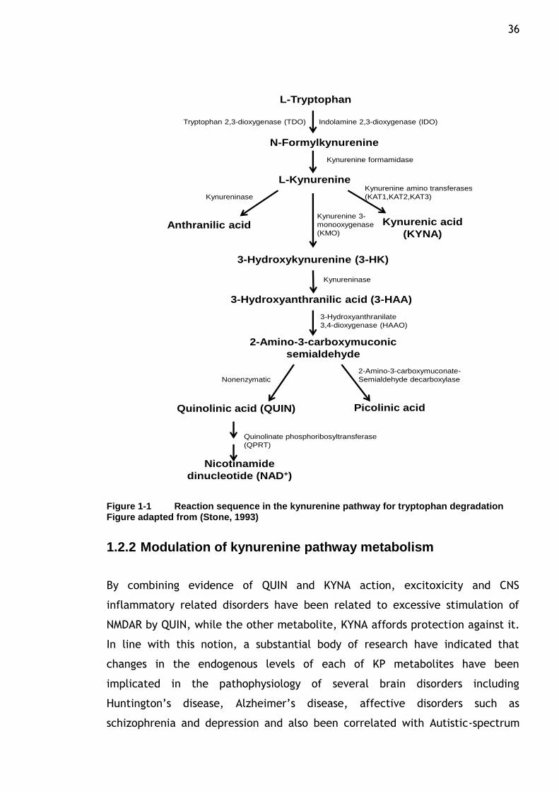
several metabolic steps known as the kynurenine pathway (KP). The specific
reactions associated with each enzyme of the kynurenine pathway are shown in
Figure 1-1. Under physiological conditions, the majority of tryptophan is
metabolized in the liver by enzyme tryptophan 2,3-dioxygenase (TDO), which
acts as the rate-limiting enzyme for tryptophan in the liver and is up-regulated
by corticosteroid. Whereas indolamine 2,3-dioxygenase (IDO) is the first enzyme
of the KP in extrahepatic tissues and this enzyme is induced when tissues are
invaded by viruses, bacteria or endotoxin. Oxidation of tryptophan,
predominantly by the enzyme IDO can be found in numerous cells including
macrophages, microglia, neurons and astrocytes. Generally, induction of this
enzyme increases tryptophan metabolism leading to an increase in kynurenine
and reductions in tryptophan concentrations. Kynurenine, a central compound of
the pathway, can then be metabolized by several enzymes, namely kynurenine
aminotransferases (KAT1, KAT2, KAT3), which generate kynurenic acid (KYNA),
kynureninase which generates anthranilic acid (AA) and finally kynurenine 3-
monooxygenase (KMO) which is the enzyme responsible for the production of 3-
hydroxykynurenine (3-HK). The latter metabolite can be further metabolized
into 3-hydroxyanthranilic acid (3-HAA) which subsequently is metabolized by 3-
hydroxyanthranilate 3,4-dioxygenase (HAAO) to 2-amino-3-carboxymuconic
semialdehyde and is converted to either picolinic acid or quinolinic acid (QUIN).
Finally, the end product nicotinamide adenosine dinucleotide (NAD) is produced
via the action of the enzyme quinolinate phosphoribosyltransferase
(QPRT)(Stone, 1993, Moroni, 1999). Recent studies on the biochemical and
32 molecular biological function of the kynurenine pathway have triggered renewed
interest in the characteristics of individual kynurenine pathway enzymes.
1.2.1 Neuroactive metabolites
1.2.1.1 Quinolinic acid (QUIN)
Interestingly, this pathway generates several immunomodulatory and
neuroactive metabolites and can be induced by infections. It is up-regulated by
inflammatory molecules such as lipopolysaccharide, amyloid peptide,human
immunodeficiency virus proteins, and its potent stimulus, interferon- γ (IFN-γ),
which is able to induce both the gene expression and enzymatic activity of IDO
(Fujigaki et al., 2001; Takikawa, 2005). As tryptophan proceeds along the
kynurenine pathway to achieve the final product nicotinamide adenine
dinucleotide (NAD), a number of neuroactive intermediates are subsequently
generated, most importantly the free-radical generator 3-hydroxyanthranilic
acid (3-HA), the excitotoxin and N-methyl-D-aspartate (NMDA) receptor agonist
quinolinic acid (QUIN) and a broad spectrum excitatory amino acid receptor
antagonist with a particularly high affinity for the glycine recognition site
present on the NMDA receptor-ion channel complex kynurenic acid (KYNA) (Stone
& Perkins 1981; Perkins & Stone 1982; Stone, 1993; Schwarcz et al., 1983). In
contrast to KYNA, 3-hydroxykynurenine (3-HK) and quinolinic acid (QUIN) are
synthesized from kynurenine en route to NAD+ by the enzyme kynurenine 3-
monooxygenase (KMO). This enzyme is increasingly viewed as a major
gatekeeper of the kynurenine pathway. The ability to readily generate damage-
promoting free radicals and NMDA receptor activation accounts for QUIN‟s
unique neurotoxicity and neurotoxic profile (Stone, 1993) .In experimental
studies, high concentrations of KYNA are frequently employed to inhibit
excitatory amino acid receptor function non-specifically and to protect against
excitotoxic insults, whilst QUIN is primarily used as a tool to produce excitotoxic
brain lesions (Schwarcz & Kohler 1983). Quinolinic acid is probably the most
studied kynurenine metabolite because it may cause convulsions and
excitotoxicity activity by interacting with glutamate receptors of the NMDA type
(Stone and Perkins, 1981). The QUIN concentration is normally low within the
brain tissue. However, during an immune response activation, the level of QUIN
is highly elevated in the brain and exerts its excitatory effects at the NMDARs. In
33 this situation, the endogenous formation of kynurenine and its downstream
metabolites are greatly enhanced due to the activation of IDO by interferon-
(IFN-), cytokines, viral and bacterial insults. Infiltrating macrophages, microglia
and dendritic cells have been shown as major sources of QUIN production under
inflammatory conditions in the brain (Schrocksnadel et al., 2006, Chen and
Guillemin, 2009). QUIN has been shown to induce neuronal and astrocytic
apoptosis, and it has been thought that the over-activation of the NMDAR and
subsequent Ca2+ influx into neurons activates the downstream enzyme nitric
oxide synthase (NOS) leading to the production of nitric oxide (NO) which
promote free radical damage (Braidy et al., 2009, Stone and Perkins, 1981). In
addition to NMDAR agonism, it also induces lipid peroxidation and generates free
radicals which in part are responsible for the compound neurotoxic profiles
(Forrest et al., 2002). In certain pathological conditions, in which microglia
activation occurs, elevated QUIN levels were detected in the brain and the
accumulation of this compound has been implicated in the etiology of a broad
spectrum of neurological diseases, particularly those with inflammatory
reactions. The most notable ones are AIDS-dementia, spinal trauma, epilepsy,
Parkinson‟s disease, Huntington‟s disease and Alzheimer‟s disease (Schwarcz et
al., 2012, Schwarcz and Pellicciari, 2002, Schwarcz, 2004, Schwarcz et al.,
2010).
1.2.1.2 Kynurenic acid (KYNA)
Importance is also attached to KYNA by the fact that this is one of the few
known endogenous excitatory amino acid receptor blockers with a broad
spectrum of antagonistic properties on the glycine modulatory site of the NMDA
receptor at low concentrations. At high concentrations, antagonism at the
glutamate site of the NMDA receptor and also on the α-amino3-hydroxy-5-
methyl-4-isoxazolepropionate (AMPA) receptors, suggesting its physiological
function in glutamatergic neurotransmission (Perkins and Stone, 1982). In
addition, it also non-competitively antagonizes α 7-nicotinic acetylcholine
receptors (αNCRs). KYNA is present in the mammalian brain at low
concentrations (nanomolar) and has been described as an inhibitory component
exerting anticonvulsant and anti-excitotoxic actions (Foster et al., 1984). As
shown in Figure 1-1, KYNA is generated directly from L-Kynurenine by KAT
enzymes. Two distinct enzymes have been described in rat and human brains,

34 KAT I and KAT II, which have shown immunoreactivity in astrocytes and also
seems to be present in a small percentage of neurons. In most brain regions and
lesioned brain tissue, KYNA results primarily from KAT II activity and newly
synthesized KYNA is rapidly released into the extracellular space (Amori et al.,
2009, Alkondon et al., 2011). The levels of extracellular KYNA increase linearly
with kynurenine availability, as astrocytes generally lack KMO and therefore
favour KYNA synthesis, whereas microglia cells contain only a little enzyme KAT
and preferentially produce intermediates of the QUIN branch of the pathway
(Schwarcz and Pellicciari, 2002).
Investigation of the KYNA arm of the pathway has provided an insight into their
effects, that elevated level of this metabolite may cause cognitive impairment
by virtue of its ability to antagonize the α-7 nicotinic acetylcholine presynaptic
receptor (α7NCR) and the glycine site of the NMDA receptor. These are both
critically involved in physiological processes underlying learning, memory and
other manifestations of synaptic plasticity (Schwarcz et al., 2012). It has been
proposed that by shifting the kynurenine metabolism toward KYNA formation, it
may be possible to reduce glutamate receptor activation and excitotoxic
neuronal damage. High concentrations of KYNA are anticonvulsant and
neuroprotective, its glutamate antagonist activity is probably responsible for its
ability to prevent brain damage following various pathological settings. It was
found to be protective against brain NMDA neurotoxicity of ischemia and
demonstrated the ability to block QUIN-induced excitation of CNS neurons, thus
supporting the role of KYNA as an endogenous modulator of neurodegenerative
and seizure phenomena(Foster et al., 1984). It has been shown that a reduction
of KYNA level enhances the vulnerability to excitotoxic insults and the ability to
decrease inflammatory molecules such as tumour necrosis factor-α (TNFα) and
nitric oxide which is likely to be involved in LPS-mediated toxicity (Chen and
Guillemin, 2009, Moroni, 1999, Stone, 2001, Vamos et al., 2009).
On the other hand, up regulation of KYNA branch of the pathway has been
theoretically thought to mediate the negative effects of kynurenine pathway up
regulation on mental status. It has also been demonstrated that a minor
elevation of KYNA causes a decrease in the extracellular levels of
neurotransmitters associated with cognitive function, i.e. glutamate,
35 acetylcholine and dopamine. The reduced level of glutamate in cerebrospinal
fluid of individuals with schizophrenia was consistent with hypoglutamatergic
function (Linderholm et al., 2010). Evidence has also supported the observation
that altering the concentration of KYNA during critical times of brain
development can affect behaviour later in life. It should also be noted that
chemical manipulation by injection of kynurenine induced alterations in working
memory function in adulthood, therefore establishing a notion that altered
concentration of KYNA is capable of influencing cognitive function that depends
on intact NMDAR transmission (Akagbosu et al., 2010, Chess et al., 2007). With
regard to schizophrenia risk, exposure to a number of infectious agents during
early life or childhood has previously been associated to the later development
of symptoms related to schizophrenia. Experimental studies have shown that
exposure to various insults during early life can induce long term effects on gene
expression in the brain as well as behaviour (Shi et al., 2003, Fatemi et al.,
2009a). However, the impact of CNS infections on the KP during early life in the
brain is not fully described. Infection of neurotrophic influenza A virus during
early postnatal period (P3) in mice triggered an altered expression of several
genes in the KP whilst increased levels of IDO and KMO have been observed on P7
of virus-infected brain accompanied with transient increase of KYNA
concentration. The increased levels of KYNA appear to be caused by activation
of tryptophan degradation in the brain and the inhibition of NMDA receptors by
KYNA would be one of the potential consequences which may functionally link
infections with distorted glutamatergic signalling in the developing brain
(Holtze et al., 2008). Therefore, the elevation of KYNA as observed in
schizophrenic patients provided a new insight into the possible effect of KYNA on
the glutamatergic and dopaminergic systems and its potential role in the
pathogenesis of schizophrenia (Erhardt et al., 2007, Barry et al., 2009). Novel
treatment of the disease could rationally be directed towards brain KYNA
formation. Thus, blockade of kynurenine aminotransferase (KAT II which
catalyzes the conversion of kynurenine to KYNA) causes a decrease in brain KYNA
and may have cognition enhancing effects in the treatment of schizophrenia
(Wonodi and Schwarcz, 2010, Erhardt et al., 2009).
36
L-Tryptophan
N-Formylkynurenine
L-Kynurenine
3-Hydroxykynurenine (3-HK)
Quinolinic acid (QUIN) Picolinic acid
3-Hydroxyanthranilic acid (3-HAA)
2-Amino-3-carboxymuconic
semialdehyde
Nicotinamide
dinucleotide (NAD+)
Anthranilic acid Kynurenic acid
(KYNA)
Indolamine 2,3-dioxygenase (IDO)
Kynurenine formamidase
Tryptophan 2,3-dioxygenase (TDO)
Kynurenine amino transferases
(KAT1,KAT2,KAT3)Kynureninase
Kynurenine 3-
monooxygenase
(KMO)
Kynureninase
3-Hydroxyanthranilate
3,4-dioxygenase (HAAO)
2-Amino-3-carboxymuconate-
Semialdehyde decarboxylaseNonenzymatic
Quinolinate phosphoribosyltransferase
(QPRT)
Figure 1-1 Reaction sequence in the kynurenine pathway for tryptophan degradation Figure adapted from (Stone, 1993)
1.2.2 Modulation of kynurenine pathway metabolism
By combining evidence of QUIN and KYNA action, excitoxicity and CNS
inflammatory related disorders have been related to excessive stimulation of
NMDAR by QUIN, while the other metabolite, KYNA affords protection against it.
In line with this notion, a substantial body of research have indicated that
changes in the endogenous levels of each of KP metabolites have been
implicated in the pathophysiology of several brain disorders including
Huntington‟s disease, Alzheimer‟s disease, affective disorders such as
schizophrenia and depression and also been correlated with Autistic-spectrum
37 disorder (ASD). The presence of neuroactive kynurenines in the mammalian brain
has led to the notion that it may be possible to affect synaptic transmission of
excitatatory amino acid receptors by modulating kynurenine pathway
metabolism in the brain. Some studies have found that pharmacological
manipulations favouring KYNA in the brain extracellular space by intracerebral
administration of exogenous KYNA protected against excitotoxins which may be
beneficial in attenuating excessive NMDA receptor function and provide effective
neuroprotection after an acute excitotoxic insult (Obrenovitch and Urenjak,
2000).
The pharmacological manipulation of the kynurenine pathway is still in its
developmental stage and the pathway has been manipulated in several ways
with the aim of developing therapies for the treatment of neurodegenerative
diseases. The fact that NMDA receptor function is paramount to early brain
development and since over activation of this receptor in the brain has been
postulated as a cytotoxic mechanism involved in neurodegenerative process,
many researchers have been prompted to look into the kynurenine pathway for a
potential therapeutic approach. In this aspect, pharmacological challenges
directed to enhance the endogenous formation of KYNA in brain tissue are
considered as a successful strategy to counteract toxic events. Several ways of
increasing the brain levels of endogenous KYNA have been discovered. One
example is administration of a combination of kynurenine and probenecid, an
organic acid transport blocker. The immediate precursor to KYNA, kynurenine
has been shown to cross the brain barrier (BBB) effectively from the periphery
and increased KYNA concentration while probenecid inhibits the efflux of KYNA
from the brain via a probenecid-sensitive carrier. This association appears to be
able to increase the brain extracellular concentration of KYNA and attenuate
QUIN neurotoxicity (Santamaria et al., 1994, Santamaria et al., 1996).
Neuroprotection of this combination has also been observed in animal models of
Parkinson‟disease where systemic administration of kynurenine and probenecid
attenuates the dopaminergic damage induced by 6-hydroxydopamine, suggesting
that the combined treatment constitutes a pharmacological strategy of
considerable therapeutic value to mitigate excitotoxic events (Silva-Adaya et
al., 2011). Another approach involved the use of synthetic analogues of
kynurenic acid or kynurenine that can easily penetrate the blood brain barrier
38 (in contrast with KYNA) as antagonists at glutamatergic NMDA receptors (Stone
and Addae, 2002, Stone, 2001). A series of kynurenine analogues that have KMO
and kynureninase inhibiting properties have also been tested. This includes
nicotinylalanine (NAL) and meta-nitrobenzoylalanine (mNBA) which change the
balance of the tryptophan metabolites towards the formation of KYNA (Moroni et
al., 1991, Russi et al., 1992). The latter has been described as having more
potent competitive and selective inhibitory activity than previous compounds in
inhibiting KMO and caused an increase in the brain concentration of KYNA, an
effect which is associated with sedative and anticonvulsant actions (Carpenedo
et al., 1994). Another possible method to decrease the effects of KP
disturbances is by using enzyme inhibitors. It appears that in some pathological
states, changes in the two KP metabolites are modest and could be balanced by
elevating the level of the antiexcitotoxic metabolite, KYNA or blocking the
formation of QUIN. Development of pharmacological agents designed to augment
or attenuate the effects of specific kynurenine metabolites, such as QUIN and
KYNA is underway and several prototype inhibitors of most kynurenine enzymes
are now available. Several of these compounds have been used for proof-of-
concept in experimental animals (Nemeth et al., 2007, Moroni, 1999).
It has been shown that inhibition of kynurenine 3-monooxygenase (KMO), an
enzyme that is responsible for synthesizing quinolinic acid, will shift kynurenine
pathway from the production of excitotoxin quinolinic acid towards the
production of neuroprotective kynurenic acid. Neuroprotection provided by the
chemical inhibition of KMO has been shown in Huntington‟s disease model flies,
in which decreases in 3-HK relative to KYNA is thought to confer neuroprotection
by antagonizing NMDAR and decreasing glutamate-dependent excitoxcity as well
as by scavenging free radicals (Campesan et al., 2011, Stone, 2000). Reductions
in spontaneous locomotor activity and increases to the threshold level of
electroshock-induced seizure are the effects produced by systemic
administration of KMO inhibitors. In comparison, neuroprotective effects of KMO
inhibitors in models of brain ischemia are primarily related to their ability to
increase KYNA concentration in the brain and consequently antagonise the
activation of NMDAR. Thus, it is reasonable to assume that by inhibiting KMO it is
possible to reduce NMDAR function thus affording neuroprotection (Chiarugi et
al., 1995, Moroni, 1999). However, it should be noted that the neuroprotective
39 properties of KMO inhibitors by increasing the KYNA concentration are observed
under pathological settings such as in immune-stimulated conditions.
Administration of inhibitors of KMO under basal conditions, however do not
affect the levels of either blood or brain content of QUIN (Chiarugi and Moroni,
1999). Therefore, it is important to further evaluate the physiological and
pharmacological implications of increasing KYNA concentrations in the brain.
A range of studies has been conducted to study the inhibition of KMO as a
strategy for inducing a more favourable ratio of KYNA to QUIN in vivo. Among
related molecules from a series of N-(4-phenylthiazol-2-yl)benzenesulfonamides,
one member known as Ro61-8048 with the molecular structure as 3,4-dimethoxy-
N[4-(3-nitrophenylthiazol-2-yl]benzenesulfonamide has been characterized as a
highly effective inhibitor, and has been successfully used as a selective agent to
probe the physiological and pathological roles of KP metabolites experimentally
(Rover et al., 1997). Previous studies have shown that systemic administration of
this compound caused a robust decrease in glutamate levels in selected regions
of the brain and this intervention is useful as a pathological increase in
glutamate neurotransmission is considered detrimental in various neurological
and psychiatric disorders (Meldrum and Garthwaite, 1990, Moroni et al., 2005).
Ro61-8048 is a competitive inhibitor of cerebral KMO and caused an increase in
brain KYNA concentrations. It has been shown that a build up of this antagonistic
intermediate could inhibit activation of brain NMDARs and decrease convulsion in
hyperbaric oxygen (HBO)-induced seizure activity (Dale et al., 2000). Subsequent
studies that provides both behavioural and biochemical evidence in support of
the pharmacological interventions of KMO inhibition showed that Ro61-8048 was
neuroprotective in rodent models of ischemia and sleeping sickness, and in
model of levodopa-induced dyskinesias (movement disorders) in parkisonian
monkeys. Mechanisms other than direct NMDA receptor blockade such as KYNA-
induced αNCR-mediated presynaptic inhibition of glutamate release may
therefore have contributed to the therapeutic benefits of the compound
(Gregoire et al., 2008, Cozzi et al., 1999, Rodgers et al., 2009).
In this study, we evaluated the effects of KMO inhibition by a novel compound
Ro61-8048, administered during the late gestation period of a rat model and
later examine whether pharmacologically induced prenatal increases in brain
40 KYNA levels might interfere with developmentally essential neurological and
glutamatergic functions.
1.3 Neurodevelopmental Markers
1.3.1 NMDA receptor subunit NR2A and NR2B
In the vertebrate CNS the predominant mode of excitatory transmission is
mediated by the neurotransmitter glutamate and the ionotropic glutamate
receptors, which include N-methyl-D-aspartate (NMDA), α-amino-3-hydroxy-5-
methyl-4-isoxazolepropionic acid (AMPA) and kainate receptors. The NMDA
receptor plays critical roles in excitatory synaptic transmission, plasticity and
excitotoxicity in the CNS and several researchers have indicated that activity-
dependent changes in excitatory synaptic function such as long term
potentiation (LTP) and long term depression (LTD) are likely cellular correlates
to learning and memory (Bliss and Collingridge, 1993). Research has shown that
glutamate induced excitotoxic injury or overstimulation of the NMDA receptors
may disturb normal glutamate neurotransmission in a variety of neurological
disorders including Alzheimer‟s disease, ischemia, epileptic disorders and
Huntington‟s disease (Bi and Sze, 2002, Engelhardt et al., 2007, Gascon et al.,
2008). On the other hand, NMDA receptor hypofunction is also implicated in
other pathologies such as schizophrenia (Schmitt et al., 2007). Its involvement in
diverse processes during CNS development and implication in various psychiatric
disorders has made research enthralling around its function to govern synaptic
trafficking and stabilization, and is crucial for our understanding of the various
processes carried out by NMDA receptors (Bard and Groc, 2011). NMDA receptors
are heteromeric ligand-gated ion channels assembled from two families of
subunits, NR1 and one or more of the four closely related NR2 subunits. The
mandatory NR1 subunit is encoded by a single gene that can generate eight
splicing variant isoform and can be found ubiquitously throughout the brain. The
NR2 subunit family is composed of NR2A, NR2B, NR2C and NR2D, which are
encoded by four distinct genes and shows both regional and developmental
variations in animals, with NR2B and NR2D subunits predominating in the
neonatal brain, but over the course of development these are replaced by NR2A
and in some regions NR2C as well as a mixture of NR2A and NR2B subunits.

41 Another NMDA receptor subunit that has been characterized is known as NR3 and
comprises two subtypes (A and B). Some studies showed variations in the NR2A
and NR2B ratio which increases with age and this suggested that the composition
of NMDA receptor complexes is dependent on the availability of the subunits (Al-
Hallaq et al., 2007). The functional and pharmacological properties of the
receptors are determined by subunit composition, post-translational
modification and its cellular localization in the CNS (Monyer et al., 1992, Monyer
et al., 1994, Cull-Candy et al., 2001, Furukawa et al., 2005). Under normal
circumstances, NMDA receptor activation occurs upon the binding of glutamate
and glycine (co-agonist) to the NR2 and NR1 subunits, respectively (Figure 1).
The channel is permeable to calcium ions and blocked by magnesium ions at
resting potentials, which requires postsynaptic depolarization to release the
magnesium block and to permit activation of NMDA receptor. Thus, the NMDA
receptor is unique in that it requires membrane depolarization to remove the
Mg2+ and also simultaneous co-activation by two ligands, glutamate/aspartate
and glycine/D-serine, to be activated. In addition, the function of the receptor
can also be modified through the use of extracellular Zn2+ and polyamines. The
activated NMDA receptor is permeable to both K+ and Na+, but has particularly
high permeability for Ca2+, which acts as a second messenger in the cell. The
increased Ca2+, due to high glutamatergic activity, results in a permanent
increase in synaptic efficacy. This process is known as long term potentiation
(LTP) and is of critical importance for learning and memory. LTP is a form of
synaptic plasticity and typically consists of a prolonged increase in synaptic
transmission strength requiring NMDA receptor activation. The concept of NR2
subunit-specific signals is also an ongoing area of interest within the synaptic
plasticity field. It has been proposed that NR2A-containing NMDA receptors are
preferentially involved in potentiation of synapses, while NR2B-containing NMDA
receptors play a role principally in depression (Bartlett et al., 2007). However,
there are contradicting findings that claimed a different point, in which NR2B-
containing NMDA receptors can mediate synaptic potentiation as well. Both
excitoxicity and pro-survival effects of synaptic signalling can be mediated solely
by NR2B-containing NMDA receptors (Martel et al., 2009). Growing evidence that
indicates development in elucidating the molecular mechanisms underlying
activation of NMDARs, driven by the availability of high resolution X-ray
structures for the ligand binding domains, is likely to revitalize the search for
42 more effective and subtype-specific NMDA receptor drugs (Cull-Candy et al.
2001;Hunt and Castillo 0 AD;Hunt and Castillo 2012).
Inappropriate activation of the receptor has been implicated in the pathogenesis
of several disease states; in particular it has been related to the excessive
calcium influx through the channel that can cause excitotoxicity. Thus, blockade
of the NMDA receptors is considered neuroprotective. A number of
pharmacological agents have been shown to distinguish the function of the
various NMDA receptor subunits. Among competitive antagonists that exhibit
moderate selectivity for certain subunit combinations are channel blockers such
as MK-801(dizocipline), ketamine, phencyclidine and amantadine. Dopaminergic
hyperactivity and behavioural characteristics of schizophrenia has been observed
following treatment with non-competitive antagonists such as ketamine or
phenylcyclidine. In electrophysiological experiments, administration of this
NMDA receptor antagonists PCP, ketamine or MK-801 is associated with an
increase in the firing rate and burst firing activity of ventral tegmental area
(VTA) dopamine neurons (French et al., 1993). However, findings showed that
chronic administration of PCP resulting in a reduced dopamine release in the
prefrontal cortex and increased dopamine release in nucleus accumbens, which
are conceivably related to negative symptoms and cognitive deficits consistently
seen in schizophrenia. Administration of PCP also disrupted the prepulse
inhibition, a behavioural model reflecting sensory gating deficits in
schizophrenia (Jentsch and Roth, 1999). Ifenprodil and haloperidol are non-
competitive antagonists that are effective at suppressing the activation of NMDA
receptor. The usefulness of these drugs are however limited to their actions on
normal synaptic transmission and sometimes accompanied by additional side
effects. In addition, physiological metabolite in the brain known as kynurenic
acid also serves as a broad antagonist at ionotropic glutamate receptors
particularly those of NMDA type. In addition to its inhibition of AMPA and kainate
receptor, it acts on the glycine recognition binding site of the NMDA receptor
within low micromolar concentration and shared common characteristics with
PCP and ketamine in which it could induce psychomimetic actions in human
(Erhardt et al., 2007).
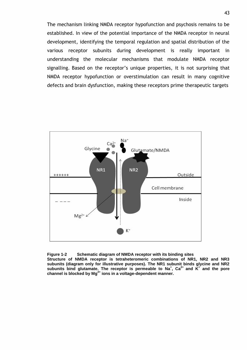
43 The mechanism linking NMDA receptor hypofunction and psychosis remains to be
established. In view of the potential importance of the NMDA receptor in neural
development, identifying the temporal regulation and spatial distribution of the
various receptor subunits during development is really important in
understanding the molecular mechanisms that modulate NMDA receptor
signalling. Based on the receptor‟s unique properties, it is not surprising that
NMDA receptor hypofunction or overstimulation can result in many cognitive
defects and brain dysfunction, making these receptors prime therapeutic targets
Figure 1-2 Schematic diagram of NMDA receptor with its binding sites Structure of NMDA receptor is tetraheteromeric combinations of NR1, NR2 and NR3 subunits (diagram only for illustrative purposes). The NR1 subunit binds glycine and NR2 subunits bind glutamate. The receptor is permeable to Na
+, Ca
2+ and K
+ and the pore
channel is blocked by Mg2+
ions in a voltage-dependent manner.
44
1.3.2 Post synaptic density 95 (PSD95)
Subunit composition determines multiple NMDA receptor function, including
regulation of posttranslational modification, channel kinetics and downstream
signalling involving intracellular protein association (Lin et al., 2004b). In this
regards, the postsynaptic density-95 (PSD95), a scaffolding molecule abundantly
enriched at glutamatergic synapses, is found to have important functions for the
modulation of NMDA trafficking and stabilization on the cell surface. It belongs
to the membrane-associated guanylate kinase (MAGUK) family which contains
three PDZ(PSD-95/Dlg/ZO-1) domains, a SH3( Src Homology 3) domain and a
carboxy terminal guanylate kinase domain (Kim and Sheng, 2004). Receptor
subunit composition especially those of NMDA are dynamically remodelled during
development and in response to neuronal activity and sensory experience. Less
is known about the remodelling process of the NMDA receptors, their scaffold
and signalling proteins. Several studies have shown that this reorganization
occurs by subunit insertion into the synaptic membrane or mobility between the
synaptic and extrasynaptic membrane. Scaffolding molecules situated at synapse
facilitate clustering of receptors by engaging in several potential interactions,
thus changes to the content or organization of scaffold modules at the synapse
could alter synaptic transmission by changing either receptor number or
distribution. It has been shown that the induction of depression or potentiation
is associated with scaffold modifications. Both AMPA and NMDA receptors
mediate the postsynaptic effects of glutamate at central synapses and PSD95
could regulate synaptic transmission and synaptic plasticity by selectively
altering the ratio of synaptic responses mediated by both receptors (MacGillavry
et al., 2011). Overexpression of PSD95 in the postnatal cultured brain slices has
been found to change synaptic properties by increasing AMPA receptor-mediated
synaptic transmission and favoured the emergence of LTD (Beique and Andrade,
2003). In addition, there is debatable evidence to show the cooperative role of
protein-protein association between NR2 subunit domains and PSD95 (Delint-
ramirez et al., 2010). Alteration in the relationship between NMDA receptor
subunit expression and PSD95 might be related to several neurodegenerative
diseases. As PSD95 plays a fundamental role in the trafficking, membrane
targeting and internalization of NMDA receptor complexes, changes to this
molecule have been reported in many cases including depression, bipolar
45 disorder and schizophrenia (Kristiansen and Meador-Woodruff, 2005, Toro and
Deakin, 2005). In a clinical sample of schizophrenic patients, alterations in the
expression of PSD95 protein and mRNA has been found in the brain, suggesting
that glutamate dysfunction may involve changes in intracellular molecules
critical for glutamate receptor-associated signal transduction (Clinton et al.,
2003, Clinton et al., 2006). Evidence also indicates that the amount of anchoring
protein PSD95 in post-mortem brain of depressed subjects are elevated and
highly correlated with an increase in NR2A subunit reactivity (Karolewicz et al.,
2009). Many of these diseases may initially involve dysregulation of synaptic
transmission resulting from defects in synaptic organization.
1.3.3 Eprin type-A receptor 4 (EphA4)
A signalling process that involves some form of cel-cell communication controls
arrangement of cells in the correct locations and establishment of the correct
pattern of neural connections. Several families of membrane-bound cell surface
proteins have been implicated in the control of cell movement, which is crucial
for tissue organization during development. These proteins include ligand-
receptor systems such as the Eph receptor tyrosine kinases. The Eph receptors
represent the largest family of receptor protein tyrosine kinases and interact
with their ligands, ephrins. The Eph receptors and ephrins are divided into two
subclasses, A and B, based on their homologies, structures, and binding
affinities. In general, EphA receptors (EphA1-EphA8) bind ephrin-A molecules
(ephrinA1-ephrinA5) whereas the EphB receptors (EphB1-EphB6) bind ephrin-B
molecules (ephrinB1-ephrinB3). However, one member of each class,
namelyEphA4 and EphB2, can be activated by both types of ephrins. Most of the
members of this family are predominantly expressed in the nervous system
during development (Gale et al., 1996, Flanagan and Vanderhaegen, 1998).
Ephs and their cell surface-associated ephrin ligands are important regulators of
central nervous system (CNS) development. They are involved in the guidance of
axonal growth and establishment of neural connectivity (O'Leary and Wilkinson,
1999). Several Eph receptors and ephrins are also widely expressed in the adult
CNS, where they appear to regulate synaptic function and plasticity (Yamaguchi
and Pasquele, 2004). Many of the Eph receptors may have overlapping functions
46 with each other. Protein expression pattern by in situ hybridization and
immunohistochemistry have indicated the expression pattern and distribution of
Eph proteins during development. Eph receptors and ephrins are involved not
only in early developmental processes, but also in the function of the adult
organism. In the mature brain, Ephs regulate neuron–glia communication and
play a role in synaptic plasticity. Among the subtypes, EphA4 shows widespread
expression throughout the CNS starting from early development and persisting in
the mature brain (Egea and Klein, 2007, Greferath et al., 2002, Pasquele, 2008).
The genes for Eph receptors and ephrins have been recognized to be
differentially expressed in various brain regions including the hippocampus.
EphA4 is highly expressed in the hippocampus and is involved in hippocampal
synaptic plasticity such as long-term potentiation, and therefore EphA4 may
assume a role in hippocampus-dependent behaviour. EphA4 is expressed on
dendritic spines of pyramidal neurons and axon terminals where it interacts with
ephrin-A3 to shape dendritic spine morphology and influence synaptic plasticity.
Eph dysfunction might be relevant to neuropsychiatric and neurodegenerative
diseases characterized by abnormal dendritic spines in the hippocampus and
cortex (Carmona et al., 2009, Glantz and Lewis, 2000, Yamaguchi and Pasquele,
2004). Increasing evidence also implicates Eph family proteins in cancer in which
interaction between Eph-ephrin regulate critical steps of tumour growth and
metastasis. This signalling mainly affects the cell shape and also influences cell
proliferation and cell fate determination, therefore it is speculated that Eph
signalling could play some role in tumourigenesis as one of their possible
consequences (Kaenal et al., 2012).
1.3.4 Sonic hedgehog (SHH)
Initially discovered in Drosophilia, sonic hedgehog (SHH) is a member of the
hedgehog family of secreted signalling molecules, which is one of the key
organizers of tissue patterning that undergo several post-translational
modifications to gain full activity. SHH signalling plays a role in many
developmental processes in different tissue types. SHH acts as a morphogen and
induces distinct transcriptional programs on certain target cells depending on its
concentration. In addition, this molecule also plays a central role in regulating
47 cell proliferation, differentiation and morphological patterning during
embryogenesis. During development, interaction of various cell types is
supported by many molecular events and inductive signals, secreted from within
the CNS to ensure proper coordination and functional neuronal development
(Marti and Bovolenta, 2002, Traiffort et al., 2010). The activation of SHH
signalling is initiated by the binding of the SHH ligand to the membrane receptor
Patched (Ptc), which relieves the Ptc-mediated inhibition of the transmembrane
protein Smoothened (Smo). Activated Smo initiates an intracellular
response/signalling cascade that ultimately drive the activation and nuclear
translocation of the Gli transcription factor family member resulting in
upregulation of hedgehog target genes including Gli and Ptc which respectively
trigger the positive and negative feedback loops following pathway activation.
This signalling cascade play fundamental roles during development and aberrant
SHH signals underlie many disease conditions (Mullor et al., 2002, Ruiz i Altaba
et al., 2002).
Given the pathway‟s significant role in tissue patterning and cell number
maintenance, events causing misregulation of the pathway are associated with
certain cancers. Evidence that show the contribution of SHH signalling to
tumourigenesis is emerging particularly in a model using in vitro approaches
(Wicking et al., 1999). Inappropriate SHH signalling has been related to tumour
initiation and maintenance, for instance in basal cell carcinoma (BCC) and
medulloblastoma. The development of tumours such as those two above is
through activation f the pathway by misregulation or mutation at the level of its
receptor Ptc or Smo (Taipale and Beachy, 2001, Stecca and Ruiz i Altaba, 2005).
These components are involved in the multi-molecular network which allows
transduction of the SHH signal which drives proliferation and differentiation
through the activation of specific downstream effectors of the pathway.
Misregulation or mutations in the various elements of the SHH signalling pathway
can lead to different phenotypes and also diseases associated with growth
deregulation. As the pathway is involved in many developmental events, it will
also be associated with human syndromes, defects and malformations (Mullor et
al., 2002, Ming et al., 1998).
48 In addition to functioning in the embryo, SHH signalling components are
expressed in postnatal and adult tissues, suggesting that they function in the
mature organism. This molecule has been studied mainly in the context of
development, however increasing evidence emerges to suggest the mitogenic
properties of SHH are retained in the adulthood and promotes oncogenesis or
tissue repair in different organs. Regenerative roles of SHH in cell proliferation
might be useful in a clinical setting, for instance in inflammation-induced events
and mechanical brain injury. Activation of the SHH pathway has been shown in
response to hypoxia, in which protein levels of SHH and it transcription factor
Gli1 were upregulated in the region of hippocampus after ischemic brain injury
(Sims et al., 2009).
1.3.5 Proliferating cell nuclear antigen (PCNA)
The cell cycle is a complex and tightly regulated process by which cells duplicate
their contents and divide into daughter cells. Among several cell cycle
regulators, the most widely employed endogenous proliferation marker is
Proliferating Cell Nuclear Antigen (PCNA). PCNA is a 36 kDa protein known as the
cofactor of DNA polymerase delta and it is involved in cellular DNA replication
and repair. Cell cycle analysis has shown that PCNA concentration increases
rapidly in the mid-G1 phase of the cell cycle, immediately preceding the onset
of DNA synthesis and remains elevated throughout the S phase, then rapidly
decreases from the G2/M phase to G1 (Kurki et al., 1986). Because of its close
relation to the cell cycle, PCNA is used as a physiological or pathological marker
protein of proliferating cells (Valero et al., 2005, Valero et al., 2011). Due to the
fact that PCNA is involved in DNA replication, it also has been used as a
proliferation marker for adult hippocampal neurogenesis where
immunoreactivity staining was detected in progenitor cells in the subgranular
zone (SGZ) and also subventricular zone (SVZ) (Ino and Chiba, 2000). As DNA
replication is a critical event in the cell; errors can be made and importantly
unrepaired DNA damage upon DNA replication may give rise to mutations. Thus,
many DNA repair proteins interact with the replication machinery in order to
ensure efficient and reliable DNA duplication. Numerous studies have
demonstrated correlations between dysregulation of the cell cycle machinery
and neuronal death. Upregulation of cell cycle proteins occurs after various
49 injuries such as trauma and stroke and in neurodegenerative disease such as
Alzheimer‟s disease. Failure of cell cycle regulation might be a common pathway
of several neurodegenerative disorders and this implies that cell cycle regulation
might be one of the promising therapeutic intervention in these disorders (Wang
et al., 2009).
1.3.6 Sex determining region of Y-chromosome related-HMG box
2 (SOX2)
The Sex determining region of Y-chromosome (SRY) was the first SOX gene
identified in mammals where it was responsible for determination of the male
sex. The transcription factor SOX2 is a member of the SOX gene family related to
the SRY with the possession of a high-mobility group (HMG) DNA binding domain,
thus the acronym SOX stands for „SRY-related HMG box‟. Through protein
sequence comparison analysis, 20 SOX genes have been identified and
categorized into eight groups, A-H with two B subgroups, B1 and B2 (SOX14 and
SOX21). The SOX-B group underwent further expansion into a B1 subgroup
consists of three transcriptional activators SOX1, SOX2, SOX3 and a B2 subgroup
of two transcriptional repressors SOX14 and SOX21. The SOX B group is of
particular interest in that the five proteins are highly related and have
overlapping expressions patterns (Lefebvre et al., 2007, Lovell-Badge, 2010,
Wegner, 1999).
Generally, SOX proteins have been characterized as important transcriptional
regulators of many developmental processes during early embryogenesis and are
essential for the maintenance of a stem/progenitor cell pool. SOX-B genes in
particular perform a wide range of functions by a complex interplay with
specific partner factors to regulate gene expression (Kamachi et al., 2000). They
are best known for their role in pluripotency and self-renewal, but also have
been implicated in specification, maturation and terminal differentiation events.
The vertebrate group B genes, SOX1, SOX2 and SOX3, have similar effects and
are expressed in progenitor cells from the early stages of CNS development.
Reduction in the expression of one of these genes usually does not result in a
significant loss of function, indicating possible compensation of function and
functional redundancy appears to be confined to subgroup members. SOX2 is
50 probably one of the best characterized SOX proteins and have a dynamic pattern
of expression throughout embryogenesis and in a variety of adult tissue types.
Expression of SOX2 is generally localized to proliferating and undifferentiated
precursors whereby expression is downregulated with differentiation (Pevny and
Placzek, 2005, Guth and Wegner, 2008). This is in line with evidence that
constitutive expression of SOX2 inhibits neuronal differentiation resulting in the
maintenance of progenitor characteristics (Graham et al., 2003). Thus, neural
stem cells (NSC) at specific locations and developmental stages are especially
vulnerable to SOX2 deficiency. It also should be noted that complex interactions
of SOX2 with other regulatory factors explain its specificity of action and
variably affect the outcome of SOX2 ablation or mutation (Pevny and Nicolis,
2010). It has been shown that SOX2 and other regulatory transcriptional factors
like Oct4 and Nanog collaborate to maintain pluripotency and self-renewal in
human embryonic stem cell. Close interaction between this three factors was
maintained in the undifferentiated state of the cells (Fong et al., 2008). In view
of this, the use of mutant allelic series to decrease/abolish SOX2 function have
shown that neural stem cells from SOX2 knockdown mice were unable to
differentiate into mature neurons and undergo apoptosis, whereas homozygous
SOX2 mutant embryos do not develop. Mutations of SOX2 in humans result in a
complex syndrome anophthalmia, defective hippocampal development and
cognitive defects and epilepsy, while mouse models reproduced a broad
spectrum of severity mirroring those seen in human patients (Fantes et al.,
2003, Favaro et al., 2009, Sisodiya et al., 2006, Avilion et al., 2003).
SOX2 represents a well known marker for NSC and progenitor cells in both
embryonic and adult brains, the distribution of SOX2 expression in the adult
brain can be found in areas where neurogenesis persists, the subventricular zone
(SVZ) in the forebrain and in the subgranular zone (SGZ) of the hippocampal
dentate gyrus (Komitova and Eriksson, 2004). Apart from their distribution in all
stem/progenitor populations throughout CNS, SOX2 also can be found in other
non-neural populations such as the stomach, lung, hair follicles and the tongue
(Ishii et al., 1998, Okubo et al., 2006, Driskell et al., 2009). Functional
redundancy roles within SOX-B1 factors in maintaining broad developmental
processes and identity of NSC could be seen during neurogenesis stages. Their
inhibition in the embryo results in premature differentiation of neural precursors
51 while mutation of SOX2 in the adult lead to a reduction in the proliferation of
neural precursors suggesting a role for SOX2 in neural stem cell maintenance or
proliferation in adult brains (Episkopou, 2005). Loss of its function induces cell
cycle exit and advances neuronal differentiation. In a way, overexpression of
SOX2 would inhibit neurogenesis while failing to reach a certain threshold of
SOX2 expression by neural progenitors, render neurons unable to properly
differentiate (Bylund et al., 2003, Avilion et al., 2003). In this regard, estimation
of both precursors and early/immature neurons at an early differentiation stage
could provide a basis indication of neurogenesis efficiency and support the
notion that these regulatory proteins have important roles in several distinct
cellular differentiation events (Suh et al., 2007). Recent evidence using
immunoflourescence labelling revealed that SOX2 and nestin/vimentin are
abundant in the primitive neuroepithelial tissue of immature teratomas of CNS
which is indicative of aggressive cellular populations (Phi et al., 2007). Higher
levels of co-expression between SOX2 and nestin (with its expression indicative
of pluripotency and regenerative potential) have also been noted in cultured
melanoma cells. Nestin expression is enriched in metastatic tumors and support
its role in cancer progression and over expression of both markers are
anticipated, considering that the tumour-initiating cell is probably a cell of an
immature nature in which SOX2 is reported to be expressed (Laga et al., 2011).
In this regard, roles and involvement of SOX2 and other SOX family members in
tumorigenesis need to be verified, as there are differences between cancerous
stem-like cells and normal neural stem cells. To some extent, this marker has
been shown to be involved in the regulation of cancer stem-like cells and the
presence of this protein along with other regulatory factors seems to important
for the self renewal capacity and the cell proliferation of cancer stem-like cells
(Phi et al., 2008).
1.3.7 Doublecortin (DCX)
Normal brain development involves generation of new neurons throughout life
and this process is known as neurogenesis, which is tightly regulated in the
specialized areas of the mammalian brain. It is a continuous physiological
process that occurs in the subventricular zone (SVZ) of the lateral ventricle and
subgranular zone (SGZ) of the hippocampus dentate gyrus. These brain regions
52 contain a pool of neural progenitor cells (NPC) which are capable of producing
new neurons (Eriksson et al., 1998, Rao et al., 2006). Normally in the adult,
neurogenesis in these regions occurs at a low level; however, increased cell
proliferation and the presence of newly generated neurons have been observed
in various experimental setting including environmental enrichment, physical
activity, learning and physiological stimulation (Valero et al., 2011). Increased
neurogenesis may occur in response to neurodegenerative disease processes in
the adult brain. In regards to this, an increased number of DCX positive cells
have been observed following kainic acid-induced seizures in mice and in
epileptic and Hungtington‟s patients, suggestive of a contribution for newborn
neurons in the disease process (Liu et al., 2008, Jessberger et al., 2005).
Conversely, formation of new neurons is decreased by age, stress and chronic
exposure to adrenal steroids. This combined evidence suggest that the adult
brain can respond to activity-induced stimulation and that differential effects in
neurogenesis were likely as part of compensatory mechanisms or an attempt by
the injured brain to replace lost cells.
As part of detection techniques in neurogenesis, doublecortin (DCX) expression
can be used as a candidate marker for adult neurogenesis. DCX is a microtubule-
associated protein and its expression corresponds to the early stages of
neurogenesis. The role of DCX in neuronal development has been associated with
microtubule stabilization, nuclear translocation during neuronal migration and
also axonal and dendritic maturation (Francis et al., 1999, Gleeson et al., 1999).
In the adult brain, prominent DCX expression continues to persist within the two
areas of continuous neurogenesis. The expression of DCX is observed in cell
division as demonstrated by co-labeling with the cell cycle marker (Ki67) and
downregulated once the cells became older and began expressing markers for
mature neurons, thus reflecting a function for DCX in cell division apart from
migration. Therefore it has been extensively incorporated as a specific marker of
newly born or immature neurons in the adult and also to study the relation
between neurogenesis, neurological and neurodegenerative disease processes in
the search for putative therapeutic strategies (Brown et al., 2003, Verwer et al.,
2007).
53
1.3.8 Neuronal nuclear antigen (NeuN)
It is known that adult neurogenesis consists of tightly regulated steps that
include proliferation, cell cycle exit, differentiation and functional maturity.
This process that takes place in the SGZ of the hippocampus and the SVZ of the
lateral ventricles involves the development of late born neurons from early
neural progenitors, committed neuronal precursors, immature migrating neurons
to mature neurons. At various stages of this process, different specific molecules
are expressed by the newly formed cells that can serve as a guide to monitor the
different stages of neurogenesis (Halbach, 2007, Kempermann et al., 2004). The
stability of new neurons might seem to be an important prerequisite for
function, in which establishment of their synaptic contacts for signalling is
functionally integrated. Neuronal nuclear antigen or NeuN is a neuron-specific
nuclear protein that is identified by immunoreactivity with a monoclonal
antibody, anti-NeuN. Because of its high specificity for most types of neuronal
cells, and its cross reactivity with multiple species, anti-NeuN has gained
widespread acceptance as a reliable tool to detect most postmitotic neuronal
cell types in various research fields, notably neuroscience, developmental
biology, stem cell research as well as diagnostic histopathology. Data from
immunohistochemical analyses in embryonic and adult murine tissues have
demonstrated the expression of NeuN throughout the nervous system,
exclusively in postmitotic and differentiating neurons, and in association with
terminal neuronal differentiation indicating permanent exit from the mitotic
cycle. The staining using the antibody was primarily localized to nuclei, thus the
name „Neuronal nuclei‟ and also in the neuronal cytoplasm of postmitotic
neurons. Although most neuronal cell types express the antigen, there are some
major cell types that do not, including cerebellar Purkinje cells, photoreceptor
and mitral cells which are all devoid of staining (Mullen et al., 1992).
NeuN has been used as a marker of postmitotic cells and labels both normal
postmitotic neurons and newly generated postmitotic neurons in the
hippocampus. Double labelling with other markers of neurogenesis such as
doublecortin can help to distinguish between stages of this process and enable
the monitoring the time course and fate of the newly generated cells in detail.
Since the availability of markers to identify distinct stages of neurogenesis is
diverse, a combination of different markers should allow for elucidation of the
54 roles and functions of adult neurogenesis under a variety of conditions,
particularly in the context of neurological disorders (Halbach, 2011). The high
specificity for neurons and the dense labeling of even small interneurons renders
NeuN preparations suitable for semiautomated evaluation of neuronal densities
by computer-assisted image analysis. In experimental animals, NeuN staining
may be helpful for semiquantitative evaluation of neuronal cell loss in ischemic
damage, epilepsy and neurodegenerative diseases. It has been proposed that a
failure of adult hippocampal neurogenesis might partially underlie the
pathogenesis of these conditions (Hartiga et al., 2009).
1.3.9 Nuclear factor kappa-light-chain enhancer of activated B
cells (NFB)
The NFB proteins are a family of inducible transcription factors that activate a
variety of cellular genes involved in control of the inflammatory response and in
regulating cellular growth and survival. The NFB signaling pathway is also
implicated in control of neuronal death and survival (Kaltschmidt et al., 2005).
The DNA binding, nuclear form of NFB is composed of one 50 kDa (p50) and one
65 kDa (p65) subunit, which independently bind to DNA and potentially activate
transcription. An inhibitory subunit called IB protein is required for the
inducible activation of NFB. In complex with IB, NFB is present in the
cytoplasm of non-stimulated cells. Extracellular stimulation causes
phosphorylation of IB and allows translocation of NFB to the nucleus which
then generally leads to an increase in the expression of target genes (Baeuerle
and Henkel, 1994, O'Neill and Kaltschmidt, 1997). In most cell types, NFB
mediates an early pathogenic response by coordinating the initiation of
transcription of numerous genes encoding cytokines, chemokines and cell
adhesion molecules. Stimuli that activate the preexisting factor mostly represent
pathogenic conditions, such as viruses, bacteria and the inflammatory cytokines
interleukin-1 (IL-1) and tumor necrosis factor. Upon stimulation,
posttranslational activation of the TF directs its participation in nuclear signaling
and activates multiple target genes which almost entirely encode proteins
relevant for pathogen defense (Baeuerle and Henkel, 1994).
55 Immunocytochemical studies in cryosection from normal mouse tissue showed a
wide distribution of NFB subunits in neurons and glial cells in the CNS, which
are predominantly enriched in the nuclei of most cells. In accordance with
ubiquitous distribution either detected as an inducible form(residing in the
cytoplasm and associated with IB) or constitutive forms at low amounts (found
in the nucleus free of IB and capable of binding DNA), suggesting NFB
participation during synaptic transmission (Kaltschmidt et al., 1993).
Immunostaining evidence indicates the presence of NFB in synaptosomes of the
cerebral cortex and striatum. Besides, the distribution of NFB in the brain has
also been noted in the cytoplasm, which suggest that the factor may also be
activated in response to postsynaptic stimulation, and has therefore the
potential to participate in fundamental brain processes (Kaltschmidt et al.,
1993). While NFB is widely expressed in most cell types, in the nervous system
the constitutively activated NFB (nuclear) was restricted to a subset of neurons
in the cortex and hippocampus, suggesting that in neurons a physiological,
endogenous stimulus was controlling the activity of the transcription factor
(O'Neill and Kaltschmidt, 1997, Kaltschmidt et al., 2005).
NFB is also expressed in the cerebellum, mainly concentrated in the cytoplasm
areas that may represent postsynaptic regions. On this basis, the effect of
neurotransmitters such as glutamate and NMDA has been tested in vitro on the
immunoreactivity of NFB distribution. The data indicated that activation of
NFB binding activity coincided with nuclear localization following glutamate
and NMDA treatment in granular neurons, suggesting the involvement of NMDAR
activation. Stimulation of glutamate receptors and elevation of intracellular
calcium levels, indicative of membrane depolarization, lead to activation of
NFB in hippocampal pyramidal neurons and cerebellar granule neurons
(Guerrini et al., 1995). In addition, a robust increase in NFB p65 mRNA was
observed after long term potentiation in vivo, which then leads to increased DNA
binding activity. This activation may contribute to alterations in target gene
expression that accompany activity-dependent synaptic plasticity (Meberg et al.,
1996). NFB activation in the hippocampus following passive avoidance learning
tasks have also been observed which suggest roles for this TF in directing the
synaptic reorganization which is required for long term memory formation. As an
abundant TF in the brain, inappropriate activation can cause the expression of
56 genes whose products interfere with normal brain function or cause the immune
system to target brain cells. Overall, alongside its well-known roles in immune
and inflammatory responses, NFB family members also occupy an important
role in neuronal processes including the regulation of the efficacy of synaptic
transmission in the CNS. Further characterization of signaling mechanisms for
this TF could shed light on physiological and pathological consequences. In
addition it also provides more conclusive information about the relationship of
these signaling processes in the CNS (O'Sullivan et al., 2010).
1.3.10 Cyclooxygenase 2 (COX2)
The discovery of isoforms of cyclooxygenase (COX) enzyme with distinct
distribution and regulation has created new interest in the function of COX
isoforms in the CNS as well as in other areas. The COX enzymes catalyze the
conversion of arachidonic acid to prostaglandins (PGs) and other lipid mediators.
COX exists in two related isoforms: COX1 and COX2. COX1 is constitutively
expressed in most tissues where it catalyzes PG synthesis and its role is to
support physiologic functions and maintaining homeostasis. In contrast, the
inducible form COX2 is tightly regulated and is predominantly induced by
endotoxins, cytokines and growth factors. In addition to proinflammatory
cytokines, oxidative stress and lipid peroxidation have also been reported to be
inducers of COX2 gene expression (O'Banion, 1999). Remarkably, COX2 is
continuously expressed within a distinct population of neurons in the brain,
mainly in hippocampal and cortical glutamatergic neurons where it has a pivotal
role in synaptic activity and long term synaptic plasticity. The pattern of COX2
immunostaining in brains of normal rats shows that under physiological
conditions, COX2 is expressed in a discrete subset of neurons. Intensity of COX2
staining within a population of neurons represents induction of COX2 expression
in response to natural excitatory synaptic activity (Breder et al., 1995, Yamagata
et al., 1993). In general agreement with an earlier study, localization of COX2
in the adult rat brain was consistently found in neuronal dendrites and spines of
excitatory pyramidal neurons in the cerebral cortex and granule cells neurons in
the dentate gyrus, CA3 and hilus of the hippocampus. Because of higher levels of
COX2 expression in hippocampal and cortical neurons involved in cognitive
function, this has led to speculation regarding the involvement of COX2 in neural
57 plasticity (Yang and Chen, 2008). In view of an active role for dendritic spines in
shaping neuronal activity and synaptic signalling, this characteristic localization
indicate a role for COX2 in cellular signalling of specific populations of excitatory
neurons (Kaufmann et al., 1996). Due to this isoform being induced in response
to inflammatory stimuli, COX2 has been linked to anti-inflammatory effects and
non-steroidal anti-inflammatory drugs (NSAIDs) have been reported to
demonstrate a neuroprotective effect at least in part by an inhibition of the
inflammatory reactions including increased COX2 expression. Thus, this makes
the COX2 isoform an appropriate pharmacological target for anti-inflammatory
therapy (Kaufmann et al., 1997).
One of the most intensely studied aspects of COX2 is its role in inflammation and
secondary brain injury. The inflammatory reaction in the brain associated with
most acute or chronic neurodegenerative diseases is often termed
neuroinflammation, which is characterized by a glial response prior to significant
losses of neurons. Neuroinflammation consists mainly of an innate immune
response involving activation of glial cells and central production of cytokines,
chemokines, prostaglandins (PGs), complement cascade proteins and reactive
oxygen species in response to a central or peripheral immune challenge. COX2 is
potentially one of the proteins that participate in the progression of many
neurodegenerative diseases, possibly by propagation of inflammatory response
elements and oxidative stress (Choi and Bosetti, 2009). In an acute phase
response murine model, robust expression of mRNA was induced in perivascular
cells throughout the brain after systemic administration of bacterial LPS (Breder
and Saper, 1996). Previous reports have demonstrated that expression of COX
enzymes in a subset of neurons as evidenced by mRNA levels and immnostaining
profiles has been associated with glutamatergic neurotransmission. In this
regard, COX2 expression in kainate models of status epilepticus has been used as
marker for glutamatergic activation in the epileptic brain. COX2 was profoundly
induced by seizures and changes in expression within selected brain regions have
been reported (Joseph et al., 2006).
Compelling evidence supports the claim that inflammatory mechanisms are likely
to contribute to some neurodegenerative conditions, such as in Parkinson‟s
disease (PD), amyotrophic lateral sclerosis (ALS) and Alzheimer‟s disease (AD). In
58 keeping with this suggestion, inflammatory processes associated with increased
expression of the enzyme COX2 and elevated levels of prostaglandin E2 (PGE2)
have been implicated in the cascade of deleterious events leading to
neurodegeneration in various pathological settings. Upregulation of COX enzymes
may influence cellular integrity by contributing to toxicity, degenerative
processes and cell death culminating in excitotoxic damage (Almer et al., 2001,
Teisman et al., 2003, Nogawa et al., 2003, Ho et al., 1999). Several lines of
research have noted an inflammatory response in the brain of schizophrenic
patients in response to injuries. Neuronal damage and the inflammation
response described in schizophrenia have been considered as a consequence
from the production of molecules that exclusively mediate immune function
(Yokota et al., 2004). In schizophrenia, COX2 inhibition showed beneficial
effects mostly in the early stages of the disease. As COX2 is expressed in regions
critically involved in cognitive function and memory, an effect on cognition
could also have been expected from the animal data of COX2 inhibitors in which
COX2 inhibition directly attenuates inflammation-induced inhibition of long-term
potentiation (LTP), an animal model of cognition. Animals with genetic over-
expression of COX2 showed more prominent deficits in cognition, which were
attenuated by a selective COX2 inhibitor (Muller et al., 2005).
1.4 Research aims
Since many infections during pregnancy in humans are caused by viruses,
poly(I:C) has been administered during the late phases of gestation to simulate
maternal immune activation response in the rat model. In the present study, we
first set out to address this issue and undertook western blot analysis of virally
exposed brains to gain insight into regulation of several molecules that play key
roles in various neurodevelopmental process including neurogenesis, axonal
guidance, synapse formation amongst many others. In another set of
experiments, we employed a known KMO inhibitor (novel compound Ro61-8048)
as a modulator of kynurenine metabolism during gestation to address the
question of whether the physiological activity of the kynurenine pathway would
have an impact in the developing brain. The general goal of the research project
was to evaluate the involvement of the kynurenine pathway during brain

59 development. To this end, we provoked a selective increase of KYNA content in
the brain to better understand its possible role in the modulation of NMDAR
mediated signalling and transmission. We compared protein expression in brains
from rat pups treated with Ro61-8048 in utero to those from saline-treated
controls. Cellular localization and composition (using immunocytochemistry) of
several neurogenesis markers in the hippocampus of mature offspring was also
included. In addition, to examine changes in neuronal morphology on selected
populations of cells in the hippocampus, dendritic architecture and the
complexity of granule cells were examined using Golgi-Cox impregnation study.
2 Research materials & methodology
2.1 Prenatal animal models set up
All procedures on animal work was performed in accordance with the guidance
established by the Animal (Scientific Procedure) Act 1986 and has been approved
by the University of Glasgow Scientific Ethics Committee. Wistar rats were
mated at about an age of 3 months and the first day after the occurrence of
vaginal plug (an indication of copulation) was defined as day one of pregnancy.
The pregnant dams were housed alone under pathogen-free environmental
conditions at a constant temperature with food and water ad libitum.
2.1.1 Prenatal immune challenge
In the rat model prenatal poly(I:C) model (Figure 2-1), pregnant dams were
exposed to the immunological manipulation at a specific gestational stage and
the brain and neurochemical analyses of the prenatal immunological
manipulation were then compared in the resulting offspring relative to offspring
born to vehicle-treated control mothers. Commercially available potassium salt
poly(I:C) (P0913: Sigma (Poole, UK)) was dissolved in sterile isotonic 0.9% w/v
NaCl solution and then heated up to 50oC to allow for complete solubility. The
mixture was allowed to cool to room temperature to allow re-annealing of the
double-stranded RNA. Solution of the drug for the treatment was sterilised and
aliquoted in small sterilised tube and kept in -20oC until further use. The
desired dosage for the injection was 10mg/kg and it has been established in
previous experiments in the lab that the dose could be given to pregnant dams
during the last seven days of gestation with no signs of stress, behavioural
changes or abnormal behaviour towards the neonatal pups after birth.
2.1.1.1 Experiment 1: Acute study on embryonic day 18 (E18)
For the acute study, gestational day 18 timed-pregnant rats were injected i.p.
with 10mg/kg of poly(I:C) and saline (0.9% NaCl). This experiment was set up for
the purpose of assessing the acute effects of maternal poly(I:C) administration
on the cytokine and certain chemokine levels in maternal blood. After the
61 injections the animals were left undisturbed for 5 hours and then anesthetized
with an overdose of sodium pentobarbital and decapitated after blood was
obtained by cardiac puncture. The blood was collected in heparinised vacutainer
tube and kept in ice cold. The animals were dissected and the uterine horn
containing embryonic day 18 pups were surgically removed. Placenta, fetal‟s
whole brain and body were dissected and immediately frozen on dry ice. The
collected whole brains were used in the western blot analysis. Maternal liver,
thymus and brain were also dissected for further analysis i.e. HPLC analysis.
2.1.1.2 Experiment 2: Postnatal day 21 (P21)
For gestational poly(I:C) administration and vehicle treatment, groups of
pregnant dams were administered intraperitoneally (i.p.) at the dose indicated
on days E14, E16 and E18 in order to extend the temporal impact of maternal
immune activation on the embryos, with no apparent ill effects on the dam or
her behaviour towards her litter after birth. The selected gestational window in
this study corresponds roughly to the middle second to early third trimester of
human pregnancy, with respect to developmental biology and comparable
percentage of gestation in humans (Clancy et al., 2001). The selection of the
time window was based on the intention to widen the time window over which
potential interference with brain development could occur, because
examination of the brains would not occur until at least the time of weaning at
postnatal day 21 (P21). The long term effects of prenatal poly(I:C) exposure
would be captured at around this postnatal age because it has been reported
that the full spectrum of prenatal poly(I:C)-induced behavioral, cognitive and
pharmacological abnormalities emerges only after the postpubertal stage of
development (Zuckerman et al., 2003, Zuckerman and Weiner, 2005). Group of
pregnant dams were injected on E14, 16 and 18 between 9am and 11am to
minimise variation in responses. Our preliminary works showed that the selected
dosage at 10mg/kg could be used repeatedly without producing signs of sickness
behaviour in the pregnant dam. Control females were injected with the same
volume of 0.9% NaCl. Gestation was allowed to proceed normally and the
offspring were undisturbed until weaning on P21 when they were taken from the
home cage for euthanasia followed by removal of the brain. Each brain was
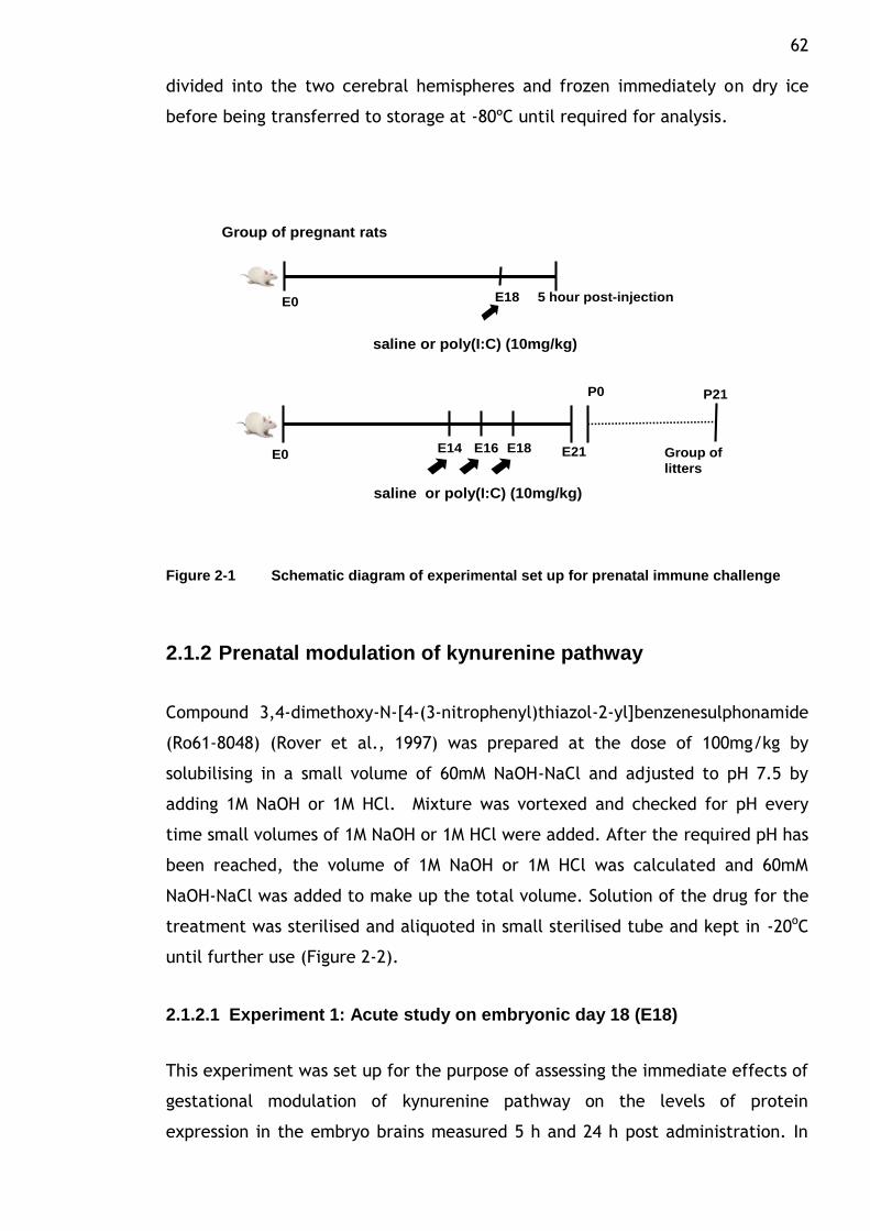
62 divided into the two cerebral hemispheres and frozen immediately on dry ice
before being transferred to storage at -80ºC until required for analysis.
Group of pregnant rats
P0 P21
E21E0E14 E16 E18 Group of
litters
saline or poly(I:C) (10mg/kg)
5 hour post-injectionE0 E18
saline or poly(I:C) (10mg/kg)
Figure 2-1 Schematic diagram of experimental set up for prenatal immune challenge
2.1.2 Prenatal modulation of kynurenine pathway
Compound 3,4-dimethoxy-N-[4-(3-nitrophenyl)thiazol-2-yl]benzenesulphonamide
(Ro61-8048) (Rover et al., 1997) was prepared at the dose of 100mg/kg by
solubilising in a small volume of 60mM NaOH-NaCl and adjusted to pH 7.5 by
adding 1M NaOH or 1M HCl. Mixture was vortexed and checked for pH every
time small volumes of 1M NaOH or 1M HCl were added. After the required pH has
been reached, the volume of 1M NaOH or 1M HCl was calculated and 60mM
NaOH-NaCl was added to make up the total volume. Solution of the drug for the
treatment was sterilised and aliquoted in small sterilised tube and kept in -20oC
until further use (Figure 2-2).
2.1.2.1 Experiment 1: Acute study on embryonic day 18 (E18)
This experiment was set up for the purpose of assessing the immediate effects of
gestational modulation of kynurenine pathway on the levels of protein
expression in the embryo brains measured 5 h and 24 h post administration. In
63 addition, the objective was to demonstrate the effect of the selected dosage in
producing changes in the levels of several metabolites of kynurenine pathway.
For this purpose, gestational day 18 timed-pregnant rats were assigned to
receive i.p. injections of the following treatment regimens: (1) 5 h group: saline
(0.9% NaCl) and (2) 5 h group: 100mg/kg Ro61-8048. After the injections the
animals were left undisturbed for 5 hours and then anesthetized with an
overdose of sodium pentobarbital and decapitated after blood was obtained by
cardiac puncture. The blood was collected in heparinised vacutainer tube and
kept in ice cold. The animals were dissected and the uterine horn containing
embryonic day 18 pups were surgically removed. Placenta, foetal‟s whole brain
and body were dissected and immediately frozen on dry ice. In another set of
experiment, two groups of pregnant rats were injected at gestational day 18
with single injection of vehicle solution and compound Ro61-8048 (100mg/kg),
respectively. Twenty four hours later, the animals were then sacrificed and
dissected to obtain foetal whole brain and body, placenta along with mother‟s
liver, brain and thymus. The levels of kynurenine and kynurenic acid from were
measured by high performance liquid chromatography (HPLC) with
electrochemical detection. HPLC analysis was solely performed by Dr Forrest
(Forrest et al., 2013a).
2.1.2.2 Experiment 2: Postnatal day 21 (P21)
For gestational treatment, groups of pregnant dams were administered
intraperitoneally (i.p.) with compound Ro61-8048 (100mg/kg) at the dose
indicated on days E14, E16 and E18 in order to extend the temporal impact of
kynurenine pathway modulation on the embryos, with no apparent ill effects on
the dam or her behaviour towards her litter after birth. Group of pregnant dams
were injected on E14, 16 and 18 between 9am and 11am to minimise variation in
responses. Control females were injected with the same volume of 0.9% NaCl.
Gestation was allowed to proceed normally and the offspring were undisturbed
until weaning on P21 when they were taken from the home cage for euthanasia
followed by removal of the brain. The long term effects of pharmacological
manipulation of kynurenine pathway during gestation would be studied at
juvenile age as to capture any maturational delay onset of the pharmacological
abnormalities. Each brain was divided into the two cerebral hemispheres and
64 frozen immediately on dry ice before being transferred to storage at -80ºC until
required for analysis.
2.1.2.3 Experiment 3: Postnatal day 60 (P60)
In a separate experiment to evaluate the extended effect of prenatal exposure
of compound Ro61-8048 in more mature animal, the neonates were being
allowed to survive until around 60 days of age (young adult) at which time all
litters were euthanized and their brains were removed. It is around this time of
age that many studies have shown behavioral changes resulting from prenatal
infection of mimetic agents. This study intended to explore the long term
functional consequences on pharmacological activities of the brain.The brain
was then sectioned into four main regions i.e hippocampus, cerebellum, cortex
and midbrain and preserved for further western blot (WB) analysis. Hippocampus
slices were prepared and examined for baseline excitability and other
electrophysiological analysis including excitatory synaptic potentials.
Electrophysiological analysis was exclusively performed by Prof Stone. Slices
were then kept frozen for examination of the selected panel of neurochemical
markers in order to detect differences in the CA1 region correlating with the
electrophysiological responses.
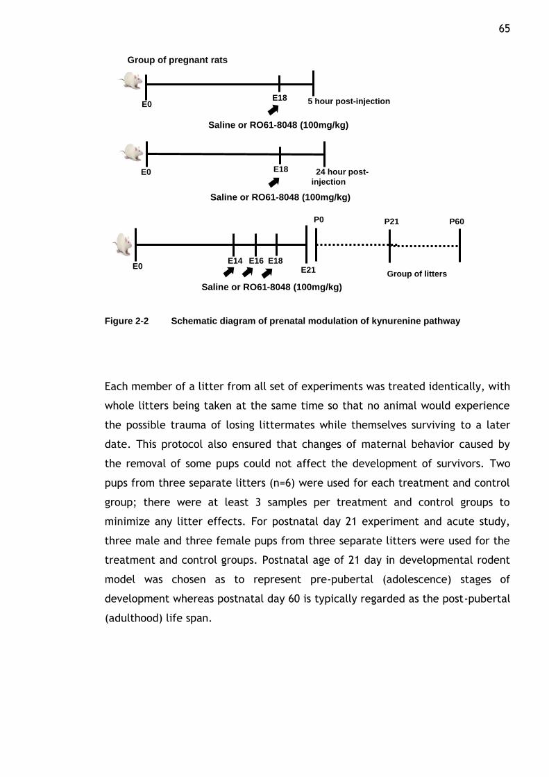
65
Group of pregnant rats
5 hour post-injectionE0E18
24 hour post-
injection
E0 E18
P0
E0E14 E16 E18
E21 Group of litters
P21 P60
Saline or RO61-8048 (100mg/kg)
Saline or RO61-8048 (100mg/kg)
Saline or RO61-8048 (100mg/kg)
Figure 2-2 Schematic diagram of prenatal modulation of kynurenine pathway
Each member of a litter from all set of experiments was treated identically, with
whole litters being taken at the same time so that no animal would experience
the possible trauma of losing littermates while themselves surviving to a later
date. This protocol also ensured that changes of maternal behavior caused by
the removal of some pups could not affect the development of survivors. Two
pups from three separate litters (n=6) were used for each treatment and control
group; there were at least 3 samples per treatment and control groups to
minimize any litter effects. For postnatal day 21 experiment and acute study,
three male and three female pups from three separate litters were used for the
treatment and control groups. Postnatal age of 21 day in developmental rodent
model was chosen as to represent pre-pubertal (adolescence) stages of
development whereas postnatal day 60 is typically regarded as the post-pubertal
(adulthood) life span.
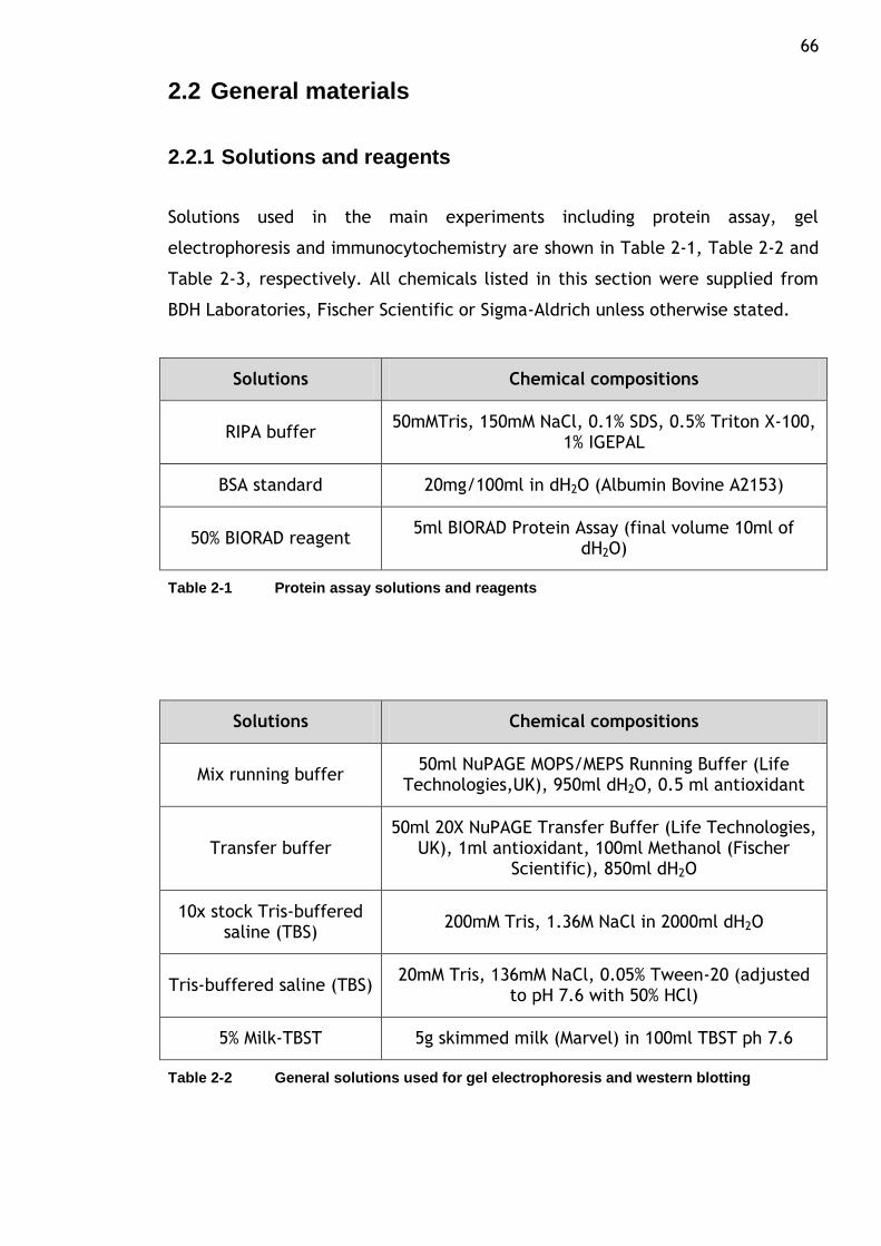
66
2.2 General materials
2.2.1 Solutions and reagents
Solutions used in the main experiments including protein assay, gel
electrophoresis and immunocytochemistry are shown in Table 2-1, Table 2-2 and
Table 2-3, respectively. All chemicals listed in this section were supplied from
BDH Laboratories, Fischer Scientific or Sigma-Aldrich unless otherwise stated.
Solutions Chemical compositions
RIPA buffer 50mMTris, 150mM NaCl, 0.1% SDS, 0.5% Triton X-100,
1% IGEPAL
BSA standard 20mg/100ml in dH2O (Albumin Bovine A2153)
50% BIORAD reagent 5ml BIORAD Protein Assay (final volume 10ml of
dH2O)
Table 2-1 Protein assay solutions and reagents
Solutions Chemical compositions
Mix running buffer 50ml NuPAGE MOPS/MEPS Running Buffer (Life
Technologies,UK), 950ml dH2O, 0.5 ml antioxidant
Transfer buffer 50ml 20X NuPAGE Transfer Buffer (Life Technologies,
UK), 1ml antioxidant, 100ml Methanol (Fischer Scientific), 850ml dH2O
10x stock Tris-buffered saline (TBS)
200mM Tris, 1.36M NaCl in 2000ml dH2O
Tris-buffered saline (TBS) 20mM Tris, 136mM NaCl, 0.05% Tween-20 (adjusted
to pH 7.6 with 50% HCl)
5% Milk-TBST 5g skimmed milk (Marvel) in 100ml TBST ph 7.6
Table 2-2 General solutions used for gel electrophoresis and western blotting
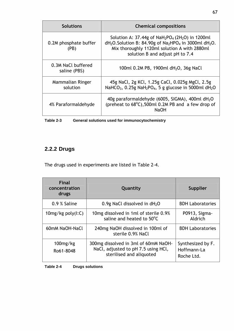
67
Solutions Chemical compositions
0.2M phosphate buffer (PB)
Solution A: 37.44g of NaH2PO4 (2H2O) in 1200ml dH2O.Solution B: 84.90g of Na2HPO4 in 3000ml dH2O.
Mix thoroughly 1120ml solution A with 2880ml solution B and adjust pH to 7.4
0.3M NaCl buffered saline (PBS)
100ml 0.2M PB, 1900ml dH2O, 36g NaCl
Mammalian Ringer solution
45g NaCl, 2g KCl, 1.25g CaCl, 0.025g MgCl, 2.5g NaHCO3, 0.25g NaH2PO4, 5 g glucose in 5000ml dH2O
4% Paraformaldehyde 40g paraformaldehyde (6005, SIGMA), 400ml dH2O
(preheat to 68oC),500ml 0.2M PB and a few drop of NaOH
Table 2-3 General solutions used for immunocytochemistry
2.2.2 Drugs
The drugs used in experiments are listed in Table 2-4.
Final concentration
drugs Quantity Supplier
0.9 % Saline 0.9g NaCl dissolved in dH2O BDH Laboratories
10mg/kg poly(I:C) 10mg dissolved in 1ml of sterile 0.9% saline and heated to 50oC
P0913, Sigma-Aldrich
60mM NaOH-NaCl 240mg NaOH dissolved in 100ml of sterile 0.9% NaCl
BDH Laboratories
100mg/kg
Ro61-8048
300mg dissolved in 3ml of 60mM NaOH-NaCl, adjusted to pH 7.5 using HCl,
sterilised and aliquoted
Synthesized by F.
Hoffmann-La
Roche Ltd.
Table 2-4 Drugs solutions
68
2.3 Western blot (WB) analysis
The western blot (WB) or also known as immunoblot is a semi-quantitative
method to detect a specific protein in biological samples, for example tissue
homogenate or extract. Generally, it involves multistep processes in which
samples are first separated by LDS-PAGE (lithium dodecyl sulphate-
polyacrylamide gel electrophoresis) and then the separated protein are
transferred to a membrane where they are incubated with an antibody specific
which binds to the protein band immobilized on the membrane (target protein).
The antibody is then visualized with a detection system that is usually based on
a secondary protein binding which is linked to a colour-yielding reaction.
2.3.1 Sample preparation
The preparation of the sample for protein analysis is the first step in the
complete workflow of WB. The main principle is to ensure that the sample is in
the best possible environment for analysis. Following prenatal administration of
compound, the offspring for each treatment group were killed by overdose
sodium pentobarbital (Euthatal) followed by decapitation. The brains were then
quickly removed and separated into two hemispheres. Each hemisphere from the
same animal was wrapped in a foil and marked before being stored at -80oC for
later use (for experiment 2). For experiment 3 of postnatal day 60, the brain was
removed rapidly from the skull and rinsed with cold artificial cerebro-spinal fluid
(ACSF) solution. The brain area under study was then separated into four main
region of interest which constitute of cerebellum, cortex, midbrain and
hippocampus. Fresh dissection of brain tissue has the advantage that particular
brain regions can easily be dissected based on visual information, such as
differences in colour of adjacent tissue and on the natural anatomical
boundaries of certain regions present in the brain. Examples of these are the
cerebellum that can be easily taken off from the medulla and pons and are
distinct in colour, and the hippocampus that differs from the occipital cortex by
colour and loosely connected to the thalamus by the fornix. For embryonic brain
dissection, the impregnated rats (E18) from respective treatment groups were
killed with overdose injection of Euthatal and a small incision was made over the
belly before a string of embryos were pulled out and transferred to a petri dish
69 on ice filled with ice-cold PBS. The embryos were dissected out of the uterus
wall and removed all the membranes including visceral yolk sac. The head was
separated from the rest of the body before being wrapped and kept frozen at -
80oC. Important consideration for successful sample preparation for the
subsequent assay analysis includes maintaining the sample at low temperature
throughout the processes and minimizing elapsed time in between procedure
(time from sacrificing an animal to homogenization of the sample) in which all
procedure steps were kept constant and as short as possible.
Careful control of presence of the target protein in the starting material and its
preparation and storage under non-degrading conditions is essential in preserving
the epitope abundance for antibody recognition. Standard and validated method
for the extraction purpose has been employed to provide good yields and
consistent results. For this purpose, brain tissues were lysed/homogenized using
a mechanical homogenizer in RIPA buffer containing 60mg TRIS, 87.6mg NaCl,
10mg SDS, 50μl TRITON X-100, 100 μl IGEPAL, 9850 μl distilled water to
encourage lysis of tissues and cells and to solublize proteins.The process of
proteolysis and denaturation of protein in the lysate were slowed down by
adding one tablet of protease inhibitor (Protease Inhibitor Cocktail tablets-
CompleteTM , Boehringer Mannheim GmBH, Mannheim, Germany).Protease
inhibitors are often added to prevent the digestion of the sample by its own
enzyme. Then, the whole lysate was centrifuged at 13 000 rpm for 5 minutes at
4oC and finally the supernatant was aspirated and aliquots kept at -80 oC prior to
use.
2.3.2 Protein concentration
Prior to western blot analysis, determination of protein concentration in each
sample was performed using the BIORAD protein assay system. Protein
concentration in the brain homogenates (half brain or regional area of the brain)
for western blot analysis was normalised against a standard curve of a known
protein content, Bovine Serum Albumin (BSA; Sigma-Aldrich) dissolved in an
appropriate volume of dH2O to give a concentration range of 0,0.25,0.50,1.0,1.5
and 2.0 (mg/ml). Then, brain samples dilution was prepared by diluting it twice
with dH2O at the ration of 1:10 and 1:100 (only hippocampal lysate was straight

70 diluted into 1:100). Bio-Rad protein reagent (Bio-Rad laboratories, UK) was
diluted 1:1 ratio before mixed thoroughly with each BSA standard reagents and
brain samples. Subsequently, each BSA standard and samples was pipetted in
duplicate into 96-well plate. An automated plate reader Opsys MR (Dynex
Technologies) was used to measure optical density at 595nm using the
BIORAD.assay program in Revelation Quicklink software (Dynex Technologies,
version 4.25). A linear curve fit was plotted using the reading from the BSA
standard concentration and was used to quantify the protein content of unknown
brain lysates. A calibration curve was established each time a protein assay was
performed with freshly prepared BSA standard dilutions. Using the standard
curve, the concentration of each sample was determined according to its
absorbance by interpolation. The measurements for each brain lysate from the
program were then multiplied by 10 (hippocampal lysates were multiplied by
100) to compensate for the dilution factor applied during the sample
preparation. Protein concentrations were then normalised to load 20-30 μg of
protein onto the gels.
2.3.3 Protein separation
To characterize immunodetection of the antibody (refer Table 2-5), western
blotting was carried out following procedures already established in the lab.
Prior to the electrophoresis process, proteins from the lysate of brain samples
(65% protein sample, 25% LDS sample buffer and 10% reducing agent) was
denatured by centrifugation and heating at 2000rpm for 2 minutes at 4oC and for
10 minutes at 70oC, respectively. Equal amount of proteins from each sample
(20-30 μg) was loaded into wells in the NuPAGE 4-12% (1.0mm) Bis-Tris
polyacrylamide gel 15 lanes (Invitrogen: Life Technologies, Paisley, UK). The
first lane on each gel usually reserved for a molecular weight marker: a mixture
of proteins with defined molecular weight (SeeBlue pre-stained standard (Life
Technologies, Paisley, UK). When voltage is applied along the gel, proteins
migrate at different speed and separate into bands based upon size and charge
(electrophoretic mobilities), characteristically smaller proteins migrate through
the gel faster than larger proteins. Proteins in the sample travel only in the
dimension along the gel for most blots. The separation of the protein which
employs polyacrylamide gels and buffers loaded with lithium dodecyl sulphate
71 (LDS) and treated with 10% reducing agents to sufficient final concentration was
electrophoresed through 4-12% Bis-Tris NuPAGE gel in 20X NuPAGE MOPS/MEPS
running buffer (Life Technologies,UK) at 150V for 100 minutes.
2.3.4 Protein transfer and blocking
The separated protein then electrophoretically transferred onto a hydrophobic
Invitrolon poly(vinylidene difluoride) (PVDF) membranes (Invitrogen: Life
Technologies, Paisley, UK). For this procedure, the stacking gel removed from
the separating gel and the back of the gel was placed on a wet filter paper
which had been soaked with transfer buffer. Thereafter the PVDF membrane was
applied on top of the gel (the membrane was activated by dipping into
methanol). Each layer was gently pressed using a small roller before the next
was applied to remove any air bubbles. The sandwich of membrane PVDF, gel,
filter papers and appropriate number of fiber pads were then stacked to gel
holder cassette and put in the Novex tank cell. Finally, the cell was filled up
with transfer buffer and an electric current at 220mAmp, 30 volts for 80 minutes
was applied to the gel so that the proteins transfer through the gel and onto the
membrane in the same pattern as they separated on the gel. To check the
uniformity and transfer efficiency of the transfer, membranes were rinsed in
dH2O and stained with Ponceau red dye (Sigma-Aldrich). Following transfer,
membranes were placed in blocking solution containing 5% (w/v) dried skimmed
milk powder (Marvel) diluted in Tris-buffered saline containing 0.05% Tween-20
(TBST) for at least one hour. The step was included to minimize background
staining due to non-specific binding of the antibody by saturating the proteins
which are not reacting with the antibody. The accessibility of a target protein
for primary antibody is achieved by washing the blocked membranes with TBST
for 3 times 15 minutes each, before incubation with primary antibody.
2.3.5 Western blotting: antibodies incubation
Evaluation of effect of maternal infection induced by poly(I:C) and also systemic
administration of kynurenine 3-monooxygenase inhibitor, Ro61-8048 were
studied on selected panel of neurodevelopmental markers that are involved in
many aspect of central nervous system (CNS) development. These target markers
72 are known to participate in various roles spanning from neural development,
neurite outgrowth, neurogenesis, synaptogenesis, neurotransmission and
intracellular signalling, among many others.
The primary antibody is the major determinant of the specificity of the target
recognition (Table 2-5). Trial set up of membrane with range of dilutions was
initiated to check reactivity and also specificity of the selected antibodies. The
optimal dilution of the primary antibody has to be determined experimentally
for the detection system used as different batch or source of the antibody may
even result in different recognition pattern when applied to the same sample
(commonly used dilution is between 1:500 – 1:10,000). The incubation with
primary antibody in blocking solution (5% milk-TBST) occurred at 4oC under
continuous shaking condition. Post incubation: the membrane was washed three
times with TBST solution for 15 minutes at room temperature (RT) to remove
unbound primary antibody. In order to detect the antibody which have bound,
the membrane was exposed to a secondary antibody reactive against the
respective primary antibody used (for instance: anti-rabbit to detect primary
antibody raised in a rabbit) (Table 2-6). This secondary antibody is known as
horse-radish peroxidise (HRP) conjugated antibody dissolved in blocking solution
and applied to the membrane and incubated for 1 hour at RT. All antibodies used
in this study were commercially available and have been used previously in
numerous regions of the human or rat brain as well as in vitro studies. Western
blot analysis was carried out using the following primary antibodies raised
against target proteins as listed in Table 2-5.
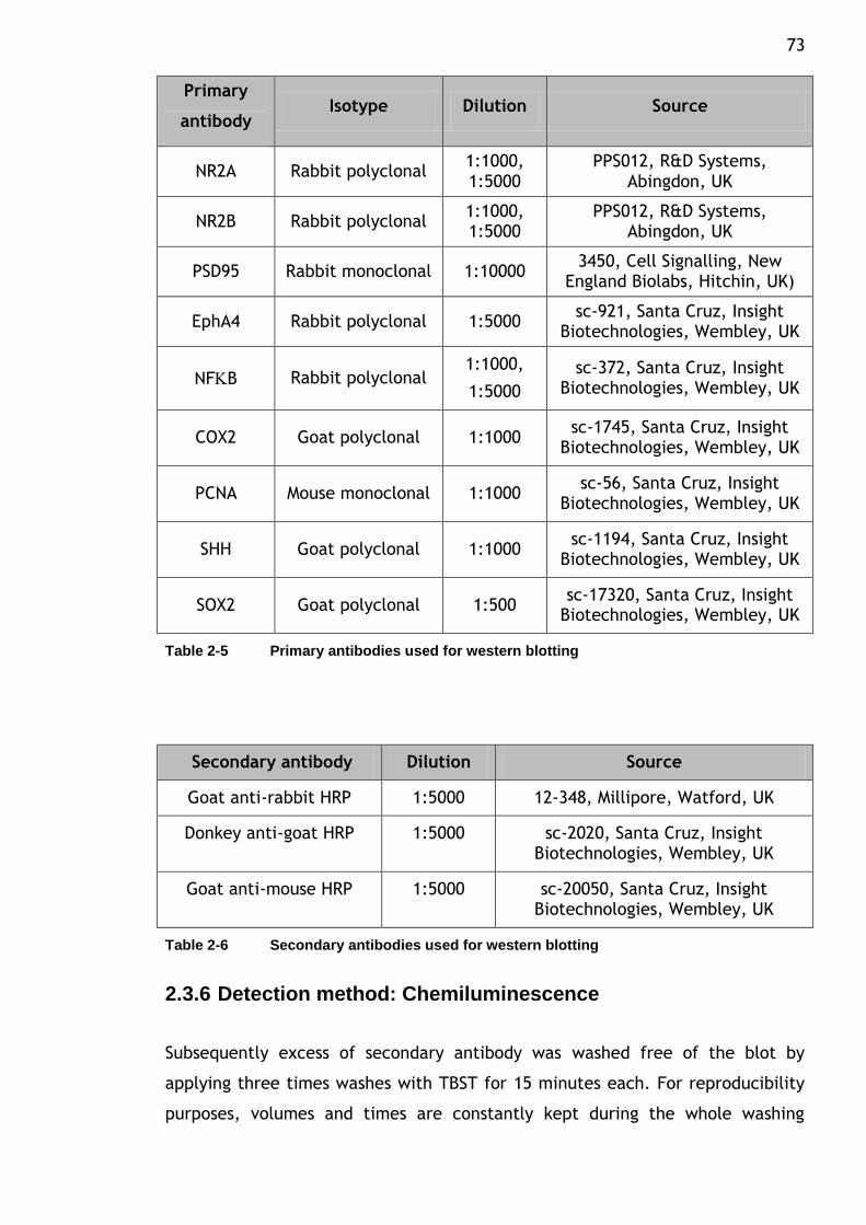
73
Primary
antibody Isotype Dilution Source
NR2A Rabbit polyclonal 1:1000, 1:5000
PPS012, R&D Systems, Abingdon, UK
NR2B Rabbit polyclonal 1:1000, 1:5000
PPS012, R&D Systems, Abingdon, UK
PSD95 Rabbit monoclonal 1:10000 3450, Cell Signalling, New
England Biolabs, Hitchin, UK)
EphA4 Rabbit polyclonal 1:5000 sc-921, Santa Cruz, Insight
Biotechnologies, Wembley, UK
NFB Rabbit polyclonal 1:1000,
1:5000
sc-372, Santa Cruz, Insight Biotechnologies, Wembley, UK
COX2 Goat polyclonal 1:1000 sc-1745, Santa Cruz, Insight
Biotechnologies, Wembley, UK
PCNA Mouse monoclonal 1:1000 sc-56, Santa Cruz, Insight
Biotechnologies, Wembley, UK
SHH Goat polyclonal 1:1000 sc-1194, Santa Cruz, Insight
Biotechnologies, Wembley, UK
SOX2 Goat polyclonal 1:500 sc-17320, Santa Cruz, Insight
Biotechnologies, Wembley, UK
Table 2-5 Primary antibodies used for western blotting
Secondary antibody Dilution Source
Goat anti-rabbit HRP 1:5000 12-348, Millipore, Watford, UK
Donkey anti-goat HRP 1:5000 sc-2020, Santa Cruz, Insight Biotechnologies, Wembley, UK
Goat anti-mouse HRP 1:5000 sc-20050, Santa Cruz, Insight Biotechnologies, Wembley, UK
Table 2-6 Secondary antibodies used for western blotting
2.3.6 Detection method: Chemiluminescence
Subsequently excess of secondary antibody was washed free of the blot by
applying three times washes with TBST for 15 minutes each. For reproducibility
purposes, volumes and times are constantly kept during the whole washing
74 procedure. The immunoblots were exposed to a substrate which will precipitate
upon reaction with the secondary, resulting in a visible band where the primary
antibody bound to the protein. For this step, enhanced chemiluminescence
solution from Thermo Scientific (Pierce® ECL2 Western Blotting Substrate) was
added onto the membrane and incubated for 5 minutes. Finally membranes were
placed into a cassette and developed using x-ray films (Kodak, UK) on a Kodak X-
OMAT 2000 processor with the exposure time varied depending on the antibody
used. In each case the aim was to achieve an optimal balance between signal-
background ratios.
2.3.7 Image analysis and membrane quantification
Protein intensity in each membrane developed on an X-ray film was quantified
by measuring their relative optical density (OD) using the analysis of gels options
in Image J software (http://rsbweb.nih.gov/ij/). First, the X-ray films developed
from chemiluminescence were scanned onto a computer in full colour using HP
Scanjet 4400cc Scanner and saved as Jpeg file. On Image J software, the
selected image was transformed into 8 bit image. The relative OD measurements
for control and treated samples run on the same gel were normalised to relative
OD of the β-actin signal within each lane, because treatment with either
poly(I:C) or Ro61-8048 at the desired dosage did not affect β-actin content in
the brain (data not shown).
2.3.8 Data analysis
The immunoreactive bands were detected using chemiluminescence and
quantification involved densitometry measurements where comparisons were
made statistically between groups of pups born to mothers treated (either with
poly(I:C) or compound Ro61-8048) and groups of pups born to mothers injected
with saline vehicle. Statistical analysis was performed using unpaired t tests to
examine differences between the two groups. The level for a statistical
difference in the data was set at P<0.05. Unless otherwise noted, statistical
difference is cited in the text when the data for the respective treatment group
were different from the data from control group. We have determined earlier
that there was no effect of gender on the treatment of Ro61-8048 at P21, the
75 data of the male and female offspring in each treatment group were combined
for the purpose of statistical analysis.
2.4 Immunocytohemistry (ICC) procedures
Immunocytochemistry is a technique that uses antibodies which target antigenic
receptors on the cell membranes. The reaction site can be identified in the
specimen and associated with specimen structure by attaching a microscopically
probe to the antigen-antibody complex. Thus, microscopical analysis by this
technique can therefore be used to obtain a unique view of macromolecular
changes within individual cells. ICC is widespread in use for light and electron
mircroscope studies in almost every area of biological research from routine
diagnostic work through to more complex research projects. The feasibility of
the technique in living specimens, making it possible to study dynamic
processes, such as gene expression, molecular binding interactions and as part to
understand the distribution and localization of specific molecular markers in
different parts of a tissue (Beesley, 1993).
2.4.1 Animals and operative procedures
Groups of litter from saline and Ro61-8048-treated animals (n=7-9/group) were
killed at postnatal day 60s after gestational treatment at E14, 16 and 18. Both
control and Ro61-8048-treated animals were deeply anesthesized using Euthatal
(sodium pentobarbiturate) before transcardially perfused via the left ventricle.
In general, conventional tissue fixation protocols for microscopy analysis involve
the use of fixative solution, which should allow excellent morphological
preservation. The fixation procedure needs to be a compromise between good
morphological preservation and the conservation of antigenicity; ideally the
composition of the fixative should ensure both. Furthermore, an ideal fixative
should stabilize and protect tissues and cells from the damaging effects
associated with subsequent treatment. There are a number of fixation
procedures used, which are based on various fixatives such glutaraldehyde,
formaldehyde and also ethanol, methanol, acetone and formalin. In this respect,
the best concentration of fixative we used in all animal study for ICC procedure
is 4% parformaldehyde (PFA), which is less damaging to many antigens and give a
76 good compromise between retention of morphology and antigenicity. After the
animal has been anesthesized, mammalian Ringer solution was flushed through
each animal to dilate blood vessels and prevent clotting and fixation of the
tissues was followed by perfusion with approximately (850ml to 1000ml) of
freshly made, warm 4% paraformaldehyde (PFA) (Sigma-Aldrich 16005) in 0.2M
phosphate buffer (PB), pH 7.2. Right after perfusion, the brains were removed
from the skull and postfixed the same fixative solution for another 4 hour at 4oC.
After thorough rinsing in 0.1M phosphate buffer, the brains were protected in a
30% sucrose solution where they remained until equilibrium. In our laboratory,
samples were fixed, stored in phosphate buffer for up to one month and
subsequently processed and successfully immunolabelled. It should be noted
that prompt fixation after perfusion is essential to achieve consistent results.
Prolonged fixation for the tissue samples were also avoided as many antigens
show a progressive reduction in immunoreactivity with increasing fixation times
by producing cross-linking with protein that could diminish the epitopes for the
applied antibodies. Therefore, 4 hour post fixation with PFA for adult brain
tissue is thought to be optimal that can give adequate untrastructural
preservation.
2.4.2 Tissue processing
Vibratome is an excellent basic vibratory tissue sectioning system. It cuts fresh
or fixed, animal or plant tissue without freezing or embedding. Brain samples
were divided into two hemispheres and cut to an appropriate size, embed in 5%
agar and cooled for a few minutes before mounted on the vibratome base plate
with the aid of super glue to stabilize the specimen. All the parameters were set
accordingly; section thickness, speed and angle of the sectioning blade. Initial
sectioning was performed one or two times to obtain a flat specimen surface. As
section quality is influenced by several factors, the setting parameters of the
vibratome (Leica 1000) were appropriately checked when performing sectioning.
In relation to brain tissue, setting parameters of: low bath temperature (a
physiological compatible buffer: 0.1M PB), slow sectioning speed (setting 1-2),
section thickness (60 µm) and low steep blade angle were adopted. After initial
sectioning, sixty-micrometer thick free floating sections were then cut coronally
through the entire hippocampus and collected serially in a series of tubs filled
77 with 0.1M PB. The sections were incubated with 50% ethanol for 30 minutes to
permeabilize and improving the penetration of the antibody, before 3 times
washed with 0.3M PB and stored at -20oC in glycerol.
2.4.3 Antibody incubation and staining procedure
For analyses of hippocampal neurogenesis, serial free-floating sections were
first washed in 0.3M phosphate buffer saline (PBS) before incubated with
optimally diluted primary antibodies in 0.1% Triton in PBS (PBST; pH 7.4) for 72 h
at 4oC under continuous shaking condition. The major consideration when
choosing the antibodies for multiple immunolabelling is the the possibility of
cross reactions between reagents. In this study we adopted simultaneous
multiple immunolabelling using antibodies raised in different species.
Immunolabelling was performed by incubating the specimen with mixtures of
chosen antibodies followed by mixtures of appropriate secondary probes. The
optimal concentration of each antibody used in this study was optimized
individually to obtain maximal contrast between specific signal and background
staining. The mixture of primary and secondary antibodies were diluted in tris-
buffered saline (TBS) containing gentle detergent (Tween 20) with the purpose
to help keeping the tissues/cells permeabilized. Primary antibodies used in our
study were monoclonal mouse anti-NeuN (1:500; MAB377 Millipore, Temecula
CA92590), polyclonal goat anti-Doublecortin (DCX, 1:250; sc-8066 Santa Cruz
Biotechnology, Santa Cruz, CA) and polyclonal rabbit anti-SHH (1:100; sc-9024
Santa Cruz Biotechnology, Santa Cruz, CA). Rinses were performed between all
steps using 0.3M PBS before or after the primary and secondary antibody
incubation.
Following primary antibody treatment, sections subsequently incubated
overnight at 4oC in species specific Alexa Flour-tagged secondary antibodies:
Alexa 488 (1:500), Alexa 647 (1:500) (Molecular Probes) and Rhodamine-
conjugated secondary antibody (1:100) (Jackson Immunoresearch Laboratories
Europe Ltd.). The uses of conjugated antibody flourochromes have been
successfully used for multiple immunostaining. The fluorescent probes are
particularly useful since each probe can be visualized separately without
hindrance from the other by use of different wavelengths of light for excitation
78 and detection. Sections were mounted and coverslipped with an aqueous
mounting medium for flouresence H-1000: Vectashield (Vector Laboratories Inc.
Burlingame, CA 94010). Covering the sections with a glass coverslip preserves
and protects the section and improves optics. Special mountant containing
fluorescent anti-fades was used to protect against photobleaching effect.
Another aspect when choosing mounting medium is to match its refractive index
with objective immersion medium, it is to avoid incorrect depth discrimination
and spherical aberration which could lead to loss of light at the detector (Smith,
2008) In all experiments designed to compare the immunolabelling in saline-
control and Ro61-8048 animals, sections from the two groups of animals were
processed identically and in parallel for each step of the immunocytochemical
procedures. Considering that there was a possible high risk of cross
contamination in all multiple immunolabelling schedules, even if totally
different species were used, a control specimen from each animal with omission
of primary antibodies is included and tested in parallel with the samples. The
specimens can be kept at 4oC if the material is going to be processed within 1-2
weeks as fluorescence may last up to 2-3 weeks, long term storage should
preferentially be at -20oC in a covered boxes. However for best results the
specimen should be viewed as soon as possible using confocal microscopy.
2.4.4 Confocal microscopy and image processing
Over the years, confocal microscopy has developed to form as essential research
tool for assessing immunocytochemical preparations. The most common
application of confocal microscopy is to compare the distributions or behavioural
characteristic of multiple probes in the same tissue samples and also reveal the
three dimensional structure of the specimen reconstructed from a series of
optical sections at different depths (Smith, 2008). Flourescent signals from each
section were examined and photographed using an inverted confocal laser
scanning microscope (Nikon Eclipse TE300) equipped with RADIANCE2100 BIORAD
and Lasersharp 2000 software. In principal, laser scanning microscope captures
images by scanning the specimen with a focussed beam of light from a laser and
collecting the emitted fluorescent signals with a photodetector. This is made
possible by the development of the system that are capable of efficiently
detects multiple flourophores either simultaneously or sequentially by its laser
79 optics with a wide range of wavelengths from ultraviolet through the infrared.
The system employs a pair of pinhole apertures to limit the specimen focal plane
by blocking the light from out of focus areas thus determines the exact emitted
fluorescence of the focal spot in the specimen (Smith, 2008). Relatively thick
specimens can be imaged in successive volumes by acquiring a series of sections
along the optical (z) axis of the microscope. Moreover, the use of oil-immersion
objective that have similar refractive index with mounting medium for
specimens would ensure better image quality.
In this experimental set up, stacks of 20 images were taken through the z-axis
using a 40X oil objective and 10X magnification and a frame box of 289µm (x) X
289µm (y) X 20µm (z) (1024x1024 pixels).Setting parameters that needs to be
controlled and standardized throughout the scanning include photomultiplier
gain for the channels, scan line speed, brightness and pinhole aperture size. By
properly adjusting the pinhole size, separation of the background out of focus
signal can be achieved thus improves image quality. Whereas the scan speeds
slider is adjusted to achieve the maximum scan rate with a minimum amount of
signal noise. As the scan speed is increased, a corresponding increase in the
amount of background noise is also captured by the photomultipliers (detector).
By using 20x magnification the regional areas (CA1, CA3 and dentate gyrus) were
delimited by morphological criteria and under 40x magnification objective the
dimensions of the area in the specimen were scanned. Within this dimension
only immunopositive cells showing a stained unit area sufficient to ensure that
the majority of their volume was contained within the selected area were scored
for counting purposes. At least 3 coronal sections per animal were
immunolabelled and in every section, two to four images were taken followed by
image processing to optimally visualize the staining area. Immunopositive cells
from the pyramidal layer of CA1 and CA3 as well as granule cell layer of the
hippocampus were quantified using a number of gridded counting frames that
were within the specific subregions of the hippocampus and cerebellum with
each individual counting frame measured 50 x 50 µm. The counting of the DCX,
SHH and NeuN positive cells was conducted in a total of 15 grids of area for
dentate gyrus and CA1 regions, 25 grids for CA3 region and grid sizes of 4 x 4 for
cerebellum. For all the selected subregions, we calculated the number of
immunopositive cells that were inside the counting frames for each section and
80 the total number of immunopositive cells across all counting frames was
averaged to give one mean value per animal per subregions. Cell quantification
was performed with adherence to several characteristics which includes (i) the
presence of nucleus, (ii) immunopostive cells were of homogenously shape and
(iii) labelling intensities of immunopositive cells were distinguishable from
background staining.
2.4.5 Data analysis
All cell counts were performed blindly with respect to treatment, using
immunolabeled sections reacted with selected antibodies. The data set obtained
by capturing a series of optical sections through the specimen was used to
compute views of the specimen from different viewing angles. Each subregion of
the hippocampus (CA1, CA3 and DG) and cerebellum was analysed using Nikon
Eclipse TE300 microscope. For counting purpose, a two-dimensional display of
both z-series projection and xy projection formed by merging multiple image
planes were conducted. The most common type of projection is a maximum
projection in which each pixel represents the intensity of the brightest pixel in
the z-axis. The cell number analysis was done on a total of 3 sections per animal
with at least 2-3 observations per section using ImageJ software (Version 1.43M;
National Institute of Health). Immunopositive cells count of 3 sections was
averaged to obtain a single value for the number of positive- stained cells per
animal. A mean ± standard error (SE) was calculated from the date in each group
(n= 7-9 animals).We chose to use mean cell counts as representative of
immunopositive labelling as this protocol would permit valid comparisons among
treatment groups. Sections from animals with faint immunolabelling for
respective antibody were excluded from statistical analyses, producing some
variability in sample sizes across groups.
2.5 Golgi staining
2.5.1 Animals and operative procedures
Pregnant rats were injected with compound Ro61-8048 (100mg/kg) on embryonic
days 14, 16 and 18, and gestation was allowed to proceed normally. Both male
81 and female pups were included in this study and they remained with their
mothers until weaning at postnatal day 21 (P21) and were assessed at postnatal
day 60 for the post-pubertal or adult group. Control animals were born from
saline-treated mothers and were evaluated at the same age. At postnatal day
60, the pups from respective group of litters (4 pups per litter) were deeply
anesthetized with sodium pentobarbital before cervical dislocation.
2.5.2 Tissue processing and staining procedure
The brains were then rapidly removed, rinsed briefly in dH2O and straight away
immersed in impregnation solution at room temperature for 2 weeks in the dark.
Golgi-Cox staining was performed using the FD rapid GolgiStain kit (FD
NeuroTechnologies) as per the manufacturer‟s instructions. The impregnation
solution was made by mixing equal volumes of solutions A and B at least 24 hours
prior to use and left unstirred at room temperature. The impregnation solution
was refreshed after initial 24 hour of immersion. The tissue is then transferred
into solution C and stored at 4oC for at least 48 hour in the dark. The solution
was replaced after the first 24 hour of immersion.
Coronal sections of 200m thickness were obtained using a vibratome (VT1000s,
Leica). Each section was then mounted on gelatin-coated microscope slides
(3 sections/slide). A tiny drop of solution C was placed onto the mounted tissue
before being dried naturally in the dark at room temperature. For the final
development process, slides were arranged in a slide rack and washed twice in
dH2O for 2 minutes each. Then the slide rack was placed in a mixture consisting
of one part solution D, one part solution E and two parts dH2O for 10 minutes
with gentle agitation every 2 minutes. The sections were rinsed two times with
dH2O, four minutes each and followed with dehydration process with graded
concentration of ethanol in successive baths of 70% (4 minutes), 90% (4 minutes)
and 100% (3 x 4 minutes). Subsequently, the sections were dipped in Histoclear
solution (2 x 4 minutes) before cover slipped with histological mounting medium
(Histomount, ThermoFisher Scientific).

82
2.5.3 Morphological and data analysis
The ability to obtain good images of neuronal processes allowed us to use this
technique for morphological and quantitative studies using appropriate image
analysis software. From each group, three litters with four pups in each litter
were used for dendritic morphology study in the hippocampus. Brain region
identification was aided by a standard atlas of the rat brain. Golgi-stained
principal cells in the dentate gyrus, the granule cells were then visualized by
light microscopy (Nikon Eclipse E400, Nikon Instruments, Inc., NY, USA). Under a
40x objective lens, Golgi-impregnated dentate granule neurons were identified
by their location within granule cell layers and by the shape of their soma and
dendritic arbor. Dendrites were visualized under a 40x objective blindly to the
pretreatment group for manual tracing and dentate granule cells quantification.
A minimum of one neuron in each hippocampus section were drawn individually.
Based on those criterion mentioned above, as a whole for each group of
treatments, a total of 68 neurons from control and 69 neurons from Ro61-8048-
treated animals were traced in their entirety. Fully stained cells within the
panel were first visualized under minimal magnification (4 x objectives). Thus,
each examined granule cell body was carefully viewed at various magnifications
(4x, 20x and 40x) to determine the continuity of processes in its vicinity. By
carefully adjusting the fine focus, it was possible to determine whether basal
dendrite was present and to distinguish a passing dendrite from one that was
attached to the examined cell body. To ensure consistency of sampling the
following criteria were applied: (1) granule cells exhibited vertically orientated
dendrites that extended into the dentate molecular layer; (2) granule cells did
not have cut off dendrites in close proximity to the cell body and (3) dendrites
had negligible overlap with the dendrites of neighbouring cells so that all
dendritic branches could be traced explicitly. In addition to those parameters,
we also generally observe other morphological criteria prior to the selection of
the neurons such as integrity of the selected neuron, homogenous impregnation
(dark and consistent impregnation seen throughout the extent of the dendrites)
and relative isolation from the blood vessels and deposits of other impregnated
cells located nearby (Zhao et al., 2006). Morphometric parameters included
were the total number of branches and total dendrite length and used to assess
83 the overall magnitude of morphological change following prenatal modulation of
kynurenine pathway. The number of dendritic branches was calculated manually
from the camera drawings. The dendritic branches originating directly from cell
bodies were designated as first order or primary segments and until they
branched off into second order segments (secondary).To measure the extent of
dendritic growth away from the soma and the branching of dendrites at different
distances from the soma, the concentric circle analysis of was performed (Sholl,
1953, Gibb and Kolb, 1998). A transparent grid with equidistant 20m was
centred over the cell soma and the number of ring intersections was used to
estimate the total dendritic length and dendritic arborisation. The branching
complexity of dendritic trees was evaluated by applying the method of
concentric rings. Rings were calculated at 20µm intervals (0-20, 20-40, 40-60,
60-80, 80-100, 100-120, 120-140). Sholl analyses were conducted on the mean
values per animal for the above morphometric parameters. In Sholl analysis, the
number of concentric ring intersections was used as an estimate of total
dendritic length. While dendritic branching was also measured by counting the
total number of dendritic branches at each order away from the cell body.
Graphically, this generates a profile which can be statistically compared with
other profiles from other groups. Morphological data were analysed using an
unpaired t-test to assess differences in the number of dendritic branches and
their length between control and Ro61-8048 treated groups of animals. The
analysis of dendritic complexity (Sholl, 1953) was subjected to a two-way
analysis of variance (ANOVA) followed by Tukey‟s post hoc analysis to examine
the contributions of drug treatment, sample number and distance from the cell
soma. A probability value of 0.05 was taken as the criterion for significance.
84
3 Effect of maternal immune activation on neurodevelopmental markers expression in foetal and postnatal day 21 (P21) brain
3.1 Introduction
A body of evidence has indicated that prenatal immune activation causes a wide
spectrum of structural and functional abnormalities implicated in a diverse
range of disease process including AIDS-dementia complex, schizophrenia,
autistic spectrum disorder, among many others. However the neurodevelopment
mechanisms and biochemical changes underlying these abnormalities are not
definitively established. The most widely debated possibility is that a maternal
antiviral antibody in some genetically predisposed mothers may cause an
immunological reaction that is harmful to the foetal brain (Gilmore and Jarskog,
1997). Maternal inflammation that caused a disruption to the maternal
environment has been linked to the above disease states in adult life. Several
reports showed that activation of the immune system and increased levels of
cytokines during gestation affects the development of brain areas involved in
social interaction and memory which later results in a schizophrenia-like
phenotype (Ashdown et al., 2006, Hsiao and Patterson, 2012). This finding is also
supported by epidemiological studies that demonstrate an association between
maternal infection and the development of schizophrenia (Brown et al., 2004).
The use of animal models to model maternal immune activation has provided
insights into the host‟s response to an immunological stimulus, as well as the
acute and lasting effects of immune activation on offspring development.
Precise manipulation of a number of variables of interest, such as the immune
stimulus, route of administration, dosage and timing of exposure, allows for the
testing of various hypotheses regarding the mechanisms of action on the
developing foetal brain. The most well known cause of inflammation during
pregnancy is due to infection by a pathogen. As previously discussed in the
introduction, the two major maternal immune activation models use either LPS
or poly(I:C) administered to pregnant dams, although there are a number of
other additional models which have used the human influenza virus and
85 individual cytokines such as TNF-α or IL-6 (Boksa and Luheshi, 2003, Boksa,
2010).
Despite the large number of studies describing abnormalities in rodent brains
following exposure to prenatal immune stimulus, the precise molecular
mechanisms by which infection during gestation and the effect of maternal
immune response on the developing central nervous system remain unclear. The
fact that a wide variety of pathogens are able to increase schizophrenia risk in
the offspring suggests that these pathogens alter foetal development by one or
more common mechanisms. A major causal link underpinning this relationship is
thought to be the cytokines released following a maternal immune response to
infection (Ashdown et al., 2006, Brown, 2006). Maternal immune activation is
thought as a plausible mechanism due to all of the implicated pathogens
triggering a maternal immune response, which will alter the biochemical
environment of the foetus in a way that increases the risk for
neurodevelopmental disorders. Despite the postnatal onset of clinical symptoms,
evidence suggests that schizophrenia have their origins in early brain
development. In line with the neurodevelopmental hypothesis of schizophrenia,
which postulates that neuronal development is affected by events that modify
physiological course of maturation in neuronal system. Therefore, maternal
immune activated rodent models have been particularly helpful for examining in
vivo effects of the maternal immune system on the developing foetal and
postnatal brain (Patterson, 2009). These models predominantly involve
administration of either bacterial lipopolysaccahride (LPS) or double-stranded
viral RNA (polyI:C) to pregnant rats or mice. This induces an immune response,
which increases circulating levels of cytokines without the confounding effects
of the pathogen itself. The offspring of mothers with infections display diverse
neuropathology, depending in part on the nature and timing of the infection.
Offspring of these immune activated have been reported to show a
„schizophrenia like‟ phenotype including deficits in prepulse inhibition (PPI),
latent inhibition (LI), anxiety, locomotion and social interaction (Meyer et al.,
2008c, Meyer et al., 2008a, Ito et al., 2010, Shi et al., 2003).
86
3.2 Research aims
Based on the in vitro evidence of pro-inflammatory cytokine action in the brain,
it is hypothesized that the presence of cytokines in the foetal brain as a result of
maternal infection could result in altered brain neurochemical development.
The main aim was to investigate if exposure to viral mimetic poly(I:C) during
early life can affect the neurochemical balance of a selected panel of
neurodevelopmental markers in the young offspring that could undermine
normal brain development and predispose the offspring to developing psychiatric
illness.
3.3 Experimental approaches
Exposure to a wide range of viruses pre-or perinatally has been implicated in the
pathogenesis of neuropsychiatric disorders. To this aim, a synthetic analog of
double stranded RNA (polyriboinosinic-polyribocytidilic acid, [poly(I:C)] was
administered intraperitoneally in mouse dams on embryonic days (E) 14, 16 and
18 in order to extend the temporal impact of maternal immune activation with
no apparent ill effects on the dam or her behaviour towards her litter after
birth. Administration of this agent to rat dams at three different time points
during pregnancy (E14, E16 and E18) correspond roughly to the later periods
during the second trimester of human pregnancy. In another set of experiments,
poly(I:C) was administered to pregnant dams on embryonic day 18 (E18) as to
demonstrate the effect of the immune activation in terms of changes in the
levels of cytokine in the maternal plasma as well as protein expression in the
embryo. Poly(I:C) is used experimentally to model viral infections because it
stimulates the antiviral activities of the innate immune system including
cytokine production, without the confounding effects of viral infection. We
have decided to focus on poly(I:C) as we find it as a more reliable and consistent
activator of the immune system and also because viral rather than bacterial
infections predominate in human pregnancy. Injection at a dose of 10mg/kg of
poly(I:C) is the highest dose which can be given to a pregnant mouse with no
signs of acute stress, behavioural changes or abnormal behaviour towards the
neonatal pups. Preliminary work on the neurochemical analyses showed little
changes in the selected molecular markers at postnatal day 2 (P2) with a
87 significant increase of EphA4 at P7 and a reduction of both EphA4 and VAMP-1
expression at P21. In this study, we therefore concentrated on the molecular
analyses of brain samples collected from E18 and P21. These two developmental
time-points were aimed at capturing early and later/persisted changes in protein
expression levels. It is therefore of considerable interest to evaluate biological
markers that are involved in the early development of the brain. The goal of the
study is to assess differences in the expression profiles of selected
neurochemical markers after poly(I:C) induced-maternal immune activation and
from unchallenged normal mice by analyzing the activated expression signalling
pathways following infection.
3.3.1 Animals and drug preparation
All procedures on animal work was performed in accordance with the guidance
established by the Animal (Scientific Procedure) Act 1986 and has been approved
by the University of Glasgow Scientific Ethics Committee. Wistar rats were
mated at an age of around 3 months and the first day after the occurrence of
vaginal plug (an indication of copulation) was defined as day 1 of pregnancy. The
pregnant dams were housed alone under pathogen-free environmental conditions
at a constant temperature with food and water ad libitum. Potassium salt
poly(I:C) (P0913: Sigma (Poole, UK)) was dissolved in sterile isotonic 0.9% w/v
NaCl solution and then heated to 50oC to allow for complete solubility. The
mixture was allowed to cool to room temperature to allow re-annealing of the
double-stranded RNA. The desired dosage for the injection was 10mg/kg and it
has been established in previous experiments in the lab that the dose could be
given to pregnant dams during the last seven days of gestation with no signs of
stress, behavioural changes or abnormal behaviour towards the neonatal pups
after birth (Forrest et al. 2012). The solution for the drug treatments was
sterilised and aliquoted in small sterilised tube and kept in -20oC until further
use.
3.3.1.1 Experiment 1: Acute study on embryo
For the acute study, gestational day 18 timed-pregnant rats were injected i.p.
with 10mg/kg of poly(I:C). Age-matched pregnant rats were injected with the
same volume of saline (0.9% NaCl). This experiment was set up for the purpose
88 of assessing the acute effects of maternal poly(I:C) administration on several
cytokine and chemokine levels in maternal blood. After the injections the
animals were left undisturbed for 5 hours and then anesthetized with an
overdose of sodium pentobarbital and decapitated after blood was obtained by
cardiac puncture. The blood was collected in a heparinised vacutainer tube and
kept ice cold. The animals were dissected and the uterine horn containing
embryonic day 18 pups were surgically removed. The placenta, foetal whole
brains and bodies were dissected and immediately frozen on dry ice. The
collected whole brains were used in the western blot analysis. Maternal liver,
thymus and brain were also dissected for further analysis for example HPLC
analysis. ELISAs for detecting cytokines and HPLC analysis was performed by
respected colleague Dr Forrest and some findings are mentioned in the text.
3.3.1.2 Experiment 2: Postnatal day 21(P21)
For gestational poly(I:C) administration and vehicle treatment, groups of
pregnant dams were administered intraperitoneally (i.p.) at the dose 10 mg/kg
on days E14, E16 and E18 in order to extend the temporal impact of maternal
immune activation on the embryos, with no apparent ill effects on the dam or
her behaviour towards her litter after birth. Repeated systemic challenges
during this time point was meant to examine the effect or repeated viral
stimulation on the progression of neurological changes at postnatal day 21 since
this represents pre-pubertal stage of development. Group of pregnant dams
were injected on E14, 16 and 18 between 9am and 11am to minimise variation in
responses. We have shown that treatment of pregnant dams with poly(I:C) at the
dose indicated did not affect litter size and pup body weight and could be given
during the last seven days of gestation with no sign of stress, behavioural
changes of abnormal behaviour towards the neonatal pups after birth. The
dosage regime at 10mg/kg was well tolerated in our experimental approach and
could provide the maximum advantage for observing any changes in the
offspring. In the control group, pregnant dams were injected with the same
volume of 0.9% NaCl. Gestation was allowed to proceed normally and the
offspring were undisturbed until weaning on P21 when they were taken from the
home cage for euthanasia followed by removal of the brain. Each brain was
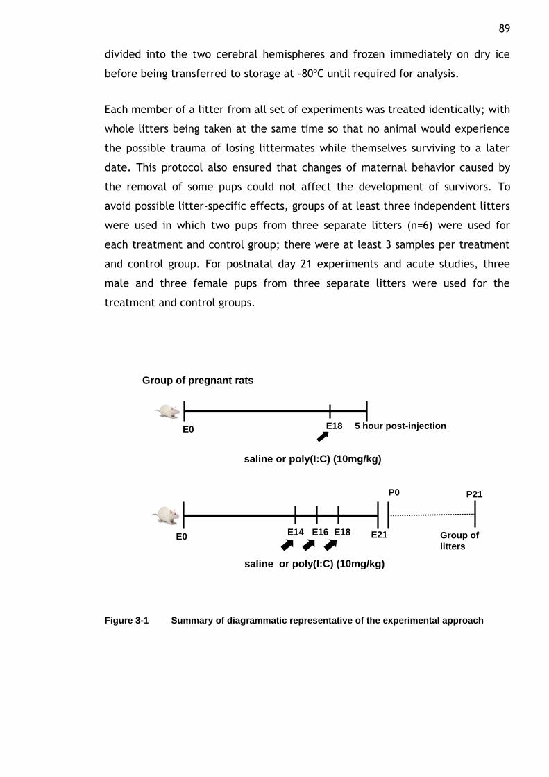
89 divided into the two cerebral hemispheres and frozen immediately on dry ice
before being transferred to storage at -80ºC until required for analysis.
Each member of a litter from all set of experiments was treated identically; with
whole litters being taken at the same time so that no animal would experience
the possible trauma of losing littermates while themselves surviving to a later
date. This protocol also ensured that changes of maternal behavior caused by
the removal of some pups could not affect the development of survivors. To
avoid possible litter-specific effects, groups of at least three independent litters
were used in which two pups from three separate litters (n=6) were used for
each treatment and control group; there were at least 3 samples per treatment
and control group. For postnatal day 21 experiments and acute studies, three
male and three female pups from three separate litters were used for the
treatment and control groups.
Group of pregnant rats
P0 P21
E21E0E14 E16 E18 Group of
litters
saline or poly(I:C) (10mg/kg)
5 hour post-injectionE0 E18
saline or poly(I:C) (10mg/kg)
Figure 3-1 Summary of diagrammatic representative of the experimental approach
90
3.3.2 Western blot analysis
The preparation of the sample is the first step in the complete protocol for
western blotting. The main principle is to ensure that the sample is in the best
possible environment for analysis. For this purpose, brain tissues were
lysed/homogenized in RIPA buffer containing 60mg TRIS, 87.6mg NaCl, 10mg
SDS, 50μl TRITON X-100, 100 μl IGEPAL, 9850 μl distilled water, with the process
of proteolysis and denaturation of protein in the lysate slowed down by adding
one tablet of protease inhibitor (Protease Inhibitor Cocktail tablets- CompleteTM
, Boehringer Mannheim GmBH, Mannheim, Germany). The whole lysate was then
centrifuged at 13 000 rpm for 5 minutes at 4oC and finally the supernatant was
aspirated and aliquots kept at -80 oC prior to use. Total protein concentrations
for all samples were determined using the BIORAD protein assay system. This
was achieved by comparing each brain lysate against a standard curve of known
protein content – Bovine Serum Albumin (BSA: Sigma-Aldrich) dissolved in dH2O
to obtain a concentration range 0, 0.25, 0.5, 0.5, 1.0, 1.5 and 2.0 (in mg/ml).
The brain lysate (diluted 1/10) and BSA standard was mixed with diluted BIO-
RAD protein assay dye reagent (1:1 in dH2O) (BIO-RAD Laboratories, UK).
Subsequently each standard and brain lysate was pipetted twice into a 96-well
plate and the absorbance measured with an Opyss MR plate reader (Dynex
Technologies,UK) using the BIORAD.assay program of Revelation Quicklink
software (Dynex Technologies, version 4.25) at a wavelength of 595nm. Protein
concentrations were then normalised to load 20-30 μg of protein onto the gels.
To characterize immunodetection of the antibody for the selected markers
mentioned above, western blotting was carried out following procedures already
established in the lab. Prior to the electrophoresis process, proteins from the
lysate of brain samples (65% protein sample, 25% sample buffer and 10% reducing
agent) was denatured by centrifugation at 2000rpm for 2 minutes at 4oC and
heated for 10 minutes at 70oC. Equal amount of proteins from each sample (20-
30μg) was separated according to molecular weight on NuPAGE 4-12% (1.0mm)
Bis-Tris polyacrylamide gels 15 lane gels (Invitrogen: Life Technologies, Paisley,
UK) for 80 min at 150 volts. SeeBlue pre-stained standard (Life Technologies,
Paisley, UK) was included on each gel as a molecular weight marker. The
separated proteins were then electrophoretically transferred onto hydrophobic
91 Invitrolon poly(vinylidene difluoride) (PVDF) membranes (Invitrogen: Life
Technologies, Paisley, UK) for 60 min at 30 volts. The membranes were
incubated for 1 hour at room temperature with 5% non-fat dried milk in Tris-
buffered saline (TBS: 20mM Tris Base and 0.14M NaCl, pH 7.6) containing 0.05%
Tween-20 to block nonspecific binding sites and then incubated with the
selected panel of primary antibody (see Antibodies section below) overnight on a
shaker at 4 oC. After washing 3 times for 15 minutes each with TBS containing
0.05% Tween 20 (TBST), membranes were incubated for 1 hour at room
temperature on a shaker with the appropriate horseradish peroxidase (HRP)
conjugated secondary antibody prepared in 5% milk-TBST. Following secondary
antibody incubation, blots were rinsed with distilled water followed by 3 times
wash with TBST (15 minutes each). Protein bands were developed using
Enhanced Chemiluminescence Plus western blotting detection reagents (GE
Healthcare, Chalfont St Giles, UK) and immediately exposed to film (KODAK
Medical X-ray) and visualized using XOMAT.
For ELISA analysis, this procedure was performed by Dr Forrest as part of the aim
to evaluate the maternal immune response following poly(I:C) administration
during pregnancy. For these experiments, maternal plasma was obtained after
centrifuging heparin-treated blood samples for 15 min at 2000 rpm. Specific rat
inflammation profiling strips (SIGNOSIS, EA-1201) which simultaneously detects 4
inflammatory cytokines was used i.e. macrophage inflammatory protein-1α (MIP-
1α), monocyte chemotactic protein-1 (MCP-1), interleukin-1β (IL-1β) and tumour
necrosis factor- α (TNF- α). The assay was performed in 96 well plates according
to the manufacturer‟s instruction. Quantification of the data obtained in ELISA
studies was performed by Dr Forrest.
3.3.3 Antibodies
All antibodies used in this study were commercially available and have been
used previously to detect NR2A, NR2B, EphA4, PSD95, NFkB, COX2, SHH, SOX2
and PCNA in numerous regions of the human or rat brain as well as in vitro
studies. Rabbit polyclonal antibodies were used to label NMDA receptor subunits
NR2A and NR2B (1:5000; PPS 012 and PPS 013 , R&D System, Abingdon UK),
Ephrin type-A receptor (EphA4) (1 : 5000; sc-921, Santa Cruz, Insight
92 Biotechnology, Wembley, UK), Nuclear factor kappa-light-chain-enhancer of
activated B cells (NFkB) (1 : 5000; sc-372, Santa Cruz, Insight Biotechnology,
Wembley, UK) and rabbit monoclonal antibody for Post synaptic density protein
95 (PSD95) (1 : 10,000; 3450, Cell Signalling, New England Biolabs, Hitchin, UK).
Goat polyclonal antibodies were used to label Cyclooxygenase 2 (COX2) (1:1000;
sc-1745, Santa Cruz, Insight Biotechnology, Wembley, UK), Sonic hedgehog (SHH)
(1 : 1000; sc-1194, Santa Cruz, Insight Biotechnology, Wembley, UK) and [Sex-
determining region of Y-chromosome (SRY) related –high mobility group (HMG)
box]SOX2 (1:250 or 1:1000; sc-17320, Santa Cruz, Insight Biotechnology,
Wembley, UK). Proliferating cell nuclear antigen (PCNA) was labelled using
mouse monoclonal antibodies diluted at 1:1000 (sc-56, Santa Cruz, Insight
Biotechnology, Wembley, UK). As a control for transfer and loading, actin was
detected on blots using anti-actin (goat polyclonal, 1:10,000; sc-1615 Santa
Cruz, Insight Biotechnology, Wembley, UK).
The following secondary HRP-conjugated antibodies were used at a 1: 5000
dilution: goat anti-rabbit HRP (12-348) (Millipore, Watford, UK), donkey anti-
goat HRP (sc-2020) and goat anti-mouse (sc-2005) (Santa Cruz, Insight
Biotechnology, Wembley, UK). The concentrations of primary and secondary
antibodies, along with the exposure time to the X-ray film were optimized to
provide optical density values that fell within the linear range.
3.3.4 Data analysis and statistics
Quantification of all neurochemical markers was achieved by means of optical
densitometry using ImageJ software (ImageJ, NIH, MD, USA). To control for
variations and differences in protein loading, each membrane was stained with
Ponceau S solution (Sigma, Poole, UK) after proteins were electrophoretically
transferred onto a hydrophobic Invitrolon poly(vinylidene difluoride) (PVDF)
membrane. Each membrane was reprobed and examined for actin expression
and the relative optical density measurements for the respective target proteins
were normalized to the relative optical density of the actin signal intensity
within each lane. Actin served as an internal standard control and was not
affected by treatments in this study. For all experiments, three pregnant rats
were injected in each of the treatment groups and two pups from each litter
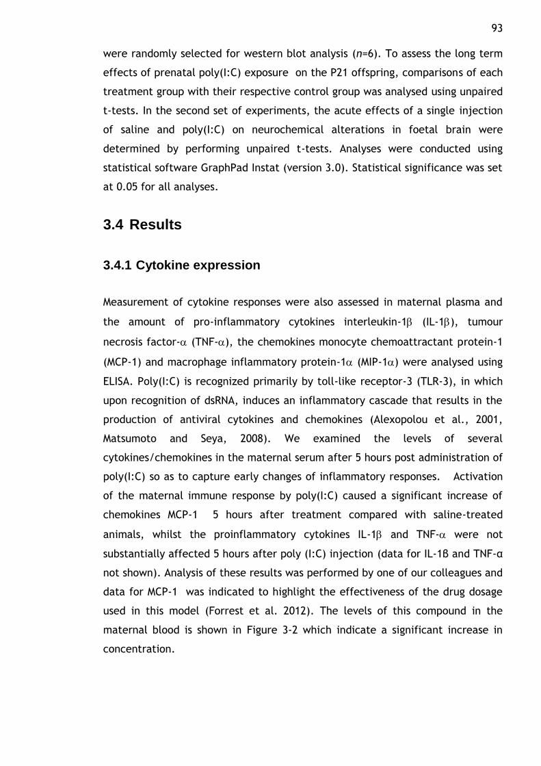
93 were randomly selected for western blot analysis (n=6). To assess the long term
effects of prenatal poly(I:C) exposure on the P21 offspring, comparisons of each
treatment group with their respective control group was analysed using unpaired
t-tests. In the second set of experiments, the acute effects of a single injection
of saline and poly(I:C) on neurochemical alterations in foetal brain were
determined by performing unpaired t-tests. Analyses were conducted using
statistical software GraphPad Instat (version 3.0). Statistical significance was set
at 0.05 for all analyses.
3.4 Results
3.4.1 Cytokine expression
Measurement of cytokine responses were also assessed in maternal plasma and
the amount of pro-inflammatory cytokines interleukin-1 (IL-1), tumour
necrosis factor- (TNF-), the chemokines monocyte chemoattractant protein-1
(MCP-1) and macrophage inflammatory protein-1 (MIP-1) were analysed using
ELISA. Poly(I:C) is recognized primarily by toll-like receptor-3 (TLR-3), in which
upon recognition of dsRNA, induces an inflammatory cascade that results in the
production of antiviral cytokines and chemokines (Alexopolou et al., 2001,
Matsumoto and Seya, 2008). We examined the levels of several
cytokines/chemokines in the maternal serum after 5 hours post administration of
poly(I:C) so as to capture early changes of inflammatory responses. Activation
of the maternal immune response by poly(I:C) caused a significant increase of
chemokines MCP-1 5 hours after treatment compared with saline-treated
animals, whilst the proinflammatory cytokines IL-1 and TNF- were not
substantially affected 5 hours after poly (I:C) injection (data for IL-1β and TNF-α
not shown). Analysis of these results was performed by one of our colleagues and
data for MCP-1 was indicated to highlight the effectiveness of the drug dosage
used in this model (Forrest et al. 2012). The levels of this compound in the
maternal blood is shown in Figure 3-2 which indicate a significant increase in
concentration.
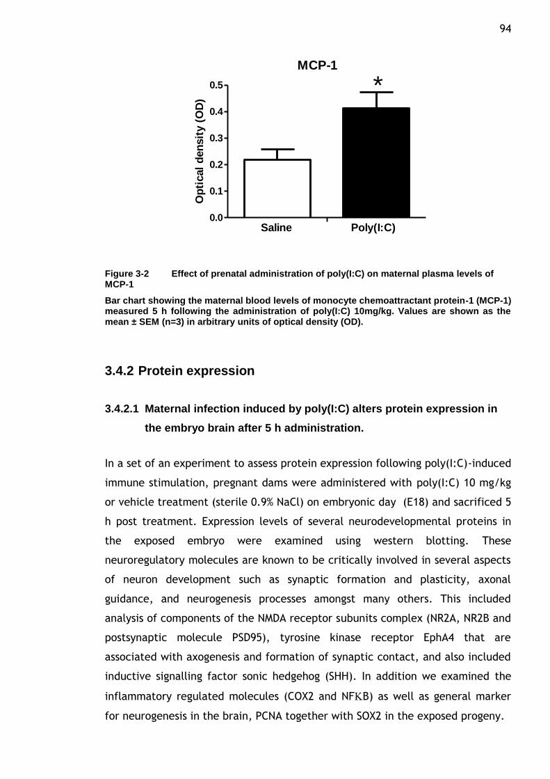
94
MCP-1
0.0
0.1
0.2
0.3
0.4
0.5
Saline Poly(I:C)
*
Op
tical
den
sit
y (
OD
)
Figure 3-2 Effect of prenatal administration of poly(I:C) on maternal plasma levels of MCP-1
Bar chart showing the maternal blood levels of monocyte chemoattractant protein-1 (MCP-1) measured 5 h following the administration of poly(I:C) 10mg/kg. Values are shown as the mean ± SEM (n=3) in arbitrary units of optical density (OD).
3.4.2 Protein expression
3.4.2.1 Maternal infection induced by poly(I:C) alters protein expression in
the embryo brain after 5 h administration.
In a set of an experiment to assess protein expression following poly(I:C)-induced
immune stimulation, pregnant dams were administered with poly(I:C) 10 mg/kg
or vehicle treatment (sterile 0.9% NaCl) on embryonic day (E18) and sacrificed 5
h post treatment. Expression levels of several neurodevelopmental proteins in
the exposed embryo were examined using western blotting. These
neuroregulatory molecules are known to be critically involved in several aspects
of neuron development such as synaptic formation and plasticity, axonal
guidance, and neurogenesis processes amongst many others. This included
analysis of components of the NMDA receptor subunits complex (NR2A, NR2B and
postsynaptic molecule PSD95), tyrosine kinase receptor EphA4 that are
associated with axogenesis and formation of synaptic contact, and also included
inductive signalling factor sonic hedgehog (SHH). In addition we examined the
inflammatory regulated molecules (COX2 and NFB) as well as general marker
for neurogenesis in the brain, PCNA together with SOX2 in the exposed progeny.
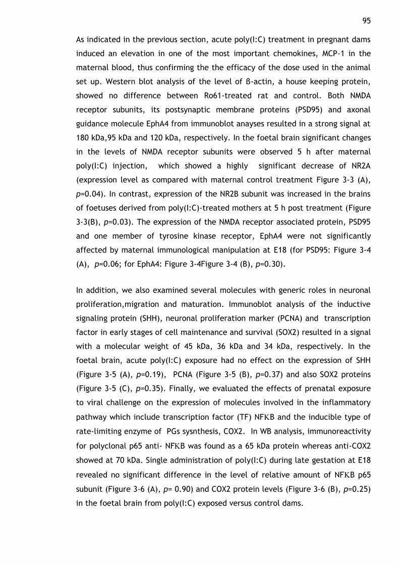
95 As indicated in the previous section, acute poly(I:C) treatment in pregnant dams
induced an elevation in one of the most important chemokines, MCP-1 in the
maternal blood, thus confirming the the efficacy of the dose used in the animal
set up. Western blot analysis of the level of β-actin, a house keeping protein,
showed no difference between Ro61-treated rat and control. Both NMDA
receptor subunits, its postsynaptic membrane proteins (PSD95) and axonal
guidance molecule EphA4 from immunoblot anayses resulted in a strong signal at
180 kDa,95 kDa and 120 kDa, respectively. In the foetal brain significant changes
in the levels of NMDA receptor subunits were observed 5 h after maternal
poly(I:C) injection, which showed a highly significant decrease of NR2A
(expression level as compared with maternal control treatment Figure 3-3 (A),
p=0.04). In contrast, expression of the NR2B subunit was increased in the brains
of foetuses derived from poly(I:C)-treated mothers at 5 h post treatment (Figure
3-3(B), p=0.03). The expression of the NMDA receptor associated protein, PSD95
and one member of tyrosine kinase receptor, EphA4 were not significantly
affected by maternal immunological manipulation at E18 (for PSD95: Figure 3-4
(A), p=0.06; for EphA4: Figure 3-4Figure 3-4 (B), p=0.30).
In addition, we also examined several molecules with generic roles in neuronal
proliferation,migration and maturation. Immunoblot analysis of the inductive
signaling protein (SHH), neuronal proliferation marker (PCNA) and transcription
factor in early stages of cell maintenance and survival (SOX2) resulted in a signal
with a molecular weight of 45 kDa, 36 kDa and 34 kDa, respectively. In the
foetal brain, acute poly(I:C) exposure had no effect on the expression of SHH
(Figure 3-5 (A), p=0.19), PCNA (Figure 3-5 (B), p=0.37) and also SOX2 proteins
(Figure 3-5 (C), p=0.35). Finally, we evaluated the effects of prenatal exposure
to viral challenge on the expression of molecules involved in the inflammatory
pathway which include transcription factor (TF) NFB and the inducible type of
rate-limiting enzyme of PGs sysnthesis, COX2. In WB analysis, immunoreactivity
for polyclonal p65 anti- NFB was found as a 65 kDa protein whereas anti-COX2
showed at 70 kDa. Single administration of poly(I:C) during late gestation at E18
revealed no significant difference in the level of relative amount of NFB p65
subunit (Figure 3-6 (A), p= 0.90) and COX2 protein levels (Figure 3-6 (B), p=0.25)
in the foetal brain from poly(I:C) exposed versus control dams.
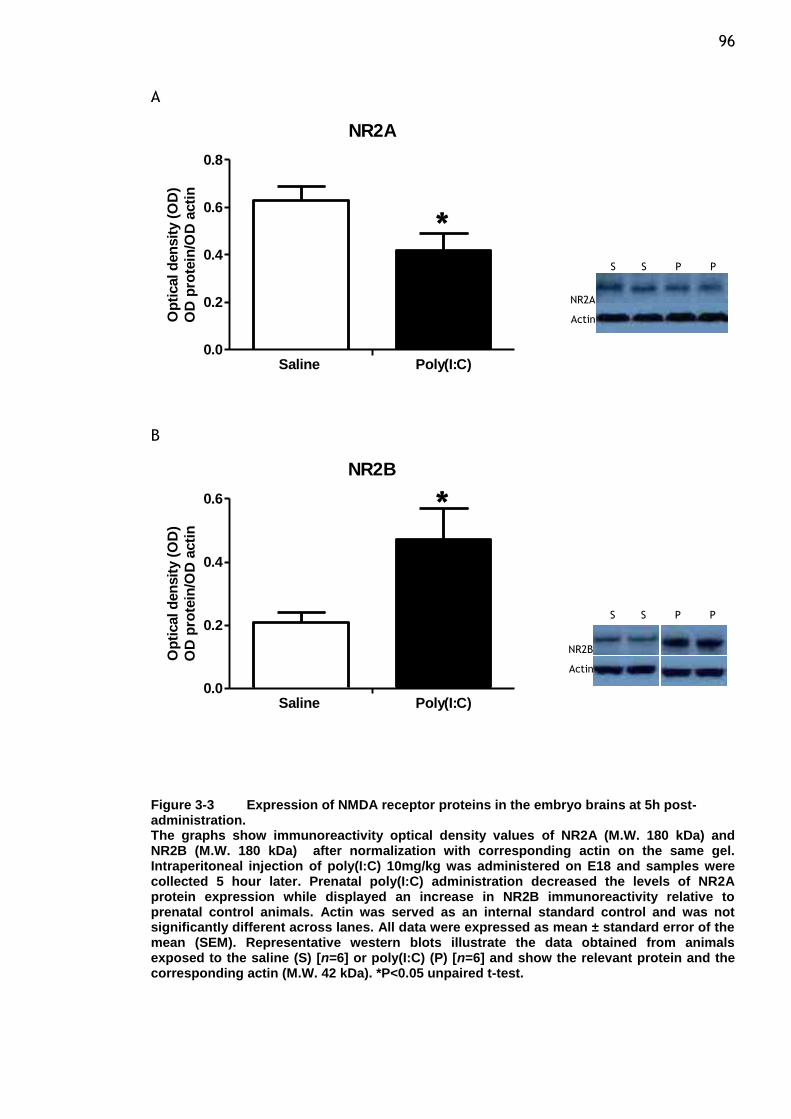
96 A
NR2A
0.0
0.2
0.4
0.6
0.8
Saline Poly(I:C)
Op
tical d
en
sit
y (
OD
)
OD
pro
tein
/OD
acti
n
B
NR2B
0.0
0.2
0.4
0.6
Saline Poly(I:C)
Op
tical d
en
sit
y (
OD
)
OD
pro
tein
/OD
acti
n
Figure 3-3 Expression of NMDA receptor proteins in the embryo brains at 5h post-administration. The graphs show immunoreactivity optical density values of NR2A (M.W. 180 kDa) and NR2B (M.W. 180 kDa) after normalization with corresponding actin on the same gel. Intraperitoneal injection of poly(I:C) 10mg/kg was administered on E18 and samples were collected 5 hour later. Prenatal poly(I:C) administration decreased the levels of NR2A protein expression while displayed an increase in NR2B immunoreactivity relative to prenatal control animals. Actin was served as an internal standard control and was not significantly different across lanes. All data were expressed as mean ± standard error of the mean (SEM). Representative western blots illustrate the data obtained from animals exposed to the saline (S) [n=6] or poly(I:C) (P) [n=6] and show the relevant protein and the corresponding actin (M.W. 42 kDa). *P<0.05 unpaired t-test.
*
*
S S P P
NR2A
Actin
NR2B
Actin
S S P P
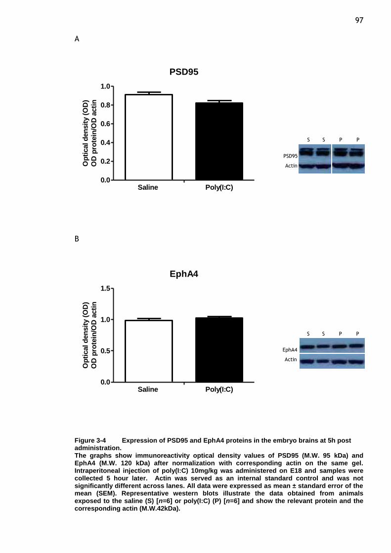
97 A
PSD95
0.0
0.2
0.4
0.6
0.8
1.0
Saline Poly(I:C)
Op
tical d
en
sit
y (
OD
)
OD
pro
tein
/OD
acti
n
B
EphA4
0.0
0.5
1.0
1.5
Saline Poly(I:C)
Op
tical d
en
sit
y (
OD
)
OD
pro
tein
/OD
acti
n
Figure 3-4 Expression of PSD95 and EphA4 proteins in the embryo brains at 5h post administration. The graphs show immunoreactivity optical density values of PSD95 (M.W. 95 kDa) and EphA4 (M.W. 120 kDa) after normalization with corresponding actin on the same gel. Intraperitoneal injection of poly(I:C) 10mg/kg was administered on E18 and samples were collected 5 hour later. Actin was served as an internal standard control and was not significantly different across lanes. All data were expressed as mean ± standard error of the mean (SEM). Representative western blots illustrate the data obtained from animals exposed to the saline (S) [n=6] or poly(I:C) (P) [n=6] and show the relevant protein and the corresponding actin (M.W.42kDa).
PSD95
Actin
S S P P
EphA4
Actin
S S P P
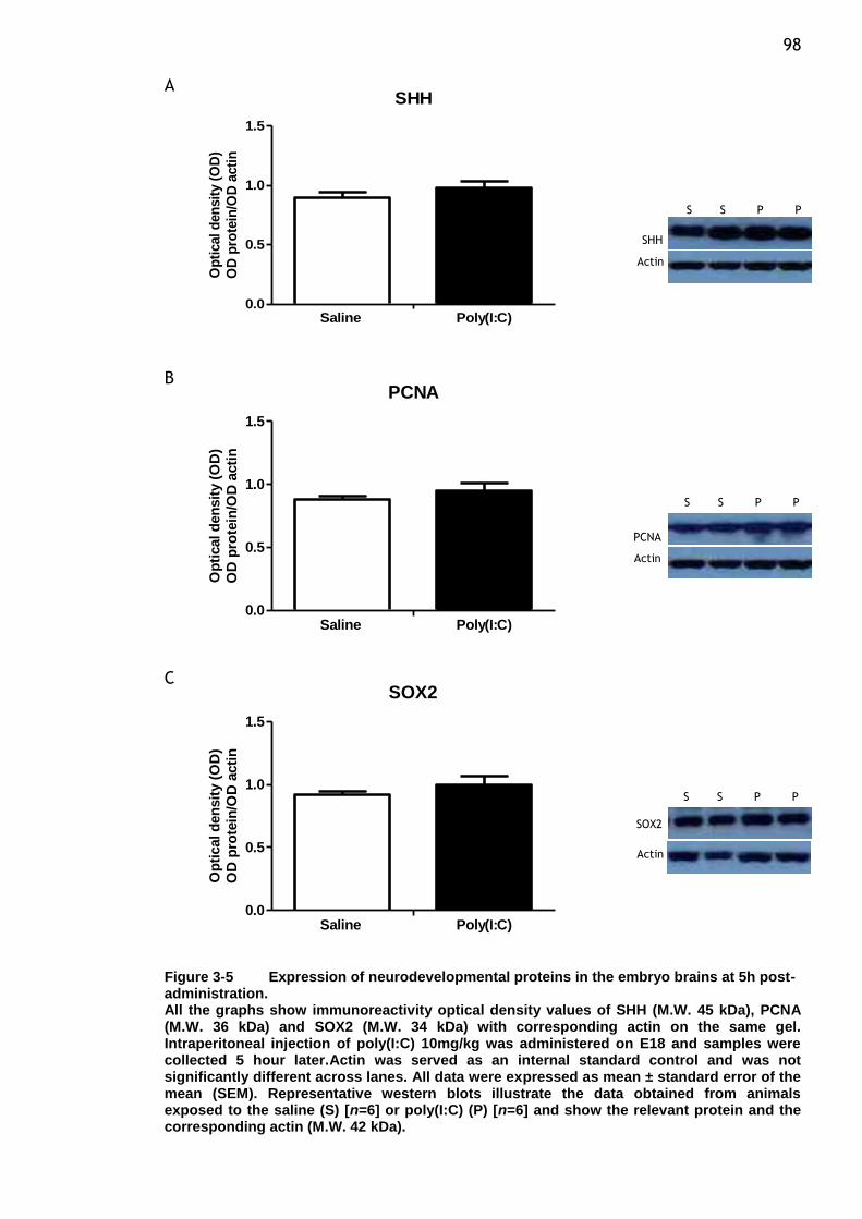
98 A
SHH
0.0
0.5
1.0
1.5
Saline Poly(I:C)
Op
tical d
en
sit
y (
OD
)
OD
pro
tein
/OD
acti
n
B PCNA
0.0
0.5
1.0
1.5
Saline Poly(I:C)
Op
tical d
en
sit
y (
OD
)
OD
pro
tein
/OD
acti
n
C SOX2
0.0
0.5
1.0
1.5
Saline Poly(I:C)
Op
tical d
en
sit
y (
OD
)
OD
pro
tein
/OD
acti
n
Figure 3-5 Expression of neurodevelopmental proteins in the embryo brains at 5h post-administration. All the graphs show immunoreactivity optical density values of SHH (M.W. 45 kDa), PCNA (M.W. 36 kDa) and SOX2 (M.W. 34 kDa) with corresponding actin on the same gel. Intraperitoneal injection of poly(I:C) 10mg/kg was administered on E18 and samples were collected 5 hour later.Actin was served as an internal standard control and was not significantly different across lanes. All data were expressed as mean ± standard error of the mean (SEM). Representative western blots illustrate the data obtained from animals exposed to the saline (S) [n=6] or poly(I:C) (P) [n=6] and show the relevant protein and the corresponding actin (M.W. 42 kDa).
SHH
Actin
S S P P
S S P P
PCNA
Actin
S S P P
SOX2
Actin
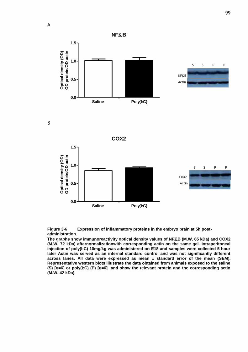
99 A
NFB
0.0
0.5
1.0
1.5
Saline Poly(I:C)
Op
tical d
en
sit
y (
OD
)
OD
pro
tein
/OD
acti
n
B
COX2
0.0
0.5
1.0
1.5
Saline Poly(I:C)
Op
tical d
en
sit
y (
OD
)
OD
pro
tein
/OD
acti
n
Figure 3-6 Expression of inflammatory proteins in the embryo brain at 5h post-administration.
The graphs show immunoreactivity optical density values of NFB (M.W. 65 kDa) and COX2 (M.W. 72 kDa) afternormalizationwith corresponding actin on the same gel. Intraperitoneal injection of poly(I:C) 10mg/kg was administered on E18 and samples were collected 5 hour later Actin was served as an internal standard control and was not significantly different across lanes. All data were expressed as mean ± standard error of the mean (SEM). Representative western blots illustrate the data obtained from animals exposed to the saline (S) [n=6] or poly(I:C) (P) [n=6] and show the relevant protein and the corresponding actin (M.W. 42 kDa).
S S P P
NFB
Actin
COX2
Actin
S S P P

100
3.4.2.2 Maternal infection induced by poly(I:C) alters protein expression in
the postnatal day (P) 21 offsprings.
Maternal viral mimicked infection was modelled in timed-pregnant rats at
embryonic days (E) 14, 16 and 18 by administering injections of poly(I:C) at
(10mg/kg, i.p.) to study the impact of the maternal inflammatory response on
the brain development of the resulting offspring at post natal day 21. Although
not significantly altered in comparison to the 5 h treatment, the level of NR2A
remained low, when observed in pups exposed to poly(I:C) at the later age of
postnatal day 21 (Figure 3-7 (A), p=0.12). Similarly with NR2A, expression of
NR2B subunit was not altered in poly(I:C)-pups at postnatal day 21 (Figure 3-7
(B), p=0.64). The expression of neither PSD95 (Figure 3-8 (A), p= 0.51) nor EphA4
(Figure 3-8 (B), p= 0.12) showed any significant difference from control pups
from saline-treated dams.
As indicated in the section 3.4.2.1, acute poly(I:C) exposure had no effect on the
expression of SHH, however repeated in utero viral challenges led to significant
changes among this neurogenesis factor only at P21. As shown in Figure 3-9 (A)
and Figure 3-5 (A), antibodies used against the SHH precursor protein (45 kDa)
was clearly detected in the embryonic and postnatal brain tissues. Although
there was no change in the level of expression of the SHH precursor protein at 5
h after poly(I:C) injection compared with that in age-matched uninfected
controls, a statistically significant reduction was shown in the brain of postnatal
day 21 (p= 0.03). Similar situation was observed when poly(I:C) was administered
during the mid and late gestation in the rodent model, long term effects of
prenatal viral challenge on survival and maintenance of cells in the brain as
indicated by the expression of SOX2 was clearly demonstrated. Prenatal poly(I:C)
exposure significantly increased the relative amount of SOX2 protein in the P21
pups compared with control pups (Figure 3-9 (C), p= 0.009). Although strong
signals were seen in the postnatal brain, PCNA protein levels of P21 offspring
were not statictically different as compared with control rats (Figure 3-9 (B), p=
0.64). Results of western blot analysis with this developmental markers showed
strong signals suggesting conserved levels of endogenous expression during
relatively early developmental period. Bands were also observed at moderate
101 intensities in the postnatal brain at the same position with molecular weight of
36 kDa.
Finally, we examined the levels of the inflammation transcription factor (TF)
NFB and the inducible type of rate-limiting enzyme of PGs sysnthesis, COX2,
whose expression is linked to inflammation-induced oxidative stress. In the
brains from P21 pups exposed to poly(I:C), the expression of neither NFB
(Figure 3-10 (A), p= 0.97) nor COX2 (Figure 3-10 (B), p= 0.35) showed any
significant difference from control treatment. Thus, prenatal viral challenge at
either E18 or three time points during mid-late gestation showed no apparent
difference between treatments on the levels of protein expression of selected
inflammatory markers.
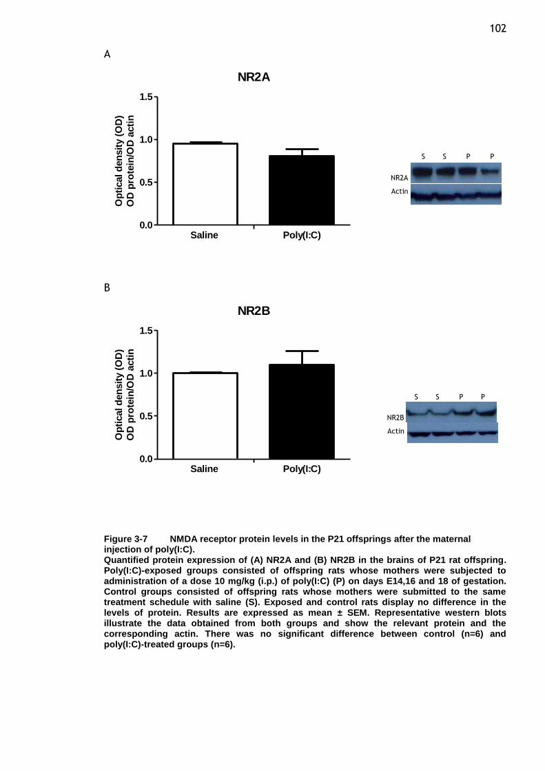
102 A
NR2A
0.0
0.5
1.0
1.5
Saline Poly(I:C)
Op
tical d
en
sit
y (
OD
)
OD
pro
tein
/OD
acti
n
B
NR2B
0.0
0.5
1.0
1.5
Saline Poly(I:C)
Op
tical d
en
sit
y (
OD
)
OD
pro
tein
/OD
acti
n
Figure 3-7 NMDA receptor protein levels in the P21 offsprings after the maternal injection of poly(I:C). Quantified protein expression of (A) NR2A and (B) NR2B in the brains of P21 rat offspring. Poly(I:C)-exposed groups consisted of offspring rats whose mothers were subjected to administration of a dose 10 mg/kg (i.p.) of poly(I:C) (P) on days E14,16 and 18 of gestation. Control groups consisted of offspring rats whose mothers were submitted to the same treatment schedule with saline (S). Exposed and control rats display no difference in the levels of protein. Results are expressed as mean ± SEM. Representative western blots illustrate the data obtained from both groups and show the relevant protein and the corresponding actin. There was no significant difference between control (n=6) and poly(I:C)-treated groups (n=6).
S S P P
NR2A
Actin
NR2B
Actin
S S P P
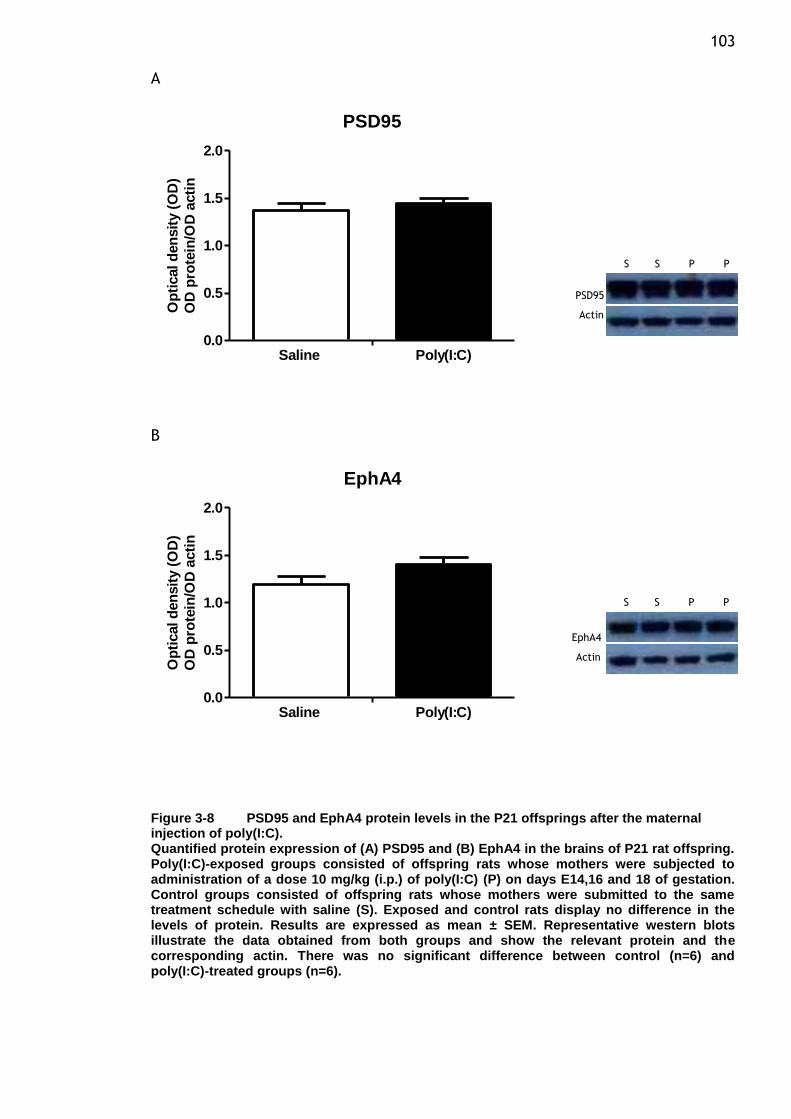
103 A
PSD95
0.0
0.5
1.0
1.5
2.0
Saline Poly(I:C)
Op
tical d
en
sit
y (
OD
)
OD
pro
tein
/OD
acti
n
B
EphA4
0.0
0.5
1.0
1.5
2.0
Saline Poly(I:C)
Op
tical d
en
sit
y (
OD
)
OD
pro
tein
/OD
acti
n
Figure 3-8 PSD95 and EphA4 protein levels in the P21 offsprings after the maternal injection of poly(I:C). Quantified protein expression of (A) PSD95 and (B) EphA4 in the brains of P21 rat offspring. Poly(I:C)-exposed groups consisted of offspring rats whose mothers were subjected to administration of a dose 10 mg/kg (i.p.) of poly(I:C) (P) on days E14,16 and 18 of gestation. Control groups consisted of offspring rats whose mothers were submitted to the same treatment schedule with saline (S). Exposed and control rats display no difference in the levels of protein. Results are expressed as mean ± SEM. Representative western blots illustrate the data obtained from both groups and show the relevant protein and the corresponding actin. There was no significant difference between control (n=6) and poly(I:C)-treated groups (n=6).
PSD95
Actin
S S P P
S S P P
EphA4
Actin
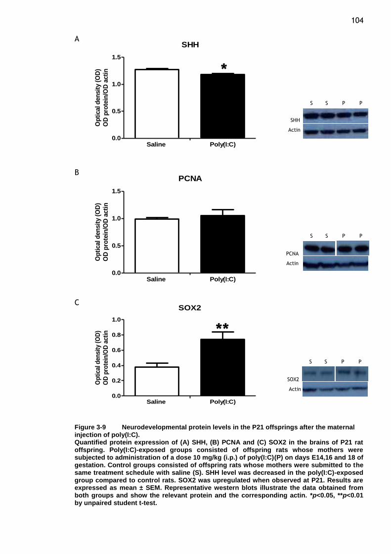
104 A
SHH
0.0
0.5
1.0
1.5
Saline Poly(I:C)
Op
tical d
en
sit
y (
OD
)
OD
pro
tein
/OD
acti
n
B PCNA
0.0
0.5
1.0
1.5
Saline Poly(I:C)
Op
tical d
en
sit
y (
OD
)
OD
pro
tein
/OD
acti
n
C SOX2
0.0
0.2
0.4
0.6
0.8
1.0
Saline Poly(I:C)
Op
tica
l den
sity
(O
D)
OD
pro
tein
/OD
act
in
Figure 3-9 Neurodevelopmental protein levels in the P21 offsprings after the maternal injection of poly(I:C). Quantified protein expression of (A) SHH, (B) PCNA and (C) SOX2 in the brains of P21 rat offspring. Poly(I:C)-exposed groups consisted of offspring rats whose mothers were subjected to administration of a dose 10 mg/kg (i.p.) of poly(I:C)(P) on days E14,16 and 18 of gestation. Control groups consisted of offspring rats whose mothers were submitted to the same treatment schedule with saline (S). SHH level was decreased in the poly(I:C)-exposed group compared to control rats. SOX2 was upregulated when observed at P21. Results are expressed as mean ± SEM. Representative western blots illustrate the data obtained from both groups and show the relevant protein and the corresponding actin. *p<0.05, **p<0.01 by unpaired student t-test.
PCNA
Actin
SHH
Actin
S S P P
S S P P
SOX2
Actin
S S P P
*
**
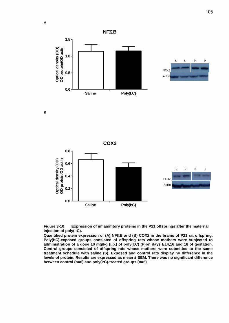
105 A
NFB
0.0
0.5
1.0
1.5
Saline Poly(I:C)
Op
tical d
en
sit
y (
OD
)
OD
pro
tein
/OD
acti
n
B
COX2
0.0
0.2
0.4
0.6
0.8
Saline Poly(I:C)
Op
tical d
en
sit
y (
OD
)
OD
pro
tein
/OD
acti
n
Figure 3-10 Expression of inflammtory proteins in the P21 offsprings after the maternal injection of poly(I:C).
Quantified protein expression of (A) NFB and (B) COX2 in the brains of P21 rat offspring. Poly(I:C)-exposed groups consisted of offspring rats whose mothers were subjected to administration of a dose 10 mg/kg (i.p.) of poly(I:C) (P)on days E14,16 and 18 of gestation. Control groups consisted of offspring rats whose mothers were submitted to the same treatment schedule with saline (S). Exposed and control rats display no difference in the levels of protein. Results are expressed as mean ± SEM. There was no significant difference between control (n=6) and poly(I:C)-treated groups (n=6).
COX2
Actin
NFB
Actin
S S P P
S S P P
106
3.5 Discussion
3.5.1 Maternal immune activation
In line with cases of infection during pregnancy, several lines of evidence
indicate that the maternal immune response rather than direct infection of the
foetus is responsible for the increased incidence of schizophrenia and also
autism in the offspring of mothers who suffer infections during pregnancy
(Patterson, 2002, Gilmore and Jarskog, 1997). This evidence is supported by
studies that show behavioural and histological abnormalities in the adult
offspring following exposure to either bacterial lipopolysaccharide (LPS) or
doublestranded RNA, poly(I:C) similar to those seen in the offspring of infected
mothers. The variability of immunological and neurological responses from using
active viral agents help rationalize the use of non-replicating viral mimetics like
poly(I:C) that act through the same toll-like receptor 3(TLR3) as all other viral
infections. Intravenous (i.v.) and i.p. administration of poly(I:C) are widely used
as inducers of the innate immune response, which mimics the first phase of
defensive mechanisms against viral infections. Systemic administration of viral
mimetic is a widely accepted model of immune activation, resulting in TLR3
stimulation which then induces a robust anti-viral response including type 1
interferons α and β and also other inflammatory cytokines (Matsumoto and Seya,
2008, Alexopolou et al., 2001). For the purpose of prenatal immune activation
models, the use of poly(I:C) instead of live pathogens has the advantages of
avoiding the use of infectious agents within the working environment and
treatments can be standardized which provides a predictable intensity and
duration by appropriate dosage. This aspect facilitates an evaluation of the
influence of the timing of maternal poly(I:C)-induced immune challenge on the
emergence of brain and behavioural dysfunctions in the resulting offspring
(Meyer et al., 2006, Meyer and Feldon, 2011).
To shed more light on the potential involvement of viruses in the etiological role
during certain stages of brain development, models of early life exposure to the
viral mimetic poly(I:C) was studied with regard to the long and short term
effects on protein expression in the brain. We choose to mimic a viral infection
instead of the most frequently used bacterial mimic agent, because viral
107 infections during pregnancy are common during the influenza season and appear
to predispose the offspring to develop psychiatric illness (Brown et al., 2004).
By using the synthetic double-stranded RNA poly(I:C) to induce maternal immune
activation (MIA), we are able to study the effects of MIA on the embryo, and
explore the impact and association between prenatal immune challenge and
postnatal brain dysfunction. We noted no obvious malformations in the pups of
poly(I:C) treated dams and no differences in body weight between the poly(I:C)-
rats and control rats. Early reports from our laboratory has confirmed the
efficiency of the poly(I:C) used in the study to activate maternal immune system
since the expression of MCP-1 is increased 5 h after its injection, indicating that
the maternal is eliciting an inflammatory chemokine response which may then
enhance the recruitment of mast cells. Mast cells express TLR-3 and are known
to be strongly activated upon treatment with poly(I:C) (Kulka et al., 2004). The
chemokine MCP-1 is reported to function in CNS inflammation and injury as it
has been found expressed rapidly following brain injury by both microglia and
astrocytes. Apart from that, chemokines contribute to normal brain
development by providing cues for the migration of newly generated neurons
and glial cells and also modulate axon path-finding. In addition, MCP-1 enhances
neuronal excitability and synaptic transmission in hippocampal neurons (Zhou et
al., 2011).
A correlation between higher expression of MCP-1 and colonization of activated
mast cells remains to be determined. Mast cells form close interactions with
neurons and transfer intracellular content by transgranulation, which may
modulate neuronal functions and affect CNS development (Wilhelm et al., 2005,
Conti et al., 1997). However, we did not observe any changes in the level of
other cytokines such as IL-1β and TNF-α in the maternal plasma (Forrest et al.
2012). The basis for the observed changes and lack of changes in the other
cytokines at 5 h following the administration of poly(I:C) is in agreement with
the other studies that showed a robust increase of TNF-α protein level in the
maternal plasma at 2 h after exposure, decreased thereafter and below the level
of detection by 6 h post injection, while plasma concentration of IL-1β did not
change after poly(I:C) injection. It also has been observed at a protein and
mRNA level that the concentration of IL-1β may vary during development
(Fortier et al., 2004). Analysis of i.p. administration of poly(I:C) indicates the
108 triggering of a broad antiviral maternal innate immune activation and represents
a unique response of the maternal environment and would have a variety of
effects on the developing brain (Gilmore et al., 2005a). The mechanisms
whereby viral infection can lead to neurodevelopmental disorders remain poorly
understood. Currently it is believed that the virus can act either directly or
indirectly to affect neurodevelopment. Following viral infection, the immune
system becomes activated and cytokines are released, which can in turn
interfere with proper neurodevelopment as already mentioned. It should be
noted that although the physiological function of cytokines might be to preserve
or restore homoeostasis, sustained or excessive production of cytokines could
cause damage. Expression levels of a subset of cytokines and chemokines in the
foetal brains somehow do not reflect the changes in concentration levels
observed in maternal serum. Since only maternal serum was assessed in this
study and we have not assessed the cytokine levels in the foetal and placenta,
measurement of foetal brain cytokines following a similar set up of prenatal
infection would provide additional information with respect to the association
between their role in inflammatory responses and effects on CNS development,
including effects on neuronal survival, differentiation and also excitotoxicity in
developing brain (Rothwell and Hopkins, 1995, Boksa, 2010).
It should be emphasized that the effect of maternal infection on the
measurement of foetal brain cytokines was complex, and is dependent on the
gestational age at which poly(I:C) was administered and the time of the
evaluation after administration. Findings with regard to the changes in protein
levels of these cytokines in foetal brain have shown inconsistency and do not
provide compelling evidence as to suggest the exact mechanism of how
cytokines changes in fetal brain could have altered the function of foetal
neurodevelopment (Meyer et al., 2006). However, causal links between maternal
infection and the emergence of a wide spectrum of behavioural and
pharmacological abnormalities in the offspring have been observed in rodent
models (Boksa and Luheshi, 2003). This has led to speculation that the idea that
the perturbations caused by the prenatal immunological manipulation are
widespread, indicative of multiple structural brain abnormalities and also
dependent on a range of normal neuropsychological functions. Therefore, it
remains to be experimentally confirmed whether cytokines are the mediators
109 responsible for long term effects on CNS function in offspring (Shi et al., 2003,
Meyer and Feldon, 2009).
3.5.2 Protein expression
Results from this study also indicate the ability of inflammatory challenges by
poly(I:C) to produce neuroadaptive changes that might alter neuronal properties
and function including changes in the magnitude of NMDA receptor subunit
expression. This receptor is known to be an important regulator in neuronal
development and function, therefore alterations in the levels of its neuronal
properties could have important consequences for synaptic responses (Hunt and
Castillo, 2012). To further address this issue, we have examined the effect of
poly(I:C) exposure during gestation on the level of two major subunits of the
NMDA receptor in the brain obtained from the embryo and also from postnatal
age 21 day. The NR2A and NR2B subunits were chosen for examination in this
study because they are the most predominant NMDA receptor subtypes in their
respective subunit families and underlie functional excitatory synaptic
transmission and activity-dependent synaptic plasticity (Cull-Candy et al., 2001,
Cull-Candy and Klein, 2007). Both subunits show greater excitotoxicity than the
other subunits and brain regions enriched in NMDA receptor composed of NR2A
and NR2B subunits (such as hippocampus and forebrain) are commonly injured in
excitotoxic insults such as ischemia, brain injury and HIV-1 infection (Waxman
and Lynch, 2005, O'Donnel et al., 2006, Schumann et al., 2008). The expression
of NMDA receptor subunit subtypes were significantly altered following 5 h post-
exposure with poly(I:C). However, the differential changes of the subunit levels
induced by poly(I:C) then reached expression levels comparable to those
observed in control offspring at postnatal day 21. Our findings suggest that both
NR2A- and NR2B-containing receptors would be particularly susceptible to
poly(I:C)-induced damage in the embryo brains. The demonstrable low level of
NR2A and robust expression of NR2B subunit suggests the likely expression of
functional NR2B, since it has been shown that this subunit predominates in early
development (Waxman and Lynch, 2005, Babb et al., 2005). The resulting
increase in the amount of this subunit may be due to new protein synthesis in
the total brain homogenates. It thus seems that the early changes to NMDA
receptor subunit expression in the foetus derived from immune-challenged
mothers may not have been sufficient to impair the subsequent expression of
110 NMDA receptor associated molecules in the brain at a later stage of postnatal
development (i.e on P21). However, it should be pointed out that infection-
induced disruption of early prenatal brain development may lead to long-lasting
structural and functional brain abnormalities leading to the emergence of
behavioural dysfunctions in later life (Fatemi et al., 2002, Meyer et al., 2008b).
Although questions remain to be answered regarding the specific mechanisms of
action for synaptic protein, their involvement in the regulation of patterns of
synaptic connectivity has been established and suggestive evidence implicates
them in schizophrenia (Cull-Candy et al., 2001, Gladding and Raymond, 2011).
Another interesting finding emerging from the present results was the expression
of SHH and SOX2. The relative protein level of inductive signal SHH was initially
unchanged in the brains of foetuses derived from poly(I:C)-treated mothers 5 h
post-treatment, but then decreased significantly at 21 days. This effect was
found in the young offspring 21 day after they were born, demonstrating that
prenatal exposure to poly(I:C) can produce long-term neurochemical alterations.
A related study of maternal poly(I:C) exposure on a different time point, during
early/middle pregnancy (E9) found reduced foetal brain gene expression of SHH
at 2, 4 and 8 days after maternal immune activation. This study provides
evidence on the involvement of SHH and other related genes in the foetal
dopaminergic development(Meyer et al., 2008a). SHH signalling is required
during embryonic development in the regulation of cell proliferation, migration
and survival and in the maintenance of cell in the adult (Bale, 2002). Evidence
that strengthen the pivotal role of SHH signalling during early developmental
period demonstrate the regulation of cellular proliferation for precise
morphogenesis of embryonic tissue and also in the early expansion of developing
midbrain and proliferation of granular cell precursors in cerebellum(Wechsler-
Reya and Scott, 1999, Britto et al., 2002, Ruiz i Altaba et al., 2002). The
influence of SHH signalling activation in response to other types of infection also
has been noted. Expression of SHH and its downstream signalling molecules, GLi1
and its receptor Patched (Ptc) were upregulated in Helicobacter pylori infection
and the level of SHH is elevated in gastric cancer cells. This suggests that SHH
signalling pathway is one of the host signals related to cancer growth and
metastasis and changes in the level of SHH expression may provide a marker for
chemo preventive measures(Kim et al., 2010). In vivo studies showed that
111 prolonging infection with H.pylori resulted in a different pattern of SHH protein
expression in the gastric mucosa of the gerbils 51 weeks after bacterial
inoculation (Suzuki et al., 2005). Both studies demonstrate that H.pylori
infection modified the expression of morphogens involved in the regulation of
gastric mucosal homoeostasis. In addition, a number of researchers have shown
that dysregulated activation of SHH signalling predispose cells to the
development of tumours, in which overexpression of SHH have been found in
many types of cancers including medullolastoma, basal cell carcinoma, lung
carcinoma, adenocarcinoma of digestive tract and oral squamus cancer cell
(Dahmane et al., 2001, Wechsler-Reya and Scott, 1999, Stecca and Ruiz i Altaba,
2005, Lupi, 2007, Watkins et al., 2003, Nishimaki et al., 2004). Apart from its
role in patterning and the proliferation of embryonic tissue, SHH has been shown
to influence the differentiation of various cell types, including dopaminergic
neurons and lymphocytes (Hynes and Rosenthal, 1999). The present finding
showed that maternal poly(I:C) during late gestation decreased the level of the
inductive signal SHH in the P21 offspring as compared with maternal control
treatment. Since the emergence of changes was demonstrated only at a
postnatal age, this leads to the possibility that SHH signalling pathway
abnormalities emerging after prenatal immune challenge are likely to be of
developmental origin starting early in foetal life (at the time of injections) and
that the pathway is influenced following maternal immune activation.
The high-mobility group transcription factor SOX2 is expressed in embryonic
neural epithelial stem cells and represent the unique expression of multipotent
stem and progenitor cells in neurogenic regions of the brain of the central
nervous system (CNS) throughout adulthood (Brazel et al., 2005, Lefebvre et al.,
2007). In this study, we showed that SOX2 protein levels was not affected by
maternal poly(I:C) exposure during embryonic development, however an
upregulation of SOX2 protein product in the brain of day 21 progeny of virally
exposed rat was observed. SOX2 expression has been most commonly associated
with non-committed stem and precursor cells in the developing nervous system.
It was also being expressed in multipotent cell types which suggest a role for
SOX2 in preserving developmental potential. Many SOX genes are expressed in
the developing and adult brain, their expression patterns overlapping with each
other since a high degree of identity are shared within the same group (Guth and
112 Wegner, 2008, Brazel et al., 2005). In vitro functional studies showed that the
collaboration of SOX2 with the other transcriptional factors, particularly Oct4
and Nanog is required to maintain pluripotency and self-renewal in human
embryonic stem cell (ESC), thus preserving developmental potential (Fong et al.,
2008). Even though the SOX-B1 members can compensate for each other‟s loss
during early development, they have distinct roles later in development
(Graham et al., 2003). Thus proper levels of SOX2 are required for stem cell
maintenance as either increased or decreased levels of SOX2 are used as cues for
differentiation. Continuous expression of SOX2 led to an increased number of
proliferating progenitors and reduction of SOX2 is associated with loss of the
pluripotent state and a propensity for differentiation and regulation of target
genes requiring cooperation between other critical transcriptional regulators like
Oct4 and Nanog (Lefebvre et al., 2007).
As a cofactor for DNA polymerase delta and DNA replication, PCNA expression is
a useful marker of cell proliferation under both normal and pathological
influences. In this study, following the poly(I:C) exposure, PCNA protein levels
were not significantly different than control levels as observed at E18 and P21.
As PCNA expression was used to identify the population of proliferating cells and
involved in the regulation of cell cycle and neurogenesis, any insult such as
ischemia or infectious agents may serve as triggers to activate the cell cycle.
Increased density of proliferating cells has been observed in several models of
experimental CNS injury such stroke and bacterial meningitis, which postulate
the idea of increased neurogenesis as a general reaction after neuronal injury of
various etiology (Gerber et al., 2009). Apart from that, it has been regarded as
one of either diagnostic or prognostic markers in many disease states, which
include predicting the biological behaviour of different types of tumours. The
higher content of PCNA positive cells in ameloblastic carcinoma samples perhaps
could explain the infiltrative growth of ameloblastoma in which PCNA positivity
reflects cell proliferation (Piattelli et al., 1998). However, conclusions drawn
from PCNA immunohistochemical studies vary considerably, reflecting
differences in case selection, sample selection, numbers of cells counted,
evidence of reproducibility and methods of statistical analysis (Kurki et al.,
1986, Valero et al., 2005).
113 We also investigated the impact of maternal immune challenge during pregnancy
on the foetal and postnatal expression of several genes known to be critically
involved in inflammatory related process. This included analysing the protein
expression of COX2 and the transcription factor, NFB. This served to ascertain
whether putative changes in inflammatory markers might be causally related, in
part to the known consequences of infection exposure in the human. It should be
noted that the nature of the predominant cellular response and the
inflammatory factors involved in immune and inflammatory process is extensive,
thus the presence of enhanced expression of MCP-1 reflects a general pro-
inflammatory status in the maternal environment following poly(I:C). However,
poly(I:C)-induced immunological challenges in rodents are short lived, ranging
from 24 to 48 h depending on the dose (Meyer and Feldon, 2011). As the peak of
detection of protein and mRNA levels of cytokines may vary following
stimulation and our results showed no changes to other inflammatory cytokines
such as IL-1β and TNF-α in maternal compartment. We suggest that there was no
ongoing inflammatory activity that could exaggerate inflammatory responses in
which we have noted no evidence of changes in the levels of expression for
NFB and COX-2 in the embryo brain or young adult offspring P21. Although
there was no demonstrable difference in immunoreactive of both markers in the
fetal and postnatal brain, low steady state levels of COX2 and NFB are
observed throughout the brain. Steady state of COX2 appears to be regulated by
normal glutamatergic synaptic activity in the adult brain (Kaufmann et al.,
1997). In this regard, endogenous synaptic transmission also might be capable of
regulating NFB activity (Meffert and Baltimore, 2005).
The widespread distribution in the CNS indicates that NFB may be sensitive to
synaptic activation as expression of this gene is increased in hippocampal
granule cells after LTP (Meberg et al., 1996). The presence of extracellular
signals such cytokines, bacteria, viral proteins or mitogen causes the activation
of NFB with rapid translocation to the nucleus and the transcriptional
activation of target genes. A common sign of NFB-activating conditions is that
they either represent a direct threat to the cells or that they are endogenous
signals produced in response to pathogens (Baeuerle and Henkel, 1994). The
activation of inflammatory transcription factors within the brain is a step that
occurs before the expression of target genes, which trigger brain-intrinsic
114 responses. This phenomenon can be assessed by measuring the expression of
markers and their activation. It is well known that NFB is one of the early
markers of inflammatory activation in the brain, and their nuclear translocation
can be induced by TNF/IL-1and IL-6 as shown in previous studies (Dharane et al.,
2010). Corresponding to the rather negligible levels of this pro-inflammatory
cytokine in the maternal plasma, no clear changes in the expression levels of
NFB was observed either at 5 h post treatment (embryo brain) or at later
developmental age of P21.
Although COX2 has limited constitutive neuronal distribution in the CNS, it can
be induced in the mammalian brain under a variety of physiological and
pathological conditions (Yamagata et al., 1993). For example, a marked
elevation of COX2 mRNA levels by kainic acid-induced seizure was detected in
the hippocampal CA1-1 pyramidal cell layers, granule layers of DG, cortex and
amygdale. Pronounced increases in brain derived neurotrophic factor (BDNF)
mRNA has also been noted in the dentate granule cell layer and pyramidal
layers. It has been suggested that marked expression of BDNF in rat brain may
occur as a trophic response to the neuronal injury caused by kainic acid-induced
seizures (Hashimoto et al., 1998). Injuries to the brain activate multiple cellular
pathways, some of which are adaptive, while others may persist and become
harmful. The severity of neural injury appears to correlate to the degree and
duration of some of these inflammatory markers. In agreement with this,
increased COX2 expression has been observed with several neurodegenerative
disorders such as AD, ALS and PD and also in traumatic brain injury and seizures
(Nogawa et al., 2003, Almer et al., 2001, Teisman et al., 2003, Joseph et al.,
2006). The present results show no marked changes in the expression of pro-
inflammatory cytokines (TNF-α and IL-1β), the inducible form of enzymes for
PGE2 (COX2) and the transcription factor (NFB) following maternal poly(I:C)
treatment. Thus, our study showed that repeated challenges do not appear to
induce an inflammatory response distinct from that induced by a single
challenge and are insufficient to produce any lasting impairment in the
inflammatory pathway.
115
3.6 Conclusion
Changes in behaviours relevant to psychiatric disorders such as schizophrenia
and autism are consistently reported following maternal challenge in the mouse
or rat, and the MIA model to study prenatal risk factors for these disorders is the
focus of much scientific research (Boksa, 2010, Smith et al., 2010). The
mechanisms by which MIA disrupts foetal development and sets into motion an
atypical trajectory of brain and behavioral development and maturation are
largely unknown. It is therefore possible that the induction of cytokines and
downstream effects of such activation in the foetal brain following MIA
interferes with neurodevelopmental processes or functioning within the brain
regions essential to social behaviours (Meyer, 2013, Meyer et al., 2009). It should
be pointed out that the current study did not examine the anatomical and
behavioural consequences of maternal poly(I:C). Based on the current findings,
it is not possible to draw any conclusions as to relate the mechanisms by which
poly(I:C)-induced MIA leads to subsequent behavioral changes. In this aspect, a
more complete behavioural phenotype of offspring born to poly(I:C)-exposed
dams is required to determine the specific behavioural effects of MIA.
Additionally, the mechanism by which MIA results in atypical performance on
behavioural tasks is unclear and may be addressed by the inclusion of
neuroanatomical studies. It is important to note that regulation of expression of
molecules used both during early development and in other processes later in
life is likely to play a major role in the correct processing of the molecule‟s
functions. Differential regulation, possibly involving different regulatory
molecules or mechanisms, occurs at different stages of life and could be
responsible for different phenotypes in different developmental periods. The
present study was conducted with the aim to characterize the effects of the
infection on CNS function in the offspring at two points of developmental stages
(embryo and P21). Assessing acute neurochemical changes in foetal brain are of
importance in understanding mechanisms that underlie persistent or longer term
CNS changes in the offspring following maternal infection.
In summary, the study carried out showed some evidence that disturbances
caused by viral infection during prenatal period of life can produce some
neuroadaptive changes that alter neuronal function and development. Some of
116 the developmental markers showed changes during the period of the insult and
disappeared or diminished as animal grew up. This may be an outcome of the
compensatory capacity of the CNS to alleviate such changes. In relation to this,
changes seen in the expression of the NMDA receptor subunits, NR2A and NR2B
could imply that alterations in the receptor number and function may impinge
on the formation and establishment of neuronal circuits and connections. The
present study demonstrates that poly(I:C) challenge in pregnant dams results in
transient alterations in the levels of NMDA receptor subunit NR2A and NR2B in
the fetal brain, representing a potential mechanism for altering synaptic
development and may contribute to abnormal brain development and risk for
neurodevelopmental disorders. On the other hand it is also possible that
prenatal poly(I:C) treatment manifested its effects on the postnatal brain
independently from its effect at the time of exposure. The acute inflammatory
response induced by prenatal poly(I:C) administration somehow induced further
long term changes in factors regulating cell proliferation and migration, in order
to produce altered neurogenesis in offspring later in development at P21. We
show that changes in markers that are involved in neurogenesis (SOX2 and SHH)
were observed at a later stage of postnatal development (P21). This could imply
that prenatal immune activation elicited by poly(I:C) in rodents may have long
term effects on neurogenesis in young postnatal offspring. Although we did not
include behavioural assessment and immunolabelling studies, previous data
together with current findings suggest such deficits or changes in neurogenesis
could conceivably contribute to the alterations in hippocampal-mediated
function that have been observed in juvenile or adult rodents following prenatal
exposure to poly(I:C) (Meyer et al., 2008b). However the results presented here
support the association between immunological manipulation at a specific
gestational age and postnatal brain dysfunctions. Unravelling the mechanisms
that mediate immunologically sensitive developmental periods that allow for
programming of adults‟ responses deserves further attention in which it permits
possibility for intervention. Taken together, our animal models indicate that
distinct neurochemical abnormalities are manifested at different stages of
postnatal development after prenatal exposure to viral-like immune challenge
during late gestation in rats.
4 The effect of prenatal inhibition of the kynurenine metabolism pathway on the expression of proteins in the rat brains
4.1 Introduction
It has been suggested that changes in the endogenous levels of neuroactive
metabolites of the kynurenine pathway are implicated in the pathogenesis of
several brain disorders, for instance Parkinson‟s disease and schizophrenia.
These neuroactive metabolites include KYNA, 3-hydroxykynurenine (3-HK) and
quinolinic acid (QUIN). The basis for kynurenine pathway up-regulation as seen
in psychotic disorders may not reside within one particular enzyme but rather
with one of many factors that control the expression of the initiating enzymes
involved in the pathway. There is evidence that emphasize the unique
antagonistic properties of KYNA as an endogenous factor which is capable of
blocking the neurotoxic and seizures caused by QUIN which support the fact that
manipulation favouring its accumulation in the brain extracellular space may be
beneficial against endogenous excitotoxic abnormalities (Foster et al., 1984).
Pharmacological blockade of the enzyme kynurenine-3-monooxygenase (KMO)
which is responsible for the formation of 3-hydroxykynurenine is one of the ways
of influencing the level of kynurenine in the brain (Stone and Addae, 2002).
For the purpose of this study, a novel compound 3,4-dimethoxy-N-[4-(3-
nitrophenyl)thizol-2-yl]benzenesulfomide or better known as Ro61-8048 which
exerts high activity as a selective inhibitor of enzyme KMO was used. The aim
was to provide a better understanding of the actions of this inhibitor in the
central nervous system and to assess the possible mechanism of action. We
examined protein expression of selected markers in brain homogenates of
offspring whose mother had been administered at 3 time points (E14, 16 and 18)
with compound Ro61-8048 at 100 mg/kg. These markers included the NMDA
receptor subunits NR2A and NR2B, NMDA receptor post synaptic density protein
PSD95, type A ephrin receptor EphA4, inflammatory markers NFB and COX2,
sonic hedgehog (SHH), stem cell marker SOX2 and proliferation marker PCNA.
The protein level of these markers was examined by using western blots and
relative protein levels are shown after normalization to corresponding actin
118 levels on each lane. We explored differential protein expression profiles in the
rat brain at different stages of postnatal development, whole brain at postnatal
day 21 (P21) and regional expression in hippocampus, cerebellum, cortex and
midbrain in postnatal day 60 (P60) rat. Protein expression in the embryo brains
following administration of KMO inhibitor, Ro61-8048 was also examined at 5 h
and 24 h after treatment. Apart from that, the ability of the administration of
the novel compound 3,4-dimethoxy-N-[4-(3-nitrophenyl)thiazol-2-
yl]benzenesulfomide or known as Ro61-8048 during gestation in altering the
balance of kynurenine pathway through reducing kynurenine pathway flux to
QUIN, either to cause a rise in brain KYNA levels or decreased formation of QUIN
has been previously determined by our colleague, Dr Forrest C.M. (HPLC results
shown in (Forrest et al., 2013a).
4.2 Research aims
What is the influence of early modulations to the kynurenine metabolism
pathway on the functional expression of proteins important to brain
development and to what extent does kynurenine-3-monooxygenase inhibitor
(Ro61-8048) play a role in reducing kynurenine pathway flux? To address this
issue we set out our aim to study the role of compound Ro61-8048 in influencing
the regulation of normal neuronal development through modulation of the
kynurenine pathway on the level of protein expressions in the embryo and
subsequent changes in the brain development of offspring at P21 and P60. The
aim was also to characterize the effects of the KP inhibition on CNS function in
the offspring at various stages of development.
4.3 Experimental approaches
Our experimental strategy is to use an animal model similar to the protocol
established for maternal immune activation by employing the western blotting
technique to examine protein expression of selected markers that play a key
role in neuronal development, synaptic plasticity, axon guidance and neurite
outgrowth. For the first phase of the study, investigations into the effect of
administration of compound Ro61-8048 during gestation on selected molecular
119 targets of neurodevelopmental relevance was carried out in the embryo brains
at two time points, 5 hour and 24 hour post-injection of Ro61-8048 on embryonic
day 18. A later phase of the project involved an examination of the effect of
prenatal inhibition of kynurenine pathway on E14/16/18 in terms of changes to
the protein expressions of a selected panel of neurodevelopmental targets in the
offspring at postnatal day 21 and 60. It is important to determine whether any
alterations in protein expression at an early stage of development either in
embryo or at young age P21 would persist into adulthood.
4.3.1 Animal treatment regimens
4.3.1.1 Acute administration of Ro61-8048
This experiment was set up for the purpose of assessing the immediate effects of
gestational modulation of the kynurenine pathway on the levels of protein
expression in the embryo brains measured 5 h and 24 h post administration. For
this purpose, gestational day 18 timed-pregnant rats were assigned to receive
i.p. injections of the following treatment regimens: (1) 5 h group: saline (0.9%
NaCl) and (2) 5 h group: 100mg/kg Ro61-8048. After the injections the animals
were left undisturbed for 5 hours and then anesthetized with an overdose of
sodium pentobarbital and decapitated after blood was obtained by cardiac
puncture. The blood was collected in a heparinised vacutainer tube and kept ice
cold. The animals were dissected and the uterine horn containing embryonic day
18 pups were surgically removed. Foetal whole brains were dissected and
immediately frozen on dry ice. In another set of experiment, two groups of
pregnant rats were injected at gestational day 18 with single injection of vehicle
solution and compound Ro61-8048 (100mg/kg), respectively. Twenty- four hours
later, the animals were sacrificed and dissected to obtain foetal whole brains
along with the mother‟s brain. In addition, this set up was partly to demonstrate
the effect of selected dosages in producing changes in the levels of several
metabolites of the kynurenine pathway through HPLC analysis. The levels of
kynurenine and kynurenic acid were measured by high performance liquid
chromatography (HPLC) with electrochemical detection as previously described
(HPLC data not shown). HPLC analysis was performed by Dr Forrest C.M.
(Forrest et al., 2013a).
120
4.3.1.2 Postnatal day 21 and 60
For gestational treatment, groups of pregnant dams were administered
intraperitoneally (i.p.) with compound Ro61-8048 (100mg/kg) at the dose
indicated on days E14, E16 and E18 in order to extend the temporal impact of
kynurenine pathway modulation on the embryos, with no apparent ill effects on
the dam or her behaviour towards her litter after birth. Group of pregnant dams
were injected on E14, 16 and 18 between 9am and 11am to minimise variation in
responses. Control females were injected with the same volume of 0.9% NaCl.
Gestation was allowed to proceed normally and the offspring were undisturbed
until weaning on P21 when they were taken from the home cage for euthanasia
followed by removal of the brain. Each brain was divided into the two cerebral
hemispheres and frozen immediately on dry ice before being transferred to
storage at -80ºC until required for analysis.
In a separate experiment to evaluate the extended effect of prenatal exposure
of compound Ro61-8048 in more mature animal, the neonates were allowed to
survive until around 60 days of age (young adult) at which time all litters were
euthanized and their brains were removed. The brain was then sectioned into
four main regions i.e. hippocampus, cerebellum, cortex and midbrain and
preserved for further western blot (WB) analysis.
4.3.2 Western blot analysis
Tissue samples were prepared as described in the chapter 2. Briefly, samples
were homogenized in RIPA buffer containing protease inhibitors and total protein
concentration was determined in the resulting supernatant using the Bio-Rad
Coomassie Blue Protein assay. Samples were normalised to 10-20µg per lane and
subjected to 4-12% Bis-Tris gel electrophoresis and run at 150V for 80 min with
SeeBlue pre-stained as standard. Each gel contained samples from three
separate litters (two pups from each litter), n=6. Proteins were separated
according to their molecular weight and transferred onto PVDF membrane and
blocked in 5% non-fat milk/TBST and then incubated (overnight at 4oC) with the
primary antibodies (see Antibodies section in chapter 2, Table 2-5). Membranes
were then washed 3 times for 15 min in TBST and incubated for 1 h at room
temperature with the appropriate secondary antibody (prepared in 5%
121 milk/TBST). After incubation, blots were washed 3 times for 15 min and
developed using enhanced chemiluminescence plus detection kit and
immediately exposed to film.
The measurement of protein level for the selected molecular targets for western
blot analysis includes NMDA receptor subunits NR2A and NR2B, an ephrin
receptor EphA4, NMDA receptor anchoring protein post synaptic density (PSD95),
inflammatory markers NFkB and COX2, the key signalling molecule for
organogenesis sonic hedgehog (SHH), proliferating cell nuclear antigen (PCNA) as
a proliferation marker and stem cell marker SOX2. Actin was used as a control for
transfer and loading and was unchanged throughout the experiment.
4.3.3 Data analysis and statistics
Immunoreactive bands for all the markers were analysed using ImageJ software.
To control for accuracy of tissue loading and efficiency of transfer, all sample
bands were examined after staining with Ponceau S stain. Data were normalised
to actin detected on the same blots. The final data are expressed in standard
protein unit and presented as a ratio of [intensity of target protein]/[intensity
actin]. The data were analysed statistically using unpaired t tests to examine
differences between groups of pups born to mothers treated with Ro61-8048 and
groups born to mothers injected with saline. A p value<0.05 was considered
significant and actual P values are indicated when >0.0001. All data are
presented as means ± SEM.
4.4 Results
Studies on the effects of inhibitors of enzymes particularly enzyme kynurenine-
3-monooxygenase (KMO) in the kynurenine pathway are interesting, since such
molecules could on the one hand increase kynurenine availability and kynurenic
acid (KYNA) production and on the other, decrease the synthesis of 3-
hydroxykynurenine and of QUIN, 2 toxic compounds which originates from a
different pathway of kynurenine metabolism.
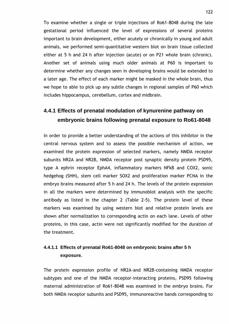
122 To examine whether a single or triple injections of Ro61-8048 during the late
gestational period influenced the level of expressions of several proteins
important to brain development, either acutely or chronically in young and adult
animals, we performed semi-quantitative western blot on brain tissue collected
either at 5 h and 24 h after injection (acute) or on P21 whole brain (chronic).
Another set of animals using much older animals at P60 is important to
determine whether any changes seen in developing brains would be extended to
a later age. The effect of each marker might be masked in the whole brain, thus
we hope to able to pick up any subtle changes in regional samples of P60 which
includes hippocampus, cerebellum, cortex and midbrain.
4.4.1 Effects of prenatal modulation of kynurenine pathway on
embryonic brains following prenatal exposure to Ro61-8048
In order to provide a better understanding of the actions of this inhibitor in the
central nervous system and to assess the possible mechanism of action, we
examined the protein expression of selected markers, namely NMDA receptor
subunits NR2A and NR2B, NMDA receptor post synaptic density protein PSD95,
type A ephrin receptor EphA4, inflammatory markers NFkB and COX2, sonic
hedgehog (SHH), stem cell marker SOX2 and proliferation marker PCNA in the
embryo brains measured after 5 h and 24 h. The levels of the protein expression
in all the markers were determined by immunoblot analysis with the specific
antibody as listed in the chapter 2 (Table 2-5). The protein level of these
markers was examined by using western blot and relative protein levels are
shown after normalization to corresponding actin on each lane. Levels of other
proteins, in this case, actin were not significantly modified for the duration of
the treatment.
4.4.1.1 Effects of prenatal Ro61-8048 on embryonic brains after 5 h
exposure.
The protein expression profile of NR2A-and NR2B-containing NMDA receptor
subtypes and one of the NMDA receptor-interacting proteins, PSD95 following
maternal administration of Ro61-8048 was examined in the embryo brains. For
both NMDA receptor subunits and PSD95, immunoreactive bands corresponding to
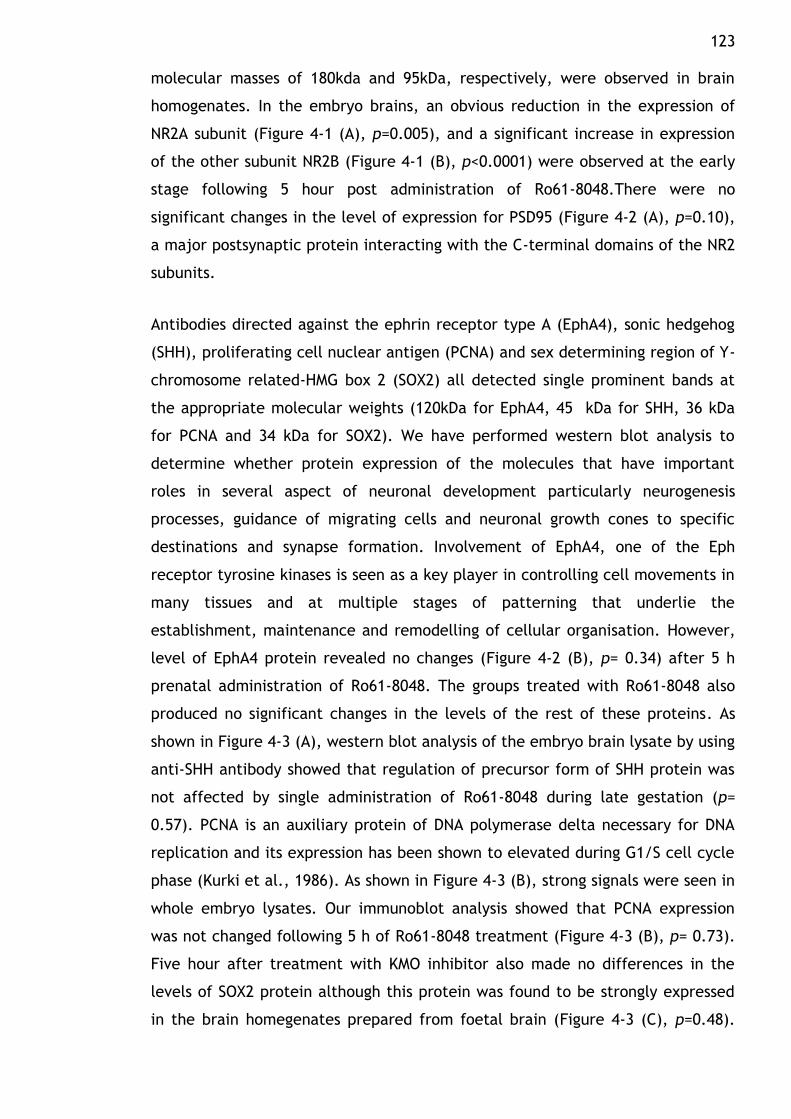
123 molecular masses of 180kda and 95kDa, respectively, were observed in brain
homogenates. In the embryo brains, an obvious reduction in the expression of
NR2A subunit (Figure 4-1 (A), p=0.005), and a significant increase in expression
of the other subunit NR2B (Figure 4-1 (B), p<0.0001) were observed at the early
stage following 5 hour post administration of Ro61-8048.There were no
significant changes in the level of expression for PSD95 (Figure 4-2 (A), p=0.10),
a major postsynaptic protein interacting with the C-terminal domains of the NR2
subunits.
Antibodies directed against the ephrin receptor type A (EphA4), sonic hedgehog
(SHH), proliferating cell nuclear antigen (PCNA) and sex determining region of Y-
chromosome related-HMG box 2 (SOX2) all detected single prominent bands at
the appropriate molecular weights (120kDa for EphA4, 45 kDa for SHH, 36 kDa
for PCNA and 34 kDa for SOX2). We have performed western blot analysis to
determine whether protein expression of the molecules that have important
roles in several aspect of neuronal development particularly neurogenesis
processes, guidance of migrating cells and neuronal growth cones to specific
destinations and synapse formation. Involvement of EphA4, one of the Eph
receptor tyrosine kinases is seen as a key player in controlling cell movements in
many tissues and at multiple stages of patterning that underlie the
establishment, maintenance and remodelling of cellular organisation. However,
level of EphA4 protein revealed no changes (Figure 4-2 (B), p= 0.34) after 5 h
prenatal administration of Ro61-8048. The groups treated with Ro61-8048 also
produced no significant changes in the levels of the rest of these proteins. As
shown in Figure 4-3 (A), western blot analysis of the embryo brain lysate by using
anti-SHH antibody showed that regulation of precursor form of SHH protein was
not affected by single administration of Ro61-8048 during late gestation (p=
0.57). PCNA is an auxiliary protein of DNA polymerase delta necessary for DNA
replication and its expression has been shown to elevated during G1/S cell cycle
phase (Kurki et al., 1986). As shown in Figure 4-3 (B), strong signals were seen in
whole embryo lysates. Our immunoblot analysis showed that PCNA expression
was not changed following 5 h of Ro61-8048 treatment (Figure 4-3 (B), p= 0.73).
Five hour after treatment with KMO inhibitor also made no differences in the
levels of SOX2 protein although this protein was found to be strongly expressed
in the brain homegenates prepared from foetal brain (Figure 4-3 (C), p=0.48).

124
We detected immunoreactive NFB p65 subunit and COX2 expression in embryo
as shown in Figure 4-4 (A & B). However, no changes in immunoreactive levels of
both markers were observed after 5 h injection with the enzyme inhibitor, Ro61-
8048 (NFB: p= 0.53; COX2: p= 0.51).
125 A
NR2A
0.0
0.2
0.4
0.6
0.8
Saline Ro61-8048
**
Op
tical d
en
sit
y (
OD
)
OD
pro
tein
/OD
acti
n
B
NR2B
0.0
0.2
0.4
0.6
0.8
1.0
Saline Ro61-8048
***
Op
tical d
en
sit
y (
OD
)
OD
pro
tein
/OD
acti
n
Figure 4-1 Expression of NMDA receptor subunits NR2A and NR2B proteins in the embryo brains after prenatal Ro61-8048. Pregnant dams were administered with Ro61-8048 i.p. on embryonic day 18 (E18) and 5 h later, embryo brains were obtained. Homogenized tissues of the brains from 6 embryos in each group were used for analysis. Blots were probed for NR2A and NR2B as marked, showing the presence of NR2A/NR2B containing complexes in the whole brain tissue. Representative immunoblots showing protein expression levels of NR2A (M.W. 180 kDa) and NR2B (M.W. 180 kDa) in the embryo brains, with actin (M.W. 42 kDa) shown as a loading control. Histogram showing relative NR2A and NR2B protein expression normalized to actin expression. Data were expressed as mean ± SEM. (A) Compared with saline-treated animals(S) (n=6), there was a signifcant decrease in NR2A protein expression in Ro61-8048 (R) (n=6)(**p<0.01). (B) Prenatal treatment with 100mg/kg Ro61-8048 showed significant increase in NR2B expression compared to control animals (n=6)(***p<0.001).
S S R R
**
** **
NR2A
Actin
S S R R
NR2B
Actin
126 A
PSD95
0.0
0.2
0.4
0.6
0.8
1.0
Saline Ro61-8048
Op
tical d
en
sit
y (
OD
)
OD
pro
tein
/OD
acti
n
B
EphA4
0.0
0.5
1.0
1.5
Saline Ro61-8048
Op
tical d
en
sit
y (
OD
)
OD
pro
tein
/OD
acti
n
Figure 4-2 Expression of PSD95 and EphA4 proteins in the embryo brains after prenatal Ro61-8048. Pregnant dams were administered with Ro61-8048 i.p. on embryonic day 18 (E18) and 5 h later, embryo brains were obtained. Homogenized tissues of the brains from 6 embryos in each group were used for analysis. Blots were probed for PSD95 and EphA4 as marked, showing the presence of PSD95 and EphA4 containing complexes in the whole brain tissue. Representative immunoblots showing protein expression levels of PSD95 (M.W. 95 kDa) and EphA4 (M.W. 120 kDa) in the embryo brains, with actin (M.W. 42 kDa) shown as a loading control. Histogram showing relative PSD95 and EphA4 protein expression normalized to actin expression. Data were expressed as mean ± SEM. The effect of the treatments were determined by performing student t-test and the differences between mean values were defined as significant when p<0.05.
** **
PSD95
Actin
EphA4
Actin
S S R R
S S R R
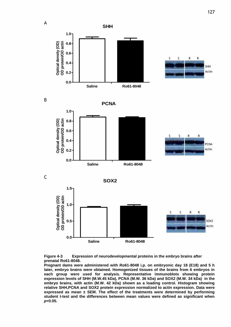
127 A
SHH
0.0
0.2
0.4
0.6
0.8
1.0
Saline Ro61-8048
Op
tical d
en
sit
y (
OD
)
OD
pro
tein
/OD
acti
n
B PCNA
0.0
0.2
0.4
0.6
0.8
1.0
Saline Ro61-8048
Op
tical d
en
sit
y (
OD
)
OD
pro
tein
/OD
acti
n
C SOX2
0.0
0.5
1.0
1.5
Saline Ro61-8048
Op
tical d
en
sit
y (
OD
)
OD
pro
tein
/OD
acti
n
Figure 4-3 Expression of neurodevelopmental proteins in the embryo brains after prenatal Ro61-8048. Pregnant dams were administered with Ro61-8048 i.p. on embryonic day 18 (E18) and 5 h later, embryo brains were obtained. Homogenized tissues of the brains from 6 embryos in each group were used for analysis. Representative immunoblots showing protein expression levels of SHH (M.W.45 kDa), PCNA (M.W. 36 kDa) and SOX2 (M.W. 34 kDa) in the embryo brains, with actin (M.W. 42 kDa) shown as a loading control. Histogram showing relative SHH,PCNA and SOX2 protein expression normalized to actin expression. Data were expressed as mean ± SEM. The effect of the treatments were determined by performing student t-test and the differences between mean values were defined as significant when p<0.05.
**
** **
SHH
Actin
PCNA
Actin
SOX2
Actin
S S R R
S S R R
S S R R
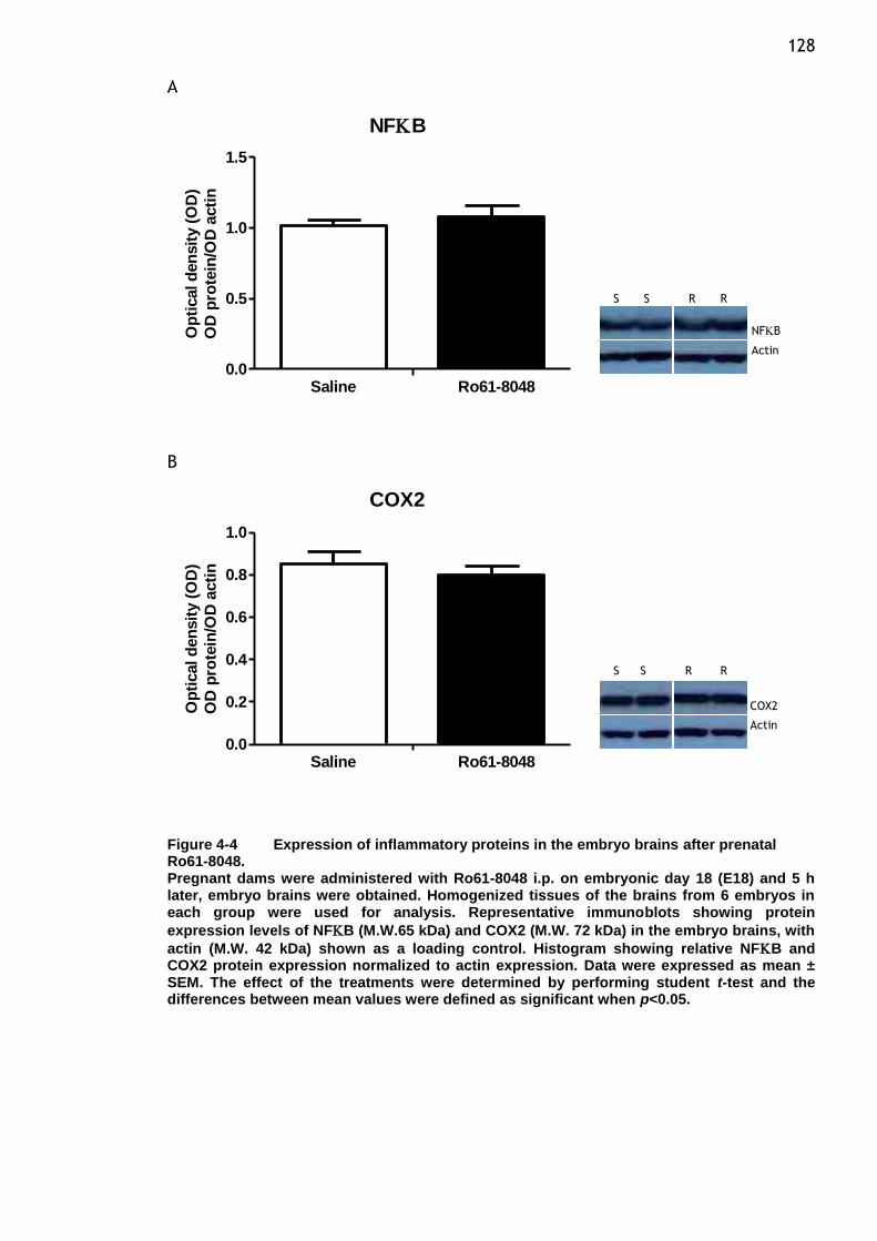
128 A
NFB
0.0
0.5
1.0
1.5
Saline Ro61-8048
Op
tical d
en
sit
y (
OD
)
OD
pro
tein
/OD
acti
n
B
COX2
0.0
0.2
0.4
0.6
0.8
1.0
Saline Ro61-8048
Op
tical d
en
sit
y (
OD
)
OD
pro
tein
/OD
acti
n
Figure 4-4 Expression of inflammatory proteins in the embryo brains after prenatal Ro61-8048. Pregnant dams were administered with Ro61-8048 i.p. on embryonic day 18 (E18) and 5 h later, embryo brains were obtained. Homogenized tissues of the brains from 6 embryos in each group were used for analysis. Representative immunoblots showing protein
expression levels of NFB (M.W.65 kDa) and COX2 (M.W. 72 kDa) in the embryo brains, with
actin (M.W. 42 kDa) shown as a loading control. Histogram showing relative NFB and COX2 protein expression normalized to actin expression. Data were expressed as mean ± SEM. The effect of the treatments were determined by performing student t-test and the differences between mean values were defined as significant when p<0.05.
** **
NFB
Actin
COX2
Actin
S S R R
S S R R
129
4.4.1.2 Effects of prenatal Ro61-8048 on embryonic brains after 24 h
exposure.
It has been shown previously that single administration of Ro61-8048 at E18 was
enough to significantly affect both subunits by a 43% reduction (NR2A) and an
almost 400% increase in the level of NR2B protein expression following 5 h post
injection. These changes were no longer observed by 24 h of treatment. It can
be seen that after the first time point, the expression remained slightly lower
compared to controls for the NR2A subunit (Figure 4-5 (A), p=0.49) while the
NR2B content returned to almost control levels after 24 h of treatment (Figure
4-5 (B),p=0.09). Twenty four hour after treatment also had no effect on the
level of PSD95, a major postsynaptic protein which interacts with the C-terminal
domain of the NR2 subunits (Figure 4-6 (A), p=0.93). Similar effect was also
observed in the level of EphA4 protein in the whole cell extracts from the brain
at 24 h (Figure 4-6 (B), p= 0.60).
The activity of SHH as a morphogen during CNS development involved regulating
the patterning of cellular responses that is controlled by the concentration and
duration of SHH exposure. The expression pattern of SHH is the result of the
combined activity of enhancers that control SHH transcription and regulate SHH
expression in different tissue (Traiffort et al., 1998). In order to further analyze
the potential roles exerted by SHH signalling in the nervous system of foetal
following gestational inhibition of kynurenine pathway, we investigated
developmental characteristic of SHH expression in whole cell lysates prepared
from embryo brain. As shown preciously in Figure 4-3 (A) we show that the time
course of Ro61-8048 mediated regulation of SHH protein expression revealed no
alteration 5 h following single administration of the compound. Interestingly,
when we examined the level of SHH protein 24 h after Ro61-8048 treatment, a
significant increase of the protein level was observed (Figure 4-7 (A), p= 0.04).
To further assess whether inhibition of the kynurenine pathway during gestation
affects expression of intrinsic proliferation marker, the levels of PCNA protein in
the Ro61-8048-treated group was compared with controls. Our immunoblot
analysis showed that PCNA expression was not changed following 24 h of Ro61-
8048 treatment (Figure 4-7 (B), p= 0.18). As SOX2 might represent a useful in
vivo marker for neural progenitor cells in the CNS, we eexmined the expression
130 patterns of SOX2 protein in the embryo 24 h after maternal administration of
KMO inhibitor. Western blot analysis with goat anti-SOX2 antibody of total
protein in the brain homogenate resulted in a single band of 34 kDa. The
antibody did not cross react with other SOX-B1 subfamily members i.e. SOX1
and SOX3. Thus the detection capability of this antibody allowed us to carefully
examine SOX2 expression in the subsequent sets of experiments. As can be seen
in Figure 4-7 (C), SOX2 was found to be strongly expressed, although no change
was seen in the expression of this protein after 24 h post treatment with Ro61-
8048 (p= 0.60). Five hour after treatment with Ro61-8048 had no significant
effect on the level of inflammtory markers used in this study. However, prenatal
Ro61-8048 treatment induced strong immunoreactivity for NFB in the embryo
brain 24 h post injection (Figure 4-8 (A), p=0.01) with no differences in COX2
protein level (Figure 4-8 (B), p= 0.55).
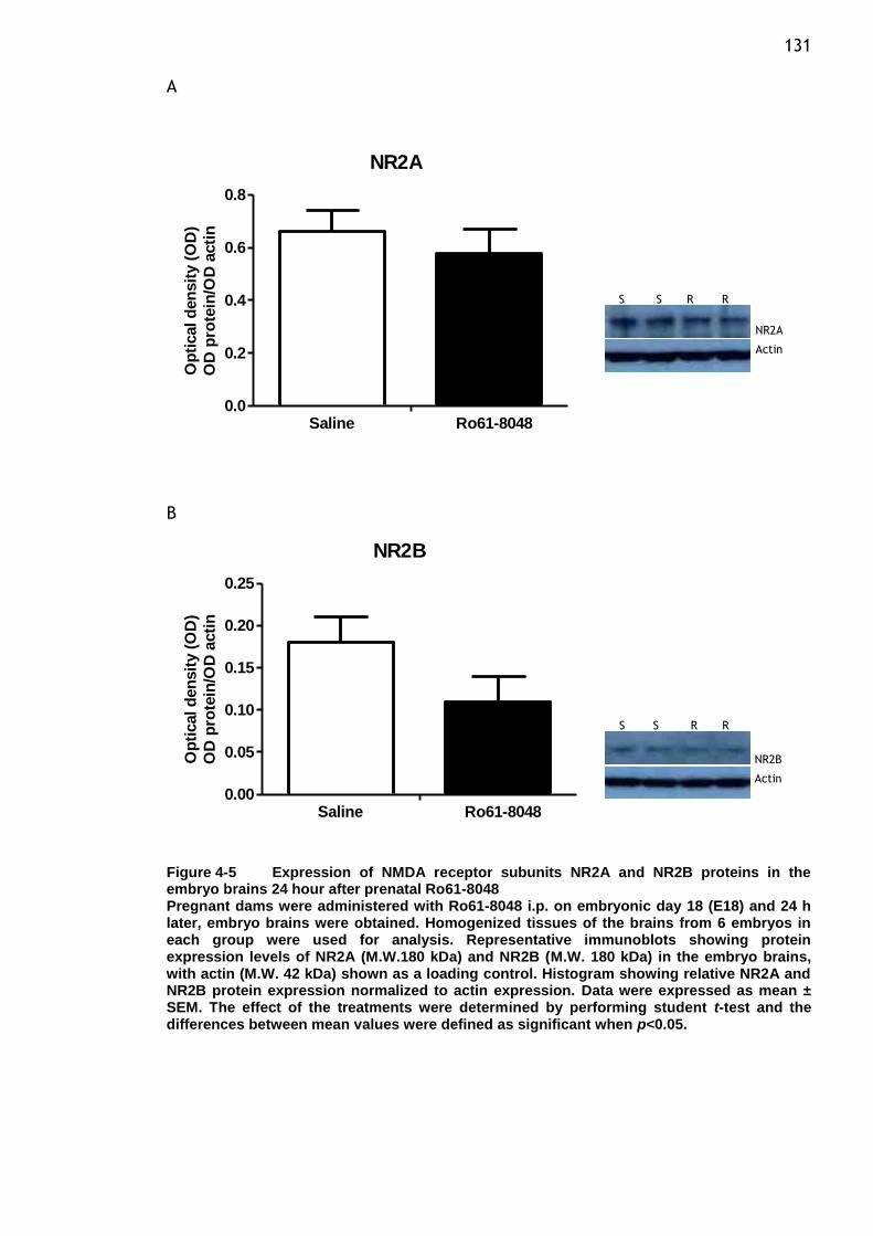
131 A
NR2A
0.0
0.2
0.4
0.6
0.8
Saline Ro61-8048
Op
tical d
en
sit
y (
OD
)
OD
pro
tein
/OD
acti
n
B
NR2B
0.00
0.05
0.10
0.15
0.20
0.25
Saline Ro61-8048
Op
tical d
en
sit
y (
OD
)
OD
pro
tein
/OD
acti
n
Figure 4-5 Expression of NMDA receptor subunits NR2A and NR2B proteins in the embryo brains 24 hour after prenatal Ro61-8048 Pregnant dams were administered with Ro61-8048 i.p. on embryonic day 18 (E18) and 24 h later, embryo brains were obtained. Homogenized tissues of the brains from 6 embryos in each group were used for analysis. Representative immunoblots showing protein expression levels of NR2A (M.W.180 kDa) and NR2B (M.W. 180 kDa) in the embryo brains, with actin (M.W. 42 kDa) shown as a loading control. Histogram showing relative NR2A and NR2B protein expression normalized to actin expression. Data were expressed as mean ± SEM. The effect of the treatments were determined by performing student t-test and the differences between mean values were defined as significant when p<0.05.
** **
NR2A
Actin
NR2B
Actin
S S R R
S S R R
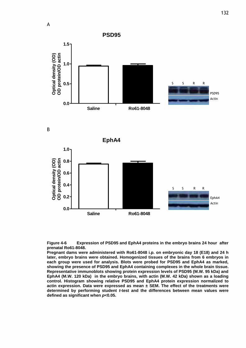
132 A
PSD95
0.0
0.5
1.0
1.5
Saline Ro61-8048
Op
tical d
en
sit
y (
OD
)
OD
pro
tein
/OD
acti
n
B
EphA4
0.0
0.2
0.4
0.6
0.8
1.0
Saline Ro61-8048
Op
tical d
en
sit
y (
OD
)
OD
pro
tein
/OD
acti
n
Figure 4-6 Expression of PSD95 and EphA4 proteins in the embryo brains 24 hour after prenatal Ro61-8048. Pregnant dams were administered with Ro61-8048 i.p. on embryonic day 18 (E18) and 24 h later, embryo brains were obtained. Homogenized tissues of the brains from 6 embryos in each group were used for analysis. Blots were probed for PSD95 and EphA4 as marked, showing the presence of PSD95 and EphA4 containing complexes in the whole brain tissue. Representative immunoblots showing protein expression levels of PSD95 (M.W. 95 kDa) and EphA4 (M.W. 120 kDa) in the embryo brains, with actin (M.W. 42 kDa) shown as a loading control. Histogram showing relative PSD95 and EphA4 protein expression normalized to actin expression. Data were expressed as mean ± SEM. The effect of the treatments were determined by performing student t-test and the differences between mean values were defined as significant when p<0.05.
** **
PSD95
Actin
EphA4
Actin
S S R R
S S R R
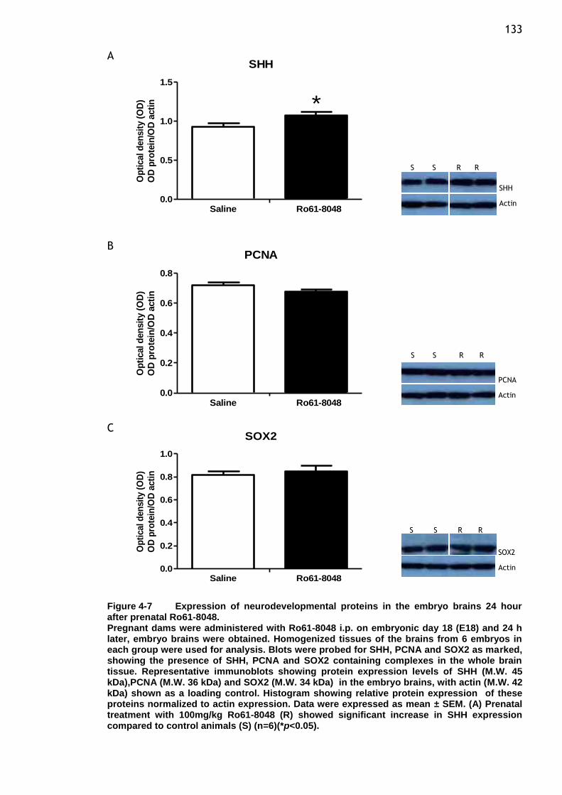
133 A
SHH
0.0
0.5
1.0
1.5
Saline Ro61-8048
*
Op
tical d
en
sit
y (
OD
)
OD
pro
tein
/OD
acti
n
B
PCNA
0.0
0.2
0.4
0.6
0.8
Saline Ro61-8048
Op
tical d
en
sit
y (
OD
)
OD
pro
tein
/OD
acti
n
C SOX2
0.0
0.2
0.4
0.6
0.8
1.0
Saline Ro61-8048
Op
tical d
en
sit
y (
OD
)
OD
pro
tein
/OD
acti
n
Figure 4-7 Expression of neurodevelopmental proteins in the embryo brains 24 hour after prenatal Ro61-8048. Pregnant dams were administered with Ro61-8048 i.p. on embryonic day 18 (E18) and 24 h later, embryo brains were obtained. Homogenized tissues of the brains from 6 embryos in each group were used for analysis. Blots were probed for SHH, PCNA and SOX2 as marked, showing the presence of SHH, PCNA and SOX2 containing complexes in the whole brain tissue. Representative immunoblots showing protein expression levels of SHH (M.W. 45 kDa),PCNA (M.W. 36 kDa) and SOX2 (M.W. 34 kDa) in the embryo brains, with actin (M.W. 42 kDa) shown as a loading control. Histogram showing relative protein expression of these proteins normalized to actin expression. Data were expressed as mean ± SEM. (A) Prenatal treatment with 100mg/kg Ro61-8048 (R) showed significant increase in SHH expression compared to control animals (S) (n=6)(*p<0.05).
**
** **
SHH
Actin
PCNA
Actin
SOX2
Actin
S S R R
S S R R
S S R R

134 A
NFB
0.0
0.5
1.0
1.5
Saline Ro61-8048
*O
pti
cal d
en
sit
y (
OD
)
OD
pro
tein
/OD
acti
n
B
COX2
0.0
0.2
0.4
0.6
0.8
Saline Ro61-8048
Op
tical d
en
sit
y (
OD
)
OD
pro
tein
/OD
acti
n
Figure 4-8 Expression of inflammatory proteins in the embryo brains 24 hour after prenatal Ro61-8048. Pregnant dams were administered with Ro61-8048 i.p. on embryonic day 18 (E18) and 24 h later, embryo brains were obtained. Homogenized tissues of the brains from 6 embryos in
each group were used for analysis. Blots were probed for NFB and COX2 as marked,
showing the presence of NFB and COX2 containing complexes in the whole brain tissue.
Representative immunoblots showing protein expression levels of NFB (M.W. 65 kDa) and COX2 (M.W. 72 kDa) in the embryo brains, with actin (M.W. 42 kDa) shown as a loading control. Histogram showing relative protein expression of these proteins normalized to actin expression. Data were expressed as mean ± SEM. (A) Prenatal treatment with
100mg/kg Ro61-8048 (R) showed significant increase in NFB expression compared to control animals (S) (n=6) (*p<0.05).
** **
NFB
Actin
COX2
Actin
S S R R
S S R R
135
4.4.2 Effects of prenatal Ro61-8048 in P21 neonates
In addition to the expression of proteins studied for the acute response to Ro61-
8048 in embryos, the levels of expression were also examined at P21. In the
brains from P21 pups exposed to Ro61-8048, the greatest changes noted in these
animals were of the NMDA receptor subunits, NR2A and NR2B. The expression of
both subunits NR2A and NR2B showed a highly significant upregulation producing
approximately 16% (Figure 4-9 (A), p= 0.0045) and 27% (Figure 4-9 (B), p= 0.03)
increases, respectively. Analysis of the levels of both subunits showed a
progressive increase in the full- length proteins, indicative of both NR2 subunits
being regulated in a time-dependent way. We also characterized the expression
of PSD95, a major postsynaptic protein interacting with the C-terminal domains
of the NR2 subunits. Five and 24 hours after maternal administration of Ro61-
8048 had no effect on the level of this protein. However, at P21 subsequently
altered the development of PSD95 protein expression, Ro61-8048-treated
animals had significantly greater expression than saline controls (Figure 4-10 (A),
p= 0.03). This finding altogether suggests an influence on the kynurenine
pathway and involvement of NMDA receptor function to maintaining normal
regulation during brain development. During postnatal period as examined at
P21, expression of the EphA4 receptor for secreted ephrin molecules remained
unchanged in the treated pups (Figure 4-10 (B), p= 0.95).
The sonic hedgehog protein (SHH), which is intimately involved in the patterning
and orientation of tissues and cell growth, was reduced to a significant extent
after exposure to Ro61-8048 (Figure 4-11 (A), p= 0.02). To further assess
whether inhibition of the kynurenine pathway during gestation affects
expression of intrinsic proliferation markers at a later developmental age,PCNA
a protein often studied as a marker of the earliest phases of DNA synthesis, was
more highly expressed in the treated animals compred with controls (Figure 4-11
(B), p= 0.02) while levels of the SOX2 protein were unaffected (Figure 4-11 (C),
p= 0.45). Finally, levels of the inflammation transcription factor, NFB and the
inducible form of cyclooxygenase (COX2), showed a significant induction of NFB
reactivity in the total homogenates of P21 brains (Figure 4-12 (A), p= 0.02) with
no change in COX2 level as observed in treated animals (Figure 4-12 (B), p=
0.64).
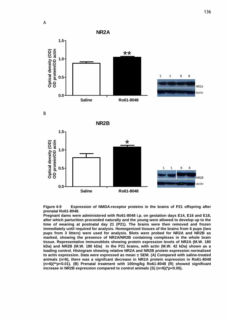
136 A
NR2A
0.0
0.5
1.0
1.5
Saline Ro61-8048
**O
pti
cal d
en
sit
y (
OD
)
OD
pro
tein
/OD
acti
n
B
NR2B
0.0
0.5
1.0
1.5
Saline Ro61-8048
*
Op
tical d
en
sit
y (
OD
)
OD
pro
tein
/OD
acti
n
Figure 4-9 Expression of NMDA-receptor proteins in the brains of P21 offspring after prenatal Ro61-8048. Pregnant dams were administered with Ro61-8048 i.p. on gestation days E14, E16 and E18, after which parturition proceeded naturally and the young were allowed to develop up to the time of weaning at postnatal day 21 (P21). The brains were then removed and frozen immediately until required for analysis. Homogenized tissues of the brains from 6 pups (two pups from 3 litters) were used for analysis. Blots were probed for NR2A and NR2B as marked, showing the presence of NR2A/NR2B containing complexes in the whole brain tissue. Representative immunoblots showing protein expression levels of NR2A (M.W. 180 kDa) and NR2B (M.W. 180 kDa) in the P21 brains, with actin (M.W. 42 kDa) shown as a loading control. Histogram showing relative NR2A and NR2B protein expression normalized to actin expression. Data were expressed as mean ± SEM. (A) Compared with saline-treated animals (n=6), there was a signifcant decrease in NR2A protein expression in Ro61-8048 (n=6)(**p<0.01). (B) Prenatal treatment with 100mg/kg Ro61-8048 (R) showed significant increase in NR2B expression compared to control animals (S) (n=6)(*p<0.05).
** **
NR2A
Actin
NR2B
Actin
S S R R
S S R R
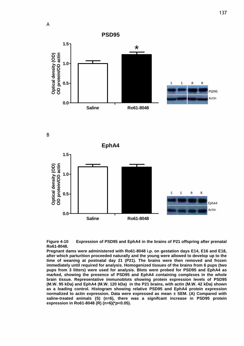
137 A
PSD95
0.0
0.5
1.0
1.5
Saline Ro61-8048
*O
pti
cal d
en
sit
y (
OD
)
OD
pro
tein
/OD
acti
n
B
EphA4
0.0
0.5
1.0
1.5
Saline Ro61-8048
Op
tical d
en
sit
y (
OD
)
OD
pro
tein
/OD
acti
n
Figure 4-10 Expression of PSD95 and EphA4 in the brains of P21 offspring after prenatal Ro61-8048. Pregnant dams were administered with Ro61-8048 i.p. on gestation days E14, E16 and E18, after which parturition proceeded naturally and the young were allowed to develop up to the time of weaning at postnatal day 21 (P21). The brains were then removed and frozen immediately until required for analysis. Homogenized tissues of the brains from 6 pups (two pups from 3 litters) were used for analysis. Blots were probed for PSD95 and EphA4 as marked, showing the presence of PSD95 and EphA4 containing complexes in the whole brain tissue. Representative immunoblots showing protein expression levels of PSD95 (M.W. 95 kDa) and EphA4 (M.W. 120 kDa) in the P21 brains, with actin (M.W. 42 kDa) shown as a loading control. Histogram showing relative PSD95 and EphA4 protein expression normalized to actin expression. Data were expressed as mean ± SEM. (A) Compared with saline-treated animals (S) (n=6), there was a signifcant increase in PSD95 protein expression in Ro61-8048 (R) (n=6)(*p<0.05).
** **
PSD95
Actin
EphA4
Actin
S S R R
S S R R
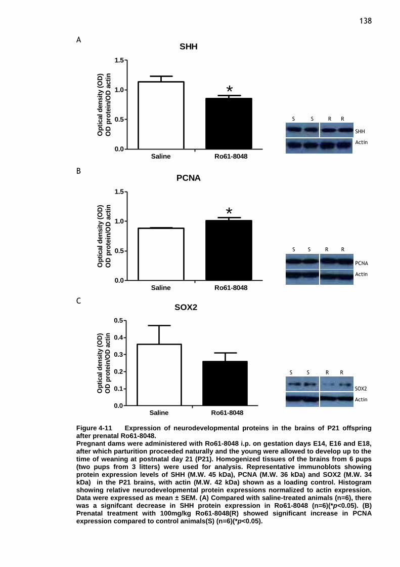
138 A
SHH
0.0
0.5
1.0
1.5
Saline Ro61-8048
*O
pti
cal d
en
sit
y (
OD
)
OD
pro
tein
/OD
acti
n
B PCNA
0.0
0.5
1.0
1.5
Saline Ro61-8048
*
Op
tical d
en
sit
y (
OD
)
OD
pro
tein
/OD
acti
n
C SOX2
0.0
0.1
0.2
0.3
0.4
0.5
Saline Ro61-8048
Op
tical d
en
sit
y (
OD
)
OD
pro
tein
/OD
acti
n
Figure 4-11 Expression of neurodevelopmental proteins in the brains of P21 offspring after prenatal Ro61-8048. Pregnant dams were administered with Ro61-8048 i.p. on gestation days E14, E16 and E18, after which parturition proceeded naturally and the young were allowed to develop up to the time of weaning at postnatal day 21 (P21). Homogenized tissues of the brains from 6 pups (two pups from 3 litters) were used for analysis. Representative immunoblots showing protein expression levels of SHH (M.W. 45 kDa), PCNA (M.W. 36 kDa) and SOX2 (M.W. 34 kDa) in the P21 brains, with actin (M.W. 42 kDa) shown as a loading control. Histogram showing relative neurodevelopmental protein expressions normalized to actin expression. Data were expressed as mean ± SEM. (A) Compared with saline-treated animals (n=6), there was a signifcant decrease in SHH protein expression in Ro61-8048 (n=6)(*p<0.05). (B) Prenatal treatment with 100mg/kg Ro61-8048(R) showed significant increase in PCNA expression compared to control animals(S) (n=6)(*p<0.05).
**
** **
SHH
Actin
PCNA
Actin
SOX2
Actin
S S R R
S S R R
S S R R
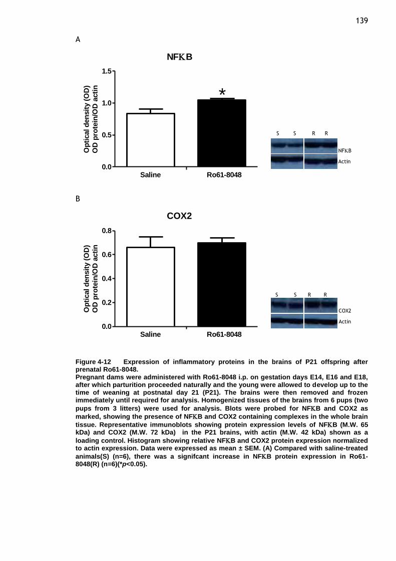
139 A
NFB
0.0
0.5
1.0
1.5
Saline Ro61-8048
*O
pti
cal d
en
sit
y (
OD
)
OD
pro
tein
/OD
acti
n
B
COX2
0.0
0.2
0.4
0.6
0.8
Saline Ro61-8048
*
Op
tical d
en
sit
y (
OD
)
OD
pro
tein
/OD
acti
n
Figure 4-12 Expression of inflammatory proteins in the brains of P21 offspring after prenatal Ro61-8048. Pregnant dams were administered with Ro61-8048 i.p. on gestation days E14, E16 and E18, after which parturition proceeded naturally and the young were allowed to develop up to the time of weaning at postnatal day 21 (P21). The brains were then removed and frozen immediately until required for analysis. Homogenized tissues of the brains from 6 pups (two
pups from 3 litters) were used for analysis. Blots were probed for NFB and COX2 as
marked, showing the presence of NFB and COX2 containing complexes in the whole brain
tissue. Representative immunoblots showing protein expression levels of NFB (M.W. 65 kDa) and COX2 (M.W. 72 kDa) in the P21 brains, with actin (M.W. 42 kDa) shown as a
loading control. Histogram showing relative NFB and COX2 protein expression normalized to actin expression. Data were expressed as mean ± SEM. (A) Compared with saline-treated
animals(S) (n=6), there was a signifcant increase in NFB protein expression in Ro61-8048(R) (n=6)(*p<0.05).
** **
NFB
Actin
COX2
Actin
S S R R
S S R R
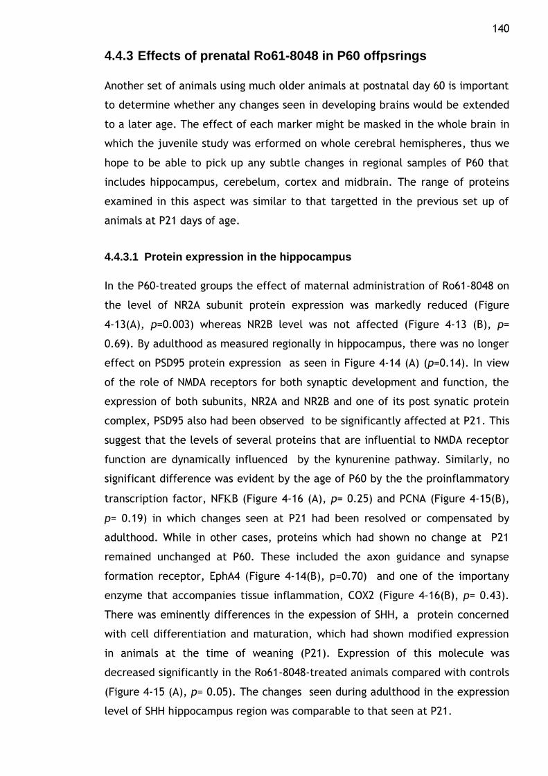
140
4.4.3 Effects of prenatal Ro61-8048 in P60 offpsrings
Another set of animals using much older animals at postnatal day 60 is important
to determine whether any changes seen in developing brains would be extended
to a later age. The effect of each marker might be masked in the whole brain in
which the juvenile study was erformed on whole cerebral hemispheres, thus we
hope to be able to pick up any subtle changes in regional samples of P60 that
includes hippocampus, cerebelum, cortex and midbrain. The range of proteins
examined in this aspect was similar to that targetted in the previous set up of
animals at P21 days of age.
4.4.3.1 Protein expression in the hippocampus
In the P60-treated groups the effect of maternal administration of Ro61-8048 on
the level of NR2A subunit protein expression was markedly reduced (Figure
4-13(A), p=0.003) whereas NR2B level was not affected (Figure 4-13 (B), p=
0.69). By adulthood as measured regionally in hippocampus, there was no longer
effect on PSD95 protein expression as seen in Figure 4-14 (A) (p=0.14). In view
of the role of NMDA receptors for both synaptic development and function, the
expression of both subunits, NR2A and NR2B and one of its post synatic protein
complex, PSD95 also had been observed to be significantly affected at P21. This
suggest that the levels of several proteins that are influential to NMDA receptor
function are dynamically influenced by the kynurenine pathway. Similarly, no
significant difference was evident by the age of P60 by the the proinflammatory
transcription factor, NFB (Figure 4-16 (A), p= 0.25) and PCNA (Figure 4-15(B),
p= 0.19) in which changes seen at P21 had been resolved or compensated by
adulthood. While in other cases, proteins which had shown no change at P21
remained unchanged at P60. These included the axon guidance and synapse
formation receptor, EphA4 (Figure 4-14(B), p=0.70) and one of the importany
enzyme that accompanies tissue inflammation, COX2 (Figure 4-16(B), p= 0.43).
There was eminently differences in the expession of SHH, a protein concerned
with cell differentiation and maturation, which had shown modified expression
in animals at the time of weaning (P21). Expression of this molecule was
decreased significantly in the Ro61-8048-treated animals compared with controls
(Figure 4-15 (A), p= 0.05). The changes seen during adulthood in the expression
level of SHH hippocampus region was comparable to that seen at P21.
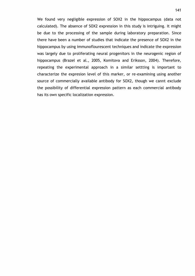
141 We found very negligible expression of SOX2 in the hippocampus (data not
calculated). The absence of SOX2 expression in this study is intriguing. It might
be due to the processing of the sample during laboratory preparation. Since
there have been a number of studies that indicate the presence of SOX2 in the
hippocampus by using immunoflourescent techniques and indicate the expression
was largely due to proliferating neural progenitors in the neurogenic region of
hippocampus (Brazel et al., 2005, Komitova and Eriksson, 2004). Therefore,
repeating the experimental approach in a similar settting is important to
characterize the expresion level of this marker, or re-examining using another
source of commercially available antibody for SOX2, though we cannt exclude
the possibility of differential expression pattern as each commercial antibody
has its own specific localization expression.
142
A
NR2A
0.0
0.5
1.0
1.5
Saline Ro61-8048
**
Op
tical
den
sit
y (
OD
)
OD
pro
tein
/OD
acti
n
B
NR2B
0.0
0.5
1.0
1.5
2.0
Saline Ro61-8048
Op
tical
den
sit
y (
OD
)
OD
pro
tein
/OD
acti
n
Figure 4-13 Expression of NMDA receptor subunits in the hippocampus of control and Ro61-8048-treated animals. Compound Ro61-8048 was administered i.p. to the pregnant dam at days E14, E16 and E18 of gestation while groups of control animals were injected with tha saline vehicle. Parturition was allowed to proceed naturally and the young were allowed to develop up to postnatal day 60 (P60). After euthanasia the brains were then removed and whole hippocampi was snap frozen until required for analysis. Homogenized tissues of the brains were used for analysis. Blots were probed for NR2A and NR2B as marked, showing the presence of NR2A/NR2B containing complexes in the whole hippocampi tissue. Representative immunoblots showing protein expression levels of NR2A (M.W. 180 kDa) and NR2B (M.W. 180 kDa) in the hippocampus, with actin (M.W. 42 kDa) shown as a loading control. Histogram showing relative NR2A and NR2B protein expression normalized to actin expression. Data were expressed as mean ± SEM (n=6) for Ro61-8048, n=4 for control). (A) Compared with saline-treated animals(S), there was a signifcant decrease in NR2A protein expression in Ro61-8048(R) (**p<0.01).
** **
NR2A
Actin
S S R R
NR2B
Actin
S S R R
143 A
PSD95
0.0
0.5
1.0
1.5
2.0
Saline Ro61-8048
Op
tical
den
sit
y (
OD
)
OD
pro
tein
/OD
acti
n
B
EphA4
0.0
0.5
1.0
1.5
2.0
Saline Ro61-8048
Op
tical
den
sit
y (
OD
)
OD
pro
tein
/OD
acti
n
Figure 4-14 Expression of PSD95 and EphA4 in the hippocampus of control and Ro61-8048-treated animals. Compound Ro61-8048 was administered i.p. to the pregnant dam at days E14, E16 and E18 of gestation while groups of control animals were injected with tha saline vehicle. Parturition was allowed to proceed naturally and the young were allowed to develop up to postnatal day 60 (P60). After euthanasia the brains were then removed and whole hippocampi was snap frozen until required for analysis. Homogenized tissues of the brains were used for analysis. Blots were probed for PSD95 and EphA4 as marked, showing the presence of PSD95 and EphA4 containing complexes in the whole hippocampi tissue. Representative immunoblots showing protein expression levels of PSD95 (M.W. 95 kDa) and EphA4 (M.W. 120 kDa) in the hippocampus, with actin (M.W. 42 kDa) shown as a loading control. Histogram showing relative PSD95 and EphA4 protein expression normalized to actin expression. Data were expressed as mean ± SEM (n=6 for Ro61-8048, n=4 for control).
** **
PSD95
Actin
EphA4
Actin
S S R R
S S R R
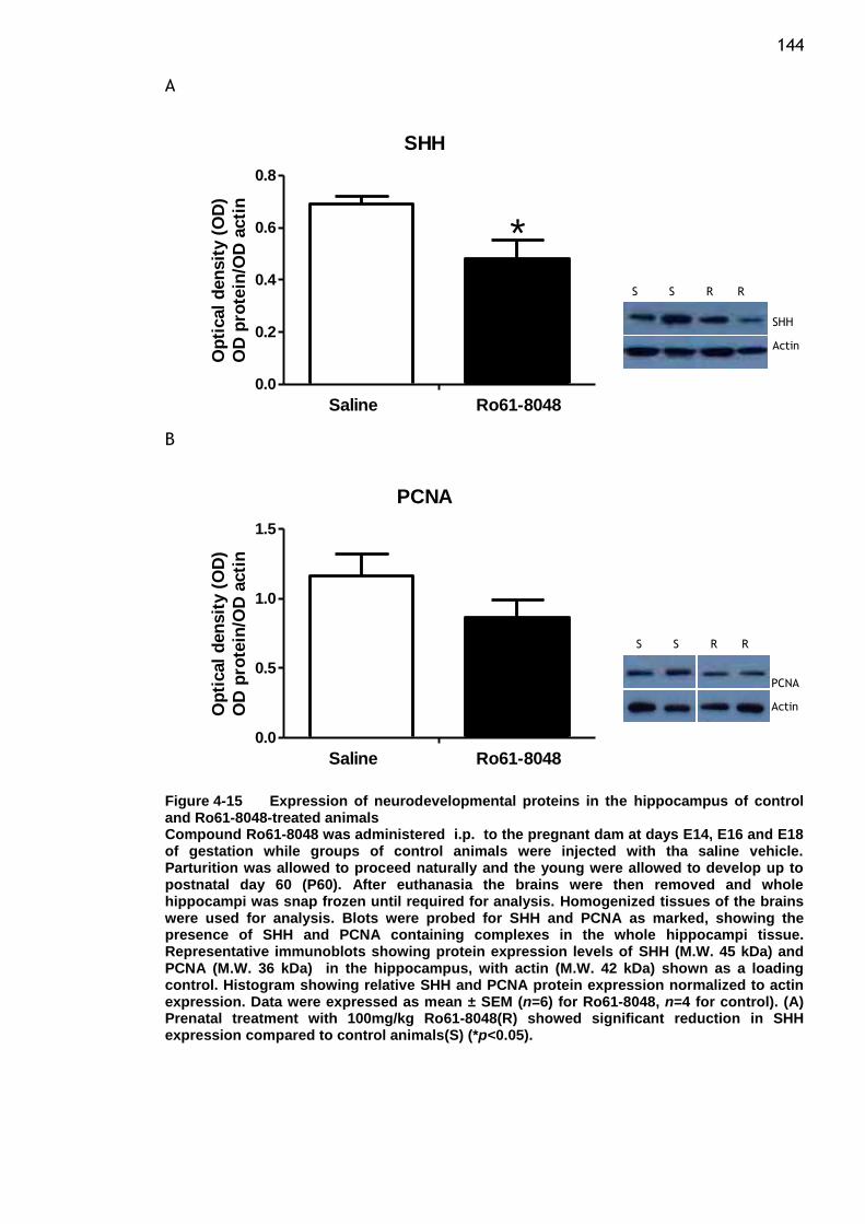
144 A
SHH
0.0
0.2
0.4
0.6
0.8
Saline Ro61-8048
*O
pti
cal
den
sit
y (
OD
)
OD
pro
tein
/OD
acti
n
B
PCNA
0.0
0.5
1.0
1.5
Saline Ro61-8048
Op
tical
den
sit
y (
OD
)
OD
pro
tein
/OD
acti
n
Figure 4-15 Expression of neurodevelopmental proteins in the hippocampus of control and Ro61-8048-treated animals Compound Ro61-8048 was administered i.p. to the pregnant dam at days E14, E16 and E18 of gestation while groups of control animals were injected with tha saline vehicle. Parturition was allowed to proceed naturally and the young were allowed to develop up to postnatal day 60 (P60). After euthanasia the brains were then removed and whole hippocampi was snap frozen until required for analysis. Homogenized tissues of the brains were used for analysis. Blots were probed for SHH and PCNA as marked, showing the presence of SHH and PCNA containing complexes in the whole hippocampi tissue. Representative immunoblots showing protein expression levels of SHH (M.W. 45 kDa) and PCNA (M.W. 36 kDa) in the hippocampus, with actin (M.W. 42 kDa) shown as a loading control. Histogram showing relative SHH and PCNA protein expression normalized to actin expression. Data were expressed as mean ± SEM (n=6) for Ro61-8048, n=4 for control). (A) Prenatal treatment with 100mg/kg Ro61-8048(R) showed significant reduction in SHH expression compared to control animals(S) (*p<0.05).
**
** **
SHH
Actin
PCNA
Actin
S S R R
S S R R
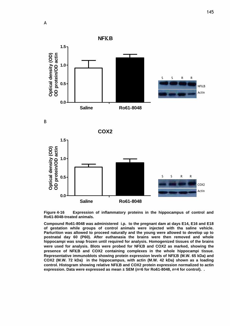
145 A
NFB
0.0
0.5
1.0
1.5
Saline Ro61-8048
Op
tical
den
sit
y (
OD
)
OD
pro
tein
/OD
acti
n
B
COX2
0.0
0.5
1.0
1.5
Saline Ro61-8048
Op
tical
den
sit
y (
OD
)
OD
pro
tein
/OD
acti
n
Figure 4-16 Expression of inflammatory proteins in the hippocampus of control and Ro61-8048-treated animals.
Compound Ro61-8048 was administered i.p. to the pregnant dam at days E14, E16 and E18 of gestation while groups of control animals were injected with tha saline vehicle. Parturition was allowed to proceed naturally and the young were allowed to develop up to postnatal day 60 (P60). After euthanasia the brains were then removed and whole hippocampi was snap frozen until required for analysis. Homogenized tissues of the brains
were used for analysis. Blots were probed for NFB and COX2 as marked, showing the
presence of NFB and COX2 containing complexes in the whole hippocampi tissue.
Representative immunoblots showing protein expression levels of NFB (M.W. 65 kDa) and COX2 (M.W. 72 kDa) in the hippocampus, with actin (M.W. 42 kDa) shown as a loading
control. Histogram showing relative NFB and COX2 protein expression normalized to actin expression. Data were expressed as mean ± SEM (n=6 for Ro61-8048, n=4 for control). .
** **
NFB
Actin
COX2
Actin
S S R R
S S R R
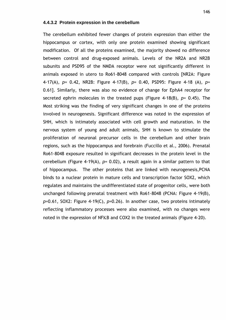
146
4.4.3.2 Protein expression in the cerebellum
The cerebellum exhibited fewer changes of protein expression than either the
hippocampus or cortex, with only one protein examined showing significant
modification. Of all the proteins examined, the majority showed no difference
between control and drug-exposed animals. Levels of the NR2A and NR2B
subunits and PSD95 of the NMDA receptor were not significantly different in
animals exposed in utero to Ro61-8048 compared with controls [NR2A: Figure
4-17(A), p= 0.42, NR2B: Figure 4-17(B), p= 0.40, PSD95: Figure 4-18 (A), p=
0.61]. Similarly, there was also no evidence of change for EphA4 receptor for
secreted ephrin molecules in the treated pups (Figure 4-18(B), p= 0.45). The
Most striking was the finding of very significant changes in one of the proteins
involved in neurogenesis. Significant difference was noted in the expression of
SHH, which is intimately associated with cell growth and maturation. In the
nervous system of young and adult animals, SHH is known to stimulate the
proliferation of neuronal precursor cells in the cerebellum and other brain
regions, such as the hippocampus and forebrain (Fuccillo et al., 2006). Prenatal
Ro61-8048 exposure resulted in significant decreases in the protein level in the
cerebellum (Figure 4-19(A), p= 0.02), a result again in a similar pattern to that
of hippocampus. The other proteins that are linked with neurogenesis,PCNA
binds to a nuclear protein in mature cells and transcription factor SOX2, which
regulates and maintains the undifferentiated state of progenitor cells, were both
unchanged following prenatal treatment with Ro61-8048 (PCNA: Figure 4-19(B),
p=0.61, SOX2: Figure 4-19(C), p=0.26). In another case, two proteins intimately
reflecting inflammatory processes were also examined, with no changes were
noted in the expression of NFB and COX2 in the treated animals (Figure 4-20).
147 A
NR2A
0.0
0.2
0.4
0.6
0.8
1.0
Saline Ro61-8048
Op
tical
den
sit
y (
OD
)
OD
pro
tein
/OD
acti
n
B
NR2B
0.0
0.5
1.0
1.5
Saline Ro61-8048
Op
tical
den
sit
y (
OD
)
OD
pro
tein
/OD
acti
n
Figure 4-17 Expression of NMDA receptor subunits in the cerebellum of control and Ro61-8048-treated animals. Compound Ro61-8048 was administered i.p. to the pregnant dam at days E14, E16 and E18 of gestation while groups of control animals were injected with tha saline vehicle. Parturition was allowed to proceed naturally and the young were allowed to develop up to postnatal day 60 (P60). After euthanasia the brains were then removed and whole cerebellum was snap frozen until required for analysis. Homogenized tissues of the brains from 6 pups (two pups from 3 litters) were used for analysis. Blots were probed for NR2A and NR2B as marked, showing the presence of NR2A/NR2B containing complexes in the whole cerebellum tissue. Representative immunoblots showing protein expression levels of NR2A (M.W. 180 kDa) and NR2B (M.W. 180 kDa) in the cerebellum, with actin (M.W. 42 kDa) shown as a loading control. Histogram showing relative NR2A and NR2B protein expression normalized to actin expression. Data were expressed as mean ± SEM.
** **
NR2A
Actin
NR2B
Actin
S S R R
S S R R
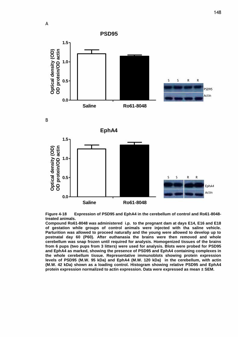
148 A
PSD95
0.0
0.5
1.0
1.5
Saline Ro61-8048
Op
tical
den
sit
y (
OD
)
OD
pro
tein
/OD
acti
n
B
EphA4
0.0
0.5
1.0
1.5
Saline Ro61-8048
Op
tical
den
sit
y (
OD
)
OD
pro
tein
/OD
acti
n
Figure 4-18 Expression of PSD95 and EphA4 in the cerebellum of control and Ro61-8048-treated animals. Compound Ro61-8048 was administered i.p. to the pregnant dam at days E14, E16 and E18 of gestation while groups of control animals were injected with tha saline vehicle. Parturition was allowed to proceed naturally and the young were allowed to develop up to postnatal day 60 (P60). After euthanasia the brains were then removed and whole cerebellum was snap frozen until required for analysis. Homogenized tissues of the brains from 6 pups (two pups from 3 litters) were used for analysis. Blots were probed for PSD95 and EphA4 as marked, showing the presence of PSD95 and EphA4 containing complexes in the whole cerebellum tissue. Representative immunoblots showing protein expression levels of PSD95 (M.W. 95 kDa) and EphA4 (M.W. 120 kDa) in the cerebellum, with actin (M.W. 42 kDa) shown as a loading control. Histogram showing relative PSD95 and EphA4 protein expression normalized to actin expression. Data were expressed as mean ± SEM.
** **
PSD95
Actin
EphA4
Actin
S S R R
S S R R
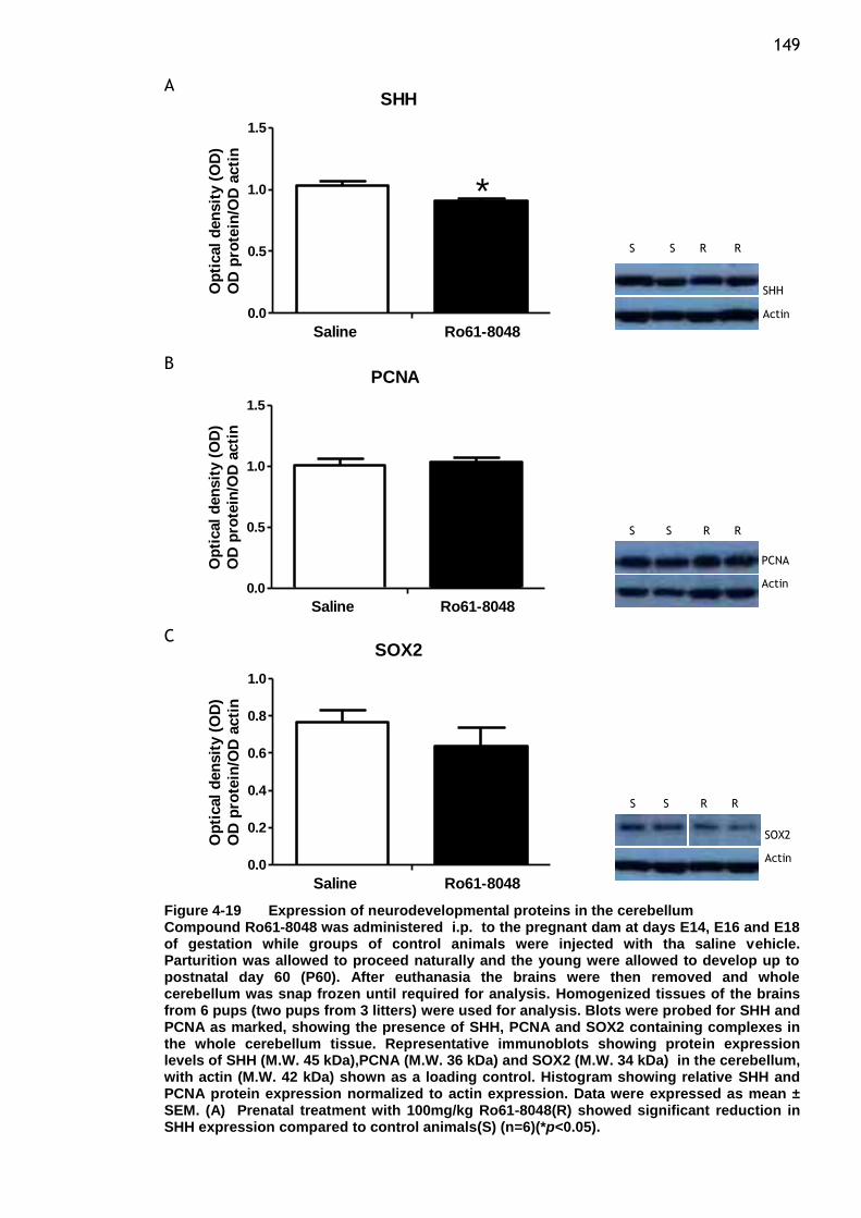
149 A
SHH
0.0
0.5
1.0
1.5
Saline Ro61-8048
*O
pti
cal
den
sit
y (
OD
)
OD
pro
tein
/OD
acti
n
B PCNA
0.0
0.5
1.0
1.5
Saline Ro61-8048
Op
tical
den
sit
y (
OD
)
OD
pro
tein
/OD
acti
n
C SOX2
0.0
0.2
0.4
0.6
0.8
1.0
Saline Ro61-8048
Op
tical
den
sit
y (
OD
)
OD
pro
tein
/OD
acti
n
Figure 4-19 Expression of neurodevelopmental proteins in the cerebellum Compound Ro61-8048 was administered i.p. to the pregnant dam at days E14, E16 and E18 of gestation while groups of control animals were injected with tha saline vehicle. Parturition was allowed to proceed naturally and the young were allowed to develop up to postnatal day 60 (P60). After euthanasia the brains were then removed and whole cerebellum was snap frozen until required for analysis. Homogenized tissues of the brains from 6 pups (two pups from 3 litters) were used for analysis. Blots were probed for SHH and PCNA as marked, showing the presence of SHH, PCNA and SOX2 containing complexes in the whole cerebellum tissue. Representative immunoblots showing protein expression levels of SHH (M.W. 45 kDa),PCNA (M.W. 36 kDa) and SOX2 (M.W. 34 kDa) in the cerebellum, with actin (M.W. 42 kDa) shown as a loading control. Histogram showing relative SHH and PCNA protein expression normalized to actin expression. Data were expressed as mean ± SEM. (A) Prenatal treatment with 100mg/kg Ro61-8048(R) showed significant reduction in SHH expression compared to control animals(S) (n=6)(*p<0.05).
**
** **
SHH
Actin
PCNA
Actin
SOX2
Actin
S S R R
S S R R
S S R R
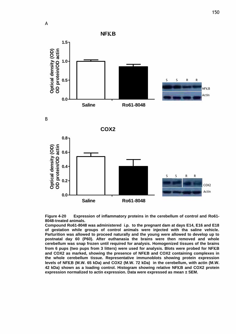
150 A
NFB
0.0
0.5
1.0
1.5
Saline Ro61-8048
Op
tical
den
sit
y (
OD
)
OD
pro
tein
/OD
acti
n
B
COX2
0.0
0.2
0.4
0.6
0.8
Saline Ro61-8048
Op
tical
den
sit
y (
OD
)
OD
pro
tein
/OD
acti
n
Figure 4-20 Expression of inflammatory proteins in the cerebellum of control and Ro61-8048-treated animals. Compound Ro61-8048 was administered i.p. to the pregnant dam at days E14, E16 and E18 of gestation while groups of control animals were injected with tha saline vehicle. Parturition was allowed to proceed naturally and the young were allowed to develop up to postnatal day 60 (P60). After euthanasia the brains were then removed and whole cerebellum was snap frozen until required for analysis. Homogenized tissues of the brains
from 6 pups (two pups from 3 litters) were used for analysis. Blots were probed for NFB
and COX2 as marked, showing the presence of NFB and COX2 containing complexes in the whole cerebellum tissue. Representative immunoblots showing protein expression
levels of NFB (M.W. 65 kDa) and COX2 (M.W. 72 kDa) in the cerebellum, with actin (M.W.
42 kDa) shown as a loading control. Histogram showing relative NFB and COX2 protein expression normalized to actin expression. Data were expressed as mean ± SEM.
** **
NFB
Actin
COX2
Actin
S S R R
S S R R
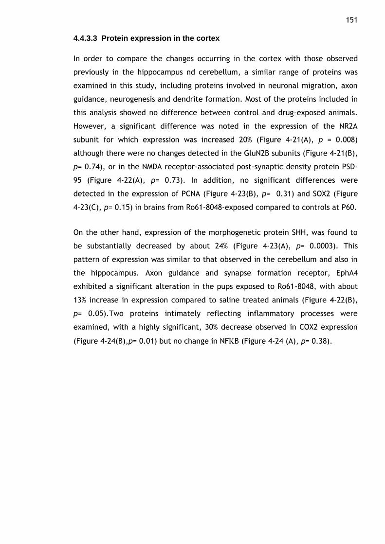
151
4.4.3.3 Protein expression in the cortex
In order to compare the changes occurring in the cortex with those observed
previously in the hippocampus nd cerebellum, a similar range of proteins was
examined in this study, including proteins involved in neuronal migration, axon
guidance, neurogenesis and dendrite formation. Most of the proteins included in
this analysis showed no difference between control and drug-exposed animals.
However, a significant difference was noted in the expression of the NR2A
subunit for which expression was increased 20% (Figure 4-21(A), p = 0.008)
although there were no changes detected in the GluN2B subunits (Figure 4-21(B),
p= 0.74), or in the NMDA receptor-associated post-synaptic density protein PSD-
95 (Figure 4-22(A), p= 0.73). In addition, no significant differences were
detected in the expression of PCNA (Figure 4-23(B), p= 0.31) and SOX2 (Figure
4-23(C), p= 0.15) in brains from Ro61-8048-exposed compared to controls at P60.
On the other hand, expression of the morphogenetic protein SHH, was found to
be substantially decreased by about 24% (Figure 4-23(A), p= 0.0003). This
pattern of expression was similar to that observed in the cerebellum and also in
the hippocampus. Axon guidance and synapse formation receptor, EphA4
exhibited a significant alteration in the pups exposed to Ro61-8048, with about
13% increase in expression compared to saline treated animals (Figure 4-22(B),
p= 0.05).Two proteins intimately reflecting inflammatory processes were
examined, with a highly significant, 30% decrease observed in COX2 expression
(Figure 4-24(B),p= 0.01) but no change in NFB (Figure 4-24 (A), p= 0.38).

152 A
NR2A
0.0
0.2
0.4
0.6
0.8
1.0
Saline Ro61-8048
**O
pti
cal
den
sit
y (
OD
)
OD
pro
tein
/OD
acti
n
B
NR2B
0.0
0.2
0.4
0.6
0.8
1.0
Saline Ro61-8048
Op
tical
den
sit
y (
OD
)
OD
pro
tein
/OD
acti
n
Figure 4-21 Protein expression of NMDA receptor subunits in P60 cortex after prenatal Ro61-8048. Compound Ro61-8048 was administered i.p. to the pregnant dam at days E14, E16 and E18 of gestation while groups of control animals were injected with tha saline vehicle. Parturition was allowed to proceed naturally and the young were allowed to develop up to postnatal day 60 (P60). After euthanasia the brains were then removed, sectioned and snap frozen until required for analysis. Homogenized tissues of the brains were used for analysis. Blots were probed for NR2A and NR2B as marked, showing the presence of NR2A/NR2B containing complexes in the cortex tissue. Representative immunoblots showing protein expression levels of NR2A (M.W. 180 kDa) and NR2B (M.W. 180 kDa) in the cortex, with actin (M.W. 42 kDa) shown as a loading control. Histogram showing relative NR2A and NR2B protein expression normalized to actin expression. Data were expressed as mean ± SEM (n=6 for Ro61-8048, n=6 for control). (A) Compared with saline-treated animals(S), there was a signifcant increase in NR2A protein expression in Ro61-8048(R) (**p<0.01).
** **
NR2A
Actin
NR2B
Actin
S S R R
S S R R
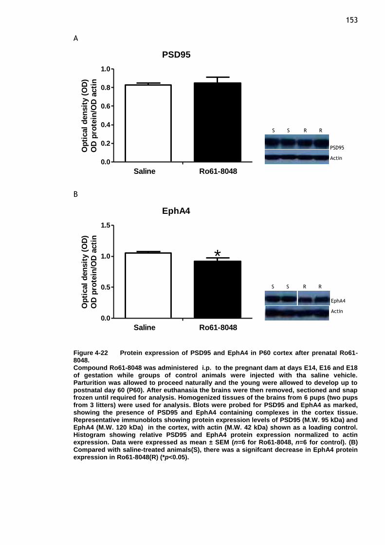
153 A
PSD95
0.0
0.2
0.4
0.6
0.8
1.0
Saline Ro61-8048
Op
tical
den
sit
y (
OD
)
OD
pro
tein
/OD
acti
n
B
EphA4
0.0
0.5
1.0
1.5
Saline Ro61-8048
*
Op
tical
den
sit
y (
OD
)
OD
pro
tein
/OD
acti
n
Figure 4-22 Protein expression of PSD95 and EphA4 in P60 cortex after prenatal Ro61-8048. Compound Ro61-8048 was administered i.p. to the pregnant dam at days E14, E16 and E18 of gestation while groups of control animals were injected with tha saline vehicle. Parturition was allowed to proceed naturally and the young were allowed to develop up to postnatal day 60 (P60). After euthanasia the brains were then removed, sectioned and snap frozen until required for analysis. Homogenized tissues of the brains from 6 pups (two pups from 3 litters) were used for analysis. Blots were probed for PSD95 and EphA4 as marked, showing the presence of PSD95 and EphA4 containing complexes in the cortex tissue. Representative immunoblots showing protein expression levels of PSD95 (M.W. 95 kDa) and EphA4 (M.W. 120 kDa) in the cortex, with actin (M.W. 42 kDa) shown as a loading control. Histogram showing relative PSD95 and EphA4 protein expression normalized to actin expression. Data were expressed as mean ± SEM (n=6 for Ro61-8048, n=6 for control). (B) Compared with saline-treated animals(S), there was a signifcant decrease in EphA4 protein expression in Ro61-8048(R) (*p<0.05).
** **
PSD95
Actin
EphA4
Actin
S S R R
S S R R

154 A
SHH
0.0
0.2
0.4
0.6
0.8
1.0
Saline Ro61-8048
**O
pti
cal
den
sit
y (
OD
)
OD
pro
tein
/OD
acti
n
B PCNA
0.0
0.2
0.4
0.6
0.8
1.0
Saline Ro61-8048
Op
tical
den
sit
y (
OD
)
OD
pro
tein
/OD
acti
n
C SOX2
0.0
0.1
0.2
0.3
0.4
Saline Ro61-8048
Op
tical
den
sit
y (
OD
)
OD
pro
tein
/OD
acti
n
Figure 4-23 Protein expression of neurodevelopmental proteins in P60 cortex after prenatal Ro61-8048. Compound Ro61-8048 was administered i.p. to the pregnant dam at days E14, E16 and E18 of gestation while groups of control animals were injected with tha saline vehicle. Parturition was allowed to proceed naturally and the young were allowed to develop up to postnatal day 60 (P60). After euthanasia the brains were then removed, sectioned and snap frozen until required for analysis. Homogenized tissues of the brains from 6 pups (two pups from 3 litters) were used for analysis. Representative immunoblots showing protein expression levels of SHH (M.W. 45 kDa), PCNA (M.W. 36 kDa) and SOX2 (M.W. 34 kDa) in the cortex, with actin (M.W. 42 kDa) shown as a loading control. Histogram showing relative SHH,PCNA and SOX2 protein expression normalized to actin expression. Data were expressed as mean ± SEM. (A) Prenatal treatment with 100mg/kg Ro61-8048(R) showed significant reduction in SHH expression compared to control animals(S) (n=6)(**p<0.01).
**
** **
SHH
Actin
PCNA
Actin
SOX2
Actin
S S R R
S S R R
S S R R
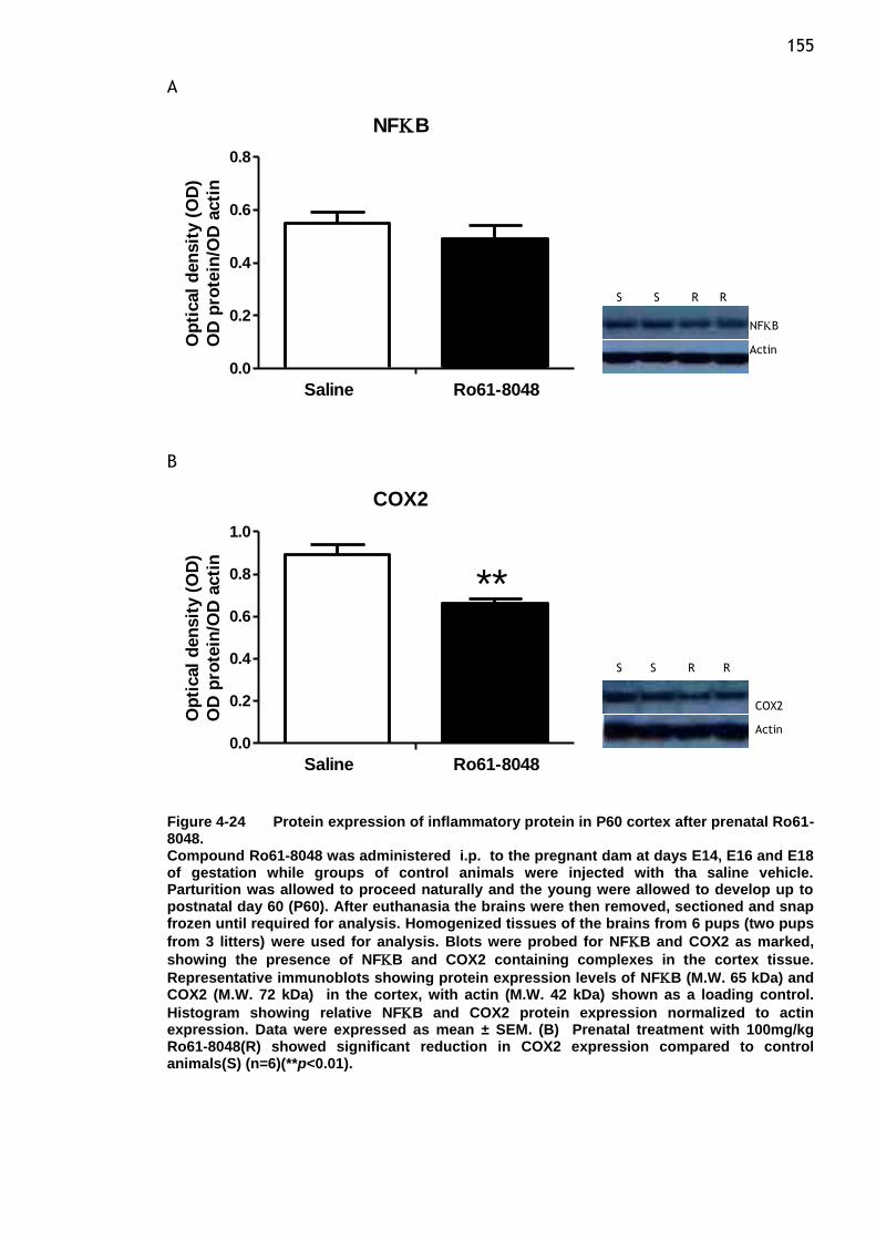
155 A
NFB
0.0
0.2
0.4
0.6
0.8
Saline Ro61-8048
Op
tical
den
sit
y (
OD
)
OD
pro
tein
/OD
acti
n
B
COX2
0.0
0.2
0.4
0.6
0.8
1.0
Saline Ro61-8048
**
Op
tical
den
sit
y (
OD
)
OD
pro
tein
/OD
acti
n
Figure 4-24 Protein expression of inflammatory protein in P60 cortex after prenatal Ro61-8048. Compound Ro61-8048 was administered i.p. to the pregnant dam at days E14, E16 and E18 of gestation while groups of control animals were injected with tha saline vehicle. Parturition was allowed to proceed naturally and the young were allowed to develop up to postnatal day 60 (P60). After euthanasia the brains were then removed, sectioned and snap frozen until required for analysis. Homogenized tissues of the brains from 6 pups (two pups
from 3 litters) were used for analysis. Blots were probed for NFB and COX2 as marked,
showing the presence of NFB and COX2 containing complexes in the cortex tissue.
Representative immunoblots showing protein expression levels of NFB (M.W. 65 kDa) and COX2 (M.W. 72 kDa) in the cortex, with actin (M.W. 42 kDa) shown as a loading control.
Histogram showing relative NFB and COX2 protein expression normalized to actin expression. Data were expressed as mean ± SEM. (B) Prenatal treatment with 100mg/kg Ro61-8048(R) showed significant reduction in COX2 expression compared to control animals(S) (n=6)(**p<0.01).
** **
NFB
Actin
COX2
Actin
S S R R
S S R R
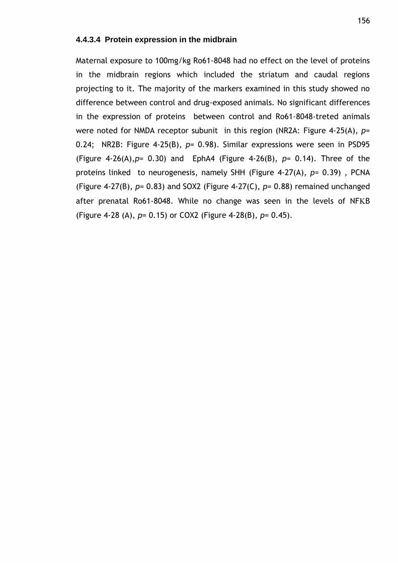
156
4.4.3.4 Protein expression in the midbrain
Maternal exposure to 100mg/kg Ro61-8048 had no effect on the level of proteins
in the midbrain regions which included the striatum and caudal regions
projecting to it. The majority of the markers examined in this study showed no
difference between control and drug-exposed animals. No significant differences
in the expression of proteins between control and Ro61-8048-treted animals
were noted for NMDA receptor subunit in this region (NR2A: Figure 4-25(A), p=
0.24; NR2B: Figure 4-25(B), p= 0.98). Similar expressions were seen in PSD95
(Figure 4-26(A),p= 0.30) and EphA4 (Figure 4-26(B), p= 0.14). Three of the
proteins linked to neurogenesis, namely SHH (Figure 4-27(A), p= 0.39) , PCNA
(Figure 4-27(B), p= 0.83) and SOX2 (Figure 4-27(C), p= 0.88) remained unchanged
after prenatal Ro61-8048. While no change was seen in the levels of NFB
(Figure 4-28 (A), p= 0.15) or COX2 (Figure 4-28(B), p= 0.45).
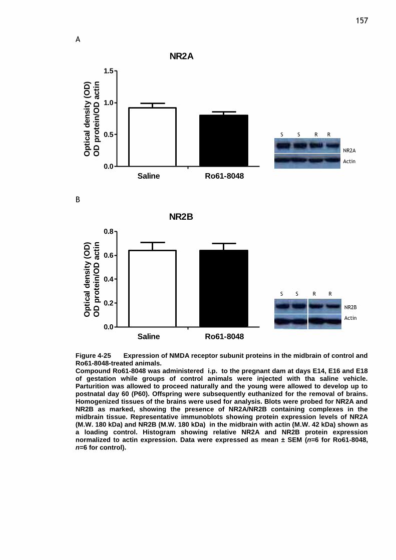
157 A
NR2A
0.0
0.5
1.0
1.5
Saline Ro61-8048
Op
tical
den
sit
y (
OD
)
OD
pro
tein
/OD
acti
n
B
NR2B
0.0
0.2
0.4
0.6
0.8
Saline Ro61-8048
Op
tical
den
sit
y (
OD
)
OD
pro
tein
/OD
acti
n
Figure 4-25 Expression of NMDA receptor subunit proteins in the midbrain of control and Ro61-8048-treated animals. Compound Ro61-8048 was administered i.p. to the pregnant dam at days E14, E16 and E18 of gestation while groups of control animals were injected with tha saline vehicle. Parturition was allowed to proceed naturally and the young were allowed to develop up to postnatal day 60 (P60). Offspring were subsequently euthanized for the removal of brains. Homogenized tissues of the brains were used for analysis. Blots were probed for NR2A and NR2B as marked, showing the presence of NR2A/NR2B containing complexes in the midbrain tissue. Representative immunoblots showing protein expression levels of NR2A (M.W. 180 kDa) and NR2B (M.W. 180 kDa) in the midbrain with actin (M.W. 42 kDa) shown as a loading control. Histogram showing relative NR2A and NR2B protein expression normalized to actin expression. Data were expressed as mean ± SEM (n=6 for Ro61-8048, n=6 for control).
** **
NR2A
Actin
NR2B
Actin
S S R R
S S R R
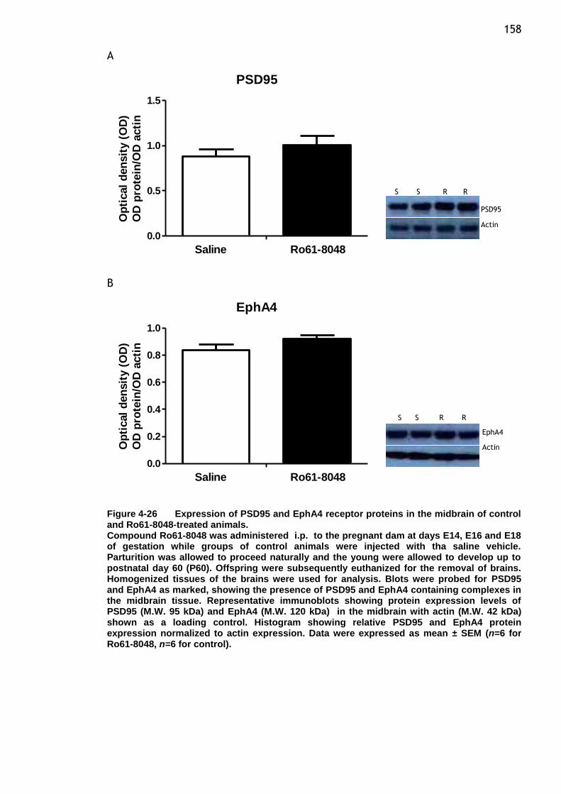
158 A
PSD95
0.0
0.5
1.0
1.5
Saline Ro61-8048
Op
tical
den
sit
y (
OD
)
OD
pro
tein
/OD
acti
n
B
EphA4
0.0
0.2
0.4
0.6
0.8
1.0
Saline Ro61-8048
Op
tical
den
sit
y (
OD
)
OD
pro
tein
/OD
acti
n
Figure 4-26 Expression of PSD95 and EphA4 receptor proteins in the midbrain of control and Ro61-8048-treated animals. Compound Ro61-8048 was administered i.p. to the pregnant dam at days E14, E16 and E18 of gestation while groups of control animals were injected with tha saline vehicle. Parturition was allowed to proceed naturally and the young were allowed to develop up to postnatal day 60 (P60). Offspring were subsequently euthanized for the removal of brains. Homogenized tissues of the brains were used for analysis. Blots were probed for PSD95 and EphA4 as marked, showing the presence of PSD95 and EphA4 containing complexes in the midbrain tissue. Representative immunoblots showing protein expression levels of PSD95 (M.W. 95 kDa) and EphA4 (M.W. 120 kDa) in the midbrain with actin (M.W. 42 kDa) shown as a loading control. Histogram showing relative PSD95 and EphA4 protein expression normalized to actin expression. Data were expressed as mean ± SEM (n=6 for Ro61-8048, n=6 for control).
** **
PSD95
Actin
EphA4
Actin
S S R R
S S R R
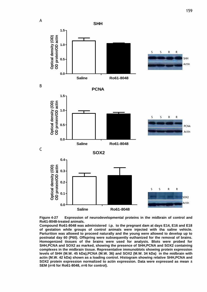
159 A
SHH
0.0
0.5
1.0
1.5
Saline Ro61-8048
Op
tical
den
sit
y (
OD
)
OD
pro
tein
/OD
acti
n
B PCNA
0.0
0.5
1.0
1.5
Saline Ro61-8048
Op
tical
den
sit
y (
OD
)
OD
pro
tein
/OD
acti
n
C SOX2
0.0
0.1
0.2
0.3
0.4
Saline Ro61-8048
Op
tical
den
sit
y (
OD
)
OD
pro
tein
/OD
acti
n
Figure 4-27 Expression of neurodevelopmental proteins in the midbrain of control and Ro61-8048-treated animals. Compound Ro61-8048 was administered i.p. to the pregnant dam at days E14, E16 and E18 of gestation while groups of control animals were injected with tha saline vehicle. Parturition was allowed to proceed naturally and the young were allowed to develop up to postnatal day 60 (P60). Offspring were subsequently euthanized for the removal of brains. Homogenized tissues of the brains were used for analysis. Blots were probed for SHH,PCNA and SOX2 as marked, showing the presence of SHH,PCNA and SOX2 containing complexes in the midbrain tissue. Representative immunoblots showing protein expression levels of SHH (M.W. 45 kDa),PCNA (M.W. 36) and SOX2 (M.W. 34 kDa) in the midbrain with actin (M.W. 42 kDa) shown as a loading control. Histogram showing relative SHH,PCNA and SOX2 protein expression normalized to actin expression. Data were expressed as mean ± SEM (n=6 for Ro61-8048, n=6 for control).
**
** **
SHH
Actin
PCNA
Actin
SOX2
Actin
S S R R
S S R R
S S R R
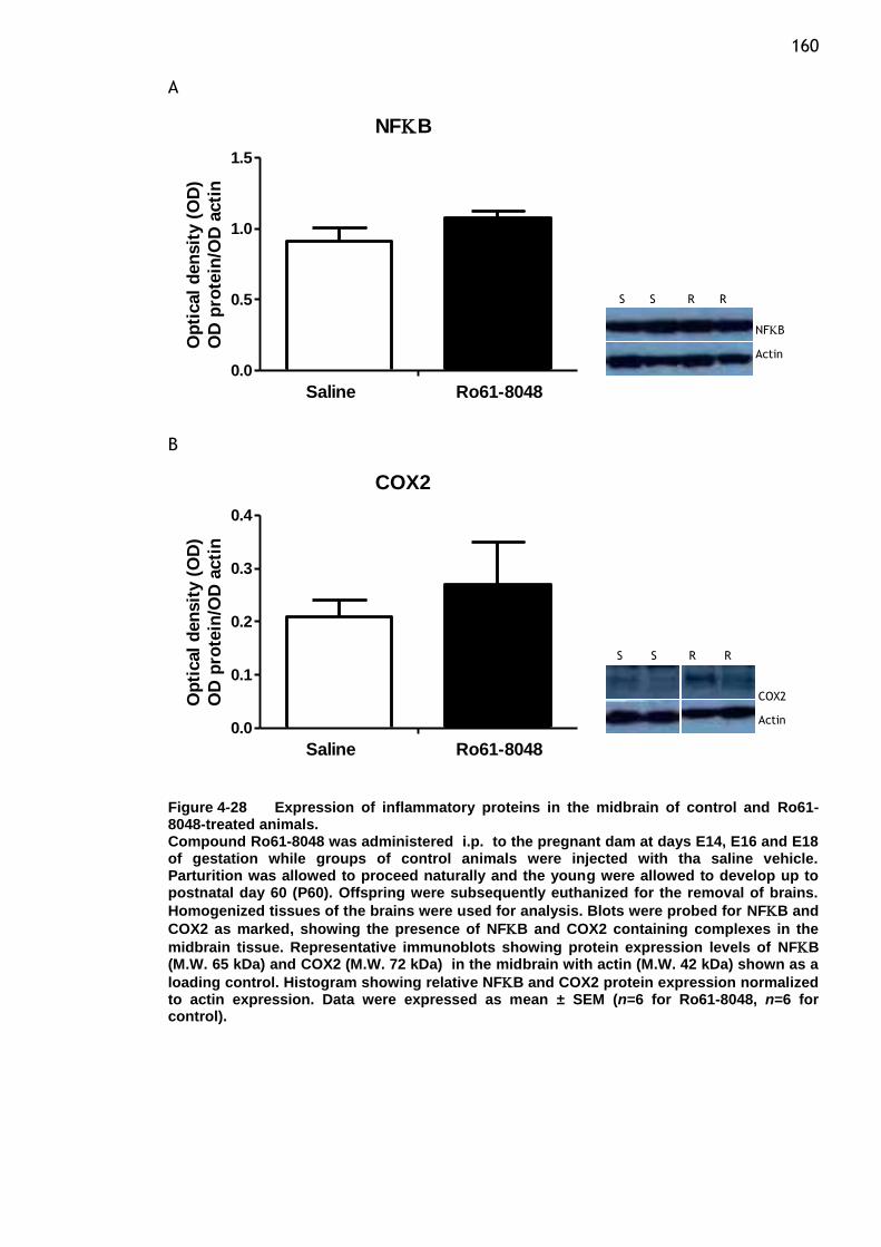
160 A
NFB
0.0
0.5
1.0
1.5
Saline Ro61-8048
Op
tical
den
sit
y (
OD
)
OD
pro
tein
/OD
acti
n
B
COX2
0.0
0.1
0.2
0.3
0.4
Saline Ro61-8048
Op
tical
den
sit
y (
OD
)
OD
pro
tein
/OD
acti
n
Figure 4-28 Expression of inflammatory proteins in the midbrain of control and Ro61-8048-treated animals. Compound Ro61-8048 was administered i.p. to the pregnant dam at days E14, E16 and E18 of gestation while groups of control animals were injected with tha saline vehicle. Parturition was allowed to proceed naturally and the young were allowed to develop up to postnatal day 60 (P60). Offspring were subsequently euthanized for the removal of brains.
Homogenized tissues of the brains were used for analysis. Blots were probed for NFB and
COX2 as marked, showing the presence of NFB and COX2 containing complexes in the
midbrain tissue. Representative immunoblots showing protein expression levels of NFB (M.W. 65 kDa) and COX2 (M.W. 72 kDa) in the midbrain with actin (M.W. 42 kDa) shown as a
loading control. Histogram showing relative NFB and COX2 protein expression normalized to actin expression. Data were expressed as mean ± SEM (n=6 for Ro61-8048, n=6 for control).
** **
NFB
Actin
COX2
Actin
S S R R
S S R R
161
4.5 Discussion
In the current study, we have demonstrated that the protein expression of some
of the important molecules involved during neurodevelopmental events in the
brain undergoes a differential pattern of regulation. We investigated possible
alterations in the inhibitory activity of compound Ro61-8048 on acute and long
term changes in the expression of proteins in the brain as well as regional areas
of the brain (P60) of animals at various stages of development. Evaluation of
region-specific protein expression presented in P60 animal allows us to make
some speculations on the involvement of relevant brain regions that provide a
guide for further investigation. Several findings are of interest in this study and
will be discussed further.
We demonstrate that pharmacological manipulation of the kynurenine pathway
during the prenatal period can be accomplished by treating pregnant dams
during late gestation. Our previous data indicate that the levels of KYNA in
maternal plasma and embryo brain can be elevated by the systemic
administration of KMO inhibitor, Ro61-8048 which deflects kynurenine
degradation towards enhanced KYNA formation. Administration of this compound
to pregnant rats at three time points during late gestation caused comparable
substantial increases in maternal plasma and embryo brain of KYNA level
(Forrest et al. 2013b). In accordance with this, increases in the concentrations of
brain KYNA has been reported following administration of the KMO inhibitor,
Ro61-8048 to immune-stimulated mice and this was accompanied by reduced
concentrations of 3-hydroxykynurenine and QUIN, suggesting that in pathological
situations, KMO is a rate limiting enzyme for both 3-hydroxykynurenine and also
QUIN (Chiarugi et al., 2001). Administration of Ro61-8048 also provoke an
increase in brain KYNA concentrations and reduced the severity of dystonia
(Richter and Hamann, 2003). Beneficial effects of KMO inhibition have been
shown in a murine model of cerebral malaria, where an increase in KYNA and
anthranilic acid in R061-8048-treated infected mice would protect against the
activation of NMDAR. However, when Ro61-8048 was given to the controls, no
alteration in QUIN concentrations was observed(Clark et al., 2005). The effect of
maternal KMO inhibition on embryo brain KYNA and neurochemical alterations
seen in later stages of developmental suggests evidence for acute endogenous
162 changes in KP metabolism which might be potentially responsible for the
changes seen in this study.
4.5.1 Protein expression of NMDA receptor subunits and
associated protein
To date, no study has examined the influence of maternal administration of KMO
inhibitor on NMDA receptor subunits expression in the embryo and also in the
postnatal brains. To examine the possibility that NMDA receptors contribute to
the kynurenine –mediated regulation of NMDA subunits level in the brain, the
influence of Ro61-8048 on the inhibition of the kynurenine pathway was
addressed. The present study demonstrates that prenatal modulation of the
kynurenine pathway during pregnancy is associated with alterations in NMDA
receptor subunits protein levels as early as 5 hour following single injection. In
addition, we found that both NMDA receptor subunits and PSD95 followed a
different profile of expressions during postnatal development in the brain.
The NMDA receptors modulate glutamate postsynaptic neurotransmission by
generating long lasting calcium channel opening for calcium influx through the
channel and causing longer depolarizations. This process is responsible for
mediating synaptic plasticity changes such as LTP and LTD that underpins many
higher functions including learning and memory. Activation of the receptor in
the mechanism of LTP is considered and understood to be the synaptic model of
learning and memory (Bliss and Collingridge, 1993, Gladding and Raymond,
2011). The subunits NR2A and NR2B were chosen for examination in this study
because they are the most predominant NMDA receptor subunit subtypes in their
respective subunit families and would therefore be the most likely receptor
subunits responsible for changes. Our results demonstrate that both these major
subunits of the NMDA receptor, NR2A and NR2B, are differentially expressed
during pre-and postnatal development in the rat brain. Both NMDA receptor
subunit proteins were already expressed in the embryo brains as observed at E18
(following 5 hour post injection). Developmental expression using immunoblot
analysis showed that the NR2B proteins were detectable as early as E14,
continued increasing at P10, P20 and into late adulthood P60. It also pointed out
that environmental insults during prenatal development may result in
163 morphological abnormalities in which aberrant co assembly of the NMDA
receptor complex may reflect the differentiation of synaptic rearrangement and
receptor affinities (Babb et al., 2005). Five hours of Ro61-8048 administration to
the pregnant dams had different effects on two major NR2 subunits of NMDA
receptor, with reduced protein expression for NR2A and increased levels of NR2B
protein at embryonic day 18. A consistent set of chronic changes in the protein
levels of both subunits was observed in young animals at P21. It is important to
note that a change in the NR2A expression in the hippocampus and cortex after
Ro61 treatment was maintained into adulthood and likely to have influenced
structural and functional aspects of synaptic plasticity at the receptor. The
observed increase in NR2A subunits in the 21 day postnatal offspring produced by
prenatal Ro61 exposure may therefore be a compensatory mechanism for a
potential decrease in the receptor subunit function at earlier time points in
development. This may also hold a similar explanation for the differential
pattern of NR2A expression observed in mature animals. There may be brain
regional and developmental age selectivity for the effects of prenatal
kynurenine inhibition on NMDA receptors. For PSD95, we showed that acute
exposure to the enzyme inhibitor produced no alteration in the level of protein
expression in the whole brain lysate. However, the level of protein detected was
progressively increased at P21. In accordance with this, we did observe
significant differences in the level of NR2A and NR2B proteins in the same age
group of postnatal rats. Concerning the expression of the major subunit NR1
(results presented in (Forrest et al., 2013a), no significant alteration was
observed in all ages of development. The present study demonstrates that there
are differential long term-effects of prenatal modulation of the kynurenine
pathway on the NMDA receptor complex in the offspring. This implies that a
change in NMDA receptor number or function could underlie altered
neurotransmission and receptor pharmacology both during normal physiological
activity and in response to neuroactive metabolites. The fact that we observed
reduced NR2A subunit, but not NR1 and NR2B protein levels in homogenates
prepared from hippocampus isolated from P60 rat in the presence of unaltered
mRNA expression of the subunits (data presented in (Forrest et al., 2013a)
indicates that the reduction in NR2A protein may be the result of differential
post-transcriptional processing of the protein in the Ro61-treated animal. This
observation is consistent with other studies showing that, in some cases, NMDA
164 receptor subunit mRNA levels do not correlate with protein levels (Gazzaley et
al., 1996). The exact mechanisms for how NR2A expression is decreased in the
hippocampus was not formally addressed in our study, thus reduced translation
or increased degradation of the NR2A protein in the hippocampus of exposed
animals should be evaluated in future studies. Up regulation of NR2A subunits in
the cortex has been noted suggesting that prenatal kynurenine inhibition
enhanced neural activity in the cortex. Therefore, changes in NR2A protein
levels seen in only two regions could be a compensatory response to prolonged
NMDA receptor inhibition in utero that persist into adulthood. As our western
blot analysis indicates, protein expression of NMDA subunits NR2A and NR2B and
also PSD95 is enriched in the hippocampus compared with other regions. This is
supported by the evidence demonstrating that in the hippocampal region, the
expression of NR2A, NR2B and PSD95 increased with age. During the
development of the hippocampus, the pyramidal cells in CA1 and CA3 grow
rapidly after birth and are gradually stabilized after postnatal week 3. Since the
granule cells had self-renewal capacities for their lifetime, the number of
granule cells in the dentate gyrus gradually increases with age and maintain the
ability to proliferate in adulthood. The obviously different expression patterns of
the subunits implicated their distinct roles in the postnatal development of
hippocampus (Al-Hallaq et al., 2007). NMDA receptor function is regulated
mainly through association with many signalling proteins that are concentrated
with the postsynaptic density (PSD) compartment. Of the many PSD proteins that
can potentially modulate NMDA receptor properties, PSD95 has gained the most
attention because this molecule associates with various signalling protein and
exerted its effect by recruiting kinases or phosphatases to the NMDA receptor
complex (Li et al., 2003, Tezuka et al., 1999). However, the present study could
not justify the exact functional composition and localization of NMDA subunits
since we used a total pool of proteins that included intracellular pools of
assembled and unassembled NMDA receptors expressed at the cell membrane.
The homogenates are heterogeneous, containing a percentage from the synaptic
region itself, as well as from extrasynaptic membranes, the soma and the axons.
Apart from that, interaction between the major subunits with its associated
protein, like PSD95 using immunoprecipitation studies was not examined in this
study. Although the distribution of NMDA receptor compositions across brain
region were not addressed in this study, these data indirectly support a
165 regulated mechanism of subunit assembly because NMDA receptor subunit
composition differs across regions in which NR2A and NR2B are the predominant
NR2 subunits, such as in hippocampus and cortex (Wenzel et al., 1997). In
addition, physical interaction between NR2 subunits and PSD proteins have been
shown to play an important role in regulating the localization of the NMDA
receptor and this interaction is mediated by the cytoplasmic tail of NR2 subunits
(Al-Hallaq et al., 2007, Delint-ramirez et al., 2010).
The present studies add to a growing body of evidence that environmental
insults during pregnancy are associated with alterations in NMDA receptor
subunit levels. Our results report changes in the protein levels of NMDA receptor
subunits in different developmental ages as a result of either acute or persistent
effects of kynurenine modulation during pregnancy. The persistence of these
alterations into adulthood indicates that modifications of glutamatergic
neurotransmission is highly involved in the adaptive responses that underlie
homoeostatic and compensatory changes in the function of the receptor complex
that contribute to the neurochemical imbalance associated with learning and
memory deficits that are observed in adult animals that were exposed prenatally
to KMO inhibitor.
4.5.2 Differential expression of developmental molecules
following prenatal Ro61-8048 administration
We have examined the expression level of developmental molecules responsible
for some important processes including neuronal proliferation and migration,
axon guidance and synapse formation in the embryo and postnatal brains. The
increasing number of developmental events associated with these molecules has
generated considerable interest in studies regarding their molecular mechanisms
as well as putative involvement in a range of neurodegenerative diseases. Our
study showed that EphA4 was expressed in the developing brain from embryonic
day 18 with reduced levels detected 24 hour after Ro61 administration and also
at P21. In P60 brain, there was a dynamic pattern of expression, with a
persistent but differential pattern of expression in the several regions of the
mature brain. Although no significant changes was seen in the hippocampal level
of EphA4 as compared to control levels, this molecule has been found to be
166 highly expressed in the hippocampus and involved partly in synaptic plasticity
events such as long term potentiation. While many of the molecular mechanisms
regulating the induction of LTP remain unclear, Ephs receptor protein kinase
activity is known to be involved in this process (Pasquele, 2008). We
demonstrate here that the regional pattern of EphA4 expression changes
markedly during postnatal development as shown at P60, with the observation of
lowered expression in the cortex suggesting that prenatal modulation of the
kynunerine pathway may preferentially affect particular regions of the brain.
These data suggest that EphA4 expression is regulated in a NMDA-dependent
synaptic response through regulation of kynurenine metabolites in the brain and
that EphA4 kinase activity may play a functional role in maintaining the
organization of dendritic spines postnatally. The fact that EphA4 regulates axon
guidance during embryonic development and postnatal synaptic function
emphasizes the varied and complex roles for this receptor family in regulating
cell-cell interactions within the CNS. The molecular mechanisms of many Ephs
and ephrin related synaptic functions are still largely unknown, thus a more
elaborate examination of the roles and mechanisms of this receptor complex will
be needed to further establish them as important synaptogenic molecules in the
nervous system (Lai and Ip, 2009).
The physiological function of PCNA is well described, being necessary for DNA
replication and repair in proliferating cells. Changes in cell proliferation
dynamics in response to various insults have been assessed previously using
multiple cell cycle markers, in which the pattern of changes was reflected in
double/triple immunolabelling using a combination of PCNA with other
proliferation markers including 5-bromo-2‟-deoxyuridine (BrdU) and/or Ki-67.
The combination of different cell cycle regulators permits study of the
proliferative, migratory and survival characteristics of neuronal progenitors cells
in the developing and adult rodent brain (Halbach, 2011, Valero et al., 2005).We
showed that in the early developmental period, kynurenine pathway modulation
during gestation caused no changes in the proliferative event, as demonstrated
by PCNA expression in the embryo. However, it progressively appeared that the
resistance of the immature brain has its limitations, in which we noted an
elevated extent of proliferation as shown by PCNA expression at later stage of
development only at P21 and regionally unaffected when examined at P60.
167 Upon binding to its receptor Patched (Ptc), SHH activates the highly conserved
hedgehog (HH) signalling cascade, resulting in activation of the Gli family of
transcription factors. The SHH signalling cascade is involved in the development
and patterning of several vertebrate embryonic structures, anteroposterior
patterning of the limbs and dorsoventral patterning of the neural tube (Ruiz i
Altaba et al., 2002, Palma et al., 2005). Analysis of SHH protein expression after
administration of enzyme inhibitor Ro61-8048 has paved a way for the
characterization of the potential roles for this molecule in the normal and
treated brain. Robust SHH expression was found in the embryo, suggesting
localized signalling in the developing brain. Interestingly, the expression is high
before birth and markedly reduced at postnatal day 21. Application of antibodies
specific for SHH led to a pattern of regional protein expression in the adult rat
brain. In protein extracts isolated from animals exposed to Ro61-8048, western
blot analysis indicated that a 45kDa SHH protein SHH was broadly expressed in
all brain areas examined with reduced expression found in three main areas of
cerebellum, cortex and hippocampus. Such a pattern of expression was found
conserved for the SHH mRNA, albeit a low expression was detected within these
brain regions, suggesting that this molecule may be implicated in local signalling
circuitry (Forrest et al., 2013b). Although in this study, we did not focus on the
regulation of neural stem cells, a number of reports have demonstrated that in
the nervous system of young and adult animals, SHH is known to regulate the
proliferation of the cerebellar granule cell precursors, neural progenitors lining
the subventricular zone of the forebrain and in the dentate gyrus of the
hippocampus. These studies partly demonstrate that concerted cell
proliferation, migration and differentiation in response to SHH protein are
important for normal embryonic developmental and postnatal homeostasis
(Palma et al., 2005, Lai et al., 2003, Machold et al., 2003, Bertrand and
Dahmane, 2006). Expression studies of SHH and other genes involved in
hedgehog signal- transduction have indicated a function of hedgehog signalling in
the adult brain in addition to its roles during early development (Traiffort et al.,
1999, Charytoniuk et al., 2002). A number approaches including in vitro cell
culture and animal model have been used to unravel the SHH signalling pathways
linking relationship between various perturbations or exposure to the regulation
of the expression of SHH signalling cascade in the embryonic and adult rodent
brain. It has been shown that this powerful developmental morphogen is
168 upregulated in adult organisms after injury and involved in a repair mechanism
in which it enhances adult hippocampal neurogenesis following electroconvulsive
seizure (Banarjee and Rajendran, 2005). It appears that the expression of SHH
also increased during ischemia (Sims et al., 2009) and under oxidative stress
induced by hydrogen peroxide (H2O2) (Dai et al., 2011). Other molecular events
that has influence on the modulation of the SHH pathway includes exposure to
caffeine in primary neuronal and astroglial cultures (Sahir et al., 2004) and in
the presence of teratogen valproic acid (VPA), which altered the expression of
some regulatory genes reported to be involved in the serotonergic neuronal
development, and that SHH and its components expression were all decreased in
the VPA-treated cells compared to the control (Miyazaki et al., 2005). Exposure
of zebrafish embryos to alcohol during gastrulation impaired SHH signal
transduction and suggesting a role for defective SHH signalling in the
pathogenesis of alcohol-induced birth defects (Li et al., 2007). Our results
demonstrate that in the adult brain, this pathway remains active and has a
continuous role in the adult neural stem cells, and mature neurons are likely to
be the source of SHH (Ihrie et al., 2011).We show that the SHH pathway, one of
a key developmental signals in the brain, may be differentially regulated
following kynurenine modulation. At present we are not entirely certain of the
exact mechanisms that underlie the effects seen in this study. There remains
room for further exploration to gain a greater understanding of the processes of
SHH signalling mechanism.
We analysed the expression of SOX2 in brain homogenates prepared from foetal
and also from rat offspring at P21 and regional areas at P60 to determine
whether this transcription factor was affected after modulation of kynurenine
pathway during gestation. The biological roles of SOX2 have been thoroughly
studied in the context of embryonic developmental regulation as it is known as
one of the key factors in the regulation of embryonic stem cell self-renewal and
is associated with neural stem cell (NSC) populations in the mature rodent brain
(Brazel et al., 2005, Komitova and Eriksson, 2004). It is worth mentioning that
all proteins of the SOX-B1 group (SOX1,SOX2,SOX3) are expressed in the
developing brain to a different extent during development and many of the SOX
proteins coexist throughout the CNS (Bylund et al., 2003). Our study shows that
expression of SOX2 is abundant in the control embryonic brains, during a time
169 window in which the pool of neural progenitors expands extensively which in
agreement with a previous study that indicate an essential role for SOX2 in early
development (Avilion et al., 2003). Acute treatment with Ro61-8048 caused no
alteration in the SOX2 levels suggesting the important role of SOX2 in neural
stem cell maintenance in the developing brain. The prominent expression of
SOXB1 protein in general, in the neurogenic regions of adult brain has prompted
many researchers to analyse the expression pattern of these markers in the
neurogenic lineage using a culture model of different types of dividing precursor
cells with the aim of comparing the expression level of the markers that are
expressed at different stages of adult neurogenesis. The reason for the lack of
expression in the hippocampus of P60 animal, even in a control setting might
arise from the hippocampal sections used in this study. The sections were
obtained after it had been exposed to electrophysiological procedures. Thus, it
is likely to reflect the overall cellular contents of the homogenates. As SOX2
proteins bind to specific DNA sequences via their HMG box domains and
synergizing with other transcription factors to fulfil variable roles, their mode of
action may depend both on the cellular context and on the target gene
functionally regulating active transcriptional complexes (Lefebvre et al., 2007).
It has been shown that SOX2 is expressed mainly in dividing neural stem cells and
transient amplifying progenitor cells in the hippocampus which indicates that
SOX2 is expressed stage-specifically in cells of the adult neurogenic lineage (Suh
et al., 2007, Ellis et al., 2004). In another series of experiments, studies have
shown that SOX2 continues to be expressed in specific populations of adult cells
of other brain regions including cerebellum, where SOX2 positive cells have been
observed in Bergmann glia which is not considered as a stem cell
population(Alcock et al. 2009). Our results show a differential expression pattern
in the adult brain regions after kynurenine pathway regulation as we
demonstrate that endogenous concentrations of SOX2 varies between distinct
classes of neural progenitor cells in those regions. The present findings only
indicate that SOX2 is differentially expressed between regions. It is worth
mentioning that isolated populations of these types of progenitor cells based on
their SOX2 expression and subsequent gene expression profile of each sorted
population can be used to accurately ascertain distinct neural progenitors
(Hutton and Pevny, 2011), though at this stage we cannot identify the stem cell
population pool in vivo as the antibody used in this study resulted in both
170 cytoplasmic and nuclear localization. As SOX2 represents a molecular signature
for neural stem cells, this finding is particularly interesting and highlights the
need to fully characterize the cell populations that express this marker in the
adult brain.
These observations suggest that prenatal modulation of the kynurenine pathway
might lead to changes in the expression levels of several molecules that are
involved in the regulation of cell cycle, proliferation as well as the neurogenesis
process in developing and adult brains. An increased expression of the
transduction molecule SHH was observed 24 h after treatment with enzyme
inhibitor during late gestation. However, when repeated challenges of the
inhibitor were given, a different pattern of expression was seen with reduced
levels of expression demonstrated at developmental age P21 and it remained
repressed in some regional areas of the brain including the hippocampus,
cerebellum and cortex. Substantial studies have gathered evidence supporting
the idea that dysregulation of the cell cycle in neural cells plays an important
role in diverse pathological processes in the CNS. Further study on the
characterization of cell cycle properties in this process and comparative studies
on the expression of individual members of each related proteins under
physiological conditions, including various developmental stages and in exposure
to pathological stimuli may provide better and conclusive explanations.
4.5.3 Expression profiles of inflammatory associated proteins
As both NFB and COX2 are critical to host defense to various pathological
stimuli, their common activating conditions represent either a direct threat to
cells or that they are endogenous signals produced in response to pathogens. In
this respect, administration of foreign substances might induce an inflammatory
response; we explored the expression profiles of relevant inflammatory markers
following administration of an enzyme inhibitor of the kynurenine pathway,
Ro61-8048. The impetus of the present study was to test the hypothesis that
prenatal inhibition of KP by Ro61-8048 would have impacts on the transcriptional
activity of NFB and COX2 expression in the offspring. The ability of Ro61-8048
to shift the production of kynurenine metabolites towards greater levels of KYNA
has been reported and we show a substantial increase of KYNA in the maternal
171 plasma and in the brains of the embryos after 5 h of Ro61 administration. Level
of KYNA remained significantly elevated in the embryonic brains after 24 h post
administration (Forrest et al. 2013b). This effect was accompanied by increased
NFB activity in the embryo brains 24 h post treatment, indicating a role for this
TF in inducing activity of other relevant target genes in the presence of the
kynurenine pathway metabolite, KYNA. The effect of Ro61-8048 in shifting the
balance of KP as seen in the embryonic brains also suggests that there is some
constitutive NFB activity that normally influences activity of KP during
embryonic development. Increased activity of NFB in gestational tissue has
been thought to be an integral part of the final phase of pregnancy and involved
in the biochemical changes associated with labor (Choi et al., 2007).
It has been reported that NFB mediates the increased expression of both IDO
and TDO in the human placenta following infection (Dharane et al., 2010). IDO is
considered to be an important immunoregulator, where its induction during
pregnancy has led to the suggestion that it has as essential role in suppressing
the maternal immune response to the presence of the fetus (Munn et al., 1998).
NFB is a primary transcription factor specialized in cytoplasmic-nuclear
signaling and coordinate the activation of various genes following extracellular
stimulation (O'Neill and Kaltschmidt, 1997). Extracellular signals, in particular
neurotransmitters might regulate gene expression through activation of NFB. In
this regard, glutamate and NMDA caused an induction of NFB binding activity in
the nucleus of mouse cerebellar cells through activation of NMDAR. It seems that
NFB appears to be a good candidate as a mediator for glutamate induced
immediate early gene transcription in the CNS (Guerrini et al., 1995). Sustained
expression with significant increased of the level of NFB protein level was
observable in the total brain homogenates from P21 offspring whose mother had
been injected with Ro61-8048. However, the levels of this TF were detectable
with no apparent changes in term of protein levels when observed in the
hippocampus, cerebellum, cortex and midbrain of P60 brains. Nuclear
translocation of activated NFB must be demonstrated in order to determine its
DNA binding activity. However, current findings applied to the quantification of
expression levels generated from whole extracts of whole brain or selected
regional areas i.e. hippocampus, cortex, cerebellum and midbrain.
Demonstration of an activation of transcription factors (described before in the
172 text) within these specialized brain sites is therefore a useful tool to evaluate
inflammatory activation of the brain to a given insult. Owing to the fact that
these transcription factors are constitutively present in the cytoplasm and
migrate and accumulate in the nucleus of a stimulated cell, the activation of
NFB within specific brain sites can be further analyzed by means of
immunohistochemistry. Given that the TF also appear to be expressed
throughout CNS in both neurons and in non-neuronal cells, we sought to
demonstrate the presence of NFB complexes within brain areas following
maternal administration of Ro61-8048. It has been found that the majority of
NFB present in whole cell extracts (not separated into subcellular fractions of
nuclear and cytosolic fractions) is bound to its inhibitory unit IB, thus, specific
DNA binding activity was not determined in this study. It warrants further
investigation for the functional consequences of differential subcellular
localization of NFB following KP modulation. In conclusion, the presence of a
well-known primary transcription factor in the brains throughout developmental
ages and its induction at different times during development in response to
prenatal KP modulation by enzyme inhibitor suggests a possible involvement of
NFB in the regulation of gene expression during CNS development and also
represents possible physiological consequences from stimulation.
Different reports have demonstrated the involvement of NFB in the
transcriptional regulation of COX2 expression after stimulation with LPS or
cytokines i.e. TNF-α. Coordinated increases in COX2 expression were associated
with enhanced PGE2 production. Functional relevance of NFB activation in this
aspect was seen using NFB inhibitors in the transfected macrophages prior to
stimulation with agent (Diaz-Munoz et al., 2010). In addition, rapid activation of
NFB upon stimulation with IL-1 has been demonstrated in human myocyte
tissue from term pregnant women and this factor is functionally linked to the
expression of COX2 mRNA and protein that NFB can induce COX2 enzyme (Choi
et al., 2007). Our results show that the expression of COX2 protein in the
embryonic brain was not affected following acute stimulation of Ro61-8048.
Increased expression of NFB in the total protein from embryo brain after 24 h
treatment (correspond to E19) and P21 offspring was not accompanied by any
apparent changes in COX2 levels. Differential expression of COX2 was presented
in the P60 animals while a relatively strong repressive effect of COX2 were
173 observed in the cortex of Ro61-reated rats than control animals. Neurons of
various regions including the hippocampus, cerebral cortex and hypothalamus
express COX2 which is thought to play a role in fundamental brain functions,
such as synaptic activity and memory formation. COX2 expression has been
shown to be rapidly upregulated in neurons in response to excitatory synaptic
transmission mediated by NMDAR activation or neurotoxic stimuli (Yamagata et
al., 1993).Taken together with our findings, these characteristic localizations
suggest regional and tissue-specific roles for COX2 in cellular signaling.
4.6 Conclusion
A number of mechanisms modulating kynurenine metabolism in the brain during
early life can be envisioned. In this respect, we noted that the in vivo use of
Ro61-8048, not only caused KYNA levels to be pharmacologically enhanced, but
also confirmed the ability of this compound to inhibit KMO, thus demonstrating
the intimate relationship between maternal and foetal KP metabolism.
Administration of Ro61-8048 to pregnant rats during the late gestation period
caused comparable increases in maternal environments (plasma and brain) and
fetal brain KYNA, demonstrating that the endogenous changes in KP metabolism
were compromised by this pharmacological intervention. In the context of the
study, we provided evidence of how variations in brain development mechanisms
are affected by regulation of the kynurenine pathway during pregnancy. The
brain is very sensitive to noxious environmental influences, in particular during
early stages of life. Severity and duration of the insults/agents are of critical
importance for the brain outcome of any detectable lesions. One implication of
this finding is that exposure to high levels of KYNA may begin early in life, as we
have previously shown an increased endogenous KYNA concentration in the
maternal plasma and foetal brain, and that increases in KYNA concentration and
the resulting inhibition of NMDAR during early developmental stages could have
lasting impacts on brain development.
5 Molecular effects in the adult rat brain following prenatal inhibition of the kynurenine pathway
5.1 Introduction
Kynurenine pathway metabolites, particularly the pivotal QUIN and KYNA (Zhao
et al., 2008) concentrations in the foetal brain are controlled locally and are
also responsive to maternal infections and pharmacological manipulations.
Moreover, the pathway is intimately associated with excitatory amino acid
neurotransmission systems that play central roles in neuronal growth,
differentiation and synapse formation. It has been shown that various anomalies
of glutamate-mediated synaptic transmission can lead to excessive activation of
neurons (excitotoxicity), which may in turn initiate their death or compromise
their survival in some neurological disorders. Considering that the plasticity of
glutamate-mediated receptors could be affected by changes in the balance
between endogenous levels of KP metabolites, several pharmacological agents
have been successfully used to probe the physiological and pathological roles of
kynurenines (Schwarcz et al., 2012, Schwarcz and Pellicciari, 2002, Carlsson et
al., 2001). It is well know that NMDA subtypes of glutamate receptors are
involved in plasticity processes during brain development and excessive
activation of this receptor during early stages of brain development may
manifest its effects only later in life. It is therefore important to consider the
possibility that pharmacologically induced prenatal increases in brain KYNA
levels might induce neurochemical alterations which could interfere with
developmentally essential glutamatergic neuronal function in the offspring.
Generation of new neurons (known as neurogenesis), that persists lifelong
postnatally in the subventricular zone of the lateral ventricle and subgranular
zone of the dentate gyrus in the hippocampus forms crucial neuronal networks
responsible for cognitive, emotional and memory function. The brain regions
that are capable of producing new neurons contain pools of neural progenitor
cells (NPC) which are functionally integrated into the existing neuronal circuitry
and regulate hippocampus-dependent learning and memory processes. These
tightly regulated processes takes place within an appropriate time frame and
sequence and consists a balance between NPC proliferation, migration,
differentiation and survival. Any defect along these processes could have some
175 implications and lead to neurological and psychiatric disorders (Zhao et al.,
2008). Study on the regulation of these processes are continually researched as
they represent prominent forms of brain plasticity, potentially involved in
learning and memory and partly associated with cognitive deficits of many
neurodevelopmental disorders. Various neuropsychiatric disorders such as
depression, schizophrenia, Huntington‟s disease and also in stroke and drug
addiction patients are characterized by alterations in neurogenesis in the
hippocampus as indicated by the increased cell proliferation and the presence of
newly generated neuron in the neurogenic regions of the brain (Curtis et al.,
2003b, Curtis et al., 2003a, Jin et al., 2004). There is also growing interest on
the evidence that neurodevelopmental disorders are associated with cerebellar
deficits and learning impairments. Thus, the cerebellum is likely one of the
structures of choice to investigate the relationship between regional
development defects and learning. Its main role not only has been primarily
associated with motor control, but also in motor learning and cognition. The
presence of cognitive deficits and structural changes together with functional
cerebellar alterations have also occasionally been implicated in neuropsychiatric
pathology (Hoppenbrouwers et al., 2008).
In the present study, to elucidate the effects of developmental exposure to an
enzyme inhibitor of the KP on neurogenesis, the distribution of granule cell
lineages in the hippocampus formation were analysed in the offspring of rats
exposed to Ro61-8048 during pregnancy. The hippocampus represents a unique
structure for ongoing neurogenesis throughout postnatal life and is crucial for
higher brain functions such as learning and memory. Moreover, NMDA receptors
are abundant in this region and it is suggested that hippocampal NMDA receptors
are closely related to the cause of various psychiatric disorders such as anxiety
and depression as well as the functions of learning and memory. Apart from the
hippocampus, the cerebellum with its well-defined cell types positioned in a
layered fashion also makes the region particularly amenable for the study of
morphogenesis. An increase in cerebellar volume during development is due to
the expansion and proliferation of the granule cell precursor (GCP) during early
postnatal periods. Various stimuli are involved in the regulation and induction of
granule cell proliferation. Among these SHH, a secreted factor expressed in
Purkinje cells from the embryonic period is the most efficacious endogenous
176 mitogen known to be involved in the regulation of GCP in the cerebellum in
which inhibition of SHH signalling has been shown to reduce GCP proliferation
(Dahmane and Ruiz i Altaba, 1999, Lewis et al., 2004).
5.2 Research aims
In the present study, to detect a key molecular event reflecting permanent
disruptions of neuronal development due to modulation of the kynurenine
pathway during gestation that can interfere with NMDA receptor signalling and
function, we examined cellular distribution and localization of several markers
that have been implicated in neurogenesis process of adult rats. We
hypothesized that early modulation on NMDA receptor function via kynurenine
pathway metabolites may affect ongoing neurodevelopmental processes
including postnatal hippocampal neurogenesis in the offspring. The aim was to
address whether prenatal inhibition of the kynurenine pathway regulates the
expression of key components of the cell genesis in the hippocampus and also in
the cerebellar cortex and to demonstrate whether the patterns of morphological
findings were consistent with those of protein expression levels. Moreover, we
also sought to corroborate if the changes described in these regions are
representative of the whole brain or whether prenatal exposure to Ro61-8048
induced region-specific changes in the adult offspring.
5.3 Experimental approaches
5.3.1 Prenatal treatment and operative procedures
Timed pregnant Wistar rats were injected intraperitoneally (i.p.) with 100mg/kg
of Ro61-8048, a compound inhibiting a key enzyme in the kynurenine pathway
which shifts the balance of tryptophan metabolism towards the production of
kynurenic acid (Forrest et al., 2013a), or saline (0.9% NaCl) at E14, 16 and 18
(n=3 per group). Then, gestation was allowed to proceed normally and at
postnatal day 60 (adult) their brains were perfusion-fixed for
immunocytochemical processing (Figure 5-1). Animals were deeply anesthesized
with Euthatal and intracardially perfused with mammalian Ringer solution
followed by 4% paraformaldehyde in 0.2M PB.
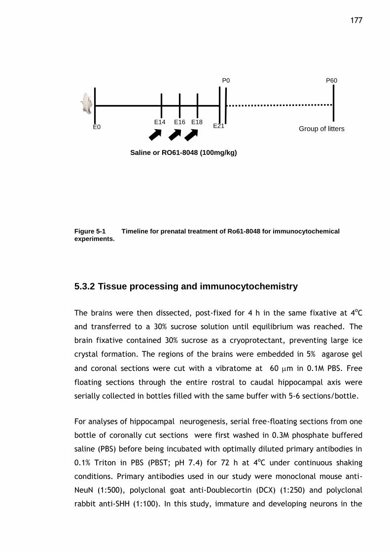
177
P0
E0E14 E16 E18
E21 Group of litters
P60
Saline or RO61-8048 (100mg/kg)
Figure 5-1 Timeline for prenatal treatment of Ro61-8048 for immunocytochemical experiments.
5.3.2 Tissue processing and immunocytochemistry
The brains were then dissected, post-fixed for 4 h in the same fixative at 4oC
and transferred to a 30% sucrose solution until equilibrium was reached. The
brain fixative contained 30% sucrose as a cryoprotectant, preventing large ice
crystal formation. The regions of the brains were embedded in 5% agarose gel
and coronal sections were cut with a vibratome at 60 m in 0.1M PBS. Free
floating sections through the entire rostral to caudal hippocampal axis were
serially collected in bottles filled with the same buffer with 5-6 sections/bottle.
For analyses of hippocampal neurogenesis, serial free-floating sections from one
bottle of coronally cut sections were first washed in 0.3M phosphate buffered
saline (PBS) before being incubated with optimally diluted primary antibodies in
0.1% Triton in PBS (PBST; pH 7.4) for 72 h at 4oC under continuous shaking
conditions. Primary antibodies used in our study were monoclonal mouse anti-
NeuN (1:500), polyclonal goat anti-Doublecortin (DCX) (1:250) and polyclonal
rabbit anti-SHH (1:100). In this study, immature and developing neurons in the
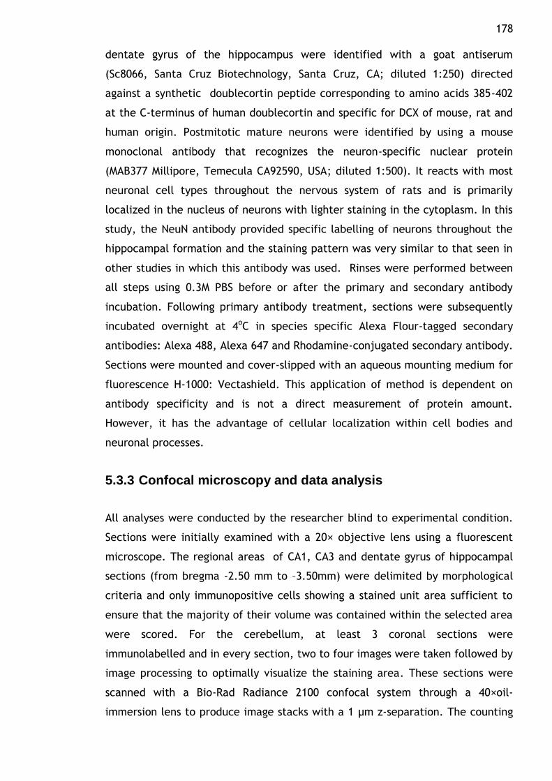
178 dentate gyrus of the hippocampus were identified with a goat antiserum
(Sc8066, Santa Cruz Biotechnology, Santa Cruz, CA; diluted 1:250) directed
against a synthetic doublecortin peptide corresponding to amino acids 385-402
at the C-terminus of human doublecortin and specific for DCX of mouse, rat and
human origin. Postmitotic mature neurons were identified by using a mouse
monoclonal antibody that recognizes the neuron-specific nuclear protein
(MAB377 Millipore, Temecula CA92590, USA; diluted 1:500). It reacts with most
neuronal cell types throughout the nervous system of rats and is primarily
localized in the nucleus of neurons with lighter staining in the cytoplasm. In this
study, the NeuN antibody provided specific labelling of neurons throughout the
hippocampal formation and the staining pattern was very similar to that seen in
other studies in which this antibody was used. Rinses were performed between
all steps using 0.3M PBS before or after the primary and secondary antibody
incubation. Following primary antibody treatment, sections were subsequently
incubated overnight at 4oC in species specific Alexa Flour-tagged secondary
antibodies: Alexa 488, Alexa 647 and Rhodamine-conjugated secondary antibody.
Sections were mounted and cover-slipped with an aqueous mounting medium for
fluorescence H-1000: Vectashield. This application of method is dependent on
antibody specificity and is not a direct measurement of protein amount.
However, it has the advantage of cellular localization within cell bodies and
neuronal processes.
5.3.3 Confocal microscopy and data analysis
All analyses were conducted by the researcher blind to experimental condition.
Sections were initially examined with a 20× objective lens using a fluorescent
microscope. The regional areas of CA1, CA3 and dentate gyrus of hippocampal
sections (from bregma -2.50 mm to –3.50mm) were delimited by morphological
criteria and only immunopositive cells showing a stained unit area sufficient to
ensure that the majority of their volume was contained within the selected area
were scored. For the cerebellum, at least 3 coronal sections were
immunolabelled and in every section, two to four images were taken followed by
image processing to optimally visualize the staining area. These sections were
scanned with a Bio-Rad Radiance 2100 confocal system through a 40×oil-
immersion lens to produce image stacks with a 1 μm z-separation. The counting
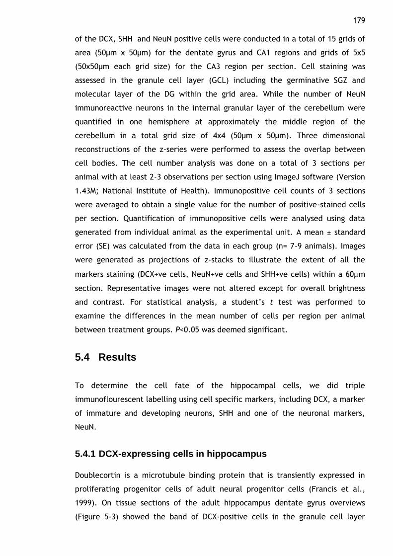
179 of the DCX, SHH and NeuN positive cells were conducted in a total of 15 grids of
area (50µm x 50µm) for the dentate gyrus and CA1 regions and grids of 5x5
(50x50µm each grid size) for the CA3 region per section. Cell staining was
assessed in the granule cell layer (GCL) including the germinative SGZ and
molecular layer of the DG within the grid area. While the number of NeuN
immunoreactive neurons in the internal granular layer of the cerebellum were
quantified in one hemisphere at approximately the middle region of the
cerebellum in a total grid size of 4x4 (50µm x 50µm). Three dimensional
reconstructions of the z-series were performed to assess the overlap between
cell bodies. The cell number analysis was done on a total of 3 sections per
animal with at least 2-3 observations per section using ImageJ software (Version
1.43M; National Institute of Health). Immunopositive cell counts of 3 sections
were averaged to obtain a single value for the number of positive-stained cells
per section. Quantification of immunopositive cells were analysed using data
generated from individual animal as the experimental unit. A mean ± standard
error (SE) was calculated from the data in each group (n= 7-9 animals). Images
were generated as projections of z-stacks to illustrate the extent of all the
markers staining (DCX+ve cells, NeuN+ve cells and SHH+ve cells) within a 60m
section. Representative images were not altered except for overall brightness
and contrast. For statistical analysis, a student‟s t test was performed to
examine the differences in the mean number of cells per region per animal
between treatment groups. P<0.05 was deemed significant.
5.4 Results
To determine the cell fate of the hippocampal cells, we did triple
immunoflourescent labelling using cell specific markers, including DCX, a marker
of immature and developing neurons, SHH and one of the neuronal markers,
NeuN.
5.4.1 DCX-expressing cells in hippocampus
Doublecortin is a microtubule binding protein that is transiently expressed in
proliferating progenitor cells of adult neural progenitor cells (Francis et al.,
1999). On tissue sections of the adult hippocampus dentate gyrus overviews
(Figure 5-3) showed the band of DCX-positive cells in the granule cell layer
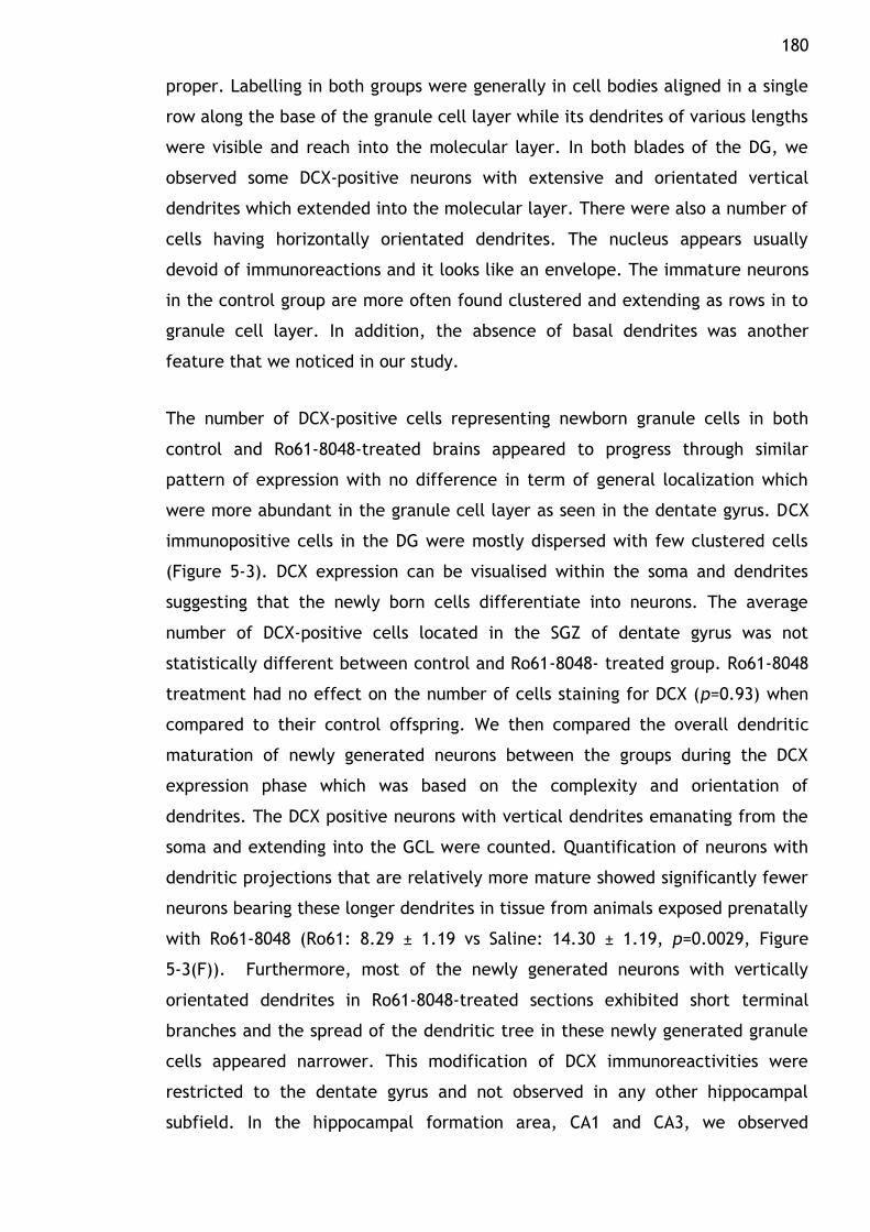
180 proper. Labelling in both groups were generally in cell bodies aligned in a single
row along the base of the granule cell layer while its dendrites of various lengths
were visible and reach into the molecular layer. In both blades of the DG, we
observed some DCX-positive neurons with extensive and orientated vertical
dendrites which extended into the molecular layer. There were also a number of
cells having horizontally orientated dendrites. The nucleus appears usually
devoid of immunoreactions and it looks like an envelope. The immature neurons
in the control group are more often found clustered and extending as rows in to
granule cell layer. In addition, the absence of basal dendrites was another
feature that we noticed in our study.
The number of DCX-positive cells representing newborn granule cells in both
control and Ro61-8048-treated brains appeared to progress through similar
pattern of expression with no difference in term of general localization which
were more abundant in the granule cell layer as seen in the dentate gyrus. DCX
immunopositive cells in the DG were mostly dispersed with few clustered cells
(Figure 5-3). DCX expression can be visualised within the soma and dendrites
suggesting that the newly born cells differentiate into neurons. The average
number of DCX-positive cells located in the SGZ of dentate gyrus was not
statistically different between control and Ro61-8048- treated group. Ro61-8048
treatment had no effect on the number of cells staining for DCX (p=0.93) when
compared to their control offspring. We then compared the overall dendritic
maturation of newly generated neurons between the groups during the DCX
expression phase which was based on the complexity and orientation of
dendrites. The DCX positive neurons with vertical dendrites emanating from the
soma and extending into the GCL were counted. Quantification of neurons with
dendritic projections that are relatively more mature showed significantly fewer
neurons bearing these longer dendrites in tissue from animals exposed prenatally
with Ro61-8048 (Ro61: 8.29 ± 1.19 vs Saline: 14.30 ± 1.19, p=0.0029, Figure
5-3(F)). Furthermore, most of the newly generated neurons with vertically
orientated dendrites in Ro61-8048-treated sections exhibited short terminal
branches and the spread of the dendritic tree in these newly generated granule
cells appeared narrower. This modification of DCX immunoreactivities were
restricted to the dentate gyrus and not observed in any other hippocampal
subfield. In the hippocampal formation area, CA1 and CA3, we observed
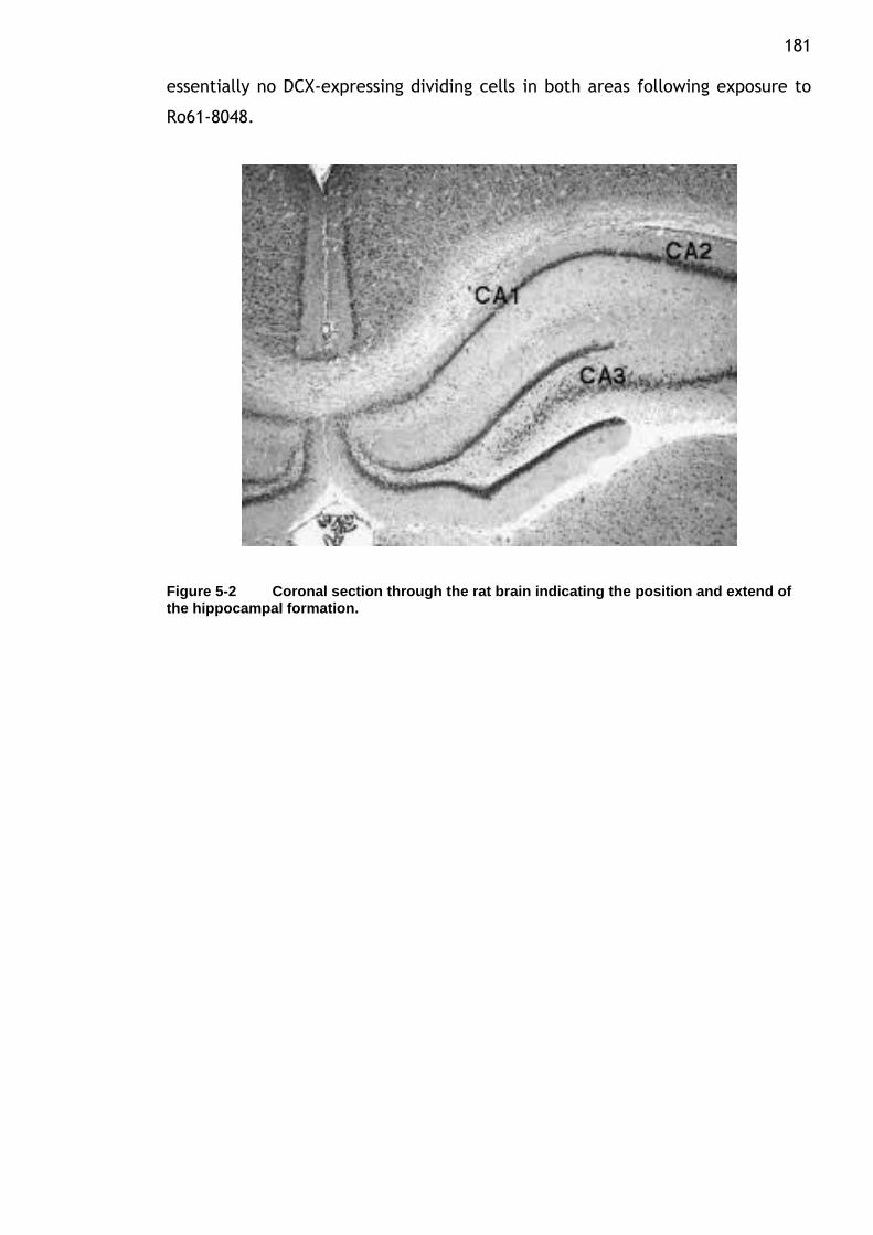
181 essentially no DCX-expressing dividing cells in both areas following exposure to
Ro61-8048.
Figure 5-2 Coronal section through the rat brain indicating the position and extend of the hippocampal formation.
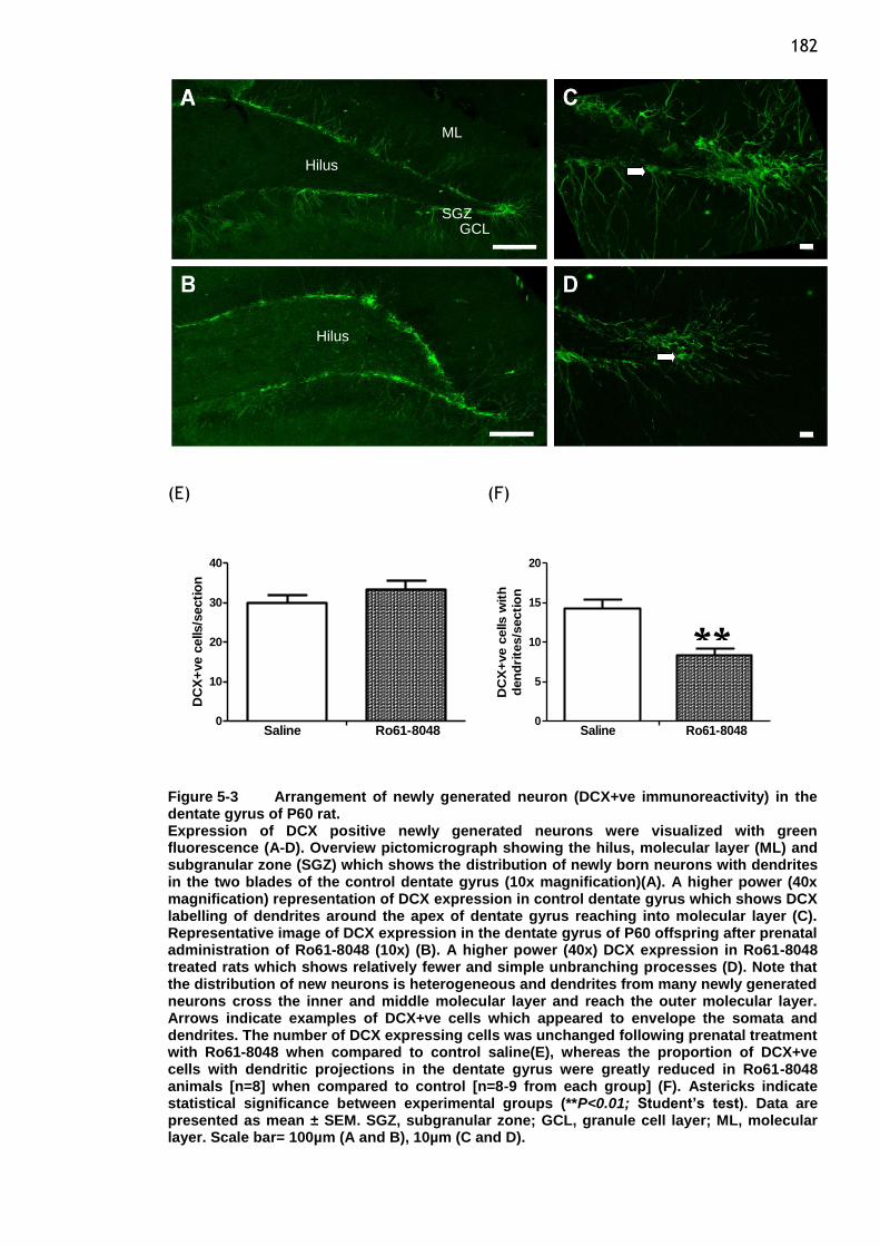
182
(E) (F)
0
10
20
30
40
Saline Ro61-8048
DC
X+
ve c
ells/s
ecti
on
0
5
10
15
20
Saline Ro61-8048
DC
X+
ve c
ells w
ith
den
dri
tes/s
ecti
on
Figure 5-3 Arrangement of newly generated neuron (DCX+ve immunoreactivity) in the dentate gyrus of P60 rat. Expression of DCX positive newly generated neurons were visualized with green fluorescence (A-D). Overview pictomicrograph showing the hilus, molecular layer (ML) and subgranular zone (SGZ) which shows the distribution of newly born neurons with dendrites in the two blades of the control dentate gyrus (10x magnification)(A). A higher power (40x magnification) representation of DCX expression in control dentate gyrus which shows DCX labelling of dendrites around the apex of dentate gyrus reaching into molecular layer (C). Representative image of DCX expression in the dentate gyrus of P60 offspring after prenatal administration of Ro61-8048 (10x) (B). A higher power (40x) DCX expression in Ro61-8048 treated rats which shows relatively fewer and simple unbranching processes (D). Note that the distribution of new neurons is heterogeneous and dendrites from many newly generated neurons cross the inner and middle molecular layer and reach the outer molecular layer. Arrows indicate examples of DCX+ve cells which appeared to envelope the somata and dendrites. The number of DCX expressing cells was unchanged following prenatal treatment with Ro61-8048 when compared to control saline(E), whereas the proportion of DCX+ve cells with dendritic projections in the dentate gyrus were greatly reduced in Ro61-8048 animals [n=8] when compared to control [n=8-9 from each group] (F). Astericks indicate statistical significance between experimental groups (**P<0.01; Student’s test). Data are presented as mean ± SEM. SGZ, subgranular zone; GCL, granule cell layer; ML, molecular layer. Scale bar= 100µm (A and B), 10µm (C and D).
GCL SGZ
ML
Hilus
Hilus
**
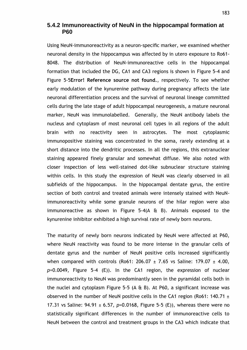
183
5.4.2 Immunoreactivity of NeuN in the hippocampal formation at P60
Using NeuN-immunoreactivity as a neuron-specific marker, we examined whether
neuronal density in the hippocampus was affected by in utero exposure to Ro61-
8048. The distribution of NeuN-immunoreactive cells in the hippocampal
formation that included the DG, CA1 and CA3 regions is shown in Figure 5-4 and
Figure 5-5Error! Reference source not found., respectively. To see whether
early modulation of the kynurenine pathway during pregnancy affects the late
neuronal differentiation process and the survival of neuronal lineage committed
cells during the late stage of adult hippocampal neurogenesis, a mature neuronal
marker, NeuN was immunolabelled. Generally, the NeuN antibody labels the
nucleus and cytoplasm of most neuronal cell types in all regions of the adult
brain with no reactivity seen in astrocytes. The most cytoplasmic
immunopositive staining was concentrated in the soma, rarely extending at a
short distance into the dendritic processes. In all the regions, this extranuclear
staining appeared finely granular and somewhat diffuse. We also noted with
closer inspection of less well-stained dot-like subnuclear structure staining
within cells. In this study the expression of NeuN was clearly observed in all
subfields of the hippocampus. In the hippocampal dentate gyrus, the entire
section of both control and treated animals were intensely stained with NeuN-
immunoreactivity while some granule neurons of the hilar region were also
immunoreactive as shown in Figure 5-4(A & B). Animals exposed to the
kynurenine inhibitor exhibited a high survival rate of newly born neurons.
The maturity of newly born neurons indicated by NeuN were affected at P60,
where NeuN reactivity was found to be more intense in the granular cells of
dentate gyrus and the number of NeuN positive cells increased significantly
when compared with controls (Ro61: 206.07 ± 7.65 vs Saline: 179.07 ± 4.00,
p=0.0049, Figure 5-4 (E)). In the CA1 region, the expression of nuclear
immunoreactivity to NeuN was predominantly seen in the pyramidal cells both in
the nuclei and cytoplasm Figure 5-5 (A & B). At P60, a significant increase was
observed in the number of NeuN positive cells in the CA1 region (Ro61: 140.71 ±
17.31 vs Saline: 94.91 ± 6.57, p=0.0168, Figure 5-5 (E)), whereas there were no
statistically significant differences in the number of immunoreactive cells to
NeuN between the control and treatment groups in the CA3 which indicate that

184 existing mature neurons are not affected (Figure 5-5 (F); p=0.8071). These
results provide evidence that Ro61-8048 administration during pregnancy has a
stimulatory effect on the development of new neurons and also demonstrate
that this effect is long lasting, being evident in pubertal offspring 9 weeks after
their first exposure to the compound.
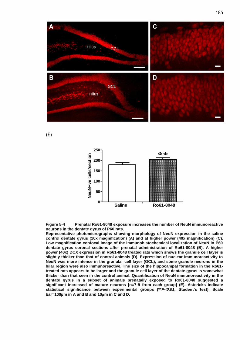
185
(E)
0
50
100
150
200
250
Saline Ro61-8048
Neu
N+
ve c
ells
/secti
on
Figure 5-4 Prenatal Ro61-8048 exposure increases the number of NeuN immunoreactive neurons in the dentate gyrus of P60 rats. Representative photomicrographs showing morphology of NeuN expression in the saline control dentate gyrus (10x magnification) (A) and at higher power (40x magnification) (C). Low magnification confocal image of the immunohistochemical localization of NeuN in P60 dentate gyrus coronal sections after prenatal administration of Ro61-8048 (B). A higher power (40x) DCX expression in Ro61-8048 treated rats which shows the granule cell layer is slightly thicker than that of control animals (D). Expression of nuclear immunoreactivity to NeuN was more intense in the granular cell layer (GCL), and some granule neurons in the hilar region were also immunoreactive. The size of the hippocampal formation in the Ro61-treated rats appears to be larger and the granule cell layer of the dentate gyrus is somewhat thicker than that seen in the control animal. Quantification of NeuN immunoreactivity in the dentate gyrus in a subset of animals prenatally exposed to Ro61-8048 suggested a significant increased of mature neurons [n=7-9 from each group] (E). Astericks indicate statistical significance between experimental groups (**P<0.01; Student’s test). Scale
bar=100µm in A and B and 10m in C and D.
GCL
GCL Hilus
Hilus
**
186
(E) (F)
0
50
100
150
200
Saline Ro61-8048Neu
N+
ve c
ells in
CA
1/s
ecti
on
0
50
100
150
Saline Ro61-8048Neu
N+
ve c
ells in
CA
3/s
ecti
on
Figure 5-5 Immunocytochemical localization of NeuN in hippocampal coronal sections of CA1 and CA3 regions Representative photomicrographs of NeuN positive neurons from control and Ro61-8048 animals are shown. NeuN immunopositive pyramidal cells in CA1 region in the control (A) and Ro61-8048-treated rats with an apparent neuronal increase in the hippocampal CA1 region (B). Immunostaining of NeuN in the CA3 region of the hippocampus in the control (C) and Ro691-8048-treated sections (D). Bar graphs show the quantified immunopositive cells to NeuN in CA1 and CA3 regions. The quantified score based on the number of cells showed significantly increased number of NeuN+ve neurons in the CA1 hippocampus of Ro61-8048-treated rats while no differences in terms of immunoreactive cells were observed
in the CA3 region (E & F). Values are expressed as mean SEM [n=8-9 from each group]. Asterick indicates statistical significance between experimental groups (*P<0.05; Student’s
test). Scale bar= 20m (A,B,C and D).
*
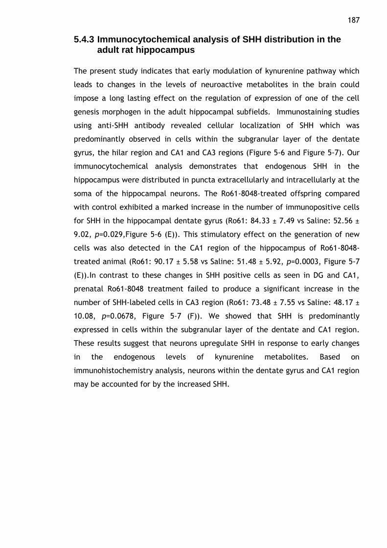
187
5.4.3 Immunocytochemical analysis of SHH distribution in the adult rat hippocampus
The present study indicates that early modulation of kynurenine pathway which
leads to changes in the levels of neuroactive metabolites in the brain could
impose a long lasting effect on the regulation of expression of one of the cell
genesis morphogen in the adult hippocampal subfields. Immunostaining studies
using anti-SHH antibody revealed cellular localization of SHH which was
predominantly observed in cells within the subgranular layer of the dentate
gyrus, the hilar region and CA1 and CA3 regions (Figure 5-6 and Figure 5-7). Our
immunocytochemical analysis demonstrates that endogenous SHH in the
hippocampus were distributed in puncta extracellularly and intracellularly at the
soma of the hippocampal neurons. The Ro61-8048-treated offspring compared
with control exhibited a marked increase in the number of immunopositive cells
for SHH in the hippocampal dentate gyrus (Ro61: 84.33 ± 7.49 vs Saline: 52.56 ±
9.02, p=0.029,Figure 5-6 (E)). This stimulatory effect on the generation of new
cells was also detected in the CA1 region of the hippocampus of Ro61-8048-
treated animal (Ro61: 90.17 ± 5.58 vs Saline: 51.48 ± 5.92, p=0.0003, Figure 5-7
(E)).In contrast to these changes in SHH positive cells as seen in DG and CA1,
prenatal Ro61-8048 treatment failed to produce a significant increase in the
number of SHH-labeled cells in CA3 region (Ro61: 73.48 ± 7.55 vs Saline: 48.17 ±
10.08, p=0.0678, Figure 5-7 (F)). We showed that SHH is predominantly
expressed in cells within the subgranular layer of the dentate and CA1 region.
These results suggest that neurons upregulate SHH in response to early changes
in the endogenous levels of kynurenine metabolites. Based on
immunohistochemistry analysis, neurons within the dentate gyrus and CA1 region
may be accounted for by the increased SHH.
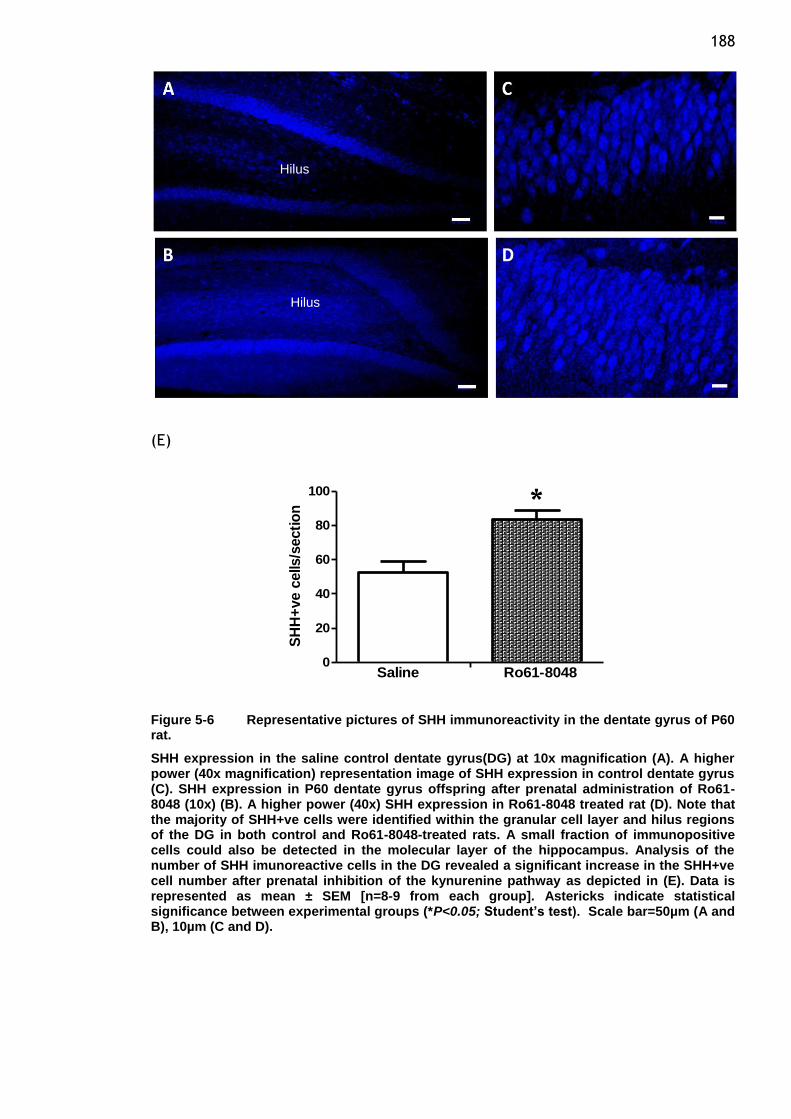
188
(E)
0
20
40
60
80
100
Saline Ro61-8048
SH
H+
ve c
ells
/secti
on
Figure 5-6 Representative pictures of SHH immunoreactivity in the dentate gyrus of P60 rat.
SHH expression in the saline control dentate gyrus(DG) at 10x magnification (A). A higher power (40x magnification) representation image of SHH expression in control dentate gyrus (C). SHH expression in P60 dentate gyrus offspring after prenatal administration of Ro61-8048 (10x) (B). A higher power (40x) SHH expression in Ro61-8048 treated rat (D). Note that the majority of SHH+ve cells were identified within the granular cell layer and hilus regions of the DG in both control and Ro61-8048-treated rats. A small fraction of immunopositive cells could also be detected in the molecular layer of the hippocampus. Analysis of the number of SHH imunoreactive cells in the DG revealed a significant increase in the SHH+ve cell number after prenatal inhibition of the kynurenine pathway as depicted in (E). Data is represented as mean ± SEM [n=8-9 from each group]. Astericks indicate statistical significance between experimental groups (*P<0.05; Student’s test). Scale bar=50µm (A and B), 10µm (C and D).
Hilus
Hilus
*
189
(E) (F)
0
50
100
150
Saline Ro61-8048SH
H+
ve c
ells in
CA
1/s
ecti
on
0
20
40
60
80
100
Saline Ro61-8048SH
H+
ve c
ells in
CA
3/s
ecti
on
Figure 5-7 Immunocytochemistry for SHH in the CA1 and CA3 region of the hippocampus. SHH immunolabelling of neurons in coronal sections through the hippocampal formation subfield CA1 and CA3 region in P60 animals after prenatal treatment with Ro61-8048. SHH immunopositive staining shows increased expression within the CA1 region following kynurenine pathway inhibition. Representative photomicrograph shows distribution of SHH at 40x magnification in the CA1 region of saline control animals (A). Representative image of SHH expression in Ro61-8048-treated animals in the CA1 (B). SHH expression in the CA3 region of P60 offspring in control animals (C). A higher power (40x) SHH expression in the CA3 region of Ro61-8048-treated animals (D). Analysis of the number of SHH immunoreactive cells in CA1 revealed a significant increased in the SHH+ve cell number after prenatal inhibition of the kynurenine pathway as depicted in (E) while no differences were observed in the CA3 region (F). Astericks indicate statistical significance between experimental groups (***P<0.001; Student’s test). Data represented as mean ± SEM (n=8-9). Scale bar=20µm (A,B,C and D).
***
190
5.4.4 Cerebellar NeuN-positive cells after prenatal Ro61-8048
We examined on closely spaced triple-labelling coronal sections of the adult
cerebellum in an attempt to identify and characterize the expression of these
developmental markers following Ro61-8048 treatment during the late phase of
gestation. Inspection of these sections revealed no evidence of staining for DCX
which marks postmitotic, migrating neurons, while high levels of diffuse staining
were observed for SHH in the granule cell layer which made it impossible to
count individual neurons for both markers. Previous studies have demonstrated
that SHH expression is confined to the purkinje layer, in line with its production
in the purkinje cells (Traiffort et al., 1998). However, observations in our
sections exhibited a very faint reactivity for SHH antibody in the purkinje layer,
which on the basis of its distribution might reflect labelling of purkinje cells. In
this respect, increased secretion or reduced removal and altered metabolism of
SHH could possibly be attributable to the indistinct diffuse staining
intracellularly and on the extracellular surface of the soma. Immunoreactivity
for NeuN was observed in the small granule neurons located within the granular
layer, showing cytoplasmic reactivity in addition to nuclear immunolocalization.
As for the number of NeuN-positive cells in this area, there were no statistically
significant differences in both control and treatment groups (Ro61: 137.85 ± 4.39
vs Saline: 148.76 ± 5.29, p=0.1348, Figure 5-8 (E)).
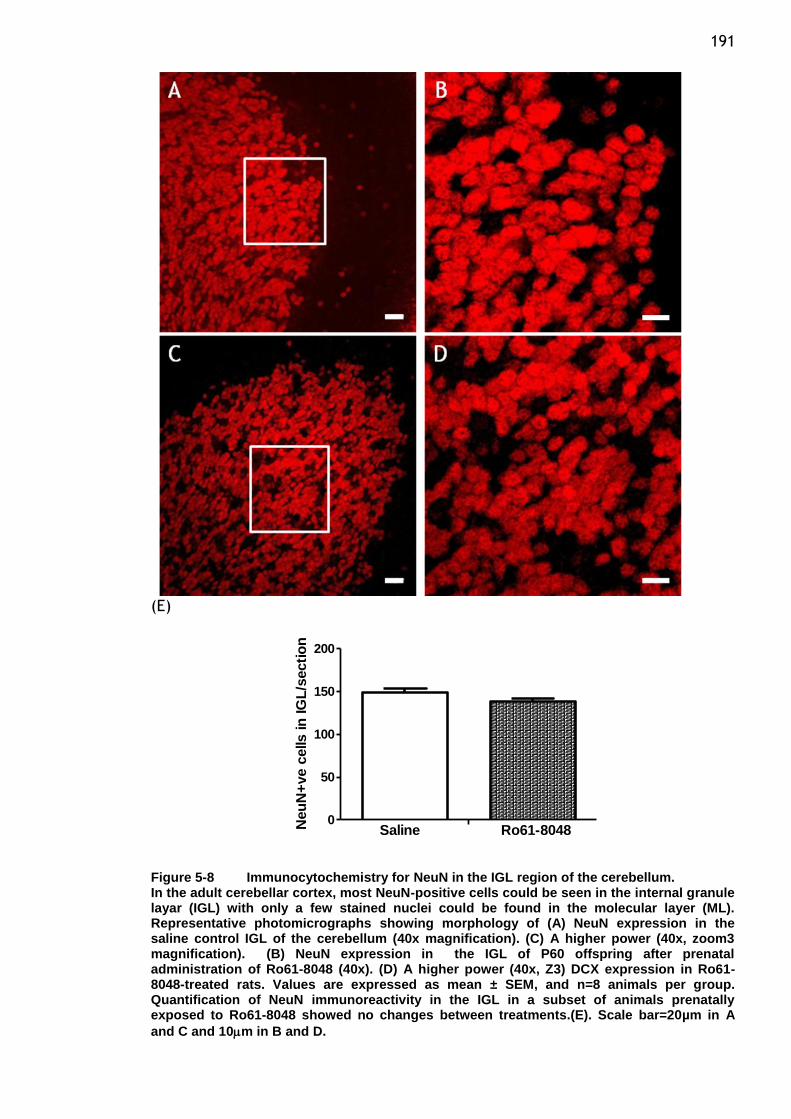
191
(E)
0
50
100
150
200
Saline Ro61-8048Neu
N+
ve c
ells in
IG
L/s
ecti
on
Figure 5-8 Immunocytochemistry for NeuN in the IGL region of the cerebellum. In the adult cerebellar cortex, most NeuN-positive cells could be seen in the internal granule layar (IGL) with only a few stained nuclei could be found in the molecular layer (ML). Representative photomicrographs showing morphology of (A) NeuN expression in the saline control IGL of the cerebellum (40x magnification). (C) A higher power (40x, zoom3 magnification). (B) NeuN expression in the IGL of P60 offspring after prenatal administration of Ro61-8048 (40x). (D) A higher power (40x, Z3) DCX expression in Ro61-8048-treated rats. Values are expressed as mean ± SEM, and n=8 animals per group. Quantification of NeuN immunoreactivity in the IGL in a subset of animals prenatally exposed to Ro61-8048 showed no changes between treatments.(E). Scale bar=20µm in A
and C and 10m in B and D.
192
5.5 Discussion
Confocal microscopy was used to determine the phenotype of all the
immunoreactive cells in selected region of interest. Immunolabeling with DCX,
SHH and NeuN allowed visualization of newly generated cells that were
developing and maturing according to a neuronal fate in both treatment groups.
The observation of variable conditions including physiological, pathological and
pharmacological stimuli could modulate adult neurogenesis. Therefore the
present study was designed to identify possible changes in granule cell number,
proliferation and maturation in the hippocampus as to corroborate the molecular
data at the morphological and protein level. We also sought to examine whether
the modified expression of these proteins are associated with structural changes
at the cellular level that might contribute for the modification of neuronal
structure and development.
We studied the effects of modulation of NMDA receptor function by
administering a known enzyme inhibitor, Ro61-8048 on the localisation of
neuronal structure with respect to neurogenesis processes that includes
neuroblast differentiation using DCX staining, neuronal migration and orientation
using SHH and the maturity of neurons using NeuN immunocytochemistry, in the
hippocampus at P60. During early development of the brain, NMDA subtypes of
glutamate receptors and α7NCR receptors of glutamatergic and cholinergic
mechanisms are critically involved and play central roles in neuronal growth,
differentiation and synapse formation (Bard and Groc, 2011, Cull-Candy et al.,
2001). It has been established that endogenous KYNA functions as an
endogenous agent capable of directly affecting both NMDARs and α7NCRs.
Antagonistic properties of KYNA at the NMDA receptor account for its ability to
reduce excitation and are responsible for its neuroprotective and anticonvulsant
properties (Foster et al., 1984). However, these effects may also pose for a
pathophysiologically relevant function in neuropsychotic diseases such as
schizophrenia, which includes abnormal brain KYNA levels (Muller and Schwarcz,
2006). As endogenous KYNA might also be involved in the processes underlying
learning and more generally synaptic plasticity, fluctuations in KYNA levels can
be envisioned to influence these processes. As elaborated in previous studies, a
range of pharmacological agents have been used to probe the physiological and
193 pathological roles of KP metabolites. One approach to influence the levels of KP
metabolites is based on the pharmacological blockade of KMO, the enzyme
responsible for the formation of 3-HK and QUIN (Stone et al., 2012, Stone,
2000). This concept is supported by our previous studies showing that the
administration of Ro61-8048 during late gestation of rats were reported to alter
the relative concentrations of endogenous KP metabolites and resulted in
disordered neuronal development.
We have demonstrated an increased in the levels of kynurenine and KYNA in the
maternal plasma and also in the embryo brains 5 hour post-injection. The
maternal levels of these metabolites normalize after 24 hour but remain
elevated in the embryonic brain. It seemed that the outcome of administering
pharmacological compounds that block KMO somehow shunted the metabolism
of KP toward the production of KYNA. It has been suggested that the changes in
the protein expressions of some of the target molecules observed at
developmental ages of P21 and P60 were likely to be caused by the increased
levels of these KP metabolites, particularly KYNA (Forrest et al., 2013a, Forrest
et al., 2013b).
5.5.1 Protein localisation in the hippocampus
To test the hypothesis that an early modulation in the endogenous levels of
KYNA induced by blocking one of the main enzymes in the KP, would produce
long lasting changes in the generation and development of neurons, we focused
on the hippocampus because it possesses a substantial capacity for regeneration
and presents itself as a unique form of neural circuit plasticity. A substantial
body of evidence regards a crucial role for hippocampal neurogenesis in
mediating specific cognitive functions. In line with its well-recognised role in
cognitive processes, any modification as in decreased or abnormal process is
strongly correlated with deficits in hippocampal structure and function in various
neurological disorders including epilepsy, ischemia and schizophrenia(Curtis et
al., 2003b, Eriksson et al., 1998).
The dentate gyrus of the hippocampus is one of the few mammalian brain
structures that continue to generate new neurons through adulthood. It has been
demonstrated that adult-generated cells especially in the dentate gyrus of rats
194 develop the morphological characteristics of granule cells, receive synaptic
input, then extend axons into the mossy fiber pathway and express a number of
markers of mature neurons. These sequential steps of adult neurogenesis ranging
from neural precursor proliferation, migration, differentiation to synaptic
integration of newborn neurons are tightly regulated by a variety of extrinsic
and intrinsic factors, most of which still remain uncharacterized (Zhao et al.,
2008, Kempermann et al., 2004).The extended period of granule cells genesis
and the ability for continual restructuring of neurogenic areas according to the
current environment and involvement of signalling cues, represent a dynamic
process that could be regulated by natural factors including physical activity,
hippocampal-dependent tasks, NMDA receptor activity, hormones and growth
factors. The late-generated neurons in this brain region might render the
dentate gyrus particularly sensitive to environmental perturbations that may
adversely alter hippocampal structure and function. In addition, it has been
brought forward into consideration that treatments which typically enhance or
normalize the cell generation process could ameliorate the behavioural
alterations in animal models or humans. This data substantiates a ground cause
that adult hippocampal neurogenesis may be part of intrinsic requirement of
therapeutic targets for behavioural of distinct neurological diseases (Eriksson et
al., 1998, McDonald and Wojtowicz, 2005, Kempermann et al., 2004).
In most studies to examine morphological details of newborn neurons have been
made possible by the application of either retrovirus-mediated gene transfer or
the use of thymidine analogue, 5‟-bromodeoxyurindine (BrdU) which
incorporates into the DNA of dividing cells during the S-phase of the cell cycle
(Zhao et al., 2006, Rao and Shetty, 2004).The identification of newly born
neurons has also been accomplished by immunostaining with DCX. In animal
studies of adult rodents, DCX has been identified as a promising candidate for
the identification of newborn immature neurons in which its expression
specifically persists in the hippocampus with clear labelling detected in both cell
bodies and the dendritic processes that extend into the granule cell and
molecular layers of the dentate gyrus. Previous studies have showed that the
expression of DCX appears to be persist for a prolonged period and overlapped
with both immature as well as mature neuronal markers(NeuN), indicating that
DCX expression in postmitotic granule cells is quite extensive during the early
195 growth period of the neurons including dendritic and axon growth. Counting of
neurons that are positive for DCX in hippocampal sections particularly in the
dentate gyrus gives a good estimate of the newly generated neurons that exhibit
the phenotype of differentiated granule cells. Thus, transient expression of DCX
during specific times of cell genesis justifies the ability of DCX as a reliable
marker of newly generated neurons in the adult hippocampus (Rao and Shetty,
2004, Rao et al., 2005, Brown et al., 2003).
In our study, DCX positive staining exhibits the phenotype of neurons with
staining around the soma and also in dendrites. DCX immunostaining revealed
newly formed neurons in the DG and the overall production of new neurons were
maintained at the same levels as controls. Characterization of DCX-positive cells
having the phenotype of differentiated granule cells showed that dendritic
process in the control group showed a comparatively complex branching of
apical dendrite in the molecular layer which presumably indicate more mature
new neurons while neurons in Ro61-8048-treated specimens exhibited either no
or with short processes which did not reach deep into the molecular layer.
Analysis of the dendrites revealed that the new granule cells in the Ro61-8048-
treated sections exhibit fewer number of cells with a complex dendritic spread.
The shorter apical dendrites with narrower dendritic spread in newly born
granule cells of the adult, most likely reflect changes in differentiation of the
cells having a relatively immature morphology. Although the precise functions
that depend on the production of new neurons in the DG throughout life are not
defined, a range of studies have implied that the cells might be involved in
aspects of normal hippocampal function, such as learning and memory. Along
this line, studies reported an age-related decrease in adult neurogenesis and has
further been proposed as a factor in the age-related decline of cognitive ability
(Ben Abdallah et al., 2010, McDonald and Wojtowicz, 2005, Farioli-Vecchioli et
al., 2008).
Stressful experiences during development may also potentially be involved in
impaired performance of hippocampal-dependent learning in adulthood by
suppressing the production of granule neurons. Elevation of glucocorticoid
corticosterone and stimulation of glutamate release following stress diminishes
the number of proliferating cells in the DG. This evidences suggested that stress
196 exhibits a suppressive effect of glucocorticoid and NMDA receptor activation
which decreases the production of granule neurons in the developing brain and
might possibly have a negative impact on learning and memory in adulthood
(Gould and Tanapat, 1999). In contrast, several other types of brain injuries such
as traumatic brain injury, ischemia, epileptic seizure and stroke enhance the
production of new neurons in the adult DG(Wennstrom et al., 2003, Yu et al.,
2008). Similarly, studies have suggested that the therapeutic mechanisms of
antipsychotics are related to their stimulatory effect on neurogenesis.The
effects on proliferation in the neurogenic regions of the adult rat brain have
been observed by incorporating the proliferation marker BrdU, while
neurogenesis has been described following an increase in markers of neuronal
differentiation and survival such DCX and NeuN (Toro and Deakin, 2007).
The secreted factor SHH is a signalling protein that plays multiple roles in the
regulation of early patterning of neural tube and subsequently has been found to
stimulate the production of progenitor cells by actively being involved in the
regulation of neuronal precursor proliferation, migration, differentiation and cell
survival. This indicates that SHH is a powerful regulator of stem cell
maintenance in the developing embryonic brain (Traiffort et al., 2010).
Involvement of this morphogen in the control of stem cell properties in the adult
brain have been studied in vivo and in vitro. SHH has been shown to regulate the
proliferation of adult hippocampal neural stem cells and represents its function
in the adult nervous system. This proliferative effect of SHH through activity-
dependent synthesis or secretion within the hippocampal neurogenic area may
indicate a mechanism for regulating neurogenesis and could be implied as one of
the therapeutic potential for the regeneration of neural tissue in
neurodegenerative disorders (Lai et al., 2003, Palma et al., 2005). Several
pathological conditions for instance ischemia, hypoxia and seizures have been
shown to induce a rapid regulation of key genes of the SHH signalling cascade
and that SHH signalling is required for adult neurogenesis. This implies that
activation of SHH and its receptor components through activity-dependent
neural plasticity may be involved in the regulation of adult brain neurogenesis
(Banarjee and Rajendran, 2005, Sims et al., 2009).
197 Another major finding of this study was the localization of SHH in the
hippocampal formation. In the study described here, we found that prenatal
modulation of kynurenine pathway by inhibiting KMO enzyme upregulated the
expression of SHH. Most notable is our finding that the newly generated neurons
produced by in utero exposure to Ro61-8048 are able to express SHH, as
demonstrated by a significant increase in the number of cells expressing SHH in
the hippocampus. This stimulatory effect has been observed in the dentate gyrus
and CA1 region. Although it is unclear by what mechanism NMDA antagonistic
properties of KYNA up regulates SHH, the increase may likely involve other
intermediate regulatory steps that regulate the expression of SHH. It is possible
to suggest that SHH produced in the hippocampal subregions particularly the CA3
and dentate gyrus, might play a role in regulating axonal guidance from the
newborn granular neurons of the dentate gyrus as the CA3 region receives the
mossy fiber output from the granular neurons of the dentate gyrus(Charytoniuk
et al., 2002).
Our results show that despite high levels of SHH expression seen in the CA3
region, it did not reach statistical significance. In addition to that, we also
observed a diffuse staining of granule cells throughout the hilar region of the
hippocampus. As the function of secreted protein SHH has generally been
attributed in relation to cell proliferation and migration, the expression of SHH
in fully differentiated neurons in this area could be associated with errors or
modification in cell migration process. These data demonstrate that an in utero
challenge with Ro61-8048 has a significant, stimulatory effect on the generation
and development of new neurons in the selected areas of hippocampal
formation. The increased activation of the SHH pathway in this region could
potentially increase the growth and survival of newly generated granule cells
and their developing axons. These findings raise the possibility that the early
modulation of NMDA receptor function through KYNA on the expression of SHH
and its unidentified receptor components could contribute to the influence on
adult neurogenesis. It also suggest that the SHH pathway may continue to have
an important role in adulthood in regulating structural plasticity. Further studies
are thus required to address whether the effects of KP inhibition also involve
other SHH receptor components which could further regulate the response of
SHH signalling mechanisms. Unlike our protein expression study (chapter 4)
198 which showed significant decreases of SHH protein in the hippocampus after
exposure with Ro61-8048 while no changes were seen in mRNA levels may
suggest a possible explanation for this discrepancy. We assumed that the
reduced protein level seen in whole hippocampal lysates of adult animals is
possibly due to decreased translation or altered stability or degradation of the
protein at the time of examination.
Of several molecular markers that could be linked to neural development, NeuN
has gained widespread acceptance as a reliable immunocytochemical marker for
neurons in neurodevelopmental research. It is also has been incorporated as a
useful tool in diagnostic neuropathology where its specific expression in
postmitotic neurons has made it possible to distinguish and localize neuronally
differentiated cells. Immunostaining analysis showed that NeuN was primarily
localized to the nuclei of postmitotic neurons throughout the CNS and the
immunoreactivity can be detected in multiple vertebrate species suggests a
considerable conservation of this antigen (Mullen et al., 1992). The present
findings are consistent with previous studies that indicate preferential nuclear
localization of the protein, but also show NeuN immunoreactivity in the
cytoplasm (Lind et al., 2005). The hippocampal granule cells and pyramidal cells
of the CA1 and CA3 regions are finely stained by this antibody and the dendrites
emanating from these cells are clearly devoid of staining.
5.5.2 Protein localisation in the cerebellum
Numerous studies have confirmed that the process of generating new neurons in
the adult brain, after the foetal and early postnatal development has ceased is a
robust and continual step that occurs within specific neurogenic areas of the
adult brain. Although it remains controversial, recent findings have proposed
that adult neurogenesis could also occur in brain areas such as the cortex and
the substantia nigra (Gould et al., 1999, Zhao et al., 2003, Magavi et al., 2000).
It is conceivable that addition of new neuron to these areas where neurogenesis
is not normally detected in adulthood play a special role in functions associated
with cognitive processes. For instance, in one study on adult maraque by using a
combined technique of BrdU labelling and fluorescent retrograde tracing, the
research suggested that the SVZ is primarily the source for an additional
population of new neurons that migrate through fiber tracts to neocortical
199 regions (Gould et al., 1999). Moreover, cells with proliferative properties also
appear to reside in some other adult CNS regions where neurogenesis occurs
rarely under non-pathological conditions. The neurogenic potential of these cells
could only be demonstrated after injury, such as stroke and ischemia
(Lichtenwalner and Parent, 2006). This assumption is based on a finding that
injuries to the brain accompanied by responses that also include mitogenic
recruitment of progenitor cells besides other local activation such as astrocytes
and angiogenic response. Although a low level of neurogenesis in other brain
regions is detected, appropriate combination of multiple stimulatory exogenous
and endogenous factors may be necessary for inducing the regeneration
potential of neurons at lesions in areas with slow turn-over rates (Yamamoto et
al., 2001).
Certainly, elucidation of the molecular nature and mechanisms of the actions of
these signals will be essential for further understanding the regeneration
capacity of the adult CNS. In this study, we examined the expression of selected
developmental markers in relation to cell genesis in the adult cerebellum. We
demonstrated regional specificity for DCX, SHH and NeuN immunoreactivity in
the adult brain. Here, no detectable expression of DCX was seen in the
cerebellar cortex. The lack of expression for this marker could be explained as
part of adaptation or resistance to change during cerebellar development.
Another possibility is that, since the DCX expression reflects ongoing
neurogenesis and migration of immature cells in the adult, the failure for
staining and no effect in the overall number of mature neurons at this point of
developmental age might suggest that adult born cells have long past both
neurogenesis and the neuronal migration stage. Despite no quantification of SHH
expression in the adult cerebellum, the presence of SHH staining within small
granule neurons and extracellular area demonstrate that SHH is required for
granule cell development. In support of this, a number of lines of evidence have
indicated the role of mitogenic factor SHH in regulating cerebellar growth and
development. This is based on the findings that SHH control granule neuron
precursors proliferate during embryogenesis and postnatally, and highlight a
mechanism responsible for modulating rapid proliferation of GCP in the
germinative layer, external granular layer (EGL). Inhibition of SHH antibodies
results in reduced GCP proliferation and caused reduction in the formation and
200 growth of the folia(Wechsler-Reya and Scott, 1999). SHH, secreted by purkinje
cells and binds to Ptc, a receptor on GCPs, thereby unleashing the Smo
transmembrane protein to cause the activation of downstream intracellular
events. It has been demonstrated that GCP proliferation is dependent on
interactions with purkinje cells, as demonstrated by the observed decrease in
granule cell number when purkinje cells are lost because of genetic mutations
(Charytoniuk et al., 2002, Corrales et al., 2006). In this regard, identification of
other components of this signalling pathway during postnatal cerebellar
development may provide some better understanding on the functional
mechanism and interaction between the Purkinje cell and granule neurons
development.
As NeuN has been used widely to identify postmitotic neurons and is found to be
expressed by neurons throughout the nervous system, we sought to examine the
developmental progression of the rat cerebellum by including NeuN. Based on
initial observations which in agreement with previous studies that NeuN is not
expressed in cerebellar purkinje neurons, it might be useful to include NeuN as a
marker to contrast developmental expression of cerebellar neurons for example
excitatory granule cells to that of purkinje neurons (Mullen et al., 1992). We
observed that expression of this antigen was restricted to granule neurons of the
adult cerebellum. The heterogeneity of NeuN immunoreactivity among
cerebellar granule cells, as also evident in our specimens might suggest a
subpopulation of distinct postmitotic cells which could be related to granule
cells activity, synaptic input or NMDA receptor subunits(Vallano et al., 1996). We
have shown earlier that the expression of NMDA receptor subunits in this region
were not affected by the prenatal inhibition of the KP, thus might explain the
lack of changes on the number of these neurons seen within the cerebellar
cortex.
201
5.6 Conclusion
Our study supports the evidence that cell genesis occurs and retains the
potential for self renewal throughout life, especially in the adult as part of the
brain‟s adaptive cellular plasticity to changes. We report here that gestational
exposure to a compound that is able to modulate the balance of kynurenine
metabolites, has a long lasting stimulatory effect on the generation and
development of new neurons in the hippocampal formation, particularly in the
dentate gyrus. This indicates that these effects of in utero exposure to a
pharmacological substance induced alterations in protein levels which were
accompanied by changes in cell numbers and survival that were anatomically
localized and involved specific subpopulations of neurons. Our results indicate
that maternal exposure to Ro61-8048 maintained the numbers of proliferative
progenitors and immature granule cell populations in the hippocampal dentate
gyrus. As the DCX immunopositive neurons with vertically orientated dendrites
are relatively more mature among the overall DCX-expressing newly born
neurons, a smaller fraction of such neurons in the Ro61-8048-treated specimens
suggests that the early production and dendritic growth of newly born neurons
happened at a much slower rate and also suggest that developmental exposure
to Ro61-8048 suppresses the maturation of late stage granule cell lineages. It
can be assumed at this point of study that the increased activation of growth
and survival pathways as indicated by SHH, could lead to multiple changes in
granule cell morphology that might include dendritic as well as axonal growth.
What is striking is that the regulation of the cell genesis was affected months
after the animals were initially exposed to Ro61-8048 in utero, suggesting that
the neural stem cell or progenitor cell populations that give rise to new
hippocampal granule cells are impaired early on at the time during which the
injections take place. It remains unclear whether this alteration represents a
slowing of the cell cycle or maturation process. However, it does appear that
these effects are at least in part mediated by the KP metabolites imbalance
produced by administration of Ro61-8048. Future studies will be needed to
determine the contributions of other SHH signaling molecules and the
mechanism involved in the increase in metabolism and growth of cells. Such
studies could prove to be particularly useful in the context of neurogenesis in
determining the relationship between cellular growth pathway and modulation
202 of NMDA receptor. The possibility that the present changes in the postnatal brain
could also be interpreted as the brain‟s attempt to compensate for the
receptor‟s decreased activity must be considered. However, the present findings
of an increased numbers of postmitotic cells and SHH-labeled granule cells
suggest that, under certain conditions, increased generation of granule cells can
occur in the more commonly recognized dentate gyrus of the hippocampus and
also in other hippocampal subfields of adult animals. Although our results
demonstrate that cells in the adult brain undergo cell division and that some of
the newly generated cells can survive and differentiate into cells with
morphological and phenotypic characteristics of neurons as indicated by NeuN
expression, we have not proven that these newly generated cells are functional.
However, this does provide a basis to investigate the influence of environmental
stimulation on the fate as well as rate of neurogenesis.
Our interest was to further examine any alteration in the expression of
neurogenesis markers in the cerebellum as it has a unique developmental
profile. The cerebellum plays a pivotal role in motor function including speech,
muscle tone, posture and coordination of movement. However, there is
currently growing awareness that neurodevelopmental disorders are also
associated with cerebellar deficits and learning impairments. Cognitive
impairments are also occasionally related to cerebellar pathologies and it has
been suggested that functional connections between the cerebellum and other
regions of the CNS are linked with its function in emotional processing and
cognition(Hoppenbrouwers et al., 2008).The results of our immunocytochemical
analysis showed that Ro61-8048 administration had no effect on the pattern and
distribution of granule neurons in specimens examined at P60 when compared to
control. However, these observations were in contrast by our western blot
results which indicate a significant downregulation of SHH and also DCX at the
same developmental age in the whole cerebellar tissues. qPCR analysis also
found that SHH mRNA levels remained unchanged in these Ro61-8048-prenatally
exposed groups by comparison to control counterparts.
203
6 Prenatal modulation of the kynurenine pathway alters the morphology of neurons in the hippocampus of the adult rat brain.
6.1 Introduction
Relatively modest elevations of extracellular KYNA, caused by the direct
application of kynurenine or KYNA itself or by systemic kynurenine-3-
monooxygenase (KMO) inhibition have remarkable biological consequences.
Moreover, KYNA levels are significantly elevated in the brain and the
cerebrospinal fluid of individuals with schizophrenia and Alzheimer‟s disease
patients. The most influential evidence of the involvement of NMDA receptors
stemmed from studies in which blockade of the receptor prevented neuronal
degeneration, raising the possibility that enhanced inhibition of NMDA receptors
and also αNCR by KYNA has a causative role in the defining cognitive deficits
seen in these diseases (Erhardt et al., 2009, Erhardt et al., 2007).
It is important to note that dendrites are not completely static structures and its
diversity is a striking characteristic of the brain. The size and shape of dendrite
trees determine where it can receive inputs and how it can integrate into
neuronal networks. Evidences have highlighted continual dynamic changes of the
dendrites as part of the establishment of neuronal circuitry during development.
Neuronal activity including membrane depolarization, electrophysiological
stimulation and NMDA receptor-mediated activity are among potent stimulator
of dendritic remodelling and stabilization. As dendrite branches are becoming
more stable in mature neurons and through with combination of neurotrophic
signalling factor and associated protein, this stability is essential for the proper
connectivity of neuronal networks. In addition, dendrites can also undergo rapid
structural changes in response to a variety of stimuli and this plasticity is
essential to the cellular response of learning and memory. By contrast, any
alteration in dendrite growth and structure can result in impaired brain function,
and contributes to the pathology of various illnesses such as schizophrenia and
Alzheimer‟s disease (Koleske, 2013, Cline and Haas, 2003).
204 The Golgi impregnation method has been utilised in many morphological studies
that have attempted to describe cellular changes. Structural changes provoked
by external stimuli later in life are very much related to plasticity. Changes in
dendritic morphology represent one persisting form of plasticity that is an
important neural correlate of NMDA-mediated neural activity. Plasticity of
dendritic morphology has been demonstrated in hippocampal formation as
implicated in NMDA receptor activity. One key idea is that the hippocampus is
one of the most plastic regions in the brain and plays an essential role in
inducing plasticity enabling certain forms of learning and memory consolidation
(Kempermann et al., 2000, Bannerman et al., 2014). In this regard, the long
term effects of KMO inhibition were assessed as structural modifications of
dentate granule cells which include somata size and dendritic segments and
length, since these anatomic parameters can be correlated with NMDA receptor
mediated synaptic responses in the hippocampus.
6.2 Research aims
The aim of the present study was to assess the consequences of prenatal
inhibition of the kynurenine pathway on the morphology of dentate granule cells
in the hippocampus by performing a quantitative study of dendritic
arborizations using the Golgi impregnation method. For the quantification of
neuron morphology, the total length and segments of dendrites in individual
granule cells were measured, and the complexity of the dendritic branches were
determined by Sholl analysis.
6.3 Experimental approaches
6.3.1 Prenatal treatment
Pregnant rats were injected with the compound Ro61-8048 (100mg/kg) on
embryonic days 14, 16 and 18, and gestation was allowed to proceed normally.
Both male and female pups were included in this study and they remained with
their mothers until weaning at postnatal day 21 (P21) and were assessed at
postnatal day 60. Control animals were born from saline-treated mothers and
were evaluated at the same age. At postnatal day 60, the pups from the
205 respective groups of litters (4 pups per litter, 3 litters per group) were deeply
anesthetized with sodium pentobarbital before cervical dislocation.
6.3.2 Golgi-impregnation method
Golgi staining was performed using the FD Rapid Golgi Stain Kit according to the
manufacturer‟s instructions. Blocks from experimental and control brains were
immersed in a freshly prepared solution of equal parts of Solutions A and B, and
stored at room temperatures for 2 weeks. This solution was replaced with fresh
solution after the first 24 h of immersion. Then, the brain tissues were immersed
in Solution C and stored at 4oC for at least 48 h and shielded from light. Solution
C was also refreshed after the initial 24 h of immersion. Brain blocks were
blotted dry with tissue paper and sectioned at 200 µm and mounted with
Solution C on gelation-coated microscope slides. The slides were allowed to dry
naturally at room temperature while being shielded from light. They were then
stained with using a solution containing one part of Solution D, one part of
Solution E and two part of distilled water for 10 min after which the slides were
rinsed with dH2O twice for four min each. A dehydration process with graded
concentrations of ethanol were done, and cleared with Histoclear solution.
Finally, the sections were cover slipped with Histomount.
6.3.3 Morphometric analysis
From the time of killing the rats, all histological, drawing and Sholl analysis
scoring were carried out on rats coded as not to reveal the treatment condition
in order to preclude bias. Neurons were sampled from the dentate gyrus of the
hippocampus. The exact localization was registered for the granular cell layer
with a 40x objective lens. By considering all the morphological criteria for the
selection of neurons as described earlier, drawings of the Golgi-impregnated
granule cells from both treatment groups were made manually by hand with the
aid of a camera at a final magnification of 40x to illustrate the pattern of
dendritic segments. Each neuron was therefore examined for soma size, for
number of primary dendrites, for number of dendritic arborizations (branch
points) and for number of dendritic intersections crossing each concentric ring.
The number of impregnated granule cells that fulfilled the morphological
criteria varied in a given coronal section from any brain, ranging from one to
206 four cells per section. As a whole, 137 neurons (control: 69 neurons/11 animals,
Ro61-8048: 68 neurons/9 animals) were drawn individually and used for Sholl
analysis. The Sholl analysis evaluates the amount and distribution of the
dendritic material at increasing distances from the cell body. This is
accomplished by placing a template comprising a series of concentric circles of
diameters at equidistant farther (20µm) intervals from the soma and quantifying
the numbers of intersections of the branches with each of the circles (Sholl,
1953). Morphometric parameters included were the total number of branches
and total dendrite length and were used to assess the overall magnitude of
morphological changes following prenatal modulation of the kynurenine
pathway. An estimate of mean total dendritic length (in µm) was made by
multiplying the mean total number of intersections by 20. While for branch order
analysis (to indicate primary and secondary dendrites), branches emerging from
cell bodies were considered primary segments. After the first bifurcation,
branches were considered second order of segments. Sholl analyses were
conducted on the mean values per animal for the above morphometric
parameters. Data on mean group differences in soma size, total dendritic length
and number of branching points were analysed by Student‟s t test. The analysis
of dendritic complexity was subjected to a two-way analysis of variance (ANOVA)
followed by Tukey‟s post hoc analysis to examine the contributions of drug
treatment, sample number and distance from the cell soma. The results were
expressed as mean ± S.E.M. and significance was set at p<0.05.
6.4 Results
6.4.1 Morphology of granule cells in the dentate gyrus
Our aim was to characterize the possible morphological alterations of granule
cells in hippocampal slices taken from control animals and from experimental
rats subjected to prenatal administration of the KMO inhibitor, Ro61-8048. To
this end, rapid Golgi method was adapted to study the morphology of granule
cells in these rats and their age-matched controls. We chose to study the
morphology of granule cells, as this neuron represents the principal neurons of
the dentate gyrus. In the adult rodent brain, these neurons are arranged in a
densely packed layer and show a highly uniform morphology of their V-shaped
apical dendritic trees (Rahimi and Claiborne, 2007). In the Golgi preparations,

207 granule cells in the DG were easily identified according to their characteristic
soma location and dendritic morphology. Granule cell bodies were round or oval
shaped with a greater diameter oriented perpendicular to the granule cell layer.
Typically, a granule cell gave rise to an apical dendrite that extended into and
arborized within the molecular layer. Whereas the axons of granule cells
originated from the hilar pole of the soma and projected into the hilus. The
dendritic trees of impregnated neurons were visualized in photomicrographs of
the upper and lower blade of the dentate gyrus as seen in Figure 6-1 (A&C). In
general, the appearance of granule cell bodies and the arborisation pattern of
their apical dendrites within the molecular layer of the DG in treated animals
were similar to those of the control group which seemed to be evenly distributed
and formed a relatively homogenous cell population. Granule cells with well-
branched dendrites could be seen extending into the molecular layers, especially
in the upper blade of the DG (Figure 6-1 (B&D)). Our observation in the
specimens showed that granule cells from control and treated adult rats have no
thick processes arising from the hilar side of their somata that could be
interpreted as basal dendrites. An example of camera drawings used for Sholl
analysis of dendritic tree complexity is shown in Figure 6-2. Even though overall
observations of dendritic tree shapes for granule cells are very much alike and
similar for both control and treated rats, there is a relatively significant
decrease in the analysis of the general growth parameters of the dentate
granule cells (Figure 6-3).
Several aspects of the dendritic tree including soma size were analysed. The
area of cell bodies (soma) and branching points of the neurons were not affected
by prenatal administration of Ro61-8048. We noted that the cell body of an adult
granule neuron in the rat is approximately in a range between 14-17 µm wide
and 15-20 µm long. Although in general, the appearance of granule cell bodies
and the arborisation pattern of their dendritic trees in the control group were
somewhat similar to those of Ro61-8048, the Sholl analysis on the total dendritic
number which represents an average dendritic segments in each neuron was
significantly lower in the Ro61-8048-exposed specimens than in controls (Ro61:
6.36 ± 0.22 vs Saline: 7.39 ± 0.05, p= 0.01). The number of dendrite segments of
each order characterizes the degree of branching arborization. When the data
were subdivided into distinct parts of the dendritic segment, it was apparent
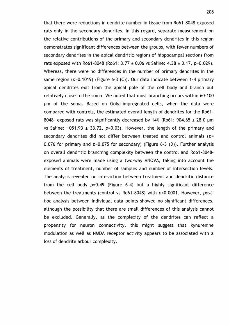
208 that there were reductions in dendrite number in tissue from Ro61-8048-exposed
rats only in the secondary dendrites. In this regard, separate measurement on
the relative contributions of the primary and secondary dendrites in this region
demonstrates significant differences between the groups, with fewer numbers of
secondary dendrites in the apical dendritic regions of hippocampal sections from
rats exposed with Ro61-8048 (Ro61: 3.77 ± 0.06 vs Saline: 4.38 ± 0.17, p=0.029).
Whereas, there were no differences in the number of primary dendrites in the
same region (p=0.1019) (Figure 6-3 (C)). Our data indicate between 1-4 primary
apical dendrites exit from the apical pole of the cell body and branch out
relatively close to the soma. We noted that most branching occurs within 60-100
µm of the soma. Based on Golgi-impregnated cells, when the data were
compared with controls, the estimated overall length of dendrites for the Ro61-
8048- exposed rats was significantly decreased by 14% (Ro61: 904.65 ± 28.0 µm
vs Saline: 1051.93 ± 33.72, p=0.03). However, the length of the primary and
secondary dendrites did not differ between treated and control animals (p=
0.076 for primary and p=0.075 for secondary) (Figure 6-3 (D)). Further analysis
on overall dendritic branching complexity between the control and Ro61-8048-
exposed animals were made using a two-way ANOVA, taking into account the
elements of treatment, number of samples and number of intersection levels.
The analysis revealed no interaction between treatment and dendritic distance
from the cell body p=0.49 (Figure 6-4) but a highly significant difference
between the treatments (control vs Ro61-8048) with p<0.0001. However, post-
hoc analysis between individual data points showed no significant differences,
although the possibility that there are small differences of this analysis cannot
be excluded. Generally, as the complexity of the dendrites can reflect a
propensity for neuron connectivity, this might suggest that kynurenine
modulation as well as NMDA receptor activity appears to be associated with a
loss of dendrite arbour complexity.
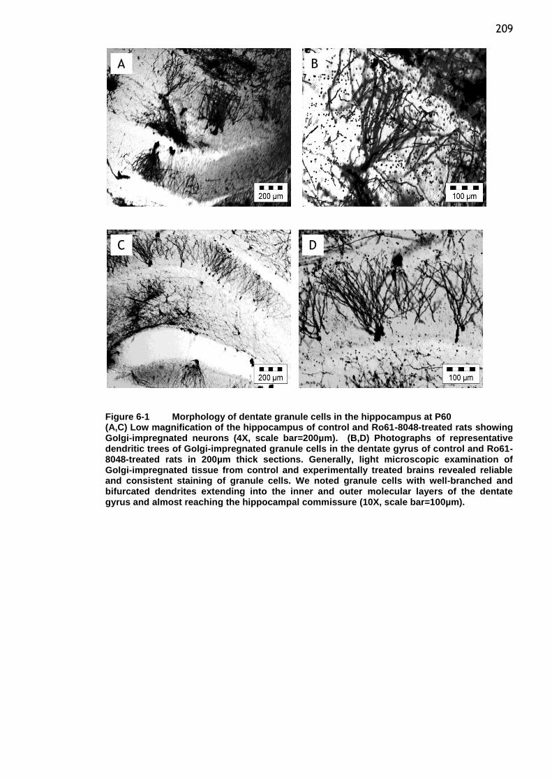
209
Figure 6-1 Morphology of dentate granule cells in the hippocampus at P60 (A,C) Low magnification of the hippocampus of control and Ro61-8048-treated rats showing Golgi-impregnated neurons (4X, scale bar=200µm). (B,D) Photographs of representative dendritic trees of Golgi-impregnated granule cells in the dentate gyrus of control and Ro61-8048-treated rats in 200µm thick sections. Generally, light microscopic examination of Golgi-impregnated tissue from control and experimentally treated brains revealed reliable and consistent staining of granule cells. We noted granule cells with well-branched and bifurcated dendrites extending into the inner and outer molecular layers of the dentate gyrus and almost reaching the hippocampal commissure (10X, scale bar=100µm).
A B
C D
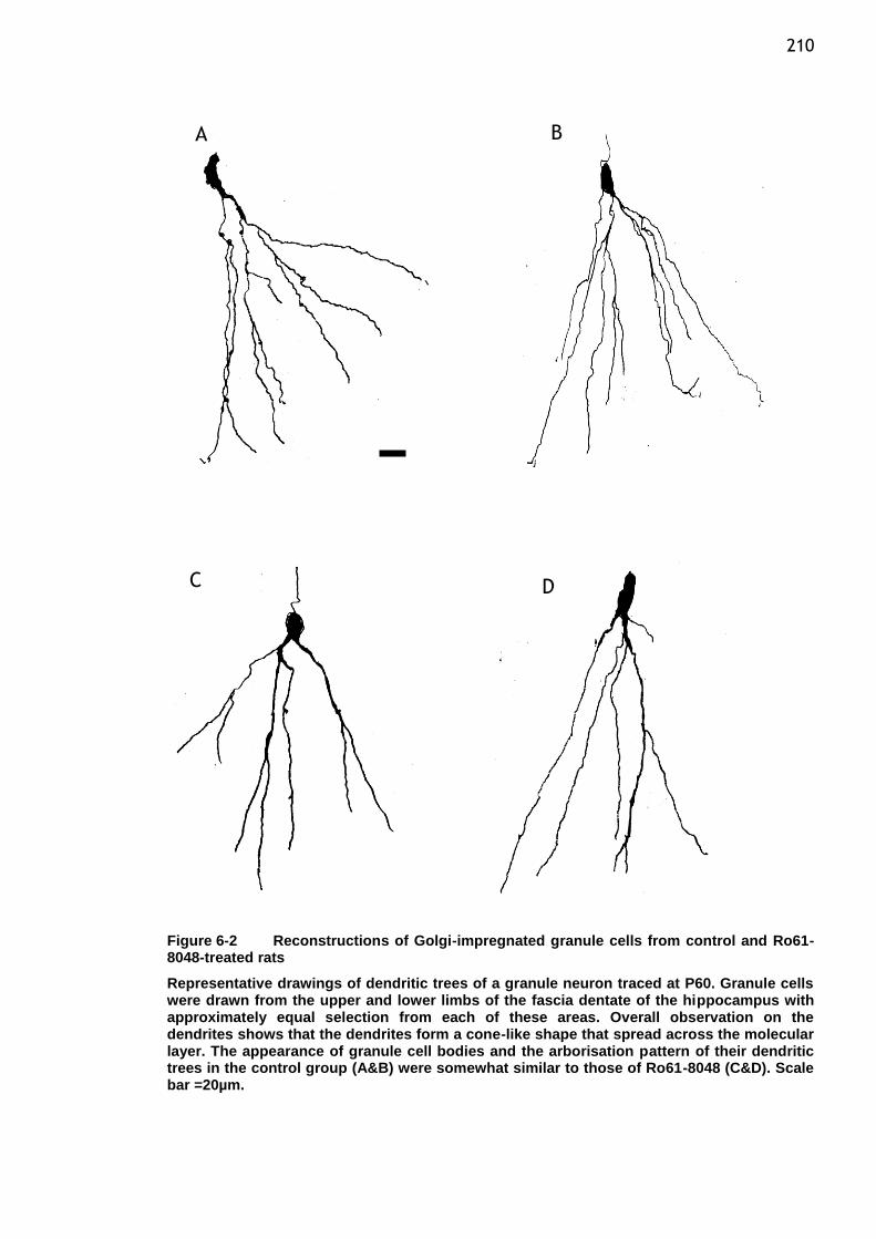
210
Figure 6-2 Reconstructions of Golgi-impregnated granule cells from control and Ro61-8048-treated rats
Representative drawings of dendritic trees of a granule neuron traced at P60. Granule cells were drawn from the upper and lower limbs of the fascia dentate of the hippocampus with approximately equal selection from each of these areas. Overall observation on the dendrites shows that the dendrites form a cone-like shape that spread across the molecular layer. The appearance of granule cell bodies and the arborisation pattern of their dendritic trees in the control group (A&B) were somewhat similar to those of Ro61-8048 (C&D). Scale bar =20µm.
A B
C D
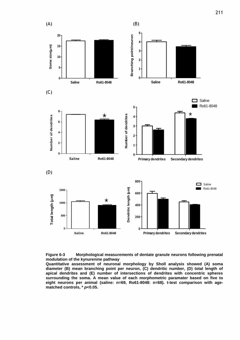
211 (A) (B)
0
5
10
15
20
Saline Ro61-8048
So
ma s
ize(
m)
0
1
2
3
4
5
Saline Ro61-8048
Bran
ch
ing
po
int/
neu
ro
n
(C)
0
2
4
6
8
*
Saline Ro61-8048
Nu
mb
er
of
de
nd
rite
s
0
1
2
3
4
5
*
Saline
Ro61-8048
Primary dendrites Secondary dendrites
Nu
mb
er
of
de
nd
rite
s
(D)
0
500
1000
1500
*
Saline Ro61-8048
To
tal le
ng
th (
m)
0
200
400
600
800Saline
Ro61-8048
Primary dendrites Secondary dendrites
De
nd
riti
c le
ng
th (
m)
Figure 6-3 Morphological measurements of dentate granule neurons following prenatal modulation of the kynurenine pathway Quantitative assessment of neuronal morphology by Sholl analysis showed (A) soma diameter (B) mean branching point per neuron, (C) dendritic number, (D) total length of apical dendrites and (E) number of intersections of dendrites with concentric spheres surrounding the soma. A mean value of each morphometric paramater based on five to eight neurons per animal (saline: n=69, Ro61-8048: n=68). t-test comparison with age-matched controls, * p<0.05.
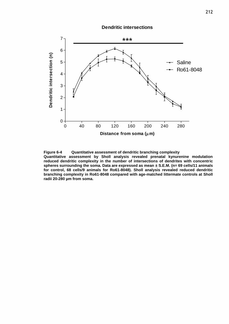
212
Dendritic intersections
0 40 80 120 160 200 240 2800
1
2
3
4
5
6
7
Saline
Ro61-8048
Distance from soma (m)
De
nd
riti
c i
nte
rse
cti
on
(n
)
Figure 6-4 Quantitative assessment of dendritic branching complexity Quantitative assessment by Sholl analysis revealed prenatal kynurenine modulation reduced dendritic complexity in the number of intersections of dendrites with concentric spheres surrounding the soma. Data are expressed as mean ± S.E.M. (n= 69 cells/11 animals for control, 68 cells/9 animals for Ro61-8048). Sholl analysis revealed reduced dendritic branching complexity in Ro61-8048 compared with age-matched littermate controls at Sholl radii 20-280 µm from soma.
***
213
6.5 Discussion
The granule cells are the principal neurons of the dentate gyrus. In the adult
rodent brain, these neurons are arranged in a densely packed layer and show a
highly uniform morphology of their V-shaped apical dendritic trees. The
dendrites which represent the main receptive structures of neurons are
branched and all dendritic segments are covered with spines. The primary
contribution of dendrites to a neuron‟s mode of connectivity is through their
characteristic branching and extension into specific domains. This branch
ordering schemes wherein dendrites emerging from the cell soma are primary
and their respective branches are considered secondary and so on, have been
used to characterize the density of dendritic arborization (Rahimi and Claiborne,
2007) .
The use of Golgi method which requires complete impregnation of neurons and
separation between stained neurons to permit the visualisation of dendrites had
enabled us to study the general morphological changes of granule neurons in the
adult rat. The results of the present study have shown that prenatal modulation
of the kynurenine pathway which alters the balance of its neuroactive
metabolites regulates the dendritic remodelling and stabilization in the
developing neuron. The results indicate that the dendritic morphology of the
selected hippocampal area present important changes in rats prenatally exposed
to Ro61-8048. Overall observations on the morphological characteristics of the
granule cells in term of soma size and branch points did not differ between
saline and Ro61-8048-treated animals. In our study, a simpler scheme of
counting the number of branch points in the entire dendritic arbour provides a
sense of how branched a neuron is, but this does not indicate the degree to
which they fill the space and its functional connectivity. The main changes found
were reductions in the number of dendritic segments and in the total dendritic
length of neurons from dentate granule cells. The complexity of the dendritic
system in the treatment group also appeared to be less robust than that seen in
the control rats. In this study, we also aimed to determine whether basal
dendrites appear on granule cells of adult rats as a consequence of KP
modulation. Such an analysis would provide information about the formation of
dendrites. The lack of basal dendrites in the dentate granule cells of control
animals in this study confirmed previous finding that showed the adult rat
214 hippocampal granule cells do not display basal dendrites. In contrast to humans
and monkeys, hippocampal formation basal dendrites which are studded with
spines appear to be a functional and integral part of normal granule cells (Seress
and Mrzljak, 1987).
In the treated specimens, no appearance of basal dendriteswere observed.
However, formation of basal dendrites have been observed in granule cells in
response to epileptogenic stimuli and this may represent one of the adaptive
cellular changes of neuroplasticity of granule cells (Spigelman et al., 1998).
Although we did not measure the spine density for dentate granule neurons, due
to limited numbers of good impregnated neurons which could produce clear
identifiable spines or spine heads, the data obtained with this technique could
indicate that Ro61-8048 produced a reduced number, length and complexity of
the dendritic system. Based on the increased KYNA concentration in the
embryonic brain in response to Ro61-8048 administration during late gestation
(Forrest et al., 2013b), we suggested that NMDA receptor blockade could be a
possible mechanism underlying these effects.
Previous reports have demonstrated that early dendritic arbour growth relies on
NMDA receptor activation and that this receptor also involved in the refinement
of synaptic connections during postnatal development, suggesting an intimate
association between glutamatergic transmission and dendritic arbour
development. In this regard, NMDA receptor activity might affect dendritic
arbour development during an early developmental window, in which coincide
with the occurring of the granule cell genesis in late embryogenesis when they
started to contribute to the synaptic current (Cline, 2001). The role of this
receptor might change as the granule neurons in the dentate gyrus undergo
periods of development and maturation. In the rat, the neurons go through a
period of maturation during which the dendritic tree is refined and the dendritic
remodelling occurs with elongation of some branches and the loss of others
between days 14 to 60 (Rahimi and Claiborne, 2007). Data from our lab
demonstrated that granule cells have less dendritic segments which are largely
attributable to a significant decrease in the number of secondary dendrites and
in turn have slightly shorter dendritic trees. This indicates that dendritic branch
loss in response to Ro61-8048 may be affected by incoming neuronal activity.
215 The changes in dendritic structure seen here are likely to have effects on
neuronal function. To correlate with this finding, our recent findings
demonstrated a reduction in the amplitude of LTP and paired-pulse inhibition
which could be in part, a consequence of the altered expression of one of the
NMDA receptor subunits in the offspring following Ro61-8048 exposure where we
noted earlier a significant reduction in expression of the NR2A subunit in the
hippocampus (Forrest et al., 2013a).
Established roles for NMDARs in dendrite development have been mentioned
earlier and this suggests specific roles for particular subunits especially NR2A
and NR2B in determining the functional properties of the NMDAR. In this context,
subunit composition of synaptic NMDAR controls normal development of synapses
by regulating the rate of synapse formation and overall dendritic morphological
outcome (Ewald et al., 2008, Vastagh et al., 2012). Contribution of specific
NMDAR subunits during synaptogenesis are still not clearly defined, but in vitro
experiments have showed that reduced glutamatergic synaptic transmission
following upregulation of NR2A might be associated with a reduction in the
number of synapse. Whereas expression of NR2B provides greater structural
plasticity by establishing appropriate synaptic contacts (Gambrill and Barria,
2010). Following exposure to KP modulation during late gestation, NR2A protein
level decreased significantly in the foetal brain and remained low in the
hippocampus at P60. A different observation was noted in NR2B expression, with
significantly increased expression after 5h and P21 (Forrest et al., 2013a, Forrest
et al., 2013b). Interestingly, out results also suggest that the observed variation
of the levels of both subunits expression during development following prenatal
exposure to Ro61-8048 might underlie the neuronal modification changes seen in
the hippocampus. Total dendritic branch length and segment numbers provide a
good indication of the growth behaviour of a neuron. Notably, both indicators in
the treated group were comparably fewer than those in control. These data
demonstrate that shift or imbalance in the synaptic NR2A and NR2B levels and
the persistence of these alterations into adulthood suggests that modification of
glutamatergic neurotransmission leads to structural changes in the general
growth parameters of the dentate granule neurons in the hippocampus. Since
the results emphasize the interdependence of receptor subunits, selectivity
216 effects of Ro61-8048 on synaptic NMDAR transmission by utilising respective
subunits antagonist is important for future consideration of in vitro assessment.
It seemed that blockade of the NMDA receptor (in this case, by KYNA) could have
some implications on the dendritic branch dynamics. It is well established that
the glutamate receptors of the NMDA subtype play a central role in the process
of LTP and also synaptic plasticity. The effects of NMDA receptor-mediated
synaptic plasticity on dendritic arbour growth have been well studied. It has
been shown that NMDA-mediated responses promote dendritic arbour growth and
also causes a transient increase in both the size and density of dendritic spines,
whereas blocking the receptor reduces the growth of dendrites (Lin et al.,
2004a, Wedzony et al., 2005). In congruent with our data which support an
association between synaptic transmission and dendritic development, it has
been shown that glutamate receptor antagonists (either competitive or non-
competitive antagonists) decrease dendrite growth and led to a corresponding
reduction of the number of synaptic contacts, as well as to changes in synaptic
morphology (Wedzony et al., 2005).
We showed that prenatal kynurenine pathway inhibition led to long term
changes in neuronal dendritic morphology of the P60 offspring as a reduction in
the number and length of apical branches of dentate granule cells. Changes in
the endogenous levels of KYNA during early development induced dendritic
retraction, indirectly through NMDA receptor blockade.
217
7 General discussion
7.1 Neurochemical alterations in rats offspring following maternal inflammation during pregnancy
Epidemiological studies have indicated that exposure to wide range of viruses
during early development has been implicated in the pathogenesis of
neuropsychiatric disorders (Adams et al., 1993, Brown et al., 2004, Mednick et
al., 1988). The precise mechanisms associating the virus and prenatal
neurodevelopmental consequences are not yet fully defined. Common mediators
of maternal immune activation are suspected to be involved in producing a
range of responses which are likely to contribute to the neuropsychiatric
disturbances. In this respect, intrauterine elevations of proinflammatory
cytokines are thought to play a role. The outcomes on the developing brain
following the maternal immune activation includes alteration in gene and
protein expression relating to various neurodevelopmental processes,
excitotoxicity or changes in the molecular pathways involved in the generation
of cells and also in the neurotransmitter functions (Damman and Leviton, 1997,
Fatemi et al., 2008, Patterson, 2002). Here, we presented the relative
contributions of prenatal viral infections to the emergence of neurochemical
imbalances including glutamate-related pharmacological molecules after
systemic administration of poly(I:C), a synthetic double-stranded RNA used to
mimic viral exposure and is known to elicit immune responses by inducing the
release of proinflammatory cytokines.
Pregnant rats were exposed to the inflammatory agent, poly(I:C) (10mg/kg) or
saline during late pregnancy (E14,16 and 18). Our preliminary experiments
showed that the indicated dose given to pregnant dams during the last seven
days of gestation imposed no apparent adverse effects on the dam or her
behaviour towards her litter after birth. We have demonstrated in previous
report the ability of poly(I:C) to activate maternal immune system. The
associated changes in which we observed an increase in MCP-1 with the absence
of other proinflammtory cytokines at 5 h post injection may therefore exhibit
the specificity of maternal host‟s response to an immunological challenge which
could modulate and precipitate related aberrations in the expression of
selected neurodevelopmental proteins (Forrest et al., 2012) .
218 Our results show a number of significant changes in the expression of several
proteins important to early brain development following inflammatory challenge
with poly(I:C). Some of the developmental markers showed changes during the
time of insult takes place and disappeared or diminished as animal grew up. This
may be an outcome of the compensatory capacity of the CNS to alleviate such
changes. In light of fundamental role of NMDA receptor during brain
development which include neuronal migration and plasticity, we found that
prenatal poly(I:C) exposure affected the subunits composition of the receptor 5
h post treatment. Transient changes in the levels of NMDA receptor subunit NR2A
and NR2B in the fetal brain could signify a potential mechanism of altering
synaptic development and may impinge on the establishment of neuronal circuits
and connection.
On the other hand it is also possible that prenatal poly(I:C) treatment
manifested its effects on the postnatal brain that are independent from its
effect at the time of exposure. The acute inflammatory response induce by
prenatal poly(I:C) administration somehow induce further long term changes in
factors regulating cell proliferation and migration, in order to produce altered
neurogenesis in offspring later in development at P21. We show that changes in
markers involved in neurogenesis, SOX2 and SHH were observed at later stage of
postnatal development (P21).This could imply that prenatal immune activation
elicited by poly(I:C) in rodents may have long term effects on neurogenesis in
young postnatal offspring. The pattern of changes seen in SOX2 and SHH suggests
such deficits or changes in neurogenesis could conceivably contribute to the
alterations in hippocampal-mediated function that could have some implications
in neuronal development. It seems important to note that activation of the
immune system during gestation can precipitate brain mal-development, in
which changes in the NMDA receptor subunits composition appear to be a key
factor and more likely to be associated with long term developmental brain and
relevant behavioural outcome in the offspring (Forrest et al., 2012, Khalil et al.,
2013).
219
7.2 Regulation of kynurenine pathway metabolism in the brain after prenatal inhibition with Ro61-8048
Involvement of the KP occurs in a variety of CNS disorders, infection and
inflammatory conditions. Moreover, the pathway has been found to be an
important determinant of immune response which is thought to be related
primarily to the activation of IDO by IFN- and other immune-active molecules
and subsequent depletion of tryptophan leading to impairment of protein
synthesis and modification in the cell proliferation and differentiation by a
number of downstream KP metabolites such as QUIN. This support an evidence
on the importance of the KP as a key link between inflammatory reactions and
immune response (Stone and Darlington, 2013). The presence of neuroactive KP
metabolites and their role in regulating excitatory amino acid receptors such as
NMDA, AMPA and αNCR has been implicated in a number of diseases, thus raising
out a possible intervention of manipulating the pathway by modulating the
balance of the metabolites(Stone et al., 2012, Stone and Addae, 2002). Indirect
approach to influence the metabolic function of NMDA receptor of glutamate
subtype by stimulating endogenous KYNA formation may provide advantages
compared to blocking the receptors indiscriminately with exogenous receptor
antagonists. In this respect, to probe the physiological roles of KP metabolites
experimentally, it appear sensible to inhibit enzyme kynurenine 3-
monooxygenase (KMO) by administering known compound Ro61-8048 which
would affect the synthesis of endogenous KYNA in accordance with changing
physiological condition and indirectly reduce the degradation of the pathway
along the QUIN branch(Chiarugi and Moroni, 1999, Richter and Hamann, 2003).
Our previous study has been carried out to examine the effect of a prenatal
exposure to the KP enzyme inhibitor on the maternal, fetal and postnatal levels
of major KP metabolites i.e. kynurenine and KYNA, considering that an
imbalance in the production of these substances could have profound
implications in cerebral neurotransmission and brain development. We have
shown that administration with Ro61-8048 at an early stage of embryonic
development elevated the levels of kynurenine and KYNA in both maternal
plasma and brain within a few hours of administration (Forrest et al., 2013a,
Khalil et al., 2013). The presence of kynurenine, a common parent compound of
all neuroactive kynurenine metabolites, has shown that the pathway is readily
220 available in the CNS in normal physiological level. In the CNS, this substrate is
then either transaminated to form KYNA or hydroxylated to produce 3-HK and
other downstream metabolites including QUIN. Although we did not measure the
level of QUIN , previous investigations have failed to show changes in QUIN
concentrations following administration of Ro61-8048 in mice(Clark et al., 2005).
The level of KYNA kept rising in the embryonic brain while maternal brain‟s
KYNA levels had returned to basal levels when observed 24 hour later. When we
observed the levels of KYNA and kynurenine in the offspring at P21, the levels of
both substrates had returned to normal control level as evidenced by its effect
on the brain content. Importantly, the dose of Ro61-8048 caused the expected
increment in the formation of KYNA in the brain during early period of
development (Forrest et al., 2013a).Taken together with biochemical data
showing alteration in the protein expression levels of various developmentally
target molecules , these results suggest that the increased level of KYNA in the
brain caused by the administration of Ro61-8048 in utero can influence the
extent of excitability and viability of NMDA receptor function that could be
partly responsible for the observed molecular and functional changes we have
reported earlier.
We have shown in our previous study that raising brain tissue KYNA
concentrations has lead to pronounced biochemical and neurochemical
consequences in the rat offspring. It is noteworthy that the prenatal exposure to
KP regulator, Ro61-8048 in rodents has been shown to induce a range of
neurochemical alterations in the offspring detectable at various developmental
ages. Considering that the plasticity of glutamate receptors could be affected by
changes in the endogenous ligands of NMDA receptors and the role of the
glutamatergic system in synaptic transmission, neuronal plasticity and also in the
learning and memory processes, we focussed our attention on this paradigm and
demonstrated that the exposure in utero to Ro61-8048 during late stage of
gestation in the rodent was able to influence the level of expression of NMDA
receptors and in particular those of NR2 subtype family in the brain. There were
highly significant changes in the levels of both subunits NR2A and NR2B as
observed 5 h post injection with Ro61-8048.
221 It is important to note that acute changes in NMDA receptor subunits expression
in the developing brain immediately after injection were largely transient, the
levels of both subunits were not different with control levels when observed 24 h
later. The differential pattern of expression in the embryo brain (around late
gestational period) for both subunits might reflect a compensatory adaptation
of specific balance between these subunits. That is, NR2A expression is generally
low at birth but increases over several weeks, whereas a relatively high NR2B
expression at birth and declines slightly through adulthood (Janssen et al., 2005,
Truman et al., 2002). In this aspect, appropriate NR2A:NR2B ratio and
fundamental shifts in receptor properties based on subunit representation
clearly have the capacity to influence neuronal function. We observed long term
changes in the NR2 subunits when the offspring has reached young adulthood
(P21). Alteration in the expression level of NR2A was maintained into adulthood
(P60) as observed in the hippocampus and cortex regions. In fact, changes in the
hippocampus in particular appeared to be the opposite of those observed
earlier which might indicate regional specificity of the ratio between both
subunits. We have previously shown that in the process of LTP, relative ratio of
NR2A and NR2B may play a part in the molecular machinery of synaptic plasticity
and contribute to the functional synaptic changes described in the hippocampus
(Forrest et al., 2013b).
Given these differing developmental profiles, early life challenge with KP
inhibition might be expected to have differential effects on subunit expression
levels. This continuing change with respect to subunit representation in the
receptor would impose overall dynamic regulation in NMDA receptor (Janssen et
al., 2005). Furthermore, it has been shown that NMDA-mediated responses
promote dendritic arbour growth and also cause a transient increase in both the
size and density of dendritic spines, whereas blocking the receptor reduces the
growth of dendrites (Wedzony et al., 2005). We showed that prenatal kynurenine
pathway inhibition led to long term changes in neuronal dendritic morphology of
the P60 offspring as a reduction in the number and length of apical branches of
dentate granule cells. Therefore, changes in the endogenous level of KYNA
during early development could have some implications on overall neuronal
structure and the dendritic branch dynamics by antagonistic properties at the
NMDA receptor.
222 The mechanisms responsible for the Ro61-8048‟s effects on neurogenesis remain
unclear. It seems probable that the regulation of the neurogenesis was strikingly
evident months after the initial treatment with KP modulator, suggesting the
process could be affected early on. Increased KYNA following the inhibition of
KMO has been expected to cause an effect on the NMDA receptor function. We
have described changes in term of protein expression of several
neurodevelopmental markers that are involved in range of developmental
process including neurogenesis, neuronal plasticity and transmission,
inflammatory related process among many others (Forrest et al., 2013b, Forrest
et al., 2013a). Hippocampal neurogenesis in this study was notably affected
which indicates that this cell genesis process might be influenced by elevated
levels of one of the kynurenine metabolites, KYNA (Forrest et al., 2013a, Forrest
et al., 2013b). As SHH is conserved and functions prominently in both developing
nervous system and the germinal zones of the adult brain. Enhancement effect
of SHH particularly on the proliferation of neural progenitor cells and neuronal
differentiation within the entire course of differentiation has been shown in
vitro. In support with this evidence, the presence of SHH receptor complexes
including Ptc and Smo within adult neurogenic areas emphasises the role for SHH
in adult neurogenesis (Traiffort et al., 1998).
Our results suggest that prenatal modulation of kynurenine pathway might lead
to changes in the expression levels of several molecules that involved in the
regulation of cell cycle, proliferation as well as neurogenesis process in
developing and adult brains. An increased expression of the transduction
molecule SHH was observed 24 h after treatment with enzyme inhibitor during
late gestation. However, when repeated challenge of the inhibitor was given,
different pattern of expression was seen with reduced levels of expression was
demonstrated at developmental age P21 and remained repressed in some
regional areas of the brain including hippocampus, cerebellum and cortex.
Further analysis using immunocytochemistry indicates that maternal exposure to
Ro61-8048 maintained the numbers of proliferative progenitors and immature
granule cell presented by DCX immunoreactivity in the hippocampal dentate
gyrus. Whereas the increased number of SHH immunopositive cells in this region
could potentially affect the growth and survival of newly generated granule cells
and their developing axons. These findings raise the possibility that the early
223 modulation of NMDA receptor function through KYNA on the expression of SHH
could contribute to the influence on adult neurogenesis. It also suggest that the
SHH pathway may continue to have an important role in adulthood in regulating
structural plasticity. We show that these effects of in utero exposure to
pharmacological substance induced alteration in protein levels which was
accompanied by a subtle structural modification that can results in changes in
the cell numbers and survival that was anatomically localized and involved
specific subpopulations of neurons. We report here that gestational exposure to
a compound that is able to modulate the balance of KP metabolites, has a long
lasting stimulatory effect which occurs in concert with other events on the
generation and development of new neurons in the hippocampal formation,
particularly in the dentate gyrus. Overall, this scope of research explains on the
effect of KP modulation which extends from prenatal exposure to postnatal
outcomes. It provides a partial explanation for the exceptionally brain
development on long lasting and delayed effects following gestational exposure.
7.3 Future directions of study
The alterations in neurodevelopmental proteins especially seen in NMDA
receptor subunits and synaptic physiology may be responsible to account for
behavioural abnormalities. It is quite uncertain based on data generated from
this study, to elucidate which developmental processes are responsible for
different profile of behavioural abnormalities which constitute a biological
mechanism of cognitive performance. Our behavioural findings on simple step
down-avoidance test may not be appropriate to reflect a more complex task of
cognitive changes, thus raising some limitations on conclusive interpretation
(Forrest et al., 2013b). Thus, further and elaborate studies on the behavioural
paradigms in relation to antagonistic profiles of Ro61-8048 are warranted. It has
been shown that increases in endogenous KYNA concentration resulted in
prepulse inhibition deficits and interferes with spatial working memory in the
radial arm maze task. These findings are in line with the assumption that
enhanced inhibition of NMDA receptor and αNCR by KYNA may contribute to the
observed deficits in working memory in persons with schizophrenia (Pocivavsek
et al., 2011, Chess et al., 2007). As KYNA is known to act on both glutamatergic
and cholinergic systems that are critically involved in cognitive function and
normal brain development , thus alterations in KP neuroactive metabolites can
224 influence the levels of neurotransmitters i.e. glutamate that have been
implicated in schizophrenia (Muller and Schwarcz, 2006, Stone and Darlington,
2013). Therefore, further animal studies investigating mRNA/protein levels of
receptors, neurotransmitter levels and treatment effects of Ro61-8048 in a more
complex behavioural testing should improve the validity of this animal model. In
addition, elucidating mechanisms that underlie the differential effects of Ro61-
8048 at P21 and P60 will require further studies on the anatomical and molecular
changes with particular interest on some molecular targets that has been linked
to both glutamatergic and dopaminergic systems.
It is unclear by what mechanism alteration in the KP metabolites which favour
KYNA formation upregulates SHH. The increase in expression is likely to involve
an intermediate regulatory step of transcription factor that transcribes SHH. As
the SHH pathway is important in the maintenance and proliferation of neural
progenitor cells in the adult rodent brain, the activation of SHH and its receptor
components is responsible for the effects that occur during this process. The
pathway begins when the secreted SHH pep tide binds to its membrane-bound
receptor Ptc, thus relieving its inhibition of Smo which then lead to a complex
signalling cascade involving the transcription factors of the Gli family. Future
studies, possibly involving the suppression of SHH pathway with its antagonist
will be useful to determine whether SHH signalling is necessary for KP regulation
to promote the proliferation of adult‟s pool of progenitor cells in the
hippocampus. In this respect, incorporating an application of cyclopamine,
natural product of plant alkaloid that is selectively known to inhibit SHH
signalling by directly binding to one of the signalling component of the SHH
receptor complex, is warranted (Charytoniuk et al., 2002, Britto et al., 2002).
Because high expression of Ptc and Gli1 is an indicator of activity in the SHH
pathway, it is also important to analyse the expression patterns this signalling
targets either at mRNA levels or in tissue specimen of immunolabelling
technique. By incorporating quantitative analysis with other signalling
components of SHH pathway and inhibition with antagonist may further support
the evidence of the requirement of the SHH signalling for adult neural progenitor
cell expansion and development and that KP metabolism effects on
hippocampus dependent-activity could be mediated by SHH signalling pathway.

225 Substantial studies have gathered evidence supporting the idea that
dysregulation of the cell cycle of neural cells plays an important role in diverse
pathological processes in the CNS. Characterization the cell cycle properties in
this process and comparative studies of the expression of individual members of
related proteins under physiological condition, including various developmental
stages and in exposure to pathological stimuli may provide better and conclusive
explanation.
226
List of References
ADAMS, W., KENDELL, R. E., HARE, E. H. & MUNK-JORGENSEN, P. 1993. Epidemiological evidence that maternal influenza contributes to the aetiology of schizophrenia. British Journal of Psychiatry, 163, 522-534.
AKAGBOSU, C. O., EVANS, G. C., GULICK, D., SUCKOW, R. F. & BUCCI, D. J. 2010. Exposure to kynurenic acid during adolescence produces memory deficits in adulthood. Schizophrenia Bulletin, 1-10.
AL-HALLAQ, R. A., CONRADS, T. P., VEENSTRA, T. D. & WENTHOLD, R. J. 2007. NMDA Di-Heteromeric Receptor Populations and Associated Proteins in Rat Hippocampus. Journal of Neuroscience, 27, 8334-8343.
ALEXOPOLOU, L., HOLT, A. C., MEDZHITOV, R. & FLAVELL, R. A. 2001. Recognition of double-stranded RNA and activation of NF-kappaß by Toll-like receptor 3. Nature, 413, 732-738.
ALKONDON, M., PEREIRA, E. F. R., EISENBERG, H. M., KAJII, Y., SCHWARCZ, R. &
ALBUQUERQUE, E. X. 2011. Age dependency of inhibition of Nicotinic receptors and tonically active N-methyl-D-aspartate receptors by endogenously produced kynurenic acid in the brain. The Journal of Pharmacology and Experimental Therapeutics, 337, 572-582.
ALMER, G., GUEGAN, C., TEISMANN, P., NAINI, A., ROSOKLIJA, G., HAYS, A. P., CHEN, C. & PRZEDBORSKI, S. 2001. Increased expression of the pro-inflammatory enzyme cyclooxygenase-2 in Amyotrophic Lateral Sclerosis. Ann Neurol, 49, 176-185.
AMORI, L., GUIDETTI, P., PELLICCIARI, R., KAJII, Y. & SCHWARCZ, R. 2009. On the relationship between the two branches of the kynurenine pathway in the rat brainin vivo. Journal of Neurochemistry, 109.
ASHDOWN, H., DUMONT, Y., NG, M., POOLE, S., BOKSA, P. & LUHESHI, G. N. 2006. The role of cytokines in mediating effects of prenatal infection on the fetus: implications for schizophrenia. Molecular Psychiatry, 11, 47-55.
AVILION, A. A., NICOLIS, S. K., PEVNY, L. H., PEREZ, L., VIVIAN, N. & LOVELL-BADGE, R. 2003. Multipotent cell lineages in early mouse development depend on SOX2 function. Genes & Development, 17, 126-140.
BABB, T. L., MIKUNI, N., NAJM, I., WYLIE, C., OLIVE, M., DOLLAR, C. & MACLENNAN, H. 2005. Pre-and postnatal expressions of NMDA receptors 1 and 2B subunit proteins in the normal rat cortex. Epilepsy Research, 64, 23-30.
BABULAS, V., FACTOR-LITVAK, P., GOETZ, R., C.A., S. & BROWN, A. S. 2006. Prenatal Exposure to Maternal Genital and Reproductive Infections and Adult Schizophrenia. American Journal of Psychiatry, 163, 927-929.
BAEUERLE, P. A. & HENKEL, T. 1994. Function and activation of NFB in the immune system. Annual Review of Immunology, 12, 141-179.
BALE, A. E. 2002. Hedgehog signaling and human disease. Annu.Rev.Genomics Hum.Genet, 3, 47-65.
BANARJEE, S. B. & RAJENDRAN, R. 2005. Recruitment of the Sonic hedgehog signalling cascade in electroconvulsive seizure-mediated regulation of adult rat hippocampal neurogenesis. European Journal of Neuroscience, 22, 1570-1580.
BANNERMAN, D. M., SPRENGEL, R., SANDERSON, D. J., MCHUGH, S. B., RAWLINS, J. N. P., MONEYR, H. & SEEBURG, P. H. 2014. Hippocampal synaptic plasticity, spatial memory and anxiety. Nature Review Neuroscience, 15, 181-193.
227 BARD, L. & GROC, L. 2011. Glutamate receptor dynamics and protein
interaction: lessons from the NMDA receptor. Molecular and Cellular Neuroscience, 48, 298-307.
BARRY, S., CLARKE, G., SCULLY, P. & DINAN, T. G. 2009. Kynurenine pathway in psychosis:evidence of increased tryptophan degradation. Journal of Psychopharmacology, 23, 287-294.
BARTLETT, T. E., BANNISTER, N. J., COLLET, V. J., DARGAN, S. L., MASSEY, P. V., BORTOLOTTO, Z. A., FITZJON, S. M., BASHIR, Z. I., COLLINGRIDGE, G. L. & LODGE, D. 2007. Differential roles of NR2A and NR2B-containing NMDA receptors in LTP and LTD in the CA1 region of two-week old rat hippocampus. Neuropharmacology, 52, 60-70.
BEESLEY, J. E. 1993. Immunocytochemistry. A practical approach, New York, Oxford University Press Inc.
BEIQUE, J.-C. & ANDRADE, R. 2003. PSD-95 regulates synaptic transmission and plasticity in rat cerebral cortex. Journal of Physiology, 546, 859-867.
BEN ABDALLAH, N., SLOMIANKA, L., VYSSOTSKI, A. L. & LIPP, H. P. 2010. Early age-related cahnges in adult hippocapal neurogenesis in C57 mice. Neurobiology of Aging, 31, 151-161.
BERTRAND, N. & DAHMANE, N. 2006. Sonic hedgehog signaling in forebrain development and its inetractions with pathways that modify its effects. Trends in Cell Biology, 16, 597-606.
BI, H. & SZE, C. I. 2002. N-methyl-D-aspartate receptor subunit NR2A and NR2B messenger RNA levels are altered in the hippocampus and entorhinal cortex in Alzheimer's disease. Journal of Neurological Sciences, 200.
BLISS, T. V. & COLLINGRIDGE, G. L. 1993. A synaptic model of memory:long term potentiation in the hippocampus. Nature, 361, 31-39.
BOKSA, P. 2010. Effects of prenatal infection on brain development and behavior: A review of findings from animal models. Brain,Behavior and Immunity, 24, 881-897.
BOKSA, P. & LUHESHI, G. N. 2003. On the use of animal modeling to study maternal infection during pregnancy and prenatal cytokine exposure as risk factors for schizophrenia. Clinical Neuroscience Research, 3, 339-346.
BORRELL, J., VELA, J. M., ANGEL, A.-M., MOLINA-HOLGADO, E. & GUAZA, C. 2002. Prenatal immune challenge disrupts sensorimotor gating in adult rats: Implications for the etiopathogenesis of schizophrenia. Neuropsychopharmacology, 26, 204-215.
BRAIDY, N., GRANT, R., ADAMS, S., BREW, B. J. & GUILLEMIN, G. J. 2009. Mechanism for quinolinic acid cytotoxcity in human astrocyts and neurons. Neurotoxicity Research, 16, 77-86.
BRAZEL, C. Y., LIMKE, T. L., OSBORNE, J. K., CAI, J., PEVNY, L. & RAO, M. S. 2005. Sox2 expression defines a heterogeneous population of neurosphere-forming cells in the adult murine brain. Aging Cell, 4, 197-207.
BREDER, C. D., DEWITT, D. & KRAIG, R. P. 1995. Characterization of inducible cyclooxygenase in rat brain. Journal of Comparative Neurology, 355, 296-315.
BREDER, C. D. & SAPER, C. B. 1996. Expression of inducible cyclooxygenase mRNA in the mouse brain after systemic administration of bacterial lipopolysaccharide. Brain Research, 713, 64-69.
BRITTO, J., TANNAHILL, D. & KEYNES, R. 2002. A critical role for sonic hedgehog signaling in the early expansion of the developing brain. Nature Neuroscience, 5, 103-110.
228 BROWN, A. S. 2006. Prenatal Infection as a Risk Factor for Schizophrenia.
Schizophrenia Bulletin, 32, 200-202. BROWN, A. S., BEGG, M. D., GRAVENSTEIN, S., SCHAEFER, C. A., WYATT, R. J.,
BRESNAHAN, M., BABULAS, V. P. & SUSSER, E. S. 2004. Serologic evidence of prenatal influenza in the etiology of schizophrenia. Archives of General Psychiatry, 71, 774-780.
BROWN, A. S., COHEN, P., HARKAVY-FRIEDMAN, J., BABULAS, V., MALASPINA, D., GORMAN, J. M. & SUSSER, E. S. 2001. Prenatal Rubella, Premorbid Abnormalities, and Adult Schizophrenia. Biological Psychiatry, 49, 473-486.
BROWN, A. S. & DERKITS, E. J. 2010. Prenatal infection and schizophrenia: A review of epidemiologic and translational studies. American Journal of Psychiatry, 167, 261-280.
BROWN, A. S., SCHAEFER, C. A., QUESENBERRY, C. P., LIU, L., BABULAS, V. P. & SUSSER, E. S. 2005. Maternal Exposure to Toxoplasmosis and Risk of Schizophrenia in Adult Offspring. American Journal of Psychiatry, 162, 767-773.
BROWN, J. P., COUILLARD-DESPRES, S., COOPER-KUHN, C. M., WINKLER, J., AIGNER, L. & JKUHN, G. 2003. Transient expression of doublecortin during adult neurogenesis. The Journal of Comparative Neurology, 467, 1-10.
BYLUND, M., ANDERSSON, E., NOVITCH, B. G. & MUHR, J. 2003. Vertebrate neurogenesis is counteracted by Sox1-3 activity. Nature Neuroscience, 6, 1162-1168.
CAMPESAN, S., GREEN, E. W., BREDA, C., SATHYASAIKUMAR, K. V., MUCHOWSKI, P. J., SCHWARCZ, R. & GIORGINI, F. 2011. The kynurenine pathway modulates neurodegeneration in Drosophila model of Huntington's disease. Current Biology, 21, 961-966.
CARLSSON, A., WATERS, N., HOLM-WATERS, S., TEDROFF, J., NILSSON, M. & CARLSSON, M. L. 2001. Interactions between monoamines, glutamate and GABA in schizophrenia: new evidence. Annu Rev.Pharmacol.Toxicol., 41, 237-260.
CARMONA, M. A., MURAI, K. K., WANG, L., ROBERTS, A. J. & PASQUELE, E. B. 2009. Glial ephrin-A3 regulates hippocampal dendrites spine morphology and glutamate transport. Proc Natl Acad Sci, 106, 12524-12529.
CARPENEDO, R., CHIARUGI, A., RUSSI, P., LOMBARDI, G., CARLA, V., PELLICCIARI, R., MATTOLI, L. & MORONI, F. 1994. Inhibitors of kynurenine hydroxylase and kynureninase increase cerebral formation of kynurenate and have sedative and anticonvulsant activities. Neuroscience, 61, 237-244.
CHARYTONIUK, D. A., PORCEL, B., GOMEZ, J. R., FAURE, H., RUAT, M. & TRAIFFORT, E. 2002. Sonic hedgehog signalling in the developing and adult brain. Journal of Physiology, 96, 9-16.
CHEN, Y. & GUILLEMIN, G. J. 2009. Kynurenine pathway metabolites in humans: Disease and healthy states. International Journal of Tryptophan Research, 2, 1-19.
CHESS, A. C., SIMONI, M. K., ALLING, T. E. & BUCCI, D. J. 2007. Elevations of endogenous kynurenic acid produce spatial working memory deficits. Schizophrenia Bulletin, 33, 797-804.
CHIARUGI, A., CARPENEDO, R., MOLINA, M. T., MATTOLI, L. & PELLICIARI, R. 1995. Comparison of the neurochemical and behavioral effects resulting from the inhibition of kynurenine hydroxylase and /or kynureninase. Journal of Neurochemistry, 65, 1176-1183.
229 CHIARUGI, A., COZZI, A., BALLERINI, C., MASSACES, L. & MORONI, F. 2001.
Kynurenine-3-monooxygenase activity and neurotoxic kynurenine metabolites increase in the spinal cord of rats with experimental allergic encephalomyelitis. Neuroscience, 102, 687-695.
CHIARUGI, A. & MORONI, F. 1999. Quinolinic acid formation in immune-activated mice: studies with (m-nitrobenzoyl)-alanine (mNBA) and 3,4-dimethoxy-[-N-4-(3-nitrophenyl)thiazol-2-yl]-benzenesulfonamide (Ro61-8048), two potent and selective inhibitors of kynurenine kydroxylase. Neuropharmacology, 38, 1225-1233.
CHOI, S. H. & BOSETTI, F. 2009. The distinct roles of cyclooxygenase-1 and -2 in neuroinlfmmation: implications for translational research. Trends in Pharmacological Sciences, 30, 174-181.
CHOI, S. J., OH, S. Y., KIM, J. H. & ROH, C. H. 2007. Changes of nuclear factor
kappa B (NFB), cyclooxygenase-2 (COX-2) and matrix metalloproteinase-9 (MMP-9) in human myometrium before and during term labor. European Journal of Obstetrics & Gynecology and Reproductive Biology, 132, 182-188.
CLANCY, B., DARLINGTON, R. B. & FINLAY, B. L. 2001. Translating developmental time across mammalian species. Neuroscience, 105, 7-17.
CLARK, C. J., MACKAY, G. M., SMYTHE, G. A., BUSTAMANTE, S., STONE, T. W. & PHILLIPS, R. S. 2005. Prolonged survival of a murine model of cerebral malaria by kynurenine pathway inhibition. Infection and Immunity, 73, 5249-5251.
CLINE, H. & HAAS, K. 2003. The regulation of dendritic arbor development and plasticity by glutamatergic synaptic input: a review of the synaptotrophic hypothesis. Journal of Physiology, 586, 1509-1517.
CLINE, H. T. 2001. Dendritic arbor development and synaptogenesis. Current Opinion in Neurobiology, 11, 118-126.
CLINTON, S. M., HAROUTUNIAN, V., DAVIS, K. L. & MEADOR-WOODRUFF, J. H. 2003. Altered transcript expression of NDA receptor associated postsynaptic proteins in the thalamus of subjects with schizohrenia. American Journal of Psychiatry, 160, 1100-1109.
CLINTON, S. M., HAROUTUNIAN, V. & MEADOR-WOODRUFF, J. H. 2006. Upregulation of NMDA receptor subunit and post-synaptic density protein expression in the thalamus of elderly patients with schizophrenia. Journal of Neurochemistry, 98, 1114-1125.
CONTI, P., PANG, X., BOUCHER, W., LETOUMEAU, R., REALE, M., BARBACANE, R. C. & THIBOULT, J. 1997. Impact of Rantes and MCP-1 chemokines on in vivo basophilic cell recruitment in rat skin injection model and their role in modifying the protein and mRNA levels for histidine decarboxylase. Blood, 89, 4120-4127.
CORRALES, J., BLAESS, S., MAHONEY, E. M. & JOYNER, A. L. 2006. The level of sonic hedgehog signaling regulates the complexity of cerebellar foliation. Development, 133, 1811-1821.
COZZI, A., CARPENEDO, R. & MORONI, F. 1999. Kynurenine hydroxylase inhibitors reduce ischemic brain damage: Studies with (m-Nitrobenzoyl)-Alanine (mNBA) and 3,4-dimethoxy-[-n-4-(Nitrophenyl)thiazol-2-yl]-benzenesulfonamide (Ro61-8048) in models of focal or global brain ischemia. Journal of Cerebral Bloof Flow and Metabolism, 19, 771-777.
CULL-CANDY, S., BRICKLEY, S. & FARRANT, M. 2001. NMDA receptor subunits: diversity, development and disease. Current Opinion in Neurobiology, 11, 327-335.
230 CULL-CANDY, S. & KLEIN, R. 2007. Signalling mechanisms. Current Opinion in
Neurobiology, 17, 277-280. CURTIS, M., PENNEY, E. B., PEARSON, A. G., VAN ROON-MOM, W. M. C.,
BUTTERWORTH, N. J., DRAGUNOW, M., CONNOR, B. & FAULL, R. L. M. 2003a. Increased cell proliferation and neurogenesis in the adult human Huntington's disease brain. PNAS, 100, 9023-9027.
CURTIS, M. A., CONNOR, B. & FAULL, R. L. M. 2003b. Neurogenesis in the diseased adult human brain. Cell Cycle, 2, 428-430.
DAHMANE, N. & RUIZ I ALTABA, A. 1999. Sonic hedgehog regulates the growth and patterning of the cerebellum. Development, 126, 3089-3100.
DAHMANE, N., SANCHEZ, P., GITTON, Y., PALMA, V., SUN, T., BEYNA, M., WEINER, H. & RUIZ I ALTABA, A. 2001. The sonic hedgehog-Gli pathway regulates dorsal brain growth and tumorigenesis. Development, 128, 5201-5212.
DAI, R.-L., ZHU, S.-Y., XIA, Y.-P., MAO, L., MEI, Y.-W., YAO, Y.-F., XUE, Y.-M. & HU, B. 2011. Sonic hedgehog protects cortical neurons against oxidative stress. Neurochemical Research, 36, 67-75.
DALE, W. E., DANG, Y., AMIRIDZE, N. & BROWN, O. R. 2000. Evidence that kynurenine pathway metabolites mediate hyperbaric oxygen-induced convulsions. Toxicology Letters, 117, 37-43.
DAMMAN, O. & LEVITON, A. 1997. Maternal Intrauterine Infection, Cytokines, and Brain Damage in the Preterm Newborn. Pediatric Research, 42, 1-8.
DELINT-RAMIREZ, I., FERNANDEZ, E., BAYES, A., KICSI, E., KOMIYAMA, N. H. & GRANT, G. N. 2010. In vivo composition of NMDA receptor signalling complexes differs between membrane subdomains and is modulated by PSD-95 and PSD-93. Journal of Neuroscience, 30, 8162-8170.
DHARANE, P., MANUELPILLAI, U., WALLACE, E. & WALKER, D. W. 2010. NFB-dependent increase of kynurenine pathway activity in human placenta: Inhibition of sulfasalazine. Placenta, 31, 997-1002.
DIAZ-MUNOZ, M., OSMA-GARCIA, I. C., CACHEIRO-LLAGUNO, C. C., FRESNO, M. & INIGUEZ, M. A. 2010. Coordinated up-regulation of cyclooxygenase-2 and microsomal prstglandin E synthase 1 transcription by nuclear kappa B and early growth response-1 in macrophages. Cellular Signalling, 22, 1427-1436.
DRISKELL, R. R., GIANGRECO, A., JENSEN, K. B., MULDER, K. W. & WATT, F. M. 2009. Sox2-positive dermal papilla cells specify hair follicle type in mammalian epidermis. Development, 136, 2815-2823.
EGEA, J. & KLEIN, R. 2007. Eph-ephrin signaling during axon guidance. Trends in Cell Biology, 17, 230-238.
ELLIS, P., FAGAN, B. M., MAGNESS, S. T., HUTTON, S., TARANOVA, O., HAYASHI, S., MCMAHON, A., RAO, M. & PEVNY, L. 2004. SOX2, a persistent marker for multipotential neural stem cells derived from embryonic stem cells, the embryo or the adult. Developmental Neuroscience, 26, 148-165.
ENGELHARDT, J. V., COSEREA, I., PAWLAK, V., FUCHS, E. C., KOHR, G., SEEBURG, P. H. & MONYER, H. 2007. Excitotoxicity in vitro by NR2A- and NR2B-containing NMDA receptors. Neuropharmacology, 53.
EPISKOPOU, V. 2005. SOX2 functions in adult neural stem cells. TRENDS in Neuroscience, 28, 219-221.
ERHARDT, S., OLSSON, S. K. & ENGBERG, G. 2009. Pharmacological manipulation of kynurenic acid. CNS Drugs, 23, 91-101.
231 ERHARDT, S., SCHWIELER, L., NILSSON, L., LINDERHOLM, K. R. & ENGBERG, G.
2007. The kynurenic acid hypothesis of schizophrenia. Physiology and Behavior, 92, 203-209.
ERIKSSON, P. S., PERFILIEVA, E., BJORK-ERIKSSON, T., ALBORN, A. M., NORDBORG, C., PETERSON, D. A. & GAGE, F. H. 1998. Neurogenesis in the adult human hippocampus. Natural Medicine, 4, 1313-1317.
EWALD, R. C., VAN KEUREN-JENSEN, K. R., AIZENMAN, C. D. & CLINE, H. T. 2008. Roles of NR2A and NR2B in the development of dendritic arbor morphology in vivo. The Journal of Neuroscience, 28, 850-861.
FANTES, J., RAGGAE, N. K., LYNCH, S. A., MCGILL, N. I., COLLIN, J. R. & HOWARD-PEEBLES, P. N. 2003. Mutations in sox2 cause anopththalmia. Nature Genetic, 33, 461-463.
FARIOLI-VECCHIOLI, S., SARAULLI, D., COSTANZI, M., PACIONI, S., CINA, I., ACETI, M., MICHELI, L., BACCI, A. & CESTARI, V. 2008. The timing of differentiation of adult hippocampal neurons is crucial for spatial memory. PLOS Biology, 6, 2188-2203.
FATEMI, S. H., EARLE, J., KANODIA, R., KIST, D., EMAMIAN, E. S. & PATTERSON, P. H. 2002. Prenatal viral infection leads to pyramidal cell atrophy and macrocephaly in adulthood:impliocations for genesis of autism and schizophrenia. Cellular and Molecular Neurobiology, 4, 145-154.
FATEMI, S. H., FOLSOM, T. D., REUTIMAN, T. J., HUANG, H., OISHI, K. & MORI, S. 2009a. Gene expression is altered in hippocampus following prenatal viral infection in mouse at E16. European Neuropsychopharmacology, 19, 648-653.
FATEMI, S. H., FOLSOM, T. D., REUTIMAN, T. J., HUANG, H., OISHI, K. & MORI, S. 2009b. Prenatal viral infection of mice at E16 causes changes in gene expression in hippocampi of the offspring. European Neuropsychopharmacology, 19, 648-653.
FATEMI, S. H., REUTIMAN, T. J., FOLSOM, T. D., HUANG, H., OISHI, K., MORI, S., SMEE, D. F., PEARCE, D. A., WINTER, C., SOHR, R. & JUCKEL, G. 2008. Maternal infection leads to abnormal gene regulation and brain atrophy in mouse offspring: Implications for genesis of neurodevelopmental disorders. Schizophrenia Research, 99, 56-70.
FAVARO, R., VALOTTA, M., FERRI, A. L. M., MARIANI, J., LATORRE, E., GIACHINO, C., LANCINI, C., TOSETTI, V. & NICOLIS, S. K. 2009. Hippocampal development and neural stem cell maintenance require Sox2-dependent regulation of Shh. Nature Neuroscience, 12, 1248-1256.
FLANAGAN, J. G. & VANDERHAEGEN, P. 1998. The ephrins and Eph receptors in neural development. Annual Review of Neuroscience, 21, 309-345.
FONG, H., HOHENSTEIN, K. A. & DONOVAN, P. J. 2008. Regulation of self-renewal and pluripotency by Sox2 in human embryonic stem cells. Stem Cells, 26, 1931-1938.
FORREST, C. M., KHALIL, O. S., PISAR, M., DARLINGTON, L. G. & STONE, T. W. 2013a. Prenatal inhibition of the tryptophan-kynurenine pathway alters synaptic plasticity and protein expression in the rat hippocampus. Brain Research, 1504, 1-15.
FORREST, C. M., KHALIL, O. S., PISAR, M., MCNAIR, K., KORNISIUK, E., SNITCOFSKY, M., GONZALEZ, N., JERUSALINSKY, D., DARLINGTON, L. G. & STONE, T. W. 2013b. Changes in synaptic transmision and protein expression in the brains of adult offspring after prenatal inhibition of the kynurenine pathway. Neuroscience, 254, 241-259.
232 FORREST, C. M., OMARI, S. K., PISAR, M., SMITH, R. A., DARLINGTON, L. G. &
STONE, T. W. 2012. Prenatal activation of toll-like receptors-3 by administration of the viral mimetic poly(I:C) changes syanptic prteins, N-methyl-D-aspartate receptors and neurogenesis markers in offspring. Molecular Brain, 5, 1-12.
FORREST, C. M., YOUD, P., KENNEDY, A., GOULD, S. R., DARLINGTON, L. G. & STONE, T. W. 2002. Purine, kynurenine, neopterin and lipid peroxidation levels in inflammatory bowel disease. Journal of Biomedical Science, 9, 436-442.
FORTIER, M. E., KENT, S., ASHDOWN, H., POOLE, S., BOKSA, P. & LUHESHI, G. N. 2004. The viral mimic, polyinosinic:polycytidylic acid, induces fever in rats via an interleukin-1-dependent mechanism. American Journal of Physiology Regulatory Integrative and Comparative Physiology, 287, R759-R766.
FORTIER, M. E., LUHESHI, G. N. & BOKSA, P. 2007. Effects of prenatal infection on prepulse inhibition in the rat depend on the nature of the infectious agent and the stage of prenancy. Behavioral Brain Research, 181, 270-277.
FOSTER, A. C., VEZZANI, A., FRENCH, E. D. & SCHWARCZ, R. 1984. Kynurenic acid blocks neurotoxicity and seizures induced in rats by the related brain metabolite quinolinic acid. Neuroscience Letters, 48, 273-278.
FRANCIS, F., KOULAKOFF, A., BOUCHER, D., CHAFEY, P., SCHAAR, B., VINET, M.-C., FRIOCOURT, G., MCDONNEL, N., REINER, O., KAHN, A., MCCONNEL, S. K., BERWALD-NETTER, Y., DENOULET, P. & CHELLY, J. 1999. Doublecortin is a developmentally regulated,microtubule-associated protein expressed in migrating and differentiating neurons. Neuron, 23, 247-256.
FRENCH, E. D., MURA, A. & WANG, T. 1993. MK-801,phencyclidine(PCP) and PCP-like drugs increase burst firing in rat A10 dopamine neurons:comparison to competitive NMDA antagonists. Synapse, 13, 108-116.
FUCCILLO, M., JOYNER, A. L. & FISHELL, G. 2006. Morphogen to mitogen: the multiple roles of hedgehog signalling in vertebrate neural development. Nature Review Neuroscience, 7, 772-783.
FURUKAWA, H., SINGH, S. K., MANCUSSO, R. & GOUAUX, E. 2005. Subunit arrangement and function in NMDA receptors. Nature, 438, 185-192.
GALE, N. W., HOLLAND, S. J., VALENZUELA, D. M., FLENNIKEN, A., PAN, L., RYAN, T. E., HENKEMEYER, M., STREBHARDT, K., HIRAI, H., WILKINSONB, D. G., PAWSON, T., DAVIS, S. & YANCOPOULOS, G. D. 1996. Eph receptors and ligands comprise two major specificity subclasses and are reciprocally compartmentalized during embryogenesis. Neuron, 17, 9-19.
GAMBRILL, A. C. & BARRIA, A. 2010. NMDA receptor subunit composition controls synaptogenesis and synapse stabilization. PNAS, 6-10.
GASCON, S., SOBRADO, M., RODA, J., RODRIGUEZ-PENA, A. & DIAZ-GUERRA, M. 2008. Excitotoxicity and focal cerebral ischemia induce truncation of the NR2A and NR2B subunits of the NMDA receptor and cleavage of the scaffolding protein PSD-95. Molecular Psychiatry, 13, 99-114.
GAZZALEY, A. H., WEILAND, N. G., MCEWEN, B. S. & MORRISON, J. H. 1996. Differential regulation of NMDAR1 mRNA and protein by estradiol in the rat hippocampus. Journal of Neuroscience, 16, 6830-6838.
GERBER, J., TAUBER, S. C., ARMBRECHT, I., SCHMIDT, H., BRUCK, W. & NAU, R. 2009. Increased neuronal proliferation in human bacterial meningitis. Neurology, 73, 1026-1032.
233 GIBB, R. & KOLB, B. 1998. A method for vibratome sectioning of Golgi-Cox
stained whole rat brain. Journal of Neuroscience Methods, 79, 1-4. GILMORE, J. H. & JARSKOG, L. F. 1997. Exposure to infection and brain
development: cytokines in the pathogenesis of Scizophrenia. Schizophrenia Research, 24, 365-367.
GILMORE, J. H., JARSKOG, L. F. & VADLAMUDI, S. 2003. Maternal infection regulates BDNF and NGF expression in fetal and neonatal brain and maternal-fetal unit of the rat. Journal of Neuroimmunology, 138, 49-55.
GILMORE, J. H., JARSKOG, L. F. & VADLAMUDI, S. 2005a. Maternal poly I:C
exposure during pregnancy regulates TNF, BDNF and NGF expression in neonatal brain and the maternal-fetal unit of the rat. Journal of Neuroimmunology, 159, 106-112.
GILMORE, J. H., JARSKOG, L. F. & VADLAMUDI, S. 2005b. Maternal poly(I:C)
exposure during pregnancy regulates TNF, BDNF and NGF expression in neonatal brain and the maternal-fetal unit of the rat. Journal of Neuroimmunology, 159, 106-112.
GLADDING, C. M. & RAYMOND, L. A. 2011. Mechanisms underlying NMDA receptor synaptic/extrasynaptic distribution and function. Molecular and Cellular Neuroscience, 48.
GLANTZ, L. A. & LEWIS, D. A. 2000. Decreased dendritic spine density on prefrontal cortical pyramidal neurons in Schizophrenia. Archives of General Psychiatry, 57, 65-73.
GLEESON, J. G., LIN, P. T., FLANAGAN, L. A. & WALSH, C. A. 1999. Doublecortin is a microtubule-associated protein and is expressed widely by migratory neurons. Neuron, 23, 257-271.
GOULD, E., REEVES, A. J., GRAZIANO, M. S. & GROSS, C. G. 1999. Neurogenesis in the neocortex of adult primates. Science, 286, 548-552.
GOULD, E. & TANAPAT, P. 1999. Stress and hippocampal neurogenesis. Biological Psychiatry, 46, 1472-1479.
GRAHAM, V., KHUDYAKOV, J., ELLIS, P. & PEVNY, L. 2003. SOX2 functions to maintain neural progenitor identity. Neuron, 39, 749-765.
GREFERATH, U., CONTY, A. J., MESSENGER, J. & MURPHY, M. 2002. Developmental expression of EphA4-tyrosine kinase receptor in the mouse brain and spinal cord. Mech.Dev., 119, S231-S238.
GREGOIRE, L., RASSOULPOUR, A., GUIDETTI, P., SAMADI, P., BEDARD, P. J., IZZO, SCHWARCZ, R. & DI PAOLO, T. 2008. Prolonged kynurnenine 3-hydroxylase inhibition reduced development of levodopa-induced dyckinesias in Parkinsonian monkeys. Behavioral Brain Research, 186, 161-167.
GUERRINI, L., BLASI, F. & DENIS-DONINI, S. 1995. Synaptic activation of NFB by glutamate in cerebellar granule neurons in vitro. Proc Natl Acad Sci USA, 92, 9077-9081.
GUTH, S. I. E. & WEGNER, M. 2008. Having it both ways:Sox protein function between conservation and innovation. Cell and Molecular Life Science, 65, 3000-3018.
HALBACH, O. V. B. U. 2007. Immunohistochemical markers for staging neurogenesis in adult hippocampus. Cell Tissue Research, 329, 409-420.
HALBACH, O. V. B. U. 2011. Immunohistological markers for proliferative events, gliogenesis and neurogenesis within the adult hippocampus. Cell Tissue Research, 345, 1-19.
234 HAO, L. Y., HAO, X. Q. & LI, X. H. 2010. Prenatal exposuree to
lipopolysaccharide results in cognitive deficits in age-increasing offspring rats. Neuroscience, 166, 763-770.
HARTIGA, W., REICHENBACHA, A., VOIGT, C., BOLTZEC, J., BULAVINAC, L. & SCHUHMANN, M. U. 2009. Triple flourescence labelling of neuronal, glial and vascular markers revealing pathological alterations in various animal models. Journal of Chemical Neuroanatomy, 37, 128-138.
HASHIMOTO, K., WATANABE, K. I., NISHIMURA, T., IYO, M., SHIRAYAMA, Y. & MINABE, Y. 1998. Behavioral changes and expression of heat shock protein hsp-70 mRNA, brain-derived neuotrophic factor mRNA and cyclooxygenase-2 mRNA in rat brain following seizures induced by systemic admnistration of kainic acid. Brain Research, 804, 212-223.
HAYASHI-TAKAGA, A. & SAWA, A. 2010. Disturbed synaptic connectivity in schizophrenia: convergence of genetic risk factors during neurodevelopment. Brain Reseacrh Bulletin, 83.
HO, L., PIERONI, C., WINGER, D., PUROKIT, D. P., AISEN, P. S. & PASINETTI, G. M. 1999. Regional distribution of cyclooxygenase-2 in the hippocampal formation in Alzheimer's disease. Journal of Neuroscience Research, 57, 295-303.
HOLTZE, M., ASP, L., SCHWIELER, L., ENGBERG, G. & KARLSSON, H. 2008. Induction of the kynurenine pathway by neurotropic influenza A virus infection. Journal of Neuroscience Research, 86, 3674-3783.
HOPPENBROUWERS, S. S., SCHUTTER, D. J. L. G., CHEN, R. & DASKALAKIS, Z. J. 2008. The role of the cerebellum in the pathophysiology and treatment of neuropsychiatric disorders: A review. Brain Research Review, 59, 185-200.
HSIAO, E. & PATTERSON, P. H. 2012. Immune involvement in autism spectrum disorder as a basis for animal models. Autism, S1-003, 1-8.
HUNT, D. L. & CASTILLO, P. E. 2012. Synaptic plasticity of NMDA receptors: mechanisms and functional implications. Current Opinion in Neurobiology, 22, 496-508.
HUTTON, S. R. & PEVNY, L. H. 2011. SOX2 expression levels distinguish between neural progenitor populations of the developing dorsal telencephalon. Developmental Biology, 352, 40-47.
HYNES, M. & ROSENTHAL, A. 1999. Specification of dopaminergic and serotenergic neurons in the vertebrate CNS. Current Opinion in Neurobiology, 9.
IHRIE, R. A., SHAH, J. K., HARWELL, C. C., LEVINE, J. H., GUINTO, C. D., LEZAMETA, M., KRIEGSTEN, A. R. & ALVAREZ-BUYLLA, A. 2011. Persistent sonic hedgehog signaling in adult brain determines neural stem cell positional identity. Neuron, 71, 250-262.
INO, H. & CHIBA, T. 2000. Expression of proliferating cell nuclear antigen (PCNA) in the adult and developing mouse nervous system. Molecular Brain Research, 78, 163-174.
ISHII, Y., REX, M., SCOTTING, P. J. & YASUGI, S. 1998. Sox2 in the developing gut and lung epithelium:regulation by epithelial-mesenchymal interactions. Development Dynamic, 213, 464-475.
ITO, H. T., SMITH, S. E. P., HSIAO, E. & PATTERSON, P. H. 2010. Maternal immune activation alters nonspatial information processing in the hippocampus of the adult offspring. Brain,Behavior and Immunity, 24, 930-941.
235 JANSSEN, W. G. M., VISSAVAJJHALA, P., ANDREWS, G., MORAN, T., HOF, P. R. &
MORRISON, J. H. 2005. Cellular and synaptic distribution of NR2A and NR2B in macaque monkey and rat hippocampus as visualized with subunit specific monoclonal antibodies. Experimental Neurology, 191, S28-S44.
JENTSCH, J. D. & ROTH, R. H. 1999. The neuropsychopharmacology of phencyclidine:from NMDA receptor hypofunction to the dopamine hypothesis of schizophrenia. Neuropsychopharmacology, 20, 201-225.
JESSBERGER, S., ROMER, B., BABU, H. & KEMPERMANN, G. 2005. Seizures induce proliferation and dispersion of doublecortin-positive hippocampal progenitor cells. Experimental Neurology, 196, 342-351.
JIN, K., PEEL, A. L., MAO, X. O., COTTRELL, B. A., HENSHALL, D. C. & GREENBERG, D. A. 2004. Increased hippocampal neurogenesis in Alzheimer's disease. PNAS, 101, 343-347.
JOSEPH, S. A., LYND-BALTA, E., O'BANION, M. K., RAPPOLD, P. M., DASCHNER, J., ALLEN, A. & PADOWSKI, J. 2006. Enhanced cyclooxygenase-2 expression in olfactory limbic forebrain following kainate-induced seizures. Neuroscience, 140, 1051-1065.
KAENAL, P., MOSIMANN, M. & ANDRES, A. C. 2012. The multifaceted roles of Eph/ephrin signaling in breast cancer. Cell Adhesion & Migration, 6, 138-147.
KALTSCHMIDT, B., WIDERA, D. & KALTSCHMIDT, C. 2005. Signaling via NFB in the nervous system. Biochimica et Biophysica Acta, 1745, 287-299.
KALTSCHMIDT, C., KALTSCHMIDT, B. & BAEUERLE, P. A. 1993. Brain synapses
contain inducible forms of the transcription factor NFB. Mechanisms of Development, 43, 135-147.
KAMACHI, Y., UCHIKAWA, M. & KONDOH, H. 2000. Pairing SOX off with partners in the regulation of embryonic development. Trends in Genetic, 16, 182-187.
KAROLEWICZ, B., SZEBENI, K., GILMORE, T., MACIAG, D., STOCKMEIER, C. A. & ORDWAY, G. A. 2009. Elevated levels of NR2A abd PSD-95 in the lateral amygdala in depression. International Journal of Neuropsychopharmacology, 12, 143-153.
KATAFUCHI, T., KONDO, T., YASAKA, T., KUBO, K., TAKE, S. & YOSHIMURA, M. 2003. Prolonged effects of polyriboinosinic:polyribocytidilic acid on spontaneous runing wheel activity and brain interferon-alpha mRNA in rats: a model for immunologically induced fatigue. Neuroscience, 120, 837-845.
KAUFMANN, W. E., ANDREASSON, K. I., ISAKSON, P. C. & WORLEY, P. W. 1997. Cyclooxygenases and the central nervous system. Prostaglandins, 54, 601-624.
KAUFMANN, W. E., WORLEY, P. F., PEGG, J., BREMER, M. & ISAKSON, P. 1996. COX-2, a synaptically induced enzyme is expressed by excitatory neurons at postsynaptic sites in rat cerebral cortex. Proc Natl Acad Sci, 93, 2317-2321.
KEMPERMANN, G., VAN PRAAG, H. & GAGE, F. H. 2000. Activity-dependent regulation of neuronal plasticity and self repair. Progress in Brain Research, 127, 35-46.
KEMPERMANN, G., WISKOTT, L. & GAGE, F. H. 2004. Functional significance of adult neurogenesis. Current Opinion in Neurobiology, 14, 186-191.
236 KHALIL, O. S., FORREST, C. M., PISAR, M., SMITH, R. A., DARLINGTON, G. L. &
STONE, T. W. 2013. Prenatal activation of maternal TLR3 receptors by viral-mimetic poly(I:C) modifies GluN2B expression in embryos and sonic hedgehog in offspring in the absence of kynurenine pathway activation. Immunopharmacology Immunotoxicology, 35, 581-593.
KIM, E. J. & SHENG, M. 2004. PDZ domain proteins of synapses. Nature Review Neuroscience, 5, 771-781.
KIM, J.-H., CHOI, Y. J., LEE, S. H., SHIN, H. S., LEE, I. O., KIM, Y. J., KIM, H., YANG, W. I., KIM, H. & LEE, Y. C. 2010. Effect of Helicobacter pylori infection on the sonic hedgehog signaling pathway in gastric cancer cells. Oncology Reports, 23, 1523-1528.
KIMURA, M., TOTH, L. A., AGOSTINI, H., CADY, A. B., MAJDE, J. A. & KRUEGER, J. M. 1994. Comparison of acute phase responses inducd in rabbits by lipopolysaccharide and double-stranded RNA. American Journal of Physiology Endrocrinology Metabolism, 267, R1596-R1605.
KOLESKE, A. J. 2013. Molecular mechanisms of dendrite stability. Nature Review Neuroscience, 1-15.
KOMITOVA, M. & ERIKSSON, P. S. 2004. Sox-2 is expressed by neural progenitors and astroglia in the adult rat brain. Neuroscience Letters, 369, 24-27.
KRISTIANSEN, L. V. & MEADOR-WOODRUFF, J. H. 2005. Abnormal striatal expression of transcripts endoding NMDA interacting PSD proteins in schizophrenia, bipolar disorder and major depression. Schizophrenia Research, 78, 87-93.
KULKA, M., ALEXPOLOU, L., FLAVELL, R. A. & METCAFE, D. D. 2004. Activation of mast cells by doublestranded RNA: evidence for activation through Toll-like receptor 3. Journal of Allergy and Clinical Immunology, 114, 174-182.
KURKI, P., VANDERLAAN, M., DOLBEARE, F., GRAY, J. & TAN, E. M. 1986. Expression of prliferating cell nuclear antigen (PCNA)/cyclin during the cell cycle. Experimental Cell Research, 166, 209-219.
LAGA, A. C., ZHAN, Q., WEISHAUPT, C., MA, J., FRANK, M. H. & MURPHY, G. F. 2011. SOX2 and nestin expression in human melanoma:an immunohistochemical and experimentals tudy. Experimental Dermatology, 20, 339-345.
LAI, K.-O. & IP, N. Y. 2009. Synapse development and plasticity: roles of ephrin /Eph receptor signaling. Current Opinion in Neurobiology, 19, 275-283.
LAI, K., KASPAR, B. K., GAGE, F. H. & SCHAFFER, D. V. 2003. Sonic hedgehog regulates adult neural progenitor proliferation in vitro and in vivo. Nature Neuroscience, 6, 21-27.
LEFEBVRE, V., DUMITRIU, B., PENZO-MENDEZ, A., HAN, Y. & PALLAVI, B. 2007. Control of cell fate and differentiation by Sry-related high-mobility-group box (Sox) transcription factors. The International Journal of Biochemistry &Cell Biology, 39, 2195-2214.
LEWIS, P. M., GRITLI-LINDE, A., SMEYNE, R., KOTTMAN, A. & MCMAHON, A. P. 2004. Sonic hedgehog is required for expansion of granule neuron precursors and patterning of the mouse cerebellum. Developmental Biology, 270, 393-410.
LI, B., OTSU, Y., MURPHY, T. H. & RAYMOND, L. A. 2003. Developmental decrease in NMDA recptor desensitization associated with shift to synapse and interaction with postsynaptic density-95. The Journal of Neuroscience, 23, 11244-11254.
237 LI, Y. X., YANG, H. T., ZFANOWICZ, M., SICKLICK, M., QI, Y., CAMP, T. J. &
DIEHL, A. M. 2007. Fetal alcohol exposure impairs hedgehog cholesterol modification and signaling. Laboratory Investigation, 87, 231-240.
LICHTENWALNER, R. J. & PARENT, J. M. 2006. Adult neurogenesis and the ischemic forebrain. Journal of Cerebral Blood Flow & Metabolism, 26, 1-20.
LIN, H., HUGANIR, R. & LIAO, D. 2004a. Temporal dynamics of NMDA receptor-induced changes in spine morphology and AMPA receptor recruitment to spines. Biochemical and Biophysical Research Communications, 316, 501-511.
LIN, Y., SKEBERDIS, V. A., FRANCESCONI, A., BENNET, M. V. & ZUKIN, R. S. 2004b. Postsynaptic density protein-95 regulates NMDA channel gating and surface expression. Journal of Neuroscience, 24, 10138-10148.
LIND, D., FRANKEN, S., KAPPLER, J., JANKOWSKI, J. & SCHILLING, K. 2005. Characterization of the neronal marker NeuN as a multiply phosphorylated antigen with discrete subcellular localization. Journal of Neuroscience Research, 79, 295-302.
LINDERHOLM, K. R., SKOGH, E., OLSSON, S. K., DAHL, M. L., HOLTZE, M., ENGBERG, G., SAMUELSSON, M. & ERHARDT, S. 2010. Increased levels of kynurenine and kynurenic acid in the CSF of patients with schizophrenia. Schizophrenia Bulletin, 1-5.
LIU, Y. W. J., CURTIS, M. A., GIBBONS, H. M., MEE, E. W., BERGIN, P. S., TEOH, H. H., CONNOR, B., DRAGUNOW, M. & FAULL, R. L. M. 2008. Doublecortin expression in the normal and epileptic adult human brain. European Journal of Neuroscience, 28, 2254-2265.
LIVERMAN, C. S., KAFTAN, H. A., CUI, L., HERSPERGER, S. G., TABOADA, E., KLEIN, R. M. & BERAMAN, N. E. J. 2006. Altered expression of proinflammatory and developmental genes in the fetal brain in a mouse model of maternal infection. Neuroscience Letters, 399, 220-225.
LOVELL-BADGE, R. 2010. The early history of the sox genes. The International Journal of Biochemistry &Cell Biology, 42, 378-380.
LUPI, O. 2007. Correlations between the sonic hedghog pathway and basal cell carcinoma. International Journal of Dermatology, 46, 1113-1117.
MACGILLAVRY, H. D., KERR, J. M. & BLANPIED, T. A. 2011. Lateral organization of the postsynaptic density. Molecular and Cellular Neuroscience, 48, 321-331.
MACHOLD, R., HAYSAHI, S., RUTLIN, M., MUZUMDAR, M. D., NERY, S., CORBIN, J. G., GRITLI-LINDE, A., DELLOVADE, T., PORTER, J. A. & RUBIN, L. L. 2003. Sonic hedgehog is required for progenitor cell maintenance in telencephalic stem cell niches. Neuron, 39, 937-950.
MAGAVI, S. S., LEAVITT, B. R. & MACKLIS, J. D. 2000. Induction of neurogenesis in the neocortexof adult mice. Nature, 405, 951-955.
MARTEL, M.-A., WYLLIE, D. J. & HARDINGHAM, G. E. 2009. In developing hippocampal neurons, NR2B-containing NMDA receptors can mediate signalling to neuronal survival and synaptic potentiation, as well as neuronal death. Neuroscience, 158, 334-343.
MARTI, E. & BOVOLENTA, P. 2002. Sonic hedgehog in CNS development: one signal, multiple outputs. TRENDS in Neuroscience, 25, 89-96.
MATSUMOTO, M. & SEYA, T. 2008. TLR3:Interferon induction by double-stranded RNA including poly(I:C). Advanced drug Delivery Reviews, 60, 805-812.
MCDONALD, H. Y. & WOJTOWICZ, J. M. 2005. Dynamics of neurogenesis in the dentate gyrus of adult rats. Neuroscience Letters, 385.
238 MEBERG, P. J., KINNEY, W. R., VALCOURT, E. G. & ROUTTENBERG, A. 1996.
Gene expression of the transcription factor NFB in hippocampus: regulation by synaptic acivity. Molecular Brain Research, 38, 179-190.
MEDNICK, S. A., MACHON, R. A., HUTTUNEN, M. O. & BONETT, D. 1988. Adult Schizophrenia Following Prenatal Exposure to an Influenza Epidemic. Archives of General Psychiatry, 45, 189-192.
MEFFERT, M. K. & BALTIMORE, D. 2005. Physiological functions for brain NFB. TRENDS in Neuroscience, 28, 37-43.
MELDRUM, B. & GARTHWAITE, J. 1990. Excitatory aminoacid neurotoxicity and neurodegenerative disease. Trends Pharmacological Science, 11, 379-387.
MEYER, U. 2013. Developmental neuroinflammation and schizophrenia. Progress in Neuro-Psychopharmacology & Biological Psychiatry, 42, 20-34.
MEYER, U., ENGLER, A., WEBER, L., SCHEDLOWSKI, M. & FELDON, J. 2008a. Preliminary evidence for a modulation of fetal dopaminergic development by maternal immune activation during pregnancy. Neuroscience, 154, 701-709.
MEYER, U. & FELDON, J. 2009. Neural basis of psychosis-related baheviour in the infection model of schizophrenia. Behavioral Brain Research, 204, 322-334.
MEYER, U. & FELDON, J. 2011. To poly(I:C) or not poly(I:C): advancing precilinical schizophrenia research through the use of prenatal immune activation models. Neuropharmacology, 62, 1-14.
MEYER, U., FELDON, J. & YEE, B. K. 2009. A review of the fetal brain cytokine imbalance hypothesis of Schizophrenia. Schizophrenia Bulletin, 35, 959-972.
MEYER, U., MURRAY, P. J., URWYLER, A., YEE, B. K., SCHEDLOWSKI, M. & FELDON, J. 2008b. Adult behavioral and pharmacological dysfunctions following disruption of the fetal brain balance between pro-inflammatory and IL10-mediated anti-inflammatory sinalling. Molecular Psychiatry, 13, 208-221.
MEYER, U., NYFFELER, M., ENGLER, A., URWYLER, A., SCHEDLOWSKI, M., KNUESEL, I., YEE, B. K. & FELDON, J. 2006. The time of prenatal immune challenge determines the specificity of inflammation-mediated brain and behavioral pathology. The Journal of Neuroscience, 26, 4752-4762.
MEYER, U., NYFFELER, M., SCHWENDENER, S., KNUESEL, I., YEE, B. K. & FELDON, J. 2008c. Relative prenatal and postnatal maternal contributions to schizophrenia-related neurochemical dysfunction after in utero immune challenge. Neuropsychopharmacology, 33, 441-456.
MING, J. E., ROESSLER, E. & MUENKE, M. 1998. Human developmental disorders and the sonic hedgehog pathway. Molecular and Medicine Today, 343-349.
MIYAZAKI, K., NARITA, N. & NARITA, M. 2005. Maternal administration of thalidomide or valproic acid causes abnormal serotonergic neurons in the offspring: implication for pathogenesis of autism. International Journal of Developmental Neuroscience, 23, 287-297.
MONYER, H., BURNASHEV, N., LAURIE, D. J., SAKMANN, B. & SEEBURG, P. H. 1994. Developmental and regional expression in the rat brain and functional properties of four NMDA receptors. Neuron, 12, 529-540.
MONYER, H., SPRENGEL, R., SCHOEPFER, R., HERB, A., HIGUCHI, M., LOMELI, H., BURNASHEV, N., SAKMANN, B. & SEEBURG, P. H. 1992. Heteromeric NMDA receptors:molecular and functional distinction of subtypes. Science, 256, 1217-1221.
239 MORONI, F. 1999. Tryptophan metabolism and brain function: focus on
kynurenine and other indole metabolites. European Journal of Pharmacology, 375, 87-100.
MORONI, F., COZZI, A., CARPENEDO, R., CIPRIANI, G., VENERONI, O. & IZZO, E. 2005. Kynurenine 3-mono-oxygenase inhibitors reduce glutamate concentration in the extracellular spaces of the basal ganglia but not in those of the cortex and hippocampus. Neuropharmacology, 48, 788-795.
MORONI, F., RUSSI, P., GALLO-MEZO, M. A., MONETI, G. & PELLICCIARI, R. 1991. Modulation of quinolinic and kynurenic acid content in the rat brain: Effects of endotoxin and Nicotinylalanine. Journal of Neurochemistry, 57, 1630-1635.
MULLEN, R. J., BUCK, C. R. & SMITH, A. M. 1992. NeuN, a neuronal specific nuclear protein in vertebrates. Development, 116, 201-211.
MULLER, N., RIEDEL, M., SCHWARZ, M. J. & ENGEL, R. R. 2005. Clinical effects of COX-2 inhbitors on cognition in schizophrenia. Eur Arch Psychiatry Clin Neurosci, 255, 149-151.
MULLER, N. & SCHWARCZ, R. 2006. Schizophrenia as an inflammation-mediated dysbalance of glutamatergic neurotransmission. Neurotoxicity Research, 10, 131-148.
MULLOR, J. L., SANCHEZ, P. & RUIZ I ALTABA, A. 2002. Pathways and consequences: Hedgehog signaling in human disease. Trends in Cell Biology, 12, 562-569.
MUNN, D. H., ZHOU, M., ATTWOOD, J. T., BONDAREV, I., CONWAY, S. J. & MARSHALL, B. 1998. Prevention of allogeneic fetal rejection by tryptophan catabolism. Science, 281, 1191-1193.
NEMETH, H., ROBOTKA, H., TOLDI, J. & VECSEI, L. 2007. Kynurenines in the central nervous system: recent developments. Central Nervous System Agents in Medicinal Chemistry, 7, 45-56.
NISHIMAKI, H., KASAI, K., KOZAKI, K., TAKEO, T., IKEDA, H., SAGA, S., NITTA, M. & ITOH, G. 2004. A role of activated sonic hedgehog signaling for the cellular proliferation of oral squamus cell carcinoma cell line. Biochemical and Biophysical Research Communications, 314, 313-320.
NOGAWA, S., TAKAO, M., SUZUKI, S., TANAKA, K., KOTO, A., FUKUUCHI, Y. & HAYAKAWA, I. 2003. COX-2 expression in brains of patients with familial Alzheimer's disease. International Congress Science, 1252, 363-372.
NYFFELER, M., MEYER, U., YEE, B. K., FELDON, J. & KNUESEL, I. 2006. Maternal immune activation during pregnancy increases limbic GABAA receptor immunoreactivity in the adult offspring : implications for schizophrenia. Neuroscience, 143, 51-62.
O'BANION, M. K. 1999. Cyclooxygenase-2:molecular biology, pharmacology and neurobiology. Critical Review of Neurobiology, 13, 45-82.
O'DONNEL, L. A., AGRAWAL, A., JORDAN, K. L., DICHTER, M. A., LYNCH, D. R. & KOLSON, D. L. 2006. Human immunodeficiency virus(HIV)-induced neurotoxicity: Roles for the NMDA receptor subtypes 2A and 2B and the calcium-activated protease calpain by CSF-deric=ved HIV-1 strain. Neurobiology of Disease, 18, 981-990.
O'LEARY, D. D. M. & WILKINSON, D. G. 1999. Eph receptors and ephrins in neural development. Current Opinion in Neurobiology, 9, 65-73.
O'NEILL, L. A. J. & KALTSCHMIDT, C. 1997. NFB: acrucial transcription factor for glial and neuronal cell function. Trends Neuroscience, 20, 252-258.
240 O'SULLIVAN, N., CROYDON, L., MCGETTIGAN, P. A., PICKERING, M. & MURPHY, K.
J. 2010. Hippocampal region-sprcific regulation of NFB may contribute to learning-associated synaptic reorganisation. Brain Research Bulletin, 81, 385-390.
OBRENOVITCH, T. P. & URENJAK, J. 2000. In vivo assessment of kynurenata neuroprotective potency and quinolinate excitotoxicity. Amino Acids, 19, 299-309.
OH-NISHI, A., OBAYASHI, S., SUGIHARA, I., MINAMIMOTO, T. & SUHARA, T. 2010. Maternal immune activation by polyriboinosinic-polyribocytidilic acid injection produces synaptic dysfunction but not neuronal loss in the hippocampus of juvenile offspring. Brain Research, 1363, 170-179.
OKUBO, T., PEVNY, L. H. & HOGAN, B. L. 2006. Sox2 is required for development of taste bud sensory cells. Genes & Development, 20, 2654-2659.
OZAWA, K., HASHIMOTO, K., KISHIMOTO, T., SHIMIZU, E., ISHIKURA, H. & IYO, M. 2006. Immune activation during pregnancy in mice leads to dopaminergic hyperfunction and cognitive impairment in the offspring: a neurodevelopmental animal model of sczhizophrenia. Biological Psychiatry, 59, 546-554.
PALMA, V., LIM, D. A., DAHMANE, N., SANCHEZ, P., BRIONNE, T. C., HERZBERG, C. D., GITTON, Y., CARLETON, A., ALVAREZ-BUYLLA, A. & RUIZ I ALTABA, A. 2005. Sonic hedgehog controls stem cell behavior in the postnatal and adult brain. Development, 132, 335-344.
PASQUELE, E. B. 2008. Eph-ephrin bidirectional signaling in physiology and disease. Cell, 133, 38-52.
PATTERSON, P. H. 2002. Maternal infection: window on neuroimmune interactions in fetal brain development and mental illness. Current Opinion in Neurobiology, 12, 115-118.
PATTERSON, P. H. 2009. Immune involvement in schizophrenia and autism: Etiology,pathology and animal models. Behavioral Brain Research, 204, 313-321.
PEARCE, B. 2001. Schizophrenia and viral infection during neurodevelopment: a focus on mechanisms. Molecular Psychiatry, 6, 634-646.
PERKINS, M. N. & STONE, T. W. 1982. Specificity of kynurenic acid as an antagonist of synaptic transmission in rat hippocampal slices. Neuroscience Letters, 18, 432-436.
PEVNY, L. & NICOLIS, S. K. 2010. SOX2 roles in neural stem cells. The International Journal of Biochemistry &Cell Biology, 42, 421-424.
PEVNY, L. & PLACZEK, M. 2005. SOX genes and neural progenitor identity. Current Opinio in Neurobiology, 15, 7-13.
PHI, J. H., PARK, S. H., KIM, S. K., PAEK, S. H., KIM, J. H. & LEE, Y. J. 2008. Sox2 expression in brain tumors: a reflection of the neuroglial differentiation pathway. The American Journal of Surgical Pathology, 32, 103-112.
PHI, J. H., PARK, S. H., PAEK, S. H., KIM, S. K., LEE, Y. J., PARK, C. K., CHO, B. K., :LEE, D. H. & WANG, K. C. 2007. Expression of SOX2 in mature and immature teratomas of central nervous system. Modern Pathology, 20, 742-748.
PIATTELLI, A., FIORONI, M., SANTINELLI, A. & RUBINI, C. 1998. Expression of proliferating cell nuclear antigen in ameloblastomas and odontogenic cysts. Oral Oncology, 34, 408-412.
241 POCIVAVSEK, A., WU, H. Q., POTTER, M. C., ELMER, G., PELLICCIARI, R. &
SCHWARCZ, R. 2011. Fluctuations in endogenous kynurenic acid control hippocampal glutamate and memory. Neuropsychopharmacology, 36, 2357-2367.
RAHIMI, O. & CLAIBORNE, B. J. 2007. Morphological development and maturation of granule neuron dendrites in the rat dentate gyrus. In: SCHARFMAN, H. E. (ed.) Progress in Brain Research. Elsevier.
RAO, M. S., HATTIANGADY, B., ABDEL-RAHMAN, A., STANLEY, D. P. & SHETTY, A. K. 2005. Newly born cells in the ageing dentate gyrus display normal migration, survival and neuronal fate choice bu endure retarded early maturation. European Journal of Neuroscience, 21, 464-476.
RAO, M. S., HATTIANGADY, B. & SHETTY, A. K. 2006. The window and mechanisms of major age-related decline in the production of new neurons within the dentate gyrus of the hippocampus. Aging Cell, 5, 545-558.
RAO, M. S. & SHETTY, A. K. 2004. Efficacy of doublecortin as a marker to analyse the absolute number and dendritic growth of newly generated neurons in the adult dentate gyrus. European Journal of Neuroscience, 19, 234-246.
RAPOPORT, J. L., ADDINGTON, A. M. & FRANGOU, S. 2005. The neurodevelopmental model of schizophrenia:update 2005. Molecular Psychiatry 449.
RICHTER, A. & HAMANN, M. 2003. The kynureninen 3-hydroxylase inhibitor Ro61-8048 improves dystonia in a genetic model of paroxysmal dyskinesia. European Journal of Pharmacology, 478, 47-52.
RODGERS, J., STONE, T. W., BARRET, M. P., BRADLEY, B. & KENNEDY, P. G. E. 2009. Kynurenine pathway inhibition reduces central nervous system inflammation in a model of human African trypanosomiasis. Brain, 132, 1259-1267.
ROTHWELL, N. J. & HOPKINS, S. J. 1995. Cytokines and the nervous system II: actions and mechanism of actions. Trends Neuroscience, 18, 130-136.
ROVER, S., CESURA, A. M., HUGUENIN, P., KETTLER, R. & SZENTE, A. 1997. Synthesis and Biochemical Evaluation of N-(4-Phenylthiazol-2-yl)benzenesulfonamides as High-Affinity Inhibitors of Kynurenine 3-Hydroxylase. Journal of Medicinal Chemistry, 40, 4378-4385.
RUIZ I ALTABA, A., PALMA, V. & DAHMANE, N. 2002. Hedgehog-Gli signaling and the growth of the brain. Nature Review Neuroscience, 3, 24-33.
RUSSI, P., ALESIANI, M., LOMBARDI, G., DAVOLIO, P., PELLICIARI, R. & MORONI, F. 1992. Nicotinylalanine increases the formation of kynurenic acid in the brain and antagonizes convulsions. Journal of Neurochemistry, 59, 2076-2080.
SAHIR, N., EVRARD, P. & GRESSENS, P. 2004. Caffeine induces sonic hedgehog gene expression in cultured astrocytes and neurons. Journal of Molecular Neuroscience, 24, 201-205.
SANTAMARIA, A., ALTAGRACIA, M., SOLIS, F., KRAVZOV, J., GONZALEZ, R. & RIOS, C. 1994. Kynurenine and probenecid prevent quinolinic acid-induced neurotoxicity in rat corpus straitum. Proc.West.Pharmacol.Soc., 37, 29-30.
SANTAMARIA, A., RIOS, C., SOLIS, F., ORDAZ-MORENO, J., GONZALEZ-REYNOSO, L., ALTAGRACIA, M. & KRAVZOV, J. 1996. Systemic DL-kynurenine and probenecid pretreatment attenuates quinolinic acid-induced neurotoxicity in rats. Neuropharmacology, 35, 23-28.
242 SCHMITT, A., FENDT, M., ZINK, M., EBERT, U., STARKE, M., BERTHOLD, M.,
HERB, A., PETROIANU, G., FALKAI, P. & HENN, F. 2007. Altered NMDA receptor expression and behavior following postnatal hypoxia: potential relevance to schizophrenia. Journal of Neural Transmission, 114.
SCHROCKSNADEL, K., WIRLEITNER, B., WINKLER, C. & FUCHS, D. 2006. Monitoring tryptophan metabolism in chronic immune activation. Clinica Chimica Acta, 364, 82-90.
SCHUMANN, J., ALEXANDROVICH, A. G., BIEGON, A. & YAKA, R. 2008. Inhibition of NR2B phosphorylation restores alterations in NMDA receptor expression and improves functional recovery following traumatic brain injury in mice. Journal of Neurotrauma, 25, 945-957.
SCHWARCZ, R. 2004. The kynurenine pathway of tryptophan degradation as a drug target. Current Opinion in Pharmacology, 4, 12-17.
SCHWARCZ, R., BRUNO, J. P., MUCHOWSKI, P. J. & WU, H. Q. 2012. Kynurenines in the mammalian brain: when physiology meets pathology. Nature Review Neuroscience, 13, 465-477.
SCHWARCZ, R., GUIDETTI, P., SATHYASAIKUMAR, K. V. & MUCHOWSKI, P. J. 2010. Of mice, rats and men: Revisiting the quinolinic acid hypothesis of Huntington's disease. Progress in Neurobiology, 90, 230-245.
SCHWARCZ, R. & PELLICCIARI, R. 2002. Glial targets, neuronal effects and clinical opportunities. Journal of Experimental Therapeutic, 303, 1-10.
SERESS, L. & MRZLJAK, L. 1987. Basal dendrites of granule cells are normal features of the fetal and adult dentate gyrus of both monkey and human hippocampal formations. Brain Research, 405, 169-174.
SHI, L., FATEMI, S. H., SIDWELL, R. W. & PATTERSON, P. H. 2003. Maternal influenza infection causes marked behavioral and pharmacological changes in the offspring. The Journal of Neuroscience, 23, 297-302.
SHOLL, D. A. 1953. Dendritic Organization in the Neurons of the Visual and Motor Cortices of the Cat. Journal of Anatomy, 87, 387-406.
SILVA-ADAYA, D., PEREZ-DE LA CRUZ, V., VILLEDA-HERNANDEZ, J., CARILLO-MORA, P., GONZALEZ-HERRERA, I. G., GARCIA, E., COLIN-BARENQUE, L., PEDRAZA-CHAVERRI, J. & SANTAMARIA, A. 2011. Protective effect of L-kynurenine and probenecid on 6-hydroxydopamine-induced striatal toxicity in rats: implications of modulating kynurenate as a protective strategy. Neurotoxicology and Teratology, 33, 303-312.
SIMS, J. R., LEE, S. W., TOPALKARA, K., QIU, J., XU, J., ZHOU, Z. & MOSKOWITZ, M. A. 2009. Sonic hedgehog regulates ischemia/hypoxia-induced neural progenitor proliferation. Stroke, 40, 3618-3626.
SISODIYA, S. M., RAGGAE, N. K., CAVALLERI, G. L., HEVER, A., LORENZ, B. & SCHEIDER, A. 2006. Role of SOX2 mutations in human hippocampal malformations and epilepsy. Epilepsia, 47, 534-542.
SMITH, C. L. 2008. Basic confocal microscopy. Current Protocol in Molecular Biology, 81, 1-15.
SMITH, S. E. P., HSIAO, E. & PATTERSON, P. H. 2010. Activation of the maternal immune system as a risk factor for the neuropsychiatric disorders. In: ZIMMERMAN, A. W. & CONNORS, S. L. (eds.) Maternal Influences on Fetal Neurodevelopment: Clinical and Research Aspects. Springer Science Bussiness Media, LLC.
SMITH, S. E. P., LI, J., GARBETT, K., MIRNICS, K. & PATTERSON, P. H. 2007. Maternal immune activation alters fetal brain development through Interleukin-6. The Journal of Neuroscience, 27, 10695-10702.
243 SORENSEN, H. J., MORTENSEN, E. L., REINISCH, J. M. & MEDNICK, S. A. 2009.
Association Between Prenatal Exposure to Bacterial Infection and Risk of Schizophrenia. Schizophrenia Bulletin, 35, 631-637.
SPIGELMAN, I., YAN, X. X., OBENAUS, A., LEE, Y. S., WASTERLAIN, C. G. & CE, R. 1998. Dentate granule cells form novel basal dendrites in rat model of temporal lobe epilepsy. Neuroscience, 86, 109-120.
STECCA, B. & RUIZ I ALTABA, A. 2005. Brain as a paradigm of organ growth: Hedgehog-gli signaling in neural stem cells and brain tunors. Journal of Neurobiology, 64, 476-490.
STONE, T. W. 1993. Neuropharmacology of quinolinic and kynurenic acids. Pharm Revs, 45, 309-379.
STONE, T. W. 2000. Inhibitors of the kynurenine pathway. European Journal of Medicinal Chemistry, 35, 179-186.
STONE, T. W. 2001. Kynurenines in the CNS: from endogenous obscurity to therapeutic importance. Progress in Neurobiology, 64, 185-218.
STONE, T. W. & ADDAE, J. I. 2002. The pharmacological manipulation of glutamate receptors and neuroprotection. European Journal of Pharmacology, 447, 285-296.
STONE, T. W. & DARLINGTON, L. G. 2013. The kynurenine pathway as a therapeutic target in cognitive and neurodegenerative disorders. British Journal of Pharmacology, 169, 1211-1227.
STONE, T. W., FORREST, C. M. & DARLINGTON, G. L. 2012. Kynurenine pathway inhibition as a therapeutic strategy for neuroprotection. FEBS Journal, 1-12.
STONE, T. W. & PERKINS, M. N. 1981. Quinolinic acid: a potent endogenous excitant at amino acid receptors in CNS. European Journal of Pharmacology, 72, 411-412.
SUH, H., CONSIGLIO, A., RAY, J., SAWAI, T., D'AMOUR, K. A. & GAGE, F. H. 2007. In vivo fate analysis reveals the multipotent and self-renewal capacities of Sox2(+) neural stem cells in the adult hippocampus. Cell Stem Cell, 1, 515-528.
SUZUKI, H., MINEGISHI, Y., NOMOTO, Y., OTA, T., MASAOKA, T., VAN DEN BRINK, G. R. & HIBI, T. 2005. Down regulation of a morphogen (sonic hedgehog) gradient in the gastric epitehlium of Helicobacter pylori-infected Mongolian gerbils. Journal of Pathology, 206.
TAIPALE, J. & BEACHY, P. A. 2001. The hedgehog and wnt signalling pathways in cancer. Nature, 411, 349-354.
TEISMAN, P., TIEU, K., CHOI, D. K., WU, D. C., NAINI, A., HUNOT, S., VILA, M. & JACKSON-LEWIS, V. 2003. Cyclooxygenase-2 is instrumental in Parkinson's disease neurodegeneration. PNAS, 100, 5473-5478.
TEZUKA, T., UMEMORI, H., AKIYAMA, T., NAKANISHI, S. & YAMAMOTO, T. 1999. PSD95 promotes Fyn-mediated tyrosine phosphorylation of the N-methyl-D-aspartate receptor subunit NR2A. Proc Natl Acad Sci, 96, 435-440.
TORO, C. & DEAKIN, J. 2005. NMDA receptor subunit NR1 and postsynaptic protein PSd-95 in hippocampus and orbifrontal cortex in schizophrenia and mood disorder. Schizophrenia Research, 80, 323-330.
TORO, C. T. & DEAKIN, J. F. W. 2007. Adult neurogenesis and schizophrenia: A window on abnormal early brain development. Schizophrenia Research, 90, 1-14.
TORREY, E. F., RAWLINS, R. & WALDMAN, I. N. 1988. Schizophrenic births and viral diseases in two states. Schizophrenia Research, 1, 73-77.
244 TRAIFFORT, E., ANGOT, E. & RUAT, M. 2010. Sonic hedgehog signaling in the
mammalian brain. Journal of Neurochemistry, 113, 576-590. TRAIFFORT, E., CHARYTONIUK, D. A., FAURE, H. & RUAT, M. 1998. Regional
distribution of sonic hedgehog,patched and smoothened mRNA in the adult brain. Journal of Neurochemistry, 70, 1327-1330.
TRAIFFORT, E., CHARYTONIUK, D. A., WATROBA, L., FAURE, H., SALES, N. & RUAT, M. 1999. Discrete localizations of hedgehog signalling components in the developing and adult rat nervouos system. European Journal of Neuroscience, 11, 3199-3214.
TRUMAN, J. E., LEE, O. K. & CHANDLER, S. H. 2002. Differential NR2A and NR2B expression between trigeminal neurons during early postnatal development. Synapse, 44, 76-85.
URAKUBO, A., JARSKOG, L. F., LIEBERMAN, J. A. & GILMORE, J. H. 2001. Prenatal exposure to maternal infection alters cytokine expression in the placenta, amniotic fluid, and fetal brain. Schizophrenia Research, 47, 27-36.
VALERO, J., ESPANA, J., PARRA-DAMAS, A., MARTIN, E., RODGUEZ-ALVAREZ, J. & SAURA, C. A. 2011. Short-term environmental enrichment rescues adult neurogenesis and memory deficits in APPSW,IND transgenic mice. PLoS ONE, 6, 1-9.
VALERO, J., WERUAGA, E., MURIAS, A. R., RECIO, J. S. & ALONSO, J. R. 2005. Proliferation markers in the adult rodent brain: Bromodeoxyuridine ad proliferating cell nuclear antigen. Brain Research Protocols, 15, 127-134.
VALLANO, M. L., LAMBOLEZ, B., AUDINAT, E. & ROSSIER, J. 1996. Neuronal activity differentially regulates NMDA receptor subunit expression in cerebellar granule cells. Journal of Neuroscience, 16, 631-639.
VAMOS, E., PARDUTZ, A., KLIVENYI, P., TOLDI, J. & VECSEI, L. 2009. The role of kynurenines in disorders of the central nervous system: Possibilities for neuroprotection. Journal of the Neurological Science, 283, 21-27.
VASTAGH, C., GARDONI, F., BAGETTA, V., STANIC, J., ZIANNI, E., GIAMPA, C., PICCONI, B., CALABRESI, P. & DI LUCA, M. 2012. N-mehyl-D-aspartate (NMDA) receptor composition modulates dendritic spine morphology in straital medium spiny neurons. The Journal of Biological Chemistry, 287, 18103-18114.
VERWER, R. W. H., SLUITER, A. A., BALESAR, R. A., BAAYEN, R. A., NOSKE, D. P., DIRVEN, C. M. F., WOUDA, J., VAN DAM, A. M., LUCASSEN, P. J. & SWAAB, D. F. 2007. Mature astrocytes in the adult human neocortex express the early neuronal marker doublecortin. Brain, 130, 3321-3335.
VICENTE, A. M. & KENNEDY, J. L. 1997. The genetics of neurodevelopment and schizophrenia. In: KESHAVAN, M. S. & MURRAY, R. M. (eds.) Neurodevelopment and Adult Psychopathology. 1 ed.: Cambrideg University Press.
WANG, W., BU, B., XIE, M., ZHANG, M., YU, Z. & TAO, D. 2009. Neural cell cycle dysregulation and central nervous system diseases. Progress in Neurobiology, 89, 1-17.
WATANABE, Y., SOMEYA, T. & NAWA, H. 2010. Cytokine hypothesis of schizophrenia pathogenesis: evidence from human studies and animal models. Psychiatry and Clinical Neurosciences, 64, 217-230.
WATKINS, D. N., BERMAN, D. M., BURKHOLDER, S. G., WANG, B., BEACHY, P. A. & BAYLIN, S. B. 2003. Hedgehog signaling within airway epithelial progenitors and in smal-cell lung cancer. Nature, 422, 313-317.

245 WAXMAN, E. A. & LYNCH, D. R. 2005. N-methyl-D-aspartate receptor subtypes:
multiple roles in excitotoxicity and neurological disease. The Neuroscientist, 11, 37-49.
WECHSLER-REYA, R. J. & SCOTT, M. P. 1999. Control of neuronal precursor proliferation in the cerebellum by sonic hedgehog. Neuron, 22.
WEDZONY, K., FIJAA, K. & MACKOWIAK, M. 2005. Alterations in the dendritic morphology of prefrontal pyramidal neurons in adult rats after blockade of NDA receptors in the postnatal period. Brain Research, 1062, 166-170.
WEGNER, M. 1999. From head to toes:the multiple facets of sox proteins. Nucleic Acids Research, 27, 1409-1420.
WENNSTROM, M., HELLSTEN, J., EKDAHL, C. T. & TINGSTROM, A. 2003. Electroconvulsive seizures induce proliferation of NG2-expressing glial cells in adult rat hippocampus. Biological Psychiatry, 54, 1015-1024.
WENZEL, A., FRITSCHY, J. M., MOHLER, H. & BENKE, D. 1997. NMDA receptor heterogeneity during postnatal development of the rat brain: differential expresion of the NR2A,NR2B and NR2C subunit proteins. Journal of Neurochemistry, 68, 469-478.
WICKING, C., SMYTH, I. & BALE, A. E. 1999. The hedgehog signalling pathway in tumorigenesis and development. Oncogene, 18, 7844-7851.
WILHELM, M., SILVER, R. & SILVERMAN, A. J. 2005. Central nervous system neurons acquire mast cell products via transgranulation. European Journal of Neuroscience, 22, 2238-2248.
WONODI, I. & SCHWARCZ, R. 2010. Cortical kynurenine pathway metabolism: A novel target for cognitive enhancement in schizophrenia. Schizophrenia Bulletin, 36, 211-218.
YAMAGATA, K., ANDREASSON, K. I., KAUFMANN, W. E., BARBES, C. A. & WORLEY, P. F. 1993. Expression of a mitogen-inducible cyclooxygenase in brain neurons: regulation by synaptic activity and glucocorticoids. Neuron, 11, 371-386.
YAMAGUCHI, Y. & PASQUELE, E. B. 2004. Eph receptors in the adult brain. Current Opinion in Neurobiology, 14, 288-296.
YAMAMOTO, S., YAMAMOTO, N., KITAMURA, T., NAKAMURA, K. & NAKAFUKU, M. 2001. Proliferation of parenchymal neural progenitors in response to injury in the adult rat spinal cord. Experimental Neurology, 172, 115-127.
YANG, H. & CHEN, C. 2008. Cyclooxygenase-2 in synaptic signalling. Current Pharmaceutical Design, 14, 1443-1451.
YOKOTA, O., TERADA, S., ISHIHARA, T., NAKASHIMA, H., KUGO, A., UJIKE, H., TSUCHIYA, K., IKEDA, K., SAITO, Y., MURAYAMA, S., ISHIZU, H. & KURODA, S. 2004. Neuronal expression of cyclooxygenase-2 a proinflammatory proetins, in the hippocampus of patients with schizoprenia. Progress in Neuro-Psychopharmacology & Biological Psychiatry, 28, 715-721.
YU, T. S., ZHANG, G., LIEBL, D. J. & KERNIE, S. G. 2008. Traumatic brain injury-induced hippocampal neurogenesis requires activation of early nestin-expressing progenitors. The Journal of Neuroscience, 28, 12901-12912.
ZHAO, C., DENG, W. & GAGE, F. H. 2008. Mechanisms and Functional Implications of Adult Neurogenesis. Cell, 132, 645-660.
ZHAO, C., TENG, E. M., SUMMERS JR, R. G., MING, G.-L. & GAGE, F. H. 2006. Distinct Morphological Stages of Dentate Granule Neuron Maturation in the Adult Mouse Hippocampus. The Journal of Neuroscience, 26, 3-11.
ZHAO, M., MOMMA, S., DELFANI, K., CARLEN, M. & CASSIDY, R. M. 2003. Evidence for neurogenesis in the adult mammalian substantia nigra. Proc Natl Acad Sci USA, 100, 7925-7930.

246 ZHOU, Y., TANG, H., LIU, J., DONG, J. & XIONG, H. 2011. Chemokine CCL2
modulation of neuroanl excitability and synaptic transmission in rat hippocampal slices. Journal of Neurochemistry, 116, 406-414.
ZUCKERMAN, L., REHAVI, M., NACHMAN, R. & WEINER, I. 2003. Immune activation during pregnancy in rats leads to a postpubertal emergence of disrupted latent inhibition, dopaminergic hyperfunction and altered imbic morphology in the offspring: A novel neurodevelopmental model of schizophrenia. Neuropsychopharmacology, 28, 1778-1789.
ZUCKERMAN, L. & WEINER, I. 2005. Maternal immune activation leads to behavioral and pharmacological changes in the adult offspring. Journal of Psychiatric Research, 39, 311-323.
247
List of Publications
Forrest, C.M., Omari, S.K., Pisar, M., Smith, R.A., Darlington, L.G., & Stone,
T.W. 2012. Prenatal activation of toll-like receptors-3 by administration of the
viral mimetic poly(I:C) changes syanptic prteins, N-methyl-D-aspartate receptors
and neurogenesis markers in offspring. Molecular Brain, 5, 1-12
Forrest, C.M., Khalil, O.S., Pisar, M., McNair, K., Kornisiuk, E., Snitcofsky, M.,
Gonzalez, N., Jerusalinsky, D., Darlington, L.G., & Stone, T.W. 2013a. Changes
in synaptic transmision and protein expression in the brains of adult offspring
after prenatal inhibition of the kynurenine pathway. Neuroscience, 254, 241-259
Forrest, C.M., Khalil, O.S., Pisar, M., Darlington, L.G., & Stone, T.W. 2013b.
Prenatal inhibition of the tryptophan-kynurenine pathway alters synaptic
plasticity and protein expression in the rat hippocampus. Brain Research, 1504,
1-15
Khalil, O.S., Forrest, C.M., Pisar, M., Smith, R.A., Darlington, G.L., & Stone,
T.W. 2013. Prenatal activation of maternal TLR3 receptors by viral-mimetic
poly(I:C) modifies GluN2B expression in embryos and sonic hedgehog in offspring
in the absence of kynurenine pathway activation. Immunopharmacology
Immunotoxicology, 35, (5) 581-593.
Pisar, M., Forrest, C.M., Khalil, O.S., McNair,K., Vincenten, M.C.J., Qasem,S.,
Darlington, L.G. & Stone, T.W. 2014. Modified neocortical ad cerebellar protein
expression and morphology in adult rats following prenatal inhibition of the
kynurenine pathway. Brain Research, 1576, 1-17
Khalil, O.S., Pisar, M., Forrest, C.M., Vincenten, M.C.J., Darlington, L.G., Stone,
T.W. 2014. Prenatal inhibition of the kynurenine pathway leads to structural
changes in the hippocampus of adult rat offspring. Europ. J. Neurosci. In press.


![Contabilitate manageriala.[conspecte md]](https://static.fdokumen.com/doc/165x107/634610f6df19c083b1085ad3/contabilitate-managerialaconspecte-md.jpg)

