Metal Complexation and Isosteric Modifications in Drug Designs
653
By: Bilal Tessema March, 2020 Biopharmaceutics & Clinical Pharmacokinetics
-
Upload
khangminh22 -
Category
Documents
-
view
3 -
download
0
Transcript of Metal Complexation and Isosteric Modifications in Drug Designs

By: Bilal Tessema
March, 2020
Biopharmaceutics &
Clinical Pharmacokinetics

Session Objectives
After this session you will be able to
Define biopharmaceutics
Describe barriers of drug transport
Describe mechanisms of drug transport
2

Introduction…
A young child given an IM injection who might ask "How
will that 'ouch' get from there to my sore throat"?
3

Introduction…
Development of pharmaceuticals brought a revolution in
human health
The drug in its dosage form is taken by the patient
The drug is released from the dosage form in a
predictable and characterizable manner
Some fraction of the drug is absorbed from the site of
administration
Then drug reaches the site of action
Pharmacologic response results
4

Introduction…
Drugs ability to reach the site of action depends on;
Physicochemical nature
The dosage form
Physiological factors
The nature of the drug molecule, the formulation of the dosage
form and the route of delivery can determine whether an
administered drug is therapeutically effective, toxic, or has no
apparent effect at all5

Biopharmaceutics
Biopharmaceutics concerned with the interrelationship
b/n the physicochemical properties of a drug, the dosage
form in which the drug is given, and the route of
administration on the rate and extent of systemic drug
absorption
Considers the properties of the drug and dosage form in a
physiologic environment, the drug's intended therapeutic
use, and the route of administration
6
Cont…
Biopharmaceutics involves factors that influence;
Stability of the drug within the drug product
Release of the drug from the drug product
Rate of dissolution/release of the drug at the
absorption site
Systemic absorption of the drug
7

Cont…
The aim of biopharmaceutics is to adjust the delivery
of drug from the drug product in such a manner as to
provide optimal therapeutic activity and safety for
the patient
The study of biopharmaceutics is based on fundamental
scientific principles and experimental methodology
In vitro (test apparatus and equipment) and
In vivo (laboratory animals or human subjects)
8

Cont… Biopharmaceutic studies allow for the rational design of
drug products based on;
The physical and chemical properties of the drug substance
The route of drug administration, including the anatomic and
physiologic nature of the application site ( eg , oral, topical, etc)
Desired pharmacodynamic effect ( eg: immediate or prolonged
activity)
Toxicologic properties of the drug
Safety of excipients
Effect of excipients and dosage form on drug delivery
9
Cont…
Biopharmaceutics is concerned with getting drug from
its route of administration to blood
Pharmacokinetics is concerned with time course of
drug absorption, distribution and elimination (ADME)
→ Effect of body on drug
Pharmacodynamics is concerned with relationship b/n
concentration of drug at site of action and clinical response
→ Onset, duration, intensity of action
→ Effect of drug on body
10

Cont…
11

Biopharmaceutics
12
Scope of Biopharmaceutics
13
Encompasses all possible effects observed following
the administration of the drug in the various dosage
forms
Encompasses all possible physiological factors which
may affect the drug in various dosage forms

Barriers of drug transport
The plasma membrane
Composed primarily of phospholipids bilayer interdispersed
with carbohydrates and protein groups
Hydrophilic exterior and hydrophobic interior
14
Barriers…Are semi-permeable lipoidal sieve that act as selective
barriers to the passage of molecules
Allows passage of small, lipid-soluble molecules across it
Passage of water and small hydrophilic molecules through its
aqueous pores
Highly charged and large molecules:
Number of transporter proteins or carrier molecules transport
materials back and forth across it
Trans membrane movement of drugs is influenced by the
composition and structure of the plasma membranes
15

Barriers…
Cholesterol
They are tucked in between the phospholipid molecules
Interacts with the phospholipids, thereby changing the
fluidity of the membrane
Prevent the fatty acid chains of the phospholipids from
packing together and crystallizing
Help stabilize the phospholipid's position
16

Barriers…
Carbohydrates
Are present on the exterior surface of cells
Attach cells to one another
Act as receptor substances for binding hormones, such as
insulin
Bind to proteins or lipids at the exterior surface to form
glycoproteins or glycolipids, respectively
Together, glycoproteins and glycolipids are referred to as
the glycocalyx
Glycocalyx (hydrophilic) attracts large amounts of water to the
surface of the cell17

Barriers…
Membrane proteins
Peripheral proteins
Attached only to one surface of the membrane
Function as enzymes or as controllers of transport of
substances through the cell membrane pore
18

Barriers…
Integral proteins
Protrude through the membrane, act as channel, carrier, cell
recognition, receptor, and enzymatic proteins
Many of them provide structural channels (or pores) through which
water molecules and water-soluble substances, especially ions, can
diffuse between the extracellular and intracellular fluids
Other integral proteins act as carrier proteins for transporting
substances that otherwise could not penetrate the lipid bilayer
19

Barriers…
P-glycoprotein
P-glycoprotein is an ATP-dependent transporter which is
capable of transporting an extremely wide variety of drugs
out of the cell
Expressed in intestinal tract epithelia, liver, brain, adrenal
gland, kidney
20

Barriers…P-gp can transport a variety of compounds with very
different chemical structures
The number of drugs that can be effluxed from the cell by
P-gp include
cyclosporin A, digoxin, ß-blockers, erythromycin, antibiotics,
cimetidine
Over expressed in human cancer cells
Contribution to the blood-brain barrier
many drugs are not efficiently delivered to the brain, despite the
fact that the drugs are hydrophobic enough to diffuse through the
membranes21

Barriers…
Epithelia
Cells closely packed and arranged in one or more
layers
Specialised to form the covering or lining of all internal
and external body surfaces
Epithelial cells are packed tightly together, with almost no
intercellular spaces
Only a small amount of intercellular substance
22

Barriers…
Attached to the underlying tissue by a thin sheet of
connective tissue; basement membrane (basal lamina)
Functions: Protection, Secretion, Absorption, Excretion-
determined by the cell type and number of cell layers
23

Barriers…
Classification
24

Barriers…
Simple:
One cell layer, provides a selective barrier for diffusion,
filtration, secretion, or absorption of selected substances
Stratified:
Epithelial with two or more cell layers
25

Barriers…Several morphologically distinct common epithelial types
Simple squamous epithelium
a thin layer of flattened cells and consequently is relatively
permeable
lines most of the blood vessels
Simple columnar epithelium
Single layer of columnar cells
Epithelium of organs such as the stomach and small
intestine
Transitional epithelium
composed of several layers of cells of different shapes
lines epithelia which are required to stretch (eg. Urinary
bladder, urethra, etc)26

Barriers…
Stratified squamous epithelium
Several layers of cells, are found in areas which have to
withstand wear and tear, (eg. Mouth, throat, oesophagus,
vagina and anal canal)
In the skin the outer cells become filled with keratin, and
then die and slough off from the outside
Termed keratinized and provides a major permeability barrier as
well as protection from the environment
27

28

Barriers…
Cell junctions
Epithelial cells are bonded together by a number of
different types of junctions which prevent diffusion of
solutes around the cells
The primary types are
Tight junctions
Gap junctions
Desmosomes
29

30

Barriers…
Tight junctions
A belt-like structure composed of many protein strands
completely encircles each cell in the epithelium, attaching
it to its neighbors and sealing the outer (luminal) space
from the interior of the tissue or organ
At a tight junction, the interacting plasma membranes are
so close
No intercellular space and the membranes are within 2A° of
each other
31

Barriers…
Play a critical part in maintaining the selective barrier
function of cell sheets
Regulate the passage of ions, molecules and water through
the paracellular pathway
Are key players in the blood-brain, blood-spinal cord,
blood-retinal and blood-testis barriers
32
Barriers…
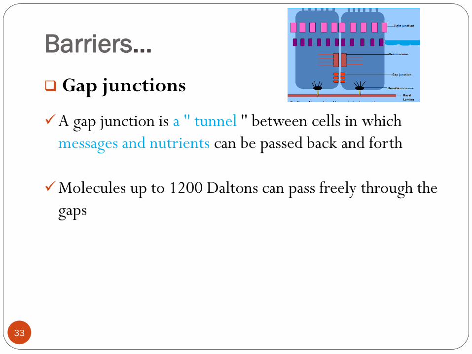
Gap junctions
A gap junction is a '' tunnel '' between cells in which
messages and nutrients can be passed back and forth
Molecules up to 1200 Daltons can pass freely through the
gaps
33

Barriers…
Desmosomes:
Button like points between adjacent cells
Bond adjacent cells together, enable groups of cells to
function as structural units
Most abundant in tissues that are subject to severe
mechanical stress, such as cardiac muscle, skin epithelium
and the neck of the uterus
PLASMA MEMBRANE
34

Recap
Define biopharmaceutics
Describe barriers of drug transport
35

Mechanisms of drug transport
Mechanisms of drug transport:
Three broad categories of drug transport mechanisms
involved in absorption are;
Paracellular/ intercellular transport (between cells)
Transcellular/intracellular transport
Vesicular transport
36

Mechanisms of drug transport across cell
membrane
1. Paracellular (between cells)
2. Transcellular (across cells)
Simple passive diffusion
Facilitated diffusion
Active transport
Vesicular transport
Pore transport
Ion-pair Formation37

Mechanisms…
Paracellular pathway
Transport of materials in aqueous pores b/n cells
Cells are joined together via tight junctions
→ Intercellular spaces occupy ~ 0.01% of total SA of
epithelium
Tightness of junctions vary b/n different epithelia
→Absorptive epithelia, such as SI tend to be leakier
38

Mechanisms…
Paracellular absorption is important for transport of:
Ions such as calcium, sugars, amino acids and peptides at
conc. above carrier capacity
Small hydrophilic and charged drugs cross via paracellular
pathway
Molecular weight cut-off ~ 200 Da
Absorption is quite limited because the paracellular
pathway comprises a very small percentage of the total
surface area(~ 0.01% )
39
Mechanisms…
Transcellular /intracellular transport
Movement across the intracellular space
Passage of drugs across the GI epithelium
Permeation of GI epithelial membrane
40
Mechanisms …
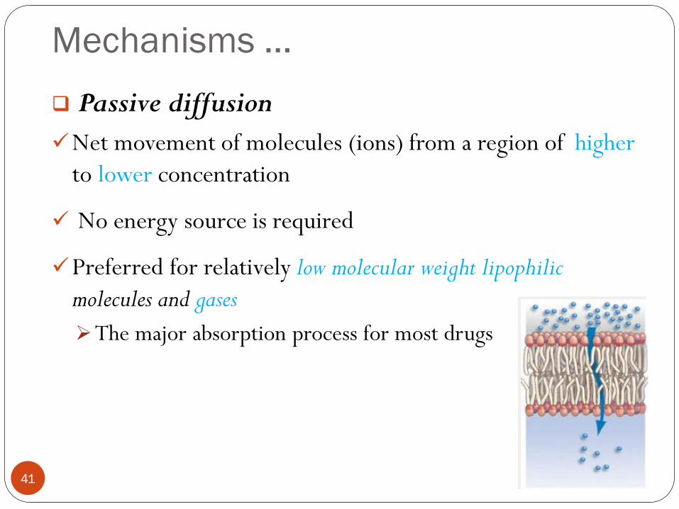
Passive diffusion
Net movement of molecules (ions) from a region of higher
to lower concentration
No energy source is required
Preferred for relatively low molecular weight lipophilic
molecules and gases
The major absorption process for most drugs
41

Mechanisms…
The driving force is higher drug concentrations on the
mucosal side compared to the blood
Low conc. is maintained by blood flow (Sink Condition)
⇒ From lumen to blood
42
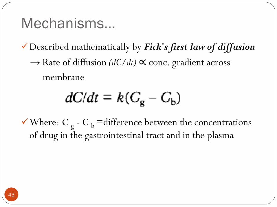
Mechanisms…
Described mathematically by Fick's first law of diffusion
→ Rate of diffusion (dC/dt) ∝ conc. gradient across
membrane
Where: C g - C b =difference between the concentrations
of drug in the gastrointestinal tract and in the plasma
43

Mechanisms…
k incorporates diffusion coefficient in GI membrane (D), thickness (h)
and SA of membrane (A)
Where: D = diffusion coefficient in GI membrane, h= membrane thickness,
A= SA of membrane, C GIT - C p =difference between the
concentrations of drug in the gastrointestinal tract and in the plasma
44
h
CCDA
dtdC PGIT

Mechanisms…
Rate of passive diffusion depends on
Physicochemical properties (molecular size, partition coefficients, ...)
Many drugs are weak electrolytes
→ Exist as unionized and ionized species (pKa, pH)
→ GI membrane is more permeable to unionized form
(greater lipid solubility)
→ Non-polar molecules diffuse more easily than polar
molecules
⇒ Rate is related to fraction of unionized form
Drug having molecular weight between 100- 400 Daltons are
effectively absorbed passively45

Mechanisms…
Diffusion coefficient(D)
Is related to the size and lipid solubility of the drug
As lipid solubility increases or molecular size decreases
then D increases and thus dC/dt also increases
D is a constant (for a specific molecule in a specific
environment)
46
h
CCDA
dtdC PGIT

Mechanisms…
Nature of membrane (SA and thickness of
membrane)
Greater the area and lesser the thickness of the
membrane, faster the diffusion
Surface area
As the surface area increases the rate of diffusion also
increase
The surface of the intestinal lining (with villae and
microvillae) is much larger than the stomach
Generally absorption is faster from the intestine compared
with absorption from the stomach47
h
CCDA
dtdC PGIT
Mechanisms…
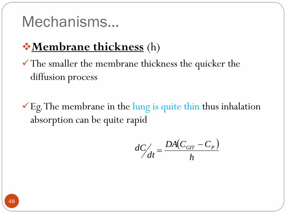
Membrane thickness (h)
The smaller the membrane thickness the quicker the
diffusion process
Eg. The membrane in the lung is quite thin thus inhalation
absorption can be quite rapid
48
h
CCDA
dtdC PGIT

Mechanisms…
Concentration difference (C GIT – Cp )
The rate of drug transport is directly proportional
to the concentration gradient
The bigger the difference between the two sides of
the membrane the quicker the rate of diffusion
Equilibrium is attend when the concentration on
either side of the membrane become equal
49

Mechanisms…
Often, CP <<CGIT, blood acts as 'Sink' for absorbed drug
It is this concentration gradient which allows the rapid
complete absorption of many drugs
Because for given membrane, D, A and h are constants:
A first-order kinetic process
→ Rate ∝ conc. in GI fluids
→Valid for most drugs50
GITkCdt
dC
h
CDA
dtdC GIT
Mechanisms…
Carrier-mediated transport
Most drugs are absorbed from GIT via passive
diffusion
Few lipid insoluble drugs and many nutrients are
absorbed by carrier-mediated transport
→ Carrier binds drug and transports it across membrane
51

Mechanisms…
Explained by shuttling process across epithelial membrane
→ Drug forms complex with carrier
→ Drug-carrier complex moves across membrane
→ Drug liberated on other side
∗ Free carrier returns to initial position in cell membrane adjacent to GI lumen
52
Mechanisms…
Carrier-mediated transport:
• Active transport
• Facilitated transport
53

Mechanisms…
Active transport
Energy-dependent movement of compounds across
membranes
Against their concentration gradient
Active transport;
• Requires energy from the cell in the form of ATP
• Requires specialized proteins (carrier) to bind with
the particle and transport
54

Mechanisms…
Drug combines with a specific carrier protein, on one side
of the membrane
Binding stimulates breakdown (hydrolysis) of ATP
Complex formed diffuses across the membrane to the
opposite side, where the complex dissociates, thus
releasing the drug
The carrier protein then return to its initial side to bind
more drug
55

Mechanisms…
Large number of active transport systems in SI
Peptide T, nucleosideT, sugarT, bile acidT, amino acid T, organic
anion T and vitamin T
Each carrier system is concentrated in specific segment
of GIT and has its own substrate specificity
Eg: Bile acid transporters only found in ileum
Developed for nutrients and chemicals essential to life
Electrolytes, nutrients, vitamins, bile salts
56
Mechanisms…
Drug structurally resembling natural substance that is
actively transported is likely to be transported by same
carrier
Penicillins, cephalosporins, ACE inhibitors and renin inhibitors
rely on peptide transporters for efficient absorption
Nucleosides and their analogues for antiviral and anticancer
drugs depend on nucleoside transporters
5-fluorouracil is transported by pyrimidine transport
system
L-dopa and α-methyldopa are transported by amino acid
transporters57

Mechanisms…
Rate of absorption ∝ conc. of absorbable species
only at low conc.
Becomes saturated at higher concentration
Capacity limited process
Further increase in conc. will not increase rate of
absorption
58
(Passive process)
(Carrier-mediated Process)

Mechanisms…
Characteristics of active carrier-mediated
transport:
Transportation against concentration gradient
Selectivity to substrate
Specific location in GIT
Saturability
Competitive inhibition by substrate analogues
59

Mechanisms…
Facilitated diffusion
The process where molecules combine with membrane
bound carrier proteins to cross the membrane
Solutes are transported downhill but at much faster rate
based on molecular size and polarity
Does not require energy input
→ Require conc. gradient as driving force
Examples: absorption of steroids, amino acids & glucose
from the gut lumen
60
Mechanisms…
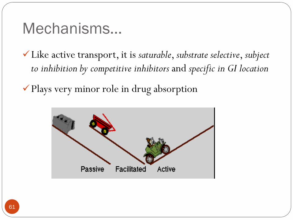
Like active transport, it is saturable, substrate selective, subject
to inhibition by competitive inhibitors and specific in GI location
Plays very minor role in drug absorption
61

Mechanisms…
Vesicular transport
Large macromolecules (e.g., proteins, lipoprotein
particles)
Require more complex mechanisms to traverse
membranes,
Are transported into and out of cells selectively via
endocytosis and exocytosis
It is the process of engulfing particles or dissolved
materials by the cell
62

Mechanisms…
63
1. Cell membrane invaginates to surround the material,
2. Engulfs the material into the cell
3. Cell membrane containing
the material forms a vesicle or
vacuole within the cell

Mechanisms…
Pinocytosis: causes a small amount of extracellular fluid
to enter the cell
Phagocytosis: the engulfment of larger particles or
macromolecules
Usually used for molecules that are too large to traverse the membrane
easily via another mechanism
E.g. Vesicular transport is the proposed process for the
absorption of Vitamin A, D, E, and K, peptides in newborn
64

Mechanisms…
Pore (convective) transport
Transport protein may form an open channel (narrow
pores) across the lipid membrane of the cell
Very small molecules, such as urea, water and sugars are
able to rapidly cross the cell membrane through these pores
Facilitate the trafficking of hydrophilic and charged
molecules
Does not require an additional input of energy
Carry out passive transport
65

Mechanisms…
Channels have an advantage over carrier proteins in terms
of the speed of transport
Up to a hundred million ions can pass through an ion channel
per second
Drugs do not typically rely on ion channels for transport
(the channel opening is too small)
66

Mechanisms…
Ion-pair formation
Strong electrolyte drugs, such as quaternary nitrogen
compounds, are highly ionized or charged ionized over
entire GI pH
Cannot partition directly into lipoidal membrane
Too large to pass through aqueous filled pores in membrane
When linked with an oppositely charged ion, an ion pair is
formed in which the overall charge of the pair is neutral
This neutral complex diffuses more easily across the
membrane
e.g. the formation of an ion pair for propranolol with oleic acid,
quinine with hexylsalicylate67

Mechanisms…
68
Drug transport
Transcellular
Simple passive
diffusion
Active transport
Facilitated diffusion
convective/pore
transport
Vesicular transport
Ion-pair Formation
Paracellular
Passive diffusion
•Low
MW,
lipophilic
•Structurall
y
resembling
natural
substance
•Very
small,
hydrophili
c and
charged
molecules
•Macro
molecule
s
•Large
MW,
Strong
electroly
te drugs
•Low MW,
water-
soluble
drugs
Drug transport Vesicular transport

March, 2020
Factors affecting oral drug
absorption

Session Objectives
After this session you will be able to
Describe GIT anatomy and physiology
Describe factors that affects oral drug delivery
Physiologic factors
Physiochemical factors
Formulation factors
70 28-Apr-20BT

Introduction
Oral route is the most common and convenient of the
existing administration routes
Natural and convenient for the patient
affords high patient acceptability, compliance, and ease
of administration
Oral dosage forms
are easy to manufacture
are compact, do not need to be sterilized
can be produced in large quantities by automated
machines
71 28-Apr-20BT

Introduction…
However, a number of factors can influence drug
absorption from GIT
A fraction of an administered dose of the drug reaches the
systemic circulation in the unchanged form
Bioavailability is not 100%
By IV route the entire drug reaches the systemic
circulation
The drug is 100% bioavailable
72 28-Apr-20BT
Introduction…
Absorption of drugs from GIT depends on:
The physiology of the GIT
Physico-chemical nature of drugs
Formulation/Dosage form factors
73 28-Apr-20BT

GIT anatomy and physiology
GI tract
Is complex muscular tube (~ 6 m long) with varying
diameters
4 anatomical parts (mouth to anus)
Esophagus
Stomach
Small intestine (> 60%)
Large intestine or colon
74 28-Apr-20BT

GIT anatomy…
GIT wall consists four principal histological layers
75 28-Apr-20BT

76 28-Apr-20BT

GIT anatomy…
Serosa:
Outer layer of epithelium
Provides structural support to organs
Muscularis externa:
Contains two layers of smooth muscle
Outer longitudinal layer
mixing, propulsive movement of GI contents
Thicker inner circular layer
prevents food traveling backward
77 28-Apr-20BT
GIT anatomy…
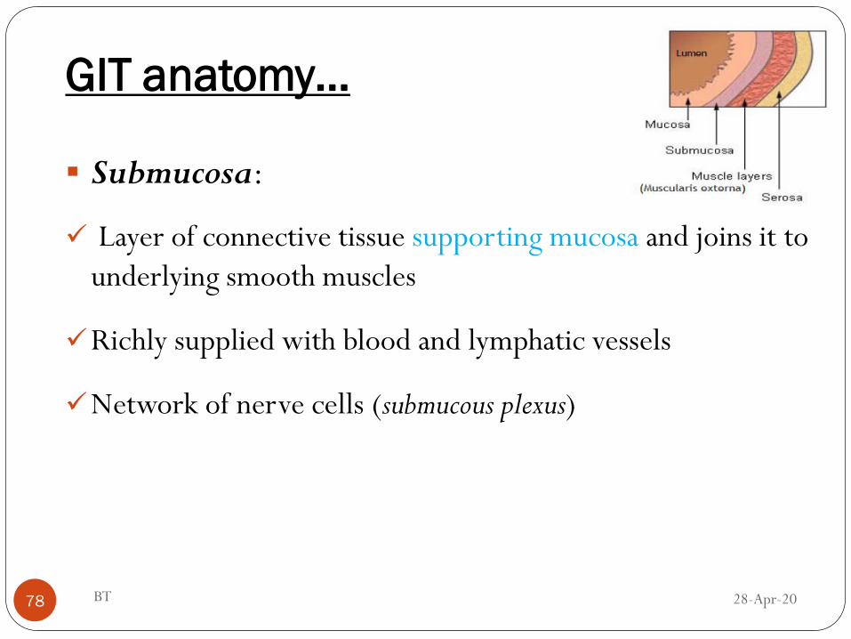
Submucosa:
Layer of connective tissue supporting mucosa and joins it to
underlying smooth muscles
Richly supplied with blood and lymphatic vessels
Network of nerve cells (submucous plexus)
78 28-Apr-20BT

GIT anatomy…
Mucosa: composed of three layers
Epithelium
Selective absorption, secretion, protective functions
Layer of mucus covers majority of GI epithelium
Protective layer and mechanical barrier
Components: mucin (large glycoproteins) + water
(~95%)
Mucus layer: 5 μm to 500 μm, with 80 μm average
79 28-Apr-20BT

GIT anatomy…
Lamina propria
Thin layer of connective tissue
Contains capillaries and lymph vessels
Contain glands with ducts opening to mucosal
epithelium
Secrete mucus
Muscularis mucosa
Thin layer of smooth muscle
Enhance contact b/n epithelium and contents of lumen80 28-Apr-20BT

GIT anatomy…
Esophagus
Links oral cavity with stomach
Composed of thick muscular layer ~ 25 cm long and 2 cm
diameter
pH of lumen ~ 5 – 6
The stratified squamous epithelium provides a tough
impermeable lining resisting the abrasive nature of food
boluses
81 28-Apr-20BT

GIT anatomy…
In upright position, transit of materials is assisted by gravity
→ Esophageal transit of DFs is 10 - 14 s
Limited biopharmaceutical importance
→ Lodging of DFs
82 28-Apr-20BT

GIT anatomy…
Stomach
Divided into four regions:
Fundus, Body, Antrum, Pylorus
Fundus and Body: little muscular tones (reservoir)
Antrum and Pylorus: mix and pump (gastric emptying)
Two major functions:
1. Temporary reservoir for ingested food
→ Capacity: ~ 1.5 l
2. Mix and reduce ingested components into slurry
(chyme)
83 28-Apr-20BT

GIT anatomy…
Secretions of Gastric glands
Parietal or oxyntic cells secrete Hydrochloric acid
Chief or zymogen cells secrete pepsinogen
Pepsin hydrolyses several peptide bonds protein molecules
Mucoid cells secrete mucus (a thick glycoprotein)
Endocrine cells or endocrine-like cells secrete the acid-
stimulating hormone, gastrin
84 28-Apr-20BT

GIT anatomy…
Very little drug absorption occurs
Small SA (around 0.2m2)
Stomach pH, 1–3.5
pH of 1–2.5 most common
High biopharmaceutical importance:
Gastric emptying can dictate drug absorption from SI
Dosage form may remain in the stomach for 0.5–2
h prior to moving to the SI
85 28-Apr-20BT

GIT anatomy…
Small intestine
Longest (4 - 5 m) and most convoluted part
→ Extend from pyloric sphincter to ileocaecal junction
2 Main functions:
- Digestion
- Absorption
3 parts:
– Duodenum: 20 - 30 cm
– Jejunum: ~ 2 m
– Ileum ~ 3 m86 28-Apr-20BT

GIT anatomy…
Folds of Kerckring: folds of SI
mucous membrane that project
within lumen
Several mm in depth
Well developed in duodenum and jejunum
Villi: finger-like projections into
lumen (~ 0.5 - 1.5 mm long & 0.1
mm wide)
→Well supplied with blood vessels
→Contain arterioles, venules and blind-ending lymphatic
vessels 87 28-Apr-20BT

GIT anatomy…
Microvilli: brush-like structures covering
villi
- ~ 1 μm long & 0.1 μm wide
→ ~ 600 – 1000 per villus
→ largest increase in SA
SI has enormous SA ~ 200 m2 in adult
Significant biopharmaceutical importance:
→ Most nutrients and drugs absorbed from
SI
88 28-Apr-20BT

GIT anatomy…
89 28-Apr-20BT
GIT anatomy…
SI wall has rich network of blood and lymphatic vessels
→ ~ 1/3 cardiac output flows through GI viscera
SI receives blood from superior mesenteric artery
Blood leaves SI via hepatic portal vein to liver and then
to systemic circulation
Drugs susceptible to metabolism by liver are degraded
→ hepatic presystemic clearance, first-pass metabolism
90 28-Apr-20BT

GIT anatomy…
91 28-Apr-20BT

GIT anatomy…
Secretions into the small intestine
Brunner’s glands (confined to the duodenum) secrete
bicarbonate and mucus
Intestinal cells (throughout the small intestine) secrete
mucus and a few enzymes
Pancreas secretes ̰ 1 L/day of pancreatic juice
alkaline fluid (bicarbonate) & enzymes (amylases,
lipases, proteases)
92 28-Apr-20BT

GIT anatomy…
Liver secretes bile which is necessary for the digestion and
absorption of lipids
Bile salts:
Emulsification of the fat content of food, producing small
droplets of fat in aqueous suspension
Assisting in the absorption of fatty acids, monoglycerides,
cholesterol and lipids from the intestinal tract by forming
mixed micelles
SI pH 5-8
93 28-Apr-20BT

GIT anatomy…
Large intestine/Colon
Final part - Ileocaecal junction to anus (~ 1.5 m long)
→ caecum ( ~ 8.5 cm)
→ ascending colon ( ~ 20 cm)
→ transverse colon ( > 45 cm)
→ descending colon (~ 30 cm)
→ sigmoid colon (~ 40 cm)
→ rectum (~ 12 cm)
94 28-Apr-20BT

GIT anatomy…
2 Functions:
→ absorption of Na+, Cl−, water from lumen for K+,
HCO3-
→ store and compaction of faeces
No specialized villi but microvilli on absorptive epithelial
cells
→ SA ~ l/30th of SI
95 28-Apr-20BT
GIT anatomy…
Permanently colonized by variety of bacteria
→ capable of several metabolic reactions
Recent interest to exploit enzymes produced by these
bacteria to targeted drug delivery to this region
Some biopharmaceutical importance:
→Targeted drugs to colon (prodrug)
96 28-Apr-20BT

Summary, comparisonStomach SI Colon
PH range 1-3 5-7.5 5.5-7.8
Length (cm) 20 485 110
SA(m2) 0.1-0.2 200 6
Blood flow (L/min) 0.15 1 0.02
Transit time (h) 1-2 3-6 15-48
Absorptive role Lipophilic, acidic
and neutral
All types Some drugs, water
and electrolytes
Absorption
mechanism
Passive diffusion,
convective
transport
All
mechanisms
Passive diffusion,
convective transport
97 28-Apr-20BT

Physiologic factors influence drug
absorption from GIT
Blood flow
GI motility, emptying and transit times
GI pH
Effect of Food
Presystemic metabolism
Stability in GIT
Other drugs
Disease state, Malabsorption
98 28-Apr-20BT

Physiologic factors…
Blood flow
Blood flow to the GI tract is important in carrying
absorbed drug to the systemic circulation
The splanchnic circulation receives about 1/3 of the
cardiac output and is increased after meals
Decrease in mesenteric blood flow, as in the case of
congestive heart failure, will decrease drug
bioavailability
99 28-Apr-20BT

Physiologic factors…
Transit of Pharmaceuticals In GIT
Most DFs transit esophagus in < 15 s
Tablets/capsules taken in supine position are liable to lodge
in esophagus esp. when taken without water
Chance of adhesion depend on shape, size and type of
formulation
Delay in reaching stomach may delay drug's onset of action
Cause damage or irritation to esophageal wall, e.g. KCl
tablets
100 28-Apr-20BT

Physiologic factors…
Gastric emptying time
Gastric emptying time is time DFs take to traverse stomach
Gastric emptying rate, Gastric residence time
Plays a major role in absorption of both basic and acidic
drugs (major site of absorption for both is the Small
Intestine)
Any factor that delay emptying rate (gastric transit time)
generally delay absorption
101 28-Apr-20BT

Physiologic factors…Gastric emptying time is highly variable
Normal Gastric emptying time range b/n 5 min and 2 hrs
Gastric emptying time depends on
DF type - longer for large single units DFs
Fed/fasted state of stomach
In fed state
Liquids, pellets and disintegrated tablets empty with food
Large unit DFs (Sustained R or Controlled R) can be retained
for long
In fasted state:
Stomach is less discriminatory b/n DFs102 28-Apr-20BT

Factors influence gastric emptying
103
Factors Factors promoting GE Factors delaying GE
Food Fasting, liquids, light diet,
Hot
Fats and fatty acids in diet, High
viscosity of diet, solid, cold food
Postural
position
Lying on right side Lying on left side
Dosage form Liquid, multiparticulate DFs solid, unit DFs
Drugs Metoclopramide (antiemetic
and gastroprokinetic)
propantheline, atropine
(antimuscarinic)
Emotional
state
Stress, aggression Depression, vigorous exercise
28-Apr-20BT

Physiologic factors…
Small Intestinal Transit
Small intestinal transit of a dosage forms is not affected by
their physical state, size or the presence or absence of food
Relatively constant ~ 3 hrs
Does not discriminate b/n solids and liquids, b/n DFs
Food, anticholinergic drugs, pregnancy retard intestinal
transit
Anticholinergic drugs promote absorption of poorly soluble
drugs (eg. Nitrofurantoin, hydrochlorothiazide)
Diarrhea, laxatives promote intestinal transit104 28-Apr-20BT

Physiologic factors…
SITT is particularly important for
Drugs that dissolve/release slowly from DF (eg. Sustained
release)
Enteric-coated DFs (release drug in SI)
Drugs absorbed by intestinal carrier-mediated transporters
(Eg. Vit B)
Colonic Transit
Long and variable, vary from 2 - 48 hrs
105 28-Apr-20BT

Physiologic factors…
GI pH
Luminal pH varies considerably along GIT
Gastric fluid is highly acidic (pH ~ 1 - 3.5) in fasted state
Following ingestion of meal, buffered to less acidic pH
pH 3 - 5 following meal & returns to fasted-state value in 2 -
3 hrs depending on meal size
Intestinal pH is higher due to neutralization with HCO3-
secreted by pancreas
Gradual rise in pH along SI, 5-7.5
106 28-Apr-20BT

Physiologic factors…
GI pH influence drug absorption in several ways:
Disintegration of enteric coated DF is pH sensitive
For basic and acidic drugs, depending on their pka, pH
determines the unionized form at the absorption site
pH influences chemical stability of drugs
pH-dependent hydrolysis
Penicillin G (benzylpenicillin)
Degradation depends on gastric residence time and pH
Gastric instability preclude oral use
Erythromycin and omeprazole degrade rapidly at acidic pH
Formulated as enteric-coated DFs107 28-Apr-20BT

Physiologic factors…
Luminal enzymes
Pepsins and proteases degrade protein and peptide drugs
Drugs resembling nutrients, such as nucleotides and fatty
acids, may be susceptible to enzymatic degradation
Colonic bacteria secrete enzymes capable of range of
reactions
Explored for colon targeted drug delivery systems
108 28-Apr-20BT

Effect of food on drug absorption
Some effects of food
Delay in gastric emptying
Stimulation of bile flow
Stimulation of blood flow to GIT
A change in the pH of the GI tract
A change in luminal metabolism of the drug substance
Physical or chemical interaction of the meal with the drug
product or drug substance
109 28-Apr-20BT

Effect of food…
Food influence rate and extent of absorption in various
ways;
Complexation of drugs with components in diet
TTC forms non-absorbable complexes with Ca2+ and Fe2+
Not taken with Ca2+ and Fe2+ containing products such as
milk, iron preparations
Alteration of pH
Food increase stomach pH by acting as a buffer
Decrease rate of dissolution and absorption of weakly basic drug
and increase that of weakly acidic ones
110 28-Apr-20BT

Effect of food…
Increased viscosity of gastrointestinal contents
The presence of food in the GIT provides a viscous
environment which may result in:
Reduction in the rate of drug dissolution
Reduction in the rate of diffusion of drug in solution from the
lumen to the absorbing membrane lining the GIT
There is reduction in drug bioavailability
111 28-Apr-20BT

Effect of food…
Alteration of gastric emptying time
Fatty foods reduce gastric emptying
Delayed gastric emptying is
Reduce rate of absorption of most drugs
Beneficial for absorption of poorly soluble drugs such as
griseofulvin
112 28-Apr-20BT
Effect of food…
Stimulation of GI secretions
Pepsins produced in response to food may degrade drugs
Fats stimulate secretion of bile
Merit: Bile salts are surface-active agents
Increase dissolution of poorly soluble drugs (e.g. griseofulvin)
and enhance their absorption
Demerit: Bile salts form insoluble and non-absorbable
complexes with some drugs, such as neomycin, kanamycin and
nystatin113 28-Apr-20BT

Effect of food…
Competition between food components and drugs
for specialized absorption mechanisms
There is a possibility of competitive inhibition of drug
absorption in case of drugs that have a chemical structure
similar to nutrients required by the body for which
specialized absorption mechanisms exist
E.g. L-dopa and methyldopa use the same transporter
mechanism as aromatic amino acids from proteins,
Absorption of these drugs is decreased if high-protein meal is
taken
114 28-Apr-20BT

Effect of food…
Food-induced changes in blood flow Ingestion of food increases the bioavailability of drugs
which are metabolized during first pass through the gut wall or liver
Two mechanisms have been put forwarded:
1. Enhanced blood flow will increase the load of drug delivered to saturable enzyme systems so that a greater proportion escapes metabolism
2. Nutrients may compete for hepatic enzymes so that less drug is metabolized
Eg. Concurrent food intake enhances the bioavailability of drugsincluding: propranolol, metoprolol, labetalol, and hydralazine, whichare greatly metabolized by hepatic enzyme systems
115 28-Apr-20BT

Effect of food… Food-induced changes in presystemic metabolism
E.g. Grape fruit juice is capable of inhibiting the
intestinal cytochrome P450 (CYP3A4)
Thus, taken with drugs that are susceptible to CYP3A4
metabolism, result in increase of their bioavailability
Abnormally high levels of some drugs
can be dangerous and can lead to
toxic side effects
Eg. Lovastatin, simvastatin
Verapamil, Cyclosporin
Saquinavir116 28-Apr-20BT

Effect of food,…
117
Drug absorption may be delayed, reduced, increased
or, may not be affected by the presence of food
Generally, drugs are better absorbed under fasting condition
If a drug is to be taken on an empty stomach, it should be taken half
an hour or more before meals or two hours or more after meals!
Delayed Decreased Increased Unaffected
Aspirin
Paracetamol
Diclofenac
Digoxin
Penicillins
Erythromycin
Tetracyclin
Levodopa
Iron
Gresiofulvin
Diazepam
Actively absorbed
water soluble
vitamins
Propylthiuracil
sulfasomidin
28-Apr-20BT

Diseases
Diseases Malabsorption:
Impaired absorption of nutrients (failure to completely absorb nutrients from the gastrointestinal tract)
Caused by; Inadequate production of bile salts by the liver, or digestive
enzymes by the pancreas
Damage to the intestinal absorptive cells
Chronic pancreatitis (often related to alcohol abuse)
Abnormal drug absorptionsEg. Fat malabsorption may affect absorption of lipophilic drugs
118 28-Apr-20BT

Cont…
GI Surgery:
Can affect drug absorption by altering transit time
Hepatic cirrhosis:
Increase bioavailability of drugs that undergo
considerable first-pass hepatic metabolism
eg. propranolol
119 28-Apr-20BT

Presence of other drugs
Presence of other drugs can affect drug absorption from
GIT in a number of ways;
Adsorption:
Antidiarrheal preparations containing adsorbents such as
kaolin-pectin & attapulgite can retard or prevent absorption
of co-administered drugs
eg. Promazin, lincomycin
Forming unabsorbable complexes:
Antacids or other formulations containing Al+3, Ca+2, Mg+2
Zn+2, Fe+3(2) retard absorption of tetracyclines
120 28-Apr-20BT

Presence of other drugs,… Altering pH:
Basic drugs (eg tetracycline) dissolve in gastric pH, co-administration with basic drugs (eg. Sodium bicarbonate) will decrease their dissolution
Decrease GI motility, retard GI transit: eg. Atropine, propanthelin, morphine, alfentanil
Promote absorption of drugs which are absorbed slowly (eg ranitidine, digoxin, vitamins B12 & C)
Delayed gastric emptying Retard absorption of enteric coated drugs Reduce bioavailability of drugs unstable in the stomach
(eg. penicillines)121 28-Apr-20BT

Presence of other drugs,…
Increased GI motility
Eg. Metoclopramide, Domperidone
Enhance absorption of tetracyclines, pivampicillin,
levodopa,…- drugs which are affected in stomach
Reduce absorption of drugs which are absorbed slowly
Alter GI metabolism
Antibiotics inhibit bacterial metabolism of drugs
Eg. Digoxin is inactivated in the lower intestine by bacterial
metabolism
Absorption enhanced by co-administration with antibiotics
122 28-Apr-20BT

Pharmacists in every practice setting need to be
vigilant in monitoring for potential drug-food
and drug–drug interactions and advising
patients regarding foods or beverages and
drugs to avoid when taking certain medications
123 28-Apr-20BT

Factors affecting oral drug absorption
Physiological factors affecting oral absorption
Physical-chemical factors affecting oral absorption
Formulation factors affecting oral absorption
28-Apr-20BT124

Physical-chemical factors affecting oral absorption
Drug dissolution and Noyes-Whitney equation
Particle size and surface area
Salt formation of drug
Crystal forms (Polymorphism, solvates)
Pka and pH, pH-partition hypothesis
Lipid solubility
125 28-Apr-20BT
Physical-chemical factors…
Dissolution
Dissolution is the process by which a solid substance
solubilizes in a given solvent
Precondition for absorption of drugs from dosage forms
126 28-Apr-20BT

Physical-chemical factors…
Dissolution of solid in liquid is composed of two consecutive
steps:
Drug molecules in the surface layer dissolve, leading to a
formation of a saturated solution around the particle to form
the diffusion layer (stagnant film)
• This step is usually rapid
Diffusion of soluble solute from diffusion layer into the bulk
solution
Once solute passes boundary layer, rapid mixing occurs and
Conc. of solution changes from being Cs at particle surface to bulk
conc. C at its outermost limit127 28-Apr-20BT

Physical-chemical factors…
Rate of diffusion of dissolved solute across boundary layer
determines rate of dissolution
128 28-Apr-20BT

Physical-chemical factors…The dissolution of drugs can be described by the
Noyes-Whitney equation:
Where
dC/dt is the rate of dissolution of the drug particles
D is the diffusion coefficient of the drug
A is the effective surface area of the drug particles in contact with
the gastrointestinal fluids
h is the thickness of the diffusion layer around each drug particle,
Cs is the saturation solubility of the drug in solution in the
diffusion layer and
C is the concentration of the drug in the bulk solution (GI fluids)
129
h
C)(C
dt
dc s DA
28-Apr-20BT
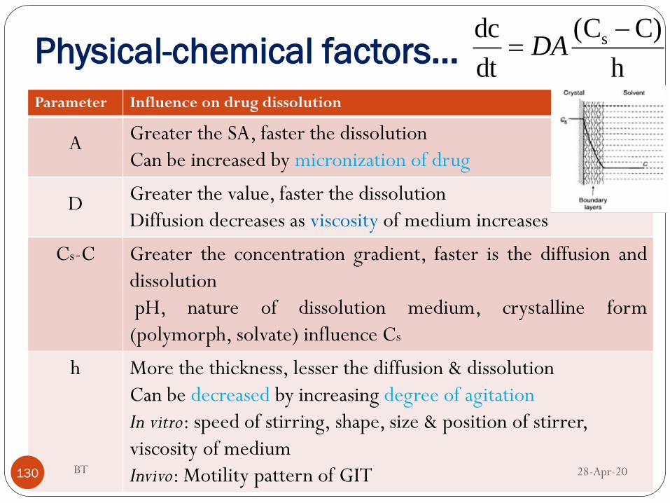
Physical-chemical factors…
Parameter Influence on drug dissolution
A Greater the SA, faster the dissolution
Can be increased by micronization of drug
D Greater the value, faster the dissolution
Diffusion decreases as viscosity of medium increases
Cs-C Greater the concentration gradient, faster is the diffusion and
dissolution
pH, nature of dissolution medium, crystalline form
(polymorph, solvate) influence Cs
h More the thickness, lesser the diffusion & dissolution
Can be decreased by increasing degree of agitation
In vitro: speed of stirring, shape, size & position of stirrer,
viscosity of medium
Invivo: Motility pattern of GIT130 28-Apr-20BT
h
C)(C
dt
dc s DA

Physical-chemical factors…
Surface area and Particle size
The smaller the particle size, the greater the effective
surface area exposed to dissolution medium & the higher
the dissolution rate
Increase bioavailability (dissolution-rate limited)
Eg, Griseofulvin
Reduction of particle size from ~ 10 μm (specific SA = 0.4
m2 g-1) to 2.7 μm (specific SA = 1.5 m2 g-1) approximately
doubled amount of drug absorbed in humans
131 28-Apr-20BT

Physical-chemical factors…
Many poorly soluble, slowly dissolving drugs are presented
in micronized form to increase SA
Digoxin (Cardiac glycoside), Nitrofurantoin (Antifungal),
Medroxyprogesterone acetate (Hormone)
Tolbutamide (Antidiabetic), Sulphadiazine (Antibacterial)
Naproxen and Ibuprofen (NSAID), Phenacetin (Analgesic)
132 28-Apr-20BT

Physical-chemical factors…
It is important to control particle size
Improvement in bioavailability can result in increased side
effects
For drugs which are unstable in gastric fluid, particle size
reduction increase chemical degradation
Penicillin G and erythromycin
For hydrophobic drugs, micronization and other dry
particle size reduction techniques can result in
aggregation
Reduce dissolution rate and bioavailability
133 28-Apr-20BT

Physical-chemical factors…
Effective SA of hydrophobic drugs can be increased by
Addition of wetting agent to formulation
Tween-80 in fine suspension of phenacetin (< 75 μm)
improved bioavailability vs. same-size suspension without
wetting agent
Increase wetting and solvent penetration
Minimize aggregation of suspended particles
Incorporation of hydrophilic diluents (such as PVP, PEG,
dextrose,…)
Coat surface of hydrophobic drugs and render them hydrophilic
134 28-Apr-20BT

Physical-chemical factors…
Solubility, Cs
Aqueous solubility depends on pH
In weak electrolytes, dissolution rate depend on solubility
and pH in diffusion layer
Difference in dissolution rate is expected in different regions
of GI tract
Solubility of weakly acidic drugs increases with pH, as drug
move down GI tract from stomach to SI
Solubility of weak bases decrease with increasing pH, down
GI tract 135 28-Apr-20BT

Physical-chemical factors…
Important for poorly soluble weak bases to dissolve rapidly
in stomach as rate of dissolution in SI is much slower
Dosing ketoconazole 2 hrs after H2 blocker cimetidine
(reduces gastric acid secretion) results in significantly
reduced rate and extent of absorption
Pretreatment with H2 blocker famotidine reduces peak
plasma conc. of dipyrimidole (antiplatelet) by factor of up
to 10
136 28-Apr-20BT

Physical-chemical factors…
Salt forms of Drugs
Dissolution rate of a salt form of a drug is generally quite
different from that of the parent compound
Salt form alters pH of diffusion layer by neutralization
Dissolution rate of weakly acidic drug in gastric fluid (pH 1 -
3.5) is relatively low
If pH in diffusion layer increased, solubility (Cs) in this layer, and hence
its dissolution rate in gastric fluids, is increased even though bulk
pH remain low
pH of diffusion layer would be increased if basic salt is formed137 28-Apr-20BT

Physical-chemical factors…
E.g., Na+ or K+ salts of free acid
pH of diffusion layer surrounding each particle is higher (e.g.
5 - 6) than bulk (1 - 3.5) because of neutralizing action of Na+ or
K+ ions present in diffusion layer
138 28-Apr-20BT

Physical-chemical factors…
When dissolved drug diffuses out of diffusion layer into
bulk of gastric fluid, where pH is lower, precipitation of
free acid form is likely to occur
Precipitated free acid will be in the form of very fine, wetted
particles which exhibit very large total effective SA in
contact with gastric fluids;
Facilitate rapid re-dissolution and absorption compared to
the drug administered just in its acid form
28-Apr-20139 BT

Physical-chemical factors…
Examples:
Dissolution rate of tolbutamide sodium (oral
hypoglycemic) in 0.1 M HC1 is 5000 times faster than the
free acid
Barbiturates are often administered as sodium salts to
achieve rapid onset of sedation and provide more
predictable effects
Naproxen sodium is absorbed faster and is more effective
than naproxen in treating mild to moderate pain
140 28-Apr-20BT

Physical-chemical factors…
Strong acid salt of weakly basic drugs dissolve more rapidly in
the intestinal fluids than free bases
Chlorpromazine vs chlorpromazine HCl
Strong acidic Cl- anion in diffusion layer ensures lower
pH than bulk
Increase solubility (Cs) in diffusion layer
Sodium salts of acidic drugs and hydrochloride salts of basic drugs
are by far most common
141 28-Apr-20BT

Physical-chemical factors…
Hydrochloride salts may experience common ion effect owing to
presence of chloride ions in stomach
In vitro dissolution of sulphate salt of indinavir is significantly
greater than hydrochloride salt
Bioavailability of sulphate salt is more than 3X greater than the
hydrochloride salt
142 28-Apr-20BT

Physical-chemical factors…
Some salts have lower solubility & dissolution rate than free
form
Eg. Aluminum salt of weak acids and palmoate salt of weak bases
Insoluble films of aluminum hydroxide or palmitic acid
formed to coat dissolving solids when exposed to basic or acidic
environment, respectively
Poorly soluble salts delay absorption and may be used to sustain
release
143 28-Apr-20BT

Physical-chemical factors…
Factors such as chemical stability, are considered during salt selectionEg. Sodium acetylsalicylate is much more prone to hydrolysis
than aspirin itself
One way to overcome chemical instabilities or other undesirable features of salts is to form salt in situ or to add basic/acidic excipients to formulation
Basic excipients in the formulation of acidic drugs ensures that a relatively basic diffusion layer is formed around each dissolving particle
Inclusion of basic ingredients aluminium dihydroxy aminoacetate and magnesium carbonate in aspirin tablets increase dissolution rate and bioavailability
144 28-Apr-20BT

Physical-chemical factors…
Polymorphism
Solid drug materials may occur as:
Amorphous particles without definite structure (no
particular order of molecules)
Pure crystalline substances of definite identifiable
shape
Molecules arrange themselves in two or more different
ways in the crystal- polymorphs
145 28-Apr-20BT

Physical-chemical factors…
Many drugs exist in more than one crystalline form
Eg., Chloramphenicol palmitate, cortisone acetate,
tetracyclines and sulphathiazole
Crystal habit and internal structure of polymorphs affect
physicochemical properties:
Melting point, density, hardness, crystal shape
Solubility and dissolution rate
At given T and P, only one crystalline form is stable and
others are metastable
146 28-Apr-20BT

Physical-chemical factors…
Metastable tend to transform to most stable form
Metastable polymorphs have higher energy and usually
lower m.pt., greater solubility and dissolution rates
One polymorph may be therapeutically active than another!
Eg. Chloramphenicol palmitate
Exists in 3 crystalline forms designated A, B and C
C is too unstable to be included in DFs
B is sufficiently stable
A is stable – therapeutically inactive
147 28-Apr-20BT

Physical-chemical factors…
The extent of absorption of oral chloramphenicol
increases as the proportion of form B increased
Rapid in vivo dissolution rate of polymorph B
28-Apr-20148 BT

Physical-chemical factors…
28-Apr-20BT149
Fig. Comparison of serum levels (μ g cm03) obtained with suspensions of
chloramphenicol palmitate after oral administration of a dose equivalent to 1.5 g
of chloramphenicol.

Physical-chemical factors…
Amorphous solids
Some drugs may exist in amorphous form
Amorphous form usually dissolves more rapidly than crystalline
form(s)
Significant differences in bioavailability exists b/n
amorphous and crystalline forms of drugs (dissolution-
rate limited bioavailability)
Eg. Novobiocin (antibiotic)
Amorphous is at least 10X more soluble than crystalline form
Amorphous form is readily absorbed from oral suspension150 28-Apr-20BT

Physical-chemical factors…
Solvates
Many drugs can associate with solvent molecules to
produce crystalline forms known as solvates
When water is solvent, solvate is called hydrate
Solvated and non-solvated forms usually exhibit differences
in dissolution rates
Exhibit differences in bioavailability
The solvate has already interacted intimately with the
solvent
151 28-Apr-20BT

Physical-chemical factors…Greater solvation of crystal means lower solubility and
dissolution rate in solvent identical to solvation molecule
Eg. Ampicillin (antibiotic)
Anhydrous form is ~ 25% more soluble than trihydrate form
Absorbed to greater extent from both capsule and aqueous
suspension forms than trihydrate form
Analog of Indinavir (protease inhibitor)
Anhydrous form of HCl salt has much faster dissolution rate
than dihydrate form in water
Anhydrous form achieves > 2X bioavailability
152 28-Apr-20BT

Physical-chemical factors…
Solvate forms of drugs with organic solvents may dissolve faster
than non solvated form
Chloroform solvate of griseofulvin ,Griseofulvin chloroformate,
has greater solubility than non solvated form
has significantly higher bioavailability than non solvated form
Mono ethanol solvate of prednisolone has an absorption
rate in vivo which is nearly five times greater than that of the
anhydrous
153 28-Apr-20BT

Physical-chemical factors…
Drug dissociation and lipid solubility
The dissociation constant and lipid solubility of a drug and
the pH at the absorption site often influence the absorption
characteristics of a drug throughout the GIT
pH–partition theory states that for drug molecules
which are primarily transported by passive diffusion, the
process of absorption is governed by:
1. The dissociation constant, pKa,
2. The lipid solubility of a drug, and
3. The pH at the absorption site
154 28-Apr-20BT

Physical-chemical factors…
This theory is based on the following assumptions:
The drug is absorbed by passive transfer
The drug is preferentially absorbed in unionized form
The drug is sufficiently lipid soluble
28-Apr-20BT155

Physical-chemical factors…
According to pH-partition hypothesis, GI epithelia act as
lipid barrier toward drugs absorbed by passive diffusion
(lipophilic drug)
Unionized form of weak electrolyte drugs (i.e., lipid-soluble
form) pass across GI epithelia
GI epithelia is impermeable to ionized (i.e., poorly lipid-soluble)
form of such drugs
Absorption of weak electrolyte is determined chiefly by extent of unionized
form at site of absorption
156 28-Apr-20BT
Physical-chemical factors…
According to pH-PH, Weakly acidic drug is more likely to
be absorbed from stomach & weakly basic drug from
intestine
The fraction of drug available in unionized form is a
function of both the dissociation constant (pka) of the
drug and the pH of the solution at the site of
administration
28-Apr-20BT157

Physical-chemical factors…
Extent to which weakly acid or base drug ionize in solution in
GI fluid is calculated using Henderson-Hasselbalch
equations
For weakly acidic drug:
[HA] and [A-] are conc. of unionized and ionized forms
E.g., Aspirin, phenylbutazone, salicylic acid
For weakly basic drug:
[BH+] and [B] are conc. of ionized and unionized forms
E.g., Chlorpromazine
158 28-Apr-20BT

Physical-chemical factors…
Eg. For Aspirin (pka 3) Loratadine (pka 6) and Guanethidine
(pka 11)
1. Determine percent drug unionization in the stomach(pH-
2.0) and intestine(pH- 5).
2. Based on pH-partition hypothesis, from which site the drug
will be best absorbed?
3. Delayed Gastric transit and increasing gastric pH by food
or drug will be beneficial for absorption of which drug?
159 28-Apr-20BT

Physical-chemical factors…
In fact, most drugs, regardless of their pKa, are absorbed
from the small intestine
Despite seemingly unfavorable ratio of unionized to ionized
molecules, most weak acids are absorbed predominantly in
the small intestine
Attributed to:
a large surface area,
a relatively long residence time and
limited absorption of the ionized species
160
(Factors not
considered by the
pH–partition
theory)
28-Apr-20BT

Physical-chemical factors…
Limitations of pH-partition hypothesis
1. Extent to which drug exists in unionized form is not the
only factor determining rate and extent of absorption
Despite high degree of ionization, weak acids are well
absorbed from SI
Intestinal absorption of weak acid is often higher than in
stomach
Huge SA in SI more than compensates for high degree of
ionization
Longer SI residence time at intestinal surface
161 28-Apr-20BT
Physical-chemical factors…
2. pH-partition hypothesis cannot explain why certain
drugs
E.g:- quaternary ammonium compounds and tetracyclines
are readily absorbed despite being ionized over the entire
pH range of the gastrointestinal tract
Ion-pair formation
3. Convective flow or solvent drag
The movement of water molecules into and out of the
gastrointestinal tract will affect the rate of passage of small
water-soluble molecules across the gastrointestinal barrier
162 28-Apr-20BT

Physical-chemical factors…
Lipid solubility
Number of drugs are poorly absorbed from GIT despite their
unionized forms predominate
Eg, Barbiturates: Barbitone and Thiopentone
Similar dissociation constants (pKa) 7.8 and 7.6
Similar degrees of ionization at intestinal pH
But Thiopentone is absorbed much better than barbitone!
o More lipid soluble
o Exhibit greater affinity for GI membrane
28-Apr-20BT163

Physical-chemical factors…
Ideally for optimal absorption, a drug should have
Sufficient aqueous solubility to dissolve in fluids at the
absorption site and
Lipid solubility high enough to facilitate its partitioning into
the biomembrane
Measure of lipid solubility is partition coefficient, P
Determined by drug partitioning b/n water and suitable
organic solvent (Octanol) at constant temperature
28-Apr-20BT164

Physical-chemical factors…
Polar (poorly lipid soluble) (log P < 0) and relatively large
molecules such as gentamicin, ceftriaxone, heparin
and streptokinase are poorly absorbed after oral
administration
Given by injection
Lipid soluble drugs with favorable partition coefficients
(i.e. log P > 0) are usually absorbed after oral administration
28-Apr-20BT165

Physical-chemical factors…
To be absorbed, a drug should not be so-lipid soluble
that it will not dissolve in the aqueous fluids of the
GIT, nor so water-soluble that it will not penetrate the
lipid GI membrane.
28-Apr-20BT166

Physical-chemical factors…
Improving lipid solubility
Substituting hydrophilic by hydrophobic group
Clindamycin (Cl) is absorbed more than lincomycin (OH)
If structure cannot be modified to yield lipid solubility,
medicinal chemists may make prodrugs to improve absorption
28-Apr-20BT167

Physical-chemical factors…
Molecular size and hydrogen bonding
For paracellular absorption, mol. wt. ideally be < 200 Da
For transcellular passive diffusion, mol. wt. < 500 Da is
prefered
28-Apr-20BT168

Formulation factors affecting oral drug
absorption
Drug release from Solution, suspension, capsules
and tablets
Effects of excipients
28-Apr-20BT169

Formulation factors.…
The bioavailability of a drug can also be influenced by
factors associated with the formulation of the dosage
form
The type of dosage form
The excipients used
Method of preparation
28-Apr-20BT170

Formulation factors.…
Type of dosage form
The type of dosage form influence the number of possible
intervening steps between administration and the
appearance of dissolved drug in the GI fluids
Greater number of intervening steps means greater
number of potential obstacles to absorption
28-Apr-20BT171

Formulation factors.…
28-Apr-20BT172

28-Apr-20BT173
Fastest

Formulation factors.…
Generally, bioavailability of drug from:
Solution > Suspension > Capsule > Uncoated
tablet > Coated tablet
Ranking is not universal, it provides useful guideline!
Solutions and suspensions are most suited for drugs
intended to be rapidly absorbed
28-Apr-20BT174

Formulation factors.…
Aqueous solution
With rare exceptions, drugs are absorbed more
rapidly from solution than any other oral DF
Eliminates in vivo dissolution step and presents drug in most
readily available form for absorption
28-Apr-20BT175

Formulation factors.…
Factors influencing drug bioavailability from aqueous
solutions
The chemical stability exhibited by the drug in
aqueous solution and the gastrointestinal fluids
Excipients added to the dosage form
The viscosity of a solution dosage form
viscous solutions will retard the diffusion of the
drug & retard the rate of gastric emptying
hence retard the absorption of most drugs
28-Apr-20BT176

Formulation factors.…
Aqueous suspension
Suspension is useful for insoluble or poorly soluble drug
Absorption from suspensions is usually dissolution-rate
limited
Well formulated, finely subdivided suspension is regarded
as efficient oral delivery system
Present huge total SA to GI fluid
Facilitates dissolution and absorption
Dissolution of particles begin immediately on dilution in
GI fluids
28-Apr-20BT177

Formulation factors.…
Factors influencing bioavailability of suspension DFs:
Particle size and effective SA of dispersed drug
Inclusion of surfactant as wetting, flocculating or
deflocculating agent
Viscosity (affect stability, dissolution & gastric emptying)
Suspension Stability: If suspensions are stored for a long
period of time, there is a possibility of crystal growth as a
result temperature fluctuations-may reduce dissolution &
hence absorption 28-Apr-20BT178

Formulation factors.…
Liquid-filled capsule
Liquids can be filled into soft or hard gelatin capsules
Combine convenience of unit DFs with rapid drug
absorption associated with solution and suspension DFs
Drug is dissolved or dispersed in non-toxic, non-aqueous
vehicles
Vegetable oils (water immiscible)
PEGs, surfactants (polysorbate-80) (Water miscible)
28-Apr-20BT179
Formulation factors.…
Following release of contents
Water-miscible vehicle readily disperse and/or dissolve
in GI fluid
Liberate drug as solution or fine suspension
Conducive to rapid absorption
28-Apr-20BT180

Formulation factors.…
Formulation factors affecting bioavailability
the solubility of the drug in the vehicle and GI fluids
the particle size of the drug if suspended in the vehicle
the nature of the vehicle, i.e. hydrophilic or lipophilic
the inclusion of a surfactant as a wetting/emulsifying
agent
the inclusion of a suspending agent (viscosity enhancing
agent)28-Apr-20BT181

Formulation factors.…
Powder-filled capsule
Bioavailability of well formulated powder-filled HGC is
better than or at least equal to compressed tablet
If HG shell dissolves rapidly in GI fluids and encapsulated
mass disperses rapidly and efficiently
Large effective SA of drug will be exposed facilitating
dissolution
28-Apr-20BT182

28-Apr-20BT183
Formulation factors.…
Overall rate of dissolution of drugs from capsules is
a complex function of:
The dissolution rate of the gelatin shell
The rate of penetration of the gastrointestinal fluids into
the encapsulated mass
The rate at which the mass deaggregates (i.e. disperses)
in the gastrointestinal fluids
The rate of dissolution of the dispersed drug particles28-Apr-20BT184

Formulation factors.…
Excipients have significant effect on rate of
dissolution (especially for poorly soluble and
hydrophobic drugs
Hydrophilic diluent (e.g. sorbitol, lactose)
often serves to increase rate of penetration of
aqueous GI fluids into contents of capsule
aid dispersion and subsequent dissolution of drug
28-Apr-20BT185

Formulation factors.…
Capsule-filling process
Affect packing density and liquid permeability of capsule
contents
High packing density result in decrease liquid permeability
and dissolution rate,
particularly if drug is hydrophobic, or if hydrophilic drug is
mixed with hydrophobic lubricant such as magnesium
stearate
28-Apr-20BT186

Formulation factors.…
Tablets
Tablets are the most widely used dosage form
When a drug is formulated as a compressed tablet there is
an enormous reduction in the effective surface area of
the drug
This necessitate the addition of excipients, which serve to
return the surface area of the drug back to its original
precompressed state
28-Apr-20BT187

Formulation factors.…
Generally produced by
Granulation method
Direct compression
Wet granulation consist
Mixing drug with powdered additives
Wetting mixture with aqueous binder solution (gelatin
or starch)
Screening granules (flowability and compressibility)
Compression into tablet
28-Apr-20BT188
Formulation factors.…
Direct compression
Mixing drug with additives
Compression of mix
Drug must have desirable crystallinity and cohesiveness
Suitable direct compression diluents (dicalcium phosphate
dihydrate, tricalcium phosphate, calcium phosphate, calcium
sulfate, anhydrous lactose, spray dried lactose, pregelatinized
starch, microcrystalline cellulose)
28-Apr-20BT189

Formulation factors.…
Most bioavailability problems of compressed
tablets are related to:
Large reduction in effective SA in tableting
Difficulty in regenerating well-dispersed primary drug
particles
28-Apr-20BT190

Formulation factors.…
Dissolution from intact tablet is extremely limited
Small effective SA of drug exposed to GI fluid
Dissolution from granules is comparable to coarse,
aggregated suspension
Dissolution from primary drug particles is comparable to
fine, well-dispersed suspension
28-Apr-20BT191

Formulation factors.…
28-Apr-20BT192

Formulation factors.…
Formulation factors affecting bioavailability
The physicochemical properties of the liberated drug
particles in the gastrointestinal fluids, e.g. wettability,
effective surface area, crystal form, chemical stability
Method of granulation (wet vs. dry) and size of granules
The nature and quantity of the diluent, binder,
disintegrant, lubricant and any wetting agent
28-Apr-20BT193

Formulation factors.…
Type of the tablet (uncoated, coated, enteric-coated )
Drug-excipient interactions (e.g. complexation)
The compaction pressure and speed of compression used in
tableting
28-Apr-20BT194

Formulation factors.…
Coated tablets
Tablet coating may be used simply for aesthetic reason or to
mask unpleasant taste or odor or to protect ingredient
from decomposition during storage
Film coating is currently most commonly used
Several older preparations, such as vitamins and ibuprofen,
still have sugar coats
Presence of coating presents physical barrier b/n core and GI fluid
Physicochemical nature and thickness of coating
28-Apr-20BT195

Formulation factors.…
Sugar coating
Tablet core is usually sealed with thin film of poorly water
soluble polymer such as shellac or cellulose acetate phthalate
Protect core from aqueous fluids used in subsequent steps
Water-impermeable sealing potentially retard drug release
28-Apr-20BT196

Formulation factors.…
Film coating
Coating of tablet core by thin film of water-soluble polymer,
such as HPMC, have no significant effect on rate of
disintegration and dissolution
If hydrophobic water-insoluble film-coating materials, such as
ethylcellulose or certain acrylic resins, are used
Film coat acts as barrier to delay and/or reduce rate of drug
release
• Affect bioavailability
• Used in controlled release drug delivery
28-Apr-20BT197

Formulation factors.…
Enteric-coated tablets
Designed to resist low pH of gastric fluids but to disrupt in
higher pH of SI
Protect drugs unstable in gastric fluid
Protect stomach against drugs causing nausea or irritation
Eg. Aspirin, Ibuprofen
Drug release depends on gastric residence time
Significant delay in release of drug results from longer gastric
residence time
28-Apr-20BT198

Formulation factors.…
Gastric emptying of intact tablets is all-or-nothing process
i.e. Tablet is either in stomach or in duodenum (not
released or released)
Small individually enteric-coated granules or pellets
(multiparticulates) contained in rapidly dissolving HGC
If granules or pellets are sufficiently small (< 1 mm), they
will be able to empty from stomach with liquids
Exhibit gradual and continual emptying into duodenum
Avoid delay of onset of action
28-Apr-20BT199

Formulation factors.…
Dosage form factors
The dosage form factors affecting drug absorption are:
Disintegration time: Directly related to the amount of binder & the compression force Incorporation of suitable amount of disintegrant will aid
disintegration
Manufacturing/processing variables
Method of granulation Wet granulation involves a number of steps each of which can negatively affect drug dissolution
Direct compression yields tablets that dissolve at a faster rate
28-Apr-20BT200

Formulation factors.… Compression force:
Influences density, porosity, hardness, disintegration
time and dissolution
Influence of compression force on dissolution rate is
difficult to predict, should be studied on each
formulation
Intensity of packing of capsule contents
Intense packing result in decrease in pore size of compact
and poor penetrability by GI fluid
Poor drug release and bioavailability
Pharmaceutical excipients28-Apr-20BT201

Formulation factors.…
Influence of excipients
Drugs are almost never administered alone but in form of
DFs
Drug(s) + Excipients
Excipients are added to insure acceptability, physico-
chemical stability during the shelf life, uniformity of
composition and dosage, manufacturability and optimum
bioavailability
Excipients are historically considered as inert
But have ability to influence rate and/or extent of absorption28-Apr-20BT202

Formulation factors.…
Diluents(fillers)
Hydrophilic diluents (eg. Lactose, microcrytalline
cellulose)
Promote dissolution of poorly water soluble and hydrophobic
drugs (such as spironolactone) by forming a coat on
hydrophobic drug particles and rendering them hydrophilic
Drug-diluent interaction may result in poor bioavailability
Eg. poorly soluble, unabsorbable complex b/n
tetracyclines and dicalcium phosphate
amphetamine and sodium CMC
phenobarbitone and PEG 400028-Apr-20BT203

Formulation factors.…
Disintegrants
Required to break up capsules, tablets and granules into
primary powder particles
Increase SA
Amount & type of disintegrants is crucial
A decrease in the amount of disintegrant can significantly
lower BA
28-Apr-20BT204

Formulation factors.…
Disintegrants with high swelling or hydration capacity will
exhibit high disitegration rate & hence rapid dissolution
rate of the drug
Superdisintegrants such as crosscarmelose, crospovidone,
sodium starch glycolate increase the rate and extent of
dissolution, and improve bioavailability
28-Apr-20BT205
Formulation factors.…
Binders
Concentration & chemical nature of the binder may
interfere or retard the disintegration of the tablet & hence
the dissolution rate of the drug
Increasing the conc. of the binder, will increase the tablet
cohesiveness, increases hardness & hence retards its
disintegration
Certain binders sometimes forms a coat around the drug
particles retarding their dissolution,
eg. methyl hydroxy ethyl cellulose
28-Apr-20BT206
Formulation factors.…
Hydrophilic binders
imparts hydrophilic properties to granule surface
better dissolution of poorly wettable drug. e.g.
starch, gelatin, PVP
28-Apr-20BT207

Formulation factors.…
Lubricants
Commonly included during tablet compression and
capsule filling operations to reduce friction b/n
powder and metal surfaces during manufacture
Often hydrophobic in nature
eg. Magnesium stearate
28-Apr-20BT208

Formulation factors.…
Retards wettability and liquid penetration into
capsule and tablet
Decrease dissolution rate (& BA)
Over comed by:
Addition of wetting agent (i.e. water-soluble surfactant)
and use of hydrophilic diluent
Use of soluble lubricants such as sodium stearyl
fumarate, carbowaxes
28-Apr-20BT209
Formulation factors.…
Surfactants
Used in DFs as emulsifying , wetting, solubilizing agents or
suspension stabilizers
Capable of either increasing or decreasing BA by interacting
with either the drug, the membrane or both
Surfactants can increase absorption of drugs by:
Promoting wetting egTween 80 with phenacetin:
BA of poorly soluble drugs from tablets and HGCs may be
increased by inclusion of surfactants28-Apr-20BT210

Formulation factors.…
Enhance membrane permeability
• Surfactant monomers potentially disrupt integrity and
function of biological membrane, Enhance drug
penetration across GI barrier
Surfactants can decrease absorption of drugs by:
Formation of unabsorbable drug-micelle complex at
surfactant concentration above CMC
Laxative action induced by large surfactant concentration28-Apr-20BT211

Formulation factors.…
Viscosity-enhancing agents
Employed in liquid DFs for oral use to control palatability,
pourability and rate of sedimentation of dispersed particles
Often hydrophilic polymer(eg. Acacia, tragacanth) or semi
synthetic gums(eg. Carboxymethylcellulose(CMC), MC)
28-Apr-20BT212
Formulation factors.…
May produce change in GI absorption of drug, by:
Complex formation b/n drug and hydrophilic
polymer
Reduce drug in solution available for absorption
Eg. Sodium CMC forms complex with amphetamine
Increase viscosity
act as mechanical barrier to the diffusion of drug from
dosage form into the bulk of GI fluid and from GI fluid to
mucosal lining
Retard GI transit of drugs
Decrease dissolution rate and/or rate of movement of
drug molecules to absorbing membrane28-Apr-20BT213

Formulation factors.…
Vehicles or solvent systems
Could be:
Aqueous (eg. Water, syrup)
Non aqueous water miscible (eg. Propylene glycol, glycerol,
sorbitol) or
Aqueous & water miscible vehicle are miscible with GI
fluid, drugs from them are rapidly absorbed
Release from water immiscible vehicles depend on the
partition of the drug from the oil phase into the aqueous GI
fluid
Viscosity of the vehicle is another factor
28-Apr-20BT214
Formulation factors.…
Coatings
Coating can add another barrier between the solid drug
and GI fluid
Sugar coating : Sealing by shellac, and thickness of the
coat delay dissolution
Film coating, non-enteric: soluble, dispersible in GI
fluid
Hydroxypropyl methylcellulose, povidove, polyethylene
glycol
28-Apr-20BT215

Formulation factors.…
Film coating, enteric: resistant to gastric fluid, soluble
at PH≥5
Cellulose acetate phthalate, eudragit (acrylate polymer)
In general, the deleterious effect of coating materials on
drug dissolution from tablets is in the order of enteric
coat>sugar coat>non enteric film coat
28-Apr-20BT216

February, 2020
Bioavailability and Bioequivalence

Session Objectives
After this session you will be able to
Define bioavailability and bioequivalence
Differentiate types of bioavailability (absolute, relative)
Describe methods of Assessing Bioavailability (in vivo
methods, in vitro methods)
Describe bioequivalence studies
218 4/28/2020BT

219
A primary concern in Biopharmaceutics is the bioavailability of drugs
Bioavailability
refers to the measurement of the rate and extent of active drug that reaches the systemic circulation.
means access to the bloodstream
4/28/2020BT

What is Bioavailability?
The FDA define bioavailability as "the rate and extent to
which the active drug ingredient or therapeutic moiety is
absorbed from a drug product and becomes available at the
site of drug action"
Drug conc. usually cannot be readily measured at site of
action
Most bioavailability studies involve determination of drug
conc. in blood or urine
Drug at site of action is in equilibrium with drug in blood
Indirect measure of drug response
4/28/2020BT220

Bioavailability…
Bioavailability is more commonly defined as "the rate and
extent that the active drug is absorbed from a dosage form
and becomes available in the systemic circulation”
Bioavailability is concerned with how quickly and how much of
drug appears in blood after specific dose is administered
It concerns with fraction of dose that actually reaches
blood stream
Represents "effective dose" of drug
Bioavailability of drug from DF is critical element of drug
product's clinical efficacy
4/28/2020BT221

Purposes of Bioavailability
Bioavailability studies are performed for both therapeutic
moieties not yet approved for marketing and approved
active drugs
For new drugs
To establish essential PhK parameters including
Rate and extent of systemic absorption
Rates of excretion and metabolism
Elimination half-life
4/28/2020BT222

Purposes…
To establish dosage regimens
Dose, frequency of administration, treatment duration
To determine influence of
Excipients
Manufacturing procedures
Patient related factors on biological performance of
new drug formulation
4/28/2020BT223
Purposes…
For approved drugs
To develop new DF or to improve on existing DF
In approving drug product for marketing, FDA
ensure that drug product is safe and effective for its
labelled indications of use
Drug product must meet all applicable standards of
identity, strength, quality, and purity
Bioavailability studies are required to ensure safety and
efficacy
4/28/2020BT224

Methods for Assessing Bioavailability
Plasma drug concentration
Time for peak plasma (blood) concentration (t max)
Peak plasma drug concentration (C max)
Area under the plasma drug concentration–time curve
(AUC)
Urinary drug excretion
Cumulative amount of drug excreted in the urine (Du)
Rate of drug excretion in the urine (dDu/dt)
Time for maximum urinary excretion (t)
4/28/2020BT225

Methods…
Clinical observations
Well-controlled clinical trials
In-vitro studies
Drug dissolution
4/28/2020BT226

Methods…
Plasma drug concentration
Plasma is most commonly used for drug measurement
Assuming the drug concentration in the plasma is in
equilibrium with the tissues
Data is generated by obtaining the drug concentration
in plasma samples taken at various time intervals after a
drug product is administered
4/28/2020BT227

Plasma Level-Time Curve.…
The concentration of drug in each plasma sample is plotted on
rectangular-coordinate graph paper against the
corresponding time at which the plasma sample was
removed
4/28/2020BT228

Plasma Level-Time Curve.…
As the drug reaches the systemic circulation, plasma drug
concentrations will rise up to a maximum
Initially, absorption of a drug is more rapid than elimination
As the drug is being absorbed into the systemic circulation,
the drug is distributed to all the tissues in the body and is
also simultaneously being eliminated
4/28/2020BT229

4/28/2020BT230

Figure: Plasma level–time curve following
administration of single doses of (A) 250 mg,
(B) 500 mg, and (C) 1000 mg of a drug.
Figure: plasma con data following
administration of identical doses of
three different formulations of the
same drug.
231 4/28/2020BT

Plasma Level-Time Curve.…
Peak Plasma concentration (Cmax):
The peak represents the point of time when absorption rate equalselimination rate of drug
Provides indications that drug is sufficiently
systemically absorbed to provide
therapeutic response
Provides warning of possibly toxic levels of
drug
Often used as surrogate measure for rate of
drug bioavailability
As Rate ↑ Cmax ↑ 4/28/2020BT232

Plasma Level-Time Curve,…
Time of Peak Concentration (tmax):
Is the time of maximum drug concentration
in the plasma and is a rough marker of
average rate of drug absorption
If all other factors are constant, such as the
extent of absorption and rate of elimination,
then
Cmax is proportional to the rate of
absorption and
Tmax is inversely proportional to the
absorption rate
4/28/2020BT233

Plasma Level-Time Curve.…
Tmax
The faster the absorption of a drug the higher the
maximum concentration will be and the less time it will
take to reach the maximum concentration
Peak drug absorption occurs
Rate of absorption exactly equals rate of drug
elimination
Drug absorption still continues after tmax is reached, but
at slower rate
4/28/2020BT234

Plasma Level-Time Curve….
235
Minimum Effective Concentration (MEC):
It is defined as the minimum concentration of drug in
plasma required to produce the therapeutic effect
It reflects the minimum concentration of drug at the
receptor site to elicit the desired pharmacological
response
The concentration of drug below MEC is said to be in
the sub‐therapeutic level
BT 4/28/2020

Plasma Level-Time Curve….
236
Minimum toxic concentration (MTC)
Also called maximum safe concentration
It is the concentration of drug in plasma above which
adverse or unwanted effects are precipitated
Concentration of drug above MTC is said to be in the
toxic level
BT 4/28/2020

Plasma Level-Time Curve….
237
Onset of action
The time at which the administered drug reaches the
therapeutic range and begins to produce the effect
Termination of action
The time at which the drug concentration in the plasma
falls below the minimum effective concentration (MEC)
BT 4/28/2020

Plasma Level-Time Curve….
238
Therapeutic range
The plasma or serum concentration range within which
the drug is likely to produce the therapeutic activity or
effect
Duration of action
The time span from the beginning of the onset of action
up to the termination of action
BT 4/28/2020

Plasma Level-Time Curve….
Area Under the curve (AUC)
It represents the total integrated area under the plasma
level time profile
Is related to the amount of drug absorbed systemically
is representative of, and proportional to, the total
amount of drug absorbed into the circulation
It is used to quantitate the extent of drug absorption
It is the most important parameter in evaluating the
bioavailability of a drug from its dosage form
4/28/2020BT239

Determination of AUC
1. Use of planimeter
The method appear to be simple and most reliable but
less popular-due to lack of availability of the instrument
4/28/2020BT240
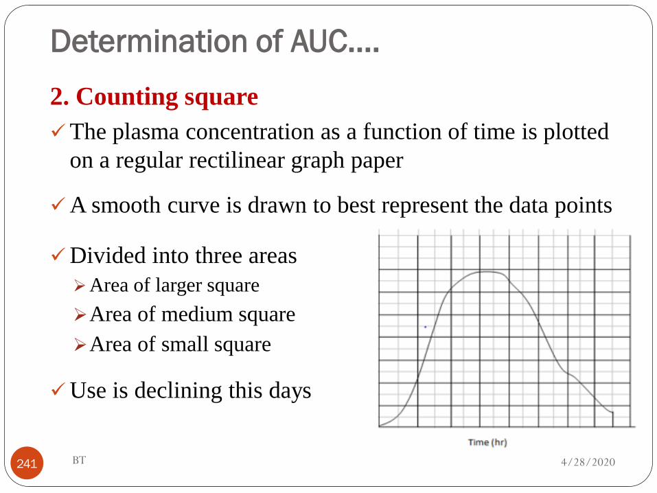
Determination of AUC….
2. Counting square
The plasma concentration as a function of time is plotted
on a regular rectilinear graph paper
A smooth curve is drawn to best represent the data points
Divided into three areas
Area of larger square
Area of medium square
Area of small square
Use is declining this days
4/28/2020BT241

Determination of AUC….
3. Trapezoidal method
Most accepted method-simple, least time consuming
4/28/2020BT242
AUC = ½ ( C1 + C2) (t2 – t1) + ½ (C2 + C3) (t3 – t2) +…….
½ (C n-1 + C n ) (tn – tn-1 )
where C = Concentration, t = time, n, subscript= sample number,
AUC = Area Under the Curve

Plasma Level-Time Curve….
E.g. Figure below shows the plasma profile of threeformulations (A, B and C) of the same dose of the same drug
BT243 4/28/2020

Plasma Level-Time Curve….
The area under the curves for formulations A and B are
similar indicating that the drug is absorbed to a similar
extent from these two formulations
The drug absorbed faster from formulation A than from
formulation B
Formulation A shows a fast onset of therapeutic action but its
peak plasma concentration exceeds the maximum safe
concentration
BT244 4/28/2020

Plasma Level-Time Curve….
The duration of action of the therapeutic effect obtained
with formulation B is longer than that obtained with A
Formulation C gives a much smaller area under the
plasma concentration-time curve, indicating that a lower
proportion of the dose has been absorbed
Slower rate of absorption from formulation C (the time of
peak concentration is longer than for formulations A and B)
BT245 4/28/2020

Plasma Level-Time Curve….
the peak plasma concentration not reaching the minimum
effective concentration
Therefore,
Formulation B appears to be superior to formulation A from a clinical
viewpoint, in that its peak plasma concentration lies within the
therapeutic range of the drug and the duration of the therapeutic effect
is longer
Formulation C does not produce a therapeutic effect and consequently
is clinically ineffective as a single dose
BT246 4/28/2020

Practice example
From the following blood data obtained after the oral
administration of 50mg of drug A. calculate the AUC? Identify
Cmax &Tmax
Time in hr Plasma drug con in µg/ml
1 5.5
2 9.2
3 14.9
4 10.3
5 7.1
6 2.2
247 BT 4/28/2020

Advantages & Disadvantages of Plasma
drug concentration
Advantages
the results are accurate, reliable, reproducible
Disadvantage
Invasive
4/28/2020BT248

Urinary drug excretion
The drug must be excreted in significant quantities as
unchanged drug in the urine
The rate and extent of drug excreted in the urine reflects
the rate and extent of systemic drug absorption
Urine samples are collected periodically after
administration of a drug product
4/28/2020BT249

Urinary drug excretion…
Each urine specimen is analyzed for free drug using a
specific assay
A graph is constructed that relates the cumulative drug
excreted to the collection-time interval
4/28/2020BT250

Figure: Corresponding plots relating the plasma
level–time curve and the cumulative urinary drug
excretion.
When the drug is almost
completely eliminated (point C)
the plasma concentration
approaches zero and
the maximum amount of drug
excreted in the urine, D ∞u, is
obtained
251
Urinary drug excretion…
4/28/2020BT

Urinary drug excretion…
Cumulative amount of drug excreted in the urine (Du∞)
Is related directly to the total amount of drug absorbed
The rate of drug excretion (dD u/dt)
the slope of the curve segment A-B is related to the
rate of drug absorption
4/28/2020BT252

dD u/dt - rate of drug excretion,…
Plot of dD u/dt vs. time is similar in shape as plasma
level–time curve for that drug
The maximum rate of drug excretion,
(dD u/dt)max is at point B
whereas the minimum rate of drug
excretion is at points A and C
4/28/2020BT253

Urinary drug excretion,…
The total time for the drug to be excreted (t∞)
Point C is related to the total time required after drug
administration for the drug to be systemically absorbed and
completely excreted
Urinary drug excretion is primarily useful for assessing
extent of drug absorption
Used in conjunction with blood level data for
confirmatory purposes
4/28/2020BT254

Urinary drug excretion,…
Advantages
It involves non invasive method of sampling
The concentration of the drug in the urine is often greater
than that in blood /serum allowing easy estimation of
drug
The amount of the drug excreted in urine is obtained
directly
4/28/2020BT255

Urinary drug excretion,…
Disadvantages
The metabolites of the drug are also concentrated in the
sample
May interfere with the estimation of the unchanged drug in
the urine sample
Urine samples should be collected for 10 half lives of the
drug to ensure a 99.9% of drug elimination
Presence of other routes of excretion
4/28/2020BT256

Clinical Observations
Demonstration of a clinically significant effect
Theoretically this method seems to be the best among
methods, but practically it is not
Clinical studies are complex, expensive, time-consuming and
require a sensitive and quantitative measure of the desired
response
Response is often quite variable, requiring a large test
population
4/28/2020BT257

Clinical Observations….Observed differences cannot be attributed only to
formulation
Clinical trials approach is least accurate, least sensitive, and
least reproducible of general approaches for determining in-vivo
bioavailability
Practical considerations, therefore, preclude the use of this
method except in initial stages of development while
proving the efficacy of a new chemical entity
FDA considers this approach only when analytical methods
are not available
4/28/2020BT258

In-Vitro Studies
If a correlation between some physicochemical property of a
dosage form and the biological availability of the drug from
that dosage form (in-vitro/in-vivo correlation) could be
established
it would be possible to use in-vitro data to predict a drug’s in-
vivo bioavailability
possible if in-vitro tests could reliably and accurately predict
drug absorption and reflect the in-vivo performance of a
drug in humans
o In-vitro–in-vivo correlation
4/28/2020BT259

In-Vitro Studies.…
Formulation that demonstrates most rapid rate of drug
dissolution in vitro have most rapid rate of drug
bioavailability in vivo
For highly permeable, rapidly dissolving, and orally
administered drug products, using an in vitro approach
(dissolution studies) is appropriate
4/28/2020BT260

In-Vitro Studies.…
Advantages
Reduce, or in some cases, completely eliminate the need
for bioavailability tests
Reduce cost and time
Solve safety issues
4/28/2020BT261

In-Vitro Studies.…
Disadvantage
Problems which make correlation with in- vivo availability
difficult:
Instrument variance
Difference between the in-vitro and in-vivo environments in
which dissolution occurs
Factors such as drug-drug interactions, age, food effects,
health, physical activity,..
Make in-vivo environment far more complex, variable, and
unpredictable 4/28/2020BT262

In vitro-in vivo correlation (IVIVC)
Refers to a predictive relationship of the in vitro properties of drug
substances or dosage forms with their in vivo performance
For orally administered drug products, it is usually a correlation
between the extent or rate of dissolution of a dosage
form and its pharmacokinetic parameters, such as
rate, duration, and extent of drug absorption
4/28/2020BT263

In vitro-in vivo correlation (IVIVC),…
The correlation established may be categorized into Levels
A, B, and C
Level A IVIVC
IVIVC is generally linear and represents a point-to-point
relationship between in vitro dissolution rate and in vivo input rate
(e.g., the in vivo dissolution rate of the drug from the dosage
form)
A Level A relationship may generate predictable plasma
profile, including Cmax, AUC, Tmax, shape of profile and
elimination half-life
4/28/2020BT264

In vitro and in vivo correlation…
Level B IVIVC
the mean in vitro dissolution time is compared to the
mean dissolution time in the gastrointestinal fluid
the entire plasma concentration profile cannot be
predicted
4/28/2020BT265

In vitro and in vivo correlation…
Level C IVIVC
establishes a single point relationship between a
dissolution parameter and a pharmacokinetic parameter
it does not reflect the complete shape of the plasma
concentration versus time curve
example of a Level C IVIVC is the establishment of the
correlation between amount of drug released at a certain
time point and Cmax
4/28/2020BT266

Absolute and Relative Bioavailability
Absolute bioavailability (F)
Absolute bioavailability is fraction of administered dose which
actually reaches systemic circulation
F is determined by comparing the respective AUCs of the test
product and the same dose of drug administered IV
F ranges from F = 0 (no drug absorption) to F = 1 (complete
absorption)
BT267 4/28/2020

Absolute and Relative…
IV is reference standard as dose is completely available, F = 1
Absolute bioavailability of drug is systemic availability
of drug after extravascular administration (e.g., oral,
rectal, transdermal, subcutaneous) compared to IV dose
BT268 4/28/2020

Absolute and Relative…
4/28/2020BT269
Practice example
If the AUC for an oral dose of a drug administered by tablet is
4.5 mg.hr/ml, and the intravenous dose is 11.2 mg.hr/ml,
calculate the bioavailability of the oral dose of the drug?
BT270
Absolute and Relative…
4/28/2020

Relative bioavailability
"Relative" or “Comparative” or “Apparent” bioavailability
refers to availability of drug product as compared to
recognized Standard
The relative bioavailability of product A compared to product B,
both products containing the same dose of the same drug, is
obtained by comparing their respective AUCs
BT271
Absolute and Relative…
4/28/2020

BT272
Absolute and Relative…
Where drug product B is reference/recognized standard
4/28/2020

BT273
Absolute and Relative… Practice examples
Assume that an intravenous injection (Product A) and two oral dosageforms (Product B and Product C), all containing the same dose of the samedrug, are given to a group of subjects in a crossover study. Suppose eachproduct gave the values for AUC indicated in the Table below:
Q. Calculate absolute Bioavailability for the oral dosage forms and relative bioavailability of Product C as compared to Product B
Drug Product AUC (mcg/ml) x hr
A Intravenous injection 100
B Oral dosage form, brand or reference standard
50
C Oral dosage form, generic Product 40
4/28/2020

BT274
Bioequivalence
4/28/2020

Bioequivalence…
Drugs are marketed from different sources:
Patented drug product (innovator or brand
product): produced by the first manufacturer who
has got a patent
Generic drug product: produced by different
manufacturer after the period of patent for the
innovator brand has ended
Generic products are cheaperBT275 4/28/2020

Bioequivalence…
Generic products to receive approval, the manufacturer
generally must establish that its product is therapeutically
equivalent with the innovator product
BT276 4/28/2020

Bioequivalence…
Equivalence: Relationship in terms of bioavailability,
therapeutic response, or a set of established standards of one
drug product to another
Bioequivalence is defined as: the absence of a significant
difference in the rate and extent of absorption of test
and standard dosage forms containing equal doses of the
same drug
4/28/2020BT277

Bioequivalence…
‘Two pharmaceutical products are considered to be
equivalent when their concentration vs. time profiles, from
the same molar dose, are so similar that they are unlikely to
produce clinically relevant differences in therapeutic
and/or adverse effects.’
Following parameters:
Maximum plasma concentration (C max)
Time to peak height concentration (Tmax)
Area under the plasma concentration-time curve (AUC)
4/28/2020BT278

Bioequivalence…
Bioequivalence is actually the comparison of the
bioavailability of two drug products
BT279
Fig. Mean concentration–time curves for two products of a drug after single oral doses
4/28/2020

Bioequivalence…
Only those products that have been proven to bebioequivalent should be used interchangeably
On scientific grounds there is no reason to be concernedabout substituting a generic product for a branded productthat is flagged as being bioequivalent
Products that are not bioequivalent should not besubstituted for each other Switching inequivalent products may lead to lower or higher
blood concentrations of a drug in a patient
This may increase the risk of therapeutic failure or drug-related toxicity
4/28/2020BT280

Bioequivalence…
Pharmaceutically equivalent:
Drug products that contain the same active ingredient(s),
(i.e, the same salt or ester) in the same dosage form, use
the same route of administration, and are identical in
strength or concentration
But may differ in characteristics such as shape, release
mechanisms, packaging, excipients (including colors, flavors,
preservatives), expiration time, and, within certain limits,
labeling
BT281 4/28/2020

Bioequivalence…
Ingredient Function Tablet A Tablet B Tablet C
Acetaminophen Drug 300 mg - 300 mg
Aspirin Drug - 300 mg -
Lactose Filler 100 mg 100 mg -
Avicel Filler - - 100 mg
Starch Disintegrant 50 mg 50 mg -
Avicel Disintegrant - - 50 mg
Mg stearate Lubricant 2 mg 2 mg 2 mg
Gelatin Binder 10 mg 10 mg 10 mg
BT282
Which of the following three tablet formulations are pharmaceutically equivalent?
4/28/2020

Bioequivalence…
Formulations A and B contain identical quantities of the same
inert ingredients, but different active ingredient
Not pharmaceutical equivalent
Formulations B and C contain different excipients and active
ingredients
Not pharmaceutical equivalent
Formulations A and C contain different excipients, but same
active ingredient
Pharmaceutical equivalent
BT283 4/28/2020
Bioequivalence…
Pharmaceutical Equivalence does not necessarily imply
bioequivalence. Why? b/s :
Excipients and/or the manufacturing process can lead to
faster or slower dissolution and/or absorption
• They may differ in release mechanisms
BT284 4/28/2020

Bioequivalence…
Pharmaceutical alternatives: drug products that contain the
same therapeutic moiety but are different salts, esters, or
complexes of that moiety, or are different dosage forms or
strengths
E.g
BT285
TTC hydrochloride, 250mg capsules TTC phosphate, 250mg capsules
Quinidine sulfate, 200mg tabletsQuinidine sulfate, 200mg capsules
4/28/2020

Bioequivalence…
BT286
Ingredient Tablet A Tablet B Capsule
C
Capsule
D
Suspension
E
Ibuprofen 200 mg - - - 200 mg
Aspirin - - 300 mg - -
Acetaminophen - 250mg - 250 mg -
Example 2: Which of the following five formulations arepharmaceutical alternatives?
4/28/2020

Bioequivalence…
Products A, B, and C; A,C, and D; B, C, and E; and C, D, and Econtain different drug entities
Therefore neither pharmaceutical equivalents not pharmaceuticalalternatives
Products A and E contain the same active ingredient in thesame amount
Pharmaceutical alternatives
Products B and D contain the same active ingredient in thesame amount
Pharmaceutical alternatives
BT287 4/28/2020

Bioequivalence…
Therapeutic equivalents:
Drug products are considered to be therapeutic equivalents only
if :
They are pharmaceutical equivalents and bioequivalent
Pharmaceutical equivalence + bioequivalence = therapeutic
equivalence
A generic drug product that is therapeutically equivalent may be
substituted for the innovator product (Generic substitution)
BT288 4/28/2020

Bioequivalence…
Assume
A drug substance is manufactured in a tablet and capsule dosage
form containing the same active ingredient
The two dosage forms provide the same extent and rate of drug
absorption
Is it possible to consider the two dosage forms therapeutically
equivalent?
BT289 4/28/2020

Bioequivalence…
The distinction between bioequivalence and therapeutic
equivalence is significant in that a tablet and a capsule
dosage form containing the same active ingredient can
(sometimes do) provide the same extent and rate of drug
absorption and thus could be considered bioequivalent but
these two products are not therapeutically equivalent
because they are different dosage forms
BT290 4/28/2020

Bioequivalence…
Therapeutic alternatives: Drug products
containing different active ingredients that are indicated
for the same therapeutic or clinical objectives
Active ingredients in therapeutic alternatives are
from the same pharmacologic class and
are expected to have the same therapeutic effect when
administered to patients for such condition of use
e.g:- Ibuprofen vs.Aspirin; Cimetidine vs. Ranitidine.
4/28/2020BT291

Bioequivalence studies
The significance of BE studies is that when two
pharmaceutically equivalent products are shown to be
bioequivalent, the two products are judged to be
therapeutically equivalent
Therapeutically equivalent products are expected to
have the same safety and efficacy profiles, when
administered under the conditions listed in the product
labeling
4/28/2020BT292
Bioequivalence studies…
This is the basis for the approval and use of generic drug
products
Bioequivalence studies requires determination of relative
bioavailability after administration of a single dose of test
and reference formulations by the same route, in equal
doses, but at different times
The reference product is generally a previously approved product,
usually innovator’s product or some suitable reference standard
BT293 4/28/2020

Bioequivalence studies…
Bioequivalence studies are conducted:
When a generic formulation is tested against an
innovator brand
Where a proposed dosage form is different from that
used in a pivotal clinical trial
When significant changes are made in the
manufacture of the marketed formulation
BT294 4/28/2020

Bioequivalence studies…
Subjects
The typical number of subjects is 24–36 with the minimum
number of subjects in the study being 12
Healthy subjects are recommended for BE studies for many
reasons:
Other drugs the patients may be taking, physiological
changes, etc. may modify drug absorption pattern
BT295 4/28/2020

Bioequivalence studies…
Strict study conditions such as fasting state are difficult for
patients
Patients require continuous treatment that does not allow
for a washout period
Patients are generally used only when drug is not safe to
administer in healthy subjects
20-50 years of age and 120-200 lb of body weight
BT296 4/28/2020
Bioequivalence studies…
Males are preferred
Menstrual cycle, pregnancy, lactation and menopause
stages may affect blood level profiles of drug
• Females are included taking these points into
consideration
Subjects are distributed randomly to different groups to
achieve uniform distribution with respect to age, sex, and
body weight to avoid bias
BT297 4/28/2020

Bioequivalence studies…
Study design
Parallel Design and Cross-over Design
Parallel Design
Subjects are divided randomly into groups, each group
receiving only one treatment (take either the test or the
reference product)
Two formulations are administered to two groups of
volunteers
BT298 4/28/2020

Bioequivalence studies…
Parallel Design…..
Separate groups of subjects receive the test and reference
products
Formulations are administered randomly to volunteers to
avoid bias
Recommended when the half life of the drug is so long (b/s
each subject take only one treatment only once: no washout
period, no carryover effect)
BT299 4/28/2020

Bioequivalence studies…
Parallel Design…
Disadvantage:
Inter-subject variation isn’t addressed
Require larger numbers of subjects
BT300 4/28/2020

Bioequivalence studies
Cross-over Design
Each subject receives two or more different
treatments on successive occasions
Minimizes inter-subject variability
Each subject serves as his own control (i.e., we can
compare T to R on each subject)
BT301 4/28/2020

Waivers of In-Vivo Bioequivalence
Studies (Biowaivers)
A Biowaiver means that in vivo bioavailability and/or
bioequivalence studies may be waived
Instead of conducting expensive and time consuming in vivo
studies, a dissolution test could be adopted as the surrogate
basis for the decision as to whether the two pharmaceutical
products are equivalent
BT302 4/28/2020
Waivers of In-Vivo...
For some drug products, there is sufficient understanding of
the physicochemical properties and biological factors that
affect BA that there is no need for in vivo BE studies
in-vitro dissolution testing may be used in lieu of in-vivo
bioequivalence studies
Sponsors may request waivers of BE studies (biowaivers) for solutions, products with a range of strengths, and biopharmaceutical classification system (BCS) Class I drugs
4/28/2020BT303

Waivers of In-Vivo...
Solutions: In vivo BA/BE is self-evident for certain drug
products, such as topical solutions, solution nasal spray, oral
solutions, elixirs, syrups, tinctures, or other solubilized forms
of the drug
In vivo BA/BE can be waived, assuming that release of the
drug substance from the drug product is self-evident and
that the solutions do not contain any excipient that
significantly affects drug absorption
BT304 4/28/2020

Waivers of In-Vivo...
Lower Strengths: Waiver of in vivo studies for different
strengths of a drug product can be granted when:
The drug product is in the same dosage form, but in a
different strength (e.g., a tablet of 100mg, 50mg, 25mg)
This different strength is proportionally similar in its active
and inactive ingredients to the strength of the product for
which the same manufacturer has conducted an
appropriate in vivo study; and
BT305 4/28/2020

Waivers of In-Vivo...
The new strength meets an appropriate in vitrodissolution test
E.g. Immediate-release tablet available in 200mg, 100mg
and 50mg strengths
100mg and 50mg tablets are made in same way as200mg
Human bioequivalence study is performed on highestor 200mg strength
Comparative in-vitro dissolution studies are performedon 100-mg and 50-mg dose strengths and meetrequirements
BT306 4/28/2020

Waivers of In-Vivo...
If products have no known bioavailability problems, are well
absorbed systemically, are well correlated with in-vitro
dissolution, and have large margin of safety
Manufacturer needn’t perform additional in-vivo
bioequivalence studies on lower-strength products if they
meet all in-vitro criteria
BT307 4/28/2020

Biopharmaceutical Classification
System (BCS)
A scientific framework that predicts the in vivo absorption of
drugs from aqueous solubility and intestinal permeability
data
A drug substance is considered highly soluble when the
highest dose strength is soluble in 250 ml or less of aqueous
media over the pH range of 1.0–7.5
Otherwise, the drug substance is considered poorly
soluble
BT308 4/28/2020

Biopharmaceutical Classification…
BT309
(The volume estimate of 250 ml is derived from the
minimum volume anticipated in the stomach when a dosage
form is taken in the fasted state with a glass of water
4/28/2020

Biopharmaceutical Classification…
A drug substance is considered to be highly permeable when
gastrointestinal absorption in humans is determined to be
90% or more of an administered dose
Otherwise, the drug substance is considered to be poorly
permeable
BT310 4/28/2020

Biopharmaceutical Classification…
According to BCS, drug substances are classified as:
Class I: High Solubility – High Permeability E.g. verapamil,
propranolol, Metoprolol
Class II: Low Solubility – High Permeability E.g. Ketoprofen,
naproxen, glibenclamide
Class III: High Solubility – Low Permeability E.g. Cimetidine,
ranitidine, atenolol
Class IV: Low Solubility – Low Permeability E.g.
Hydrochlorothiazide, furosemide
BT311 4/28/2020

Biopharmaceutical Classification…
BT312
Table: Biopharmaceutical Classification System
4/28/2020

Biopharmaceutical Classification…
Limitation of this classification method
This approach assumes that no other components in the
formulation affect the membrane permeability and/or
intestinal transport.
BT313 4/28/2020

May, 2020
Clinical pharmacokinetics

Session objectives
Upon completion of this session, you will have the
ability to:
Define pharmacokinetics
Describe applications of Pharmacokinetic
Describe pharmacokinetic and Pharmacodynamic
relationships
Review ADME
Describe pharmacokinetic models315

Pharmacokinetics
Pharmaco-comes from the Greek word for “drug,”
pharmackon, and kinetics comes from the Greek
word for “moving” kinetikos
Pharmacokinetics (PK) is the study of drug
movement into, around, and out of the body
The study of kinetics (time course) of drug’s
ADME (Absorption, Distribution, Metabolism, and
Elimination)
316

Pharmacokinetics
It also concerns the relationship of these processes to
pharmacologic effects (therapeutic or toxic) of drug
in man and animals
“What the body does to the drug”
317

Cont…
318

Cont…
319

Pharmacokinetic Studies
There are two approaches of pharmacokinetic studies;
Experimental approach
Theoretical approach
Experimental aspect
Involves development of biological sampling techniques
Procedures that facilitate data collection and manipulation
Analytical methods for measurement of drug and
metabolites concentration in biological samples320

Cont…
Theoretical aspect
Involves development of pharmacokinetic models to
predict drug disposition after its administration
The application of statistics is an integral part of
pharmacokinetic studies
321

Cont…
Clinical pharmacokinetics
Is the discipline that applies pharmacokinetic
principles in humans in order to design
individualized dosage regimens
Optimize the therapeutic response of a medication
Minimizing the chance of an adverse drug reaction
322

Applications of pharmacokinetic studies
Bioavailability measurements
Effects of physiological and pathological conditions on drug disposition and absorption
Dosage adjustment of drugs in disease states, if and when necessary
Correlation of pharmacological responses with administered doses
Clinical prediction: using pharmacokinetic parameters to individualize the drug dosing regimen and thus
provide the most effective drug therapy323

Pharmacodynamics
Is the study concerned with the relationship between
drug concentration at the site of action and
pharmacological response (therapeutic and/or toxic
effect)
“Drug effect on the body”
The change in drug effect is usually not proportional to
the change in drug dose or concentration
324

Cont…
325

Relationship b/n the administered dose
and amount of drug in the body
Only that fraction of the administered dose which
actually reaches the systemic circulation will be
available to elicit a pharmacological effect
For an intravenous solution
the amount of drug that reaches general
circulation is the dose administered
326

Cont…
Where;
oAUC is the area under curve of plasma drug
concentration versus time (AUC) from time
zero to time infinity;
oK is the first-order elimination rate constant
oV (or Vd) is the drug’s volume of distribution
327

Cont…
Volume of distribution
may be thought of as the apparent volume into
which a given mass of drug would need to be
diluted in order to give the observed
concentration
For the extravascular route,
the amount of drug that reaches general
circulation is the product of the bioavailable
fraction (F) and the dose administered
328

Cont…
Equation suggest that we must know or determine
all the parameters (i.e. AUC, K, V, F) for a given
drug;
329

Cont…
Onset of action:
The time at which the administered drug reaches
the therapeutic range and begins to produce the
effect
Termination of action:
The time at which the drug concentration in the
plasma falls below the minimum effective
concentration (MEC)
330

Cont…
Duration of action:
The time span from the beginning of the onset of
action up to the termination of action
Therapeutic range:
The plasma or serum concentration (e.g. μg/mL)
range within which the drug is likely to produce the
therapeutic activity or effect
331

Cont…
332
Fig. Typical plasma concentration versus time profile following
administration of a drug by an extravascular route (MTC: min.
toxic concentration; MEC: min. effective concentration)

Routes of drug administration
Routes of drug administration are classified into
two categories:
intravascular routes
extravascular routes
Intravascular routes
Intravascular administration can be intravenous or
intra-arterial
333

Cont…
Important features of the intravenous route of drug
administration
There is no absorption phase
There is immediate onset of action
The entire administered dose is available to produce
pharmacological effects
Used more often in life-threatening situations
Adverse reactions are difficult to reverse or control
334

Cont…
Fig. Typical plasma concentration versus time profile
following administration of a drug by an
intravascular route335

Cont…
Extravascular routes of drug administration
Includes all routes of administration other than iv
route
Important features of extravascular routes of drug
administration
An absorption phase is present
The entire administered dose of a drug may not
always reach the general circulation (i.e.
incomplete absorption)336

Cont…
The onset of action is determined by factors such
as;
Formulation and type of dosage form
Route of administration
Physicochemical properties of drugs and other
physiological variables
337
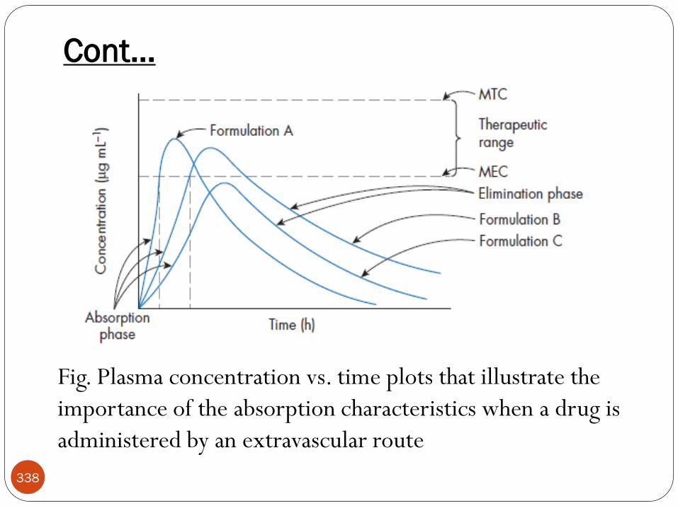
Cont…
Fig. Plasma concentration vs. time plots that illustrate the
importance of the absorption characteristics when a drug is
administered by an extravascular route
338

Review of ADME processes
Drug absorption
Is the process of movement of drug from the site of
administration into the site of measurement (usually
blood, plasma or serum)
In order to be absorbed,
the drug molecules must pass through several
physiological barriers before reaching the vascular
system
339

Cont…
Drug Distribution
The process of reversible transfer of drug to and from
the site of measurement (usually blood, plasma or
serum)
Any drug that leaves the site of measurement and
does not return has undergone elimination
340

Cont…
The rate and extent of drug distribution is
determined by:
how well the tissues and/or organs are perfused
with blood
the binding of drug to plasma proteins and tissue
components
the permeability of tissue membranes to the drug
molecule
341

Cont…
Elimination
The irreversible loss of drug from the site of
measurement
Elimination of drugs occur by one or both of
metabolism or excretion
Metabolism of drugs
The process of conversion of one chemical species
to another chemical species
Usually, metabolites will possess little or none of the
activity of the parent drug342

Cont…
Excretion
Defined as the process whereby drugs and/or their
metabolites are irreversibly transferred from internal
to external environment
The two principal organs responsible for drug elimination
are the kidney and the liver
The kidney is the primary site for removal of a drug in a
chemically unaltered or unchanged form (i.e. excretion)
as well as for metabolites
343
Cont…
Liver is the primary organ where drug metabolism
occurs
Occasionally, lungs may be an important route of
elimination for substances of high vapor pressure
(i.e., gaseous anesthetics, alcohol, etc)
Another potential route of drug removal is a
mother’s milk
Although not a significant route for elimination of a
drug for the mother, the drug may be consumed in
sufficient quantity to affect the infant344

Cont…
Disposition
Is defined as all the processes that occur subsequent
to the absorption of the drug
Is the description of drug distribution and
elimination, i.e., process after absorption
Once a drug is in the systemic circulation, it is
distributed simultaneously to all tissues including
the organ responsible for its elimination
345

Cont…
The distinction between elimination and
distribution is often difficult
When such a distinction is either not desired or is
difficult to obtain, disposition is the term used
Accumulation
Is the increase in drug concentration in blood and
tissue upon multiple dosing until steady state is
reached
346

Cont…
Steady state
Is a level of drug accumulation in blood or tissue upon
multiple dosing when input and output are at equilibrium
The steady state drug concentrations fluctuate
between a maximum and a minimum steady state
concentration within each dosing interval347

Cont…
Biophase
Is the actual site of action of drugs in the body
The biophase may be the surface of a cell or within
the cell, i.e., one of the organelles
Receptor
Is the site in the biophase to which drug molecules
can be bound
A receptor is usually a protein or proteinaceous
material348

Cont…
349
Dosage regimen or dose rate
Is the systematized dosage schedule for therapy,
i.e., the proper dose sizes and proper dosing intervals
required to produce clinical effectiveness or to
maintain a therapeutic concentration in the body
Loading dose, priming dose or initial dose
Is the dose size used in initiating therapy so as to yield
therapeutic concentrations which will result in clinical
effectiveness

Cont…
Maintenance dose
Is the dose required to maintain the clinical effectiveness
or therapeutic concentration according to the dosage
regimen
350
Rate Processes
After a drug is administered, it is subjected to a
number of processes (ADME)
the rates of these process control the concentration of
drug in the ‘‘site of action’’
These processes affect the onset of action, as well as
the duration and intensity of pharmacological
response
351

Cont…
After administration of a drug, there will be change
in the mass or concentration of drug in the plasma
with time:
• Where dC/dt is the instantaneous rate of change in
function C with respect to an infinitesimal time
interval (dt)352
12
12/tt
CCdtdC

Order of a process
In the equation:
The numerical value (n) of the exponent of the
substance (C) undergoing the change is the order of
the process
Types of orders process encountered in science
include: zero order, first order, second order, etc
Zero- and first-order are most useful for
pharmacokinetics353

Zero-order process
n = 0, the equation becomes:
C Product
• Where Ko is the zero-order rate constant and the minus
sign shows negative change over time (elimination)
This equation clearly indicates that C changes at a
constant rate, since Ko is a constant
This means that the change in C must be a
function of factors other than the amount of C
present at a given time354

Cont…
The integration of the Eqn. yields the following:
• where C is amount present at time t and Co is
amount at time zero
355

Cont…
Applications of zero-order processes
Zero-order processes are employed in;
Administration of a drug as a constant rate i.v.
infusion
Formulation and administration of controlled
release DFs
Administration of drugs through transdermal drug
delivery systems, drug elimination
356

Cont…
Unit of the rate constant (Ko) for zero order
elimination of drug
357

Zero order Half life
Half life (t1/2) or half time is defined as the time
period required for the concentration of drug to
decrease by one half
When t = t1/2, C = C0/2 and the equation becomes:
358

First-order process
n = 1, equation becomes:
• where K is the first-order rate constant.
• Up on integration of the above Eqn., we obtain:
Or
359
C = C0 e-Kt
ln C = ln C0 – Kt

Cont…
360

Cont…
In first order process, the amount of drug
eliminated per time is directly proportional to the
amount of drug in the body
First-order elimination is extremely important in
pharmacokinetics
since the majority of therapeutic drugs are
eliminated by this process
361

First order Half life
Substituting the value of C = C0/2 at t1/2 in first order
equation and solving it yields:
The half life of a first order process is a constant and
independent of initial drug concentration
i.e. irrespective of what the initial drug concentration is the
time required for the concentration to decrease by one half
remains the same
362

Comparison b/n Zero & First order
First Order Elimination
Rate = k C
[drug] decreases
exponentially w/ time
Rate of elimination is
proportional to [drug]
Plot of log [drug] or
ln[drug] vs. time are
linear
t 1/2 is constant
regardless of [drug]
Zero Order Elimination
Rate = k
[drug] decreases
linearly with time
Rate of elimination is
constant
Rate of elimination is
independent of [drug]
t 1/2 is not constant
363

Problems
1. The plasma drug concentration of gentamicin was 8 mg/L
and the patient's elimination rate constant is 0.25 hr-1.
What will be the concentration after 8 hours?
What will be the elimination half life?
(assuming first order)
364

Cont…2. A pharmacist weighs exactly few grams of a drug and dissolves
it in 100 mL of water. The solution is kept at room temperature,
and samples are removed periodically and assayed for the drug.
The pharmacist obtains the following data:
365
Drug Concentration (mg/mL) Time (hr)
95 2
90 4
85 6
80 8
75 10
70 12

Cont…
a. Is the decomposition of this drug zero or first order?
b What is the rate of decomposition of the drug?
c. How many grams of drug were in the solution
prepared by the pharmacist?
366

Problems
3. Plot the following data on both semi-logarithmic paper
and normal rectilinear paper.
a. Does the decrease in the amount if drug appear to be first
order or zero order?
b. What is the rate constant?
c. What is the half-life?
367
Time Drug (mg) Time Drug (mg)
10 96 90 34
20 89 120 10
40 73 130 25
60 57

Pharmacokinetic models
The handling of a drug by the body can be very complex, as
several processes (such as ADME) work to alter drug
concentrations in tissues and fluids
Simplifications of body processes are necessary to predict a
drug's behavior in the body
Application of mathematical principles to the various
processes
• A model of the body must be selected
368

Cont…
To understand the movement of drug in biological
system pharmacokinetic models come under
consideration
Various mathematical models can be devised to simulate
the rate processes of drug absorption, distribution,
metabolism and elimination
369

Cont…
These mathematical models are useful in the
development of equations
to describe drug concentrations in the body as a
function of time
Because amount of drug in the body is dependent on
the two variables drug concentration and time
370

Cont…
371
In practice, pharmacokinetic parameters are
not measured directly
but are determined experimentally from a set of
dependent and independent variables collectively
known as data
From these data, a pharmacokinetic model is
estimated and the pharmacokinetic parameters are
obtained

Cont…
Pharmacokinetic models are divided into
Compartment model
Physiologic model (flow model)
Non-compartmental model
The most useful model is the compartment
model
372

Compartment Model
The living system is conceived to be composed of a
number of mathematically interconnected
compartments
A compartment is a group of tissues which behaves
uniformly with respect to the drug movement
The compartments do not represent a specific tissue or fluid but may represent a group of similar tissues or fluids
Organs and tissues in which drug distribution issimilar are grouped into one compartment
373

Compartment model…
For example, distribution into muscle and adipose
tissue differs from distribution into renal tissue for
most drugs
The highly perfused organs (e.g., heart, liver, and
kidneys) often have similar drug distribution
patterns, so these areas may be considered as one
compartment
374

Characteristics of Pharmacokinetic
compartment
Pharmacokinetic compartments are not real physiological or
anatomical regions
All the compartments may be assumed as various organs or
tissues that altogether handle a drug like a homogenous
compartments
Grouping of a set of tissues in a compartment is based on
similar blood flow and drug affinity
375

Characteristics…
Compartments are interconnected by first-order rate
constants. Input rate constants may be zero order
In each compartment drug is considered to be rapidly and
uniformly distributed
The rate constants are used to represents the overall rate
processes of the drug entry into and exit from the
compartments
376
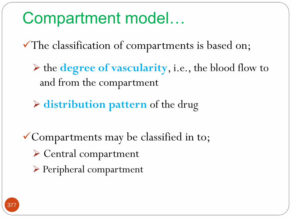
Compartment model…
The classification of compartments is based on;
the degree of vascularity, i.e., the blood flow to
and from the compartment
distribution pattern of the drug
Compartments may be classified in to;
Central compartment
Peripheral compartment
377

Compartment model…
Central compartment
Is the sum of all body regions (organs and tissues)
in which the drug concentration is in instantaneous
equilibrium with that in blood or plasma
Drugs in circulating fluids (plasma) and rapidly
perfused tissues (e.g., heart, liver, kidney, lungs)
are assigned to the central compartment
378

Compartment model…
Peripheral compartment
Is the sum of all body regions (organs and tissues)
to which a drug eventually distributes, but not in
instantaneous equilibrium
Drugs in fluids of distribution (i.e., lymph and
extracellular water) and poorly perfused tissues
(e.g., large muscle mass and skin) are assigned to
the peripheral compartment
379

Compartment model…
There are three types of compartment models in
pharmacokinetics:
One-compartment model
Two-compartment model
Multicompartment model
380

One Compartment Model
The body is viewed as a single compartment
Considers the entire body, and all of the organs and
tissues to be one giant bucket
Drug is rapidly distributed following its
administration,
Rate of transfer of drug from blood to all organ
and tissues and back into blood have become
equal instantaneously381

Compartment model…
All organs and tissues behave in similar fashion
toward the administered drug
One-compartment model will be applied to
characterize the plasma concentration versus time
data
382

Compartment model…
383

Compartment model…Drug enters the central compartment from somewhere
outside of the body
Drug then leaves the central compartment
This is analogous to the drug leaving the body
Drug recirculation does not occur (output line does not
reconnect with input line)
384

Two Compartment Model
Considers the entire body, and all of the organs and tissues
to be two buckets
The body is viewed as two compartments
The central compartments
The peripheral compartments
Central compartment consists of the plasma and tissues that
take up the drug so rapidly that distribution can be
considered to be instantaneous.
Peripheral compartment consists of tissues that take up the
drug at slower rate than tissues in the central compartment
385

Compartment model…
The slower the drug distribution in the body,
The distribution equilibrium (b/n blood to various
organs and tissues) is attained slowly and at a finite
time
The vasculature, tissues and organs are not
behaving in a similar fashion towards the drug
We consider the body to comprise two
compartments or, if necessary, more than two
compartments
386

Compartment model…
387

Compartment model…Drug enters the central compartment from somewhere
outside of the body
Drug then leaves the central compartment by one of two
paths:
the peripheral compartment (also called compartment 2) or
drug leaves the body
Drug that is in the peripheral compartment can return to
the central compartment
All drug must leave the body through a single bucket
388

Compartment model…
Three compartment model
An extension of the two compartment model, where a
sizable amount of the drug distributes to certain very
poorly perfused tissues, such as fat and bone, at an
extremely slow rate
The three compartment model has three group of tissues:
389

Compartment model…
Central compartment tissues: take up the drug very
rapidly
Peripheral tissues: compartment take the drug more
slowly
Deep tissue compartment: take the drug at an
extremely slow rate
390
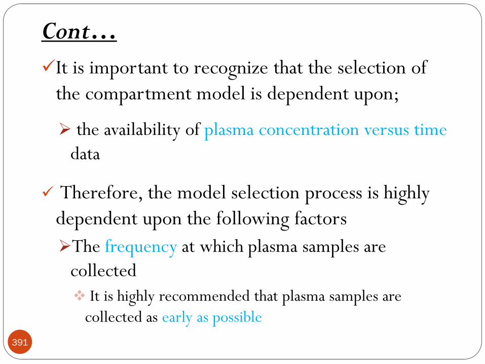
Cont…
It is important to recognize that the selection of
the compartment model is dependent upon;
the availability of plasma concentration versus time
data
Therefore, the model selection process is highly
dependent upon the following factors
The frequency at which plasma samples are
collected
It is highly recommended that plasma samples are
collected as early as possible
391

Cont…
The sensitivity of the procedure employed to
analyze drug concentration in plasma samples
The physicochemical properties (e.g., the
lipophilicity) of a drug
The equation required to characterize the plasma
concentration versus time data depends upon;
the compartment model chosen and
the route of drug administration
392

Cont…
The plasma concentration versus time data permits
us to obtain accurate estimates of selected
fundamental pharmacokinetics parameters such as;
the apparent volume of drug distribution
the elimination half life and
the elimination rate constant of a drug
393

Cont…
These parameters allow for the calculation of;
the dosage regimen (dose and dosing interval) that
will provide the desired plasma concentration and
duration of action for an administered drug
394

Cont…
Compartment models are subdivided into:
Mammillary model
Caternary model
395

Mammillary model
Arrangement of compartments in a manner similar
to connection of satellites to a planet
The model consists of one or more peripheral
compartments connected to a central compartment
Central compartment (compartment 1)
Plasma & highly perfused tissues (lung, liver, kidney)
Peripheral compartment (denoted by no 2, 3, etc)
Other organ
396
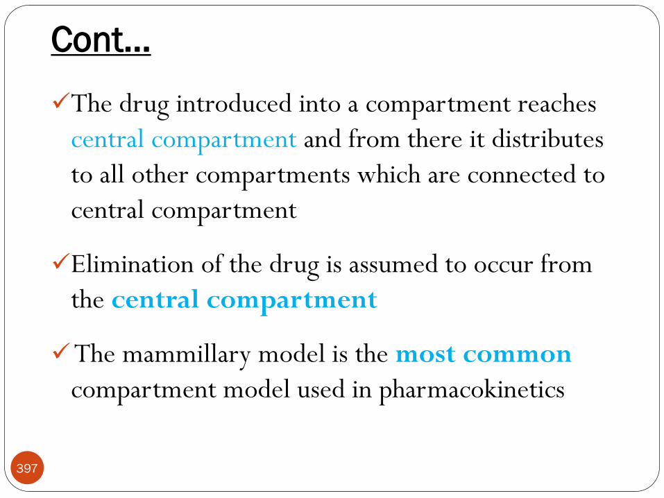
Cont…
The drug introduced into a compartment reaches
central compartment and from there it distributes
to all other compartments which are connected to
central compartment
Elimination of the drug is assumed to occur from
the central compartment
The mammillary model is the most common
compartment model used in pharmacokinetics
397

Cont…
Movement of drug between compartments is
defined by characteristic first order rate constant
denoted by letter K, the number represents the
direction of drug movement between the
compartments
The number of rate constant in a particular
compartment model is given by R
For IV administration R = 2n-1
For extravascular administration R = 2n
Where n= number of compartment398

Cont…
The figures below shows several types of
compartment models
Model 1. One-compartment open model, IV
injection
Model 2. One-compartment open model with first-
order absorption
399

Cont…
Model 3. Two-compartment open model, IV
injection
Model 4. Two-compartment open model with first
order absorption
400

Cont…
Model 5. Multi-compartment open model, IV
injection
401

Cont…
The drawing of models
Gives a visual representation of the rate processes
involved,
Shows how many pharmacokinetic rate constants are
necessary to describe the process adequately, and
Enables the pharmacokineticist to develop
differential equations to describe drug concentration
changes in each compartment
402

Cont…
If the amount of drug absorbed and eliminated per
unit time is obtained by sampling compartment 1,
Then the amount of the drug contained in the tissue
compartments can be estimated mathematically
For this purpose, appropriate mathematical equations
for describing these models and evaluating the
various pharmacokinetic parameters are to be used
403

Cont…
Caternary Models
The caternary model consists of compartments joined to
one another in a series like the compartments of a train
Since most of the functional organs of the body are directly
connected to the plasma,
the caternary model is not used as often as the mammillary
model404

Advantages of compartment modeling
It is simple and flexible approach
It gives a visual representation of various rate
processes involved in drug disposition
it is important in development of dose regimens
Used for comparison of multiple therapeutic
agents
It show how many rate constant required for
describing these process
405

Disadvantages of compartment modeling
o The compartment and parameters have no relationship
with physiological functions or anatomical structure of the
species
o This approach is limited to specific drug only
o Difficulties generally arises when using model to interpret
the differences b/n results from human and animal
experiments
406
Physiologic Pharmacokinetic Model
Also known as blood flow or perfusion models
Are pharmacokinetic models based on known
anatomic and physiologic data
The rate of drug carried to a tissue/organ or tissue
drug uptake is dependent upon two major factors:
Rate of blood flow to the organ
Tissue/Blood partition coefficient or diffusion
coefficient of drug
407

Cont…
In contrast to the tissue volume of distribution, the
actual tissue volume is used
Because there are many tissues/ organs in the body,
each tissue volume must be estimated and its drug
concentration described
The model would potentially predict a
realistic tissue drug concentration, which a
compartment model fails to do so
408

Cont…
An example of the perfusion model is shown below:
409

Cont…
To describe each organ separately with a differential
equation would make the model very complex and
mathematically difficult
A simpler but equally good approach is to group all
the tissues with similar blood perfusion properties
into a single entity as;
RET (rapidly equilibrating tissue) and
SET (slowly equilibrating tissue)
410

Cont…
The number of tissue compartments in a perfusion
model varies from drug to drug
Tissues or organs that have no drug penetration are
excluded from consideration
Such organs as the bones, teeth and others
411

Cont…
The information that is required for adequately
describing a physiologic model is experimentally
difficult to obtain
Physiologic pharmacokinetic model does provide a
much better insight of how physiologic factors may
change drug distribution from one animal species to
another
412

Cont…
Physiologically based pharmacokinetic models can
be applied to several species,
For some drugs human data may be extrapolated
Extrapolation is not possible with compartment
models
Numerous drugs (including digoxin, lidocaine,
methotrexate, and thiopental) have been described
with perfusion models
413

Advantages of physiologic modeling
Mathematical treatment is straightforward
Data fitting is not required
Exact description of drug concentration-time
profile in any organ or tissue can be obtained
The influence of altered physiology or pathology on
drug disposition can be easily predicted from
changes in various pharmacokinetic parameter
414

Disadvantages of physiologic modeling
oObtaining experimental data, which is very
exhaustive
o Prediction of individualized dosing is difficult
415

Reading assignment on
o Non-compartmental Model
416

June, 2020
Drug Distribution &
Drug Elimination

Session objectives
After this session you will be able to:
Describe drug distribution (Tissue permeability,
distribution co-efficient, binding of drugs,
volume of distribution)
Discuss drug elimination (drug metabolism,
renal and extra-renal excretion, concept of
clearance)
418

419
Drug Distribution

Drug distribution
Reversible transfer of drug from one location to another within the body
Transfer of drug between blood/plasma and tissues/ organ/ body fluids
420

Drug distribution…
Figure: Major water volumes in average 70- Kg human421

Drug distribution…
422

Factors affecting drug distribution
Rate of distribution Membrane permeability
Blood perfusion
Extent of Distribution Lipid Solubility
pH - pKa
Plasma protein binding
Intracellular binding
423

Rate of distribution
Can be:
424
Rate of distribution…
Perfusion limited
Prevails when the tissue membranes present essentially no barrier to distribution
This condition is likely to be met by small lipophilic drugs
Distribution throughout the body occurs mainly by
blood flow through organs and tissues
425

Rate of distribution…
Distribution of drugs dramatically affected if the
rate of blood flow to the tissue is altered
Perfusion is usually expressed in units of milliliters
of blood per minute per volume of tissue
(ml/min/ml of tissue)
Well-perfused tissues take up a drug much more
rapidly than do poorly perfused tissues
426

Rate of distribution…Table: perfusion rate, cardiac out put under basal conditions
in a standard 70-Kg human
Organ Perfusion Rate
(ml/min/ml of tissue)
% of cardiac output
Lung 10 100
Kidneys 4.0 22
Liver 0.8 27
Heart 0.6 4
Brain 0.5 14
Fat 0.03 4
Muscle 0.025 15
Skin 0.024 6
Bone 0.02 5427

Rate of distribution…
Total blood flow is greatest to brain, kidneys, liver,
and muscle
Highest perfusion rates to lung, brain, kidney, liver,
and heart
It would be expected that total drug concentration
would rise most rapidly in these organs
428
Rate of distribution…
For example, thiopental gets into the brain faster
than muscle
Thiopental is only partly ionized and passes into
the brain or muscle easily
Since brain has a higher perfusion rate the
thiopental can transfer in and out more quickly
Perfusion limited distribution
429

Rate of distribution…
If the blood flow rate increases, the distribution of
the drug to the tissue increases
Therefore, drugs apparently distribute more
rapidly to areas with higher blood flow
Highly perfused organs rapidly attain drug
concentrations approaching those in the plasma;
Less well-perfused tissues take more time to
attain such concentrations
430

Rate of distribution…
After a drug begins to distribute to tissue, the
concentration in tissue increases until it reaches an
equilibrium
the amounts of drug entering and leaving the tissue
are the same
The drug concentration in a tissue at equilibrium
depends on;
the plasma drug concentration and
the rate at which drug distributes into that tissue
431

Rate of distribution…
In highly perfused organs, such as the liver, the
distribution rate is relatively high; for most agents,
the drug in that tissue rapidly equilibrates with the
drug in plasma
For tissues in which the distribution rate is lower
(e.g., fat), reaching equilibrium may take much
longer
432

Rate of distribution…
Figure: Shows blood perfusing a tissue in which distribution is
perfusion rate-limited and no elimination occurs
KP, is the equilibrium distribution ratio (CT/CV)
ACQtationdrugpresenofRate .
VA CCQuptakeofrateNet .
VPT CKVtissueindrugofAmount ..
433

Rate of distribution…
The fractional rate of exit, KT
Where Q/VT is the perfusion rate of the tissue
The parameter KT , a distribution rate constant
with units of reciprocal time, may be regarded as a
measure of how rapidly drug would leave the tissue
P
T
VPT
VT
K
VQ
CKV
CQ
tissueinamount
exitofRateK
..
.
434

Rate of distribution…
The kinetics of tissue distribution can be
characterized by a tissue distribution half-life
If the approach towards equilibrium and the loss of
drug from a tissue take longer
the poorer the perfusion
the greater the partitioning of drug into a tissue
T
P
TV
Q
K
Klifehalf
693.0693.0
435

Rate of distribution…
Permeability limited
Drugs diffusing slowly across the capillary
endothelium
Changes in blood flow will not significantly affect
distribution to that tissue
Tissue permeability is influenced by the;
Lipid – water partition coefficient
Degree of ionization
drug's pKa and the pH of the blood 436

Rate of distribution…
Capillary walls are quite permeable
Lipid soluble drugs pass through very rapidly
Water soluble compounds penetrate more slowly
at a rate more dependent on their size
Permeability limited distribution arises particularly for
polar drugs diffusing across tightly interlocked lipoidal
membranes
blood-to-brain barrier
blood-to-cerebrospinal fluid barrier
437

Rate of distribution…
For example, penicillin gets into muscle more
quickly than it gets into brain
Penicillin is quite polar and is thus slowly permeable
Thus transfer of penicillin is faster in muscle than
brain as muscle capillaries are less restrictive
Permeability limited transfer
438

Extent of distribution
Apparent Volume of Distribution
At equilibrium, the extent of distribution is defined
by an apparent volume of distribution (V):
Apparent Volume of distribution is defined as the
volume that would accommodate all the drugs in
the body
C
A
ionconcentratdrugplasma
mequilibriuatbodyinAmountV
439
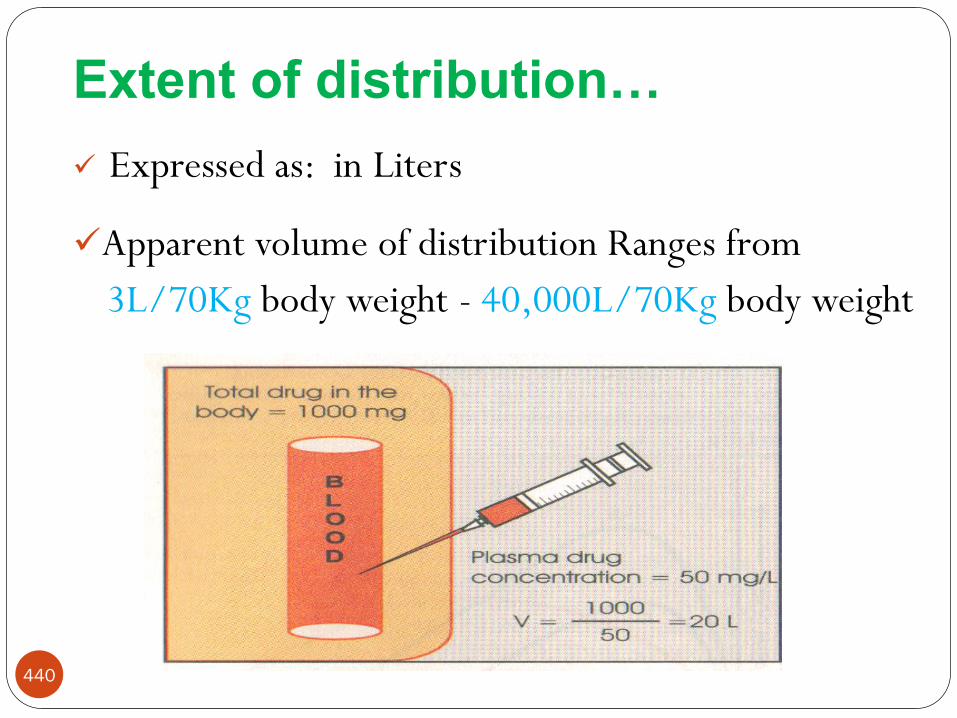
Extent of distribution…
Expressed as: in Liters
Apparent volume of distribution Ranges from
3L/70Kg body weight - 40,000L/70Kg body weight
440

Extent of distribution…
A large volume of distribution usually indicates that
the drug distributes extensively into body tissues
and fluids
Conversely, a small volume of distribution often
indicates limited drug distribution
441

Extent of distribution…
The larger the volume of distribution, the smaller
is the fraction in plasma
The remaining fraction given by
V
VVplasmaoutsidebodytheindrugofFraction P
V
VplasmainbodyindrugofFraction P
442

Extent of distribution…
At distribution equilibrium
, and dividing by C
CKVCVA PTP ...
CVA .
443

Extent of distribution…
Some tissues – high Kp which explains why the
volume of distribution of some drugs can be much
greater than total body size
Fat, for example, occupies approximately 20% of
body volume. If the Kp value in fat is 5, then 100%
V
PTP KVVV .
444

Extent of distribution…
Factors affecting extent of distribution
Binding within Blood
Within blood, drug can bind to many components
including blood cells and plasma proteins
the concentration of drug in whole blood (Cb), in
plasma (C) and unbound in plasma water (Cu) can
differ greatly
plasma (C) - most common fluid analyze
ease of chemical analysis
445

Extent of distribution…
One of the primary goals of measuring
concentration is to relate the measurement to
pharmacologic response and toxicity
Only unbound drug can pass through most cell
membranes, the protein-bound form being too
large
Unbound drug concentration is undoubtedly more
closely related to the activity of the drug than is the
total plasma concentration
446

Extent of distribution…
447
Cu
A
ionconcentratdrugsmaUnboundpla
mequilibriuatbodyinAmountVu
Cb
A
bloodioninwholeconcentrat
mequilibriuatbodyinAmountVb

Extent of distribution…
Where,
Cb = blood concentration of drug
VB = blood volume
C= Plasma concentration of drug
Vp =plasma volume
Cbc = blood cell concentration of drug
Vbc = Blood volume occupied by blood cells
bcbcPBb VCVCVC ...
448

Extent of distribution…
The ratio of concentration in blood cells to that
unbound in plasma, Cu, is a measure of affinity of
blood cells for drug
Using p for this ratio
At equilibrium the ratio of blood-to-plasma
concentrations depends on
Plasma protein binding
Partitioning into the blood cells
The volume occupied by the blood cells
u
bc
CC
449

Extent of distribution…
Plasma Protein Binding
Many drugs bind reversibly to plasma proteins to
form drug–protein complexes
Binding is a function of the affinity of the protein
for the drug
Extensive plasma protein binding will cause more
drug to stay in the central blood compartment
Drugs which bind strongly to plasma protein tend
to have lower volumes of distribution450
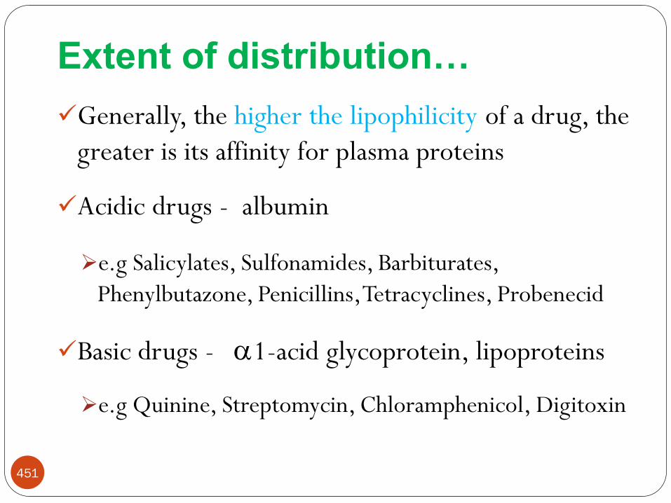
Extent of distribution…
Generally, the higher the lipophilicity of a drug, the
greater is its affinity for plasma proteins
Acidic drugs - albumin
e.g Salicylates, Sulfonamides, Barbiturates,
Phenylbutazone, Penicillins, Tetracyclines, Probenecid
Basic drugs - 1-acid glycoprotein, lipoproteins
e.g Quinine, Streptomycin, Chloramphenicol, Digitoxin
451
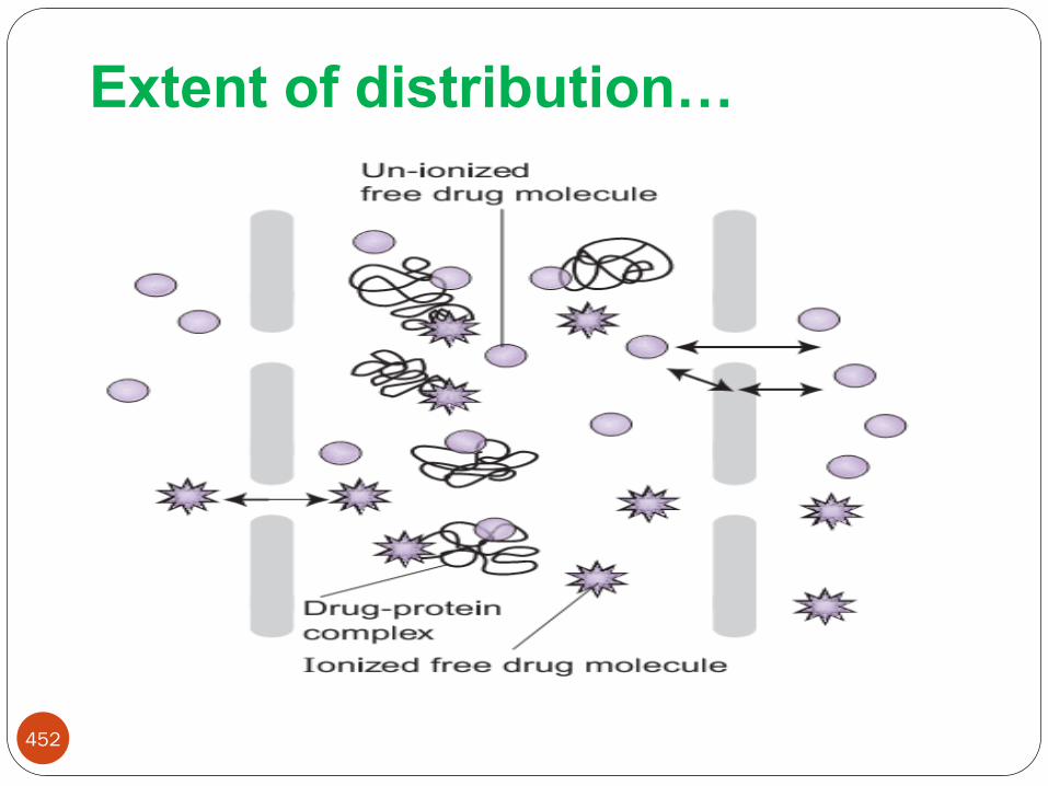
Extent of distribution…
452

Extent of distribution…
Binding is a function of the affinity of the protein
for the drug
Characterized by an association constant, Ka
For a single binding site on the protein, the
association is simply summarized by the following
reaction
complexproteindrugproteindrug
453

Extent of distribution…
High affinity implies that equilibrium lies far to the
right
From mass law considerations, the equilibrium is
expressed in terms of the concentrations of
unbound-drug, Cu, unoccupied protein, P, and
bound drug (Cbd)
PC
CK
U
bda
.
454

Extent of distribution…
Unbound concentration is important in
therapeutics
The fraction of drug in plasma unbound, fu, is given
by
C
Cf U
u
455

Extent of distribution…
Tissue Binding
The fraction of drug in body located in plasma depends on its binding to both plasma and tissue components
A drug may have a great affinity for plasma proteins,
but may still be located primarily in tissue if the tissue has an affinity even greater than that of plasma
Unlike plasma binding, tissue binding of a drug cannot be measured directly
456
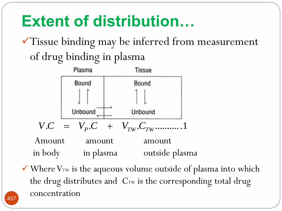
Extent of distribution…
Tissue binding may be inferred from measurement
of drug binding in plasma
Amount amount amount
in body in plasma outside plasma
Where VTW is the aqueous volume outside of plasma into which
the drug distributes and CTW is the corresponding total drug
concentration457
1.............. TWTWP CVCVCV

Extent of distribution…
ondistributi
tissueofvolumeplasmaofvolume
apparentofvolumeapparent
C
CVVV TW
TWP 2................
1.............. TWTWP CVCVCV
Dividing by C will give
458

Extent of distribution…
we know that
Similarly, for tissue,
Given that distribution equilibrium is achieved when the
unbound concentration in plasma, Cu and in tissues, CUT
are equal, then
TW
uu C
Cf T
T
C
Cf Uu
3..................
TU
uTW
f
f
C
C
459

Extent of distribution…
Substituting this equation into equation 2, we get;
Apparent volume of distribution increases when fu
is increased, decreases when fUT increased
4...................
TU
uTWP
f
fVVV
460

Extent of distribution…
When drug is restricted to plasma; expected for small
molecular weight drugs that are highly bound to
plasma proteins but not bound in the tissues
the volumes of distribution, apparent and real, are then
the same, about 3 L in an adult
For drugs of high molecular weight (greater than
approximately 70,000 g/mole),
extra- vascular distribution is very slow to non-existent
volume of distribution then tends to approximate that of
plasma, 3 L
461

Extent of distribution…
For drugs that are bound in neither tissue nor plasma,
Volume of distribution varies between the extracellular
fluid volume (15 L) and the total body water, 42 L,
depending on the degree to which the drug gains access
to the intracellular fluids
462

Extent of distribution…
Table: Extent of plasma protein binding of selected drugs
463
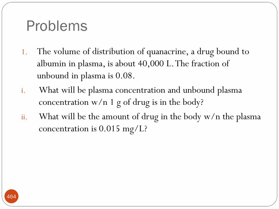
Problems
1. The volume of distribution of quanacrine, a drug bound to
albumin in plasma, is about 40,000 L. The fraction of
unbound in plasma is 0.08.
i. What will be plasma concentration and unbound plasma
concentration w/n 1 g of drug is in the body?
ii. What will be the amount of drug in the body w/n the plasma
concentration is 0.015 mg/L?
464

Cont…
2. Drug A and drug B have V app of 20 and 100 L, respectively.
Both drugs have a V p of 4 L and a V t of 10 L, and they are 60%
bound to plasma protein. What is the fraction of tissue binding of
the two drugs?
465

466
Drug Elimination

Elimination
Elimination is the irreversible loss of drug
from the site of measurement
Usually divided into two major components:
excretion and biotransformation
467

468
Elimination…
Drugs are removed from the body by various
elimination processes
4/28/2020 468

469
Drug Metabolism

Metabolism of drugs
Chemical alteration of the drug in the body
Aim: to convert non-polar lipid soluble compounds to polar
lipid insoluble compounds readily excretable products (avoid
re-absorption in renal tubules)
Most hydrophilic drugs are less bio-transformed and
excreted unchanged – streptomycin
470

Metabolism…
The liver is the major site for drug metabolism, but specific
drugs may undergo biotransformation in other tissues, such
as the kidney and the intestines
471

Fig: Elimination of lipophilic drugs. Lipophilic drugs are metabolized to formpolar metabolites that are more easily excreted in urine.
472

Biotransformation-Classification
Two Phases of Biotransformation:
Phase I or Non-synthetic
Metabolite may be active or inactive
Metabolites are too lipophilic to be retained in the kidney
tubules
473

Biotransformation-Classification…
Phase II or Synthetic
Results in polar, usually more water-soluble compounds
that are most often therapeutically inactive
Such as glucuronic acid, sulfuric acid, acetic acid, or an
amino acid
474

Biotransformation-Classification…
475

Kinetics of drug metabolism
Enzyme-catalyzed reactions are governed by Michaelis–
Menten kinetics, in which the rate of metabolism of a
substrate by an enzyme is given by the equation
Where:
[S] is the concentration of the substrate (drug),
Vmax is the maximum rate of metabolism, and
Km is the Michaelis constant
476

Kinetics of drug metabolism…
The exact concentration of drug at the site of metabolism is
usually not known
However, because blood circulation carries drug to this site,
it is reasonable to assume that [S] is equal to drug plasma
concentration [Cp], which is readily measured by taking
blood samples
In this situation, the Michaelis–Menten equation can be
written as
477

Kinetics of drug metabolism…
478

Kinetics of drug metabolism…
When plasma drug concentration is low, [Cp] << Km
The equation simplifies to
Where km is called the apparent metabolic rate constantgiven by Vmax/Km
Thus, at low plasma concentrations of drug, the rate ofmetabolism is directly proportional to plasma drugconcentration and can be described as a first-order process
479

Kinetics of drug metabolism…
If plasma drug concentration is high such that [Cp]>>Km, the
rate of metabolism becomes
In other words, the rate has reached the maximum that the
available enzyme can handle; the enzyme is saturated
The rate of metabolism is now independent of drug
concentration, a characteristic of a zero-order process
480

Factors that affect the rate of drug
metabolism
Age
Liver disease
Drug tolerance
481

482
Drug excretion

Drug excretion
Excretion
The irreversible loss of chemically unchanged drug or
intact drug
Renal and non-renal routes of drug excretion
Non-renal routes that a drug can be excreted from the body
Biliary excretion
Breast milk
Lungs
Sweat
Saliva
483

484
Renal drug excretion
The kidney is the primary site for removal of a drug in a
chemically unaltered or unchanged form (i.e. excretion) as
well as for metabolites
Plays a major role in maintaining the normal fluid volume
and electrolyte composition in the body
Drugs that are non volatile and water soluble are slowly
biotransformed by the liver
Nonvolatile drugs are excreted mainly by renal excretion

485
Renal drug excretion…
Nephrons are the basic functional units of kidney
Basic components are the glomerulus, proximal tubule,
loop of henle, distal tubule and collecting tubule

486
Renal drug excretion…
Fig : The structure of the kidney nephron

487
Renal drug excretion…
The process by which a drug is excreted via the kidney may
include any combination of the following:
Glomerular filtration
Active tubular secretion
Tubular reabsorption

488
Glomerular filtration
This is where blood is filtered and is where urine formation
begins
Approximately 20% to 25% of cardiac out put or 1.1 L of
blood per minute goes to the kidney
Of this volume, about 10% is filtered at the glomerulus
Porosity of glomerular membrane allows only small m.wt
particles to be filtered(m.wt less than 2000 g/mole)

489
Glomerular filtration…
The rate at which plasma water is filtered is conventionally
called the glomerular filtration rate, GFR
GFR is a good measure of Kidney function
120 ml/min in a 70-kg 20-year old man
Glomerular filtration…
Drug clearance is often proportional to GFR
GFR is reduced in: newborn, elderly, kidney and
heart disease
Reduced GFR: lower drug dose, increase dose
interval or both
490

491
Cont…
Rate of filtration=GFR.Cu…………………1
Where
GFR is glomerular filtration rate
Cu is concentration of unbound drug in plasma or
Rate of filtration=fu.GFR.C……………..2
where
fu is the ratio of the unbound drug to total plasma
drug concentration
C is total plasma drug concentration

492
Cont…
If a drug is only filtered and all filtered drug is
excreted into the urine, then the rate of excretion
is the rate of filtration
Since renal clearance (Clr), by definition is
It follows that for such a drug its clearance(by
filtration) is fu.GFR
3.......................ionconcentratplasma
excretionofrateClr
493
Active tubular secretion
Drugs secreted from blood to lumen of nephron against
concentration gradient
Require active transport
Occurs mostly in the proximal tubule
Two separate carrier-mediated systems for bases and acids
Bases: acetylcholine, histamine, atropine, meperidine, etc
Acids: salicylate, penicillin, probenecid, cephalosporins , etc.

494
Active tubular secretion…
Competition for carriers within groups [acids with acids, ]
E.g: Probencid and penicillin
Saturable kinetics
The active secretion process are saturable, so renal
excretion can reach a plateau at high doses of drug

495
Renal tubular reabsorption
Drug moves from nephron lumen to blood
Active reabsorption for many endogenous compounds
Vitamins, electrolytes, glucose, aminoacids
Passive reabsorption for vast majority of exogenous
compounds
Occurs all along the nephron, associated with water filtered
at the glomerulus
99% of water reabsorbed increases drug level in lumen

496
Renal tubular reabsorption…
The degree of reabsorption depends on:
Properties of the drug
Polarity, state of ionization and molecular weight
Urine flow and urine PH

Renal tubular reabsorption…
It is sometimes clinically useful, particularly in drug overdose
cases, to alter the pH of the urine (of the patient)
For drugs which are weak acids, urine alkalinization favors the
ionized form and promotes excretion. Alternatively,
acidification promotes the renal clearance of weak bases
497

498
Fig: Cumulative urinary excretion of Metamphetamine (11 mg orally) in man varies with the urine PH

Biliary excretion
Although the primary role of the liver in drug elimination is
via metabolism, the liver also secretes a fluid called bile that
may play a role in drug excretion
Bile is an aqueous solution of bile acids, bile salts,
cholesterol, and inorganic salts
The extent of bile production depends on the type of food
present, with food high in proteins resulting in greatest bile
secretion
499

Biliary excretion
500
Bile formed in large volumes in the liver
Most of the water re-absorbed
Concentrated bile stored in the gall bladder
Bile secreted into the upper small intestine

Biliary excretion…
Drugs secreted into bile are emptied along with bile into the
duodenum, from where they can be removed from the body
in the feces; this elimination pathway is called biliary
excretion
Bile is significant route of excretion for high molecular
weight drugs (> 500 Da):
Glucuronide conjugates (e.g. morphine)
Limited number of ionised drugs with very high Mol Wt
501

502
Miscellaneous route of excretion
Just as drugs are excreted in to fluids such as urine in the
kidney and bile in the liver, they can be excreted in to other
fluids
Expired air
Breast milk
Sweat

503
Excretion in expired air
The lung is a major organ of excretion for gaseous and
volatile substances, and is a significant route of excretion for
volatile drugs such as anesthetics and ethanol
Most organs that excrete drugs eliminate polar compounds.
The exception to this premise is the lungs, in which volatility
of drug or metabolite is more important than its polarity
Examples of drugs that appear in expired air are
sulfanilamide and sulfapyridine

504
Clearance Concept

Clearance
Clearance is a more useful concept in measuring drug
elimination than kel since it takes into account blood flow
rate
Is a PK term for describing drug elimination from the body
without identifying the mechanism of the process
The Clearance (Cl) of a drug is defined as the volume of a
biological fluid (generally plasma) from which that drug is
removed ("cleared") in the unit of time (min)
505
506
Clearance…
Expresses the ability of kidney and liver to remove drug from the systemic circulation
Drug clearance (body clearance, total body clearance, or
Cl T) considers the entire body as a single drug
eliminating system from which many unidentified
elimination processes may occur
4/28/2020 506
507
Clearance…Drug elimination rate is expressed in terms of
amount of drug removed per unit time (eg,
mg/min)
Drug clearance described in volume of fluid clear of
drug per unit time (eg, mL/min)
Drug clearance is also defined as the fixed volume
of fluid (containing the drug) cleared of drug per
unit of time
The units for clearance are volume/time (eg,
mL/min, L/hr)4/28/2020 507

Clearance…
NB: Clearance doesn’t indicate the amount of drug being
removed, it indicates the volume of plasma(blood) from
which the drug is removed or cleared, in a given time period
Clearance is the constant of proportionality between the
rate of drug elimination and the plasma concentration
Rate of elimination = Cl X Cp
Cl T may be defined as the rate of drug elimination divided
by the plasma drug concentration508

509
Clearance…
…………………..(1)
………………(2)
Where D E is the amount of drug eliminated and
dDE/dt is the rate of elimination
Rearrangement of Equa. 2 gives Equation 3
................(3)
4/28/2020 509

510
Clearance…
It is also known that a first-order elimination rate,
dD E/dt, is equal to kDB or kCpVD
Based on equation 1, substituting elimination rate
for kCpVD,
ClT = kCpVD/CP = kVD……………………(4)
Equation 4 shows that clearance is the product of
VD and k, both of which are constant
4/28/2020 510

511
Clearance…
As the plasma drug concentration decreases during
elimination, the rate of drug elimination, dD E/dt,
decreases accordingly, but clearance remains
constant
Clearance is constant as long as the rate of drug
elimination is a first-order process
Drugs can be cleared from the body by different
mechanisms, pathways or organs, including hepatic
biotransformation and renal and biliary excretion
4/28/2020 511

512
Clearance…
Renal clearance= keVD = rate of elimination by kidney
C
Lung clearance= k1VD= rate of elimination by organ
C
Hepatic clearance= kmVD = rate of elimination by liver
C
CL total = CL renal + CL hepatic + CL pulmonary + CL others
Body clearance= keVD + k1VD + kmVD
=(ke + k1 + km )VD = kVD ……(5)
4/28/2020 512
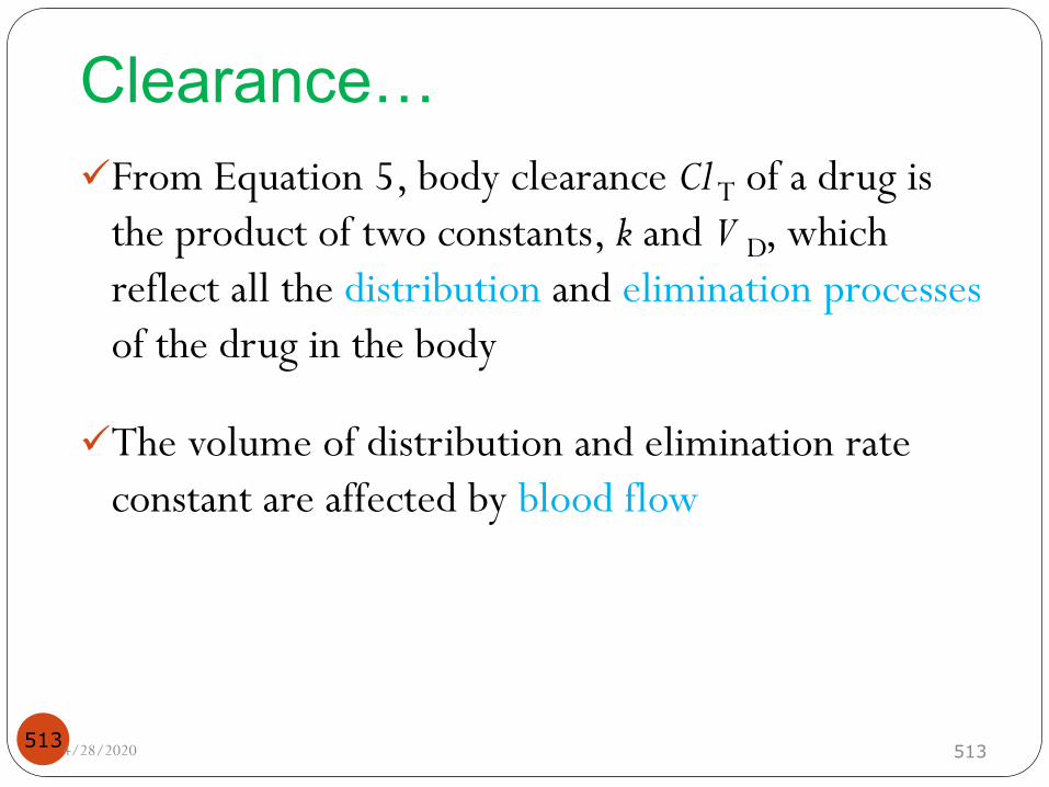
513
Clearance…
From Equation 5, body clearance Cl T of a drug is
the product of two constants, k and V D, which
reflect all the distribution and elimination processes
of the drug in the body
The volume of distribution and elimination rate
constant are affected by blood flow
4/28/2020 513

Problems
1. Penicillin has a ClT of 15 mL/min. Calculate the elimination
rate for penicillin when the plasma drug concentration, C p,
is 10 µg/mL.
2. Determine the total body clearance for a drug in a 70-kg
male patient. The drug follows the kinetics of a one-
compartment model and has an elimination half-life of 3
hours with an apparent volume of distribution of 100
mL/kg.
514

515
Clearance…
Physiologic/Organ Clearance
Many organs in the body have the capacity for drug
elimination
The kidneys and liver are the most common organs
involved in excretion and metabolism, respectively
Physiologic PK models are based on drug clearance
through individual organs or tissue groups
4/28/2020 515

Clearance…
Let’s consider a single organ, kidneys and Q (mL/minute)
which is blood flow through the organ, Cin , the drug conc. in
the blood entering the organ and Cout , the drug conc. in the
exiting blood
Clearance is dependent on the blood flow to the organ (Q)
and on the ability of the organ to extract the drug from the
blood stream and eliminate it (E)
516
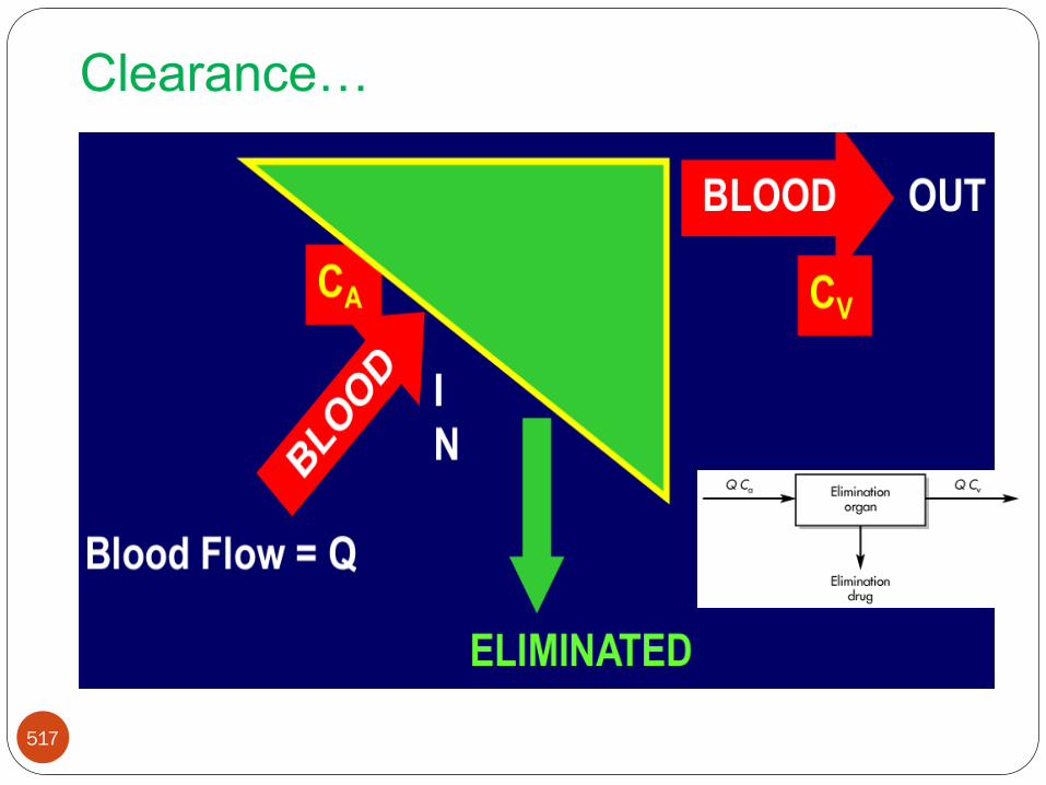
Clearance…
517
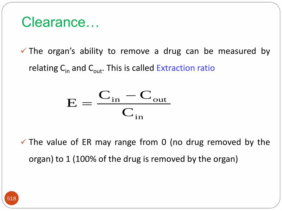
Clearance…
The organ’s ability to remove a drug can be measured by
relating Cin and Cout. This is called Extraction ratio
The value of ER may range from 0 (no drug removed by the
organ) to 1 (100% of the drug is removed by the organ)
518
in
outin
C
CCE

Clearance…
Organs that are very efficient at eliminating a drug
will have an extraction ratio approaching one
ER is a ratio with no units
An ER of 0.25 indicates that 25% of the incoming drug
concentration is removed by the organ as the drug passes
through
519

520
Cont…
Drug clearance of any organ is determined by blood flow and
the extraction ratio:
Organ Clearance = Blood flow X extraction ratio
Clearance = Q(ER)………………………………. (6)
4/28/2020 520

521
Cont… Substituting for ER into Equation 6 yields
. …………………………. (8)
The physiologic approach to clearance shows that
clearance depends on the blood flow rate and the
ability of the organ to eliminate drug
4/28/2020 521

522
Renal clearance
The appearance of drug in the urine is the net result
of filtration, secretion and reabsorption processes
Rate of excretion = Rate of filtration
+ Rate of secretion
- Rate of reabsorption
The first two processes add drug to proximal part
of the lumen of the nephron;
the last process involves the movement of the drug
from the lumen back to blood stream

523
Renal clearance…
The actual renal clearance of a drug is not generally
obtained by direct measurement
The clearance value for the drug is often compared
to that of a standard reference, such as inulin, which
is cleared completely through the kidney by
glomerular filtration only
The clearance ratio, which is the ratio of drug
clearance to inulin clearance, may give an indication
for the mechanism of renal excretion of the drug

524
Renal clearance…
Clearance Ratio Probable Mechanism of Renal Excretion
Drug is partially reabsorbed
Drug is filtered only
Drug is actively secreted
Table: Comparison of Clearance of a Sample Drug to Clearance of Inulin
1Clinulin
CldrugClr
1Clinulin
CldrugClr
1Clinulin
CldrugClr

525
Renal clearance
Renal clearance is defined as the volume of plasma
that contained the amount of drug appearing in urine
per unit time
Is a component of overall drug clearance, and
represents the efficiency of removal of unchanged
drug in urine by renal excretion
Renal clearance of a drug can be expressed as :
4......................../
cp
dtdXuClr

526
Cont…
Since excretion rate is the product of the urinary
excretion rate constant and the amount of drug in
the body, we can write:
Since X/Cp = Vd(volume of distribution), we can
write:
5..................CP
KeXClr
6................KeVdClr

Problems
1. Two drugs, A and B, are entirely eliminated through the
kidney by glomerular filtration (125 mL/min), with no
reabsorption. Drug A has half the distribution volume of
drug B, and the V D of drug B is 20 L. What are the drug
elimination rate constant for each drug?
2. An antibiotic is given by IV bolus injection at a dose of 500
mg. The apparent volume of distribution was 21 L and the
elimination half-life was 6 hours. Urine was collected for 48
hours, and 400 mg of unchanged drug was recovered. What
is the fraction of the dose excreted unchanged in the urine?
Calculate k and Cl T.
527

By: Bilal Tessema
June, 2020
Pharmacokinetics of
Intravascular & Extravascular administration

Pharmacokinetics of
Intravenous administration
Drugs may be administered to patients by one of
several routes, including oral, topical, or parenteral
routes of administration
In developing pharmacokinetic models to describe
and predict drug disposition kinetically,
Model must account for both the route of administration
and the kinetic behavior of the drug in the body
529 28-Apr-20

Cont…
Parenteral routes of administration include
intravenous, subcutaneous, intramuscular, etc
Intravenous (IV) drug solutions may be given either
as;
Bolus dose (injected all at once) or
Infused slowly through a vein into the plasma
530 28-Apr-20

531
Intravenous bolus administration
28-Apr-20

A drug is administered as an injection of a sterile
solution formulation which is injected all at once
(into a box, or compartment)
There are several important points to note;
Ensures that the entire administered dose reaches the
general circulation
The drug distributes instantaneously and homogenously
throughout the compartment
Drug elimination also occurs from the compartment in
first order fashion532
IV bolus Administration
28-Apr-20

Intravenous bolus…
533 28-Apr-20

Intravenous bolus…
One-compartment model offers the simplest way
to describe the process of drug distribution and
elimination in the body
The following assumptions are made;
One-compartment model, first-order process
The drug is being monitored in blood (plasma/serum)
and urine
534 28-Apr-20

Intravenous bolus…
Consider a single IV bolus injection of drug X. As time
proceeds, the amount of drug in the body is:
dCp/dt = rate in (availability) – rate out (elimination)
Since rate in or absorption is absent, equation becomes
dCp/dt = - rate out
If rate out or elimination follows first order kinetic
535 28-Apr-20

Intravenous bolus…
536
pel
pC .k
dt
dC Integrating this formula gives
tk0
teleCpCp
Where,
Cpt is the concentration at any time t
Cp0 is the concentration at time 0
k is the elimination rate constant28-Apr-20

Intravenous bolus…
Example: If we know that the plasma drug concentration just
after a gentamicin dose is 8 mg/L and the patient's
elimination rate constant is 0.25 hr-1, calculate the
concentration 8 hours later?
solution:
C = C0e-Kt , C0 = 8 mg/L, K = 0.25 hr-1, and t = 8 hours
Cat 8 hr = 8 mg/L X e-0.25 hr-1(8 hr) = 8 mg/L (0.135) = 1.1 mg/L
537 28-Apr-20

Intravenous bolus…
NB: The term e-Kt indicates the fraction of the initial dose of
drug that remains in the body at time t;
0.135 (or 13.5%) remains in the body 8 hours after the initial
dose in this example
Conversely, the term 1 - e-Kt would indicate the percent or
fraction excreted after time (t)
538 28-Apr-20

Intravenous bolus…
The above equation describes the single exponential decline
in drug concentration as a function of time
This fall in plasma concentration is called mono-exponential
decay
If we know kel and Cp0 we could calculate Cp at any time
after a single IV bolus dose
However, it still isn't very convenient for estimating a value
of kel from concentration versus time data
539 28-Apr-20

Intravenous bolus…
For estimation purposes it is preferable to use a straight line
equation
A straight line equation can be achieved by taking the natural
logarithm of both side of Equation
540
tk Cpln Cpln el
0
t
2.303
tkCp logCp log el0
t
28-Apr-20

Intravenous bolus…
Hence, Plotting ln(Cp) versus t should give a straight line with
a slope of - kel and an intercept of ln(Cp0) (y = mX + b with a =
intercept and m = slope)
541 28-Apr-20

Intravenous bolus…
542
C0 can’t be directly measuredexperimentally, must bedetermined by extrapolationof the Cp-time line back to they-axis
K is the slope
28-Apr-20

Intravenous bolus…
Summarizes the relationship
With real Cp-time data (clinical data), the first data
point is normally collected approximately 15
minutes after administration
Therefore, the value of ln Cp o is determined by
extrapolation of the lnCp-time line back to to rather
than as an experimental data point543 28-Apr-20

Elimination rate constant
Elimination rate constant (k) represents the fraction of drug
removed per unit of time and has units of reciprocal time
(e.g., minute-1, hour-1, and day-1).
544
21
21 lnln
tt
CCkel
• Where t1 and C1 are the
first time/concentration
pair and t2 and C2 are
the second time/concentration
28-Apr-20

Half life
It is the time required for the concentration of drug in the
plasma to decrease by half
Consider: ln C = ln C0-Kt
By definition, at one half-life, the concentration (C) at the
time (t) is half of what it was initially (C0)
So we can say that at t = t1/2, C = 1/2C0
545 28-Apr-20

Half life…
546
21el
00 tkCpln Cp2
1ln
elelel
oo
el
oo1/2
k
0.693
k
ln2
k
/Cln2C
k
1/2CClnt
28-Apr-20

Half life…
As a rule of thumb, a drug is said to be completely
eliminated after a time interval of five half-lives
After five half-lives, the amount of drug in the body
is only around 3% of the theoretical Cp o value
547 28-Apr-20

Problem1. If a dose of gentamicin is administered and a peak plasma
concentration is 6 mg/L after the infusion is completed and is 1.5 mg/L 4 hours later, calculate half life of the drug?
2. What fraction of a drug remains from an IV bolus after five
half-lives have elapsed?
3. The following table presents Cp-time data for a drug. Use the
data to construct a ln Cp versus time graph. Determine the
best-fit line of the graph & calculate the half-life of the drug.
548 28-Apr-20

Clearance
Total body clearance is an important pharmacokinetic
parameter that is often defined as the volume of blood or
plasma completely cleared of the drug per time
It is also the proportionality constant relating the rate of
elimination and drug concentration
549
del
p
el
p
el
p
p V kC
A . k
C
A) k_(
C
dt
dA
CLC . CL dt
dA
28-Apr-20

Clearance…
Clearance can also be calculated using the integral of
equation
550
pC
dt
dA
CL
AUC
Dose
dt C
dt dt
dA
C
dt
dA
CL
pp
28-Apr-20

Apparent volume of distribution
Vd is the constant of proportionality that relates the total
amount of drug in the body at any time to the corresponding
plasma concentration
551
p
dC
X
plasmain measuredion concentrat
body in the drug ofAmount V
28-Apr-20

Apparent volume of distribution…
Immediately after the IV dose is administered the AB is the
IV dose
At this time elimination is not yet started
It has an insignificant effect on the administered dose
Combining the above Equation and Equation for IVbolus, C = C0e-Kt we are able to derive an equation fordrug concentration as a function of time given valuesof Dose, Vd, and kel.552
o
p
dC
Dosev
d
o
pv
DoseC
28-Apr-20

Apparent volume of distribution…
Calculation may be the determination of kel and half-life from
CL and Vd
553
tk
d
pele .
V
DoseC
CL
V 0.693
k
0.693t
V
CLk d
el2
1
d
el
28-Apr-20

Problem
1. A patient is given a theophylline loading dose of 400 mg
intravenously over 20 minutes. Because the patient received
theophylline during previous hospitalizations, it is known that
the volume of distribution is 30 L and the half-life (t1/2) is 6
hours. Compute the expected theophylline concentration 4
hours after the dose was given?
554 28-Apr-20

Problem…
2. A patient is given an IV dose of vancomycin 1000 mg. Since
the patient has received this drug before, it is known that the
Vd equals 50 L, the elimination rate constant is 0.077 h-1, and
the half-life equals 9 h (t1/2 = 0.693/kel = 0.693/0.077 h-1 = 9 h).
Calculate the expected vancomycin concentration 12 hours
after the dose was given?
555 28-Apr-20

Problem…
3. A patient was given an intravenous loading dose ofPhenobarbital 600 mg over a period of about an hour. One dayand four days after the dose was administered phenobarbitalserum concentrations were 12.6 mg/L and 7.5 mg/L,respectively. Compute Kel, t1/2, Vd and C0.
4.The following table present some Cp-time data points for an IV bolus
dose of 325 mg.What is CL for this drug?
556 28-Apr-20

Area under the plasma concentration time
curve (AUC)
AUC: reflects the actual body exposure to drug after
administration of a dose of the drug and is expressed in mg X
h/L
This area under the curve is dependent on
The rate of elimination of the drug from the body
The dose administered
557 28-Apr-20

Calculation of AUC using the Trapezoidal
Rule
AUC can be calculated directly from the Cp versus time data
can be determined by the application of trapezoidal
rule and can be obtained by using an equation
558 28-Apr-20

Calculation of AUC…
Trapezoidal rule is the simplest and most common approach
of calculating AUC
We can calculate the AUC of each segment if we consider the
segments to be trapezoids [Four sided figure with two
parallel sides]
559 28-Apr-20

Calculation of AUC…
560 28-Apr-20

Calculation of AUC…
The area of each segment can be calculated by multiplying
the average concentration by the segment width. For the
segment from Cp2 to Cp3:
561 28-Apr-20
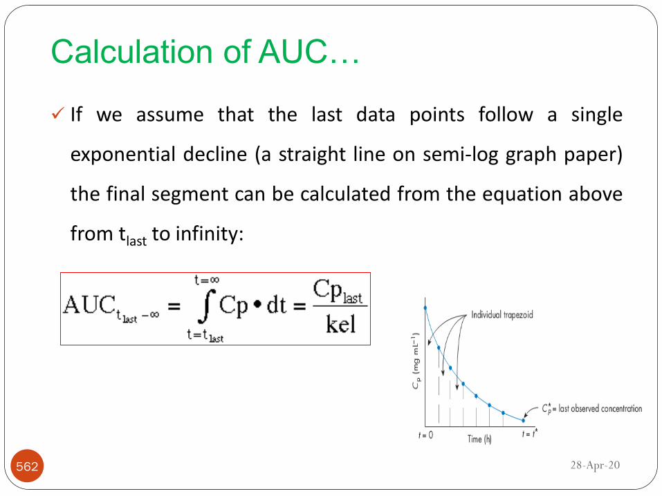
Calculation of AUC…
If we assume that the last data points follow a single
exponential decline (a straight line on semi-log graph paper)
the final segment can be calculated from the equation above
from tlast to infinity:
562 28-Apr-20
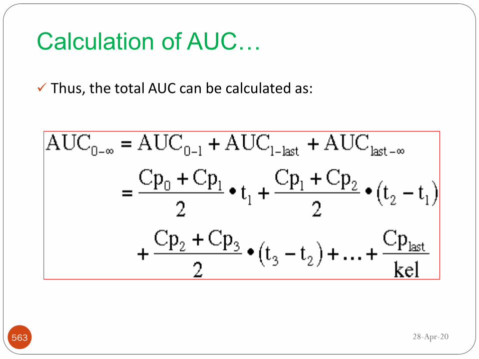
Calculation of AUC…
Thus, the total AUC can be calculated as:
563 28-Apr-20

Calculation of AUC…
Example: An 80-mg dose of drug Y is administered as an
intravenous bolus, and the following plasma concentrations
result:
564 28-Apr-20

Exercise
A 250 mg dose of a drug was administered intravenously to
60 yr. 80 kg male subject. The blood concentration time data
is presented in table below. Calculate the AUC and
clearance?
565 28-Apr-20

566
Continuous infusion
28-Apr-20

Continuous Intravenous Infusion
Most drugs given intravenously cannot be given as an actual
intravenous bolus because
Side effects related to rapid injection
For instance, vancomycin IV bolus: hypotension and red
flushing around the head and neck areas
Unsuitable when it is necessary to maintain plasma or
tissue concentrations at a concentration that will prolong
the duration of drug effect
567 28-Apr-20

568
F
Figure: A representation of the plasma concentration (Cp) versus time profile
following the administration of a single intravenous (IV) bolus dose. MTC, minimum
toxic concentration; MEC, minimum effective concentration.
28-Apr-20

Continuous intravenous….
Therefore it may be given by slower IV infusion over 15 or 30
minutes or more
If a drug is chemically stable and compatible with the IV fluid
it may be added to the fluid and thereby be given by slow
infusion
The pharmacokinetics of a drug given by constant IV infusion
follows a zero-order input process in which the drug is
infused directly into the systemic blood circulation
569 28-Apr-20

Continuous intravenous….
The main advantage for giving a drug by IV infusion is that;
IV infusion allows precise control of plasma drug
concentrations to fit the individual needs of the patient
IV infusion of drugs, such as antibiotics, may be given
with IV fluids that include electrolytes and nutrients
The duration of drug therapy may be maintained or
terminated as needed using IV infusion
570 28-Apr-20

Continuous intravenous…
Therefore, the infused drug follows zero-order input and first-
order output
k0 represents infusion rate which is a zero order process so
the units of k0 are amount per time, for example 25 mg/min
571 28-Apr-20

Continuous intravenous….
Infusion Rate Constant
An intravenous infusion involves a continuous flow
of drug into a patient
at a rate defined by the infusion rate constant, k0/Rinf,
with units of mass/time
During an infusion, Cp rises with the start of the
infusion
As Cp builds, elimination processes begin to
counteract the infusion and slow the rate of increase
in Cp572 28-Apr-20

Continuous intravenous….
If the infusion continues long enough, Cp approaches
a maximum called the steady-state concentration (Cp ss)
at which the rate of infusion is balanced by the rate of
elimination
As soon as the infusion is stopped, Cp is influenced
only by elimination and will decrease accordingly
573 28-Apr-20

Continuous intravenous….
Figure: Cp vs. time for an IV infusion
574 28-Apr-20

Continuous intravenous….
The change in the amount of drug in the body at any time
(dAB/dt) during the infusion is the rate of input minus the rate
of output. The differential equation for V • Cp is then:
After dividing both sides by the apparent volume of
distribution, V or substituting A with V x Cp
575
1elo1 A .kk
dt
dA
Integrating thedifferentialequation
tk-
1ele-1 .
el
o
k
kA
28-Apr-20

576
t. k-
del
o
tele-1 .
V . k
kCp
Continuous intravenous…
28-Apr-20
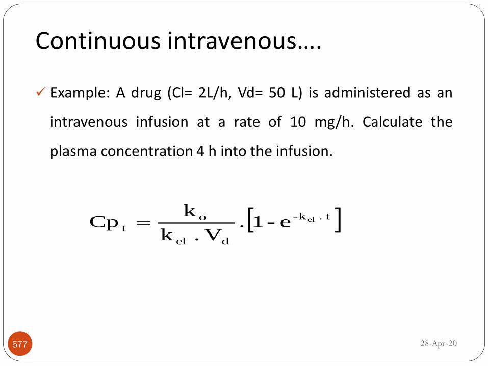
Continuous intravenous….
Example: A drug (Cl= 2L/h, Vd= 50 L) is administered as an
intravenous infusion at a rate of 10 mg/h. Calculate the
plasma concentration 4 h into the infusion.
577
t. k-
del
o
tele-1 .
V . k
kCp
28-Apr-20

Steady-State Drug Concentration (CpSS)
Steady-State Drug Concentration (CpSS)
When an IV infusion is started at a constant rate in one-
compartment model, there is no drug present initially, so
plasma concentration (C) is zero
As the infusion continues, Cp increases, initially quickly, but
then more slowly, as Kel<< k0
If we continue the infusion indefinitely, the k0 will be equal
to Kel
Steady state plasma concentration
578 28-Apr-20

Steady-State Drug Concentration (CpSS)…
In other words, there is no net change in the amount of drug
in the body, D B, as a function of time during steady state
579 28-Apr-20

Steady-State Drug Concentration (CpSS)…
We can determine the steady state concentration from the
differential equation by setting the rate of change of Cp, i.e.
dCp/dt = 0
This could also be calculated from the integrated equation by
setting e- kel • t = 0 at t = ∞
580
CL
k
V . k
kCC . VkC . V . kk0
dt
dCo
del
oss
p
ss
pdo
ss
pdelo
p
28-Apr-20

Steady-State Drug Concentration (CpSS)…
Since the apparent volume of distribution, elimination rate
constant, and elimination half life (t1/2) are constants for a
given drug administered to a particular patient
The absolute value of steady-state plasma concentration is
determined only by the rate of infusion
For instance, if the rate of infusion is increased by a factor of
two, the steady-state plasma concentration will also
increase by a factor of two
581 28-Apr-20

Steady-State Drug Concentration (CpSS)…
Example1: An antibiotic has a volume of distribution of 10 L
and a k of 0.2 hr– 1. A steady-state plasma concentration of
10μg/mL is desired. Calculate the infusion rate needed to
maintain this concentration?
Assume the above patient developed a uremic condition and
the elimination rate constant has decreased to 0.1 hr– 1. To
maintain the steady-state concentration of 10μg/mL, we
must determine a new rate of infusion as follows.
582 28-Apr-20

Steady-State Drug Concentration (CpSS)…
When the k decreases, the infusion rate must decrease
proportionately to maintain the same C SS
However, because the elimination rate constant is smaller
(ie, the elimination t 1/2 is longer), the time to reach C SS
will be longer
583 28-Apr-20

Steady-State Drug Concentration (CpSS)…
Example2: A desired steady state plasma concentration of
theophylline may be 15 mg/L. The average half-life of
theophylline is about 4 hr and the apparent volume of
distribution is about 25 liter. What infusion rate is
necessary?
584 28-Apr-20

Steady-State Drug Concentration (CpSS)…
Exercise: An adult male patient (43 years old, 80 kg) is to be
given an antibiotic by IV infusion. According to the literature,
the antibiotic has an elimination t 1/2 of 2 hours, a V D of 1.25
L/kg, and is effective at a plasma drug concentration of 14
mg/L. The drug is supplied in 5-mL ampules containing 150
mg/mL.
a) Recommend a starting infusion rate in milligrams per hour
and liters per hour.
585 28-Apr-20

Steady-State Drug Concentration (CpSS)…
Blood samples were taken from the above patient at 12, 16,and 24 hours after the start of the infusion. Plasma drugconcentrations were as shown below:
b) From this additional data, calculate the total body clearance
Cl T & estimate the elimination half-life for the antibiotic in this
patient.
586
T (hr) Cp(mg/L)
12 16.1
16 16.3
24 16.528-Apr-20

Time to Reach Steady State
Clinical practice, the activity of the drug will be observed
when the drug concentration is close to the desired plasma
drug concentration, which is usually the target or desired
steady-state drug concentration
Mathematically, the time to reach true steady-state drug
concentration, C SS, would take an infinite time
The time to reach 90%, 95%, and 99% of the steady-state
drug concentration, CSS, may be calculated.
587 28-Apr-20

Time to Reach Steady State…
For therapeutic purposes, the time for the plasma drug
concentration to reach more than 95% of the steady-state
drug concentration in the plasma is often estimated
588 28-Apr-20
Table: Number of t1/2 to Reach a Fraction of CSS
Time to Reach Steady State…
Example: How many half-lives does it take to get to 95%
steady state?
589 28-Apr-20

Time to Reach…
NB:
The steady-state concentration (C SS) is dependent on the
volume of distribution, the elimination rate constant, and the
infusion rate.
For a drug with one-compartment characteristics, the time to
reach steady state is independent of the dose, the number of
doses administered, and the dosing interval, but it is directly
proportional to the half-life.
590 28-Apr-20

Time to Reach…
591
Exercise: How many half-lives does it take to get to 99%
steady state?
A patient was given an antibiotic (t 1/2 = 6 hr) by constant IV
infusion at a rate of 2 mg/hr. At the end of 2 days, the serum
drug concentration was 10 mg/L. Calculate the total body
clearance Cl T for this antibiotic?
28-Apr-20
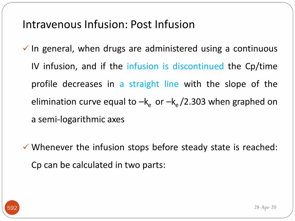
Intravenous Infusion: Post Infusion
In general, when drugs are administered using a continuous
IV infusion, and if the infusion is discontinued the Cp/time
profile decreases in a straight line with the slope of the
elimination curve equal to –ke or –ke /2.303 when graphed on
a semi-logarithmic axes
Whenever the infusion stops before steady state is reached:
Cp can be calculated in two parts:
592 28-Apr-20

Intravenous Infusion: Post Infusion…
First, calculate the concentration of drug final infusion
concentration, CPD when t = D using Equation:
After the infusion is stopped' the situation is similar with that
for the bolus injection. Hence once the infusion is stopped all
we have is first order elimination.
593
D . k-
del
o ele-1 . V . k
kC D
P
28-Apr-20

Intravenous Infusion: Post Infusion…
Where t is time counted from the start of the infusion or t =
Total time (infusion + post-infusion) and
D is length of time of infusion period
Thus t - D is the time since the end of the infusion (length of
time after infusion has stopped)
For convenience, the two equations can be combined as
follows.594
D-t . -kD
P
t
pele . CC
28-Apr-20

Intravenous Infusion: Post Infusion…
If the infusion is stopped after steady state is reached, the
slope of the elimination curve remains the same but the
concentration at the end of infusion is the steady state
concentration.
595
D-t . k-D . k-
del
ot
pelel e . e-1
V . k
kC
28-Apr-20

Intravenous Infusion: Post Infusion
596 28-Apr-20

Intravenous Infusion: Post Infusion
Example: A patient is administered theophylline
intravenously with infusion rate of k0 = 735 mg/hr. It is known
from prior clinic visits that the patient has kel of 0.17 hr-1 , V
of 25 L; D (Infusion duration) of 0.5 hour. What would be the
plasma concentration at 4.5 hours (t = 4.5 hours)? That is if
we stop the loading infusion and don't start the maintenance
infusion.
597 28-Apr-20

Intravenous Infusion: Post Infusion
Solution
Where
Cpost-infusion is post-infusion serum concentrations,
Cend is the concentration at the end of infusion,
kel is the elimination rate constant and
tpost-infusion is the post-infusion time (tpostinfusion = 0 at end ofinfusion and increases from that point)
598
mg/L 7.050.5 X 1.14 1 Xe-1 25 X 17.0
735e . e-1
V . k
kC 0.5-4.5 X 17.00.5 X 0.17-D-t . k-D . k-
del
ot
pelel e
infusion-postel t. -k
endinfusion-post e CC
28-Apr-20

Intravenous Infusion: Post Infusion
Exercise 1: A patient was infused for 6 hours with a drug (k =
0.01 hr– 1; V D = 10 L) at a rate of 2 mg/hr. What is the
concentration of the drug in the body 2 hours after cessation
of the infusion?
599 28-Apr-20

Cont…
An advantage to infusions over an IV bolus is that
less drug hits the patient’s blood stream at once
This avoids elevated values of Cp o
Infusions are also able to provide a steady Cp level
One disadvantage of infusions is that they require
time to reach a therapeutic concentration
Another disadvantage of infusions is that the patient
must have an IV line during the entire time of the
infusion600 28-Apr-20
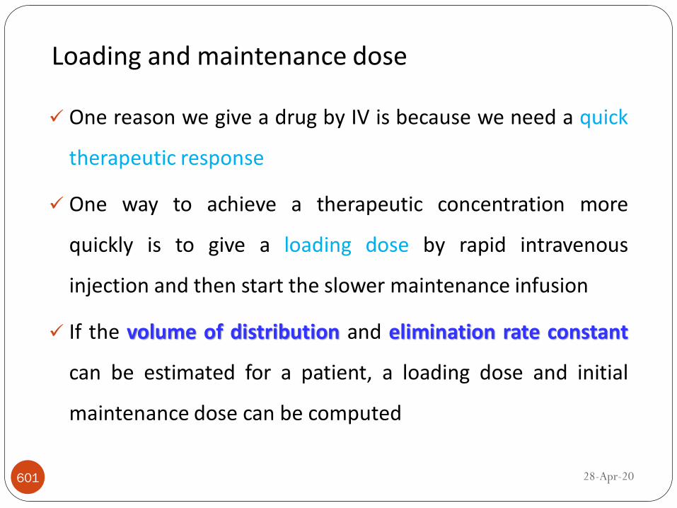
Loading and maintenance dose
One reason we give a drug by IV is because we need a quick
therapeutic response
One way to achieve a therapeutic concentration more
quickly is to give a loading dose by rapid intravenous
injection and then start the slower maintenance infusion
If the volume of distribution and elimination rate constant
can be estimated for a patient, a loading dose and initial
maintenance dose can be computed
601 28-Apr-20

Loading dose…
Loading Bolus
Because IV infusions can require considerable time
to build Cp to effective levels, an IV bolus may be
used to “jump start” Cp
Such dosing is called a loading bolus
Through joint use of an IV bolus and infusion, a
patient may receive immediate and constant
therapeutic effect from a drug
602 28-Apr-20

Loading dose…
The loading dose can simply be calculated from the product
of target concentration and volume of distribution VD.
If the target concentration is steady state concentration for a
continuous intravenous infusion dosage regimen DL is
computed using the following equation:
NB:
Knowledge of volume of distribution allows calculation of
the drug dose required to achieve a target concentration603
Ld
ss
p
el
o
del
ss
p
ss
po DV . Ck
kV . k . CCL . Ck
28-Apr-20
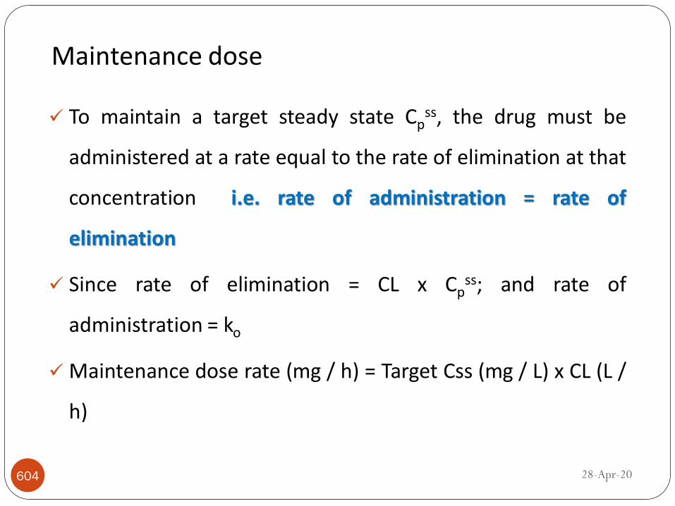
Maintenance dose
To maintain a target steady state Cpss, the drug must be
administered at a rate equal to the rate of elimination at that
concentration i.e. rate of administration = rate of
elimination
Since rate of elimination = CL x Cpss; and rate of
administration = ko
Maintenance dose rate (mg / h) = Target Css (mg / L) x CL (L /
h)
604 28-Apr-20

Maintenance dose
Knowledge of clearance allows calculation of the dose rate
required to maintain a target steady state concentration
(Cpss)
This means that if the dose is altered, the steady state
concentration will change in direct proportion to the change
in dose.
605 28-Apr-20

Example: A patient with a ventricular arrhythmia after a
myocardial infarction needing treatment with lidocaine at a Cpss
of 3.0 mg/L (population pharmacokinetic parameters used: Vd =
50 L, Cl = 1.0 L/min). Calculate the loading and maintenance
dose?
Example: Calculate gentamicin dose required to achieve a peak
gentamicin concentration of 10 mg/l in a neonate weighing 1 kg
and Vd of 0.5 litre/kg ?
606
Loading and maintenance dose
28-Apr-20
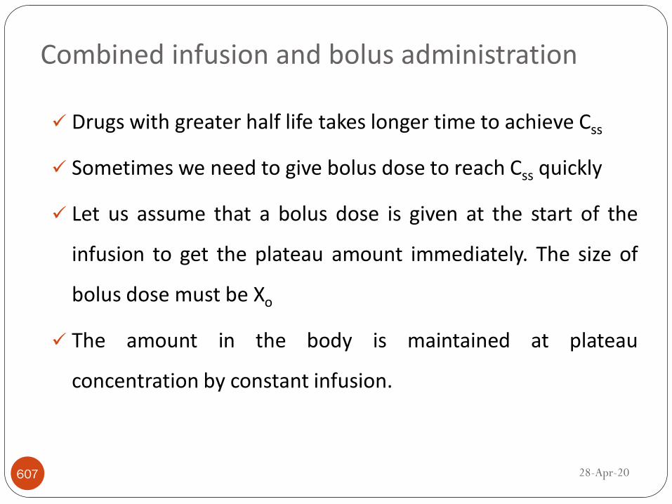
Drugs with greater half life takes longer time to achieve Css
Sometimes we need to give bolus dose to reach Css quickly
Let us assume that a bolus dose is given at the start of the
infusion to get the plateau amount immediately. The size of
bolus dose must be Xo
The amount in the body is maintained at plateau
concentration by constant infusion.
607
Combined infusion and bolus administration
28-Apr-20

The bolus dose reduces exponentially and at any time the
amount remaining in the body from bolus dose can be given as
But as long as the infusion is maintained this decline will be
matched by the gain from the infusion
The sum of these two amount is the same as the amount at
plateau Ass.
608
tk
ssinfssele AAA
tk
tele XoAss
Combined…
ktkt eVk
koe
V
DlCss 1
28-Apr-20

Cont…
Figure: Use of a loading bolus with an IV infusion
609 28-Apr-20

When the bolus dose and infusion rate are exactly matched, the
gain from one administration offset the loss from the other.
610
Combined…
28-Apr-20

Problem
1. What is the concentration of a drug 6 hours after
administration of a loading dose of 10 mg and simultaneous
infusion at 2 mg/hr (the drug has a t1/2 of 3 hr and a volume
of distribution of 10 L)?
2. For a 70-kg patient, the Vd of morphine is 230 L, and CLT is
1,680 mL/min. Calculate the infusion rate (Ko/Rinf) of morphine
required to sustain a Cp ss of 15 ng/mL in a 70-kg patient
3. Determine the appropriate dose for a loading bolus that will
provide a Cp o of 15 ng/mL of morphine in a 70-kg patient. The
Vd of morphine is 230 L, and CLT is 1,680 mL/min. If this bolus
is combined with the infusion in the previous Sample Calculation,
at what time will the Cp contribution from both the bolus and
infusion be the same?611 28-Apr-20

Cont…
3. A physician wants to administer an anesthetic agent at a rate
of 2 mg/hr by IV infusion. The elimination rate constant is 0.1
hr– 1, and the volume of distribution (one compartment) is 10
L. What loading dose should be recommended if the doctor
wants the drug level to reach 2 g/mL immediately?
4. What is the concentration of a drug 6 hours after
administration of a loading dose of 10 mg and simultaneous
infusion at 2 mg/hr (the drug has a t 1/2 of 3 hr and a volume of
distribution of 10 L)?
612 28-Apr-20

613
Extravascular administration
28-Apr-20

Pharmacokinetics of extravascular dose
Extravascular drug administration refers to any route
of drug administration where the drug is not
administered directly into the systemic circulation
Extravascular delivery routes, particularly oral dosing,
are important and popular means of drug
administration
Pharmacokinetic models after extravascular drug
administration must consider systemic drug absorption
from the site of administration, eg, the lung, the gut,
etc., into the plasma
614 28-Apr-20

Extravascular…
615
Fig: Typical plasma concentration–time profile after extravascular drug administration.. 28-Apr-20

Extravascular…
Immediately after drug administration (area A), plasma
concentrations increase because the rate of absorption is
greater than the rate of elimination
The amount of drug in the gastrointestinal tract is at its
maximum, so the rate of absorption is also maximum
In contrast, initially the amount of drug in the body is small,
so the rate of elimination is low
616 28-Apr-20

Extravascular…
As the absorption process continues, drug is depleted from
the gastrointestinal tract, so the rate of absorption decreases
At the same time, the amount of drug in the body increases,
so the rate of elimination increases.
At the peak (B), the rate of absorption is momentarily
equal to the rate of elimination.
617 28-Apr-20

Extravascular…
After this time the rate of elimination exceeds the rate of
absorption and plasma concentrations fall (area C)
Eventually, all the drug is depleted from the intestinal tract
and drug absorption stops.
At this time (area D) the plasma concentration is influenced
only by elimination
618 28-Apr-20

Model for first-order absorption in a one-compartment model
The absorption of drugs from the gastrointestinal tract often
follows first-order kinetics
As a result, the pharmacokinetic model can be created
simply by adding first-order absorption into the central
compartment of the one-compartment model
This model can be represented as:
619 28-Apr-20

Model for first-order absorption…
620
Where
Xg is the amount of drug to be absorbed
Xp is the amount of drug in the body
ka is the first order absorption rate constant
kel is first order elimination rate constants
28-Apr-20

Model for first-order absorption…
The amount of drug in the body at any time will depend on
the relative rates of drug in(absorption) and out
(elimination):
Rearranging and integrating the above equation yields:
621
elpga
pk . X X .k
dt
dX
28-Apr-20

Model for first-order absorption…
where
(Xp) t is the mass (amount) of drug in the body at time t;
Xg0(Dose) is the mass of drug at the site of administration att=0 (the administered dose);
F is the fraction of drug absorbed;
(Xg)t=o = FXg0 and is the mass of administered dose that isavailable to reach the general circulation
622
tktk-
ela
atktk-
ela
0
gatktk-
ela
0tga
tPaelaelael ee
kk
Dose Fkee
kk
FXkee
kk
XkX
28-Apr-20

The above Equation can be written in concentration (Cp) terms
by substituting Cp X Vd for Xp:
Where
KaFXg0/Vd (Ka – Kel) is the intercept of the plasma drug
concentration versus time plot.
623
tk-tk-
elad
0
ga
tpael e e
k kV
FXkC
Model for first-order absorption…
28-Apr-20

Model for first-order absorption…
624
Notice that the right hand side of this equation (Equation
above) is a constant multiplied by the difference of two
exponential terms, A bi-exponential equation.
28-Apr-20

When time is large, because of the fact that Ka >> Kel, e-kat
approaches zero, and Cp reduces to:
625
tk-
elad
0
ga
tpele
k kV
FXkC
Determination of elimination half life (t1/2)
28-Apr-20

Method of Residuals
1. Back extrapolate the log linear portion of the decline phase,
say C’ denotes the plasma concentration along this
extrapolated line.
2. Subtract the plasma concentration Cp from the
corresponding extrapolated value at each point.
3. Plot the residual (C’-Cp) against time on the semi-log graph
paper.
626
Absorption rate constant (Ka)
28-Apr-20

It gives a straight line, the slope of which is the
absorption constant.
627
Absorption rate constant (Ka)…
28-Apr-20
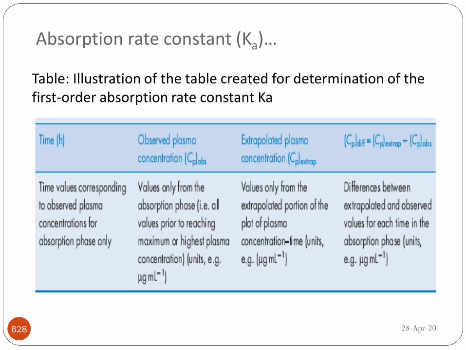
Absorption rate constant (Ka)…
628
Table: Illustration of the table created for determination of the first-order absorption rate constant Ka
28-Apr-20
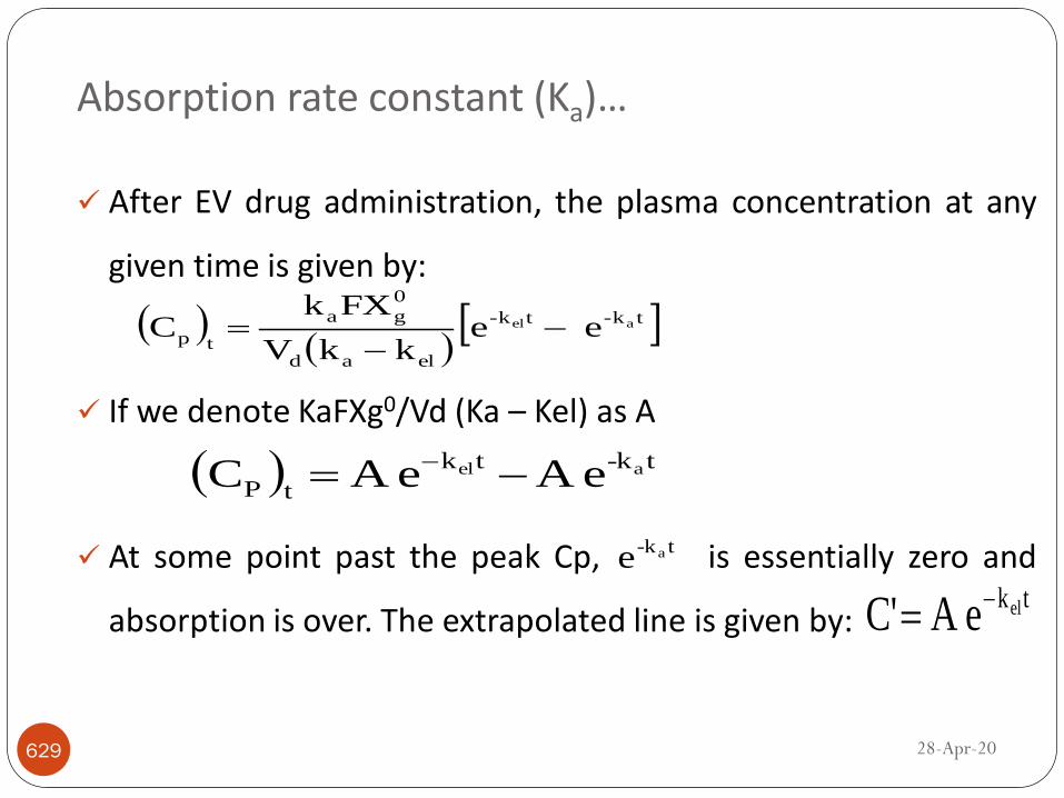
Absorption rate constant (Ka)…
After EV drug administration, the plasma concentration at any
given time is given by:
If we denote KaFXg0/Vd (Ka – Kel) as A
At some point past the peak Cp, is essentially zero and
absorption is over. The extrapolated line is given by:
629
tk-tk-
elad
0
ga
tpael e e
k kV
FXkC
t-ktk
tPael eA eA C
t-kae
tkeleA C'
28-Apr-20

Absorption rate constant (Ka)…
Subtracting C’ from Cp we get
Taking natural logs
630
tk-
elad
0
gadiff
ae k kV
FXkCpor CpC'
t. k-
k kV
FXkCpC'ln a
elad
0
ga
t. k-A CpC'ln a
28-Apr-20
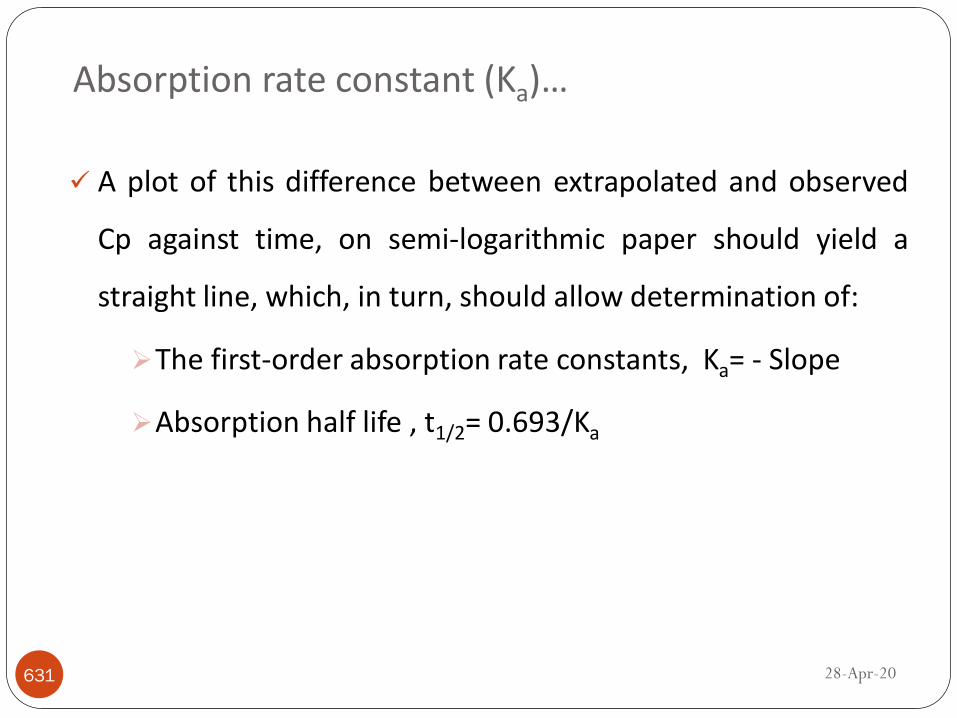
Absorption rate constant (Ka)…
A plot of this difference between extrapolated and observed
Cp against time, on semi-logarithmic paper should yield a
straight line, which, in turn, should allow determination of:
The first-order absorption rate constants, Ka= - Slope
Absorption half life , t1/2= 0.693/Ka
631 28-Apr-20

632
Absorption rate constant (Ka)…
28-Apr-20

Apparent volume of distribution (V)
For a drug administered EV route of administration, the Vd
can be calculated from Cp data in two ways.
If F (the fraction of administered dose that reaches the
general circulation) is known:
If F is not known, the ratio of V/F will be calculated as
follow:
633
elad
a
kkV
FDosekIntercept
Intercept
1
kk
Dosek
F
V
ela
ad
28-Apr-20

Time of Cp max, Peak time (tmax)
The peak time (tmax) is the time at which the body displays
the maximum plasma concentration, (Cp) max
It occurs when the rate of absorption is equal to the rate of
elimination (i.e., when KaXg = Kel Xp)
At the peak time, therefore, Ka (Xg)tmax = Kel (Xp)tmax.
634
maxtp
Xel
k
maxtg
Xa
kor 0el
k .
maxtp
X
maxtg
X .a
kdt
pdX
28-Apr-20

Time of Cp max, Peak time (tmax)….
The peak time can be used:
To determine comparative bioavailability and/ or
bioequivalence
To determine the preferred route of drug administration and
the desired dosage form for the patient
To assess the onset of action
635
elk
ak
elk
ak
ln
maxt
28-Apr-20

Maximum [peak] plasma concentration [Cp] max
There are two methods available for determining peak
plasma concentration (Cp) max
Method 1: Peak plasma concentration obtained from the
graph of plasma concentration versus time
636 28-Apr-20

Maximum [peak] plasma concentration [Cp] max
Method 2: Peak plasma concentration obtained by substituting t
with t max in the original extravascular equation.
As the ka value is becoming smaller, the Cp max gets lower
and the onset becomes slower.
Thus the higher the F value the higher the concentration
values at each time point.
637
maxamaxelael tktk
elad
o
ga
maxp
tk-tk-
elad
0
ga
tp eekkV
FXkCe e
k kV
FXkC
28-Apr-20

Maximum (peak) plasma…
The peak plasma concentration:
Is one of the parameters used to determine the comparative
bioavailability and/or the bioequivalence between two products
(same and or different dosage forms) but containing the same
chemical entity or therapeutic agent
May be used to determine the superiority between two different
dosage forms or two different routes of administration
May correlate with the pharmacological effect of a drug.
638
maxamaxel tktk
maxp ee IC
28-Apr-20
Lag time
The lag time (t0) indicates that absorption did not start
immediately following the administration of drug by the oral
or other extravascular route
This delay in absorption may be attributed to some
formulation-related problems
Slow tablet disintegration,
Slow and/or poor drug dissolution from the dosage form
Incomplete wetting of drug owing to the hydrophobicnature of the drug
Poor formulation
639 28-Apr-20

Lag time…
Theoretically, intercepts of the terminal linear portion and
the feathered line should be the same; however, sometimes,
these two lines do not have the same intercepts.
As a result the lag in time for a drug will be observed at a
point greater than t=0 on the x-axis.
This time at the point of intersection on the x-axis is called
the lag time
640 28-Apr-20

Lag time…
641 28-Apr-20

Lag time…
whenever there is lag time during absorption:
The lag time t0 is subtracted from each time point.
642
0a0el t-tk-t-tk-
elad
0
ga
tp e e k kV
FXkC
28-Apr-20

PROBLEM
An oral drug has a dose of 500 mg, oral bioavailability of
80%, Vd of 70 L, kel of 0.3 h-1, and kab of 0.8 h-1. The drug
has been prescribed to be taken every 4 hours. If the
minimum effective concentration of the drug is 1.5 mg/mL,
will the patient still have an effective concentration of the
drug when the second dose is administered?
643 28-Apr-20

The following data were obtained when a 500 mg dose of anantibiotic was given orally. Calculate the pharmacokineticparameters, assuming 100% of the administered dose wasabsorbed.
644
Time(h) 1 2 3 4 5 6 8 16 18
C((mcg/ml
)
26.501 36.091 37.512 36.055 32.924 29.413 22.784 7.571 5.734
Time(h) 20
C((mcg/ml
)
4.343
Example
28-Apr-20

Solution: first we have to draw a graph. Then, we will go forcalculating different pharmacokinetic parameters. Hence, thegraph looks like this
645
Example…
28-Apr-20

The rate constant of elimination is calculated from the terminal
linear portion of plasma profile. To determine it, we need to
calculate slope of the straight line having y-intercept = B. if natural
log are used the rate constant of elimination (b) = negative slope
of this straight line. Therefore
646
1
el 0.139h2
0.2778
2018
ln4.343ln5.734kSlope
The biological half life (t1/2) is determined using the equation t1/2=
0.693/kel = 0.693/0.139/h = 4.98/hr.
Elimination rate constant
28-Apr-20

The Y-intercept of this straight line is B and is determined using the first
order equation: ln Ct = ln B – kelt Which upon rearrangement gives ln B =
ln Ct + kelt ln 4.343 + (0.139/hr) (20hr) =1.4686 + 2.78 = 4.2486 B =
Inverse ln 4.2486 = 70.0 mcg/ml
To obtain the straight line which represents absorption phase, the
technique of feathering or the method of residuals is used. for example,
to feather the first plasma conc. point at 1 hr, the plasma conc. at 1 hr
on the straight line having the y intercept = B is subtracted from the
plasma conc. data provided in the data.
647
Absorption constant
28-Apr-20

ln Ct = ln B - kelt ln Ct = ln 70 – (0.139) (1) = 4.2485 – 0.139 =
4.1095 Ct = inverse ln 4.109 = 60.916 mcg/ml
The residual conc. at 1 hr is obtained by subtracting from this
concentration at 1 hr provided in the data. Therefore the residual
concentration at 1hr is, 1 hr = 60.916 – 26.501 = 34.415mcg/ml.
With similar method the residual concentration for every hour will
be calculated and the residual cure will look like this.
648
Absorption constant…
28-Apr-20

Absorption constant…
649 28-Apr-20
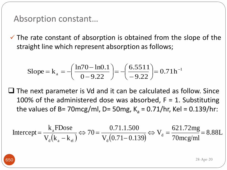
The rate constant of absorption is obtained from the slope of thestraight line which represent absorption as follows;
650
1
a h 0.719.22
6.5511
9.220
ln0.1ln70kSlope
The next parameter is Vd and it can be calculated as follow. Since100% of the administered dose was absorbed, F = 1. Substitutingthe values of B= 70mcg/ml, D= 50mg, Ka = 0.71/hr, Kel = 0.139/hr:
8.88L
70mcg/ml
621.72mgV
0.1390.71V
0.71.1.50070
kkV
FDosekIntercept d
delad
a
Absorption constant…
28-Apr-20

From the Pk parameters found, Ka = 0.71/hr and Kel = 0.139/hr wecan calculate tmax.
651
2.856hr0.571
1.6308
0.1390.71
ln0.139ln0.71
kk
lnklnk
elk
ak
elk
ak
ln
maxt
ela
ela
From the data given we can also calculate the Cp max of the drugin plasma. The parameters we need are I or B = 70 mcg /ml, Ka =0.71 /hr, Ke = 0.139 /hr, and tmax = t’ = 2.856 hr
l37.85mcg/m70X0.5407ee70Xee IC 0.71X2.85660.139X2.85tktk
maxpmaxamaxel
T max and C max
28-Apr-20

Reading assignment;
Clinical pharmacokinetics
652 28-Apr-20

653