The Proceedings of the 2012 Equine Chapter Meeting at the ...
173
THE EXOTIC EQUID The Proceedings of the 2012 Equine Chapter Meeting at the ACVSc Science Week 28 June - 30 June 2012 Gold Coast International Hotel Gold Coast, Queensland, Australia Proceedings Editor – John Chopin
-
Upload
khangminh22 -
Category
Documents
-
view
3 -
download
0
Transcript of The Proceedings of the 2012 Equine Chapter Meeting at the ...

THE EXOTIC EQUID
The Proceedings of the 2012 Equine Chapter Meeting at the ACVSc Science Week
28 June - 30 June 2012
Gold Coast International Hotel Gold Coast, Queensland, Australia
Proceedings Editor – John Chopin
2
SPONSORS

3
4

5
PRESENTERS:
David Berman has been conducting research to improve management of overabundant
vertebrate animals for 28 years. Species studied include horses, rabbits, camels, pigs, pigeons,
dingoes, and sparrows. His PhD study of the ecology of wild horses led to a significant reduction
in the damage caused by their overabundance in central Australia. In Queensland Dr Berman
has determined the distribution and abundance of horses in Beerburrum State Forest, removed
horses from the Greenbank Military Training Area and commenced work in the Toolara State
Forest to reduce the risk of collisions between vehicles and horses. Dr Berman’s family sport is
showjumping. He has had two exceptional brumby showjumpers and has a keen interest in
finding suitable homes for brumbies captured during management programmes. Dr Berman has
seen first hand the devastating consequences of allowing wild horse populations to increase
unchecked. He seeks long-lasting solutions to the problems caused by overabundant horse
populations.
Since attaining a first class honours degree in animal science, Simon Collins has worked extensively on
the functional anatomy of the horse foot, and on the effects of laminitis. Simon has worked previously
at DeMontfort University, The University of Nottingham, Loughborough University, and the Animal
Health Trust in research collaborations with The Donkey Sanctuary (UK), the Japan Racing Association,
and the Laminitis Trust (UK). Simon spent over 6 years working with the donkey population managed by
the donkey sanctuary, undertaking his PhD and initial post-doctoral research investigating the form and
function of the donkey foot, and laminitis in the donkey. Simon has considerable experience in the
application of radiographic, magnetic resonance imaging (MRI) and computed tomography (CT)
techniques for the assessment of the foot. Simon has pioneered the use of powerful computer
modelling technology to further elucidate the biomechanics of the equid foot, and to gain new
understanding of the impact of foot disorders. Simon is currently completing a 2 year research
fellowship with Prof. Chris Pollitt at the Australian Equine Laminitis Research Unit, the University of

6
Queensland. However, Simon will shortly start work on developing 3-D computer models for
undergraduate teaching of basic and applied veterinary anatomy at the university.
Emma Croser after 6 years in mixed practice in both Australia and the UK I switched to the dark
side and became a pathologist working at the University of Queensland and the University of
Melbourne as well as several commercial providers. I am now investigating aspects of Hendra
virus infection and immunity at CSIRO (AAHL), an interest stemming from my days working with
horses in Queensland.
Charles El-Hage graduated in 1984 from the University of Melbourne. Spent many years in
private large animal mainly practices in Australia and the UK. Became a member of the ANZCVS
in 1994 (Equine Medicine). Returned to Melbourne Uni in 2001 as a clinician and following
several hiatuses, is employed as a lecturer in clinical studies with interests in reproduction,
immunology and virology of horses.
Vic Epstein graduated Sydney University in Veterinary Science in 1973. Worked in mixed
practice in Coonamble, Western NSW before moving to equine only practice in Brisbane, then

7
NZ. Returned to Australia to complete a MVSc in equine cardiology and the effect of potassium
in 1980 and then worked in Sydney in equine practice for 10 years before leaving the equine
industry to join the public service in Tasmania. Worked in the Department of Agriculture for 10
years before travelling and working overseas in various roles and various countries. Joined
University of Queensland as lecturer, tutor, practitioner and manager at Goondiwindi. Worked
in equine stud practice in Turkey and China. Volunteered in Egypt in donkey practice for 6
months in 2009. This position is the basis of the talk.
James Gilkerson completed his veterinary studies at the University of Sydney, where he
graduated with a BVSc, a BSc (Vet) and a PhD in equine herpesvirus epidemiology and vaccine
development. James is particularly interested in equine infectious diseases, but is also involved
in research into poultry infectious diseases and is developing an interest in infectious diseases
of Australian and African wildlife – much to his surprise! James has been teaching infectious
diseases to vet students at the University of Melbourne since 2002, with a special emphasis on
the host-parasite balance and control of infectious diseases in populations. As past-President of
both the Victorian Division of the AVA, and Equine Veterinarians Australia, James has been very
actively involved with the profession, in addition to his teaching and research work in the
Faculty.
Allan Gunn was born and raised in Zimbabwe. He completed a degree in Agriculture in Natal
South Africa. After farming and travelling, he attended the Royal (Dick) School of Veterinary
Studies at the University of Edinburgh where he obtained a BVM&S degree. After 3 years of
working in the UK, he returned to Zimbabwe and set up his own rural practice. Since then he
has worked in China (Beijing), the UK and in Australia where he has lived for the last 12 years.

8
Since arriving in Australia he obtained two MANZCVS qualifications- horse medicine and
reproduction- and became a Diplomate of the ACT via the alternate route in 2008. Allan is now
lecturer in equine studies and reproduction at Charles Sturt University, Wagga Wagga.
Griet Haitjema is a registered specialist in veterinary anaesthesia and works at Murdoch
University Veterinary Hospital as a senior registrar providing anaesthesia and pain management
service to the hospital patients and through consultations to outside patients via the Pain Clinic.
Griet is also involved with final year student teaching and resident supervision. Griet originally
graduated from Murdoch University (1986). Has completed a Masters degree in veterinary
studies in small animal medicine and surgery and is a Fellow of the Australian New Zealand
College of Veterinary Scientists in veterinary anaesthesia and critical care and a Diplomat of the
European College of Veterinary Anaesthesia and Analgesia.
Kris Hughes graduated from the University of Sydney in 1995 and spent 3 years as an associate
in a mixed-animal practice in Myrtleford, Victoria, before undertaking a residency in equine
medicine at the University of Sydney from 1999 until 2003. In 2005, he took a position as senior
lecturer in equine medicine at the Faculty of Veterinary Medicine, University of Glasgow, where
he was involved in a referral medicine service, clinical research and undergraduate and
postgraduate teaching. After 2 Australian Ashes defeats while in the UK and enduring the
British weather for 5 years, he returned to Australia in 2009 and is a senior lecturer in equine
medicine at Charles Sturt University. His research interests include inflammatory disorders of
the lower airways, diagnostic tests for metabolic and endocrine diseases, cardiology, neurology

9
and intensive care of horses. He is a Fellow of the Australian and NZ College of Veterinary
Scientists in Equine Medicine and a Diplomate of the European College of Equine Internal
Medicine. When not at work he enjoys music, travel, cycling, hiking and supporting the
Collingwood football team.
Craig Johnson qualified from Liverpool Veterinary School in 1989. After a spell in small animal
practice, he moved to The Animal Health Trust in Newmarket, UK as Resident in Veterinary
Anaesthesia. He obtained his RCVS Diploma in Veterinary Anaesthesia in 1992 and after a time
as a locum lecturer in Pretoria, South Africa returned to the UK and gained a PhD from
Cambridge University. From 1996 – 2001 Craig was a Lecturer in Veterinary Anaesthesia at
Bristol University, UK. In April 2001 he moved to Massey and is now Associate Professor of
Veterinary Neurophysiology.
Kieren Maddern graduated from the University of Queensland with a Bachelor of Veterinary
Science (Honours) in 2000. Following graduation she spent time in general practice and at the
University of Queensland emergency center she also worked for two years at the University of
Sydney as a Clinical Registrar in Veterinary Anaesthesia. In 2005 she became a Member of the
Australian & New Zealand College of Veterinary Scientists in the field of Anaesthesia & Critical
care. Kieren decided to further her anaesthetic training and began a senior clinical training
scholarship at the Animal Health Trust in Newmarket (UK) In 2006. This was completed in 2009
and she became a Diplomate of the European College of Veterinary Anaesthesia & Analgesia
the same year. Since the completion of her residency program Kieren has worked as a
consultant anaesthetist at a number of practices. In 2010, Kieren relocated to Singapore with
her husband and Rupert the dachshund; she continues to provide anaesthetic consultancy
services within the Asia Pacific region.

10
David McLelland graduated from the University of Sydney with BSc (Vet) 2000, and
BVSc, 2001. I worked in mixed practice in Darwin and Sydney for 2.5 years. I completed
a DVSc residency in zoo medicine and pathology at the University of Guelph and Toronto
Zoo, 2004-07. I undertook locum work with Wellington Zoo and Zoos Victoria in early
2008, before joining Zoos South Australia where I am currently employed. I am a Clinical
Senior Lecturer with the School of Animal and Veterinary Sciences at the University of
Adelaide. I became a Member of the College in Medicine of Zoo Animals in 2008, and a
Diplomat of the American College of Zoological Medicine in 2011. I am the President of
the Chapter of Zoo and Wildlife Medicine, and have previously served as
Secretary/Treasurer.
Tony Mogg graduated from the University of Sydney in 1985. Since graduation he has worked in mixed private practice; as well as at the University of Queensland, the University of Pennsylvania in the USA, and Massey University in New Zealand. He is currently Senior Lecturer in Equine Medicine at the University of Sydney Veterinary Teaching Hospital Camden. Tony is a Fellow of the Australian College of Veterinary Scientists (Equine Medicine), a Diplomate of the American College of Veterinary Internal Medicine (Large Animal), a Diplomate of the American College of Veterinary Clinical Pharmacology and a Fellow of the American Academy of Veterinary Pharmacology and Therapeutics. Tony’s professional interests include general equine internal medicine, equine clinical pharmacology, equine ophthalmology, equine cardiovascular disease and veterinary medical education. He is a registered specialist in equine medicine.

11
Tias Muurlink graduated from The University of Queensland in 1988 and has since pursued his
interest in Equine Practice. After a number of positions in practices in Queensland and Victoria,
Tias co-founded a practice in Warwick, Queensland which he managed for some 10 years. Here
he was involved in all aspects of Equine practice, but predominantly focused on horse stud
services and surgery. After selling this business, Tias spent a season working with the Scone
Equine Hospital before joining the University of Melbourne. During his time in Melbourne, he
completed his specialist training in Equine Surgery and went on to lead the team at the Equine
Centre at the University of Melbourne Veterinary Hospital. Tias also serves on the Board of
Examiners of the ANZCVS.
Chris Quinn is currently a lecturer in veterinary anaesthesia at Charles Sturt University’s School
of Animal and Veterinary Sciences in Wagga Wagga NSW. His main professional interests are
anaesthesia and critical care of horses and other large animals. He completed an anaesthesia
internship at Scone Equine Hospital and also worked at Scone in the neonatal foal intensive
care unit. He completed a residency program in veterinary anaesthesia at Murdoch University
in WA, before returning to NSW to work at CSU.

12
Louise Sharp graduated from the University of Melbourne in 2005 with a Bachelor of
Veterinary Science (Honours). Since then Louise has completed a Master of Veterinary
Public Health Management at the University of Sydney and is a Member of the
Epidemiology Chapter of the ANZCVS. Louise has worked at the Australian Government
Department of Agriculture, Fisheries and Forestry since 2007. She currently works in the
Animal Biosecurity Branch in the Non-Ruminant and Zoo Animals Section.
Tony Smithyman was born in Nyasaland (Central Africa) and educated at the Universities of St
Andrews and Glasgow, Dr. Smithyman is a bacteriologist and immunologist with a particular
interest in Tropical and infectious diseases. In recent years he has turned his attention to the
rapidly escalating problem of antibiotic resistance and in 2004 formed Special Phage Services, a
research company aimed at the development of phage therapy products for the treatment of
bacterial infection. Ongoing projects cover not only human and veterinary pathogens but also
plant, aquaculture, and environmental targets. As a Scottish African Australian he combines the
worst elements of those three cultures into his ambition to achieve a single figure handicap in
golf and can be found most weekends battling the gale force winds at Long Reef in Sydney.

13
Larry Vogelnest graduated in 1984 from the University of Sydney. In 1990 he did an internship
with the Taronga Conservation Society Australia (Taronga Zoo). Since completing his internship
Larry has been employed at Taronga Zoo and has been Senior Veterinarian since 1994. In
1996 he gained membership to the Australian College of Veterinary Scientists by examination in
Zoo Animal Medicine. Larry is a member of the Editorial Advisory Board of the Australian
Veterinary Journal and is the Oceania Representative of the American Association of Zoo
Veterinarians. He is also the Principal Editor of “Medicine of Australian Mammals”, CSIRO
Publishing (2008) and author and co-author on several chapters. In 2009 Larry was awarded an
Australian Public Service Medal for his contribution to conservation medicine and service to the
Taronga Conservation Society Australia. Larry has worked with elephants at Taronga Zoo since
1990 and has also worked on various elephant projects in Nepal and Thailand. He was heavily
involved in the importation of elephants from Thailand to Australia in 1996. The zoo now has 8
elephants providing plenty of opportunity to develop his knowledge and skill in elephant
reproduction and medicine.
Chris Walzer University of Economics, Vienna, Austria; University of Veterinary Medicine,
Vienna Austria, Mag. med. Vet. 1989; Dr. med. Vet. 1993; Habilitation 2003; Dipl. ECZM
(wildlife population health) Appointments: Univ. Prof., Chair Conservation Medicine, Co-
director Research Institute of Wildlife Ecology, University of Veterinary Medicine, Austria,
2005; Member of the board, Director of Science, International Takhi Group, Zurich Switzerland,
since 1999; Zoological co-director, head veterinarian, staff researcher, Zoo Salzburg Austria
1993; Contract veterinarian, Herberstein wild animal park, Austria, 1989; private veterinary
practice, Kaindorf, Austria, 1990. Chris is a creative interdisciplinary idea initiator and problem
solver; critical and strategic conservationist; non-territorial worker and indefatigable optimist.

14
He has international recognized expertise working with wildlife, especially wild equids and
carnivores, gained from combined years of work and research in Europe, Asia and Africa. Chris
has a very diverse international research track record with some 300 + scientific publications to
his name. He is sought as a consultant in wildlife matters by various organizations such as
WWF, UNDP, WCS, Panthera, OIKOS, SOS Rhino, World Bank, and several other GO’s; NGOs
and universities. Based on some 15+ years work experience in zoological institutions, Chris
regularly consults zoological institutions on all aspects of animal husbandry, veterinary
medicine, collection planning, enclosure design and conservation research. Over the past years
Chris has successfully managed numerous large research teams with varied funding sources
such as the EU and the Austrian Research Fund.
Magdalena Zabek is a postgraduate student at the University of Queensland, conducting a
research project on feral horse ecology in Australia. Prior to her current position,
she was involved with several research projects dedicated to semi-wild and feral horse ecology
and social behaviour in Poland and Australia. Raised in Poland, Magdalena now lives in
Brisbane, where she divides her life between studying and tutoring veterinary students.
Working with horses is not just a part of her academic commitment; she also trains horses
and is a dedicated riding instructor. In her free time, she travels to the most remote
locations throughout Australia in a search for unique photographic opportunities of the
Australian outback and its free ranging horses.

15
2012 Equine Chapter Program THURSDAY 28 JUNE
Collaboration with Zoo and Wildlife Medicine Chapter
9.00 EHV Fatality – a case report Emma Croser
9.20 EHV in captive and wild equids James Gilkerson
10.00 MORNING TEA
10.30 Australia’s Asian Elephant breeding program: management of reproduction, pregnancy and
parturition in 5 nulliparous cows Larry Vogelnest
11.10 Przewalski Horse Anaesthesia David McLelland
11.50 Asiatic Wild Ass Project – field capture and anaesthesia techniques
Chris Walzer
12.10 Equine Dental Evolution Ian Dacre
12.30 LUNCH
1.30 Australian Wild Horse Science & Management David Berman
2.10 Equine Exotic Disease Threats Kris Hughes
2.30 Importance of a transdisciplinary approach to landscape-level Asiatic Wild Ass conservation
Chris Walzer
3.00 AFTERNOON TEA
Chair -
3.30 Ecology of Feral Horses in a Forest Environment of Toolara State Forest
Magdalena Zabek
4.10 Przewalski Horse reintroduction – Veterinary Considerations and management
Chris Walzer
4.50 Perissodacyl Import Risk Assessment Louise Sharp
5.30 FINISH
FRIDAY 29 JUNE
Donkeys, Miniature Horses & Ponies
8.30 Use & Management of Donkeys in Egypt Vic Epstein

16
8.50 Donkey hoof structure Simon Collins
9.10 Foot Problems in Donkeys Vic Epstein
9.30 Comparison of Donkey Foot Structure with other Equids Simon Collins
10.00 MORNING TEA
10.30 Radiology of Normal & Laminitic Donkey Feet Simon Collins
10.50 Animal Care in Egypt – A Welfare Charity Vic Epstein
11.10 International Equine Welfare Charities Ian Dacre
11.30 Common Diseases and Treatments in Donkeys Vic Epstein
11.50 Donkey Dentistry Ian Dacre
12.10 Exotic Diseases in Donkeys Vic Epstein
12.30 LUNCH
1.30 Surgery of Miniature Horses Tias Muurlink
2.10 Pharmacologic and Pharmokinetic Peculiarities Tony Mogg
2.20 Donkey Castration Tias Muurlink
2.40 Lipidaemias in Ponies Kris Hughes
3.00 AFTERNOON TEA
3.30 Other Diseases common in “small” equids Tony Mogg
3.50 Equine Metabolic Syndrome & Pars Intermedia Dysfunction in Ponies
Kris Hughes
4.30 EQUINE CHAPTER AGM
5.10 FINISH
SATURDAY 30 JUNE
8.30 Equine Ophthalmology Tony Mogg
9.10 Equine Neurology Kris Hughes
9.30 Equine Cardiology Tony Mogg
10.00 MORNING TEA
Collaboration with Animal Reproduction Chapter

17
10.30 Reproduction in the Zebra Allan Gunn
10.50 Przewalski herd fertility investigations Charles El-Hage
11.10 P-horse reintroduction to the Great Gobi B strictly protected area in SW Mongolia – status and
outlook Chris Walzer
11.50 Reproduction in the Miniature Horse Allan Gunn
12.10 From theory to practice: bacteriophage therapy in the Thoroughbred breeding industry
Anthony Smithyman
12.30 LUNCH
Collaboration with Anaesthesia, Emergency and Critical Care
1.30 Research abstract prize giving (5min) Chair
1.35 Standing Sedation for the less than typical equid and equids in less than typical situations
Chris Quinn
2.10 Pain in the … … donkey Griet Haitjema
3.00 AFTERNOON TEA
3.30 Regional anesthesia Kieren Maddern
4.10 EEGs in horses during general anaesthesia Craig Johnson
5.00 FINISH

18
CONTENTS
Sponsors 2 Presenter Biographies 5 Equine Chapter Program 15 Contents 18
FATAL SYSTEMIC HERPESVIRUS INFECTION IN A PRZEWALSKI HORSE DUE TO EHV-1-LIKE VIRUS EL Croser 21 EQUINE HERPESVIRUSES 1 AND 4 IN CAPTIVE AND WILD EQUIDS
JR Gilkerson 22
LAUNCHING AUSTRALIA'S ASIAN ELEPHANT BREEDING PROGRAM: MANAGEMENT OF
REPRODUCTION, PREGNANCY AND PARTURITION IN FIVE NULLIPAROUS COWS
Larry Vogelnest 25
ANAESTHESIA OF NON-DOMESTIC EQUIDAE
David J McLelland 29
ASIATIC WILD ASS PROJECT – FIELD CAPTURE AND ANESTHESIA TECHNIQUES
Chris Walzer 32
FERAL HORSE SCIENCE AND MANAGEMENT IN AUSTRALIA
David Berman 36
EQUINE EXOTIC DISEASE THREATS
Kristopher Hughes 39
IMPORTANCE OF A TRANSDISCIPLINARY APPROACH TO LANDSCAPE-LEVEL ASIATIC WILD ASS
CONSERVATION
Chris Walzer 46
POPULATION GROWTH ANALYSIS OF FERAL HORSES IN A COASTAL FOREST ENVIRONMENT
M A Zabek 49
VETERINARY CONSIDERATIONS IN THE REINTRODUCTION OF PRZEWALSKI'S HORSES TO
MONGOLIA
Chris Walzer 51
ZOO PERISSODACTYL IMPORT POLICY REVIEW
Louise Sharp 57
THE USES AND ABUSES OF DONKEYS/HORSES IN EGYPT
Vic Epstein 59
DONKEY HOOF STRUCTURE
Simon Collins 61
HOOF PROBLEMS AND TREATMENTS

19
Vic Epstein 63
COMPARISON OF DONKEY FOOT STRUCTURE WITH OTHER EQUIDS
Simon Collins 64
RADIOLOGY OF NORMAL & LAMINITIC DONKEY FEET
Simon Collins 68
ANIMAL CARE EGYPT (ACE)
Vic Epstein 70
COMMON AILMENTS AND TREATMENTS IN EGYPT
Vic Epstein 73
SURGERY OF MINIATURE HORSES
Tias Muurlink 82
PHARMACOLOGIC AND PHARMACOKINETIC PECULIARITIES Tony D Mogg 86 DONKEY CASTRATION
Tias Muurlink 92
EQUINE HYPERLIPIDAEMIAS
Kristopher Hughes 94
OTHER DISEASES COMMON IN SMALL EQUIDS (MINIATURE HORSES AND PONIES) Tony D Mogg 101 EQUINE METABOLIC SYNDROME & PITUITARY PARS INTERMEDIA DYSFUNCTION
Kristopher Hughes 108
EQUINE OPHTHALMOLOGY - AN UPDATE
Tony D Mogg 118
EQUINE NEUROLOGY
Kristopher Hughes 123
EQUINE CARDIOLOGY - AN UPDATE
Tony D Mogg 130
ZEBRA THERIOGENOLOGY
Allan Gunn 134
FERTILITY INVESTIGATIONS IN A PRZEWALSKI HERD
Charles El-Hage 146
P-HORSE REINTRODUCTION TO THE GREAT GOBI B STRICTLY PROTECTED AREA IN SW
MONGOLIA – STATUS AND OUTLOOK
Chris Walzer 148
MINIATURE HORSE THERIOGENOLOGY
Allan Gunn 153
FROM THEORY TO PRACTISE: BACTERIOPHAGE THERAPY IN THE THOROUGHBRED BREEDING
INDUSTRY
Tony Smithyman 158

20
STANDING SEDATION FOR THE LESS THAN TYPICAL EQUID AND EQUIDS IN LESS THAN TYPICAL
SITUATIONS
Chris T Quinn 159
PAIN IN THE ….DONKEY
Griet Haitjema 167
ELECTROENCEPHALOGRAMS IN HORSES DURING GENERAL ANAESTHESIA
Craig Johnson 173

21
FATAL SYSTEMIC HERPESVIRUS INFECTION IN A PRZEWALSKI HORSE DUE TO EHV-1-LIKE VIRUS E. L. CROSER, D.J. MCLELLAND, N. P FICORILLI, J. L. CUXSON, C.M. EL-HAGE, C. A. HARTLEY, J. R. GILKERSON Abstract
A mature female Przewalski’s horse in an Australian Open Range Zoo died after an episode of colic. Necropsy examination revealed severe enteritis and pulmonary oedema. Intranuclear inclusions typical of herpesvirus infection were detected in affected intestinal epithelial cells, liver, adrenal gland and lung. Immunohistochemical staining for equine herpesvirus 1 was positive in these organs. Transmission electron microscopy identified characteristic herpesvirus virions. Virological studies confirmed the presence of herpesvirus by polymerase chain reaction. Nucleotide sequence analysis of the ORF 30 sequence demonstrated that the amplicon from the Przewalski’s horse samples had 97.6% nucleotide identity to the EHV-1 type strain Ab4. Although no virus was isolated from clinical specimens from this case, these results were all consistent with the cause of the death being enteritis associated with an EHV-1-like virus infection.

22
EQUINE HERPESVIRUSES 1 AND 4 IN CAPTIVE AND WILD EQUIDS
SL BOND, AJ GUTHRIE1, CA HARTLEY, NP FICORILLI, DJ MCLELLAND 2 AND JR GILKERSON*.
Centre for equine infectious disease. Faculty of Veterinary Science. The University of Melbourne, Victoria 3010
Australia
1. Equine Research Centre, Faculty of Veterinary Science. The University of Pretoria, Onderstepoort 0110 South
Africa
2. Zoos South Australia, Adelaide, South Australia 5000 Australia
Herpesviruses infect an enormous diversity of animal species, including mammals, birds,
reptiles and amphibians. Serious, systemic disease manifestations, often affecting the nervous
system have been reported in species as diverse as Black bears (Ursus americanus), Polar bears
(Ursus maritimus), Guinea pigs (Cavia porcellus f. dom.), Thomson’s gazelle (Eudorcas thomsoni)
and giraffe (Giraffa camelopardalis reticulata) (Donovan et al., 2009; Hoenerhoff, 2006,
Kennedy, 1996; Wohlsein et al., 2011). Equine herpesvirus 1 (EHV1) encephalitis has also been
reported in a camel (Camelus bactrianus) (Bildfell et al., 1996). An EHV1- like virus has been
identified in a group of alpacas (Lama pacos) and llamas (Lama glama) showing clinical signs of
encephalitis and blindness related to vitritis, retinitis and optic neuritis (Rebhun et al., 1988),
although studies of wild, free-ranging camelids in Argentina found no serological evidence of
EHV-1 infection (Karesh et al., 1998; Marcoppido et al., 2010). These case reports and the
apparent absence of evidence of natural infection in the wild raise the question of whether
these animal species were infected as a result of cross-species herpesvirus transmission while in
captivity.
The greatest potential for cross-species transmission of equid herpesviruses in captivity is likely
to occur between different equid species. EHV1 was identified as the cause of abortion and
perinatal mortality in a range of equid species in zoological collections, including onager (Equus
hemionus onager) and Grevy’s zebras (Equus grevyi), in addition to a case of myelitis in a
Burchell’s zebra (Equus burchelli) (Montali et al., 1985; Wolff et al., 1986). In contrast to studies
of wild non-equid species, serological surveys of wild populations of a variety of equid species
have demonstrated antibody to EHV1 and EHV4 like viruses (Barnard, 1997; Barnard and
Paweska, 1993; Borchers, 1997, 2005; Smith and Borchers, 2001), however, the assays used in
these studies cannot differentiate EHV1 antibody from EHV4 antibody. Extensive antigenic
cross reactivity between EHV1 and EHV4 has limited the usefulness of seroepidemiological
studies prior to the development of a type specific ELISA based on recombinant glycoprotein G (
Crabb et al., 1993, Crabb et al., 1995). This ELISA has been used extensively to describe the
seroepidemiology of EHV1 in Australian horses (Gilkerson et al., 1999a, b) and has few of the
antigenic cross-reactivity problems associated with complement fixation or serum
neutralisation assays (Hartley et al., 2005).

23
The seroprevalence of EHV1 and EHV4 like viruses in zebra in several Australian zoos was
investigated and these results were subsequently compared to results from samples collected
from South African zebra. Of the 25 zebra sampled in Australia, 20 (80%) had antibody to an
EHV1-like virus, while 21 (84%) were antibody positive to an EHV4-like virus. This was
significantly different to the seroprevalence of these viruses in samples collected from zebra in
game parks in South Africa, where only 2 (5%) of the 40 zebra sampled were antibody positive
for an EHV1-like virus and 24 (60%) were antibody positive for an EHV4-like virus. These results
indicate that although these viruses may be circulating in wild populations of zebra, there are
factors that increase the exposure of captive animals. Also, previous studies in these animals
may have over-estimated the prevalence of these viruses in wild populations, by not utilising a
type-specific antibody detection test.
References
Barnard, B.J., 1997, Onderstepoort J Vet Res 64, 95-110.
Barnard, B.J., Paweska, J.T., 1993, Onderstepoort J Vet Res 60, 175-179.
Bildfell, R., Yason, C., Haines, D., McGowan, M., 1996, J Zoo Wildl Med 27, 409-415.
Borchers, K., Frolich, K., 1997, J Wildl Dis 33, 812-817.
Crabb, B.S., MacPherson, C.M., Reubel, G.H., Browning, G.F., Studdert, M.J., Drummer, H.E.,
1995, Arch Virol 140, 245-258.
Crabb, B.S., Studdert, M.J., 1993, J Virol 67, 6332-6338.
Donovan, T.A., Schrenzel, M.D., Tucker, T., Pessier, A.P., Bicknese, B., Busch, M.D., Wise, A.G.,
Maes, R., Kiupel, M., McKnight, C., Nordhausen, R.W., 2009, Vet Pathol 46, 1138-1143.
Gilkerson, J.R., Whalley, J.M., Drummer, H.E., Studdert, M.J., Love, D.N., 1999a, Vet Microbiol
68, 15-25.
Gilkerson, J.R., Whalley, J.M., Drummer, H.E., Studdert, M.J., Love, D.N., 1999b, Vet Microbiol
68, 27-34.
Hartley, C.A., Wilks, C.R., Studdert, M.J., Gilkerson, J.R., 2005, Am J Vet Res 66, 921-928.
Hoenerhoff, M.J., 2006, Vet Pathol 43, 769.
Karesh, W.B., Uhart, M.M., Dierenfeld, E.S., Braselton, W.E., Torres, A., House, C., Puche, H.,
Cook, R.A., 1998, J Zoo Wildl Med 29, 134-141.
Kennedy, M.A., 1996, J Zoo Wildl Med, 533.
Marcoppido, G., Parreno, V., Vila, B., 2010, J Wildl Dis 46, 608-614.
Montali, R.J., Allen, G.P., Bryans, J.T., Phillips, L.G., Bush, M., 1985, J Am Vet Med Assoc 187,
1248-1249.
Rebhun, W.C., Jenkins, D.H., Riis, R.C., Dill, S.G., Dubovi, E.J., Torres, A., 1988, J Am Vet Med
Assoc 192, 953-956.

24
Smith, K.C., Borchers, K., 2001, J Comp Pathol 125, 304-310.
Wohlsein, P., Lehmbecker, A., Spitzbarth, I., Algermissen, D., Baumgartner, W., Boer, M.,
Kummrow, M., Haas, L., Grummer, B., 2011, Vet Microbiol 149, 456-460.
Wolff, P.L., Meehan, T.P., Basgall, E.J., Allen, G.P., Sundberg, J.P., 1986, J Am Vet Med Assoc
189, 1185-1186.

25
LAUNCHING AUSTRALIA'S ASIAN ELEPHANT BREEDING PROGRAM: MANAGEMENT OF
REPRODUCTION, PREGNANCY AND PARTURITION IN FIVE NULLIPAROUS COWS
LARRY VOGELNEST,1 MICHAEL LYNCH,2 FRANCES HULST,1 KATE BODLEY,2 HELEN McCRACKEN,2
TAMARA KEELEY,3 THOMAS B. HILDEBRANDT4
1Taronga Zoo, Taronga Conservation Society Australia, Mosman, New South Wales, Australia;
2Melbourne Zoo,
Zoos Victoria, Victoria, Australia; 3Taronga Western Plains Zoo, Taronga Conservation Society Australia, Dubbo,
New South Wales, Australia; 4Institute for Zoo Biology and Wildlife Research, Berlin, Germany
Three extant elephant species are recognised today: the African savanna elephant (Loxodonta
africana), the African forest elephant (Loxodonta cyclotis) and the Asian elephant (Elephas
maximus). The African elephants are listed as vulnerable and the Asian elephant as endangered
(IUCN Red list 2011). The estimated population of the Asian elephant is about 45,000 and
declining. Zoos play a crucial role in securing a future for these species through ex situ breeding
programs and in situ conservation programs. Australasian zoos are making a valuable
contribution to the global effort to save these species.
In November 2006 eight Asian Elephants were imported into Australia from Thailand. Despite
elephants having been held in zoos and circuses in Australia since the late 1800’s no elephant
had ever been bred. A requirement of the Australian government’s approval to import these
elephants was to establish a sustainable breeding herd within Australia. The importation also
faced significant criticism and opposition from various non-government organisations and
animal welfare groups who expressed concern that the Zoo’s involved would not achieve the
requirements for breeding and welfare standards. Both Taronga and Melbourne Zoos had built
world class facilities and were committed to breeding their elephants.
Elephant populations in zoos and Asian range country elephant camps are not self-sustaining as
historically breeding success of elephants in human care has been poor. A number of key
factors have been recognised as contributing towards poor breeding success, reproductive
failure, dystocia and survival of calves. These include: inadequate facilities, limited number of
available breeding bulls, incompatibility of bulls and cows, low libido, maternal inexperience,
nulliparous status, advanced age of nulliparous cows, maternal obesity, poor nutrition, poor
physical fitness of the cow, maternal stress and anxiety and infections (Hermes, Saragusty,
Schaftenaar et al. 2008). The recognition and management of each of these factors has
improved the success of breeding elephants in zoos. Significant advances have been made in
our understanding and knowledge of elephant behaviour, husbandry, nutrition, reproductive
anatomy and physiology, reproductive pathology and senescence, breeding, pregnancy and
birth management. In addition to these there have been significant advances in assisted
reproductive technology in elephants. Elephants are quite unique relative to many other

26
wildlife species in that assisted reproduction has been very successful with about 40 calves born
as a result of artificial insemination to date. Factors contributing toward this success are the
relative ease of semen collection, the unique female reproductive cycle allowing precise
prediction of ovulation and thus timing of insemination and the ability to have animals trained
for procedures. The majority of elephant artificial inseminations have been with fresh or chilled
semen however there have been some recent successes using frozen-thawed semen.
With all these factors in mind, Taronga and Melbourne Zoos developed detailed preventative
medicine, nutrition and exercise programs and breeding, pregnancy and birth plans for their
elephants. As a result, within 4 years of having imported the elephants, five healthy calves were
born and 3 cows are currently pregnant.
Pregnancy, parturition and neonatal parameters for elephants have been described based
primarily on observations of elephants in zoos. The five pregnancies and births in Australia
provided an opportunity to obtain detailed data through behavioural and physical observations,
serum progesterone analysis and ultrasound examination. The outcomes of these five
successful births challenged some of the previously observed and documented parameters.
Three out of the five cows were artificially inseminated. Ages at conception were 7, 8, 15, 15.5
and 16 years. Serum progesterone was monitored throughout pregnancy with the frequency of
monitoring increasing in late gestation to assist in predicting parturition. Total and ionised
calcium and urinary fractional excretion of calcium were used to assess calcium status
throughout pregnancy and during labour. Leading up to parturition progesterone dropped to
baseline over 48 hours for 4 cows and 36 for the 5th. The time between progesterone reaching
baseline and birth ranged from 60-209hrs. Total labour time (from signs of first stage labour)
ranged from 4-125hrs (2nd stage from 42min to 14 hours). Four of the births occurred between
1:00 and 3:30am with one at 10:30am. Three calves were in posterior and two in anterior
presentation. The calves’ weights and sexes were: 96kg (male), 116kg (male), 120kg (female),
110.5kg (female), 142.2kg (male). The gestation periods ranged from 649-659d (mean 654.6d).
Just prior to and during labour all cows were monitored by twice daily transrectal ultrasound
examination. Staff monitoring was remote via CCTV with minimal disturbance of the elephants
during labour. Management during labour and birth ranged from isolation of the cow in a barn
stall with other elephants in adjacent stalls, no tethering, tethering to freedom to choose to
give birth in the barn or outside with other elephants. Oxytocin was administered to four of the
cows postpartum to facilitate passage of the placenta. Placenta delivery was between 16min
and 10hr 20min post partum. Four placentas were recovered and weighed from 14-18.8kg.
Blood samples were taken from two of the calves postpartum, one prior to receiving colostrum
and one after. Gammaglobulin levels were 13.9g/L and 12.8g/L respectively representing
greater than 75% of the dam’s gammaglobulin. This may represent placental transfer of

27
globulins in elephants. Urine was collected postpartum in three of the calves. All had varying
degrees of haematuria which resolved with time and may represent a normal state in neonatal
elephant calves.
Two of the cows experienced dystocia (one due to presumed foetal malposition and one due to
relative foetal oversize and obstruction). Both resolved by allowing the cows more freedom to
move around. Ultrasound examination proved to be a valuable tool in assessing progress and
assisting in decision making.
The success of an elephant pregnancy and birth relies on detailed planning, managing staff,
visitors and the media and complete cooperation and coordination between and within Zoo
Departments and Management. Fit, healthy cows in appropriate body condition on a good
plane of nutrition prior to and during pregnancy are important. Nulliparous elephants must be
bred early, ideally at around 10 years of age. The management of individual cows during labour
and parturition depends on individual circumstances however allowing more freedom and
choice and the presence of other cows and calves appears to be advantageous. This however
must be balanced against the potential risk to the calf. Late gestation progesterone monitoring
proved invaluable in predicting time of onset of labour. Between the 5 elephants, 3 different
assays were used (enzyme immunoassay [EIA], radioimmunoassay [RIA], chemiluminescence
immunoassay [CLIA]). It is essential to have a laboratory that can analyse samples within a few
hours and has a reliable assay that can detect the low levels of progesterone in elephant serum
(elephant corpora lutea secrete predominantly the reduced 5α-progesterone metabolites 5α-
pregnane and 3α-hydroxy-5α-pregnane). Stored sera from luteal and follicular (baseline) phases
of the cows cycle should be used to validate the assay ahead of time. The baseline
progesterone level for the cow, using the laboratory and assay selected for late gestation
monitoring, should be determined as this may vary between cows and assays.
Regular ultrasound examinations are invaluable in assessing the reproductive tract status and
the position of the calf leading up to parturition and guiding management decisions. Minimal
disturbance and interference during labour, parturition and the immediate neonatal period
appear advantageous. Only personnel directly involved with the birth should be present.
Decision making should be consultative but ultimately made quickly by key personnel to avoid
prolonged debate that may impact on the cow and calf. For zoos with less experience in
elephant reproduction and parturition engaging a consultant is recommended. Success also
requires the need for flexibility in management and less descriptive plans that allow for
adaptive changes to be made based on actual circumstances and events particularly during
labour and parturition.
Areas for further investigation include calcium status monitoring and management, factors that
influence calf size, and transfer of immunity.

28
References
Hermes R, Saragusty J, Schaftenaar W, Goritz F, Schmitt DL and Hildebrandt TB (2008).
Obstetrics in elephants. Theriogenology 70: 131-144
IUCN. IUCN Red List of Threatened Species. Version 2011.2. Gland, Switzerland: International
Union for the Conservation of Nature, 2011.

29
ANAESTHESIA OF NON-DOMESTIC EQUIDAE
DAVID J. MCLELLAND
Zoos South Australia, Frome Road, Adelaide, 5000, Australia. [email protected]
Non-domestic equids, notably plains zebra (Equus burchellii) and Przewalski’s horses (P. horses,
Equus caballus prsewalskii), are commonly maintained in zoos in Australia and New Zealand.
The equids can be among the more challenging species to anaesthetise, with often inconsistent
responses (intra- and inter-species) to induction protocols, and frequently a poor quality of
anaesthesia. Protocols continue to evolve with the aim of improving the quality and consistency
of anaesthesia. This presentation presents an overview of anaesthesia in non-domestic equids,
with discussion centred on personal experiences with anaesthetizing captive P. horses.
Most zebras and P. horses cannot be approached safely, and drug delivery is typically achieved
via dart or pole syringe. This necessitates that induction doses have a small volume, and are
delivered via deep intramuscular injection. Dart rifles and pistols are associated with a range of
potential hazards to both the animal and personnel. They are regulated as for conventional
firearms in most jurisdictions, and require appropriate permits to possess and operate.
The potent opioids, especially etorphine, represent the basis for most anaesthetic induction
protocols in non-domestic equids. Etorphine is a pure µ-receptor agonist that can deliver rapid
induction to recumbency with a very small volume. However, the adverse effects of potent
opioids can be significant. The excitement phase can be dramatic; ensuring sufficient dose to
quickly get beyond this is required. Muscle rigidity and tremors are typically seen, increasing
the risk of hyperthermia and hypertension, and the difficulty of performing required
procedures such as hoof trimming. Respiratory depression with breath holding can be
pronounced.
A range of sedative and anaesthetic drugs are typically combined with etorphine to allow for
reduced dosage, and to counter many of the adverse effects of potent opioids. The availability
of increasingly concentrated formulations of many sedatives and anaesthetics is expanding the
range of drugs that can be incorporated into remotely delivered protocols.
The potent opioids are readily reversible. Some reversal agents are mixed agonist/antagonists
which can result in incomplete reversal. Some pure µ-antagonists (notably naloxone, which is
licensed for use in humans) have a short half-life relative to the agonist; hence renarcotization
can occur which can result in trauma or other misadventure if not monitored. Pure antagonists
with a long half life, notably naltrexone, have been developed and have dramatically reduced
the incidence of renarcotization.

30
Human safety is paramount when using potent opioids; exposure to small volumes through skin
abrasions or mucous membranes can be fatal, principally due to respiratory depression.
Protocols that minimise the risk of exposure and facilitate prompt medical attention in the
event of exposure is mandatory.
Wild P. horses have been successfully captured using a single dart containing etorphine (3mg),
detomidine (10mg) and butorphanol (10mg) (Walzer 2007). However, this can be inconsistent
in captive animals.
Administration of a sedative prior to induction often employed to achieve a good level of
sedation and muscle relaxation prior to the administration of etorphine. In my experience, this
is generally more effective in achieving a good quality of anaesthesia than attempting to
overcome muscle rigidity etc with supplemental drugs after induction. Examples I have
experience with in captive P. horses include:
1. Detomidine (10-20mg) + butorphanol (10mg) provides good standing sedation 10-15
minutes, followed by etorphine (3mg) + ketamine (300mg) to achieve recumbency.
Muscle relaxation and respiration is generally acceptable for minor procedures.
Increasing the interval between pre-med and induction doses beyond 15 minutes can
significantly reduce the quality of anaesthesia.
2. Detomidine (30 mg) + ketamine (400-600 mg) gives heavy sedation prior to induction.
Using detomidine to reconstitute powdered ketamine reduces the dose volume.
Subsequent induction with a lower dose of etorphine (2 mg) + ketamine (150-300mg)
generally effects recumbency with good muscle relaxation and subjectively adequate
respiration.
Supplemental drugs, generally administered intravenously, are often employed to improve the
depth, quality or duration of anaesthesia. Ketamine is frequently used in this manner and
appears to be a very safe drug in these species. Intravenous guaiphenesin has been used
successfully in some protocols as a continuous infusion or boluses to improve muscle
relaxation.
Butorphanol is a partial κ-agonist and μ-antagonist. Administered as a pre-med, the former
mode of action contributes to good pre-induction sedation, and the latter may theoretically
reduce the adverse effects of etorphine. However, administration as a pre-med may also
reduce the efficacy of etorphine to effect recumbency, necessitating a higher dose.
Administration of butorphanol following recumbency may have a beneficial effect to reduce
adverse effects of etorphine, though the clinical significance of any such effects have not been
determined.

31
Supplemental oxygen, delivered via mask, nasal insufflation or endotracheal intubation, should
be employed to improve oxygen haemoglobin saturation in the face of respiratory depression.
Endotracheal intubation should be considered to achieve airway control and allow positive
pressure ventilation (ensuring adequate oxygen flow rates and demand valve pressure);
intubation can improve the quality of anaesthesia by increasing oxygenation and reducing
respiratory acidosis.
With the availability of increasingly concentrated formulations of sedatives and tranquilizers (eg
medetomidine now up to 40mg/mL), non-potent-opioid protocols are increasingly being trialled
in ungulate species. Medetomidine (30-40 mg; 0.1mg/kg) + ketamine (0.75-1mg; 2-3 mg/kg) has
been used to anaesthetise P. horses with good induction, muscle relaxation and recovery
following atipamezole. However, inter-individual variability has been reported, with 3/11
animals unable to be adequately anaesthetized on multiple occasions (Matthews, Petrini and
Wolff. 1995). The required volume of ketamine to deliver the required dose to achieve
anaesthesia in a single dose, and the expense of the antagonist, may be limiting factors in some
situations.
Matthews NS, KR Petrini, Wolff PL. 1995. Anesthesia of Przewalski’s horses (Equus przewalskii
przewalskii) with medetomidine/ketamine and antagonism with atipamezole. Journal of Zoo
and Wildlife Medicine. 26: 231–236.
Walzer, C. 2007. Non-domestic equids. In: West, G., Heard D., Caulkett N (eds). Zoo Animal and
Wildlife Immobilization and Anesthesia. Ames, Iowa. Blackwell.

32
ASIATIC WILD ASS PROJECT – FIELD CAPTURE AND ANESTHESIA TECHNIQUES
CHRIS WALZER1,2, PETRA KACZENSKY1,2
1)Research Institute of Wildlife Ecology, Department of Integrative Biology and Evolution, University of Veterinary
Medicine, Vienna, Austria; 2) International Takhi Group - Mongolia, Baigal Ordon, Ulaanbaatar-38, Mongolia
Applying drugs over a greater distance requires specific remote delivery systems. For an
excellent review of the various available systems the reader is referred to West, Heard, Caulkett
(2007). The author recommends the use of a CO2 propelled dart gun such as the Daninject JM
model (Daninject JM™, Wildlife Pharmaceuticals, Fort Collins, CO 80524, USA). When working in
the wild this author prefers to use new 3 ml darts discharged by expanding compressed air
(Daninject, Wildlife Pharmaceuticals, Fort Collins, CO 80524, USA). Old darts are not used, as
these are never as accurate. By shortening the dart stabilizers to 3 cm the effective range is 70
meters under ideal conditions (Lengger, Walzer, Silinski 2002). However, this distance is
significantly reduced in the windy conditions. A sufficiently long dart needle of 38-55 mm is
required to efficiently dart an Asian wild equid in good condition during the late summer and
fall, due to significant layers of fat in the rump region. The use of wire barbs or collars on the
needle to securely retain the dart in the animal is recommended in order to enable complete
drug expulsion. Once an animal is successfully darted one should attempt to keep it in sight.
However, it is very important at this stage to not disturb the animal any further by chasing it or
approaching before the drugs have taken full effect. Once the animal has become recumbent,
an approach on foot from behind and immediate fixation of the head is recommended. Be
aware that in the first few minutes of recumbency the animal may become aroused by voices or
loud noises and attempt to rise and flee further. Anaesthesia monitoring should be
implemented as soon as the animal is fixed.
The agent of choice for wild equid immobilization and anaesthesia is the potent opiate
ethorphine. The opiates interact in the central nervous system (CNS) with stereo-specific and
saturable receptors. Various receptors have been identified. These are classified as kappa,
delta, sigma and mu receptors. A major advantage in the use of the opiates, are the specific
opiate antagonists that allow for the complete reversal of the anaesthetic effects. Whereas
some agents can be classed as sole antagonists (e.g. naltrexone) others have agonist-antagonist
properties (e.g. diphrenorphine). The opiate ethorphine is an analogue of thebaine and is in
humans 500 times more potent then morphine. Ethorphine at 2,45 mg / ml is available in
Europe and many other parts of the world in combination with acepromazine 10 mg / ml (Large
Animal Immobilon, C-Vet Veterinary Products, Leyland, UK). Furthermore ethorphine is
available as a mono substance at 4,9 mg / ml and 9,8 mg / ml (M99, Vericore Ltd., Dundee,
Scotland). All products are supplied in a container together with the antidote diprenorphine or

33
M5050 in the respective adequate dosages. The use of diprenorphine to antagonize etorphine
in non-domestic equids may increase the likelihood of renarcotizations. In North America, due
to difficulty in obtaining ethorphine, a similar, more potent opiate, carfentanil (Wildlife
Pharmaceuticals, USA) has been used extensively in equids (e.g. Allen, 1992, Klein and Citino,
1995). However, the effects of carfentanil cannot be equated with those of ethorphine as the
procedure is markedly rougher with significant muscle contractions (Morris, 1992). In the past
years several additional non-narcotic immobilization protocols have been developed and used
more or less successfully in wild equids (e.g. Morris, 1992; Vitaud, 1993).
For wild equid anaesthesia, this author presently recommends a combination of the opiate
ethorphine (M99, C-Vet Veterinary Products, Lancs, UK), the sedative alpha2 agonist
detomidine-HCl (Domosedan, Orion Corp. Farmos Finland) and the mixed antagonist-agonist
opioid buthorphanol (Torbugesic, Fort Dodge Animal Health, Iowa, USA). Detomidine acts on
the alpha2-adrenergic receptors where it inhibits the release of norephinephrine. Butorphanol
is a mu-opioid receptor antagonist that alleviates the marked respiratory depression induced by
the ethorphine at the mu-receptor and potentiates the sedative effect at the kappa and sigma
receptors. Furthermore this combination has significantly limited the ethorphine specific
pacing which greatly reduces the distance an equid travels after darting. This is particularly
important in open steppe habitats where equids darted without the addition of butorphanol
can cover several kilometres before becoming recumbent. However the combination still allows
for “walk-in” crate loading. Ethorphine is reversed with the opioid antagonist naltrexone at a
rate of 20:1 (Trexonil, Wildlife Laboratories Inc., Fort Collins, Colorado, USA). Naltrexone has a
longer half-life than the antagonist-agonist diprenorphine (Revivon, C-Vet Veterinary Products,
Lancs, UK) and eliminates in- and post-transport renarcotization. Renarcotization is an effect
that occurs when using opioids. Several hours after antagonist application it can wear off and the
animal once again comes under the influence of the opioid agonist. Especially in equids
captured in the wild this effect could be fatal as it potentially makes an individual more prone
to predation and injury. However, it is important to note that due to the long half-life a
subsequent anaesthesia induction with ethorphine (or any other opioid), in case of emergency,
would not be possible and an alternative method (e.g. the alpha2-agonist medetomidine and
ketamine) needs to be considered.
In Mongolia the wild ass (E.h.hemionus) is extremely skittish – most probably due to poaching
activities – and in some areas flees human presence at several kilometres distance (e.g. in Great
Gobi B SPA). We have employed three distinct techniques to capture this species in the wild. In
the summers of 2002 and 2005 we used a modified high-pressure CO2 dart gun (Daninject JM™,
Wildlife Pharmaceuticals, Fort Collins, CO 80524, USA) from a pre-placed hide, 60-80 meters
distant from water points. This method was especially useful in the south Gobi as the khulan
are readily approached in the area. Some water points additionally offer good cover that allows
for a shooting distance of 40-55 meters. If possible, it is a distinct advantage to take a position

34
high above the animal (e.g. cliff face), as they are unwary of danger from above. As open water
is lacking in large parts of the distribution range in the south Gobi the wild ass must dig to a
depth of approximately 45 cm to access ground water. At this time it is very difficult for animal
to see movements in its vicinity. Furthermore the use of ground water increases the amount of
time the animals have to remain stationary which additionally greatly facilitates darting.
In the past decade we also employed a chase method where the khulan was darted from a
moving jeep. Wild ass poachers with 12 gauge shotguns traditionally employ this method.
When using the local UAZ jeeps it is important to remove the window from the passenger side
and to provide seatbelts for the driver and shooter. If using the Daninject JM CO2 dart gun, a
short 4 cm barrel can be used instead of the standard barrel as this greatly facilitates
movement in the jeep. Once an animal is identified, it is chased till the jeep is able to approach
within approximately 10-15 meters on a parallel track. The animal is then easily darted in the
rump musculature using standard pressure settings. It is essential to define a chase cut off time
before the procedure is started. Our experience has shown that a cut off time of 15 minutes is
adequate for the Asiatic wild ass. To date we have captured some 50+ animals with this very
time-efficient method. The shortest chase time was 2 minutes and the longest 13 minutes. In all
cases induction was extremely rapid and smooth (4-8 minutes) and body temperature was
below 400 C. A severe limitation to this method is that one is only able to capture males or
juveniles without foals. A chase of a female with a foal would result in (permanent) separation
of the young from the mare and is therefore unacceptable (Walzer, Kaczensky, Ganbataar et al.
2006; Walzer, 2007).
Finally we have used a video-enabled remote controlled CO2 gun (Walzer and Boegel, 2003) at
several water points in attempts to capture khulan in 2003 and 2005. To date this method has
not been successful for wild ass mainly due to the abundance of water in the areas it was
employed. In the authors view this method has great potential in areas with small waterholes
that the animals have to visit.
In all wild ass procedures, anaesthesia was induced with a single 3 ml dart containing a
combination of 4.4 mg Ethorphine (M99, C-Vet Veterinary Products, Lancs, UK), 10 mg
Detomidine–HCl (Domosedan, Orion Corp. Farmos Finland) and 10 mg Butorphanol
(Torbugesic, Fort Dodge Animal Health, Iowa, USA). Anaesthesia was initially reversed with an
IV combination of 200 mg Naltrexone (Trexonil Wildlife Laboratories Inc., Fort Collins, Colorado,
USA) and the alpha2-antagonist 20 mg Atipamezole (Antisedan, Orion Corp. Farmos Finland).
Reversal was rapid and generally smooth but some signs of excitation related to radio collar
placement – head shaking - were noted. Subsequently, the opioid antagonist-agonist
diprenorphine (Revivon, C-Vet Veterinary Products, Lancs, UK) was used. This eliminated head
shaking and provided a smoother reversal. All animals were standing and alert approximately
two minutes following administration of the antagonists. It is important to note that the head
of all equids should be fixed to the ground as long as possible following antagonist application

35
to prevent premature uncoordinated attempts at getting up, as these could result in injury
(Walzer et al., 2006)
References
Allen, J.L. 1992. Immobilization of Mongolian Wild Horses (Equus przewalskii przewalskii) with
Carfentanil and Antagonism with Naltrexone. Journal of Zoo and Wildlife Medicine. 23:4:422-
425.
Klein, L. and Citino, S.B. 1995. Comparison of Detomidine/Carfentanil/Ketamine and
Medetomidine/Ketamine Anesthesia in Grevy’s Zebra. Proc. AAZV/WDA/AAWV. Pp.290-293.
Lengger J. Walzer C. and Silinski S. 2002. A simple method of range extension in remote
injection systems. Proc. European Association of Zoo- and Wildlife Veterinarians (EAZWV).
Heidelberg, Germany. Pp. 467-470.
Morris, P.J. 1992. Evaluation of potential adjuncts for equine chemical immobilization. 1992
Proceeding Joint Meeting AAZV-AAWV. Pp. 235-50.
Vitaud, C. 1993. Utilisation de la combinaison anesthesique tiletamine/zolazepam et
detomidine chez les zebres de Grant (Equus burchelli boehmi) premiers resultats. Proc. 35 th.
International Symposium on Diseases of Zoo and Wild Animals. Pp. 277-80.
Walzer C, Kaczensky P, Ganbataar O, Lengger J, Enkhsaikhan N and Lkhagvasuren D. 2006.
Capture and anaesthesia of wild Mongolian equids - the Przewalski's horse (E. ferrus
przewalskii) and the Khulan (E. Hemionus). Mongolian J. Biol. Sci. 4:19-28.
Walzer, C (2007): Non domestic equids. IN: West, G; Heard, D; Caulkett, N (Eds.): Zoo Animal
and Wildlife Immobilization and Anesthesia. Ames, Iowa, Blackwell, pp. 523-531.
West, G; Heard, D; Caulkett, N (2007) Zoo Animal and Wildlife Immobilization and Anesthesia.
Ames, Iowa, Blackwell
36
FERAL HORSE SCIENCE AND MANAGEMENT IN AUSTRALIA DAVID BERMAN Australian Wild Horse Management Services Wellcamp Queensland Australia 4350
Domestic horses were first introduced to Australia by European settlers 223 years ago. Some were released or escaped and formed wild populations. These horses have been referred to as feral horses because they were once domesticated but have reverted to a wild existence. Wild horses were a source of stock and cavalry horses in Australia. Capture for these purposes probably restricted their population growth. During the last fifty or so years increased mechanisation meant that feral populations were more often left to increase unchecked. Feral horses were accused of damaging the environment and reducing the productivity of the cattle industry. Those wishing to protect the cattle industry and environment were calling for a reduction in feral horse numbers while animal welfare and horse protection groups were strongly opposed to control. In 1984 aerial surveys indicated there were 206,000 feral horses in the Northern Territory of Australia (Bowman 1985). There were a further 100,000 horses in the state of Queensland according to a questionnaire survey (Mitchell, Merrell and Allen 1982) with smaller numbers in other states contributing to the total population for Australia of 349,000 feral horses (Dobbie, Berman and Braysher 1993). This was probably an underestimate at that time and numbers have most likely increased in the 20 to 30 years since the surveys were conducted. Calculations based on feral horse density determined for 24 sites suggest there could now be more than one million feral horses in Australia. Most Australian feral horses are in the rangelands of central and northern Australia with small populations in the east and south east coastal and mountainous National Parks and Forests. Suitable control options vary with the location. In central Australia capture and transport to abattoir has been promoted as the preferred option. Contractors or landholders receive around $100 per head profit providing incentive and funding to reduce populations to acceptable levels. Once horse density becomes too low for live capture, shooting from helicopter is employed. This method, when conducted by highly trained experienced officers, adhering to strict operational guidelines is considered the most humane method for reducing horse numbers in remote and rugged areas (Bryan 2001; Senate-Select-Committee 1991). Shooting from helicopter however, is expensive and still is not generally considered acceptable by the public. This is particularly so for smaller feral horse populations closer to areas heavily populated by people. Capture and re-homing programs are favoured in these areas. The use of fertility control in combination with re-homing is being considered for small populations of horses. Scientific studies and management experience in central Australia were the basis for national guidelines for managing feral horses ‘Managing Vertebrate Pests: Feral Horses’ (Dobbie,

37
Berman and Braysher 1993). The central Australian feral horse control program was effective because it was supported by detailed research to measure the damage being caused and understand the ecology of the horses (Berman 1991; Graham and Bell 1989). Interest groups were well informed with facts derived from the research. There was strong justification for continuing to use previously unpopular options such as shooting from the air, mustering and long distance transport to abattoirs. Unfortunately very little management has occurred on land held by the traditional owners in central Australia and there remains large populations of feral horses, increasing in numbers and expanding in distribution. During dry years many horses still die of starvation or thirst and for many kilometres out from drinking water there is very little food or shelter for native wildlife. None of the control options are nice but perhaps the worst option for the horses and the environment is to do nothing. The Following steps are essential to ensure successful feral horse population management. 1…Determine distribution and abundance. 2…Define the problem. What damage are the horses causing? What risks do they pose? 3…Identify suitable management techniques. 4…Engage interest groups to decide on management action. 5…Implement agreed action. 6…Assess the value of management by determining change in distribution and abundance and reduction in damage or risk. 7…Repeat 3. to 6. until an acceptable level of damage or risk is achieved. The first two steps are crucial but often bypassed. These steps require an adequate amount of scientific work. A good understanding of ecology is essential for quantifying the impact of feral horses and improving methods for management. Difficulties have arisen when management actions occurred without being supported by adequate research, lack of public consultation and perhaps because of the use of inappropriate control methods. Culling of over 600 feral horses in Guy Fawkes River National Park in October 2000 caused a major community upheaval. Studies necessary to improve the management of horses in the Guy Fawkes River National Park have since been conducted during the past ten years (Lenehan 2011) and a successful capture and re-homing program has reduced the density of horses at least where trapping is possible. While this program now appears successful, success may have been achieved more rapidly if the science had been conducted before the management action. It is important to think (do the science) before you act. References

38
Berman DM (1991) The ecology of feral horses in central Australia. Doctoral Thesis Thesis, University of New England, Armidale Bowman AJ (1985) Analysis of aerial survey data from the Alice Springs District and implications for feral horse management. Unpublished. B.Nat.Res. Project Thesis, University of New England, Armidale Bryan R (2001) Humane control of feral horses in central Australia. In '12th Australasian Vertebrate Pest Conference', Melbourne, Dobbie WR, Berman DM, Braysher ML (1993) 'Managing Vertebrate Pests:Feral Horses.' (Australian Government Publishing Service: Canberra) Graham A, Bell R (1989) Investigating observer bias in aerial survey by simultaneous double- counts. Journal of Wildlife Management 53(4), 1009-1016. Lenehan J (2011) Ecological Impacts of Feral Horses in Grassy Woodland and Open-Forest Gorge Country in a Temperate-Subtropical Wilderness. University of New England, Armidale Mitchell J, Merrell P, Allen L (1982) Vertebrate Pests of Queensland. In. ' Ed. Lands). (Stock Routes and Rural Lands Protection Board: Brisbane) Senate-Select-Committee (1991) Culling of large feral animals in the Northern Territory. In. ' Ed. Co Australia))

39
EQUINE EXOTIC DISEASE THREATS
KRISTOPHER HUGHES
School of Animal and Veterinary Sciences, Charles Sturt University, Locked Bag 588 Wagga Wagga, NSW 2678
Introduction
The water borders of Australia and New Zealand are an important aspect of biosecurity measures for animal industries. However, the 2007 outbreak of equine influenza (EI) in NSW and Queensland demonstrated how an exotic disease incursion can result when breaches in biosecurity and quarantine measures occur. The outbreak had major impacts on all equine industries in both affected and non-affected states and territories through animal disease, restrictions on horse movement and breeding activities, cancellations of horse events and racing and resources required to contain the epidemic, with substantial economic implications. In Australia, the major livestock industries have cost-sharing arrangements with the Federal and state/territory governments as part of the Animal Health Australia’s Emergency Animal Disease Response Agreement (EADRA). These arrangements ensure appropriate and equitable proportioning of the costs incurred in the response to an EAD between the industry and government. The industry’s financial obligations are met through legislated levies. Despite the size of the equine industry in Australia and prior interest in involvement in the EADRA, no such levy system or admission to the EARDA was in place at the time of the EI outbreak and the direct financial cost of the EI outbreak was met solely by the Commonwealth and the states and territories. In light of the EI epidemic and the requirement for a levy to support any future EAD responses in the equine industries of Australia, four national horse organizations (Australian racing Board, Harness Racing Australia, Australian Horse Industry Council and Equestrian Australia) signed the EADRA in March 2011 on behalf of the racing, performance and recreation sectors. Subsequently, in September 2011, four years after the EI outbreak, the Horse Disease Response Levy Bills were passed by both houses of Federal Parliament. The purpose of the bills is to impose and collect levies on the disposal of manufactured feed or anthelmintics used for horses in Australia and to administer the funds to meet the horse industry’s obligations in the cost-sharing arrangements under the EADRA (Stathopoulus and Hicks, 2011). Admission of the equine industry to the EADRA ensures a rapid, co-coordinated and funded response to outbreak of an exotic disease of horses in Australia, helping to minimise the impact of the disease on animal health, function of the industry, the environment and public health.
Important exotic diseases of horses
There are a number of pathogens which may cause disease in horses which are exotic to Australia. Further, there are some infectious diseases of horses which are endemic to Australia which are considered EADs (e.g Hendra virus infection). Using the framework of the EADRA (http://www.animalhealthaustralia.com.au/programs/emergency-animal-disease-preparedness/ead-response-agreement/, accessed 16th May 2012), EADs can be categorised

40
into one of four categories. Below is an outline of the categorisation of EADs and those diseases which are EADs of horses.
Category Descriptor EADs which affect horses
1 Predominantly a concern for human health and/or the environment, minimal direct consequences to livestock industries
Rabies, Japanese encephalitis (JE), Western, eastern & Venezuelan equine encephalomyelitides (WEE/EEE/VEE)
2 Have potential to cause major national socio-economic consequences and very severe losses in the affected livestock industries and/or major public health and/environmental consequences
Hendra virus infection*, glanders, screw worm fly, vesicular stomatitis, brucellosis
3 Potential to cause moderate socio-economic consequences and severe losses in the affected livestock industries, but minimal/no affect on human health or the environment
African horse sickness (AHS), anthrax*
4 Predominantly production losses which although may result in local disruption to the livestock industry and international trade losses, they would not be expected to significantly affect the national economy. A successful EAD response would principally benefit the affected livestock industry(s)
Borna disease, contagious equine metritis (CEM), dourine, epizootic lymphangitis, equine piroplasmosis, equine encephalosis, equine influenza (EI), Potomac fever, surra
*pathogen present in Australia
For each category, there is a set proportion of the cost-sharing between the government and
industry for an EAD response (Animal Health Australia):
EAD Category
Government Funding
Industry Funding
1 100% 0% 2 80% 20% 3 50% 50% 4 20% 80%
In addition, there are several exotic pathogens/diseases of horses which have not been
classified by Animal Health Australia as an EAD, including West Nile virus (WNV), equine
protozoal encephalomyelitis (EPM) (Sarcocystis neurona infection), the neuropathic strain of
equine herpesvirus 1 (EHV1), equine granulocytic ehrlichiosis (Anaplasma phagocytophilium),
Getah virus infection, Salmonella enterica serovar Abortus-equi (equine paratyphoid),

41
Trichinellosis, Lyme disease, horse pox, warble-fly myiasis, Echinococcocus multilocularis, E.
equinus, Taylorella asinogenitalis, Psoroptic mange, and Schistosomiasis.
The consequences of an EAD for the individual animal are dependent on the pathogen, animal immune-competence and exposure (e.g. pathogen strain, exposure dose), while for an animal population the consequences are more dependent on the transmissibility of the pathogen, population dynamics and composition and opportunities for transmission/dissemination of the pathogen. Accordingly, pathogens which are reliant on an intermediate host and/or a vector for transmission between animals tend to result in slower progression of epidemics, while pathogens which can undergo direct transmission may result in rapid progression of an outbreak. However, the epizoological role of an intermediate host/vector can make long-term control and/or eradication of the pathogen problematic and difficult. For example, West Nile virus is unlikely be eradicated from North America since its introduction onto that continent in 1999, due to the establishment of sylvatic cycles involving birds and the mosquito vectors. Conversely, although EI created a major outbreak involving 10000 properties in Australia in 2007, the direct transmission pathway (either through horse-horse contact or fomites) allowed containment and eradication of the pathogen within 7 months. Those exotic pathogens/diseases for which direct transmission is possible include: rabies, EI, CEM, Hendra virus, vesicular stomatitis, brucellosis, S. Abortus-equi, EHV1, while those reliant on intermediate hosts and/or vectors include WNV, EPM, EEE, WEE, VEE, AHS, equine encephalosis, surra, Lyme disease and equine piroplasmosis. Import Risk Analysis
In 2010, a substantial import risk analysis (IRA) report for horses from approved countries was released by Biosecurity Australia (http://www.daff.gov.au/ba/ira/final-animal/horses). The IRA has recently undergone a biannual review process to take into account any relevant advances in the in scientific knowledge of testing methods, vaccines and vaccination protocols and biosecurity controls for importation of horses into Australia from approved countries. This comprehensive document presents the method for IRA, risk assessments for several equine diseases, risk management, transport from approved countries and quarantine measures for importation. Risk assessment involves the evaluation of the likelihood and consequences (biological and economic) of the entry, establishment and spread of a pathogen in an importing country. The likelihood of entry is further assessed through consideration of (1) the probability of release of the pathogen into the country and (2) the probability of exposure between an imported horse and susceptible horses occurring. Consequence assessment involves (1) determination of the possible consequences of exposure (e.g. establishment and/or spread of the pathogen, animal morbidity/mortality, socioeconomic effects) and (2) the probability of these consequences developing. Collectively, these assessments allow the determination of the risk estimate for the pathogen. Then, if the risk is considered to be greater than Australia’s appropriate level of protection (ALOP), a protocol for risk management can be instigated with the aim of lowering the risk. Through the process of IRA, the risk estimates for most pathogens were considered to be ‘very low’ to ‘negligible’ which achieve the ALOPs. For some pathogens,

42
however, the risk estimates were ‘moderate’ to ‘low’, which exceeded the ALOP and recommendations for risk management were provided (Biosecurity Australia 2010). Those pathogens for which the risk estimates exceeded the ALOP were; EI, vesicular stomatitis, surra, equine piroplasmosis, Lyme disease. For each of these pathogens, consideration and implementation of recommended risk management measures would be expected to reduce the risk to ‘very low’ and achieve the ALOP. A brief summary of the clinical and epidemiological aspects of diseases caused by the pathogens identified in the IRA which had risk estimates exceeding the ALOP is provided below: Equine influenza The clinical and epidemiological aspects of EI have become well known in Australia due to the 2007 outbreak. EI is endemic to many regions, including the UK, USA and Europe and is the most important cause of viral respiratory disease in horses (Landolt, Townsend & Lunn 2007). The virus is highly contagious in horse populations and causes an acute respiratory disease in immunologically naive animals. Signs of disease are typically seen within 2 days of exposure to the virus and include initial pyrexia and then dry hacking cough, serous nasal discharge, lethargy and inappetence. The duration of disease is variable, but most horses recover in 1-2 weeks, although horses may have a cough that persists for 3 weeks (Landolt, Townsend & Lunn 2007). Complications such as secondary bacterial infection of the upper and/or lower respiratory tracts or viral pneumonia occasionally occur. Morbidity can approach 100% in susceptible populations and the mortality rate is approximately 1%. Transmission may occur by aerosol inhalation, direct contact or indirectly via fomites. Infected horses shed virus soon after exposure and may continue to shed for periods of up to 7 days. Aerosolised particles can travel moderate distances and the virus can survive for up to 3 days in protected conditions (Landolt, Townsend & Lunn 2007). These epidemiological aspects explain the explosive outbreaks of EI that may occur in naive populations. While vaccination can reduce the clinical attack rate and magnitude of clinical signs of EI, viral shedding can occur in vaccinated horses in the absence of clinical signs for up to four days (Crouch, Daly, Hannant et al 2004), making early detection of EI difficult and contributing to the controversy surrounding vaccination against EI in the wake of the 2007 outbreak. Diagnosis is via serology and virus detection methods (virus culture and antigen/nucleic acid detection). Equine piroplasmosis Equine piroplasmosis is a protozoal disease, caused by one of two haemoprotozoal parasites; Babesia caballi and Theileria equi. Ticks (predominantly ixodid ticks) are vectors of the pathogens, and accordingly, the prevalence of the disease tends to be higher in regions with suitable tick populations. Australia has at least two suitable tick vectors for the pathogens, however is free of disease. Infection is widespread worldwide, including Africa, Asia, the Americas, Middle East and many countries in Europe. In addition to transmission to susceptible animals by ticks, iatrogenic infection via transmission of infected blood (e.g. blood transfusions, needles, stomach tubes, surgical instruments) is possible. For B. caballi, both ticks and horses serve as reservoir hosts, while for T. equi, only horses are the reservoir for infection (de Wall 1990). The incubation period is between 10-30 days. Spectrum of clinical disease in infected

43
animals is variable and sub-clinical infection is common. Clinical findings of infection include dullness, inappetence, pyrexia, periods of recumbency, limb oedema, tachycardia, tachypnoea, anaemia, haemolysis, icterus and pigmenturia (Rothschild & Knowles 2007). Infection with T. equi results in life-long carriage of the pathogen, while prolonged infection of several years can develop with B. caballi. Some horses have chronic signs of infection including weight loss, lethargy and exercise intolerance. The fatality rate in previously uninfected animals (i.e. outside of endemic regions or introduced animals into endemic regions) is approximately 50% (Rothschild & Knowles 2007). Diagnosis of equine piroplasmosis is problematic due to non-specific clinical signs and often low-level parasitaemia. Methods for diagnosis include examination of blood smears, serology and molecular tests. Lyme disease Borrelia burgdorferi is a tick-borne spirochaete that causes Lyme disease and occurs in Asia, Europe, the UK and the USA. Several species, including horses, can be infected by the pathogen, with or without development of clinical disease. The pathogen is zoonotic. In addition to the role of ticks (ixodid ticks), small mammals are required to maintain the sylvatic cycle of B. burgdorferi. Rodents, white-tailed deer, rabbits and sheep may serve as suitable reservoir hosts. The role of B. burgdorferi in development of clinical disease in horses is understood poorly as while a wide range of clinical signs have been attributed to infection with this organism, Koch’s postulates have not been fulfilled and cause and effect thus remain uncertain. Clinical findings most often attributed to infection include lethargy, behavioural changes, hyperaesthesia, anterior uveitis, pyrexia, stiffness and lameness in more than one limb, synovial effusion and neonatal mortality (Divers 2007). Rarely, signs of encephalitis or anterior uveitis have been associated with B. burgdorferi infection (Hahn, Mayhew, Whitwell et al 1996). In light of the uncertainty of the role of infection with clinical disease, diagnosis of Lyme disease is difficult and contentious. Diagnosis is based on clinical findings, dark-field microscopy, culture, serology and response to treatment. Because of the role of ticks in the transmission of B. burgdorferi, diagnosis can be confounded by co-transmission of T. equi, B. caballi or A. phagocytophilum. In many endemic regions, there is high seroprevalence in horses to B. burgdorferi. Surra Surra is caused by the flagellate protozoan Trypanosoma evansi, which can cause disease in a number of species, including horses. The pathogen is present Asia, Africa, Central and South Americas, Indonesia, the Philippines and the Middle East. T. evansi is spread mechanically by haematophagous biting fly vectors (especially tabanid flies; e.g. horse or March flies). After infection, the incubation period is up to 9 weeks (usually 1-2 weeks) and acute disease is most common, including pyrexia, anaemia, wasting, ventral oedema, petechial/ecchymotic haemorrhages on mucous membranes and hindlimb paresis and ataxia (Sellon 2007). Mortality rates are high in susceptible animals. Outbreaks are most likely in areas with large numbers of susceptible co-mingled horses, large numbers of suitable vectors and reservoir hosts (Sellon 2007). Chronic disease and sub-clinical infection occasionally occur and the latter may serve as a reservoir of infection within a population. Diagnosis of surra relies on direct and indirect

44
laboratory methods, including direct parasitological tests for detection of trypanosomes in the buffy coat or CSF of acutely infected horses, immunological tests and molecular tests. Vesicular stomatitis Vesicular stomatitis (VS) virus is a single-stranded RNA virus which is endemic in South America and occurs sporadically in south-western USA. Vesicular stomatitis viruses are considered arboviruses, however the epidemiology and roles of viral reservoirs, amplification hosts and specific insects as mechanical and/or biological vectors are incompletely understood (McCluskey 2007). Very high concentrations of virus are present in vesicular fluids which may serve as a route of direct infection or indirectly via fomites (McCluskey & Mumford 2000). Virus may gain access to the body through minor epithelial trauma to the skin or mucosa. The incubation period is typically 3-7 days and is followed by development of pyrexia and vesicles on the oral mucous membranes, lips, tongue, palate and muco-cutaneous junctions. Lesions may also develop on the muzzle, coronary bands and external genitalia (McCluskey 2007). Vesicles are short-lived and rupture to release virus-rich straw-coloured fluid and leave erosions. Affected horses are often lethargic, reluctant to eat or drink and have pytalism and halitosis. Sub-clinical infections may occur during outbreaks. Diagnosis is achieved viral detection (virus culture and/or detection of viral RNA by RT-PCR) or immunological methods. Vesicular stomatitis can also affect cattle, and is particularly concerning from a socioeconomic perspective, as it is very similar clinically to foot and mouth disease. In addition, VS is zoonotic and can result in a debilitating disease in infected humans. Phases of an exotic disease response To be able to respond to an exotic disease, the pathogen must be identified and appropriate actions taken to contain and eradicate the pathogen. Simplistically, the phases are:
(i) Incident definition: identification of the pathogen involved, and determination as to whether it is able to be contained or eradicated. Notification by the CVO of the incident to the Government and also to the veterinary profession and industry members
(ii) Emergency response: veterinary, laboratory and regulatory responses to contain/eradicate the pathogen
(iii) Containment/eradication of the pathogen and proof of achievement: this phase predominantly involves surveillance activities and accruement of data to determine the response has been successful.
Because of differences between exotic diseases in the biological characteristics, zoonotic potential, nature and circumstances, the response plan should be individualised to the individual exotic pathogen and outbreak. References
Animal Health Australia (AHA), ‘Emergency Animal Disease Response Agreement, AHA website, Accessed 16 May 2012, http://www.animalhealthaustralia.com.au/programs/emergency-animal-disease-preparedness/ead-response-agreement/

45
Animal Health Australia, submission to Senate Standing Committee on Rural Affairs and Transport, Inquiry into the Australian horse industry and an Emergency Animal Disease Response Agreement, op. cit., p. 6. Biosecurity Australia (2010) Import risk analysis report for horses from approved countries: final report. Biosecurity Australia, Canberra. Accessed 20th May 2012, http://www.daff.gov.au/ba/ira/final-animal/horses Crouch CF, Daly J, Hannant D, Wilkins J & Francis MJ (2004) Immune responses and protective efficacy in ponies immunised with an equine influenza ISCOM vaccine containing an 'American lineage' H3N8 virus. Vaccine 23:418-425. de Waal DT (1990) The transovarial transmission of Babesia caballi by Hyalomma truncatum. Onderstepoort Journal of Veterinary Research 57:99-100. Hahn CN, Mayhew IG, Whitwell KE, Smith KC, Carey D, Carter SD & Read RA (1996) A possible case of Lyme borreliosis in a horse in the UK. Equine Veterinary Journal 28:84-88. Landolt GA, Townsend HGG & Lunn DP (2007) Equine influenza infection. In Sellon DC & Long MT, editors: Equine Infectious Diseases, Philadelphia, Saunders. McCluskey (2007) Vesicular stomatitis. In Sellon DC & Long MT, editors: Equine Infectious Diseases, Philadelphia, Saunders. McCluskey BJ, Mumford EL (2000) Vesicular stomatitis and other vesicular, erosive, and ulcerative diseases of horses. Emerging Infectious Diseases 16:457-469. Rothschild CM & Knowles DP (2007) Equine piroplasmosis. In Sellon DC & Long MT, editors: Equine Infectious Diseases, Philadelphia, Saunders. Sellon DC (2007) Miscellaneous parasitic diseases. In Sellon DC & Long MT, editors: Equine Infectious Diseases, Philadelphia, Saunders. Stathopoulus X, Hicks P (2011) Horse Disease Response Levy Bill 2011, Horse Disease Response Levy Collection Bill 2011 and the Horse Disease Response Levy (Consequential Amendments) Bill 2011, Bills Digest, no. 42, 2011–12, Parliamentary Library, Canberra, 2011, Accessed 16 May 2012, http://parlinfo.aph.gov.au/parlInfo/download/legislation/billsdgs/1078788/upload_binary/1078788.pdf;fileType=application%2Fpdf#search=%22r4623%22

46
IMPORTANCE OF A TRANSDISCIPLINARY APPROACH TO LANDSCAPE-LEVEL ASIATIC WILD ASS
CONSERVATION
CHRIS WALZER1,2, PETRA KACZENSKY1,2
1)Research Institute of Wildlife Ecology, Department of Integrative Biology and Evolution, University of Veterinary
Medicine, Vienna, Austria; 2) International Takhi Group - Mongolia, Baigal Ordon, Ulaanbaatar-38, Mongolia
Mongolia, often called the “land without fences,” provides the last remaining refuge for a
number of migratory species that require large areas of habitat. The Asiatic wild ass
(E.h.hemionus), or khulan, is one such species. A member of the horse family (Equidae), it has
suffered an immense reduction in range and numbers over the last century. The population
decline has left dwindling and fragmented populations scattered across Eurasia. One large
population remains in the sparsely populated Gobi region of Mongolia. Protecting this
population is crucial for the long-term survival of the species. This region also supports a
growing human population, including a large number of livestock herders, who maintain a
tenuous grip on survival after enduring the political and economic upheaval wrought by the
collapse of the socialist command economy. With Mongolia’s transition to a privatized market
economy, more people and exploitative economic activities—notably mining and road
construction—could further impact environmental security and habitat needs of the khulan and
associated wildlife in the region.
For wildlife, people, and livestock alike, water is critical for survival in the Gobi. One method of
improving livelihoods for rural people, especially herders, is to rehabilitate or construct new
wells. This may increase the amount of pasture available for livestock, which in turn could
adversely affect threatened large wild herbivores. Resolving such conflicts between human and
indigenous wildlife needs requires a greater understanding of ecosystem and resource use in
these areas. The overall objectives of our research projects are to i) improve our knowledge of
large wild and domestic herbivore ecology, especially khulan ecology; ii) identify conflicts
between human and khulan habitat use; and iii) develop management recommendations that
will facilitate the protection of important khulan habitat and mitigate khulan-human conflicts,
enabling the long-term survival of this species in Mongolia.
Several different approaches are used in order to develop and implement project objectives.
Collaring with Global Positioning System (GPS) units and tracking their positions by satellite
determine the basic range, response to human presence, and resource use. Examining the
position points—in relation to vegetation community types, forage growth, water locations,
and human infrastructure—provides insights into how wildlife and livestock interact with the
unpredictable and ever-changing environment. Satellite tracking revealed that khulan are highly
47
mobile and cover wide areas. Telemetry data shows that in the Dzungarian and Transaltai Gobi
individual wild ass rarely ventured outside of the protected areas, whereas in the southeast
Gobi asses only spend a small fraction of their time within the protected area network. They are
highly sensitive to fragmentation of the landscape—for example, the Trans-Mongolia railway
acts as a barrier that the khulan are unable to cross, despite suitable habitat on the eastern side
of the railway.
In addition to tracking khulan and livestock, surveys and interviews were conducted to shed
light on resource conflicts between herders and khulan, and the threat to khulan from illegal
poaching. Interviews confirmed that traditionally, the khulan, like most other wildlife species, is
an honoured animal in Mongolian culture. Most local herders felt the khulan should be
protected from extinction. However, there is a widespread belief that the khulan population is
increasing, and consequently damage is occurring to range resources required by the herders to
survive the harsh winters. Local people identified poaching as the main factor threatening
khulan survival. Illegal hunting seems to be undergoing a shift from local resource use towards
market hunting organized in the capital city of Ulaanbaatar. However, without reliable
population estimates, the impact on khulan and other wildlife populations in the Gobi are
impossible to quantify.
Our results demonstrate that addressing human-wildlife conflicts in the Gobi requires a
strategic transdisciplinary approach on a landscape scale. Khulan are wide-ranging, and cover
large areas in their search for areas of suitable vegetation. Development of fences and other
linear or large-scale infrastructure break the habitat into smaller, often non-contiguous
patches, which significantly decrease the capacity to escape locally poor habitat conditions.
Conserving the continuity of the wild ass population will need a landscape level approach, also
including multi-use landscapes outside of protected areas, particularly in the southeast Gobi. In
the southwest Gobi, implementing gaps in the border fence to China and managing the border
area as an ecological corridor would connect three large protected areas together covering over
70,000 km2 of wild ass habitat. All future developments must be preceded by an independent
environmental impact assessment (EIA), and mitigation measures such as migration corridors
and wildlife-friendly designs must be implemented. The potential for sustainable local
khulan/wildlife harvests should be explored as an incentive for wildlife conservation. This needs
to be tightly linked with sound monitoring and enforcement schemes. Hunting regulations must
also be enforced, as illegal poaching of khulan is becoming a key threat. In order to address
these project needs, more and more scientific disciplines have been integrated: in addition to
biologists and veterinarians; botanists, bio-geographers, geneticists and experts in remote
sensing now work on the project. However, dedicated training and empowerment of local
scientists and residents constitute the essential prerequisites for the species’ future in the Gobi
region of Mongolia.

48
Additional Reading
Kaczensky, P (2012): Attitude of rural Mongolians towards wild ass. 56-57.-Biodiversity
Research in Mongolia; MARCH 25-29, 2012; Halle an der Saale, GERMANY. Exploration into the
Biological Resources of Mongolia - Book of Abstracts.
Kaczensky, P; Kuehn, R; Lhagvasuren, B; Pietsch, S; Yang, WK; Walzer, C (2011): Connectivity of
the Asiatic wild ass population in the Mongolian Gobi Biol Conserv (144), 2, 920-929.
Kaczensky, P; Ganbaatar, O; von Wehrden, H; Walzer, C (2008): Resource selection by sympatric
wild equids in the Mongolian Gobi J Appl Ecol (45), 6, 1762-1769.
Kaczensky, P (2007): Wildlife Value Orientations of Rural Mongolians. Human Dimensions of
Wildlife (12) 317-329.
Kaczensky, P; Sheehy, D.P; Walzer, C; Johnson, D.E; Lkhagvasuren, D; Sheehy, C.M. (2006):
Mongolia: Room to Roam? The Threat to Khulan (Wild Ass) from Human Intrusion World Bank,
Washington D.C.
(http://siteresources.worldbank.org/INTEAPREGTOPENVIRONMENT/Resources/final_khulan_p
ublication.pdf)

49
POPULATION GROWTH ANALYSIS OF FERAL HORSES IN A COASTAL FOREST ENVIRONMENT M. A. ZABEK1, D. BERMAN2, C. W. COLLINS1 1
The University of Queensland, School of Veterinary Science, Gatton QLD 4343, Australia 2
Robert Wicks Pest Animal Research Centre, Department of Agriculture, Fisheries & Forestry, 203 Tor Street, Toowoomba, QLD 4350, Australia
Toolara State Forest is the largest exotic commercial pine plantation located in the Sunshine
Coast area of Queensland. Due to an abundant supply of resources, there is a significant
population of feral horses (Equus caballus), which is facing an increased risk of overpopulation.
Evidence from previous surveys suggests that over ten years the population has increased from
180 to 800 individuals, which could potentially lead to welfare issues (Crittle and Jackson 2004;
Berman 2011). The objective of the study was to characterise population dynamics and growth
in order to define the needs for long-term population control and management.
The feral horse population was studied in the forest by direct observations from September
2011 through April 2012. Population composition, stability of harems, habitat preferences and
foaling rate for 2011/2012 breeding season were estimated. Observations were performed over
one week in each month. Horses were identified on the basis of body colour, natural head and
leg markings, gender, age and group associations. The composition and location of horse
groups was recorded at each observation. Detailed observations were made on a focal
population of 319 horses, consisting of 54 breeding groups (277 horses), 15 bachelor groups (35
individuals) and 4 mares and their offspring groups (7 horses), which occupied the study area of
110 km2. Gender ratio of adult horses was slightly biased towards females; with the ratio being
1.0:0.85 females to males. The age structure of the focal population constituted of 68.3% adult
horses, 10.3% juveniles (2-3 years old), and 6.89 yearlings. Foals made 14.42% of the total
population in 2011/2012 breeding season. Based on observation of live foals present at foot
(n=46), the foaling rate was calculated at 39%. The temporal trend indicated that reproduction
in the focal population was seasonal, with the greater part of foaling events occurring between
September and February; and reaching two foaling peaks in September (27%) and in January
(23%), respectively. Estimated foal survival was 89% (n = 41), of which 60% (n=3) of recorded
mortalities occurred in younger foals (age 0 to 1 month).The reproduction values for 2011/2012
breeding season reached 0.21 offspring per adult horse. Using finite rate of population increase
( ) proposed by Eberhardt, Majorowicz, and Wilcox (1982), we estimated that the maximum
rate for the focal population growth was 21.1% and was similar to estimates proposed for other
feral horse populations (e.g. Berger 1986; Cameron, Linklater, Minot et al. 2001; Garrott, Siniff,
Eberhardt et al. 1991; Goodloe, Warren, Osborn et al. 2000). Based on our data, such as the
female biased gender ratio, the large number of horses that are in reproductive age, and the
potential of the population to grow per year at a rate of 21%, this population of feral horses in

50
Toolara State Forest has the ability to increase considerably and will require active
management to control numbers.
References: Berger J (1986) Reproduction and Ecology. In Wild horses of the Great Basin. Chicago, University of Chicago Press, pp. 1-317. Berman DM (1991) The ecology of feral horses in central Australia. Armidale, University of New England. PhD Thesis. Berman DM (2011) Wild horse management plan for Tuan and Toolara State Forests. Gympie, Forestry Plantations Queensland. Cameron EZ, Linklater WL, Minot EO, Stafford KJ (2001) Population dynamics 1994-98, and management, of Kaimanawa wild horses. S. R. U. Science Publications. Wellington, New Zealand, pp.1-166. Crittle T, Jackson S (2004) Risk assessment-collisions between motor vehicles and feral horses on high use roads in Tuan and Toolara State Forests. Gympie, Queensland Parks and Wildlife, p.5. Eberhardt LL, Majorowicz AK, Wilcox JA (1982) Apparent rates of increase for two feral horse herds. Journal of Wildlife Management 46:2, 367-374. Garrott RA, Siniff DB, Eberhardt LL (1991) Growth Rates of Feral Horse Populations. Journal of Wildlife Management 55:4, 641-648. Goodloe RB, Warren RJ, Osborn DA, Hall C (2000) Population characteristic of feral horses on Cumberland Island, Georgia and their management implications. Journal of Wildlife Management 64: 1, 114-121. Linklater W (2000) Adaptive explanation in socio-ecology: Lessons from Equidae. Biological Reviews 75:1, 1-20.

51
VETERINARY CONSIDERATIONS IN THE REINTRODUCTION OF PRZEWALSKI'S HORSES TO
MONGOLIA
CHRIS WALZER1,2
1)Research Institute of Wildlife Ecology, Department of Integrative Biology and Evolution, University of Veterinary
Medicine, Vienna, Austria; 2) International Takhi Group - Mongolia, Baigal Ordon, Ulaanbaatar-38, Mongolia
Introduction
The Przewalski’s horse (Equus caballus przewalskii), or ‘takhi’ in Mongolian, became extinct in
the wild by the mid 1960’s. The last recorded sightings of Przewalski’s horses occurred in the
Dzungarian Gobi desert in SW Mongolia. The species has only survived due to captive breeding
based on 13 founder animals. The private Christian Oswald Fund (COS) and the Mongolian
Society for the Conservation of rare animals (MSCRA) of the Ministry of Environment initiated
the Takhin Tal Project with the support of various international sponsors. In 1999 the
International Takhi Group (ITG) was established to continue and extend this project in
accordance with the IUCN reintroduction guidelines. In 1992 the first group of captive born
Przewalski’s horses were airlifted to the Takhin Tal site (45.53.80 N, 93.65.22 E) at the edge of
the 9,000-km2 Great Gobi-B national park and international biosphere reserve. Subsequent
transports were carried out in the following years and to date a total of 77 (29, 48) horses have
been transported. In 1997 the first harem group was released into the wild from the adaptation
enclosures and 1999 the first foals were successfully raised in the wild. As of 20 July 2008 a
total of 136 Przewalski’s horses, organized in 11 harem groups and several bachelor groups,
ranged freely in the Gobi-B national park. Information concerning the actual group
compositions and locations of the individual free-ranging groups are updated regularly in the
public domain and can be viewed at http://www.savethewildhorse.org.
Przewalski’s horse transport
During the pre-shipment phase various procedures need to be completed: Veterinary
examinations, sample collection, freeze branding and therapeutic actions. These need to be
carried out to fulfill the various authorities requests and also to fulfill IUCN and project
guidelines. All horses were bled prior to shipment and serological examinations for following
diseases: equine infectious anemia, equine arteritis, stomatitis vesicularis, Equine Herpes Virus
1 and 4, Equine Herpes Virus 3, Pseudomonas mallei (Glanders), Taylorella equigenitalis (CEM)
and Trypanosoma equiperdum were carried out. The horses received 2 doses of a broad-
spectrum anthelmintic and were vaccinated against Equine Influenza and Tetanus (Prevacun
FT® Hoechst Roussel Vet, Vienna).
52
Over the years specific techniques and methods have been developed in order to facilitate
loading of the horses. For the first time in 2002 non-chemical capture and crating was possible
for the majority of the horses. The system was based on training each individual horse to feed
in large crate. These crates could subsequently be closed using a remote system. The use of
long acting neuroleptics has greatly facilitated the in-crate phase during flight and re-loading.
All horses are presently pre-treated with 0.2 - 0.3 mg/kg haloperidol (Haldol, Janssen-Cilag,
Vienna, Austria) and 150-200 mg perphenazine (Decentan-Depot, Merck KgaA, Darmstadt,
Germany). It is important to carry out this treatment at least 12-24 hours prior to transport. The
transport crates are based on the IATA recommendations but have been adapted over the
years for this specific transport scenario. The size of the actual crate is primarily restricted due
to the limitations in the cargo hold of the Antonov An-26 turbo-prop airplane that flies the final
leg of the trip. Head and neck movement in the crate has been limited in order to reduce the
risk of a horse turning onto it’s back while inside the crate (especially during take-off). The
headroom of the crates is additionally lined with high-density foam mats to prevent injuries due
to rubbing. In order to unload the crates at Takhin Tal removable carrying handles are mounted
at each corner of the crate. These handles make it possible for 8 people to carry the crate out of
the cargo bay. While the transport is but a small part in any reintroduction program it is
essential that it be well planed and regularly evaluated in order to guarantee the arrival in the
best possible condition. At a cost of some 10,000 Euro per horse by the time it reaches SW
Mongolia, the transport constituted a major cost factor in the initial years of the project.
Przewalski’s horse capture and anesthesia
Wild equid capture and anesthesia is a complex operation requiring a multitude of skills. Prior
to capture the purpose and circumstances of the procedure must be considered to be both
practical and essential. Every anesthetic event bears the inherent risk of significant injury and
potentially death. Though this risk is for the most part very small it must be ascertained that the
procedure is necessary and that the potential gains outweigh the risks. Necessary permits must
be procured well in advance of the planed event. Various aspects in the anesthetic
management of the Przewalski’s horse in Mongolia have to be considered. On the one hand the
capture and transport of animals to Mongolia from abroad and the subsequent translocation
within Mongolia. On the other hand the capture of animals for medical management reasons
following injury or disease within a controlled environment at the respective project sites, and
finally the capture of horses in the wild for the placement of radio-telemetry devices or the
collection of biomedical materials. In this report we will only deal with capture in the wild.
Chemical restraint and anesthesia of horses has been markedly refined over the years. The
agent of choice for wild equid immobilization and anesthesia is the potent opiate ethorphine.
Specifically in the Przewalski’s horse this author presently recommends a combination of the

53
opiate ethorphine (M99, C-Vet Veterinary Products, Lancs, UK), the sedative alpha2 agonist
detomidine-HCl (Domosedan, Orion Corp. Farmos Finland) and the mixed antagonist-agonist
opioid buthorphanol (Torbugesic, Fort Dodge Animal Health, Iowa, USA). Detomidine is acts on
the alpha2-adrenergic receptors where it inhibits the release of norephinephrine. Butorphanol
is a mu-opioid receptor antagonist and through its action alleviates the marked respiratory
depression induced by the ethorphine at the mu-receptor and potentiates the sedative effect at
the kappa and sigma receptors. Furthermore this combination has significantly limited the
ethorphine specific pacing which greatly reduces the distance a horse travels after darting. This
is particularly important in the open steppe habitat where horses darted without the addition
of butorphanol can cover several kilometers before becoming recumbent. However the
combination still allows for “walk-in” crate loading. The recommended dosage for a healthy,
wild adult Przewalski’s horse is 2,5-3,0 mg ethorphine, 10 mg detomidine and 10 mg
butorphanol. Ethorphine is reversed with the opioid antagonist naltrexone (Trexonil, Wildlife
Laboratories Inc., Fort Collins, Colorado, USA) that has a longer half-life than the standard
antagonist-agonist diprenorphine (Revivon, C-Vet Veterinary Products, Lancs, UK) and
eliminates in- and post-transport renarcotization. Renarcotization is an effect that occurs when
using opioids. Several hours after antagonist application it can wear off and the animals once
again comes under the influence of the opioid agonist. Especially in horses captured in the wild
this effect could be fatal as it potentially makes an individual more prone to predation and
injury. However, it is important to note that due to the long half-life a subsequent anesthesia
induction with ethorphine (or any other opioid), in case of emergency, would not be possible
and an alternative method (e.g. the alpha2-agonist medetomidine and ketamine) needs to be
considered.
In crated animals the drugs are easily applied with a jab-stick or standard syringe with an
attached butterfly-type peripheral venous catheter that allows for evasive animal movement.
However, when the drugs need to be applied over a greater distance specific remote delivery
systems need to be used. This author recommends the use of a CO2 propelled dart gun such as
the Daninject JM model (Daninject JM™, Wildlife Pharmaceuticals, Fort Collins, CO 80524, USA).
This type of gun is considered more versatile when compared to models that use gas generated
from .22 cal blank cartridges. When working in the wild this author prefers to use new 3 ml
darts discharged by expanding compressed air (Daninject, Wildlife Pharmaceuticals, Fort
Collins, CO 80524, USA). Old darts are not used, as these are never as accurate. By shortening
the dart stabilizers to 3 cm the effective range is 80 meters under ideal conditions. However,
this distance is significantly reduced in the windy conditions often encountered in the Gobi
region. A sufficiently long dart needle of 55 mm is required to safely dart a wild horse during
the summer and fall seasons due to significant layers of fat in the rump region. The use of wire
barbs or collars on the needle to securely retain the dart in the animal is recommended in order
to enable complete drug expulsion. Once an animal is successfully darted one should attempt

54
to keep it in sight. However, it is very important at this stage too not disturb the animal any
further by chasing it or approaching before the drugs have taken full effect. Once the animal
has become recumbent, an approach on foot from behind and immediate fixation of the head is
recommended. Be aware that in the first few minutes of recumbency the animal may become
aroused by voices or loud noises and attempt to rise and flee further. Anesthesia monitoring
should be implemented as soon as the animal is fixed. Sequential rectal temperature
measurements, thorax excursion to determine breathes per minute and auscultation for heart
rate is the absolute bare minimum in anesthesia monitoring. Relative percent oxyhemoglobin
saturation measured with a battery-powered pulse oximeter (e.g. Nellcor NP-20, Nellcor
Incorporated, Pleasanton, California, USA) is an extremely useful tool to determine anesthesia
depth and progression. Due to the general lack of cover in the Gobi area Przewalski’s horses are
extremely difficult to approach in the field. During the past years we have employed various
methods to get within shooting range such as approaching on a motorcycle and horseback or
waiting at water points. Using the protocol described above we have been able to successfully
anaesthetize 20+ horses in the wild and additionally have carried out approximately 35
procedures in the very large adaptation enclosures at Takhin Tal.
Disease issues
Since 1999 all dead horses recovered (n = 34) have been examined and samples collected and
submitted for further investigation. Equine piroplasmosis, a tick transmitted disease caused by
Babesia caballi or Theileria equi, is endemic in Takhin Tal and was identified as the cause of
death of four stallions and one stillborn foal. Maternal antibodies to piroplasmosis protect foals
born in an endemic area, but naïve horses from Europe are susceptible to piroplasmosis.
Transplacental infection by piroplasms also results in abortion, especially in the last trimester.
During the initial phase of the project piroplasmosis was identified as an important mortality
factor in newly introduced Przewalski’s horses and management procedures were implemented
to decrease the incidence of further cases e.g., transport of horses from Europe to Mongolia
after the tick season (late June), remote dart application of imidocarb (Carbesia®, Schering-
Plough, F-92307 Levallois, France) at a sub therapeutic dose during tick season, transmission of
B. caballi and T. equi while the horses are under control in the adaptation enclosures.
Predation by wolves is a potential threat to the reintroduction project. Several attacks by
wolves on Przewalski’s horses have been observed in past years. The high mortality caused by
wolf predation reported in the catastrophic winter 2000 - 2001 (extremely low temperatures (<
– 50°C), high snow drifts and storms; “dzud”, a local 30 yr cyclic weather condition) was
assumed to have been promoted by other factors. Clinical examination and pathologic
investigation of the carcasses, as well as bacteriologic cultures, demonstrated lesions typical of
strangles. Purulent pharyngitis and lymphadenitis of the upper respiratory tract caused by

55
Streptococcus equi are seen in equine strangles. It is a highly contagious disease, particularly
with overcrowding, poor housing, inadequate nutrition, or adverse climate, causing severe
depression in affected horses. The infection was likely acquired by the first Przewalski’s horse
group through contact with domestic horses; strangles is endemic within domestic horses of
the region. Poor body condition and environmental stress likely primed the horses for a
Streptococcus equi infection and subsequent predation by wolves. Horses surviving strangles
should develop an enduring immunity. Other occasional causes of death since 1999 were
trauma, exhaustion, wasting, urolithiasis, pneumonia, abortion and stillbirth. The pathologic
examination of the Przewalski’s horses did not result in a definitive diagnosis in each case.
Whereas veterinary medical considerations were extremely important in the initial phases of
the project they are presently only relevant in the implementation of various research projects
and the monitoring of the population. Today a self-sustaining financial base in conjunction with
dedicated training and empowerment of local scientists and residents constitute essential
prerequisites for the project’s future. Defining success and thereby inferring an end-point can
easily lead to complacency compromising species persistence. As others have stated the
ultimate project objective must be a constantly re-evaluated state of population persistence
without intervention.
Additional Reading
Painer, J; Kaczensky, P; Ganbaatar, O; Huber, K; Walzer, C (2011): Comparative parasitological
examination on sympatric equids in the Great Gobi "B" Strictly Protected Area, Mongolia. Eur J
Wildl Res (57), 2 225-232.
Rüegg, SR; Torgerson, PR; Doherr, MG; Deplazes, P; Böse, R; Robert, N; Walzer, C (2006): Equine
piroplasmoses at the reintroduction site of the Przewalski's horse (Equus ferus przewalskii) in
Mongolia. J Wildl Dis. 2006; 42(3):518-526
Walzer, C; Kaczensky, P (2011): Asian Wild Horse reintroduction Program. IN: Miller, RE:
Fowler's zoo and wild animal medicine. 7. Ed. St. Louis, Elsevier-Saunders, pp. 562-567.
Walzer, C; Kaczensky, P; Ganbataar, O; Lengger, J; Enkhsaikhan, N; Lkhagvasuren, D (2006):
Capture and anaesthesia of wild Mongolian equids - the Przewalski's horse (E. ferrus
przewalskii) and the Khulan (E. Hemionus). Mongolian Journal of Biological Sciences (4) 19-28.
Robert, N; Walzer, C; Rüegg, SR; Kaczensky, P; Ganbaatar, O; Stauffer, C (2005): Pathologic
findings in reintroduced Przewalski's horses (Equus caballus przewalskii) in southwestern
Mongolia. J Zoo Wildl Med. 2005; 36(2):273-285
56
Walzer C. Baumgartner R. Robert N. Suchebaatar Z.and Bajalagmaa N. 2000. Medical aspects in Przewalski horse (Equus przewalskii) reintroduction to the Dzungarian Gobi, Mongolia. Proc. American Association of Zoo Veterinarians. New Orleans, Lo. USA. Pp. 7-21. Walzer C. Baumgartner R. Ganbataar O. and Stauffer C. 2004. Boxing a wild horse for Mongolia - tips, tricks and treats. Proc. of the European Association of Zoo- and Wildlife Veterinarians (EAZWV). Ebeltoft, Denmark. Pp. 153-157.

57
ZOO PERISSODACTYL IMPORT POLICY REVIEW
LOUISE SHARP
Department of Agriculture, Fisheries and Forestry, Australia
The Importation of zoo perissodactyls from approved countries: final policy review considers the
biosecurity risks for Australia associated with the importation of zoo perissodactyls.
Perissodactyls are hoofed, odd-toed, relatively large mammals that include Przewalski’s horses,
zebras, tapirs and rhinoceroses. The last review of biosecurity measures for the importation of
zoo perissodactyls into Australia was conducted in 2003.
Following the outbreak of equine influenza in Australia in August 2007, the Australian
Government commissioned an inquiry into the outbreak. On 12 June 2008, the Australian
Government announced that it had accepted all 38 recommendations of the Commission of
Inquiry. The Government’s response to recommendation 34 stated that Biosecurity Australia
would conduct an import risk analysis for horses from approved countries. Because of the
potential implications for imports of related species, the importation of zoo equids was
suspended until the policy was reviewed taking into account the final report of the import risk
analysis for horses from approved countries.
The policy review for zoo perissodactyls from approved countries was developed by the
Department of Agriculture, Fisheries and Forestry (DAFF) with the assistance of technical and
scientific experts. It provides an assessment of the risks of introduction and spread of potential
disease agents associated with the importation of zoo perissodactyls from approved countries
and, where appropriate, recommends biosecurity measures in accordance with Australia’s risk-
based approach to biosecurity.
Countries, administrative regions and territories from which Australia previously permitted the
importation of zoo perissodactyls, are referred to in the policy review as approved countries.
These are: Canada, certain member states of the European Union (Austria, Belgium, Denmark,
Finland, France, Germany, Greece, Ireland, Italy, Luxembourg, the Netherlands, Portugal, Spain,
Sweden and the United Kingdom), New Zealand, Singapore and the United States.
The policy review recommends the biosecurity measures necessary to achieve Australia’s
appropriate level of protection for the importation of zoo perissodactyls from approved
countries. Stakeholders were given the opportunity to comment on the draft policy review and
these were taken into account when finalising the biosecurity measures.
The policy review concludes that for zoo equids, risk management is warranted for the
following diseases: African horse sickness, anthrax, dourine, equid herpesviruses 6 and 9,
equine infectious anaemia, equine influenza, equine piroplasmosis, equine rhinopneumonitis

58
(EHV-1), equine viral arteritis, glanders, Lyme disease, rabies, Rift Valley fever, surra,
Trypanosoma vivax, Venezuelan equine encephalitis and vesicular stomatitis.
For non-equid perissodactyls, the policy review concludes that risk management is warranted
for the following diseases: anthrax, bovine tuberculosis, Lyme disease, rabies, Rift Valley fever,
surra, Trypanosoma vivax, Venezuelan equine encephalitis and vesicular stomatitis. In addition,
for rhinoceroses, risk management is also warranted for Johne’s disease and for tapirs, risk
management is also warranted for foot-and-mouth disease.
The biosecurity measures for the importation of zoo perissodactyls into Australia differ from
previous biosecurity measures for several diseases. Diseases that did not previously require
biosecurity measures are anthrax, equid herpesviruses 6 and 9, equine viral arteritis, foot-and-
mouth disease, Johne’s disease, Lyme disease and rabies. For epizootic lymphangitis,
heartwater and West Nile fever it is determined that zoo perissodactyls do not play a significant
role in the epidemiology of these diseases and therefore biosecurity measures are no longer
required.
The policy review is also based on general risk management measures common to most current
import policies for zoo animals, including:
• the animal/s must be resident in an approved, licensed or registered zoo or wildlife park
in the exporting country since birth or for at least 12 months immediately before export, unless
otherwise approved by DAFF; the residency requirement may be achieved in more than one
approved country or holding institution if specifically authorised by DAFF and the conditions for
each country of residence and holding institution must be met
• the premises of origin must be under veterinary supervision and have a health
monitoring program
• the animal must be held in pre-export quarantine for at least 30 days, during which it is
inspected at least daily for signs of disease, treated for internal and external parasites, and
tested for diseases in accordance with recommendations arising from the policy review
• the animal must be transported to a quarantine approved premises in Australia in a
manner that ensures no direct exposure to Australian animals en route, and must undergo a
period of post-arrival quarantine of at least 30 days
• the receiving institution must be approved under relevant Australian state or territory
legislation to hold the species being imported.
Full details of the analysis and the conclusions reached for each disease of biosecurity concern
are available at http://www.daff.gov.au/ba/reviews/final-animal/zoo_perissodactyls/2012-11.

59
THE USES AND ABUSES OF DONKEYS/HORSES IN EGYPT
VIC EPSTEIN MVSc MACVSC Grad Dip Prof Mgt PO Box 162 Whitemark Flinders Island TASMANIA 7255
phone 0363 598495 or 0447829650
Breeds aside, equines in Egypt fall into categories based on their use.
Donkeys are the poor man's beast of burden. They are mainly used to pull carts but some are
ridden (controlled by head collar and/or pole).
Many horses are used as calesh (carriage) horses. These are the ones that pull the tourist
carriages. They are used to make a living and may be on duty 24 hours (if business is good) or
24 hours on standby (if business is bad). They are fed by head bag feeders. They are usually
shod but suffer chronic osteoarthritis, often in multiple joints. They are often old (over 20).
Some mares are put in foal and the foal follows the calesh. Sometimes they are involved in car
accidents as cars see the calesh but not the foal. They can be retired or sold to pull carts. The
calesh drivers look after their carriages more than the horse as this is what attracts the tourist.
ACE is running a campaign to get tourists to check out the horses before they ride. If horses
appear poor, thin or neglected they should choose another. Occasionally we have had tourists
order their calesh drivers to come to ACE, wash their horses and take the tourist home again.
Other horses are used for dancing and entertainment. These are equivalent to the Austrian
Lipizanos, although not as well trained. They are owned by the rich, but although owned by rich
they are trained by “professional” trainers. They appear at weddings and social events and
there are competitions from time to time. Meetings were banned when I was in Egypt.
Currently there has been diagnosed in Egypt a new strain of Foot and Mouth disease, for which
there is no vaccine available there, so movement controls have been placed on all animals,
which prevents the aggregation or travelling of any animal. Consequently the use of dancing
horses has been much restricted. These horses are well fed and cared for. They are mostly
stallions, which are considered stronger and fitter than females and because they are stallions
and well fed they are restrained by being hoppled with front to back sidelines which may be
applied to both sides.
Arabs are present too, owned by the rich as a social status. The ones I encountered were very
expensive but untrained and the boys looking after them untrained.
Very few donkey x horse (either hinny or mule) are seen. This is because there is a big
difference in price between horses and donkeys so anyone with a mare would mate with a

60
stallion to produce a foal. Mules or hinnys, I think only come from “opportunity” mating as
there are a lot of males around, both donkeys and horses.

61
DONKEY HOOF STRUCTURE
S.N.Collins and B Van Thielen
Post-Doctoral Research Fellow, School of Veterinary Science, University of Queensland, Gatton Campus, Queensland, 4343
e-mail [email protected]
There is a general paucity of information specific to the donkey, hence there has been a
widespread tendency to assume that the donkey is a small horse, and to manage and treat
accordingly. It is however important to recognise that donkey is a unique equid species, and
although the donkey and horse share a common equid progenitor, divergence occurred
approximately 4.3 mya. Whilst the equine species adapted to a temperate plain environment,
the direct ancestor of the donkey adapted to the arid plains.
It is generally accepted that Equus assinus (The donkey) evolved directly from 2 distinct clades
of the African wild ass. Evidence suggests that the ancestry of Donkey Clade 1 was from the
Nubian wild ass. However the ancestry of Donkey Clade 2 is still to be fully resolved, specifically
as to whether this clade evolved from a Nubian wild ass population distinct from that which
gave rise to Clade 1, or whether the Somali, or the now extinct Cape wild ass was the direct
progenitor.
Divergent evolution within distinct environmental niches has inevitably lead to physiological
and anatomical differences between the donkey and horse. Furthermore this fact questions the
assertion that the donkey is but a small horse, and whether it is indeed appropriate to apply
equine management guidelines to the donkey without appropriate supportive evidence.
This series of 3 presentations will highlight distinct anatomical and radiological differences
evident within the distal limb of the donkey and horse. Collectively, these differences indicate
that the donkey possesses a unique and distinct anatomy, and that new guidelines must be
devised and implemented for the effective welfare management of this unique equid species.
The first presentation will specifically focus on the structure and function of the donkey hoof.
The second presentation will highlight specific ‘between species’ difference in foot anatomy,
whilst the final presentation will detail the radiological anatomy of the normal and laminitic
donkey foot and will compare radiometric data for the donkey with corresponding data
previously published for the horse and pony.
Evolution of an unguligrade morphology within the soliapeds resulted in a unique mode of
locomotion in which the hoof capsule forms the platform for athletic performance, with the

62
axial skeleton suspended within the hoof capsule via the suspensory apparatus of the distal
phalanx (SADP).
Therefore the hoof capsule primarily serves to bring about painless force cycling between
ground and skeleton, whilst affording protection to the sensitive structures of the foot. Both
concussive and peak loading forces have to be accommodated and ultimately resisted. The hoof
must also provide the animal with appropriate levels of traction, and be resistant to damage to
its structural and functional integrity, and to excessive levels of wear in harsh abrasive
environments. It must also achieve an appropriate balance between hoof growth and wear to
maintain optimal distal limb conformation to ensure biomechanical efficiency. Finally, the hoof
must provide an adequate barrier to environmental pathogens. Consequently in common with
all other equids, the donkey is totally reliant upon the ‘health status’ of the hoof for effective
locomotor performance.
In achieving these performance demands, the hoof capsule has been described previously as a
miracle of bioengineering. However despite the biomechanical importance of the hoof,
relatively little is known of the structure-function relationships present within this modified
epidermal structure. Research has however established that the hoof displays a highly complex
structural hierarchy evident at the gross anatomical, macroscopic, microscopic and
ultrastructural level. It is generally believed that the finer functioning of the hoof capsule is
ultimately determined by its geometric shape, and the structural organisation of the hoof horn
material and its material properties. In addition, hydration levels within the hoof capsule
further modulate its biomechanical performance.
This first presentation will provide an overview of the anatomy of the donkey foot and the hoof
capsule in particular, the structural hierarchy evident within hoof, hydration levels and the
material characteristics of the hoof, and latest information regarding structure-function
relationships, and donkey hoof function. Focus will be given to structure-function relationships
evident within the hoof wall, the principal weightbearing component of the hoof capsule.

63
HOOF PROBLEMS AND TREATMENTS
VIC EPSTEIN MVSc MACVSC Grad Dip Prof Mgt PO Box 162 Whitemark Flinders Island TASMANIA 7255
phone 0363 598495 or 0447829650
The normal donkey foot is blocky and stands more upright than the horse.
Commonly seen foot problems were either:
1. Overgrown foot causing a very long turned up toe called a “turkish slipper.” Flexor tendons and joints may be under more stress. The pedal bone usually stays in the same place relative to the coronary band, X-rays can confirm this and overgrown horn can then be removed. Treat by cleaning, trimming and shaping the frog and sole first. Trim hoof wall starting at the heels. When shaping the hoof bear in mind the position of the pedal bone. The overgrown foot can be repaired by removing the excess toe as the pedal bone stays in the same place relative to the coronary band. A finger’s width of horn at the top of the coronary band can be used as a guide for the angle of the front surface of the hoof wall.
2. Contracted tendons cause the donkey foot to roll over and to walk on the dorsal wall of
the hoof. Sometimes this can be repaired by trimming the foot and using an extended toe shoe. Local farriers are proficient at this type of shoeing indicating it is not unusual.
3. Hoof abscess: An unusual cause of hoof abscess is due to palm needle injection in the coronet and migrating down behind the hoof wall presenting a discharging wound at the coronet but no point of abscess on the hoof white line. The only treatment is paring the wall until the thorn is found.
Some Egyptian farriers have had training from UK farriers brought to Egypt by Brooke Charity but they use their own unique technique of trimming the hoof using a sharpened piece of flat metal struck by a hammer.
Most donkey owners cannot afford shoeing so donkeys go unshod. Those used on the road keep their feet in trim but those used on the land (in agriculture) or not used for some other reason develop foot problems if trimming is not undertaken. Many older donkeys presented for euthanasia (i.e. for sale because they were no longer usable) had feet in very bad condition.
No cases of founder were see in donkeys but one case was seen in a dancing horse. Dancing horses are owned by the rich so are the only ones likely to be overfed! The farriers don't seem to understand the heartbar shoe. An attempt to get a heartbar shoe built met with a most unusual result.

64
COMPARISON OF DONKEY FOOT STRUCTURE WITH OTHER EQUIDS
S.N.Collins and B Van Thielen
Post-Doctoral Research Fellow, School of Veterinary Science, University of Queensland, Gatton Campus, Queensland, 4343
e-mail [email protected]
Despite the obvious similarities between the distal limb and foot of the donkey and the horse
and other equid, there are marked anatomical and structural differences. Differences are
evident both in foot and distal limb anatomy, and also in the geometric shape, structural
organisation and hydration levels of the hoof. These differences, summarized below, properly
need to be considered in respect to the evolutionary adaptation of the 2 species and the
functional biomechanics of the foot.
Table 0 - Summary of comparative anatomy of the distal limb of the horse and donkey with specific regard to the evolutionary adaptation and biomechanics of the foot (Modified after Collins et al. 2002).
Parameter Horse Equus caballus
Donkey Equus asinus
Evolutionary progenitor (1,10,12)
Native Horse Breeds1, 12 African Wild Ass?1,10,12
Environmental adaptation (1,12,13,31,32)
Adapted to the grassland plains of the temperate Steppe environment1, 12, 13
Trickle feeding on quality grasses12
Athleticism13
Adapted to the rocky terrain of the Arid plains12,
13
Browsing on low quality herbage31, 32
Surefootedness and athleticism13
Survival strategy (1,10,12,13,16,24)
Highly evolved locomotor flight response1,12,13,16
Locomotive flight response?10,13
Stoical nature10, Tendency to ‘freeze’10,24
Anatomical adaptation of distal limb (1,12,13,16,24)
Perissodactyl organisation1,12,16
Unguligrade stance1,12,13,16
Perissodactyl organisation1,12,13
Unguligrade stance1,12,13
Additional accessory check ligament24
Hoof Pastern Axis (3,8,9,26)
‘Straight’ HPA3,26 ‘Straight’8,9 / ‘Broken Forward’?26 HPA – Axis masked by developed CB?26

65
HPA is more upright9
Capsular shape (5,7,9,11,12,14,20,26)
Circular solear profile20
Capsule inclined20
Truncated cone?5,20
‘U’ shaped profile with flare at heels7,9,26
Capsule of upright, ‘boxy’12 / quadrilateral appearance11. Proportionally narrower than horse7,14
Truncated cylinder?5
Dorsal Hoof Wall Angle (3,7,14,20)
Fore 45-50°3, 7,14,20
Fore @ ~55°7,14 More upright by 5-10°14
Lateral/medial angles (7, 14, 20)
Inclined20
Lateral 101.5°14
Medial 101.5°14
Almost perpendicular7,14
Lateral 91°14
Medial 88.5°14
Capsular Dimensions Height ratio (7,10,14,25)
Relatively large hoof capsule10,25
Midline: Quarters : Heel Ratio 3:2:17,14
Relatively small hoof capsule10,25
Midline: Quarters : Heel Ratio 3:3:1.57,14
Heel (3,7,9,33)
Sloping heel3 Strongly developed heel buttress7,9,33 to give upright appearance9
Solear weight bearing (9,14)
Sole does not normally bear weight9
Sole height up to 10mm14
Evidence of solear weight bearing?9
Sole height up to 13mm14
Frog (11)
Intimate association with capsule. Contained within the other structures of the capsule11
Frog appears to be separate from the other structures of the capsule11
Perioplic groove (5, 7, 14)
Merges with coronary groove7
Widens at heel and fuses with frog7,14
Perioplic hyperplasia?5
White line (2,9)
2 - 3.5mm dorso-palmar depth Dependant upon bodyweight2
No greater than 1 mm dorso-palmar depth9
Dorso-palmar hoof wall depth (HWD) (17,26,30)
Tapers from the midline to the heel30
HWD at MDC ~ 10mm17
Does not appear to taper26
HWD at MDC > than the horse?17

66
SADP (2,14)
Approx. 600 Dermal lamellae2,14
Approx. 350 Dermal lamellae14
Lamellae broader than the horse14
Zona nonpigmentosa (5,17)
Evident within the horse5,17
Functional significance unknown
Not present5,17
Tubule Density (17,19,27,28,29,34)
Dorso-palmar decrease in horn tubule numbers per unit area (TD)17,19,27,28
4 TD regions or TD Zones,27,28
Zonal Boundaries @ ~25, 50, and 75 %HWD27,28
Stepped pattern27,28
Dorso-palmar decrease in horn tubule numbers per unit area (TD)17,29
3 TD Zones?17,29
Zonal Boundaries @ ~ 33, and 50 %HWD17
Curvilinear pattern17,29
Tubular horn organization of the SM (5,6,14,19,26,30,34)
Distinct tubules types13,
22,30
Tubule types vary ‘across and around’ the HWD13,22
Distinct regional populations of tubule types13
Broadly similar tubule types5,14
Tubule types vary ‘across and around’ the HWD5,6,26
Distinct regional populations of tubule types5,6
Pattern of regional distribution of tubule types (Tubule Zonation) different to the horse5,30,34
Moisture content of the SM in temperate environment (4,17,18)
Physiological moisture content in the order of ~ 25%17,18
Dorso-palmar decrease across HWD17
Zonal variation across the HWD17
Greater than the horse @ ~33%,17,18
Dorso-palmar decrease across HWD17
Zonal variation across the HWD17
Z1 23%, Z2 33%, Z3 38%5,17
Mechanical Properties (4, 15,17,19,21)
Modulus of Elasticity at physiological moisture levels @ ~ 450 MPa17,19
Zonal moduli decreases across HWD15,17,19
Comparable values for the donkey are significantly lower @ ~170 MPa,4,17
Zonal moduli decreases across the HWD17
Zonal values < horse17
Hoof Function (22,25,34)
Recognised pattern of deformation during loadbearing22
Pattern not known

67
Modelled deformation indicates differences25
Deformation not as pronounced?34
Key: 1 Bennett (1980, 1992) 2 Budras and Schiel (1996) 3 Colles (1983) 4 Collins et al. (1998) 5 Collins et al. (2002) 6 Collins and Reilly 2004a,b) 7 Doguer (1943) 8 Eley (1988) 9 Fowler (1995)
10 French (2000) 11 Getty (1975) 12 Groves (1974) 13 Groves (1986) 14 Hifny and Misk (1983) 15 Hinterhofer (1996) 16 Hildebrand (1987) 17 Hopegood (2002) 18 Hopegood et al. (2003b)
19 Kasapi and Gosline (1999) 20 Leach (1990b) 21 Leach and Zoerb (1983) 22 Lungwitz (1891) 23 Mathews (2000, 2002) 24 Mathews (2002 - Pers Com) 25 Newlyn et al. (1998) 26 Reilly (1997)
27 Reilly et al. (1996) 28 Reilly et al. (1998a) 29 Reilly et al. (2003) 30 Schummer et al. (1981) 31 Seegmiller and Ohmart (1981) 32 Svendsen (1995) 33 Symons (1994) 34 Tohara (1948)
This second presentation will provide a summary overview of these species specific differences,
and will highlight the potential functional consequences of these anatomical, structural and
material property differences. Whilst work is at an early stage in unravelling the functional
consequences of these differences, it is evident that these differences affect the biomechanics
of the foot, and consequently represent important ‘species specific’ differences in locomotor
function. These differences indicate that the response of the donkey hoof capsule to loading
differs from that of the horse in the following ways:
• Different amounts of movement occur
• Different modes of movement are experienced
• Different patterns of mechanical stress and strain develop
• Loading forces are accommodated differently
The functional consequences of these anatomical and structural differences question the
validity of applying an equine model to the management and treatment of the foot of this
unique equid species. In addition, these differences, and their functional consequences further
highlight a need for new guidelines specific to the donkey.

68
RADIOLOGY OF NORMAL & LAMINITIC DONKEY FEET
S.N.Collins and B Van Thielen
Post-Doctoral Research Fellow, School of Veterinary Science, University of Queensland, Gatton Campus, Queensland, 4343
e-mail [email protected]
Degenerative conditions of the foot including laminitis pose a particular welfare concern to the
donkey, this is because the stoic nature of the pain response is such that overt signs of
lameness are often absent until significant anatomical and/or pathological change has occurred
within affected foot. By this stage in the progression of the disease treatment options are
limited and too often euthanasia on welfare grounds is demanded. Laminitis, and secondary
pathologies associated with the disease is arguably the greatest health risk to donkey
populations within temperate grassland regions.
This presents a significant challenge in the welfare management of the donkey. The early
detection of anatomical change within the foot of the laminitic donkey is therefore of
fundamental importance to instigate appropriate treatment. Routine foot inspection and planar
radiography represent the clinical mainstay to assess the health status of the donkey foot.
However in the absence of species specific information horse guidelines have traditionally been
applied to the donkey foot, despite the obvious differences in the visual appearance of the foot
of the donkey. Baseline radiological data is therefore needed for the normal donkey, against
which the detection of anatomical change associated with laminitis can be assessed and
evaluated.
Work sponsored by the Donkey Sanctuary in the UK has quantified the radiological anatomy of
the donkey forefoot from lateromedial radiographs of 83 normal and 74 laminitic donkeys,
using a computer based imaging system. A series of 20 direct and derived, angular and linear
radiometric parameters were evaluated. ‘Between group’ comparisons were evaluated by 2-
Sample t test, and ‘between variable’ associations assessed by Pearson Correlation.
This study indicates that the radiographic anatomy of the normal donkey foot does not strictly
conform to the theoretical idea detailed for the horse. In general, the anatomy of the donkey
foot is more upright with a broken forward hoof pastern axis, and important differences were
evident in the finer anatomical detail throughout, with absolute values for key radiographic
parameters different to those reported for the horse.
Laminitis was associated with significant rotational and distal displacement events, increases in
integument depth, and changes to the morphometric characteristics of the distal phalanx
69
(P<0.05). Modest to strong correlations (r>0.5) where observed between the degree of capsular
rotation and the palmar cortical length, reflex angle, and apex angle of the distal phalanx.
These data provide an objective basis for the identification of anatomical change associated
with laminitis in the donkey foot. This study specifically challenges the validity of applying an
equine model to the radiographic anatomy of the donkey foot. Hence the diagnosis of
anatomical change associated with laminitis cannot be based on baseline data previously given
for the horse, and clinical guidelines should be revised accordingly for the donkey.

70
ANIMAL CARE EGYPT (ACE)
VIC EPSTEIN MVSc MACVSC Grad Dip Prof Mgt PO Box 162 Whitemark Flinders Island TASMANIA 7255
phone 0363 598495 or 0447829650
ACE is a charity caring for donkeys located in Luxor, Egypt. It was opened in July 2000 and grew
from a perceived need for a facility to wash donkeys. It was set up by a British born woman,
Kim Taylor with the help of her aunt, Julie Wartenberg, after she went to Luxor on a holiday.
She saw a need to have a place to wash donkeys and horses, instead of in the local canals of the
Nile River. The Nile subsidiaries, with their slow moving water, are home for the parasite that
transmits Bilharzia (Schistosomiasis) to humans. The only way to wash donkeys in the Nile is for
the owner to swim with their animals.
From washing horses and donkeys on a small piece of rented land, a veterinary clinic evolved
and after a bequest from a concerned British patron (David Herbert Milnes) and a few years of
negotiations with the local bureaucracy a clinic was built.
The charity is fully funded by donations, the fundraising centres being in England and at the
clinic, where tourists are invited to visit. Clients are not charged.
The CEO and driving force of the charity is Kim Taylor who lives in Luxor. There are several
Egyptian directors from business and government and several UK based fund raising directors.
There is a full time staff of 3 Egyptian veterinarians and lots of local helpers (strappers) as well
as volunteers, mainly from the UK.
The ACE compound comprises a walled area of about a square hectare, inside which is an
administration/accommodation building and a clinic area. The building has 4 bedrooms and a
kitchen/dining/TV room for visiting volunteers upstairs and downstairs a foyer for visitors and
merchandising, an office, small animal treatment rooms, drug and equipment storage rooms
and an education room. This room is used to teach school children about animals and is run by
a volunteer teacher. ACE funds visits by children as an initiative to educating them about animal
care and welfare. Outside facilities consist of a wash area, sand rolling area, examination area
for outpatients and an inpatient examination/ treatment area and 25 stables.
The clinic is divided into inpatients and outpatients. It is opened 6 days a week (8-5) with 1 hour
closed for lunch. On the day the clinic is closed a vet is available to treat emergencies and
inpatients.
The facilities are basic but adequate for diagnosis and treatment of most problems.

71
Diagnostic aids include:
1. Lab- centrifuge,refractometer and microscope with some staining facilities 2. X-ray – with manual development system 3. Uni of Glasgow (Dr Patrick Pollock) contactable by e-mail and Skype 4. Some reference books 5. Lots of experience and ideas from the 3 local employed vets and visiting volunteer vets
and vet students. 6. A good supply of clean water
Owners bring their animals to the clinic. Animals are washed (by the owners) and then, if
veterinary care is required, presented to the vets. They are assessed and treated either as
outpatients or admitted if several days of treatment is considered necessary. Only clinical cases
are seen. No reproductive work is undertaken and the majority of cases are horses and
donkeys. Some small animals and the odd camel and buffalo (gemusu) are seen but due to
financial constraints these cases are kept to a minimum. Occasional expat pets are treated and
desexed or stray dogs or cats rehomed.
In Muslim countries all animals are left entire. No desexing and castration is undertaken.
In-patient records are kept on paper on a clipboard attached to their stalls. There are 25 stalls
but, if necessary 2 donkeys can be accommodated in one stall. A summary and “to do” white-
board is maintained in the surgery/treatment room. Each day begins with every patient being
examined (Temp, HH, MM) and treated (eg morning antibiotics). Individuals are treated as
required e.g. change bandages, hosing, flush wounds. Owners, whose animals are ready for
discharge are contacted. They are told if animals are not picked up the next day a charge will be
made for hospitalisation as sometimes animals are left to be “fattened up” and rested at no
charge! At discharge a summary is written on the treatment sheets and filed.
Euthanasia on humanitarian grounds is a foreign concept in Muslim cultures. Owners don't
want to have their animals put down so ACE offers to buy any untreatable cases. After a legal
contract made the animal is euthanased as the property of ACE. The bodies are disposed of on
the local rubbish tip in the desert. If owners refuse to sell their animals (property law is strong
in Egypt), and it is a hopeless case e.g. hoof fallen off, broken leg etc. these animals sadly go
home with their owners; sometimes to return to be sold for euthanasia at a later date. The
going price, set by ACE, for donkeys was 40 (AU$8) Egyptian pounds and horses 70 (AU$15)
when I was there.
Veterinary equipment has mostly been donated. Veterinary drugs and bandages are purchased
from local medical wholesalers. Wound irrigating solutions are “manufactured” in house from

72
boiled water and salt. Many drugs are not available in Egypt e.g. butorphanol, torbugesic and
some are readily available e.g. xylazine and some are available illegally e.g. ketamine. Hence the
choice of drug is associated with what is available at any time.
There are 13 veterinary schools in Egypt, each graduating approximately 120 students each
year. All veterinary courses are taught in English.
There is another equine charity in Luxor called Brooke. This is also based in UK but is a much
bigger charity with branches in several countries and several cities in Egypt. Their philosophy is
to use local vets and provide an ambulatory service, with minimal hospitalisation. In my
experience the staff did not seem to have the same level of continuing education as the vets in
ACE.

73
COMMON AILMENTS AND TREATMENTS IN EGYPT
VIC EPSTEIN MVSc MACVSC Grad Dip Prof Mgt PO Box 162 Whitemark Flinders Island TASMANIA 7255
phone 0363 598495 or 0447829650
INJURY
Fighting wounds/injuries are very common as adult donkeys are NEVER castrated and males
together will fight. Bites are deep and can penetrate bone. This can lead to bone sequestra and
non healing discharging sinuses. Injuries are common on the hind quarters (and tail) and the
legs including the joints.
Treatment: Always debride the wound as they can be deep into the bone. Frequent washing
with clean water allows most open wounds to heal. If there is a non-healing wound suspect
bone sequestra, x-ray and treat appropriately by curettage.
Road traffic injury
These are common. Bad accidents may result in broken limbs or loss of hoofs.
Treatment: as required. See tethering for hoof loss.
Tack and harness injury
These are common due to ill-fitting and dirty harnesses. These are especially seen along the
back.
Treatment: Prevent by education regarding cleanliness of the tack and hygiene and use of well
fitting gear.
Beating
Not uncommon and seen over the sacral area. Skin hypertrophy and loss of hair due to chronic
hitting
Treatment: Education
Tethering
Horses and donkeys are tethered. Donkey tethering is done by tying a rope around the pastern
of one foreleg and then after twisting it on itself, to the other foreleg. If the rope gets tight,
especially if thin rope is used, it has a tourniquet effect resulting in the loss of the hoof.

74
Treatment: If the hoof is lost treatment is impossible but if the hoof is holding (although
tenuously) by bandaging it in place it can act as a “support” and sometimes they heal back. An
education campaign, with posters has been run by Brooke to demonstrate a tethering
technique that will not result in strangulation of the hoofs.
Joint wounds
Joints are commonly compromised by traffic accidents and donkey fights. The wounds may be
open or puncture type.
Treatment: Probe wounds and examine discharges microscopically for indications of infection.
If the joint capsule appears to be penetrated the joints are irrigated with sterile saline,
sometimes daily for several days followed, in selected cases, by intra-articular and oral
antibiotics. Irrigation is by ingress and egress needles and manual pressure on a saline drip bag
to flush the joint. Depending on the joint and the clinical presentation 1 or 2 litres are flushed.
An excellent reference on how to enter joints named “A Manual of Equine Diagnostic
Procedures” (see references below) was referred to extensively and the chapter on joint is now
available of the IVIS website. .
DISEASE
Piroplasmosis/theileriosis/ babesiosis
There are 2 causes- Babesia caballi which causes a less severe disease and Theileria equi
(previously Babesia equi) causes more severe disease. Anaemias may be very severe. Many
cases of PVC as low as 12 survive without transfusion.
Treatment: Babesia caballi can respond to tetracyclines. Theileria equi responds to imidocarb at
a dose rate of 2 mg/kg per day for 2 days. Imidocarb can be extremely toxic so it is always
diluted 1:1 with sterile saline before injection. The chest muscles are used in divided doses
twice daily and ice packs are applied on the injection site for 15 minutes after injection.
Imidocarb has caused sloughing of the entire chest in one horse which required extensive
treatment to heal so is always given as a last resort. This drug always causes some degree of
swelling when given either intramuscular or subcutaneously.
Oedema of the face and limbs often occurs after treatment, assumed to be due to the death of
the intracellular organisms and red cell losses.
Rabies

75
Diagnosis of rabies was made by clinical examination. The clinical description, in turn was taken
from a description of rabies in a book written by Knottenbelt (2006). Rabies causes self trauma
especially of the penis. As I couldn't get the information about the rabies status in Egypt before
I left I was unvaccinated so any animal presented in this way was immediately euthanased. To
confirm the diagnosis serum samples were taken from affected donkeys but were never tested
as testing in the UK was prohibitively expensive and the test could not be done in Egypt
Treatment: None. FATAL ZOONOTIC SO BEWARE!
Bilharzia
This is NOT a disease of donkeys but one of the reason for building the clinic. There has been a
long term campaign against bilharzia in Egypt with treatment of all school children and
education not to swim in the Nile especially near water weed. Hence donkeys are not washed
as often as required as, if they are washed in the Nile, the owners swim them into the river. ACE
is a centre where animals can be washed safely without the owners entering the river.
High Temperature
As the summer temperature in Luxor is in the 50oCs, high temperatures and dehydration in
donkeys is not uncommon. Donkeys are a poor man's car and get use and abused as such!
A protocol was developed to help decide the cause of hyperthermia and if the animals should
be admitted.
The normal temp for a donkey range 36.5oC to 38.5oC but action limit was 39.5oC.
The normal packed cell volume (PCV) range is 26-32 and is always measured in conjunction
with total plasma protein (TPP) with a refractometer (normal range 7-8.5).
Treatment: When temp is higher than 39.5oC then the animal is showered for 1/2 hour.
If still high NSAIDS were given and PCV and TPP measured.
Then:
(i)- If normal PCV and TP then it could be truly normal or it could be dehydration accompanied
with anaemia. If there are no clinical signs of dehydration the donkey is sent home with orders
to rest and revisit in 24 hours or if the owner is untrustworthy the donkey is admitted for rest.
(ii) -If the PCV is low (lower than 20) with a normal TP (70-85) it is truly anaemic and admitted.
Nutritional anaemia and disease are then differentiated by checking temperature twice daily to
see the pattern of temperature (intermittent or continuously high etc.) and urine dip stick
76
check and check mucous membrane for colour and if there is any petechial or ecchymotic
haemorrhage. Also check the colour of spun plasma grossly to see if it is reddish.
If all is normal nutritional anaemia is diagnosed; if not, if bacterial infection is suspected use penicillin and flunixin as a first choice BID for at least 5 days or if no response to this and PCV worsens or blood smear turns up positive for babesia then this disease is treated (see above).
Exhaustion / Anaemia
Animals are presented as an old car is presented to the car dealer. Just buggered! BUT a few
days off and a bit of fuel in the tank and they are ready for another 1000 km of work. They
present without energy, just lethargy. They are usually anaemic (PCV < 20) and dehydrated and
just stand.
Musculoskeletal Problems
Chronic tie-up is associated with dehydration. Animals show clinical signs of short stepping.
Treatment: Rest and nutritional support
Endoparasites
Treatment: Few donkeys are ever treated for internal parasites. On occasions wandering
ascarids are found in the abdomen on PM. It has been shown that routine treatment of equids
in third world countries with anthelmentics reduces faecal egg counts and there “may be
beneficial effect on animal health and productivity” (Crane et al, 2011). Donkeys in poor
condition, without other signs are dewormed (if wormer is available), usually fenbedazole or
the injectable form of ivermectin used orally, at the injectable dose.
Mouth Problem
Often animals are presented with the history of refusing to eat. A quick way to test the
assertion that an animal can't or won't eat is to offer grain, especially when the owner has
requested hospitalisation. Usually the animal is ravenous and hospitalisation refused!

77
Teeth
No routine rasping of teeth is undertaken. Remarkably few teeth problems associated with
refusing to eat are seen. Occasional cases of not eating are associated with malocclusion and
tooth penetrating the gum opposite.
Eye problem
Habronema is diagnosed clinically and microscopically from a smear.
The position of the exit of the nasolacrimal duct is dorsal in the nares compared to ventral in
the horse (Herman, 2009).
COLIC
The most common donkey colic observed I classified as 'dehydration” colic. Donkeys with colic
exhibit clinic signs different to the horse. The donkey tends to be more stoic demonstrating
reluctance to move and lethargy rather than pawing and rolling. Clinically they may be
cyanosed with rapid heart rate, dehydrated, gastric reflux and be close to death but still not
exhibit the classical signs associated with horse colic.
Maybe donkeys have a far greater pain tolerance or the reaction to pain is not demonstrated by
rolling and pawing but just lying or standing and reluctance to do anything. “Dehydration colics”
are associated with dry chaff diet and no free water ingestion with a mistaken belief that if
donkeys are given water while working they will develop tenosynovitis! During autumn, dates
are often fed, which also can lead to colic.
SKIN
Sarcoid
Sarcoids are frequently diagnosed. These could be quite extensive and occur on any part of the
body. The most obvious type (hence the owner presenting the donkey) requiring treatment
were fibroblastic.
Treatment: They are difficult to treat but a tourniquet is tightened around pedunculated ones
using dental floss coated with AW4 LUDES sarcoid cream (active ingredient being 5-fluorouracil.
It also contains a variety of heavy metals, cytotoxic chemicals and natural plant oils). This

78
caused the pedunculated area to fall off but the other parts of the sarcoid remain. The cream is
continued to be applied to the remaining part until it sloughs.
Flies
Besides stomxys fly there was a large fly (Hippobosca equina) which had a ferocious bite. If a
donkey would not stay still while being examined, usually it is due to annoyance by these flies.
Tick
Some animals were covered by ticks. Besides donkeys, dogs and cats are often presented
covered with ticks. The species of ticks was never investigated.
Treatment: Removal of individual ticks with tweezers and putting them into a jar of vaseline if
no medication was available. Frontline spray or oral ivermectin were used when available.
Mites/Lice
These can be contracted as a transient host from birds when stabled together or forage and
cause itching or due to donkey/horse sucking louse (Haematopinus asini) and the horse biting
louse (Damalinia equi).
Treatment: frontline spray and oral ivermectin.
OTHERS
Burns
Donkeys tethered in dry paddocks that are burnt off, sometimes are caught in the grass-fires
and suffer severe burns.
Firing
This is still a common procedure carried out by non-veterinarians. They line fire the affected
area usually with a big fire heated knife.
Malnourishment
This occurs sometimes due to a relative deficiency of nutritional intake compared to work
requirements. Donkeys are commonly fed a diet of chaff, with very low nutritional value and
hashish (lucerne equivalent), and sorghum if the owners can afford it.

79
Clipper Art
Long hair is clipped from both donkeys and horses. Hair clipping is an art form and some
owners love their donkeys so much they send messages with this art form. Calesh drivers clip in
order to “beautify” their horses to attract customers.
EUTHANASIA
Donkeys (and horses) are anaesthetised with thiopentone (2.5-4 g depending on weight) and
ACP and then immediately given KCl IV (saturated solution made from KCl salt at a dose of
about 120 to 200 ml) when they are recumbent. Euthanasia can be accomplished with the use
of IV infusion of saturated KCl but it not as “aesthetic” when the animal collapses.
DRUGS COMMONLY USED
Sedation
Xylazine 2% (4ml /100kg) (note in Egypt use 2% as 10% not available). If sedation is insufficient
increase the sedation by adding ¼ to ½ the first dose ...if still kicky can add butorphanol OR
detomidine 20-40ng/kg + butorphanol 0.05-0.2mg/kg
or sedate with detomidine 20-40 ng/kg + butorphanol 0.05-0.2 mg/kg which can be
supplemented with diazepam 0.05-0.4 (average 0.22) mg/kg to improve heavy sedation or G.A.
or ACP 0.02-0.11mg/kg
or romifidine 80ng/kg
Anaesthetic
Ketamine 2-2.2mg/kg (used with xylazine as in the horse)
or thiopental sodium 10-15mg/kg given in drip form to effect iv after premedication with xylazine but recovery not as good as xylazine/ketamine combination.
ANTIBIOTICS

80
Penicillin procaine IM 15000-20000 i.u./kg BID
Benzyl penicillin IV 10mg/kg QID
Gentamicin IV 6.6mg/kg SID or intraarticular 150 mg (3 ml of 5%)
TMPS (= trimidine) PO 15mg/kg BID
Enrofloxacin PO 7.5mg/kg SID
Erythromycin PO 37.5mg/kg BID
Rifampicin PO 10mg/kg BID
Doxycycline PO 10mg/kg BID
Oxytetracycline IV 6.6mg/kg BID Metronidazole PO 15mg/kg TID or 30mg/kg PR
ANTI-INFLAMMATORY
Ketoprofen IV 2.2mg/kg SID
Flunixin meglumine IV or IM 1.1mg/kg BID
Meloxicam IV 0.6mg/kg SID
Carprofen IV 0.7mg/kg SID
Phenylbutazone PO 4.4mg/kg BID (gradually decreased)

81
REFERENCES
Crane, M.A., Khallaayoune,K., Scantlebury,C. and Christley,R.M (2011) A randomized triple blind trial to assess the effect of an anthelmintic programme for working equids in Morocco BMC Veterinary Research 7:1 (The electronic version of this article is the complete one and can be found online at:http://www.biomedcentral.com/1746-6148/7/1) Equine piroplasmosis http://www.oie.int/fileadmin/Home/eng/Animal_Health_in_the_World/docs/pdf/EQUINE_PRIOPLASMOSIS_FINAL.pdf elt
Herman, C.L.(2009) The Anatomical Differences between the Donkey and the Horse
In: Veterinary Care of Donkeys, Matthews, N.S. and Taylor, T.S. (Eds.). International Veterinary Information Service, Ithaca NY (www.ivis.org), Last updated: 9-Nov-2009; A2906.1109
Knottenbelt DC (2006) Saunders equine formulary.Elsevier Health Sciences ISBN:9780702027901
Schumacher, J. and Moll, H. D. (2006) A Manual of equine diagnostic procedures. Teton New Media; edition 1 Jackson, WY, USA (www.tetonnm.com/). Internet Publisher: International Veterinary Information Service, Ithaca NY (www.ivis.org), Last updated: 30-Apr-2012; A5426.0412 ISBN-10:1893441997 ISBN-13:978-1893441996
General reading
Animal Care Egypt (ACE) http://www.ace-egypt.org.uk/
Donkey foot care – notes for farriers (2006) www.thedonkeysanctuary.org.uk
The Dick Vet Equine Practice Sarcoid fact sheet http://www.ed.ac.uk/polopoly_fs/1.33422!/fileManager/sarcoids.pdf
Veterinary care of donkeys http://www.ivis.org/advances/Matthews/toc.asp

82
SURGERY OF MINIATURE HORSES
TIAS MUURLINK BVSc FACVSc
University of Queensland Medical Centre, Equine Hospital, Gatton Campus, Gatton Q 4343 Australia
Miniature horses suffer from many similar surgical conditions to larger horses, though there are
a number of syndromes that are much more frequently seen in this breed. This lecture will
focus on some of the more commonly seen surgical conditions of miniature horses. Miniature
horses are often good surgical candidates due to their smaller size allowing surgeons to
attempt procedures that are unlikely to be successful in larger breeds. This is particularly the
case with some orthopaedic procedures where current implants and techniques are more likely
to be successful in miniature horses where loading forces are much lower.
Dwarfism
Miniature horses may be affected by dwarfism. Dwarfism is likely a genetic defect and breed
societies are attempting to reduce its incidence and generally do not allow registration of
individuals affected. Two main syndromes are recognised:
A. Achondroplastic which mainly effects leg length and the individuals have relatively
normal head, neck and torsos.
B. B. Brachycephalic in which effected individuals typically have domed heads, compressed
muzzles and may have other upper respiratory tract issues. These dwarfs can be more
susceptible to dystocia. Care may be needed with these horses with general
anaesthesia due to the multiple defects which can give problems with intubation and
maintaining airways, in a similar way as with brachycephalic dog breeds (Hatfield and
Riley 2007).
Stifle Conditions
Upward fixation of the Patella (UFP) can be a common problem in some groups of miniature
horses. This syndrome is more commonly seen in horses with straight stifle conformation. In
larger breeds there are now many treatments promoted for effected individuals including
exercise programs sometimes with the additions of anti-inflammatory medications, use of
systemically delivered oestrogens to “tighten” ligaments, injection of irritants into or around
the medial patella ligament (MPL), MPL splitting, either under general anaesthetic or standing
(Tnibar 2002; Reiners 2005), and in refractory cases medial patella ligament desmotomy, most
83
commonly performed standing. In larger breeds desmotomy of the MPL has been associated
with patella instability and subsequent degeneration and fragmentation of the patella and stifle
osteoarthritis (Martins, Silva et al. 2006; Baccarin, Martins et al. 2009). This has led to this
procedure being much less commonly performed on large breed horses today than previously.
All of the methods used in large breed horses can be used in miniatures as well. Many
miniature horses are not required to perform any athletic activities and the smaller physical size
may mean that adverse effects of MPL desmotomy are less frequently seen. Nevertheless, my
approach when conservative treatment fails is to perform less invasive procedures like MPL
splitting in the first instance and reserve MPL desmotomy for refractory cases where possible.
Lateral luxation of the patella is seen in miniature horses with poorly developed trochlear
ridges allowing either permanent in intermittent lateral luxation of the patella. Bilaterally
effected foals have a characteristic crouched hind limb stance with inability to fully extend the
stifles. Surgical techniques reported for this condition include lateral release with medial
imbrication, and trochleoplasty techniques (Engelbert, Tate et al. 1993; Rieland, Nowak et al.
2002).
Shoulder Dysplasia
Scapulohumeral dysplasia in miniature horses may result in unusual gaits and presentations
(Clegg, Dyson et al. 2001; Parth, Svalbe et al. 2008). Significant shoulder instability and
osteoarthritis is often seen in these horses and treatments can be challenging. Surgical
management has been reported using arthrodesis which may lead to significant improvement
in comfort levels (Semevolos, Watkins et al. 2003).
Angular Limb Deformities
Miniature horses are commonly affected by congenital angular limb deformities and in large
part these are managed in a similar manner as in large horse breeds. Cases of incomplete
ossification of the cuboidal bones in the carpus or tarsus with immaturity or dysmaturity are
managed with careful restriction of exercise and in some cases external coaptation. Diagnosis
and monitoring relies on ultrasonographic or radiographic imaging.
Treatment of these deformities is similar to larger horse breeds. Less severely affected cases
may be treated with confinement and attention to hoof trimming. Excessive hoof wear may
need to be supported with hoof acrylics or shoes. More severely affected individuals are
treated with growth retardation, growth acceleration or a combination of these techniques
during active bone growth. Severe angular limb deformities can be treated with osteotomy
techniques.

84
Tracheal Collapse
A difficult to manage respiratory problem of miniature horses is tracheal collapse. Generally it is
seen in mature horses when exerted, stressed or with pregnancy. Clinical signs include
increased inspiratory or expiratory effort, often associated with a honking noise. The condition
is likely associated with chondromalacia of the tracheal rings. Chondromalacia may involve one
portion of the trachea or the entire trachea. If the extrathoracic trachea is involved the
obstruction will be inspiratory, if intrathoracic expiratory. Diagnosis can be confirmed by
endoscopy, radiography or fluoroscopy in most cases.
Treatment options are dependent on the location of the collapse. In an emergency situation,
tracheotomy may be useful, though often the obstruction will be distal to the site so not much
benefit will accrue. Anatomical relationships with other important structures, for example the
carotid artery or oesophagus may be altered, therefore care needs to be taken to avoid
iatrogenic damage(Aleman, Nieto et al. 2008). Intraluminal and extra luminal stents may be
used though long term results have been poor.
Abdominal Conditions
Miniature horses are susceptible to a large range of gastrointestinal problems as are other
larger breed horses. Impactions of the small colon are especially common in this breed. These
faecaliths tend to occur more frequently in younger individuals, with approximately half seen in
horses less than 6 months of age in one study (Haupt, McAndrews et al. 2008). While surgery of
the small colon has historically been associated with poorer outcomes than other parts of the
intestine this has not been the more recent experience (Dart, Snyder et al. 1992). Miniature
horses may have better success rates after surgical colic due to their small size, reduced
susceptibly to post-operative laminitis and have proportionately less ischaemic intestinal
lesions.
Dystocia
There appears to be an increased incidence of dystocia in miniature horses. Close monitoring
and early intervention can be important to achieving good outcomes. Both foetotomy and
caesarean section surgery can be used. Although there is some indication that caesarean
surgery is more likely to preserve subsequent fertility, well performed foetotomy can achieve
good outcomes (Carluccio, Contri et al. 2007). Caesarian section is mostly performed under
general anaesthesia in miniatures using a ventral midline approach. Complications directly
related to the procedure can include retained foetal membranes, excessive haemorrhage from
the uterotomy incision and peritonitis.
85
References
Aleman, M., J. E. Nieto, et al. (2008). "Tracheal collapse in American Miniature Horses: 13 cases (1985-2007)." Javma-Journal of the American Veterinary Medical Association 233(8): 1302-1306. Baccarin, R. Y. A., E. A. N. Martins, et al. (2009). "Patellar instability following experimental medial patellar desmotomy in horses." Veterinary and Comparative Orthopaedics and Traumatology 22(1): 27-31. Carluccio, A., A. Contri, et al. (2007). "Survival rate and short-term fertility rate associated with the use of fetotomy for resolution of dystocia in mares: 72 cases (1991-2005)." Javma-Journal of the American Veterinary Medical Association 230(10): 1502-1505. Clegg, P. D., S. J. Dyson, et al. (2001). "Scapulohumeral osteoarthritis in 20 Shetland ponies, miniature horses and falabella ponies." Veterinary Record 148(6): 175-179. Dart, A. J., J. R. Snyder, et al. (1992). "ABNORMAL CONDITIONS OF THE EQUINE DESCENDING (SMALL) COLON - 102 CASES (1979-1989)." Journal of the American Veterinary Medical Association 200(7): 971-978. Engelbert, T. A., L. P. Tate, et al. (1993). "LATERAL PATELLAR LUXATION IN MINIATURE HORSES." VETERINARY SURGERY 22(4): 293-297. Hatfield, C. L. and C. B. Riley (2007). "Management of airway difficulties during induction of general anesthesia in an American miniature horse with dwarfism." Canadian Veterinary Journal-Revue Veterinaire Canadienne 48(2): 188-191. Haupt, J. L., A. G. McAndrews, et al. (2008). "Surgical treatment of colic in the miniature horse: A retrospective study of 57 cases (1993-2006)." EQUINE VETERINARY JOURNAL 40(4): 364-367. Martins, E. A., L. C. Silva, et al. (2006). "Ultrasonographic changes of the equine stifle following experimental medial patellar desmotomy." Can Vet J 47(5): 471-474. Parth, R. A., L. S. Svalbe, et al. (2008). "Suspected primary scapulohumeral osteoarthritis in two Miniature ponies." Australian Veterinary Journal 86(4): 153-156. Reiners, S. (2005). "How to Perform a Standing Medial Patellar Ligament Splitting." AAEP Proceedings. Rieland, E., M. Nowak, et al. (2002). "Surgical treatment of lateral patellar luxation in mini-shetland foals by partial osteotomy of the femoral trochlea." Praktische Tierarzt 83(3): 246. Semevolos, S. A., J. P. Watkins, et al. (2003). "Scapulohumeral arthrodesis in miniature horses." VETERINARY SURGERY 32(5): 416-420. Tnibar, M. A. (2002). "Medial patellar ligament splitting for the treatment of upward fixation of the patella in 7 equids." VETERINARY SURGERY 31(5): 462-467.

86
PHARMACOLOGIC AND PHARMACOKINETIC PECULIARITIES DR TONY D. MOGG Faculty of Veterinary Science The University of Sydney
Introduction Pharmacologic differences within or between species can be the result of pharmacokinetic (PK) and/or pharmacodynamic (PD) factors:
PK – ability of the drug to reach the site of action and maintain therapeutic concentrations at the site
PD – ability of the drug to exert its effect once at the site of action Equus caballus vs Equus caballus There are numerous reports in the veterinary literature where ponies have been used, either solely or in combination with larger breed horses, as the subjects for PK and/or PD investigations in “horses”. It is assumed that ponies are used in such experiments (rather than larger horse breeds) due to the decreased costs and the ease of working with small equids, although rarely are the reasons for selecting ponies for such studies documented in the published reports. In most cases, the number of animals used is small (total = 4-8), and thus where a mix of ponies and large breed horses are used the numbers of each are even smaller! In most cases, investigators appear to assume that there are no pharmacologic differences between ponies and the larger horse breeds, and almost uniformly extrapolate their results to “horses” in general! Despite these limitations, the veterinary literature does not abound with reports of significant pharmacologic differences between small breed horses (ponies and miniature horses) and the larger breeds. Similarly, equine drug formularies do not include specific dosing regimens for small equids. Iatrogenic drug overdoses have been reported in ponies and miniature horses, including NSAIDs (especially phenylbutazone) and anthelmintics (macrocyclic lactones, organophosphates). Ponies reported to be more susceptible to phenylbutazone toxicity than larger breeds of horses Are there pharmacologic differences between breeds of horses? Not that require significant adjustments to dosing regimens! Are miniature horses and ponies “true horses”? Pharmacologically – YES!

87
Equus asinus vs Equus caballus There are numerous reports in the veterinary literature documenting significant differences in the PK and/or PD of drugs in donkeys compared to horses Antimicrobial drugs – penicillins:
Na penicillin G – elimination t1/2 shorter than horses
Ampicillin – AUC less than horses and body clearance greater than horse, +/- due to more rapid renal excretion in donkeys
Summary – penicillins may need be administered more frequently in donkeys Antimicrobial drugs – trimethoprim/sulphonamide combinations:
Sulfonamides – AUC and MRT less than horses, body clearance greater than horses
Trimethoprim – MRT less than horses and body clearance greater than horses
Differences in hepatic metabolism may be responsible for these differences
Summary – TMS may need be administered more frequently in donkeys Antimicrobial drugs – aminoglycosides:
Gentamicin – pharmacokinetics similar to horses and no change to dosing regimen (except for Mammoth asses – smaller Vd, thus require a lower dose rate than horses)
Amikacin – elimination t1/2 shorter than horses, may require more frequent dosing
Note – once daily dosing regimens have not been investigated in donkeys Antimicrobial drugs – tetracyclines:
Oxytetracycline – AUC less than horses, body clearance greater than horses, +/- due to more rapid excretion in donkeys
Summary – tetracycline drugs may need be administered more frequently in donkeys
Note – doxycycline has not been investigated in donkeys Antimicrobial drugs – fluoroquinolones:
Marbofloxacin – elimination t1/2 longer than horse and body clearance less than horse
Summary – despite PK differences no alteration to dosing regimen required in donkeys
Note – enrofloxacin has not been investigated in donkeys Analgesics drugs – phenylbutazone
MRT less than horse, body clearance greater than horse, more rapid and greater metabolism to oxyphenbutazone (active metabolite) than horse
PK differences even greater in miniature donkeys
Summary – dosage may need to be higher and dosing interval shorter in the donkey Analgesics drugs – other NSAIDs:
Flunixin – AUC and MRT less than horse, body clearance greater than horse, may require more frequent dosing than horse

88
Ketoprofen – Vd and body clearance greater than horse, +/- due to more rapid renal excretion, horse dosing regimen less efficacious in donkeys?
Carprofen – AUC and MRT greater than horse, body clearance less than horse for both inactive R(-) and active S(+) enantiomers, may require less frequent dosing than horse
Meloxican – MRT less than horse, body clearance greater than horse, very rapid clearance may preclude use in donkeys
Analgesics drugs – opioids:
Tramadol – most parent drug metabolised to inactive metabolites but oral bioavailability greater than horse, more efficacious than in horses?
Fentanyl patches – +/- donkeys achieve analgesic concentrations more rapidly than horses and may require more frequent patch changes
Anaesthetic drugs:
Ketamine – metabolism more rapid than horse, may require more frequent re-dosing for maintenance of anaesthesia
Guaifenesin – AUC and MRT less than horse, body clearance greater than horse, donkeys require a lower dose rate to produce recumbency
Anthelmintic drugs:
Macrocyclic lactones – some PK differences reported but recommended dosing regimens same as horse
Triclabendazole (a flukicide) – some differences in metabolism between horse and donkey, but recommended dosing regimens same as horse
The literature supports the need to reduce dosing intervals in donkeys for many commonly used drugs, either as the result of increased metabolism or renal excretion compared to horses. Dosing adjustments should be based on PK/PD investigations in donkeys, preferably in studies directly comparing drug disposition with horses. Are there pharmacologic differences between donkeys and horses? YES – many drugs require dosing adjustments from regimens used in horses. Are donkeys the same as horses? Pharmacologically – NO!

89
References and Further Reading Cheng Z, McKellar QA, Nolan A and Lees P (1996) Pharmacokinetics and pharmacodynamics of phenylbutazone and oxyphenbutazone in the donkey. Journal of Veterinary Pharmacology and Therapeutics 19:149-151. Cheng Z, McKellar Q, A. Nolan A and Lees P (1996) Preliminary pharmacokinetic and pharmacodynamic studies on flunixin meglumine in donkeys. Veterinary Research Communications 20:469-472. Giorgi M, Del Carlo S, Sgorbini M and Saccomanni G (2009) Pharmacokinetics of tramadol and its metabolites M1, M2, and M5 in donkeys after intravenous and oral immediate release single-dose administration. Journal of Equine Veterinary Science 29:569-574. Gokbulut C, Boyacioglu M and Karademir U (2005) Plasma pharmacokinetics and faecal excretion of ivermectin (Eqvalan® paste) and doramectin (Dectomax® 1%) following oral administration in donkeys. Research in Veterinary Science 79:233–238. González F, Rodríguez C, De Lucas JJ, Waxman S, San Andrés MD, Serres C, Nieto J and San Andrés MI (2007) Pharmacokinetics of a single intravenous dose of marbofloxacin in adult donkeys. Veterinary Record 161:133-136. Grosenbaugh DA, Reinemeyer CR and Figueiredo MD (2011) Pharmacology and therapeutics in donkeys. Equine Veterinary Education 23:523-530. Horspool LJI and McKellar QA (1990) Disposition of oxytetracycline in horses, ponies and donkeys after intravenous administration. Equine Veterinary Journal 22:284-285. Horspool LJI and McKellar QA (1995) Disposition of penicillin G sodium following intravenous and oral administration to equidae. British Veterinary Journal 151:401-412. Horspool LJI, Sarasola P and McKellar QA (1992) Disposition of ampicillin sodium in horses, ponies and donkeys after intravenous administration. Equine Veterinary Journal 24(Supplement 11):59-61. Horspool LJI, Taylor DJ and McKellar QA (1994) Plasma disposition of amikacin and interactions with gastrointestinal microflora in Equidae following intravenous and oral administration. Journal of Veterinary Pharmacology and Therapeutics 17:291-298. Kinabo LDB and Bogan JA (1989) Disposition of triclabendazole in horses, ponies and donkeys. Equine Veterinary Journal 21:305-307.

90
Lizarraga I, Sumano H and Brumbaugh GW (2004) Pharmacological and pharmacokinetic differences between donkeys and horses. Equine Veterinary Education 16:102-112. Matthews NS, Taylor TS and Hartsfield SM (1997) Anaesthesia of donkeys and mules. Equine Veterinary Education 9:198-202. Matthews NS, Peck KE, Mealey KL, Taylor TS And Ray AC (1997) Pharmacokinetics and cardiopulmonary effects of guaifenesin in donkeys. Journal of Veterinary Pharmacology and Therapeutics 20:442-446. Matthews NS, Peck KE, Taylor TS and Mealey KL (2001) Pharmacokinetics of phenylbutazone and its metabolite oxyphenbutazone in miniature donkeys. American Journal of Veterinary Research 62:673-675. Matthews NS, Taylor TS, Hartsfield SM, Hayton WL and Jones DH (1994) Pharmacokinetics of ketamine in mules and mammoth asses premedicated with xylazine. Equine Veterinary Journal 26:241-243. Mealey KL, Matthews NS, Peck KE, Burchfield ML, Bennett BS and Taylor TS (2004) Pharmacokinetics of R(–) and S(+) carprofen after administration of racemic carprofen in donkeys and horses. American Journal of Veterinary Research 65:1479-1482. Miller SM, Matthews NS, Mealey KL, Taylor TS and Brumbaugh GW (1994) Pharmacokinetics of gentamicin in Mammoth asses. Journal of Veterinary Pharmacology and Therapeutics 17:403-406. Oukessou M and Alsouss L (1998) Pharmacokinetics of sulfonamides and trimethoprim in the donkey (Equus asinus). Journal of Veterinary Medicine A 45:191-198. Oukessou M, Bouljihad M, Van Gool F and Alvinerie M (1996) Pharmacokinetics of ketoprofen in the donkey (Equus asinus). Journal of Veterinary Medicine A 43:423-426. Peck KE, Matthews NS, Taylor TS and Mealey KL (2002) Pharmacokinetics of sulfamethoxazole and trimethoprim in donkeys, mules, and horses. American Journal of Veterinary Research 63:349–353. Sinclair MD, Mealey KL, Matthews NS, Peck KE, Taylor TS and Bennett BS (2006) Comparative pharmacokinetics of meloxicam in clinically normal horses and donkeys. American Journal of Veterinary Research 67:1082-1085.

91
Snow DH, Douglas TA, Thompson H, Parkins JJ and Holmes PH (1981) Phenylbutazone toxicosis in equidae: a biochemical and pathophysiologic study. American Journal of Veterinary Research 42:1754-1759. Svendson ED (2008) The Professional Handbook of the Donkey. Whittet Books, Wiltshire, UK Welfare RE, Mealey KL, Matthews NS and Taylor TS (1996) Pharmacokinetics of gentamicin in donkeys. Journal of Veterinary Pharmacology and Therapeutics 19:167-169.
92
DONKEY CASTRATION
TIAS MUURLINK BVSc FACVSc
University of Queensland Medical Centre, Equine Hospital, Gatton Campus, Gatton Q 4343 Australia
The surgical castration of donkeys remains widespread worldwide. There is increased interest in
immunological and chemical techniques in both horses and donkeys, however these are either
not widely available, may not be uniformly successful, or may have undesirable side-effects
(Ahmed and Al-Badrany 2009). It is likely that surgical castration will remain common for the
foreseeable future.
When comparing the surgical castration of donkeys to that of horses there are some
differences. Donkeys are often castrated later in life, may have larger vessels and may be more
prone to excessive haemorrhage post castration, and the physical size of donkeys makes
standing castration more difficult (Sprayson and Thiemann 2007). Surgical castration is
commonly performed under general anaesthesia using a variety of surgical techniques. There
are also reports of standing or recumbent laparoscopic transection of the spermatic cord as a
technique of castration. Cryptorchid castration is commonly performed using techniques similar
to horses.
The blood supply to the testes in many species is complex. There are difficulties in achieving
complete necrosis of the testes in stallions when the testicular cord is ligated (Voermans,
Rijkenhuizen et al. 2006). It is likely there is the potential of some degree of collateral blood
supply to the testes and epididymis in donkeys as well. At least in some individuals a degree of
collateral blood supply between the cremaster or branches of the external pudendal arteries
and parts of the epididymis and testes exists. In stallions, incomplete necrosis of the testes and
epididymis is seen in a reasonable proportion of cases (between 7 and 11%) after intra-
abdominal spermatic cord transection in both normally descended and inguinally retained
testes. In donkeys significant haemorrhage can occur post open castration even when the
spermatic cord is ligated and transected and these vessels may be implicated.
Laparoscopic castration of donkeys by intra-abdominal transection of the spermatic cord using
a stapling device has been reported in normal Sardinian donkeys. Both recumbent and standing
techniques were used and may have advantages of reduced morbidity, smaller incisions, and
faster recovery times. Disadvantages include the necessity for extensive equipment and a
moderate level of laparoscopic expertise is needed (Pepe, Gialletti et al. 2005). Also, though not
encountered in this small scale trial (n=6), there is the potential for incomplete testicular

93
necrosis as has been reported in horses (Bergeron, Hendrickson et al. 1998; Kellewald and
Gerhards 2006; Voermans, Rijkenhuizen et al. 2006).
The relatively frequent complication of excessive and sometimes fatal haemorrhage after
castration in donkeys may suggest a technique where there is effective haemostasis using
ligation of both the testicular and epididymal arteries in the spermatic cord and of the
accessory blood supply from the cremasteric and branches of the external pudendal arteries
would have advantages. Closed castration techniques can achieve these goals. However,
especially in more mature donkeys effective ligation of a very large spermatic cord, tunic and
cremaster muscle with one ligature can be difficult. An alternative is to use a semi-closed
technique were the tunic is opened and the spermatic cord ligated and transected. The tunic
and cremaster muscle are subsequently dissected free, ligated and the distal tunic removed.
This can usually achieved with short intravenous general anaesthesia and is now my procedure
of choice in donkeys of all ages.
Cryptorchid donkey castration can be performed by similar techniques as used in horses.
Donkeys, if reasonably handled, are good candidates for standing laparoscopic surgery and
recovery of intra-abdominal testes by this approach may have reduced morbidity compared
with more invasive inguinal, parainguinal or other ventral approaches. Using laparoscopic
techniques, the testes are usually easily located, and haemostasis of the spermatic cord can be
achieved using ligatures, stapling devices or electrocautery devices. My preferred approach to
removal of intra-abdominal testes in donkeys is to use a standing laparoscopic approach and a
Ligasure® to provide haemostasis and division of the spermatic cord. Inguinal testes may be
best removed by inguinal approaches though in some cases can be drawn back into the
abdomen using laparoscopic techniques before removal.
Ahmed, O. S. and M. S. Al-Badrany (2009). "Chemical castration in equidae." Iraqi Journal of Veterinary Sciences 23(Suppl.1): Ar81-Ar92. Bergeron, J. A., D. A. Hendrickson, et al. (1998). "Viability of an inguinal testis after laparoscopic cauterization and transection of its blood supply." Journal of the American Veterinary Medical Association 213(9): 1303-+. Kellewald, V. and H. Gerhards (2006). "Laparoscopic castration of pony-stallions." Pferdeheilkunde 22(6): 785-792. Pepe, M., R. Gialletti, et al. (2005). "Laparoscopic sterilization of Sardinia donkeys using an endoscopic stapler." VETERINARY SURGERY 34(3): 260-264. Sprayson, T. and A. Thiemann (2007). "Clinical approach to castration in the donkey." In Practice 29(9): 526-531. Voermans, M., A. B. M. Rijkenhuizen, et al. (2006). "The complex blood supply to the equine testis as a cause of failure in laparoscopic castration." EQUINE VETERINARY JOURNAL 38(1): 35-39.

94
EQUINE HYPERLIPIDAEMIAS KRISTOPHER HUGHES School of Animal and Veterinary Sciences, Charles Sturt University, Locked Bag 588 Wagga Wagga, NSW 2678
Introduction Hyperlipidaemias are defined by increased circulating concentrations of lipids and may be physiological or pathological (dyslipidaemias). Both physiological and pathological hyperlipaemias are associated with periods of negative energy balance, which precipitates lipolysis and mobilisation of fatty acid and glycerol energy reserves. When controlled, this lipolysis represents a normal physiological response and supports the animal until adequate ingestion and absorption of energy can resume. However, in some animals, particularly where certain predisposing factors are present, this lipolysis is uncontrolled and exaggerated, which may lead to increased morbidity and even death. The distinction between physiological and pathological lipolysis is important in terms of requirements for treatment/management and prognosis. An understanding of the physiology and pathophysiology of lipolysis is important and will permit recognition of animals at risk of dyslipidaemias and instruct most effective methods of treatment and prevention. Normal energy metabolism and physiological lipolysis Normal equine energy metabolism is based on the ingestion of energy-poor roughage dominant diets. These diets (i.e. in grass land environments) contain a predominance of structural carbohydrates (cellulose, lignin), variable quantities of non-structural carbohydrates (NSCs; simple sugars, starches) and small amounts of protein and fats. Following roughage ingestion, most of the NSCs, proteins and fats are absorbed in the small intestine, however structural carbohydrates are not able to be digested in this location. The structural carbohydrates are digested by caecal and colonic flora to volatile fatty acids (VFAs), namely propionate, butyrate and acetate. Glucose from NSCs is utilised for energy directly, or can be converted to energy stores in the form of glycogen (liver and muscle) or triglycerides (TGA; in adipose tissue). Proteins can be used for anabolic purposes or can provide substrate for gluconeogenesis, while fats can be used as energy (liver, muscle) or contribute to energy stores as triglycerides in adipose tissue. After absorption of VFAs, propionate can be used for gluconeogenesis, while butyrate and acetate can be used directly by tissues as an energy source or be used for synthesis of fatty acids and triglycerides in adipose tissue. Insulin is the principal hormonal regulator of energy metabolism in health and functions to ensure blood glucose concentrations are maintained in a narrow homeostatic range. In addition, insulin promotes cellular uptake of glucose, and promotes development of energy stores (and inhibits utilisation of energy stores), through promotion of esterification and inhibition of lipolysis, inhibition of gluconeogenesis and inhibition of glycogenolysis. Inhibition of lipolysis is through inhibition of the action of the hormone, hormone-sensitive lipase (HSL).

95
During periods of reduced feed intake and negative energy balance, pancreatic secretion of insulin (and blood concentrations) decrease, while the concentration of glucagon increases, with an net effect of gluconeogenesis, glycogenolysis and increased activity of HSL and increased lipolysis; the net effect of these effects is mobilisation of energy reserves to meet tissue demands. The tissue stores of glycogen are limited and maintenance of blood glucose concentrations is largely dependent on gluconeogenesis using amino acids released by protein catabolism in tissues. The predominant energy source in negative energy balance is the mobilised free fatty acids (FFAs) from adipose tissue. FFAs that are not used by the tissues are removed from the circulation by the liver, where they may undergo complete oxidation for energy, ketogenesis (poorly developed pathways in the horse) or re-estification to TGAs which are processed to form very-low density lipoproteins which are released into circulation. In the circulation, VLDLs are transported to peripheral tissues where they are hydrolysed by lipoprotein lipase (LPL) to FFAs and glycerol which are used by the tissues for energy. Once the tissue energy requirements have been met in the healthy animal, the activity of HSL is suppressed by the action of insulin preventing further release of FFAs. This response to negative energy balance can be increased by the effects of exercise, increased circulating concentrations of cortisol (e.g. from physiological stress) and catecholamines. Both cortisol and catecholamines (and other hormones, including ACTH, growth hormone and thyroid hormones) increase the activity of HSL directly and indirectly through reducing insulin sensitivity. Abnormal energy metabolism and pathological lipolysis A critical contributor to pathological lipolysis in the context of disturbances to energy homeostasis in horses, ponies and donkeys is the development of temporary or prolonged insulin resistance (IR). When present, IR reduces regulation of HSL, predisposing the at-risk animal to uncontrolled and excessive lipolysis if negative energy balance occurs. Major risk-factors for IR include breed (ponies and donkeys), obesity, inactivity, increasing age, stress, diets with a high concentration of NSC, pituitary pars intermedia dysfunction (PPID) and pregnancy/lactation. In addition, systemic inflammation may lead to IR, particularly when endotoxaemia is present (e.g. colitis, intestinal compromise). In predisposed animals with IR, negative energy balance can precipitate uncontrolled lipolysis from marked increase in HSL activity. FFAs and glycerol are released into circulation at a rate that exceeds those of peripheral utilisation and hepatic processing via oxidative pathways. In the liver, most FFAs are re-esterfied with a subsequent increase in the rate of TGC-enriched VLDL production and release. In hyperlipaemia, the release of VLDL overwhelms the clearance mechanisms in the peripheral tissues, resulting in markedly increased circulating concentrations of TGAs. The increased circulating concentrations of FFAs and TGAs may exacerbate IR, through interfering with the post-receptor actions of insulin, resulting in further dysregulation of HSL and lipolysis. Azotaemia may contribute to the development of hypertriglyceridaemia by interfering with the action of LPL. However, in most animals with hyperlipaemia, the disease arises from excessive production of TGC laden VLDL, rather than impaired clearance, as the activity of LPL has been shown to be increased two-fold (Watson, Burns Packard et al 1992).

96
In ponies, hyperlipaemia is usually a primary condition with pre-existing IR and stress serving as important predisposing factors and an underlying disease process is present in a minority of cases (Gay, Sullivan, Wilkinson et al 1978; Watson, Murphy & Love 1992). In donkeys, the condition may similarly be primary (Hughes, Hodgson & Dart 2004), however a recent study reported that 72% of aged donkeys with hyperlipaemia had concurrent disease (Burden, Du Toit, Hazell-Smith et al 2011). In miniature horses and donkeys, the condition is usually secondary to an underlying disease (e.g. colitis, septicaemia, PPID, peritonitis, intestinal impaction, parasitism etc); 83-100% of cases in two studies (Rush Moore, Abood & Hinchcliff 1994; Mogg & Palmer 1995). Hyperlipaemia is rare in horses, and when present is usually associated with concurrent disease and azotaemia (Hughes, Hodgson & Dart 2004). While hyperlipaemia is rare horses, severe hypertriglyceridaemia (TGA concentrations >5 mmol/l without gross lipaemia or fat infiltration of organs) has been reported to occur in systemically ill horses (Dunkel & McKenzie 2003). Clinical findings The clinical findings of dyslipidaemias are non-specific and variable, depending on the severity of lipolysis and development of organ involvement. For hyperlipaemia signs of depression, lethargy, weakness and inappetence predominate initially. With progression, diarrhoea, colic, ventral oedema, central nervous dysfunction (e.g. ataxia, head-pressing, sham drinking) and recumbency may develop (Hughes, Hodgson & Dart 2004) (Table 1). Signs of an underlying disease may be present also. Hyperlipaemia is rapidly progressive and in fatal cases, the interval between the onset of clinical signs and death is usually 1-10 days. In severe hypertriglycerideaemia, clinical abnormalities are predominated by those of the underlying condition, lethargy and inappetence (Dunkel & McKenzie 2003). Table 1: clinical findings in hyperlipaemia
Stage of disease Clinical signs
Initial Signs of depression and lethargy
Inappetence & adipsia
Weakness Reduced gastrointestinal motility/faecal output, mucous coated faeces
Mid/progressive Reluctance to move, muscle fasciculations
GIT signs : Intermittent abdominal pain, diarrhoea
CNS dysfunction; ataxia, sham drinking, dysphagia, head-pressing, circling
Late/terminal Recumbency Convulsions
Abortion
Diagnosis

97
The history and consideration of the clinical findings and any predisposing factors in an animal can raise the index of suspicion of the presence of a dyslipidaemia. Diagnostic confirmation is achieved by quantification of serum/plasma TGA concentrations. In hyperlipaemia a preliminarily diagnosis can be made on the gross appearance of plasma/serum which is opalescent, however gross lipaemia is not present in other dyslipidaemias including severe hypertriglyceridaemia. Haematological and blood biochemical changes are non-specific and may reflect an underlying disease and/or organ damage and dysfunction subsequent to lipaemia and metabolic status. Haematological changes may include haemoconcentration, stress leukogram, neutrophilia or neutropenia. Typical blood biochemical changes in hyperlipaemia include azotaemia, increased concentrations of hepatic enzymes (e.g AST, GLDH, GGT) bilirubin and bile acids, hypogylcaemia, electrolyte derangements, hyperlactataemia and metabolic acidosis (Hughes, Hodgson & Dart 2004). There is conflicting information in the scientific literature regarding the prognostic value of TGA concentrations in animals with hyperlipaemia. Some authors did not find an association between TGA concentrations and outcome (Watson, Murphy & Love 1992; Rush Moore, Abood & Hinchcliff 1994), while mortality rates were significantly associated with peak TGA concentration in other studies (Mogg & Palmer 1995; Burden, Du Toit, Hazell-Smith et al 2011). Necropsy findings of advanced hyperlipaemia are characteristically those of lipid deposition in body tissues, most notably the liver and kidneys. Occasionally, lipid deposition in muscle, the adrenal glands, pancreas and myocardium may occur and changes associated with an underlying disease may be found. Treatment principles The treatment of any dyslipidaemia requires identification and treatment of any underlying condition and correction of the negative energy balance. Any correctable risk factor should be addressed also, however many are not easily addressed, at least in the immediate period. Other treatment aims include correction of fluid, electrolyte and acid-base deficits and enhancing clearance of plasma lipids. Correction of the negative energy balance is pivotal to the successful treatment of dyslipidaemias; nutritional support reverses negative energy, increases blood glucose concentrations, promotes release of endogenous insulin and controls lipolysis. Intuitively, correction of the negative energy balance necessitates provision of at least the daily maintenance energy requirements. However, animals with severe dyslipidaemias are often confined and inactive and the resting energy requirements (RER) may be less than maintenance requirements. The actual energy requirements of dyslipidaemic animals are unknown, however use of RER maybe reasonable and can be calculated as RER (MCal) = (0.021 x BW) + 0.975, which is approximated by 22-23 kcal/kg/day. However, systemic illness, existing metabolic derangements and organ dysfunction mean it can be difficult to deliver RER initially in patients

98
with severe dyslipidaemias and excessive nutritional provision can lead to gastrointestinal dysfunction (e.g. colic, diarrhoea) and worsening of metabolic dysfunction and electrolyte derangements when given enterically and parenterally, respectively (McKenzie 2011). Consequently, cautious introduction of nutritional support is recommended and should not exceed 60% of RER initially (Durham 2006). In addition, consideration should be given that an unknown proportion of the FFAs and glycerol released during lipolysis may be used as an energy source by the tissues. Feeding small amounts of carbohydrates (5-10 kcal/kg/day) may sufficiently stimulate endogenous insulin secretion to suppress the activity of HSL, particularly in horses with severe hypertriglyceridaemia. However in hyperlipaemic animals with IR, administration of carbohydrates is likely to result in hyperglycaemia (McKenzie 2011). Varying intensities of nutritional support include provision of highly palatable feedstuffs (e.g. freshly cut grass, hay, grains, sweetfeeds, pellets, pasture), enteral feeding and parenteral nutrition (Hughes, Hodgson & Dart 2004; McKenzie 2011). Many animals with hyperlipaemia are inappetent and voluntary intake will be inadequate and supplementation will be required. Enteric supplementation is preferred over parenteral nutrition as it is more physiological and supports the integrity and nutrition of the intestinal mucosa. Prior to implementation of enteric nutrition it is imperative to ensure that no evidence of gastrointestinal dysfunction is present. Options for enteric nutrition include glucose and electrolyte solutions, commercially available liquid preparations for human alimentation, ‘home-made’ preparations/gruels of soaked pellets, cottage cheese etc. Gruels require administration through large bore stomach tubes, while liquid solutions can be given via indwelling 16-20 French feeding tubes. Although the administration of oral solutions of simple sugars cannot be administered in volumes to achieve RER, small amounts (e.g. 60ml via a dosing syringe every 2 hours in a 500 kg animal) can provide 5 kcal/kg/day. This strategy is often particularly effective in hospitalised horses with severe hypertriglyceridaemia as it will stimulate endogenous insulin production and control of HSL activity. Options for parenteral nutrition include 5% glucose solutions, 50% glucose solution and glucose-amino acid solutions (Durham 2006). The addition of amino acids to parenteral nutrition solutions has the advantage of providing precursors for gluconeogenesis and limits endogenous protein catabolism, however the cost of treatment is increased substantially. Lipids are seldom added to the parenteral nutrition solutions for the treatment of hyperlipaemia due to concerns of exacerbating the hypertriglyceridaemia. Simplistic parenteral nutrition can be provided as glucose and electrolyte solution as a short term caloric supplementation. A 5% glucose/dextrose solution at 1ml/kg/hr will provide 4 kcal/kg/day, while a 50% solution at 0.5 ml/kg/hr provides close to 20 kcal/kg/day. However, because of the hypertonicity of 50% glucose/dextrose solution and risk of damage to venous endothelium, this solution should be administered concurrently with isotonic crystalloid fluids and using an infusion pump (McKenzie 2011).

99
The administration of insulin to animals with dyslipidaemias has been advocated for the maintenance of blood glucose concentrations within normal ranges, inhibition of HSL activity and stimulation of LPL (Mogg and Palmer 1995; Durham 2006; Waitt and Cebra 2009). The benefit of insulin in animals with pre-existing IR is uncertain as inhibition of HSL may not be achieved in these animals (Hughes et al 2004), however a recent study of horses, ponies and donkeys with hypertriglyceridaemia (including animals with gross lipaemia) found treatment with insulin was associated with a decrease in TGA concentrations, suggesting a beneficial role in the treatment of dyslipidaemias (Waitt and Cebra 2009). Reported dosing regimens of insulin for treatment of dyslipidaemias vary and include ultralente insulin (0.4 U/kg SC, SC q 24 hr, 2 IU SC q 24 hr), insulin zinc suspension (0.15 U/kg SC q 12 hr, 0.1-0.3 U/kg SC/IM q 12-24 hr) and regular insulin (0.2 U/kg IV q 1-6 hr) (Mogg & Palmer 1995; Durham 2006; Waitt & Cebra 2009). In animals with dyslipidaemias treated with parenteral glucose solutions, a continuous rate infusion of regular insulin (0.07 U/kg/hr) may help maintain blood glucose concentrations and the rate can be changed as required. It is very important to monitor blood glucose closely in animals treated with insulin and have 50% glucose/dextrose solution available to administer (0.25ml/kg IV) if hypoglycaemia occurs. Heparin has been used in the treatment of hyperlipaemia to facilitate clearance of TGAs from circulation through stimulation of LPL (Mogg & Palmer 1995; Rush Moore, Abood & Hinchcliff 1994; Watson, Murphy & Love 1992). However, the rationale for the use of heparin has been questioned as the activity of LPL is increased two-fold in ponies with hyperlipaemia (Watson, Burns, Packard et al 1992) and there are risks of coagulopathies. Hyperlipaemia is associated with high mortality rates in ponies with the condition reported to be fatal in 57-80% of cases (Gay, Sullivan, Wilkinson et al 1978; Watson, Murphy & Love 1992). Cases of secondary hyperlipaemia in miniature breeds appear to have a better prognosis, with mortality rates of 22-50% (Rush Moore, Abood & Hinchcliff 1994; Mogg & Palmer 1995) and survival is influenced by the ability to successfully treat the underlying primary disease. More recently, improved survival rates in animals with dyslipidaemias have been reported, likely reflective of improvements in nutritional support and supportive medicine.
Prevention Prevention of dyslipidaemias is based on identification of susceptible animals and risk factors and implementation of appropriate management strategies to minimise the risk (Hughes, Hodgson & Dart 2004). Prevention should focus on risk factors that are modifiable, including body condition, exercise, stress, IR and energy balance. Minimisation of stress (e.g. transport, climatic, changes in management), ensuring adequate (not excessive) nutritional intake and provision of exercise will minimise IR and prevent uncontrolled activity of HSL. Breed and gender (i.e. female) risk factors cannot be altered, however serve to highlight the increased risk of dyslipidaemia and need for close monitoring, particularly in pregnant/lactating animals. Similarly, appreciation of the caloric requirements of systemically ill hospitalised patients (e.g. horses with gastro-intestinal disease) will enable nutritional support and prevention of severe hypertriglyceridaemia.

100
The use of pharmacological means to increase insulin sensitivity in at risk animals is controversial; the use of metformin and levothyroxine sodium have been described (McKenzie 2011). References 1. Burden FA, Du Toit N, Hazell-Smith E, Trawford AF (2011) Hyperlipaemia in a population of
aged donkeys: description, prevalence and potential risk factors. Journal of Veterinary Internal Medicine 25:1420-1425.
2. Dunkel B, McKenzie HC 3rd (2003) Severe hypertriglyceridaemia in clinically ill horses: diagnosis, treatment and outcome. Equine Veterinary Journal 35:590-595.
3. Durham AW (2006) Clinical application of parenteral nutrition in the treatment of five ponies and one donkey with hyperlipaemia. Veterinary Record 158:159-164.
4. Gay CC, Sullivan ND, Wilkinson JS, McLean JD, Blood DC (1978) Hyperlipaemia in ponies. Australian Veterinary Journal 54:459-462.
5. Hughes KJ, Hodgson DR, Dart AJ (2004) Equine hyperlipaemia: a review. Australian Veterinary Journal 82: 136-142.
6. McKenzie, HC (2011) Equine hyperlipidaemias. Veterinary Clinics of North America: Equine Practice 27: 59-72.
7. Mogg TD, Palmer JE (1995) Hyperlipidemia, hyperlipemia, and hepatic lipidosis in American miniature horses: 23 cases (1990-1994). Journal of the American Veterinary Medical Association 207:604-607.
8. Rush Moore B, Abood SK, Hinchcliff KW (1994) Hyperlipemia in 9 miniature horses and miniature donkeys. Journal of Veterinary Internal Medicine 8:376-381.
9. Waitt LH, Cebra CK (2009) Characterization of hypertriglyceridaemia and response to treatment with insulin in horses, ponies, and donkeys: 44 cases (1995-2005). Journal of the American Veterinary Medical Association 234:915-919.
10. Watson TDG, Burns L, Love S, Packard CJ, Shepherd J (1992) Plasma lipids, lipoproteins and post-heparin lipases in ponies with hyperlipaemia. Equine Veterinary Journal 24:341-346.
11. Watson TDG, Murphy D, Love S (1992) Equine Hyperlipaemia in the United Kingdom: clinical features and blood biochemistry of 18 cases. Veterinary Record 131:48-51.

101
OTHER DISEASES COMMON IN SMALL EQUIDS (MINIATURE HORSES AND PONIES) DR TONY D. MOGG Faculty of Veterinary Science The University of Sydney
Gastrointestinal Disease Small colon impaction:
common cause of colic in ponies and miniature horses
frequently occurs in young animals
feed impactions and faecoliths
indiscriminate grazing, poor quality roughage, dental disease, poor water intake
may require surgical intervention Other:
a malabsorption syndrome (similar histologically to human coeliac disease) has been described in a family of Shetland ponies – ponies showed signs of recurrent colic and responded to a diet low in gluten
increased risk of enterolithiasis?
increased risk of rectal tears? Respiratory Disease Idiopathic tracheal collapse:
described in both ponies and miniature horses
occurs in all age groups
trachea flattened dorsoventrally
often involves a significant proportion of trachea
pathogenesis - trachealis muscle paresis, chondrodysplasia, cartilage matrix alterations
clinical signs - “honking” respiratory noise, dyspnoea and severe respiratory distress
diagnosis - clinical signs, endoscopy and radiography
treatment - supportive only (unless collapse is focal), intraluminal stents
prognosis - poor when majority of trachea involved

102
Neurologic Disease Cerebellar ataxia in Swedish Gotland ponies:
signs of diffuse cerebellar disease become apparent soon after birth
autosomal recessive inheritance Narcolepsy in miniature horses:
excessive sleepiness and collapse when handled or suckling
clinical signs begin at or within days of birth
familial occurrence Tick paralysis:
small equids predisposed due to size? Ophthalmic Disease Congenital stationary night blindness in miniature horses:
non-progressive inherited condition most common in Appaloosa horses
characterised by impaired vision in dark conditions with no ophthalmic abnormalities
diagnosed by electroretinography
affected horses are homozygotes for the leopard complex spotting allele Congenital ophthalmic abnormalities in ponies and miniature horses:
ciliary body cysts, retinal dysplasia, +/- multiple ocular abnormalities
most common in horses with a chocolate coat and a flaxen mane and tail, which is associated with the silver dapple locus gene
congenital ocular abnormalities have been reported in other horse breeds with this coat colour gene (e.g. Rocky Mountain Horses)
Musculoskeletal Disease Pasture - associated laminitis:
more common in ponies than miniature horses
recurrent episodes of laminitis when grazing lush pasture
possible associations between laminitis, hyperlipemia, equine Cushing’s disease and equine metabolic syndrome

103
Polysaccharide storage myopathy:
reported in “draft-related” ponies
clinical signs - weakness, muscle atrophy, hindlimb gait abnormalities, muscle fasciculations, recumbency, exertional and/or post-anaesthetic rhabdomyolysis
diagnosis - muscle biopsies
treatment - may respond to high fat, low CHO diet Scapulohumeral dysplasia and osteoarthritis:
reported in ponies and miniature horses
arthrodesis reported to be successful Coxofemoral luxation:
reported in ponies and miniature horses
usually traumatic aetiology
various surgical treatments described (excision arthroplasty, toggle-pin) Patellar luxation:
reported in ponies and miniature horses
lateral luxation more common than medial
+/- due to lateral trochlear ridge hypoplasia
may be bilateral or unilateral
various surgical approaches Skeletal atavism:
reappearance of a trait that was seen in all earlier evolutionary specimens of a species
usually manifest by the presence of complete ulnas and fibulas
reported in ponies and miniature horses Dwarfism in miniature horses:
significantly smaller than normal
angular limb and /or flexural deformities
dental malocclusion and facial deformities
vertebral abnormalities (scoliosis, kyphosis, lordosis)
disproportion between height at withers and pelvis
incidence of 0.7% in one NZ study
unknown mode of inheritance
breeding dwarf animals, repeat breeding of a sire and dam that have produced a dwarf, and breeding of animals with dwarf characteristics should be discouraged
animals with signs of dwarfism unable to be registered with most breed societies

104
Other
kyphoscoliosis
prognathia/brachygnathia
angular limb deformities
upward fixation of patella
Other Diseases Immunodeficiency syndrome in British Fell ponies:
clinical signs - usually normal at birth, ill thrift, anaemia, respiratory infections, diarrhoea
pathogenesis - severe B-cell lymphopaenia and inability to produce IgM and IgG, foals develop opportunistic infections once colostral antibodies wane
single autosomal recessive genetic basis? Iatrogenic drug overdoses in ponies and miniature horses:
NSAIDs (especially phenylbutazone)
anthelmintics (macrocyclic lactones, organophosphates) Phenylbutazone toxicity:
ponies reported to be more susceptible than larger breeds of horses Dental maleruptions Prekallikrein deficiency in a family of miniature horses References and Further Reading Aleman M, Nietro JE, Benak J and Johnson LR (2008) Tracheal collapse in American Miniature Horses: 13 cases (1985–2007). Journal of American Veterinary Medical Association 233:1302-1306. Arighi M and Wislo JW (1993) Surgical correction of medial luxation of the patella in a Miniature Horse. Canadian Veterinary Journal 34:499-501. Bell SC, Savidge C, Taylor P, Knottenbelt DC and Carter SD (2001) An immunodeficiency in Fell ponies: a preliminary study into cellular responses. Equine Veterinary Journal 33:687-692.

105
Bjorck G, Everz KE, Hansen HJ and Henricson B (1973) Congenital cerebellar ataxia in the Gotland pony breed. Zentralblatt fur Veterinarmedizin A, 20:341-354. Boswell JC, Schramme MC, Wilson AM and May SA (1999) Radiological study to evaluate suspected scapulohumeral joint dysplasia in Shetland ponies. Equine Veterinary Journal 31:510-514. Claes A, Ball BA, Brown JA and Kass PH (2008) Evaluation of risk factors, management, and outcome associated with rectal tears in horses: 99 cases (1985–2006). Journal of American Veterinary Medical Association 233:1605-1609. Clegg PD, Dyson SJ, Summerhays GES and Schramme MC (2001) Scapulohumeral osteoarthritis in 20 Shetland ponies, miniature horses and falabella ponies. Veterinary Record 148:175-179. Cohen ND, Vontur CA and Rakestraw PC (2000) Risk factors for enterolithiasis among horses in Texas. Journal of American Veterinary Medical Association 216:1787-1794. Couetil LL, Gallatin LL, Blevins W and Khadra I (2004) Treatment of tracheal collapse with an intraluminal stent in a Miniature Horse. Journal of American Veterinary Medical Association 225:1727-1732. Dixon JB, Savage M, Wattret A, Taylor P, Ross G, Carter SD, Kelly DF, Haywood S, Phythian C, Macintyre AR, Bell SC, Knottenbelt DC and Green JR (2000) Discriminant and multiple regression analysis of anemia and opportunistic infection in Fell pony foals. Veterinary Clinical Pathology 29:84-86. Dreismann GM (1993) Malabsorption syndrome in a Shetland pony family. Pferdeheikunde 9:115-117. Earley ET. (2007) How to Manage Maleruptions of Upper Fourth Premolars in the Miniature Horse. American Association of Equine Practitioners Proceedings 53:487-497. Engelbert TA, Tate LP, Richardson DC, Honore EK and Little EDE (1993) Lateral Patellar Luxation in Miniature Horses. Veterinary Surgery, 22:293-297. Field JR, McLaughlin R and Davies M (1992) Surgical repair of coxofemoral luxation in a Miniature horse. Canadian Veterinary Journal 33:404-405. Frankeny RL (2003) Miniature Horses - A Veterinary Guide of Owners and Breeders. Trafalgar Square Publishing, North Pomfret, VT.
106
Haupt JL, McAndrews AG, Chaney KP, Labbe KA and Holcombe SJ (2008) Surgical treatment of colic in the miniature horse: A retrospective study of 57 cases (1993–2006). Equine Veterinary Journal 40:364-367. Hermans WA, Kersjes AW, van der Mey GJW and Dik KJ (1987) Investigation into the heredity of congenital lateral patellar subluxation in the Shetland pony. Veterinary Quarterly 9:1-8. Hughes KJ, Dowling BA, Matthews SA and Dart AJ (2003) Results of surgical treatment of colic in miniature breed horses: 11 cases. Australian Veterinary Journal 81:260-264. Komaromy AM, Rowlan JS, La Croix NC and Mangan BG (2011) Equine Multiple Congenital Ocular Anomalies (MCOA) syndrome in PMEL17 (Silver) mutant ponies: five cases. Veterinary Ophthalmology 14:313–320. Lunn DP, Cuddon PA, Shaftoe S and Archer RM (1993) Familial occurrence of narcolepsy in Miniature Horses. Equine Veterinary Journal 25:483-487. Malark JA, Nixon AJ, Haughland MA and Brown MP (1990) Equine coxofemoral luxations: 17 cases (1975-1990). Cornell Veterinarian 82:79-90. McClure JT, Kobluk C, Voller K, Geor RJ, Ames TR and Sivula N (1992) Fecalith impaction in four miniature foals. Journal of American Veterinary Medical Association 200:205-207. Parth RA, Svalbe LS, Hazard GH and Church S (2008) Suspected primary scapulohumeral osteoarthritis in two Miniature ponies. Australian Veterinary Journal 86:153-156. Plummer CE and Ramsey DT (2011) A Survey of ocular abnormalities in Miniature Horses. Veterinary Ophthalmology 14:239–243. Ragle CA, Synder JR, Meagher DM and Honnas CM (1992) Surgical treatment of colic in American miniature horses: 15 cases (1980-1987). Journal of American Veterinary Medical Association 201:329-331. Richards AJM, Kelly DF, Knottenbelt DC, Cheeseman MT and Dixon JB (2000) Anaemia, diarrhoea and opportunistic infections in Fell ponies. Equine Veterinary Journal 32:386-391. Rogers CW, Gee EK, Hangoor E and Firth EC (2006) Preliminary survey of congenital and reproductive disorders in the New Zealand Miniature horse population. Proceedings of the New Zealand Society of Animal Production 66:274-278.

107
Ruppin M, Sullivan S, Condon F, Perkins N, Lee L, Jeffcott LB and Dart AJ (2012) Retrospective study of 103 presumed cases of tick (Ixodes holocyclus) envenomation in the horse. Australian Veterinary Journal 90:175-180. Sandmeyer LS, Bellone RR, Archer S, Bauer BS, Nelson J, Forsyth G and Grahn BH (2012) Congenital stationary night blindness is associated with the leopard complex in the miniature horse. Veterinary Ophthalmology 15: 18–22. Scholes SFE, Holliman A, May PDF and Holmes MA (1998) A syndrome of anaemia, immunodeficiency and peripheral ganglionopathy in Fell pony foals. Veterinary Record 142:128-134. Semevolos SA, Watkins JP and Auer JA (2003) Scapulohumeral arthrodesis in Miniature Horses. Veterinary Surgery 32:416-420. Snow DH, Douglas TA, Thompson H, Parkins JJ and Holmes PH (1981) Phenylbutazone toxicosis in equidae: a biochemical and pathophysiologic study. American Journal of Veterinary Research 42:1754-1759. Turrentine MA, Sculley PW, Green EM and Johnson GS (1986) Prekallikrein deficiency in a family of miniature horses. American Journal of Veterinary Research 47:2464-2467. Tyson R, Graham JP, Colahan PT and Berry CR (2004) Skeletal atavism in a miniature horse. Veterinary Radiology and Ultrasound 45:315–317. Valentine BA (1999) Polysaccharide storage myopathy in draft and draft-related horses and ponies. Equine Practice 21:16-19. Wong DM, Sponseller BA, Riedesel EA, Couetil LL and Kersh k (2008) The use of intraluminal stents for tracheal collapse in two horses: Case management and long-term treatment. Equine Veterinary Education 20:80-90.

108
EQUINE METABOLIC SYNDROME & PITUITARY PARS INTERMEDIA DYSFUNCTION KRISTOPHER HUGHES School of Animal and Veterinary Sciences, Charles Sturt University, Locked Bag 588 Wagga Wagga, NSW 2678
Introduction Equine metabolic syndrome (EMS) and pituitary pars intermedia dysfunction (PPID) are common and important causes of endocrinopathic laminitis in horses. While laminitis is a shared aspect of the clinical characteristics of both conditions, many differences exist and they should be considered as different syndromes. Importantly, methods of diagnosis and disease management differ between EMS and PPID and accurate assessment of horses examined for suspected endocrinopathic laminitis is required. Horses with either EMS or PPID are often presented with laminitis, without a history of predisposing factors such as gastrointestinal disease or a focus of infection. Endocrinopathic laminitis is often associated with grazing pasture and ingestion of non-structural carbohydrates (NSCs) (pasture-associated laminitis) and is under-pinned by and associated with varying endocrine and metabolic dysfunction. The severity of endocrinopathic laminitis is variable; occasionally severe forelimb or quadrilateral lameness may develop, however milder forms are more common and sometimes overt lameness may not be noticed by the owner. Equine metabolic syndrome is a term used to encompass the inter-relationships between obesity, insulin resistance (IR), hyperinsulinaemia and laminitis. Insulin resistance is the reciprocal of insulin sensitivity (SI), which is the concentration of insulin required to exert a half-maximal physiological response in the tissues. Typically, hyperinsulinaemia is present; however hyperglycaemia is uncommon due an often substantial pancreatic β cell compensatory response. Rarely, uncompensated IR develops, where pancreatic β cell insulin production is insufficient (e.g. through exhaustion of secretory ability) to regulate blood glucose concentrations and hyperglycaemia with a variable insulin concentration results (type-2 diabetes mellitus) (Durham, Hughes, Cottle et al 2009). Other components of EMS which are often present include dyslipidaemias, hypertension, systemic inflammation and alterations in adipokine concentrations (Bailey, Habershon-Butcher, Ransom et al 2008; Carter, Treiber, Geor et al 2009; Frank 2011). EMS shares some epidemiological and pathophysiological similarities to metabolic syndrome in humans; however, atherosclerosis and coronary heart disease are not features of EMS. The principal vascular dysfunction in EMS can be thought of in terms of the development of laminitis. The pathophysiology of EMS and the inter-relationships between dysregulation of glucose and insulin dynamics, obesity and laminitis are complex. In addition to overfeeding and limited activity, predisposition to obesity may involve genetic predisposition (so called ‘thrifty genes’), including the melanocortin-4 receptor (MC4R) gene which regulates feed intake, SI and adiposity (Frank 2011). Hyperinsulinaemia results from increased insulin secretion by the pancreatic β cells in response to reduced SI of the tissues (compensated IR). In addition,

109
hyperinsulinaemia may result from impaired hepatic clearance of insulin (Toth, Frank, Martin-Jimenez et al 2010). In animals with EMS, IR develops in both the liver and peripheral tissues. Reduced insulin clearance by the liver is a manifestation of hepatic IR and probably arises through lipid deposition and upregulation of inflammatory pathways (Frank 2011). Mechanisms of peripheral IR may include defects in the insulin receptor, signalling pathways or synthesis, translocation or function of the glucose transporter 4 (GLUT4) (Frank 2011; Waller, Burns, Mudge et al 2011). The effect of obesity on insulin dynamics in variable; not all obese horses are IR. In line with known risk factors for EMS, excessive weight gain often results in IR in certain breeds including Arabians and pony breeds, while in other breeds (e.g. Thoroughbreds), SI may not be affected (Frank 2011). Proposed mechanisms for the relationship between obesity and IR include the roles of intracellular lipid accumulation (lipotoxicity), inflammatory mediators and adipokines (Frank 2011). Lipotoxicity involves the uptake of lipids by skeletal muscle and the liver which results in altered cellular energy metabolism, production of lipid metabolites and disruption to insulin-signalling pathways. The production of inflammatory mediators and adipokines (hormones) by adipocytes is an important feature of human metabolic syndrome and potentially also EMS. Evidence for the role of adipokines in EMS includes the finding of increased concentrations of leptin (a satiety factor) in IR ponies (Carter, Treiber, Geor et al 2009) and reduced concentrations of adiponectin (an insulin sensitising hormone) in obese horses (Wooldridge, Taylor, Zhong et al 2010). The role of inflammatory mediators in EMS is unclear. Increased expression of TNFα and IL-1β have been found in blood of obese horses (Vick, Adams, Murphy et al 2007), while no difference in cytokine expression in adipose tissue of IR ponies compared to control animals was found in another study (Burns, Geor, Mudge et al 2010). However, in this latter study, higher expression of IL-1β and IL-6 in nuchal ligament adipose tissue were found compared to tail head and intra-abdominal deposits, supporting the role of a cresty neck as a phenotypic marker for IR and EMS (Carter, Treiber, Geor et al 2009). In addition, altered cortisol production may potentially occur in adipose tissue through increased 11-β-hydroxysteroid dehydrogenase 1 activity, contributing to development of IR and obesity (Frank 2011). While infusion of supraphysiological concentrations of insulin can induce laminitis in an experimental setting (Asplin, Sillence, Pollitt et al 2007), the mechanisms for this and the relationships between hyperinsulinaemia, IR, obesity and laminitis are understood poorly. Potential mechanisms include alterations in glucose metabolism at the lamellar level, vascular dysfunction, endothelial dysfunction, matrix metalloproteinase activation and a pro-coagulant state (Frank, Geor, Bailey et al 2010). Pituitary pars intermedia dysfunction has been a recognised syndrome of middle-aged to older horses for several decades, including the propensity for development of laminitis. The aetiopathogenesis of PPID is not well understood, however there is evidence of oxidative-stress mediated neurodegeneration in the hypothalamus and pituitary gland which results in reduction in dopaminergic neurons and control of melanotropes in the pars intermedia (MacFarlane 2011). While oxidative stress occurs at the local level in the hypothalamic and pituitary gland tissues, systemic oxidative stress and anti-oxidant failure is not a feature and are

110
unlikely to contribute to the condition (McFarlane 2011). Dopamine released in the pars intermedia by the dopaminergic neurons from the hypothalamus acts on dopamine D2 receptors on melanotropes, resulting in reduced transcription, synthesis and secretion of pro-opiomelanocortin (POMC) derived peptides, including adrenocorticotrophin (ACTH), melanocyte-stimulating hormones (MSHs), β-endorphins and corticotrophin-like intermediate lobe peptide. With loss of dopaminergic regulation of melanotropes, hyperplasia and hypertrophy of the pars intermedia and uncontrolled production of the POMC-derived peptides result. These peptides likely contribute to the development of the clinical findings typical of PPID, although the mechanisms are not understood well. The following sections outline important aspects of the epidemiology, clinical findings, diagnostic options and principles of treatment for both EMS and PPID. Epidemiology An important (but overlapping) distinction between EMS and PPID is age predisposition. Equine metabolic syndrome typically affects young to middle-aged adult animals (5-15 years of age), while PPID is a condition of middle-aged to geriatric animals. The mean age of animals with PPID is around 20 years and rarely is the condition reported in animals younger than 10 years (Schott 2002). Breed is an important risk factor for both EMS and PPID. For EMS, ponies are over-presented, although the condition may occur in Quarter horses, Arabians, Morgan horses, Saddlebreds and Tennessee Walking Horses (Frank 2011). Ponies and Morgan horses are considered to have an increased risk of developing clinical signs of PPID (Donaldson, LaMonte, Morresey et al 2002; McFarlane 2011), although no breed predisposition has been found by other authors (McGowan, Pinchbeck & McGowan 2012). There is no gender predisposition for EMS or PPID (Frank 2011; McGowan, Pinchbeck & McGowan 2012). PPID is the most common endocrinopathy of older horses and ponies, and is reported to have a prevalence of up to 30% in the aged equine populations (≥15 years) (Brosnahan & Paradis 2003; McGowan, Pinchbeck & McGowan 2012). The condition can markedly decrease the quality of life of affected horses, particularly if left un-treated (Donaldson, LaMonte, Morresey et al 2002). In a large study of horses aged 15 years, the prevalence of owner-reported hirsutism (a pathognomonic sign of PPID) was 16.7% (McGowan, Pinchbeck & McGowan 2012) further substantiating the prevalent nature of the condition with PPID. Of note in that study was the prevalence of PPID diagnosed by laboratory methods (21.2%) was greater than that of hirsutism, indicating this is a specific but insensitive indicator, particularly in earlier stages of PPID. Clinical findings Common clinical findings in horses with EMS are laminitis and generalised or regional adiposity. Altered reproductive cycling may occur in mares. Signs of laminitis in EMS are variable; some horses will have typical signs of laminitis, while in other affected animals, signs are more subtle and may be limited to divergent growth rings on the hooves. The typical presentation of horses

111
with EMS is general obesity with or without regional adiposity and owners often refer to affected animals as ‘easy keepers’ (Frank 2011). The most common site for regional adiposity is enlargement of adipose tissues of the neck around the nuchal ligament, referred commonly as a ‘cresty neck’. A 0-5 neck crest scoring system has been devised and a score of ≥ 3 is typical in animals with EMS and can be used for prediction of laminitic episodes (Carter, Treiber, Geor et al 2009). Other areas that may demonstrate regional adiposity are behind the shoulder, in the prepuce or mammary gland or the tail head (Frank, Geor, Bailey et al 2010). Neck adiposity can also be estimated through obtaining measurements of neck circumference and may be useful for monitoring response to management plans for EMS (Frank, Elliot, Brandt et al 2006). One of the most common clinical signs of PPID is hirsutism (Frank, Andrews, Sommardahl et al 2006; McFarlane 2011). Other clinical signs associated with PPID include laminitis, a failure of hair shedding, long curly coat, hyperhidrosis, partial shedding with patches of alopecia and/or a loss of pigment, muscle atrophy, abnormal fat deposition, polyuria, polydipsia, behavioural abnormalities, infertility, neurological signs and opportunistic infections (Schott 2002; McFarlane 2011). Opportunistic infections that may develop include dermatophilosis, sinus infection, pneumonia, abscesses and endoparasitism (McFarlane 2011). Laminitis is the most serious consequence of PPID and can result in considerable morbidity, which may necessitate euthanasia of some affected animals. Recently, evidence of an endocrinopathy was found in 89% of horses presented for assessment of laminitis, of which one third were diagnosed with PPID (Karikoski, Horn, McGowan et al 2011). In contrast to EMS, generalised obesity is uncommon in PPID and body conformation is often influenced by wasting of the epaxial musculature. Abnormal adipose tissue deposition may occur, particularly to the superorbital fossa. However, this finding is not specific to PPID, as it may occur in horses with EMS. Neurological abnormalities are uncommon, however blindness, ataxia, narcolepsy and seizures may occur (McFarlane 2011). Diagnostic testing With consideration of the presenting complaint(s), historical information and clinical examination findings, a clinical suspicion of EMS and/or PPID is often possible, prompting further diagnostic testing. Haematological and blood biochemical parameters are usually unremarkable or exhibit non-specific derangements. In particular, hypertriglyceridaemia and increased GGT concentrations may be present in EMS, especially if periods of negative energy balance or hepatic lipid are present, respectively. Hypertriglyceridaemia is less common in PPID, while increases in liver enzymes may be present, likely reflecting a ‘steroid hepatopathy’. EMS The diagnosis of EMS requires documentation of IR (reduced SI) and exclusion of other conditions that may result in IR, principally PPID. Screening tests

112
Single sample blood tests that may be used to increase the index of suspicion of EMS are fasting insulin, glucose or leptin concentrations. Of these, fasting insuin concentrations are most useful; reference ranges for insulin concentrations of healthy animals vary between laboratories, however 20 µU/ml is often considered the upper limit in healthy horses and a concentration >100 µU/ml is a good indicator of EMS (Frank 2011). Limitations of this parameter are the effects of inadvertent feeding, stress and pain and also lower insulin concentrations in uncompensated IR. The use of proxies as methods to predict tissue SI and ß-cell response based on basal glucose and insulin concentrations has been described (Treiber, Kronfield, Hess et al 2005). While the proxies are cheap and easy to perform, they are subject to limitations which reduce diagnostic accuracy. Dynamic testing Dynamic testing of insulin and glucose dynamics is recommended for confirmation of IR, particularly when the results of screening tests are equivocal. In addition, these tests can be used to monitor responses to management strategies for IR in EMS. The euglycaemic-hyperinsulinaemic clamp technique and the frequently sampled intravenous glucose tolerance test are robust methods applicable for research studies, however they are not practical for clinical use. Methods that can be used clinically include the combined glucose-insulin test (CGIT) and the enteric/oral glucose tolerance tests (OGTT). The CGIT is an intravenous tolerance test to qualitatively assess glucose clearance and tissue SI (Eiler, Frank, Andrews et al 2005; Frank, Elliott, Brandt et al 2006). A bolus of glucose (150 mg/kg) is administered intravenously followed immediately by insulin (0.1 U/kg). Over the next 150 minutes, blood samples are collected at pre-determined intervals and concentrations of glucose are measured. Insulin concentration is measured at 45 minutes. The delivery of exogenous insulin eliminates the possibility of a poor endogenous insulin response due to pancreatic ß-cell insufficiency. The time taken for glucose concentration to return to baseline is indicative of SI and is useful for differentiation of normal horses and horses with IR. In healthy horses, this is achieved by 30 minutes, with times >45 minutes indicative of IR. Insulin concentration at 45 minutes reflects both administered insulin and endogenous insulin released in response to the glucose challenge; a value >100 μU/mL may reflect either poor SI or a disproportionately large release of endogenous insulin indicative of IR (Frank, Elliott, Brandt et al 2006). Examples of CGIT curves from healthy horses and horses with IR are shown in Figure 1. The OGTTs are similar in principle to the CGIT, although exogenous insulin is not administered. A bolus of glucose or similar (e.g. corn syrup) is administered by stomach tube or orally (dosing syringe) and blood glucose and insulin concentrations are recorded to assess dynamic responses. Exaggerated and prolonged glucose peaks are indicative of delayed glucose clearance, which may be due to IR and an insulin concentration > 60 µU/ml at 60, 75 and/or 90 minutes suggest IR (Frank 2011).

113
Figure 1: CGIT curves from 8 healthy horses (left) and 8 horses with IR (right) PPID There is no gold-standard test for the diagnosis of PPID; all available tests have inherent limitations in sensitivity and/or specificity. Even gross and histological examination of the pars intermedia have limitations in diagnosing and excluding PPID, in part due to variable association between morphological and functional disturbances. In addition, only fair agreement has been found between pathologists for histological diagnosis of PPID, indicating this is an inappropriate standard for validation of antemortem diagnostic tests for detection of early PPID (McFarlane, Miller, Craig et al 2005). Diagnosis of early stages of PPID can be difficult as a result of the insidious onset of disease, often subtle clinical abnormalities initially, seasonal variation in concentrations of several hormones and overlapping endocrine responses to various diseases (McFarlane 2011). Diagnosis of PPID typically involves the use of endocrinologic tests of the physiological integrity of hypothalamic-pituitary-adrenal axis (HPAA), particularly for identification of early PPID. Testing of the HPAA can be undertaken using either dynamic endocrine tests or single sample assays (McFarlane 2011). The number of diagnostic tests for PPID available denotes the lack of a gold standard test. Each available test is subject to limitations and it is likely that no one test is suitable for all stages of the disease or for all patients. False positive results for several tests are more likely during autumn due to seasonal effects and false negative results are more likely in early stages of PPID. Single sample assays available are endogenous ACTH concentration and α-MSH concentration. While endogenous ACTH may be influenced by factors such as stress, pain, disease or exercise and seasonal variation exists (higher in autumn), this hormone has been found to be useful for the diagnosis of PPID (McGowan, Pinchbeck & McGowan 2012). The seasonal increase in both ACTH and α-MSH concentrations during autumn months (McFarlane, Donaldson, McDonnell et al 2004; Donaldson, McDonnell, Schanbacher et al 2005;) indicate that reference ranges appropriate for each season should be used to limit false positive results. A number of dynamic tests for PPID diagnosis have been described. Similar to single sample assays, seasonal variation is evident in the results of many of these tests in both healthy horses
Serum glucose concentration during
the first CGIT in 8 normal horses
Time (min)
[se
rum
glu
co
se
] (m
mo
l/L
)
0 50 100 1500
5
10
15
20
Serum glucose concentration during CGIT 1 in 7 horses in which
insulin resistance has been experimentally induced.
Time (min)
[se
rum
glu
co
se
] (m
mo
l/L
)
0 50 100 1500
5
10
15
20

114
and those with PPID. The overnight dexamethasone suppression test (DST) has long been considered the gold standard ante-mortem test, however false negative (early stages of disease) and false positive (especially during autumn) results may occur. The TRH stimulation test, where blood cortisol concentrations are measured before and after intravenous administration of 1mg TRH, is limited by false positive results (McFarlane 2011). Subsequently, it has been found that measuring ACTH concentrations before and after administration of TRH is superior as a diagnostic test for PPID (Beech, Boston & Linborg 2011), although seasonal variation occurs (Funk, Steart, Wooldridge et al 2011). An ACTH concentration of > 35 pg/ml at 0 and 14 to 30 minutes post-TRH has high sensitivity and specificity for diagnosis of PPID (Beech, Boston & Linborg 2011). The combined DST/TRH stimulation test with cortisol as the measured parameter improves the specificity and sensitivity compared to either test alone, however multiple samples are required within 24 hours. Other tests reported for diagnosis of PPID include urinary cortisol/creatinine rations, ACTH stimulation test, domperidone response test and loss of cortisol circadian rhythm, however poor sensitivity and/or specificity make their use unsuitable as diagnostic tests (McFarlane 2011). Patient management & treatment
Treatment of EMS Management of EMS involves strategies to improve SI and reduce the risk of laminitis through weight reduction, exercise and regulating pasture access. Dietary manipulation to facilitate weight loss includes reduction of caloric intake, while ensuring adequate intake of vitamins and minerals. Access to pasture should be reduced or eliminated, particularly during at-risk periods. The diet should be based on provision of a forage diet (hay with <10% NSC) at no less than 1% of bodyweight (usually 1.5%) and mineral/vitamin supplementation (Frank 2011) and all sources of concentrates should be eliminated from the diet. Increasing exercise (provided active laminitis is not present) will encourage weight loss and may improve SI. If these management strategies are implemented effective, pharmacological management of EMS is seldom required and the true benefits of medications in the treatment of this disease have yet to be conclusively established. Pharmacological intervention may be indicated in refractory cases and as a short-term measure while management changes take effect (Frank 2011). Metformin is a biguanide drug which inhibits gluconeogenesis and glycogenolysis and improves SI in other species. However, the drug has very poor oral bioavailability and treatment of IR horses was not associated with improvements in SI (Tinworth, Boston, Harris et al 2012). Levothyroxine sodium induces weight loss and improves SI in horses when supraphysiological doses are used (Frank 2011). Doses of 48 mg/day for horses and 24 mg/kg for miniature horses over a period of 3-6 months may be used while dietary and exercise interventions are instituted (Frank 2011). Treatment of PPID The principles of treatment of horses with PPID include optimised geriatric care and if necessary, pharmacological treatment of the disease. Supportive care includes optimisation of

115
nutritional needs, dental care, hoof care and integrated parasite control programs, clipping of the coat and rugging as required. Because muscle wasting is a common finding, ensuring adequate protein intake in addition to caloric requirements is important. Diets prepared for older horses, e.g. commercial senior diets, which are easily masticated and digested are of benefit. The treatment of choice for pharmacological treatment of PPID is pergolide mesylate, an ergot-derived dopamine D2-receptor agonist, which down-regulates POMC production by melanotropes in the pars intermedia. Treatment is commenced with a dose of 0.002 mg/kg (i.e. 1mg for a 500 kg horse) which is titrated to effect in 50% increments , based on clinical and laboratory responses. Although it may take several weeks to months, clinical improvement to pergolide treatment is common, and is more consistent than improvement in laboratory measures of the HPAA. Other treatments described for PPID are cyproheptadine and trilostane. References Asplin KE, Sillence MN, Pollitt CC, McGowan CM (2007) Induction of laminitis by prolonged hyperinsulinaemia in clinically normal ponies. The Veterinary Journal 174:530-535. Bailey SR, Habershon-Butcher JL, Ransom KJ, Elliot J & Menzies-Gow NJ (2008) Hypertension and insulin resistance in a mixed-breed population of ponies predisposed to laminitis. American Journal of Veterinary Research 69:122-129. Beech J, Boston R & Lindborg S (2011) Comparison of cortisol and ACTH responses after administration of thyrotropin releasing hormone in normal horses and those with pituitary pars intermedia dysfunction. Journal of Veterinary Internal Medicine 25:1431-1438. Brosnahan MM & Paradis MR (2003) Demographic and clinical characteristics of geriatric horses: 467 cases (1989-1999). Journal of the American Veterinary Medical Association 223, 93-98. Burns TA, Geor RJ, Mudge MC, McCutcheon LJ, Hinchcliff KW & Belknap JK (2010) Proinflammatory cytokine and chemokine gene expression profiles in subcutaneous and visceral adipose tissue depots of insulin-resistant and insulin-sensitive light breed horses. Journal of Veterinary Internal Medicine 24:932-939. Carter RA, Treiber KH, Geor RJ, Douglass L & Harris PA (2009) Prediction of incipient pasture-associated laminitis from hyperinsulinaemia, hyperleptinaemia and generalised and localised obesity in a cohort of ponies. Equine Veterinary Journal 41:171-178. Donaldson MT, McDonnell SM, Schanbacher BJ, Lamb SV, McFarlane D & Beech J (2005) Variation in plasma adrenocorticotrophic hormone concentration and dexamethasone suppression test results with season, age and sex in healthy ponies and horses. Journal of Veterinary Internal Medicine 19:217-222.

116
Frank NR (2011) Equine metabolic syndrome. Veterinary Clinics of North America: Equine Practice 27:73-92. Durham AE, Hughes KJ, Cottle HJ, Rendle DI & Boston RC (2009) Type 2 diabetes mellitus with pancreatic B cell dysfunction in 3 horses confirmed with minimal model analysis. Equine Veterinary Journal 41:924-929. Eiler H, Frank N, Andrews FM, Oliver JW, Fecteau KA. Physiologic assessment of blood glucose homeostasis via combined intravenous glucose and insulin testing in horses. American Journal of Veterinary Research 66:1598-1604 Frank N, Elliot SB, Brandt LE & Keisler DH (2006) Physical characteristcs, blood hormone concentrations, and plasma lipid concentrations in obese horses with insulin resistance. Journal of the American Veterinary Medical Association 228:1383-1390. Frank N, Andrews FM, Sommardahl CS, Eiler H, Rohrbach BW & Donnell RL (2006) Evaluation of the Combined Dexamethasone Suppression/Thyrotropin Releasing Hormone Stimulation Test for Detection of Pars Intermedia Pituitary Adenomas in Horses. Journal of Veterinary Internal Medicine 20:987-993. Frank NR, Geor RJ, Bailey SR, Durham AE & Johnson PJ (2010) Equine metabolic syndrome. Journal of Veterinary Internal Medicine 24:467-475. Funk RA, Stewart AJ, Wooldridge AA, Kwessi E, Kemppainen RJ, Behrend EN, Zhong Q & Johnson AK (2011) Seasonal Changes in Plasma Adrenocorticotropic Hormone and a-Melanocyte-Stimulating Hormone in Response to Thyrotropin-Releasing Hormone in Normal, Aged Horses. Journal of Veterinary Internal Medicine 25:579-585. Karikoski N, Horn I, McGowan T & McGowan C (2011) The prevalence of endocrinopathic laminitis among horses presented for laminitis at a first-opinion/referral equine hospital. Domestic Animal Endocrinology 41:111-117. McFarlane D (2011) Equine pituitary pars intermedia dysfunction. Veterinary Clinics of North America: Equine Practice 27:93-113. McFarlane D, Donaldson MT, McDonnell SM & Cribb AE (2004) Effect of season and sample handling on measurement of plasma a-melanocyte-stimulating hormone concentrations in horses and ponies. American Journal of Veterinary Research 65:1463-1468. McFarlane D, Miller LM, Craig LE, Dybdal NO, Habecker PL, Miller MA, Patterson JS and Cribb AE (2005) Agreement in histologicla assessments of the pituitary pars intermedia in aged horses. American Journal of Veterinary Research 66:2055-2059.

117
McGowan T, Pinchbeck G, McGowan CM (2012) Prevalence, risk factors and clinical signs predictive for equine pituitary pars intermedia dysfunction in aged horses. Equine Veterinary Journal epub ahead of print. doi: 10.1111/j.2042-3306.2012.00578.x. Schott HC (2002) Pituitary pars intermedia dysfunction: equine Cushing’s disease. Veterinary Clinics of North America: Equine Practice 18:237-270. Tinworth KD, Boston RC, Harris PA, Sillence MN, Raidal SL, Noble GK. (2012) The effect of oral metformin on insulin sensitivity in insulin-resistant ponies. The Veterinary Journal 191:79-84. Toth F, Frank N, Martin-Jimenez T, Elliot SB, Geor RJ & Boston RC (2010) Measurement of C-peptide concentrations and responses to somatostatin, glucose infusion, and insulin resistance in horses. Equine Veterinary Journal 42:149-155. Treiber KH, Kronfield DS, Hess TM, Boston RC & Harris PA (2005) Use of proxies and reference quintiles obtained from the minimal model analysis for determination of insulin senstivitiy and pancreatic beta-cell responsiveness in horses. American Journal of Veterinary Research 66:2114-2121.
Vick MM, Adams AA, Murphy BA, Sessions DR, Horohov DW, Cook RF, Shelton BJ & Fitzgerald
BP (2007) Relationships among inflammatory cytokines, obesity, and insulin sensitivity in the
horse. Journal of Animal Science 85:1144-1155.
Waller AP, Burns TA, Mudge MC, Belknap JK & Lacombe VA (2011) Insulin resistance selectivity alters cell-surface glucose transporters but not their total protein expression in equine skeletal muscle. Journal of Veterinary Internal Medicine 25:315-321. Wooldridge, AA, Taylor DR, Zhong Q, Applegate R, Plaisance EP & Judd RL (2010) High molecular weight adiponectin is reduced in horse with obesity and inflammatory disease (abstract). Journal of Veterinary Internal Medicine 24:781.
118
EQUINE OPHTHALMOLOGY - AN UPDATE
DR TONY D. MOGG
Faculty of Veterinary Science
The University of Sydney
Prevalence of ophthalmic disease in geriatric horses
Ireland JL, Clegg PD, McGowan CM, McKane SA, Chandler KJ and Pinchbeck GL (2012) Disease
prevalence in geriatric horses in the United Kingdom: veterinary clinical assessment of 200
cases. Equine Veterinary Journal 44:101–106.
Two hundred geriatric (> 15 y.o.) horses and ponies were examined by the primary author.
Ninety-four percent of animals examined had at least one abnormality identified on external
ocular and ophthalmic examinations. There was no significant difference between horses and
ponies with respect to the prevalence of any ophthalmic abnormality. The following ophthalmic
abnormalities were detected:
Vitreous degeneration in 66% of animals (86% bilateral)
Cataracts in 59% of animals (74% bilateral)
Senile retinopathy in 34% of animals (72% bilateral)
Other retinal abnormalities (chorioretinopathies, optic atrophy, proliferative optic
neuropathy) in 25% of animals (48% bilateral)
Senile sclerosis in 24% of animals (79% bilateral)
Uveal abnormalities (uveal cysts, hypertrophy of corpora nigra, iris atrophy and
synechiae) in 23% of animals (20% bilateral)
Corneal abnormalities (corneal oedema, opacity or scarring) in 2.6% of animals (0%
bilateral)
The median ages of animals with cataracts or senile retinopathy were greater than other
animals. The median ages of animals with bilateral cataracts or bilateral senile retinopathy were
greater than animals with the same unilateral abnormalities.
Ireland JL, Clegg PD, McGowan CM, McKane SA, Chandler KJ and Pinchbeck GL (2012)
Comparison of owner-reported health problems with veterinary assessment of geriatric horses
in the United Kingdom. Equine Veterinary Journal 44:94–100.

119
Only 3% of horses were reported by their owners to have had one or more episodes of ocular
disease within the previous year, despite 94% of animals having ocular abnormalities. Ten
animals (5%) had owner reported visual deficits within the previous year, of which 9 had
cataracts (8 bilateral) and 6 had senile retinopathy (5 bilateral). Five of the 10 horses with
owner-reported visual deficits were still being ridden.
Congenital ophthalmic diseases
Sandmeyer LS, Bellone RR, Archer S, Bauer BS, Nelson J, Forsyth G and Grahn BH (2012)
Congenital stationary night blindness is associated with the leopard complex in the miniature
horse. Veterinary Ophthalmology 15: 18–22.
Congenital stationary night blindness is a non-progressive inherited condition most common in
Appaloosa horses. It is characterised by impaired vision in dark conditions with no ophthalmic
abnormalities and is diagnosed by electroretinography. Affected horses are homozygotes for
the leopard complex spotting allele. The latter is a group of genetically-related coat patterns in
horses - these patterns range from progressive increases in interspersed white hair similar to
graying or roan to distinctive, Dalmatian-like leopard spots on a white coat.
Komaromy AM, Rowlan JS, La Croix NC and Mangan BG (2011) Equine Multiple Congenital
Ocular Anomalies (MCOA) syndrome in PMEL17 (Silver) mutant ponies: five cases. Veterinary
Ophthalmology 14:313–320.
Plummer CE and Ramsey DT (2011) A Survey of ocular abnormalities in Miniature Horses.
Veterinary Ophthalmology 14:239–243.
These two reports describe for the first time the occurrence of multiple congenital ocular
anomalies in ponies and miniature horses. The types of anomalies detected include ciliary body
cysts, retinal dysplasia, +/- multiple ocular abnormalities (cornea globosa, iris hypoplasia,
encircling granula iridica and cataracts). These anomalies were most common in horses with a
chocolate coat and a flaxen mane and tail, which is associated with the silver dapple locus gene.
Congenital ocular abnormalities have previously been reported in other horse breeds with this
coat colour gene (e.g. Rocky Mountain Horses).
Ophthalmic disease and acute phase proteins
Labelle AL, Hamor RE, MacNeill AL, Lascola KM, Breaux CB and Tolar EL (2011) Effects of
ophthalmic disease on concentrations of plasma fibrinogen and serum amyloid A in the horse.
Equine Veterinary Journal 43:460-465.

120
Plasma fibrinogen and serum amyloid A (SAA) concentrations were measured in blood samples
collected from 4 groups of horses:
Horses with ulcerative keratitis
Horses with uveitis
Horses with systemic inflammatory diseases but no ocular disease (positive control)
Normal horses (negative control)
Both fibrinogen and SAA concentrations were significantly higher in the positive control group
compared to the other 3 groups. Fibrinogen and SAA concentrations in horses with ulcerative
keratitis or uveitis were not significantly different from the negative control group. It was
suggested that horses presented for ulcerative keratitis or uveitis that have elevated
concentrations of acute phase proteins should be investigated for an extraocular focus of
inflammation.
Severe ulcerative keratitis
Utter ME, Davidson EJ and Wotman KL (2009) Clinical features and outcomes of severe
ulcerative keratitis with medical and surgical management in 41 horses (2000–2006). Equine
Veterinary Education 21:321-327.
A retrospective study was perfrmed to investigate the clinical features and outcomes for equine
ulcerative keratitis with and without conjunctival graft surgery. The inclusion criteria for this
study were:
Diagnosis of melting ulcer, descemetocele or iris prolapse
Recommendation for surgery due to severity of corneal disease
Aggressive medical therapy using a subpalpebral catheter
Forty-one horses met the inclusion criteria, of which 21 were treated surgically and 20
medically. Of the forty-one horses, 37 had a melting ulcer, 17 had a descemetocele and 3 had
iris prolapse. The average hospital stay was 24 days, with no significant difference between the
surgical or medical managed groups. Horses treated surgically were hospitalised for an average
of 5 days prior to surgery. Infectious organisms (bacteria or fungi) were detected on cytology in
23/30 cases and organisms were cultured in 22/39 cases (including 10 Aspergillus spp. and 8
Pseudomonas spp.). Cases that had surgery cost 24% more than those that did not, although
this difference was not statistically significant. Thirty-five of the 41 horses retained an intact
globe (17/21 treated surgically and 18/20 treated medically), with no significant difference

121
between groups. No assessment of the degree of visual impairment at time of discharge was
made due to the retrospective nature of the study.
Ophthalmic therapeutics
Baker A, Plummer CE, Szabo NJ, Barrie KP and Brooks DE (2008) Doxycycline levels in preocular
tear film of horses following oral administration. Veterinary Ophthalmology 11:381–385.
Eight normal horses were given doxycycline (20 mg/kg PO sid) for 5 days. Tears were collected 1
hour after each dose of doxycycline daily for 10 days. Doxycycline was detected in the tears of
all horses at low concentrations with the peak between days 3-5. A second peak of doxycycline
was detected in tears on day 8 (3 days after cessation of Rx), which may be the result of local
storage and release in the lacrimal gland (high lipophilicity?). The authors concluded that once
daily oral administration of doxycycline may be a convenient method for utilising the
antiproteinase effects of this drug.
Hilton HG, Magdesian KG, Groth AD, Knych H, Stanley SD, and Hollingsworth SR (2011)
Distribution of Flunixin Meglumine and Firocoxib into Aqueous Humor of Horses. Journal of
Veterinary Internal Medicine 25:1127–1133.
Ten normal horses were given either flunixin (1.1 mg/kg PO sid, N=5) or firocoxib (0.1 mg/kg PO
sid, N=5) for 7 days. Anterior chamber paracentesis was performed standing under sedation on
days 1, 3 and 5. Firocoxib (12:1 aqueous:serum concentration ratio) penetrated the aqueous
humor to a significantly greater extent than did flunixin (3.6:1) at days 3 and 5. The authors
cobcluded that firocoxib (a COX-2 selective NSAID) may be indicated for ocular anti-
inflammatory and analgesic therapy in horses at risk of developing adverse reactions to non-
selective NSAIDs such as flunixin.
Clode AB, Davis JL, Salmon J, LaFevers H and Gilger BC (2010) Aqueous humor and plasma
concentrations of ciprofloxacin and moxifloxacin following topical ocular administration in
ophthalmologically normal horses. American Journal of Veterinary Research 71:564–569.
Six normal horses were treated with either 0.3% ciprofloxacin or 0.5% moxifloxacin topically
into one eye every 4 hours for a total of 7 treatments. Blood samples were collected at 15 min.
intervals for 1 hour beginning immediately after the final topical dose, and anterior chamber
paracentesis was performed 1 hour after the final dose. Aqueous humor concentrations of
moxifloxacin were significantly higher than concentrations of ciprofloxacin Moxifloxacin was
detected in jugular venous blood samples at all four collection times, while plasma ciprofloxacin
was below the limit of quantification at all collection times. The authors suggested that

122
moxifloxacin might be more efficacious than ciprofloxacin in the treatment of deep corneal or
intraocular bacterial infections caused by susceptible organisms.
Mathes RL, Reber AJ, Hurley DJ and Dietrich UM (2010) Effects of antifungal drugs and delivery
vehicles on morphology and proliferation of equine corneal keratocytes in vitro. American
Journal of Veterinary Research 71:953–959.
The cytotoxic effects of three antifungal drugs (itraconazole, miconazole and natamycin) and a
range of drug delivery vehicles (DMSO, benzalkonium chloride, and carboxymethylcellulose)
were investigated in primary cultures of equine keratocytes. All three antifungals caused
marked cytotoxic changes at high concentrations. At lower concentrations itraconazole had
significantly less cytotoxic effects than did miconazole or natamycin. All vehicles tested were
significantly less cytotoxic than the antifungal drugs, and DMSO had minimal cytotoxic effects,
even at high concentrations.
Weiner AL and Gilger BC (2010) Advancements in ocular drug delivery. Veterinary
Ophthalmology 13:395–406.
This review describes some of the novel methods being investigated for ocular drug
administration, including:
Gel technologies - in situ ophthalmic gelling systems
Permeability enhancement using either pro-drugs or solubilization agents
Nanoparticle technologies
Iontophoresis – use of an electrical current to “drive” drugs into ocular tissues
Microneedles – solid or hollow 200-750 μm “needles” containing drug inserted into
ocular tissues
Drug-eluting contact lenses
Eye misters and microdroplets
Eroding or non-eroding implants
Noneroding active pump implants
Intraocular lens – drug delivery post cataract surgery
123
EQUINE NEUROLOGY KRISTOPHER HUGHES School of Animal and Veterinary Sciences, Charles Sturt University, Locked Bag 588 Wagga Wagga, NSW 2678
Introduction Neurological diseases of horses are seen commonly in equine practice. The range and variation of neurological diseases that affect horses are substantial and implications for survival, athletic function and prognosis similarly vary considerably. Neurological disease in horses may have traumatic, infectious, metabolic, degenerative, vascular, congenital, toxic and developmental causes. Consequently, it is important that horses with suspected or confirmed neurological disease undergo a thorough neurological examination and appropriate ancillary diagnostic testing, when indicated. Fortunately, many infectious agents capable of causing neurological disease in horses (and for certain pathogens, humans also) are not present in Australia and New Zealand. Conversely, there are some conditions which are more commonly or solely encountered in Australia (particularly in specific geographic areas), including bilateral stringhalt, ryegrass staggers, swainsonine poisoning, Birdsville horse disease and Hendra virus infection. A comprehensive discussion of equine neurology is outside the scope of this paper; however discussion of the equine neurological examination and an update of specific neurological conditions of horses relevant to Australia will be discussed. Neurological diseases/pathogens of horses exotic to Australia Pathogens which may result in neurological disease and are exotic in Australia include: Viral: Alphavirus myeloencephatides (Eastern, Western and Venezuelan myeloencephalitis), rabies, Borna virus, neuropathic strain of EHV1 (herpesvirus myeloencephalitis), West Nile virus, equine encephalosis, Louping ill virus, Japanese encephalitis (mainland Australia) Bacterial/Fungal: Equine granulocytic anaplasmosis/ehrlichiosis (Anaplasma phagocytophilum), Lyme disease (Borrelia burgdorferi) Parasitic: equine protozoal myeloencephalitis (Sarcocystis neurona), Surra (Trypanosoma evansi), Dourine (T. equiperdum), Micronema gingivalis Neurological examination of the horse The neurological examination of the adult horse presents some challenges and limitations given animal size, yet a thorough examination is frequently achievable within 20-30 minutes. Importantly a systematic approach should be followed to avoid omission of aspects of the examination, and the use of a standard neurological examination recording sheets helps in this regard. The principal goals of the neurological examination are (1) to confirm/exclude the presence of neurological dysfunction and (2) localise the lesion(s) if neurological disease is

124
present (i.e. achieve a neuroanatomical diagnosis). Subsequently a differential diagnoses list can be formulated and a diagnostic and/or treatment plan devised for the animal. There are several thorough descriptions of the equine neurological examination in equine texts (Furr & Reed 2008, Mayhew 2008) and a summary of important aspects follows. Examination of a horse with suspected or confirmed neurological disease from a distance can provide valuable information on mentation, behaviour, posture and gait prior to manipulation of the patient. In general, an examination is undertaken in a cranial to caudal manner. Mentation and behaviour Mentation and behaviour is assessed and if alterations are present, usually denote cerebral and/or brainstem involvement (reticular activating system; mentation). Often, intra-cranial disease results in reduced mental state (e.g. obtunded mentation, somnolence), however increased mental awareness may occur in some conditions (e.g. metabolic disease). Manifestations of abnormal behaviour include head pressing, belligerence, yawning, self-mutilation, compulsive behaviours (e.g. licking, compulsive walking) and seizures (focal or generalised). Seizures are uncommon in adult horses, but may occur with various intrinsic and extrinsic diseases (e.g. trauma, infectious disease, toxicities, electrolyte derangements and idiopathic epilepsy) that result in involuntary and paroxysmal neuronal activation in the cerebrum. Cranial nerves Examination of the cranial nerves can be daunting, particularly when trying to assess the function of each of the 12 nerves individually. However, assessment of cranial nerves in the context of functional groups is easier and more meaningful and these include those involved with the eye, the face, larynx/ pharynx/mouth and vestibular function. For example, simple assessment of eye/eyelid function and position can assess seven cranial nerves that contribute to various integrated functions (afferent and efferent pathways); the menace response (II & VII), PLR (II & III), palpebral response/reflex (V & VII), globe position and movement (III, IV, VI, VIII), pupil symmetry (CNIII and sympathetic pathways) and eye retraction (V, VI). Assessment of the face involves the palpebral response/reflex, symmetry of facial symmetry expression (VII), facial sensation (V, cerebrum), and symmetry of masticatory muscles (V). The larynx/pharynx/mouth function can be difficult to assess directly, however tongue tone, sensation and symmetry can be assessed (V, XII), while laryngeal/pharyngeal function can be assessed by voice (X), observation of eating (IX, X, XII) and the slap test (thoracic nerves, X). Function of the vestibulocochlear nerve (VIII) is assessed through hearing, the physiological eye drop and physiological nystagmus (also III, IV, VI), pathological nystagmus and head tilt, body lean and ipsilateral vestibular ataxia (dysequilibrium). Physiological nystagmus occurs in healthy horses with sideways head movement; rhythmic oscillations of the globe occur with the fast phase in the direction of head movement. Bilateral peripheral vestibulocochlear dysfunction can be problematic to detect as the characteristic findings of unilateral disease are absent (e.g. pathological nystagmus, head tilt, body lean). However, close observation will often reveal a hesitant ataxic gait, base-wide stance, truncal sway, swinging movements of the head and absence of paresis.

125
Reflexes Cervical, cervicofacial and cutaneous trunci reflexes can be used to determine integrity of local reflex arcs, however can present difficulties in interpretation as some healthy horses have somewhat subdued reflexes in the absence of neurological dysfunction. Conversely, these reflexes may be exaggerated in conditions where reduced inhibition of motor neurons is present, e.g. tetanus. Neurological gait assessment The principles of neurological gait assessment are to determine whether one of more of paresis (weakness), ataxia (unconscious proprioceptive deficits), dysmetria (alteration in rate, range or force of movement) and spasticity (increased muscle tone) are present in ≥ 1 limb. In addition, aspects of body and limb posture and movement should be assessed, including whether truncal sway, conscious proprioceptive deficits (abnormal limb positions at rest), body lean, abnormal hoof wear and spontaneous involuntary movements (e.g. tremors, myoclonus) are present. Paresis should be further differentiated into lower motor neuron (LMN) or upper motor neuron (UMN) weakness, through consideration of co-existing muscle atrophy, muscle tremors, tail pull test (extensor thrust reflex) and sway tests. LMN weakness may be associated with muscle atrophy, muscle tremors, reduced resistance to the tail pull and sway tests and horses will often walk better than they stand, while UMN weakness is not associated with atrophy or tremors and there is typically resistance to tail pull and sway tests while the horse is standing. Ataxia is typified by in-coordination, resultant from impaired detection of the body/limb position and movement, while dysmetria exists when the regulation of movement is abnormal. Most commonly, dysmetria is appreciated as reduced joint flexion and limb stiffness (hypometria), often described as a ‘tin-solder’ gait, or exaggerated range of joint and limb motion (hypermetria) and may be seen with cerebellar of spinal cord disease (damage to spinocerebellar tracts). Spasticity is an increase in muscle tone, principally in the anti-gravity muscles and is associated with UMN lesions due to loss of inhibition to extensor motor neurons. Gait deficits can be exaggerated by various manipulative tests, including walking up/down a slope with/without raising of the head, tight circling, backing, serpentine walking, limb hopping and walking tail pulls. Tail, anus and perineum Other aspects of a neurological examination include testing of integrity of nerves of the cauda equina and connections with the terminal spinal cord, through assessment of anal tone, perineal sensation, tail tone and evidence of muscle atrophy. Neurological examination of the recumbent horse The recumbent horse provides several challenges for neurological examination, particularly alteration of responses, inability to assess gait, possible poor patient compliance, personnel safety and potential complications such as dehydration, secondary neuropathies (e.g. peripheral neuropathies from recumbency). Mentation, behaviour and cranial nerves can be assessed as per the standing patient and if abnormal are consistent with intra-cranial involvement (in the absence of peripheral cranial nerve lesions). If an animal is able to dog-sit with good movement, sensation and co-ordination of the forelimbs, a lesion caudal to T2 is

126
likely. The Schiff-Sherrington syndrome (recumbency and extensor rigidity associated with a lesion between T2-L4) is extremely rare in horses. Loss of strength in all limbs suggests a lesion between C1-T2, or generalised lower motor neuron disease (e.g. botulism); other components of the neurological examination will help differentiate between UMN and LMN weakness in these horses (e.g. reflexes, tongue tone, jaw tone etc). Recumbency however provides and opportunity to perform spinal reflexes which are not possible in the standing horse. These reflexes involve an arc comprising a sensory nerve, spinal cord segment, motor nerve(s) and muscle and provide information on the integrity of all the components. The spinal reflexes which can be tested in the horse are the biceps, triceps and withdrawal (flexor) reflexes in the forelimbs and the withdrawal and patellar reflexes. Assessing the magnitude of the motor response can help differentiate between UMN and LMN weakness. Neurological examination of foals In general, the principles of neurological examination of foals are the same as adult horses. Some aspects of the neurological examination of foals is facilitated by their size, particularly in neonates as recumbency can be induced, permitting testing of spinal reflexes. However, many challenges also occur, including age variable responses (e.g. the menace response is frequently not present until at least 2 weeks of age), foal behaviour and frequent reluctance to lead, impeding gait analysis. To assess the gait of foals, it is often best to assess while the foal is following the lead mare. In comparison to adult horses, the gait of foals may almost be dysmetric. Head position of the foal is more upright than the adult and jerky and exaggerated head movements are common. Restraining foals when undertaking a neurological examination can result in certain responses that need to be recognised as normal and not associated with neurological dysfunction, including reduced limb tone and chewing behaviour. Update on selected neurological conditions Cervical vertebral stenotic myelopathy (CVSM; wobbler syndrome) CVSM is a common cause of spinal ataxia and paresis in horses that results from stenosis of the cervical vertebral canal and static or dynamic compression of the spinal cord (Nout and Reed 2003). The disease is seen commonly in young, rapidly growing horses. Certain breeds, including Thoroughbreds, Quarter horses, draft breeds and Warmbloods appear predisposed and male horses are at increased risk for CVSM (Mayhew, Donawick, Green et al 1993; Levine, Ngheim, Levine et al 2008). Two forms of CVSM have been described; type 1 is caused by vertebral malformation and/or malarticulation leading to dynamic instability of the vertebral canal and is most common in young horses, while type 2 is more often seen in older horses and results from cervical osteoarthritis leading to static compression of the spinal cord (Levine, Adam, Mackay et al 2007). However, both types of CVSM may coexist in an individual horse (Hahn and others 2008). A presumptive diagnosis is based on neurological findings and examination of plain lateral radiographs for evidence of cervical vertebral malformation or vertebral canal stenosis using intra- and intervertebral sagittal ratios (Moore and others 1994,

127
Hahn and others 2008). Recently, intra-operator and inter-operator variability of measuring intra- and intervertebral sagittal ratios of up to 10% been reported (Laidlaw, James, Reed et al 2010; Scrivani, Levine, Holmes et al 2011). In addition, intra-vertebral SRVs were found to be more repeatable within and between operators than inter-vertebral SRVs (Laidlaw, James, Reed et al 2010). These findings indicate this inherent variability in ratio value measurement should be considered as measurement variability can result in mis-diagnosis. Confirmation of CVSM is obtained by gross and histological post-mortem examination or myelography (Nout and Reed 2003). Recently, the annual prevalence of CVSM in a population of Thoroughbreds was found to be up to 2% (Oswald, Love, Parkin et al 2009), suggesting this condition is an important cause of wastage in the Thoroughbred industry. Flavivirus-associated encephalomyelitis West Nile virus (WNv) is a member of the Japanese encephalitis virus complex (flaviviruses) and while it is an exotic pathogen to Australia, there are several flaviviruses present in this country that can cause neurological disease in horses. This was demonstrated in 2011, when large numbers of horses in New South Wales, Victoria and South Australia, and smaller numbers in Queensland and Western Australia developed neurological disease associated with infection by Kunjin virus (Kv) or Murray Valley Encephalitis virus (MVEv). In addition, 8 cases of flavivirus-associated disease in humans occurred during this outbreak (2 fatal), highlighting the important zoonotic potential of these viruses. Kunjin virus is considered to be a variant of WNv endemic to Australia and is not the WNv strain (NY-99) that has resulted in over 40000 cases of neurological disease in North America. The total number of horses which developed neurological disease during the 2011 outbreak is not clear, although information would suggest more than 700-800 horses were affected. The case fatality rate during the outbreak was approximately 11% which is substantially lower than those reported for WNv myeloencephalitis in North America which ranged from 22.8-38% (Ostlund, Crom, Pedersen et al 2001; Ward, Levy, Thacker 2004). During the MVEv and Kv outbreak, there was no breed, gender or age predispositions. Common neurological abnormalities in affected horses were similar to those described for WNv encephalomyelitis in the North American outbreaks (Ostlund, Crom, Pedersen et al 2001; Porter, Long, Getman et al 2003; Schuler Khaitsa, Dyer et al 2004) and included alterations in mentation and behaviours (e.g. dullness, apprehension, hyper-excitability), cranial nerve deficits (usually CN VII and XII), tetraparesis and ataxia, dysmetria, muscle tremors over the shoulder, thorax and flank, base-wide stance, and hyper-responsiveness to stimuli and during testing of reflexes. Paresis was usually UMN in nature and worse in the hind limbs; however a small number of horses had evidence of LMN paresis. In addition, paresis was either symmetrical or asymmetrical and occasionally some horses became recumbent. Rare neurological signs included central blindness and seizures. In the outbreak, pyrexia was uncommon and it is unknown whether this is an accurate reflection of alterations in body temperature in these horses or whether pyrexia was transient and easily missed. In the WNv outbreaks considerable variation in the prevalence of pyrexia were reported; 6-60% (Ostlund, Crom, Pedersen et al 2001; Porter, Long, Getman et al 2003; Schuler Khaitsa, Dyer et al 2004). Both Kv and MVEv are endemic to northern Australia and it is plausible the warm and

128
wet climatic conditions and abundance of surface water in regions with many horses during this period are plausible contributors for the development of these outbreaks in more southern regions of Australia with susceptible horses. These conditions are conducive for development and maintenance of vector (mosquitoes) and reservoir/amplification host (water birds) populations. Similar climatic conditions preceded the 1974 outbreak of MVEv in NSW and Victoria. A likely further contributor to the magnitude of the recent outbreak is the increase in virulence of the Kv strain involved, which has nucleic acid sequences similar to the NY-99 strain of WMv (sequencing and monoclonal antibody testing confirmed the Kv strain was of Australian origin) (P Kirkland, personal communication). Treatment of affected horses frequently involved supportive care (fluid therapy, nutritional support) and anti-inflammatory medications (NSAIDs, corticosteroids), however the efficacy of pharmacological interventions for treatment of flavivirus-associated neurological disease has not been established (Salazar et al. 2004). Many horses with signs of encephalomyelitis improved over a period of 1-2 weeks, although some horses demonstrated a waxing and waning clinical course. The likelihood of future outbreaks of Kv/MVEv encephalomyelitis is unknown, however seroprevalence studies suggest many horses unaffected in the 2011 outbreak have not seroconverted, suggesting susceptibility to infection and possibly clinical disease with virus exposure, however such seroprevalence testing does not consider the potential protective role of cell-mediated immunity. Tremorgenic toxins Horses may be exposed to tremorgenic toxins by ingestion of endophyte-infected grasses. Paspalum staggers is caused by toxins (paspalitrems) which are produced by the ergot Claviceps paspali which infects the seed head of paspalum grasses, while perennial ryegrass staggers is caused by ingestion of lolitrem toxins produced by Neotyphodium lolii. While these represent common mycotoxicoses in ruminants and toxicity of horses has been described, there are few descriptions of the signs of toxicity in horses. Recently, a detailed description of the neurological manifestations of experimental lolitrem B intoxication as a model of ryegrass staggers has been reported (Johnstone, Mayhew & Fletcher 2012). Horses fed ryegrass seed and hay containing a prescribed amount of lolitrem B (the principal toxin of ryegrass staggers) resulted in neurological abnormalities in all animals. All horses developed neurological abnormalities, including tremor and ataxia (exacerbated by blindfolding), truncal sway and irregular limb placements, consistent with vestibular and general proprioceptive dysfunction. In horses at pasture which develop tremors, ataxia and signs of vestibular dysfunction, plant-associated mycotoxicosis should be considered. References
1. Furr M & Reed S (2008) Neurological examination. In: Furr M & Reed S editors: Equine Neurology, Ames, Blackwell Publishing.
2. Hahn C, Handel I, Green SL, Bronsvoort MB & Mayhew IG (2008) Assessment of the utility of using intra- and intervertebral minimum sagittal diameter ratios in the diagnosis of cervical vertebral malformation in horses. Veterinary Radiology and Ultrasound 49:1-6.

129
3. Johnstone LK, Mayhew IG & Fletcher, LR (2012) Clinical expression of lolitrem B (perennial ryegrass) intoxication in horses. Equine Veterinary Journal 44:304-309.
4. Laidlaw EH, James O, Reed SM, Abbott J, Keen J, Hammond G, Trevail T, Parkin TDH, Love S, Hughes KJ (2010) Intra- and inter-operator variability in sagittal ratio values obtained from cervical radiographs of horses with neurological disease. Proceedings of BEVA Congress, Birmingham 2010.
5. Levine JM, Adam E, Mackay RJ, Walker MA, Frederick JD & Cohen ND (2007) Confirmed and Presumptive cervical vertebral compressive myelopathy in older horses: a retrospective study (1992-2004). Journal of Veterinary Internal Medicine 21:812-819.
6. Levine JM, Ngheim PP, Levine GJ & & Cohen ND (2008) Associations of sex, breed, and age with cervical vertebral compressive myelopathy in horses: 811 cases (1974-2007). Journal of the American Veterinary Medical Association 233:1453-1458.
7. Mayhew J (2008) Large Animal Neurology, 2nd edition, Ames, Blackwell Publishing. 8. Mayhew IG & Green SL (2000) Accuracy of diagnosing CVM from radiographs. In:
Proceedings of the 39th British Equine Veterinary Association Congress, Birmingham, UK, September 13 to 16, 2000. pp 74-75.
9. Mayhew IG, Donawick WJ, Green SL, Galligan DT, Stanley EK & Osborne J (1993) Diagnosis and prediction of cervical vertebral malformation in thoroughbred foals based on semi-quantitative radiographic indicators. Equine Veterinary Journal 25:435-440.
10. Moore BR, Reed SM, Biller DS, Kohn CW & Weisbrode SE (1994) Assessment of vertebral canal diameter and bony malformations of the cervical part of the spine in horses with cervical stenotic myelopathy. American Journal of Veterinary Research 55:5-13.
11. Nout YS & Reed SM (2003) Cervical vertebral stenotic myelopathy. Equine Veterinary Education 15:212-223.
12. Ostlund EN, Crom RL, Pedersen DD, Johnson DJ, Williams WO & Schmitt BJ (2001) Equine West Nile encephalitis, United States. Emerging Infectious Diseases 7:665-669.
13. Oswald J, Love S, Parkin TDH. & Hughes, KJ (2009) Prevalence of cervical vertebral stenotic myelopathy in a population of thoroughbred horses. Veterinary Record 166:82-83.
14. Porter MB, Long MT, Getman LM, Giguere S, MacKay RJ, Lester GD, Alleman AR, Wamsley HL, Franklin RP, Jacks S, Buergelt CD & Detrisac CJ (2003) West Nile virus encephalomyelitis in horses: 46 cases (2001) Journal of the American Veterinary Medical Association 222:1241-1247.
15. Schuler LA, Khaitsa ML, Dyer NW, Stoltenow CL (2004) Evaluation of an outbreak of West Nile virus infection in horses: 569 cases (2002). Journal of the American Veterinary Medical Association 225:1084-1089.
16. Scrivani PV, Levine JM, Holmes NL, Furr M, Divers TJ & Cohen ND (2011) Observer agreement study of cervical-vertebral ratios in horses. Equine Veterinary Journal 43:399-403.
17. Ward MP, Levy M, Thacker HL, Ash M, Norman SKL, Moore GE & Webb PW (2004) Investigation of an outbreak of encephalomyelitis caused by West Nile virus in 136 horses. Journal of the American Veterinary Medical Association 225: 84-89.
130
EQUINE CARDIOLOGY - AN UPDATE DR TONY D. MOGG Faculty of Veterinary Science The University of Sydney
Prevalence of cardiac disease in geriatric horses Ireland JL, Clegg PD, McGowan CM, McKane SA, Chandler KJ and Pinchbeck GL (2012) Disease prevalence in geriatric horses in the United Kingdom: veterinary clinical assessment of 200 cases. Equine Veterinary Journal 44:101–106. Two hundred geriatric (> 15 y.o.) horses and ponies were examined by the primary author. Twenty percent of animals examined had a cardiac murmur and 7% had an arrhythmia on auscultation. There was no significant difference between horses and ponies with respect to the prevalence of murmurs. The median age of animals with murmurs (24.8 y.o.) was significantly greater than that of animals without audible murmurs (19.0 y.o.). The median grade of murmur was II/VI, with approximately 40% of murmurs being grade >III/VI. Systolic murmurs with point of maximum intensity (p.m.i.) over the left atrioventricular valve area occurred in 9% of animals and diastolic murmurs with p.m.i. over the aortic valve area occurred in 7.5% of animals. Right-sided systolic murmurs were identified in 5% of animals and were present in addition to a left-sided murmur in all cases. Five percent of animals had a regularly irregular arrhythmia (most likely second-degree atrioventricular block) and 2% had an irregularly irregular arrhythmia (most likely atrial fibrillation). Ireland JL, Clegg PD, McGowan CM, McKane SA, Chandler KJ and Pinchbeck GL (2012) Comparison of owner-reported health problems with veterinary assessment of geriatric horses in the United Kingdom. Equine Veterinary Journal 44:94–100. Cardiac murmurs, which were detected in 20% of horses by a veterinarian, were reported by only 0.5% of owners (1 owner for 1 horse). Reasons suggested for the owner under-recognition of their animals cardiac disease were: low incidence of overt cardiac disease in horses, low frequency of veterinary visits to geriatric horses, and/or low intensity of exercise. Biochemical markers of cardiac disease Nostell K, Bröjer J, Höglund K, Edner A and Häggström J (2012) Cardiac troponin I and the occurrence of cardiac arrhythmias in horses with experimentally induced endotoxaemia. The Veterinary Journal 192:171–175. Cardiac troponin I concentrations increased following a six hour infusion of endotoxin in normal horses. cTnI was significantly greater than baseline from 4 hours after the beginning of the infusion to 3 hours after cessation of the infusion (peak cTnI occurred at 1 hour after cessation of the infusion). Continuous ECG recording identified ventricular extrasystoles and non-

131
sustained ventricular rhythms that occurred with low incidence during the endotoxin infusion but increased in frequency during the first 3 hours post-infusion. This study suggests that endotoxin infusion results in myocardial injury, which results in elevations of cTnI, followed by ventricular arrhythmias. Slack J, Boston R, Driessen B and Reef V (2011) Effect of general anesthesia on plasma cardiac troponin I concentrations in healthy horses. Journal of Veterinary Cardiology 13:163-169. Cardiac troponin I concentrations were measured in 29 horses before and after general anaesthesia for elective surgery or MRI imaging. All horses had cTnI values within the reference range in the first 24 hours following uncomplicated general anaesthesia. In most horses cTnI concentrations were below the detectable limit. Risk factors for the development of detectable cTnI concentrations following anaesthesia include increasing age, dorsal recumbency and low-normal diastolic and mean arterial blood pressures. Nostell K and Häggström J (2008) Resting concentrations of cardiac troponin I in fit horses and effect of racing. Journal of Veterinary Cardiology 10:105-109. Cardiac troponin I concentrations were examined before and after racing in Thoroughbred and Standardbred racehorses. All horses had undetectable cTnI concentrations prior to racing and mild elevations were detected in some horses 1-2 and 10-14 hours post-race. Viljoen A, Saulez MN, Carstens A, Schulman M and Thompson PN (2010) Changes in serum cardiac troponin I concentration after semen collection in stallions. Journal of Veterinary Internal Medicine 24:778. Cardiac troponin I concentrations were measured before and after semen collection in 11 stallions. cTnI was significantly elevated at 4 and 6 hours post-semen collection. Jesty SA, Kraus M, Gelzer A, Rishniw M and Moıse NS (2009) Effect of transvenous electrical cardioversion on plasma cardiac troponin I concentrations in horses with atrial fibrillation. Journal of Veterinary Internal Medicine 23:1103–1107. Cardiac troponin I concentrations were measured before and after transvenous electrical cardioversion (TVEC) in 14 horses with atrial fibrillation. Median cTnI concentration was significantly increased after TVEC. The increase in cTnI was not associated with the number of shocks delivered, maximal energy delivered, cumulative energy delivered, chronicity of atrial fibrillation before cardioversion, or positioning of the pulmonary artery catheter. It was suggested that the increase in cTnI may be the result of a mild atrial cardiomyopathy induced by the TVEC procedure. Trachsel DS, Grenacher B, Weishaupt MA and Schwarzwald CC (2012) Plasma atrial natriuretic peptide concentrations in horses with heart disease: A pilot study. The Veterinary Journal 192:166–170.

132
Atrial natriuretic peptide concentrations were compared between horses with heart disease but normal chamber size and function (Group A), horses with heart disease associated with left atrial (LA) enlargement, LA dysfunction, and/or left ventricular (LV) enlargement (Group B), and horses with no clinically apparent cardiovascular disease (Group C). Median ANP concentrations were significantly higher in Group B, compared to the other groups. ANP increases in horses with heart disease were related to LA dimensions, but not to LV size, LA function, or LV function. ANP in horses might be useful in detecting LA enlargement and may be related to the severity of cardiac disease. Trachsel DS, Schwarzwald CC, Bitschnaua C, Grenacherb B and Weishaupta MA (2012) Atrial natriuretic peptide and cardiac troponin I in healthy horses and horses with mitral regurgitation at rest and after exercise. Journal of Veterinary Internal Medicine 26:426. Atrial natriuretic peptide and cardiac troponin I concentrations were measured in 15 horses with left atrioventricular valve regurgitation at rest and after a submaximal standardised treadmill exercise test, and compared to normal horses. ANP concentrations increased significantly after exercise, peaked between 0 and 10 min, and returned to baseline values 120 min after exercise. ANP concentrations were significantly higher in horses with mitral regurgitation compared to normal horses at all time points. Elevated ANP in horses with mitral regurgitation may be the result of elevated LA pressure or LA enlargement. cTnI concentrations were low and no difference could be observed between groups, neither at rest nor after exercise. Lack of elevation in cTnI concentrations indicated an absence of clinically relevant myocardial damage in horses with mitral regurgitation at that level of exercise. Valvular regurgitation and mortality Stevens KB, Pfeiffer DU, Perkins JD, Campbell J, Elliott J, Marr CM, Horn JNR, Bowen IM and Allan EJ (2009) Effect of left-sided valvular regurgitation on mortality and causes of death among a population of middle-aged and older horses. Veterinary Record 164:6-10. The incidence of left-sided valvular regurgitation (LSVR) was determined by auscultation in a population of 1153 horses. Follow-up was performed by repeat veterinary examinations and postal questionnaires every two years. After four years information on 773 animals was available, representing a wide range of ages (median = 14 y.o.), breeds and uses. Murmurs were detected in 21% of the original cohort (5.5% aortic regurgitation, 2.9% mitral regurgitation, 1.1% tricuspid regurgitation and 10.1% physiologic murmurs). Multiple murmurs were detected in 1.4% of animals. Horses had a significantly higher risk of having LSVR than ponies and there was no significant difference in the mortality of the horses with and without LSVR. Owner reported deaths were predominately due to orthopaedic disease (29%) and gastrointestinal disease (23%), compared to cardiovascular disease (8%). Incidence of cardiac arrhythmias Barbesgaard L, Buhl R and Meldgaard C (2010) Prevalence of exercise-associated arrhythmias in normal performing dressage horses. Equine Veterinary Journal 42(Suppl. 38): 202-207.

133
The incidence of arrhythmias was investigated in 21 dressage horses before, during and after a standardised dressage exercise programme using continuous radiotelemetry. Supra-ventricular premature complexes were detected in 76% of horses, most commonly during the recovery period. Ventricular premature complexes were detected in only one horse during exercise. Sinus pauses were detected in 57% of horses during the recovery period. Buhl R, Meldgaard C and Barbesgaard L (2010) Cardiac arrhythmias in clinically healthy showjumping horses. Equine Veterinary Journal 42(Suppl. 38):196-201. The incidence of arrhythmias was investigated in 28 healthy showjumping horses before, during and after a standardised show jumping course using continuous radiotelemetry. Supra-ventricular premature complexes were detected in 32% of horses at rest, 89% of horses during exercise and 54% during recovery. Ventricular premature complexes were detected in 18% of horses during exercise and 7% during recovery. Morgan RA, Raftery AG, Cripps P, Senior LM and McGowan CM (2011) The prevalence and nature of cardiac arrhythmias in horses following general anaesthesia and surgery. Acta Veterinaria Scandinavica 53:62-69. The incidence of arrhythmias was investigated in 104 horses in the 24 hour period following recovery from general anaesthesia and surgery. Sixty-four percent of horses had surgery for gastrointestinal disease and 36% for orthopaedic disease. No significant differences in prevalence of any type of arrhythmias between the horses with or without gastrointestinal disease. The following arrhythmias were detected: Supra-ventricular premature complexes (84%), sinus arrhythmia (81%), ventricular premature complexes (58%), second-degree atrioventricular block (41%), sinoatrial block (6%) and ventricular tachycardia (2%). None of the arrhythmias identified required specific treatment or were directly associated with death or euthanasia.

134
ZEBRA THERIOGENOLOGY
ALLAN GUNN BSc.Agric BVM&S MANZCVS (horse med; repro) MRCVS Diplomate ACT Charles Sturt University, Wagga Wagga, NSW.
INTRODUCTION
There are a number of Equus species, including those of the zebra.
Equus (ferus) przewalskii - Przewalski’s horse and the extinct Tarpan.
2N= 66
Equus (ferus) caballus - domesticated horse.
2N= 64
Equus (africanus)asinus - The ass ‘group’ including donkeys.( E.hemionus- Asian asses?).
2N= 62
Equus quagga, various sub-spp including burchelli, chapmani and boehmi (Plains zebra).
2N=44 (Sub genus hippotigris) They are located in low areas of Mozambique, South Africa,
Zimbabwe, Botswana, Namibia and Angola, Zambia, Kenya, Tanzania, Ethiopia depending on
species. Their appearance is striped, typically with a ‘shadow’ stripe and striped belly.
Equus zebra, various sub-spp including zebra (Cape Mountain) and hartmannae (Hartmanns).
2N= 32 (Sub genus hippotigris). These are located in arid areas in the Cape of South Africa and
in Namibia. Their appearance is marked by a white belly, a dewlap, and a grid at the croupe.
Equus grevyi (Grevy’s zebra) is the largest zebra with an elongated head and is listed as
endangered.
2N= 46 (Sub genus dolichohippus) have narrower stripes, ass-like larger ears. Their territory is
mainly Kenya and Ethiopia.
There are a number of zebra species, consisting of at least three groups - the Plains Zebra
(quagga spp), Mountain Zebra (zebra spp) and Grevy’s zebra (grevyi spp). They are African
perissodactylan mammals with overlapping ranges. They are not reported to naturally
interbreed.
There are relatively few studies regarding the reproductive physiology of the zebra species.
Reports are opportunistic investigations, such as when the animals are restrained for another
purpose, or from culling studies. Apart from Grevy’s zebra, the other species are not classified

135
as endangered. This is likely to be a reason for a lack of research as well as the intractability of
the creatures.
They have some obviously different behavioural and physiological differences between the
species such that extrapolation between them is not necessarily a valid assumption.
Plains and Mountain zebra society is harem based with single sire multi mare herds. They are
classified as ‘monandrous’ depending on harem control. The harem structure is defense driven.
Grevyi Zebra society is single animal or groups of mares and foals. Their society is polyandrous
with multiple mating. The males have large testes. Society structure is resource defense driven.
They live in arid areas that are water and food resource scarce. Territorial control of water
resources by males tends to guarantee greater numbers of females (and mating opportunites)
and diversity than those males that primarily control grazing areas.
Based on these attributes, reproductive and behavioural physiology will be discussed according
to these groups.
Mare:
Anatomy:
Zebra mare reproductive anatomy is similar to that of the domestic horse but proportionally
smaller. The clitoris is ventral to the labia, which are ventral to the anus. The clitoris does not
appear to have clitoral fossae. The vestibulo-vagina appears to be relatively larger than the
horse mare, and is reportedly elastic. Plains Zebra tracts have been more frequently studied,
during culling operations. The vagina (approximately 15-20 cm) continues cranially to the cervix
(approximately 5 cm) which attaches to the uterine body which is 10-15cm long. There are two
uterine horns (15 cm long) that are attached to the ovaries via the uterine tubes. The ovaries
are 5 cm in diameter with the same anatomical structure as the horse. The ovarian follicular
activity occurs within the body of the ovary and ovulation is via the ovulation fossa. The
ovulation fossa develops after puberty in the Plains and Mountain Zebra, but is present at
puberty in Grevy’s Zebra. Grevy’s Zebra reproductive tracts are approximately 10% larger than
that of the Plains Zebra.
Behaviour:
Typically female-female interactions are weak in all species of zebra. There is competition for
dominance or rank especially in the harem societies. In Cape Mountain Zebra there is an overall
increase in successful rearing and continuation of breeding by offspring of higher ranked
females. Mating behaviour is typically initiated and controlled by the females, similar to that
observed in horses. Offspring of all species tend to leave the herd by 3 years of age.

136
Seasonality:
Foals (360-400 day gestation) of Plains Zebra are born more frequently in the Summer months
(>80% Plains Zebra from September to April in Southern and Central Africa), but are born
throughout the year. In captive zebra, a similar distribution pattern occurs around the Summer
period, with a peak period of births from May to August, in the Northern hemisphere. A similar
distribution of births occurs with Mountain Zebra.
Grevy’s Zebra foals (400 day gestation) tend to breed according to the availability of scarce food
resources. Progesterone levels appear to be lower in mares ovulating during Winter, possibly
indicating a reason for decreased fertility with Winter mating. (Natural distribution is equatorial
and tropical Northern hemisphere.)
Puberty:
Plains Zebra
As always, the definition of puberty used in studies can be a bit confusing, as is the method of
determining cyclicity. Typically puberty has been defined as the first oestrus cycle associated
with oestrous behaviour. Wild animals tend to have observed puberty at 13 to 36 months of
age. The age at parturition of captive zebra has been reported at 20-23 months (via the Plains
Zebra North American Regional Studbook). Reproductive efficiency has been measured in
captive zebras with zebra mare’s producing a foal annually from about 3.5 to 16 years of age.
Although in wild animals reproductive efficiency appears to be 0.7 foals/year.
Mountain Zebra
The females typically leave the herd at puberty which is approximately 20 months. The age at
first foaling has been recorded as 24 months to 5.5 years. Sexual maturity does not appear to
occur until a few months after the first oestrus cycle. Inter foal intervals are estimated to be 24
months in duration.
Hartmanns Zebra appear to mature slightly earlier at 15 months of age, when they are expelled
from the herd. Their first young are born when the zebra mare is 24 months of age. The inter
foals interval being 18 months.
Grevy’s Zebra
Mares appear to cycle after about 40 months of age, but cycle earlier when in captivity. It is
likely that young females are more likely to abort pregnancies, implying that cyclicity is not
necessarily synonymous with maturity. Maturity is markedly affected by nutritional status, but
possibly also on social status. Inter foaling interval is approximately 24 months.

137
All species appear to have a top mare age of foaling approaching 30 years old.
Oestrous Cycle:
The oestrous cycle of the zebra is similar to that of the mare, and are seasonally polyoestrous.
Oestrus is obvious with prominent behavioural changes with the mare showing the display
posture. The hind limbs are slightly apart with the tail up - laudosis. She also shows an open
mouthed expression, known as jawing.
There is relatively scant information on zebra cyclicity. Most information is based upon
behavioural observation, faecal and urine steroid and eCG monitoring in small numbers of
mares, and from culling or other serendipitous examinations. The ovarian follicular activity and
findings in the zebra mimic those of the horse mare.
The Mountain Zebra does not have corpora lutea (CL) present at any time other than dioestrus.
The size of the dominant ovarian follicle may reach 40 mm in diameter. Intermediate ovarian
follicles (11-20mm) are present at all stages of the oestrous cycle.
In Grevy’s Zebra (Asa et al 2001) the oestrous cycle length was approximately 28-35 days
(longer than the horse mare) with oestrus varying from 1-9 days but typically shorter at 3-6
days. Oestrus occurred when faecal progesterone levels were low. Faecal oestrogen (E2) levels
increased towards the end of oestrus, with a mid cycle increase in E2. (This is likely to indicate a
two ovarian follicle wave cycle.) The lag in E2 concentration could be due to the
passage/excretion of the hormone in faeces.
Gestation:
Gestation lengths are typically derived from behavioural signs, and mating observations.
However, as in the horse mare, some show signs of oestrus during pregnancy. This can affect
the reliability of reported gestation lengths.
Plains Zebra: Gestation varies between 341 and about 400 days (361-385; Klingel 1969
cited by Nunez (2011)). Wild zebras have longer gestation periods and captive animals have
slightly shorter gestational lengths.
Mountain Zebra: Gestation length is 360-375 days.
Grevy’s Zebra: Gestation length is 390-400 days in the wild, and varies from 358-438
days, in captive animals gestation is typically 400 days. Primiparous zebra mares tend to have a
longer gestation length.
Ovarian follicular activity during pregnancy appears to be similar amongst the different zebra
groups, and continues throughout pregnancy and is similar to the horse mare. However, there

138
appears to be more ovarian activity in the first 100 days of gestation. In the studied Grevy’s
Zebra, faecal oestrogens and progestagens during gestation were similar to that reported in
horses. Urinary equine chorionic gonadotrophin (eCG) during gestation was also similar to that
in the horse, with detectable levels occurring between 35 and 180 days of gestation.
Parturition:
All the stages of labour appear to be similar in those zebra that have been observed foaling
(Plains and Grevy’s Zebra). The stages are the same as that described in the horse mare. Foaling
is relatively quick, typically within 15 minutes, with foetal membranes being expelled within 90
minutes. The Grevy’s Zebra does appear to eat the placenta, which is not described for the
Plains Zebra. The foal is active within 15 minutes and standing within an hour of birth. In the
Plains Zebra the harem male is present and in visual contact with the process. With time the
mare and foal innocuously rejoin the herd. The Grevy’s Zebra mare separates from the herd
and seeks out bush cover to give birth.
Single foals are the expectation, although aborted twins have been recorded. Anterior
presentation, dorso-sacral position and extended posture of the foals during birth was
reported.
Post-partum:
All zebra appear to have oestrus within two weeks post partum (foal heat). This does not
always appear to be a fertile oestrus as determined by inter-foaling intervals. No reasons have
been offered or speculated upon.
Stallion:
Anatomy:
Zebra male anatomy is similar to the horse. The testes lie in the scrotum between the thighs,
and caudal to the penis. The accessory sex glands are similar to the horse, with ampulla,
seminal vesicle (vesicular gland), and a bilobed prostate caudal to the vesicular gland and
cranial to the bulbourethral glands at the base of the penis. The penis and prepuce are similar
to that of the horse, and the penis is musculocavernosus. The corpus spongiosum surrounds the
urethra and is continuous with the corona glandis. The major erectile part of the shaft is the
corpus cavernosum penis.
The notable points regarding the differences between the zebra species is the large size of the
Grevy’s zebra’s testes with large vesicular glands, up to 1 l in volume. The Mountain Zebra
testes are smaller than the other species.
139
Behaviour:
Interactions observed have been between stallions, and between stallions and mares. Male-
male interactions are typically aggressive.
Plains Zebra: These animals tend to live in harem herds. They can be in multi levelled
tiers of social groups. The basic structure is a stallion and his harem of mares. There are also
groups of bachelor stallions, with these groups joining and fragmenting regularly. More
complex groups arise where resource abundance is high to support larger numbers of animals.
Harem groups tend to band together, with harem groups co-ordinating to drive away bands of
marauding bachelors.
Mountain Zebra: Social structure is similar to the Plains Zebra.
Grevy’s Zebra: Social groups are transitory. Males tend to be territorial, with mares
passing through their territories for water and food. All matings occur within the male’s
territory. The male attempts to keep the females there as long as possible when in oestrus.
When the male’s territory is devoid of females, males tend to search out bachelors to assert
their dominance.
Seasonality:
Plains Zebra: These zebras are less seasonal then most other African plains mammals,
as foals are born throughout the year. However, most foals (>80%) are produced in Summer
with a January peak in the Southern hemisphere. Sperm production does not cease during
Winter but testis mass increases in the Summer period.
Mountain Zebra: Most foals are born in Spring or Summer (November to April) with a peak
in January and February. This coincides with rainfall and pasture growth.
Grevy’s Zebra: Most of their reproductive activity occurs in conjunction with the long
rains in June and July which signals the mare’s to cycle.
Puberty:
The timing of puberty varies as to when spermatozoa are produced and to when sufficient
morphologically normal spermatozoa are produced to impregnate a mare.
Plains Zebra: Most males leave their herd of birth at approximately 12-24 months,
although some remain for 52 months. Most harem stallions are between 4.5 and 12 years old.
Puberty starts at about 2.5 years and ends at about 4.5 years when there are spermatozoa in
appreciable numbers.

140
Mountain Zebra: Puberty is attained at about 4.5 years of age. Puberty is precipitated by
increasing day length. Testis size in this species appears to be the smallest of all the zebra.
Grevy’s Zebra: Puberty is attained at about 4 years old. Males remain bachelors until
about 5-6 years old. Males can be fertile at ages varying from 2 years old to 26 years old. These
zebra have disproportionately large testes, typically weighing about 500g compared with about
80-100 g for Mountain Zebra and up to about 300 g for Plains Zebra.
Foals:
All foals are very precocious, typically standing within an hour of birth. Mares are very
protective of their foals in the first few days. This strong maternal behaviour might enhance
protection of the foal and imprinting.
Interesting published information.
EHV-1
There has been a report of systemic EHV-1 infection in a Grevy’s Zebra stallion that had virus
isolated from the reproductive tract. (Blunden et al 1998.) The 4-year old stallion zebra was
kept in a zoological park in the UK where it had contact with other zebra but not other equids.
This was the first report of a spontaneous EHV-1 infection in the genitals of an adult male
equid. EHV-1 titres have been detected in 87% of a sample zebra population in the Republic of
South Africa (RSA). The reported severe EHV-1 infection with localisation in the testes and
epididymis after natural exposure indicates that venereal shedding of EHV-1 by stallions may
occur in the field. This is interesting not only for zebra, but also for this possibility occurring in
other equids.
Zebra Cyclicity
Of more than 4000 mammalian species, complete reproductive hormone profiles have been
characterised for <50 species (Lasley and Kirkpatrick 1991.) This has imroved with the
monitoring and assessment of excreted hormones. This method has been used in a number of
diverse species. The reason for success is that the steroid hormone molecule is consistent
across many taxa, and the molecules are stable in excreta. In general, conjugated steroid
hormone metabolites are measured in the urine, and unconjugated metabolites in the faeces.
In order to standardise the results, values are often expressed in concentration per gram of
faeces, or relative to creatinine concentration. In the female this is primarily the measurement
of oestrogens or progestagens. This measurement does not require restraint, and often the
metabolite concentration is higher than that in blood. In males androgen levels have been
141
measured. An added benefit is the decreased fluctuation in excreta compared with that in
blood. This technology has been used to diagnose pregnancy in feral horse mares, and
assessing androgen status in free range breeding zebra stallions.
This is particularly true of a study by Asa CS, Bauman JE, Houston EW et al on the endangered
Grevy’s Zebra. After a century of being in captivity the reproductive physiology and behaviour
have been documented. Zebra are reportedly more susceptible to anaesthesia complications
than most animals. Thus behavioural and hormonal monitoring via faeces or urine is particularly
useful in these species. Faecal and urine samples were collected from the floor three times a
week, and the mares were teased by a sexually experienced stallion and behaviour observed.
Oestrogens, progestagens were determined from faecal samples, and eCG from urine. Faecal
steroids allowed determination of both the oestrous cycle and for pregnancy. Gestation
progestagen levels were variable, especially within the first trimester, with marked weekly
fluctuations. Oestrogen levels were low in early gestation and began increasing after about 70-
100 days. Levels were highest in mid gestation, decreasing during the last few weeks. Urinary
eCG was detected at approximately 40 days after the last mating. Peaking at approximately 80
days, and returning to base line at by 195 days, which is similar to that in horses. Gestation
length in 2 mares was determined to be 391 and 406 days, with foal weights of 52 and 50 kg.
Another mare had two abortions, followed by a 425 day pregnancy with a foal born weighing 37
kg.
Whilst cycling, copulation occurred when P4 levels were low, and when E2 levels were
increasing. The lag could be due to the delay in metabolites being excreted. Courtship and
mating was observed to be similar to horses, with mating occurring on 3-6 consecutive days.
Oestrous cycle length ranged from 28-35 days, with up to a 9 day mating period (oestrus).
There was an observed increase in E2 in mid cycle, indicating possible waves, or a wave, within
the cycle.
Similar progestagen and oestrogen profiles were obtained by Ncube et al (2011) in pregnant
Plains Zebra (E.quagga burchelli) as those reported in Grevy’s Zebra. That is an elevated
progestagen during pregnancy, with a biphasic pattern, and high oestrogen levels in mid
pregnancy.
Barnier et al (2012) determined that the mean interval between births in their study of Wild
Plains Zebra was 480 days with a range of 375-821 days, median 462 days. The effect of age and
season did not appear to alter the inter-foaling interval, whereas the sex of the foal did. When
the initial foal was a colt, the inter foaling interval was significantly (p=0.04) longer than
expected. This is postulated to be the increased cost of raising a colt foal.

142
In a zoo situation Pluhacek et al (2006) found that high ranking Plains Zebra mares had a
greater reproductive performance than lower ranked mares. Rank was typically correlated to
age, the older the mare the higher the social ranking. This reflects the length of time in the
herd/harem. Reproductive performance was measured by the number of surviving offspring.
They also found that the probability of a mare conceiving declined with the number of
dominant mares, and that high ranking mares had shorter inter-birth intervals than other
mares. This illustrates the effects of dominance or social hierarchy on reproductive
performance.
A similar social rank finding was reported by Fugazzola and Stancampiano (2012) where social
rank significantly affected helminth parasitism in Plains Zebra in Uganda. The higher social rank
of an individual the lower its general parasite burden. This was also an age related effect.
Infanticide, the killing of newborns or young juveniles, by conspecific adult males has been
reported by Pluhacek and Bartos (2000) to occur in captive Plains Zebra. As has foeticide, the
killing of foetuses, putatively via forced copulation. Abortions, induced feticide, were two- three
times more likely to occur in herds with introduced males in this study. Nine cases of infanticide
were documented. The authors suggesting that captive Plains Zebra show the highest
occurrence of infanticide reported amongst ungulates.
Members of the Equidae show two types of social arrangements. Plains and Mountain Zebra
are typically found in small herds with a single male and a few females. Unlike the male Grevy’s
Zebra is territorial without any bonds between unrelated animals. Chaudhuri and Ginsberg
(1990) collected urine for androgen analysis to ascertain an endocrinological basis for this
behaviour. They showed that Plains harem breeding males had higher androgen levels than did
bachelor, or non mating males. Grevy’s territorial males on their territory had significantly
higher androgen levels than did bachelor males, and when those males were off their territory
their androgen levels also dropped. There was no significant difference in urinary androgen
concentrations between off-territory males and bachelor males. The authors concluded that
despite the social differences, mating males tended to have higher androgen concentrations
than non mating males. The androgen levels are interesting considering the sizes of the testes
in each species.
As previously discussed Plains Zebra females are classified as monandrous, and typically mate
with one male. Grevy’s females are polyandrous and will mate with the stallion within whose
territory they are passing through. Although with foals, mares tend to stay closer to water and
can end up with a more monandrous relationship with that particular male. Males from
polyandrous female species (promiscuous females) tend to develop behavioural and
physiological adaptations to enhance their competitiveness. They tend to produce more
spermatozoa, mate more frequently, and control more advantageous territories. As a result

143
they develop larger testes to produce more spermatozoa. Ginsberg and Rubenstein (1990)
showed that Grevy’s Zebra tended to vocalise more, copulate more frequently, produce more
spermatozoa and had larger testes than Plains Zebra. Grevy’s Zebra tended to have larger
testes than would be expected for their body mass.
Plains Zebra males associated with females between 9 and 26 months preceding the onset of
oestrus. Whereas Grevy’s males associated with females between 3 and 59 days preceeding
oestrus, and with polyandrous females for 1-48 hours. Grevy’s Zebra males in association with
monandrous females mated less frequently and on fewer occasions than those males
associated with polyandrous females, similar to the Plains Zebra. In Grevy’s Zebra the interval
between the first two copulations were a lot shorter than the interval between the last two
copulations with the same female. The polyandrous females were re-mated much sooner than
monadrous Grevy’s females (68 vs 208 minutes).
Interestingly, prior to this study, no matings by bachelor stallions had been observed in either
Grevy’s or Plains Zebra. They noted 9% bachelors, or males ‘out of territory’ ‘sneaked’ matings.
These were typically quick quiet affairs not followed up with a re-mating.
In the same study, Ginsberg and Rubenstein (1990) noticed that polyandrous Grevy’s female
oestrus periods were shorter than their monandrous counterparts (2 vs 3.5 days).
The large testis size of the Grevy’s Zebra is likely an adaptation to enhancing polyandrous
success of an individual male by increasing sperm competition. There is also a possibility that
more frequent copulations may advance oestrus in polyandrous females. The apparently rapid
evolutionary increase in testis size indicates that inheritance of testis size is highly heritable.
Penzhorn and van der Merwe (1988) showed that Cape Mountain Zebra (Equus zebra)
conversely had much smaller testis size than either of the other two species of zebra.
The Grevy’s Zebra is reportedly an endangered animal, and the ability to collect and freeze
semen from the live animal is potentially species saving. The collection of electroejaculated
sperm is unreliable and stressful in equids. Crump and Crump (1994) trained a male Grevy’s
zebra to be manually collected. This allowed regular assessment of that stallions ejaculate, and
for the processing and use of that semen. Having previously trained a donkey jack, with similar
complex ritualised courtship, the authors trained a young Grevy’s zebra. The zoo born zebra
stallion was housed with a donkey jenny from 16 months of age. He was hand fed concentrate
feed daily by a single handler. Two jennies were maintained as mount animals. Eventually the
procedure could occur with the jenny haltered but unrestrained while collection occurred, and
handler voice commands used to return animals to their respective stalls. The semen was
assessed 10 minutes after collection, and data recorded. Semen was frozen in one of two
extenders, Lactose-yolk-glycerol (LYG) or Milk-yolk-glycerol extender (MYG). MYG extender was

144
reported to be superior to the LYG extender, and used exclusively after test freezings. A horse
mare was inseminated with 800 million motile spermatozoa and was pregnant after one service
yielding a colt foal after 360 days gestation. The Grevy’s Zebra sperm characteristics were
compared with that of a Grants zebra.
Conclusion:
There is relatively little reproductive information on all zebra species. Most of it is based on
relatively small observations, or from a few animals.
References:
Asa CS, Bauman JE, Houston EW, Fischer MT, Read B, Brownfield CM, Roser JF. (2001) Patterns
of excretion of fecal estradiol and progesterone and urinary chorionic gonadotropin in Grevy’s
zebras (Equus grevyi): ovulatory cycles and pregnancy. Zoo Biology Vol 20. 185-195.
Barnier F, Grange S, Ganswindt A, Ncube H, Duncan P. (2012) Inter-birth interval in zebras is
longer following birth of male foals than after female foals. Acta Oecologica XXX 1-5.
Blunden AS, Smith KC, Whitwell KE, Dunn KA. (1998). Systemic infection by equid herpesvirus-1
in a Grevy’s zebra stallion (Equus grevyi) with particular reference to genital pathology. J. Comp.
Path. Vol 119. 485-493.
Chaudhuri M and Ginsberg JR. (1990) Urinary androgen concentrations and social status in two
species of free ranging zebra (Equus burchelli and E. grevyi). J. Reprod. Fert 88 pp 127-133.
Crump JP Jnr and Crump JW. (1994) Manual semen collection from a grevy’s zebra stallion
(Equus grevyi), onset of sperm production, semen characteristics, and cryopreservation of
semen, with a comparison to the sperm production from a grant’s zebra stallion (Equus
burchelli boehmi). Theriogenology 41. Pp 1011-1021.
Fuggazola MC, Stancampiano L. (2012) Host social rank and parasites: Plains zebra (Equus
quagga) and intestinal helminths in Uganda. Veterinary parasitology. In press.
Ginsberg JR and Rubenstein DJ. (1990) Sperm competition and variation in zebra mating
behaviour. Behav Ecol Sociobiol 26 Pp 427-434
Lasley BL, Kirkpatrick JF. (1991) Monitoring ovarian function in captive and free-ranging wildlife
by means of urinary and faecal steroids. Journal of zoo and wildlife medicine. Vol 22 (1). 23-31

145
Ncube H, Duncan P, Grange S, Cameron EZ, Barnier F, Ganswindt A. (2011) Patterns of faecal
20-oxopregnane and oestrogen concentrations during pregnancy in wild plains zebra mares.
General and Comparative Endocrinology. 172. 358-362
Nunez CMV, Asa CS, Rubenstein DI. (2011)Zebra Reproduction. In Equine Reproduction (second
edition, vol 2). Eds McKinnon AO, Squires EL, Vaala WE, Varner DD. Chapter 302 pp 2851-2865.
Penhorn BN and van der Merwe NJ. (1984) Testis size and onset of spermatogenesis in cape
mountain zebras (Equus zebra zebra) J. Reprod. Fert. 83 Pp 371-375
Pluhacek J and Bartos L. (2000) Male infanticide in captive plains zebra, Equus burchelli. Animal
Behaviour 59. Pp 689-694.
Pluhacek J, Bartos L, Culik L. (2006) High-ranking mares of captive plains zebra Equus burchelli
have greater reproductive success than lo-ranking mares. Applied Animal Behaviour Science. 99
315-329.

146
FERTILITY INVESTIGATIONS IN A PRZEWALSKI HERD
CHARLES EL-HAGEA AND NATALIE ROURKEB
a. Department of Veterinary Science, The University of Melbourne. Equine Centre 250 Princes Highway, Werribee, Victoria. 3030. [email protected]
b. Veterinary Department Werribee Open Range Zoo, PO Box 460 Werribee, Victoria.3030.
The Przewalski’s horse (Equus ferus przewalskii) is considered a critically endangered species.
Numbers are as few as 2,000 with the vast majority contained in captive herds and re-
introduction sites in Mongolia, China and Kazakhstan. Captive populations are hence
considered vital to the survival of this species. The current population is derived from only 13-
14 original horses1. Sub-fertility of Przewalski’s horses has been well documented as a likely
result of such a limited gene pool.
Przewalski mares share many endocrine dynamics with domestic horses being seasonally
polyoestrous and generally ovulating one dominant follicle following several days of
behavioural oestrous 2.
This report documents ongoing reproductive investigations into a small Przewalski herd
consisting of one stallion and (originally) 4 mares located in an open range zoo approximately
35 kilometres south west of Melbourne, Victoria. An investigation into the poor reproductive
performance of the herd was performed on the basis of only two foals being reared over the
previous decade.
Opportunistic reproductive examinations were carried out in conjunction with routine
husbandry procedures requiring general anaesthesia. Transrectal ultrasound exams were
performed on all mares at least once using a portable B-mode ultrasound with a 5 MHz linear
transducer. Particular attention was paid to ovarian structures and uterine appearance and
contents.
Although an anovulatory haemorrhagic follicle was recorded in one mare, most examinations
revealed ovaries containing follicles of varying sizes consistent with those expected for the
particular season. Obvious luminal fluid was detected in one mare that had earlier recorded
elevated eCG levels.
A breeding soundness examination was performed on the 14 year-old stallion. Zoological staff
had witnessed the stallion mounting mares on occasions but could not positively confirm
intromission. Examination procedures included electro-ejaculation, per rectal palpation and
ultrasound examination of accessory sex glands. There were no obvious abnormalities. Of the
sperm that were recovered, any morphological changes were considered to be within normal
limits.

147
A specialist veterinary group specialising in fertility of wildlife species headed by Dr Thomas
Hildebrandt could find no specific problem with the stallion some 5 months later. A younger
stallion was introduced into the herd as a replacement in 2010.
This was a limited investigation into the reproductive inadequacies of a small herd of Przewalski
horses, hence it is difficult to make significant findings. The poor reproductive function of this
herd attributed to less than optimal fertility of both the stallion and an aging mare population
are consistent with previous findings due to limited genetic diversity of the surviving Przewalski
horse population2.
Current gene diversity amongst the Przewalski horse population is well below the average of
most managed mammalian species and low enough to be associated with compromised
reproductive function and increased neonatal mortalities1-3.
Emphasis on selecting animals as genetically diverse as possible with a focus on fertility remains
a challenge for managers of Przewalski herds to ensure replenishment of wild populations and
indeed survival of the species.
Post Script: Following the introduction of a new stallion an oestrone sulphate test was positive
the last time a younger mare was anaesthetised and tested. This mare has since been
confirmed pregnant and staff are eagerly awaiting the clunker clunker of little hooves!
This presentation is given in the memory of Dr. Grant Frazer whose unfailing and ever willing assistance in this study and many others was invaluable to me and the many that benefited from such expert and encouraging support. Acknowledgements This work was only possible due to the skill and efforts of staff at WORZ in particular Dr s David McLelland, Peter Holz and Paul Ramos also Dr Wynne Collins. The assistance of Phillipa Will was also invaluable.
REFERENCES 1. Boyd L, Zimmermann W, King S. Equus ferus. In: IUCN 2010. IUCN Red List of Endangered Species, Version 2010.4 ed; 2008. 2. Collins CW, Songsasen NS, Vick MM, Wolfe BA, Weiss RB, Keefer CL, Monfort SL. Abnormal reproductive patterns in Przewalski’s mares are associated with a loss in gene diversity. Biol Reprod. 2012 Feb 9;86(2):28 3. Roldan ER, Gomendio M, Garde JJ, Espeso G, Ledda S, Berlinguer F, del Olmo A, Soler
AJ,Arregui L, Crespo C, Gonzalez R. Inbreeding and reproduction in endangered ungulates:
preservation of genetic variation through the organization of Genetic Resource Banks. Reprod
Domest Anim 2006; 41 Suppl 2:82-92.

148
P-HORSE REINTRODUCTION TO THE GREAT GOBI B STRICTLY PROTECTED AREA IN SW MONGOLIA –
STATUS AND OUTLOOK
CHRIS WALZER1,2, PETRA KACZENSKY1,2, OOGII GANBATAAR, NAMTAR ENKSAIKHAN2, CHRISTIAN
STAUFFER2,4
1)Research Institute of Wildlife Ecology, Department of Integrative Biology and Evolution, University of Veterinary
Medicine, Vienna, Austria; 2) International Takhi Group - Mongolia, Baigal Ordon, Ulaanbaatar-38, Mongolia 3)
Great Gobi B Strictly Protected Area Administration, Takhin Tal, Gobi-Altai Province, Mongolia, 4) International
Takhi Group - Switzerland, c/o Zurich Wilderness Park Foundation, Alte Sihltalstrasse 38, CH-8135 Sihlwald,
Switzerland
The Przewalski’s horse (Equus ferus przewalskii), “takhi” in Mongolian, became extinct in the wild during
the 1960s. The very last recorded sightings of the Przewalski’s horse in the wild occurred in the
Dzungarian Gobi of southwestern Mongolia. The takhi only survived thanks to captive breeding based on
13 founder animals. The reasons for the extinction of Przewalski’s horse are seen in the combined
effects of pasture competition with livestock and overhunting. Subsequent to the establishment of the
Przewalski’s horse studbook at Prague Zoo and the initiation of a European Endangered Species
Programme under the auspices of Cologne Zoo, the captive population grew to over 1,000 individuals by
the mid-1980s. This fact was an important prerequisite to initiating a reintroduction programme. With
Mongolian independence in 1990, the “Takhiin Tal” project was initiated with the support of various
international sponsors. By the late 1990s project leadership and management was over-hauled with
research and scientific data firmly integrated into the decision-making process. In 1999, the
International Takhi Group (ITG; www.savethewildhorse.org) was established as a non-governmental
organisation to continue and extend this project in accordance with the IUCN reintroduction guidelines.
The vision of ITG is the integral protection of the Gobi habitat and to conserve the Great Gobi B Strictly
Protected Area (SPA) as a biosphere reserve in the sense of the IUCN. The 9,000 km2 Great Gobi B SPA
was chosen as the reintroduction site. This SPA was established in 1975 and encompasses some 9,000
km2 of desert steppes and semi-deserts. Plains in the east and rolling hills to the west dominate the
landscape, with the mighty Altai Mountains flanking the park in the north. The Takhin Shar Naruu
mountain range in the south forms the international border with China. Elevations range from 1,000 to
2,840 m. The climate is continental with long cold winters and short hot summers. Average annual
temperature is a frigid 1°C and average annual rainfall a mere 96 mm. Snow cover lasts an average of 97
days. Defining factor for this landscape is that rain and snowfall are highly variable in space and time.
Open water (rivers and springs) is unevenly distributed with almost no water in the central and western
part of the SPA.
In 1992 the first group of captive-born takhi were selected from various zoos and then airlifted to
Takhiin Tal at the edge of the Great Gobi B SPA. The logistics were a nightmare at the time and the
journey for the horses was long and exhausting. Five years later a harem group was released into the
wild from the adaptation enclosures, and in 1999 the first foals were successfully raised in the wild. In
the following years several further airlifts occurred and a total of 88 horses from various European zoos
were brought to the Gobi.

149
In the summer of 2009 some 150 takhi roamed over 3,000-km2 in the SPA and the positive growth trend
in the free-ranging Przewalski’s horse population was very encouraging. This trend has lead to the
down-listing of the species in the IUCN’s Red List from <extinct in the wild> at the onset of the project to
<critically endangered> and most recently to <endangered>. Initially largely confined to the
northeastern corner of the SPA, range use of the reintroduced horse population increased gradually. In
2005 one harem group was successfully translocated to the Takhiin Us water point about 120 km west
of the original release site to speed up the expansion of the distribution range within the area. The Gobi
is prone to large inter-annual environmental fluctuations, but the winter 2009/2010 was particularly
severe. Millions of livestock died and the Przewalski’s horse population in the Gobi crashed. Herders in
and around the Great Gobi B Strictly Protected Area lost on average 67% of their livestock. Snow depth
varied locally, resulting in livestock losses following an east-west gradient. Herders had few possibilities
for evasion, as competition for available winter camps was high. Przewalski’s horses used three different
winter ranges, two in the east and one in the west. Losses averaged 60%, but differed hugely between
east and west. Space use of Przewalski’s horses was extremely conservative, as groups did not attempt
to venture beyond their known home ranges. In contrast Asiatic wild asses seemed to have suffered few
losses by shifting their range westwards. The catastrophic winter 2009/2010 provided a textbook
example for how vulnerable small and spatially confined populations are in an environment prone to
environmental fluctuations and catastrophes. This highlights the need for disaster planning by local
herders, multiple re- introduction sites with spatially dispersed populations for re-introduced
Przewalski’s horses, and a landscape-level approach beyond protected area boundaries to allow for
migratory or nomadic movements in Asiatic wild asses.
Public and media interest focused heavily on the international transports from Europe to Mongolia.
However, the establishment of a permanent field station with the necessary infrastructure (solar power,
laboratory, office, vehicles and petrol) and communication abilities (VHF communication, satellite-based
e-mail and telephone) proved equally important. In 2004 the Takhiin Tal camp hosted the second
international workshop on the reintroduction of the Przewalski’s horse. In 2005 facilities at the Takhiin
Tal camp were further upgraded with the construction of the SPA headquarters funded by the Austrian
Ministry of the Environment and ITG. Facilities now allow year-round living and research. The camp
provides training possibilities for young Mongolian and international scientists, has created local
employment options and is run by well-trained and motivated local staff.
For this reintroduction programme to be successful in the long run it is necessarily embedded in a
broader context of ecosystem conservation. Over the years the project greatly expanded on various
fronts. Early scientific input was focused on the Przewalski’s horse and concentrated on determining
causes of death and low reproductive rates. The elucidation of the effects of an endemic parasite
disease (piroplasmosis) on the population and subsequent management changes led to remediation of
this deadly problem. Horses were fitted with satellite-tracking collars in order to determine their
position, home range and habitat preferences. Ten individual Przewalski’s horses were monitored with
such positioning devices. Home range sizes based on telemetry showed that individual horse groups
cover non-exclusive areas of 152 to 826 km2. Simultaneously, the Asiatic (Mongolian) wild ass (Equus h.
hemionus) and the grey wolf (Canis lupus) have been studied with these methods in the shared habitat.

150
Satellite-based technologies provide the backbone for all habitat-related project issues. At the onset
data collection was restricted to the eastern part of the SPA but today the spatial scale encompasses the
entire Gobi region in Mongolia and northern Xingjian in China.
Research has also focused on socio-economic aspects of local herders, their impact on the SPA and its
surroundings, and their attitude towards wildlife and management issues. In 2005 training workshops
on the construction and application of fuel-efficient stoves was conducted in order to reduce the
pressure for illegal saxaul and juniper collection. In 2006 a concept for environmental education for
children in Takhiin Tal was developed, yet still needs to be implemented. In 2007, with support from the
Italian region of Lombardia and under the lead of the Instituto Oikos, a trans-boundary project in
collaboration with the Xinjiang Institute of Ecology and Geography of the Chinese Academy of Sciences
was initiated.
This project aims to support rural communities of nomadic pastoralists living in the trans-boundary area
of the Dzungarian Gobi in China and Mongolia and investigates the possibilities of expanding the ranges
of the Przewalski’s horses and the Asiatic wild ass across the international border. Local livelihoods will
be improved through the strengthening of international collaboration on sustainable development
issues and the integration of an environmental component in the respective development processes.
The project currently seeks additional funds to further strengthen local involvement and community
development in the project area.
Mongolia, often called the “land without fences”, provides the last remaining refuge for a number of
migratory species that require large areas of habitat. This region also supports a growing human
population, including a large number of livestock herders, who maintain a fragile grip on survival after
enduring the political and economic upheaval wrought by the collapse of the socialist command
economy. With Mongolia’s transition to a privatised market economy, more people and exploitative
economic activities – notably mining and road construction – will further impact environmental security
and habitat needs of the wildlife in the Gobi region.
Starting out initially as a single-species reintroduction project, the magnitude of the conservation
activities has greatly expanded in recent years. Seen from a species perspective, integrated research
projects dealing with the Asiatic wildass, grey wolf, wild Bactrian camel (Camelus ferus), various rodent
species and the vegetation have been implemented. Whereas the initial reintroduction efforts were
driven mostly by health concerns for the horses, the disciplinary scope has significantly broadened with
zoologists, biologists, botanists and remote sensing experts performing habitat mapping and
assessment, and with community development experts establishing a socio-economic framework for
future project development. Away from the field an important aspect for project advancement has
proven to be lobbying activities both in Ulaanbaatar and among the international community. Lobbying
activities not only enhance information flow and political support for the project, but also create
collaborative opportunities and necessary alliances. Comprehensive transdisciplinary monitoring and
research are the foundation for management decisions at the present, but a sustainable financial base,
training, capacity building and empowerment of local scientists and residents constitute the future of

151
this programme.
Additional Reading
Boyd, L. & King, S.R.B. 2011. Equus ferus ssp. przewalskii. In: IUCN 2011. IUCN Red List of Threatened
Species. Version 2011.2. <www.iucnredlist.org>. Downloaded on 02 June 2012.
International Takhi Group (2012) http://savethewildhorse.org and
http://www.facebook.com/InternationalTakhiGroup
Kaczensky P, Enkhsaikhan N, Ganbaatar O, Walzer C (2007) Identification of herder-wild equid conflicts
in the Great Gobi B Strictly Protected Area in SW Mongolia. Exploration into the Biological Resources of
Mongolia 10: 99–116.
Kaczensky P, Ganbaatar O, von Wehrden H, Walzer C (2008) Resource selection by sympatric wild equids
in the Mongolian Gobi. J Appl Ecol 45: 1662–1769.
Kaczensky P, Ito TY, Walzer C (2010) Satellite telemetry of large mammals in Mongolia: what
expectations should we have for collar function? Wildl Biol Pract 6: 108–126.
Kaczensky P, Kuehn R, Lhagvasuren B, Pietsch S, Yang W, Walzer C (2011) Connectivity of the Asiatic wild
ass population in the Mongolian Gobi. Biol Conserv 144: 920–929.
Kaczensky P, Ganbataar O, Altansukh N, Enkhsaikhan N, Stauffer C, Walzer C (2011) The Danger of
Having All Your Eggs in One Basket—Winter Crash of the Re-Introduced Przewalski’s Horses in the
Mongolian Gobi. PLoS ONE 6(12): e28057. doi:10.1371/journal.pone.0028057
Kaczensky, P; Walzer, C; Ganbataar, O; Enkhsaikhan, N; Altansukh, N; Stauffer, C (2011): Reintroduction
of the "extinct in the wild" Przewalski's horse to the Mongolian Gobi. IN: Soorae, PS [Hrsg.]: Global
Reintroduction Perspectives: 2011 More case studies from around the globe. Gland, Switzerland,
IUCN/SSC, pp. 199-204. ISBN: 978-2-8317-1432-5.
Kaczensky, P.; Walzer, C.; Steinhauer-Burkhart, B.(2004): Great Gobi B Strictly Protected Area – the wild
horse refuge. Guide book. 3000. Ed. Oberaula, Germany, Eco Nature Edition, pp. 64.
Robert N, Walzer C, Ruegg SR, Kaczensky P, Ganbaatar O, Stauffer C. (2005) Pathologic findings in
reintroduced Przewalski’s horses (Equus caballus przewalskii) in Southwestern Mongolia. J Zoo Wildlife
Med 36: 273–285.
Slotta-Bachmayr L, Boegel R, Kaczensky P, Stauffer C, Walzer C (2004) Use of population viability analysis
to identify management priorities and success in reintroducing Przewalski’s horses to southwestern
Mongolia. J Wildlife Manage 68: 790–798.
Walzer, C; Kaczensky, P (2011): Asian Wild Horse reintroduction Program. IN: Miller, RE [Hrsg.]: Fowler's
zoo and wild animal medicine. 7. Ed. St. Louis, Elsevier-Saunders, pp. 562-567. ISBN: 978-1-437-71986-4.

152
Walzer, C; Kaczensky, P; Ganbataar, O; Enksaikhan, N; Stauffer, C
(2010): Coming home: the return of the Przewalski's horse to the Mongolian Gobi. IN: Dick, G; Gusset, M
[Hrsg.]: Building a future for Wildlife - Zoos and Aquariums Committed to Biodiversity Conservation.
Gland, WAZA, pp. 123-128. ISBN: 978-3-033-02234-8.

153
MINI HORSE THERIOGENOLOGY
ALLAN GUNN BSc Agric BVM&S MANZCVS (horse med; repro) MRCVS Diplomate ACT Charles Sturt University, Wagga Wagga, NSW.
Introduction:
The Miniature Horse is a member of the genus Equus. They typically look like horses and are
small. Height classification varies slightly, but usually they measure less than 34 inches (86cm)
for A type, and between 34-38 inches (87-96 cm) for B type. FEI size for ponies is less than 148
cm (58.3 inches).
There are differences that are not just size related, some of which pertain to reproduction. Few
studies have elucidated these differences (De Vries and Paccamonti 2011.)
Mare:
Oestrous Cycle:
The Miniature Horse is typically seasonally polyoestrus. Cyclicity typically initiates later in the
year than larger mares, and polyoestrus during Summer. The mares are very seasonal, which
may also be as a result of more extensive management. Oestrus is reportedly longer, and an
interovulatory interval (IOI) of 23 days. This is slightly longer than normal horses, and appears
to be due to a longer follicular phase. The size of ovulatory follicles tends to be smaller
(c.38mm) than larger horses. The uterine oedema pattern during oestrus in a pony begins to
diminish approximately 4 days prior to ovulation.
Pregnancy Diagnosis:
Ultrasonography of mini horses, especially per rectum is a challenge. The size of the operators
arm has a substantial impact on this technique. Transabdominal ultrasonography may also
provide its challenges. Teasing or lack of teasing may be used to indicate pregnancy, or more
often a lack of pregnancy when teasing elicits oestrus behaviour. Blood progesterone analysis
may used to assess the luteal phase, with a cut off value of approximately 2ng/ml. Henderson
et al (1998) suggested the use of blood equine chorionic gonadotrophin (eCG) levels either on
their own or in conjunction with blood or faecal oestrogen analysis could be used to accurately
diagnose pregnancy in mini horses. eCG could be used to determine that a pregnancy had
occurred and endometrial cups formed from the invading trophoblast, from approximately 50
days of pregnancy. eCG was excreted and detected earlier in some ponies, but was reliably
detected after 50 days. eCG levels tend to decrease after approximately 150 days.

154
Elevated eCG indicates endometrial cup formation. It is not an indicator of a viable foteo-
placental unit. As a result, the detection of oestrone sulphate in blood or faeces later in
pregnancy can be used to determine if a viable foeto-placental unit is present at that time.
Blood oestrone sulphate (OS) can reliably indicate pregnancy from approximately 100 days of
gestation, whereas faecal only becomes reliable after approximately 150 days of pregnancy.
Faecal OS measurement is less invasive than blood collection, but for approximately 5% of
pregnant mares and 15% of non-pregnant mares, and ‘inconclusive’ result may require
additional sampling. Blood sampling does allow earlier pregnancy detection than faecal analysis
for OS. Using a combination of the above tests enhances reliability - eCG between 50-100 days,
followed by blood OS from 100 days, and faecal OS from 150 days.
Trans rectal palpation can also be used to assess the reproductive tract in Miniature Horses.
The ability to undertake this procedure has more mare and operator variables than in other
horses. Variables include the relative size of anal sphincter of the mare and size of arm and
probe of the operator. A ‘probe extension’ has been made from 2 cm long PVC piping, or using
nasogastric tubing. Typically the reproductive tract can be palpated in the pelvis with
introduction of the hand and wrist. Trans abdominal ultrasound has been used after practice,
from about 30 days post ovulation.
Campbell (1992) reports the detection of conceptuses from 12 days of gestation, and that the
relative size of the vesicles and conceptus is fairly similar to that of a normal horse in early
pregnancy. A heart beat often detected by day 26. Progesterone levels were similar to that
observed in larger breeds. Miniature horses tend to have more abortions/pregnancy loss,
especially the smaller (<86cm- Type A) mares tend to have more reproductive problems
(Campbell M, 1992). Most of the abortions reportedly occur later (>120 days) than in larger
breeds. Typically young mares (2-3 year old) and the smaller types (A), tend to have
compromised fertility, increased early embryonic loss/death (EED), and higher rates of dystocia.
Hyperlipidaemia/hyperlipaemia is more common in ponies, and may lead to abortion in
affected mares not treated early enough in the disease process. Gestation length is shorter in
the miniature mares, typically around 320 days, varying from 300-350 days.
Determination of Time to Parturition:
Due to the often extensive nature of breeding these animals, service dates are not always
known or reliable. Turner et al (2006) used foetal eye length to derive an equation for
determining the time from examination to parturition. The measurement of foetal eye length
was the long axis of the retina.
Days to parturition= 265.16 less 0.21x (vitreous body length (mm))2

155
No other parameters increased the strength of this equation. In breeds other than Shetland
Ponies, relative eye length to head size may be a more accurate measure of time to parturition.
Udder development and ‘waxing’ is not as obvious as in larger breeds. Changes in udder
consistency based on daily palpation are reported to be the most reliable indicator of
imminence of parturition. Changes noted during palpation were noted as soft to hard or vice
versa. Milk electrolyte concentrations may be difficult to analyse due the difficulty in obtaining
samples. The changes appear to occur very rapidly.
Parturition:
The stages of parturition are reportedly the same as in other species. However, determining the
time of parturition appears to be more challenging. The smaller, type A, foals appear to be
unable to break their own amniotic sac, hence close surveillance is imperative in these animals.
Dystocia appears to be more common in Miniature Mares, probably as a result of the relative
size of foal to mare. The relatively high proportion of ‘contracted’ fore limbs and other limb
deformities as well as a relatively large ‘domed’ head even in normal foals may exacerbate this
problem. Foetal posture, position and presentation are often compromised as a result, and
often difficult to resolve. Often caesarean is the most successful method of resolving the
situation. On farm, left flank or paracostal approaches have proven successful in retaining the
life of a mare.
Postpartum Complications:
Same season conception appears to be lower in mares that have had dystocia. Campbell (1992)
showed that the endometrium had histological change possibly related to the dystocia.
Post partum colic has been reported in approximately 1/3 of foalings, typically responding to
NSAID administration. Retained foetal membranes (RFM) appear to be relatively common in
the Miniature Mare, and should receive veterinary attention within 6-8 hours. Hypocalcaemia
may be a contributory factor. Manual traction may lead to horn eversion and uterine prolapse.
Adhesions are common post dystocia.
Foal Complications:
Miniature foals appear to be susceptible to many congenital defects. Flexural deformities, are
especially common. As are stifle abnormalities such as upward fixation and lateral luxations of
the patella. Ocular problems such as entropion, corneal ulceration, lens luxation, blepharitis
and uveitis have been reported by Campbell (1992).
Dwarves are a commonly reported problem, but with an unknown prevalence. This is
apparently due to the reluctance to report on relative dwarf numbers. In Campbell’s study

156
(1992), 6 dwarves were born out of 20 foals, with no obvious inbreeding. Dwarves are
recognised by mal-aligned jaws, exacerbation of the head ‘dome’, congenital limb deformities
in at least two limbs, proportionately short legs to body size, short stubby neck and a large
head. Some ‘borderline’ foals may grow out of some of these characteristics.
Stallion:
Smaller males tend to have smaller testes, and inherently produce less semen. This was shown
to be the case by Paccamonti et al (1999). Group A stallions were shown to have smaller testes,
and produce less spermatozoa per ejaculate compared with larger horses and ponies. There
was no apparent effect of age on testes or semen parameters in the study. Left and right testes
sizes were similar in all animals. Ponies less than 96.5 cm tall had smaller testes than those that
were taller. Smaller stallions tended to produce less volume of ejaculate, but concentration was
similar between groups. Hence, smaller stallions tended to produce less spermatozoa per
ejaculate.
These results indicate that miniature stallions inherently have smaller testes and less sperm
production. This needs to take into account when they are being assessed for breeding
purposes. The stallions in this study often produced a larger ejaculate on the second collection
within a few hours, unlike other stallions. This was postulated to be as a result of their relative
inexperience. Total proportion of motile and morphologically normal spermatozoa was similar
to that of other stallions.
Conclusion:
There are substantial differences in the reproductive parameters of miniature horses compared
with larger sized horses. In the mare the oestrus cycle tends to be longer, with a longer time in
oestrous. Follicular size at ovulation tends to be smaller than the larger horses. Pregnancy
tends to be fairly similar, as does the diagnosis, apart from the physical size affecting per
rectum examination. The hormones eCG and OS can be used to reliably diagnose pregnancy.
Determination of parturition is not easy, but foetal eye length does give a reliable indicator of
time to parturition. Gestation is typically shorter than larger mares, about 320 (300-350) days.
Dystocia appears to be more common, probably as a result of relative dam and foetal sizes.
Miniature foals tend to be susceptible to more congenital abnormalities than normal horses,
including dwarfism. Smaller stallions tend to have smaller testes, and produce less spermatozoa
than larger horses.
References:

157
Campbell ME. (1992) Selected aspects of miniature horse reproduction: serologic and
ultrasonographic characteristics during pregnancy, and clinical observations. Proc. SFT No 37.
Pp 889-96.
De Vries PJ and Paccamonti DL. 2011. Miniature Pony Reproduction. In Equine Reproduction.
(Second Edition). Eds McKinnon AO, Squires EL, Vaala WE and Varner DD. Pp 2839-2845.
Henderson K, Stevens S, Bailey C, Hall G, Stewart J, and Wards R. (1998) Comparison of the
merits of measuring equine chorionic gonadotrophin (eCG) and blood and faecal concentraions
of oestrone sulphate for determining the pregnancy status of miniature horses. Reprod. Fert.
Dev. Pp 441-444.
Paccamonti DL, Buiten AV, Parlevliet JM, Colenbrander B. ((1998) Reproductive parameters of
miniature stallions. Theriogeology 51. Pp 1343-1349.
Turner RM, McDonnell SM, Feit EM, Grogan EH, Foglia R. (2006) Real-time ultrasound measure
of the foetal eye (vitreous body) for prediction of parturition date in small ponies.
Theriogenology 66. Pp 331-337.

158
FROM THEORY TO PRACTISE: BACTERIOPHAGE THERAPY IN THE THOROUGHBRED BREEDING
INDUSTRY
TONY SMITHYMAN
Special Phage Services Pty Ltd
Persistent bacterial infections of the reproductive and genito-urinary tract pose a financially
significant problem for the Thoroughbred horse breeding industry, particularly at the upper end
of the market where stud fees may reach tens of thousands of dollars. Moreover treatment of
these intractable infections is proving increasingly difficult as bacterial strains emerge which are
resistant to commonly used antibiotics.
At this same meeting two years ago the theoretical proposal was advanced that bacteriophage
(phage) therapy might be useful as an alternative or complementary method of treating such
infections, particularly as phages have a completely different mode of action to antibiotics, and
are thus unaffected by antibiotic resistance status.
Since that time we have used phage therapy to treat a number of racehorses, both broodmares
and stallions, for a variety of clitoral, uterine, or penile infections. The bacteriophages were
formulated as “cocktails” to minimise resistance, and the dosage, timing, format, and route of
administration chosen to suit the individual circumstances. Wherever possible swabs were
collected before, during, and after treatment and tested for numbers of bacteria and their
corresponding phages.
As far as we are aware this is the first time that such a treatment has been attempted, either in
Australia or overseas. While this made treatment choices somewhat empirical the results
suggest that phage therapy may prove to be a safe and valuable new antibacterial weapon in
the equine industry.

159
STANDING SEDATION FOR THE LESS THAN TYPICAL EQUID AND EQUIDS IN LESS THAN TYPICAL
SITUATIONS
C.T. QUINN
School of Animal and Veterinary Sciences, Charles Sturt University, Wagga Wagga, New South Wales, Australia.
Abstract
Sedation of horses and other equids is frequently required to perform a wide range of
procedures safely. Information regarding sedation of normal adult horses is readily available.
Information regarding sedation of critically ill horses, pregnant mares, foals and donkeys is less
readily available; as is information regarding alternative methods of drug delivery. This talk will
discuss sedation for these less typical situations.
Introduction
Sedative drugs are frequently administered to horses to facilitate a wide range of procedures
including diagnostic and surgical procedures. A considerable amount of literature is available
concerning the pharmacology and clinical use of sedatives in horses (Muir 2009; Taylor & Clarke
2007). However this information predominantly concerns the use of sedatives in clinically
normal adult horses and is based on experimental studies where a single dose of one or more
common sedatives was administered to relative young adult horses. Information regarding
appropriate sedation for horses that do not fit this description or where administration of
common drugs by the usual means is not feasible is less readily available. This talk will review
the available literature regarding sedation of horses and other equids in situations that do not
fit the typical situation of a normal healthy adult horse. In particular sedation for critically ill
horses, pregnant mares, foals and donkeys will be discussed. Options for sedating horses where
the typical single injection of sedatives is not feasible will also be discussed.
Equine sedative pharmacology
There are four classes of drugs commonly utilised to produce sedation in horses - alpha-2
adrenoceptor agonists such as xylazine; the phenothiazine tranquiliser acepromazine;
benzodiazepines such as diazepam and opiods including butorphanol and methadone.
Alpha-2 adrenoreceptor (α2) agonists are probably the most widely used class of drug used for
sedating horses and other equids. In Australia 3 α2 agonists are registered for use in horses:
xylazine, romifidine and detomidine. Medetomidine which is more selective for the α2 receptor
is registered for use in small animals and its use in horses has been described (Bettschart-

160
Wolfensberger, Clarke, Vainio et al 1999). They produce dose dependant sedation, analgesia
and muscle relaxation. However they also produce considerable cardiovascular side effects in
the form of initial hypertension followed by bradycardia resulting in decreased cardiac output
(Wagner, Muir, Hinchcliff 1991). Alpha-2 agonists also cause significant reductions in
gastrointestinal motility (Merritt, Burrow, Hartless 1998; Zullian, Menozzi, Pozzoli, et al 2011)
and increase urine production (Thurmon, Steffey, Zinkl, et al 1984) which needs to be
considered prior to their use in compromised animals. α2 agonists are used both singly and in
combination with acepromazine and/or opiods where strong synergism is seen resulting heavy
and reliable sedation with good analgesia. Reversal α2 agonists with antagonists such as
Yohimbine and Atipamazole have been described (Muir 2009; Taylor & Clarke 2007) (Table 1).
When reversing the longer acting α2 agonist’s detomidine and romifidine care should be taken
as re-sedation may occur when the antagonist is metabolised before the sedative. Over dosage
of α2 antagonists may result in excitement, vasodilatation and tachycardia.
Phenothiazine tranquilisers have been in use in equine practice for many years, however only
acepromazine remains in common use. In horses acepromazine causes long lasting mild to
moderate calming effect (tranquilisation) without any ataxia or analgesia (Muir 2009; Taylor &
Clarke 2007). The sedative properties of acepromazine are synergistic with opiods and α2
agonists. Clinically relevant side effects include vasodilatation and reduced blood pressure
(Parry, Anderson, Gay 1982) which can result in significant hypotension if the horse is
hypovolaemic. In male horses penile prolapse consistently occurs and there is a risk of
priaprism and permanent penile damage, therefore breeding stallions are listed as a
contraindication to the use of acepromazine (Taylor & Clarke 2007). There is no effective
reversal agent for phenothiazines.
In Australia the benzodiazepine diazepam is registered for use in horses. Midazolam is also
available in Australia in a range of formulations registered for human use. Benzodiazepines
produce dose dependant central nervous system sedation and significant muscle relaxation; the
resulting ataxia can cause adult horses to fall or panic. In adult horses this limits
benzodiazepines to use primarily as adjuncts to general anaesthetics such as ketamine (Muir
2009; Taylor & Clarke 2007). However in young foals they provide effective sedation and useful
recumbency. Benzodiazepines produce little or no cardiovascular, respiratory or
gastrointestinal side effects (Muir 2009) and they are also effective in the treatment of seizures
in all ages of horses. Sedative properties are highly synergistic with opiods. A benzodiazepine
antagonist (Flumazenil) is available (Muir 2009) (table 1).
Opioids are best known for their use as analgesics, however they act synergistically with all of
the above classes of sedatives to produce effective sedation. Concurrent use of an opioid and
sedative will often provide more reliable sedation than can be produced with a single drug.

161
Opiod sedative combinations permit the dose of a more cardiovascular depressive sedative
(such as α2 agonists or acepromazine) to be reduced thus reducing unwanted cardiovascular
side effects. Used alone in pain free horses opiods will produce an impractical degree of
compulsive movement and possibly excitement (Muir 2009), however this does not appear to
occur in young foals and is less likely to occur in severely painful or critically ill horses. Opiods
also cause respiratory depression and reduction in gastrointestinal motility. All opiods including
the commonly used mixed agonist-antagonist butorphanol can be reversed with the antagonist
Naloxone (Muir 2009) (table 1). Unlike α2 antagonists opioid antagonists do not produce
excitement or other untoward side effects apart from loss of any analgesia.
Sedation of critically ill adult horses
Sedation of the critically ill horse poses a number of challenges. Firstly the size and
temperament of horses may make it unsafe or impossible to perform some procedures without
restraint and they are too strong for effective physical restraint that may be used in ill foals or
small animals. The most common concern with sedating theses cases is pre-existing
cardiovascular instability which may be due to factors such as severe dehydration, blood loss or
endotoxaemia. However the sedatives with the least cardiovascular side effects (opioids and
benzodiazepines) are the least suitable for providing sedation in adult horses. As hypovolaemia
and/or vasodilatation are the most common causes of this cardiovascular instability
acepromazine should in general be avoided. In this situation a α2 agonist may be unavoidable
but it is important to consider that the required dose may be much lower if the animal is
already depressed due to the effects of disease. Further reduction in dose and therefore side
effects can be achieved by concurrent use of an opioid. If pain is part of the animals problem
analgesia from opioid use may also have a calming effect. Cardiovascular depression may also
result in delayed onset of action of injected drugs and must be considered in severely
compromised horses prior to increasing the dose of sedatives.
Sedation of pregnant mares
There are two primary considerations when sedating pregnant mares, firstly the direct effect of
the sedatives on the uterus/placenta/foetus and secondly cardiovascular side-effects that may
impact on uterine blood flow and oxygen delivery to the foetus (Wilson 1994). Although all α2
agonists increase uterine contractions in non-pregnant mares (Schatzmann, Jossfck, Stauffer
1994) this does not occur during pregnancy (Jedruch, Gajewski, Kuussaari 1989) Furthermore,
repeated administration of detomidine to pregnant mares does not result in abortion,
premature delivery or developmental abnormalities (Katila & Oijala 1988; Luukkanen, Katila,
Koskinen 1997). Acepromazine and opioids do not appear to increase the risk of pregnancy loss
(Wilson 1994). Therefore drugs from all 3 of these classes may be considered for use during
pregnancy. However all of the described sedatives will cross the placenta and result in foetal

162
sedation/depression which must be considered before assessing foetal viability or prior to use
for obstetric procedures (Luukkanen, Katila, Koskinen 1997). Although α2 agonists do not affect
blood flow within the uterus (Araujo & Ginther 2009) the systemic cardiovascular side-effects of
both α2 agonists and acepromazine may impact on uterine blood flow and oxygen delivery to
the foetus. As with critically ill horses acepromazine should be avoided in the face of
hypovolaemia or vasodilatation and α2 agonist dosage can be reduced with concurrent opioid
usage. As the foal appears to be particularly vulnerable to in-utero hypoxia oxygen therapy
during heavy sedation of late term mares with compromised pregnancies or systemic illness
should be considered.
Sedation of neonatal foals
Neonatal foals do not respond to sedatives in the same manner as adult horses and heavy
sedation and recumbency can be produced with moderate doses of benzodiazepines and/or
opiods even in healthy neonates (Arguedas, Hines, Papich, et al. 2008) (table 2). Due to
immature hepatic and renal function duration of action of many drugs including most sedatives
will be longer than in adults (Arguedas, Hines, Papich, et al. 2008; Taylor & Clarke 2007).
Neonates have significantly less cardiovascular reserve than adults and will not be able to
compensate for the cardiovascular derangements caused by acepromazine or α2 agonists.
Furthermore the significant changes in vascular resistance caused by α2 agonists might re-open
foetal circulation in very young foals resulting in shunting of blood and hypoxaemia. Fortunately
effective sedation can be achieved without the use of acepromazine or α2 agonists by the use of
benzodiazepines and/or opiods which have significantly fewer side effects (Taylor & Clarke
2007). Due to the respiratory depressant effects of opioids which may be exacerbated by
synergism with benzodiazepines and the effects of recumbency oxygen therapy via mask or
nasal insufflation should be considered in heavily sedated sick foals.
Donkeys
Donkeys are periodically presented to veterinarians for a wide range of procedures that may
require sedation. Although they can be treated like horses in many ways, they do pose some
challenges to those unfamiliar with this species. Firstly estimation of weight in donkeys is
different to that of horses leading to a risk of under or over dosage of drugs (Taylor & Clarke
2007) – where possible accurate weighting is highly recommended. IV access is different to that
of horses, the skin is thicker and the jugular vein deeper and more mobile. The sedative drugs
used in donkeys are the same as those that can be used in horses, however there are some
significant differences in pharmacokinetics between the two species (Matthews, Taylor,
Hartsfield 1997). Donkeys may require higher xylazine dosage than horses, however this
requirement appears to be variable (Taylor & Clarke 2007). Dosage for other α2 agonists,

163
acepromazine and butorphanol do not seem to be significantly different (Matthews, Taylor,
Hartsfield 1997; Matthews 2009).
Alternative routes of administration and sedation of difficult horses
Although intravenous (IV) injection is the most common method for administering sedatives to
horses most sedatives can be administered by other routes when required. This becomes
important where the horse cannot be restrained sufficiently to permit IV injection (or any
injection) or where the readily accessible veins have been damaged to such a degree that
injection is undesirable or impossible.
Intramuscular (IM) injection is the next most obvious route after IV. All of the common drugs
can be administered via IM injection (Muir 2009; Taylor & Clarke 2007). As a general rule 2-3
times the anticipated IV dose will be required and onset of action will take at least 10 to 15
minutes. In the case of acepromazine peak effect will not be seen until at least 30 min after
injection (Muir 2009; Taylor & Clarke 2007). It is important that the horse not be disturbed
during this time, otherwise the desired sedation may not occur. When IM injection is required
for fractious or aggressive horses the combination of detomidine and butorphanol is often a
good choice due the very low volumes required to produce good sedation compared to other
α2 agonists and opioids.
Sublingual administration is effective with many sedatives and may be possible in horses that
will not tolerate injection. It is important that the drug is NOT swallowed as with the exception
of acepromazine most of the available drugs are ineffective when taken orally (poor
gastrointestinal bioavailability). Suitable drugs include detomidine, medetomidine, many opiods
and midazolam. A commercially preparation of detomidine gel has been released for sub lingual
administration in horses (Kaukinen, Aspergren, Hyyppa, et al 2010). Injectable detomidine
(0.01-0.04 mg/kg) delivered via a small syringe under the tongue is also effective (Kaukinen,
Aspergren, Hyyppa, et al 2010; Taylor & Clarke 2007). Other methods of transmucosal delivery
that can be effective in horses depending access and what will be tolerated include rectal
admistration and spraying the drug up a nostril (head elevation is then required to prevent loss
of drug prior to absorption). Similar doses to sublingual administration will be effective.
Detomidine (0.02-0.05 mg/kg) with or without opioids such as butorphanol (0.05 mg/kg) or
fentanyl (0.005 mg/kg) can be placed in treats or baits made from small volumes of sticky food
such as molasses or honey. The bait should be sticky and small to ensure mucosal (tongue and
gums) contact without ingestion. Great care should be taken in this situation to prevent
accidental uptake by non target animals or people.
Acepromazine can be administered orally (0.1-0.2 mg/kg) using the commercially available
acepromazine pastes or small animal acepromazine tablets hidden in feed (again care should be

164
taken to prevent non-target ingestion). Onset of action is slow with peak sedation not seen for
at least 60 min (Muir 2009). Sedation is typically mild and may be inadequate for some
procedures. Further increases in dose are unlikely to deepen sedation but will extend the
duration of action and increase the risk of untoward side effects.
Another drug that may be utilised to provide chemical restraint in difficult and dangerous
horses is ketamine (Wagner, Mama, Contino, et al 2011). After sedation with conventional
doses of α2 agonists and opioids (with or without acepromazine) ketamine can be administered
at up to 0.2 mg/kg by IV or IM injection. Onset of effects is within 5 min and initially a short
period of ataxia may be seen followed by a period (10-20 minutes) of sedation and
unresponsiveness deeper than what can be achieved with sedatives alone. The ataxia may
make this technique in appropriate in some situations.
Table 1. Reversal agents for common equine sedatives.
Drug Reversal of Described dosage
(mg/kg)
Routes of
administration
α2 antagonists
Yohimbine Xylazine 0.02 - 0.15 IM
Atipamazole Detomidine 0.05-0.2 IV/IM
Benzodiazepine antagonist
Flumazenil Diazepam/Midazolam 0.01-0.05 IV/IM
Opioid Antagonist
Naloxone All opioids 0.01-0.05 IV/IM
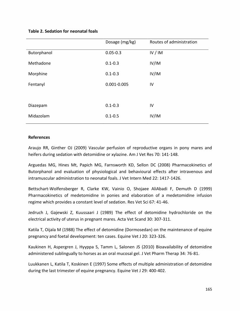
165
Table 2. Sedation for neonatal foals
Dosage (mg/kg) Routes of administration
Butorphanol 0.05-0.3 IV / IM
Methadone 0.1-0.3 IV/IM
Morphine 0.1-0.3 IV/IM
Fentanyl 0.001-0.005 IV
Diazepam 0.1-0.3 IV
Midazolam 0.1-0.5 IV/IM
References
Araujo RR, Ginther OJ (2009) Vascular perfusion of reproductive organs in pony mares and
heifers during sedation with detomidine or xylazine. Am J Vet Res 70: 141-148.
Arguedas MG, Hines Mt, Papich MG, Farnsworth KD, Sellon DC (2008) Pharmacokinetics of
Butorphanol and evaluation of physiological and behavioural effects after intravenous and
intramuscular administration to neonatal foals. J Vet Intern Med 22: 1417-1426.
Bettschart-Wolfensberger R, Clarke KW, Vainio O, Shojaee AliAbadi F, Demuth D (1999)
Pharmacokinetics of medetomidine in ponies and elaboration of a medetomidine infusion
regime which provides a constant level of sedation. Res Vet Sci 67: 41-46.
Jedruch J, Gajewski Z, Kuussaari J (1989) The effect of detomidine hydrochloride on the
electrical activity of uterus in pregnant mares. Acta Vet Scand 30: 307-311.
Katila T, Oijala M (1988) The effect of detomidine (Dormosedan) on the maintenance of equine
pregnancy and foetal development: ten cases. Equine Vet J 20: 323-326.
Kaukinen H, Aspergren J, Hyyppa S, Tamm L, Salonen JS (2010) Bioavailability of detomidine
administered sublingually to horses as an oral mucosal gel. J Vet Pharm Therap 34: 76-81.
Luukkanen L, Katila T, Koskinen E (1997) Some effects of multiple administration of detomidine
during the last trimester of equine pregnancy. Equine Vet J 29: 400-402.

166
Matthews NS (2009) Anaesthesia and analgesia for donkeys and mules. In: Muir WW, Hubbell
JAE, editors: Equine Anaesthesia – Monitoring and Emergency Therapy, St. Louis, Saunders
Elsevier.
Matthews NS, Taylor TS, Hartsfield SM (1997) Anaesthesia of donkeys and mules. Equine
Veterinary Education 9: 198-202.
Merritt AM, Burrow JA, Hartless CS (1998) Effect of xylazine, detomidine and a combination of
xylazine and butorphanol on equine duodenal motility. Am J Vet Res 59: 619-623.
Muir WW (2009) Anxiolytics, nonopioid sedative-analgesics, and opioid analgesics. In: Muir
WW, Hubbell JAE, editors: Equine Anaesthesia – Monitoring and Emergency Therapy, St. Louis,
Saunders Elsevier.
Parry PW, Anderson GA, Gay CG (1982) Hypotension in the horse induced by acepromazine
maleate. Aust Vet J 59: 148.
Schatzmann U, Jossfck H, Stauffer JL, Goossens L (1994) Effects of alpha 2-agonits on
intrauterine pressure and sedation in horses: comparison between detomidine, romifidine and
xylazine. Zentralbl Veterinarmed A. 41: 523-529.
Taylor PM, Clarke KW (2007) Handbook of equine anaesthesia 2nd Ed, St. Louis, Saunders
Elsevier.
Thurmon JC, Steffey EP, Zinkl JG, Woliner M, Howland D (1984) Xylazine causes transient dose-
related hyperglycaemia and increased urine volumes in mares. Am J Vet Res 45: 224-227.
Wagner AE, Mama KR, Contino EK, Ferris DJ, Kawcak CE (2011) Evaluation of sedation and
analgesia in sanding horses after administration of xylazine, butorphanol and sub-anaesthetic
doses2 of ketamine. J Am Vet Med Assoc 238: 1629-1633.
Wagner AE, Muir WW, Hinchcliff KW (1991) Cardiovascular effects of xylazine and detomidine
in horses. Am J Vet Res 52: 651-657.
Wilson DV (1994) Anaesthesia and sedation for late-term mares. Veterinary Clinics of North
America 10(1): 219-236.
Zullian C, Menozzi A, Pozzoli C, Poli E, Bertini S (2011) Effects of α2-adrenergic drugs on small
intestinal motility in the horse: An in vitro study. Vet J 187: 342-346.

167
PAIN IN THE ….DONKEY
GRIET HAITJEMA BVMS MVS FANZCVS Dip ECVAA
Murdoch University Veterinary Hospital
Donkeys have been used for over 5000 years as pack animals and transport mostly in
underdeveloped countries. Donkeys have the ability to survive in much harsher and arid
conditions than horses. Donkeys vary from horses in their muscle type fiber, milk composition,
hoof structure as well as their physiology and behaviour. Equine and mixed animal veterinary
practitioners usually provide donkey’s veterinary care. The donkey belongs in the equidea
family as does the horse, but a different subgenus asinus (vs. caballus for the horse).
In regards to pain expression, their behavioural changes tend to be stoic in nature. This makes
pain assessment and treatment in this species a true challenge as we need to detect very subtle
quiet and depressed expressions vs. overt behaviour. A donkey whose owner notices it is
moving much less may be seriously ill. We need to be aware that donkeys respond differently
to than horses, both in pharmacokinetic and pharmacodynamics parameters. Pain is defined by
the international association for the study of pain as an unpleasant sensory and emotional
experience associated with actual or potential tissue damage or described in such terms of such
damage. It results in suffering of the animal and should be assessed in routine physical
examination as the fifth vital sign with heart rate respiratory rate temperature and blood
pressure.
We routinely use behaviour and often heart rate to assess pain in the horse; the latter however
has been shown not to be good indicator of pain esp. for chronic pain. Ashley (2007) has
reviewed pain behaviour in both the horse and donkey with the main sign of pain in the donkey
being depression and reluctance to move. Hopefully further research being undertaken at the
University of Bristol will aid us in assessing pain in the donkey better. Pain assessment and
treatment in the horse and the donkey tend to be inconsistent and suboptimal. Areas such as
laminitis are consistently reported as painful and receive analgesia but other areas like elective
surgery such as castration shows marked variation in pain assessment and treatment. Pain
assessment can be done by simple numerical rating scale (NRS) or visual analog scale (VAS).
These methods are quick and simple to use but do suffer from inter and intra observational
variation. Equine pain scores have been developed. Raekallio et al (1997) assessed post-op
arthroscopic pain and showed difficulties with developing a pain score. Pritchett et al (2003)
developed a subjective NRS for gastrointestinal (GI) surgery noting behaviours associated with
pain but overall the horse displayed less behaviour in the post operative period. Rietmann et al
(2004) has looked at laminitic pain. Brussières et al (2008) developed a multifactorial numerical

168
rating orthopedic pain scale but also found that measuring indirect blood pressure might be of
use in assessing pain in the horse rather than heart rate. Price et al (2002) investigated
orthopedic pain and showed that assessing pain over time budget might be important as
intermitted observation of a horse in a stall may not allow you to pick up pain behaviour as the
animal is temporarily “distracted” by the observer. Composite pain score was used by Dutton
et al (2009) for severe hoof pain being treated by multimodal analgesia and is based on the
Glasgow composite pain score validated for dogs. Obel scoring system and the American
association of equine veterinarian lameness guidelines have also been used to assess laminitis
and lameness. Specific pain scores for the donkey need to be developed.
Dr Nora Matthews (2008) warns about the possibility of cardiovascular collapse on
anaesthetizing severely painful donkeys with laminitis or osteomyelitis that have not yet
received any form of analgesia. Preemptive analgesia is very important before inducing
unconsciousness in these animals. Intraoperative analgesia can be provided by a combination
of alpha 2 agonist; opioids such as methadone, buprenorphine and butorphanol as well as local
anesthesia. The analgesia effects of ketamine are additive to the previous groups when
hypersensitivity has developed due to NMDA receptor activation. Ketamine doses for analgesia
are much lower compared to those needed for anaesthesia. Post-operative analgesia is
provided using combination of non-steroidal anti-inflammatory drugs (NSAIDs), opioids as well
as various forms of local anaesthesia.
It is important to be aware that the donkey metabolises drugs different to the horse, i.e.
specific donkey pharmacokinetics (and dynamics) should be used to work out donkey doses.
Simple allometric scaling can probably be done if a drug is renal excreted; has minimal hepatic
metabolism or is flow limited. When the drug target does not differ much between species e.g.
antibiotic or is a drug that is not distribute extensively. Scaling does not apply to drugs that are
highly protein bound; undergo extensive metabolism and active transport; undergo significant
biliary excretion (mw> 500); have a target receptor that has species variation in expression
affinity and distribution; undergo extensive renal secretion or are drugs with significant
receptor binding effects.
NSAIDs such as phenylbutazone and flunixin are faster metabolised in the standard donkey and
maybe even faster in the miniature donkey requiring more frequent dosing. Ketoprofen and
Vedoprofen have been used in donkeys. Meloxicam appears to short a mean residence time to
be of use in the donkey, but perhaps carprofen is useful in the donkey as its affect appears to
be longer than the horse. See references below for dose rates.
Local anaesthetic drugs such as lignocaine and mepivacaine are commonly used for orthopedic
diagnostics but are also useful as surgical analgesia adjuvants. They can be use assess
desensitizing surgical incision site especially the testes and cremaster muscle during castration.
169
Analgesia can also be provided by using intravenous lidocaine or perineural infusions of most
common local anaesthetics and by application of a lidocaine dermal patch. When treating pain,
lower concentrations of lignocaine are able to block sodium (Na) channels associated with
neurogenic pain than those that would block full nerve transmission. It has become possible to
assess plasma levels of these drugs by kit form, which can give us clinical useful tool to start to
work towards target specific plasma levels. Local anaesthetic drugs main mode of action is
blocking active sodium channels. The antinociceptive effect of lidocaine is thought to work also
by affecting other channels such as calcium (Ca) and potassium (K) channels, blocking
presynaptic muscarinic receptors, NMDA receptors, TRPV1 receptors, sensory neurons and
decreasing substance P release; as well as activating GABA + Glycine receptors. Anti
inflammatory actions are postulated to include inhibition of sequestration migration and
activation on PMN’s; inhibition and adherence of PMN’s to endothelial cells; suppression of
histamine releases; decreasing chemotactic factors, cytokines and proinflammatory
lipoxygenase product release; inhibition of toxic oxygen metabolite release and decreases in
expression of iNO synthase.
Epidural injection is achieved in the donkey via the 2nd intercoccygeal space. Drugs than have
been described in the donkey include Lidocaine 2% 0.22mg/kg at 0.2ml/kg volume. Xylazine at
0.35mg/kg – 20mg/ml appears to have a shorter onset time of 20 min (vs. the horse).
Bupivacaine 0.06-0.08mg/kg in ml intrathecal had immediate onset lasting 80 min. Opioids
failed to produce analgesia to pin prick in healthy donkeys (morphine & fentanyl). Opioids
however are not expected to block A delta fiber pain that a pin prick would elicit. Ketamine at
0.8 – 1.2mg/kg in volume 0.15ml/kg) intrathecal provides analgesia for 40 min. Care should be
taken as ketamine may have local toxic tissue effects.
Giorgio (2009) has assessed Tramadol in the donkey. A dose of 2.5mg/kg IV over 30 min & per
os was investigated. No adverse effects seen. Tramadol’s mode of action is multimodal through
the active M1 metabolite thought to give an opioid effect as well as via the adrenergic and
serotonergic pathways. The M1 MEC remained above human known effective levels for 6- 8
hours after IV but was at this level for only 2 hours after PO administration. Tramadol has been
shown to be effective via epidural route in horses. Efficacy has not been assessed in the
donkey.
Ketamine as an analgesic works by NMDA receptor antagonist and decreasing wind-up. It is
useful as an adjunct analgesic when with hyperalgesia or opioid tolerance. Prolonged use can
lead to dysphoria. Excessive NMDA blockade is associated with memory impairment, excitatory
behaviour, dementia, ataxia, and motor incoordination. Ketamine has been assessed in horses
as an infusion or by IM injection (infusions dose: 0.8-1.5mg/kg/hr – aim for 0.13-0.25 ug/ml).
Lower dose infusions enhance opioid, alpha2 and gabapentin.

170
Gabapentin has been investigated in the horse and recommended when neurogenic based pain
is suspected. Its mode of action is via the interaction with α2-δ voltage gated Ca channel, which
results in decreasing neurotransmitter release associated with peripheral and central
sensitization. Dosages investigated in the horse show poor bioavailability. Some sedation, an
increased drinking frequency and less standing at rest were seen immediately after IV dose of
20mg/kg. This dose resulted in therapeutic levels shown to be effective in people a previous
study using 5mg/kg did not reach those levels. The pharmacokinetic of gabapentin still needs to
be investigated for the donkey.
Laminitis as a disease especially in its chronic form is difficult to treat and pain and suffering it
causes often results in the animal being put down. Recently Jones et al (2007) showed
neuropathic changes in laminitic horses. They demonstrated decreased myelinated and
demyelinated axons. The dorsal root ganglion of C8 but not C4 showed increased expression of
activation transcription factor 3 and an increase in Neuropeptide Y both of which are markers
for nerve damage. Neuropathic pain results in increased ectopic afferent activity from injured
nerve and DRG. The ectopic activity is due to increased Na channels and increased sensitivity to
cytokines and chemokines that are excitatory. There is a decreased inhibition from GABA and
Glycine. Sympathetic sprouting increases excitatory coupling with the peripheral neuroma and
its DRG. Activation of astrocytes and microglia leads to more excitatory products. These finding
means that pain should not only be looked at as a dependent variable due to the proposed
cause of laminitis (vascular; thromboembolic ischaemia; inflammatory; biomechanical etc).
Developments in pain physiology now show that pain itself can affect and become an
independent disease process in this disease. Pain is variable finding in laminitis with some
animals suffering more than others. It also means that a more multimodal approach to pain
management in these complex conditions is required. The current challenge lies in improving
firstly our ability to recognize and assess pain, secondly diagnosing the pathophysiology of the
pain as well as the condition the donkey is experiencing and then providing it with effective
pain relief and veterinary care.
References:
Ashley FH et al (2005) Behavioural Assessment of pain in horses and donkeys: application to
clinical practice and future studies. Eq Vet J 37(6):565-575.
Buda and Butrass (2005) Segmental nerve supply of the equine hoof Pferdeheilkunde 21(4):280-
284.
Bussie`res G (2008) Development of a composite orthopaedic pain scale in horses Research in Veterinary Science 85:294–306.

171
Collins SN et al Lamintic Pain: Parallels with Pain States in Humans and Other Species Vet Clin Equine 26(3):643-671.
Doherty TJ et al Local Anesthetics as Pain therapy in Horses Vet Clin Equine
36(3):533-549.
Dirikolu L et al (2008) Pharmacokinetics of gabapentin in horses. J vet pharm Therap 31:175-
177.
Dutton DW et al (2009) Managing severe hoof pain in a horse using multimodal analgesia and a modified composite pain score Equine vet Educ 21: 37-43.
Driesen B et al (2010) Neuropathic pain management in Chronic Laminitis Vet Clin Equine 26:315-337.
Fielding CL et al (2006) Pharmacokinetics and clinical effects of a subanesthetic continuous rate infusion of ketamine in awake horses AJVR 67(9):1484-1490.
Giorgio M (2009) Pharmacokinetics of tramadol and its metabolites M1,M2,M5 in Donkeys after intravenous and oral immediate release single dose administration J Eq vet science 29(7):569.
Grint N (2011) A Donkey is not a horse; developing nociceptive threshold testing in a new species AVA conference, Liverpool
Jones E et al (2007) Neuropathic changes in equine laminitis pain Pain 132:321–331
Lizarraga I et al (2004) Pharmacological and pharmacokinetic differences between donkeys and horses Eq Vet Educ 16(2):102-112.
Muir WW NMDA Receptor Antagonists and Pain: Ketamine Vet Clin Equine 26(3):565-578.
Naeni et al (1999) Comparison of Morphine, Fentanyl, Methadone, Lidocaine/Epinephrine as
Caudal Epidural Analgesia in the Donkey J App Animal Research 15:181-185.
Price J et al (2005) Current practice relating to equine castration in the UK Research in Veterinary Science 78:277–280.
Price J et al (2002) Pilot epidemiological study of attitudes towards pain in horses Vet Record 151:570-575
Raekallio M et al (1997) Preliminary Investigations of Pain and Analgesia Assessment in Horses Administered Phenylbutazone or Placebo After Arthroscopic Surgery Vet surg 26:150-155.

172
Rietmann TR et al (2004) The Association between Heart Rate, Heart Rate Variability, Endocrine and Behavioural Pain Measures in Horses Suffering from Laminitis J Vet Med A 51:218–225.
Sharma V et al (2009) To scale or not to scale: the principles of dose extrapolation British Journal of Pharmacology 157:907–921.
Sinclair MD et al (2006) Comparative pharmacokinetics of meloxicam in clinically normal horses and donkeys AJVR 67(6):1082-1085.
Terry RL et al (2010) Pharmacokinetic profile and behavioral effects of gabapentin in the horse J vet Pharmacol Therap 33:485–494.
Van Eps A (2010) Acute Laminitis: Medical and Supportive Therapy Vet Clin Equine 26:103-114.
Yaksh TL (2010) The Pain State Arising From the Laminitic Horse: Insights Into Future Analgesic Therapies Journal of Equine Veterinary Science 30(2).
Zarucco et al (2007) Continuous perineural block of Palmar Nerves: A New Technique for Pain Relief in the Distal Equine Forelimb Clin Tech Equine Pract 6:154-164.
References for drug doses:
Matthews N (2008) Anaesthesia and Sedation in The Professional Handbook of the Donkey 4th
ed pp 288 – 296.
Mathews N et al (1997) Anesthesia for donkeys and mules Eq Vet Ed 9(4):198-202.
Mathews NS and Van Wijk P (2004) Anesthesia and Analgesia in Donkeys in Veterinary Care of
Donkeys, NS Matthews and TS Taylor (Eds) Publisher: International Veterinary Information
Service (www.ivis.org), Ithaca, New York, USA,

173
ELECTROENCEPHALOGRAMS IN HORSES DURING GENERAL ANAESTHESIA
CRAIG JOHNSON
Massey University
This presentation will discuss the history and development of electroencephalographic (EEG)
techniques in the veterinary field with a particular emphasis on its applications in veterinary
anaesthesia research and practice and a focus on the equine.
After a brief coverage of the EEG usage prior to the 1990’s the methodologies and techniques
that led to the development of the minimal anaesthesia model will be outlined and illustrated
by a number of applications of this model to date. The presentation will conclude with some
thoughts about current and possible future uses of the EEG as an adjunct to anaesthetic
monitoring in clinical anaesthetic practice.