The microwell control of embryoid body size in order to regulate cardiac differentiation of human...
18
The Microwell Control of Embryoid Body Size in order to Regulate Cardiac Differentiation of Human Embryonic Stem Cells Jeffrey C. Mohr a,e,* , Jianhua Zhang b,e,* , Samira M. Azarin a,e , Andrew G. Soerens b,e , Juan J. de Pablo a , James A. Thomson c,d,e , Gary E. Lyons c , Sean P. Palecek a,e , and Timothy J. Kamp b,e a Department of Chemical and Biological Engineering, University of Wisconsin-Madison, WI, USA b Division of Cardiovascular Medicine in Department of Medicine, University of Wisconsin-Madison, WI, USA c Department of Anatomy, University of Wisconsin-Madison, WI, USA d Genomics Center of Wisconsin, University of Wisconsin-Madison, WI, USA e WiCell Research Institute, Madison, WI, USA Abstract The differentiation of human embryonic stem cells (hESCs) into cardiomyocytes (CMs) using embryoid bodies (EBs) is relatively inefficient and highly variable. Formation of EBs using standard enzymatic disaggregation techniques results in a wide range of sizes and geometries of EBs. Use of a 3-D cuboidal microwell system to culture hESCs in colonies of defined dimensions, 100 to 500 μm in lateral dimensions and 120 μm in depth, enabled formation of more uniform sized EBs. The 300 μm microwells produced highest percentage of contracting EBs, but flow cytometry for myosin light chain 2A (MLC2a) expressing cells revealed a similar percentage (~3%) of cardiomyocytes formed in EBs from 100 μm and 300 μm microwells. These data, and immunolabeling with anti- Corresponding author: Timothy J. Kamp, MD, PhD, University of Wisconsin School of Medicine and Public Health, Division of Cardiovascular Medicine, H6/370 CSC – MC 3248, 600 Highland Avenue, Madison, WI 53792 USA, Phone: (608) 263-4856 Fax: (608) 263-0405, [email protected]. * Co-first authors Author Contact Information Jeffrey C. Mohr, University of Wisconsin College of Engineering, 2732 Engineering Hall, 1415 Engineering Drive, Madison WI 53706, (608) 265-3413, Fax: (608) 262-5434, [email protected] Jianhua Zhang, University of Wisconsin School of Medicine and Public Health, Division of Cardiovascular Medicine 24 SMI, 470 N Charter Street, Madison WI 53706, (608) 263-0435, Fax: (608) 263-1144, [email protected] Samira M. Azarin, University of Wisconsin College of Engineering, 2732 Engineering Hall, 1415 Engineering Drive, Madison WI 53706, (608) 265-3413, Fax: (608) 262-5434, [email protected] Andrew G. Soerens, University of Wisconsin School of Medicine and Public Health, Division of Cardiovascular Medicine 24 SMI, 470 N Charter Street, Madison WI 53706, (608) 263-1049, Fax: (608) 263-1144, [email protected] Juan J. de Pablo, University of Wisconsin College of Engineering, 3018 Engineering Hall, 1415 Engineering Drive, Madison WI 53706, (608) 262-7727, Fax: (608) 262-5434, [email protected] James A. Thomson, PhD, VMD, University of Wisconsin School of Medicine and Public Health, Genomics Center of Wisconsin, 3420 Genetics-Biotechnology Center Bldg, 425 Henry Mall, Madison WI 53706, (608) 263-3585, Fax: (608) 890-0181, [email protected] Gary E. Lyons, PhD, University of Wisconsin School of Medicine and Public Health, Department of Anatomy, 318 Service Memorial Institute, 470 N. Charter St., Madison, WI 53706, (608) 262-2874, Fax: (608) 263-0150, [email protected] Sean P. Palecek, University of Wisconsin College of Engineering, 2732 Engineering Hall, 1415 Engineering Drive, Madison WI 53706, (608) 265-3413, Fax: (608) 262-5434, [email protected] A preliminary report was presented at the 6 th annual meeting of the International Society for Stem Cell Research. Publisher's Disclaimer: This is a PDF file of an unedited manuscript that has been accepted for publication. As a service to our customers we are providing this early version of the manuscript. The manuscript will undergo copyediting, typesetting, and review of the resulting proof before it is published in its final citable form. Please note that during the production process errors may be discovered which could affect the content, and all legal disclaimers that apply to the journal pertain. NIH Public Access Author Manuscript Biomaterials. Author manuscript; available in PMC 2011 March 1. Published in final edited form as: Biomaterials. 2010 March ; 31(7): 1885. doi:10.1016/j.biomaterials.2009.11.033. NIH-PA Author Manuscript NIH-PA Author Manuscript NIH-PA Author Manuscript
-
Upload
independent -
Category
Documents
-
view
0 -
download
0
Transcript of The microwell control of embryoid body size in order to regulate cardiac differentiation of human...
The Microwell Control of Embryoid Body Size in order to RegulateCardiac Differentiation of Human Embryonic Stem Cells
Jeffrey C. Mohra,e,*, Jianhua Zhangb,e,*, Samira M. Azarina,e, Andrew G. Soerensb,e, JuanJ. de Pabloa, James A. Thomsonc,d,e, Gary E. Lyonsc, Sean P. Paleceka,e, and Timothy J.Kampb,ea Department of Chemical and Biological Engineering, University of Wisconsin-Madison, WI, USAb Division of Cardiovascular Medicine in Department of Medicine, University of Wisconsin-Madison,WI, USAc Department of Anatomy, University of Wisconsin-Madison, WI, USAd Genomics Center of Wisconsin, University of Wisconsin-Madison, WI, USAe WiCell Research Institute, Madison, WI, USA
AbstractThe differentiation of human embryonic stem cells (hESCs) into cardiomyocytes (CMs) usingembryoid bodies (EBs) is relatively inefficient and highly variable. Formation of EBs using standardenzymatic disaggregation techniques results in a wide range of sizes and geometries of EBs. Use ofa 3-D cuboidal microwell system to culture hESCs in colonies of defined dimensions, 100 to 500μm in lateral dimensions and 120 μm in depth, enabled formation of more uniform sized EBs. The300 μm microwells produced highest percentage of contracting EBs, but flow cytometry for myosinlight chain 2A (MLC2a) expressing cells revealed a similar percentage (~3%) of cardiomyocytesformed in EBs from 100 μm and 300 μm microwells. These data, and immunolabeling with anti-
Corresponding author: Timothy J. Kamp, MD, PhD, University of Wisconsin School of Medicine and Public Health, Division ofCardiovascular Medicine, H6/370 CSC – MC 3248, 600 Highland Avenue, Madison, WI 53792 USA, Phone: (608) 263-4856 Fax: (608)263-0405, [email protected].*Co-first authorsAuthor Contact InformationJeffrey C. Mohr, University of Wisconsin College of Engineering, 2732 Engineering Hall, 1415 Engineering Drive, Madison WI 53706,(608) 265-3413, Fax: (608) 262-5434, [email protected] Zhang, University of Wisconsin School of Medicine and Public Health, Division of Cardiovascular Medicine 24 SMI, 470 NCharter Street, Madison WI 53706, (608) 263-0435, Fax: (608) 263-1144, [email protected] M. Azarin, University of Wisconsin College of Engineering, 2732 Engineering Hall, 1415 Engineering Drive, Madison WI 53706,(608) 265-3413, Fax: (608) 262-5434, [email protected] G. Soerens, University of Wisconsin School of Medicine and Public Health, Division of Cardiovascular Medicine 24 SMI, 470N Charter Street, Madison WI 53706, (608) 263-1049, Fax: (608) 263-1144, [email protected] J. de Pablo, University of Wisconsin College of Engineering, 3018 Engineering Hall, 1415 Engineering Drive, Madison WI 53706,(608) 262-7727, Fax: (608) 262-5434, [email protected] A. Thomson, PhD, VMD, University of Wisconsin School of Medicine and Public Health, Genomics Center of Wisconsin, 3420Genetics-Biotechnology Center Bldg, 425 Henry Mall, Madison WI 53706, (608) 263-3585, Fax: (608) 890-0181,[email protected] E. Lyons, PhD, University of Wisconsin School of Medicine and Public Health, Department of Anatomy, 318 Service MemorialInstitute, 470 N. Charter St., Madison, WI 53706, (608) 262-2874, Fax: (608) 263-0150, [email protected] P. Palecek, University of Wisconsin College of Engineering, 2732 Engineering Hall, 1415 Engineering Drive, Madison WI 53706,(608) 265-3413, Fax: (608) 262-5434, [email protected] preliminary report was presented at the 6th annual meeting of the International Society for Stem Cell Research.Publisher's Disclaimer: This is a PDF file of an unedited manuscript that has been accepted for publication. As a service to our customerswe are providing this early version of the manuscript. The manuscript will undergo copyediting, typesetting, and review of the resultingproof before it is published in its final citable form. Please note that during the production process errors may be discovered which couldaffect the content, and all legal disclaimers that apply to the journal pertain.
NIH Public AccessAuthor ManuscriptBiomaterials. Author manuscript; available in PMC 2011 March 1.
Published in final edited form as:Biomaterials. 2010 March ; 31(7): 1885. doi:10.1016/j.biomaterials.2009.11.033.
NIH
-PA Author Manuscript
NIH
-PA Author Manuscript
NIH
-PA Author Manuscript

MF20 and MLC2a, suggest that the smaller EBs are less likely to form contracting EBs, but thosecontracting EBs are relatively enriched in cardiomyocytes compared to larger EB sizes where CMsmake up a proportionately smaller fraction of the total cells. We conclude that microwell-engineeredEB size regulates cardiogenesis and can be used for more efficient and reproducible formation ofhESC-CMs needed for research and therapeutic applications.
Keywordshuman embryonic stem cells; microwells; embryoid body; cardiomyocytes; differentiation
1. IntroductionEfficiently guiding the differentiation of human embryonic stem cells (hESCs) has proven tobe a key roadblock in the development of therapeutic applications and further scientificdiscovery using hESCs. In the absence of controlling factors, spontaneous hESC differentiationis highly variable, dictated by factors such as hESC colony characteristics including size andcell density [1,2], as well as the cellular microenvironment [3,4]. Multiple methods have beenemployed to induce hESC differentiation, including embryoid body (EB) formation, inductiveco-culture with specific cell lines, and directed differentiation using particular growth factors[1,5–12]. However, for most cell lineages, the protocols are inefficient and poorly reproducible.
Differentiation of ESCs into cardiomyocytes has been extensively studied since the initialreport of cardiogenesis from mouse embryonic stem cells (mESCs) in EBs in 1985 [13].Research utilizing mESCs identified several variables which influence the extent ofcardiogenesis in EBs including the particular cell line, the starting number of cells used to formeach EB, medium and growth factors, and the duration of suspension culture prior to EB plating[14]. Early studies using enzymatic digestion of the mESC colonies resulted in a large rangeof EB sizes which differed in their ability to undergo cardiac differentiation [14–16]. Thissource of variability was overcome by using the hanging drop method of EB formation in whicha defined number of enzymatically isolated mESCs could be added to a drop where theyaggregate [17,18].
In the case of differentiation of hESCs to cardiac cell lineages, the differentiation protocolshave been relatively inefficient and are still undergoing rapid development. Formation of EBsis the most common approach to hESC cardiac differentiation, but substantial variability existswith this technique, likely in part due to the inconsistency in starting aggregate size.Unfortunately extrapolating the hanging drop technique directly to hESCs has not been possiblein most hands because enzymatically isolated single hESCs fail to aggregate and form an EB[19,20]. A variety of techniques have recently been explored to promote formation of uniform-sized aggregates for reproducible differentiation of hESCs. Enzymatically isolated hESCs havebeen subjected to forced aggregation using centrifugation to promote EB-mediatedhematopoietic and cardiac differentiation [19,21]. Micro-contact printing has been used topatterned 2-D aggregates of isolated hESCs to promote uniform EB size [22,23]. Anotherapproach involves use of microtextured surfaces composed of square-pyramidal pits in a siliconwafer in which ES cells could be seeded [24]. Each approach has a variety of advantages anddisadvantages. For example, the enzymatic isolation to single cells and forced-aggregationcould be stressful to hESCs and disrupt the cell-cell signaling required for hESC growth,survival, and differentiation. Some approaches are labor intensive and not readily scalable.Overall, these techniques have provided advances, but the goal of highly reproducible,efficient, scalable cardiac differentiation has not been obtained.
Mohr et al. Page 2
Biomaterials. Author manuscript; available in PMC 2011 March 1.
NIH
-PA Author Manuscript
NIH
-PA Author Manuscript
NIH
-PA Author Manuscript
Our recent study and others have demonstrated that hESC colonies can be efficiently grownand maintained in engineered 3-D microwells [25,26]. The microwells, which are surroundedby a cell and protein repellant self-assembled monolayer (SAM), promote hESC self-renewalfar longer than standard cultures, while still allowing differentiation to derivatives of each ofthe three primary germ layers upon removal from microwells. By constraining colony growthwithin microwells we are able to maintain cell-cell signaling and colony characteristicsnecessary for hESC survival and proliferation better than unconstrained standard culturetechniques. Thus we postulated that microwells could be used to impose a uniform 3-D sizeof hESC colonies used for formation of EBs and allow the use of uniform populations ofundifferentiated hESCs for EB formation. The present study examines the impact of varyingmicrowell dimensions on cardiogenesis from hESCs in EBs.
2. Materials and methods2.1 Microwell formation and functionalization
The detailed microwell manufacturing and functionalization protocol are described elsewhere[25]. Briefly, silicon masters were manufactured through photolithography and plasma etching.Masters were passivated under a fluorinated silane vapor to facilitate detachment of PDMSmolds. PDMS stamps were attached to glass slides and Norland optical adhesive 61 (NorlandProducts Inc., Cranbury, NJ, USA) polyurethane prepolymer was distributed between stampand glass slide via capillary action. After crosslinking, stamps were removed, yieldingmicrowells (Figure 1).
Using e-beam evaporation, microwells were coated with a thin layer of gold at oblique angles(>45°) to coat the sides of wells and areas between wells. Samples were then sterilized priorto self-assembled monolayer (SAM) formation and cell seeding by immersion in 100% ethanoland 1 hour UV sterilization. Microwells were functionalized with a tri-ethylene glycolterminated alkanethiol (EG3) (Prochimia, Poland) by immersion in a 2 mM ethanolic EG3solution for 2 hours. Microwell slides were then washed in 100% ethanol and air dried priorto cell seeding.
2.2 hESC culturehESC line H9 was used throughout all experiments. hESCs were cultured on either 6-welltissue culture polystyrene culture dishes (unconstrained), or in microwells. Both were coatedby growth factor reduced Matrigel (Beckton-Dickinson Bioscience, Medford, MA, USA).Culture medium was conditioned on MEFs for 24 hours and supplemented with 4 ng/ml bFGF.MEF medium is composed of DMEM (Invitrogen), fetal bovine serum (10%, heat inactivated,Invitrogen), and MEM non-essential amino acid solution (1%, Invitrogen). The hESC mediumwithout bFGF (UMF-) consists of DMEM/F12 (Invitrogen), Knockout Serum Replacer(KOSR, 20%, Invitrogen), L-glutamine (2 mmol/L, Invitrogen), and MEM non-essential aminoacid solution (1%, Invitrogen). The MEF conditioned hESC medium with bFGF (CMF+) issupplemented with 4 ng/ml bFGF. hESCs were passaged from unconstrained dishes tomicrowells using pre-warmed 1 mg/ml dispase in DMEM/F12. Plates were incubated at 37°Cuntil colonies were easily washed out of wells or off the plate.
2.3 Microwell cell seedingPrior to seeding cells in microwells, each microwell slide was placed in a well of a 6-well plateand was immersed in 100% ethanol to wet the surface and prevent air bubble entrapment inmicrowells. Microwell slides were washed in cold DMEM/F12 to remove all traces of ethanol.Matrigel solution was prepared by resuspending 2 mg growth factor reduced Matrigel in 24ml cold DMEM/F12. Microwells were coated with Matrigel by adding 2 ml of cold Matrigelsolution to each well of a 6-well plate and incubated for one hour at 37°C. Following the
Mohr et al. Page 3
Biomaterials. Author manuscript; available in PMC 2011 March 1.
NIH
-PA Author Manuscript
NIH
-PA Author Manuscript
NIH
-PA Author Manuscript

Matrigel incubation, microwell slides were washed in 2 ml PBS and then transferred to a fresh6-well plate. hESCs were passaged from 1 well of a 6-well plate to one microwell slide. hESCswere incubated up to 5 minutes in 1 mg/ml dispase solution until colony edges began liftingoff plate. Cells were gently scraped off the plate and washed once in UMF-. Cells were thenfiltered by 70 μm cell strainer to remove large clumps to facilitate seeding in microwell slides.Cells were washed twice in UMF- and pelleted. The pellet was then resuspended in 300 μlCMF+. In order to maximize cell seeding efficiency, the cell suspension was aliquoted to thetop of the microwell slide, taking care to maintain the entire cell suspension on the slide. Slideswere held at room temperature at least 30 minutes to allow cells to settle into wells and then 2ml/well CMF+ was added carefully to each well of the 6-well plate to prevent cells beingwashed out of microwells.
2.4 EB culture and differentiationThe general strategy for using microwells to form EBs and for the culture of the resulting EBsis shown schematically in Fig. 1. In brief, EBs were formed from hESC colonies removed frommicrowells or standard 6-well plates as described above and then cultured in suspension for 5days in 4 ml/well medium in Corning 3471 low attachment plates. EBs were suspended 1 dayin UMF-, followed by 4 days in an EB medium consisting of DMEM/F12 (Invitrogen), fetalbovine serum (20%, cardiac differentiation qualified, Hyclone, Catalog No. SH3007003, LotNo. ARH 27209), L- glutamine (2 mmol/L, Invitrogen), and MEM non-essential amino acidsolution (1%, Invitrogen). EBs were plated on 0.1% gelatin-coated 6-well plates or coverslipsin 24-well plates and cultured in EB medium for the duration of the experiment. After 10 daysof differentiation, FBS concentration in EB medium was reduced to 2%. At day 7, the totalattached EBs were counted under the microscope. EBs were observed daily, and contractingEBs were counted every three days beginning at day 9. The percentage of contracting EBs wasobtained by dividing the number of contracting EBs by the total number of EBs for each wellof 6-well plate and for each independent experiment.
2.5 QPCR analysisTotal RNA was isolated using Trizol Reagent (Invitrogen, Carlsbad, CA) from one well of EBculture in 6-well plate according to the manufacturer’s instruction. 2 μg of total RNA was usedper RT reaction using High-Capacity cDNA Archive Kit (Applied Biosystems, Foster City,CA). Each PCR was performed in triplicate with TaqMan® Universal PCR Kit (AppliedBiosystems, Foster City, CA) and TaqMan® Gene Expression Assays. TaqMan Primers forGAPDH (Hs99999905_m1), NKX2-5 (Hs00231763_m1), TNNT2 (Hs00165960_m1), MYL7(Hs00221909_ml) and PLN (Hs00160179_m1) were used. Fold change was calculated usingthe comparative Ct method (ΔΔCT method) [27].
2.6 Flow cytometryDifferentiated cells were detached from culture dishes for flow cytometry analysis by firstwashing twice in 2 ml/well PBS for 5 minutes at 37°C. Cells were then incubated in 1 ml/well0.25% trypsin-EDTA (Invitrogen), 2% chick serum (Sigma) for 10 minutes at 37°C, followedby neutralization using 4 ml/well FACS buffer (PBS without Ca/Mg2+, 2% FBS, 0.1% NaN3).Remaining cell aggregates were disrupted by gently pipetting. Cells were centrifuged 5 minutesat 1000 rpm, supernatant discarded, and the pellet was resuspended in 1 ml PBS with 60 μl16% paraformaldehyde for fixation. Samples were incubated 10 minutes in 37°C water bathand then chilled on ice 1 minute. Samples were centrifuged, pellet resuspended in 1 ml ice-cold 90% methanol, and incubated on ice for 30 minutes to permeabilize the cells. Cells werewashed once in FACS buffer plus 0.1% Triton, centrifuged, and supernatant discarded leavingabout 50 μl. Primary antibody was diluted in 50 μl/sample FACS buffer plus 0.1% Triton andaliquoted to each sample for total sample volume of 100 μl. Samples were incubated overnight
Mohr et al. Page 4
Biomaterials. Author manuscript; available in PMC 2011 March 1.
NIH
-PA Author Manuscript
NIH
-PA Author Manuscript
NIH
-PA Author Manuscript
at 4°C. MF20 and MLC2a primary antibodies and their dilutions were the same as describedin immunolabeling. Cells were washed twice in 3 ml FACS buffer plus 0.1% Triton,centrifuged, and supernatant discarded leaving ~ 50 μl. Secondary antibody, Alexa Fluor 633goat anti-mouse IgG2b (Invitrogen, Carlsbad, CA), was diluted 1:1000 in 50 μl/sample FACSbuffer plus Triton and aliquoted to each sample (final sample volume 100 μl). Samples wereincubated 30 to 45 min in the dark at room temperature, then washed twice in FACS buffer.Samples were centrifuged, resuspended in 300 μl FACS buffer, transferred to flow cytometrytubes, and stored on ice until analysis. Data were collected on a FACSCaliber flow cytometer(Beckton Dickinson) and analysis performed using CellQuest (Beckton Dickinson) software.All cells were gated according to light scatter and fluorescence.
2.7 ImmunolabelingEBs plated on coverslips were fixed in 4% Paraformaldehyde by diluting 16%Paraformaldehyde (EMS Catalog No. 15710-S) in 1X PBS (Invitrogen, Catalog No. 14190–144) and incubating for 15 minutes at room temperature. Cells were rinsed twice in PBS afterfixation, followed by permeabilization in 0.2% Triton X-100 in PBS solution (Sigma CatalogNo. T-9284) for 1 hour at room temperature. Samples were blocked by preparing fresh blockingsolution, 5% non-fat dry milk in 0.2% Triton X-100 solution, and incubated for two hours atroom temperature on a rotator, or at 4°C overnight covered with Parafilm. Samples werewashed twice with PBS, 5 minutes per wash. Primary antibodies, MF20 (DevelopmentalStudies Hybridoma Bank, Iowa City, IA) and anti-MLC2a (Synaptic Systems, Germany, Cat.No. 311011) were diluted 1:20 or 1:400, respectively, in 0.1% Triton X-100, 1% BSA in PBSsolution and incubated overnight at 4oC. Cells were washed with 0.2% Tween 20 in PBS threetimes, 5 minutes each, and 1X PBS once. Secondary antibody, Alexa Fluor 594 goat anti-mouseIgG2b (Invitrogen, Carlsbad, CA), was diluted in the same solution as the primary antibodyand incubated at room temperature for 1.5 hours in the dark. Three washes in 0.2% Tween 20in PBS, 5 minutes each, were followed by one 1X PBS wash. One drop of antifade reagentwith DAPI (Invitrogen, Catalog No. P36935) was placed on each slide, and coverslips wereapplied with cell surface down.
2.8 StatisticsData are presented as mean ± standard error of the mean (SEM). Statistical significance wasdetermined by one-way ANOVA or blocked one-way ANOVA where appropriate with posthoc testing using Tukey method using Microcal Origin, v7.5 and R. P < 0.05 was consideredstatistically significant. Data histograms were fit to Gaussian distributions using nonlinear leastsquares regression with Microcal Origin, version 7.5.
3. Results3.1 Size distribution of EBs from microwells
hESCs were seeded into Matrigel-coated cuboidal microwells, 100 to 500 μm in lateraldimensions and 120 μm in depth. After 6 days of growth in CMF+, hESC colonies wereenzymatically removed from microwells and cultured in EB medium in suspension to initiatedifferentiation. Upon removal from microwells and suspension culture for one day, sphericalEBs formed and exhibited relatively homogeneous sizes (Fig 2A-E) directly related to theinitial microwell dimensions in contrast to the more heterogeneous size distribution of EBsformed from standard hESC culture (Fig 2F). EB size distributions were quantified using imageanalysis by measuring the diameter of each EB and calculating the volume with the assumptionof spherical EBs. The volume of each size of EB was then normalized to the total EB volumein culture for each microwell size. Fig 2H shows the volume-weighted size distribution basedon EB diameter, and the data for each microwell size could be well described by a Gaussiandistribution. The average EB diameters (±SD) at one day of culture for the different microwell
Mohr et al. Page 5
Biomaterials. Author manuscript; available in PMC 2011 March 1.
NIH
-PA Author Manuscript
NIH
-PA Author Manuscript
NIH
-PA Author Manuscript
sizes were 88±16 μm (100), 162±24 μm (200), 256±28 μm (300), 322±36 μm (400), 350±51μm (500), and 196±68 μm (control). Thus the microwell constrained hESC culture system ledto predictable, uniform-sized EBs, particularly for the smaller sized microwells, in contrast tomore heterogeneous EB sizes resulting from unconstrained, conventional hESC culture onMatrigel.
If microwell size directly regulates EB size, then we postulated that the number of cells presentin EBs should be directly proportional to the microwell volume. Microwell EBs were placedin suspension culture for 1 day, and then subjected to enzymatic separation using trypsin toisolate individual cells which were counted using a hemacytometer. The average number ofcells per EB for each microwell size is plotted in Fig. 2G, and the data were fit to a simplepolynomial function based on the square of the X-Y dimension given that the depth of themicrowells was kept constant at 120 μm. Thus the microwell system allowed us tosystematically vary the number of cells in an EB after one day of culture over a relatively widerange, from ~140 cells/EB to ~3900 cells/EB.
We next assessed the effect of microwell size on EB dimensions following five days ofsuspension culture. The growth of EBs in culture reflects a complex combination of factorsincluding cellular proliferation, differentiation, apoptosis, and possible disaggregation. EB sizedistributions were quantified using image analysis by measuring the diameter of each EB asdescribed for day 1 EBs. The volume-weighted percent size distributions of EBs from thedifferent microwells could all be well described by Gaussian distributions, in contrast to moreheterogeneous EB size from the control EBs, as shown in Fig 2I. We observed that the 100μm and 200 μm microwells resulted in distinct sized EBs with mean diameters (±SD) of 135±37 μm and 206±44 μm, respectively; however, the large sized microwell EBs (300–500 μmdimensions) all tended to form similar sized EBs; mean EB diameters were 258±53 μm, 264±58 μm and 285±61 μm, respectively. Thus the smaller sized microwell EBs (100 and 200μm) tended to grow in size during five days of suspension culture, but the EBs from largersized microwells (300,400, and 500 μm) either did not increase in size or slightly decreased.Thus starting EB size can impact the growth properties of similarly maintained EBs.
3.2 Impact of microwell size on cardiogenesisOnce predictable EB size distributions had been obtained via microwell culture, we tested theeffect of EB size on cardiogenesis. To induce differentiation, hESCs were removed frommicrowells 120 μm deep x 100–500 μm lateral dimensions, after six days of culture in CMF+. The aggregates were cultured in suspension for one day in UMF which minimized aggregateto aggregate adhesion which otherwise occurred in the presence of serum containing medium.After the first day, the medium was changed to EB medium and after four days in suspension,the EBs were attached to gelatin-coated plates and cultured in EB medium for a total of 30days.
We first measured the fraction of EBs that formed spontaneously contracting areas as a functionof microwell dimensions and time in culture. Contracting EBs were initially observed withinthree days of plating on gelatin coated plates, and the fraction of contracting EBs increasedthrough time to reach a plateau at approximately 21 days (Fig 3). The 300 and 400 μmmicrowells produced the most spontaneously contracting EBs, achieving ~18–20% at 30 dayscompared to 100 and 200 μm microwells which produced ~8–10% contracting EBs. The 500μm microwells were intermediate with 14% contracting EBs at 30 days. In contrast to therelatively high percentages of contracting EBs afforded through microwell culture,unconstrained controls yielded only 5% contracting EBs, significantly less than 300 and 400μm microwell EBs. The time over which contracting EBs developed was comparable for allmicrowell sizes and the control EBs.
Mohr et al. Page 6
Biomaterials. Author manuscript; available in PMC 2011 March 1.
NIH
-PA Author Manuscript
NIH
-PA Author Manuscript
NIH
-PA Author Manuscript
To provide another assessment of cardiogenesis from the microwell EBs compared to standardunconstrained culture for formation of EBs, we performed real-time quantitative PCRexamining the expression of cardiac genes after 30 days of culture. TaqMan gene expressionassays for cardiac markers included a transcription factor critical for cardiac development,Nkx2.5 (human genome NKX2-5), a cardiac specific myofilament protein, cardiac troponin T(human genome TNNT2), and another cardiac myofilament protein, myosin light chain 2A(human genome MYL7). We compared expression of these three genes in EBs generated fromthree distinct microwell sizes to control EBs and found that like the measurements of percentageof contracting EBs, the greatest expression of NKX2-5, TNNT2, and MYL7 was in the 300 μmmicrowells with a 2–4 fold increase and were significantly different from the gene expressionof the control EBs (Fig. 4). The 100 μm microwell EBs also resulted in a significant increaseof NKX2-5 and TNNT2 expression over the control EBs. The 500 μm microwells showed nosignificant difference in expression of the genes under study compared to control EBs. Overall,the quantitative PCR data were consistent with the measurements of percentage of contractingEBs, with the 300 μm microwells demonstrating the greatest expression of cardiac markers.
Flow cytometry provided another quantitative assessment of cardiogenesis in the EBs bymeasuring the fraction of cells expressing cardiac-specific proteins. EBs were subjected toenzymatic separation to single cells, which were labeled with an antibody recognizing MLC2a,a ubiquitous myofilament protein expressed in all early cardiomyocytes types [28]. Data wereobtained from samples composed of a collection of EBs from a single well of a six well platecontaining 50-200 EBs for each experiment, so both contracting and noncontracting EBs wereincluded in the sample. In this collection of cells, the number of MLC2a positive cells fromcontrol EBs was ~1.2%. Evaluation of EBs from the different sized microwells revealed that500 μm microwells resulted in a comparable number of MLC2a positive cells (~2.0%)compared to control EBs; however, the 100 and 300 μm microwells produced on averageapproximately three fold more cardiomyocytes (~3.3%) in comparison to the control cells (Fig.5). Interestingly, the 100 μm microwell EBs formed as many or more cardiomyocytes than the300 μm microwell EBs in contrast to the finding that the 300 μm microwell EBs formed abouttwice as many contracting EBs compared to 100 μm EBs. This apparent discrepancy could beexplained by differences in the size of contracting areas in EBs, the presence of MLC2a innoncontracting EBs, or technical limitations in the flow cytometry assay. To evaluate forlimitations related to the chosen antibody against MLC2a, we also performed flow cytometryusing the antibody MF20 which recognizes sarcomeric myosin present in cardiomyocytes, andfound quantitatively similar results (data not shown).
To understand the distribution of cardiomyocytes in the differentiating EBs, we next performedimmunolabeling for cardiac proteins in EBs. The EBs were fixed at day 30 after plating ongelatin coated coverslips and immunolabeling was performed to identify all hESC-derivedcardiomyocytes using antibodies to sarcomeric myosin (MF20) and MLC2a. Only 5–20% ofthe EBs exhibited any immunolabeling for the cardiac proteins, and this fraction wasqualitatively similar to the fraction of contracting EBs. In the EBs immunolabeled with anti-MF20 or anti-MLC2a, positive cells were tightly clustered, consistent with observations oflocalized regions of contraction in EBs. Typically, only a single outgrowth of cardiomyocyteswas observed for an EB with the exception of some large EBs that contained multiple regions.Fig. 6 compares representative immunolabeled EBs from 100 μm, 300 μm, and 500 μmmicrowells. These images demonstrate that smaller EBs typically formed from the 100 μmmicrowell cultures, but the density of cardiomyocytes in the EBs was high, often composingthe majority of the cells in the EB. In contrast, the presence of cardiomyocytes in larger EBssuch as those from 300 μm and 500 μm microwell EBs was more restricted and limited to arelatively small fraction of the EB. These data demonstrate that the size of microwells used toform EBs can strongly impact the efficiency of cardiac differentiation in EBs with 100 μm
Mohr et al. Page 7
Biomaterials. Author manuscript; available in PMC 2011 March 1.
NIH
-PA Author Manuscript
NIH
-PA Author Manuscript
NIH
-PA Author Manuscript

microwells leading to EBs which are greatly enriched in cardiomyocytes relative to other celltypes.
4. DiscussionCardiac differentiation of hESCs in EBs has demonstrated a wide range of efficiency ranging0% to 70% of EBs containing contracting cardiomyocytes in published studies [5,16,19,29,30]. Some of the variability may be related to the use of different hESC lines with distinctpredispositions for cardiogenesis [19,31]. But even within a laboratory and using the samehESC line, there can be substantial variability. In recent years, microscale engineeringapproaches have been utilized to improve reproducibility, efficiency and scalability of stemcell differentiation. The present study used engineered microwells to produce hESC coloniesof defined 3-dimensional sizes and to generate specific-sized aggregates of hESCs for EBformation. By testing a range of microwell sizes, we demonstrated improved efficiency andreproducibility of cardiac differentiation using microwells compared to standard enzymaticmethods to form EBs.
The described microwell approach has several potential advantages over standard EBformation as well as over some micro-engineered techniques. First, the use of feeder free cultureof hESCs on Matrigel removes the potential confounding factors related to feeder cells whichcan inhibit cardiogenesis [32]. Secondly, the microwells provide a confined 3 dimensionalstructure that permits the hESC colonies to grow to uniform sizes and shapes that show lessvariability compared to standard culture of hESCs [25]. Uniformity in starting hESC colonyproperties is important for reproducibility of differentiation, and 2-dimensional patterning ofhESC culture using micro-contact printing could not eliminate the variability in the startinghESC gene expression patterns [33]. Finally, controlled 3-dimensional aggregate size used inEB formation is advantageous as we demonstrated that 300 μm wells were most efficient atgenerating contracting EBs, comparable to the 250–350 μm range found in the forcedaggregation method for forming human EBs as well as similar work with mouse ESCs (~300μm) [19,34]. Another microwell approach has been recently described which generatesdifferent sized inverted cuboidal pyramid microwells using similar silicon micropatterning[24]. However, seeding of the inverted cuboidal pyramid microwells requires enzymaticallyisolated single cells and exposes the cells to stresses from centrifugation or chemical means topromote aggregation in contrast to the present work where the ESCs are grown in themicrowells. An advantage of the pyramidal microwells is that they can be seeded with variouscell numbers to readily generate different sized aggregates which is not readily possible withour rectangular cuboidal microwells which require formation of different sized microwells forsuch variation in aggregate size. Whether the shape of the microwell and thus the initialaggregate shape impacts the differentiation process is not yet known.
The present study revealed that controlling EB size regulates cardiogenesis from hESCs notonly by impacting the likelihood an EB will generate some cardiomyocytes, but also the densityof cardiomyocytes relative to other cell types in the differentiating EB. By measuring both thepercentage of EBs contracting as well as the number of cardiomyocytes by flow cytometry,we discovered that the likelihood of a smaller 100 μm microwell EB undergoing cardiogenesiswas lower than 300 μm microwell EB, but if the 100 μm microwell EB developedcardiomyocytes, a much greater density of cardiomyocytes was found in the EB. Thisobservation has not been previously described in part because most prior studies only quantifycontracting EBs and not absolute cardiomyocytes number. Although the mechanism of thissize effect remains to be determined, this provides a potential tool to get the highest density ofcardiomyocytes from preparations if conditions can be optimized to initiate cardiogenesis insmall wells.
Mohr et al. Page 8
Biomaterials. Author manuscript; available in PMC 2011 March 1.
NIH
-PA Author Manuscript
NIH
-PA Author Manuscript
NIH
-PA Author Manuscript
The importance of EB size in regulating cardiac differentiation likely is related to diffusion ofcritical substrates as well as the role of various chemical cues such as growth factors.Observations of the patterns of expansion of the EBs revealed that the smaller sized EBs (100and 200 μm) showed the greatest growth in five days and larger EBs either did not expand orcontracted in size during this time period. Whether these distinct growth properties are relatedto a maximal size for EBs that can be supported by passive diffusion of substrates or due toother regulatory mechanisms is not known. Spatial gradients of a variety of signaling moleculesare essential for normal cardiac embryological development. For example, factors secretedfrom neighboring anterior lateral endoderm such as BMPs promote cardiogenesis whilecanonical Wnt signals from adjacent neuroectoderm inhibit cardiogenesis from mesoderm[35–39]. In EB differentiation, previous studies have suggested that at least a fraction of EBsexhibit radial multilaminar organization with an outer layer of extraembryonic endoderm thatmay be critical for the inner mass of cells to undergo cardiogenesis [24,40] [41–43]. Simplegeometrical considerations suggest that changes in EB diameter will greatly impact the relativeratio of this outer layer of extraembryonic endoderm and related signaling molecules to theinner mass of cells. Future studies will be needed to test how microwell technology can regulatesuch signaling.
Progress in using signaling molecules to direct differentiation of hESCs to cardiomyocytes hasbeen made using Activin A, BMP4, bFGF, and Wnt signaling inhibitor [6,12]. Most of thesetechniques involve the formation of aggregates of ESCs which potentially could bestandardized by microwell technology. Ultimately combining microwell technology toregulate the size of hESC colonies with controlled growth factor delivery in definedmicroenvironments may provide a powerful next step in the reproducible differentiation ofcardiomyocytes from ESCs as well as the more recently described induced pluripotent stemcells [44–46].
5. ConclusionOur results demonstrate that microwell technology can be used to culture hESCs and form EBsof defined sizes and cell numbers in contrast to more highly variable EBs formed using standardtechniques based on enzymatic or mechanical procedures. Furthermore, systematically varyingthe microwell size demonstrated that EBs from intermediate sized (300 μm) microwellsgenerated the highest percentage of contracting EBs. Evaluation of the EBs demonstrated thatmicrowell size also influenced the density of CMs in EBs with the smallest microwells (100μm) showing the greatest percentage of cells that were CMs in a contracting EB. Therefore,the process of cardiac differentiation of hESCs can be modulated in multiple ways by confinedculture conditions using microwells to grow hESCs and form EBs to improve the efficiencyof cardiogenesis. Decreasing the heterogeneity inherent in traditional EB methods will beinvaluable in optimizing the process for guiding differentiation to CMs and likely many otherlineages. Microwell technology can advance both basic research applications using hESCs aswell as contribute to the processes necessary to generate highly reproducible and abundant cellpopulations for clinical applications using human pluripotent stem cells.
AcknowledgmentsNIH/NIBIB R01 EB007534, NIH/NHLBI R01 HL08846150, NSF EFRI-0735903, and the WiCell Research Instituteprovided support for the study. The authors express appreciation for the assistance in manuscript preparation providedby Thankful Sanftleben. The authors confirm that there are no known conflicts of interest associated with thispublication, and there has been no significant financial support for this work that could have influenced its outcome.
Mohr et al. Page 9
Biomaterials. Author manuscript; available in PMC 2011 March 1.
NIH
-PA Author Manuscript
NIH
-PA Author Manuscript
NIH
-PA Author Manuscript
Abbreviations
hESC human embryonic stem cell
CM cardiomyocytes
EB embryoid body
References1. Odorico JS, Kaufman DS, Thomson JA. Multilineage differentiation from human embryonic stem cell
lines. Stem Cells 2001;19:193–204. [PubMed: 11359944]2. Watt FM, Hogan BL. Out of Eden: stem cells and their niches. Science 2000;287:1427–30. [PubMed:
10688781]3. Spradling A, Drummond-Barbosa D, Kai T. Stem cells find their niche. Nature 2001;414:98–104.
[PubMed: 11689954]4. Streuli C. Extracellular matrix remodelling and cellular differentiation. Curr Opin Cell Biol
1999;11:634–40. [PubMed: 10508658]5. Mummery C, Ward-van Oostwaard D, Doevendans P, Spijker R, van den BS, Hassink R, et al.
Differentiation of human embryonic stem cells to cardiomyocytes: role of coculture with visceralendoderm-like cells. Circulation 2003;107:2733–40. [PubMed: 12742992]
6. Laflamme MA, Chen KY, Naumova AV, Muskheli V, Fugate JA, Dupras SK, et al. Cardiomyocytesderived from human embryonic stem cells in pro-survival factors enhance function of infarcted rathearts. Nat Biotechnol 2007;25:1015–24. [PubMed: 17721512]
7. Schuldiner M, Yanuka O, Itskovitz-Eldor J, Melton DA, Benvenisty N. From the cover: effects of eightgrowth factors on the differentiation of cells derived from human embryonic stem cells. Proc NatlAcad Sci U S A 2000;97:11307–12. [PubMed: 11027332]
8. Itskovitz-Eldor J, Schuldiner M, Karsenti D, Eden A, Yanuka O, Amit M, et al. Differentiation ofhuman embryonic stem cells into embryoid bodies compromising the three embryonic germ layers.Mol Med 2000;6:88–95. [PubMed: 10859025]
9. Dvash T, Benvenisty N. Human embryonic stem cells as a model for early human development. BestPract Res Clin Obstet Gynaecol 2004;18:929–40. [PubMed: 15582547]
10. Tian X, Morris JK, Linehan JL, Kaufman DS. Cytokine requirements differ for stroma and embryoidbody-mediated hematopoiesis from human embryonic stem cells. Exp Hematol 2004;32:1000–9.[PubMed: 15504555]
11. Wang L, Li L, Shojaei F, Levac K, Cerdan C, Menendez P, et al. Endothelial and hematopoietic cellfate of human embryonic stem cells originates from primitive endothelium with hemangioblasticproperties. Immunity 2004;21:31–41. [PubMed: 15345218]
12. Yang L, Soonpaa MH, Adler ED, Roepke TK, Kattman SJ, Kennedy M, et al. Human cardiovascularprogenitor cells develop from a KDR+ embryonic-stem-cell-derived population. Nature2008;453:524–8. [PubMed: 18432194]
13. Doetschman TC, Eistetter H, Katz M, Schmidt W, Kemler R. The in vitro development of blastocyst-derived embryonic stem cell lines: formation of visceral yolk sac, blood islands and myocardium. JEmbryol Exp Morphol 1985;87:27–45. [PubMed: 3897439]
14. Boheler KR, Czyz J, Tweedie D, Yang HT, Anisimov SV, Wobus AM. Differentiation of pluripotentembryonic stem cells into cardiomyocytes. Circ Res 2002;91:189–201. [PubMed: 12169644]
15. Singla, DK.; Jayaraman, S.; Zhang, J.; Kamp, TJ. Cardiomyocyte Derivation from Human EmbryonicStem Cells. In: Master, JR.; Palsson, BO.; Thomson, JA., editors. Human Cell Culture 6: EmbryonicStem Cells. New York: Springer-Verlag; 2007. p. 211-34.
16. Xu C, Police S, Rao N, Carpenter MK. Characterization and enrichment of cardiomyocytes derivedfrom human embryonic stem cells. Circ Res 2002;91:501–8. [PubMed: 12242268]
17. He J-Q, January CT, Thomson J, Kamp TJ. Human embryonic stem cell-derived cardiomyocytes:Drug discovery and safety pharmacology. Expert Opinion on Drug Discovery 2007;2:39–753.
Mohr et al. Page 10
Biomaterials. Author manuscript; available in PMC 2011 March 1.
NIH
-PA Author Manuscript
NIH
-PA Author Manuscript
NIH
-PA Author Manuscript
18. Segev H, Kenyagin-Karsenti D, Fishman B, Gerecht-Nir S, Ziskind A, Amit M, et al. Molecularanalysis of cardiomyocytes derived from human embryonic stem cells. Dev Growth Differ2005;47:295–306. [PubMed: 16026538]
19. Burridge PW, Anderson D, Priddle H, Barbadillo M, Chamberlain S, Allegrucci C, et al. Improvedhuman embryonic stem cell embryoid body homogeneity and cardiomyocyte differentiation from anovel V-96 plate aggregation system highlights interline variability. Stem Cells 2007;25:929–38.[PubMed: 17185609]
20. Reubinoff BE, Pera MF, Fong CY, Trounson A, Bongso A. Embryonic stem cell lines from humanblastocysts: somatic differentiation in vitro. Nat Biotechnol 2000;18:399–404. [PubMed: 10748519]
21. Ng ES, Davis RP, Azzola L, Stanley EG, Elefanty AG. Forced aggregation of defined numbers ofhuman embryonic stem cells into embryoid bodies fosters robust, reproducible hematopoieticdifferentiation. Blood 2005;106:1601–3. [PubMed: 15914555]
22. Bauwnes CL, Peerani R, Niebruegge S, Woodhouse KA, Kumacheva E, Husain M, et al. Control ofhuman embryonic stem cell colony and aggregate size heterogeneity influences differentiationtrajectories. Stem Cells Express 2008;26:2300–10.
23. Peerani R, Rao BM, Bauwens C, Yin T, Wood GA, Nagy A, et al. Niche-mediated control of humanembryonic stem cell self-renewal and differentiation. The EMBO Jorunal 2007;26:4744–55.
24. Ungrin MD, Joshi C, Nica A, Bauwens C, Zandstra PW. Reproducible, ultra high- throughputformation of multicellular organization from single cell suspension-derived human embryonic stemcell aggregates. PLoS ONE 2008;3:e1565. [PubMed: 18270562]
25. Mohr JC, de Pablo JJ, Palecek SP. 3-D microwell culture of human embryonic stem cells. Biomaterials2006;27:6032–42. [PubMed: 16884768]
26. Khademhosseini A, Ferreira L, Blumling J III, Yeh J, Karp JM, Fukuda J, et al. Co-culture of humanembryonic stem cells with murine embryonic fibroblasts on microwell-patterned substrates.Biomaterials 2006;27:5968–77. [PubMed: 16901537]
27. Livak KJ, Schmittgen TD. Analysis of relative gene expression data using real-time quantitative PCRand the 2(-Delta Delta C(T)) Method. Methods 2001;25:402–8. [PubMed: 11846609]
28. Kubalak SW, Miller-Hance WC, O'Brien TX, Dyson E, Chien KR. Chamber specification of atrialmyosin light chain-2 expression precedes septation during murine cardiogenesis. J Biol Chem1994;269:16961–70. [PubMed: 8207020]
29. Kehat I, Kenyagin-Karsenti D, Snir M, Segev H, Amit M, Gepstein A, et al. Human embryonic stemcells can differentiate into myocytes with structural and functional properties of cardiomyocytes. JClin Invest 2001;108:407–14. [PubMed: 11489934]
30. He JQ, Ma Y, Lee Y, Thomson JA, Kamp TJ. Human embryonic stem cells develop into multipletypes of cardiac myocytes: action potential characterization. Circ Res 2003;93:32–9. [PubMed:12791707]
31. Osafune K, Caron L, Borowiak M, Martinez RJ, Fitz-Gerald CS, Sato Y, et al. Marked differencesin differentiation propensity among human embryonic stem cell lines. Nat Biotechnol 2008;26:313–5. [PubMed: 18278034]
32. Tomescot A, Leschik J, Bellamy V, Dubois G, Messas E, Bruneval P, et al. Differentiation in vivoof cardiac committed human embryonic stem cells in postmyocardial infarcted rats. Stem Cells2007;25:2200–5. [PubMed: 17540853]
33. Bauwens CL, Peerani R, Niebruegge S, Woodhouse KA, Kumacheva E, Husain M, et al. Control ofhuman embryonic stem cell colony and aggregate size heterogeneity influences differentiationtrajectories. Stem Cells 2008;26:2300–10. [PubMed: 18583540]
34. Rudy-Reil D, Lough J. Avian precardiac endoderm/mesoderm induces cardiac myocytedifferentiation in murine embryonic stem cells. Circ Res 2004;94:e107–16. [PubMed: 15192018]
35. Schultheiss TM, Burch JB, Lassar AB. A role for bone morphogenetic proteins in the induction ofcardiac myogenesis. Genes Dev 1997;11:451–62. [PubMed: 9042859]
36. Sugi Y, Lough J. Anterior endoderm is a specific effector of terminal cardiac myocyte differentiationof cells from the embryonic heart forming region. Dev Dyn 1994;200:155–62. [PubMed: 7919501]
37. Climent S, Sarasa M, Villar JM, Murillo-Ferrol NL. Neurogenic cells inhibit the differentiation ofcardiogenic cells. Dev Biol 1995;171:130–48. [PubMed: 7556890]
Mohr et al. Page 11
Biomaterials. Author manuscript; available in PMC 2011 March 1.
NIH
-PA Author Manuscript
NIH
-PA Author Manuscript
NIH
-PA Author Manuscript
38. Cohen ED, Tian Y, Morrisey EE. Wnt signaling: an essential regulator of cardiovasculardifferentiation, morphogenesis and progenitor self-renewal. Development 2008;135:789–98.[PubMed: 18263841]
39. Wagner M, Siddiqui MA. Signal transduction in early heart development (I): cardiogenic inductionand heart tube formation. Exp Biol Med (Maywood ) 2007;232:852–65. [PubMed: 17609501]
40. Conley BJ, Trounson AO, Mollard R. Human embryonic stem cells form embryoid bodies containingvisceral endoderm-like derivatives. Fetal Diagn Ther 2004;19:218–23. [PubMed: 15067230]
41. Coucouvanis E, Martin GR. Signals for death and survival: a two-step mechanism for cavitation inthe vertebrate embryo. Cell 1995;83:279–87. [PubMed: 7585945]
42. Li X, Chen Y, Scheele S, Arman E, Haffner-Krausz R, Ekblom P, et al. Fibroblast growth factorsignaling and basement membrane assembly are connected during epithelial morphogenesis of theembryoid body. J Cell Biol 2001;153:811–22. [PubMed: 11352941]
43. Conley BJ, Ellis S, Gulluyan L, Mollard R. BMPs regulate differentiation of a putative visceralendoderm layer within human embryonic stem-cell-derived embryoid bodies. Biochem Cell Biol2007;85:121–32. [PubMed: 17464352]
44. Yu J, Vodyanik MA, Smuga-Otto K, Antosiewicz-Bourget J, Frane JL, Tian S, et al. Inducedpluripotent stem cell lines derived from human somatic cells. Science 2007;318:1917–20. [PubMed:18029452]
45. Takahashi K, Tanabe K, Ohnuki M, Narita M, Ichisaka T, Tomoda K, et al. Induction of pluripotentstem cells from adult human fibroblasts by defined factors. Cell 2007;131:861–72. [PubMed:18035408]
46. Zhang J, Wilson GF, Soerens AG, Koonce CH, Yu J, Palecek SP, et al. Functional cardiomyocytesderived from human induced pluripotent stem cells. Circ Res 2009;104:e30–41. [PubMed: 19213953]
Mohr et al. Page 12
Biomaterials. Author manuscript; available in PMC 2011 March 1.
NIH
-PA Author Manuscript
NIH
-PA Author Manuscript
NIH
-PA Author Manuscript
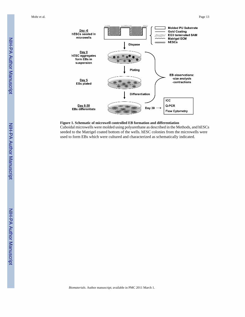
Figure 1. Schematic of microwell controlled EB formation and differentiationCuboidal microwells were molded using polyurethane as described in the Methods, and hESCsseeded to the Matrigel coated bottom of the wells. hESC colonies from the microwells wereused to form EBs which were cultured and characterized as schematically indicated.
Mohr et al. Page 13
Biomaterials. Author manuscript; available in PMC 2011 March 1.
NIH
-PA Author Manuscript
NIH
-PA Author Manuscript
NIH
-PA Author Manuscript
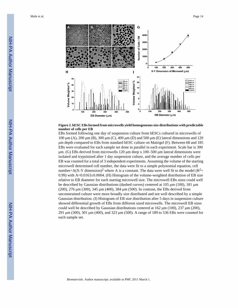
Figure 2. hESC EBs formed from microwells yield homogeneous size distributions with predictablenumber of cells per EBEBs formed following one day of suspension culture from hESCs cultured in microwells of100 μm (A), 200 μm (B), 300 μm (C), 400 μm (D) and 500 μm (E) lateral dimensions and 120μm depth compared to EBs from standard hESC culture on Matrigel (F). Between 68 and 185EBs were evaluated for each sample set done in parallel in each experiment. Scale bar is 300μm. (G) EBs derived from microwells 120 μm deep x 100–500 μm lateral dimensions wereisolated and trypsinized after 1 day suspension culture, and the average number of cells perEB was counted for a total of 3 independent experiments. Assuming the volume of the startingmicrowell determined cell number, the data were fit to a simple polynomial equation, cellnumber=A(X-Y dimension)2 where A is a constant. The data were well fit to the model (R2=0.99) with A=0.0163±0.0004. (H) Histogram of the volume-weighted distribution of EB sizerelative to EB diameter for each starting microwell size. The microwell EBs sizes could wellbe described by Gaussian distributions (dashed curves) centered at 105 μm (100), 181 μm(200), 276 μm (300), 345 μm (400), 384 μm (500). In contrast, the EBs derived fromunconstrained culture were more broadly size distributed and not well described by a simpleGaussian distribution. (I) Histogram of EB size distribution after 5 days in suspension cultureshowed differential growth of EBs from different sized microwells. The microwell EB sizescould well be described by Gaussian distributions centered at 162 μm (100), 237 μm (200),291 μm (300), 301 μm (400), and 323 μm (500). A range of 189 to 536 EBs were counted foreach sample set.
Mohr et al. Page 14
Biomaterials. Author manuscript; available in PMC 2011 March 1.
NIH
-PA Author Manuscript
NIH
-PA Author Manuscript
NIH
-PA Author Manuscript
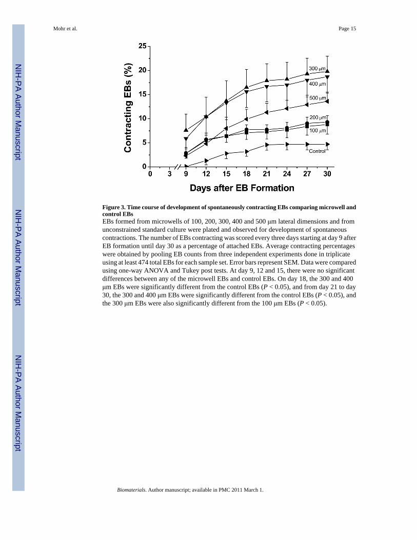
Figure 3. Time course of development of spontaneously contracting EBs comparing microwell andcontrol EBsEBs formed from microwells of 100, 200, 300, 400 and 500 μm lateral dimensions and fromunconstrained standard culture were plated and observed for development of spontaneouscontractions. The number of EBs contracting was scored every three days starting at day 9 afterEB formation until day 30 as a percentage of attached EBs. Average contracting percentageswere obtained by pooling EB counts from three independent experiments done in triplicateusing at least 474 total EBs for each sample set. Error bars represent SEM. Data were comparedusing one-way ANOVA and Tukey post tests. At day 9, 12 and 15, there were no significantdifferences between any of the microwell EBs and control EBs. On day 18, the 300 and 400μm EBs were significantly different from the control EBs (P < 0.05), and from day 21 to day30, the 300 and 400 μm EBs were significantly different from the control EBs (P < 0.05), andthe 300 μm EBs were also significantly different from the 100 μm EBs (P < 0.05).
Mohr et al. Page 15
Biomaterials. Author manuscript; available in PMC 2011 March 1.
NIH
-PA Author Manuscript
NIH
-PA Author Manuscript
NIH
-PA Author Manuscript
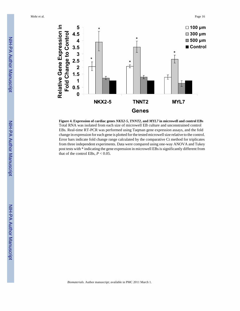
Figure 4. Expression of cardiac genes NKX2-5, TNNT2, and MYL7 in microwell and control EBsTotal RNA was isolated from each size of microwell EB culture and unconstrained controlEBs. Real-time RT-PCR was performed using Taqman gene expression assays, and the foldchange in expression for each gene is plotted for the tested microwell size relative to the control.Error bars indicate fold change range calculated by the comparative Ct method for triplicatesfrom three independent experiments. Data were compared using one-way ANOVA and Tukeypost tests with * indicating the gene expression in microwell EBs is significantly different fromthat of the control EBs, P < 0.05.
Mohr et al. Page 16
Biomaterials. Author manuscript; available in PMC 2011 March 1.
NIH
-PA Author Manuscript
NIH
-PA Author Manuscript
NIH
-PA Author Manuscript
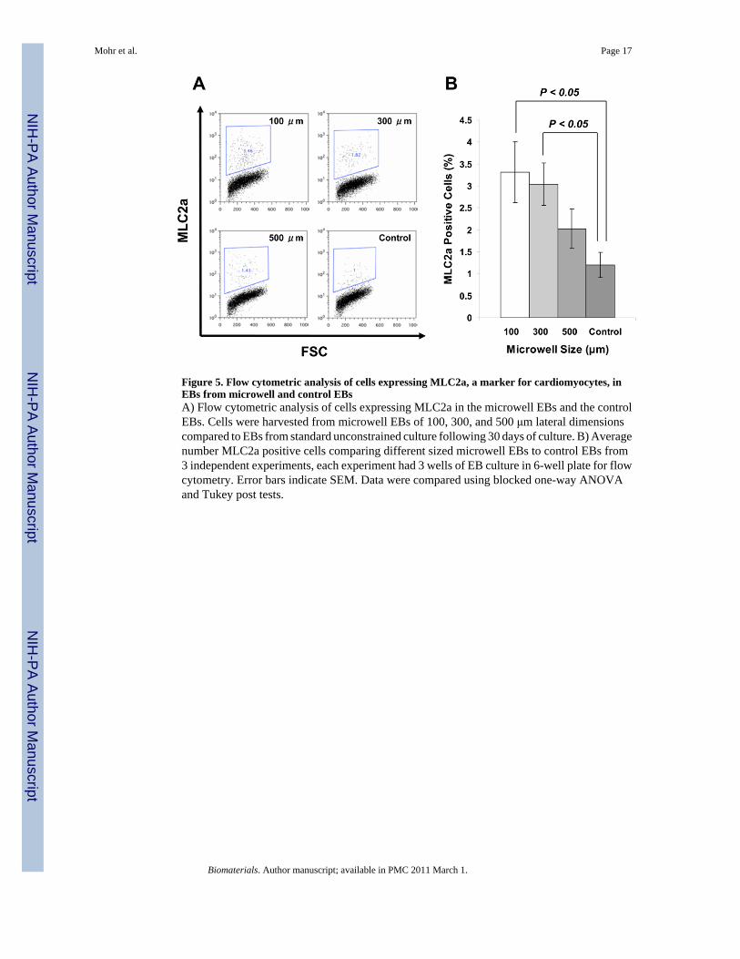
Figure 5. Flow cytometric analysis of cells expressing MLC2a, a marker for cardiomyocytes, inEBs from microwell and control EBsA) Flow cytometric analysis of cells expressing MLC2a in the microwell EBs and the controlEBs. Cells were harvested from microwell EBs of 100, 300, and 500 μm lateral dimensionscompared to EBs from standard unconstrained culture following 30 days of culture. B) Averagenumber MLC2a positive cells comparing different sized microwell EBs to control EBs from3 independent experiments, each experiment had 3 wells of EB culture in 6-well plate for flowcytometry. Error bars indicate SEM. Data were compared using blocked one-way ANOVAand Tukey post tests.
Mohr et al. Page 17
Biomaterials. Author manuscript; available in PMC 2011 March 1.
NIH
-PA Author Manuscript
NIH
-PA Author Manuscript
NIH
-PA Author Manuscript

Figure 6. Immunolocalization of cardiomyocytes in EBs formed from different sized microwellsPanels A – C show immunolabeling with MF20 antibody (red) which recognizes sarcomericmyosin present in cardiomyocytes and nuclei are stained with DAPI (blue), in 30 days old EBsformed from 100 μm, 300 μm and 500 μm microwells , respectively. Panels D - F showimmunolabeling with anti-MLC2a antibody (red), which recognizes a myofilament proteinexpressed in all early embryonic cardiomyocytes, and nuclei are stained with DAPI (blue), in30 day old EBs formed from 100 μm, 300 μm and 500 μm microwells, respectively. Scale baris 300 μm.
Mohr et al. Page 18
Biomaterials. Author manuscript; available in PMC 2011 March 1.
NIH
-PA Author Manuscript
NIH
-PA Author Manuscript
NIH
-PA Author Manuscript