Embryoid Body–Mediated Differentiation of Mouse Embryonic Stem Cells Along a Hepatocyte Lineage:...
11
1515 INTRODUCTION P LURIPOTENT EMBRYONIC STEM CELLS (ES) represent a promising renewable cell source to generate a vari- ety of differentiated cell types including cardiomy- ocytes, 1 neural precursors, 2 and hepatocyte lineage cells, 3 and may ultimately be incorporated into implantable en- gineered tissue constructs, or ex vivo cell-based thera- peutic devices. 4–7 In vitro aggregation of murine ES cells initiates the formation of embryoid bodies (EBs), which can facilitate spontaneous, unguided differentiation anal- ogous to that seen in developing mouse embryos. Dur- ing mouse embryogenesis, the primitive ectoderm gives rise to three distinct embryonic germ layers: definitive endoderm, mesoderm, and ectoderm. 8,9 The primitive en- doderm gives rise to both the visceral and parietal endo- derm and while the definitive endoderm ultimately dif- ferentiates into mature hepatic tissue, the visceral endo- derm is an extraembryonic tissue, which expresses many genes also commonly expressed in differentiating liver cells. 10 Analyses characterizing the extent of EB differentia- tion have been limited to one or several lineage-specific protein or gene expression patterns. For example, after extracellular matrix (ECM) and exogenous growth fac- tor supplementation, several groups of investigators have demonstrated EB-mediated differentiation of a popula- tion of albumin-positive cells, 3,11–13 or cells that express genes for albumin (ALB), alpha-feto protein (AFP), and transthyretin protein (TRT). 14–18 Spontaneous differenti- TISSUE ENGINEERING Volume 12, Number 6, 2006 © Mary Ann Liebert, Inc. Embryoid Body–Mediated Differentiation of Mouse Embryonic Stem Cells Along a Hepatocyte Lineage: Insights from Gene Expression Profiles ERIC I. NOVIK, TIM J. MAGUIRE, KSENIA ORLOVA, RENE S. SCHLOSS, and MARTIN L. YARMUSH ABSTRACT Pluripotent embryonic stem (ES) cells represent a promising renewable cell source for the genera- tion of functional differentiated cells. Previous studies incorporating embryoid body (EB)-mediated stem cell differentiation have, either spontaneously or after growth factor and extracellular matrix protein supplementation, yielded populations of hepatocyte lineage cells expressing mature hepato- cyte markers such as albumin (ALB). In an effort to promote ES cell commitment to the hepato- cyte lineage, we have evaluated the effects of four culture conditions on albumin and gene expres- sion in differentiating ES cells. Quantitative in situ immunofluorescence and cDNA microarray analyses were used to describe not only lineage specificity but also to provide insights into the ef- fects of disparate culture environments on the mechanisms of differentiation. The results of these studies suggest that spontaneous and collagen-mediated differentiation induce cells with the high- est levels of ALB expression but mature liver specific genes were only expressed in the spontaneous condition. Further analysis of gene expression profiles indicated that two distinct mechanisms may govern spontaneous and collagen-mediated differentiation. Department of Biomedical Engineering, Rutgers University, Piscataway, New Jersey.
-
Upload
independent -
Category
Documents
-
view
2 -
download
0
Transcript of Embryoid Body–Mediated Differentiation of Mouse Embryonic Stem Cells Along a Hepatocyte Lineage:...

1515
INTRODUCTION
PLURIPOTENT EMBRYONIC STEM CELLS (ES) represent apromising renewable cell source to generate a vari-
ety of differentiated cell types including cardiomy-ocytes,1 neural precursors,2 and hepatocyte lineage cells,3
and may ultimately be incorporated into implantable en-gineered tissue constructs, or ex vivo cell-based thera-peutic devices.4–7 In vitro aggregation of murine ES cellsinitiates the formation of embryoid bodies (EBs), whichcan facilitate spontaneous, unguided differentiation anal-ogous to that seen in developing mouse embryos. Dur-ing mouse embryogenesis, the primitive ectoderm givesrise to three distinct embryonic germ layers: definitiveendoderm, mesoderm, and ectoderm.8,9 The primitive en-
doderm gives rise to both the visceral and parietal endo-derm and while the definitive endoderm ultimately dif-ferentiates into mature hepatic tissue, the visceral endo-derm is an extraembryonic tissue, which expresses manygenes also commonly expressed in differentiating livercells.10
Analyses characterizing the extent of EB differentia-tion have been limited to one or several lineage-specificprotein or gene expression patterns. For example, afterextracellular matrix (ECM) and exogenous growth fac-tor supplementation, several groups of investigators havedemonstrated EB-mediated differentiation of a popula-tion of albumin-positive cells,3,11–13 or cells that expressgenes for albumin (ALB), alpha-feto protein (AFP), andtransthyretin protein (TRT).14–18 Spontaneous differenti-
TISSUE ENGINEERINGVolume 12, Number 6, 2006© Mary Ann Liebert, Inc.
Embryoid Body–Mediated Differentiation of Mouse Embryonic Stem Cells Along a Hepatocyte Lineage:
Insights from Gene Expression Profiles
ERIC I. NOVIK, TIM J. MAGUIRE, KSENIA ORLOVA, RENE S. SCHLOSS, and MARTIN L. YARMUSH
ABSTRACT
Pluripotent embryonic stem (ES) cells represent a promising renewable cell source for the genera-tion of functional differentiated cells. Previous studies incorporating embryoid body (EB)-mediatedstem cell differentiation have, either spontaneously or after growth factor and extracellular matrixprotein supplementation, yielded populations of hepatocyte lineage cells expressing mature hepato-cyte markers such as albumin (ALB). In an effort to promote ES cell commitment to the hepato-cyte lineage, we have evaluated the effects of four culture conditions on albumin and gene expres-sion in differentiating ES cells. Quantitative in situ immunofluorescence and cDNA microarrayanalyses were used to describe not only lineage specificity but also to provide insights into the ef-fects of disparate culture environments on the mechanisms of differentiation. The results of thesestudies suggest that spontaneous and collagen-mediated differentiation induce cells with the high-est levels of ALB expression but mature liver specific genes were only expressed in the spontaneouscondition. Further analysis of gene expression profiles indicated that two distinct mechanisms maygovern spontaneous and collagen-mediated differentiation.
Department of Biomedical Engineering, Rutgers University, Piscataway, New Jersey.

ation of human EBs into hepatocyte lineage cells, in theabsence of both ECM and exogenous growth factor sup-plementation, has also been identified using gene mi-croarray analysis of several known mature liver specificproteins.20,21 Despite the growing number of studies re-porting ES/hepatocyte differentiation, isolation of purepopulations of neither committed hepatocyte precursorsnor fully differentiated hepatocytes have been reported.Furthermore, it is unclear whether both spontaneous anddirected EB-mediated differentiations are controlled bythe same regulatory pathways or whether they yield iden-tical hepatocyte lineage cells.
In the present study, we have evaluated the effects offour culture configurations on albumin and gene expres-sion in order to gain further insight into the regulation ofhepatocyte lineage commitment. We have assessed thedynamic expression of albumin within individual EBs. Inaddition, using complete mouse genome cDNA microar-ray analyses, we have assessed not only mature hepato-cyte gene expression but have also explored the mecha-nism of hepatocyte lineage differentiation from ES cells.These studies identified unique hepatocyte-specific dif-ferentiation profiles among the culture conditions thatmay assist in unraveling the mechanisms that control EB-mediated hepatocyte differentiation and/or albuminexpression.
MATERIALS AND METHODS
ES cell culture
All cell cultures were incubated at 37°C in a humidi-fied 5% CO2. The ES cell line D3 (ATCC, Manassas,VA) was maintained in an undifferentiated state in T-75gelatin-coated dishes (Biocoat, BD-Biosciences, Bed-ford, MA) in Knockout Dulbecco’s Modified EagleMedium (Gibco, Grand Island, NY) containing 15%knockout serum (Gibco), 4 mM L-glutamine (Gibco), 100U/mL penicillin (Gibco), 100 U/mL streptomycin(Gibco), 10 �g/mL gentamicin (Gibco), 1000 U/mL ESGRO (Chemicon, Temecula, CA), 0.1 mM 2-mercap-toethanol (Sigma-Aldrich, St. Louis, MO). Media waschanged every 2 days. In order to induce differentiation,cells where suspended in Iscove’s modified Dulbecco’smedium containing 20% fetal bovine serum (Gibco), 4 mM L-glutamine (Gibco), 100 U/mL penicillin, 100U/mL streptomycin (Gibco), 10 �g/mL gentamicin(Gibco). Embryoid bodies were formed and cultured for2 days using the hanging drop method (1 � 103 ES cellsper 30-�L drop). Hanging drops were transferred to sus-pension culture in 100-mm Petri dishes and cultured foran additional 3 days. The EBs were then plated, one EBper well, in six-well plates coated with or without colla-gen type I (40–60 �g/mL, Discovery Labware, Bedford,MA). When growth factors were supplemented, 100
NOVIK ET AL.
ng/mL acidic fibroblast growth factor (aFGF) (days8–11) and 20 ng/mL hepatocyte growth factor (HGF)(Sigma, St. Louis, MO) (days 11–17) were added to theculture medium. Growth factors were selected to induceearly hepatocyte commitment and not late-stage differ-entiation. The Hepa 1-6 cell line (ATCC, Manassas, VA)was maintained in Dulbecco’s Modified Eagle Medium(Gibco) containing 10% fetal bovine serum (Gibco), 100 U/mL penicillin (Gibco), 100 U/mL streptomycin(Gibco), and 4 mM L-glutamine (Gibco).
EB size
EB size was evaluated as a function of cell number/EB.Individual EBs were trypsinized and cells were counted.Twelve EBs were counted for each experimental day, andthe experiments were repeated three times. The same pro-cedure was followed for all culture conditions.
In situ indirect immunofluorescence
On evaluation days 11, 14, and 17, EBs were washedin phosphate-buffered saline (PBS) (Gibco) and fixed in4% paraformaldehyde (Sigma-Aldrich) in PBS for 15min at room temperature. The EBs were washed twicefor 10 min in cold PBS and then twice for 10 min in coldsaponin/PBS (SAP) membrane permeabilization buffercontaining 1% bovine serum albumin (Sigma-Aldrich),0.5% saponin (Sigma-Aldrich), and 0.1% sodium azide(Sigma-Aldrich). The EBs were subsequently incubatedfor 30 min at 4°C in a SAP solution containing rabbitanti-mouse albumin antibody (150 �g/mL) (MP Bio-medicals, Irvine, CA), or normal rabbit serum (NRS) (150�g/mL) (MP Biomedicals) as an isotype control of thebackground intensity generated by nonspecific binding,washed twice for 10 min in cold SAP buffer, and thentreated for 30 min at 4°C with the secondary antibody,fluorescein isothiocyanate–conjugated donkey anti-rab-bit, diluted 1:500 (Jackson Immuno Labs, Westgrove,PA). EBs were then washed once with cold SAP bufferand once with cold PBS. Fluorescent images were ac-quired with an Olympus IX70 microscope and Olympusdigital camera using an excitation filter of 515 nm. Flu-orescent albumin expressing cells were only observed inthe outer EB region and not in the dense intermediate re-gion or the central core. Image fluorescence was quanti-fied with Olympus Microsuite. To generate intensity val-ues for each of the samples, 10 cells located within thisouter monolayer EB region were chosen at random fromthe phase contrast image. The average intensity value for10 randomly chosen cells was measured in the NRS (con-trol) case, and this average background intensity was thensubtracted from each value of the 10 randomly chosencells in the anti-albumin (experimental) case. This wasdone in triplicate experiments yielding a total of 30 cellsper condition.
1516

Each data point represents the mean, and the error barsrepresent the standard error of the mean. Statistical sig-nificance was determined using the student t-test for un-paired data. Differences were considered significantwhen the probability was less then, or equal to, 0.05. Eachexperiment was repeated a minimum of three times, witha minimum of three replicates per experiment.
RNA preparation for microarray analysis
RNA was prepared from ES- and EB-derived cells.Cells were homogenized with a tissue grinder in TRIzol(Invitrogen). Chloroform was added to the TRIzol ho-mogenate to separate phases, then the aqueous phase wasremoved, mixed with an equal volume of 70% ethanol,and loaded onto an RNeasy column (Qiagen, Valencia,CA). The column was washed and RNA was eluted ac-cording to manufacturer’s recommendations. RNA wassubjected to spectroscopic analysis of quantity and pu-rity, with A260/A280 ratios, at pH 8.0, between 1.9 and2.1 for all samples. All RNA samples were subjected tocapillary electrophoresis on an Agilent 2100 Bioanalyzer(Palo Alto, CA), with all samples demonstrating sharp18S and 28S ribosomal RNA bands.
Microarray design, printing, and processing
The 21,997 probes on the custom microarrays containa collection of oligonucleotides specific for mouse cDNAclusters purchased from Compugen/Sigma-Gnesis Oli-gos. The probes, 65–70 nt in length, are standardized formelting temperature and homology is minimized. Mi-croarrays were printed at the Rutgers University KeckCenter on poly-L-lysine-coated glass slides using an OmniGrid microarrayer (GeneMachines, San Carlos,CA) and quill-type printing pins. Poly-L-lysine slideswere prepared and scanned at 532 nm and 635 nm in anAxon GenePix 4000B Scanner (Axon Instruments, UnionCity, CA) to evaluate surface quality. Slides were storedin a bench-top desiccator for at least 3 weeks prior to use.Oligonucleotides were resuspended to 40 �M in 3� SSCand rehydrated with shaking at room temperature. Print-ing was performed at 24°C with a relative humidity of50%. After printing, arrays were stored overnight in aParafilm (Fisher Scientific)-sealed plastic slide box in adesiccator at room temperature and postprocessed bystandard procedures. Slides were stored at room temper-ature in a sealed plastic slide box in a desiccator and usedfor up to 3 months after printing.
Hybridization
Fluorescent probes were prepared using the Geni-sphere 3DNA dendrimer system (Genisphere, Hatfield,PA). Two micrograms of total cellular RNA was reverse-transcribed from a “capture-sequence”-containing oligo-d(T)18 primer using SuperScript II (Invitrogen). After al-
MOUSE EMBRYONIC STEM CELL DIFFERENTIATION
kaline hydrolysis and neutralization, the cDNA target washybridized with probes on the array at 58°C for 12 h us-ing a Ventana Discovery workstation (Ventana MedicalSystems, Tuscon, AZ) and washed to high stringency.Dye- and capture sequence–specific fluorescent den-drimers (Genisphere) were then hybridized at 58°C for 2 h. The arrays were washed after removal from the Dis-covery workstation and scanned on an Axon GenePix4000B.
Microarray data analysis
Intensity values were obtained from the 16-bit tif. im-ages generated from the scanner using TIGR Spotfinder(TIGR, Rockville, Maryland). A quality control check forspot size (using the Otsu algorithm) and intensity was ap-plied at this point. Spots smaller than 2 pixels in diame-ter, larger than 25 pixels in diameter, or with fewer than50% of the pixels being higher than 2 standard deviationsof the background were flagged for later filtering. Localbackground values were also subtracted from spot inten-sity values using TIGR Spotfinder (TIGR, Rockville,Maryland). Intensity values were then normalized perchannel per chip using the Lowess function contained inTIGR Midas (TIGR, Rockville, MD). The normalizeddata set was passed through a series of two filters to ob-tain a list of annotated genes that demonstrated differen-tial expression in intensity between the experimental andcontrol case in each of the respective experimental sce-narios. In filter 1, genes are discarded in each experi-mental condition if any replicate within either the cy3 orthe cy5 data set did not pass the aforementioned TIGRSpotfinder quality control check. The genes that passedthis criterion were subjected to a second filter where anal-ysis of variance (ANOVA) was performed to test eachgene independently for a statistical difference in expres-sion between the experimental condition and its respec-tive control. The output of the analysis is the probability(p value) that a difference in expression can be observedby chance, i.e., probability of getting a false positive. Al-though the occurrence of false positives can be controlledby choosing a higher significance level (0.01 or 0.05), italso concomitantly increases the false negatives. There-fore, in this study we have chosen to work with ANOVAp value cutoff of 0.1. However, the large majority ofgenes reported here had p values below 0.05. To calcu-late the p value, we created an algorithm using the VBApackage in Excel (Microsoft, Redmond, WA), which uti-lizes a two-sample, one-way ANOVA, analogous to aglobal two-tailed t-test. Three biological replicates wereperformed for each differentiation condition.
Gene network construction
Signaling pathways were modeled using Pathway As-sist software (Ariadne Genomics, Rockville, MD). The
1517

Pathway Assist package utilizes a natural language algo-rithm (MedScan) to extract information on gene interac-tions from the ResNet database. In our analysis, we uti-lized the shortest path approach to link differentiallyexpressed genes, with filters inclusive for protein nodesas well as regulatory controls.
RESULTS
Dynamic studies of hepatocyte function after ES differentiation
Cultures were established to study the differentiation ofindividual EBs in the absence of outer wall mechanicalconstraint or interacting EB interference. Hepatocyte lin-eage commitment was initially assessed by examining thedynamics of albumin expression after LIF removal, withinthe four culture environments, spontaneous, thin-layercollagen supplementation (C), FGF and HGF supple-mentation (GF), and a combination of thin-layer collagenand growth factor supplementation (C�GF). One EB wasplated into the center of a well and evaluated on days 11,14, and 17 postplating. Cell number continued to increasethroughout the 17-day culture period, but the cells did notreach the outer well boundary (Fig 1A). Cell numbers, ap-proximately 1.2 � 105 cells per EB, were similar in alldifferentiation conditions. At the end of the culture pe-riod, the EB was characterized by a variety of cell mor-phologies contained outside the dense center core. Cellswere rounded and densely packed within the intermedi-ate region located adjacent to and surrounding the centercore. The outer region was populated by elongated cells(Fig. 1B). These regions generally did not become pro-nounced until day 11 post-differentiation induction.
Experiments were designed to evaluate the differentia-tion of individual cells within the EB population and out-side the EB central core by assessing in situ intracellularalbumin expression. EB cultures were initiated as outlinedabove, and albumin levels were qualitatively assessed onday 17 post-differentiation induction using indirect im-munofluorescence with either primary anti-albumin anti-body or an immunoglobulin control serum and subse-quently fluorescently labeled secondary antibody. Imageswere captured using digital microscopy in order to deter-mine the location of albumin expressing cells within theEB. As depicted in Fig. 2, albumin expression was ap-parent within the EB outer region and not in the core ordense intermediate region, in both the spontaneous and Cconditions. C�GF cultured cells expressed intermediatealbumin levels. Albumin was not detected within eitherundifferentiated ES or GF cultured cell populations.
A dynamic profile of intracellular albumin expressionwas then established by quantifying the relative fluores-cence intensity values in the digitized images (Fig. 3A).Intracellular albumin was expressed on day 11 in all dif-
NOVIK ET AL.
ferentiation conditions, but expression was not main-tained in the GF condition. In fact, albumin levels werenegligible in GF-cultured cells by day 17, where only10% of the cells outside the EB core were albumin pos-
1518
A
B
FIG. 1. EB size and morphology characterization. (A) EB cellnumber was assessed by counting total cells per EB dissociatedafter trypsinization. Asterisk (*) indicates time points at which thecentral condensed core was not dissociated by trypsin. (B) All im-ages where acquired on day 17 post- differentiation induction. (a)4� magnification, phase contrast image of the dense EB core andsparser migrating outer cells. b) 10� magnification, phase con-trast image of core border with monolayer cells that have migratedout of the core. (c) 40� magnification, phase contrast image ofcells of an elongated morphology, located on the outer edge ofthe growing EB. (d) 40� magnification, phase contrast image ofdensely packed cells located in between the core and outer regionof elongated cells. These cells are reminiscent of the typical cob-blestone morphology indicative of hepatic cells.

itive (Fig. 3B). In contrast, both C and spontaneous cul-tures expressed the highest albumin levels in 80% of theevaluated cells at day 17, whereas C � GF cultured cellsexpressed intermediate levels, with only 33% albumin-expressing cells outside the EB core.
Microarray analysis
In order to better evaluate maturational commitmentto the hepatocyte lineage within the four culture envi-ronments, cDNA microarrays were used to generate geneexpression profiles 17 days post-differentiation induc-tion. The entire EB population was analyzed after EB in-cubation with trypsin and cell recovery. It is importantto note that at later stages of differentiation, the inner cen-tral core was resistant to trypsin treatment, and therefore
MOUSE EMBRYONIC STEM CELL DIFFERENTIATION
these cells were not included in the analysis. Differentialgene expression was measured using a 22k completemouse cDNA microarray and gene expression within dif-ferentiated cells quantified relative to undifferentiated EScells. Data analysis revealed that 3079 genes were dif-ferentially expressed in the spontaneous condition, 2079genes were differentially expressed in the GF condition,1137 genes were differentially expressed in the C condi-tion, and 2053 genes were differentially expressed in theC�GF condition relative to control ES cells (Table 1).
Hepatocyte lineage gene expression
Hepatocyte lineage differentiation was assessed by ex-amining differential expression of liver-specific genes.Data analysis indicated that spontaneously cultured cells
1519
FIG. 2. 10� images of phase contrast and in situ albumin staining on day 17 of differentiation. Differentiated EBs in the fourculture conditions were fixed in place with paraformaldehyde and stained with an anti-albumin antibody followed by fluores-cently labeled anti-immunoglobulin antibody. The images (phase contrast, left and fluorescent, right) were captured with a com-puter-interfaced Zeiss inverted microscope. (A) Spontaneous, (B) C, (C) GF, (D) C�GF, (E) ES control. (Color images are avail-able at �www.liebertpub.com/ten�.)

upregulated seven cytochrome P450 enzymes expressedin mature liver cells and downregulated fetal liver cy-tochrome P450 3A13. In addition, these cells expressedcytokeratin 18, a hepatocyte-specific cytoskeletal inter-mediate filament, cadherin 17, a liver and intestine cad-herin, and transthyretin. Alcohol dehydrogenase 1 andaldehyde dehydrogenase 3A2 were upregulated in boththe spontaneous and GF conditions (Table 2). Interest-ingly, of the 174 genes expressed in both the spontaneousand C conditions, which express albumin at almost equallevels on day 17, 138 or �80% of the genes upregulatedin one condition were downregulated in the other (datanot shown). In fact, although albumin was expressed inday 17 C cultured cells, mature hepatocyte genes werenot. The disparity between the differentially expressedgenes in these two conditions suggested that different dif-ferentiation mechanisms may result in similar albuminexpression levels.
NOVIK ET AL.
Regulation of hepatocyte lineage commitment
In an effort to probe the mechanism of in vitro EB dif-ferentiation, we examined the expression of genes knownto regulate liver development during normal in vivo em-bryogenesis, including growth factors and their receptors,ECM proteins, cell–matrix adhesion proteins, andcell–cell adhesion proteins.22,23 FGF2, HGF, and FGF re-ceptor 1 were only expressed in the spontaneous condi-tion, as shown in Table 3. FGF2 receptor was upregu-lated in the spontaneous condition as well as the GFcondition. Exposure to collagen significantly decreasedthe number of differentially expressed growth factors andgrowth factor receptors.
Expression profiles of the fibronectin, laminin, andprocollagen families of ECM proteins were also investi-gated. Fibronectin 1 was upregulated in both the sponta-neous and GF conditions, but the expression was signif-icantly greater in the spontaneous condition. Lamininexpression was upregulated or downregulated in thespontaneous, GF, and C�GF conditions, but expressionwas unchanged in the C condition. Ten different mem-bers of the procollagen gene family were differentiallyexpressed in the spontaneous condition, but only twowere upregulated. A variety of cell adhesion genes, no-tably, integrins and cadherins, were also differentially ex-pressed (Table 3). Thus, spontaneously differentiatingcells, which expressed the most mature hepatocyte genes,also expressed the largest number of genes known to di-rect the differentiation of mature hepatocytes during em-bryogenesis.22–24
Acute phase gene expression
In order to gain further insight into the differentiatedstate of albumin-producing EB cells, acute phase genes,known to be expressed in both regenerating liver andin primitive endodermally derived cells, were exam-ined. Interleukin 1 alpha (IL1�) was upregulated in both C and C�GF conditions, and interleukin 6 and tumornecrosis factor were downregulated in the spontaneous
1520
FIG. 3. Intracellular albumin expression. (A) Time course ofalbumin expression levels in the four conditions. Undifferenti-ated ES cells were used as a negative control (average inten-sity (0)). Each data point represents the mean of a sample sizeof 30 cells from the monolayer, where the mean average in-tensity value across the entire cell is used in the average of the30 cells and error bars represent standard error of the mean of30 cells. Asterisk indicates statistically significant differences(p � 0.05) from all other conditions on that day. (B) Timecourse of the percentage of outer region EB cells expressing al-bumin in the four conditions. Each data point represents thepercentage of cells with an intensity reading above 0. The av-erage of three experiments is presented. All values were statis-tically significant as compared to the ES control.
TABLE 1. EFFECTS OF THE FOUR CULTURE CONDITIONS
ON DIFFERENTIAL GENE EXPRESSION
Conditions Upregulated Downregulated Total
Spontaneous 1410 1663 3073�GF 996 1082 2078C 626 511 1137C�GF 1261 792 2053
Microarray analysis of differentially expressed genes 17 daysafter the start of differentiation. Expression levels of differenti-ated cells were compared to expression profiles of ES cells.Here, all genes that were upregulated or downregulated as com-pared to ES cells (p � 0.1) are shown for the four conditions.
A
B

condition. In addition, cAMP responsive element(CREMP), activator protein 1 (AP-1), and STAT 1genes were upregulated in the C�GF condition. Ex-tracellular matrix remodeling genes, metalloproteinase2 and 9, were also upregulated in the C�GF condition.These results are summarized in Table 4. The data sug-gest that both C and C�GF cell populations, which ex-press albumin and acute phase but not mature hepato-cyte genes, represent a maturational stage that isdramatically different from spontaneously differentiat-ing cell populations.
Regulation of albumin expression
Our experimental results indicated that two conditions,spontaneous and C, induced high levels of albumin ex-pression. However, although the spontaneous conditionexpressed hepatocyte specific genes, the C condition ex-pressed genes commonly expressed during the acutephase response. In order to gain further insight into al-bumin regulation in these conditions, Pathway Assistsoftware was used in conjunction with differentially ex-pressed genes identified by our microarray analysis. Thisprogram generates regulatory pathways for input genes,upregulated in these conditions, from gene and proteininteractions defined in current literature. As shown in Fig.4A, albumin expression in the spontaneous condition isregulated by HGF and FGF. Interestingly, although epi-dermal growth factor and insulin were not differentiallyexpressed in our system, these genes were identified askey mediators in this pathway. In contrast, a distinct al-bumin regulatory pathway was identified in the C con-dition (Fig. 4B). Albumin expression in the C condition
MOUSE EMBRYONIC STEM CELL DIFFERENTIATION
was found to be potentially regulated by IL1 and IL2,and albumin in turn could regulate matrix metallopro-teinase 9.
DISCUSSION
The development of implantable engineered liver tis-sue constructs and ex vivo hepatocyte-based therapeuticdevices are limited by an inadequate hepatocyte cellsource. Pluripotent embryonic stem cells may solve thiscell source limitation problem, but their utility is contin-gent upon correct functional characterization and isola-tion. In the present study, we compared the effectivenessof guided and spontaneous differentiation from ES cellpopulations to generate a population of committed hepa-tocyte precursors. The present studies indicate that he-patocyte lineage commitment, characterized by a combi-nation of hepatocyte specific gene expression andalbumin production, was induced in the absence of bothgrowth factors and ECM supplementation. In addition,although collagen-supplemented cultures produced albu-min, their gene profiles and associated regulatory path-ways are distinct from those of spontaneously inducedcultures.
Although others have reported spontaneous differenti-ation of hepatocyte lineage cells.20,21 the mechanism ofdifferentiation induction has not been reported. In vivo,FGF is known to direct embryonic liver development. Infact, during in vivo mouse embryogenesis, induction ofhepatic gene expression from the definitive endoderm isinitiated after FGF signaling.24 Cells committed to thehepatocyte lineage subsequently respond to HGF and
1521
TABLE 2. REPRESENTATIVE GENES DIFFERENTIALLY EXPRESSED IN
AT LEAST ONE OF THE FOUR DIFFERENTIATION CONDITIONS
Gene Spontaneous GF C C�GF
Cytochrome P450sCYP 1B1 0.891 N/S N/S N/SCYP 19A1 0.414 N/S N/S N/SCYP 2E1 0.385 N/S N/S 1.405CYP 2J6 1.098 N/S N/S N/SCYP 2S1 �0.567 N/S N/S N/SCYP 3A13 �0.346 N/S N/S N/SCYP 3A24 1.981 1.590 N/S N/SCYP 46A1 �0.18 N/S N/S N/S
Cytokeratin 18 0.607 N/S N/S N/SAlcohol dehydrogenase 1 0.919 1.919 N/S N/SAldehyde dehydrogenase family 3A2 1.679 0.985 N/S N/SCadherin 17 0.755 N/S N/S N/STransthyretin 0.551 N/S N/S 0.769
All numbers represent the log ratio (base 2) of the (cy3/cy5 mean), log2 (mean cy3/cy5) of that specific gene. N/S is representa-tive of all nonsignificant differences (p � 0.1) in the log2 (mean cy3/cy5) ratio. All positive numbers indicate upregulation and neg-ative numbers indicate downregulation. Undifferentiated mouse ES cells were used as the control cell type.
other late-stage growth factors to complete their func-tional differentiation.26,27 In addition, ES cells differen-tiated in the presence of cardiac mesoderm, an FGF-pro-ducing tissue, induces differentiation of neighboring cellsinto hepatocytes in vitro.23,25 The results of our mi-croarray analyses indicate that FGF2, HGF, and FGF1receptors were all upregulated in spontaneously differ-entiating EBs, but not in the other differentiation condi-tions tested. This suggests that an autoregulatory growthfactor mechanism, similar to that of FGF-stimulated embryonic liver development, controls spontaneous EBdifferentiation and likely parallels normal embryonic de-velopment. FGF10, FGF17, and FGF receptor 2 regula-tion was also observed in other culture conditions. Thedata also suggest that the addition of this growth factorcocktail induces the upregulation of FGF10 and that col-lagen supplementation suppresses the expression of FGFreceptor 2.
Interestingly, when both aFGF and HGF were supple-
NOVIK ET AL.
mented to the EB cultures, either in the presence or ab-sence of collagen, albumin expression actually diminishedand/or mature hepatocyte genes were not expressed. Infact, in the absence of ECM supplementation, the additionof aFGF initially increased albumin expression on day 11,but expression was not sustained after aFGF removal andsubsequent HGF addition. These data suggest that opti-mum concentrations of growth factors and/or their recep-tors may be autoregulated in the spontaneous EB cultureand that the growth factor cocktail added in these experi-ments voids the autoregulatory EB response. It is impor-tant to note that in our system, EBs cultured on collagen-coated surfaces in the presence of these growth factors,generated cells with significantly less albumin than eitherspontaneous or collagen-mediated differentiation and didnot express mature hepatocyte genes. However, in ourstudies we evaluated only early (aFGF) and midstage(HGF) growth factors in order to evaluate commitment to the hepatocyte lineage. In contrast, others such as
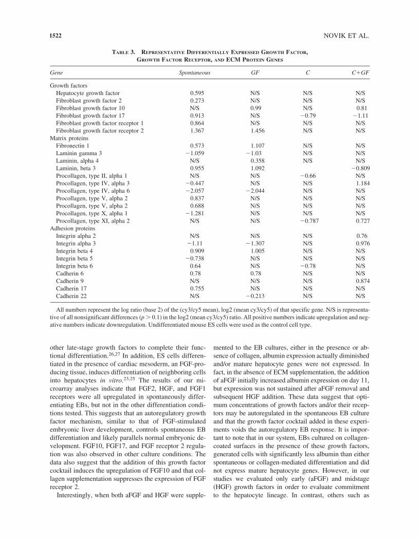
1522
TABLE 3. REPRESENTATIVE DIFFERENTIALLY EXPRESSED GROWTH FACTOR, GROWTH FACTOR RECEPTOR, AND ECM PROTEIN GENES
Gene Spontaneous GF C C�GF
Growth factorsHepatocyte growth factor �0.595 N/S N/S N/SFibroblast growth factor 2 �0.273 N/S N/S N/SFibroblast growth factor 10 N/S 0.99 N/S 0.81Fibroblast growth factor 17 �0.913 N/S �0.79 �1.11Fibroblast growth factor receptor 1 �0.864 N/S N/S N/SFibroblast growth factor receptor 2 �1.367 �1.456 N/S N/S
Matrix proteinsFibronectin 1 �0.573 �1.107 N/S N/SLaminin gamma 3 �1.059 �1.030 N/S N/SLaminin, alpha 4 N/S �0.358 N/S N/SLaminin, beta 3 �0.955 �1.092 �0.809Procollagen, type II, alpha 1 N/S N/S �0.66 N/SProcollagen, type IV, alpha 3 �0.447 N/S N/S �1.184Procollagen, type IV, alpha 6 �2.057 �2.044 N/S N/SProcollagen, type V, alpha 2 �0.837 N/S N/S N/SProcollagen, type V, alpha 2 �0.688 N/S N/S N/SProcollagen, type X, alpha 1 �1.281 N/S N/S N/SProcollagen, type XI, alpha 2 N/S N/S �0.787 �0.727
Adhesion proteinsIntegrin alpha 2 N/S N/S N/S 0.76Integrin alpha 3 �1.11 �1.307 N/S �0.976Integrin beta 4 �0.909 �1.005 N/S N/SIntegrin beta 5 �0.738 N/S N/S N/SIntegrin beta 6 0.64 N/S �0.78 N/SCadherin 6 0.78 0.78 N/S N/SCadherin 9 N/S N/S N/S �0.874Cadherin 17 �0.755 N/S N/S N/SCadherin 22 N/S �0.213 N/S N/S
All numbers represent the log ratio (base 2) of the (cy3/cy5 mean), log2 (mean cy3/cy5) of that specific gene. N/S is representa-tive of all nonsignificant differences (p � 0.1) in the log2 (mean cy3/cy5) ratio. All positive numbers indicate upregulation and neg-ative numbers indicate downregulation. Undifferentiated mouse ES cells were used as the control cell type.

Hamazaki et al. added late-stage growth factors in addi-tion to the ones used here, to induce end-stage differenti-ation. Further studies will evaluate the effect of late-stagegrowth factors such as insulin, transferrin, and selenousacid (ITS) and oncostatin M (OSM) on either functionalaugmentation or terminal differentiation of cells isolatedfrom either the spontaneous or supplemented cultures.
Many investigators have measured ALB, AFP, andTRT expression in order to identify hepatocyte lineagecells. However, although these markers are widely usedto assess function of mature hepatocytes isolated from
MOUSE EMBRYONIC STEM CELL DIFFERENTIATION
adult livers, they may also be expressed in nonhepatic lin-eage embryonic cells.14–19 In addition, other markers of-ten used in evaluating hepatocyte differentiation, such asglucose-6-phosphatase, have also been found, using re-verse transcription-polymerase chain reaction (RT-PCR),in cells of a variety of other tissues including pancreaticislets.28 In general, although RT-PCR is a powerful tool,only a limited number of genes are generally evaluated,and many of these genes are expressed in a variety of em-bryonic and adult tissues. Because EB differentiation pro-duces a very heterogeneous population, we have elected
1523
TABLE 4. ACUTE PHASE RESPONSE GENES
Gene Spontaneous GF C C�GF
Interleukin 1 alpha N/S N/S 1.020 0.920Interleukin 6 �0.470 N/S N/S N/STumor necrosis factor �0.980 �0.922 N/S N/SCAMP responsive element modulator N/S N/S N/S 0.694AP-1, gamma 1 subunit N/S N/S N/S 1.281STAT 1 N/S N/S N/S 1.732Protein inhibitor of activated STAT 3 N/S N/S 0.937 0.877Matrix metalloproteinase 2 N/S N/S N/S 5.132Matrix metalloproteinase 9 N/S N/S N/S 0.868
All numbers represent the log ratio (base 2) of the (cy3/cy5 mean), log2 (mean cy3/cy5) of that specific gene. N/S is representa-tive of all nonsignificant differences (p � 0.1) in the log2 (mean cy3/cy5) ratio. All positive numbers indicate upregulation and neg-ative numbers indicate downregulation. Undifferentiated mouse ES cells were used as the control cell type.
FIG. 4. Pathway Assist Generated Pathways. The pathways were generated utilizing the shortest path approach to link differ-entially expressed genes. (A) Input genes were regulated in the spontaneous condition only. Asterisk indicates genes identifiedand inserted by Pathway Assist. (B) Input genes were regulated in the C�GF and C conditions. Asterisk indicates genes identi-fied and inserted by Pathway Assist. (Color images are available at �www.liebertpub.com/ten�.)

to evaluate gene expression within the entire mouse ge-nome. Although this approach may not be as quantitativeas PCR, it provides a comparative platform of gene ex-pression among parallel experimental cultures. The resultsof these analyses indicate that although high levels of al-bumin were expressed in both the spontaneous and C con-ditions, and intermediate albumin levels were expressedin C�GF condition, mature hepatocyte genes were onlyexpressed in spontaneously differentiating cells. This sug-gests that albumin expression can be regulated by dis-parate mechanisms in these culture conditions and thatdistinct albumin-expressing cell lineages may be differ-entiated in unique culture configurations.
In addition, EB cultures can yield not only hepatocytelineage cells but may also induce albumin-expressing vis-ceral endodermal lineage cells of the primitive endoderm.10
This is also suggested in our studies by the expression ofmany acute phase response genes in EB-derived cells andis supported by the Pathway Assist analysis suggesting dis-tinct albumin regulatory networks in the different cultureconditions. Although primitive endodermal cells may ini-tially be generated in the spontaneous culture condition aswell, these cells may be replaced by the differentiating he-patocyte lineage cells in this culture configuration. A dy-namic gene array study will assist in elucidating both thedifferentiation profile and the pathways that may be acti-vated during hepatocyte differentiation of ES cells.
Because neither the C�GF nor the C condition expresseda significant number of mature hepatocyte genes, we ex-plored alternative mechanisms to explain the appearance ofembryonic albumin-producing cells. IL1�, in conjunctionwith collagen, has been shown to induce myofibroblasticactivation, through matrix remodeling proteins such as ma-trix metalloproteinase 9 (MMP-9), during the liver acutephase response to injury.29 In addition, expression of ex-tracellular MMPs is developmentally controlled during thedifferentiation and spreading of the parietal endoderm.30 Inboth C and C�GF conditions, gene expression of IL1� wassignificantly increased. Although metalloproteinase wasnot upregulated in the C condition, the C�GF conditiondemonstrated not only increased MMP-9 gene expressionbut also a variety of other early acute phase genes listed inTable 4. These data suggest that the mechanism activatedupon exposure to collagen may initiate upregulation ofacute phase genes within primitive endodermal cells andultimately an increased number of primitive endodermalcells within the EB. The effect was more pronounced in theC�GF condition, which also demonstrated decreased al-bumin expression and may therefore be regulated by theaddition of early-stage growth factors. Therefore, althoughcollagen may initiate the acute phase response during EBdifferentiation, the effect may be more pronounced in thepresence of growth factors.
Unlike the other culture conditions established in ourstudies, GF did not express albumin, acute-phase genes,
NOVIK ET AL.
or mature hepatocyte genes. Although we cannot defini-tively assign these cells to a specific embryonic lineage,they may represent a more primitive embryonic stage. Itis also important to note that although identifying up-regulated genes may assist in characterizing the cell lin-eage of differentiating cells, these data may not reflectactual protein synthetic levels. Future studies will assessthe protein expression profiles of the genes identified inthese studies. In addition, although we have identified he-patocyte lineage cells in spontaneously differentiatingcultures, the incorporation of these cells into tissue con-structs is contingent upon optimizing differentiated func-tion and cell purification. The intrinsic heterogeneity ofthe differentiating EB makes characterization of a spe-cific cell type difficult, and further cell purification mayalso help yield a more accurate analysis. This is espe-cially important since we have identified other lineagespecific cells including nerve, lymphoid and muscle inour EB cultures (data not shown). In fact, even thoughthe dense inner region did not contain albumin-express-ing cells, it contains cells more reminiscent of mature he-patocyte morphology.
Nevertheless, our data indicate that spontaneous dif-ferentiation induces high ALB expression and liver-spe-cific gene upregulation. In addition, we have identifieddisparate pathways that may regulate albumin expressionduring EB differentiation. Experimental manipulation ofthese pathways may assist in engineering more efficienthepatocyte differentiation from ES cells.
ACKNOWLEDGMENTS
These studies were supported by NIH DK43371, agrant from the NJ Commission on Higher Education, andThe Graduate Fellowship Program on Integratively En-gineered Biointerfaces at Rutgers, NSF DGE 0333196.The technical assistance of the following students is alsoappreciated: Patrick Leung, Fred Martin III, NripenShrama, and Eric Wallenstein.
REFERENCES
1. Klug, M.G., Soonpaa, M.H., Koh, G.Y., and Field, L.J. Ge-netically selected cardiomyocytes from differentiating em-bryonic stem cells form stable intracardiac grafts. J. Clin.Invest. 98, 216, 1996.
2. Lee, S.H., Lumelsky, N., Studer, L., Auerbach, J.M., andMcKay, R.D. Efficient generation of midbrain and hind-brain neurons from mouse embryonic stem cells. Nat.Biotechnol. 18, 675, 2000.
3. Hamazaki, T., Iiboshi, Y., Oka, M., Papst, P.J., Meacham,A.M., Zon, L.I., and Terada, N. Hepatic maturation in dif-ferentiating embryonic stem cells in vitro. FEBS Lett. 497,15, 2001.
1524

4. Tilles, A.W., Berthiaume, F., Yarmush, M.L., Tompkins,R.G., and Toner, M. Bioengineering of liver assist devices.J. Hepatobiliary Pancreat. Surg. 9, 686, 2002.
5. Tilles, A.W., Berthiaume, F., Yarmush, M.L., and Toner,M. Critical issues in bioartificial liver development. Tech-nol. Health Care 10, 177, 2002.
6. Yarmush, M.L., Dunn, J.C., and Tompkins, R.G. Assess-ment of artificial liver support technology. Cell Transplant1, 323, 1992.
7. Yarmush, M.L., Toner, M., Dunn, J.C., Rotem, A., Hubel,A., and Tompkins, R.G. Hepatic tissue engineering. De-velopment of critical technologies. Ann. N Y Acad. Sci.665, 238, 1992.
8. Leahy, A., Xiong, J.W., Kuhnert, F., and Stuhlmann, H.Use of developmental marker genes to define temporal andspatial patterns of differentiation during embryoid body for-mation. J. Exp. Zool. 284, 67, 1999.
9. Keller, G.M. In vitro differentiation of embryonic stemcells. Curr. Opin. Cell Biol. 7, 862, 1995.
10. Asahina, K., Fujimori, H., Shimizu-Saito, K., Kumashiro,Y., Okamura, K., Tanaka, Y., Teramoto, K., Arii, S., andTeraoka, H. Expression of the liver-specific gene Cyp7a1 re-veals hepatic differentiation in embryoid bodies derived frommouse embryonic stem cells. Genes Cells 9, 1297, 2004.
11. Chinzei, R., Tanaka, Y., Shimizu-Saito, K., Hara, Y., Kak-inuma, S., Watanabe, M., Teramoto, K., Arii, S., Takase,K., Sato, C., Terada, N., and Teraoka, H. Embryoid-bodycells derived from a mouse embryonic stem cell line showdifferentiation into functional hepatocytes. Hepatology 36,22, 2002.
12. Hu, A.B., Cai, J.Y., Zheng, Q.C., He, X.Q., Shan, Y., Pan,Y.L., Zeng, G.C., Hong, A., Dai, Y., and Li, L.S. High-ra-tio differentiation of embryonic stem cells into hepatocytesin vitro. Liver Int. 24, 237, 2004.
13. Shirahashi, H., Wu, J., Yamamoto, N., Catana, A., Wege,H., Wager, B., Okita, K., and Zern, M.A. Differentiationof human and mouse embryonic stem cells along a hepa-tocyte lineage. Cell Transplant 13, 197, 2004.
14. Choi, D., Oh, H.J., Chang, U.J., Koo, S.K., Jiang, J.X.,Hwang, S.Y., Lee, J.D., Yeoh, G.C., Shin, H.S., Lee, J.S.,and Oh, B. In vivo differentiation of mouse embryonic stemcells into hepatocytes. Cell Transplant 11, 359, 2002.
15. Miyashita, H., Suzuki, A., Fukao, K., Nakauchi, H., andTaniguchi, H. Evidence for hepatocyte differentiation fromembryonic stem cells in vitro. Cell Transplant 11, 429, 2002.
16. Yamamoto, H., Quinn, G., Asari, A., Yamanokuchi, H.,Teratani, T., Terada, M., and Ochiya, T. Differentiation ofembryonic stem cells into hepatocytes: biological functionsand therapeutic application. Hepatology 37, 983, 2003.
17. Yamada, T., Yoshikawa, M., Kanda, S., Kato, Y., Naka-jima, Y., Ishizaka, S., and Tsunoda, Y. In vitro differenti-ation of embryonic stem cells into hepatocyte-like cellsidentified by cellular uptake of indocyanine green. StemCells 20, 146, 2002.
18. Jochheim, A., Hillemann, T., Kania, G., Scharf, J., Attaran,M., Manns, M.P., Wobus, A.M., and Ott, M. Quantitativegene expression profiling reveals a fetal hepatic phenotype ofmurine ES-derived hepatocytes. Int J Dev Biol 48, 23, 2004.
MOUSE EMBRYONIC STEM CELL DIFFERENTIATION
19. Moshage, H. Cytokines and the hepatic acute phase re-sponse. J Pathol 181, 257, 1997.
20. Amit, M., and Itskovitz-Eldor, J. Derivation and sponta-neous differentiation of human embryonic stem cells. JAnat 200, 225, 2002.
21. Lavon, N., Yanuka, O., and Benvenisty, N. Differentiationand isolation of hepatic-like cells from human embryonicstem cells. Differentiation 72, 230, 2004.
22. Shiojiri, N., and Sugiyama, Y. Immunolocalization of ex-tracellular matrix components and integrins during mouseliver development. Hepatology 40, 346, 2004.
23. Sekhon, S.S., Tan, X., Micsenyi, A., Bowen, W.C., andMonga, S.P. Fibroblast growth factor enriches the embry-onic liver cultures for hepatic progenitors. Am J Pathol 164,2229, 2004.
24. Jung, J., Zheng, M., Goldfarb, M., and Zaret, K.S. Initia-tion of mammalian liver development from endoderm byfibroblast growth factors. Science 284, 1998, 1999.
25. Fair, J.H., Cairns, B.A., Lapaglia, M., Wang, J., Meyer,A.A., Kim, H., Hatada, S., Smithies, O., and Pevny, L. In-duction of hepatic differentiation in embryonic stem cellsby co-culture with embryonic cardiac mesoderm. Surgery134, 189, 2003.
26. Kamiya, A., Kinoshita, T., and Miyajima, A. Oncostatin Mand hepatocyte growth factor induce hepatic maturation viadistinct signaling pathways. FEBS Lett 492, 90, 2001.
27. Suzuki, A., Iwama, A., Miyashita, H., Nakauchi, H., andTaniguchi, H. Role for growth factors and extracellular ma-trix in controlling differentiation of prospectively isolatedhepatic stem cells. Development 130, 2513, 2003.
28. Shingu, R., Nakajima, H., Horikawa, Y., Hamaguchi, T.,Yamasaki, T., Miyagawa, J., Namba, M., Hanafusa, T., andMatsuzawa, Y. Expression and distribution of glucose-6-phosphatase catalytic subunit messenger RNA and itschanges in the diabetic state. Res Commun Mol PatholPharmacol 93, 13, 1996.
29. Han, Y.P., Zhou, L., Wang, J., Xiong, S., Garner, W.L.,French, S.W., and Tsukamoto, H. Essential role of matrixmetalloproteinases in interleukin-1-induced myofibroblas-tic activation of hepatic stellate cell in collagen. J BiolChem 279, 4820, 2004.
30. Adler, R.R., Brenner, C.A., and Werb, Z. Expression of ex-tracellular matrix-degrading metalloproteinases and metal-loproteinase inhibitors is developmentally regulated duringendoderm differentiation of embryonal carcinoma cells.Development 110, 211, 1990.
31. Ott, M.O., Rey-Campos, J., Cereghini, S., and Yaniv, M.vHNF1 is expressed in epithelial cells of distinct embry-onic origin during development and precedes HNF1 ex-pression. Mech Dev 36, 47, 1991.
Address reprint requests to:Martin L. Yramush
617 Bowser RoadPiscataway, NJ 08854
E-mail: [email protected]
1525