
The large-scale organization of "visual" streams emerges without visual experience
12
The large-Scale Organization of ‘‘Visual’’ Streams Emerges Without Visual Experience Ella Striem-Amit 1 , Ornella Dakwar 1 , Lior Reich 1 and Amir Amedi 1,2 1 Department of Medical Neurobiology, The Institute for Medical Research Israel-Canada, Faculty of Medicine and 2 The Edmond and Lily Safra Center for Brain Sciences (ELSC), The Hebrew University of Jerusalem, Jerusalem 91220, Israel Address correspondence to Amir Amedi. Email: [email protected]. A key question in sensory perception is the role of experience in shaping the functional architecture of the sensory neural systems. Here we studied dependence on visual experience in shaping the most fundamental division of labor in vision, namely between the ventral ‘‘what’’ and the dorsal ‘‘where and how’’ processing streams. We scanned 11 fully congenitally blind (CB) and 9 sighted individuals performing location versus form identification tasks following brief training on a sensory substitution device used for artificial vision. We show that the dorsal/ventral visual pathway division of labor can be revealed in the adult CB when perceiving sounds that convey the relevant visual information. This suggests that the most important large-scale organization of the visual system into the 2 streams can develop even without any visual experience and can be attributed at least partially to innately determined constraints and later to cross-modal plasticity. These results support the view that the brain is organized into task-specific but sensory modality-independent operators. Keywords: blindness, neuroimaging, plasticity, sight restoration, visual cortex Introduction Ever since the seminal work by Ungerleider and Mishkin (1982), it has been repeatedly shown that visual processing is carried out in 2 parallel pathways. The ventral occipitotemporal ‘‘what’’ pathway, or the ‘‘ventral stream,’’ has been linked with visual processing of form, object identity, and color. Its counterpart is considered to be the dorsal occipitoparietal ‘‘where/how’’ pathway, or the ‘‘dorsal stream,’’ which analyzes visuospatial information about object location and participates in visuomotor planning and visually guided movement (Goodale and Milner 1992; Goodale 2008). This double dissociation between the processing of the 2 streams has been thoroughly validated by studies of localized lesions separately affecting visual object identity recognition (visual agnosia) and object visuomotor spatial manipulation (optic ataxia: for a review, see Goodale 2008) and numerous human imaging studies (e.g., Haxby et al. 1991; Shmuelof and Zohary 2005).The anatomical basis for this division has also been studied and shows a complex pattern of bottom-up connectivity, beginning with the primary visual cortex and creating 2 parallel (though not completely independent) anatomical connectivity streams, one of which leads dorsally, through the posterior parietal cortex toward the premotor cortex, thus creating a natural ‘‘path’’ toward preparation for motion and spatial processing, while the other leads through area V4, which is selective for the color and size of visual objects (Zeki 1983; Desimone and Schein 1987), to inferotem- poral areas containing complex visual object representations (Desimone 1991; Tanaka 1997), and up to the prefrontal cortex (O’Scalaidhe et al. 1997). This hardwired bottom-up connec- tivity pattern creates a strong constraint toward the generation of these streams in the presence of normal visual input during development. However, the creation of 2 separate visual streams and their functional selectivities may not be so trivial in the absence of visual input, which deprives the streams of their natural input. Thus, while years of specialized research have established visual cortex segregation into functional streams as one of most fundamental characteristics of the visual system, what remains unclear is the role of visual experience in shaping this functional architecture of the brain. Does this fundamental large-scale organizational principle depend on visual experi- ence? Or are innately determined constraints sufficient for the emergence of the division of labor between the 2 streams, even in the absence of vision? One way to study these questions is to investigate whether traces of the visual functional division of labor can be identified in fully CB individuals who have never had any visual experience, by using sensory substitution devices (SSDs). SSDs are noninvasive devices that provide visual information to the blind via their existing senses (mainly audition and touch, see Fig. 1A; Meijer 1992; Bach-y-Rita and Kercel 2003) which may thus provide a valuable test for the existence of critical (or sensitive) periods for developing this fundamental functional segregation between the 2 visual streams. If the CB show no such differentiation, then visual experience is necessary and stream formation may be restricted to the presence of visual input. Conversely, if there are indications of a functional ‘‘visual’’ segregation in the absence of visual experience, the streams may emerge at least partially from innate biases, connectivity patterns or functional differentiation of process- ing shape and location functions which are not necessarily intrinsically visual by nature, but reflect a task- or computa- tional-selectivity that is independent of the sensory input modality (e.g., Reich et al. 2011) and may thus be flexible at later ages regardless of visual experience (see also Pascual- Leone and Hamilton 2001; Renier et al. 2005a, 2005b, 2010; Poirier, Collignon et al. 2006; Amedi et al. 2007; Beauchamp et al. 2007; Mahon et al. 2009, 2010; Ptito et al. 2009; Ricciardi et al. 2009; Matteau et al. 2010; Sani et al. 2010). In addition to the important theoretical implications of such questions, answering them could also contribute to clinical rehabilitation of individuals suffering from peripheral blindness, who may regain some level of visual peripheral sensory input through novel cutting-edge medical approaches. Although these approaches can restore some visual information in the periphery, this does not guarantee that such information can be easily and adequately understood by the individual (e.g., Fine et al. 2003; Gregory 2003; Ostrovsky et al. 2006, 2009). Because the blind brain undergoes vast cross-modal plastic changes due Ó The Author 2011. Published by Oxford University Press. All rights reserved. For permissions, please e-mail: [email protected] Cerebral Cortex July 2012;22:1698– 1709 doi:10.1093/cercor/bhr253 Advance Access publication September 21, 2011 at Hebrew University of Jerusalem on July 1, 2012 http://cercor.oxfordjournals.org/ Downloaded from
Transcript of The large-scale organization of "visual" streams emerges without visual experience

The large-Scale Organization of ‘‘Visual’’ Streams Emerges Without Visual Experience
Ella Striem-Amit1, Ornella Dakwar1, Lior Reich1 and Amir Amedi1,2
1Department of Medical Neurobiology, The Institute for Medical Research Israel-Canada, Faculty of Medicine and 2The Edmond and
Lily Safra Center for Brain Sciences (ELSC), The Hebrew University of Jerusalem, Jerusalem 91220, Israel
Address correspondence to Amir Amedi. Email: [email protected].
A key question in sensory perception is the role of experience inshaping the functional architecture of the sensory neural systems.Here we studied dependence on visual experience in shaping themost fundamental division of labor in vision, namely between theventral ‘‘what’’ and the dorsal ‘‘where and how’’ processingstreams. We scanned 11 fully congenitally blind (CB) and 9 sightedindividuals performing location versus form identification tasksfollowing brief training on a sensory substitution device used forartificial vision. We show that the dorsal/ventral visual pathwaydivision of labor can be revealed in the adult CB when perceivingsounds that convey the relevant visual information. This suggeststhat the most important large-scale organization of the visualsystem into the 2 streams can develop even without any visualexperience and can be attributed at least partially to innatelydetermined constraints and later to cross-modal plasticity.These results support the view that the brain is organized intotask-specific but sensory modality-independent operators.
Keywords: blindness, neuroimaging, plasticity, sight restoration, visualcortex
Introduction
Ever since the seminal work by Ungerleider and Mishkin
(1982), it has been repeatedly shown that visual processing is
carried out in 2 parallel pathways. The ventral occipitotemporal
‘‘what’’ pathway, or the ‘‘ventral stream,’’ has been linked with
visual processing of form, object identity, and color. Its
counterpart is considered to be the dorsal occipitoparietal
‘‘where/how’’ pathway, or the ‘‘dorsal stream,’’ which analyzes
visuospatial information about object location and participates
in visuomotor planning and visually guided movement
(Goodale and Milner 1992; Goodale 2008).
This double dissociation between the processing of the
2 streams has been thoroughly validated by studies of localized
lesions separately affecting visual object identity recognition
(visual agnosia) and object visuomotor spatial manipulation
(optic ataxia: for a review, see Goodale 2008) and numerous
human imaging studies (e.g., Haxby et al. 1991; Shmuelof and
Zohary 2005).The anatomical basis for this division has also
been studied and shows a complex pattern of bottom-up
connectivity, beginning with the primary visual cortex and
creating 2 parallel (though not completely independent)
anatomical connectivity streams, one of which leads dorsally,
through the posterior parietal cortex toward the premotor
cortex, thus creating a natural ‘‘path’’ toward preparation for
motion and spatial processing, while the other leads through
area V4, which is selective for the color and size of visual
objects (Zeki 1983; Desimone and Schein 1987), to inferotem-
poral areas containing complex visual object representations
(Desimone 1991; Tanaka 1997), and up to the prefrontal cortex
(O’Scalaidhe et al. 1997). This hardwired bottom-up connec-
tivity pattern creates a strong constraint toward the generation
of these streams in the presence of normal visual input during
development. However, the creation of 2 separate visual
streams and their functional selectivities may not be so trivial
in the absence of visual input, which deprives the streams of
their natural input.
Thus, while years of specialized research have established
visual cortex segregation into functional streams as one of most
fundamental characteristics of the visual system, what remains
unclear is the role of visual experience in shaping this
functional architecture of the brain. Does this fundamental
large-scale organizational principle depend on visual experi-
ence? Or are innately determined constraints sufficient for the
emergence of the division of labor between the 2 streams, even
in the absence of vision?
One way to study these questions is to investigate whether
traces of the visual functional division of labor can be identified
in fully CB individuals who have never had any visual
experience, by using sensory substitution devices (SSDs). SSDs
are noninvasive devices that provide visual information to the
blind via their existing senses (mainly audition and touch, see
Fig. 1A; Meijer 1992; Bach-y-Rita and Kercel 2003) which may
thus provide a valuable test for the existence of critical (or
sensitive) periods for developing this fundamental functional
segregation between the 2 visual streams. If the CB show no
such differentiation, then visual experience is necessary and
stream formation may be restricted to the presence of visual
input. Conversely, if there are indications of a functional
‘‘visual’’ segregation in the absence of visual experience, the
streams may emerge at least partially from innate biases,
connectivity patterns or functional differentiation of process-
ing shape and location functions which are not necessarily
intrinsically visual by nature, but reflect a task- or computa-
tional-selectivity that is independent of the sensory input
modality (e.g., Reich et al. 2011) and may thus be flexible at
later ages regardless of visual experience (see also Pascual-
Leone and Hamilton 2001; Renier et al. 2005a, 2005b, 2010;
Poirier, Collignon et al. 2006; Amedi et al. 2007; Beauchamp
et al. 2007; Mahon et al. 2009, 2010; Ptito et al. 2009; Ricciardi
et al. 2009; Matteau et al. 2010; Sani et al. 2010).
In addition to the important theoretical implications of such
questions, answering them could also contribute to clinical
rehabilitation of individuals suffering from peripheral blindness,
who may regain some level of visual peripheral sensory input
through novel cutting-edge medical approaches. Although
these approaches can restore some visual information in the
periphery, this does not guarantee that such information can be
easily and adequately understood by the individual (e.g., Fine
et al. 2003; Gregory 2003; Ostrovsky et al. 2006, 2009). Because
the blind brain undergoes vast cross-modal plastic changes due
� The Author 2011. Published by Oxford University Press. All rights reserved.
For permissions, please e-mail: [email protected]
Cerebral Cortex July 2012;22:1698– 1709
doi:10.1093/cercor/bhr253
Advance Access publication September 21, 2011
at Hebrew
University of Jerusalem
on July 1, 2012http://cercor.oxfordjournals.org/
Dow
nloaded from
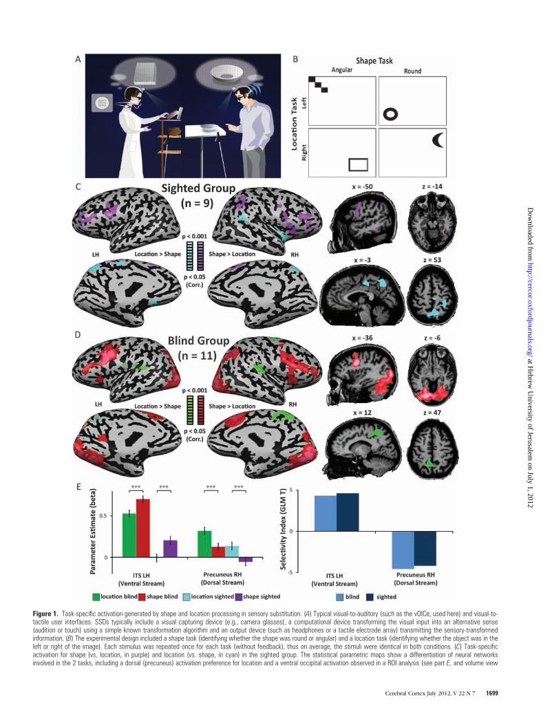
Figure 1. Task-specific activation generated by shape and location processing in sensory substitution. (A) Typical visual-to-auditory (such as the vOICe, used here) and visual-to-tactile user interfaces. SSDs typically include a visual capturing device (e.g., camera glasses), a computational device transforming the visual input into an alternative sense(audition or touch) using a simple known transformation algorithm and an output device (such as headphones or a tactile electrode array) transmitting the sensory-transformedinformation. (B) The experimental design included a shape task (identifying whether the shape was round or angular) and a location task (identifying whether the object was in theleft or right of the image). Each stimulus was repeated once for each task (without feedback), thus on average, the stimuli were identical in both conditions. (C) Task-specificactivation for shape (vs. location, in purple) and location (vs. shape, in cyan) in the sighted group. The statistical parametric maps show a differentiation of neural networksinvolved in the 2 tasks, including a dorsal (precuneus) activation preference for location and a ventral occipital activation observed in a ROI analysis (see part E, and volume view
Cerebral Cortex July 2012, V 22 N 7 1699
at Hebrew
University of Jerusalem
on July 1, 2012http://cercor.oxfordjournals.org/
Dow
nloaded from

to prolonged sensory deprivation (even to the extent of
processing language and memory in the occipital cortex,
especially in the ventral stream: Roder et al. 2001; Amedi et al.
2003; Bedny et al. 2011), it may have lost, or never developed,
its ability to properly process vision and the functional neural
architecture supporting it. Thus, it may be relevant also for
clinical sight restoration to question whether the functional
architecture of the visual cortex critically and exclusively
depends on visual experience and whether congenital and
longitudinal visual deprivation causes the visual streams to
entirely lose their natural visual roles, which may prevent them
from reverting to natural vision processing if peripheral
input could be restored (and vice versa if residual stream
specialization can be found).
Materials and Methods
Visual-To-Auditory Sensory SubstitutionWe used a visual-to-auditory SSD called ‘‘The vOICe’’ (Meijer 1992),
which enables ‘‘seeing with sound’’ for highly trained users (seeing with
sounds can also be achieved using other algorithms, e.g., PSVA see
Renier and De Volder 2010). In a clinical or everyday setting, users
wear a video camera connected to a computer and stereo headphones;
the images are converted into ‘‘soundscapes’’ using a predictable
algorithm, allowing them to listen to and then interpret the visual
information coming from a digital video camera. Remarkably, proficient
users are able to differentiate the shapes of different objects, identify
the actual objects, and also locate them in space (Amedi et al. 2007;
Auvray et al. 2007; Proulx et al. 2008). The functional basis of this
visuoauditory transformation lies in spectrographic sound synthesis
from any input image, which is then further perceptually enhanced
through stereo panning and other techniques. Time and stereo panning
constitute the horizontal axis in the sound representation of an image,
tone frequency makes up the vertical axis, and loudness corresponds to
pixel brightness.
ParticipantsThe study included a total of 20 subjects, 9 normally sighted individuals
(sighted controls [SCs]), and 11 blind individuals. Our group of legally
blind subjects was relatively homogeneous in terms of blindness in that
all of them were congenitally blind (CB) and 9 of the blind subjects did
not have any form of light perception. The remaining 2 had faint light
perception but they were unable to localize light or recognize any
shape or form. The age range of the subjects was wide, from 18 to 60,
all had normal hearing, and had no neurological or psychiatric
conditions. For a full description of the subjects, causes of blindness,
etc. see Supplementary Table 1. Sighted subjects had normal vision
(corrective lenses permitted) and hearing. The Tel-Aviv Sourasky
Medical Center Ethics Committee approved the experimental pro-
cedure, and written informed consent was obtained from each subject.
Training Procedures and PerformanceAll subjects had their first training session, which lasted between 1 and
1.5 h, on sensory substitution using the vOICe software immediately
before the functional magnetic resonance imaging (fMRI) session
reported here (i.e., the subjects were completely naıve to the principles
of the vOICe before the training session). During the session, subjects
were first taught the visual-to-auditory transformation rules and
proceeded to practice the very simple shape and location perception
of a standardized set of stimuli which is part of the training set of stimuli
used in our laboratory to teach CB individuals to use the vOICe
(including small lines, rectangles, and round objects presented at
4 possible locations on the screen; see Fig. 1B). Feedback on
performance was given by showing the participants the sensory image
that they had heard following each training trial, using vision for the
sighted subjects, and haptic dimensional models of all the stimuli for the
CB individuals. Critically, none of the stimuli delivered during training
was repeated during the scan, in which they were introduced to
completely new stimuli. Testing fully CB participants without any visual
experience and using SSD enabled us to test the dependence of visual
stream segregation on visual experience directly. Furthermore, given that
such short training probably does not enable any long-term or extensive
learning-induced plasticity (Pascual-Leone et al. 2005), this design also
enabled us to isolate and research the baseline state of the visual system
in CB in relation to form and location processing. In contrast to most
previous studies using SSDs, we also collected the behavioral results from
inside the scanner in order to ensure that subjects were deeply engaged
in the shape and localization tasks (even if performance was not very
high due to the brief training, prompted by our interest in the
baseline—innate—state of the visual cortex prior to extensive training
which may influence it). Performance was comparable (2-way analysis of
variance [ANOVA], group effect, F = 0.65, P < 0.56) in the blind and
sighted groups, which was a critical comparison in order to determine
whether the blind also recruit shape and location centers and to allow
comparison between groups. However, performance differed, on
average, between the tasks, as the shape task was more difficult than
the location task for both groups (2-way ANOVA, F = 297, P < 0.05, 47.5 ±4.2% and 44.4 ± 4.4% for shape in the CB and SC, 83.5 ± 5.8% and 82.2 ±4.3% for location in the CB and SC). No significant task (F = 0.62, P <
0.57), group (F = 18.1, P < 0.15), or interaction (F = 0.05, P < 0.82) effects
were observed in a 2-way ANOVA for reaction time (10 ± 0.7 s and 10.1 ±0.8 s for shape in the CB and SC, 9.9 ± 0.5 s and 10.2 ± 0.8 s for location in
the CB and SC). It is worth noting that the activation pattern on the
shape task in this study replicates to a large extent the pattern seen
following 40 h of training in sighted subjects (Amedi et al. 2007),
suggesting that subjects focused their attention on extracting shape.
Moreover, in order to address the potential influence of behavioral
differences, we analyzed various subgroups that had both: 1) above
chance performance in both tasks and 2) no significant difference
between the 2 tasks. All the data from these subgroups were subjected
to several converging analyses, including random-effect (RFX, see
details below; Friston et al. 1999) analyses. The subgroups included
a group of 5 briefly-trained participants with matched performance at
the individual level (no more than 10% accuracy difference between
the tasks; see details below), a group of 12 participants which included
better-trained participants, and a critical group as regards our main
research question; namely, a group of 7 CB participants subjected to an
RFX analysis. First, we examined a subgroup of 5 briefly-trained
subjects, 3 of whom were fully CB, who performed similarly on the
2 tasks (at an individual subject level) and exhibited higher than
average performance on the shape task (student’s paired t-test
t4 = 1.132, P < 0.29, 62 ± 5.4% and 72 ± 8.2% for shape and location,
respectively) in more detail. All these individual subjects (Supplemen-
tary Fig. 3), as well as the group analysis of this subgroup (Fig. 3A),
showed similar effects to the ones reported for the main groups,
suggesting that the task-specific activation did not result from general
difficulty biases. To further control for the effect of performance in
a larger group of subjects, we scanned again as many subjects as
possible from our original cohort (an average of 11 months after the
presented at an uncorrected threshold to illustrative the cluster location). (D) Task-specific activation for shape (vs. location, in red) and location (vs. shape, in green) in the CBgroup reveals differentiation in the occipital cortex which does not depend on visual experience. In addition to the dorsal activation for location exhibited by the sighted, the CBgroup also shows robust ventral stream selectivity for shape. (E) Beta values (GLM-beta) and a selectivity indices (the difference between the T values of the 2 tasks, Tshape �Tlocation) were sampled from the second repetition of the experiment in ROIs defined by the peak shape and location activation in the first repetition of the experiment across theentire dataset (n 5 20). The results of the independent ROI analysis are consistent with the statistical parametric maps, demonstrating the task specificity of the ventral stream(inferior temporal sulcus, ITS; blind P \ 0.00005, sighted P \ 0.00001) and dorsal stream (precuneus; blind P \ 0.000001, sighted P \ 0.0001) foci for shape and locationrespectively in each group separately. Error bars represent standard error of the mean. *P \ 0.05, **P \ 0.005, ***P \ 0.0005.
The ‘‘Visual’’ Streams of the Blind d Striem-Amit et al.1700
at Hebrew
University of Jerusalem
on July 1, 2012http://cercor.oxfordjournals.org/
Dow
nloaded from

original scan) after being further trained for 40 additional hours on
various tasks and visual stimuli using the vOICe SSD, enabling them to
achieve better task performance. We thus inspected a mixed group of
12 participants (7 blind, 5 sighted) at both training levels, in which
performance was matched between the tasks (2-way ANOVA, no effect
of task—F = 3.5, P < 0.31, group—F = 1.5, P < 0.44 or interaction—F =1.16, P < 0.29) and showed that they also manifested the double task
dissociation between the visual streams, at the group level (using RFX
analysis) (Fig. 3B), in a group of blind participants only (Fig. 4; n = 7, no
behavioral task effect, F = 3.75, P < 0.07), and in all the individual
subjects (Supplementary Fig. 3), regardless of their training.
General Experiment DesignTwenty novel simple visual stimuli were created using 2 different shape
categories: round and angular shapes (e.g., a circle and a square; see Fig.
1B) and 2 different locations (left and right). The stimuli also varied in
their vertical location (up and down), but this factor was irrelevant to the
tasks required, enabling generalization of shape and location to various
locations within the ‘‘visual’’ field. The use of novel stimuli is very
demanding but also further enabled us to inspect the neural correlates of
the online computation of discerning shapes and locations, as opposed to
possible memory effects (in themselves activating the occipital cortex of
the blind; Roder et al. 2001; Amedi et al. 2003). During each trial, subjects
were presented with an auditory instruction: either ‘‘shape’’ or ‘‘location,’’
which directed their attention to the task. They were then presented
with a 1-s soundscape (SSD sound rendering of the visual stimulus) that
was repeated 4 times (total presentation time—4 s), given 5 additional
seconds to reconstruct the image in their mind and were then instructed,
by an auditory cue, to respond using a response box. Subjects used a two-
button response box to indicate the parameter specified (Is the shape
round? Is it in the left of the picture?). Each stimulus was presented
twice: once for the shape task and once for the location task (without
feedback), in a pseudorandomized order, such that half the stimuli were
first presented in the shape task and half in the location task. Therefore,
the location condition and shape condition each repeated a total of 20
times, once for each of the different stimuli. Half of the stimuli (and thus
also the trials) were round; similarly, half of the stimuli were on the right
side of the visual field. Subjects were not allowed to see or touch the
pictures that generated the vOICe stimuli used in the fMRI testing, and
none of the stimuli presented during training were used for the scan.
Sighted subjects wore blindfolds and had their eyes shut for the duration
of the scan to control for the lack of visual information during the scan
between the groups.
fMRI Recording ParametersThe blood oxygen level--dependent (BOLD) fMRI measurements were
performed in a whole-body 3-T GE scanner. The pulse sequence used
was the gradient-echo echo planar imaging sequence. We used 29
slices of 4 mm in thickness. The data in-plane matrix size was 64 3 64,
field of view (FOV) 20 cm 3 20 cm, time to repetition (TR) = 1500 ms,
flip angle = 70�, and time to echo (TE) = 30 ms. Each experiment had
320 data points with 2 repetitions (runs), whose order of presentation
was controlled for across individual subjects. The first 5 images (during
the first baseline rest condition) were excluded from the analysis
because of non-steady state magnetization. Separate 3D recordings
were used for coregistration and surface reconstruction. High-
resolution 3D anatomical volumes were collected using T1-weighted
images using a 3D turbo field echo T1-weighted sequence (equivalent
to magnetization prepared rapid gradient echo). Typical parameters
were: FOV 23 cm (RL) 3 23 cm (VD) 3 17 cm (AP); fold over—axis: RL,
data matrix: 160 3 160 3 144 zero filled to 256 in all directions
(approximately 1-mm isovoxel native data), TR/TE = 9/6 ms, flip
angle = 8�.
Data AnalysisData analysis was performed using the Brain Voyager QX 1.10 software
package (Brain Innovation, Maastricht, Netherlands) using standard
preprocessing procedures. fMRI data preprocessing included head
motion correction, slice scan time correction, and high-pass filtering
(cutoff frequency: 3 cycles/scan) using temporal smoothing in the
frequency domain to remove drifts and to improve the signal to noise
ratio. No data included in the study exceeded motion of 2 mm in any
given axis or had spike-like motion of more than 1 mm in any direction.
Functional and anatomical data sets for each subject were aligned and
fit to standardized Talairach space (Talairach and Tournoux 1988).
Single-subject data were spatially smoothed with a minimal 3
dimensional 6-mm half-width Gaussian (2 functional voxels) in order
to reduce intersubject anatomical variability and then grouped using
a general linear model (GLM) in a hierarchical random effects analysis
(RFX; Friston et al. 1999; see for instance implementation in Amedi
et al. 2007). In addition to the main contrast, all GLM contrasts reported
in this study also included a conjunction (or a mask) of the comparison
of the main condition to baseline, to verify that only positive BOLD for
the main predictor would be included in the analysis (e.g., in a contrast
of location vs. shape, location was also contrasted with baseline, and the
2 contrasts were analyzed in conjunction, thus only voxels that showed
significant RFX positive BOLD to location and also significantly higher
activation to location vs. shape were highlighted in the maps). This also
precluded misleading comparisons including the default mode network
(DMN; Raichle et al. 2001; Raichle and Mintun 2006; Raichle and Snyder
2007) in areas showing deactivation to one condition and a larger
deactivation to another (e.g., to preclude an area showing for instance
deactivation to location and significantly more deactivation to shape
from appearing on the map, to further demonstrate the dissociation of
our findings from the DMN, see Supplementary Fig. 1). In order to
directly compare the effects of blindness and task across the entire data
set, a 2-way ANOVA was computed (Fig. 2), taking into account all the
sighted subjects (n = 9), and, in order to control for group size and
complete blindness, the 9 fully CB who did not have any form of light
perception (although the remaining 2 CB had merely faint light
perception and no ability to recognize visual shapes). Post hoc
contrasts were further computed from the ANOVA design and
presented within the statistically significant main effect statistical
parametric maps. Similar ANOVA analyses were also computed for the
performance-matched subgroups (Figs 3 and 4). The minimum
significance level of all results presented in the study was set to P <
0.05 taking into account the probability of a false detection for any
given cluster (Forman et al. 1995), thus correcting for multiple
comparisons. This was done based on the Forman et al. (1995) Monte
Carlo simulation approach, extended to 3D data sets using the
threshold size plug-in Brain Voyager QX. We also conducted
a complementary independent regions of interest (ROIs) analysis. ROIs
(Fig. 1E) were derived from the occipital peaks for the shape versus
location and location versus shape contrasts (in conjunction with
positive activation for the main condition, i.e., shape and location
accordingly) in the first run of the experiment on the entire (n = 20)
group. Additionally, we sampled the peaks of activation in the ANOVA
interaction statistical parametric map (sampled from the interaction
effect on the first run of the experiment for both groups) in each of the
groups (Fig. 2D). Activation peak beta and selectivity indices (contrast T
values, the T value for the shape task minus that of the location task)
were sampled from these ROIs at the group level of activation on the
second run (repetition) of the experiment, thus making the ROI
definition and parameter sampling independent of each other. Separate
3D recordings were used for surface reconstruction. Anatomical
cortical reconstruction procedures included the segmentation of the
white matter using a grow-region function embedded in the Brain
Voyager QX 1.9.10 software package (Brain Innovation). The Talairach
normalized cortical surface was then inflated, and the obtained
activation maps were superimposed onto it.
Results
To test for the putative dorsal--ventral division of labor
dissociation, we tested what happens when a group of CB
and blindfolded sighted individuals are trained to extract shape
and location information using a unique visual-to-auditory
sensory substitution algorithm utilized for visual rehabilitation
by embedding the visual information in sounds (soundscapes).
Cerebral Cortex July 2012, V 22 N 7 1701
at Hebrew
University of Jerusalem
on July 1, 2012http://cercor.oxfordjournals.org/
Dow
nloaded from

We analyzed the data in our experiment using several
complementary methods of analysis. First, we tested for task
preference in each group independently using GLM RFX
statistical parametric maps (Fig. 1C,D) and independent ROI
analysis (Fig. 1E). Additionally, we conducted a 2-way ANOVA
analysis that directly tested the TASK, GROUP, and interaction
effects in the entire data set (Fig. 2). We also examined several
subgroups of subjects with matched performance between the
2 tasks (Figs 3 and 4) including the critical group of fully CB
individuals with controlled performance using RFX analysis
(Fig. 4).
First, we examined task selectivity separately for the 2
participant groups (Fig. 1C,D, see map peaks coordinates in
Supplementary Table 2). We found a clear differentiation
between the dorsal and ventral pathways for the processing of
location and shape of soundscapes, respectively. In addition to
the network of multisensory areas (such as the intraparietal
sulcus and inferior frontal sulcus; Amedi et al. 2005, 2007; Lacey
and Campbell 2006; Naumer et al. 2008; Striem-Amit, Dakwar,
et al. 2011), shape processing activated the ventral occipital
inferior temporal sulcus (ITS) located in the midst of the
ventral visual stream. In contrast, the localization task
preferentially activated a network involving both auditory
regions (such as the supramarginal gyrus in the inferior parietal
lobe; Weeks et al. 1999) as well as the precuneus, correspond-
ing to Brodmann area 7, a higher order part of the visual dorsal
stream. Therefore, despite the identical perceptual auditory
stimulation in the 2 tasks, we observed differential recruitment
of the ventral versus dorsal stream for shape and location
processing. Most critically, we found a division of labor for form
and location in the visual system of CB (Fig. 1C). Thus, we find
visual stream/task-specific selectivity in the CB in the same
experiment and show that this division of labor is common to
both groups (Fig. 1C,D). One clear difference in the magnitude
and distribution between the 2 groups was found in the ventral
visual stream. Whereas the SC group (Fig. 1C) showed a weaker
soundscape activation limited to higher order object-related
areas in the inferior temporal ventral cortex (significant in the
ROI analysis; see Fig. 1E but not significant enough to pass the
strict multiple comparison correction applied across the entire
volume of the brain), the CB group (Fig. 1D) showed robust
and vast ventral visual cortex preference for shape conveyed by
sounds, demonstrating task-selective cross-modal plasticity
(also see below, a direct examination of this effect using the
interaction between GROUP and TASK effects in ANOVA, Fig.
2). This activation was even found within the early ventral
stream areas, which corresponds to ventral retinotopic areas
reaching as far as the calcarine sulcus (V1). Although activation
Figure 2. Effects of task and sight show increased involvement of the occipital cortex in the blind in a 2-way ANOVA. (A) ANOVA TASK effect was calculated across groups of 9fully blind and 9 sighted subjects (for the main effect maps, see Supplementary Fig. 2). Within the statistically significant areas of the main TASK effect, the ANOVA TASK post-hoc contrasts are displayed (shape vs. location and location vs. shape in orange and green, respectively), replicating the visual stream segregation displayed in the GLM random-effect analysis of each group separately (Fig. 1). (B) ANOVA GROUP effect was calculated. Within the statistically significant areas of the main GROUP effect, the ANOVA GROUPpost-hoc contrasts are displayed (CB vs. SC and SC vs. CB in purple and blue, respectively), demonstrating the increased involvement of the posterior occipital cortex of the CB forprocessing sensory substitution relative to the sighted. This increased activation of the visual cortex was accompanied by decreased reliance on the auditory cortex in the CB. (C)ANOVA interaction (TASK 3 GROUP) was calculated. Within the statistically significant areas of the interaction effect, contrasts of interaction were calculated, showing that theshape versus location contrast in the CB relative to the sighted was responsible for the observed interaction, supporting the increased involvement of the ventral posterioroccipital cortex in shape processing in the blind group alone. (D) The peaks of activation for the interaction effect were sampled in both groups in the posterior ventral occipitalcortex (Talairach coordinates �22, �65, �10 and 21, �54, �8 in the left and right hemispheres, respectively, corresponding to ventral Brodmann area 19). While the CB groupshows preferential activation for shape, the sighted demonstrate significant deactivation for soundscape shape processing. *P \ 0.05, **P \ 0.005, ***P \ 0.0005.
The ‘‘Visual’’ Streams of the Blind d Striem-Amit et al.1702
at Hebrew
University of Jerusalem
on July 1, 2012http://cercor.oxfordjournals.org/
Dow
nloaded from

was stronger in the precuneus of the CB in the location task, no
additional activation was found in this group in early areas
corresponding to dorsal retinotopic areas or V1.
To further investigate task preference of the ventral and
dorsal visual cortex through an additional independent
method, we defined ROIs from the peaks of selective activation
of the first run of the experiment in the entire group (n = 20,
combined SC and CB group) for shape and location processing
(Talairach coordinates: ITS LH –43, –60, –18, Precuneus RH 11,
–52, 51) and examined the activation generated in the second
run of the experiment in each group separately in these ROIs
(Fig. 1E). The beta values of both the ITS and precuneus regions
(ventral and dorsal stream peaks, respectively) showed a highly
significant difference (Fig. 1E; at least P < 0.0005 for all
contrasts) between the 2 tasks in both groups. Interestingly,
activations in both peaks were higher in the CB group
(P < 0.05).
To critically and directly investigate the separate effects and
interaction between the task preference of the ventral and
dorsal visual cortex and the group (with and without visual
Figure 3. Task-specific activation in performance-matched control groups. (A) Task-specific activation for shape (vs. location, in orange) and location (vs. shape, in green) ina subgroup of 5 subjects (3 fully CB and 2 SC) who had similar performance on the shape and location tasks fully replicates the ventral--dorsal differentiation seen in the entiregroup, negating the possibility that the stream differentiation resulted from general task difficulty or performance bias. This is shown in the ANOVA analysis and independently in theROI analysis (GLM-betas and selectivity indices, ITS: P \ 0.000001, precuneus: P \ 0.005) sampled from the peaks of the main group. *P \ 0.05, **P \ 0.005, ***P \ 0.0005.(B) Similar results were obtained in a random-effect (RFX) 2-way ANOVA of a larger group of participants (n 5 12, 7 CB, and 5 SC) with matched performance and variable training(for detail, see Materials and Methods). ROI analysis (ITS: P \ 0.0005, precuneus: P \ 0.000001) and single-subject analysis of all the subjects (Supplementary Fig. 3) alsosupported these findings.
Cerebral Cortex July 2012, V 22 N 7 1703
at Hebrew
University of Jerusalem
on July 1, 2012http://cercor.oxfordjournals.org/
Dow
nloaded from

experience), we computed a two-way ANOVA (see Supple-
mentary Fig. 2), with a TASK factor (shape and location) and
a GROUP factor (SC and CB). In this analysis, we included all 9
SC participants and the 9 fully CB subjects (the 2 other CB
were completely blind with minimal light perception and thus
were omitted to fully control for both group size and for
absolute blindness; results are similar when including these 2
blind subjects, data not shown). Performance in both groups
was comparable (no significant group effect, F = 0.02, P < 0.92).
Post hoc contrasts were computed within the significant
statistical parametric maps of the main effects analysis. The
TASK effect showed, as seen in the GLM analysis, a significant
effect in the visual cortex (see Supplementary Fig. 2A). The
post hoc TASK contrasts (Fig. 2A; see map peaks for the
ANOVA in Supplementary Table 3) replicated the stream
segregation seen between shape preference in the ventral
stream and location preference in the precuneus in the dorsal
stream, which were independent of the GROUP effect.
Furthermore, this analysis suggests there was an additional
region within the more posterior dorsal stream (Fig. 2A), in the
bilateral lateral-occipito-parietal cortex (in the middle temporal
gyrus/sulcus) showing preference for the location task across
the groups. The GROUP effect indicated a main effect of long-
term blindness in the posterior occipital cortex (see Supple-
mentary Fig. 2B), in that an increased involvement of the
posterior occipital cortex for processing soundscapes was
identified in the CB relative to the SC (Fig. 2B). This increase
was accompanied by a decrease in the activation of auditory
cortices, which was similar to previously reported decreases in
auditory cortex activation in CB for auditory localization
(Weeks et al. 2000). Furthermore, the interaction of the 2 main
effects (TASK 3 GROUP; Supplementary Fig. 2) was significant,
indicating different task selectivity between the 2 groups. Post
hoc contrasts revealed that this effect stems from differential
preference for shape between the groups (Fig. 2C, other
contrasts showed no significant activation). The posterior
ventral cortex showed greater preference for shape in CB,
even at a highly conservative threshold (P < 0.001 corrected
for multiple comparisons; see Fig. 2C), which stretched all the
way to the primary visual cortex (calcarine sulcus) at a more
permissive yet significant threshold of P < 0.05 (corrected). In
fact, areas more posterior to the inferior temporal cortex, in
the retinotopic ventral posterior occipital cortex (ventral
Brodmann area 19), showed a robust shape selective activation
in the CB, in contrast to a significant deactivation in both tasks
in the sighted (P < 0.05 for both groups; see GLM-beta values in
Fig. 2D). These findings support the increased involvement of
the ventral posterior occipital cortex in soundscape shape
processing in the CB group alone (similarly, compare Fig. 1C
and D).
Even though a general performance bias would not easily
explain this complex task-specific stream-specific activation
pattern or the similarity of the shape preference network to
previous findings in highly trained sighted subjects who
exhibited high performance (Amedi et al. 2007), the perfor-
mance did differ between the tasks in both groups in favor of
the easier localization task. We thus controlled for any general
task performance biases and additionally inspected activation
in several ways. We inspected several subgroups of participants
that had higher and controlled performance across the tasks at
Figure 4. Task-specific activation in the performance-matched (RFX analyzed) CB group. Random effect (RFX) ANOVA analysis of a large group of 7 CB with matchedperformance between the tasks shows significant stream-specific task selectivities for shape in the ventral stream ITS and dorsal stream precuneus. The effect is evident in bothANOVA and independently in the ROI analysis sampled from the peaks of the main group (ITS: P \ 0.05, precuneus: P \ 0.0005), demonstrating that the main effect of thestream functional segregation is independent of visual experience. *P \ 0.05, **P \ 0.005, ***P \ 0.0005.
The ‘‘Visual’’ Streams of the Blind d Striem-Amit et al.1704
at Hebrew
University of Jerusalem
on July 1, 2012http://cercor.oxfordjournals.org/
Dow
nloaded from

both the single-subject level and the group level (including RFX
analysis) and in independent ROI analyses for the peaks of
activation derived from the main group effects.
Specifically, we first inspected a subgroup of 5 subjects
(3 fully CB and 2 SC) who showed similar behavioral
performance in both tasks at the individual level (student’s
paired t-test, t4 = 1.132, P < 0.29 across the group, maximal
difference of 10% performance in each subject), even after
being very briefly trained. The results indicated a task-specific
differentiation between visual streams in ‘‘all’’ single subjects in
this subgroup (Supplementary Fig. 3; including 3 CB) and in the
data pooled across them (Fig. 3A, fixed-effect ANOVA, map
peaks are reported in Supplementary Table 4; for similar results
using GLM analysis, see also Supplementary Fig. 4A), including
a preference for the stream-matching task in the independent
ROI analysis (Fig. 3A; P < 0.005), confirming that the ventral/
dorsal division of labor could not stem from performance
differences alone.
Moreover, to fully control for behavioral effects in a RFX
analysis of a larger group, we further scanned participants who
had trained for a longer period of time and achieved better and
more matched performance between the 2 tasks (for details,
see Materials and Methods). Both the whole group analysis (n =12, random-effect ANOVA, Fig. 3B, see also map peaks in
Supplementary Table 4; comparable GLM analysis in Supple-
mentary Fig. 4B), the analysis of the blind group alone (n = 7,
random-effect ANOVA; Fig. 4, map peaks in Supplementary
Table 4; comparable GLM analysis in Supplementary Fig. 4C),
the complementary independent ROI analyses (Figs 3B and 4),
and the individual subject analysis level (of all the participants;
Supplementary Fig. 3) confirmed that the stream dissociation
does not result from performance differences. In all these types
of independent analyses, for groups and individual subjects, we
found a clear dissociation between the visual streams, regard-
less of task difficulty, including in RFX analyses in the CB.
Therefore, the findings suggest that some aspects of large-scale
dissociation between the ventral and dorsal streams are clearly
independent of visual experience.
Discussion
Our study shows a double dissociation between the distinct
activation of areas anatomically consistent with part of what is
known to be visual ventral and dorsal streams in response to
shape and location tasks using soundscape stimuli derived from
visual origin in the same experiment. This pattern was seen
across several independent analyses, in both groups (Fig. 2A
and Fig. 1C,D) and most importantly, in the CB group separately
(Figs 1D and 4). All the results in both groups and most
critically in the CB group remain identical as well in the
matched performance subgroups and analyses (Figs 3 and 4,
Supplementary Fig. 3). The main regions showing task-specific
activation across groups were the inferior temporal cortex for
shape in the ventral stream and the precuneus and middle
temporal sulcus/gyrus (Figs 1 and 2A) for location in the dorsal
stream (e.g., Martinkauppi et al. 2000; Sestieri et al. 2006).
Furthermore, the CB group also showed additional extensive
recruitment of the posterior ventral stream for the soundscape
shape task in ventral Brodmann area 19 (Figs 1D and 2C,D,
Supplementary Fig. 2). The most crucial aspect of our findings
suggests that despite life-long blindness, lack of visual
experience and the use of novel stimuli with short training,
a large extent of the ventral visual cortex in CB can be
recruited to process visual-from-auditory shapes, while at least
part of the dorsal stream processes visual-from-auditory
location information. Therefore, life-long existence without
vision does not render the 2 visual streams completely
unresponsive to their classical division of labor.
The activation observed in the ventral stream is consistent
with a previous study in sighted (as well as one late blind and
one CB; Amedi et al. 2007) which showed that LOtv, a tactile-
visual shape area, is activated for shape information conveyed
using SSD soundscapes. However, that study, similar to other
studies in sighted subjects (Renier et al. 2005a, 2005b; Poirier
et al. 2006, 2007), could not entirely avoid the visual imagery
confound, which may have contributed to any reported visual
cortex activation. Moreover, most studies used a combination
of only highly trained proficient SSD-users as participants
(following as many as 40 h of training; Amedi et al. 2007) and
familiar, well-practiced stimuli (Renier et al. 2005a, 2005b;
Amedi et al. 2007; Ptito et al. 2009; Matteau et al. 2010;
although sometimes as part of a training paradigm, Arno et al.
2001; Ptito et al. 2005; Kim and Zatorre 2011). All these might
complicate the interpretation and strength of previous results.
For instance, the brains of proficient users may have already
undergone significant plastic changes due to the extensive use
of SSDs (see, e.g., increased occipital cortex activation
following SSD training; Ptito et al. 2005), and the use of familiar
stimuli could generate activation due to memory rather than
shape processing in the occipital cortex of the blind subjects
(Roder et al. 2001; Amedi et al. 2003). More critically, since
most of these studies focused only on one individual task (and
did not contrast, e.g., shape and motion or shape and location),
they were unable to directly test the double-dissociation
division of labor of the visual cortex.
By circumventing these possible confounds, our study is the
first to show the segregation between the ventral and dorsal
streams in CB using an SSD in the same subjects, the same
experimental setup and using novel stimuli. Thus, we are able
to demonstrate the selective activation of visual areas by
auditory stimuli in the absence of any experience that could
support visual imagery. Interestingly, the activation and even
task selectivity of the visual streams were more robust in the
blind group than in the sighted group (Fig. 1C--E and Fig. 2B,D,
Supplementary Fig. 2), particularly in the posterior occipital
cortex (Figs 1 and 2B, Supplementary Fig. 2). While previous
studies have reported increased activation in the occipital
cortex of the blind (as compared with sighted) for various
nonvisual tasks (Ptito et al. 2005, 2009; Matteau et al. 2010;
Renier et al. 2010); in our study, we found a more complex
interaction between plasticity in the blind and specific task
preference in the posterior ventral occipital cortex (Fig. 2C,D).
In the anterior ventral ITS, we found activation in both groups
(though significantly stronger for the blind) while in the
posterior ventral stream in retinotopic areas (BA 19; Fig. 2) we
observed robust activation and preference for shape in blind
and significant deactivation in the sighted (Fig. 2D). This
stream- and task-specific cross-modal plasticity effect shows
that not only is the ventral stream still selective for shape, this
preference is enhanced (as compared with the sighted) when
the shape is encoded through sound. Both results argue against
a visual imagery explanation as the main basis for the ventral
activation for shapes of auditory inputs which represent visual
entities, suggesting instead that cross-modal plasticity biases
Cerebral Cortex July 2012, V 22 N 7 1705
at Hebrew
University of Jerusalem
on July 1, 2012http://cercor.oxfordjournals.org/
Dow
nloaded from

(Pascual-Leone and Hamilton 2001; Pascual-Leone et al. 2005)
are a stronger factor in driving the visual ventral stream.
These findings have important theoretical implications, as
they contribute further evidence supporting recent theories of
brain organization which argue that the selectivity of the
different functional cortical regions is not according to their
input modality but rather according to task selectivity, which
may be computed with various modalities (Amedi et al. 2001,
2007; Pascual-Leone and Hamilton 2001; Mahon and Caramazza
2009; Reich et al. 2011). The current results are consistent with
several other studies demonstrating multisensory or task-
dependent metamodal processing of specific brain areas within
the visual system (e.g., LOtv; Amedi et al. 2002, 2007; James
et al. 2002). Such studies showed recently that spatial
processing of both simple auditory chords or vibrotactile
stimulation selectively activate the middle occipital gyrus
(MOG) of the blind (Renier et al. 2010; Collignon et al.
2011), suggesting a task-specific role for an additional unique
area of the visual system. Our data are in line with this finding,
as in addition to the robust task selectivity of the precuneus
(which also showed multisensory properties in the sighted;
Renier et al. 2009), we observed spatial selectivity in sensory
substitution artificial vision input in an area in close proximity
to the MOG, the posterior middle temporal sulcus/gyrus (Fig.
2A). Similarly, specificity for tool stimuli over other, non-
manipulable objects was observed in 2 regions of the parietal
cortex of the blind (Mahon et al. 2010), activation for Braille
reading was found in the ‘‘visual word form area’’ (Reich et al.
2011), activation for kinesthetically guided hand movements
was found in primary somatosensory cortex independent of the
visual experience of participants (Fiehler et al. 2009), and
activation of the human MT region was found for nonvisual
motion in the blind (Poirier et al. 2006; Beauchamp et al. 2007;
Ricciardi et al. 2007; Ptito et al. 2009; Matteau et al. 2010; Sani
et al. 2010). All these suggest that the brain might be comprised
of flexible task-selective but modality-independent operators
(Reich et al. 2011). Another recent study that is particularly
relevant to the conclusions drawn here looked beyond area-
specific computation and showed that the larger scale animate/
inanimate organization within the high-order anterior ventral
visual cortex is independent of vision (in a group of sighted and
3 CB individuals; Mahon et al. 2009). Mahon et al. (2009)
concluded from their results that modality dependence is
secondary as a hierarchical organizational factor to the object
domain (e.g., living vs. non-living, also see a review of
conceptual object categories; Mahon and Caramazza 2009) in
the ventral visual cortex. Our findings extend such concepts of
a-modal innately determined developmental constraints to the
more fundamental organizational principle of the segregation
between the 2 processing streams. In doing so, it extends the
findings beyond visual object conceptual categories to postu-
lating that the whole brain may be task specific but sensory
modality independent, if the relevant computation and task can
be achieved from the sensory input (even if this is not an
ecological way to do so, i.e., via SSD).
In this respect, sensory substitution is an ideal tool to study
task-dependent operations as it teases apart the effect of the
modality from the computation or task in question and also
makes it possible to study tasks using untrained, novel,
‘‘modalities’’ and stimuli. The functional recruitment in the
brain of CB following such a short training period makes it
highly improbable that they reflect any extensive plastic
changes (Pascual-Leone et al. 2005). Instead, it suggests that
the division of labor between the ventral and dorsal streams for
form and location in the visual cortex must already be present
and the short training presumably ‘‘revealed’’ these innate
preferences under our special experimental conditions. The
life-long use of these areas for visual input more than for
information originating in other senses (along with the
usefulness of vision to decipher shape) makes the streams
appear as though they are only or mostly visual. This study
suggests this is not the case and that cross-modal plasticity can
still result in their activation for their original visual tasks.
What are the developmental endogenous, or innate, con-
straints that might contribute to such a sensory-independent
task-selective organization in the CB? We speculate that 2
factors, which are not mutually exclusive, could have taken
part. The first are intrinsic modality-independent preferences
for a particular (different) type of content or computation in
each brain area (in our case, in the dorsal and ventral regions).
For example, an area might specialize in computing motion due
to computing subtractions of a motion coincidence detector
regardless of sensory input. If this is true then all these areas
were always multisensory, possibly with visual dominance since
it is perhaps the most reliable sensory input in the sighted.
Alternatively, the task specificity might stem from the different
connectivity pattern of each area. In our case, the visual
streams may differ in their connectivity pattern to other
cortical areas, which together drive their task-selectivity
organization (e.g., via top-down modulation). For example, it
has been suggested that premotor--posteromedial parietal
connections are likely to subserve abstract cognitive processes
involving visuospatial information in the precuneus (Cavanna
and Trimble 2006), while feedback connectivity from frontal
and somatosensory cortices to the ventral (inferior temporal)
occipital cortex may underlie its multisensory function for
object recognition (Amedi et al. 2001, 2003; Deshpande et al.
2008). In addition to the preexisting connectivity, connectivity
between the visual cortex and other sensory cortices may also
be strengthened by sensory deprivation (e.g., between A1 and
V1; Klinge et al. 2010). Thus, although the input in our case was
auditory rather than visual, the preserved functional connec-
tivity of each stream still dictates development toward
processing shape or location, which may even be strengthened
for the nonvisual modalities.
This type of top-down modulation based on the existing
connectivity pattern might also originate from the correspond-
ing auditory streams. Similar to the visual streams, auditory
processing is also divided into what and where pathways,
whose functional and anatomical segregation has been thor-
oughly validated in many species, including humans (Pandya
and Vignolo 1969; Romanski et al. 1999; Kaas and Hackett 2000;
Rauschecker and Tian 2000; Alain et al. 2001; Kubovy and Van
Valkenburg 2001; Kraus and Nicol 2005; Lomber and Malhotra
2008; van der Zwaag et al. 2011). Some selective activation of
these auditory streams is also seen in our contrasts in addition
to the visual cortical streams. While our results do not clearly
show the auditory division of labor between the rostral and
caudal parts of the early-stage auditory areas on the supra-
temporal plane (which can better be depicted by ultra high-
field 7-T scanners due to the relatively small size of the auditory
areas and the integrated what and where processing of some of
these regions; Griffiths and Warren 2002; van der Zwaag et al.
2011), we do, however, find evidence for the stream
The ‘‘Visual’’ Streams of the Blind d Striem-Amit et al.1706
at Hebrew
University of Jerusalem
on July 1, 2012http://cercor.oxfordjournals.org/
Dow
nloaded from

differentiation in the inferior parietal lobe (supramarginal gyrus
and even post STG) and frontal lobe (between the inferior and
superior what and where regions), in accordance with the
auditory stream division. Therefore, it may be speculated that
the same auditory connectivity may partially underlie the visual
cortex differentiation and selectivities found in the current
report. However, previous studies of purely auditory localiza-
tion and object identification in the sighted or blind (as
opposed to visual-to-auditory processing using an SSD) have
not shown clear and consistent activation of the ITS (Amedi
et al. 2002, 2007) or precuneus (Collignon et al. 2009, although
the precuneus may sometimes be activated by nonvisual
localization in sighted; Renier et al. 2009). An exception to
this may be the MOG, which shows selective activation for
auditory localization in the blind (Collignon et al. 2007; Renier
et al. 2010). This suggests that the main occipital cortical
regions shown here (ITS and precuneus) are less likely to
partake in the auditory processing streams per se but rather
with processing computations that resemble vision (e.g., object
shape). Thus, their roles are less likely to develop as regular
parts of the auditory streams.
Interestingly, one difference between the shape and location
activation was the lack of more posterior recruitment of the
dorsal stream even in the CB (Figs 1 and 2B--D). One possible
explanation is that the location task was simply easier (possibly
resulting in less activation for this task). However, the
replication of the group results in our subgroup of 5 subjects,
in the larger mixed group (n = 12) and critically, in the large
group of the CB (n = 7, RFX analysis), who had similar
performance on both tasks, as well as in all the individual
subjects comprising these groups (Figs 3 and 4, Supplementary
Fig. 3), rules out this explanation. Alternatively, these differ-
ences might be due to different developmental timelines of the
2 streams. Although both streams may be not only visual but
task-specific and sensory-input independent to some extent,
the dorsal stream matures earlier in development (Lewis and
Maurer 2005), whereas it has been shown that the ventral
stream may continue to develop until adolescence (Golarai
et al. 2007). This suggests that the ventral stream may continue
to be plastic later in life (e.g., have longer sensitive periods)
relative to dorsal stream regions and thus be more likely to
reorganize differently and adaptively (e.g., as seen in our study,
to sounds). Supporting this notion, previous studies have
shown robust changes of the ventral visual cortex to other
nonvisual functions such as language and memory (Sadato et al.
1996; Cohen et al. 1997; Roder et al. 2001, 2002; Burton,
Snyder, Conturo, et al. 2002; Burton, Snyder, Diamond, et al.
2002; Amedi et al. 2003; Pascual-Leone et al. 2005; Noppeney
2007; Bedny et al. 2011). Moreover, studies of sensory
restoration after long-term visual deprivation (Fine et al.
2003; Gregory 2003; Ostrovsky et al. 2006, 2009) suggest that
ventral stream visual functions may remain deficient after visual
peripheral recovery even after months of training following the
procedure (perhaps due to the aforementioned robust plastic
cross-modal changes), whereas motion perception (a dorsal
stream function, processed in the early-maturing MT; Lewis and
Maurer 2005) appears to recover almost immediately following
sensory restoration.
Does this condemn the critical ventral stream functions to
remaining largely deficient following sight restoration? While
previous studies (Fine et al. 2003; Gregory 2003; Ostrovsky
et al. 2009) indeed show very serious deficits in object shape
recognition and segregation from background that might
hinder sight restoration efforts regardless of the exact
clinical/technological approach, our results imply that the
ventral stream could hypothetically be shifted back toward its
original task preference. The preferential activation of the
posterior ventral stream for shape in the CB (Figs 1 and 2,
Supplementary Fig. 2) suggests that while visual deprivation
may modify the role of these regions, the general stream
preference remains and can be revealed after learning to
extract the relevant information from other modalities (in our
case audition). The adaptation of the blind to processing
auditory information more than the sighted may even result in
quicker recruitment of cross-modal visual shape processing
transmitted by SSD. Although our subjects were not studied
under a clinical sight restoration protocol (in the more
conventional sense of restoration of visual qualia using, e.g.,
retinal prostheses; Dowling 2008), we feel it is important to
discuss our results in the context of clinical settings and to
speculate on their putative importance. For example, future
work should examine whether this unique combination of
sensory-input independent organization, baseline biases to
shape and location, and longitudinal long-term plasticity
enables the use of SSDs as neuro-rehabilitative aids to train
the visual cortex to analyze visual information. Therefore, SSDs
can theoretically support visual rehabilitation both before such
procedures, for example, to help reprogram or awaken the
hypothesized visual streams to selectivity process ‘‘vision’’
(rather than language and memory; Sadato et al. 1996; Cohen
et al. 1997; Roder et al. 2001, 2002; Burton, Snyder, Conturo,
et al. 2002; Burton, Snyder, Diamond, et al. 2002; Amedi et al.
2003; Pascual-Leone et al. 2005; Noppeney 2007; Bedny et al.
2011; Striem-Amit, Bubic, et al. 2011) and then by serving as
a ‘‘sensory interpreter,’’ providing explanatory input to the
novel visual signal arriving from an alien invasive device when it
is first introduced to the visually restored individual.
Interestingly, in a recent study, the more intact and faster
recovering dorsal stream functions (e.g., detecting moving
stimuli) were successfully used to train the deficient ventral
stream functions (visual parsing and object recognition) in
blind individuals who regained sight through medical in-
tervention using shape-from-motion training (Ostrovsky et al.
2009). SSDs could thus be used in a similar way to train the
visual cortex via other modalities.
To conclude, this study shows that visual experience is not
necessary in order for the dorsal--ventral division of labor
within the visual system to emerge, at least to some extent.
This suggests the operation of innately determined constraints
on the emergence of the most important large-scale organiza-
tion of the visual cortex. Our results favor the view that these
preferences are determined, in part, by dimensions of domains
of knowledge or task similarity that cannot be reduced to the
visual experience of individuals (e.g., Mahon and Caramazza
2009; Renier et al. 2010, 2011). Finally, our results support the
notion that large parts of the visual system are task-specific
modality invariant in nature and can be accessed, via cross-
modal mechanisms, by any sensory modality.
Funding
International Human Frontiers Science Program Organization
Career Development Award (CDA-0015/2008-070509 to A.A.);
EU-FP7 MC International Reintegration grant (MIRG-CT-2007-
Cerebral Cortex July 2012, V 22 N 7 1707
at Hebrew
University of Jerusalem
on July 1, 2012http://cercor.oxfordjournals.org/
Dow
nloaded from

205357-250208 to A.A.); James S. McDonnell Foundation
scholar award (220020284 to A.A.); Israel Science Foundation
(ISF 1684/08); The Sieratzki family award (to A.A.); Vision
Center grant from the Edmond and Lily Safra Center for Brain
Sciences (to A.A.).
Supplementary Material
Supplementary material can be found at: http://www.cercor.
oxfordjournals.org/
Notes
We thank D.R. Chebat and A. Bubic for their in-depth review of the final
draft of the paper and other very useful discussions. We would also like
to thank the Hebrew University Hoffman Leadership and Responsibility
Fellowship Program support (to E.S.A.) and the Samuel and Lottie Rudin
Foundation support (to L.R.). Conflict of Interest : None declared.
References
Alain C, Arnott SR, Hevenor S, Graham S, Grady CL. 2001. ‘‘What’’ and
‘‘where’’ in the human auditory system. Proc Natl Acad Sci U S A.
98:12301--12306.
Amedi A, Jacobson G, Hendler T, Malach R, Zohary E. 2002.
Convergence of visual and tactile shape processing in the human
lateral occipital complex. Cereb Cortex. 12:1202--1212.
Amedi A, Malach R, Hendler T, Peled S, Zohary E. 2001. Visuo-haptic
object-related activation in the ventral visual pathway. Nat Neurosci.
4:324--330.
Amedi A, Raz N, Pianka P, Malach R, Zohary E. 2003. Early ‘‘visual’’
cortex activation correlates with superior verbal memory perfor-
mance in the blind. Nat Neurosci. 6:758--766.
Amedi A, Stern WM, Camprodon JA, Bermpohl F, Merabet L, Rotman S,
Hemond C, Meijer P, Pascual-Leone A. 2007. Shape conveyed by
visual-to-auditory sensory substitution activates the lateral occipital
complex. Nat Neurosci. 10:687--689.
Amedi A, von Kriegstein K, van Atteveldt NM, Beauchamp MS,
Naumer MJ. 2005. Functional imaging of human crossmodal
identification and object recognition. Exp Brain Res. 166:559--571.
Arno P, De Volder AG, Vanlierde A, Wanet-Defalque MC, Streel E,
Robert A, Sanabria-Bohorquez S, Veraart C. 2001. Occipital
activation by pattern recognition in the early blind using auditory
substitution for vision. Neuroimage. 13:632--645.
Auvray M, Hanneton S, O’Regan JK. 2007. Learning to perceive with
a visuo-auditory substitution system: localisation and object
recognition with ‘‘The vOICe’’. Perception. 36:416--430.
Bach-y-Rita P, Kercel SW. 2003. Sensory substitution and the human-
machine interface. Trends Cogn Sci. 7:541--546.
Beauchamp MS, Yasar NE, Kishan N, Ro T. 2007. Human MST but not
MT responds to tactile stimulation. J Neurosci. 27:8261--8267.
Bedny M, Pascual-Leone A, Dodell-Feder D, Fedorenko E, Saxe R. 2011.
Language processing in the occipital cortex of congenitally blind
adults. Proc Natl Acad Sci U S A. 108:4429--4434.
Burton H, Snyder AZ, Conturo TE, Akbudak E, Ollinger JM, Raichle ME.
2002. Adaptive changes in early and late blind: a fMRI study of Braille
reading. J Neurophysiol. 87:589--607.
Burton H, Snyder AZ, Diamond JB, Raichle ME. 2002. Adaptive changes
in early and late blind: a FMRI study of verb generation to heard
nouns. J Neurophysiol. 88:3359--3371.
Cavanna AE, Trimble MR. 2006. The precuneus: a review of its
functional anatomy and behavioural correlates. Brain. 129:564--583.
Cohen LG, Celnik P, Pascual-Leone A, Corwell B, Falz L, Dambrosia J,
Honda M, Sadato N, Gerloff C, Catala MD, et al. 1997. Functional
relevance of cross-modal plasticity in blind humans. Nature.
389:180--183.
Collignon O, Lassonde M, Lepore F, Bastien D, Veraart C. 2007.
Functional cerebral reorganization for auditory spatial processing
and auditory substitution of vision in early blind subjects. Cereb
Cortex. 17:457--465.
Collignon O, Vandewalle G, Voss P, Albouy G, Charbonneau G,
Lassonde M, Lepore F. 2011. Functional specialization for auditory-
spatial processing in the occipital cortex of congenitally blind
humans. Proc Natl Acad Sci U S A. 108:4435--4440.
Collignon O, Voss P, Lassonde M, Lepore F. 2009. Cross-modal plasticity
for the spatial processing of sounds in visually deprived subjects.
Exp Brain Res. 192:343--358.
Deshpande G, Hu X, Stilla R, Sathian K. 2008. Effective connectivity
during haptic perception: a study using Granger causality analysis of
functional magnetic resonance imaging data. Neuroimage.
40:1807--1814.
Desimone R. 1991. Face-selective cells in the temporal cortex of
monkeys. J Cogn Neurosci. 3:1--8.
Desimone R, Schein SJ. 1987. Visual properties of neurons in area V4 of
the macaque: sensitivity to stimulus form. J Neurophysiol.
57:835--868.
Dowling J. 2008. Current and future prospects for optoelectronic
retinal prostheses. Eye. 23:1999--2005.
Fiehler K, Burke M, Bien S, Roder B, Rosler F. 2009. The human dorsal
action control system develops in the absence of vision. Cereb
Cortex. 19:1--12.
Fine I, Wade AR, Brewer AA, May MG, Goodman DF, Boynton GM,
Wandell BA, MacLeod DI. 2003. Long-term deprivation affects visual
perception and cortex. Nat Neurosci. 6:915--916.
Forman SD, Cohen JD, Fitzgerald M, Eddy WF, Mintun MA, Noll DC.
1995. Improved assessment of significant activation in functional
magnetic resonance imaging (fMRI): use of a cluster-size threshold.
Magn Reson Med. 33:636--647.
Friston KJ, Holmes AP, Worsley KJ. 1999. How many subjects constitute
a study? Neuroimage. 10:1--5.
Golarai G, Ghahremani DG, Whitfield-Gabrieli S, Reiss A, Eberhardt JL,
Gabrieli JD, Grill-Spector K. 2007. Differential development of high-
level visual cortex correlates with category-specific recognition
memory. Nat Neurosci. 10:512--522.
Goodale MA. 2008. Action without perception in human vision. Cogn
Neuropsychol. 25:891--919.
Goodale MA, Milner AD. 1992. Separate visual pathways for perception
and action. Trends Neurosci. 15:20--25.
Gregory RL. 2003. Seeing after blindness. Nat Neurosci. 6:909--910.
Griffiths TD, Warren JD. 2002. The planum temporale as a computa-
tional hub. Trends Neurosci. 25:348--353.
Haxby JV, Grady CL, Horwitz B, Ungerleider LG, Mishkin M, Carson RE,
Herscovitch P, Schapiro MB, Rapoport SI. 1991. Dissociation of
object and spatial visual processing pathways in human extrastriate
cortex. Proc Natl Acad Sci U S A. 88:1621--1625.
James TW, Humphrey GK, Gati JS, Servos P, Menon RS, Goodale MA.
2002. Haptic study of three-dimensional objects activates extras-
triate visual areas. Neuropsychologia. 40:1706--1714.
Kaas JH, Hackett TA. 2000. Subdivisions of auditory cortex and
processing streams in primates. Proc Natl Acad Sci U S A.
97:11793--11799.
Kim JK, Zatorre RJ. 2011. Tactile-auditory shape learning engages the
lateral occipital complex. J Neurosci. 31:7848--7856.
Klinge C, Eippert F, Roder B, Buchel C. 2010. Corticocortical
connections mediate primary visual cortex responses to auditory
stimulation in the blind. J Neurosci. 30:12798--12805.
Kraus N, Nicol T. 2005. Brainstem origins for cortical ‘‘what’’ and ‘‘where’’
pathways in the auditory system. Trends Neurosci. 28:176--181.
Kubovy M, Van Valkenburg D. 2001. Auditory and visual objects.
Cognition. 80:97--126.
Lacey S, Campbell C. 2006. Mental representation in visual/haptic
crossmodal memory: evidence from interference effects. Q J Exp
Psychol (Colchester). 59:361--376.
Lewis TL, Maurer D. 2005. Multiple sensitive periods in human visual
development: evidence from visually deprived children. Dev
Psychobiol. 46:163--183.
Lomber SG, Malhotra S. 2008. Double dissociation of ‘‘what’’ and
‘‘where’’ processing in auditory cortex. Nat Neurosci. 11:609--616.
Mahon BZ, Anzellotti S, Schwarzbach J, Zampini M, Caramazza A. 2009.
Category-specific organization in the human brain does not require
visual experience. Neuron. 63:397--405.
The ‘‘Visual’’ Streams of the Blind d Striem-Amit et al.1708
at Hebrew
University of Jerusalem
on July 1, 2012http://cercor.oxfordjournals.org/
Dow
nloaded from
Mahon BZ, Caramazza A. 2009. Concepts and categories: a cognitive
neuropsychological perspective. Annu Rev Psychol. 60:27--51.
Mahon BZ, Schwarzbach J, Caramazza A. 2010. The representation of
tools in left parietal cortex is independent of visual experience.
Psychol Sci. 21:764--771.
Martinkauppi S, Rama P, Aronen HJ, Korvenoja A, Carlson S. 2000.
Working memory of auditory localization. Cereb Cortex. 10:889--898.
Matteau I, Kupers R, Ricciardi E, Pietrini P, Ptito M. 2010. Beyond visual,
aural and haptic movement perception: hMT+ is activated by
electrotactile motion stimulation of the tongue in sighted and in
congenitally blind individuals. Brain Res Bull. 82:264--270.
Meijer PB. 1992. An experimental system for auditory image repre-
sentations. IEEE Trans Biomed Eng. 39:112--121.
Naumer MJ, Doehrmann O, Muller NG, Muckli L, Kaiser J, Hein G. 2008.
Cortical plasticity of audio-visual object representations. Cereb
Cortex. 19:1641--1653.
Noppeney U. 2007. The effects of visual deprivation on functional and
structural organization of the human brain. Neurosci Biobehav Rev.
31:1169--1180.
O’Scalaidhe SP, Wilson FA, Goldman-Rakic PS. 1997. Areal segregation of
face-processing neurons in prefrontal cortex. Science.
278:1135--1138.
Ostrovsky Y, Andalman A, Sinha P. 2006. Vision following extended
congenital blindness. Psychol Sci. 17:1009--1014.
Ostrovsky Y, Meyers E, Ganesh S, Mathur U, Sinha P. 2009. Visual
parsing after recovery from blindness. Psychol Sci. 20:1484--1491.
Pandya DN, Vignolo LA. 1969. Interhemispheric projections of the
parietal lobe in the rhesus monkey. Brain Res. 15:49--65.
Pascual-Leone A, Amedi A, Fregni F, Merabet LB. 2005. The plastic
human brain cortex. Annu Rev Neurosci. 28:377--401.
Pascual-Leone A, Hamilton R. 2001. The metamodal organization of the
brain. Prog Brain Res. 134:427--445.
Poirier C, Collignon O, Scheiber C, Renier L, Vanlierde A, Tranduy D,
Veraart C, De Volder AG. 2006. Auditory motion perception
activates visual motion areas in early blind subjects. Neuroimage.
31:279--285.
Poirier C, De Volder A, Tranduy D, Scheiber C. 2007. Pattern
recognition using a device substituting audition for vision in
blindfolded sighted subjects. Neuropsychologia. 45:1108--1121.
Poirier CC, De Volder AG, Tranduy D, Scheiber C. 2006. Neural changes
in the ventral and dorsal visual streams during pattern recognition
learning. Neurobiol Learn Mem. 85:36--43.
Proulx MJ, Stoerig P, Ludowig E, Knoll I. 2008. Seeing ‘‘where’’ through
the ears: effects of learning-by-doing and long-term sensory
deprivation on localization based on image-to-sound substitution.
PLoS One. 3:e1840.
Ptito M, Matteau I, Gjedde A, Kupers R. 2009. Recruitment of the
middle temporal area by tactile motion in congenital blindness.
Neuroreport. 20:543--547.
Ptito M, Moesgaard SM, Gjedde A, Kupers R. 2005. Cross-modal
plasticity revealed by electrotactile stimulation of the tongue in
the congenitally blind. Brain. 128:606--614.
Raichle ME, MacLeod AM, Snyder AZ, Powers WJ, Gusnard DA,
Shulman GL. 2001. A default mode of brain function. Proc Natl
Acad Sci U S A. 98:676--682.
Raichle ME, Mintun MA. 2006. Brain work and brain imaging. Annu Rev
Neurosci. 29:449--476.
Raichle ME, Snyder AZ. 2007. A default mode of brain function: a brief
history of an evolving idea. Neuroimage. 37:1083--1090; discussion
1097--1089.
Rauschecker JP, Tian B. 2000. Mechanisms and streams for processing
of ‘‘what’’ and ‘‘where’’ in auditory cortex. Proc Natl Acad Sci U S A.
97:11800--11806.
Reich L, Szwed M, Cohen L, Amedi A. 2011. A ventral visual stream reading
center independent of visual experience. Curr Biol. 21:363--368.
Renier L, Collignon O, Poirier C, Tranduy D, Vanlierde A, Bol A,
Veraart C, De Volder AG. 2005a. Cross-modal activation of visual
cortex during depth perception using auditory substitution of
vision. Neuroimage. 26:573--580.
Renier L, Collignon O, Poirier C, Tranduy D, Vanlierde A, Bol A,
Veraart C, De Volder AG. 2005b. Cross-modal activation of visual
cortex during depth perception using auditory substitution of
vision. J Vis. 5:902--902.
Renier L, De Volder AG. 2010. Vision substitution and depth
perception: early blind subjects experience visual perspective
through their ears. Disabil Rehabil Assist Technol. 0:1--9.
Renier LA, Anurova I, De Volder AG, Carlson S, VanMeter J,
Rauschecker JP. 2009. Multisensory integration of sounds and
vibrotactile stimuli in processing streams for ‘‘what’’ and ‘‘where’’.
J Neurosci. 29:10950--10960.
Renier LA, Anurova I, De Volder AG, Carlson S, VanMeter J, Rauschecker JP.
2010. Preserved functional specialization for spatial processing in
the middle occipital gyrus of the early blind. Neuron. 68:138--148.
Ricciardi E, Bonino D, Sani L, Vecchi T, Guazzelli M, Haxby JV, Fadiga L,
Pietrini P. 2009. Do we really need vision? How blind people ‘‘see’’
the actions of others. J Neurosci. 29:9719--9724.
Ricciardi E, Vanello N, Sani L, Gentili C, Scilingo EP, Landini L,
Guazzelli M, Bicchi A, Haxby JV, Pietrini P. 2007. The effect of visual
experience on the development of functional architecture in hMT+.Cereb Cortex. 17:2933--2939.
Roder B, Rosler F, Neville HJ. 2001. Auditory memory in congenitally
blind adults: a behavioral-electrophysiological investigation. Brain
Res Cogn Brain Res. 11:289--303.
Roder B, Stock O, Bien S, Neville H, Rosler F. 2002. Speech processing
activates visual cortex in congenitally blind humans. Eur J Neurosci.
16:930--936.
Romanski LM, Tian B, Fritz J, Mishkin M, Goldman-Rakic PS,
Rauschecker JP. 1999. Dual streams of auditory afferents target
multiple domains in the primate prefrontal cortex. Nat Neurosci.
2:1131--1136.
Sadato N, Pascual-Leone A, Grafman J, Ibanez V, Deiber MP, Dold G,
Hallett M. 1996. Activation of the primary visual cortex by Braille
reading in blind subjects. Nature. 380:526--528.
Sani L, Ricciardi E, Gentili C, Vanello N, Haxby JV, Pietrini P. 2010. Effects
of visual experience on the human MT+ functional connectivity
networks: an fMRI study of motion perception in sighted and
congenitally blind individuals. Front Syst Neurosci. 4:159.
Sestieri C, Di Matteo R, Ferretti A, Del Gratta C, Caulo M, Tartaro A,
Olivetti Belardinelli M, Romani GL. 2006. ‘‘What’’ versus ‘‘Where’’ in
the audiovisual domain: an fMRI study. Neuroimage. 33:672--680.
Shmuelof L, Zohary E. 2005. Dissociation between ventral and dorsal
fMRI activation during object and action recognition. Neuron.
47:457--470.
Talairach J, Tournoux P. 1988. Co-planar stereotaxic atlas of the human
brain. New York: Thieme.
Striem-Amit E, Bubic A, Amedi A. 2011. Neurophysiological mechanisms
underlying plastic changes and rehabilitation following sensory loss
in blindness and deafness. In: Murray MM, Wallace MT, editors.
Frontiers in the neural bases of multisensory processes. Oxford:
Taylor and Francis.
Striem-Amit E, Dakwar O, Hertz U, Meijer P, Stern W, Merabet L,
Pascual-Leone A, Amedi A. 2011. The neural network of sensory-
substitution object shape recognition. Funct Neurol Rehabil Ergon.
1:271--278.
Tanaka K. 1997. Mechanisms of visual object recognition: monkey and
human studies. Curr Opin Neurobiol. 7:523--529.
Ungerleider LG, Mishkin M. 1982. Two cortical visual systems. In: Ingle
DJ, Goodale MA, Mansfield RJW, editors. Analysis of Visual Behavior.
Boston (MA): MIT Press. p. 549--586.
van der Zwaag W, Gentile G, Gruetter R, Spierer L, Clarke S. 2011.
Where sound position influences sound object representations: a 7-T
fMRI study. Neuroimage. 54:1803--1811.
Weeks R, Horwitz B, Aziz-Sultan A, Tian B, Wessinger CM, Cohen LG,
Hallett M, Rauschecker JP. 2000. A positron emission tomographic
study of auditory localization in the congenitally blind. J Neurosci.
20:2664--2672.
Weeks RA, Aziz-Sultan A, Bushara KO, Tian B, Wessinger CM, Dang N,
Rauschecker JP, Hallett M. 1999. A PET study of human auditory
spatial processing. Neurosci Lett. 262:155--158.
Zeki S. 1983. Colour coding in the cerebral cortex: the reaction of cells
in monkey visual cortex to wavelengths and colours. Neuroscience.
9:741--765.
Cerebral Cortex July 2012, V 22 N 7 1709
at Hebrew
University of Jerusalem
on July 1, 2012http://cercor.oxfordjournals.org/
Dow
nloaded from