
The Human Inmaunodeficiency Virus gpl20 Binding Site on CD4: Delineation by Quantitative Equilibrium...
11
The Human Inmaunodeficiency Virus gpl20 Binding Site on CD4: Delineation by Quantitative Equilibrium and Kinetic Binding Studies of Mutants in Conjunction with a High-Resohtion CD4 Atomic Structure By Ulrich Moebius,*~Linda K. Clayton,*~Sheena Abraham; Stephen C. Harrison, II and Ellis L. Reinherz*g From the *Laboratory of Immunobiology, Dana Farber Cancer Institute, and the Departments of *Patholog2/ and SMedicing Harvard Medical School, Boston, Massachusetts02115; and the IIDepartmentof Biochemistry and MolecularBiology, and HowardHughes MedicalInstitute, Harvard University, Cambridge, Massachusetts02138 Summary The first immunoglobulin V-like domain of CD4 contains the binding site for human immunodeficiencyvirus gp120. Guided by the atomic structure of a two-domain CD4 fragment, we have examined gp120 interaction with informative CD4 mutants, both by equilibrium and kinetic analysis. The binding site on CD4 appears to be a surface region of about 900 A 2 on the C" edge of the domain. It contains an exposed hydrophobic residue, Phe43, on the C" strand and four positively charged residues, Lys29, Lys35, LYS46, and Arg59, on the C, C', C", and D strands, respectivdy. Replacement of Phe43 with Ala or Ile reduces affinity for gp120 by more than 500-fold; Tyr, Trp, and Leu substitutions have smaller effects. The four positively charged side chains each make significantcontributions (7-50-fold). This CD4 site may dock into a conserved hydrophobic pocket bordered by several negatively charged residues in gpi20. Class II major histocompatibility complex binding includes the same region on CD4; this overlap needs to be considered in the design of inhibitors of the CD4-gp120 interaction. T he CD4 transmembrane glycoprotein participates in key steps of thymocyte differentiation (1-3) and T lympho- cyte activation (4-9) by interacting with nonpolymorphic regions of class II MHC molecules (10--12). It is expressed on most thymocytes (1) and a subset of T lymphocytes, in- cluding helper T cells and class II MHC-specific cytotoxic T cells (5-7, D, 14). CD4 in humans is also the receptor for HIV (15-18). It is required for viral attachment and sub- sequent entry into ceils (19), and these functions account for HIV T cell tropism. The virus binds through a contact made by the gp120 component of its envelope glycoprotein (20). The binding of gp120 to CD4 has been implicated in the cellular depletion of CD4 + lymphocytes in AIDS (21-24), and circulating free gp120 is thought to interfere with normal immune function (12, 25). The central role of the CD4-gp120 interaction in the pathogenesis of AIDS justifies a detailed analysis in molecular terms, and inhibition of that interac- tion is also an attractive target for antiviral intervention. The extracellular part of CD4 contains four Ig-like do= mains (26). The gp120 binding site lies in the most NH2 terminal of these domains (27, 28). A number of studies era- ploying escape mutants (29), insertion mutagenesis (30), ho- mology scanning mutagenesis (12, 28, 31), and alanine scan- ning mutagenesis (32) have helped to restrict the binding site to a segment surrounding the region homologous to the CDR2 of an Ig V domain. The atomic structure of a fragment containing the first two domains of CD4 has been determined by x-ray crystal- lography (33, 34). The first domain contains two antiparallel sheets, as predicted by its Ig V homology. The mutations that affect gp120 binding lie mostly in the antiparalld strands, denoted C' and C", that form a ridge at one edge of the do- main as well as in the adjacent D strand. The C'C" loop is longer than the CDR2 loop of a typical Ig V domain, and it projects prominently from the surface of the molecule (33). The experiments reported here were designed to obtain quantitative measurements of the contributions made by in- dividual amino acid side chains to gp120 binding. Informed by the atomic structure, we have substituted exposed and bu- ried residues in domain 1 of CD4 and we have analyzed these mutants, in which only local perturbations of structure occur. 507 J. Exp. Med. 9 The Rockefeller University Press 9 0022-1007/92/08/0507/11 $2.00 Volume 176 August 1992 507-517 on April 24, 2014 jem.rupress.org Downloaded from Published August 1, 1992
-
Upload
independent -
Category
Documents
-
view
1 -
download
0
Transcript of The Human Inmaunodeficiency Virus gpl20 Binding Site on CD4: Delineation by Quantitative Equilibrium...

The Human Inmaunodeficiency Virus gpl20 Binding Site on CD4: Delineation by Quantitative Equilibrium and Kinetic Binding Studies of Mutants in Conjunction with a High-Resohtion CD4 Atomic Structure By Ulrich Moebius,*~ Linda K. Clayton,*~ Sheena Abraham; Stephen C. Harrison, II and Ellis L. Reinherz*g
From the *Laboratory of Immunobiology, Dana Farber Cancer Institute, and the Departments of *Patholog2/ and SMedicing Harvard Medical School, Boston, Massachusetts 02115; and the IIDepartment of Biochemistry and Molecular Biology, and Howard Hughes Medical Institute, Harvard University, Cambridge, Massachusetts 02138
S u m m a r y
The first immunoglobulin V-like domain of CD4 contains the binding site for human immunodeficiency virus gp120. Guided by the atomic structure of a two-domain CD4 fragment, we have examined gp120 interaction with informative CD4 mutants, both by equilibrium and kinetic analysis. The binding site on CD4 appears to be a surface region of about 900 A 2 on the C" edge of the domain. It contains an exposed hydrophobic residue, Phe43, on the C" strand and four positively charged residues, Lys29, Lys35, LYS46, and Arg59, on the C, C', C", and D strands, respectivdy. Replacement of Phe43 with Ala or Ile reduces affinity for gp120 by more than 500-fold; Tyr, Trp, and Leu substitutions have smaller effects. The four positively charged side chains each make significant contributions (7-50-fold). This CD4 site may dock into a conserved hydrophobic pocket bordered by several negatively charged residues in gpi20. Class II major histocompatibility complex binding includes the same region on CD4; this overlap needs to be considered in the design of inhibitors of the CD4-gp120 interaction.
T he CD4 transmembrane glycoprotein participates in key steps of thymocyte differentiation (1-3) and T lympho-
cyte activation (4-9) by interacting with nonpolymorphic regions of class II MHC molecules (10--12). It is expressed on most thymocytes (1) and a subset of T lymphocytes, in- cluding helper T cells and class II MHC-specific cytotoxic T cells (5-7, D, 14). CD4 in humans is also the receptor for HIV (15-18). It is required for viral attachment and sub- sequent entry into ceils (19), and these functions account for HIV T cell tropism. The virus binds through a contact made by the gp120 component of its envelope glycoprotein (20). The binding of gp120 to CD4 has been implicated in the cellular depletion of CD4 + lymphocytes in AIDS (21-24), and circulating free gp120 is thought to interfere with normal immune function (12, 25). The central role of the CD4-gp120 interaction in the pathogenesis of AIDS justifies a detailed analysis in molecular terms, and inhibition of that interac- tion is also an attractive target for antiviral intervention.
The extracellular part of CD4 contains four Ig-like do= mains (26). The gp120 binding site lies in the most NH2 terminal of these domains (27, 28). A number of studies era-
ploying escape mutants (29), insertion mutagenesis (30), ho- mology scanning mutagenesis (12, 28, 31), and alanine scan- ning mutagenesis (32) have helped to restrict the binding site to a segment surrounding the region homologous to the CDR2 of an Ig V domain.
The atomic structure of a fragment containing the first two domains of CD4 has been determined by x-ray crystal- lography (33, 34). The first domain contains two antiparallel
sheets, as predicted by its Ig V homology. The mutations that affect gp120 binding lie mostly in the antiparalld strands, denoted C' and C", that form a ridge at one edge of the do- main as well as in the adjacent D strand. The C'C" loop is longer than the CDR2 loop of a typical Ig V domain, and it projects prominently from the surface of the molecule (33).
The experiments reported here were designed to obtain quantitative measurements of the contributions made by in- dividual amino acid side chains to gp120 binding. Informed by the atomic structure, we have substituted exposed and bu- ried residues in domain 1 of CD4 and we have analyzed these mutants, in which only local perturbations of structure occur.
507 J. Exp. Med. �9 The Rockefeller University Press �9 0022-1007/92/08/0507/11 $2.00 Volume 176 August 1992 507-517
on April 24, 2014
jem.rupress.org
Dow
nloaded from
Published August 1, 1992

The affinity of these mutants for gp120 has been determined at 37~ where equilibrium can be reached, rather than at the lower temperature used in many previous reports (12, 28, 30-32). We can therefore distinguish effects on the equilib- rium binding constant from effects on the association rate constant. We have also measured the kinetics of association and dissociation. Our results show that the key binding sur- face for gp120 contains an exposed hydrophobic residue with spatially adjacent, positively charged amino acids. The struc- ture of the hydrophobic side chain and the positive charges on the surrounding residues are both important for the gpl20 interaction. These features are critical for the interaction with gp120 from different HIV strains, despite substantial sequence differences between them.
Materials and M e t h o d s
Antibodies and Cells. CD4 mAbs 19Thy5D7 and 18T3A9 have been described previously (7). 1LFT4 was provided by Dr. G. Janossy (Royal Free Hospital, London), and BL410T4 and VIT4 were ob- tained through the Third Leukocyte Typing Workshop. Anti-HIV-I gp120 mAb specific for HIVIIIB gp120 was purchased from New England Nuclear-DuPont (Wilmington, DE). mAb F59.1 reactive with gp120 from strain SF2 was obtained from Repligen (Cam- bridge, MA). tLecomhinant gp120 from HIV strains IIIB and SF2 were provided by the AIDS program, National Institute of Allergy and Infectious Diseases, National Institutes of Health (Rockville, MD) and Chiron Inc. (Emeryville, CA), respectively. OKT4 hy- bridomas ceils were obtained from American Type Culture Collec- tion (gockville, MD). MA5.8 cells were the kind gift of J. Ash- well (NIH). PBL were prepared from whole blood by gradient centrifugation employing FicoLl-Hypaque (Pharmacia Fine Chem- icals, Piscataway, NJ). Culture medium was tLPMI (Gibco Labora- tories, Grand Island, NY) containing 10% FCS (Sigma Chemical Co., St. Louis, MO), 200 mM glutamine (Gibco Laboratories), 100 mM Na-pyruvate (Gibco Laboratories), penicillin/streptomycin (Gibco Laboratories), and 5 x 10 -s M 2-ME (Sigma Chemical Co.).
Site-directed Mutagenesis and Transfection. A transmembrane ver- sion of CD4 that has been previously subdoned into MDmp18 (35) was used as temphte for oligonudeotide-directed mutagen- esis using a mutagenesis kit (Amersham Corp., Arlington Heights, IL). Single-stranded DNA was prepared and mutagenesis performed according to the manufacturer's recommendations. Oligonucleo- tides encoding single amino acid substitutions (18-22 bases) were synthesized using standard cyanoethyl phusphoramodite chemistry. The presence of mutations was confirmed by sequencing MDmplS- CD4 constructs. Mutant CD4 inserts were excised with XbaI and subdoned into the XbaI side of the vector Pink-2 (36). Pink-2- CD4 plasmid DNA was purified using a plasmid kit (Qiagen, Chats- worth, CA) and was sequenced using double-stranded DNA as template in order to confirm each mutation in the expression vector. All mutations were verified by sequencing the DNA encoding do- main 1. Mutant T45A contained a second point mutation, I24T. However, since an independent I24T mutation did not affect HIV binding (35), we consider the effect of this mutant to be due to the alteration of residue 45. Mouse T hybridoma ceil line MA5.8 (2 x 10⁷ cells) was transfected with 15 /zg linearized (XmnI digested) plasmid DNA by electroporation (Cell Porator; 300 V, 1,100 ~tF; Bethesda Research Laboratories, Gaithersburg, MD). Cells were plated at 3 x 10 s cdls/ml in culture medium in 24-well plates. At 48 h posttransfection, the medium was replaced with
culture medium containing 1 mg/ml G418 (Gibco Laboratories). Growing colonies were transferred, expanded in medium with 0.5 mg/ml G418 and subsequently tested for CD4 expression by im- munofluorescence using OKT4.
gpl20 Binding. For equilibrium binding assays, 2 x 10 s cells in 4-ml tubes and 0.1 ml culture medium containing 0.002% Na- Azide were incubated with different concentrations of gp120 at 37~ in 5% CO2-humiditied atmosphere for 8-12 h. Ceils were washed and processed for immunofluorescence. In on-rate experi- ments ceils were incubated as above, but gp120 (about threefold Ka) was added at different time points. All samples were processed together for immunofluorescence. In off-rate experiments, 3 x 10⁶ cells were incubated in 0.5 ml culture medium containing 0.002% Na-Azide and saturating concentrations ofgpl20 at 37~ for I h. Samples were washed twice and incubated at 4~ Aliquots of 3 x 10 s cells (0.1 ml) were transferred to 1 ml prewarmed 07~ waterbath) medium and incubated for the time period indicated. Samples were subsequently washed and processed for im- munofluorescence at 4~ Identical kinetics for the off-reaction were observed when cells preincubated with gp120 and subsequently washed were incubated at 37~ with or without 100 nM soluble CD4.
Immunofluorescence. Call-bound gp120 was determined by in- direct immunofluorescence. Cells were incubated with saturating amounts of anti-gp120 mAb (anti-HIVIIIB mAb, 0.1/~g/ml; F59.1, 10/~g/ml) in 0.1 ml RPMI containing 2% FCS at 4~ for 30 rain. A PITC-hbded goat anti-mouse Ab (1:100; Caltag, San Francisco, CA) was used as second-step reagent, and cell-bound fluorescence was determined using a FACS | analyzer (Becton Dickinson & Co., Mountain View, CA). Dead cells were excluded after addition of propidium iodide. Linear equivalents of mean fluorescence were obtained using the Concert 30 program (Becton Dickinson & Co.). Immunofluorescence using CD4 mAbs aimed at mapping mAb binding sites was performed similarly, except that two different nonsaturating CD4 mAb concentrations yielding between 50 and 90% of maximum fluorescence against wild type (wt)-CD4 were used. Nonsaturating concentrations were used in order to max- imize sensitivity.
Calculations. The fraction of bound gp120 was determined ac- cording to the formula: f = FL-FL,~/FLm~ - FLn~, where FLm~ *, anti-gp120 fluorescence at saturation or at the beginning (t =~ 0) of an off-rate experiment, FL ** anti-gp120 fluorescence at a given gp120 concentration or at a given time point, and FLy = anti-gp120 fluorescence in the absence of gp120, respectively. We assume that FLm~ - FLy represents the concentration of CD4-gp120 complex at equilibrium and that gp120 concentrations are always in excess over the CD4 concentrations. Thus, the mass action law was transformed into FL - FLy ~- [gp120~ x (FLm~ - FL~)]/(Ka + gp120tot), with gp120to~ as the total gp120 con- centration. FLm~ and Ka were fitted using nonlinear regression of the Systat Program (Systat Inc., Evanston, IL). On-rates were de- termined by plotting -ln(1 - f ) vs. time with slope m (/eo~ + kon) x gp120 (37). Results from off-rate experiments were plotted as In (f) vs. time with slope m -/e,~ (37). Slopes were determined using a linear regression program. Note that the soluble recom- binant gp120 is monomeric and its binding to soluble CD4 is monovalent (data not shown). Our calcuhtions are based on the assumption that transmembrane CD4-gp120 interaction is monova- lent as well.
Graphics. For computer graphics of the CD4 structure 03), a Silicon Graphics IRIS workstation (413/80) was used together with the Insight II program (Biosym Technologies, San Diego, CA).
508 The CD4 Binding Site for HIV gp120
on April 24, 2014
jem.rupress.org
Dow
nloaded from
Published August 1, 1992

Figure 1. Location of point mutatiom and effects on CD4 mAb binding. Ribbon diagram representing CD4 domain 1. ~ strands are labded in cap- ital letters (float face strands, open boxes; back face strands, filled boxes) and residues analyzed herein are hbeled with numbers according to their position. Residues that affect CD4 mAb binding are indicated with symbols: Leu3a (O), 19Thy5D7 ([2]), 18T3A9 (A), RFT4 (V), VIT4 (O), BL4IOT4 (W).
Results Generation and Expression of CD4 Mutant Molecules. 26 in-
dividual CD4 variants, substituted at 20 distinct amino acid positions, were generated by site-directed mutagenesis in order to define more precisely the key determinants of HIV gp120 binding. As shown in Fig. 1, the mutated residues include 13 amino adds located in strands C', C", and D (residues 35-62). Close to the region referred to as the C'C" ridge are residues 27 and 29, located in the C strand (Fig. 1). Amino adds 77, 81, and 85 on the F strand, and residues 19 and 89 at the top of strands B and G, respectively, lie outside the C'C" ridge (Fig. 1). Mutant CD4 cDNAs were dectropo- rated into the murine T cell hybridoma MA5.8, and stable transfectants were characterized for surface CD4 expression. Compared with peripheral blood T lymphocytes, CD4 trans- fectants expressed ~10-fold less CD4 as judged by im- munofhorescence (data not shown). This level of expression was comparable for individual cell lines transfected with the different CD4 mutants.
CD4 raAb Binding. To exclude the possibility that dimin- ished HIV gp120 binding capacity was a trivial consequence of overall disruption of conformation in the first CD4 do- main, each mutant was analyzed with a pand of anti-CD4 mAbs directed at the native domain 1. These anti-CD4 mAbs were selected according to published epitope mapping studies
T I b h 1. Reactivity of CD4 Mutants with CD4 raAbs
Mutation L3a 19Thy 18T3 RFT4 VIT4 BL4 OKT4
SlOY CIOD CIDO DOD DCID ml~ DDD DC]D H27A 1-11-11-I mid ODD DDD ODD DOD DOD
K35A i l l I l l ODD Ill 00[3 00[3 ODD G38A I-IC]D DDCI ODD DOD 00[3 OnE] [3[2]0 Q40A GIID DD[-I Dl-]l-11-11-11-I DDCI CIDO DCID F43A DOE] nDD mnm DCID DDD DDO DDD F43I I l l O n u n , , n i l DDn DDD DOO DOD F43L DI-II-I DDI-I [-IDl-1 DI71D l-l[q[-I [-IDl-I lql-ID F43W 1-11-11-I 1-11-I[-I m i d DDD DDD DDD DrTr
F43Y DDD mid mml DDD FIOE] DCID ODD T45A OFl[-I ODD O[-IO ODD OOO O[Z]D OOO K46A OFIO n o o ODD n o n nOD [qOD O[30 P48Q Ol-']O On[]] ODD DE]C] CIDD 0OO On[]] $49V OOl-I Ol-lO O[Z]O OO0 OOO OOO DON N52A O[-IO OOO OOO ODD OOO ODD Ol-lO A55V DOn DOn nOD DOn EIOD OI3E] []]nO D56A OO[-I [Z]OO ann ooo ooo ooo oDD R59A DDO OOO OOO OOO [-IOD F-IOO DO0 R59K OOO Don miid OOO OOO D O 0 0 0 0 W62Y 0OO n n o iRm oo~] [3c]o o o o ODD E77Q DO0 ODD O[-IO FIO[-I ClOD i m o oDD T81A O0[-I ODD O[-IO DO0 l-IFIO DO[Z] ODO E85A O/-lO [-IOO ODD GOD OO~ GOD OOO E85Q DO0 [-IOO DOD ODD nOD [300 E]OI3 K29A/
E85A F'I[]D [-][-ID D[-I[-1 H i D [-1DF'I [-1['-1~ DF'I[-] Q89L I-IDI-I DFID DDI-I 1-11-11-I B R R 0[3[3 DI-IO
Binding of CD4 raAbs to cells expressing CD4 mutants was determined by immunofluorescence and compared with binding of OKT4. Reactivi- ty was calculated as follows: {[mutant-FL(mAb) - mutant-FL(neg)]/ [mutant-FL(OKT4)-mutant-FL(neg)]]/[[wt-FL(mAb)-wt-FL(neg)]/ [wt-FL(OKT4)-wt-FL(neg)]]; where FL = linear fluorescence obtained with the specified mAb. Symbol code for reactivity is: [:]/qO, 0.8-1; mUlE], 0.5-0.8; R i l l , 0.2-0.5; I l l l l no mAb binding.
(29, 38, 39). Leu3a, 19ThySD7, 18T3Ag, and RFT4 have been mapped to the C'C" ridge; RFT4 also to the C strand; and VIT4, and BL4IOT4 to the side of CD4 domain 1 op- posite that of the C'C" ridge, mAb OKT4, which binds to a membrane proximal domain of CD4, reacted with all mu- tants. We used it to determine levels of CD4 expression and as a reference to compare reactivity of the other CD4 mAbs.
As shown in Table 1, each mutant had a distinct binding pattern with the panel of anti-CD4 mAbs. None of the mu- tants led to loss of the majority of antibody epitopes. The binding of mAbs known to interact with the C'C" ridge was reduced or eliminated by mutations in this region (e.g., at positions 35, 43, and 62), but the binding of other mAbs interacting with different parts of domain 1 was unaffected. Note that several mutations, including G38A and Q40A,
509 Moebius et al.
on April 24, 2014
jem.rupress.org
Dow
nloaded from
Published August 1, 1992

showed completely unchanged binding patterns with this set of mAbs. The restricted effect of substitutions on mAb reac- tivity demonstrates that these mutations exert only local effects on the structure of CD4 and suggests that the overall con- formation of domain 1 is preserved. Based on these results, we interpret the effects of surface-exposed mutations to be a direct consequence of the mutated residue rather than an indirect effect of disrupting the entire domain.
These antibody binding experiments contribute additional precision to previous mapping of anti-CD4 mAbs. Substitu- tions that affect binding of CD4 mAbs are summarized in Fig. 1, and their positions are indicated on the model of CD4 domain 1. Leu3a, 19Thy5D7, and 18T3A9 all interact with the C'C" ridge, but their detailed pattern of sensitivity to changes in this region is different. Thus, the K35A mutant no longer binds Leu3a or 19Thy5DT, but it binds 18T3A9 with normal affinity. In contrast, 18T3A9 is more sensitive to changes at F43, D56, R59, and W62 than either of the other two mAbs. These differences probably reflect a combi- nation of distinct footprints of antibody contact and different kinds of pockets in the antigen combining site.
Kinetics and Temperature Dependence of CD4-gpl20 Interac- tion. We established conditions for measurement of equi- librium affinities and kinetics of gp120 interaction with wild- type and mutant CD4 molecules. As shown in Fig. 2 A, binding of gp120 to CD4 expressed on human T lympho- cytes was slow at 4~ a plateau was not reached even after 12 h using 10 nM gp120. The ko~ in this experiment was 0.46 rain-1 x #M-t. Furthermore, no substantial dissoci- ation of gp120 from CD4 was observed, even after 24 h. In contrast, association of gp120 with CD4 occurred more quickly at 37~ and binding was complete after 2 h (Fig. 2 B). Moreover, gp120 almost completely dissociated from CD4 at 37~ within 2 h of incubation (Fig. 2 B). The tem- perature dependence of CD4-gp120 interaction noted here is consistent with a recent independent analysis (40). Similar kinetics for the CD4-gp120 on- and off-reactions were ob- served when wt-CD4 transfectants were analyzed at 37~ (Fig. 2 C). We note that the level of surface CD4 expression was stable after incubation with gp120. These results show that equilibrium in the CD4-gp120 interaction can be achieved by incubating for several hours at 37~ To verify this asser- tion further, untreated CD4 transfectants and gp120-
0.8
0 . 6
0.42
0.2-
60 120 180 240
time, rain
Figure 3. Equilibrium binding of CD4-gp120 intmction, wt-CD4 trans- fectants, either untreated (0) or prein- cubated with 20 nM gp120 at 37~ for 1 h (@), were washed and sub- sequently incubated with 1.5 nM HIVIIIB gp120. Call-bound gp120 was determined by immunofluores- cence.
preincubated CD4 transfectants were incubated in parallel at 37~ with an intermediate concentration of gp120 (1.5 nM) for various periods of time. Subsequently, the level of cell- bound gp120 was determined. As shown in Fig. 3, similar fractions of cell-bound gp120 were observed after 1.5-4 h, regardless of whether the experiment was started with gp120- loaded or untreated cells. Equilibrium was achieved, with similar kinetics, when human CD4 + T cells were analyzed (not shown). Based on these findings, we used incubation periods of 8-12 h at 37~ to determine equilibrium affinities of mutant CD4 and gp120.
Analysis of CD4 Mutants for Binding of gpl20 Identifies a Key Hydrophobic Contact and Several Positively Charged Residues. The affinity of wt-CD4 and CD4 mutants for HIVIIIB gp120 was then determined. Representative equi- librium binding experiments (Fig. 4 A) and kinetic experi- ments (Fig. 4, B and C) obtained with wild-type CD4 and mutants H27A, G38A, and Q40A are shown. The summary of results obtained on all mutants is tabulated in Table 2. As shown in Fig. 4 A, using cells expressing wt-CD4, a Kd with a mean value of 0.9 nM was determined for HIVIIIB gp120. Mutant H27A showed a very similar binding curve (Ka = 1.6 riM); mutant G38A showed a reduced affinity (Ka = 9.4 nM); and mutant Q40A showed a slightly but significantly enhanced affinity (Kd = 0.5 riM). Experiments aimed at analyzing the on-reaction for these mutants yielded relatively similar time courses for saturation (Fig. 4 B). The summary of the binding rate constants listed in Table 2 demon- strates that relative to wild-type CD4, there is no change in ken for H27A, a slight reduction for G38A and a two- to threefold increase for Q40A. Experiments shown in Fig.
08
0.6
0.4
O.
0 6 12 18 24 30
time, h
11
0.8-
06 -
04-
0.2-
O, 60 120 180 240 300 0 60 120 180 240 300
time, rain
Figure 2. Kinetics of CD4-gpl20 inter- action. CD4-gp120 association (0) and dis- sociation (0) as determined with human T lymphocytes and HIVIIIB gp120 at 4~ (A) and 37~ (B), respectively. In associa- tion experiments, gp120 concentrations were 10.0 (A) and 1.0 nM (B), respectively. Kinetics for wt-CD4-transfected MAS.8 at 37~ are shown in C; HIVIIIB gp120 was used at 1,5 nM for the association experi- ments, Cell-bound gp120 was determined by immunofluorescence and expression of CD4 (I-3) in the presence of gp120 by using mAb OKT4.
510 The CD4 Binding Site for HIV gp120
on April 24, 2014
jem.rupress.org
Dow
nloaded from
Published August 1, 1992
t . ~ A
0.8-
0.6~
0,4~
0.2
0 ~ . , . . . . , . . . . , . . . . , . . .
5 10 15 20 25
i B
0 . 8 ~
0.6
0,4
0,2
0 0 60 120 180
C
0.8�9
0.6-
0�9
0�9
0 o / , 8 b gh ~2o
gp120, nM time, rnin I~ne, rain
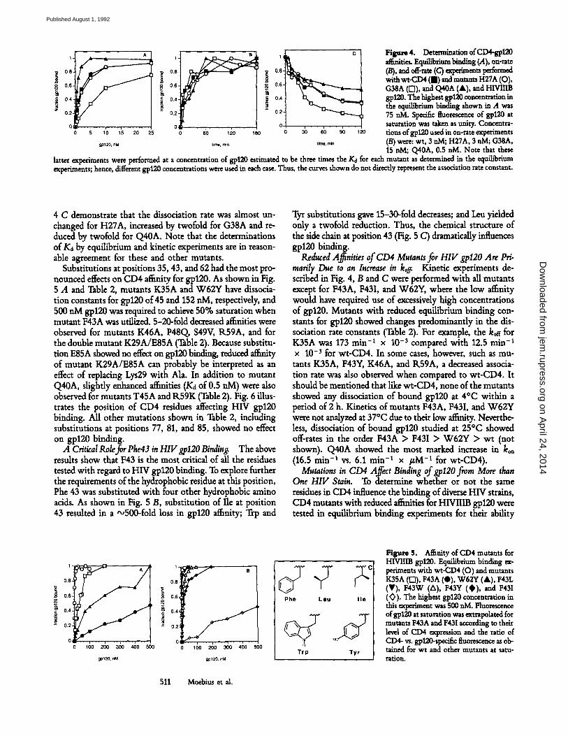
H ~ r e 4. Determin=tion of CD4-gpl20 .~.ities. Eqnih~riam bindin s (.4), on-rate (/3), and o~-.= (C) ~'l~im~ts l~e'~med with wt-CD4 (m) and mutants H27A (O), G38A ([]), and Q40A (A), and HIVIIIB gp120. The highest gp120 concentration in the equilibrium binding shown in A was 75 aM. Specific fluo1~cence of gpl20 at saturation was taken as unity. Concentra- tions of gp120 used in on-rate experiments (B) were: wt, 3 aM; H27A, 3 aM; G38A, 15 aM; Q40A, 0.5 nM. Note that these
latter experiments were performed at a concentration of g, p120 estimated to be three times the Kd for each mutant as determined in the equilibrium experiments; hence, different gp120 concentrations were used in each case. Thus, the curves shown do not directly represent the association rate constant.
4 C demonstrate that the dissociation rate was almost un- changed for H27A, increased by twofold for G38A and re- duced by twofold for Q40A. Note that the determinations of Ka by equilibrium and kinetic experiments are in reason- able agreement for these and other mutants.
Substitutions at positions 35, 43, and 62 had the most pro- nounced effects on CD4 affinity for gp120. As shown in Fig. 5 A and Table 2, mutants K35A and W62Y have dissocia- tion constants for gp120 of 45 and 152 raM, respectively, and 500 nM gp120 was required to achieve 50% saturation when mutant F43A was utilized. 5-20-fold decreased affinities were observed for mutants K46A, P48Q, $49V, IL59A, and for the double mutant K29A/E85A (Table 2). Because substitu- tion E85A showed no effect on g'p120 binding, reduced aff~ty of mutant K29A/E85A can probably be interpreted as an effect of replacing Lys29 with Ala. In addition to mutant Q40A, slightly enhanced affinities (Kd of 0.5 riM) were also observed for mutants T45A and R59K (Table 2). Fig. 6 illus- trates the position of CD4 residues affecting HIV gp120 binding. All other mutations shown in Table 2, including substitutions at positions 77, 81, and 85, showed no effect on gp120 binding.
A Critical golefor Phe#3 in HIVgp120 Binding. The above results show that F43 is the most critical of all the residues tested with regard to HIV gp120 binding. To explore further the requirements of the hydrophobic residue at this position, Phe 43 was substituted with four other hydrophobic amino acids. As shown in Fig. 5 B, substitution of Ile at position 43 resulted in a ',~500-fold loss in gp120 affinity; Trp and
Tyr substitutions gave 15-30-fold decreases; and Leu yidded only a twofold reduction. Thus, the chemical structure of the side chain at position 43 (Fig. 5 C) dramatically influences gp120 binding.
Reduced A.~nities of CD4 Mutants for HIV gp120 Are Pri. raarily Due to an Increase in kog. Kinetic experiments de- scribed in Fig. 4, B and C were performed with all mutants except for F43A, F43I, and W62Y, where the low ai~nity would have required use of excessively high concentrations of gp120. Mutants with reduced equilibrium binding con- stants for gp120 showed changes predominantly in the dis- sociation rate constants (Table 2). For example, the ko~ for K35A was 173 min -1 x 10 -3 compared with 12.5 rain -t x 10-3 for wt-CD4. In some cases, however, such as mu- tants K35A, F43Y, K46A, and R59A, a decreased associa- tion rate was also observed when compared to wt-CD4. It should be mentioned that like wt-CD4, none of the mutants showed any dissociation of bound gp120 at 4~ within a period of 2 h. Kinetics of mutants F43A, F43I, and W62Y were not analyzed at 37~ due to their low aff~ty. Neverthe- less, dissociation of bound gp120 studied at 25~ showed off-rates in the order F43A > F43I > W62Y > wt (not shown). Q40A showed the most marked increase in ko, (16.5 min -1 vs. 6.1 min -t x ~M -t for wt-CD4).
Mutations in CD4 Affect Binding of gpl2O from More than One HIV Stain. To determine whether or not the same residues in CD4 inSuence the binding of diverse HIV strains, CD4 mutants with reduced affnfities for HIVIIIB gp120 were tested in equilibrium binding experiments for their ability
0.6
0 . � 9 . . . . i . . . . i . . . . i . . . .
0 100 200 300 400 500
gp120, nM
0.8 i
08,
0.4~
0.2J
B
0 ~ . . . . i . . . . i . . . . I . . . . i . . . . 100 200 300 400 500
gp120, nM
Phe Leu l i e
T r p T y r
Figure 5. Affinity of CD4 mutants for HIVIIIB 87120. Equilibrium binding m- periments with wt-CD4 (O) and mutants K35A (D), F43A (Q), W62Y (A), F43L (V), F43W (A), F43Y (~) , and F43I (~) . The highest g10120 concentration in this experiment was 500 aM. Fluorescence of gp120 at saturation was ~trapohted for mutants F43A and F43I according to thdr level of CD4 expression and the ratio of CD4- vs. gpl20-specific fluorescence as ob- tained for wt and other mutants at satu- ration.
511 Moehius et al.
on April 24, 2014
jem.rupress.org
Dow
nloaded from
Published August 1, 1992

Table 2. A.i~nity of CD4 Mutants for HIVIIIB gp120
Mutation kon
Kinetic experiments Equilibrium
koa koa/kon conditions (Kd)
min -1 x /,tM -1 rain -~ • 10 -3 nM nM
wt 6.1 _+ 2.4 12,5 _+ 1.2 2.0 0.9 _+ 0.3 S19Y 5.9 13.1 2.2 1.2 H27A 5.5 13,3 2.4 1.6 K35A 1.8 173.3 98 45 G38A 3.2 27.7 8.7 9.4 Q40A 16.5 5.8 0.4 0.5 F43A ND ND ND 500 F43I ND ND ND 500 F43L 5.9 21.6 3.7 2.5 F43W 3.9 76.9 20 16 F43Y 1.7 53.3 32 32 T45A 7.7 9.6 1.3 0.6 K46A 2.0 33.0 16 19 P48Q 4.5 34.6 7.6 4.5 $49V 3.6 46.2 13 13 N52A 5.7 10.2 1.8 0.8 A55V 5.0 18.2 3.6 2.4 D56A 4.3 12.2 2.9 1.6 R59A 2.4 26.6 11 7.9 R59K 7.1 9.5 1.3 0.6 W 6 2 Y ND ND ND 152 E77Q 8.9 11.6 1.3 1.3 T81A 3.5 18.2 5.2 1.5 E85A 7.2 16.9 2.4 1.3 E85Q 3,6 16.9 4.7 1.5 K29A/E85A 3.7 49.5 13 6.7 Q89L 7.3 12.8 1.8 1.2
to bind gpl20 derived from the structurally distinct HIV strain SF2. The affinity of wt-CD4 for SF2 gpl20 is more than one order of magnitude lower than for HIVIIIB gp120 (Ka = 20 vs. 0.9 nM). As shown in Table 3, all mutations with reduced affinities for HIVIIIB gp120 showed diminished affini- ties also for SF2 gp120. Moreover, for each individual mu- tant the change in affinity for SF2 gp120 was comparable to the change observed with HIVIIIB gp120 (Table 3).
Discussion
We have established experimental conditions for measuring the affinity of gp120 and mutant CD4 at equilibrium, as well as for determining the rate constants for association and dis- sociation. The kinetics are strongly temperature dependent. The rate of dissociation is negligible at 4~ and it was there-
fore necessary to perform the studies at 37~ in order to achieve equilibrium. CD4 is not rapidly endocytosed when expressed on the surface of lymphoid ceils (41), and we could deter- mine gp120 binding curves over an 8-12-h period in stable murine T cell line transfectants. Most previous experiments on gp120 binding to expressed mutants of CD4 were per- formed at lower temperature (12, 28, 30-32), and they were therefore primarily measurements of relative on-rates rather than of equilibrium affinities. The properties of the K35A mutant are a useful illustration: the effect of the change on the rate of CD4-gpl20 association is much smaller than its effect on dissociation. A measurement sensitive only to kon would have shown a small change in apparent binding. In other cases, there are changes in both association and dissoci- ation rate constants, but only the equilibrium binding mea- surements, which take both into account, reveal the full effect of the substitution.
512 The CD4 Binding Site for HIV gpl20
on April 24, 2014
jem.rupress.org
Dow
nloaded from
Published August 1, 1992
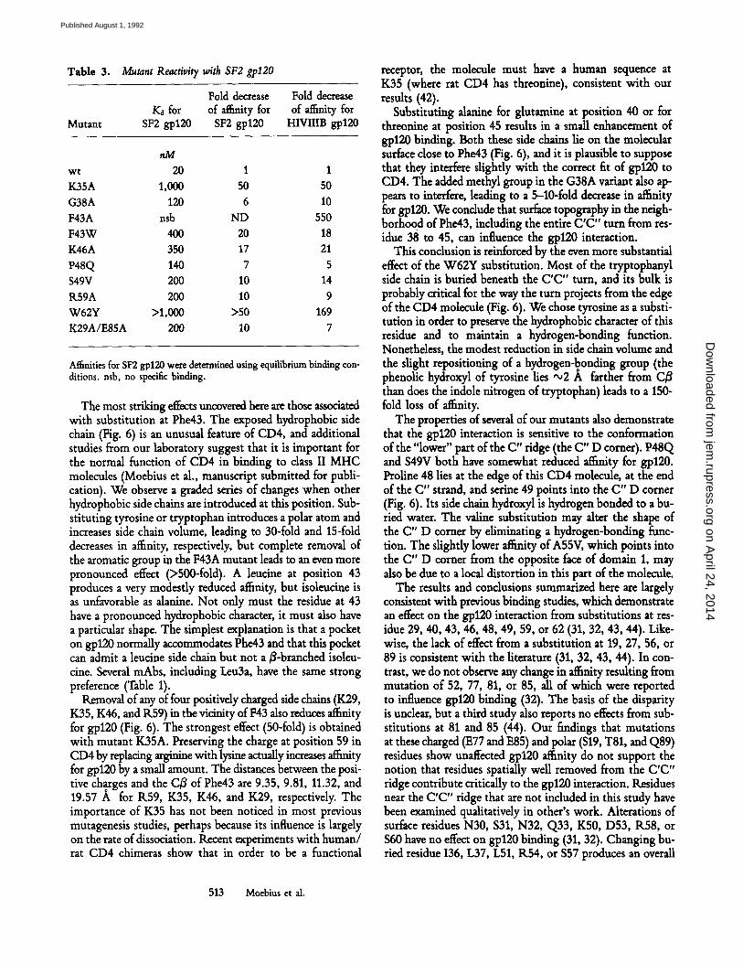
Table 3. Mutant Reactivity with SF2 gpl20
Mutant
Fold decrease Fold decrease Ka for of a~nity for of af~nity for
SF2 gp120 SF2 gp120 HIVIIIB gp120
nM wt 20 1 1 K35A 1,000 50 50 G38A 120 6 10 F43A nsb ND 550 F43W 400 20 18 K46A 350 17 21 P48Q 140 7 5 $49V 200 10 14 R59A 200 10 9 W62Y >1,000 ~>50 169 K29A/E85A 200 10 7
Af~nities for SF2 gp120 were determined using equilibrium binding con- ditions, nsb, no specific binding.
The most striking effects uncovered here are those associated with substitution at Phe43. The exposed hydrophobic side chain (Fig. 6) is an unusual feature of CD4, and additional studies from our laboratory suggest that it is important for the normal function of CD4 in binding to dass II MHC molecules (Moebius et al., manuscript submitted for publi- cation). We observe a graded series of changes when other hydrophobic side chains are introduced at this position. Sub- stituting tyrosine or tryptophan introduces a polar atom and increases side chain volume, leading to 30-fold and 15-fold decreases in affinity, respectively, but complete removal of the aromatic group in the F43A mutant leads to an even more pronounced effect (•500-fold). A leucine at position 43 produces a very modestly reduced affinity, but isoleucine is as unfavorable as alanine. Not only must the residue at 43 have a pronounced hydrophobic character, it must also have a particular shape. The simplest explanation is that a pocket on gp120 normally accommodates Phe43 and that this pocket can admit a leucine side chain but not a 3-branched isolen- cine. Several mAbs, including Leu3a, have the same strong preference (Table 1).
Removal of any of four positively charged side chains (K29, K35, K46, and R59) in the vicinity ofF43 also reduces affufity for gp120 (Fig. 6). The strongest effect (50-fold) is obtained with mutant K35A. Preserving the charge at position 59 in CD4 by replacing arginine with lysine actually increases affinity for gp120 by a small amount. The distances between the posi- tive ch~ges and the C/~ of Phe43 are 9.35, 9.81, 11.32, and 19.57 A for IL59, K35, K46, and K29, respectively. The importance of K35 has not been noticed in most previous mutagenesis studies, perhaps because its influence is largely on the rate of dissociation. Recent experiments with human/ rat CD4 chimeras show that in order to be a functional
receptor, the molecule must have a human sequence at K35 (where rat CD4 has threonine), consistent with our results (42).
Substituting alanine for glutamine at position 40 or for threonine at position 45 results in a small enhancement of gp120 binding. Both these side chains lie on the molecular surface dose to Phe43 (Fig. 6), and it is plausible to suppose that they interfere slightly with the correct fit of gpl20 to CD4. The added methyl group in the G38A variant also ap- pears to interfere, leading to a 5-10-fold decrease in affinity for gp120. We conclude that surface topography in the neigh- borhoo d of Phe43, including the entire C'C" turn from res- idue 38 to 45, can influence the gp120 interaction.
This conclusion is reinforced by the even more substantial effect of the W62Y substitution. Most of the tryptophanyl side chain is buried beneath the C'C" turn, and its bulk is probably critical for the way the turn projects from the edge of the CD4 molecule (Fig. 6). We chose tyrosine as a substi- tution in order to preserve the hydrophobic character of this residue and to maintain a hydrogen-bonding function. Nonetheless, the modest reduction in side chain volume and the slight repositioning of a hydrogen-bonding group (the phenolic hydroxyl of tyrosine lies ,x,2 A farther from C/3 than does the indole nitrogen of tryptophan) leads to a 150- fold loss of affinity.
The properties of several of our mutants also demonstrate that the gp120 interaction is sensitive to the conformation of the "lower" part of the C" ridge (the C" D corner). P48Q and $49V both have somewhat reduced affinity for gp120. Proline 48 lies at the edge of this CD4 molecule, at the end of the C" strand, and serine 49 points into the C" D corner (Fig. 6). Its side chain hydroxyl is hydrogen bonded to a bu- fled water. The valine substitution may alter the shape of the C" D corner by eliminating a hydrogen-bonding func- tion. The slightly lower affinity of A55V, which points into the C" D corner from the opposite face of domain 1, may also be due to a local distortion in this part of the molecule.
The results and conclusions summarized here are largely consistent with previous binding studies, which demonstrate an effect on the gp120 interaction from substitutions at res- idue 29, 40, 43, 46, 48, 49, 59, or 62 (31, 32, 43, 44). Like- wise, the lack of effect from a substitution at 19, 27, 56, or 89 is consistent with the literature (31, 32, 43, 44). In con- trast, we do not observe any change in affinity resulting from mutation of 52, 77, 81, or 85, all of which were reported to influence gp120 binding (32). The basis of the disparity is unclear, but a third study also reports no effects from sub- stitutions at 81 and 85 (44). Our findings that mutations at these charged (E77 and E85) and polar ($19, T81, and Q89) residues show unaffected gp120 affinity do not support the notion that residues spatially well removed from the C'C" ridge contribute critically to the gp120 interaction. Residues near the C'C" ridge that are not included in this study have been examined qualitatively in other's work. Alterations of surface residues N30, $31, N32, Q33, K50, D53, K58, or $60 have no effect on gp120 binding (31, 32). Changing bu- fled residue 136, L37, L51, K54, or $57 produces an overall
513 Moebius et al.
on April 24, 2014
jem.rupress.org
Dow
nloaded from
Published August 1, 1992

Figure 6. Illustration of the gp120 binding site. Stereo views of (A) space-falling model of CD4 domains 1 and 2, and (B) a-carbon trace model of CD4 domain 1 according to the atomic structure of CD4 (33). Charged residues involved in gp120 binding are shown in red (K29, K35, K46, and R59). The hydrophobic residue (F43) involved in gp120 binding is shown in yellow. The buried residue that affects gp120 binding by altering the C'C" ridge (W62) and the buried ($49) and exposed (P48) residues altering the C"D turn are shown in green. Amino acids (Q40 and T45) whose mutation to alanine enhanced gp120 binding are shown in dark blue.
loss of conformational stability in domain 1, as deduced from the effect of reactivity with a varity of CD4 mAbs (32). Mu- tations at G41, L44, and G47 are reported to decrease affinity for gp120 (31, 32), and examination of the modal shows that
the changes in question would be expected to perturb the conformation of the C'C" turn or the C"D corner. For ex- ample, leucine 44 packs against tryptophan 62 and helps de- termine the projecting conformation of phenylalanine 43. The
514 The CD4 Binding Site for HIV gp120
on April 24, 2014
jem.rupress.org
Dow
nloaded from
Published August 1, 1992
Q40P and T45P mutations, reported to interfere with gp120 binding (43), would also be expected to alter the C'C" turn.
We conclude that the principal determinant of gp120 binding is a continuous region on the surface of domain 1 covering ,v900 A 2 of accessible surface. This site contains Phe43 near its center partially surrounded by positiwly charged residues Lys29, Lys35, Lys46, and Arg59. The importance of positively charged residues in the vicinity of Phe43 is in- teresting in view of a previous report where three negatively charged residues in gp120 have been demonstrated to be crit- ical for binding to CD4 (45). Two of the three residues are conserved not only among HIV-1 isolates, but also in HIV-2 and in SIV strains. The contact between the viral glycopro- tein and its receptor might therefore include a set of ionic interactions flanking a central hydrophobic patch.
The binding site on CD4 for its normal interaction with class II MHC molecules is more complex than the site for gp120 (12, 43, 46). It includes several other parts of the mo-
lecular surface in domains 1 and 2, in addition to the C'C" ridge. It is striking, however, that when the same CD4 mu- tations analyzed here were examined for their capacity to bind class II MHC molecules, similar effects were observed (Moebius et al., manuscript submitted for publication). In particular, the alterations that reduce gp120 binding (K35A, F43A, K46A, $49V, R59A, and W62Y) also diminish ro- sette formation between CD4-transfected COS-1 cells and class II MHC-expressing B cells. A surface region with a projecting hydrophobic side chain (Phe43) and adjacent posi- tively charged residues thus also appears to be important for the class II MHC interaction. The overlap o fHIV gp120 and class II binding sites requires that development of receptor- based HIV inhibitors avoids drugs with immunosuppressive properties. Our detailed analysis of the contribution of residues in CD4 domain 1 to HIV gp120 binding gives a quantitative assessment of key components in the virus binding site, with such developments in mind.
We acknowledge Drs. Jiahuai Wang and Youwei Yan for ongoing collaboration and discussion. We recog- nize the assistance of the DFCI Flow Cytometry Facility with FACS | analysis, A. Diener for technical help, Dr. A. Kister for help with computer graphics, Dr. N. Tweedy for help with data calculations, Chiton Inc., for providing HIV gp120 of SF2 strain, and Repligen Inc. for anti-gp120 mAb F59.1., respec- tively.
This work was supported in part by National Institutes of Health grants AI-27336 to E. L. Reinherz, P30 AI-28691 to U. Moebius, and AI-30361 to S. C. Harrison. Dr. Moebius is supported by an AIDS Stipendium from the Bundesministerium flit Forschung und Technologic.
Address correspondence to Ulrich Moebius, Laboratory of Immunobiology, Dana Farber Cancer Institute, 44 Binney Street, Boston, MA 02115.
Received.for publication 18 March 1992 and in revised forra 18 May 1992.
Ref-er~nces 1. Reinherz, E.L., P.C. Kung, G. Goldstein, R.H. Levey, and S.F.
Schlossman. 1980. Discrete stages of intrathymic differentia- tion: Analysis of normal thymocytes and leukemic lympho- blasts of T lineage. Proa Natl. Acad. Sci. USA. 77:1588.
2. Kruisbeek, A.M., J.J. Mong, B.J. Fowlkes, J.A. Carmen, S. Bridges, and D.L. Longo. 1985. Absence of the Lyt- 2-,L3T4 + lineage of T cells in mice treated neonatally with anti-I-A correlates with absence of intrathymic I-A-bearing antigen-presenting cell function. J. Exl~ Med. 161:1029.
3. Ramsdell, F., and B.J. Fowlkes. 1989. Engagement of CD4 and CD8 accessory molecules is required for T cell matura- tion. J. Imraunol. 143:1467.
4. Engleman, E.G., C.J. Benike, C. Grumet, and R.L. Evans. 1981. Activation of human T lymphocyte subsets: helper and suppressor/cytotoxic T cells recognize and respond to distinct histocompatibility antigens. J. Immunol. 127:2124.
5. Biddison, W.E., P.E. Rao, M.A. TaUe, G. Goldstein, and S. Shaw. 1982. Possible involvement of the OKT4 molecule in T cell recognition of class II HLA antigens: evidence from studies of cytotoxic T lymphocytes specific for SB antigens. f Ex F Med. 156:1065.
6. Krensky, A.M., C.S. Reiss, J.W. Mier, J.L. Strominger, and S.J. Burakoff. 1982. Long-term human cytolytic T cell lines allospecific for HLA-DR6 antigen are OKT4+. Proa Natl. Acac~ Sci. USA. 79:2365.
7. Meuer, S.C., S.E Schlossman, and E.L. Rdnherz. 1982. Clonal analysis of human cytotoxic T lymphocytes: T4 + and T8 § effector T cells recognize products of different major histocom- patibility complex regions. Proa Natl. Acad. Sci. USA. 79:4395.
8. Eichmann, K., J.-I. J6nsson, I. Falk, and F. Emmrich. 1987. Effective activation of resting mouse T lymphocytes by cross- linking submitogenic concentrations of the T cell antigen receptor with either Lyt-2 or L3T4. Eur J. Immunol. 17:643.
9. Owens, T., D.S.B. Fazekas, andJ.F.A.P. Miller. 1987. Coaggre- gation of the T-cell receptor with CD4 and other T-cell sur- face molecules enhances T-cell activation. Proc. Natl. Acad. Sci. USA. 84:9209.
10. Meuer, S.C., K.E. Hussey, J.C. Hodgdon, T. Hercend, S.F. Schlossman, and E.L. Reinherz. 1982. Surface structures in- volved in target recognition by human cytotoxic T lympho- cytes. Science (Wash. DC). 218:471.
11. Doyle, C., andJ.L. Strominger. 1987. Interaction between CD4
515 Moebius etal.
on April 24, 2014
jem.rupress.org
Dow
nloaded from
Published August 1, 1992
and class II MHC molecules mediates cell adhesion. Nature (Lond.). 330:256.
12. Clayton, L.K., M. Sieh, D.A. Pious, and E.L. Reinherz. 1989. Identification of human CD4 residues affecting class II MHC versus HIV-1 gp120 binding. Nature (Lond.). 339:548.
13. Reinherz, E.L., PC. Kung, G. Goldstein, and S.F. Schlossman. 1979. A separation of functional subset of human T cells by a monoclonal antibody. Proc Natl. Acad. Sci. USA. 76:4061.
14. Relnherz, E.L., PC. Kung, G. Goldstein, and S.F. Schlossman. 1979. Further characterization of the human inducer T cell subset defined by monoclonal antibody.J. Iramu,ol. 234:2894.
15. Dalgleish, A.G., P.C.L. Beverley, Elk. Clapham, D.H. Craw- ford, M,F. Greaves, and R.A. Weiss. 1984. The CD4 (T4) an- tigen is an essential component of the receptor for the AIDS retrovirus. Nature (Lond.). 312:763.
16. Klatzmann, D., E. Champagne, S. Chamaret, J. Gruest, D. Guetard, T., Hercend, J.-C. Gluckman, and L. Montagnier. 1984. T lymphocyte T4 molecule behaves as the receptor for human retrovirus LAV. Nature (Lond.). 312:767.
17. McDougal, J.S., A. Mawle, S.P. Cort, J.K.A. Nicholson, G.D. Cross, J.A. Schepper-Campbdl, D. Hicks, and J. Sligh. 1985. Cellular tropism of the human retrovirus HTLV/LAV. I. Role of T cell activation and expression of the T4 antigen. J. Im- raunol. 135:3151.
18. Maddon, P.J., A.G. Dalgleish, J.S. McDougal, P.R. Clapham, R.A. Weiss, and IL. Axel. 1986. The T4 gene encodes the AIDS virus receptor and is expressed in the immune system and the brain. Cell. 47:333.
19. Stein, B.S., S.D. Gouda, J.D. Lifson, R.C. Penhallow, K.G. Bensch, and E.G. Engleman. 1987. pH-independent HIV entry into CD4-positive T cells via virus envelope fusion to the plasma membrane. Ceil 49:659.
20. McDougal, J., M. Kennedy, J. Sligh, S. Cort, A. Mawle, and J. Nicholson. 1986. Binding of the HTL VIII/LAV to T4 § T cells by a complex of the 110k viral protein and the T4 mol- ecule. Science (Wash. DC). 231:382.
21. Lane, M.C., J.L. Depper, W.C. Green, G. Whalen, T Wald- mann, and A.S. Fanci. 1985. Qualitative analysis of immune function in patients with the acquired immunodeficiency syn- drome. N. Engl. J. Med. 313:79.
22. Lyerly, H.K., T.J. Matthews, A. Langlois, D. Bolognesi, and K. Weinhold. 1987. Human lymphotropic virus IIIb glyco- protein (gp120) bound to CD4 determinants on normal lym- phoctyes and expressed by infected cells serves as target for im- mune attack. Proc Natl. Acad. Sci. USA. 84:4601.
23. Sodroski, J., W. Gob, C. Rosen, K. Campbell, and W. Hasel- tine. 1986. Role of the HTLVIII/LAV envelope in syncitia for- mation and cytopathicity. Nature (Lond.). 322:470,
24. Siliciano, R., T. Lawton, C. Knall, P. Berman, T Gregory, and E. Reinherz. 1988. Analysis of host-virus interactions in AIDS with anti-gp120 T cell clones: effect of HIV sequence variation and a mechanism for CD4 + T cell depletion. Cell. 54:561.
25. Shalaby, M.R., J.F. Krowka, T.J. Gregory, S. Hirabayashi, S. McCabe, D. Kaufman, D. Stites, and A. Ammann. 1987. The effects of human immunodeficiency virus recombinant enve- lope protein on immune cell functions in vitro. Cell. Immunol. 110:140.
26. Maddon, P.J., D.R. Littman, M. Godfrey, D.E. Maddon, L. Chess, and R. Axel. 1985. The isolation and nucleotide se- quence of a cDNA encoding the lymphocyte protein T4: a new member of the immunoglobulin family. Cell. 42:93.
27. Richardson, N.E., N.R. Brown, R.E. Hussey, A. Vaid, T.J.
Matthews, D.P. Bolognesi, and E.L. Reinherz. 1988. Binding site for human immunodeficiency virus coat protein gp120 is located in the NHz-terminal region of T4 (CD4) and requires the intact variable-region-like domain. Proc. Natl. Acad. Sci. USA. 85:6102.
28. Arthos, J., K.C. Deen, M.A. Chaikin, J.A. Fornwald, G. Sathe~ Q.J. Sattentan, P.R. Clapham, R.A. Weiss, J.S. McDougal, C. Pietropaolo, R. Axel, A. Truneh, P.J. Maddon, and R.W. Sweet. 1989. Identification of the residues in human CD4 crit- ical for the binding of HIV. Cell. 57:469.
29. Peterson, A., and B. Seed. 1988. Genetic analysis of mono- clonal antibody and HIV binding sites on the human lympho- cyte antigen CD4. Cell. 54:65.
30. Mizukami, T., T.R. Fuerst, E.A. Berger, and B. Moss. 1988. Binding region for human immunodeficiency virus (HIV) and epitopes for HIV-blocking monoclonal antibodies of the CD4 molecule defined by site-directed mutagenesis. Proc Natl. Acad. Sci. USA. 85:9273.
31. Brodsky, M.H., M. Warton, R.M. Myers, and D.R. Littman. 1990. Analysis of the site in CD4 that binds to the HIV enve- lope glycoprotein. J. Immunol. 144:3078.
32. Ashkenazi, A., L.G. Presta, S.C. Marsters, T.R. Camerato, K.A. Rosenthal, B.M. Fendly, and D.J. Capon. 1990. Mapping the CD4 binding site for human immunodeficiency virus by alanine- scanning mutagenesis. Proc Natl. Acad. Sci. USA. 87:7150.
33. Wang, J., Y. Yah, T.P.J. Garrett, J. Liu, D.W. Rodgers, R.L. Garlick, G.E. Tart, Y. Husain, E.L. Reinherz, and S.C. Har- rison. 1990. Atomic structure of a fragment of human CD4 containing two immunoglobulin-hke domains. Nature (Lond.). 348:411.
34. Ryu, S.-E., P.D. Kwong, A. Truneh, T.G. Porter, J. Arthos, M. Rosenberg, X. Dai, N.-H. Xuong, R. Axel, R.W. Sweet, and W.A. Hendrickson. 1990. Crystal structure of an HIV- binding recombinant fragment of human CD4. Nature (Lond.). 348:419.
35. Clayton, L.K., R.E. Hussey, Ik. Steinbrich, H. Ramachan- dran, Y. Husain, and E.L. Reinherz. 1988. Substitution of rou- tine for human CD4 residues identifies amino acids critical for HIV gp120 binding. Nature (Lond.). 335:363.
36. Ohashi, O.S., T.W. Mak, P. van den Elsen, Y. Yanagi, Y. Yoshikai, A.E Caiman, C. Terhorst, J.D. Stobo, and A. Weiss. 1985. Reconstitution of an active surface T3/T-cell antigen receptor by DNA transfer. Nature (Lond.). 316:606.
37. Munck, A. 1976. General aspects of steroid hormone-receptor interactions. In Receptors and Mechanism of Action of Ste- roid Hormones, Part I. J.R. Pasqualini, editor. Marcel Dekker, Inc., New York. 1-40.
38. Sattentan, Q.J., J. Arthos, K. Deen, N. Hanna, D. Healey, P.C.L. Beverley, Ik. Sweet, and A. Truneh. 1989. Structural analysis of the human immunodeficiency virus-binding domain of CD4. j . Exi~ Med. 170:1319.
39. Healey, D.G., L. Dianda, D. Buck, K. Schroeder, A. Truneh, Q.J. Sattentau, and P.C.L. Beverley. 1991. A highly selected panel of anti-CD4 antibodies fails to induce anti-idiotypic an- tisera mediating human immunodeficiency virus neutralization. Eur. J. Immunol. 21:1491.
40. Dimitrov, D.S., K. Hillman, J. Manischewitz, R. Blumen- thai, and H. Golding. 1992. Kinetics of soluble CD4 binding to cells expressing human immunodeficiency virus type 1 enve- lope glycoprotein. J. Virol. 66:132.
41. Pelchen-Matthews, A., J.E. Armes, G. Griffiths, and M. Marsh. 1991. Differential endocytosis of CD4 in lymphocytic cells. J. Extx Med. 173:575.
516 The CD4 Binding Site for HIV gpl20
on April 24, 2014
jem.rupress.org
Dow
nloaded from
Published August 1, 1992
42. Schockmel, G.A., C. Somoza, S.J. Davis, A.F. Williams, and D. Healey. 1992. Construction of a binding site for human immunodeficiency virus type I gp120 in rat CD4.f Extx Med. 175:301.
43. Fleury, S., D. Lamarre, S. Mdoche, S.-E. Ryu, C. Cant-in, W.A. Hendrickson, and R.-P. Sekaly. 1991. Mutational analysis of the interaction between CD4 and class II MHC: class II an- tigens contact CD4 on a surface opposite the gp120 binding site. Cell. 66:1037.
44. Choe, H.-IL., andJ. Sodroski. 1992. Contribution of charged
amino acids in the CDR2 region of CD4 to HIVol gpl20 binding. J. Acquired Immune Defl~ Syndr. 5:204.
45. Olshevsky, U., E. Helseth, C. Furman, J. Li, W. Haseltine, and J. Sodroski. 1990. Identification of individual human im- munodeficiency virus type 1 gpl20 amino acids important for CD4 receptor binding. J. Virol. 64:5701.
46. Lamarre, D., A. Ashkenazi, S. Fleury, D.H. Smith, R.-P. Sekaly, and D.J. Capon. 1989. The MHC-binding and gp120-binding functions of CD4 are separable. Science (Wash. DC). 245:743.
517 Moebius et al.
on April 24, 2014
jem.rupress.org
Dow
nloaded from
Published August 1, 1992


![In conjunction with Venus [planetary radar astronomy]](https://static.fdokumen.com/doc/165x107/631a4f09bb40f9952b01f2bc/in-conjunction-with-venus-planetary-radar-astronomy.jpg)