Differential antibody binding to the surface TCR{middle dot}CD3 complex of CD4+ and CD8+ T...
12
International Immunology, Vol. 20, No. 10, pp. 1247–1258 doi:10.1093/intimm/dxn081 ª The Japanese Society for Immunology. 2008. All rights reserved. For permissions, please e-mail: [email protected] Differential antibody binding to the surface abTCRÁCD3 complex of CD4 1 and CD8 1 T lymphocytes is conserved in mammals and associated with differential glycosylation Nineth E. Rossi 1 , Jesu ´ s Reine ´ 1 , Miguel Pineda-Lezamit 2 , Manuel Pulgar 2 , Ne ´stor W. Meza 3 , Mahima Swamy 4 , Ruth Risueno 2 , Wolfgang W. A. Schamel 4 , Pedro Bonay 2 , Edgar Ferna ´ndez- Malave ´ 1,2 and Jose ´ R. Regueiro 1 1 Inmunologı ´a, Facultad de Medicina, Universidad Complutense, Madrid, Spain 2 Centro de Biologı ´a Molecular Severo Ochoa, Consejo Superior de Investigaciones Cientı ´ficas, Universidad Auto ´noma de Madrid, Cantoblanco, Madrid, Spain 3 Centro de Investigaciones Energe ´ticas, Medioambientales y Tecnolo ´ gicas, Madrid, Spain 4 Department of Molecular Immunology, Max Planck-Institute for Immunobiology and Faculty of Biology, University of Freiburg, Freiburg, Germany Keywords: cell surface molecules, T cells, TCRs Abstract We have previously shown that the surface ab T cell antigen receptor (TCR)ÁCD3 complex borne by human CD4 1 and CD8 1 T lymphocytes can be distinguished using mAbs. Using two unrelated sets of antibodies, we have now extended this finding to the surface abTCRÁCD3 of seven additional mammalian species (six non-human primates and the mouse). We have also produced data supporting that differential glycosylation of the two main T cell subsets is involved in the observed TCRÁCD3 antibody-binding differences in humans. First, we show differential lectin binding to human CD4 1 versus CD8 1 T lymphocytes, particularly with galectin 7. Second, we show that certain lectins can compete differentially with CD3 mAb binding to human primary CD4 1 and CD8 1 T lymphocytes. Third, N-glycan disruption using swainsonine was shown to increase mAb binding to the abTCRÁCD3. We conclude that the differential antibody binding to the surface abTCRÁCD3 complex of primary CD4 1 and CD8 1 T lymphocytes is phylogenetically conserved and associated with differential glycosylation. The differences may be exploited for therapeutic purposes, such as T cell lineage-specific immunosuppression of graft rejection. Also, the impact of glycosylation on CD3 antibody binding requires a cautious interpretation of CD3 expression levels and T cell numbers in clinical diagnosis. Introduction ab T lymphocytes recognize peptide–MHC ligands by means of a multimeric protein ensemble termed the ab T cell antigen receptor (TCR)ÁCD3 complex. This structure is com- posed of a variable ab TCR dimer which binds antigens and three invariant dimers (CD3ce, de and ff) which are involved in TCRÁCD3 surface transport, stabilization and signal trans- duction (1). The minimal stoichiometry, therefore, is believed to be abcedef 2 , although larger clusters of this basic mono- valent TCRÁCD3 unit have been observed in certain condi- tions (2). The ratio of monovalent to multivalent TCRÁCD3 varies between different T cell clones and lines, even when the receptors have the same specificity (3). Mature CD4 + and CD8 + ab T cells differ sharply in their MHC ligands, but their TCRÁCD3 complex is believed to be qualitatively identical. The fact that most TCRÁCD3-specific mAb bind to CD4 + ab T cells better than to CD8 + ab T cells was thus taken as evidence of quantitative differences in the number of TCRÁCD3 complexes on the surface of each cell type (4). Unexpectedly, abTCRÁCD3 expression was shown to be more impaired in CD8 + than in CD4 + cells when CD3c (5, 6) or CD3d (7) was absent. These observations were followed by the description of conformational and bio- chemical differences in the TCRÁCD3 complex between CD8 + and CD4 + CD3c-deficient (c ) T lymphocytes (8). Correspondence to: Jose ´ R. Regueiro; E-mail: [email protected] Received 14 September 2007, accepted 26 June 2008 Transmitting editor: I. Pecht Advance Access publication 24 July 2008
Transcript of Differential antibody binding to the surface TCR{middle dot}CD3 complex of CD4+ and CD8+ T...
International Immunology, Vol. 20, No. 10, pp. 1247–1258doi:10.1093/intimm/dxn081
ª The Japanese Society for Immunology. 2008. All rights reserved.For permissions, please e-mail: [email protected]
Differential antibody binding to the surfaceabTCR�CD3 complex of CD41 and CD81
T lymphocytes is conserved in mammals andassociated with differential glycosylation
Nineth E. Rossi1, Jesus Reine1, Miguel Pineda-Lezamit2, Manuel Pulgar2, Nestor W. Meza3,Mahima Swamy4, Ruth Risueno2, Wolfgang W. A. Schamel4, Pedro Bonay2, Edgar Fernandez-Malave1,2 and Jose R. Regueiro1
1Inmunologıa, Facultad de Medicina, Universidad Complutense, Madrid, Spain2Centro de Biologıa Molecular Severo Ochoa, Consejo Superior de Investigaciones Cientıficas, Universidad Autonoma deMadrid, Cantoblanco, Madrid, Spain3Centro de Investigaciones Energeticas, Medioambientales y Tecnologicas, Madrid, Spain4Department of Molecular Immunology, Max Planck-Institute for Immunobiology and Faculty of Biology, University of Freiburg,Freiburg, Germany
Keywords: cell surface molecules, T cells, TCRs
Abstract
We have previously shown that the surface ab T cell antigen receptor (TCR)�CD3 complex borne byhuman CD41 and CD81 T lymphocytes can be distinguished using mAbs. Using two unrelated sets ofantibodies, we have now extended this finding to the surface abTCR�CD3 of seven additionalmammalian species (six non-human primates and the mouse). We have also produced data supportingthat differential glycosylation of the two main T cell subsets is involved in the observed TCR�CD3antibody-binding differences in humans. First, we show differential lectin binding to human CD41
versus CD81 T lymphocytes, particularly with galectin 7. Second, we show that certain lectins cancompete differentially with CD3 mAb binding to human primary CD41 and CD81 T lymphocytes. Third,N-glycan disruption using swainsonine was shown to increase mAb binding to the abTCR�CD3. Weconclude that the differential antibody binding to the surface abTCR�CD3 complex of primary CD41 andCD81 T lymphocytes is phylogenetically conserved and associated with differential glycosylation. Thedifferences may be exploited for therapeutic purposes, such as T cell lineage-specificimmunosuppression of graft rejection. Also, the impact of glycosylation on CD3 antibody bindingrequires a cautious interpretation of CD3 expression levels and T cell numbers in clinical diagnosis.
Introduction
ab T lymphocytes recognize peptide–MHC ligands bymeans of a multimeric protein ensemble termed the ab T cellantigen receptor (TCR)�CD3 complex. This structure is com-posed of a variable ab TCR dimer which binds antigens andthree invariant dimers (CD3ce, de and ff) which are involvedin TCR�CD3 surface transport, stabilization and signal trans-duction (1). The minimal stoichiometry, therefore, is believedto be abcedef2, although larger clusters of this basic mono-valent TCR�CD3 unit have been observed in certain condi-tions (2). The ratio of monovalent to multivalent TCR�CD3varies between different T cell clones and lines, even whenthe receptors have the same specificity (3).
Mature CD4+ and CD8+ ab T cells differ sharply in theirMHC ligands, but their TCR�CD3 complex is believed to bequalitatively identical. The fact that most TCR�CD3-specificmAb bind to CD4+ ab T cells better than to CD8+ ab T cellswas thus taken as evidence of quantitative differences inthe number of TCR�CD3 complexes on the surface of eachcell type (4). Unexpectedly, abTCR�CD3 expression wasshown to be more impaired in CD8+ than in CD4+ cells whenCD3c (5, 6) or CD3d (7) was absent. These observationswere followed by the description of conformational and bio-chemical differences in the TCR�CD3 complex betweenCD8+ and CD4+ CD3c-deficient (c�) T lymphocytes (8).
Correspondence to: Jose R. Regueiro; E-mail: [email protected] Received 14 September 2007, accepted 26 June 2008
Transmitting editor: I. Pecht Advance Access publication 24 July 2008

More recently, we reported that a CD3-specific mAb termedRW28C8 bound normal human CD8+ ab T lymphocytes bet-ter than CD4+ ab T cells (9). Collectively, these results arenot consistent with the quantitative interpretation but, rather,with the existence of constitutive differences (i.e. qualitative)in antibody binding to the surface abTCR�CD3 complexesborne by CD4+ versus CD8+ T cells. These differences couldbe due to the existence of T cell lineage-associated differen-ces in the way the abTCR�CD3 complex is assembled, gly-cosylated, trimmed or topologically pre-clustered at the cellsurface.In the present study, we have extended our observations
to other mammalian species. Additionally, we haveaddressed the hypothesis of differential glycosylation of hu-man CD4+ and CD8+ cells by studying surface lectin bindingand competition with CD3-specific mAb. Lastly, we haveshown that the TCR�CD3 complexes from both CD4+ andCD8+ cells co-exist in monovalent and multivalent com-plexes and the ratio between them appears to be similar.This excludes that different degrees of pre-clustering werethe cause for differential antibody accessibility.
Methods
Cells
Human venous blood was obtained with informed consentfrom healthy donors (20 to 34 years old). Venous blood fromnon-human primates was obtained from anesthetized healthyadult animals belonging to Cercopithecidae (Papio sp. or ba-boon, Macaca sylvanus or barbary macaque and Mandrillussphinx or mandrill), Hominidae (Gorilla gorilla and Pan trog-lodytes or chimpanzee), Hylobatidae (Hylobates lar or gib-bon) and Lemuridae (Eulemur fulvus mayottensis or brownlemur) families. Heparinized blood or lymphoid tissue cellswere obtained from 4- to 12-week-old wild-type (c+) orCD3c-deficient (c�) C57BL/6 euthanized mice (6). Theorgans were placed in complete RPMI 1640 cell culture me-dium (Gibco Invitrogen; Paisley, Scotland, UK), washed andmacerated through a 70-lm cell strainer to remove aggre-gates. To remove adherent cells, thymocytes were incubatedin plastic flasks with RPMI 1640 + 10% FCS for 30 min at37�C. Splenocytes were re-suspended in 2 ml 0.83% w/vNH4Cl to lyse erythrocytes.PBMCs were isolated and in some cases activated for 12–
18 h with 5 lg ml�1 PHA as described (10). Constitutivelyactivated Herpesvirus saimiri-transformed human T cellswere obtained and grown as described (11). This study wasapproved by the Institutional Review Board of ComplutenseUniversity.
Antibodies
The following unconjugated mAbs were used for humanand, where indicated, non-human primate samples (clonenames in parentheses): anti-CD3ec/ed (UCHT1 and IOT3b)from Coulter Immunotech, Marseille, France; anti-CD3e(X35) from D. Bourel, C.R. de Transfusion Sanguine, Rennes,France; anti-CD3e (2Ad2 and RW28C8) from E. L. Reinherz,Dana-Farber Cancer Institute, Boston, MA, USA; anti-CD3e(SPV.T3b) from J. E. de Vries, The Netherlands Cancer Insti-tute, Amsterdam, The Netherlands; anti-CD3 (F101.01) and
anti-CD3ec/ed (OKT3) from Bent Rubin, CHU de Purpan,Toulouse, France; anti-CD3e (Cris7) from R. Vilella, HospitalClınico, Barcelona, Spain; anti-CD3 (WT31), anti-TCRab(BMA031), anti-CD43 (1G10) and anti-CD3e (SP34) from BDPharmingen (San Diego, CA, USA); anti-CD3ec/ed (Leu4/SK7) and anti-TCRCb1 (JOVI1) from B. Alarcon, Centro deBiologıa Molecular Severo Ochoa (CBMSO), Consejo Superiorde Investigaciones Cientıficas (CSIC), University Autonoma,Cantoblanco, Madrid, Spain; anti-CD18 (TS2/16 and Lia 3/2)from C. Cabanas, Instituto de Farmacologıa y ToxicologıaConsejo Superior de Investigaciones Cientıficas – UniversidadComplutense de Madrid, Facultad de Medicina, UniversityComplutense, Madrid, Spain and anti-CD3 (MEM57) fromV. Horejsi, Institute of Molecular Genetics, – Academy of Sci-ences, Praha, Czech Republic. For these mAb, PE-conjugatedgoat anti-mouse IgG (H + L) or IgM (l) secondary antibod-ies from Caltag (Burlingame, CA, USA), Coulter Immunotech(Miami, FL, USA) or Southern Biotechnology Associates wereused. Several conjugated mAbs were used for human samples(clone names in parentheses): anti-CD4PE, FITC or PECy5(13B8.2) and anti-CD8PE, FITC or PECy5 (B9.11), anti-TCRVb17PE (E17.5F3), anti-TCRVb2FITC (MBB2D5), anti-TCRVb3PE (CH92) and TCRVb8.1 + 2PE (56C5) from CoulterImmunotech; anti-CD69FITC (L78), anti-CD3PE (Leu4/SK7)and anti-CD5PE (UCHT2) from BD Pharmingen. RW28C8 wasderived by immunization and screening with a CD4+ ragweed-specific T cell clone (R. E. Hussey and E. L. Reinherz,personal communication).For non-human primate samples, the following conjugated
mAbs were used (clone names in parentheses): anti-CD4FITC(M-T477) and anti-CD8aPECy5 (RPA-T8) from BD Pharmin-gen. The Rhesus (Macaca mulatta)-specific anti-CD3 mAbFN18 was kindly donated by D. Neville (Laboratory of Molecu-lar Biology, National Institute of Health, Bethesda, MD, USA).The following mAbs were used for mouse samples (clone
names in parentheses): anti-CD4PE or FITC (L3T4), anti-CD8PE or FITC (Ly2), anti-TCRbFITC (H57-597), anti-CD3e-biotin (145-2C11), anti-CD3e purified from hamster (500A2),anti-LFA-1PE (CD11a), anti-CD3PE (17A2) and a hamster orrat IgG isotype control from Pharmingen (San Diego, CA,USA); anti-CD3 purified from rat (KT3) was purchased fromSerotec (Oxford, UK), anti-intracellular CD3e (iCD3e, APA1/1) and CD3d (iCD3d, APA1/2) were obtained from B. Alarcon(CBMSO, CSIC, Universidad Autonoma, Cantoblanco,Madrid, Spain). The secondary reagents (goat anti-hamsterFITC, goat anti-rat FITC and streptavidin–FITC) were alsofrom Pharmingen.
Lectins
Several lectins were used to detect leucocyte cell surfaceglycans. Biotinylated peanut agglutinin from Arachis hypo-gaea (PNA), FITC-labeled Sambucus nigra (SNA, VectorLaboratories, Burlingame, CA, USA) bark lectin, and FITC-labeled Griffonia simplicifolia lectin I (IB4, used as a negativecontrol) from Vector Laboratories; FITC-labeled wheat germlectin Triticum vulgaris (WGA) and FITC-labeled red kidneybean Phaseolus vulgaris (PHA-L) from Sigma (St Louis, MO,USA); Alexa fluor 488-labeled soybean agglutinin Glycinemax (SBA) from Molecular Probes (Eugene, OR, USA).Recombinant human FITC-labeled galectins (Gals) Gal-1, -4, -7
1248 Conserved lineage-associated variation in abTCR�CD3
glutathione-S-transferase (GST) and -8GST were preparedas described (12–16). Recombinant human FITC-labeledGal-3 was kindly donated by A. Karlsson (The PhagocyteResearch Laboratory, Department of Rheumatology and In-flammation Research, Goteborg University, Sweden).To analyze lectin binding to PBMC, 105 cells in 100 ll
PBS–BSA (PBS, 1% bovine serum albumin, 0.05% sodiumazide) were exposed to 2–4 lg ml�1 conjugated lectins(60.1 M b-lactose where indicated) for 15 min at 4�C ina 96-well flat-bottom plate. Cells were washed twice in PBS–BSA and stained with anti-CD4PE and anti-CD8PECy5 mAb.Flow cytometry of at least 20 000 cells was done on anEpics Elite Cytometer (Beckman Coulter, Fullerton, CA,USA). Isolectin IB4-FITC in PBS 0.1 mM CaCl2 or GST–FITCwas used as irrelevant controls.For lectin competition experiments, four-step stainings
were performed. Briefly, human PBMCs were pre-incubatedwith increasing concentrations of each lectin for 15 min at4�C, washed twice in PBS–BSA, incubated with anti-humanTCR�CD3 mAb for 15 min at 4�C, washed twice in PBS–BSA, stained with anti-mouse IgG–PE (1/80) by incubationfor 20 min at 4�C, washed twice in PBS–BSA and incubatedwith anti-CD4PECy5 or anti-CD8PECy5.For N-glycosylation inhibition, Jurkat T cells were incu-
bated for up to 3 days with 5 lM swainsonine (Sigma) andmonitored for deglycosylation using PHA–FITC or stainedwith anti-TCR�CD3 mAb.
Flow cytometry
Electronic gates were used to identify CD4+ and CD8bright
lymphocytes, which are >99% abTCR�CD3+ (as measuredwith BMA031) and <0.2% cdTCR�CD3+ (as measured with11F2 or Inmuno510, data not shown). Human abTCR�CD3-associated epitopes within CD4+ or CD8bright subsets wereanalyzed by three-color flow cytometry on PBMC as de-scribed elsewhere (11). Briefly, 5 3 105 cells were stainedwith CD3- or TCR-specific mAb (30 min, 4�C), washed twicein PBS–BSA and incubated for 20 min at 4�C with anti-mouse IgG–PE, followed by two washes in PBS–BSA anda subsequent incubation for 20 min at 4�C with anti-CD4FITC and anti-CD8PECy5. For comparative stainings,the mean fluorescence intensity (MFI) was used. Sampleswere analyzed as above.For other primates, whole blood samples were analyzed.
Briefly, 100 ll of thoroughly mixed blood was incubated for15 min at 4�C in 12 3 75 mm polypropylene tubes, with CD3-or TCR-specific mAb, and processed as above. After incuba-tion, 2.5 ml of lysis solution (0.15 M NH4Cl, 0.1 mM KHCO3,0.1 mM EDTA) was added, the tubes were incubated for 10min at room temperature (22 6 3�C) in the dark and centri-fuged for 10 min at 4�C per 200 g. The cell pellets were re-suspended in 500 ll of PBS–BSA before analysis as above.For mouse samples, 0.5–1 3 106 cells were washed and
re-suspended in PBS–BSA with 20 lg ml�1 mouse IgG,incubated for 20 min at 4�C and washed twice with PBS–BSA. Two-color stainings were performed with purified ham-ster anti-mouse mAb followed by anti-hamster Ig–FITC (20min, 4�C) and two washes with PBS–BSA before anti-CD4PEor anti-CD8PE stainings. Control stainings with secondaryantibody only were performed routinely. Twenty thousand
events from each sample were analyzed in a FACScan usingthe CellQuest software (Becton Dickinson, San Jose, CA,USA). Intracellular stainings were performed as describedpreviously (17). Briefly, lymph node cells from 6-week-oldC57BL/6 mice were directly stained with PE-labeled anti-CD4 or anti-CD8 and fixed and permeabilized using theCytofix/Cytoperm kit (Pharmingen). Cells were then incu-bated with 4 lg ml�1 biotin-labeled APA1/1 or APA1/2 for 30min on ice, stained with streptavidin–FITC and analyzed byflow cytometry in a FACSCalibur.
Membrane preparation, blue native–PAGE andimmunoblotting
The 5 3 106 sorted CD4+ and CD8+ lymphocytes or JurkatT cells were allowed to swell for 10 min in 1 ml hypotonicbuffer (10 mM HEPES, pH 7.4, 42 mM KCl, 5 mM MgCl2and protease inhibitors). The cells were then disrupted usinga Dounce homogenizer and the membranes were pelleted inan ultracentrifuge TLA 100 (Beckman Instruments, Gagny,France) at 48 000 r.p.m. for 30 min. Membranes were lysedin 25 ll blue native (BN) buffer (500 mM 6-aminohexanoicacid, 20 mM NaCl, 10% glycerol, 2 mM EDTA, 20 mM bis–Tris, pH 7 and protease inhibitors) containing the appropri-ate detergent (0.3% Brij96 or 0.5% digitonin). The clarifiedmembrane lysates were separated by BN–PAGE (4–9%) asdescribed (3). Semi-dry transfer of the proteins to a polyviny-lidene difluoride membrane was done. Immunoblots weredeveloped with an anti-f antiserum. Ferritins in its 24mer(f1; 440 kD) and 48mer (f2; 880 kD) forms were included asmolecular mass standards.
Results
Differential antibody binding to the abTCR�CD3 complex ofprimary CD4+ and CD8+ T lymphocytes is phylogeneticallyconserved
We reported previously that the abTCR�CD3 complex borneby human primary CD4+ and CD8+ T lymphocytes is notidentical because certain mAbs, such as SP34 or UCHT1,bind the former better, whereas other mAb, such asRW28C8, bind the latter better (9). In order to extend theseobservations to other species, we studied six non-humanprimates and mice. The results are summarized in Fig. 1 incomparison to humans. First, not all human-specific CD3mAb bound non-human primate T cells, but those whichdid (denoted by + and +/� under the graph in Fig. 1B) con-firmed the findings above (Fig. 1A and B). Interestingly, insome cases CD4+, but not CD8+, T cells were bound verypoorly by RW28C8 (Mandrill, for instance, Fig. 1A). Second,mouse T cells were scanned using an entirely different setof mAb (Fig. 1A–C). The result indicated that the mousesurface abTCR�CD3 complex from CD4+ T cells, as shownfor primates, was bound better than that of CD8+ T cells bymost (2C11, 17A2, 500A2), but not all (KT3) CD3-specificmAb. This was most likely due to extracellular differences,since differential binding was not apparent upon stainingintracellular CD3 epitopes, such as those recognized byAPA1/1 (iCD3e) or APA1/2 (iCD3d, Fig. 1B and C). Thus,KT3 in mice mimics RW28C8, as it bound better to CD8+
than to CD4+ cells. This indicates that the antibody-binding
Conserved lineage-associated variation in abTCR�CD3 1249

Fig. 1. Differential antibody binding to the abTCR�CD3 of normal (c+) or CD3c-deficient (c�) CD4+ and CD8+ T lymphocytes in severalmammalian species. (A) Representative reactivity patterns of selected mAb in CD4+ (open histograms) and CD8bright (filled histograms) PBMC.Profiles are shown as logarithm of relative fluorescence versus cell number. The vertical line in each panel indicates the upper limit of backgroundfluorescence using isotype-matched irrelevant mAb. (B) MFI ratios6 SD of CD4+ relative to CD8+ peripheral blood Tcells with the indicated mAbin n independent individuals. For non-human primates, the (similar) results of different species were pooled to simplify the figure. ‘+’ means clearbinding of the indicated CD3 mAb to CD4+ or CD8+ T cells as shown in (A) for Mandrill; ‘�’ and ‘+/�’ stand for no or weak binding to both T cellsubsets, respectively; ND, not done; eCD3, extracellular CD3. MFI ratios were calculated only for + and +/� samples. Brown lemur T cells wereomitted, as they did not show specific binding to the mAbs assayed. MFI ratios above or below 1 (indicated by the horizontal dotted line) reflectan increased or decreased mAb binding to CD4+ cells, respectively. Statistical significance (Student’s t-test) is shown only for selected mAb. Asinvariant controls, CD18, CD5, CD43 or CD11a were evaluated, together with intracellular CD3 (iCD3; APA1/1 and APA1/2) in mouse samples (C)MFI ratios as in panel (B) are shown for several lymphoid tissues both in c+ (filled symbols) and in c� (open symbols) mouse T cells. L.NODE,lymph node. 17A2 does not bind c� cells. APA1/1 was not tested in c� samples. (D) MFI ratios as in panel (B) are shown for the indicated mAband activated T-lineage human cells, as compared with resting Tcells. Representative reactivity patterns as in panel (A) are shown for H. saimiri-transformed T cells (HVS).
1250 Conserved lineage-associated variation in abTCR�CD3
differences to the surface abTCR�CD3 complex of CD4+
and CD8+ T lymphocytes are conserved in evolution. Theresults were confirmed in all tested mouse lymphoid tis-sues, including early (thymus) and late (blood, lymph node,spleen) T cell differentiation stages and also in the absenceof CD3c (Fig. 1C) as reported in c� humans (9). The ob-served differences in KT3 binding between CD3c+ andc� samples suggest that the KT3 epitope is more CD3cdependent than other mAb, such as 17A2 or 2C11. Fromthese experiments, we conclude that the antibody-bindingdifferences observed in humans between the surfaceabTCR�CD3 complex of primary CD4+ and CD8+ T cellsare conserved in phylogeny, as well as along mouse T celldifferentiation.
Activation-independent differential binding of RW28C8 tohuman CD4+ and CD8+ T-lineage lymphocytes
Antigen recognition causes profound surface changes inT lymphocytes (18) and in the TCR�CD3 complex (19).To determine whether the observed abTCR�CD3 antibody-binding differences were regulated by activation, we com-pared antibody reactivities in human CD4+ and CD8+
T lymphocytes, either resting or recently activated (short-term CD69+ PBMC blasts) or constitutively activated by H.saimiri transformation (20). Activation had an impact onabTCR�CD3 antibody binding as measured with UCHT1,but the changes did not affect recognition of the RW28C8epitope, which remained more strongly expressed by CD8+
T lymphocytes even after H. saimiri transformation (Fig. 1D).Specific interference by the CD4 molecule is unlikely, asbinding of RW28C8 was enhanced even further in humanCD4+CD8+ immature T cells (9).
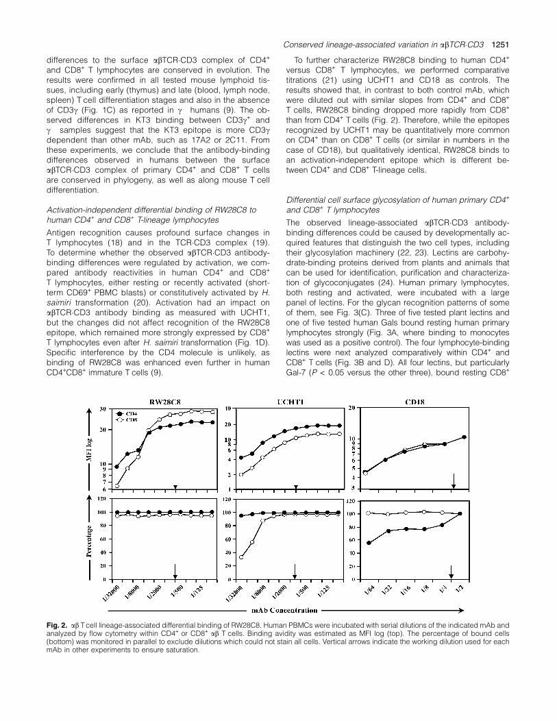
To further characterize RW28C8 binding to human CD4+
versus CD8+ T lymphocytes, we performed comparativetitrations (21) using UCHT1 and CD18 as controls. Theresults showed that, in contrast to both control mAb, whichwere diluted out with similar slopes from CD4+ and CD8+
T cells, RW28C8 binding dropped more rapidly from CD8+
than from CD4+ T cells (Fig. 2). Therefore, while the epitopesrecognized by UCHT1 may be quantitatively more commonon CD4+ than on CD8+ T cells (or similar in numbers in thecase of CD18), but qualitatively identical, RW28C8 binds toan activation-independent epitope which is different be-tween CD4+ and CD8+ T-lineage cells.
Differential cell surface glycosylation of human primary CD4+
and CD8+ T lymphocytes
The observed lineage-associated abTCR�CD3 antibody-binding differences could be caused by developmentally ac-quired features that distinguish the two cell types, includingtheir glycosylation machinery (22, 23). Lectins are carbohy-drate-binding proteins derived from plants and animals thatcan be used for identification, purification and characteriza-tion of glycoconjugates (24). Human primary lymphocytes,both resting and activated, were incubated with a largepanel of lectins. For the glycan recognition patterns of someof them, see Fig. 3(C). Three of five tested plant lectins andone of five tested human Gals bound resting human primarylymphocytes strongly (Fig. 3A, where binding to monocyteswas used as a positive control). The four lymphocyte-bindinglectins were next analyzed comparatively within CD4+ andCD8+ T cells (Fig. 3B and D). All four lectins, but particularlyGal-7 (P < 0.05 versus the other three), bound resting CD8+
Fig. 2. ab Tcell lineage-associated differential binding of RW28C8. Human PBMCs were incubated with serial dilutions of the indicated mAb andanalyzed by flow cytometry within CD4+ or CD8+ ab T cells. Binding avidity was estimated as MFI log (top). The percentage of bound cells(bottom) was monitored in parallel to exclude dilutions which could not stain all cells. Vertical arrows indicate the working dilution used for eachmAb in other experiments to ensure saturation.
Conserved lineage-associated variation in abTCR�CD3 1251

T cells better than resting CD4+ T cells, supporting the exis-tence of differential glycosylation between them. DifferentialGal-7 binding was dependent on carbohydrate recognition,since it was completely blocked by lactose in both cellstypes (Fig. 3E).T cell activation has a profound impact on the patterns of
cell surface glycosylation (25). Accordingly, activation in-creased lectin binding to lymphocytes (Fig. 3D), but again
differentially on each cell subset. In general, lectin bindingwas increased to a higher extent on CD4+ cells than onCD8+ T cells following activation, and this was particularlyevident in the case of Gal-7 binding (Fig. 3B and D).Taken together, these results are compatible with the exis-
tence of differential cell surface glycosylation patterns be-tween human primary CD4+ and CD8+ T lymphocytes, whichcan be modified upon activation.
Fig. 3. Differential surface glycosylation of human CD4+ and CD8+ peripheral blood T lymphocytes. (A) Representative lectin binding to humanfresh mononuclear leukocytes. The indicated fluorescence-conjugated lectins were incubated with PBMC and analyzed by flow cytometry aftergating for lymphocytes or monocytes based on forward versus side scatter. Open histograms represent background fluorescence using anirrelevant lectin (IB4). (B) MFI ratios 6 SD of CD4+ relative to CD8+ T cells (either resting or activated) with the indicated lectins in n independentindividuals. (C) N-linked glycan recognition patterns of several mammalian and plant lectins. Gal, galactose; GlcNAc, N-acetylglucosamine;Man, mannose; Fuc, fucose. (D) Representative reactivity patterns in resting (top) and activated (bottom) CD4+ (open histograms) and CD8bright
(filled histograms) PBMC. The vertical line in each panel indicates the upper limit of background fluorescence using an irrelevant fluorescein-conjugated IB4 lectin or GST–FITC. (E) Lactose treatment blocked Gal-7 binding to human T lymphocytes.
1252 Conserved lineage-associated variation in abTCR�CD3
Differential lectin competition with CD3 mAb binding to humanprimary CD4+ and CD8+ T lymphocytes
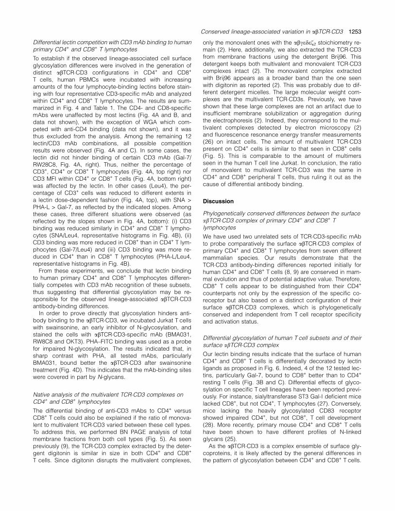
To establish if the observed lineage-associated cell surfaceglycosylation differences were involved in the generation ofdistinct abTCR�CD3 configurations in CD4+ and CD8+
T cells, human PBMCs were incubated with increasingamounts of the four lymphocyte-binding lectins before stain-ing with four representative CD3-specific mAb and analyzedwithin CD4+ and CD8+ T lymphocytes. The results are sum-marized in Fig. 4 and Table 1. The CD4- and CD8-specificmAbs were unaffected by most lectins (Fig. 4A and B, anddata not shown), with the exception of WGA which com-peted with anti-CD4 binding (data not shown), and it wasthus excluded from the analysis. Among the remaining 12lectin/CD3 mAb combinations, all possible competitionresults were observed (Fig. 4A and C). In some cases, thelectin did not hinder binding of certain CD3 mAb (Gal-7/RW28C8, Fig. 4A, right). Thus, neither the percentage ofCD3+, CD4+ or CD8+ T lymphocytes (Fig. 4A, top right) norCD3 MFI within CD4+ or CD8+ T cells (Fig. 4A, bottom right)was affected by the lectin. In other cases (Leu4), the per-centage of CD3+ cells was reduced to different extents ina lectin dose-dependent fashion (Fig. 4A, top), with SNA >PHA-L > Gal-7, as reflected by the indicated slopes. Amongthese cases, three different situations were observed (asreflected by the slopes shown in Fig. 4A, bottom): (i) CD3binding was reduced similarly in CD4+ and CD8+ T lympho-cytes (SNA/Leu4, representative histograms in Fig. 4B), (ii)CD3 binding was more reduced in CD8+ than in CD4+ T lym-phocytes (Gal-7/Leu4) and (iii) CD3 binding was more re-duced in CD4+ than in CD8+ T lymphocytes (PHA-L/Leu4,representative histograms in Fig. 4B).From these experiments, we conclude that lectin binding
to human primary CD4+ and CD8+ T lymphocytes differen-tially competes with CD3 mAb recognition of these subsets,thus suggesting that differential glycosylation may be re-sponsible for the observed lineage-associated abTCR�CD3antibody-binding differences.In order to prove directly that glycosylation hinders anti-
body binding to the abTCR�CD3, we incubated Jurkat T cellswith swainsonine, an early inhibitor of N-glycosylation, andstained the cells with abTCR�CD3-specific mAb (BMA031,RW8C8 and OKT3). PHA–FITC binding was used as a probefor impaired N-glycosylation. The results indicated that, insharp contrast with PHA, all tested mAbs, particularlyBMA031, bound better the abTCR�CD3 after swainsoninetreatment (Fig. 4D). This indicates that the mAb-binding siteswere covered in part by N-glycans.
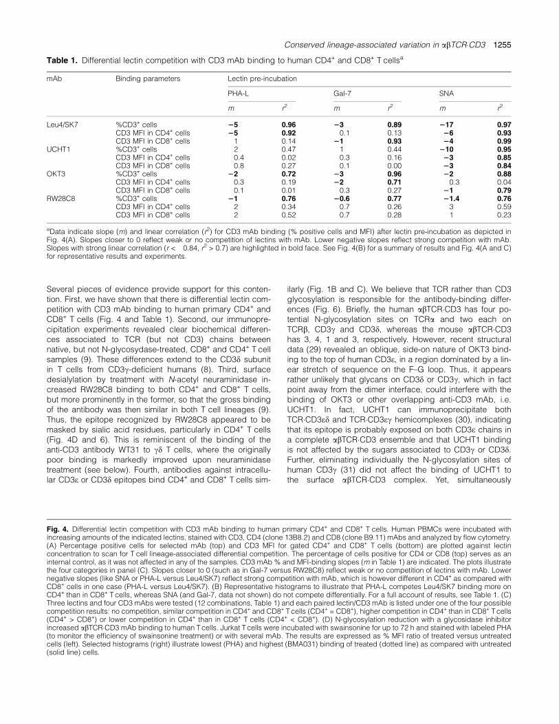
Native analysis of the multivalent TCR�CD3 complexes onCD4+ and CD8+ lymphocytes
The differential binding of anti-CD3 mAbs to CD4+ versusCD8+ T cells could also be explained if the ratio of monova-lent to multivalent TCR�CD3 varied between these cell types.To address this, we performed BN PAGE analysis of totalmembrane fractions from both cell types (Fig. 5). As seenpreviously (9), the TCR�CD3 complex extracted by the deter-gent digitonin is similar in size in both CD4+ and CD8+
T cells. Since digitonin disrupts the multivalent complexes,
only the monovalent ones with the abcedef2 stoichiometry re-main (2). Here, additionally, we also extracted the TCR�CD3from membrane fractions using the detergent Brij96. Thisdetergent keeps both multivalent and monovalent TCR�CD3complexes intact (2). The monovalent complex extractedwith Brij96 appears as a broader band than the one seenwith digitonin as reported (2). This was probably due to dif-ferent detergent micelles. The large molecular weight com-plexes are the multivalent TCR�CD3s. Previously, we haveshown that these large complexes are not an artifact due toinsufficient membrane solubilization or aggregation duringthe electrophoresis (2). Indeed, they correspond to the mul-tivalent complexes detected by electron microscopy (2)and fluorescence resonance energy transfer measurements(26) on intact cells. The amount of multivalent TCR�CD3present on CD4+ cells is similar to that seen in CD8+ cells(Fig. 5). This is comparable to the amount of multimersseen in the human T cell line Jurkat. In conclusion, the ratioof monovalent to multivalent TCR�CD3 was the same inCD4+ and CD8+ peripheral T cells, thus ruling it out as thecause of differential antibody binding.
Discussion
Phylogenetically conserved differences between the surfaceabTCR�CD3 complex of primary CD4+ and CD8+ Tlymphocytes
We have used two unrelated sets of TCR�CD3-specific mAbto probe comparatively the surface abTCR�CD3 complex ofprimary CD4+ and CD8+ T lymphocytes from seven differentmammalian species. Our results demonstrate that theTCR�CD3 antibody-binding differences reported initially forhuman CD4+ and CD8+ T cells (8, 9) are conserved in mam-mal evolution and thus of potential adaptive value. Therefore,CD8+ T cells appear to be distinguished from their CD4+
counterparts not only by the expression of the specific co-receptor but also based on a distinct configuration of theirsurface abTCR�CD3 complexes, which is phylogeneticallyconserved and independent from T cell receptor specificityand activation status.
Differential glycosylation of human T cell subsets and of theirsurface abTCR�CD3 complex
Our lectin binding results indicate that the surface of humanCD4+ and CD8+ T cells is differentially decorated by lectinligands as proposed in Fig. 6. Indeed, 4 of the 12 tested lec-tins, particularly Gal-7, bound to CD8+ better than to CD4+
resting T cells (Fig. 3B and C). Differential effects of glyco-sylation on specific T cell lineages have been reported previ-ously. For instance, sialyltransferase ST3 Gal-I deficient micelacked CD8+, but not CD4+, T lymphocytes (27). Conversely,mice lacking the heavily glycosylated CD83 receptorshowed impaired CD4+, but not CD8+, T cell development(28). More recently, primary mouse CD4+ and CD8+ T cellshave been shown to have different profiles of N-linkedglycans (25).As the abTCR�CD3 is a complex ensemble of surface gly-
coproteins, it is likely affected by the general differences inthe pattern of glycosylation between CD4+ and CD8+ T cells.
Conserved lineage-associated variation in abTCR�CD3 1253

1254 Conserved lineage-associated variation in abTCR�CD3
Several pieces of evidence provide support for this conten-tion. First, we have shown that there is differential lectin com-petition with CD3 mAb binding to human primary CD4+ andCD8+ T cells (Fig. 4 and Table 1). Second, our immunopre-cipitation experiments revealed clear biochemical differen-ces associated to TCR (but not CD3) chains betweennative, but not N-glycosydase-treated, CD8+ and CD4+ T cellsamples (9). These differences extend to the CD3d subunitin T cells from CD3c-deficient humans (8). Third, surfacedesialylation by treatment with N-acetyl neuraminidase in-creased RW28C8 binding to both CD4+ and CD8+ T cells,but more prominently in the former, so that the gross bindingof the antibody was then similar in both T cell lineages (9).Thus, the epitope recognized by RW28C8 appeared to bemasked by sialic acid residues, particularly in CD4+ T cells(Fig. 4D and 6). This is reminiscent of the binding of theanti-CD3 antibody WT31 to cd T cells, where the originallypoor binding is markedly improved upon neuraminidasetreatment (see below). Fourth, antibodies against intracellu-lar CD3e or CD3d epitopes bind CD4+ and CD8+ T cells sim-
ilarly (Fig. 1B and C). We believe that TCR rather than CD3glycosylation is responsible for the antibody-binding differ-ences (Fig. 6). Briefly, the human abTCR�CD3 has four po-tential N-glycosylation sites on TCRa and two each onTCRb, CD3c and CD3d, whereas the mouse abTCR�CD3has 3, 4, 1 and 3, respectively. However, recent structuraldata (29) revealed an oblique, side-on nature of OKT3 bind-ing to the top of human CD3e, in a region dominated by a lin-ear stretch of sequence on the F–G loop. Thus, it appearsrather unlikely that glycans on CD3d or CD3c, which in factpoint away from the dimer interface, could interfere with thebinding of OKT3 or other overlapping anti-CD3 mAb, i.e.UCHT1. In fact, UCHT1 can immunoprecipitate bothTCR�CD3ed and TCR�CD3ec hemicomplexes (30), indicatingthat its epitope is probably exposed on both CD3e chains ina complete abTCR�CD3 ensemble and that UCHT1 bindingis not affected by the sugars associated to CD3c or CD3d.Further, eliminating individually the N-glycosylation sites ofhuman CD3c (31) did not affect the binding of UCHT1 tothe surface abTCR�CD3 complex. Yet, simultaneously
Fig. 4. Differential lectin competition with CD3 mAb binding to human primary CD4+ and CD8+ T cells. Human PBMCs were incubated withincreasing amounts of the indicated lectins, stained with CD3, CD4 (clone 13B8.2) and CD8 (clone B9.11) mAbs and analyzed by flow cytometry.(A) Percentage positive cells for selected mAb (top) and CD3 MFI for gated CD4+ and CD8+ T cells (bottom) are plotted against lectinconcentration to scan for T cell lineage-associated differential competition. The percentage of cells positive for CD4 or CD8 (top) serves as aninternal control, as it was not affected in any of the samples. CD3 mAb % and MFI-binding slopes (m in Table 1) are indicated. The plots illustratethe four categories in panel (C). Slopes closer to 0 (such as in Gal-7 versus RW28C8) reflect weak or no competition of lectins with mAb. Lowernegative slopes (like SNA or PHA-L versus Leu4/SK7) reflect strong competition with mAb, which is however different in CD4+ as compared withCD8+ cells in one case (PHA-L versus Leu4/SK7). (B) Representative histograms to illustrate that PHA-L competes Leu4/SK7 binding more onCD4+ than in CD8+ T cells, whereas SNA (and Gal-7, data not shown) do not compete differentially. For a full account of results, see Table 1. (C)Three lectins and four CD3 mAbs were tested (12 combinations, Table 1) and each paired lectin/CD3 mAb is listed under one of the four possiblecompetition results: no competition, similar competition in CD4+ and CD8+ Tcells (CD4+ = CD8+), higher competition in CD4+ than in CD8+ Tcells(CD4+ > CD8+) or lower competition in CD4+ than in CD8+ T cells (CD4+ < CD8+). (D) N-glycosylation reduction with a glycosidase inhibitorincreased abTCR�CD3 mAb binding to human Tcells. Jurkat Tcells were incubated with swainsonine for up to 72 h and stained with labeled PHA(to monitor the efficiency of swainsonine treatment) or with several mAb. The results are expressed as % MFI ratio of treated versus untreatedcells (left). Selected histograms (right) illustrate lowest (PHA) and highest (BMA031) binding of treated (dotted line) as compared with untreated(solid line) cells.
Table 1. Differential lectin competition with CD3 mAb binding to human CD4+ and CD8+ T cellsa
mAb Binding parameters Lectin pre-incubation
PHA-L Gal-7 SNA
m r2 m r2 m r2
Leu4/SK7 %CD3+ cells 25 0.96 23 0.89 217 0.97CD3 MFI in CD4+ cells 25 0.92 �0.1 0.13 26 0.93CD3 MFI in CD8+ cells �1 0.14 21 0.93 24 0.99
UCHT1 %CD3+ cells �2 0.47 �1 0.44 210 0.95CD3 MFI in CD4+ cells �0.4 0.02 �0.3 0.16 23 0.85CD3 MFI in CD8+ cells �0.8 0.27 �0.1 0.00 23 0.84
OKT3 %CD3+ cells 22 0.72 23 0.96 22 0.88CD3 MFI in CD4+ cells �0.3 0.19 22 0.71 �0.3 0.04CD3 MFI in CD8+ cells �0.1 0.01 �0.3 0.27 21 0.79
RW28C8 %CD3+ cells 21 0.76 20.6 0.77 21.4 0.76CD3 MFI in CD4+ cells �2 0.34 0.7 0.26 �3 0.59CD3 MFI in CD8+ cells 2 0.52 0.7 0.28 1 0.23
aData indicate slope (m) and linear correlation (r2) for CD3 mAb binding (% positive cells and MFI) after lectin pre-incubation as depicted inFig. 4(A). Slopes closer to 0 reflect weak or no competition of lectins with mAb. Lower negative slopes reflect strong competition with mAb.Slopes with strong linear correlation (r < �0.84, r2 > 0.7) are highlighted in bold face. See Fig. 4(B) for a summary of results and Fig. 4(A and C)for representative results and experiments.
Conserved lineage-associated variation in abTCR�CD3 1255
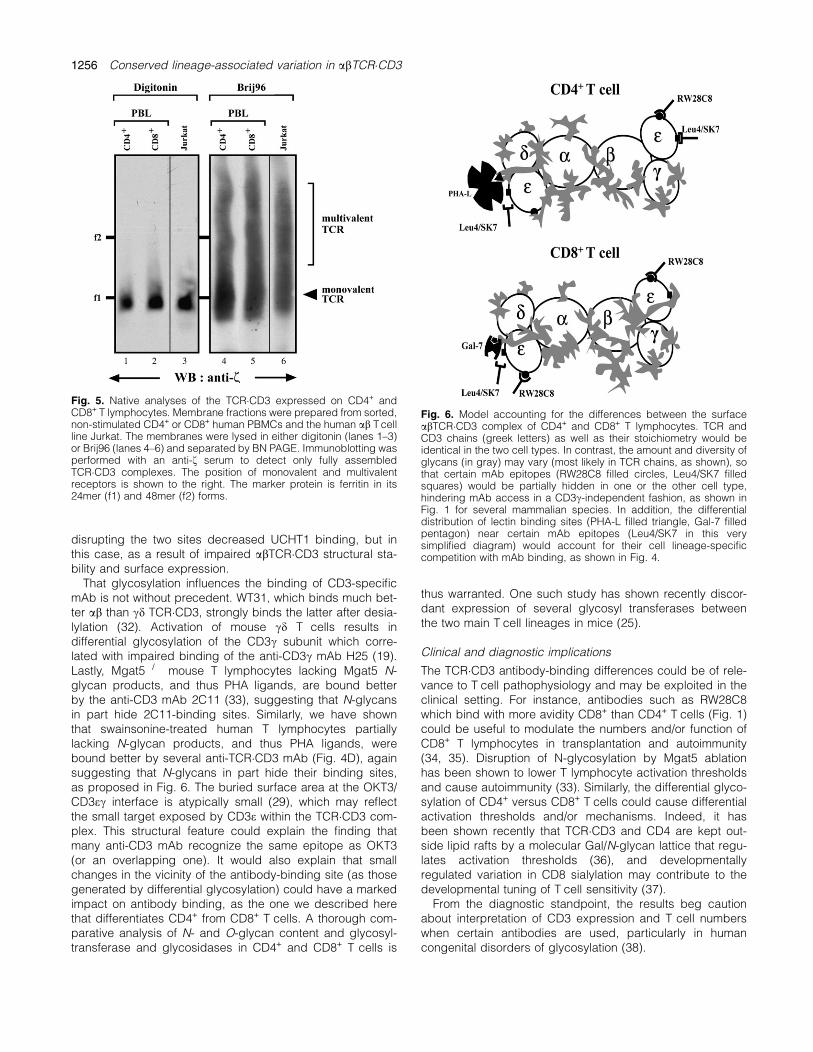
disrupting the two sites decreased UCHT1 binding, but inthis case, as a result of impaired abTCR�CD3 structural sta-bility and surface expression.That glycosylation influences the binding of CD3-specific
mAb is not without precedent. WT31, which binds much bet-ter ab than cd TCR�CD3, strongly binds the latter after desia-lylation (32). Activation of mouse cd T cells results indifferential glycosylation of the CD3c subunit which corre-lated with impaired binding of the anti-CD3c mAb H25 (19).Lastly, Mgat5�/� mouse T lymphocytes lacking Mgat5 N-glycan products, and thus PHA ligands, are bound betterby the anti-CD3 mAb 2C11 (33), suggesting that N-glycansin part hide 2C11-binding sites. Similarly, we have shownthat swainsonine-treated human T lymphocytes partiallylacking N-glycan products, and thus PHA ligands, werebound better by several anti-TCR�CD3 mAb (Fig. 4D), againsuggesting that N-glycans in part hide their binding sites,as proposed in Fig. 6. The buried surface area at the OKT3/CD3ec interface is atypically small (29), which may reflectthe small target exposed by CD3e within the TCR�CD3 com-plex. This structural feature could explain the finding thatmany anti-CD3 mAb recognize the same epitope as OKT3(or an overlapping one). It would also explain that smallchanges in the vicinity of the antibody-binding site (as thosegenerated by differential glycosylation) could have a markedimpact on antibody binding, as the one we described herethat differentiates CD4+ from CD8+ T cells. A thorough com-parative analysis of N- and O-glycan content and glycosyl-transferase and glycosidases in CD4+ and CD8+ T cells is
thus warranted. One such study has shown recently discor-dant expression of several glycosyl transferases betweenthe two main T cell lineages in mice (25).
Clinical and diagnostic implications
The TCR�CD3 antibody-binding differences could be of rele-vance to T cell pathophysiology and may be exploited in theclinical setting. For instance, antibodies such as RW28C8which bind with more avidity CD8+ than CD4+ T cells (Fig. 1)could be useful to modulate the numbers and/or function ofCD8+ T lymphocytes in transplantation and autoimmunity(34, 35). Disruption of N-glycosylation by Mgat5 ablationhas been shown to lower T lymphocyte activation thresholdsand cause autoimmunity (33). Similarly, the differential glyco-sylation of CD4+ versus CD8+ T cells could cause differentialactivation thresholds and/or mechanisms. Indeed, it hasbeen shown recently that TCR�CD3 and CD4 are kept out-side lipid rafts by a molecular Gal/N-glycan lattice that regu-lates activation thresholds (36), and developmentallyregulated variation in CD8 sialylation may contribute to thedevelopmental tuning of T cell sensitivity (37).From the diagnostic standpoint, the results beg caution
about interpretation of CD3 expression and T cell numberswhen certain antibodies are used, particularly in humancongenital disorders of glycosylation (38).
Fig. 6. Model accounting for the differences between the surfaceabTCR�CD3 complex of CD4+ and CD8+ T lymphocytes. TCR andCD3 chains (greek letters) as well as their stoichiometry would beidentical in the two cell types. In contrast, the amount and diversity ofglycans (in gray) may vary (most likely in TCR chains, as shown), sothat certain mAb epitopes (RW28C8 filled circles, Leu4/SK7 filledsquares) would be partially hidden in one or the other cell type,hindering mAb access in a CD3c-independent fashion, as shown inFig. 1 for several mammalian species. In addition, the differentialdistribution of lectin binding sites (PHA-L filled triangle, Gal-7 filledpentagon) near certain mAb epitopes (Leu4/SK7 in this verysimplified diagram) would account for their cell lineage-specificcompetition with mAb binding, as shown in Fig. 4.
Fig. 5. Native analyses of the TCR�CD3 expressed on CD4+ andCD8+ T lymphocytes. Membrane fractions were prepared from sorted,non-stimulated CD4+ or CD8+ human PBMCs and the human ab Tcellline Jurkat. The membranes were lysed in either digitonin (lanes 1–3)or Brij96 (lanes 4–6) and separated by BN PAGE. Immunoblotting wasperformed with an anti-f serum to detect only fully assembledTCR�CD3 complexes. The position of monovalent and multivalentreceptors is shown to the right. The marker protein is ferritin in its24mer (f1) and 48mer (f2) forms.
1256 Conserved lineage-associated variation in abTCR�CD3

Equal ratio of mono- versus multivalent TCR�CD3 in humanCD4+ and CD8+ T cells
The size of the monovalent TCR�CD3, as obtained by lysis indigitonin, was identical in CD4+ and CD8+ resting T cells.This demonstrates that the stoichiometry is the same in bothlineages, namely abcedef2 (2, 9, Fig. 6). The assembly ofthe TCR�CD3 takes place in the endoplasmic reticulum be-fore the enzymes of the Golgi apparatus confer differentialglycosylation. Therefore, folding and assembly occur undersimilar conditions, leading to the same stoichiometry. Lastly,we show that the TCR�CD3 of both CD4+ and CD8+ co-existin monovalent and multivalent complexes. Since both celltypes show similar amounts of multimerized receptor com-plexes, we can conclude that the presence of multivalentcomplexes does not contribute to the differential mAb bind-ing seen here. Earlier, we proposed that specific protein–protein interactions are responsible for the formation of themultivalent TCR�CD3 (2). Since variations in glycosylation ofthe TCR�CD3 do not seem to affect the ability of the complexto form multimers, the carbohydrate groups do not playa role in the generation of the multivalent TCR�CD3.In conclusion, we have described a phylogenetically con-
served trait in the ab TCR distinguishing CD4+ from CD8+ Tlymphocytes, which is likely emerging from the distinct pat-terns of glycoforms decorating the surface of these T cellsubsets. Yet, the contribution of other factors, i.e. lineage-specific lipid raft distribution (9), cannot be presently ruledout. The challenge now is to understand how the distinct gly-cosylation status of the abTCR�CD3 complex affects recep-tor signaling, gene expression and function of CD4+ andCD8+ T cells. Complex N-glycans and their degree ofbranching have been reported recently to regulate T lym-phocyte proliferation and differentiation (39). Therefore, line-age-specific glycosylation differences may have a majorimpact on CD4+ versus CD8+ T cell differentiation and func-tion (29, 30). The design of therapeutic strategies based onthese findings deserves further investigation.
Funding
Ministerio de Ciencia y Tecnologıa (BMC2002-1431,BMC2002-3247); Ministerio de Educacion (BFU2005-01738/BMC, SAF 2005-362); Comunidad Autonoma de Madrid(CAM) (GR/SAL/570/2004); Direccion General de Universi-dades e Investigacion (CAM); Universidad Complutense deMadrid (Ref. 920631); Fundacion Mutua Madrilena andDeutsche Forschungsgemeinschaft through the EmmyNoether program and the SFB620 to J.R.R., P.B., E.F-M. andW.W.A.S.; Universidad de Los Andes (Venezuela), CAM andthe Ramon y Cajal Program (Ministerio de Educacion) toN.E.R., J.R. and E.F.-M.
Acknowledgements
We greatly appreciate the generous supply of venous blood samplesfrom non-human primates by E. Gomez and E. Martınez (Zoo-Aquarium Madrid, Spain). We also thank the Fundacion Jimenez DıazBlood Bank and the Blood Transfusion Service (Madrid) for buffycoats; the mAb and lectin donors cited in Experimental Procedures;L. Perez and C. Ramırez (Inmunologıa, Facultad de Medicina,Universidad Complutense, Madrid, Spain) for technical help with cellcultures.
Abbreviations
BN blue nativeCBMSO centro de biologıa molecular severo ochoaCSIC consejo superior de investigaciones cientıficasGal galectinGST glutathione-S-transferaseMFI mean fluorescence intensityTCR T cell antigen receptor
References
1 Alarcon, B., Gil, D., Delgado, P. and Schamel, W. W. 2003.Initiation of TCR signaling: regulation within CD3 dimers. Immunol.Rev. 191:38.
2 Schamel, W. W., Arechaga, I., Risueno, R. M. et al. 2005.Coexistence of multivalent and monovalent TCRs explains highsensitivity and wide range of response. J. Exp. Med. 202:493.
3 Hellwig, S., Schamel, W. W., Pflugfelder, U., Gerlich, B. andWeltzien, H. U. 2005. Differences in pairing and cluster formationof Tcell receptor alpha- and beta-chains in Tcell clones and fusionhybridomas. Immunobiology 210:685.
4 Thibault, G. and Bardos, P. 1995. Compared TCR and CD3 epsilonexpression on alpha beta and gamma delta T cells. Evidence forthe association of two TCR heterodimers with three CD3 epsilonchains in the TCR/CD3 complex. J. Immunol. 154:3814.
5 Timon, M., Arnaiz-Villena, A., Rodriguez-Gallego, C., Perez-Aciego, P., Pacheco, A. and Regueiro, J. R. 1993. Selectivedisbalances of peripheral blood T lymphocyte subsets in humanCD3 gamma deficiency. Eur. J. Immunol. 23:1440.
6 Haks, M. C., Krimpenfort, P., Borst, J. and Kruisbeek, A. M. 1998.The CD3gamma chain is essential for development of both theTCRalphabeta and TCRgammadelta lineages. EMBO J. 17:1871.
7 Kappes, D. J., Alarcon, B. and Regueiro, J. R. 1995. T lymphocytereceptor deficiencies. Curr. Opin. Immunol. 7:441.
8 Zapata, D. A., Pacheco-Castro, A., Torres, P. S. et al. 1999.Conformational and biochemical differences in the TCR.CD3complex of CD8(+) versus CD4(+) mature lymphocytes revealedin the absence of CD3gamma. J. Biol. Chem. 274:35119.
9 Zapata, D. A., Schamel, W. W., Torres, P. S. et al. 2004.Biochemical differences in the alphabeta T cell receptor.CD3surface complex between CD8+ and CD4+ human matureT lymphocytes. J. Biol. Chem. 279:24485.
10 Lim, L. C., Fiordalisi, M. N., Mantell, J. L., Schmitz, J. L. and Folds,J. D. 1998. A whole-blood assay for qualitative and semiquanti-tative measurements of CD69 surface expression on CD4 andCD8 T lymphocytes using flow cytometry. Clin. Diagn. Lab.Immunol. 5:392.
11 Pacheco-Castro, A., Alvarez-Zapata, D., Serrano-Torres, P. andRegueiro, J. R. 1998. Signaling through a CD3 gamma-deficientTCR/CD3 complex in immortalized mature CD4+ and CD8+T lymphocytes. J. Immunol. 161:3152.
12 Andersen, H., Jensen, O. N., Moiseeva, E. P. and Eriksen, E. F.2003. A proteome study of secreted prostatic factors affectingosteoblastic activity: galectin-1 is involved in differentiation ofhuman bone marrow stromal cells. J. Bone Miner. Res. 18:195.
13 Magnaldo, T., Fowlis, D. and Darmon, M. 1998. Galectin-7,a marker of all types of stratified epithelia. Differentiation 63:159.
14 Nishi, N., Shoji, H., Seki, M. et al. 2003. Galectin-8 modulatesneutrophil function via interaction with integrin alphaM.Glycobiology13:755.
15 Chabot, S., Kashio, Y., Seki, M. et al. 2002. Regulation of galectin-9 expression and release in Jurkat T cell line cells. Glycobiology12:111.
16 Huflejt, M. E., Jordan, E. T., Gitt, M. A., Barondes, S. H. and Leffler, H.1997. Strikingly different localization of galectin-3 and galectin-4in human colon adenocarcinoma T84 cells. Galectin-4 is localizedat sites of cell adhesion. J. Biol. Chem. 272:14294.
17 Risueno, R. M., Gil, D., Fernandez, E., Sanchez-Madrid, F. andAlarcon, B. 2005. Ligand-induced conformational change in theT-cell receptor associated with productive immune synapses.Blood 106:601.
Conserved lineage-associated variation in abTCR�CD3 1257

18 Daniels, M. A., Hogquist, K. A. and Jameson, S. C. 2002. Sweet ‘n’sour: the impact of differential glycosylation on T cell responses.Nat. Immunol. 3:903.
19 Hayes, S. M., Laky, K., El Khoury, D., Kappes, D. J., Fowlkes, B. J.and Love, P. E. 2002. Activation-induced modification in the CD3complex of the gammadelta T cell receptor. J. Exp. Med.196:1355.
20 Meinl, E., Hohlfeld, R., Wekerle, H. and Fleckenstein, B. 1995.Immortalization of human T cells by Herpesvirus saimiri. Immunol.Today 16:55.
21 Geuijen, C. A., Clijsters-van der Horst, M., Cox, F. et al. 2005.Affinity ranking of antibodies using flow cytometry: application inantibody phage display-based target discovery. J. Immunol.Methods 302:68.
22 Daniels, M. A., Devine, L., Miller, J. D. et al. 2001. CD8 binding toMHC class I molecules is influenced by T cell maturation andglycosylation. Immunity 15:1051.
23 Moody, A. M., Chui, D., Reche, P. A., Priatel, J. J., Marth, J. D. andReinherz, E. L. 2001. Developmentally regulated glycosylation ofthe CD8alphabeta coreceptor stalk modulates ligand binding. Cell107:501.
24 Sharon, N. and Lis, H. 1989. Lectins as cell recognition molecules.Science 246:227.
25 Comelli, E. M., Sutton-Smith, M., Yan, Q. et al. 2006. Activation ofmurine CD4+ and CD8+ T lymphocytes leads to dramaticremodeling of N-linked glycans. J. Immunol. 177:2431.
26 Fernandez-Miguel, G., Alarcon, B., Iglesias, A. et al. 999.Multivalent structure of an alphabetaT cell receptor. Proc. NatlAcad. Sci. USA 96:1547.
27 Priatel, J. J., Chui, D., Hiraoka, N. et al. 2000. The ST3Gal-Isialyltransferase controls CD8+ T lymphocyte homeostasis bymodulating O-glycan biosynthesis. Immunity 12:273.
28 Fujimoto, Y., Tu, L., Miller, A. S. et al. 2002. CD83 expressioninfluences CD4+ T cell development in the thymus. Cell 108:755.
29 Kjer-Nielsen, L., Dunstone, M. A., Kostenko, L. et al. 004. Crystalstructure of the human T cell receptor CD3 epsilon gamma
heterodimer complexed to the therapeutic mAb OKT3. Proc. NatlAcad. Sci. USA 101:7675.
30 Arnett, K. L., Harrison, S. C. and Wiley, D. C. 2004. Crystalstructure of a human CD3-epsilon/delta dimer in complex witha UCHT1 single-chain antibody fragment. Proc Natl Acad. Sci.USA 101:16268.
31 Dietrich, J., Neisig, A., Hou, X., Wegener, A. M., Gajhede, M. andGeisler, C. 1996. Role of CD3 gamma in T cell receptor assembly.J. Cell Biol. 132:299.
32 van de Griend, R. J., Borst, J., Tax, W. J. and Bolhuis, R. L. 1988.Functional reactivity of WT31 monoclonal antibody with T cellreceptor-gamma expressing CD3 + 4-8- T cells. J. Immunol.140:1107.
33 Demetriou, M., Granovsky, M., Quaggin, S. and Dennis, J. W.2001. Negative regulation of T-cell activation and autoimmunity byMgat5 N-glycosylation. Nature 409:733.
34 van de Wetering, J., van der Mast, B. J., de Kuiper, P. et al. 2005.Reduction of immunosuppressive load in renal transplant recip-ients with a low donor-specific cytotoxic T-lymphocyte precursorfrequency is safe. Transplant. Proc. 37:779.
35 Bisikirska, B., Colgan, J., Luban, J., Bluestone, J. A. and Herold,K. C. 2005. TCR stimulation with modified anti-CD3 mAb expandsCD8+ T cell population and induces CD8+CD25+ Tregs. J. Clin.Invest. 115:2904.
36 Chen, I. J., Chen, H. L. and Demetriou, M. 2007. Lateralcompartmentalization of T cell receptor versus CD45 bygalectin-N-glycan binding and microfilaments coordinate basaland activation signaling. J. Biol. Chem. 282:35361.
37 Gil, D., Schrum, A. G., Daniels, M. A. and Palmer, E. 2008. A rolefor CD8 in the developmental tuning of antigen recognition andCD3 conformational change. J. Immunol. 180:3900.
38 Freeze, H. H. 2007. Congenital disorders of glycosylation: CDG-I,CDG-II, and beyond. Curr. Mol. Med. 7:389.
39 Lau, K. S., Partridge, E. A., Grigorian, A. et al. 2007. ComplexN-glycan number and degree of branching cooperate to regulatecell proliferation and differentiation. Cell 129:123.
1258 Conserved lineage-associated variation in abTCR�CD3