Mnferrite nanoparticles via reverse microemulsions: synthesis and characterization
Upload
independentCategory
view
1download
0
This article appeared in a journal published by Elsevier. The attachedcopy is furnished to the author for internal non-commercial researchand education use, including for instruction at the authors institution
and sharing with colleagues.
Other uses, including reproduction and distribution, or selling orlicensing copies, or posting to personal, institutional or third party
websites are prohibited.
In most cases authors are permitted to post their version of thearticle (e.g. in Word or Tex form) to their personal website orinstitutional repository. Authors requiring further information
regarding Elsevier’s archiving and manuscript policies areencouraged to visit:
http://www.elsevier.com/authorsrights

Author's personal copy
The greener synthesis of nanoparticlesOxana V. Kharissova1, H.V. Rasika Dias2, Boris I. Kharisov3, Betsabee Olvera Perez3,and Victor M. Jimenez Perez3
1 Universidad Auto noma de Nuevo Leo n, Facultad de Ciencias Fısico-Matema ticas, CIIDIT, Ciudad Universitaria, Monterrey,
NL 66450, Mexico2 Department of Chemistry and Biochemistry, The University of Texas at Arlington, Arlington, TX 76019, USA3 Universidad Auto noma de Nuevo Leo n, Facultad de Ciencias Quımicas, CIIDIT, Ciudad Universitaria, Monterrey, NL 66450,
Mexico
In this review, we examine ‘greener’ routes to nanopar-ticles of zerovalent metals, metal oxides, and salts withan emphasis on recent developments. Products fromnature or those derived from natural products, such asextracts of various plants or parts of plants, tea, coffee,banana, simple amino acids, as well as wine, table sugarand glucose, have been used as reductants and as cap-ping agents during synthesis. Polyphenols found in plantmaterial often play a key role in these processes. Thetechniques involved are simple, environmentally friend-ly, and generally one-pot processes. Tea extracts withhigh polyphenol content act as both chelating/reducingand capping agents for nanoparticles. We discuss thekey materials used in the field: silver, gold, iron, metalalloys, oxides, and salts.
Green chemistry and nanotechnologyThe ‘greener’ environmentally friendly processes in chem-istry and chemical technology are becoming increasinglypopular and are much needed as a result of worldwideproblems associated with environmental contamination.Within the last 2 years, several ‘green chemistry’ bookshave been published describing green processes in general[1–4] and their specialized aspects, including ultrasound,microwaves, and other methods in synthesis [5,6], greenanalytical chemistry [7], green tribology [8], polymers andgreen polymerization [9,10], green engineering andmanufacturing [11,12], food [13], textiles [14], hydrogenand syngas production and uses [15,16], wastewater treat-ment [17], particle technology [18], biofuels, biomass andbiocomposites [19–21], and other ‘green chemistry’ areas[22,23]. The 12 principles of green chemistry [24] have nowbecome a classic guide for chemists and chemical technol-ogists worldwide in developing less hazardous chemicalsyntheses.
Although UV irradiation, aerosol technologies, lithog-raphy, laser ablation, ultrasonic fields, and photochemicalreduction techniques have been used successfully to pro-duce nanoparticles, they remain expensive and involve theuse of hazardous chemicals. Therefore, there is a signifi-cant interest in the development of environmentallyfriendly and sustainable methods [25]. At the same time,despite intensive developments of the nanotechnology, theadverse effects of nanomaterials are still relativelyunknown. The synthesis of such materials using
environmentally friendly and biocompatible reagentscould lower the toxicity of the resulting materials andthe environmental impact of the byproducts [26,27]. Toreach this goal, non-toxic solvents (preferably water),closed reactors, ‘green’ techniques without contacting re-action media and air (ultrasound, microwave, hydrother-mal, magnetic, biological methods, among others), and lowtemperatures, can be used. In this short review whichfocuses on the most recent developments, we describethe methods which we consider as relatively greener routesto nanoparticles and nanomaterials involving extracts ofplants [28] and other natural products.
Green production of nanoparticlesThe techniques for obtaining nanoparticles using natu-rally occurring reagents such as vitamins, sugars, plantextracts, biodegradable polymers, and microorganismsas reductants and capping agents could be consideredattractive for nanotechnology. These syntheses have ledto the fabrication of limited number of inorganicnanoparticles (mainly metal nanoparticles, althoughseveral metal oxides and salts are also reported). Amongthe reagents mentioned above, plant based materialsseem to be the best candidates and they are suitablefor large-scale ‘biosynthesis’ of nanoparticles [29]. Plantparts such as leaf, root, latex, seed, and stem are beingused for metal nanoparticle synthesis. The key activeagent in some of these syntheses are believed to bepolyphenols, present for example, in tea, wine and win-ery waste, red grape pomace. Greener synthesis of nano-particles provides advancement over other methods as itis simple, cost-effective, and relatively reproducible andoften results in more stable materials [30]. Microorgan-isms can also be utilized to produce nanoparticles but therate of synthesis is slow and only limited number of sizesand shapes are amenable compared to routes involvingplant based materials. At present, fungi are gainingworldwide popularity as nano-factories for the greensynthesis of nanoparticles [31]. Overall, biological mate-rials provide an environmentally friendly or greenerchemical method to produce invaluable materials be-cause the biomaterial based routes eliminate the needto use harsh or toxic chemicals [32]. Mechanisms of thesebioreductive transformations [33–35], as well as catalyticproperties of materials obtained via these routes [36–39]have been discussed in a series of reports.
Review
Keywords: ‘green’ synthesis; plant extracts; tea extract; nanoparticles
240 0167-7799/$ – see front matter � 2013 Elsevier Ltd. All rights reserved. http://dx.doi.org/10.1016/j.tibtech.2013.01.003 Trends in Biotechnology, April 2013, Vol. 31, No. 4

Author's personal copy
Silver nanoparticles are the classic and the most pre-ferred target of the above mentioned ‘green’ methods (Ta-ble 1) [40–43]. This is related to the antibacterialproperties of zerovalent silver and easy reduction of sil-ver(I) salts to form zerovalent silver. Gold nanoparticlesare also of a considerable interest, although to a muchlesser degree than Ag. Other metal and other inorganicnanoparticles are represented by small number of reports.We discuss various greener routes for the synthesis ofthese nanoparticles below.
In general, as it will be seen below, ‘green’ methods arenot so widespread (although rapidly being developed) innanoscience and nanotechnology as classical wet-chemistryroutes for nanoparticles fabrication [44,45]. This relativelyhot topic possesses such important advantage as lesser oralmost zero contaminations for the environment. Wasteproducts are usually compatible with environmentalrequirements (except microbial synthesis of nanoparticles,where the waste material could be dangerous depending onthe type of microbe applied) because they are derived fromnatural plant extracts. Precursor costs may vary, but gen-erally they are low. On reviewing a series of reports ofauthors from different countries, we observed that in gen-eral commonly available and economical products for thoseregions are used, for example, nut putamen (India) or cactusextracts (Mexico). Obviously, the ‘greener’ methods do notpretend to substitute all conventional techniques; however,at least a small decrease in damage to nature can be reached.
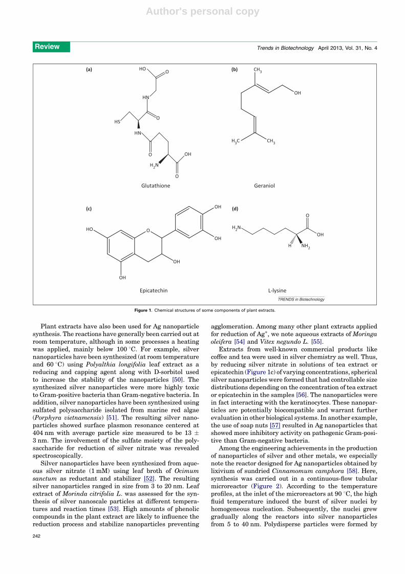
Silver (Ag)A large number of reports are dedicated to greener synthe-sis of silver nanoparticles, therefore here we will examineonly the most representative examples. Silver nanoparti-cles ranging from 5 to 10 nm in size have been synthesizedunder microwave irradiation conditions using glutathione(Figure 1a), an absolutely benign antioxidant that servesas the reducing as well as the capping agent in aqueous
medium [46]. The process takes only 30–60 min and isadaptable for the synthesis of palladium, platinum, andgold nanoparticles. The formation of dendritic nanostruc-tures on the transmission electron microscopy (TEM) gridwas observed when the reaction mixtures without completeconversion of AgNO3 were loaded on the TEM grid prior tothe washing procedure. The copper and carbon present onthe TEM grid are responsible for catalyzing the reaction.Synthesis of silver nanoparticles (1–10 nm) has also beeninvestigated using geraniol (Figure 1b) [47]. A concentrationof 1 mg/ml of these silver nanoparticles could inhibit cancercell line (Fibrosarcoma-Wehi 164) growth by less than 30%.
Sugars have been used with silver salts as a part of thewell-known Tollen’s test to produce silver mirrors. Sugarshave also been used in Ag nanoparticle fabrication. Synthe-sis of biocompatible Ag nanoparticles (10–20 nm) employingtable sugar as a capping and a reducing agent is a relativelyeconomical, easily adaptable, and greener route [48]. Aremarkable antitumor potency of the resulting Ag nanopar-ticles was seen against HT144 (malignant skin melanoma)and H157 (squamous cell lung carcinoma) cells as comparedto the clinically used reference compounds vincristine andmethotrexate. Ag nanoparticles have also been synthesizedin a natural polymeric matrix, using silver nitrate, gelatin,glucose, and sodium hydroxide as silver precursor, stabiliz-er, reducing agent, and accelerator reagent, respectively[49]. The resultant nanoparticles had faced centered cubic(fcc) structures smaller than 20 nm in diameter. Gelatin wasused as the stabilizer and matrix during this synthesis. Thepossible chemical equations (Equations 1 and 2) for prepar-ing the Ag nanoparticles are as follows:
AgþðaqÞþgel ! ½AgðgelÞ�þðaqÞ [1]
2½AgðgelÞ�þðaqÞþ2OH�þC5H11O5-CHO ! 2Ag þ 2gelþH2O þ C5H11O5-COOH [2]
Table 1. Synthesis of silver nanoparticles by greener methods (precursor AgNO3 in all cases)
Reducing agent Conditions, observations Properties of formed Ag nanoparticles Refs
Aqueous extracts of the manna of
hedysarum plant and the soap-root
(Acanthe phylum bracteatum) plant
13–20 min at 25–95 8C Average diameter of the prepared
nanoparticles in solution was about 29–68 nm
[96]
Extract of Argemone mexicana leaf 4 h at room temperature Average size of the particles synthesized was
20 nm, size range was 10–50 nm, with cubic
and hexagonal shape
[97]
Culture supernatants of Aspergil
lusterreus
24 h at 28 8C; reduced nicotinamide
adenine dinucleotide (NADH) was found
to be an important reducing agent for the
biosynthesis, and the formation of
AgNPs; this could be an enzyme-
mediated extracellular reaction process
Polydispersed spherical particles ranging in
size from 1 to 20 nm
[98]
Leaf extract of Euphorbia hirta L. 50–95 8C Relatively spherical shape with diameter range
40–50 nm
[99]
Euphorbia milii Irradiation of a silver nitrate and solution
by light from a xenon lamp followed by
ultra-short laser pulses; the irradiation
times were 1, 3, and 5 min
Nanoparticles are present after the xenon lamp
illumination, and after the laser irradiation the
silver nanoparticles sizes are reduced to the
range of 10–50 nm
[100]
F. vulgare leaves Room temperature 127 nm [101]
Basic amino acids, such as L-lysine
(Figure 1d) or L-arginine, as reducing
agents and soluble starch as a protecting
agent
The mixture was heated to 150 8C by
microwave under magnetic stirring
and was maintained at this temperature
for 10 s
26 nm (average size); this synthetic process can
be readily applied to large-scale production, for
example, a reaction yielding 0.1 g of nearly
monodisperse silver nanoparticles can be
performed in a 80 ml microwave sealed vessel
[40]
Review Trends in Biotechnology April 2013, Vol. 31, No. 4
241
Author's personal copy
Plant extracts have also been used for Ag nanoparticlesynthesis. The reactions have generally been carried out atroom temperature, although in some processes a heatingwas applied, mainly below 100 8C. For example, silvernanoparticles have been synthesized (at room temperatureand 60 8C) using Polyalthia longifolia leaf extract as areducing and capping agent along with D-sorbitol usedto increase the stability of the nanoparticles [50]. Thesynthesized silver nanoparticles were more highly toxicto Gram-positive bacteria than Gram-negative bacteria. Inaddition, silver nanoparticles have been synthesized usingsulfated polysaccharide isolated from marine red algae(Porphyra vietnamensis) [51]. The resulting silver nano-particles showed surface plasmon resonance centered at404 nm with average particle size measured to be 13 �3 nm. The involvement of the sulfate moiety of the poly-saccharide for reduction of silver nitrate was revealedspectroscopically.
Silver nanoparticles have been synthesized from aque-ous silver nitrate (1 mM) using leaf broth of Ocimumsanctum as reductant and stabilizer [52]. The resultingsilver nanoparticles ranged in size from 3 to 20 nm. Leafextract of Morinda citrifolia L. was assessed for the syn-thesis of silver nanoscale particles at different tempera-tures and reaction times [53]. High amounts of phenoliccompounds in the plant extract are likely to influence thereduction process and stabilize nanoparticles preventing
agglomeration. Among many other plant extracts appliedfor reduction of Ag+, we note aqueous extracts of Moringaoleifera [54] and Vitex negundo L. [55].
Extracts from well-known commercial products likecoffee and tea were used in silver chemistry as well. Thus,by reducing silver nitrate in solutions of tea extract orepicatechin (Figure 1c) of varying concentrations, sphericalsilver nanoparticles were formed that had controllable sizedistributions depending on the concentration of tea extractor epicatechin in the samples [56]. The nanoparticles werein fact interacting with the keratinocytes. These nanopar-ticles are potentially biocompatible and warrant furtherevaluation in other biological systems. In another example,the use of soap nuts [57] resulted in Ag nanoparticles thatshowed more inhibitory activity on pathogenic Gram-posi-tive than Gram-negative bacteria.
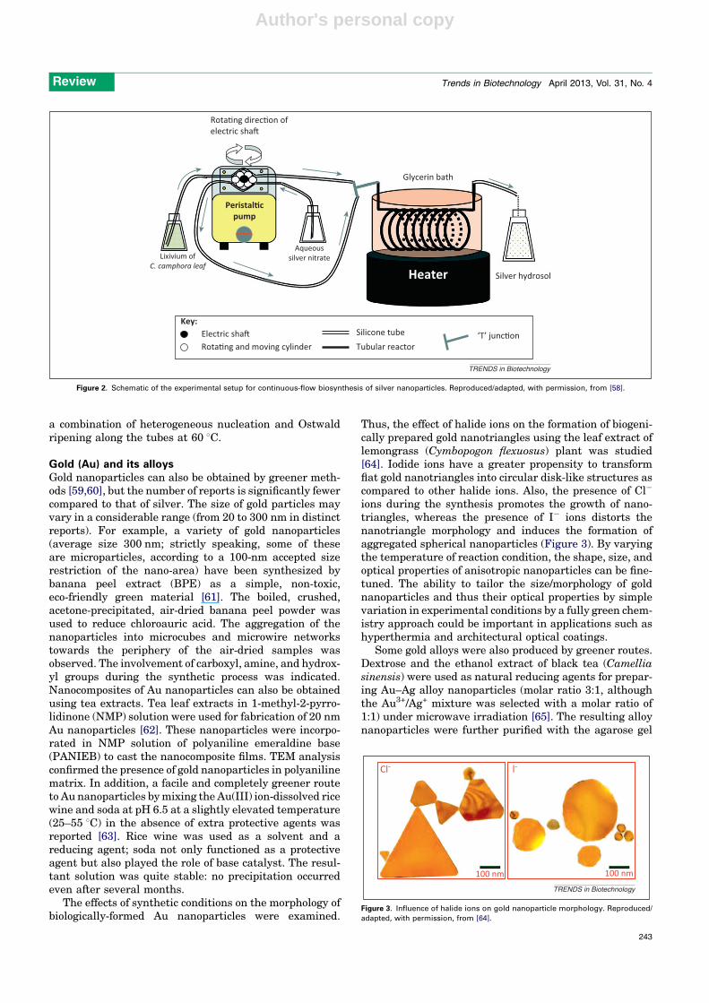
Among the engineering achievements in the productionof nanoparticles of silver and other metals, we especiallynote the reactor designed for Ag nanoparticles obtained bylixivium of sundried Cinnamomum camphora [58]. Here,synthesis was carried out in a continuous-flow tubularmicroreactor (Figure 2). According to the temperatureprofiles, at the inlet of the microreactors at 90 8C, the highfluid temperature induced the burst of silver nuclei byhomogeneous nucleation. Subsequently, the nuclei grewgradually along the reactors into silver nanoparticlesfrom 5 to 40 nm. Polydisperse particles were formed by
Glutathione
(a) (b)
(c) (d)
Geraniol
Epicatechin
HO
HN
HN
OH
OH
HO
OH
OH
OH
H2N
H3C CH3
CH3
OH
HS
O
O
O
O
O
L-lysine
H2N
NH2
OH
H
O
TRENDS in Biotechnology
Figure 1. Chemical structures of some components of plant extracts.
Review Trends in Biotechnology April 2013, Vol. 31, No. 4
242
Author's personal copy
a combination of heterogeneous nucleation and Ostwaldripening along the tubes at 60 8C.
Gold (Au) and its alloysGold nanoparticles can also be obtained by greener meth-ods [59,60], but the number of reports is significantly fewercompared to that of silver. The size of gold particles mayvary in a considerable range (from 20 to 300 nm in distinctreports). For example, a variety of gold nanoparticles(average size 300 nm; strictly speaking, some of theseare microparticles, according to a 100-nm accepted sizerestriction of the nano-area) have been synthesized bybanana peel extract (BPE) as a simple, non-toxic,eco-friendly green material [61]. The boiled, crushed,acetone-precipitated, air-dried banana peel powder wasused to reduce chloroauric acid. The aggregation of thenanoparticles into microcubes and microwire networkstowards the periphery of the air-dried samples wasobserved. The involvement of carboxyl, amine, and hydrox-yl groups during the synthetic process was indicated.Nanocomposites of Au nanoparticles can also be obtainedusing tea extracts. Tea leaf extracts in 1-methyl-2-pyrro-lidinone (NMP) solution were used for fabrication of 20 nmAu nanoparticles [62]. These nanoparticles were incorpo-rated in NMP solution of polyaniline emeraldine base(PANIEB) to cast the nanocomposite films. TEM analysisconfirmed the presence of gold nanoparticles in polyanilinematrix. In addition, a facile and completely greener routeto Au nanoparticles by mixing the Au(III) ion-dissolved ricewine and soda at pH 6.5 at a slightly elevated temperature(25–55 8C) in the absence of extra protective agents wasreported [63]. Rice wine was used as a solvent and areducing agent; soda not only functioned as a protectiveagent but also played the role of base catalyst. The resul-tant solution was quite stable: no precipitation occurredeven after several months.
The effects of synthetic conditions on the morphology ofbiologically-formed Au nanoparticles were examined.
Thus, the effect of halide ions on the formation of biogeni-cally prepared gold nanotriangles using the leaf extract oflemongrass (Cymbopogon flexuosus) plant was studied[64]. Iodide ions have a greater propensity to transformflat gold nanotriangles into circular disk-like structures ascompared to other halide ions. Also, the presence of Cl�
ions during the synthesis promotes the growth of nano-triangles, whereas the presence of I� ions distorts thenanotriangle morphology and induces the formation ofaggregated spherical nanoparticles (Figure 3). By varyingthe temperature of reaction condition, the shape, size, andoptical properties of anisotropic nanoparticles can be fine-tuned. The ability to tailor the size/morphology of goldnanoparticles and thus their optical properties by simplevariation in experimental conditions by a fully green chem-istry approach could be important in applications such ashyperthermia and architectural optical coatings.
Some gold alloys were also produced by greener routes.Dextrose and the ethanol extract of black tea (Camelliasinensis) were used as natural reducing agents for prepar-ing Au–Ag alloy nanoparticles (molar ratio 3:1, althoughthe Au3+/Ag+ mixture was selected with a molar ratio of1:1) under microwave irradiation [65]. The resulting alloynanoparticles were further purified with the agarose gel
Silver hydrosol
Silicone tubeKey:
Lixivium ofC. camphora leaf
Aqueoussilver nitrate
Peristal�cpump
Glycerin bath
Rota�ng direc�on ofelectric sha�
Heater
Electric sha�Rota�ng and moving cylinder Tubular reactor
‘T’ junc�on
TRENDS in Biotechnology
Figure 2. Schematic of the experimental setup for continuous-flow biosynthesis of silver nanoparticles. Reproduced/adapted, with permission, from [58].
Cl- l-
100 nm 100 nm
TRENDS in Biotechnology
Figure 3. Influence of halide ions on gold nanoparticle morphology. Reproduced/
adapted, with permission, from [64].
Review Trends in Biotechnology April 2013, Vol. 31, No. 4
243

Author's personal copy
electrophoresis technique. Purified Ag–Au alloy nanopar-ticles were spherical in the size range of �200 nm, andpossessed an average size of 92 nm. The final concentra-tions of glucose and plant extract should be considered asthe main parameters affecting the Ag/Au ratio in the Ag–Au alloy nanoparticles. In addition, a facile and eco-friend-ly method for the preparation of Au–Pd bimetallic nano-particles (�7 nm) was developed based on simultaneousbioreduction of Au(III) and Pd(II) precursors with Cacu-men Platycladi leaf extract in aqueous environment [66]. Itwas shown that the C=O and C–O groups in the plantextract play a critical role in capping the nanoparticles.
Iron (Fe), its alloys, and oxidesNanoscale iron particles, FexOy and FeOOH (in total 16polymorphic forms) are of significant interest because oftheir rapidly developing applications for disinfection ofwater and remediation of heavy metals from soils[67,68]. Therefore, methods using mainly tea extracts onthe polyphenol basis, as well other plant extracts, havebeen utilized for obtaining Fe-containing nanoparticles[69] and their further uses for the purposes above. Thus,nanoscale zerovalent iron (NZVI) particles (Figure 4), pre-pared using tea polyphenols [70], were assessed withmethyl tetrazolium [MTS, 3-(4,5-dimethylthiazol-2-yl)-5-(3-carboxymethoxyphenyl)-2-(4-sulfophenyl)-2H-tetrazoli-um] and lactate dehydrogenase (LDH) toxicological assays.Some of them were found to be nontoxic compared to thecontrol samples prepared using the conventional borohy-dride reduction protocols.
In addition to ‘pure’ iron nanoparticles produced usingextracts of green tea leaves, the samples may containmainly iron oxide and iron oxohydroxide [71]. The nano-particles were then utilized as a Fenton-like catalyst fordecolorization of aqueous solutions containing methyleneblue (MB) and methyl orange (MO) dyes. The resultsindicated fast removal of the dyes with the kinetic data
of MB following a second order removal rate, whereas thoseof MO were closer to a first order removal rate. Almostcomplete removal of both dyes from water takes place overa wide range of concentrations (10–200 mg/l). Superpara-magnetic Fe3O4 nanoparticles [blocking temperature (TB)of 150 K and saturation magnetization of 37.1 emu/g] weresynthesized via soya bean sprout (SBS) templates at roomtemperature and normal atmospheric pressure [72].Spherical Fe3O4 nanoparticles with an average diameterof 8 nm simultaneously formed on the epidermal surfaceand the interior stem wall of SBS, which were responsiblefor size and morphologic control during the whole forma-tion of nanoparticles. In addition, NixCu0.25Zn0.75–xFe2O4
(x = 0.25, 0.35, 0.5) polycrystalline nanopowders (15–40 nm) with spinel structure were prepared by a simplesolution method using metal nitrates and Aloe vera plantextract solution [73]. The samples exhibited ferromagneticbehavior. Finally, iron alloys are also available by thesemethods. Thus, membranes containing reactive nanopar-ticles (Fe and Fe/Pd) immobilized in a polymer film [poly-acrylic acid (PAA)-coated polyvinylidene fluoride (PVDF)membrane] were prepared with the use of tea extract [74].The membrane-supported nanoparticles were used for thedegradation of a common and highly important pollutant,trichloroethylene (TCE).
Other metalsCopper (Cu)
Other metals and their compounds are represented by aconsiderably fewer number of reports. Highly stabledispersions of nanosized copper particles with an aver-age particle size less than 2 nm have been synthesizedusing a straightforward and cost-effective method,utilizing nontoxic L-ascorbic acid as both a reducing agentand capping agent precursor in aqueous medium [75].Also, greener fabrication of nanoparticles of copperand other metals using various fruit extracts and otherproducts of natural origin (such as red wine) waspatented [76].
Palladium (Pd)
Palladium nanoparticles (PdNps), which are of interestbecause of their catalytic properties and affinity for hydro-gen, have been phyto-synthesized under moderate pH androom temperature using Solanum trilobatum [77] andconjugated with lipoic acid (S-PdNp-LA) and vitamins(S-PdNp-Vitamin-LA). S-PdNp are polydisperse and ofdifferent morphologies ranging from 60 to 70 nm (S-PdNp),65 to 80 nm (S-PdNp-LA), and 75 to 100 nm (S-PdNp-Vitamin-LA) in size. Bulk quantities of nanocrystals(20–60 nm) of Pd, as well as other noble metals such asAg, have been obtained using coffee and tea extracts atroom temperature [78]. The reported method is generaland may be extended to other noble metals such as Au andPt. One-pot biogenic fabrication of palladium nanoparticles(small size from 3.2 to 6.0 nm) was also carried out by asimple procedure using broth of Cinnamomum camphoraleaf without extra surfactant, capping agent, and/or tem-plate [79]. The size of PdNPs could be easily controlled byvariation of the initial concentration of Pd(II) ions. Like-wise, the reaction between broth of C. camphora leaf and
3000
1
2
3
400 500 600 700 800
AKey:
CB
Wavelength (nm)
Abso
rban
ce (a
.u.)
TRENDS in Biotechnology
Figure 4. UV spectra of (a) Fe(NO3)3 control, (b) control tea extract, and (c) the
reaction product (Fe nanoparticles) obtained from Fe(NO3)3 and tea extract. The
inset shows the photographic image of the tea extract, the control Fe(NO3)3solution, and after mixing them (from left to right vials). Reproduced/adapted, with
permission, from [70].
Review Trends in Biotechnology April 2013, Vol. 31, No. 4
244

Author's personal copy
the Pd(II) species might occur according to the followingequation (Equation 3):
nPdðIIÞ þ 2R-ðOHÞn! nPdð0Þ þ 2nR ¼ O þ 2nHþ [3]
Finally, palladium nanoparticles (15 nm) were synthesizedusing a Glycine max (soybean) leaf extract-mediated bio-synthesis process [80]. Protein-rich soybean leaf extractacts as an effective reducing agent for palladium ions. Also,these biosynthesized palladium nanoparticles can be usedin catalysis, specifically for the degradation of azo dyes.
Platinum (Pt)
The leaf extract of Diopyros kaki has been used as areducing agent in the eco-friendly extracellular synthesisof platinum nanoparticles (2–12 nm, depending on thereaction temperature and concentrations of the leaf brothand PtCl6
2�) from an aqueous H2PtCl6.6H2O solution [81].
A greater than 90% conversion of platinum ions to nano-particles was achieved with a reaction temperature of95 8C and a leaf broth concentration of >10%. Platinumnanoparticle synthesis using Diopyros kaki may not be anenzyme-mediated process. Other related methods for plat-inum nanoparticle formation are reviewed in [82].
Metal oxides and metal saltsTitanium(IV) oxide (TiO2)
In addition to the above-mentioned iron oxide nanoparti-cles, other metal oxide nanoparticles like TiO2 (one of themost important compounds in nanotechnology) have alsobeen obtained by greener methods. Titanium(IV) oxidenanoparticles (100–150 nm) have been prepared from tita-nium isopropoxide solution using nyctanthes leaf extract[83]. Alternatively, TiO2 nanoparticles (25–100 nm) havebeen prepared by using 0.3% aqueous extract preparedfrom latex of Jatropha curcas L. [84]. Curcain (enzyme) andcyclic peptides [namely curcacycline A (an octapeptide) andcurcacycline B (a nonapeptide)] have been identified aspossible reducing and capping agents present in the latexof J. curcas L. There are two broad categories of nanopar-ticles – the first have diameters from 25 to 50 nm and aremostly spherical in shape; the second have larger anduneven shapes.
Zinc(II) oxide (ZnO)
Highly stable and spherical zinc oxide nanoparticles (25–40 nm) are produced by using zinc nitrate and Aloe barba-densis Miller leaf extract [85]. As a result, greater than 95%conversion to nanoparticles was achieved using aloe leafbroth concentration greater than 25%. It was shown thatthe zinc oxide nanoparticles were poly dispersed. Theparticle size could be controlled by varying the concentra-tions of leaf broth solution. Alternatively, ZnO nanoparti-cles (30–60 nm) were synthesized via the sol–gel method ingelatin media [86], where long-chain gelatin compoundswere utilized to terminate the growth of ZnO nanoparticlesand to stabilize them. It was concluded by authors thatgelatin is an interesting material that can be used as astabilizer in the sol–gel technique for preparing small ZnO-NPs. In addition, formation of ZnO (and also gold, silver,copper, and gold–silver–copper alloy) in living plants isdiscussed in an extremely interesting recent review [87].
Copper oxides (Cu2O and CuO)
Cu2O nanoparticles were prepared by reduction of Barfoed’ssolution, using agriculture wastes of Arachis hypogaea L.(Fabaceae) leaf extracts containing reducing sugars, whichact as reducing agent at room temperature [88]. The alde-hyde group present in reducing sugar plays an excellent rolein the formation of cuprous oxide nanoparticles in the solu-tion. The cuprous oxide nanoparticles had antibacterialeffects on Gram-negative Escherichia coli. The morphologyof divalent copper oxide in 15–30 nm nanoparticles can becontrolled by tuning the amount of Aloe vera extract [89].This eco-friendly approach for synthesis is suitable for large-scale commercial production and health-related applica-tions of CuO nanoparticles.
Indium(III) oxide (In2O3)
In addition, cubic In2O3 nanoparticles (5–50 nm) weresynthesized by a simple, cost-effective and environmental-ly friendly route using indium acetylacetonate and Aloevera plant extract solution followed by a calcinations stepin air at 400–600 8C for 2 h [90]. Alternatively, In2O3
hollow spheres were ‘one-pot’ synthesized by hydrothermalreaction of glucose and InCl3 mixtures at 180 8C for 12 h,followed by calcination in air at 600 8C for 1 h [91]. Thesespheres were sensitive to six flammable organic vaporsincluding alcohol, acetone, 93# gasoline, formaldehyde,chloroform, and acetonitrile.
Lead(II) sulfide (PbS)
Finally, the intracellular synthesis of stable cubic leadsulfide nanoparticles by a marine yeast, Rhodosporidiumdiobovatum, has been reported [92]. The particles werefound to be composed of lead and sulfur in a 1:2 ratioindicating that PbS nanoparticles were capped by a sul-fur-rich peptide. It was revealed that 55% of lead in themedium was accumulated in the exponential phase, where-as a further 35% was accumulated in the stationary phase,thus providing 90% overall recovery of PbS nanoparticles.
Concluding remarks and future perspectivesFabrication of metal, metal oxides, and salt nanoparticlesusing natural substances is a very promising area in nano-technology, which came to prominence only a few years ago.Useful materials can be produced easily even in reasonablescale because the biomaterial-based routes eliminate theneed to use harsh or toxic chemicals. Waste products arerelatively innocuous and easier to dispose of because theyare mostly composed of leftover natural plant extracts. Themajority of greener synthetic efforts reported thus far arededicated to obtaining silver nanoparticles, perhaps due totheir ease of preparation, importance in disinfection science,and utility in several applications. Several other metals (Au,Fe, Pt, Pd, Cu), their alloys, oxides, and salts have also beenobtained by ‘greener’ methods.
In these greener routes, products from nature or derivedfrom plant materials such as extracts of various plants, tea,coffee, banana, simple amino acids, as well as wine, tablesugar, and glucose, have been utilized as reductants andcapping agents. A considerable number of efforts are de-voted to tea extracts, which, owing to a number of poly-phenols present, can act as both chelating/reducing and
Review Trends in Biotechnology April 2013, Vol. 31, No. 4
245

Author's personal copy
capping agents for the nanoparticles. Therefore, the result-ing particles are protected from further reactions andaggregation, which increases their stability and longevity.Greener methods involving plant materials that have beenused in nanoparticle synthesis are generally single-potreactions, without the use of additional surfactants, cap-ping agents, and/or templates. Frequently, the techniquedeveloped for a given metal and metal oxide nanoparticlecould also be applied for other metals.
We note that the bulk of these investigations have beencarried out at research laboratories in small scale (which isnormal) whereas there are no reports on pilot plant orindustrial-scale fabrication of nanomaterials using naturalproducts. We believe that there are good opportunities fordeveloping large-scale greener processes, where nanopar-ticles have important applications. For example, iron nano-particles derived from cheap widespread naturalprecursors for remediation of heavy metals or disinfectionof water could be an attractive option in nanotechnologicalenvironmental applications. Systematic studies are alsoneeded for uncovering some of the reaction mechanismsand to reach more firm conclusions. For example, there areseveral theories on Ag+ reduction to Ag0 and the disinfec-tion action of silver nanoparticles [93–95]. Overall, the useof natural products for greener synthesis of nanoparticles/nanomaterials is an emerging and exciting area of nano-technology and may have a significant direct impact onfurther advances in biotechnology.
References1 Anastas, P.T. and Horvath, I.T. (2012) Green Chemistry for a
Sustainable Future, (1st edn), Wiley2 Anastas, P.T. (ed.) (2012) Handbook of Green Chemistry - Green
Processes (Volume 3, 3-book set), Wiley-VCH3 Ahluwalia, V.K. (2012) Green Chemistry: Environmentally Benign
Reactions, (2nd edn), CRC Press4 Patel, J.T. et al. (2012) Green Chemistry: New Avenues in Chemical
Research: Focus in Healthcare, LAP LAMBERT Academic Publishing5 Ahluwalia, V.K. (2012) Strategies for Green Organic Synthesis, (1st
edn), CRC Press6 Chen, D. et al., eds (2011) Handbook on Applications of Ultrasound:
Sonochemistry for Sustainability (1st edn), CRC Press7 De la Guardia, M. and Garrigues, S. (2012) Handbook of Green
Analytical Chemistry, (1st edn), Wiley8 Nosonovsky, M. and Bhushan, B. (2012) Green Tribology: Biomimetics
Energy Conservation and Sustainability (Green Energy andTechnology), Springer
9 Mathers, R.T. and Meier, M.A.R. (2011) Green PolymerizationMethods (Green Chemistry), (1st edn), Wiley-VCH
10 Cheng, H. and Gross, R. (2011) Green Polymer Chemistry: Biocatalysisand Biomaterials (ACS Symposium), (1st edn), American ChemicalSociety
11 Allen, D.T. and Shonnard, D.R. (2012) Green Engineering:Environmentally Conscious Design of Chemical Processes, (2ndedn), Prentice Hall
12 Mulvaney, D.R. (2011) Green technology: an A-to-Z guide. In TheSAGE Reference Series on Green Society: Toward a SustainableFuture (Robbins, P., ed.), Sage Publications
13 Boye, J.I. and Arcand, Y. (2012) Green Technologies in FoodProduction and Processing (Food Engineering Series), (1st edn),Springer
14 Kan, C.W. (2012) A Novel Green Treatment for Textiles: PlasmaTreatment as a Sustainable Technology (Sustainability), (1st edn),CRC Press
15 Rigas, F. and Amyotte, P. (2012) Hydrogen Safety (Green Chemistryand Chemical Engineering), (1st edition), CRC Press
16 Puigjaner, L. (2011) Syngas from Waste: Emerging Technologies(Green Energy and Technology), (1st edn), Springer
17 Lofrano, G. (2012) Green Technologies for Wastewater Treatment:Energy Recovery and Emerging Compounds Removal (SpringerBriefs in Molecular Science/Springer Briefs in Green Chemistry forSustainability), Springer
18 Lee, S. and Henthorn, K.H. (2012) Particle Technology andApplications (Green Chemistry and Chemical Engineering), (1stedn), CRC Press
19 Lee, S. and Shah, Y.T. (2012) Biofuels and Bioenergy: Processes andTechnologies (Green Chemistry and Chemical Engineering), (1st edn),CRC Press
20 Sengupta, D. and Pike, R.W. (2012) Chemicals from Biomass:Integrating Bioprocesses into Chemical Production Complexes forSustainable Development (Green Chemistry and ChemicalEngineering), (1st edn), CRC Press
21 Siyamak, S. (2012) Preparation and Characterization of GreenComposites: Chemicals’ Effects on Fundamental Properties ofPoly(Butylene Adipate-Co-Terephthalate)/Oil Palm EFB FibreBiocomposites, LAP LAMBERT Academic Publishing
22 Foo, D.C.Y. (2012) Process Integration for Resource Conservation(Green Chemistry and Chemical Engineering), CRC Press
23 Schiebl, M. (2012) Green Chemical Space Propulsion: Auto IgnitionConditions in a Micro Rocket Combustion Chamber for a HydrogenPeroxide based Micro Propulsion System, AV Akademikerverlag
24 Anastas, P.T. and Warner, J.C. (1998) Green Chemistry: Theory andPractice, Oxford University Press, p. 30
25 Narayanan, K.B. and Sakthivel, N. (2010) Biological synthesis ofmetal nanoparticles by microbes. Adv. Colloid Interface Sci. 156, 1–13
26 Sanchez-Mendieta, V. and Vilchis-Nestor, A.R. (2012) Greensynthesis of noble metal (Au, Ag, Pt) nanoparticles, assisted byplant-extracts. In Noble Metals (Yen-Hsun, S., ed.), pp. 391–408,INTECH
27 Carma, R.S. (2012) Greener approach to nanomaterials and theirsustainable applications. Curr. Opin. Chem. Eng. 1, 123–128
28 Gan, P.P. (2012) Potential of plant as a biological factory to synthesizegold and silver nanoparticles and their applications. Rev. Environ.Sci. Biotechnol. 11, 169–206
29 Iravani, S. (2011) Green synthesis of metal nanoparticles usingplants. Green Chem. 13, 2638–2650
30 Kalaiarasi, R. et al. (2010) Phytosynthesis of nanoparticles and itsapplications. Plant Cell Biotechnol. Mol. Biol. 11, 1–16
31 Dhillon, G.S. et al. (2012) Green approach for nanoparticlebiosynthesis by fungi: current trends and applications. Crit. Rev.Biotechnol. 32, 49–73
32 Parsons, J.G. et al. (2007) Use of plants in biotechnology: synthesis ofmetal nanoparticles by inactivated plant tissues, plant extracts, andliving plants. Dev. Environ. Sci. 5, 463–485
33 Xie, J. et al. (2007) Identification of active biomolecules in the high-yield synthesis of single-crystalline gold nanoplates in algal solutions.Small 3, 672–682
34 Zhou, Y. et al. (2010) Biosynthesis of gold nanoparticles by foliarbroths: roles of biocompounds and other attributes of the extracts.Nanoscale Res. Lett. 5, 1351–1359
35 Huang, J. et al. (2011) Biogenic silver nanoparticles by CacumenPlatycladi extract: synthesis, formation mechanism, andantibacterial activity. Eng. Chem. Res. 50, 9095–9106
36 Sharma, N.C. et al. (2007) Synthesis of plant-mediated goldnanoparticles and catalytic role of biomatrix-embeddednanomaterials. Environ. Sci. Technol. 41, 5137–5142
37 Vilchis-Nestor, A.R. et al. (2009) Alternative bio-reduction synthesismethod for the preparation of Au(AgAu)/SiO2–Al2O3 catalysts:oxidation and hydrogenation of CO. Appl. Catal. B: Environ. 90,64–73
38 Du, M. et al. (2011) Ionic liquid-enhanced immobilization ofbiosynthesized Au nanoparticles on TS-1 toward efficient catalystsfor propylene epoxidation. J. Catal. 283, 192–201
39 Zhan, G. et al. (2012) Liquid phase oxidation of benzyl alcohol tobenzaldehyde with novel uncalcined bioreduction Au catalysts: highactivity and durability. Chem. Eng. J. 187, 232–238
40 Hu, B. et al. (2008) Microwave-assisted rapid facile ‘green’ synthesis ofuniform silver nanoparticles: self-assembly into multilayered filmsand their optical properties. J. Phys. Chem. C 112, 11169–11174
Review Trends in Biotechnology April 2013, Vol. 31, No. 4
246

Author's personal copy
41 Sunkar, S. and Valli Nachiyar, C. (2012) Microbial synthesis andcharacterization of silver nanoparticles using the endophyticbacterium Bacillus cereus: a novel source in the benign synthesis.Global J. Med. Res. 12, 43–49
42 Darroudi, M. et al. (2012) Green synthesis of colloidal silvernanoparticles by sonochemical method. Mater. Lett. 66, 117–120
43 Subba Rao, Y. et al. (2013) Green synthesis and spectralcharacterization of silver nanoparticles from Lakshmi tulasi(Ocimum sanctum) leaf extract. Spectrochim. Acta A: Mol. Biomol.Spectrosc. 103, 156–159
44 Kharissova, O.V. and Kharisov, B.I. (2008) Synthetic techniques andapplications of activated nanostructurized metals: highlights up to2008. Rec. Pat. Nanotechn. 2, 103–119
45 Kharisov, B.I. et al. (2012) Handbook of less-common nanostructures.CRC Press
46 Baruwati, B. et al. (2009) Glutathione promoted expeditious greensynthesis of silver nanoparticles in water using microwaves. GreenChem. 11, 926–930
47 Safaepour, M. et al. (2009) Green synthesis of small silvernanoparticles using geraniol and its cytotoxicity againstFibrosarcoma-Wehi 164. Avicenna J. Med. Biotechnol. 1, 111–115
48 Nazir, S. et al. (2011) Novel and cost-effective green synthesis of silvernano particles and their in-vivo antitumor properties against humancancer cell lines. J. Biosci. Technol. 2, 425–430
49 Darroudi, M. et al. (2010) Effect of accelerator in green synthesis ofsilver nanoparticles. Int. J. Mol. Sci. 11, 3898–3905
50 Kaviya, S. et al. (2011) Green synthesis of silver nanoparticles usingPolyalthia longifolia leaf extract along with D-sorbitol: study ofantibacterial activity. J. Nanotechnol. article ID 152970
51 Venkatpurwar, V. and Pokharkar, V. (2011) Green synthesis of silvernanoparticles using marine polysaccharide: study of in-vitroantibacterial activity. Mater. Lett. 65, 999–1002
52 Mallikarjuna, K. et al. (2011) Green synthesis of silver nanoparticlesusing ocimum leaf extract and their characterization. Digest J.Nanomater. Biostruct. 6, 181–186
53 Sathishkumar, G. et al. (2012) Phyto-synthesis of silver nanoscaleparticles using Morinda citrifolia L. and its inhibitory activity againsthuman pathogens. Colloids Surf. B: Biointerfaces 95, 235–240
54 Mubayi, A. et al. (2012) Evidence based green synthesis of nanoparticles.Adv. Mater. Lett. http://dx.doi.org/10.5185/amlett.2012.icnano.353
55 Mohsen Zargar et al. (2011) Green synthesis and antibacterial effect ofsilver nanoparticles using Vitex Negundo L. Molecules 16, 6667–6676
56 Moulton, M.C. et al. (2010) Synthesis, characterization andbiocompatibility of ‘green’ synthesized silver nanoparticles usingtea polyphenols. Nanoscale 2, 763–770
57 Ramgopal, M. et al. (2011) A facile green synthesis of silvernanoparticles using soap nuts. Res. J. Microbiol. 6, 432–438
58 Huang, J. et al. (2008) Continuous-flow biosynthesis of silvernanoparticles by lixivium of sundried Cinnamomum camphora leafin tubular microreactors. Ind. Eng. Chem. Res. 47, 6081–6090
59 Shiv Shankar, S. et al. (2004) Biological synthesis of triangular goldnanoprisms. Nat. Mater. 3, 482–488
60 Aswathy Aromal, S. et al. (2012) Green synthesis of gold nanoparticlesusing Trigonella foenum-graecum and its size-dependent catalyticactivity. Spectrochim. Acta A: Mol. Biomol. Spectrosc. 97, 1–5
61 Bankar, A. et al. (2010) Banana peel extract mediated synthesis ofgold nanoparticles. Colloids Surf. B: Biointerfaces 80, 45–50
62 Afzal, A.B. et al. (2009) Investigation of structural and electricalproperties of polyaniline/gold nanocomposites. J. Phys. Chem. C113, 17560–17565
63 Wu, C-C. and Chen, D-H. (2007) A facile and completely green routefor synthesizing gold nanoparticles by the use of drink additives. GoldBull. 40, 206–212
64 Rai, A. et al. (2006) Role of halide ions and temperature on themorphology of biologically synthesized gold nanotriangles.Langmuir 22, 736–741
65 Nori, N.M. et al. (2012) Microwave-assisted biosynthesis of gold-silveralloy nanoparticles and determination of their Au/Ag ratio by atomicabsorption spectroscopy. J. Exp. Nanosci. http://dx.doi.org/10.1080/17458080.2011.591003
66 Zhan, G. et al. (2011) Green synthesis of Au-Pd bimetallicnanoparticles: single-step bioreduction method with plant extract.Mater. Lett. 65, 2989–2991
67 Li, Q. et al. (2008) Antimicrobial nanomaterials for water disinfectionand microbial control: potential applications and implications. WaterRes. 42, 4591–4602
68 Kharisov, B.I. et al. (2012) Iron-containing nanomaterials: synthesis,properties, and environmental applications. RSC Adv. 2, 9325–9358
69 Hoag, G.E. et al. (2009) Green synthesis of metal nanoparticles usingplant extracts. PCT Int. Appl., WO 2009140694 A2 20091119
70 Nadagouda, M.N. et al. (2010) In vitro biocompatibility of nanoscalezerovalent iron particles (NZVI) synthesized using tea polyphenols.Green Chem. 12, 114–122
71 Shahwan, T. et al. (2011) Green synthesis of iron nanoparticles andtheir application as a Fenton-like catalyst for the degradation ofaqueous cationic and anionic dyes. Chem. Eng. J. 172, 258–266
72 Cai, Y. et al. (2010) Green synthesis of soya bean sprouts-mediatedsuperparamagnetic Fe3O4 nanoparticles. J. Magnetism MagneticMater. 322, 2938–2943
73 Laokul, P. and Maensiri, S. (2009) Aloe vera solution synthesis andmagnetic properties of Ni-Cu-Zn ferrite nanopowders. J.Optoelectronics Adv. Mater. 11, 857–862
74 Smuleac, V. et al. (2011) Green synthesis of Fe and Fe/Pd bimetallicnanoparticles in membranes for reductive degradation of chlorinatedorganics. J. Membrane Sci. 379, 131–137
75 Xiong, J. et al. (2011) Synthesis of highly stable dispersions ofnanosized copper particles using L-ascorbic acid. Green Chem. 13,900–904
76 Varma, R.S. et al. (2012) Green synthesis of nanometals using fruitextracts and use thereof. US Patent 20110110723, http://www.faqs.org/patents/app/20110110723
77 Kanchana, A. et al. (2010) Green synthesis and characterization ofpalladium nanoparticles and its conjugates from Solanum trilobatumleaf extract. Nano-Micro Lett. 2, 169–176
78 Nadagouda, M.N. and Varma, R.S. (2008) Green synthesis of silverand palladium nanoparticles at room temperature using coffee andtea extract. Green Chem. 10, 859–862
79 Yang, X. et al. (2010) Green synthesis of palladium nanoparticlesusing broth of Cinnamomum camphora leaf. J. Nanopart. Res. 12,1589–1598
80 Kumar Petla, R. et al. (2012) Soybean (Glycine max) leaf extract basedgreen synthesis of palladium nanoparticles. J. Biomater.Nanobiotechnol. 3, 14–19
81 Jae Yong, S. et al. (2010) Biological synthesis of platinumnanoparticles using Diopyros kaki leaf extract. Bioprocess Biosyst.Eng. 33, 159–164
82 Abdul Salam, H. et al. (2012) Plants: green route for nanoparticlesynthesis. Int. Res. J. Biol. Sci. 1, 85–90
83 Sundrarajan, M. and Gowri, S. (2011) Green synthesis of titaniumdioxide nanoparticles by nyctanthes arbor-tristis leaves extract.Chalcogenide Lett. 8, 447–451
84 Hudlikar, M. et al. (2012) Green synthesis of TiO2 nanoparticles byusing aqueous extract of Jatropha curcas L. latex. Mater. Lett. 75,196–199
85 Sangeetha, G. et al. (2011) Green synthesis of zinc oxide nanoparticlesby Aloe barbadensis miller leaf extract: structure and opticalproperties. Mater. Res. Bull. 46, 2560–2566
86 Khorsand Zak, A. et al. (2011) Synthesis and characterization of ZnOnanoparticles prepared in gelatin media. Mater. Lett. 65, 70–73
87 Marchiol, L. (2012) Synthesis of metal nanoparticles in living plants.Italian J. Agron. 7, 274–282
88 Ramesh, C. (2011) Effect of Arachis hypogaea L. leaf extract onBarfoed’s solution: green synthesis of Cu2O nanoparticles and itsantibacterial effect. Curr. Nanosci. 7, 995–999
89 Gunalan, S. et al. (2012) Aloe barbadensis Miller mediated greensynthesis of mono-disperse copper oxide nanoparticles: opticalproperties. Spectrochim. Acta A: Mol. Biomol. Spectrosc. 97, 1140–1144
90 Maensiri, S. et al. (2008) Indium oxide (In2O3) nanoparticles usingAloe vera plant extract: synthesis and optical properties. Optoelectron.Adv. Mater. Rapid Commun. 2, 161–165
91 Zhang, Y. et al. (2010) Green synthesis of indium oxide hollow sphereswith specific sensing activities for flammable organic vapors. SensorLett. 8, 355–361
92 Seshadri, S. et al. (2011) Green synthesis of lead sulfide nanoparticlesby the lead resistant marine yeast, Rhodosporidium diobovatum.Biotechnol. Progress 27, 1464–1469
Review Trends in Biotechnology April 2013, Vol. 31, No. 4
247

Author's personal copy
93 Wijnhoven, S.W.P. et al. (2009) Nano-silver – a review of availabledata and knowledge gaps in human and environmental riskassessment. Nanotoxicology 3, 109–138
94 Reddy Panyala, N. et al. (2008) Silver or silver nanoparticles: ahazardous threat to the environmental and human health. J. Appl.Biomed. 6, 117–129
95 Xiu, Z-M. et al. (2012) Negligible particle-specific antibacterialactivity of silver nanoparticles. Nano Lett. 12, 4271–4275
96 Forough, M. and Farhadi, K. (2010) Biological and green synthesis ofsilver nanoparticles. Turkish J. Eng. Environ. Sci. 34, 281–287
97 Singh, A. et al. (2010) Green synthesis of silver nanoparticles usingArgemone Mexicana leaf extract and evaluation of their antimicrobialactivities. Digest J. Nanomater. Biostruct. 5, 483–489
98 Guangquan, L. et al. (2012) Fungus-mediated green synthesis ofsilver nanoparticles using Aspergil lusterreus. Int. J. Mol. Sci. 13,466–476
99 Elumalai, E.K. et al. (2010) Green synthesis of silver nanoparticleusing Euphorbia hirta L and their antifungal activities. Arch. Appl.Sci. Res. 2, 76–81
100 Almeida de Matos, R. et al. (2011) Green synthesis of stable silvernanoparticles using Euphorbia milii latex. Colloids Surf. A:Physicochem. Eng. Aspects 389, 134–137
101 Bonde, S.A. (2011) Biogenic approach for green synthesis of silvernanoparticles using extract of Foeniculum vulgare and its activityagainst Staphylococcus aureus and Escherichia coli. Bioscience 3,59–63
Review Trends in Biotechnology April 2013, Vol. 31, No. 4
248
Copyright © 2022 FDOKUMEN