Activity of catalytic silver nanoparticles modulated by cappingagent hydrophobicity
Upload
independentCategory
view
3download
0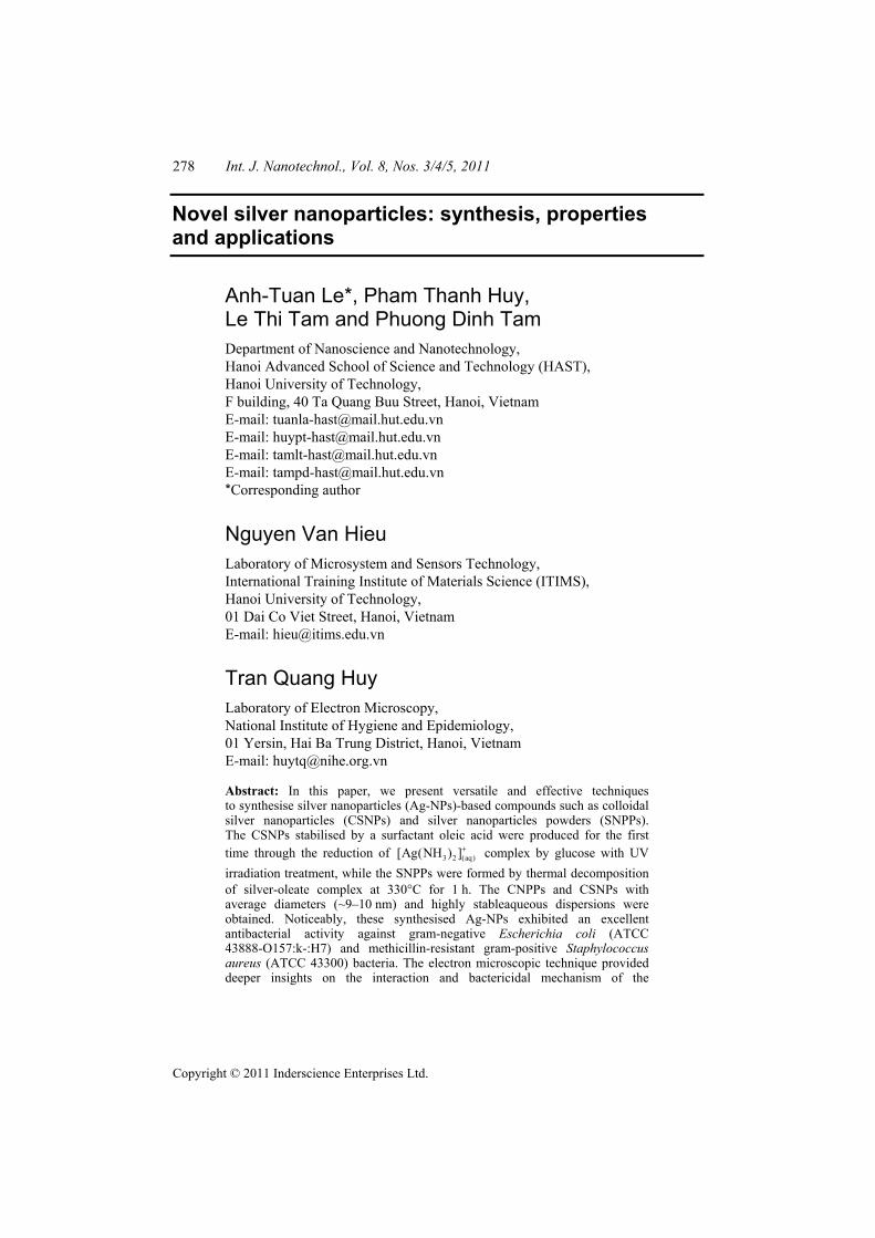
278 Int. J. Nanotechnol., Vol. 8, Nos. 3/4/5, 2011
Copyright © 2011 Inderscience Enterprises Ltd.
Novel silver nanoparticles: synthesis, properties and applications
Anh-Tuan Le*, Pham Thanh Huy, Le Thi Tam and Phuong Dinh Tam Department of Nanoscience and Nanotechnology, Hanoi Advanced School of Science and Technology (HAST), Hanoi University of Technology, F building, 40 Ta Quang Buu Street, Hanoi, Vietnam E-mail: [email protected] E-mail: [email protected] E-mail: [email protected] E-mail: [email protected] *Corresponding author
Nguyen Van Hieu Laboratory of Microsystem and Sensors Technology, International Training Institute of Materials Science (ITIMS), Hanoi University of Technology, 01 Dai Co Viet Street, Hanoi, Vietnam E-mail: [email protected]
Tran Quang Huy Laboratory of Electron Microscopy, National Institute of Hygiene and Epidemiology, 01 Yersin, Hai Ba Trung District, Hanoi, Vietnam E-mail: [email protected]
Abstract: In this paper, we present versatile and effective techniques to synthesise silver nanoparticles (Ag-NPs)-based compounds such as colloidal silver nanoparticles (CSNPs) and silver nanoparticles powders (SNPPs). The CSNPs stabilised by a surfactant oleic acid were produced for the first time through the reduction of 3 2 (aq)[Ag(NH ) ]+ complex by glucose with UV irradiation treatment, while the SNPPs were formed by thermal decomposition of silver-oleate complex at 330°C for 1 h. The CNPPs and CSNPs with average diameters (~9–10 nm) and highly stableaqueous dispersions were obtained. Noticeably, these synthesised Ag-NPs exhibited an excellent antibacterial activity against gram-negative Escherichia coli (ATCC 43888-O157:k-:H7) and methicillin-resistant gram-positive Staphylococcus aureus (ATCC 43300) bacteria. The electron microscopic technique provided deeper insights on the interaction and bactericidal mechanism of the
Novel silver nanoparticles: synthesis, properties and applications 279
Ag-NPs. Potential applications of the synthesised CSNPs and SNPPs for antibacterial – masterbatchs, acrylic emulsion paint as well as coating layer on cotton textiles were demonstrated.
Keywords: silver nanoparticles; green synthesis; thermal decomposition; modified Tollens process; colloids; antibacterial effect.
Reference to this paper should be made as follows: Le, A-T., Huy, P.T., Tam, L.T., Tam, P.D., Van Hieu, N. and Huy, T.Q. (2011) ‘Novel silver nanoparticles: synthesis, properties and applications’, Int. J. Nanotechnol., Vol. 8, Nos. 3/4/5, pp.278–290.
Biographical notes: Anh-Tuan Le received his PhD from the Department of Materials Engineering at Chungnam National University (CNU) in South Korea in 2007. From March 2008 onwards, he has been a Key Lecturer at the Department of Nanoscience and Nanotechnology at the Hanoi Advanced School of Science and Technology (HAST). He has published more than 30 papers in peer-reviewed SCI journals and a book chapter. His various research interests include amorphous and nanostructured magnetic materials, giant magnetoimpedance materials and applications, MEMS-based micro-sensors and devices, highly bactericidal functional silver nanoparticles, magnetic-based or DNA-based biochips and devices.
Pham Thanh Huy received his BSc from Hanoi National University, Hanoi, in 1993 and his PhD in Physics from the University of Amsterdam, the Netherlands, in 2001. He became an Associate Professor at HUT in 2007. Currently, he is a Director of HAST and a leading expert on the development of new micro-nanophosphors and metal oxides for use in high-efficiency lighting. His current research interests include nanomaterials, their characterisations and optoelectronic and sensing applications.
Le Thi Tam received her PhD in Biotechnology from the University of Ernst-Moritz-Arndt Greifswald in Germany in 2006. From October 2008-present, she has been a Lecturer at the Hanoi Advanced School of Science and Technology (HAST). Her research interests are molecular microbiology and biotechnology, proteomics and nano-biomaterials.
Phuong Dinh Tam received his MSc and PhD from the International Training Institute for Materials Science (ITIMS), Hanoi University of Technology (HUT), in 2004 and 2009, respectively. Currently, he is a Research Lecturer at Hanoi Advanced School Science and Technology (HAST). His current research interests include nanomaterials, nanofabrications, characterisations and applications for electronic devices and biosensors.
Nguyen Van Hieu became an Associate Professor at the International Training Institute for Material Science (ITIMS), Hanoi University of Technology (HUT), in November 2009. He received his MSc from ITIMS in 1997 and PhD from the Department of Electrical Engineering, University of Twente, Netherlands, in 2004. Currently, he is a Vice Director of ITIMS and a leading expert on the development of new nanomaterials and nanocomposites for gas-sensing applications. His current research interests include nanomaterials, nanofabrications and their characterisations and applications for electronic devices, gas sensors and biosensors.
Tran Quang Huy received his BSc in Physics from the College of Science, Vietnam National University, Hanoi (VNU), in 2000 and his MSc in Nanomaterials and Nanodevices from the College of Technology, VNU,

280 A-T. Le et al.
in 2007. Currently, he is a PhD student at the Hanoi University of Technology (HUT). He also has expertise in the area of electron microscopy (TEM and SEM) on biological samples and nanomaterials.
1 Introduction
The synthesis of noble metal nanoparticles for applications such as catalysis, electronics, optics, environmental and biotechnology has been intensively studied [1,2]. Gold, silver and copper materials have been mostly developed for the synthesis of stable dispersions of nanoparticles, which are useful in areas such as photography, catalysis, biological labelling, photonics, optoelectronics and surface-enhanced Raman scattering (SERS) detection, and among them [3].
Recently, due to the outbreak of infectious diseases caused by different pathogenic bacteria, pharmaceutical companies and researchers have been searching for new antibacterial agents [4]. The development of new strains of bacteria resistant to current antibiotics has become a serious problem in public health; therefore, there is a strong demand to search for new bactericides [5]. Among metallic nanoparticles, silver nanoparticles (Ag-NPs) are known to have inhibitory and bactericidal properties [6]. In addition, colloidal silver is of particular interest because of its distinctive properties, such as good conductivity, chemical stability, catalytic and antibacterial activity [7]. The antimicrobial property of silver is related to the amount of silver and the rate of silver released. Silver in its metallic state is inert but it reacts to skin moisture and the fluid of one’s wounds and therefore becomes ionised. The ionised silver is highly reactive, as it binds to tissue proteins and causes structural changes in the bacterial cell wall and nuclear membrane leading to cell distortion and death [8]. Silver also binds to bacterial DNA and RNA by denaturation and inhibits bacterial replication [9]. Besides, the bactericidal effect of the Ag-NPs has been attributed to their small size and high surface to volume ratio, which allows them to interact closely with microbial membranes and is not merely due to the release of metal ions in solution [10]. The Ag-NPs with bactericidal activity can be immobilised and coated on to different surfaces, which may be applied to various fields, i.e. medical instruments and devices, and water treatment and food processing [11]. The Ag-NPs may also be combined with other compounds, i.e., polymer, to form composites for better utilisation of their antimicrobial activity [12]. This has led to a growing interest from the scientific community regarding the development of Ag-NPs preparation techniques and further understanding of their fascinating properties.
In this work, we have developed new synthesis approaches for producing the Ag-NPs-based compounds of colloidal silver nanoparticles (CSNPs) by a modified Tollens technique, and silver nanoparticles powders (SNPPs) by a thermal decomposition method. Key information on synthesis techniques, structural and morphological properties, and the possible bactericidal mechanism of the Ag-NPs was described. Potential applications of as-prepared CSNPs and SNPPs for developing antibacterial-masterbatchs, acrylic emulsion paint as well as coating layers on cotton textiles were demonstrated. We believe that advanced synthetic techniques and greater stability of fine Ag-NP’s dispersions resulted in the significant enhancement of their antibacterial activity.

Novel silver nanoparticles: synthesis, properties and applications 281
2 Experimental
2.1 Feasible synthesis of silver nanoparticles powders
For a typical experiment, 1.7 g of AgNO3 (10 mmol) was dissolved in 100 ml deoxygenated water, then the resulting solution was added into 3.05 g of sodium oleate (10 mmol) and stirred vigorously for 2 h. The obtained solution was drained of the precipitate by filtration and washed three times with deionised water to free it of sodium and nitrate ions. The resulting complex powder was dried at room temperature. After drying, the Ag+1-oleate complex of white powder was transferred into a pyrex tube to perform a thermal decomposition reaction. The complex was then flushed with nitrogen, and the tube sealed at 0.3 Torr. The sample was slowly heated from room temperature to 330oC with a heating rate of 2oC/min, annealed at 330oC for 1 h, then cooled to room temperature. It was experimentally verified that the colour of the complex changed to black, indicating the formation of silver nanocrystallites [13].
2.2 Green synthesis of colloidal silver nanoparticles (CSNPs)
1.7 g (1.0 × 10⁻² mol) of silver nitrate (Aldrich, 99.9+%) was dissolved in 100 ml of deionised water. Then, the solution of silver nitrate was precipitated with 0.62 g (1.55 × 10⁻² mol) of sodium hydroxide (Aldrich, 99+%). An obtained precipitate of Ag2O was filtered and dissolved in 100 ml of aqueous ammonia (0.4% w/w, 2.3 × 10⁻² mol) until a transparent solution of silver ammonium complex, [Ag(NH3)2]+
(aq) was formed. Next, 2.5 g (8.9 × 10⁻³ mol) of oleic acid (Sigma-Aldrich, 99+%) was added dropwise into the obtained complex, and the resulting solution was gently stirred for 2 h at room temperature until the complete homogeneity of the reaction mixture was achieved. Finally, 2 g (1.11 × 10⁻² mol) of glucose was added to the mixture at room temperature with gentle stirring. The reduction process of silver complex solution (in quartz glass) was initiated with UV irradiation. UV treatment was carried out for 8 h under vigorous stirring without additional heating. A UV lamp (λ = 365 nm, 35 W) was used as a light source to stimulate the reduction process. After 8 h of irradiation, the transparent dispersion of oleic acid stabilised Ag-NPs (silver concentration ~10 mg ml–1) was obtained. The synthesis of Ag-NPs was successfully conducted with the final silver concentrations in the range of 0.1–2% [14].
2.3 Measurements
The crystalline structure of Ag-NPs was analysed by X-ray diffraction (XRD, Bruker D5005) using CuKα radiation (λ = 0.154 nm) at a step of 0.02º (2θ) at room temperature. A transmission electron microscope (TEM, JEOL-JEM 1010) was used to determine the morphology and distribution of silver nanoparticles. The UV-vis absorbance spectra of the Ag-NPs were recorded using an HP 8453 spectrophotometer. Quartz cuvettes with a 10 mm path length were used for the measurement of dispersion spectra.
2.4 Antibacterial tests
Two microbiological methods were employed to determine the antibacterial activity of the Ag-NPs. The antibacterial activity of the Ag-NPs was tested against gram-negative

282 A-T. Le et al.
Escherichia Coli (ATCC 43888-O157:k-:H7) and methicillin-resistant gram-positive Staphylococcus aureus (ATCC 43300) bacteria. These strains were obtained from the Department of Virology at the National Institute of Hygiene and Epidemiology in Hanoi.
First, the standard dilution micromethod was applied in conducting the antibacterial activity tests on the agar plates. These bacteria were cultured on a Luria-Bertani (LB) liquid nutrient broth medium with pH = 7. Aqueous dispersions of Ag-NPs of varying concentrations were prepared from the initial silver colloidal solution. A small suspension of tested bacteria was pipetted and spread onto the surface of the agar medium containing Ag-NPs. The Petri plates were incubated at 37°C for 24 h in a shaking incubator to encourage bacterial cell growth. The viable bacteria were monitored by counting the number of the colony-forming units from the appropriate dilution of bacteria concentration on the agar plates. A control sample test with no applied nanosilver was also conducted for comparison.
Second, the disc diffusion method was used to evaluate the antibacterial activity of silver nanoparticles against tested bacteria. Using the spread plate method, nutrient agar plates were inoculated with 100 µl of bacterial suspension containing 108 CFU. Sterile Whatman No. 1 filter paper discs with a diameter of 5 mm each, loaded with 0.5 µg of nanosilver, were placed on the inoculated plates. Control plates were maintained with sterile nanosilver free discs. These plates were incubated at 37°C for 24 h and the zone of inhibition (ZOI) was measured by subtracting the disc diameter from the total inhibition zone diameter.
The antimicrobial effect was quantified based on the inhibition zone measured in the disk diffusion tests conducted in plates, and by determining the minimum growth inhibitory concentrations (MIC) and minimum bactericidal concentration (MBC) of nanoparticles in liquid batch cultures [15].
3 Results and discussion
Generally, Ag-NPs can be prepared and stabilised by physical and chemical methods; the chemical approach, such as chemical reduction, electrochemical techniques and photochemical reduction, is most widely used [16,17]. It should be emphasised that the properties of Ag-NPs strongly depend on size, shape of NPs, their interactions with stabilisers and surrounding media and also the manner of their preparation [18]. Hence, the design of a synthesis method in which the size, morphology, stability and properties are controlled has become a key challenge of interest [19]. In our paper, we present versatile techniques for the preparation of finely dispersed aqueous silver colloids through a modified Tollens technique and of monodisperse silver powders through a thermal decomposition.
A fundamental reduction reaction involving the Tollens process is as follows [20]:
[Ag(NH3)2]+ (aq) + RCHO(aq) → Ag(s) + RCOOH(aq).
where RCHO – an aldehyde or a carbohydrate (i.e., glucose). The particle size, size distribution and stability of the NPs dispersion depend
on the temperature of the process, ammonia concentration and the pH of the medium during the reduction process. The properties of the synthesised NPs dispersions can be also modified by additional components such as surfactants. Ag-NPs synthesised by the conventional Tollens process or in the presence of surfactants are reported to be
Novel silver nanoparticles: synthesis, properties and applications 283
of 20–70 nm in diameter with rather broad size distribution [19,20]. In comparison with previous works where Tollens process was being used, we for the first time applied UV-irradiation simultaneously with glucose reduction of silver salt through NPs preparation. There are two distinct features made to improve traditional Tollens technique: first, an addition of oleic acid, which plays the role of a stabiliser, and second, using the UV-irradiation simultaneously with treatment by glucose during the reduction process to induce the formation of silver NPs with controllable diameter and narrow size distribution. Moreover, this synthesis route is environmentally friendly because of the use of non-toxic chemicals [21]. In order to provide further possibilities to control the process of Ag-NPs formation, we used UV irradiation at the stage of reducing silver ammonium complex. It was found that the UV treatment of the reaction medium containing silver complex, surfactant (oleic acid) and glucose resulted in the formation of a highly stable dispersion of small (9–10 nm in size) silver NPs with narrow size distribution, whereas the reduction of [Ag(NH3)2]+ complex by glucose led to unstable colloidal solutions with broad size distribution. The process of silver NPs preparation under UV treatment was controlled by the irradiation time. A time period of 8 h was found to be optimal for the complete reduction of silver complex [14].
We also developed a new synthetic method to produce monodisperse silver-powder using the thermal decomposition of a Ag+1-oleate complex, which was prepared by the reaction of AgNO3 and sodium oleate in a water solution [22]. Using a thermogravimetric analyser measurement, we observed the thermal decomposition of the Ag+1-oleate complex at 330°C (data not shown here). From these results, we reasoned that a metal-oleate complex would make an effective growth source for the synthesis of monodisperse nanocrystals. Instead of using toxic and expensive organometallic compounds, we successfully prepared the metal-oleate complex by reacting inexpensive and environmentally friendly compounds, namely sodium oleate. The large-scale synthesis of monodisperse nano-sized particles can be achieved by using inexpensive and non-toxic metal salts as reactants [23]. We were able to synthesise as much as 30–40 g of monodisperse Ag-NPs powder in a single reaction, without a size-sorting process. Moreover, the size of silver particles can be controlled simply by varying the experimental conditions. The advantage of the current synthetic procedure makes it useful for the preparation of a variety of other metallic nanoparticles [23].
Now, let us discuss the functional properties of synthesised Ag-NPs materials. First of all, the existence of Ag-NPs in the solution was determined by using a UV-visible absorption measurement. As observed in top image of Figure 1, an optical absorption band with a maximum at 420 nm was found. This is a typical feature of the absorption of metallic Ag-NPs due to the surface plasmon resonance, indicating the presence of the Ag-NPs in the solution [24]. With increasing silver concentration, the colour of the solution changed from light yellow to dark yellow to brown (see the inset of top image). It should be noted that the change in colour reveals the formation of Ag-NPs in the solution. The crystallinity of the Ag-NPs was confirmed by powder X-ray diffraction. It showed broad peaks assigned to 111, 200 and 220 planes of a face-centred cubic lattice of bulk silver (see Bottom image of Figure 1). This fact confirmed that the synthesised Ag-NPs consisted of pure silver with high crystallinity. These obtained results demonstrated that as-prepared silver colloids consisted of finely dispersed NPs with an average diameter about 10 nm and a relatively narrow size distribution.

284 A-T. Le et al.
Figure 1 (Top image) A UV-visible absorption spectrum of colloidal silver nanoparticles synthesised by a modified Tollens technique. The inset displays aqueous dispersions of Ag-NPs at different silver concentrations and (Bottom image) The X-ray diffraction pattern of the silver NPs powder prepared by a thermal decomposition technique (see online version for colours)
To determine the stability of Ag-NPs in aqueous media, the absorbance intensity of the Ag-NPs colloidal solution was monitored over time. UV-vis spectra and TEM measurements were obtained to identify the changes in particle size after the aging of Ag-NPs. Results revealed that the particle size remained almost unchanged after being stored for two months [14]. This finding suggests that as-prepared aqueous dispersions of Ag-NPs are very stable against aggregation for several months. These results also imply that reproducible dispersions of silver colloids can be created in batches and stored. Moreover, the addition of appropriate amounts of oleic acid as a stabilising agent to silver colloids not only improves their stability but also allows duplicate measurements to be made with less deviations [21].
Next, in order to obtain deeper insights on the morphology and size distribution of the obtained Ag-NPs, the TEM analyses were employed. As one can see clearly from top image of Figure 2, the nanosized silver particles were well-formed and well-dispersed. The average-sized silver particle was about 9.6 ± 0.8 nm with a relatively narrow distribution. It should be noted that the absorption band of colloidal solution of the synthesised Ag-NPs was observed at λmax = 420 nm, thus being red-shifted when compared with the plasmon absorption band of silver colloids stabilised by non-covalent interactions (λmax = 400–413 nm) [25]. This can be explained by the formation of a silver oleate layer on the surface of the silver particles. Taking into account the stability of the aqueous dispersions of the as-obtained silver NPs, oleic acid most likely forms a double layer between silver surface and liquid medium (see Bottom image of Figure 2).

Novel silver nanoparticles: synthesis, properties and applications 285
Figure 2 (Top image) TEM image of the Ag-NPs solution and (Bottom image) formation of the oleate stabilising bilayer on Ag-NPs surface
On the basis of UV-visible and TEM studies, a possible mechanism of Ag-NPs formation and growth under the applied experimental conditions is suggested as follows: the UV irradiation causes excitation of [Ag(NH3)2]+ ions followed by electron transfer from the glucose molecule to Ag+, thus producing Ag0 atoms, which then form clusters and seeds:
0 0 03 2 3[Ag(NH ) ] RCHOH Ag 2NH RCOH Ag (Ag ) ,h
nH nν+ ++ → + + + →
where RCHOH represents glucose in its cyclic form. Primarily, UV treatment leads to the substantially simultaneous formation of a large amount of silver nuclei. This results in small dimensions and narrow size distribution of the finally obtained Ag-NPs. The remaining silver ions are adsorbed onto the surface of the already-formed particles, thus bringing about successive reduction and attracting oppositely charged surfactant ions. These surfactant ions form a capping layer on the surface of the silver particles. Since the carboxyl group of the oleate ion is directed towards the silver surface, the first capping layer is highly hydrophobic from the outside. Therefore, the oleate ions from the solution start to attach to the first layer forming a stable bilayer structure with carboxyl group directed towards the liquid medium (see bottom image of Figure 2). The surfactant molecules inhibit this aggregation association through the capping/template effect, and thus act as a particle stabiliser [26]. It should be noted that the hydroxymethyl functionalities of the surfactant molecules anchor the molecule to the cluster surface, while the hydrophobic chain protects the cluster from aggregation with its next neighbour due to electrostatic repulsion and steric hindrance, thus inhibiting coalescence [27].

286 A-T. Le et al.
Based on the discovered results, the Ag-NPs with their unique chemical and physical properties have proved as an alternative for the development of new antibacterial agents. The Ag-NPs have also found diverse applications in the form of wound dressings, coatings for medical devices, Ag-NPs impregnated textile fabrics, etc. The advantage of using Ag-NPs for impregnation is that there is continuous release of silver ions and the devices can be coated by both the outer and inner side, hence enhancing its antimicrobial efficacy. Burn wounds treated with Ag-NPs showed better cosmetic appearance and scarless healing [25]. Silver is also non-toxic to humans in minute concentrations. Microorganisms are unlikely to develop resistance against silver when compared with antibiotics, as silver attacks a broad range of targets in the microbes. Thus, it can be concluded that the use of Ag-NPs will serve a variety of applications in different fields such as electronics, medicine, pharmacy and agriculture. However, with the advent of Ag-NPs and their major use as an antimicrobial agent, further studies are needed to understand the exact mechanism of the bactericidal effect as well as the interaction of Ag-NPs with the bacterial cells. Several studies propose that Ag-NPs may attach to the surface of the cell membrane, disturbing permeability and respiration functions of the cell [4–8]. It is also possible that Ag NPs not only interact with the surface of membrane, but can also penetrate inside the bacteria.
To achieve a full understanding of bactericidal and interaction mechanism of the Ag-NPs, we have conducted the antibacterial activity test of the Ag-NPs against E. coli bacteria. The origin of bactericidal action of the synthesised Ag-NPs with the Gram-negative E. coli bacteria was demonstrated by adapting the cross-sectioning technique in TEM electron microscopy [14]. The sample preparation process of E. coli samples for direct TEM observation can be found in Section 2, part E. Colloidal solution of the silver NPs (5.0 µg mL–1 concentration) was dropped onto the surface of agar plate containing E. coli. After 30 min, 1 h and 2 h, E. coli containing samples were taken out and done by sectioning method for TEM observation. At different magnifications and sections (Figure 3), it was found that there were a lot of Ag-NPs binding around E. coli cell membrane as well as coming inside the cells. It was evidently observed from Figure 3 that in addition to being fixed to the cell membrane, the obtained Ag-NPs are capable of penetrating it to be distributed inside a bacterium. The cell the cytoplasm was destroyed in the process. This probably led to the death of the E. coli cells caused by the Ag-NPs [21]. It has been clearly demonstrated that the changes in morphology presented in the membrane of the bacteria in conjunction with the possible damage caused by the Ag-NPs reacting with sulphur-containing proteins in the interior of the cell as well as with phosphorous-containing compounds such as DNA will affect the bacteria in the processes such as respiration and cell division, finally causing the death of the cell [28].
From our experimental results, a possible mechanism for bacterial action of Ag-NPs was established. The silver particles are first attached to the cell membrane and then penetrated the bacteria. The bacterial membrane contains sulphur-containing proteins, and the silver particles interact with these proteins in the cell as well as with the phosphorus-containing compounds like DNA. When silver particles enter the bacterial cell, it forms a low molecular weight region in the centre of the bacteria to which the bacteria conglomerates, thus protecting the DNA from the silver ions. The Ag-NPs preferably attack the respiratory chain and cell division processes finally leading to cell death. The Ag-NPs release silver ions in the bacterial cells, thus enhancing their bactericidal activity. Our studies indicate that the concentration of silver leading to a

Novel silver nanoparticles: synthesis, properties and applications 287
complete inhibition of bacteria growth was revealed as low as at 1.0 µg mL–1 [14] and found to be much lower compared with earlier reports ~3–10 µg mL–1 [6–9]. These advantages of the Ag-NPs materials make them ideal for green industrial, medicinal, microbiological and other applications. With the purpose of developing practical applications, we have developed some products with the use of synthesised Ag-NPs materials as an antibacterial agent including Ag-NPs-embedded masterbatch pellets, nano-silver acrylic aqueous paint, and coating layer on cotton textile (Figure 4). It is important to note that optimised conditions for complete inhibition in bacteria growth or minimum inhibitory silver concentrations (MIC) leading to inhibition of bacterial growth for tested Gram-negative E. coli (ATCC 43888-O157:k-:H7) and Gram-positive S. aureus (ATCC 43300) bacteria were revealed at minimum silver concentrations of 600 ppm for the case of masterbatch, 100 ppm for aqueous acrylic paint, and 20 ppm for coating layer on cotton textile, respectively. All standard antibacterial tests were conducted at the National Institute of Hygiene and Epidemiology. It is necessary to emphasise that the developed nano-silver-containing products have observed significant effect in inhibition of bacterial growth at the laboratory level [21]. Importantly, the excellent antibacterial effect of nano-silver-containing products over tested bacteria was observed without deterioration of colour or mechanical properties of final products [13].
Figure 3 TEM images of E. coli strain showing the interaction stages of Ag-NPs with the bacteria. After 30 min and 60 min, a lot of Ag-NPs bindings around the E. coli cell membranes as well as within the cells were found. As observed, after interacting with the E. coli bacteria, the Ag-NPs adhered to the cell wall of the bacteria and penetrated the cell membrane, resulting in the inhibition of bacterial cell growth and multiplication
288 A-T. Le et al.
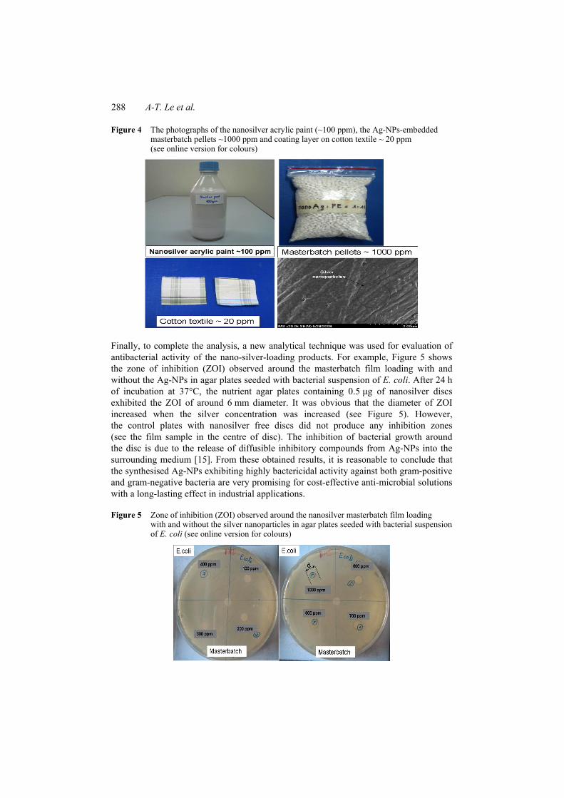
Figure 4 The photographs of the nanosilver acrylic paint (~100 ppm), the Ag-NPs-embedded masterbatch pellets ~1000 ppm and coating layer on cotton textile ~ 20 ppm (see online version for colours)
Finally, to complete the analysis, a new analytical technique was used for evaluation of antibacterial activity of the nano-silver-loading products. For example, Figure 5 shows the zone of inhibition (ZOI) observed around the masterbatch film loading with and without the Ag-NPs in agar plates seeded with bacterial suspension of E. coli. After 24 h of incubation at 37°C, the nutrient agar plates containing 0.5 µg of nanosilver discs exhibited the ZOI of around 6 mm diameter. It was obvious that the diameter of ZOI increased when the silver concentration was increased (see Figure 5). However, the control plates with nanosilver free discs did not produce any inhibition zones (see the film sample in the centre of disc). The inhibition of bacterial growth around the disc is due to the release of diffusible inhibitory compounds from Ag-NPs into the surrounding medium [15]. From these obtained results, it is reasonable to conclude that the synthesised Ag-NPs exhibiting highly bactericidal activity against both gram-positive and gram-negative bacteria are very promising for cost-effective anti-microbial solutions with a long-lasting effect in industrial applications.
Figure 5 Zone of inhibition (ZOI) observed around the nanosilver masterbatch film loading with and without the silver nanoparticles in agar plates seeded with bacterial suspension of E. coli (see online version for colours)

Novel silver nanoparticles: synthesis, properties and applications 289
Acknowledgements
This work was financially supported by a grant from the collaborative program between the National Institute of Hygiene and Epidemiology of Vietnam and Hanoi Advanced School of Science and Technology (HAST), Hanoi University of Technology (Decision 148/QĐ-VSDTTW). We also acknowledge the support from Vietnam’s National Foundation for Science and Technology Development (NAFOSTED) for a basic research project (code 106.99-2010.54).
References 1 Tuan, V.D. (2006) Nanotechnology in Biology and Medicine, CRC Press, Taylor & Francis
Group, USA. 2 Schulte, J. (2005) Nanotechnology: Global Strategies, Industry Trends and Applications,
John Wiley & Sons, UK. 3 Chen, C., Wang, L., Jiang, G. and Yu, H. (2006) ‘Chemical preparation of special-shaped metal
nanomaterials though encapsulation or inducement in soft solution’, Rev. Adv. Mater. Sci., Vol. 11, pp.1–18.
4 Rai, M., Yadav, A. and Gade, A. (2009) ‘Silver nanoparticles as a new generation of antimicrobials’, Biotechnol. Adv., Vol. 27, pp.76–83.
5 Sharma, V.K., Yngard, R.A. and Lin, Y. (2009) ‘Silver nanoparticles: green synthesis and their antimicrobial activities’, Adv. Colloid Sur. Interface, Vol. 145, pp.83–96.
6 Sondi, I. and Sondi, B.S. (2004) ‘Silver nanoparticles as antimicrobial agent: a case study of E.coli as a model for gram-negative bacteria’, J. Colloids Interface Sci., Vol. 275, pp.177–182.
7 Yin, Y., Li, Z.Y., Gates, B., Xia, Y. and Venkateswaran, S. (2002) ‘Synthesis and characterization of stable aqueous dispersions of silver nanoparticles though the Tollens process’, J. Mater. Chem., Vol. 12, pp.522–527.
8 Morones, J.R., Elechiguerra, J.L., Camacho, A., Holt, K., Kouri, J.B., Ramirez, J.T. and Yacaman, M.J. (2005) ‘The bactericidal effect of silver nanoparticles’, Nanotechnology, Vol. 16, pp.2346–2353.
9 Raffi, M., Hussain, F., Bhatti, T.M., Akhter, J.I., Hameed, A. and Hasan, M.M. (2008) ‘Antibacterial characterization of silver nanoparticles against E.coli ATCC-15224’, J. Mater. Sci. Technol., Vol. 24, pp.192–196.
10 Shrivastava, S., Bera, T., Roy, A., Singh, G., Ramachandrarao, P. and Dash, D. (2007) ‘Characterization of enhanced antibacterial effects of novel silver nanoparticles’, Nanotechnology, Vol. 18, pp.225103–225107.
11 Yang, W., Shen, C., Ji, Q., An, H., Wang, J., Liu, Q. and Zhang, Z. (2009) ‘Food storage material silver nanoparticles interfere with DNA replication fidelity and bind with DNA’, Nanotechnology, Vol. 20, pp.085102–085106.
12 Lee, K.J., Jun, B.H., Choi, J., Lee, Y.I., Joung, J. and Oh, Y.S. (2007) ‘Environmentally friendly synthesis of organic-soluble silver nanoparticles for printed electronics’, Nanotechnology, Vol. 18, pp.335601–335604.
13 Hang, T.T., Tam, P.D., Huy, T.Q. and Le, A.T. (2009) ‘Fabrication of silver-nanoparticles-embedded polymer masterbatchs with excellent antibacterial performance’, J. Chem., Vol. 47, No. 5, pp.587–593.
14 Le, A.T., Huy, P.T., Tam, P.D., Huy, T.Q., Cam, P.D., Kudrinskiy, A.A. and Krutyakov, Y.A. (2010) ‘Green synthesis of finely-dispersed highly bactericidal silver nanoparticles via modified Tollens technique’, Curr. Appl. Phys., Vol. 10, pp.910–916.
290 A-T. Le et al.
15 Kora, A.J., Manjusha, R. and Arunachlam, J. (2009) ‘Superior bactericidal activity of SDS capped silver nanoparticles: synthesis and characterization’, Mater. Sci. Eng. C., Vol. 29, pp.2104–2109.
16 Sun, Y. and Xia, Y. (2002) ‘Shape-controlled synthesis of gold and silver nanoparticles’, Science, Vol. 98, pp.2176–2179.
17 Kim, D., Jeong, S. and Moon, J. (2006) ‘Synthesis of silver nanoparticles using polyol process and the influence of precursor injection’, Nanotechnology, Vol. 17, pp.4019–4024.
18 Ghosh, S.K., Kundu, S., Mandal, M., Nath, S. and Pal, T. (2003) ‘Studies on the evolution of silver nanoparticles in micelle by UV-photoactivation’, J. Nanoparticles Research, Vol. 5, pp.577–587.
19 Kvítek, L., Panáek, A., Soukupová, J., Kolář, M., Veeřová, R., Prucek, R., Holecová, M. and Zbořil, R. (2008) ‘Effect of surfactants and polymers on stability and antibacterial activity of silver nanoparticles’, J. Phys. Chem. C., Vol. 112, pp.5825–5834.
20 Panaček, A., Kvitek, L., Prucek, R., Kolář, M., Večeřová, R., Pizúrová, N., Sharma, V.K., Nevěčná, T. and Zbořil, R. (2006) ‘Silver colloid nanoparticles: synthesis, characterization, and their antibacterial activity’, J. Phys. Chem. B, Vol. 110, pp.16248–16253.
21 Le, A.T., Tam, L.T., Tam, P.D., Huy, P.T., Huy, T.Q., Hieu, N.V., Kudrinskiy, A.A. and Krutyakov, Y.A. (2010) ‘Synthesis of oleic acid-stabilized silver nanoparticles and analysis of their antibacterial activity’, Mater. Sci. Eng. C., Vol. 30, pp.910–916.
22 Le, A.T. et al. (forthcoming) ‘The preparation of monodisperse silver nanoparticles via thermal decomposition technique’, (unpublished data).
23 Park, J., Joo, J., Kwon, S.G., Jang, Y. and Hyeon, T. (2007) ‘Synthesis of monodisperse spherical nanocrystals’, Angew. Chem. Int. Ed., Vol. 46, pp.4630–4660.
24 Vertelov, G.K., Krutyakov, Y.A., Efremenkova, O.V., Olenin, A.Y. and Lisichkin, G.V. (2008) ‘A versatile synthesis of highly bactericidal Myramistin stabilized silver nanoparticles’, Nanotechnology, Vol. 19, pp.355707–355710.
25 Krutyakov, Y.A., Kudrinskiy, A.A., Olenin, A.Y. and Lisichkin, G.V. (2008) ‘Synthesis and properties of silver nanoparticles: advances and prospects’, Russ. Chem. Rev., Vol. 77, pp.233–257.
26 Zhang, Q., Ge, J., Pham, T., Goebl, J., Hu, Y., Lu, Z. and Yin, Y. (2009) ‘Reconstruction of silver nanoplates by UV irradiation: tailored optical properties and enhanced stability’, Angew. Chem. Int. Ed, Vol. 48, pp.3516–3519.
27 Sakamoto, M., Fujistuka, M. and Majima, T. (2009) ‘Light as construction tool of metal nanoparticles: synthesis and mechanism’, J. Photochem. Photobiol. C, Vol. 10, pp.33–56.
28 Le, A.T., Huy, P.T., Huy, T.Q., Cam, P.D., Kudrinskiy, A.A., Olenin, A.Y., Lisichkin, G.V. and Krutyakov, Y.A. (2010) ‘Photochemical synthesis of highly bactericidal silver nanoparticles’, Nanotechnol. Russ., Vol. 5, pp.554–563.
Copyright © 2022 FDOKUMEN