A Novel Synthesis of Malic acid capped Silver Nanoparticles using S. lycopersicums
Upload
khangminh22Category
view
0download
0
Comparative analysis of Silver Nanoparticles prepared from
Different Plant extracts (Hibiscus rosa sinensis, Moringa oleifera,
Acorus calamus, Cucurbita maxima, Azadirachta indica) through
green synthesis method
Thesis submitted to
National Institute of Technology, Rourkela For the partial fulfilment of the Master degree in
Life Science
BY:
SONALI PRADHAN ROLL NO. 411LS2061
Under the guidance of Dr. BISMITA NAYAK
DEPARTMENT OF LIFE SCIENCE
NATIONAL INSTITUTE OF TECHNOLOGY,
ROURKELA -76900
DEPARTMENT OF LIFE SCIENCE NATIONAL INSTITUTE OF TECHNOLOGY
ROURKELA-769008
…………………………………………………………………………………………………………………………………………………………….--------------------------------------------------------------------------------------------------------------------------------------
Dr. Bismita Nayak, Ph.D.,
Assistant Professor Place: ..............
Department of Life Science Date: ...............
National Institute Of Technology
Rourkela
Certificate
This is to certify that the thesis entitled “Comparative analysis of Silver Nanoparticles prepared from Different Plant extracts (Hibiscus rosa sinensis, Moringa oleifera, Acorus calamus, Cucurbita maxima, Azadirachta indica)
through green synthesis method” Submitted to National Institute of
Technology, Rourkela for the partial fulfilment of the Master degree
in Life science is a faithful record of bonafide and original research
work carried out by Ms. Sonali Pradhan under my supervision and
guidance.
.......................................................................................................................... Phone no.: 0661-2462682 Email:[email protected]
DECLARATION
I hereby declare the thesis entitled “Comparative analysis of Silver Nanoparticles
prepared from Different Plant extracts (Hibiscus rosa sinensis, Moringa oleifera, Acorus
calamus, Cucurbita maxima, Azadirachta indica) through green synthesis method”,
submitted to the Department of Life Science, National Ins titute of Technology, Rourkela for
the partial fulfilment of the Master Degree in Life Science is a faithful record of bonafide
research work carried out by me under the guidance and supervision of Dr. Bismita Nayak,
Assistant Professor, Department of Life Science, National Institute of Technology, Rourkela.
No part of this thesis has been submitted by any other research persons or any students.
Date:
Place: Sonali Pradhan
ACKNOWLEDGEMENT
I wish to express my sincere thanks and gratitude to my guide Dr. Bismita Nayak, Assistant
Professor, Dept. of Life Science, National Institute of Technology, Rourkela, for her constant
inspiration, encouragement and guidance throughout my project. I consider myself fortunate
enough that she has given a decisive turn and boost to my career.
I take this opportunity to express my indebtedness to my Professors for their enthusiastic
help, valuable suggestions and constant encouragement throughout my work. I would also
like to express my whole hearted gratitude to the Head of the department of Life Science Dr.
Samir Kumar Patra, and other faculty members, Dr. Sujit Kumar Bhutia, Dr. Bibekanand
Mallick, Dr. Surajit Das, Dr. Suman Jha, and Dr. Rasu Jayabalan, National Institute of
Technology Rourkela, Orissa for their good wishes, inspiration and unstinted support
throughout my project.
I deeply acknowledge the constant support, encouragement, and invaluable guidance at every
step of my project by Mr. Debasis Nayak PhD scholar, Dept. of life science. I am obliged and
thankful to him for providing me the opportunity to gain knowledge and understanding of
working skills of the aspects of my work from him. Also I am thankful to Mr. Pradipta
Ranjan Rauta, Sarbani Ashe PhD scholar, Dept. of Life Science for their cooperation and
supportive nature.
I take this opportunity to thank my friends Gunjan, Krishna, Manorama for their throughout
co-operation.
Last but not the least I take this opportunity to thank my father Mr. Bishnu Charan Pradhan
and my mother Mrs. Phulamani Pradhan for weathering my minor crises of confidence, for
never doubting.
Thank you for everything Maa and Papa. I love you both.
Place: Rourkela Sonali Pradhan
Date:
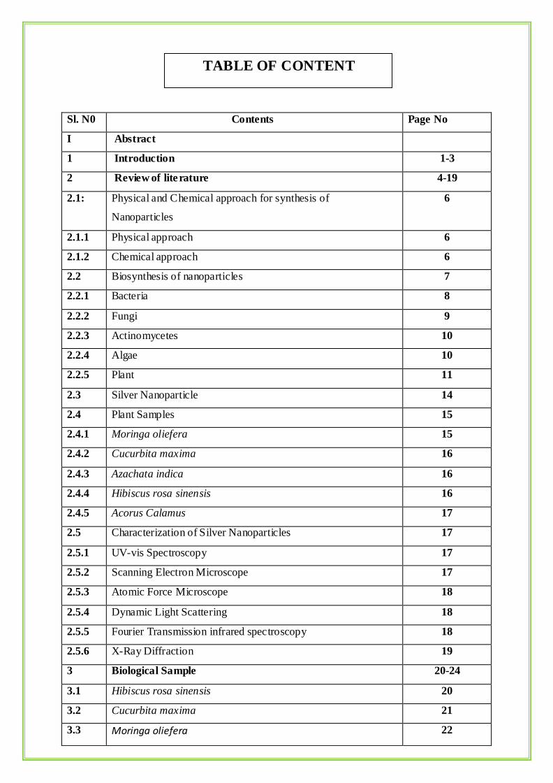
Sl. N0 Contents Page No
I Abstract
1 Introduction 1-3
2 Review of literature 4-19
2.1:
Physical and Chemical approach for synthesis of
Nanoparticles
6
2.1.1 Physical approach 6
2.1.2 Chemical approach 6
2.2 Biosynthesis of nanoparticles 7
2.2.1 Bacteria 8
2.2.2 Fungi 9
2.2.3 Actinomycetes 10
2.2.4 Algae 10
2.2.5 Plant 11
2.3 Silver Nanoparticle 14
2.4 Plant Samples 15
2.4.1 Moringa oliefera 15
2.4.2 Cucurbita maxima 16
2.4.3 Azachata indica 16
2.4.4 Hibiscus rosa sinensis 16
2.4.5 Acorus Calamus 17
2.5 Characterization of Silver Nanoparticles 17
2.5.1 UV-vis Spectroscopy 17
2.5.2 Scanning Electron Microscope 17
2.5.3 Atomic Force Microscope 18
2.5.4 Dynamic Light Scattering 18
2.5.5 Fourier Transmission infrared spectroscopy 18
2.5.6 X-Ray Diffraction 19
3 Biological Sample 20-24
3.1 Hibiscus rosa sinensis 20
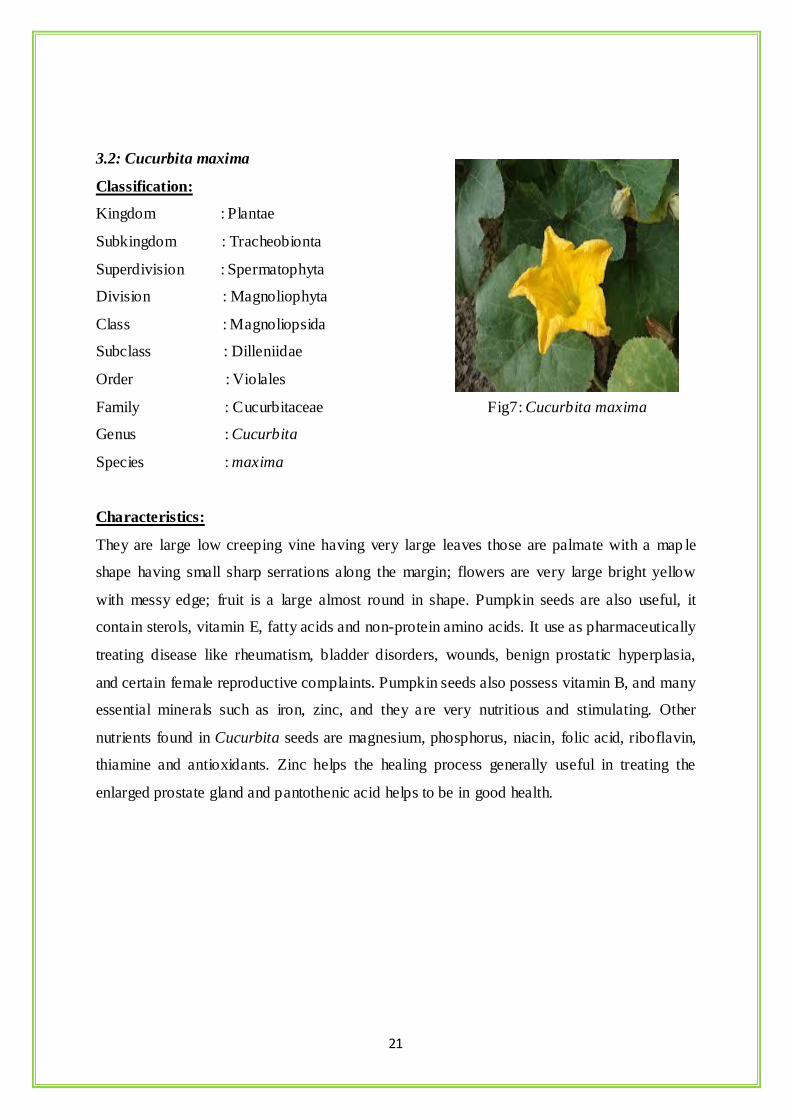
3.2 Cucurbita maxima 21
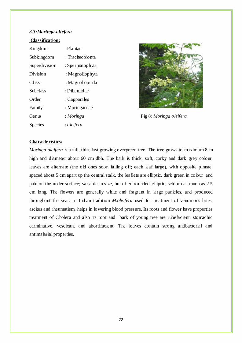
3.3 Moringa oliefera 22
TABLE OF CONTENT
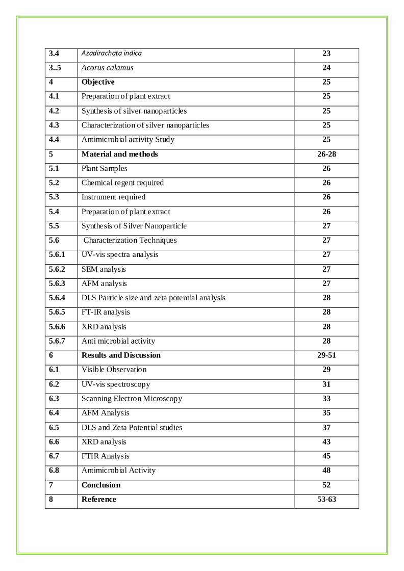
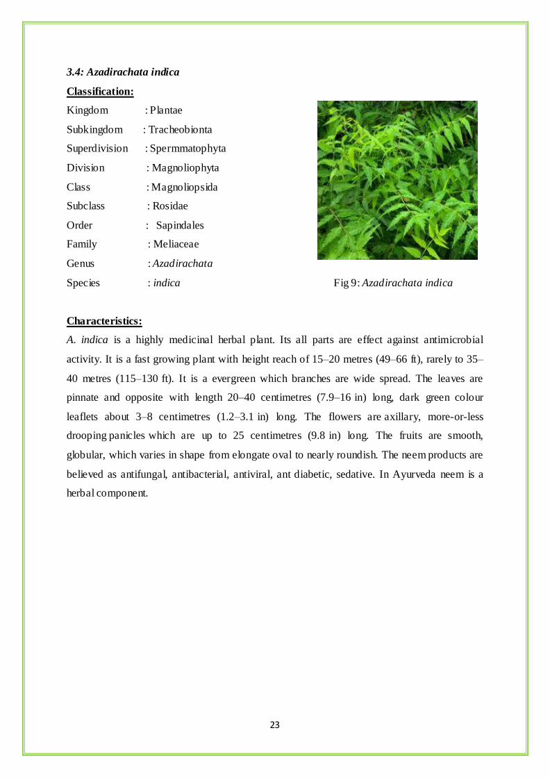
3.4 Azadirachata indica 23
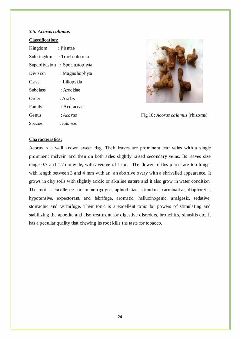
3..5 Acorus calamus 24
4 Objective 25
4.1 Preparation of plant extract 25
4.2 Synthesis of silver nanoparticles 25
4.3 Characterization of silver nanoparticles 25
4.4 Antimicrobial activity Study 25
5 Material and methods 26-28
5.1 Plant Samples 26
5.2 Chemical regent required 26
5.3 Instrument required 26
5.4 Preparation of plant extract 26
5.5 Synthesis of Silver Nanoparticle 27
5.6 Characterization Techniques 27
5.6.1 UV-vis spectra analysis 27
5.6.2 SEM analysis 27
5.6.3 AFM analysis 27
5.6.4 DLS Particle size and zeta potential analysis 28
5.6.5 FT-IR analysis 28
5.6.6 XRD analysis 28
5.6.7 Anti microbial activity 28
6 Results and Discussion 29-51
6.1 Visible Observation 29
6.2 UV-vis spectroscopy 31
6.3 Scanning Electron Microscopy 33
6.4 AFM Analysis 35
6.5 DLS and Zeta Potential studies 37
6.6 XRD analysis 43
6.7 FTIR Analysis 45
6.8 Antimicrobial Activity 48
7 Conclusion 52
8 Reference 53-63
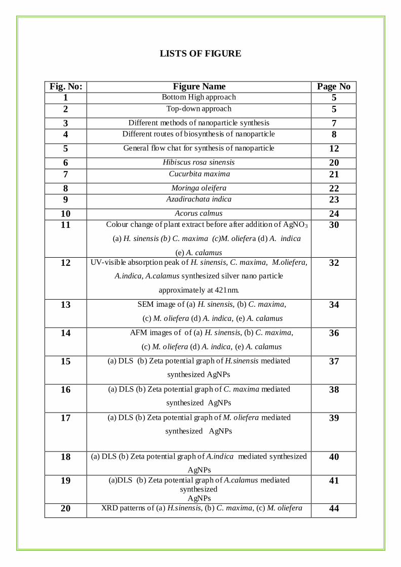
LISTS OF FIGURE
Fig. No: Figure Name Page No
1 Bottom High approach 5
2 Top-down approach 5
3 Different methods of nanoparticle synthesis 7
4 Different routes of biosynthesis of nanoparticle 8
5 General flow chat for synthesis of nanoparticle 12
6 Hibiscus rosa sinensis 20
7 Cucurbita maxima 21
8 Moringa oleifera 22
9 Azadirachata indica 23
10 Acorus calmus 24
11 Colour change of plant extract before after addition of AgNO3
(a) H. sinensis (b) C. maxima (c)M. oliefera (d) A. indica
(e) A. calamus
30
12 UV-visible absorption peak of H. sinensis, C. maxima, M.oliefera,
A.indica, A.calamus synthesized silver nano particle
approximately at 421nm.
32
13 SEM image of (a) H. sinensis, (b) C. maxima,
(c) M. oliefera (d) A. indica, (e) A. calamus
34
14 AFM images of of (a) H. sinensis, (b) C. maxima,
(c) M. oliefera (d) A. indica, (e) A. calamus
36
15 (a) DLS (b) Zeta potential graph of H.sinensis mediated
synthesized AgNPs
37
16 (a) DLS (b) Zeta potential graph of C. maxima mediated
synthesized AgNPs
38
17 (a) DLS (b) Zeta potential graph of M. oliefera mediated
synthesized AgNPs
39
18 (a) DLS (b) Zeta potential graph of A.indica mediated synthesized
AgNPs
40
19 (a)DLS (b) Zeta potential graph of A.calamus mediated synthesized
AgNPs
41
20 XRD patterns of (a) H.sinensis, (b) C. maxima, (c) M. oliefera 44
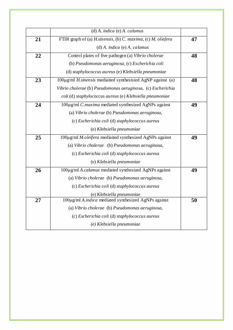
(d) A. indica (e) A. calamus
21 FTIR graph of (a) H.sinensis, (b) C. maxima, (c) M. oliefera
(d) A. indica (e) A. calamus
47
22 Control plates of five pathogen (a) Vibrio cholerae
(b) Pseudomonas aeruginosa, (c) Escherichia coli
(d) staphylococcus aureus (e) Klebsiella pneumoniae
48
23 100μg/ml H.sinensis mediaited synthesisied AgNP against (a)
Vibrio cholerae (b) Pseudomonas aeruginosa, (c) Escherichia
coli (d) staphylococcus aureus (e) Klebsiella pneumoniae
48
24 100μg/ml C.maxima mediated synthesized AgNPs against
(a) Vibrio cholerae (b) Pseudomonas aeruginosa,
(c) Escherichia coli (d) staphylococcus aureus
(e) Klebsiella pneumoniae
49
25 100μg/ml M.oleifera mediaited synthesized AgNPs against
(a) Vibrio cholerae (b) Pseudomonas aeruginosa,
(c) Escherichia coli (d) staphylococcus aureus
(e) Klebsiella pneumoniae
49
26 100μg/ml A.calamus mediated synthesized AgNPs against
(a) Vibrio cholerae (b) Pseudomonas aeruginosa,
(c) Escherichia coli (d) staphylococcus aureus
(e) Klebsiella pneumoniae
49
27 100μg/ml A.indica mediated synthesized AgNPs against
(a) Vibrio cholerae (b) Pseudomonas aeruginosa,
(c) Escherichia coli (d) staphylococcus aureus
(e) Klebsiella pneumoniae
50
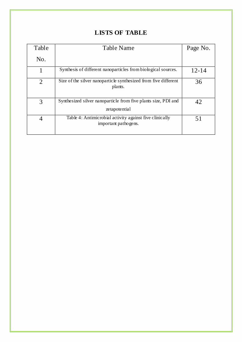
LISTS OF TABLE
Table
No.
Table Name Page No.
1 Synthesis of different nanoparticles from biological sources. 12-14
2 Size of the silver nanoparticle synthesized from five different
plants.
36
3 Synthesized silver nanoparticle from five plants size, PDI and
zetapotential
42
4 Table 4: Antimicrobial activity against five clinically
important pathogens.
51
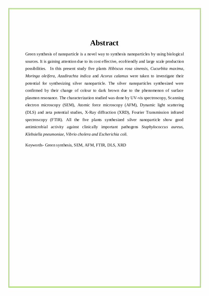
Abstract
Green synthesis of nanoparticle is a novel way to synthesis nanoparticles by using biological
sources. It is gaining attention due to its cost effective, ecofriendly and large scale production
possibilities. In this present study five plants Hibiscus rosa sinensis, Cucurbita maxima,
Moringa oleifera, Azadirachta indica and Acorus calamus were taken to investigate their
potential for synthesizing silver nanoparticle. The silver nanoparticles synthesized were
confirmed by their change of colour to dark brown due to the phenomenon of surface
plasmon resonance. The characterization studied was done by UV-vis spectroscopy, Scanning
electron microscopy (SEM), Atomic force microscopy (AFM), Dynamic light scattering
(DLS) and zeta potential studies, X-Ray diffraction (XRD), Fourier Transmission infrared
spectroscopy (FTIR). All the five plants synthesized silver nanoparticle show good
antimicrobial activity against clinically important pathogens Staphylococcus aureus,
Klebsiella pneumoniae, Vibrio cholera and Escherichia coli.
Keywords- Green synthesis, SEM, AFM, FTIR, DLS, XRD
1
1. INTRODUCTION
In recent days nanotechnology has induced great scientific advancement in the field of
research and technology. Nanotechnology is the study and application of small object which
can be used across all fields such as chemistry, biology, physics, material science and
engineering. Nanoparticle is a core particle which performs as a whole unit in terms of
transport and property (Nour et al., 2010). As the name indicates nano means a billionth or
10-9 unit. Its size range usually from 1-100nm (Nour et al., 2010) due to small size it occupies
a position in various fields of nano science and nanotechnology. Nano size particles are quite
unique in nature because nano size increase surface to volume ratio and also its physical,
chemical and biological properties are different from bulk material. So the main aim to study
its minute size is to trigger chemical activity with distinct crystallography that increases the
surface area (Osaka et al., 2006, Singh et al., 2008 & Sinha et al., 2009). Thus in recent years
much research is going on metallic nanoparticle and its properties like catalyst, sensing to
optics, antibacterial activity, data storage capacity (Nour et al., 2010 & Sharma et al., 2009).
The concept of nanotechnology emerged on 9th century. For the first time in 1959, Richard
Feynman gave a talk on the concept of nanotechnology and described about molecular
machines built with atomic precision where he discussed about nanoparticles and entitled that
“There’s plenty of space at the bottom” (T.C et al., 1974). Professor Peter Paul Speiser and
his research group were first to investigate on polyacrylic beads for oral administration and
target on microcapsule. In the year of 1960 nanoparticle develop for drug delivery and also
vaccine purpose which change the medicinal scenario. The first paper published in1980 by K.
Eric Drexler of Space Systems Laboratory Massachusetts Institute of Technology was titled
as “An approach to the development of general capabilities for molecular manipulation”.
The term “nanotechnology” first time used as scientific field by Nario Tanigushi in the 1974
his paper was “Nanotechnology” mainly consists of the processing of, separation,
consolidation, and deformation of materials by one atom or one molecule (Zhang and
Webster, 2008).
Nanotechnology is a fast growing area in the field on science which is a interdisciplinary
field of both science and technology that increase the scope of investing and regulating at cell
level between synthetic material and biological system (Du.et al., 2006 & Sinha et al.,2009).
Nanotechnology proceeds by three processes - separation, consolidation, deformation of
2
material by one atom or molecule (Taniguchi 1974). It is divided into three types- Wet
nanotechnology which deals with the biological system such as enzymes, membrane,
cellular components. Dry nanotechnology deals with the surface science, physical chemistry
& gives importance on fabrication of structure in carbon, silicon, inorganic materials.
Computational nanotechnology which deals with modelling & stimulating the complex
nanometre scale structure (Sinha et al., 2009), these three fields are interdependent to each
other. There are two methods of synthesis of metallic nanoparticles which are chemical
method and physical method. In chemical approach it include chemical reduction(Guzman et
al., 2009), electrochemical technique(Rodriguez-Sanchez et al.2000), photochemical
reduction (Balan et al., & Sharma et al., 2009).The chemical process is again subdivided into
classical chemical method where some chemical reducing agent (such as hydrazine, sodium
borohydride, hydrogen)are used, radiation chemical method generated by ionization
radiation ( Leff et al., 1995; Lisiecki and Pileni, 1995; Huang et al.,1997;Gutierrez and
Henglein, 1993; Nour et al.2010). In the physical approach it includes condensation (Raffi et
al., 2007), evaporation (Mitrakos et al.,2008) and laser ablation for metal nanoparticle
synthesis (Zamiri et al.,2012). The biological synthesis of nanoparticle is a challenging
concept which is very well known as green synthesis. The biological synthesis of nano
material can solve the environmental challenges like solar energy conservation, agricultural
production ,catalysis (Kumar et al., 2011), electronic, optics (Evanoff et al, .2005), and
biotechnological area (Soloviev and Mikhail 2007). Green synthesis of nanoparticle are cost
effective, easily available, eco friendly, nontoxic, large scale production and act as reducing
and capping agent(T.C et al., 2011)in compared to the chemical method which is a very
costly as well as it emits hazardous by-product which can have some deleterious effect on the
environment (Kaler et al., 2010). Biological synthesis utilizes naturally occupying reducing
agent such as plant extract, microorganism, enzyme, polysaccharide which are simple and
viable which is the alternative to the complex and toxic chemical processes (Du L. et al.,
2009). Plants can be described as nano factories which provide potential pathway to
bioaccumulation into food chain and environment. Among the different biological agents
plants provide safe and beneficial way to the synthesis of metallic nanoparticle as it is easily
available so there is possibilities for large scale production apart from this the synthesis route
is eco-friendly, the rate of production is faster in comparison to other biological models such
as bacteria, algae and fungi (Nour et al., 2010). From the various literature studies it can be
stated that the amount of accumulation of nanoparticle varies with reduction potential of ions
3
and the reducing capacity of plant depends on the presence of various polyphenols and other
heterocyclic compounds (Nair et al., 2010)
Nanoparticle of gold, silver, copper, silicon, zinc, titanium, magnetite, palladium formation
by plants has been reported. Colloid silver nanoparticle had exhibited distinct properties such
as catalytic, antibacterial (Sharma et al., 2009), good conductivity, and chemical stability.
Silver nanoparticles have its application in the field of bio labelling, sensor, antimicrobial,
catalysis, electronic and other medical application such as drug delivery (Jong et al., 2008)
and disease diagnosis.
4
2. REVIEW OF LITERATURE
Nanotechnology is a brainchild of modern fundamental science. It is a very complicated
professional area, uniting the efforts of professionally qualified chemists, physicists,
mathematicians, materials scientists, physicians, computer scientists, and so on. At the
present stage nanoparticle research is an intense scientific research due to its wide potential
application in biomedical, optical & electronic fields. Nanoparticles are a narrow bridge in
between bulk materials and molecular (atomic) structures. Bulk materials have constant
physical properties because they have grain structures with random grains individually
oriented in space and contacting each other across grain boundaries but nanomaterials are
made up of a single grain with all the atoms oriented in a crystalline lattice. (Sharma et al.,
2009) The main characteristics of nanomaterials that distinguish them from b ulk materials are
(1) large fraction of surface atoms; (2) high surface energy; (3) spatial confinement; (4)
reduced numbers of imperfections that do not exist in the corresponding bulk materials (Cao
2004).Nanoparticles show different properties such as quantum confinement, Surface Plasma
Resonance (SRP), decrease in melting temperature which are directly related to the crystal
lattice of the nanomaterials. The use of Nanomaterials provide the following advantages,
Firstly, as nanomaterials consist of very small particles they, promote accomplishment of
super miniaturization and thus the nanostructures can be packed very closely together which
can be useful for nanoelectronics. Secondly, because of their small dimensions, nanomaterials
have large specific surface areas which increase the interactions between them and the
environment in which they are located.
Nanoparticles can be broadly classified into two groups: Organic nanoparticles and Inorganic
nanoparticles. Organic nanoparticle are carbon nanoparticle (fullerenes) and inorganic
nanoparticles are magnetic nanoparticle, noble nanoparticle (gold and silver), semiconductor
nanoparticle (titanium oxide and zinc oxide). Especially inorganic nanoparticles have created
attention towards itself due to its superior material properties with versatile functions. Due to
nano size feature it easily used for chemical imaging drugs agents and drug. Its versatile
function used for the cellular delivery as they are widely available, rich functionality, good
biocompatibility. This is also a good carrier of targeted drug delivery and controlled drug
release (Xu et al., 2006). it is a completely advantageous material foe medical science For
example mesoporous silica combined with molecular medicines shows a excellent image o n
5
drug releasing. Gold nanoparticle is good carrier in thermo therapy of biological target
(Cheon & Horace, 2009). Silver nanoparticle shows antimicrobial activity which heals the
wounds and infectious disease (Ravishankar and Jamuna, 2011).Synthesis of nanoparticle
gets concern in nanotechnology due to the variable size, shapes, chemical composition &
controlled dispersity and their potential use in the medical science for the better treatment of
human benefits.
Traditionally, researchers generally used two methods for the synthesis nanoparticles such as
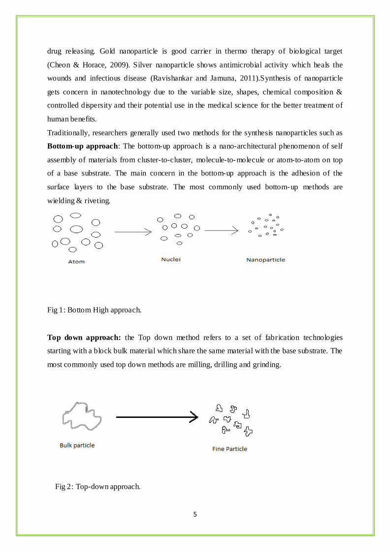
Bottom-up approach: The bottom-up approach is a nano-architectural phenomenon of self
assembly of materials from cluster-to-cluster, molecule-to-molecule or atom-to-atom on top
of a base substrate. The main concern in the bottom-up approach is the adhesion of the
surface layers to the base substrate. The most commonly used bottom-up methods are
wielding & riveting.
Fig 1: Bottom High approach.
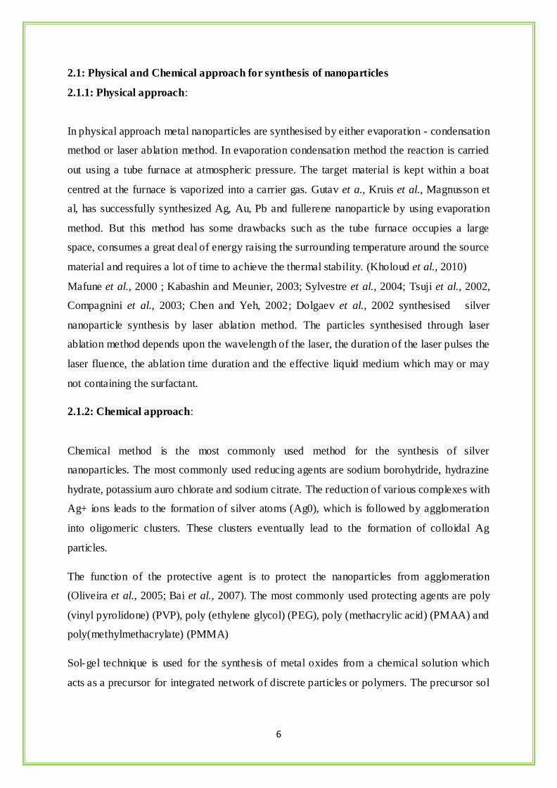
Top down approach: the Top down method refers to a set of fabrication technologies
starting with a block bulk material which share the same material with the base substrate. The
most commonly used top down methods are milling, drilling and grinding.
Fig 2: Top-down approach.
6
2.1: Physical and Chemical approach for synthesis of nanoparticles
2.1.1: Physical approach:
In physical approach metal nanoparticles are synthesised by either evaporation - condensation
method or laser ablation method. In evaporation condensation method the reaction is carried
out using a tube furnace at atmospheric pressure. The target material is kept within a boat
centred at the furnace is vaporized into a carrier gas. Gutav et a., Kruis et al., Magnusson et
al, has successfully synthesized Ag, Au, Pb and fullerene nanoparticle by using evaporation
method. But this method has some drawbacks such as the tube furnace occupies a large
space, consumes a great deal of energy raising the surrounding temperature around the source
material and requires a lot of time to achieve the thermal stability. (Kholoud et al., 2010)
Mafune et al., 2000 ; Kabashin and Meunier, 2003; Sylvestre et al., 2004; Tsuji et al., 2002,
Compagnini et al., 2003; Chen and Yeh, 2002; Dolgaev et al., 2002 synthesised silver
nanoparticle synthesis by laser ablation method. The particles synthesised through laser
ablation method depends upon the wavelength of the laser, the duration of the laser pulses the
laser fluence, the ablation time duration and the effective liquid medium which may or may
not containing the surfactant.
2.1.2: Chemical approach:
Chemical method is the most commonly used method for the synthesis of silver
nanoparticles. The most commonly used reducing agents are sodium borohydride, hydrazine
hydrate, potassium auro chlorate and sodium citrate. The reduction of various complexes with
Ag+ ions leads to the formation of silver atoms (Ag0), which is followed by agglomeration
into oligomeric clusters. These clusters eventually lead to the formation of colloidal Ag
particles.
The function of the protective agent is to protect the nanoparticles from agglomeration
(Oliveira et al., 2005; Bai et al., 2007). The most commonly used protecting agents are poly
(vinyl pyrolidone) (PVP), poly (ethylene glycol) (PEG), poly (methacrylic acid) (PMAA) and
poly(methylmethacrylate) (PMMA)
Sol-gel technique is used for the synthesis of metal oxides from a chemical solution which
acts as a precursor for integrated network of discrete particles or polymers. The precursor sol

7
can be either deposited on the substrate to form a film cast into an appropriate container
having desired shape or can be used to synthesize powders.
Solvothermal synthesis is a flexible low temperature route in which polar solvents under
pressure and at temperatures above their boiling points are used. The reaction of the reagents
under the solvothermal conditions increases significantly and enabling the reaction to take
place at lower temperature.
Fig 3: Different methods of nanoparticle synthesis.
2.2: Biosynthesis of nanoparticles:
Although chemical & physical methods are very successful to produce well- defined
nanoparticles, they have certain limitations such as increase cost of production, release of
hazardous by-products, long time for synthesis and difficulty in purification (Nagajyothi and
Lee, 2011). Global warming & climate change has induced a worldwide awareness to reduce
the toxic & hazardous waste materials, thus, the green synthesis route have raised actively the
progress in the fields of science & industry (Ahmad et al., 2011). Biosynthesis of
nanoparticles as the name indicates help in the synthesis of very complex reaction within a
fraction of minutes have now taken up the attention towards synthesis grievance the need of
environmentally benign technologies in material science (Harekrishna et al., 2009). Use of
Nanoparticle
Synthesis methods
Physical method
met
Solvothermal
method
Irradiation Method
Sol-gel method
Chemical method
8
biological organisms such as microorganism, plant extracts and biomass could be a best
alternative method of physical and chemical method for synthesis of nanoparticles because
the biological or green synthesis route is very spontaneous, economic, environmental friendly
and non-toxic. Therefore, biological sources such as bacteria, fungi, yeasts, algae and plants
can materials catalyzed specific reaction as a part of modern & rea listic biosynthetic strategy.
The current prospective on biological system has created a commercial importance due to
their enzymatic reactions, photochemical characteristics and herbal nature. Biological system
has created a specific and revolutionary change for the synthesis of nanoparticles due to their
mode of mechanism trough which the bio reduction of the metallic salts occurs is still a
mystery. Numerous researches have been done on the synthesis of nanoparticles from
biological system of for their application in the field of biomedical, pharmaceutical, cosmetic
and environmental use. Bio fabricated nanoparticles can be used for bioremediation purpose
because nanoparticles can diffuse or penetrate through the contaminants and cause a redox
reaction to clean the surface materials. Nature has some processed device to synthesis of nano
and micro sized materials which contribute to the development of relatively new and
unexplored area of research based on the biosynthesis of nanomaterials.
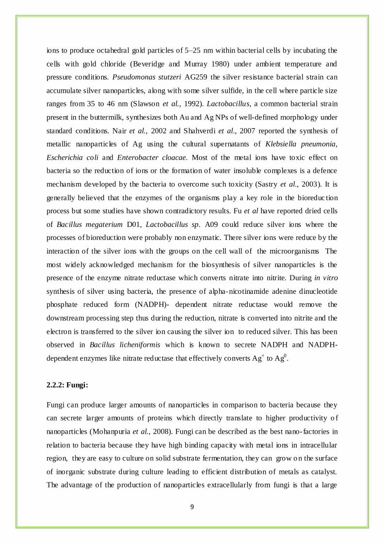
Fig 4: Different routes for biosynthesis of nanoparticles.
2.2.1: Bacteria:
Many microorganisms can synthesise inorganic nanoparticles like silver, gold, magnesium,
cadmium sulphide and silicon oxide nanoparticles. The resistance caused by the bacterial cell
for silver ions in the environment is responsible for its nanoparticles synthesis (Saklani et al.,
2012). It has been reported earlier that Bacillus subtilis 168 has the ability to reduce Au3+
Plants
Yeast
Bacteria
Fungus
Actinomycetes Algae
NANOPARTICLE
9
ions to produce octahedral gold particles of 5–25 nm within bacterial cells by incubating the
cells with gold chloride (Beveridge and Murray 1980) under ambient temperature and
pressure conditions. Pseudomonas stutzeri AG259 the silver resistance bacterial strain can
accumulate silver nanoparticles, along with some silver sulfide, in the cell where particle size
ranges from 35 to 46 nm (Slawson et al., 1992). Lactobacillus, a common bacterial strain
present in the buttermilk, synthesizes both Au and Ag NPs of well-defined morphology under
standard conditions. Nair et al., 2002 and Shahverdi et al., 2007 reported the synthesis of
metallic nanoparticles of Ag using the cultural supernatants of Klebsiella pneumonia,
Escherichia coli and Enterobacter cloacae. Most of the metal ions have toxic effect on
bacteria so the reduction of ions or the formation of water insoluble complexes is a defence
mechanism developed by the bacteria to overcome such toxicity (Sastry et al., 2003). It is
generally believed that the enzymes of the organisms play a key role in the bioreduc tion
process but some studies have shown contradictory results. Fu et al have reported dried cells
of Bacillus megaterium D01, Lactobacillus sp. A09 could reduce silver ions where the
processes of bioreduction were probably non enzymatic. There silver ions were reduce by the
interaction of the silver ions with the groups on the cell wall of the microorganisms The
most widely acknowledged mechanism for the biosynthesis of silver nanoparticles is the
presence of the enzyme nitrate reductase which converts nitrate into nitrite. During in vitro
synthesis of silver using bacteria, the presence of alpha-nicotinamide adenine dinucleotide
phosphate reduced form (NADPH)- dependent nitrate reductase would remove the
downstream processing step thus during the reduction, nitrate is converted into nitrite and the
electron is transferred to the silver ion causing the silver ion to reduced silver. This has been
observed in Bacillus licheniformis which is known to secrete NADPH and NADPH-
dependent enzymes like nitrate reductase that effectively converts Ag+ to Ag0.
2.2.2: Fungi:
Fungi can produce larger amounts of nanoparticles in comparison to bacteria because they
can secrete larger amounts of proteins which directly translate to higher productivity o f
nanoparticles (Mohanpuria et al., 2008). Fungi can be described as the best nano-factories in
relation to bacteria because they have high binding capacity with metal ions in intracellular
region, they are easy to culture on solid substrate fermentation, they can grow o n the surface
of inorganic substrate during culture leading to efficient distribution of metals as catalyst.
The advantage of the production of nanoparticles extracellularly from fungi is that a large
10
quantity of enzyme which are in pure state and free from cellular protein can be easy to apply
for the simple downstream process. Ahmad et al., 2003 reported the synthesis of fabrication
of extremely stable Ag hydrosol by using Fusarium oxysporum where the particles were
stabilized by the proteins excreted through the fungus. Bhainsa et al has reported the
extracellular biosynthesis of Ag particles in the 5-25 nm range using Aspergillus fumigates.
Vigneshwaran et al., 2006 reported the biomimetics of Ag nanoparticles by using
Phaenerochaete chrysosporium commonly known as White rot fungus. Basavaraja et al,
fabricated spherical and stable Ag nanoparticles in the range of 10-60 nm by using Fusarium
semitectum. Varshney et al., 2009 reported synthesis of Ag nanoparticles in the range of 20-
80 nm by using a novel fungi Hormoconis resinae.
The possible mechanism for the synthesis of silver nanoparticle by fungi is said to follow the
following steps: trapping of Ag+ ions at the surface of the fungal cells and the subsequent
reduction of the silver ions by the enzymes present in the fungal system (Mukherjee et al .,
2001 )
2.2.3: Actinomycetes:
Actinomycetes are microorganisms that share some of the important characteristics of fungi
and bacteria. Due to their ability to produce secondary metabolites such as antibiotics
actinomycetes are now getting focus for the synthesis of metallic nanoparticles. Sastry et al.,
2003 reported the synthesis of Au nanoparticles by using the extremophilic actinomycete,
Thermomonospora sp which yielded polydisperse Au nanoparticles. Ahmad et al reported the
intracellular synthesis of Au nanoparticles by using alkalotolerant Rhodococcus sp. They
observed that the concentration of nanoparticles were more on the cytoplasmic membrane
than on the cell wall. This could be due to reduction of the metal ions by enzymes present in
the cell wall and on the cytoplasmic membrane but not in the cytosol.
2.2.4: Algae:
Algae are a diverse group in the plant kingdom that are being explored for their application in
nanotechnology. Hosea et al reported the synthesis of Au nanoparticles on the alga Chlorella
vulgaris. Lengke et al reported the synthesis of Au nanoparticles having controlled shape by
using the blue-green algae Plectonema boryanum by treating them with aqueous Au (S2O3)23
and AuCl4 solutions. Singaravelu et al., 2007 reported the rapid formation of Au
nanoparticles through extracellular biosynthesis in marine alga Sargassum wightii. Scarano
11
and Morelli reported the fabrication of phytochelatin coated CdS nano crystals by using the
phytoplanktonic alga Phaeodactulum tricornatum. Konishi et al., 2007 reported the synthesis
of Pt nanoparticles of 5 nm from aqueous PtCl62- at neutral pH under room temperature by
using Shewanella algae.
2.2.5: Plants:
Various microorganisms such as bacteria, algae, fungi and yeasts are used for the
biosynthesis of nanoparticles but recently a new trend has come to force i.e., the use of plants
for the fabrication of nanoparticles because of its spontaneous, economical, eco-friendly
protocol, suitable for large scale production and single step technique for the biosynthesis
process (Huang et al., 2007). The main mechanism considered for the synthesis of
nanoparticles mediated by the plants is due to the presence of phytochemicals. The major
phytochemicals responsible for the spontaneous reduction of ions are flavonoids, terpenoids,
carboxylic acids, quinones, aldehydes, ketones and amides (Prabhu et al., 2012). A number
of plants are being currently investigated for their role in the synthesis of nanoparticles such
as Cinnamomum camphora leaf (Huang et al., 2007), Pelargonium graueolens leaf (Shankar
et al., 2003), Azadirachta indica leaf (Shankar et al., 2004), Emblica officinalis leaf
(Ankamwar et al., 2005), Aloe vera leaf (Chandran et al., 2006), Alfalfa sprouts (Gardea-
Torresdey et al., 2003), Helianthus annus, Basella alba, and Saccharum officinarum (Leela
et al., 2008), Carica papaya callus (Mude et al., 2009), Jatropha curcas leaf (Bar et al.,
2009), Eclipta leaf (Jha et al., 2009), Glycine max (soybean) leaf (Vivekanandan et al.,
2009), Coriandrum sativum leaf (Sathyavathi et al 2010), Syzygium cumini leaf (Kumar et al
2010), Cycas leaf (Jha et al., 2010), Argimone mexicana leaf (Khandelwal et al., 2010),
Allium cepa (Saxena et al., 2010), Stevia rebaudiana leaves (Varshney et a.,l 2010), Solanum
torvum (Govindaraju et al., 2010), Zingiber officinale (Singh et al., 2011), Capsicum annuum
(Li et al., 2007), Dillenia indica fruit (Singh et al., 2013), Alternanthera sessilis (Niraimathi
et al., 2013), Morinda citrifolia (Suman et al., 2013), Phytolacca decandra, Gelsemium
sempervirens, Hydrastis canadensis and Thuja occidentalis (Das et al., 2013) (Pinus
desiflora), Diopyros kaki, Ginko biloba, Magnolia kobus and Platanus orientalis (Song et al.,
2009), Ulva fasciata (Rajesh et al.,2012)
12
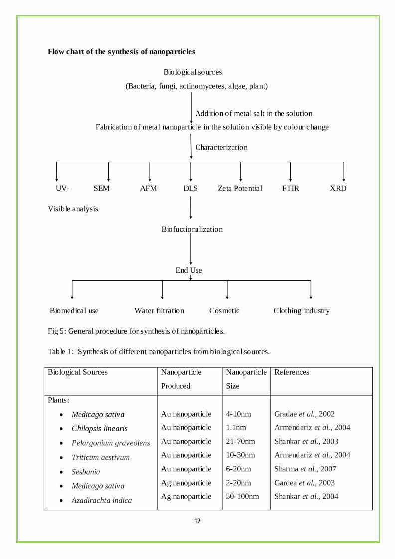
Flow chart of the synthesis of nanoparticles
Biological sources
(Bacteria, fungi, actinomycetes, algae, plant)
Addition of metal salt in the solution
Fabrication of metal nanoparticle in the solution visible by colour change
Characterization
UV- SEM AFM DLS Zeta Potential FTIR XRD
Visible analysis
Biofuctionalization
End Use
Biomedical use Water filtration Cosmetic Clothing industry
Fig 5: General procedure for synthesis of nanoparticles.
Table 1: Synthesis of different nanoparticles from biological sources.
Biological Sources Nanoparticle
Produced
Nanoparticle
Size
References
Plants:
Medicago sativa
Chilopsis linearis
Pelargonium graveolens
Triticum aestivum
Sesbania
Medicago sativa
Azadirachta indica
Au nanoparticle
Au nanoparticle
Au nanoparticle
Au nanoparticle
Au nanoparticle
Ag nanoparticle
Ag nanoparticle
4-10nm
1.1nm
21-70nm
10-30nm
6-20nm
2-20nm
50-100nm
Gradae et al., 2002
Armendariz et al., 2004
Shankar et al., 2003
Armendariz et al., 2004
Sharma et al., 2007
Gardea et al., 2003
Shankar et al., 2004
13
Bacteria:
Bacillus subtilis
Pseudomonas
aeruginosa
Rhodopseudomonas
capsulate
Escherichia coli
Desulfovibrio
desulfuricans
Pseudomonas stutzeri
AG259
Klebsiella pneumonia
Bacillus licheniformis
Clostridium
thermoaceticum
Rhodopseudomonas
palustris
Desulfobacteriaceae
Ag nanoparticle
Au nanoparticle
Au nanoparticle
Au nanoparticle
Pd nanoparticle
Ag nanoparticle
Ag nanoparticle
Ag nanoparticle
CdS nanoparticle
CdS nanoparticle
ZnS nanoparticle
5-60nm
15-30nm
10-20nm
20-25nm
20-50nm
200nm
1-6nm
40nm
20-200nm
0-0.25nm
2-5nm
Saifuddin et al., 2009
Husseiny et al., 2007
He et al., 2008
Deplanche & Macaskie,2008
Deplanche et al., 2008
Joerger et al., 2000
Mokhtari et al., 2009
Kalishwaralal et al., 2008
Cunningham & Lundie,1993
Bai et al., 2009
Labrenz et al., 2000
Yeast:
Yeast MKY3
Candida glabrata
Schizo saccharomyces
Pombe
P. jadini
Ag
CdS
CdS
Au
2-5nm
20Å
1-1.5nm
Few to
100nm
Kowshik et al., 2003
Haverkamp et al., 2007
Kowshik et al., 2002
Konishi et al., 2006
Algae:
Diatoms6
Sargassum alga
SiO2
50-100nm
Singaravelu et al., 2007
Fungi:
Aspergillus fumigates
Ag
5-25nm
Armendariz et al., 2004
14
Fusarium semitectum
Verticillum sp.
Ag
Ag
20-25nm
20-25nm
Bhainsa et al., 2006
Mukherjee et al., 2001
Actinomycetes:
Thermospora sp
Rhodococcus sp
Au
Au
8nm
5-15nm
Ahmad et al., 2003
Gericke et al., 2006
2.3: Silver Nanoparticles:
Silver nanoparticles have attracted and demandable research of interest in the field of
nanotechnology, due to its distinct properties such as good conductivity, chemically stable,
catalytic activity, surface enhanced Raman scattering and antimicrobial activity (Li. Z et al.,
2006; Chen Y.Y et al.,2005; Setua P et al.,2007). Silver is widely used as catalyst for the
oxidation of methanol to formaldehyde and ethylene oxide. Due to colloidal nature it use as
substrate for surface enhanced spectroscopy, as it partly require electrical conducting surface.
In this era silver is use as antimicrobial agent. Recent focuses towards silver nanoparticle
synthesis for increasing the treat of antibiotic resistance, caused by the misuse of antibiotic.
(Panaek et al., 2006; Sandbhy et al., 2006). Several hypotheses have been found to the
antimicrobial activity of silver nanoparticle. Silver nanoparticle have capacity inactivates
bacterial enzyme by releasing ionic silver which inactivates the thiol groups. This silver ions
inhibits bacterial DNA replication, damage cell cytoplasm, depleting levels of adenosine
triphosphate (ATP) and finally death of cell (Feng et al., 2000). As the nanoparticle distinct
property surface to volume ratio silver nanoparticle increases surface to contact with bacterial
cell which promote silver ion to dissolve and improving the bacterialsidal effectiveness
(Stobie et al., 2008).Chemical reduction is most commonly used for synthesis of silver
nanoparticle. For the chemical reduction process some reductants are used such as
borohydride, citrate, ascorbate and elemental hydrogen. (Maribel et al., 2009). By the
reduction of silver ion in aqueous solution, a nanosized colloidal silver particle formed.
During chemical reaction various complexes with Ag+ ions leads to formation silver particle
(Ag0), followed by the agglomeration into oligomeric clusters, which leads to formation of
colloidal silver nanoparticle. The first observation of nanoparticle synthesis is colour change
of the aqueous solution, if wavelength of the solution is from 380-400nm then smaller
nanoparticle is formed. This band formation after nanoparticle synthesis is due to collective
15
oscillation or excitation of electron in the solution (i.e. surface plasmo n resonance) (Yamini
et al., 2011). Borohydride is a stronger reductant which synthesized smaller monodispersed
particle but difficult to controlled larger size. Use of citrate is a weaker reductant rate of
synthesis is slower and particle size is not so small. In the chemical reduction process a
stabiliser is most use that prevent agglomeration colloidal particle. Plants like biological
sources (bacteria, fungus, algae, yeast) are used for synthesis of silver nanoparticle by using
chemical sodium nitrate (R.Geethalakshmi et al., 2010). The green synthesis of silver
nanoparticle are mainly three steps are considered: se lection of solvent medium, selection of
biological source related reducing agent, selection of nontoxic stabilizing agents. Acalypha
indica leaves synthesized 20-30nm silver nanoparticle in chemical reduction method
(Krishnaraj et al., 2010).
2.4: Plant Samples:
2.4.1: Moringa oliefera:
Moringa oliefera is a commonly found plant, which is dispersed in many countries of the
tropics and subtropics. It’s all over parts (roots, seed, bark, leaves, fruit, and immature pods,
flowers) are highly nutritious with medicinal value. The whole plant contains a sketch of
important minerals, and proteins, vitamins various phenolic compound, amino acid and very
important β – carotene which is found in high amount (Farooq et. al., 2007). This plant
carries many phytochemical such as zeatin, kaempferom, quercetin with various
combinations. This plant has highly medicinal effect on curable diseases such as cardiac a nd
circulatory drugs, possess antitumor (Makonnen et. al., 1997), antipyretic, antioxidant,
antidiabetic, antiulcer, anti inflammatory, antiepileptic (Pal et. al., 1995). Other possible
medicinal propreties are antispasmodic (Caceres et. al., 1992), diuretic (Morton, 1991),
antihypertensive (Dahot, 1988), lowering of cholesterol level (Mehta et. al., 2003),
antioxidant, antidiabetic, (Ruckmani et. al., 1998), antibacterial and antifungal activities
(Nickon et. al., 2003). M.oleifera seed act as water purifier against Gram positive and Gram
negative bacterial cells in the powder form (Muyibi and Evison, 1995; Olsen, 1987; Broin et.
al., 2002; Kawo, 2007). Its seeds have also capacity to biodegrade to heavy metals (Sharma
et. al., 2006). Other components of M.olaeifera employed as anticancerous and hypo-tensive
are 4-(4'-O-acetyl-α-L-rhamnopyranosyloxy)benzyl isothiocy-anate , 4-(α-L-
rhamnopyranosyloxy)benzyl isothiocy-anate.( Abuye C et. al., 1999 ), niazimicin ( Akhtar
AH et. al., 1995), pterygospermin (Bell PC et. al., 1986), benzyl isothiocyanate (Anwar F et.
al., 2003), and 4-(α-L-rhamnopyranosyloxy) benzyl glucosinolate (Asres K, 1995).

16
2.4.2: Cucurbita maxima:
Cucurbita maxima are well known as pumpkin. It is an herbaceous plant having medicinal
value.It contains naturally active components with polysaccharides, fixed oils, para-
aminobenzoic acid, peptides, sterol, and proteins (Buchbauer G et. al., 1998). The
phytochemical found in fruits and leaves are polysaccharides, phenolic glycosides, and 11E-
octadecatrienoic acid speciall fruits contains carotenoid and γ -aminobutyric acid (Koike K
et. al., 2005; Murkovic M et. al., 2002). Hypoglycemic a novel chemical found in Cucurbita
maxima containing 8.48% sugar(polysaccharides) which reduce blood glucose and an
upsurge in plasma insulin and protected the diabetic nephropathy( Zhang YJ et. al., 2002; Ju
LY et. al., 2001).Blood glucose level, Hypoglycemic activity, serum total cholesterol level
and triglyceride induced in diabetic rabbits when applied with pumpkin powder (Zhang ZJ
1998)
2.4.3: Azachata indica:
Neem is a natural medicine since ancient time in Ayurved. All parts of neem plant (leaf,
seed, fruits, bark, and flower) are useful for medicinal value. It has many propreties as anti
bacterial, anti fungal, anti helmintic, anti diabetic contraceptic and sedative. Neem has effect
on degenerative diseases such as Diabetes, Arthritis, Rheumatism, Cancer and Chronic
Fatigue. It also shows effect on Tuberculosis, Bronchitis, Conjunctivitis, Allergies, Stress,
and Insomnia. The phytochemicals present in neem are alkaloids, quinines, resins, tannins,
flavanoids, fats, saponins, phenolic compounds, Proteins and carboxylic acids (Khan et al.,
2010).
2.4.4: Hibiscus rosa sinensis:
Hibiscus is an ornamental plant traditional used as for anti- inflammatory, demulcent,
aphrodisiac, refrigerant, anodyne, laxative, emollient (J. Anjaria, et. al., 2002). The
phytochemicals found in this are medicinally steroids, flavonoids, tannins, reducing sugar,
anthocyanin pigment, carotene, thiamine, riboflavin, niacin and ascorbic acid (kumar et al.,
2012). It has various activities like antitumor, antidiarrheal, antiestrogenic antispermatogenic,
androgenic, antiphologistic (Kholkute SD and Udupa KN 1976), antiimplantation (Murthy
DRK et. al., 1997), wound Healing anticonvulsant (Nayak et. al., 2007).
17
2.4.5: Acorus calamus:
A. calamus has an existence in history as a traditional medicine in India and generally is
known as sweet flag, sweet grass and sweet cane (Family: Acoraceae). Root and rhizome of
A. calamus used for medicine. It has beneficial role in improved learning performance or
increase memory power and anti-aging effect (Nishiyama et. al., 1994). This act as a anti-
stressor and also antioxidative capacity which in turn could be achieved by protection of
decreasing GSH and restoring free radical scavenger‘s enzymatic activity.
(Sundaramahalingam et. al., 2005). In Ayurveda it used on counter the side effects of all
hallucinogens. The rhizome alcoholic extract has sedative and analgesic properties and causes
depression in blood pressure and respiration rate and also used to treat intestinal coli, gastritis
and gastric ulcers (Desai et. al., 1984).
`
2.5: Characterization of Silver Nanoparticles:
The characterization study of silver nanoparticle was done by the examining size, shape and
quantity of particles. Number of technique is used for this purpose, including UV-visible
spectroscopy, Scanning Electron Microscopy (SEM), Fourior Transmission Infrared
Spectroscopy (FTIR), X-Ray Diffaction (XRD), and Dynamic Light Scattering (DLS).
2.5.1: UV-vis Spectroscopy:
Absorbance spectroscopy is used to determine the optical properties of a solution. A Light is
send through the sample solution and the amount of absorbed light is measured. When the
wavelength is varied and the absorbance is measured at each wavelength. The absorbance can
be used to measure the concentration of a solution by using Beer-Lamberts Law. The
examination of nanoparticles, the optical properties are much more complicated. For instance,
the measured absorbance spectrum does not necessarily show the actual absorbance but the
extinction of the light is both the absorbed and the scattered light from the particles. These
wave lengths arise due to the surface Plasmon resonance of the particle.
2.5.2: Scanning Electron Microscope:
Scanning electron microscope (SEM) analysis the employed to characterization of size, shape
& morphologies of formed nanoparticle SEM gives high-resolution images of the surface of a
sample is desired. The scanning electron microscope works as same principle as an optical
microscope, but it measures the electrons scattered from the sample rather than photon.

18
Because electrons can be accelerated by an electric potential, the wavelength can be made
shorter than the one of photons. This makes the SEM capable of magnifying images up to
200.000 times. At the same time it is possible to achieve high resolution pictures of the
surface, making the instrument very useful in determining the size dist ribution of
nanoparticles.
2.5.3: Atomic Force Microscope:
The AFM is an instrument capable of measuring the topography of a given sample. A
nanosized tip attached on a cantilever is traded over the sample and a 3D image of the sample
topography is generated on a computer. The advantage of the AFM over SEM is the ability to
make topographical measurements for detection and investigation of the size and shape of
silver nanoparticles in three dimensions. The AFM generally measure the height of silver
nanoparticle.
2.5.4: Dynamic Light Scattering:
The DLS technique uses light to determine the size of particles in a solution. Light at a given
frequency is sent through the solution from a laser. When the light interacts with the moving
particles in the solution and is scattered, the frequency of the light is also changed. This
change of light frequency is directly related to the size of the particles in the solution; the
smaller the particles, the greater the shift in the light frequency. This difference in the light
shift is used to determine the size of the particles in the solution. DLS is capable of measuring
particles in the size range from a few nanometers to a few micrometers. It is therefore
applicable for determining the size of silver nanoparticles.
2.5.5: Fourier Transmission infrared spectroscopy:
FTIR is a chemical analytical method which measures infrared intensity v/s wavelength or
wave number of light. It used to analysis of possible bio molecule and also bonding
interaction between themselves. IR spectroscopy detects the vibration characteristics of
chemical functional groups of the sample. When an infrared light interacts with matter,
chemical bonds will shows stretch, contract and bend form. These chemical functional group
tends to adsorb infrared radiation in a specific wave number range of the structure of the rest
of the molecule. The silver nanoparticle synthesis, FTIR data measures interaction between

19
Ag salts and proteins molecules, which accurate for the reduction of silver ions and
stabilization of Ag NPS formed.
2.5.6: X-Ray Diffraction:
XRD is a technique to used go study phase composition of a sample, crystal structure, texture
or orientation. The principle of XRD is that the X-rays are passed through a material and the
pattern produced give information of size and shape of the unit cell. The atoms are crystal in
structure arranged in a periodic array and thus can diffracted light at different angle. When X-
ray passing through a crystal it produces a diffraction pattern, that diffract ion gives the
information about the atomic arrangement within the crystals. In silver nanoparticle XRD
gives phase structure and purity of the particle.
20
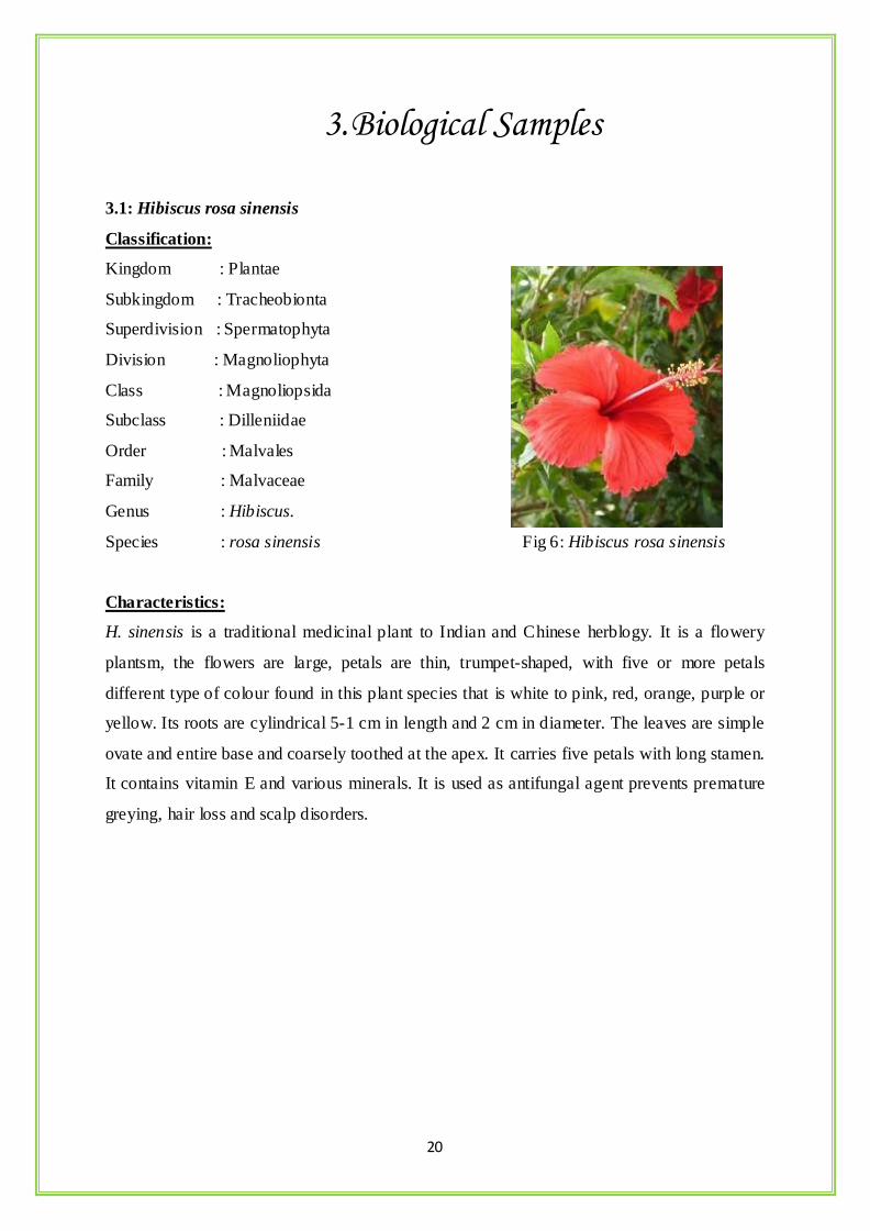
3. Biological Samples
3.1: Hibiscus rosa sinensis
Classification:
Kingdom : Plantae
Subkingdom : Tracheobionta
Superdivision : Spermatophyta
Division : Magnoliophyta
Class : Magnoliopsida
Subclass : Dilleniidae
Order : Malvales
Family : Malvaceae
Genus : Hibiscus.
Species : rosa sinensis Fig 6: Hibiscus rosa sinensis
Characteristics:
H. sinensis is a traditional medicinal plant to Indian and Chinese herblogy. It is a flowery
plantsm, the flowers are large, petals are thin, trumpet-shaped, with five or more petals
different type of colour found in this plant species that is white to pink, red, orange, purple or
yellow. Its roots are cylindrical 5-1 cm in length and 2 cm in diameter. The leaves are simple
ovate and entire base and coarsely toothed at the apex. It carries five petals with long stamen.
It contains vitamin E and various minerals. It is used as antifungal agent prevents premature
greying, hair loss and scalp disorders.
21
3.2: Cucurbita maxima
Classification:
Kingdom : Plantae
Subkingdom : Tracheobionta
Superdivision : Spermatophyta
Division : Magnoliophyta
Class : Magnoliopsida
Subclass : Dilleniidae
Order : Violales
Family : Cucurbitaceae Fig7: Cucurbita maxima
Genus : Cucurbita
Species : maxima
Characteristics:
They are large low creeping vine having very large leaves those are palmate with a map le
shape having small sharp serrations along the margin; flowers are very large bright yellow
with messy edge; fruit is a large almost round in shape. Pumpkin seeds are also useful, it
contain sterols, vitamin E, fatty acids and non-protein amino acids. It use as pharmaceutically
treating disease like rheumatism, bladder disorders, wounds, benign prostatic hyperplasia,
and certain female reproductive complaints. Pumpkin seeds also possess vitamin B, and many
essential minerals such as iron, zinc, and they are very nutritious and stimulating. Other
nutrients found in Cucurbita seeds are magnesium, phosphorus, niacin, folic acid, riboflavin,
thiamine and antioxidants. Zinc helps the healing process generally useful in treating the
enlarged prostate gland and pantothenic acid helps to be in good health.
22
3.3:Moringa-oliefera
Classification:
Kingdom :Plantae
Subkingdom : Tracheobionta
Superdivision : Spermatophyta
Division : Magnoliophyta
Class : Magnoliopsida
Subclass : Dilleniidae
Order : Capparales
Family : Moringaceae
Genus : Moringa Fig 8: Moringa oleifera
Species : oleifera
Characteristics:
Moringa oleifera is a tall, thin, fast growing evergreen tree. The tree grows to maximum 8 m
high and diameter about 60 cm dbh. The bark is thick, soft, corky and dark grey colour,
leaves are alternate (the old ones soon falling off; each leaf large), with opposite pinnae,
spaced about 5 cm apart up the central stalk, the leaflets are elliptic, dark green in colour and
pale on the under surface; variable in size, but often rounded-elliptic, seldom as much as 2.5
cm long. The flowers are generally white and fragrant in large panicles, and produced
throughout the year. In Indian tradition M.oleifera used for treatment of venomous bites,
ascites and rheumatism, helps in lowering blood pressure. Its roots and flower have properties
treatment of Cholera and also its root and bark of young tree are rubefacient, stomachic
carminative, vescicant and abortifacient. The leaves contain strong antibacterial and
antimalarial properties.
23
3.4: Azadirachata indica
Classification:
Kingdom : Plantae
Subkingdom : Tracheobionta
Superdivision : Spermmatophyta
Division : Magnoliophyta
Class : Magnoliopsida
Subclass : Rosidae
Order : Sapindales
Family : Meliaceae
Genus : Azadirachata
Species : indica Fig 9: Azadirachata indica
Characteristics:
A. indica is a highly medicinal herbal plant. Its all parts are effect against antimicrobial
activity. It is a fast growing plant with height reach of 15–20 metres (49–66 ft), rarely to 35–
40 metres (115–130 ft). It is a evergreen which branches are wide spread. The leaves are
pinnate and opposite with length 20–40 centimetres (7.9–16 in) long, dark green colour
leaflets about 3–8 centimetres (1.2–3.1 in) long. The flowers are axillary, more-or-less
drooping panicles which are up to 25 centimetres (9.8 in) long. The fruits are smooth,
globular, which varies in shape from elongate oval to nearly roundish. The neem products are
believed as antifungal, antibacterial, antiviral, ant diabetic, sedative. In Ayurveda neem is a
herbal component.
24
3.5: Acorus calamus
Classification:
Kingdom : Plantae
Subkingdom : Tracheobionta
Superdivision : Spermatophyta
Division : Magnoliophyta
Class : Liliopsida
Subclass : Arecidae
Order : Arales
Family : Acoraceae
Genus : Acorus Fig 10: Acorus calamus (rhizome)
Species : calamus
Characteristics:
Acorus is a well known sweet flag. Their leaves are prominent leaf veins with a single
prominent midvein and then on both sides slightly raised secondary veins. Its leaves size
range 0.7 and 1.7 cm wide, with average of 1 cm. The flower of this plants are too longer
with length between 3 and 4 mm with an an abortive ovary with a shrivelled appearance. It
grows in clay soils with slightly acidic or alkaline nature and it also grow in water condition.
The root is excellence for emmenagogue, aphrodisiac, stimulant, carminative, diaphoretic,
hypotensive, expectorant, and febrifuge, aromatic, hallucinogenic, analgesic, sedative,
stomachic and vermifuge. Their tonic is a excellent tonic for powers of stimulating and
stabilizing the appetite and also treatment for digestive disorders, bronchitis, sinusitis etc. It
has a peculiar quality that chewing its root kills the taste for tobacco.
25
4. OBJECTIVES
4.1: Preparation of plant extract from extracts (Hibiscus rosa sinensis, Moringa oleifera,
Acorus calamus, Cucurbita maxima, Azadirachta indica)
4.2: Synthesis of silver nanoparticles from five plant extracts.
4.3: Characterization of silver nanoparticles by UV-vis spectroscopy, Scanning electron
microscopy, Atomic force microscopy, Dynamic light scattering and zeta potential studies,
X-Ray diffraction, Fourier Transmission infrared spectroscopy
4.4: Antimicrobial activity Study against different five clinical pathogens Bacterial samples-
staphylococcus aureus, Pseudomonas aeruginosa, Klebsiella pneumonia, Vibrio cholera and
Escherichia coli
26
5. MATERIALS AND METHODS
5.1: Material Required:
5.1.: Plant Samples:
5 Plant Samples
o Moringa oleifera leaves
o Cucurbita maxima petal
o Azadirachta indica leaves
o Hibiscus rosa sinensis petal
o Acorus calamus rhizome
5.2: Chemical regent required:
Silver nitrate (AgNO3)
Potassium Bromide (KBr)
Nutrient agar
Nutrient broth
5.3: Instrument required:
UV-visible light Spectroscopy (PerkinElmer spectrophotometer)
SEM (JEOL Jsm-6480 LV)
FTIR (Shimadzu)
XRD (x’pert pananalytical )
DLS (Malvern, UK)
AFM (Veeco)
5.4: Methods:
5.4: Preparation of plant extract:
Healthy plant samples were collected from the locality of Rourkela and were cleaned
properly in running tap water.
The samples were shade dried and homogenised to fine powder using a mortar and
pestle.
27
The solution was then kept at room temperature to cool down.
The plant extract was then filtered out.
5.5: Synthesis of Silver Nanoparticle:
The plant extract was then mixed properly.
The extract solution was then heated.
The extract solution was then subjected to centrifugation.
The pellets obtained were washed.
The pellet obtained was then lyophilized.
5.6: Characterization Techniques:
5.6.1: UV-vis spectra analysis:
The silver nanoparticles were confirmed by measuring the wave length of reaction mixture in
the UV-vis spectrum of the PerkinElmer spectrophotometer at a resolution of 1 nm (from 300
to 600 nm) in 2 ml quartz cuvette with 1 cm path length.
5.6.2: SEM analysis:
The Morphological characterization of the samples was done using JEOL Jsm-6480 LV for
SEM analysis. The samples were dispersed on a slide and then coated with platinum in an
auto finecoater. After that the material was subjected to analysis.
5.6.3: AFM analysis:
The surface morphology of the nanoparticles were visualised by Atomic force microscope
(Veeco) under normal atmospheric conditions. The examined samples were dispersed on
small slide and explored on contact mode of the instrument.
5.6.4: DLS Particle size and zeta potential analysis:
The size distribution or average size of the synthesized AgNPs were determined by dynamic
light scattering (DLS) and zeta potential measurements were carried out using DLS
(Malvern, UK). For DLS analysis the samples were diluted 10 folds using 0.15M PBS (pH
7.4) and the measurements were taken in the range between 0.1 and 10,000 nm.
28
5.6.5: FT-IR analysis:
The characterization of functional groups on the surface of AgNPs by plant extracts were
investigated by FTIR analysis (Shimadzu) and the spectra was scanned in the range of 4000–
400 cm−1 range at a resolution of 4 cm−1. The sample were prepared by dispersing the AgNPs
uniformly in a matrix of dry KBr , compressed to form an almost transparent disc. KBr was
used as a standard analyse the samples.
5.6.6: XRD analysis:
XRD measurements of the reduced AgNPs perform were recorded on X-ray diffractometer
(x’pert pananalytical ) instrument operating at a voltage of 40 kV and current of 30 mA with
Cu K (α) radiation to determine the crystalline phase and material identification. The samples
were taken in lids and put under instrument for analysis.
5.6.7: Anti microbial activity:
The clinical pathogenic strains of Escherichia coli, Pseudomonas, VIbrio Cholerae,
Staphylococcus aureus and Klebsiella pneumonia were used to determine the antibacterial
activity of the silver nanoparticles by following the method according to Sondi et al. 1ml of
suspension of approximately 10 5 CFU/ml density of the microorganisms to be tested were
distributed uniformly on agar surface plate and incubated at 28°C (CFU = colony forming
units).Silver-free agar plates cultured under the same conditions were used as a control. The
plates were incubated under 24 hrs at 3 c and the numbers of colonies were counted after 24
hrs. The counts on the 10 plates corresponding to a particular sample were averaged. The
average values were expressed as Mean± Standard deviations.
29
6. Results and Discussion
6.1: Visible Observation:
(a) (b)
(c)
30
(d) (e)
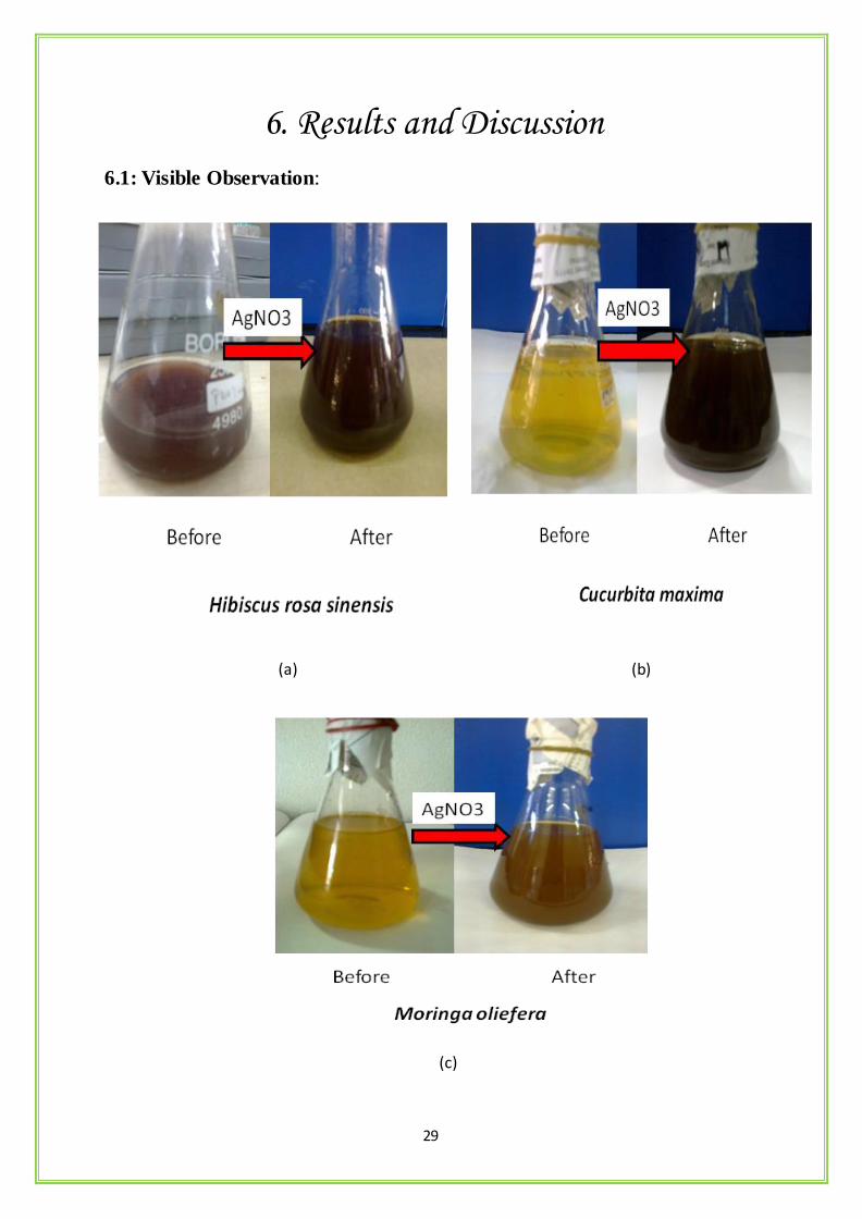
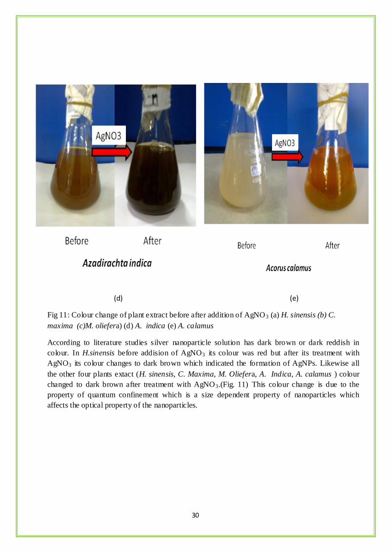
Fig 11: Colour change of plant extract before after addition of AgNO3 (a) H. sinensis (b) C.
maxima (c)M. oliefera) (d) A. indica (e) A. calamus
According to literature studies silver nanoparticle solution has dark brown or dark reddish in
colour. In H.sinensis before addision of AgNO3 its colour was red but after its treatment with
AgNO3 its colour changes to dark brown which indicated the formation of AgNPs. Likewise all
the other four plants extact (H. sinensis, C. Maxima, M. Oliefera, A. Indica, A. calamus ) colour
changed to dark brown after treatment with AgNO3.(Fig. 11) This colour change is due to the
property of quantum confinement which is a size dependent property of nanoparticles which
affects the optical property of the nanoparticles.
31
6.2: UV-vis spectroscopy:
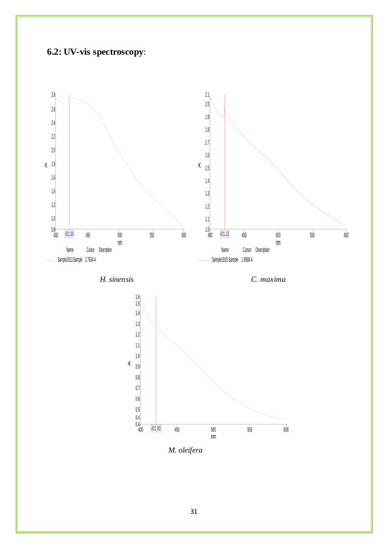
H. sinensis C. maxima
M. oleifera
Sample1915.Sample
Name
1.9568 A
Cursor Description
400 600450 500 550
2.1
1.0
1.1
1.2
1.3
1.4
1.5
1.6
1.7
1.8
1.9
2.0
nm
A
421.32
Sample1913.Sample
Name
2.7634 A
Cursor Description
400 600450 500 550
2.8
0.8
1.0
1.2
1.4
1.6
1.8
2.0
2.2
2.4
2.6
nm
A
421.00
Sample1916.Sample
Name
1.281 A
Cursor Description
400 600450 500 550
1.6
0.40.4
0.5
0.6
0.7
0.8
0.9
1.0
1.1
1.2
1.3
1.4
1.5
nm
A
421.00
32
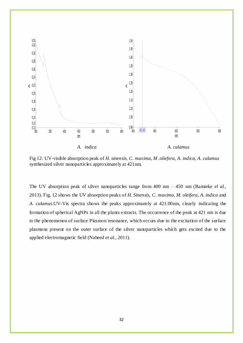
A. indica A. calamus
Fig 12: UV-visible absorption peak of H. sinensis, C. maxima, M .oliefera, A. indica, A. calamus
synthesized silver nanoparticles approximately at 421nm.
The UV absorption peak of silver nanoparticles range from 400 nm – 450 nm (Ramteke el al.,
2013). Fig. 12 shows the UV absorption peaks of H. Sinensis, C. maxima, M. oleifera, A. indica and
A. calamus.UV-Vis spectra shows the peaks approximately at 421.00nm, clearly indicating the
formation of spherical AgNPs in all the plants extracts. The occurrence of the peak at 421 nm is due
to the phenomenon of surface Plasmon resonance, which occurs due to the excitation of the surface
plasmons present on the outer surface of the silver nanoparticles which gets excited due to the
applied electromagnetic field (Naheed et al., 2011).
Sample1865.Sample
Name Description
300 600350 400 450 500 550
0.33
0.110.12
0.14
0.16
0.18
0.20
0.22
0.24
0.26
0.28
0.30
0.32
nm
A
Sample1902.Sample
Name
1.4092 A
Cursor Description
400 600450 500 550
1.50
0.99
1.05
1.10
1.15
1.20
1.25
1.30
1.35
1.40
1.45
nm
A
421.00
33
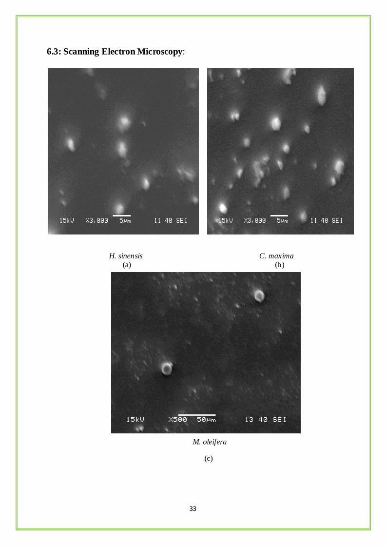
6.3: Scanning Electron Microscopy:
H. sinensis C. maxima (a) (b)
M. oleifera
(c)

34
A. indica A. calamus
(d) (e)
Fig 13: SEM image of (a) H. sinensis, (b) C. maxima, (c) M. oliefera (d) A. indica, (e) A. calamus
A scanning electron microscope was employed to analyze the shape of the silver
nanoparticles that were synthesised by green method. SEM analysis shows that the five plants
have tremendous capability to synthesize silver nanoparticles which were roughly spherical
in shape (fig.13) and were uniformly distributed.

35
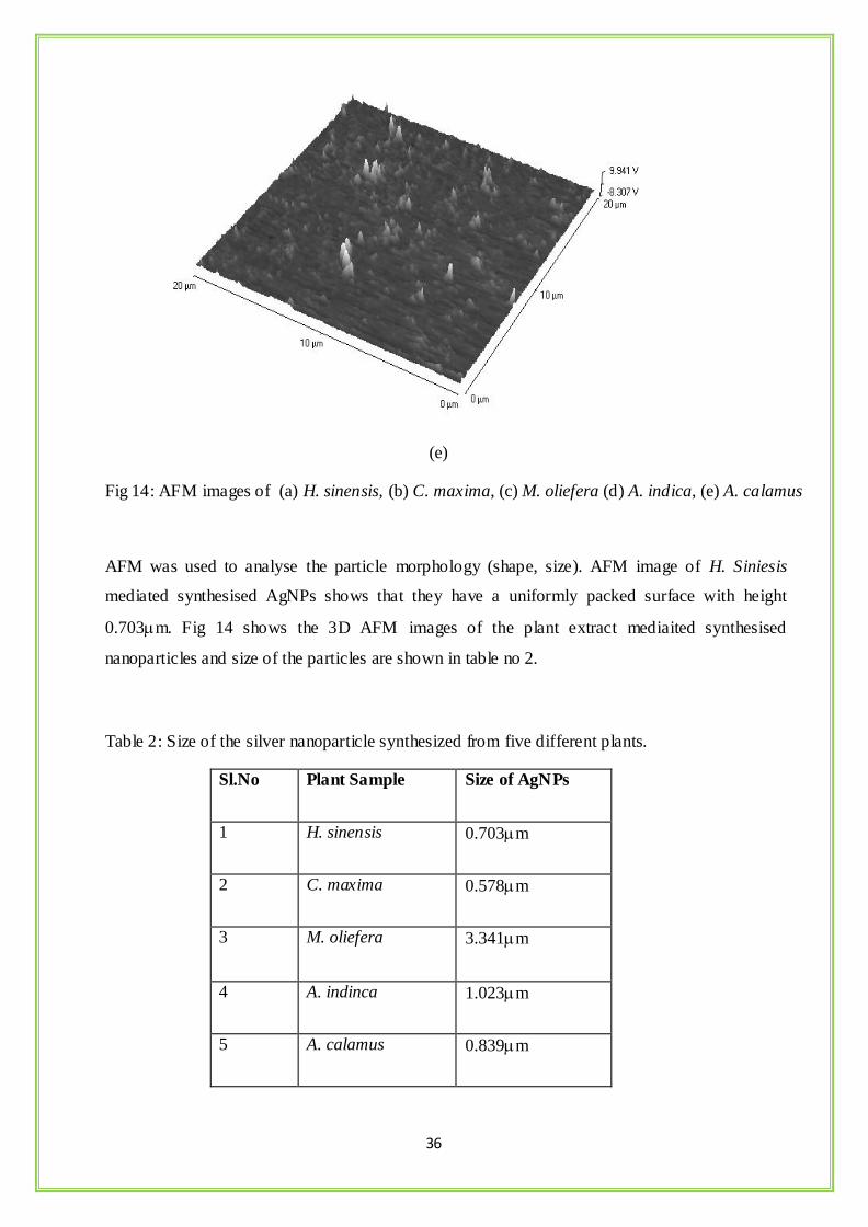
6.4: AFM Analysis:
(a) (b)
(c) (d)
36
(e)
Fig 14: AFM images of (a) H. sinensis, (b) C. maxima, (c) M. oliefera (d) A. indica, (e) A. calamus
AFM was used to analyse the particle morphology (shape, size). AFM image of H. Siniesis
mediated synthesised AgNPs shows that they have a uniformly packed surface with height
0.703m. Fig 14 shows the 3D AFM images of the plant extract mediaited synthesised
nanoparticles and size of the particles are shown in table no 2.
Table 2: Size of the silver nanoparticle synthesized from five different plants.
Sl.No Plant Sample Size of AgNPs
1 H. sinensis 0.703m
2 C. maxima 0.578m
3 M. oliefera 3.341m
4 A. indinca 1.023m
5 A. calamus 0.839m
37
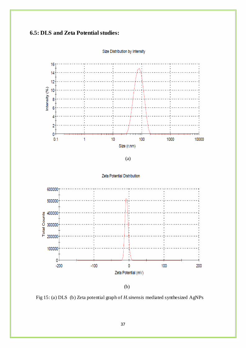
6.5: DLS and Zeta Potential studies:
(a)
(b)
Fig 15: (a) DLS (b) Zeta potential graph of H.sinensis mediated synthesized AgNPs
38
(a)
(b)
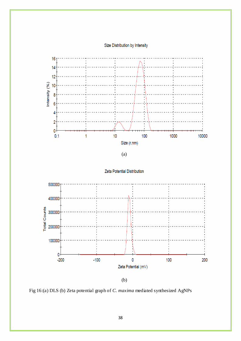
Fig 16:(a) DLS (b) Zeta potential graph of C. maxima mediated synthesized AgNPs
39
(a)
(b)
Fig17 :(a) DLS (b) Zeta potential graph of M. oliefera mediated synthesized AgNPs

40
( a)
(b)
Fig18:(a) DLS graph (b) Zeta potential graph of A.indica mediated synthesized AgNPs
41
(a)
(b)
Fig19:(a) DLS graph (b) Zeta potential graph of A.calamus mediated synthesized AgNPs
42
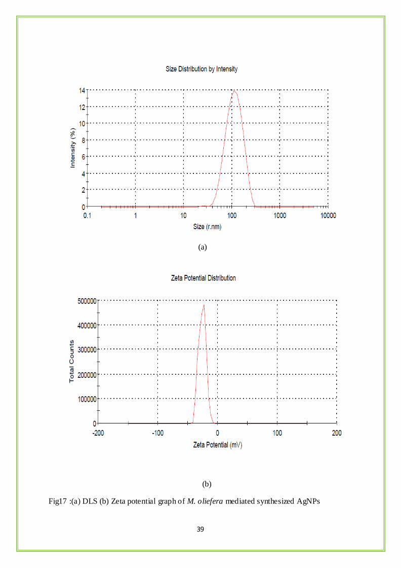
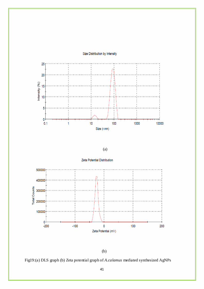
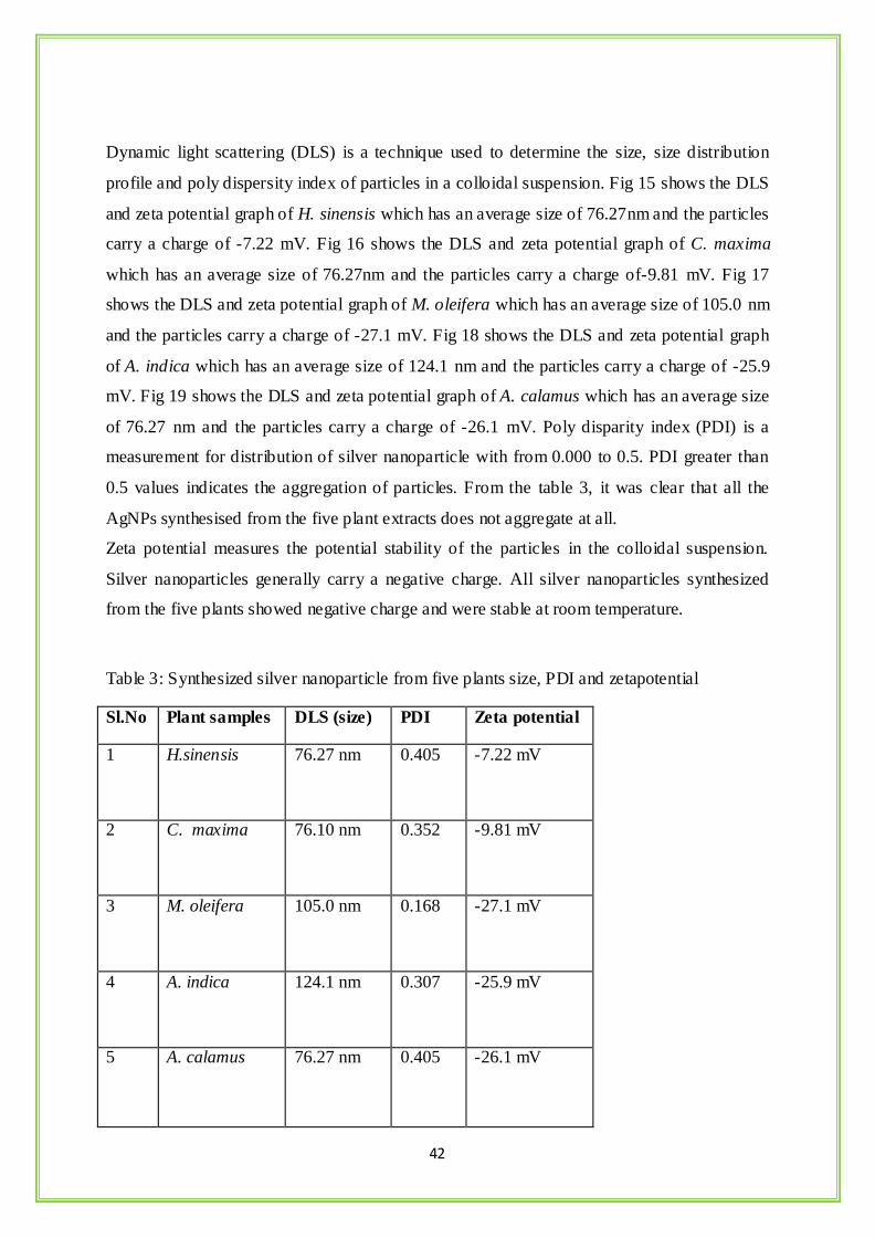
Dynamic light scattering (DLS) is a technique used to determine the size, size distribution
profile and poly dispersity index of particles in a colloidal suspension. Fig 15 shows the DLS
and zeta potential graph of H. sinensis which has an average size of 76.27nm and the particles
carry a charge of -7.22 mV. Fig 16 shows the DLS and zeta potential graph of C. maxima
which has an average size of 76.27nm and the particles carry a charge of-9.81 mV. Fig 17
shows the DLS and zeta potential graph of M. oleifera which has an average size of 105.0 nm
and the particles carry a charge of -27.1 mV. Fig 18 shows the DLS and zeta potential graph
of A. indica which has an average size of 124.1 nm and the particles carry a charge of -25.9
mV. Fig 19 shows the DLS and zeta potential graph of A. calamus which has an average size
of 76.27 nm and the particles carry a charge of -26.1 mV. Poly disparity index (PDI) is a
measurement for distribution of silver nanoparticle with from 0.000 to 0.5. PDI greater than
0.5 values indicates the aggregation of particles. From the table 3, it was clear that all the
AgNPs synthesised from the five plant extracts does not aggregate at all.
Zeta potential measures the potential stability of the particles in the colloidal suspension.
Silver nanoparticles generally carry a negative charge. All silver nanoparticles synthesized
from the five plants showed negative charge and were stable at room temperature.
Table 3: Synthesized silver nanoparticle from five plants size, PDI and zetapotential
Sl.No Plant samples DLS (size) PDI Zeta potential
1 H.sinensis 76.27 nm 0.405 -7.22 mV
2 C. maxima 76.10 nm 0.352 -9.81 mV
3 M. oleifera 105.0 nm 0.168 -27.1 mV
4 A. indica 124.1 nm 0.307 -25.9 mV
5 A. calamus 76.27 nm 0.405 -26.1 mV
43
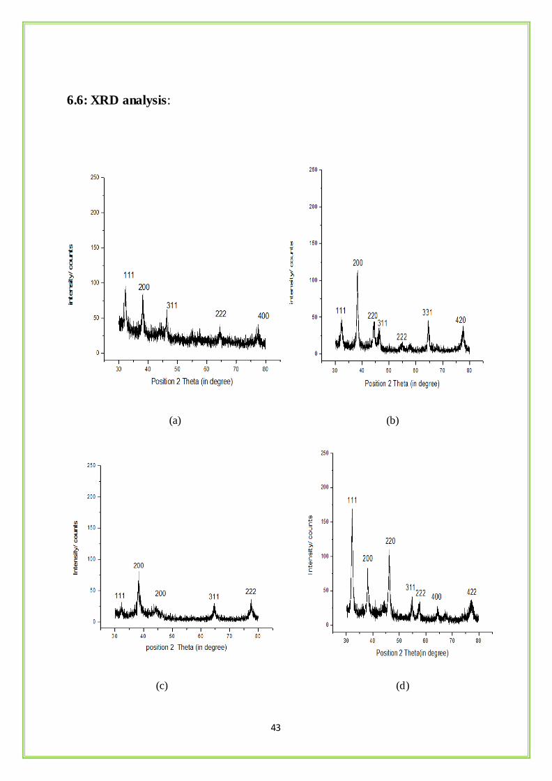
6.6: XRD analysis:
(a) (b)
(c) (d)
44
(e)
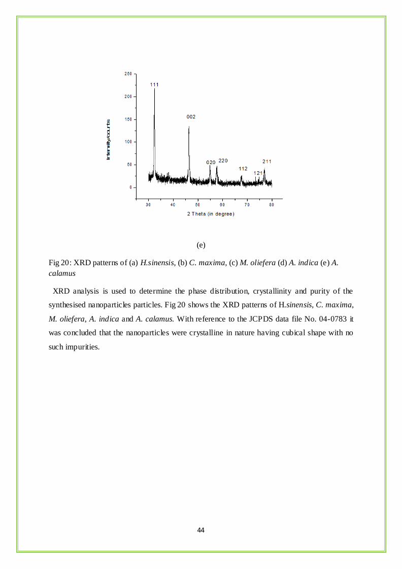
Fig 20: XRD patterns of (a) H.sinensis, (b) C. maxima, (c) M. oliefera (d) A. indica (e) A.
calamus
XRD analysis is used to determine the phase distribution, crystallinity and purity of the
synthesised nanoparticles particles. Fig 20 shows the XRD patterns of H.sinensis, C. maxima,
M. oliefera, A. indica and A. calamus. With reference to the JCPDS data file No. 04-0783 it
was concluded that the nanoparticles were crystalline in nature having cubical shape with no
such impurities.
45
6.7: FTIR Analysis:
(a)
(b)
4000 3000 2000 1000 0
0
20
40
60
80
100
120
8311390
17693697
5631030
1415
15421658
3438
669
106514021623
23562926
3402
%T
cm-1
AgNO3
Synthesized Ag-NP
Dried Hibiscus flower petal
4000 3500 3000 2500 2000 1500 1000 500
0
20
40
60
80
100
120
140
AgNO3
Synthesised AgNPs
dried petals of C.maxima
831
1390
17613697
6161407
15471640
3446
63510631370
15671640
23662924
3418
%T
cm-1
46
(c)
(d)
4000 3000 2000 1000 0
0
20
40
60
80
100
120
140
160
557139017613697
557
15291645
3400
633
10551659
29263326
%T
cm-1
AgNO3
Synthesized Ag-NP
Dried M.Oleifera leaf extract
4000 3000 2000 1000 0
0
20
40
60
80
100
8311390
17613697
5691083
1543
1647
2926
3428
6331069
1647
29223272
%T
cm-1
AgNO3
Synthesized Ag-NP
Dried A.indica leaf extract
47
(e)
Fig 21: FTIR graph of (a) H.sinensis, (b) C. maxima, (c) M. oliefera (d) A. indica (e) A.
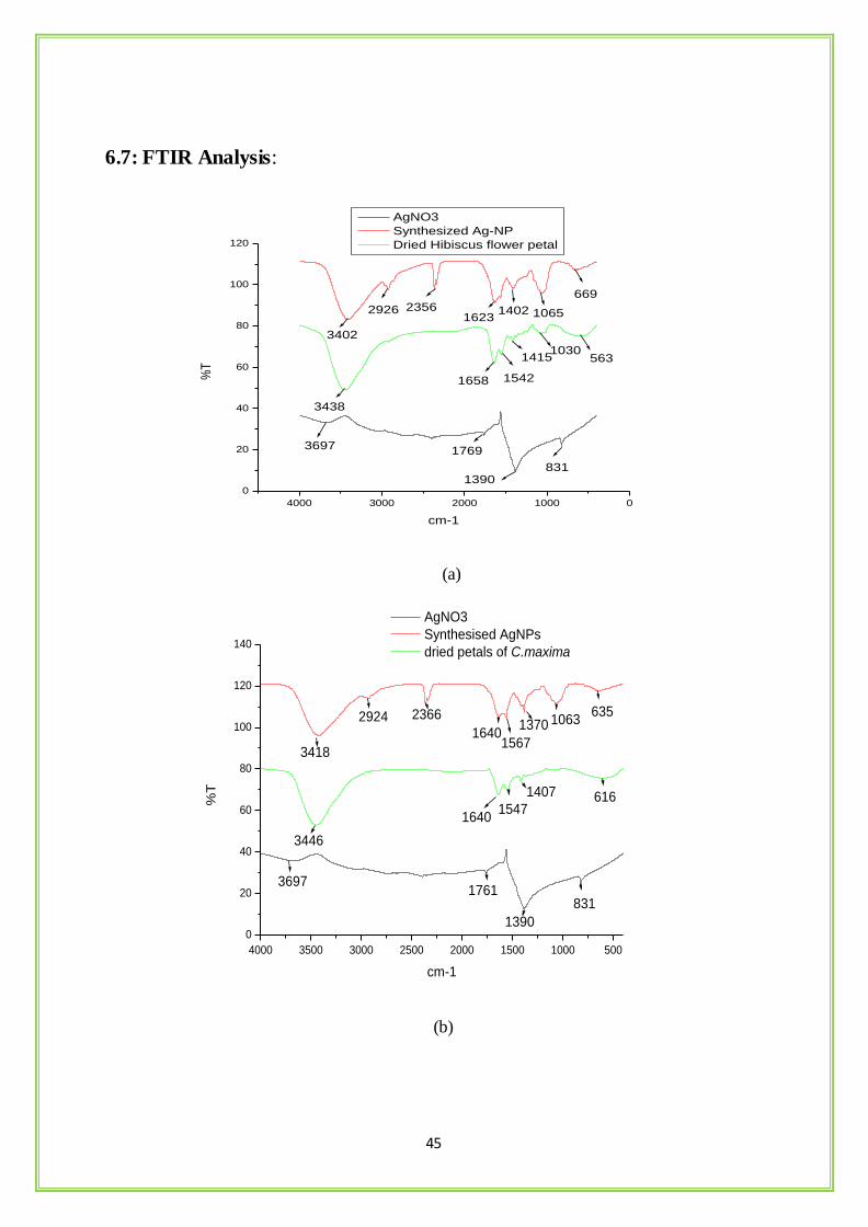
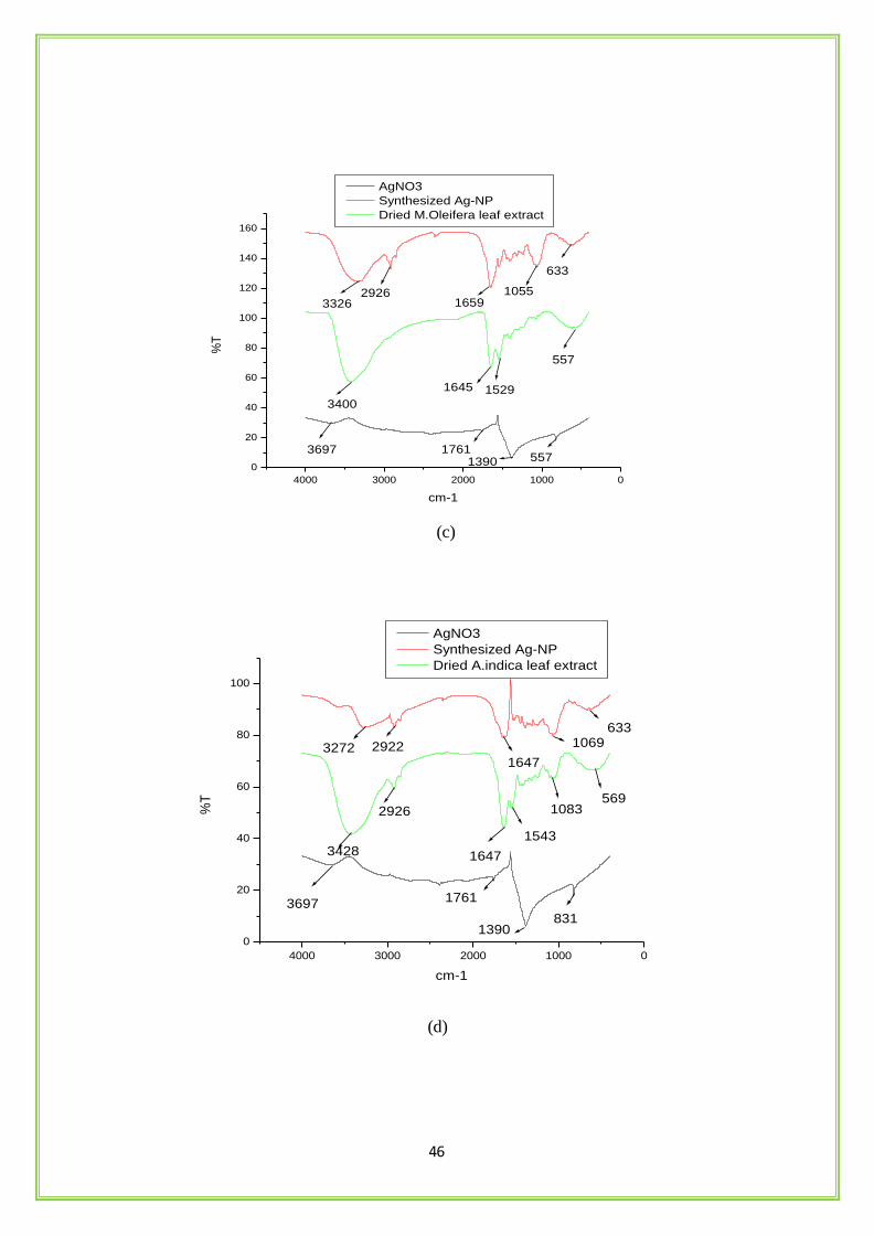
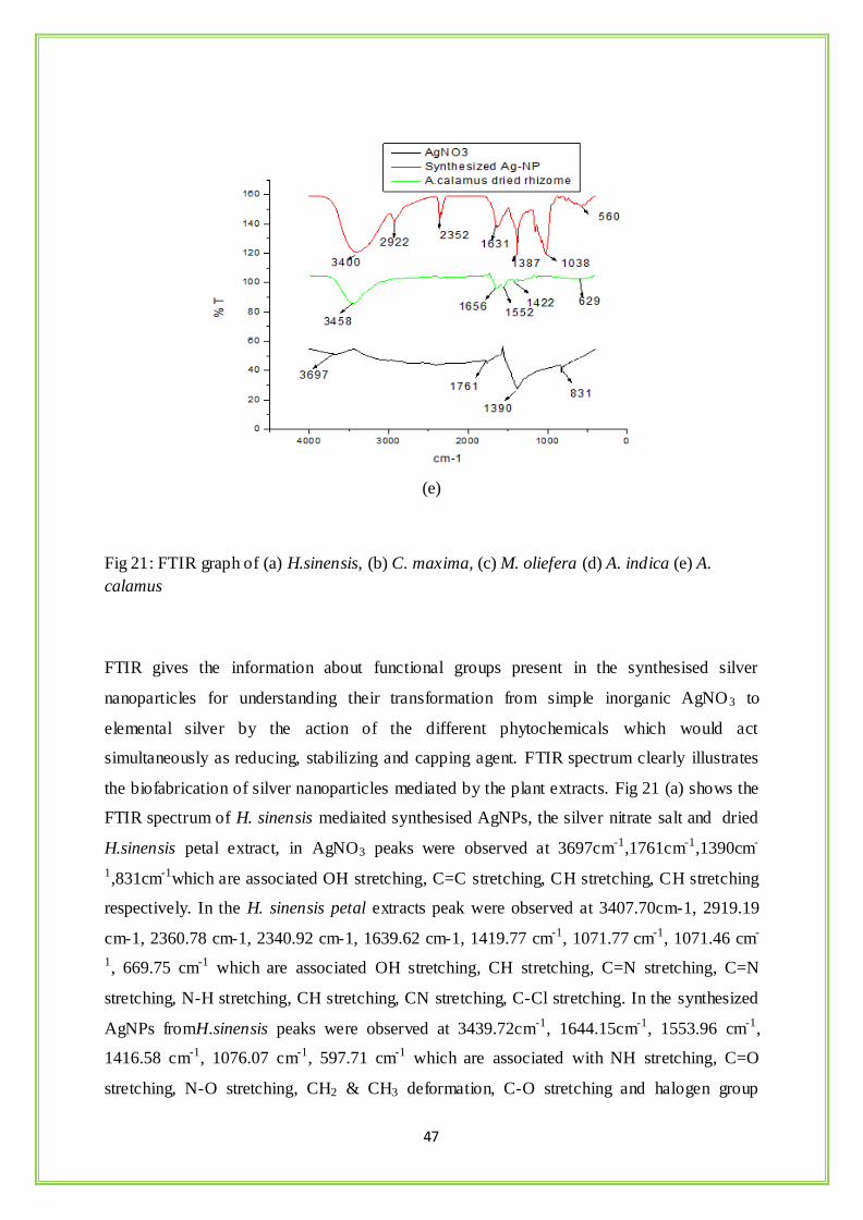
calamus
FTIR gives the information about functional groups present in the synthesised silver
nanoparticles for understanding their transformation from simple inorganic AgNO3 to
elemental silver by the action of the different phytochemicals which would act
simultaneously as reducing, stabilizing and capping agent. FTIR spectrum clearly illustrates
the biofabrication of silver nanoparticles mediated by the plant extracts. Fig 21 (a) shows the
FTIR spectrum of H. sinensis mediaited synthesised AgNPs, the silver nitrate salt and dried
H.sinensis petal extract, in AgNO3 peaks were observed at 3697cm-1,1761cm-1,1390cm-
1,831cm-1which are associated OH stretching, C=C stretching, CH stretching, CH stretching
respectively. In the H. sinensis petal extracts peak were observed at 3407.70cm-1, 2919.19
cm-1, 2360.78 cm-1, 2340.92 cm-1, 1639.62 cm-1, 1419.77 cm-1, 1071.77 cm-1, 1071.46 cm-
1, 669.75 cm-1 which are associated OH stretching, CH stretching, C=N stretching, C=N
stretching, N-H stretching, CH stretching, CN stretching, C-Cl stretching. In the synthesized
AgNPs fromH.sinensis peaks were observed at 3439.72cm-1, 1644.15cm-1, 1553.96 cm-1,
1416.58 cm-1, 1076.07 cm-1, 597.71 cm-1 which are associated with NH stretching, C=O
stretching, N-O stretching, CH2 & CH3 deformation, C-O stretching and halogen group
48
presence. The H. sinensis plant extract shows broad peak at 3407.70cm-1 which indicate the
presence of OH group or carboxyl groups and after synthesis of AgNPs there is a shift in the
broad peak to the right at 3439.72cm-1 indicating the NH stretching. These carboxyl and
amide group indicate the presence of secondary amines which is a signature marker of
proteins confirming the biofabrication of the nanoparticles by the action of the protein or
phytochemicals. Fig (b) to (e) clearly illustrates the biofabrication of the AgNPs by the action
of the phytochemicals such as phenols, terpenoids, flavonoids and alkaloids in C. maxima, M.
oliefera, A. indica and A. calamus ( Kong and Yu, 2007).
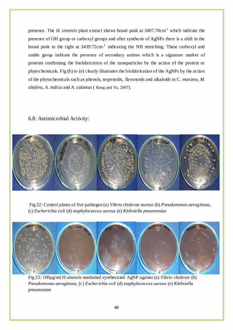
6.8: Antimicrobial Activity:
Fig 22: Control plates of five pathogen (a) Vibrio cholerae aureus (b) Pseudomonas aeruginosa,
(c) Escherichia coli (d) staphylococcus aureus (e) Klebsiella pneumoniae
Fig 23: 100μg/ml H.sinensis mediaited synthesisied AgNP against (a) Vibrio cholerae (b)
Pseudomonas aeruginosa, (c) Escherichia coli (d) staphylococcus aureus (e) Klebsiella
pneumoniae
49
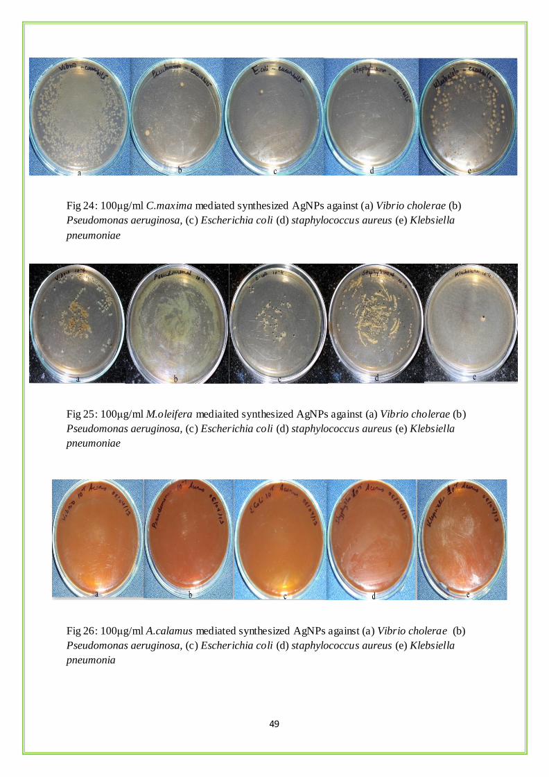
Fig 24: 100μg/ml C.maxima mediated synthesized AgNPs against (a) Vibrio cholerae (b)
Pseudomonas aeruginosa, (c) Escherichia coli (d) staphylococcus aureus (e) Klebsiella
pneumoniae
Fig 25: 100μg/ml M.oleifera mediaited synthesized AgNPs against (a) Vibrio cholerae (b)
Pseudomonas aeruginosa, (c) Escherichia coli (d) staphylococcus aureus (e) Klebsiella
pneumoniae
Fig 26: 100μg/ml A.calamus mediated synthesized AgNPs against (a) Vibrio cholerae (b)
Pseudomonas aeruginosa, (c) Escherichia coli (d) staphylococcus aureus (e) Klebsiella
pneumonia
50
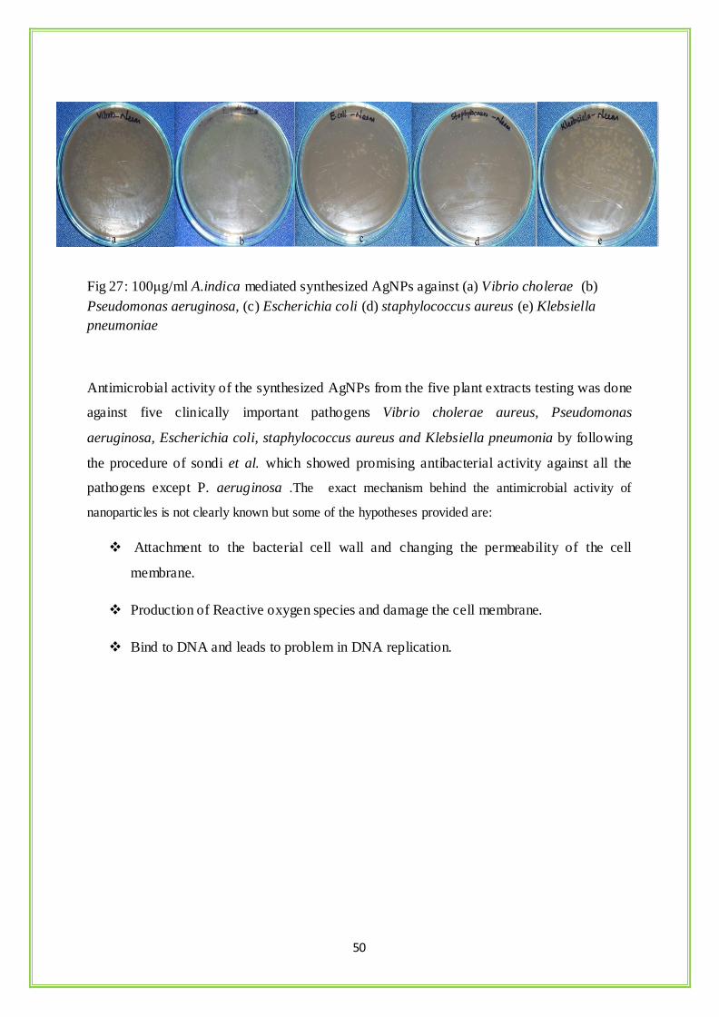
Fig 27: 100μg/ml A.indica mediated synthesized AgNPs against (a) Vibrio cholerae (b)
Pseudomonas aeruginosa, (c) Escherichia coli (d) staphylococcus aureus (e) Klebsiella
pneumoniae
Antimicrobial activity of the synthesized AgNPs from the five plant extracts testing was done
against five clinically important pathogens Vibrio cholerae aureus, Pseudomonas
aeruginosa, Escherichia coli, staphylococcus aureus and Klebsiella pneumonia by following
the procedure of sondi et al. which showed promising antibacterial activity against all the
pathogens except P. aeruginosa .The exact mechanism behind the antimicrobial activity of
nanoparticles is not clearly known but some of the hypotheses provided are:
Attachment to the bacterial cell wall and changing the permeability of the cell
membrane.
Production of Reactive oxygen species and damage the cell membrane.
Bind to DNA and leads to problem in DNA replication.
51
Table 4: Antimicrobial activity against five clinically important pathogens.
Sl.
No
Plant Sample staphylococcus
aureus
Pseudomonas
aeruginosa
Klebsiella
pneumonia
Vibrio
cholerae
Escherichia
coli
1 H.sinensis ++ + ++ ++ ++
2 C. maxima ++ + + ++ ++
3 M. oleifera + + ++ ++ ++
4 A. indica + ++ + + ++
5 A. calamus ++ ++ + ++ ++
++ = good antimicrobial activity
+ = considerably lower antimicrobial activity
52
7. Conclusion
Green synthesis of silver nanoparticles by the help of green plants is a very cost effective,
safe, non-toxic, eco-friendly route of synthesis which can be manufactured at a large scale. H.
sinensis, C. maxima,M. oleifera,A. indica and A.calamus showed great capability to synthesis
AgNPs at optimum temperature conditions. The UV absorption peak at 421nm clearly
indicates the synthesis of AgNPs. The SEM and AFM studies were helpful at deciphering
their morphology and distribution. DLS and Zeta potential studies validated the size and
charge of the nanoparticles in the colloidal system without any aggregation. FTIR studies
confirmed the biofabrication of the AgNPs by the action of different phytochemicals with its
different functional groups present in the extract solution. The XRD patterns confirmed the
purity, phase composition and nature of the synthesised nanoparticles. The AgNPs have great
antimicrobial activity against K. pneumonia, S.aureus, V. cholerae and E.coli. By comparing
the different characteristic data we could conclude that AgNPs synthesised from H. sinensis
and C. maxima were less stable then the other three plant extracts which could be due to the
absence of capping and stabilizing materials as petals mainly contains pigments for attracting
different insects for pollination.
53
7. REFERENCE
Abuye C, Omwega AM, JK Imungi (1999). Familial tendency and dietary association of goitre in
Gamo-Gofa, Ethiopia. East African Medical Journal 76:447-451.
Ahmad A, Mukherjee P, Mandal D, Senapati S, Khan MI, Kumar R, Sastry M, (2003).
Extracellular biosynthesis of silver nanoparticles using the fungus Fusarium oxysporum
composite metal particles, and the atom to metal. Colloids and Surfaces B: Biointerfaces 28:
313-318.
Ahmad A, Senapati S, Khan MI, Kumar R, Ramani R, Srinivas V, Sastry M, (2003).
Intracellular synthesis of gold nanoparticles by a novel alkalotolerant actinomycete,
Rhodococcus species. Nanotechnology 14:824–828.
Ahmad N, Sharma S, Singh V. N., Shamsi S. F., Fatma A, Mehta B. R, (2011). Biosynthesis
of Silver Nanoparticles from Desmodium triflorum : A novel approach towards weed
utilization. Biotechnology Research International Volume.
Akhtar AH, Ahmad KU (1995). Anti-ulcerogenic evaluation of the methanolic extracts of some
indigenous medicinal plants of Pakistan in aspirin-ulcerated rats. Journal of Ethnopharmacology
46:1-6.
Anwar F, and Bhanger MI (2003). Analytical characterization of Moringa oleifera seed oil grown
in temperate regions of Pakistan. Journal of Agricultural and Food Chemistry 51: 6558-6563.
Ankamwar B, Damle C, Ahmad A, Sastry M, (2005). Biosynthesis of gold and silver
nanoparticles using Emblica officinalis fruit extract, their phase transfer and transmetallation
in an organic solution. J of Nanosci. and Nanotechnol. 5: 1665-1671.
Armendariz V, Herrera I, Peralta-Videa J, Jose-Yacaman M, Troiani H, Santiago P, (2004).
Size controlled gold nanoparticle formation by Avena sativa biomass: use of plants in
nanobiotechnology. J Nanopart Res 6:377–382.
Asres K (1995). The major constituents of the acetone fraction of Ethiopian Moringa stenopetala
leaves. Mansoura Journa l of Pharmacological Science 11(1): 55-64.
54
Bai HJ, Zhang ZM, Guo Y, Yang GE, (2009). Biosynthesis of cadmium sulphide
nanoparticles by photosynthetic bacteria Rhodopseudomonas palustris. Colloids and Surfaces
B: Biointerfaces 70: 142- 146.
Bar H, Bhui DK, Sahoo GP, Sarkar P, De SP, Misra A, (2009). Green synthesis of silver
nanoparticles using latex of Jatropha curcas. Colloids and Surfaces A: Physicochemical and
Engineering Aspects 339: 134-139.
Basavaraja SS, Balaji SD, Lagashetty AK, Rajasab AH, Venkataraman A, (2008).
Extracellular biosynthesis of silver nanoparticles using the fungus Fusarium semitectum.
Materials Research Bulletin 43: 1164-1170.
Beveridge TJ and Murray RGE (1980). Site of metal deposition in the cell wall of Bacillus
subtilis. J Bacteriol 141:876–887.
Bhainsa KC and D'Souza SF, (2006). Extracellular biosynthesis of silver nanoparticles using
the fungus Aspergillus fumigates. Colloids and Surfaces B: Biointerfaces 47: 160-164.
Buchbauer G, Boucek B, Nikiforov A (1998). On the aroma of Austrian pumpkin seed oil:
correlation of analytical data with olfactoric characteristics. Ernahrung/Nutrition 22(6): 246– 249.
Chandran SP, Chaudhary M, Pasricha R, Ahmad A, Sastry M, (2006). Nanotriangles and
silver nanoparticles using Aloe vera plant extract. Biotechnology Programme 22: 577-583.
Chen Y.Y, Wang C.A, Liu H.Y, Qiu J.S. and Bao X.H, (2005). Ag/SiO2: A novel catalyst
with high activity and selectivity for hydrogenation of chloronitrobenzenes. Chem. Commun
42: 5298-5300.
Cheon, J. & Horace, G. (2009). Inorganic nanoparticles for biological sensing, imaging and
therapeutics. Journal Materials in Chemistry 19:6249- 6250.
Cunningham DP, Lundie LL, (1993). Precipitation of cadmium by Clostridium
thermoaceticum. Applied and Environmental Microbiology 59: 7-14.
Deplanche K and Macaskie LE, (2008). Biorecovery of gold by Escherichia coli and
Desulfovibrio desulfuricans. Biotechnology and Bioengineering 99: 1055-1064.
55
Deplanche K, Woods RD, Mikheenko IP, Sockett RE, Macaskie, LE, (2008). Manufacture of
stable palladium and gold nanoparticles on native and genetically engineered flagella
scaffolds. Biotechnology and Bioengineering 101: 873-880.
Du L, Jiang H, Liu X, Wang E,(2007). Biosynthesis of gold nanoparticles assisted by
Escherichia coli DH5a and its application on direct electrochemistry of haemoglobin
Electrochemistry Communications 9:1165-1170.
Evanoff DD and Chumanov G, (2005). Synthesis and Optical Properties of Silver
Nanoparticles and Arrays. Chem Phys Chem 6: 1221 – 1231.
Feng QL, Wu J, Chen GQ, Cui FZ, Kim TN and Kim JO, (2000). A mechanistic study of the
antibacterial effect of silver ions on Escherichia coli and Staphylococcus aureus. J. Biomed.
Mater 52: 662-668.
Fu JK, Liu Y, Gu P, Tang DL, Lin ZY, Yao BX & Weng SZ, (2000). Spectroscopic
characterization on the biosorption and bioreduction of Ag(I) by Lactobacillus sp. A09. Acta
Physico- Chimica Sinica 16(9): 770-782.
Gardea-Torresdey JL, Gomez E, Peralta-Videa JR, Parsons JG, Troiani H and Jose
YacamanM, (2003). Alfalfa sprouts: a natural source for the synthesis of silver nanoparticles.
Langmuir 19:1357–1361.
Gardea-Torresdey JL, Parsons JG, Gomez E, Peralta-Videa J, Troiani HE, Santiago P, et al,
(2002). Formation and growth of Au nanoparticles inside live alfalfa plants. Am Chem Soc
2:397–401.
Govindaraju K, Tamilselvan S, Kiruthiga V, Singaravelu G, (2010). Biogenic silver
nanoparticles by Solanum torvum and their promising antimicrobial activity. Journal of
Biopesticides 3(1): 394-399.
Gutierrez M, Henglein A, (1993). Formation of colloidal silver by "push-pull" reduction of
silver,J. Phys. Chem 97, 11368.
Guzman, Maribel G, Dille J, Godet S,(2009) Synthesis of silver nanoparticles by chemical
reduction method and their antibacterial activity. International Journal of Chemical and
Biological Engineering 2:3.
56
He S, Zhang Y, Guo Z, Gu N, (2008). Biological synthesis of gold nanowires using extract of
Rhodopseudomonas capsulata. Biotechnology Progress 24: 476-480.
Hosea M, Greene B, McPherson R, Henzl M, Alexander MD, Darnall DW, (1986).
Accumulation of elemental gold on the alga Chlorella vulgaris. Inorganica Chimica Acta
123: 161-165.
Huang J, Li Q, Sun D, Lu Y, Su Y, Yang X, Wang H, Wang Y, Shao W, He N, Hong J, Chen
C, (2007). Biosynthesis of silver and gold nanoparticles by novel sundried Cinnamomum
camphora leaf.Nanotechnology 18: 105104-105115.
Huang J, Li Q, Sun D, Lu Y, Su Y, Yang X (2007). Biosynthesis of silver and gold
nanoparticles by novel sundried Cinnamomum camphora leaf. Nanotechnology 18:105104–
105114.
He S, Zhang Y, Guo Z, Gu N, (2008). Biological synthesis of gold nanowires using extract of
Rhodopseudomonas capsulata. Biotechnology Progress 24: 476-480.
Hosea M, Greene B, McPherson R, Henzl M, Alexander MD, Darnall DW (1986).
Accumulation of elemental gold on the alga Chlorella vulgaris. Inorganica Chimica Acta
123: 161-165.
Huang J, Li Q, Sun D, Lu Y, Su Y, Yang X, Wang H, Wang Y, Shao W, He N, Hong J, Chen
C, (2007). Biosynthesis of silver and gold nanoparticles by novel sundried Cinnamomum
camphora leaf. Nanotechnology 18: 105104-105115.
Husseiny MI, Ei-Aziz MA, Badr Y, Mahmoud MA,(2007). Biosynthesis of gold
nanoparticlesusing Pseudomonas aeruginosa. Spectrochimica Acta Part A: Molecularand
Biomolecular Spectroscopy 67: 1003-1006.
Jae YS, and Beom SK, (2009). Rapid Biological Synthesis of Silver Nanoparticles Using
Plant Leaf Extracts. Bioprocess Biosyst. Eng. 32: 79-84.
Jha AK, Prasad K, Kumar V, Prasad K, (2009). Biosynthesis of silver nanoparticles using
Eclipta leaf. Biotechnol. Prog. 25(5): 1476-1479.
Jha AK, Prasad K, (2010). Green synthesis of silver nanoparticles using Cycas leaf. Int. J. of
Green Nanotechnology:Physics and Chemistry 1: 110-117.
57
Joerger R, Klaus T, Granqvist CG (2000). Biologically produced silver–carbon composite
materials for optically functional thin film coatings. Advanced Materials 12: 407-409.
Jong WHD and Borm PJA (2008). Drug delivery and nanoparticles: Applications and
hazards, international jounnal nanomedicine 3 (2): 133–149.
Kalishwaralal K, Deepak V, Ramkumarpandian S, Nellaiah H, Sangiliyandi (2008).
Extracellular biosynthesis of silver nanoparticles by the culture supernatant of Bacillus
licheniformis. Materials Letters 62: 4411-4413.
Khan I, Srikakolupu SR, Darsipudi S, Gotteti SD and Hemasundara AC (2010).
Phytochemical studies and screening of leaf extracts of Azadirachta indica for its anti-
microbial activity against dental pathogens. Archives of Applied Science Research 2 (2):246-
250.
Khandelwal N, Singh A, Jain D, Upadhay MK, Verma HN, (2010). Green synthesis of silver
nanoparticles using Argimone mexicana leaf extract and valuation of their antimicrobial
activities. Dig. J. Nanomater. Bios. 5 (2): 483-489.
Kholoud MM, Nour AE , Eftaiha A, Abdulrhman Al-Warthan Reda A.A. Ammar,(2010).
Synthesis and applications of silver nanoparticles Arabian Journal of Chemistry 3, 135–140.
Koike K, Li W, Liu L, Hata E, Nikaido T (2005). New phenolic glycosides from the seeds of
Cucurbita moschata. Chem Pharm Bull 53(2): 225–228.
Konishi Y, Ohno K, Saitoh N, Nomura T, Nagamine S, (2007). Bioreductive deposition of
platinum nanoparticles on the bacterium Shewanella algae. Journal of Biotechnology 128:
648-653.
Kowshik M, Deshmukh N, Vogel W, Urban J, Kulkarni SK, Paknikar KM, (2002). Microbial
synthesis of semiconductor CdS nanoparticles, their characterization, and their use in the
fabrication of an ideal diode. Biotechnology and Bioengineering 78: 583-588.
Krishnaraj C, Jagan EG, Rajasekar S, Selvakumar P, Kalaichelvan PT and Mohan N, (2010).
Synthesis of silver nanoparticles using Acalypha indica leaf extracts and its antibacterial
activity against water borne pathogens. Coll Surf B: Biointer 76, 50-56.
58
Kumar B, Naik V, Halehatty SB , Girija D and Kumar B V,(2011). ZnO nanoparticle as
catalyst for efficient green one-pot synthesis of coumarins through Knoevenagel
condensation, J. Chem. Sci.123(5).
Kumar L, Chakraborthy G, Singh V, Mazumder A, (2012). Hibiscus rosa-sinensis: A Review
on Divine Herb, Journal of Advances in Pharmacy and Healthcare Research, Volume 2, Issue
4.
Kumar V, Yadav SC, Yadav SK.,(2010). Syzygium cumini leaf and seed extract mediated
biosynthesis of silver nanoparticles and their characterization. J of chem. Technol.
Biotechnol. 2010; 85: 1301- 1309.
Labrenz M, Druschel GK, Thomsen-Ebert T, Gilbert B, Welch SA, Kemner KM, Logan GA,
Summons RE, Stasio GD, Bond PL, Lai B, Kelly SD, Banfield JF,(2000). Formation of
sphalerite (ZnS) deposits in natural biofilms of sulfate-reducing bacteria. Science 290: 1744-
1747.
Leela A and Vivekanandan M, (2008). Tapping the unexploited plant resources for the
synthesis of silver nanoparticles. African J of Biotechnology 7: 3162-65.
Lengke M, Fleet ME, Southam G, (2006). Morphology of gold nanoparticles synthesized by
filamentous cyanobacteria from gold (I) -thiosulfate and gold (III) - chloride complexes.
Langmuir 22: 2780-2787.
Li S, Shen Y, Xie A, Yu X, Qiu L, Zhang L, et al, (2007).Green synthesis of silver
nanoparticles using Capsicum annuum L. extract. Green Chem 9:852–885.
Li Z, Lee D, Sheng XX, Cohen RE and Rubner MF, (2006). Two-level antibacterial coating
with both releasekilling and contact-killing capabilities, Langmuir 22, 9820-9823 .
Lisiecki I, Pileni M, (1995). Copper Metallic Particles Synthesized "in Situ" in Reverse
Micelles: Influence of Various Parameters on the Size of the Particles. J. Phys. Chem. 99,
507.
Maribel GG, Jean D, Stephan, (2009). Synthesis of silver nanoparticles by chemical reduction
method and their antibacterial activity. International Journal of Chemical and Biological
Engineering 2:3.
59
Mitrakos D, Jokiniemi J, Backman U, Housiadas C, (2008). Aerosol flow in a tube furnace
reactor of gas-phasesynthesised silver nanoparticles. J Nanopart Res 10:153–161.
Mohanpuria P, Rana KN, Yadav SK, (2008). Biosynthesis of nanoparticles: Technological
concepts and future applications. J. Nanopart. Res. 10, 507–517.
Mokhtari N, Daneshpajouh S, Seyedbagheri S, Atashdehghan R, Abdi K, Sarkar S, Minaian
S, Shahverdi HR, Shahverdi AR, (2009). Biological synthesis of very small silver
nanoparticles by culture supernatant of Klebsiella pneumonia: The effects of visible- light
irradiation and the liquid mixing process. Materials Research Bulletin 44: 1415-1421.
Mude N, Ingle A, Gade A, Rai M., (2009). Synthesis of silver nanoparticles using callus
extract of Carica papaya- A First Report. J of Plant Biochem and Biotechnol 18: 83-86.
Mukherjee P, Ahmad A, Mandal D, Senapati S, Sainkar SR, Khan MI, Parischa, R,
Ajaykumar PV, Alam M, Kumar R, Sastry M, (2001). Fungus mediated synthesis of silver
nanoparticles and their immobilization in the mycelia matrix: a novel biological approach to
nanoparticle synthesis. Nano Lett 1, 515–519.
Nagajyothi PC and Lee KD, (2011). Synthesis of Plant-Mediated Silver Nanoparticles Using
Dioscorea batatas Rhizome Extract and Evaluation of Their Antimicrobial Activities. Journal
of Nanomaterials , 573429.
Nair B and Pradeep T, (2002). Coalescence of nanoclusters and formation of submicron
crystallites assisted by Lactobacillus strains. Crystal Growth and Design 2: 293- 298.
Nair R, Varghese SH, Nair BG, Maekawa T, Yoshida Y, Kumar SD, (2010). Nanoparticulate
material delivery to plants, Plant Science 179, 154–163.
Nayak BS, Raju SS, Orette FA, and Rao AVC (2007).Effects of Hibiscus rosa sinensis L
(Malvaceae) on wound healing activity: a preclinical study in a Sprague Dawley rat,‖
International Journal of Lower Extremity Wounds, 6 (2) 76–81.
Osaka T, Matsunaga T, Nakanishi T, Arakaki A, Niwa D, Iida H, (2006). Synthesis of
Magnetic nanoparticles and their application to bioassays. Analytical and Bioanalytical
Chemistry 384: 593-600.
60
Panaek A, Kvitek L, Prucek R, Kolar M, Veerova R, Pizurova N, Sharma VK, Nevena T and
Zboril R, (2006).Silver colloid nanoparticles: Synthesis, characterization and their
antibacterial activity. J. Phys. Chem. B 110, 16248–16253.
Geethalakshmi R and Sarada DVL, (2010). Synthesis of plant-mediated silver nanoparticles
using Trianthema decandra extract and evaluation of their anti microbial activities
International Journal of Engineering Science and Technology, 2 (5) 970-975,
Raffi M, Rumaiz AK, Hasan MM, Shah SI (2007). Studies of the growth parameters for
silver nanoparticle synthesis by inert gas condensation. Journal of Materials Research 22 (12)
30.
Saifuddin N, Wong CW, Yasimura AAN, (2009). Rapid biosynthesis of silver nanoparticles
using culture supernatant of bacteria with microwave irradiation. Ejournal of chemistry. 6(1)
61-70.
Sandbhy V, MacBride MM, Peterson BR,Sen.A,(2006).Silver bromide nanoparticles/polymer
composites: dual action tunable antimicrobial materials, J. Am. Chem. Soc., 128, 9798-9808.
Sastry M, Ahmad A, Khan MI, Kumar R (2003). Biosynthesis of metal nanoparticles using
fungi and actinomycete. Curr Sci 85:162–170.
Sathyavathi R, Balamurali KM, Venugopal S, Saritha R, Narayana RD, (2010). Biosynthesis
of Ag nanoparticles using Coriandrum sativum leaf extract and their application in non- linear
optics. Adv.Sci.Lett 3(2): 138-143.
Saxena A, Tripathi RM, Singh RP, (2010). Biological synthesis of silver nanoparticles by
using onion (Allium cepa) extract and their antibacterial activity. Dig. J. Nanomater. Bios. 5
(2): 427-432.
Scarano G, Morelli E, (2003). Properties of phytochelatin-coated CdS nanocrystallites
formed in a marine phytoplanktonic alga (Phaeodactylum tricornutum, Bohlin) in response to
Cd. Plant Science 165: 803- 810.
Setua P, Chakraborty A, Seth D, Bhatta MU, Satyam PV, Sarkar N, (2007). Synthesis, optical
properties, and surface enhanced Raman scattering of silver nanoparticles in nonaqueous
methanol reverse micelles, J. Phys. Chem. C., 111, 3901-3907.
61
Shahverdi AR, Minaeian S, Shahverdi HR, Jamalifar H, Nohi AA, (2007). Rapid synthesis of
silver nanoparticles using culture supernatants of Enterobacteriaceae: A novel b iological
approach. Process Biochem 2007; 42: 919- 923.
Shankar SS, Absar A, Murali S, (2003). Geranium leaf assisted biosynthesis of silver
nanoparticles. Biotechnol Prog 19:1627–1631.
Shankar SS, Ahmad A, Pasrichaa R and Sastry M, (2004). Bioreduction of chloroaurate ions
by geranium leaves and its endophytic fungus yields gold nanoparticles of different shapes. J
Mater Chem 13:1822–1826.
Shankar SS, Rai A, Ahmad A and Sastry M, (2004). Rapid synthesis of Au, Ag, and
bimetallic Au core–Ag shell nanoparticles using neem (Azadirachta indica) leaf broth. J
Colloid Interf Sci 275:496–502.
Shankar SS, Rai A, Ahmad A, Sastry M.,(2004). Biosynthesis of silver and gold
nanoparticles from extracts of different parts of the Geranium plant. Applications in
Nanotechnology 2004; 1: 69-77.
Sharma NC, Sahi SV, Nath S, Parsons JG, Gardea-Torresdey JL and Pal T, (2007). Synthesis
of plant-mediated gold nanoparticles and catalytic role of biomatrix-embedded nanomaterials.
Environ Sci Technol 41:5137–5142.
Sharma KV, Yngard AR, Lin Y, (2009). Silver nanoparticle: Green synthesis and their
antimicrobial activities, Advances in Colloid and Interface Science 145 83–96.
Singaravelu G, Arockiamary JS, Kumar VG, Govindaraju K, (2007). A novel extracellular
synthesis of monodisperse gold nanoparticles using marine alga, Sargassum wightii Greville.
Colloids and Surfaces B: Biointerfaces 57: 97–101.
Singh C, Sharma V, Naik PK, Khandelwal V, Singh H, (2011). Green biogenic approach for
synthesis of gold and silver nanoparticles using Zingiber officinale. Dig. J. Nanomater. Bios.
6 (2): 535- 542.
Sinha S, Pan I, Chanda P, Sen SK, (2009). Nanoparticles fabrication using ambient biological
resources, Journal of Applied Biosciences 19: 1113 – 1130
Soloviev.M,(2007). Nanobiotechnology today: focus on nanoparticles, J
Nanobiotechnology; 5(11).
62
Stobie N, Duffy B, McCormack DE, Colreavy J, Hidalgo M, McHale P, (2008) Prevention
of Staphylococcus epidermisdis biofilm formation using a low temperature processed silver
doped phenyltriethoxysilane solgel coating. Biomater, 29, 963-969.
Taniguchi N.,(1974), On the basic concept of 'Nano-Technology'. Proceedings of
International Conference on Production Engineering Tokyo, Part II, Japan Society of
Precision Engineering, Japan.
Treguer M, de Cointet C, Remita H, Khatouri J, Mostafavi M, Amblard J, Belloni J , de
Keyzer R, (1998) Dose Rate Effects on Radiolytic Synthesis of Gold-Silver Bimetallic
Clusters in Solution. J. Phys. Chem. B102:4310-4321.
Vaidyanathan R, Gopalram S, Kalishwaralal K, Deepak V, Pandian SR, Gurunathan S (2010)
Enhanced silver nanoparticle synthesis by optimization of nitrate reductase activity. Colloids
Surf. B Biointerfaces 75, 335–341.
Varshney R, Bhadauria S, Gaur MS, (2010) Biogenic synthesis of silver nanocubes and
nanorods using sundried Stevia rebaudiana leaves. Adv. Mat. Lett 1(3): 232-237.
Varshney R, Mishra AN, Bhadauria S, Gaur MS, (2009) A novel microbial route to
synthesize silver nanoparticles using fungus Hormoconis resinae. Dig. J. Nanomater. Bios.
4(2): 349-355.
Vigneshwaran N, Kathe AA, Varadarajan PV, Nachane RP, Balasubramanya RH, (2006)
Biomimetics of Ag nanoparticles by white rot fungus, Phaenerochaete chrysosporium.
Colloids and Surfaces B: Biointerfaces 53: 55-59.
Vivekanandan S, Misra M, Mohanty A, (2009) Biological synthesis of silver nanoparticles
using Glycine max (soybean) leaf extract: an investigation on different varieties. J of
nanoscience & Nanotechnol. 9(12): 6828-6833.
Xu ZP, Zeng QH, Lu GQ, Yu AB (2006). Inorganic nanoparticles as carriers for efficient
cellular delivery. Chemical Engineering Science 61: 1027- 1040.
Yamini GS, Fouzia B, Ezhilarasan, Arumugam S,(2011). Green Synthesis of Silver
Nanoparticles from Cleome Viscosa: Synthesis and Antimicrobial Activity 2011 International
Conference on Bioscience, Biochemistry and Bioinformatics IPCBEE vol.5.
63
Zamiri R, Azmi BZ, Ahangar HA, Zamiri, G, Husin, MS and Wahab ZA,(2012). Preparation
and characterization of silver nanoparticles in natural polymers using laser ablation Bull.
Mater. Sci., Vol. 35, No. 5, pp. 727–731.
Zhang H (2003). Determination of γ -amino-butyric acid and amino acids in pumpkin. Food Res
Dev 24(3): 108–109.
Copyright © 2022 FDOKUMEN