Protective role of nitric oxide in mice with Shiga toxin-induced hemolytic uremic syndrome
Upload
independentCategory
view
0download
0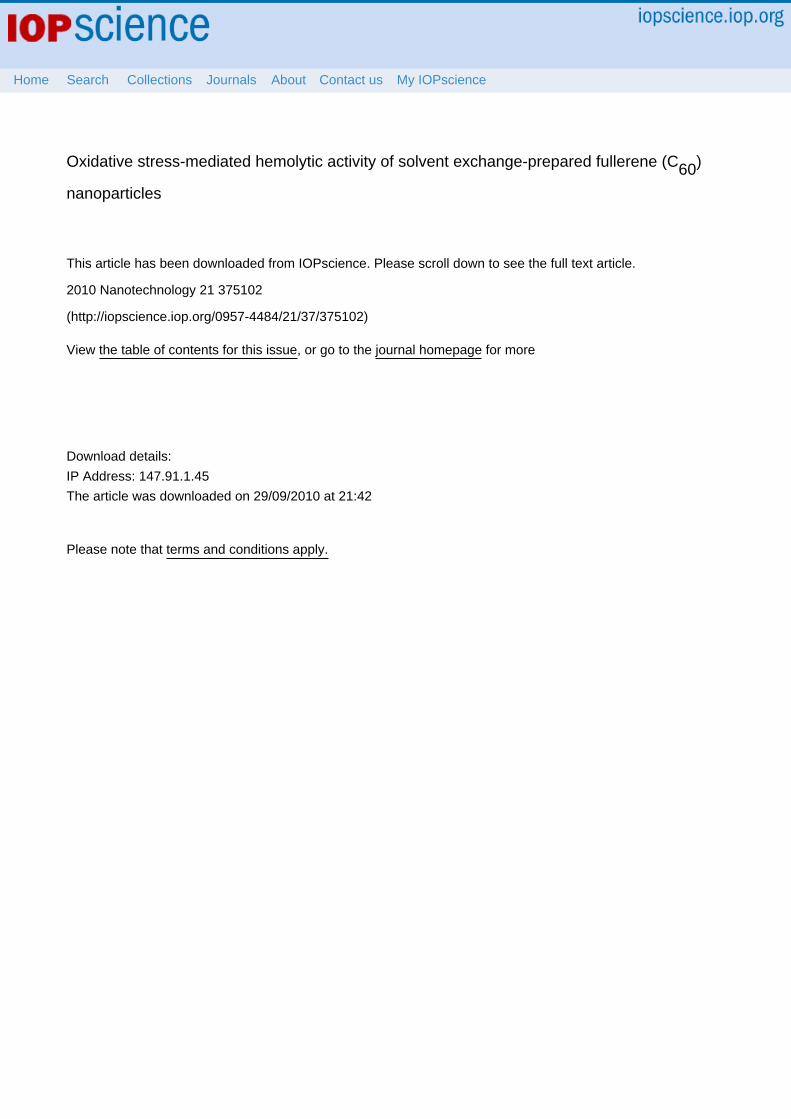
Oxidative stress-mediated hemolytic activity of solvent exchange-prepared fullerene (C60)
nanoparticles
This article has been downloaded from IOPscience. Please scroll down to see the full text article.
2010 Nanotechnology 21 375102
(http://iopscience.iop.org/0957-4484/21/37/375102)
Download details:
IP Address: 147.91.1.45
The article was downloaded on 29/09/2010 at 21:42
Please note that terms and conditions apply.
View the table of contents for this issue, or go to the journal homepage for more
Home Search Collections Journals About Contact us My IOPscience

IOP PUBLISHING NANOTECHNOLOGY
Nanotechnology 21 (2010) 375102 (10pp) doi:10.1088/0957-4484/21/37/375102
Oxidative stress-mediated hemolyticactivity of solvent exchange-preparedfullerene (C60) nanoparticlesAndreja Trpkovic1,4, Biljana Todorovic-Markovic1,4, Duska Kleut1,Maja Misirkic2,3, Kristina Janjetovic2,3, Ljubica Vucicevic2,3,Aleksandar Pantovic3, Svetlana Jovanovic1, Miroslav Dramicanin1,Zoran Markovic1,5 and Vladimir Trajkovic3,5
1 Vinca Institute of Nuclear Sciences, POB 522, University of Belgrade, Belgrade 11000,Serbia2 Institute for Biological Research ‘Sinisa Stankovic’, University of Belgrade, Serbia3 Institute of Microbiology and Immunology, School of Medicine, University of Belgrade,Dr Subotica 1, Belgrade 11000, Serbia
E-mail: [email protected] and [email protected]
Received 31 May 2010, in final form 23 July 2010Published 20 August 2010Online at stacks.iop.org/Nano/21/375102
AbstractThe present study investigated the hemolytic properties of fullerene (C60) nanoparticlesprepared by solvent exchange using tetrahydrofuran (nC60THF), or by mechanochemicallyassisted complexation with macrocyclic oligosaccharide gamma-cyclodextrin (nC60CDX) or thecopolymer ethylene vinyl acetate–ethylene vinyl versatate (nC60EVA–EVV). Thespectrophotometrical analysis of hemoglobin release revealed that only nC60THF, but notnC60CDX or nC60EVA–EVV, was able to cause lysis of human erythrocytes in a dose- andtime-dependent manner. Atomic force microscopy revealed that nC60THF-mediated hemolysiswas preceded by erythrocyte shrinkage and increase in cell surface roughness. A flowcytometric analysis confirmed a decrease in erythrocyte size and demonstrated a significantincrease in reactive oxygen species production in red blood cells exposed to nC60THF. ThenC60THF-triggered hemolytic activity was efficiently reduced by the antioxidantsN-acetylcysteine and butylated hydroxyanisole, as well as by serum albumin, the mostabundant protein in human blood plasma. These data indicate that nC60THF can cause serumalbumin-preventable hemolysis through oxidative stress-mediated damage of the erythrocytemembrane.
(Some figures in this article are in colour only in the electronic version)
1. Introduction
Because of the unique chemical and physical propertiesthat enable interaction with living cells, the C60 fullerenehas recently gained considerable attention as a promisingcandidate for various biomedical applications. Many fullerene-based compounds targeting different biological molecules havebeen synthesized, displaying a range of biological activitiespotentially useful in anticancer or antimicrobial therapy,
4 These authors equally contributed to the work.5 Authors to whom any correspondence should be addressed.
cytoprotection, enzyme inhibition, controlled drug deliveryand diagnostic imaging [1–3]. Since the C60 is virtuallyinsoluble in water, several solubilization procedures have beendevised, including chemical modification by attachment ofdifferent functional groups, complexation with water-solublehost molecules, or the solubilization in organic solventsfollowed by the solvent replacement with water (solventexchange), which all produce water suspensions of fullerenenanoparticles [4, 5]. However, C60 nanoparticles producedby different solubilization methods display profoundly distinctbiological activities. For example, while all photo-irradiated
0957-4484/10/375102+10$30.00 © 2010 IOP Publishing Ltd Printed in the UK & the USA1

Nanotechnology 21 (2010) 375102 A Trpkovic et al
C60 nanoparticles produce reactive oxygen species (ROS) asimportant cell-signaling and cytotoxic agents, non-irradiatedfullerene preparations markedly differ in this respect [5].Namely, it has been shown that in the absence ofirradiation, water-soluble C60 derivatives and complexes withdifferent host molecules protect cells by quenching cytotoxicROS [6–11], while C60 nanoparticles prepared by solventexchange using tetrahydrofuran produce large amounts of ROSand kill cells by inducing oxidative stress [12–15].
Although a high degree of blood compatibility is anobvious prerequisite for the biomedical applications of C60,only few studies thus far have examined the hemolytic activityof fullerene nanoparticles. It has been reported that bis-methanophosphonate-C60, mono-methanophosphonic acid-C60
and dimalonic acid-C60 were all able to cause ROS-mediateddamage to erythrocyte membrane upon photo-irradiation [16].In another study performed in the absence of intentional photo-irradiation, water-soluble C60 derivatives with cationic chainsattached to the carbon cage were highly hemolytic, whilethe presence of neutral or anionic moieties did not affecterythrocyte integrity [17]. The surfactant properties of thecationic fullerene derivatives, generated by a simultaneouspresence of hydrophobic and hydrophilic portions, wereproposed to be responsible for the erythrocyte membranedisruption [17]. To the best of our knowledge, the hemolyticactivity of chemically non-modified fullerenes has not beentested thus far.
The aim of this study was to investigate the possiblehemolytic activity of chemically non-modified fullerenenanoparticles produced by solvent exchange and encapsulationin water-soluble host molecules. We demonstrate thatC60 nanoparticles prepared by solvent exchange usingtetrahydrofuran, but not those produced by complexation withmacrocyclic oligosaccharide γ -cyclodextrin or the copolymerethylene vinyl acetate–ethylene vinyl versatate, cause oxidativestress-mediated lysis of human red blood cells (RBC).
2. Materials and methods
2.1. Materials
C60 (99.9% purity) was obtained from Bucky USA (Houston,TX). Tetrahydrofuran (99.99%), phosphate-buffered saline,γ -cyclodextrin, N-acetylcysteine, butylated hydroxyanisoleand bovine serum albumin (BSA) were from Sigma-Aldrich (St Louis, MO). Ethylene vinyl acetate–ethylenevinyl versatate was kindly provided by Celanese GMBH(Frankfurt, Germany). Dihydrorhodamine (DHR) and2′,7′-dichlorodihydrofluorescein diacetate (DCFDA) werepurchased from Invitrogen (Carlsbad, CA).
2.2. Preparation of fullerene nanoparticles
The water suspensions of fullerene nanoparticles wereprepared by solvent exchange, using tetrahydrofuran (THF) toinitially dissolve C60 (nC60THF) [18], or by mechanochemicallyassisted complexation with macrocyclic oligosaccharidegamma-cyclodextrin (nC60CDX) or the copolymer ethylenevinyl acetate–ethylene vinyl versatate (nC60EVA–EVV) [10].
The C60 nanoparticles were characterized by UV–vis,dynamic light scattering (DLS), and atomic force microscopy(AFM) as previously described [10, 18]. The ultravioletabsorption spectra of all three fullerene suspensions showedcharacteristic peaks around 270 and 340 nm (figure 1(A)).The average size of nanoparticles determined by DLS wasapproximately 100 nm (nC60THF), 125 nm (nC60CDX) and55 nm (nC60EVA–EVV), with relatively uniform particle sizedistribution (figure 1(B)). The representative AFM images arepresented in figure 1(C). Immediately before the experiments,10× phosphate-buffered saline (PBS) was used to preparethe fullerene suspensions in PBS (10 μg ml−1 nC60THF,100 μg ml−1nC60CDX and 100 μg ml−1 nC60EVA–EVV).
2.3. RBC preparation and determination of fullerenehemolytic activity
The research has been approved by our institutional ethicscommittee and performed in accordance with the ethicalstandards of the 1964 declaration of Helsinki. Blood of thevolunteers, who all gave informed consent, was collected in5 ml ethylenediaminetetraacetate-containing vacutainers anderythrocyte hemolysis was determined spectrophotometricallyas previously described [19]. Briefly, after blood sedimentationby centrifugation (1000g, 3 min, 4 ◦C), plasma, platelets andleukocytes were removed by pipetting and erythrocytes werewashed three times in 0.9% NaCl. The RBC suspension wasfinally diluted with freshly prepared phosphate-buffered saline(PBS; 150 mM, pH 7.4) to obtain a suspension containing107 RBC ml−1. To assess the fullerene hemolytic activity,100 μl of fullerene suspension in PBS was added to 900 μlof RBC suspension, mixed by inversion and incubated for1, 6, or 18 h at 37 ◦C in a humidified atmosphere with5% CO2. In some experiments, bovine serum albumin orantioxidants N-acetylcysteine and butylated hydroxyanisolewere used in addition to fullerenes, as described in resultsand/or figure captions. After each hour of the first six hours ofincubation, the samples were mixed by inversion. At the end ofincubation, the samples were centrifuged (1000g, 3 min, 4 ◦C),the supernatants were removed and analyzed for hemoglobincontent by spectrophotometric measurement of absorbance at540 nm. The degree of hemolysis was calculated as thehemolytic index:
%H = (As − Anc − A0)100/Apc
where As is the absorbance of the sample, Anc the absorbanceof the negative control containing only the fullerenes in PBS,A0 the absorbance of RBC suspension in PBS, and Apc theabsorbance of the positive control (RBC completely lysed indeionized water).
2.4. Flow cytometry analysis of RBC morphology andintracellular ROS production
Flow cytometry analysis was performed on a FACSCaliburflow cytometer (BD Biosciences, Heidelberg, Germany),using CellQuest Pro software for acquisition and analysis.
2

Nanotechnology 21 (2010) 375102 A Trpkovic et al
Figure 1. Characterization of C60 nanoparticles. (A) UV–vis showing C60 characteristic peaks (270 and 340 nm). (B) Normalized sizedistribution of C60 nanoparticles. (C) Representative AFM images of C60 nanoparticles.
The light-scatter channels were set on linear gains andthe fluorescence channels on a logarithmic scale. Aminimum of 10 000 cells were analyzed in each condition,adjusting the threshold settings so that the cell debris andlarge nanoparticle aggregates were excluded from the dataacquisition. Erythrocyte size and density were assessed usingforward scatter (FSC) and side scatter (SSC), respectively.The intracellular production of ROS was determined bymeasuring the intensity of green fluorescence emitted byredox-sensitive dyes dihydrorhodamine 123 (DHR) or 2′,7′-dichlorofluorescein diacetate (DCFDA). DHR (2 μM) orDCFDA (10 μM) were added to RBC suspensions 10 min priorto treatment with C60 nanoparticles. At the end of incubation,the RBC were washed in PBS and the geomean intensity of thegreen fluorescence (FL1), corresponding to ROS production,was determined.
2.5. AFM analysis of RBC morphology
After treatment with nC60THF for 15, 30 and 60 min, 10 μlof RBC suspension was dropped on the freshly cleanedglass surface, fixed with methanol and then air-dried. AFM(Quesant, Santa Cruz, CA) was performed in the tapping modein air. The sample was mounted onto the XY stage of theAFM and the integral video camera was used to locate andrelocate the regions of interest when the different tips werechanged. The curvature radius of the silicon tip was less than10 nm, while for the tips used in the tapping mode, the length ofcantilever was 125 μm, the oscillation frequency was 136 kHz,and the force constant was 10 N m−1. The scan rate was0.3–1 Hz and the scanning range of the closed-loop scannerwas 35 μm. All acquired images (256 pixels × 256 pixels)were processed with the instrument-equipped software to
3

Nanotechnology 21 (2010) 375102 A Trpkovic et al
Figure 2. Hemolytic effect of nC60THF. Red blood cells (RBC) were incubated without or with different concentrations of nC60THF (A),nC60CDX (B) or nC60EVA–EVV (C), and hemolysis was determined spectrophotometrically after 1, 6 or 18 h. The data are mean ± SD valuesobtained from three different donors (∗ p < 0.05 refers to untreated cells with 0% hemolysis).
level the images and presented in the wave mode unlessstated otherwise. The root-mean-square value of the surfaceroughness (Rrms) was calculated according to the formula:
Rrms =√√√√
[ N∑
n=1
(zn − zav)2
]
where N represents the total number of data points in a selectedarea, zn is the height of the nth point, and zav is the averageheight. The roughness was directly generated by the Quesantsoftware.
2.6. Statistics
The statistical significance of the differences was analyzed byt-test or one-way ANOVA followed by the Student–Newman–Keuls test. The value of p < 0.05 was considered significant.
3. Results
3.1. Hemolytic activity of different C60 nanoparticles
The hemolytic potentials of nC60THF, nC60CDX and C60EVA–EVV
were assessed by incubating fullerene nanoparticles withhuman RBC for 1, 6 and 18 h, followed by colorimetricanalysis of the released hemoglobin. The nC60THF
(0.25–1 μg ml−1) displayed a significant dose- and time-dependent hemolytic activity, with 0.5 μg ml−1 causinghemolysis only at 18 h, and 1 μg ml−1 both at 6 and18 h of treatment (figure 2(A)). On the other hand, neithernC60CDX nor nC60EVA–EVV at ten-fold higher concentrations(2.5–10 μg ml−1) were toxic to erythrocytes during 18 hof incubation (figures 2(B) and (C)). It therefore appearsthat nC60THF, but not nC60CDX or nC60EVA–EVV, was ableto cause lysis of human RBC. The observed hemolysis wasnot due to a direct toxicity of residual THF (<10% w/w)intercalated in nC60THF crystalline lattice [13], as THF alonewas not hemolytic after 18 h even at concentrations 100-fold higher (10 μg ml−1) than its estimated residual presence(<0.1 μg ml−1) in 1 μg ml−1 solution of nC60THF (data notshown).
3.2. Morphological changes induced by nC60THF treatment ofRBC
Next, flow cytometry and AFM were employed to evaluatethe changes in erythrocyte morphology upon treatment withnC60THF (1 μg ml−1). The analysis of flow cytometry dot plots(figure 3(A)), histograms (figure 3(B)) and geometric meanfluorescence (figure 3(C)) revealed that 1 h treatment of RBCwith nC60THF reduced both FSC and SSC values in comparisonwith untreated cells, thus indicating a decrease in size (FSC)and density (SSC) of erythrocytes. This was confirmed byAFM investigation, which demonstrated a significant reductionin both diameter and height of RBC exposed to nC60THF for 30or 60 min (figure 4). In addition, the AFM analysis has shownthat the surface of erythrocytes became scalloped/notched after30 min of treatment with nC60THF (figure 4(A)), which wasaccordingly reflected in a significant increase in Rrms surfaceroughness value (figure 4(B)). After 60 min, more prominentdefects in erythrocyte morphology corresponding to a furtherincrease in surface roughness were observed (figures 4(A)and B). Therefore, nC60THF treatment caused RBC shrinkagewith subsequent crenation and loss of a typical discoid shape.We did not observe any significant morphological changesdetectable by flow cytometry or AFM in erythrocytes treatedwith 10 μg ml−1 of nC60CDX or C60EVA–EVV (data not shown).
3.3. The role of oxidative stress in nC60THF-mediatedhemolysis
A flow cytometric analysis was used to assess the influenceof nC60THF on intracellular production of ROS in humanerythrocytes. The fluorescence of redox-sensitive dyes DHRand DCFDA markedly increased in RBC exposed to nC60THF
(1 μg ml−1) for 1 h, indicating an increase in intraerythrocyticgeneration of ROS (figures 5(A) and (B)). No significantincrease in DHR or DCFDA fluorescence was observed uponRBC treatment with 10 μg ml−1 of nC60CDX or C60EVA–EVV
(data not shown). To evaluate the involvement of oxidativestress in nC60THF-mediated hemolysis, we used the well-known antioxidant agents N-acetylcysteine (4 and 8 mM)and butylated hydroxyanisole (100 and 200 μM). Bothantioxidants reduced the hemolytic effect of nC60THF in a
4

Nanotechnology 21 (2010) 375102 A Trpkovic et al
Figure 3. Flow cytometry analysis of nC60THF-induced changes in RBC morphology. Erythrocytes were incubated without or with1 μg ml−1 ((A), (B)) or different concentrations (C) of nC60THF for 1 h and FSC/SSC values were determined by flow cytometry.Representative dot plots and histograms are presented in (A) and (B), respectively, while the mean ± SD values of FSC/SSC geomeanfluorescence obtained from three different donors are presented in (C) (∗ p < 0.05 refers to untreated cells).
dose-dependent manner (figure 5(C)), thus confirming that theobserved hemotoxicity was at least partly dependent on theinduction of oxidative stress.
3.4. Inhibition of nC60THF-mediated hemolysis by serumalbumin
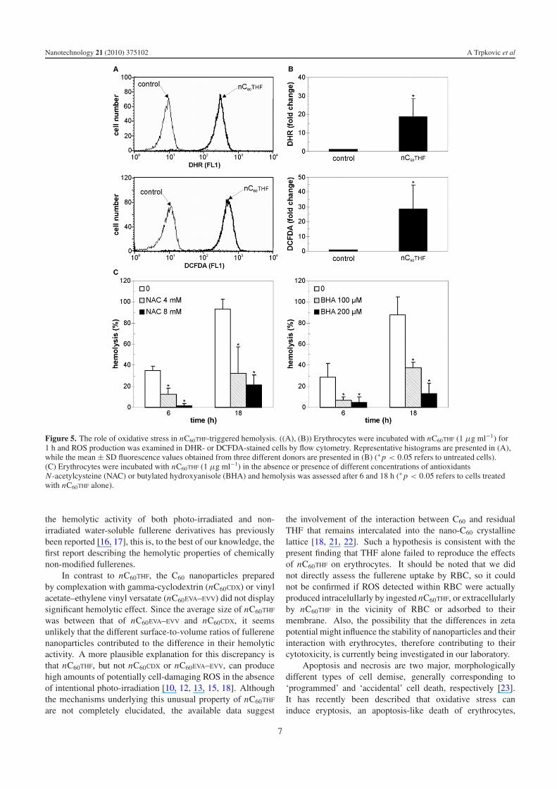
We finally investigated if serum albumin, as the mostabundant protein in human blood plasma, could affect thehemotoxic effect of fullerene nanoparticles. Indeed, thehemolytic activity of nC60THF (1 μg ml−1) was significantlyreduced if it was mixed with BSA at the concentrationpresent in blood (40 mg ml−1) 1 h prior to incubationwith erythrocytes (figure 6(A)). To assess if nC60THF makes
complexes with albumin, we heat-precipitated BSA from themixture of nC60THF (10 μg ml−1) and BSA (40 mg ml−1) at95 ◦C, as previously described [20]. The control samplecontaining only nC60THF was subjected to the same procedure.The samples were centrifuged at 6000 rpm for 30 min,supernatant was removed and investigated by UV–vis forthe presence of nC60THF. As it can be seen from theUV–vis spectra in figure 6(B), C60 absorption bands (270and 340 nm) were clearly evident in the supernatant of thecontrol nC60THF sample, but barely visible in the supernatantof the nC60THF/BSA mixture, thus indicating an interactionof nC60THF with albumin. To get some insight into themorphology of nC60THF/BSA complexes, we performed AFManalysis of particles in the nC60THF/BSA mixture (1 μg ml−1 +
5

Nanotechnology 21 (2010) 375102 A Trpkovic et al
Figure 4. AFM analysis of nC60THF-induced changes in RBC morphology. ((A), (B)) Erythrocytes were incubated with nC60THF (1 μg ml−1)for the indicated time periods and the cell width, height and surface roughness were examined by AFM. Representative AFM images andcorresponding width/height plots are presented in (A), while the mean ± SD values of height, width and surface roughness (Rrms) arepresented in (B) (∗ p < 0.05 refers to untreated cells; n = 10).
5 μg ml−1) in both wave mode and phase mode. A numberof particles in the mixture had irregular shape and were larger(approx. 200 nm) than nC60THF (100 nm; figure 1(C)) oralbumin particles (20 nm; figure 6(C), top), as shown in therepresentative AFM wave mode image (figure 6(C), middle),indicating that nC60THF/albumin complexes were formed. Wenext used the ability of the phase mode to distinguish betweendifferent materials in a sample, with the direction of the phasechange depending on the nature of the interaction between theprobe and the surface. If the particle is homogeneous, the phaseshift of the cantilever remains constant as the probe is rasteredacross the sample and the phase image displays a uniform flatimage of constant phase. However, the phase mode imageshown in figure 6(C) (bottom) is consistent with the composite
nature of the particle, with the rim probably composed ofaggregated smaller albumin particles and interacting with theprobe more intensely than the central part which presumablycontains larger nC60THF. Collectively, these data suggest thatthe expected hemotoxicity of nC60THF could be attenuated bycomplexation with blood albumin.
4. Discussion
The present study shows that C60 nanoparticles preparedby solvent exchange method (nC60THF) caused shrinkage,crenation and, eventually, hemolysis of human RBC.The erythrocyte damage by nC60THF was mediated byoxidative stress and prevented by serum albumin. While
6
Nanotechnology 21 (2010) 375102 A Trpkovic et al
Figure 5. The role of oxidative stress in nC60THF-triggered hemolysis. ((A), (B)) Erythrocytes were incubated with nC60THF (1 μg ml−1) for1 h and ROS production was examined in DHR- or DCFDA-stained cells by flow cytometry. Representative histograms are presented in (A),while the mean ± SD fluorescence values obtained from three different donors are presented in (B) (∗ p < 0.05 refers to untreated cells).(C) Erythrocytes were incubated with nC60THF (1 μg ml−1) in the absence or presence of different concentrations of antioxidantsN-acetylcysteine (NAC) or butylated hydroxyanisole (BHA) and hemolysis was assessed after 6 and 18 h (∗ p < 0.05 refers to cells treatedwith nC60THF alone).
the hemolytic activity of both photo-irradiated and non-irradiated water-soluble fullerene derivatives has previouslybeen reported [16, 17], this is, to the best of our knowledge, thefirst report describing the hemolytic properties of chemicallynon-modified fullerenes.
In contrast to nC60THF, the C60 nanoparticles preparedby complexation with gamma-cyclodextrin (nC60CDX) or vinylacetate–ethylene vinyl versatate (nC60EVA–EVV) did not displaysignificant hemolytic effect. Since the average size of nC60THF
was between that of nC60EVA–EVV and nC60CDX, it seemsunlikely that the different surface-to-volume ratios of fullerenenanoparticles contributed to the difference in their hemolyticactivity. A more plausible explanation for this discrepancy isthat nC60THF, but not nC60CDX or nC60EVA–EVV, can producehigh amounts of potentially cell-damaging ROS in the absenceof intentional photo-irradiation [10, 12, 13, 15, 18]. Althoughthe mechanisms underlying this unusual property of nC60THF
are not completely elucidated, the available data suggest
the involvement of the interaction between C60 and residualTHF that remains intercalated into the nano-C60 crystallinelattice [18, 21, 22]. Such a hypothesis is consistent with thepresent finding that THF alone failed to reproduce the effectsof nC60THF on erythrocytes. It should be noted that we didnot directly assess the fullerene uptake by RBC, so it couldnot be confirmed if ROS detected within RBC were actuallyproduced intracelullarly by ingested nC60THF, or extracellularlyby nC60THF in the vicinity of RBC or adsorbed to theirmembrane. Also, the possibility that the differences in zetapotential might influence the stability of nanoparticles and theirinteraction with erythrocytes, therefore contributing to theircytotoxicity, is currently being investigated in our laboratory.
Apoptosis and necrosis are two major, morphologicallydifferent types of cell demise, generally corresponding to‘programmed’ and ‘accidental’ cell death, respectively [23].It has recently been described that oxidative stress caninduce eryptosis, an apoptosis-like death of erythrocytes,
7
Nanotechnology 21 (2010) 375102 A Trpkovic et al
Figure 6. Serum albumin adsorbs to nC60THF and reduces its hemotoxicity. (A) Erythrocytes were incubated with nC60THF (1 μg ml−1) alone,or with nC60THF that was preincubated with BSA (40 mg ml−1) for 1 h before RBC treatment. Hemolysis was assessed after 24 h (∗ p < 0.05).(B) After 1 h co-incubation of BSA (40 mg ml−1) and nC60THF (10 μg ml−1), BSA was heat-precipitated and the supernatant was analyzed byUV–vis. The same treatment was applied to control the suspension containing only nC60THF. (C) BSA (5 μg ml−1) alone or co-incubated for1 h with nC60THF (5 μg ml−1) was spin-coated on freshly cleaved mica and analyzed by AFM. Both wave mode (middle panel) and phasemode (bottom panel) AFM images of BSA/nC60THF mixture are presented.
characterized by decrease in cell volume and cell membraneblebbing [24, 25]. Oxidative stress triggered eryptosis byactivation of Ca2+-permeable cation channels and subsequentincrease in cytosolic Ca2+ concentration [26, 27]. However,the RBC buffer in our study did not contain Ca2+, so itseems safe to conclude that Ca2+-dependent eryptosis wasnot responsible for the shrinkage and crenation of RBCtreated with nC60THF. Another possibility is that nC60THF
caused a necrosis-like erythrocyte damage by increasingthe cell membrane permeability through peroxidation of thelipid bilayer, as previously reported for other cell types,including fibroblasts, astrocytes and various types of cancercells [13, 15, 18, 28]. Indeed, our flow cytometry data are
consistent with such an assumption, as necrosis is accompaniedby an early decrease in both FSC and SSC values, while anearly decrease in FSC during apoptosis is often accompaniedby simultaneous increase in SSC [29]. The reduced ability ofa necrotic cell to scatter light simultaneously in the forward(FSC) and right angle directions (SSC) probably occurs asa consequence of plasma membrane rupture and leakage ofthe intracellular content. One should, however, keep inmind that the general rules for FSC/SSC alterations duringcell death might not apply for erythrocytes. Also, it isplausible to assume that SSC values will show an earlyincrease due to nanoparticle uptake, which we have actuallyseen previously with nC60CDX and nC60EVA–EVV [10], but
8

Nanotechnology 21 (2010) 375102 A Trpkovic et al
not with nC60THF in the present study. While the absenceof an early SSC increase might indicate that nC60THF isnot internalized by erythrocytes, this needs to be confirmedby more specific methods for determination of nanoparticleuptake. With these issues remaining to be resolved, wepropose the following sequence of events: nC60THF, havinga high affinity towards cell surface [12, 13], binds theerythrocyte membrane and causes lipid peroxidation-mediateddamage by releasing ROS. The ensuing osmotic imbalanceleads to erythrocyte dehydration, causing a reduction in cellsize and subsequent AMF-detectable crenation due to theprotruding membrane-bound spectrin/actin cytoskeleton. Asthe membrane damage progresses with time, the leakage ofhemoglobin and other cellular constituents occurs, eventuallymanifesting as profound deformations of the erythrocyte shapeand hemolysis. Also, the possibility that the accumulationof fullerene nanoparticles at the erythrocyte membrane couldcontribute to the increase in RBC surface roughness requiresfurther investigation with appropriate analytical techniques(e.g. Raman spectroscopy).
An obvious weakness of the present study is the reduc-tionist approach, which, by neglecting the possible interferenceof various blood constituents, clearly oversimplifies the inter-action of nC60THF and RBC in the in vivo conditions. In anattempt to partly overcome this drawback, we tested the in-fluence of serum albumin, which comprises about half of theblood serum protein, on the hemolytic activity of nC60THF.Our finding that serum albumin markedly attenuated nC60THF-induced hemolysis is consistent with its ability to adsorb ontothe surface of C60 nanoparticles, described previously [30, 31]and confirmed in the present study. In this way, albumin couldprevent nC60THF interaction with erythrocytes and subsequentdamage of their membrane. While it is difficult to extrap-olate these findings to the in vivo situation, it neverthelessseems plausible to assume that the blood compatibility of in-travenously injected nC60THF could be significantly improvedby subsequent complexation with serum albumin.
In conclusion, we here demonstrate that the hemolyticactivity of different preparations of chemically non-modifiedfullerenes correlates with their ability to release ROS. Ourdata suggest that fullerene complexes with non-toxic hostmolecules such as γ -cyclodextrin or EVA–EVV, because ofthe superior blood compatibility, could be better candidatesfor in vivo biomedical applications than hemolytic nC60THF orwater-soluble cationic C60 derivatives. Nevertheless, furtherstudies, both in vitro and in vivo, are required to provide newinsights into the mechanisms and biological consequences oferythrocyte interaction with different fullerene preparations.
Acknowledgments
This work was supported by the Ministry of Science andTechnological Development of the Republic of Serbia (grantNo. 145073). The authors thank Dr Ljubica Harhaji-Trajkovicfor critically perusing the manuscript.
References
[1] Partha R Conyers J L 2009 biomedical applications offunctionalized fullerene-based nanomaterials Int. J.Nanomed. 4 261–75
[2] Bakry R et al 2007 Medicinal applications of fullerenes Int. J.Nanomed. 2 639–49
[3] Bosi S, Da Ros T, Spalluto G and Prato M 2003 Fullerenederivatives: an attractive tool for biological applicationsEur. J. Med. Chem. 38 913–23
[4] Nakamura E and Isobe H 2003 Functionalized fullerenes inwater. The first 10 years of their chemistry, biology, andnanoscience Acc. Chem. Res. 36 807–15
[5] Markovic Z and Trajkovic V 2008 Biomedical potential of thereactive oxygen species generation and quenching byfullerenes (C60) Biomaterials 29 3561–73
[6] Dugan L L, Gabrielsen J K, Yu S P, Lin T S and Choi D W1996 Buckminsterfullerenol free radical scavengers reduceexcitotoxic and apoptotic death of cultured cortical neuronsNeurobiol. Dis. 3 129–35
[7] Dugan L L et al 1997 Carboxyfullerenes as neuroprotectiveagents Proc. Natl Acad. Sci. USA 94 9434–9
[8] Bisaglia M et al 2000 C3-fullero-tris-methanodicarboxylic acidprotects cerebellar granule cells from apoptosisJ. Neurochem. 74 1197–204
[9] Harhaji L et al 2008 Modulation of tumor necrosisfactor-mediated cell death by fullerenes Pharm. Res.25 1365–76
[10] Misirkic M S et al 2009 The protection of cells from nitricoxide-mediated apoptotic death by mechanochemicallysynthesized fullerene (C60) nanoparticles Biomaterials30 2319–28
[11] Lao F et al 2009 Fullerene derivatives protect endothelial cellsagainst NO-induced damage Nanotechnology 20 225103
[12] Sayes C M et al 2004 The differential cytotoxicity ofwater-soluble fullerenes Nano Lett. 4 1881–7
[13] Sayes C M, Gobin A M, Ausman K D, Mendez J, West J L andColvin V L 2005 Nano-C60 cytotoxicity is due to lipidperoxidation Biomaterials 26 7587–95
[14] Oberdorster E 2004 Manufactured nanomaterials (fullerenes,C60) induce oxidative stress in the brain of juvenilelargemouth bass Environ. Health Perspect. 112 1058–62
[15] Isakovic A et al 2006 Distinct cytotoxic mechanisms of pristineversus hydroxylated fullerene Toxicol. Sci. 91 173–83
[16] Yang X L, Huang C, Qiao X G, Yao L, Zhao D X andTan X 2007 Photo-induced lipid peroxidation of erythrocytemembranes by a bis-methanophosphonate fullerene Toxicol.In Vitro 21 1493–8
[17] Bosi S et al 2004 Hemolytic effects of water-soluble fullerenederivatives J. Med. Chem. 47 6711–5
[18] Isakovic A et al 2006 Inactivation of nanocrystalline C60
cytotoxicity by γ -irradiation Biomaterials 27 5049–58[19] Skals M, Jensen U B, Ousingsawat J, Kunzelmann K,
Leipziger J and Praetorius H A 2010 Escherichia colialpha-hemolysin triggers shrinkage of erythrocytes viaK(Ca)3.1 and TMEM16A channels with subsequentphosphatidylserine exposure J. Biol. Chem. 285 15557–65
[20] Belgorodsky B, Fadeev L, Kolsenik J and Gozin M 2006Formation of a soluble stable complex between pristineC60-fullerene and a native blood protein Chembiochem7 1783–9
[21] Markovic Z et al 2007 The mechanism of cell-damagingreactive oxygen generation by colloidal fullerenesBiomaterials 28 5437–48
[22] Andrievsky G, Klochkov V and Derevyanchenko L 2005 Is theC60 fullerene molecule toxic? Fullerenes Nanotub. CarbonNanostruct. 13 363–76
[23] Han S I, Kim Y S and Kim T H 2008 Role of apoptotic andnecrotic cell death under physiologic conditions BMB Rep.41 1–10
9

Nanotechnology 21 (2010) 375102 A Trpkovic et al
[24] Berg C P et al 2001 Human mature red blood cells expresscaspase-3 and caspase-8, but are devoid of mitochondrialregulators of apoptosis Cell Death Differ. 8 1197–206
[25] Bratosin D et al 2001 Programmed cell death in matureerythrocytes: a model for investigating death effectorpathways operating in the absence of mitochondria CellDeath Differ. 8 1143–56
[26] Lang K S et al 2003 Cation channels trigger apoptotic death oferythrocytes Cell Death Differ. 10 249–56
[27] Matarrese P et al 2005 Peroxynitrite induces senescence andapoptosis of red blood cells through the activation of aspartyland cysteinyl proteases FASEB J. 19 416–8
[28] Harhaji L et al 2007 Multiple mechanisms underlying theanticancer action of nanocrystalline fullerene Eur. J.Pharmacol. 568 89–98
[29] Vermes I, Haanen C and Reutelingsperger C 2000 Flowcytometry of apoptotic cell death J. Immunol. Methods243 167–90
[30] Deguchi S, Yamazaki T, Mukai S A, Usami R andHorikoshi K 2007 Stabilization of C60 nanoparticles byprotein adsorption and its implications for toxicity studiesChem. Res. Toxicol. 20 854–8
[31] Nikolic N et al 2009 Preparation and biodistribution ofradiolabeled fullerene C60 nanocrystals Nanotechnology20 385102
10
Copyright © 2022 FDOKUMEN


![Inclusion of methano[60]fullerene derivatives in cavitand-based coordination cages](https://static.fdokumen.com/doc/165x107/631ed46f4c5c8fb3a00e625a/inclusion-of-methano60fullerene-derivatives-in-cavitand-based-coordination-cages.jpg)


![Cycloadditions to [60]fullerene using microwave irradiation: A convenient and expeditious procedure](https://static.fdokumen.com/doc/165x107/6321fbfe807dc363600a3a3b/cycloadditions-to-60fullerene-using-microwave-irradiation-a-convenient-and-expeditious.jpg)