Electrophysiological and hemolytic activity elicited by the venom of the jellyfish Cassiopea...
11
Electrophysiological and hemolytic activity elicited by the venom of the jellyfish Cassiopea xamachana Mo ´nica Torres a , Manuel B. Aguilar a , Andre ´s Falco ´n a , Lenin Sa ´nchez a , Faisal F. Y. Radwan b , Joseph W. Burnett b , Edgar P. Heimer-de la Cotera a , Rogelio O. Arellano a, * a Centro de Neurobiologı ´a, Universidad Nacional Auto ´noma de Me ´xico, Juriquilla, Quere ´taro 76001, Me ´xico b Department of Dermatology, School of Medicine, University of Maryland 6th Floor, 405 W. Redwood Street, Baltimore. MD 21201, USA Received 7 September 2000; accepted 9 December 2000 Abstract In this study, we determined hemolysis activity in human and sheep erythrocytes, and characterized the electrical responses in Xenopus oocyte membrane elicited by the venom of the jellyfish Cassiopea xamachana (Cx). The Cx venom produced hemolysis in both species, being more potent on human red cells. The electrophysiological study showed that the Cx venom elicited three different responses in the oocytes. One current was generated in all the oocytes tested and corresponded with a slow inward current (I Cx ) associated with an increase in membrane conductance. I Cx was concentration-dependent and had a reversal potential of 210.3 ^ 0.4 mV. Ionic substitution studies indicated that the conductive pathway was mainly permeable to cations and non-selective. The oocyte membrane resistance was completely recovered after washout of the venom, this suggested that the effect was due to generation of a specific membrane conductance as opposed to a possible non-specific membrane breakdown. A comparative study with three distinct native cationic channels present in the oocyte membrane [i.e. (1) hemi-gap-junction channels, (2) mechanosensitive channels, and (3) the ouabain-sensitive channel activated by palytoxin], showed that I Cx might correspond to opening of mechanosensitive channels or to activation of an unknown cationic channel located in the oocyte membrane. The bioactive fraction eliciting I Cx were peptides and was separated from two other peptidic hemolytic fractions by chromatography. q 2001 Elsevier Science Ltd. All rights reserved. Keywords: Cassiopea; Xenopus oocyte; Ionic channels; Electrophysiology; Hemolysis 1. Introduction The marine invertebrates included in the phylum Cnidaria—jellyfishes, sea anemones and corals—express a wide variety of neurotoxic and cytolytic substances (Pong- Prayoon et al., 1991; Lotan et al., 1996). These toxic substances are principally confined to a cytoplasmic capsules, the nematocysts (Watson and Wood, 1988). Cassiopea xamachana (Cx) is a jellyfish that, like all other members of this genus, is called “the upside-down jellyfish”. The presence of Cx has been reported in all the oceans, and particularly at the Indo–Pacific, also around Hawaii, and the Caribbean Sea. Its venom can cause moder- ately painful stings in humans that commonly results in a rash, and occasionally within an hour, produces vomiting, painful joints swelling, facial edema and generalized urti- caria (Rifkin and Fennes, 1996). Isolation of Cx nematocysts has proven difficult (Fitt and Trench, 1983; Radwan et al., 2001), and has hampered research on Cx venom. Nevertheless, studies on its hemoly- tic, proteolytic and phospholipase actions exist (Pong- Prayoon et al., 1991; Radwan et al., 2001). Also, the crude venom has been reported to contain small nonpeptide antic- holinesterases and muscarinic toxins (Karlsson et al., 1991). Among the bioactivities of different jellyfishes toxins, cyto- lysis is probably one of the most documented (Endean et al., 1993; Bernheimer and Rudy, 1986; Rottini et al., 1995; Lotan et al., 1996; Malpezzi et al., 1993). In addition, it has been reported that some jellyfish venoms generate electrical activity in different cell models, for example: (1) Aurelia sp venom causes depolarization in frog muscle (Kihara et al., 1988); (2) Carybdea rastonii produces a calcium-dependent contractile Toxicon 39 (2001) 1297–1307 0041-0101/01/$ - see front matter q 2001 Elsevier Science Ltd. All rights reserved. PII: S0041-0101(01)00081-2 www.elsevier.com/locate/toxicon * Corresponding author. Tel.: 152-4-2381062; fax: 152-4- 2381062. E-mail address: [email protected] (R.O. Arellano).
-
Upload
independent -
Category
Documents
-
view
4 -
download
0
Transcript of Electrophysiological and hemolytic activity elicited by the venom of the jellyfish Cassiopea...
Electrophysiological and hemolytic activity elicited by the venomof the jelly®sh Cassiopea xamachana
MoÂnica Torresa, Manuel B. Aguilara, AndreÂs FalcoÂna, Lenin SaÂncheza, Faisal F.Y. Radwanb, Joseph W. Burnettb, Edgar P. Heimer-de la Coteraa, Rogelio O. Arellanoa,*
aCentro de NeurobiologõÂa, Universidad Nacional AutoÂnoma de MeÂxico, Juriquilla, QuereÂtaro 76001, MeÂxicobDepartment of Dermatology, School of Medicine, University of Maryland 6th Floor, 405 W. Redwood Street, Baltimore. MD 21201, USA
Received 7 September 2000; accepted 9 December 2000
Abstract
In this study, we determined hemolysis activity in human and sheep erythrocytes, and characterized the electrical responses
in Xenopus oocyte membrane elicited by the venom of the jelly®sh Cassiopea xamachana (Cx). The Cx venom produced
hemolysis in both species, being more potent on human red cells. The electrophysiological study showed that the Cx venom
elicited three different responses in the oocytes. One current was generated in all the oocytes tested and corresponded with a
slow inward current (ICx) associated with an increase in membrane conductance. ICx was concentration-dependent and had a
reversal potential of 210.3 ^ 0.4 mV. Ionic substitution studies indicated that the conductive pathway was mainly permeable
to cations and non-selective. The oocyte membrane resistance was completely recovered after washout of the venom, this
suggested that the effect was due to generation of a speci®c membrane conductance as opposed to a possible non-speci®c
membrane breakdown. A comparative study with three distinct native cationic channels present in the oocyte membrane [i.e. (1)
hemi-gap-junction channels, (2) mechanosensitive channels, and (3) the ouabain-sensitive channel activated by palytoxin],
showed that ICx might correspond to opening of mechanosensitive channels or to activation of an unknown cationic channel
located in the oocyte membrane. The bioactive fraction eliciting ICx were peptides and was separated from two other peptidic
hemolytic fractions by chromatography. q 2001 Elsevier Science Ltd. All rights reserved.
Keywords: Cassiopea; Xenopus oocyte; Ionic channels; Electrophysiology; Hemolysis
1. Introduction
The marine invertebrates included in the phylum
CnidariaÐjelly®shes, sea anemones and coralsÐexpress
a wide variety of neurotoxic and cytolytic substances (Pong-
Prayoon et al., 1991; Lotan et al., 1996). These toxic
substances are principally con®ned to a cytoplasmic capsules,
the nematocysts (Watson and Wood, 1988).
Cassiopea xamachana (Cx) is a jelly®sh that, like all
other members of this genus, is called ªthe upside-down
jelly®shº. The presence of Cx has been reported in all the
oceans, and particularly at the Indo±Paci®c, also around
Hawaii, and the Caribbean Sea. Its venom can cause moder-
ately painful stings in humans that commonly results in a
rash, and occasionally within an hour, produces vomiting,
painful joints swelling, facial edema and generalized urti-
caria (Rifkin and Fennes, 1996).
Isolation of Cx nematocysts has proven dif®cult (Fitt and
Trench, 1983; Radwan et al., 2001), and has hampered
research on Cx venom. Nevertheless, studies on its hemoly-
tic, proteolytic and phospholipase actions exist (Pong-
Prayoon et al., 1991; Radwan et al., 2001). Also, the crude
venom has been reported to contain small nonpeptide antic-
holinesterases and muscarinic toxins (Karlsson et al., 1991).
Among the bioactivities of different jelly®shes toxins, cyto-
lysis is probably one of the most documented (Endean et al.,
1993; Bernheimer and Rudy, 1986; Rottini et al., 1995; Lotan
et al., 1996; Malpezzi et al., 1993). In addition, it has been
reported that some jelly®sh venoms generate electrical activity
in different cell models, for example: (1) Aurelia sp venom
causes depolarization in frog muscle (Kihara et al., 1988); (2)
Carybdea rastonii produces a calcium-dependent contractile
Toxicon 39 (2001) 1297±1307
0041-0101/01/$ - see front matter q 2001 Elsevier Science Ltd. All rights reserved.
PII: S0041-0101(01)00081-2
www.elsevier.com/locate/toxicon
* Corresponding author. Tel.: 152-4-2381062; fax: 152-4-
2381062.
E-mail address: [email protected] (R.O. Arellano).
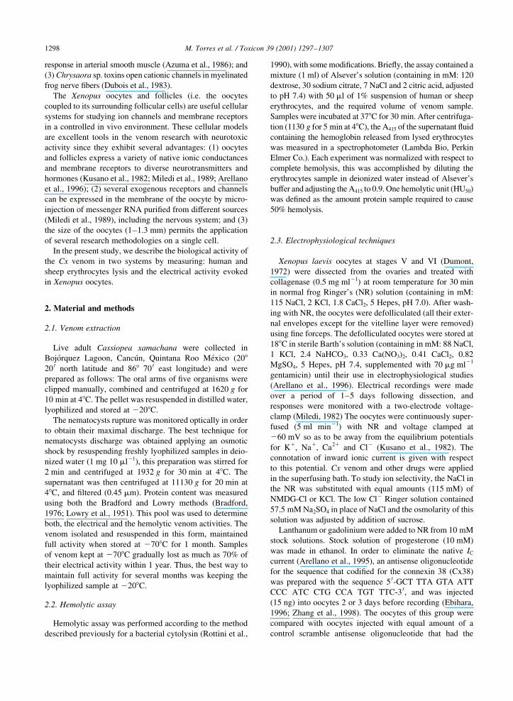
response in arterial smooth muscle (Azuma et al., 1986); and
(3) Chrysaora sp. toxins open cationic channels in myelinated
frog nerve ®bers (Dubois et al., 1983).
The Xenopus oocytes and follicles (i.e. the oocytes
coupled to its surrounding follicular cells) are useful cellular
systems for studying ion channels and membrane receptors
in a controlled in vivo environment. These cellular models
are excellent tools in the venom research with neurotoxic
activity since they exhibit several advantages: (1) oocytes
and follicles express a variety of native ionic conductances
and membrane receptors to diverse neurotransmitters and
hormones (Kusano et al., 1982; Miledi et al., 1989; Arellano
et al., 1996); (2) several exogenous receptors and channels
can be expressed in the membrane of the oocyte by micro-
injection of messenger RNA puri®ed from different sources
(Miledi et al., 1989), including the nervous system; and (3)
the size of the oocytes (1±1.3 mm) permits the application
of several research methodologies on a single cell.
In the present study, we describe the biological activity of
the Cx venom in two systems by measuring: human and
sheep erythrocytes lysis and the electrical activity evoked
in Xenopus oocytes.
2. Material and methods
2.1. Venom extraction
Live adult Cassiopea xamachana were collected in
BojoÂrquez Lagoon, CancuÂn, Quintana Roo MeÂxico (20820 0 north latitude and 868 70 0 east longitude) and were
prepared as follows: The oral arms of ®ve organisms were
clipped manually, combined and centrifuged at 1620 g for
10 min at 48C. The pellet was resuspended in distilled water,
lyophilized and stored at 2208C.
The nematocysts rupture was monitored optically in order
to obtain their maximal discharge. The best technique for
nematocysts discharge was obtained applying an osmotic
shock by resuspending freshly lyophilized samples in deio-
nized water (1 mg 10 ml21), this preparation was stirred for
2 min and centrifuged at 1932 g for 30 min at 48C. The
supernatant was then centrifuged at 11130 g for 20 min at
48C, and ®ltered (0.45 mm). Protein content was measured
using both the Bradford and Lowry methods (Bradford,
1976; Lowry et al., 1951). This pool was used to determine
both, the electrical and the hemolytic venom activities. The
venom isolated and resuspended in this form, maintained
full activity when stored at 2708C for 1 month. Samples
of venom kept at 2708C gradually lost as much as 70% of
their electrical activity within 1 year. Thus, the best way to
maintain full activity for several months was keeping the
lyophilized sample at 2208C.
2.2. Hemolytic assay
Hemolytic assay was performed according to the method
described previously for a bacterial cytolysin (Rottini et al.,
1990), with some modi®cations. Brie¯y, the assay contained a
mixture (1 ml) of Alsever's solution (containing in mM: 120
dextrose, 30 sodium citrate, 7 NaCl and 2 citric acid, adjusted
to pH 7.4) with 50 ml of 1% suspension of human or sheep
erythrocytes, and the required volume of venom sample.
Samples were incubated at 378C for 30 min. After centrifuga-
tion (1130 g for 5 min at 48C), the A415 of the supernatant ¯uid
containing the hemoglobin released from lysed erythrocytes
was measured in a spectrophotometer (Lambda Bio, Perkin
Elmer Co.). Each experiment was normalized with respect to
complete hemolysis, this was accomplished by diluting the
erythrocytes sample in deionized water instead of Alsever's
buffer and adjusting the A415 to 0.9. One hemolytic unit (HU50)
was de®ned as the amount protein sample required to cause
50% hemolysis.
2.3. Electrophysiological techniques
Xenopus laevis oocytes at stages V and VI (Dumont,
1972) were dissected from the ovaries and treated with
collagenase (0.5 mg ml21) at room temperature for 30 min
in normal frog Ringer's (NR) solution (containing in mM:
115 NaCl, 2 KCl, 1.8 CaCl2, 5 Hepes, pH 7.0). After wash-
ing with NR, the oocytes were defolliculated (all their exter-
nal envelopes except for the vitelline layer were removed)
using ®ne forceps. The defolliculated oocytes were stored at
188C in sterile Barth's solution (containing in mM: 88 NaCl,
1 KCl, 2.4 NaHCO3, 0.33 Ca(NO3)2, 0.41 CaCl2, 0.82
MgSO4, 5 Hepes, pH 7.4, supplemented with 70 mg ml21
gentamicin) until their use in electrophysiological studies
(Arellano et al., 1996). Electrical recordings were made
over a period of 1±5 days following dissection, and
responses were monitored with a two-electrode voltage-
clamp (Miledi, 1982) The oocytes were continuously super-
fused (5 ml min21) with NR and voltage clamped at
260 mV so as to be away from the equilibrium potentials
for K1, Na1, Ca21 and Cl2 (Kusano et al., 1982). The
connotation of inward ionic current is given with respect
to this potential. Cx venom and other drugs were applied
in the superfusing bath. To study ion selectivity, the NaCl in
the NR was substituted with equal amounts (115 mM) of
NMDG-Cl or KCl. The low Cl2 Ringer solution contained
57.5 mM Na2SO4 in place of NaCl and the osmolarity of this
solution was adjusted by addition of sucrose.
Lanthanum or gadolinium were added to NR from 10 mM
stock solutions. Stock solution of progesterone (10 mM)
was made in ethanol. In order to eliminate the native IC
current (Arellano et al., 1995), an antisense oligonucleotide
for the sequence that codi®ed for the connexin 38 (Cx38)
was prepared with the sequence 5 0-GCT TTA GTA ATT
CCC ATC CTG CCA TGT TTC-3 0, and was injected
(15 ng) into oocytes 2 or 3 days before recording (Ebihara,
1996; Zhang et al., 1998). The oocytes of this group were
compared with oocytes injected with equal amount of a
control scramble antisense oligonucleotide that had the
M. Torres et al. / Toxicon 39 (2001) 1297±13071298
sequence 5 0-CTT TTG ACC GCT CAT CCC TAT AGT
ATT TGC-3 0.
2.4. HPLC size exclusion chromatography
The Cx venom was processed for HPLC using a Bio-Rad
Bio-Sil SEC-125 column (600 £ 7.5 mm) equilibrated and
eluted with 10 mM ammonium acetate buffer (pH 7); ¯ow
rate of 1ml min21; 1 ml fractions collected; detection,
206 nm. The fractions were lyophilized and resuspended
in deionized water and protein concentration was deter-
mined by the Lowry method.
The column was calibrated with the following molecular
weight standards: thyroglobulin (670,000); bovine gamma
globulin (158,000); albumin (66,200); chicken ovoalbumin
(44,000); horse myoglobulin (17,000); and vitamin B-12
(1350), equilibrated and eluted with phosphate buffer pH
6.8 (sodium phosphate 0.10 M and NaCl 0.15 M).
The 1,2-bis(2-aminophenoxy)-ethane-N,N,N 0,N 0-tetraa-
cetic acid acetoxymethyl ester (BAPTA-AM) was obtained
from Molecular Probes (Eugene, OR, USA). Fetal bovine
serum (FBS), progesterone, collagenase (type I), gentami-
cin, and ouabain were purchased from Sigma (St. Louis,
MO, USA) and all salts and other reagents were obtained
from J. T. Baker (Phillipsburg, PA, USA) or Sigma.
3. Results
3.1. Cytolytic assay
The crude Cx venom had concentration-dependent hemo-
lytic activity. This was detected in both, human and sheep
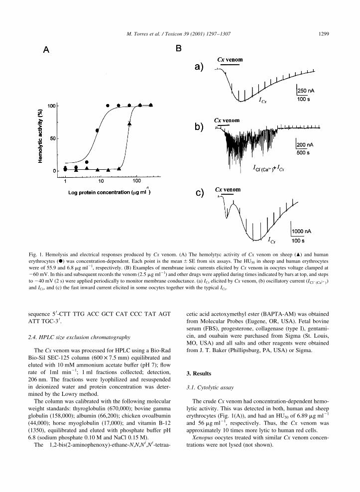
erythrocytes (Fig. 1(A)), and had an HU50 of 6.89 mg ml21
and 56 mg ml21, respectively. Thus, the Cx venom was
approximately 10 times more lytic to human red cells.
Xenopus oocytes treated with similar Cx venom concen-
trations were not lysed (not shown).
M. Torres et al. / Toxicon 39 (2001) 1297±1307 1299
Fig. 1. Hemolysis and electrical responses produced by Cx venom. (A) The hemolytyc activity of Cx venom on sheep (O) and human
erythrocytes (X) was concentration-dependent. Each point is the mean ^ SE from six assays. The HU50 in sheep and human erythrocytes
were of 55.9 and 6.8 mg ml21, respectively. (B) Examples of membrane ionic currents elicited by Cx venom in oocytes voltage clamped at
260 mV. In this and subsequent records the venom (2.5 mg ml21) and other drugs were applied during times indicated by bars at top, and steps
to 240 mV (2 s) were applied periodically to monitor membrane conductance. (a) ICx elicited by Cx venom, (b) oscillatory current (ICl2�Ca21�)and ICx, and (c) the fast inward current elicited in some oocytes together with the typical ICx.
3.2. Electrical activity elicited by Cx venom in Xenopus
oocytes
The resting membrane potential of the oocytes (n� 150,
25 frogs) used in this study was 234 ^ 6 mV (all data given
as means ^ SE) and had an average input resistance of
0.79 ^ 0.2 MV.
In oocytes, the Cx venom evoked complex inward
membrane currents associated with an increase in
membrane conductance (Fig. 1(B)). Two components of
the current response were not elicited in all cells, one of
these was a smooth inward current that inactivated rapidly
even in the presence of the venom (63 ^ 5.7 s, Fig. 1(Bc)).
This response was observed in 15 oocytes of four frogs from
a total of 25 frogs.
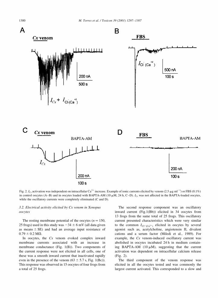
The second response component was an oscillatory
inward current (Fig.1(Bb)) elicited in 34 oocytes from
13 frogs from the same total of 25 frogs. This oscillatory
current presented characteristics which were very similar
to the common ICl2�Ca21� elicited in oocytes by several
agonist such as, acetylcholine, angiotensin II, divalent
cations and a serum factor (Miledi et al., 1989). For
example, the Cx venom-induced oscillatory current was
abolished in oocytes incubated 24 h in medium contain-
ing BAPTA-AM (10 mM), suggesting that the current
activation was dependent on intracellular calcium release
(Fig. 2).
The third component of the venom response was
elicited in all the oocytes tested and was commonly the
largest current activated. This corresponded to a slow and
M. Torres et al. / Toxicon 39 (2001) 1297±13071300
Fig. 2. ICx activation was independent on intracellular Ca21 increase. Example of ionic currents elicited by venom (2.5 mg ml21) or FBS (0.1%)
in control oocytes (A±B) and in oocytes loaded with BAPTA-AM (10 mM, 24 h, C±D). ICx was not affected in the BAPTA-loaded oocytes,
while the oscillatory currents were completely eliminated (C and D).

smooth inward current, denoted here as ICx. The activa-
tion of ICx was concentration-dependent (Fig. 3(A) and
(B), six oocytes from two frogs), but the average current
amplitudes were variable between oocytes from different
frogs. For example, the venom (1.25 mg ml21) in oocytes
(n� 5) from one frog generated ICx of 1327 ^ 133 nA,
while venom from the same extraction elicited in oocytes
(n� 5) from another frog currents of 302 ^ 87 nA. The
ICx recovered slowly, normally several minutes after the
venom was washed-out. Also, subsequent venom applica-
tions in the same oocyte produced desensitization of the
response (Fig. 3(C)).
In oocytes (28 from six frogs) incubated 24 h with
BAPTA-AM (10 mM) the ICx amplitude and its time-course
were not affected, while the oscillatory current in the same
oocytes evoked by the Cx venom or serum were completely
eliminated, this result strongly suggested that ICx was not
dependent on intracellular Ca21 increase.
3.3. Ionic basis of ICx
Given that ICx was the more consistent response elicited
by the venom, we studied in detail the electrophysiological
characteristics of this current.
Further experiments were performed in order to evaluate
the ionic basis of ICx and to establish a possible relationship
with previously described ionic channels in the native
oocyte (see below). For this, oocytes were clamped at
260 mV, and a series of voltage steps (2.6 s) from 2140
to 160 mV were applied during superfusion with NR
(control current) or at the ICx peak. For each voltage step,
the control membrane currents were substracted from those
obtained during ICx peak, and these values were plotted as in
Fig. 3(E).
The ICx had a reversal potential (Erev) of 210.3 ^ 0.4 mV
(n� 10, Fig. 3(E)). This value differs from the equilibrium
potentials for Cl2 (220 mV), K1 (2100 mV) or Na1
M. Torres et al. / Toxicon 39 (2001) 1297±1307 1301
Fig. 3. Concentration-response and current-voltage relationships. (A) Examples of membrane ionic currents elicited in an oocyte held at
260 mV. Superimposed traces are the membrane current elicited by subsequent applications of Cx venom (15 ml) at different concentrations
(in mg ml21: a� 0.62, b� 1.25, c� 2.5, e� 5) in a single oocyte with washing periods of 10 min. (B) Concentration-response relationship for
ICx obtained in six oocytes (two frogs). ICx obtained in each concentration were normalized with respect to the maximal current generated by
5 mg ml21 of the Cx venom. (C) Traces are examples of subsequent Cx venom (2.5 mg ml21) applications in a single oocyte producing
desensitization of the response. (D) Superimposed traces are examples of voltage steps, from 2140 to 160 in increases of 20 mV applied
to an oocyte held at 260 mV during superfusion with NR (control traces) and during the peak elicited by 5 mg ml21 of Cx venom. (E) Current-
voltage relationship of ICx. Each point represents the average of the current obtained in 10 oocytes from two frogs.

(160 mV) in Xenopus oocytes (Kusano et al., 1982),
suggesting that the ICx channels were permeable to more
than a single type of ion.
Ion substitution studies were performed in order to know
the participation of the principal ions during ICx. Replace-
ment of 100, 50, 75 and 25% NaCl in the NR by N-methyl-
d-glucamine gave Erev's of 239.83 ^ 2.23 mV (n� 6),
224.32 ^ 1.03 mV (n� 6), 219.66 ^ 0.16 mV (n� 6),
and 214.99 ^ 1.31 mV (n� 6), respectively (Fig. 4(A)).
As ICx was reduced throughout the voltage range studied,
this effect was stronger on the inward currents suggesting
that Na1 is a main current carrier in the response. Substitut-
ing all Na1 by K1 slightly shifted the Erev to
212.8 ^ 1.4 mV, six oocytes (Fig. 4(B)) suggesting that
ICx channels were also permeable to K1. Finally, ICx elicited
in Cl2 free medium had an Erev 214.03 ^ 0.39 (not shown).
All these results are in agreement with the behavior
expected of a channel that is permeable mainly to cations
and discriminates poorly between Na1 and K1.
3.4. Action mechanism involved in ICx generation.
Given that ICx was driven through a cationic permeable
pathway, we were interested in determining whether or not
the channels involved corresponded with any native cationic
conductance previously characterized in Xenopus oocytes.
Indirect evidence indicated that ICx might be generated by
opening of an oocyte native channel. For example, one fact
supporting this idea was obtained in experiments in which a
group of oocytes were induced to mature by incubation with
progesterone (10 mM, approximately 5 h). It is known that
several native responses of the oocyte are down regulated or
eliminated in maturated oocytes. The cholinergic responses
(Kusano et al., 1982), the IC current (Arellano et al., 1995,
see below), and several other native electrical responses are
completely eliminated in mature oocytes (Miledi, R. and
Arellano, R.O., unpublished observations). The ICx was
almost completely eliminated in maturated oocytes
(Fig. 5(B)). Experiments made in cells from a particular
donor showed that control oocytes (n� 4) responded to
venom with robust ICx of 1858 ^ 85 nA while in maturated
oocytes from the same frog only one from four oocytes
responded with an ICx of 20 nA. Thus, the electrical response
produced by the Cx venom in the oocyte membrane is regu-
lated by maturation as likewise several other native
responses.
Previous studies have shown that oocyte membrane is
endowed with at least three distinct native cationic currents
which are generated by: (1) the opening of hemi-gap-junc-
tion channels (Cx38) that generates the current named IC
(Arellano et al., 1995; Zhang et al., 1998); (2) palytoxin
application that activates an inward ouabain-sensitive
current (Wang and Horisberger, 1997; Scheiner-Bobis and
Schneider, 1997); and (3) the activation of mechanosensi-
tive channels (IMS) (Yang and Sachs, 1989, 1990). To deter-
mine if one of these channels is involved in the ICx
generation, the following experiments were performed.
(1) The IC native current is generated when the oocytes
are exposed to medium deprived of divalent cations
(Arellano et al., 1995), and is due to the opening of
Cx38 hemi-gap-junction channels commonly present in
the membrane of the oocyte. The injection of Cx38 anti-
sense oligonucleotide into the oocytes eliminates IC in
approximately 48 h (Ebihara, 1996; Zhang et al., 1998).
Therefore, we injected oocytes with an oligonucleotide
antisense to Cx38 and incubated for .48 h until IC
vanished, as evidenced by perfusing the oocytes with
Ca21-free Ringer solution. These injected-oocytes not
M. Torres et al. / Toxicon 39 (2001) 1297±13071302
Fig. 4. Ionic basis of ICx. Oocytes were superfused with solutions containing different concentrations of the principal ions in Ringer solution and
the current-voltage relations were constructed applying a series of voltage steps before and during the ICx peak. ICx was generated in NR (X) or
in one of following solutions, (A) substitution of 100% Na1 by NMDG1 (W) or 50% (O), and (B) substitution of all Na1 by K1 (O).
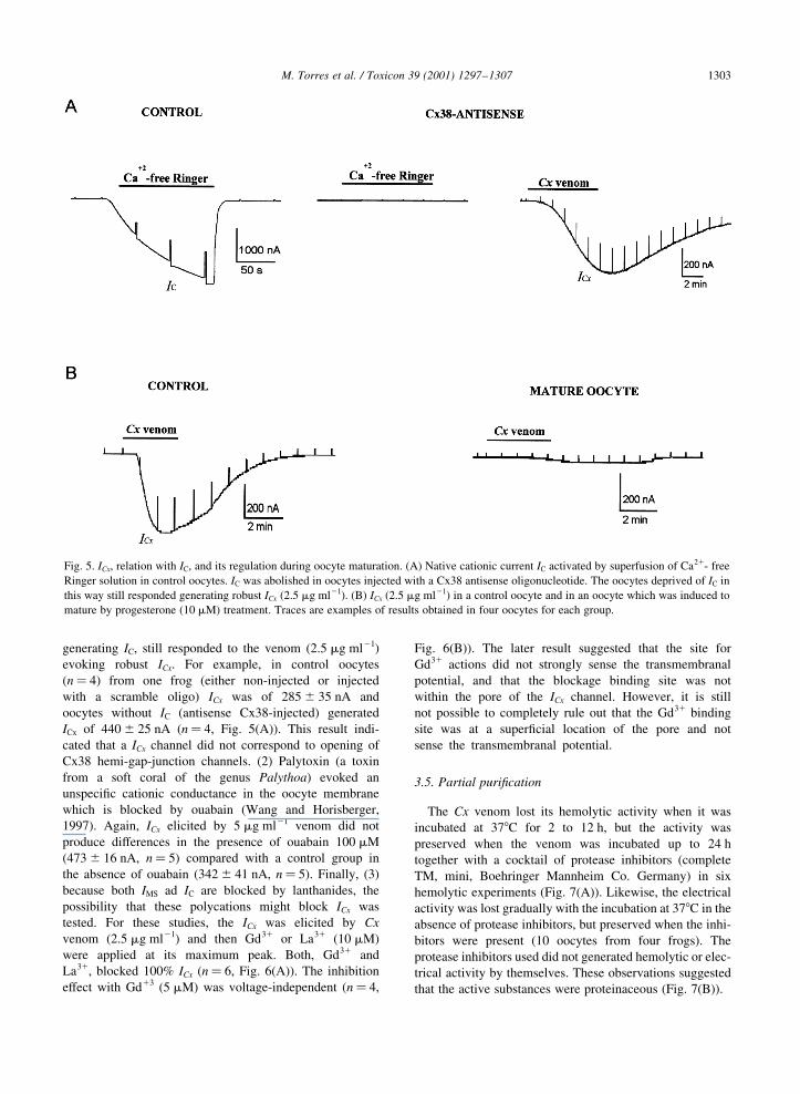
generating IC, still responded to the venom (2.5 mg ml21)
evoking robust ICx. For example, in control oocytes
(n� 4) from one frog (either non-injected or injected
with a scramble oligo) ICx was of 285 ^ 35 nA and
oocytes without IC (antisense Cx38-injected) generated
ICx of 440 ^ 25 nA (n� 4, Fig. 5(A)). This result indi-
cated that a ICx channel did not correspond to opening of
Cx38 hemi-gap-junction channels. (2) Palytoxin (a toxin
from a soft coral of the genus Palythoa) evoked an
unspeci®c cationic conductance in the oocyte membrane
which is blocked by ouabain (Wang and Horisberger,
1997). Again, ICx elicited by 5 mg ml21 venom did not
produce differences in the presence of ouabain 100 mM
(473 ^ 16 nA, n� 5) compared with a control group in
the absence of ouabain (342 ^ 41 nA, n� 5). Finally, (3)
because both IMS ad IC are blocked by lanthanides, the
possibility that these polycations might block ICx was
tested. For these studies, the ICx was elicited by Cx
venom (2.5 mg ml21) and then Gd31 or La31 (10 mM)
were applied at its maximum peak. Both, Gd31 and
La31, blocked 100% ICx (n� 6, Fig. 6(A)). The inhibition
effect with Gd13 (5 mM) was voltage-independent (n� 4,
Fig. 6(B)). The later result suggested that the site for
Gd31 actions did not strongly sense the transmembranal
potential, and that the blockage binding site was not
within the pore of the ICx channel. However, it is still
not possible to completely rule out that the Gd31 binding
site was at a super®cial location of the pore and not
sense the transmembranal potential.
3.5. Partial puri®cation
The Cx venom lost its hemolytic activity when it was
incubated at 378C for 2 to 12 h, but the activity was
preserved when the venom was incubated up to 24 h
together with a cocktail of protease inhibitors (complete
TM, mini, Boehringer Mannheim Co. Germany) in six
hemolytic experiments (Fig. 7(A)). Likewise, the electrical
activity was lost gradually with the incubation at 378C in the
absence of protease inhibitors, but preserved when the inhi-
bitors were present (10 oocytes from four frogs). The
protease inhibitors used did not generated hemolytic or elec-
trical activity by themselves. These observations suggested
that the active substances were proteinaceous (Fig. 7(B)).
M. Torres et al. / Toxicon 39 (2001) 1297±1307 1303
Fig. 5. ICx, relation with IC, and its regulation during oocyte maturation. (A) Native cationic current IC activated by superfusion of Ca21- free
Ringer solution in control oocytes. IC was abolished in oocytes injected with a Cx38 antisense oligonucleotide. The oocytes deprived of IC in
this way still responded generating robust ICx (2.5 mg ml21). (B) ICx (2.5 mg ml21) in a control oocyte and in an oocyte which was induced to
mature by progesterone (10 mM) treatment. Traces are examples of results obtained in four oocytes for each group.
M. Torres et al. / Toxicon 39 (2001) 1297±13071304
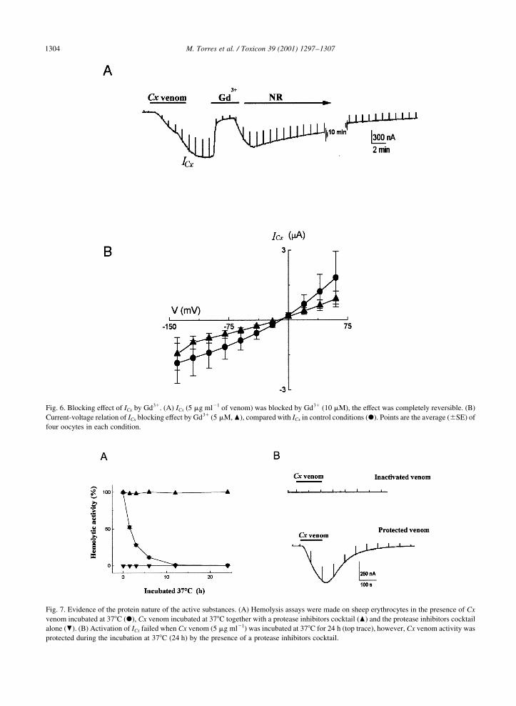
Fig. 7. Evidence of the protein nature of the active substances. (A) Hemolysis assays were made on sheep erythrocytes in the presence of Cx
venom incubated at 378C (X), Cx venom incubated at 378C together with a protease inhibitors cocktail (O) and the protease inhibitors cocktail
alone (P). (B) Activation of ICx failed when Cx venom (5 mg ml21) was incubated at 378C for 24 h (top trace), however, Cx venom activity was
protected during the incubation at 378C (24 h) by the presence of a protease inhibitors cocktail.
Fig. 6. Blocking effect of ICx by Gd31. (A) ICx (5 mg ml21 of venom) was blocked by Gd31 (10 mM), the effect was completely reversible. (B)
Current-voltage relation of ICx blocking effect by Gd31 (5 mM, O), compared with ICx in control conditions (X). Points are the average (^SE) of
four oocytes in each condition.
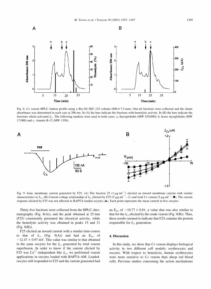
Thirty ®ve fractions were collected from the HPLC chro-
matography (Fig. 8(A)), and the peak obtained at 25 min
(F25) consistently presented the electrical activity, while
the hemolytic activity was obtained in peaks 15 and 31
(Fig. 8(B)).
F25 elicited an inward current with a similar time-course
to that of ICx (Fig. 9(A)) and had an Erev of
212.47 ^ 0.97 mV. This value was similar to that obtained
in the same oocytes for the ICx generated by total venom
superfusion. In order to know if the current elicited by
F25 was Ca21-independent like ICx, we performed venom
applications in oocytes loaded with BAPTA-AM. Loaded-
oocytes still responded to F25 and the current generated had
an Erev of 210.77 ^ 0.81, a value that was also similar to
that for the ICx elicited by the crude venom (Fig. 9(B)). Thus,
these results seemed to indicate that F25 contains the protein
responsible for ICx generation.
4. Discussion
In this study, we show that Cx venom displays biological
activity in two different cell models: erythrocytes and
oocytes. With respect to hemolysis, human erythrocytes
were more sensitive to Cx venom than sheep red blood
cells. Previous studies concerning the action mechanisms
M. Torres et al. / Toxicon 39 (2001) 1297±1307 1305
Fig. 8. Cx venom HPLC elution pro®le using a Bio-Sil SEC-125 column (600 £ 7.5 mm). One ml fractions were collected and the eluate
absorbance was determined in each case at 206 nm. In (A) the bars indicate the fractions with hemolytic activity. In (B) the bars indicate the
fractions which activated ICx. The following markers were used in both cases: a, thyroglobulin (MW 670,000); b, horse myoglobulin (MW
17,000) and c, vitamin B-12 (MW 1350).
Fig. 9. Ionic membrane current generated by F25. (A) The fraction 25 (1 mg ml21) elicited an inward membrane current with similar
characteristics to ICx. (B) Current-voltage relationship of ICx elicited by F25 (5 mg ml21, e) and total Cx venom (5 mg ml21, X). The current
response elicited by F25 was not affected in BAPTA-loaded oocytes (O). Each point represents the mean current in ®ve oocytes.

of cytolytic proteins have shown that these substances
interact directly with the membrane lipids (Bernheimer
and Rudy, 1986), either by presenting intrinsic phospholi-
pase activity or by generating transmembranal hydrophilic
pores. Some cytolytic toxins interact with target cell via
cholesterol (Rossjohn et al., 1997) and, in other cases, sphin-
gomyelin (Bernheimer and Rudy, 1986). The proportion of
these membrane lipids in erythrocytes differs between
animal species; for example, sheep erythrocyte membrane
contains about three times more sphingomyelin than the
human erythrocytes (Rottini et al., 1995). Thus, differences
in membrane lipids composition might explain the higher
selectivity of Cx venom on human erythrocytes.
The electrophysiological study shows that the Cx venom
elicits three different responses in Xenopus oocytes. One of
them, denoted ICx, was generated in all oocytes tested. ICx
was produced by the opening of cationic channels in the
oocyte membrane; these channels discriminate poorly
between Na1 and K1.
We consider at least two possible mechanisms involved in
the activation of ICx by the venom: (1) The Cx venom
peptide was incorporated to the membrane to form an ion-
conducting pore, a similar mechanism to that used by cyto-
lysins (Bernheimer and Rudy, 1986; Rossjohn et al., 1997);
and (2) Cx venom activated native cationic channels present
in the oocyte membrane. Three observations suggest that the
Cx venom exerts its action by opening a speci®c membrane
conductance rather than inducing a non-speci®c membrane
breakdown: (1) the complete recovery of the oocyte
membrane conductance after continuos exposure to Cx
venom; (2) the desensitization of ICx resulting from subse-
quent venom applications in the same oocyte; and (3) the
elimination of ICx in mature oocytes.
The electrical characteristics of ICx indicated that there are
at least three different types of native channels that might
correspond with those activated by Cx venom.
Our results show that ICx was not generated via the Na1-
K1 pump which has been proposed to be activated by paly-
toxin, neither by opening of IC channels, since oocytes
deprived of IC by injection with an antisense against Cx38
channel nucleotide sequence still responded potently to the
venom. The third option, a venom action on the gating of
mechanosensitive channels still remains as a strong possi-
bility. Indirect evidence that support a similarity between ICx
and IMS include: (1) both are cationic currents; (2) also, both
are blocked completely by a concentration close to 10 mM
Gd31; and (3) importantly, this blocking effect was
provoked through a voltage-independent mechanism, that
suggested strongly which the channels involved are blocked
by Gd31 at a binding site outside the membranal electric
®eld (cf. Yang and Sachs, 1989)
It is interesting to mention that maitotoxin, a marine
neurotoxin from the dino¯agellate Gambierdiscus toxi-
cus, can also elicit an inward cationic current (IMtx) in
Xenopus oocytes (Bielfeld-Ackermann et al., 1998).
Additionally, IMtx is blocked by Gd31 and is non-
permeable to NMDG1. Finally, maitotoxin can produces
an increase of intracellular Ca21 in addition to IMtx
(Bielfeld-Ackermann et al., 1998; Weber et al., 2000).
Because of these observations, it has been proposed that
the mechanosensitive channels are related with those
channels activated by maitotoxin (Bielfeld-Ackermann
et al., 1998). Thus, some similarity exist between ICx,
IMtx and IMS, which is of interest since IMS channels in
oocytes are regularly activated by mechanical pressure
applied via the patch-clamp pipette after the formation
of a tight seal (e.g. Yang and Sachs, 1989). There is no
clear evidence of any other stimuli activating macro-
scopic IMS currents in the whole oocyte. Thus, drug-
activated MS-conductance might represent a novel tool
for the study of the physiological signi®cance of these
channels in the oocyte cell membrane. Alternatively, Cx
venom might be activating a different and unknown
channel. The results presented here may help the under-
standing of characteristics of native conductances and
channels which are important in regulating the oocyte
development and maturation.
The protective effect produced by the protease inhibitor
cocktail on Cx venom incubated at 378C (24 h) strongly
suggested that the venom's active substances were peptides.
Other biochemical studies about the compounds found in the
venom of some jelly®shes have separated different polypep-
tides and enzymes such as collagenases, proteases, elastases,
phospholipases and nucleases (Pong-Prayoon et al., 1991;
Smith, 1936).
The chromatographic separation of Cx venom rendered
three different bioactive fractions, one of them elicited ICx
and two produced hemolysis. The fact that more than one
hemolytic protein is present in the Cx venom is a feature
common to other jelly®sh that can posses two or more cyto-
lysins in the same species (Endean, 1993).
The peptide responsible for the electrical activity might
probably be smaller than horse myoglobulin (17,000 Da).
This conclusion is not de®nitive since adsorption could
occur to the column support, or aggregation of various
proteins in the eluate, these events have been reported to
occur in several other jelly®sh venom proteins (Othman and
Burnett, 1990; Bloom et al., 1998).
Acknowledgements
We are grateful for the help during this study of Ma.
Eugenia Ramos, Bs, Pilar Galarza, Bs and Edith Garay,
PhD, from the CNB-UNAM, and Fernando Negrete, MS
and Lourdes Segura-Puertas, PhD, from the ICML-
UNAM. This work was supported by the grants: CONCy-
TEQ (B-11) to M.T. and E.H., and by DGAPA-UNAM
(IN 200198, IN-209596) and CONACyT (32364-N) to
R.O.A. Also, M.T. acknowledges PhD fellowships from
DGEP-UNAM and PAEP-UNAM (205312).
M. Torres et al. / Toxicon 39 (2001) 1297±13071306

References
Arellano, R.O., Woodward, R.M., Miledi, R., 1995. A monovalent
cationic conductance that is blocked by extracellular divalent
cations in Xenopus oocytes. J. Physiol., Lond. 484, 593±604.
Arellano, R.O., Woodward, R.M., Miledi, R., 1996. Ion channels
and membrane receptors in follicle-enclosed Xenopus oocytes.
In: Narahashi, T. (Ed.). Ion Channels. Plenum Press, New York,
pp. 203±256.
Azuma, H., Ishikawa, M., Nakajima, T., Satoh, A., Sekizaki, S.,
1986. Calcium-dependent contractile response of arterial
smooth muscle to a jelly®sh toxin (pCrTX:Carybdea rastonii).
Eur. J. Pharmac. 88, 549±559.
Bernheimer, A.W., Rudy, B., 1986. Interactions between
membranes and cytolytic peptides. Biochim. Biophys. Acta
864 (1), 123±141.
Bielfeld-Ackermann, A., Ranger, C., Korbmacher, C., 1998. Maito-
toxin (MTX) activates a non selective cation channel in Xenopus
laevis oocytes. P¯uÈgers Arch-Eur. Physiol. 436, 329±337.
Bloom, D.A., Burnett, J., Alderslade, P., 1998. Partial puri®cation
of box jelly®sh (Chironex ¯eckeri) nematocyst venom isolated
at the beach side. Toxicon 36, 1075±1085.
Bradford, M.M., 1976. A rapid and sensitive method for the
quantitation of microgram quantities of protein utilizing the
principle of protein-dye binding. Analyt. Biochem. 72, 248±
254.
Dubois, J.M., Tanguy, J., Burnett, J.W., 1983. Ionic channels
induced by sea nettle toxin in the nodal membrane. Biophys.
J. 42, 199±202.
Dumont, J.N., 1972. Oogenesis in Xenopus laevis (Daudin). I.
Stages of oocyte development in laboratory maintained animals.
J. Morphol. 136, 153±180.
Ebihara, L., 1996. Xenopus connexin 38 forms hemi-gap-junctions
channels in the nonjunctional plasma membrane of Xenopus
oocytes. Biophys. J. 71, 742±748.
Endean, R., Monks, S.A., Cameron, A.M., 1993. Toxins from the
box-jelly®sh Chironex ¯eckeri. Toxicon 31, 397±410.
Fitt, W.F., Trench, R.K., 1983. Endocytosis of the symbiotic dinof-
¯agelates Microadriaticum freudenthal by endoderm cells of
the scyphistomae of Cassiopea xamachana and resistance of
the algae to host digestion. J. Cell Sci. 64, 195±212.
Karlsson, E., Adem, A., Aneiros, A., Castaneda, O., Harve, A.,
Jolkkon, M., Sotolongo, V., 1991. New toxins from marine
organism. Toxicon 29, 1168.
Kihara, H., Anraku, M., Ohno, M., Hashimura, S., 1988. Tetrodo-
toxin-unaffected depolarization of frog muscles induced by
venom of jelly®sh (Genus Aurelia). Jpn. J. Physiol. 38, 839±
849.
Kusano, K., Miledi, R., Stinnakre, J., 1982. Cholinergic and
catecholaminergic receptors in the Xenopus oocyte membrane.
J. Physiol., Lond. 328, 143±170.
Lotan, A., Fishman, L., Zlotkin, E., 1996. Toxin compartmentation
and delivery in the cnidaria:the nematocyst's tubule as a multi-
headed poisonous arrow. J. Exp. Zoo. 275, 444±451.
Lowry, O.H., Rosebrogh, N.J., Farr, A.L., Randall, R.J., 1951.
Protein measurement with Folin phenol reagent. J. Biol.
Chem. 193, 265±275.
Malpezzi, E.L., De Freitas, J.C., Muramoto, K., Kamiya, H., 1993.
Characterization of peptides in sea anemone venom collected by
a novel procedure. Toxicon 31, 853±864.
Miledi, R., Parker, I., Sumikawa, K., 1989. Transplanting receptors
from brains into oocytes. Fidia Research Foundation Neuroscience
Award Lectures. Raven Press, New York, pp. 57±90.
Miledi, R., 1982. A calcium-dependent transient outward current
evoked in Xenopus oocytes. Proc. R. Soc. B 215, 491±497.
Othman, Y., Burnett, J.W., 1990. Techniques applicable for purifying
Chironex ¯eckeri (box-jelly®sh) venom. Toxicon 28, 821±835.
Pong-Prayoon, U., Bohloin, L., Wasuwat, S., 1991. Neutralization
of toxic effects of different crude jelly®sh venoms by an extract
of Ipomea pes-caprae (L.). R. Br. J. Ethnopharmac. 35, 65±69.
Radwan, F.F.Y., Burnett, J.W., Bloom, D.A., Coliano, T., Elde-
frawi, M.E., Erdely, H., Aurelian, L., Torres, M., Heimer, E.,
2001. A comparison of the toxinological characteristics of two
Cassiopea and Aurelia species. Toxicon 39, 245±257.
Rifkin, J., Fenner, P., 1996. Genus Cassiopea (upside-down jelly
®sh). In: Williamson, J., Fenner, P., Burnett, J., Rifkin, J. (Eds.).
Venomous and Poisonous Marine Animals: a Medical and
Biological Handbook. University of New South Wales Press,
Sydney, pp. 180±235.
Rossjohn, J., Feil, S.C., McKinstry, W.J., Twenten, R., Parker,
M.W., 1997. Structure of cholesterol-binding, thiol-activated
cytolysin and a model of its membrane form. Cell 89, 685±692.
Rottini, G., Dobrina, A., Forgiarini, O., Nardon, E., Amirante, G.,
Patriarca, P., 1990. Identi®cation and partial characterization of
a cytolitic toxin produced by Gardnerella vaginalis. Infec.
Immun. 58, 3751±3758.
Rottini, G., Gusmani, L., Parovel, E., Avian, M., Patriarca, P., 1995.
Puri®cation and properties of the jelly®sh Caribdea marsupia-
lis. Toxicon 33, 315±326.
Scheiner-Bobis, G., Schneider, H., 1997. Palytoxin-induced channel
formation within the Na 1 /K 1 -ATPase does not require a
catalytically active enzyme. Eur. J. Biochem. 248, 3717±3723.
Smith, H.G., 1936. Contribution to the anatomy and physiology of
Cassiopea frondosa. In: Papers from Tortugas Laboratory (Ed.),
Departaments of Zoology, 31,19-52. Universities of Bristol and
Aberdeen, Bristol.
Wang, X., Horisberger, J-D., 1997. Palytoxin effects through inter-
action with the Na,K-ATPase in Xenopus oocyte. FEBS Letters
409, 391±395.
Watson, M.G., Wood, L.R., 1988. Colloquium on terminology.
In: Hessinger, Lenhoff (Eds.). The Biology of Nematocysts.
Academic Press, New York.
Weber, W.M., Popp, C., Claus, W., Van Driessche, W., 2000.
Maitotoxin induces insertion of different ion channels into the
Xenopus oocyte plasma membrane via Ca21-stimulated exocy-
tosis. P¯uÈgers Arch. Eur. Physiol. 439, 363±369.
Yang, X.C., Sachs, F., 1989. Block of stretch-activated ion channels
in Xenopus oocytes by gadolinium and calcium ions. Science
243, 1068±1071.
Yang, X.C., Sachs, F., 1990. Characterization of stretch-activated
ion channels in Xenopus oocytes. J. Physiol. 431, 103±122.
Zhang, Y., McBride Jr, D., Hamill, O., 1998. The ion selectivity of a
membrane conductance inactivated by extracellular calcium in
Xenopus oocyte. J. Physiol. 508 (3), 763±776.
M. Torres et al. / Toxicon 39 (2001) 1297±1307 1307