Drug-induced immune hemolytic anemia: the last 30 ... - Exeley
11
44 IMMUNOHEMATOLOGY, Volume 30, Number 2, 2014 Sally Frank Memorial Award and Lectureship* Drug-induced immune hemolytic anemia: the last 30 years of changes P.A. Arndt Drug-induced immune hemolytic anemia (DIIHA) is a rare condition that occurs primarily as a result of drug-induced antibodies, either drug-dependent or drug-independent. Drug- dependent antibodies can be detected by testing drug-treated red blood cells (RBCs) or untreated RBCs in the presence of a solution of drug. Drug-independent antibodies react with untreated RBCs (no drug added) and cannot be distinguished from warm autoantibodies. Many changes have occurred during the last 30 years, such as which drugs most commonly cause DIIHA, the optimal testing methods for identifying them, and the theories behind the mechanisms by which they react. This article reviews the major changes in DIIHA since the early 1980s involving the immune complex mechanism, cephalosporins, nonimmunologic protein adsorption, and penicillins. Because serologic results associated with DIIHA can mimic those expected with autoimmune hemolytic anemia or hemolytic transfusion reactions, DIIHA may go undetected in some cases. Immunohematology 2014;30:44–54. Key Words: immune hemolytic anemia, drugs, antibodies, red blood cells, IgG, complement It was a case of being at the right place at the right time. Shortly after my graduation in 1983 from the Specialist in Blood Banking (SBB) program at the American Red Cross in Los Angeles, there was an opening for a research associate in Dr. George Garratty’s research laboratory, and I was hired. Dr. Garratty’s main research focus was the immune hemolytic anemias. My coworkers in the research laboratory in the 1980s were Nina Postoway and Sandy Nance. Sandy taught me how to perform flow cytometric analyses on red blood cells (RBCs) and the monocyte monolayer assay (MMA). Nina taught me how to perform the enzyme-linked antiglobulin test for direct antiglobulin test (DAT)-negative autoimmune hemolytic anemia (AIHA) workups and also how to do drug-induced immune hemolytic anemia (DIIHA) workups. Nina said to expect that most, if not all, DIIHA workups would be negative. She was right…at first…and then things started to change. I worked in Dr. Garratty’s research laboratory at the Red Cross in Southern California for a little over 30 years and during that time saw many changes relating to DIIHA. This presentation will review some of those changes. Background The incidence of DIIHA is unknown, but Dr. Garratty suggested that it may be about 1 in a million. 1 Causes of DIIHA can be classified into two broad categories: drug-induced antibodies and nonimmunologic protein adsorption (NIPA). Drug-induced antibodies can be further subdivided into drug- dependent antibodies and drug-independent antibodies. The first reported drug-dependent antibody was an antibody to stibophen (an anthelmintic drug) in 1956, 2 and the first described drug-independent antibodies were autoantibodies induced by methyldopa (an antihypertensive drug) in 1966. 3,4 The first drug noted to cause NIPA was cephalothin (an antibiotic); positive DATs were first reported in 1967, 5,6 and positive indirect antiglobulin tests (IATs), in 1971. 7 The next few paragraphs summarize knowledge with regard to DIIHA in the early 1980s, using the 1980 textbook Acquired Immune Hemolytic Anemias by Petz and Garratty 8 as a reference. At that time, 32 drugs had been reported to cause immune hemolytic anemia (IHA) or positive DATs. The Petz and Garratty book 8 described four mechanisms: (1) immune complex, (2) drug adsorption, (3) NIPA, and (4) AIHA. Immune Complex This mechanism was said to occur when drugs and drug antibodies (IgG or IgM) combined to form immune complexes, which were then nonspecifically adsorbed onto RBCs and activated complement. Most drugs that caused DIIHA reacted by this mechanism: prototype drugs were quinidine (an antiarrhythmic drug) and phenacetin (a nonsteroidal anti- inflammatory drug [NSAID]). Typical characteristics of this mechanism were (1) a previously sensitized patient needed to take only a small amount of the drug; (2) as a result of complement activation, patients had acute intravascular hemolysis (hemoglobinemia and hemoglobinuria) and often R EPORT *Presented at the 2012 AABB Annual Meeting
-
Upload
khangminh22 -
Category
Documents
-
view
2 -
download
0
Transcript of Drug-induced immune hemolytic anemia: the last 30 ... - Exeley

44 IMMUNOHEMATOLOGY, Volume 30, Number 2, 2014
Sally Frank Memorial Award and Lectureship*
Drug-induced immune hemolytic anemia: the last 30 years of changesP.A. Arndt
Drug-induced immune hemolytic anemia (DIIHA) is a rare condition that occurs primarily as a result of drug-induced antibodies, either drug-dependent or drug-independent. Drug-dependent antibodies can be detected by testing drug-treated red blood cells (RBCs) or untreated RBCs in the presence of a solution of drug. Drug-independent antibodies react with untreated RBCs (no drug added) and cannot be distinguished from warm autoantibodies. Many changes have occurred during the last 30 years, such as which drugs most commonly cause DIIHA, the optimal testing methods for identifying them, and the theories behind the mechanisms by which they react. This article reviews the major changes in DIIHA since the early 1980s involving the immune complex mechanism, cephalosporins, nonimmunologic protein adsorption, and penicillins. Because serologic results associated with DIIHA can mimic those expected with autoimmune hemolytic anemia or hemolytic transfusion reactions, DIIHA may go undetected in some cases. Immunohematology 2014;30:44–54.
Key Words: immune hemolytic anemia, drugs, antibodies, red blood cells, IgG, complement
It was a case of being at the right place at the right time. Shortly after my graduation in 1983 from the Specialist in Blood Banking (SBB) program at the American Red Cross in Los Angeles, there was an opening for a research associate in Dr. George Garratty’s research laboratory, and I was hired. Dr. Garratty’s main research focus was the immune hemolytic anemias. My coworkers in the research laboratory in the 1980s were Nina Postoway and Sandy Nance. Sandy taught me how to perform flow cytometric analyses on red blood cells (RBCs) and the monocyte monolayer assay (MMA). Nina taught me how to perform the enzyme-linked antiglobulin test for direct antiglobulin test (DAT)-negative autoimmune hemolytic anemia (AIHA) workups and also how to do drug-induced immune hemolytic anemia (DIIHA) workups. Nina said to expect that most, if not all, DIIHA workups would be negative. She was right…at first…and then things started to change. I worked in Dr. Garratty’s research laboratory at the Red Cross in Southern California for a little over 30 years and during that
time saw many changes relating to DIIHA. This presentation will review some of those changes.
Background
The incidence of DIIHA is unknown, but Dr. Garratty suggested that it may be about 1 in a million.1 Causes of DIIHA can be classified into two broad categories: drug-induced antibodies and nonimmunologic protein adsorption (NIPA). Drug-induced antibodies can be further subdivided into drug-dependent antibodies and drug-independent antibodies. The first reported drug-dependent antibody was an antibody to stibophen (an anthelmintic drug) in 1956,2 and the first described drug-independent antibodies were autoantibodies induced by methyldopa (an antihypertensive drug) in 1966.3,4 The first drug noted to cause NIPA was cephalothin (an antibiotic); positive DATs were first reported in 1967,5,6 and positive indirect antiglobulin tests (IATs), in 1971.7
The next few paragraphs summarize knowledge with regard to DIIHA in the early 1980s, using the 1980 textbook Acquired Immune Hemolytic Anemias by Petz and Garratty8 as a reference. At that time, 32 drugs had been reported to cause immune hemolytic anemia (IHA) or positive DATs. The Petz and Garratty book8 described four mechanisms: (1) immune complex, (2) drug adsorption, (3) NIPA, and (4) AIHA.
Immune ComplexThis mechanism was said to occur when drugs and drug
antibodies (IgG or IgM) combined to form immune complexes, which were then nonspecifically adsorbed onto RBCs and activated complement. Most drugs that caused DIIHA reacted by this mechanism: prototype drugs were quinidine (an antiarrhythmic drug) and phenacetin (a nonsteroidal anti-inflammatory drug [NSAID]). Typical characteristics of this mechanism were (1) a previously sensitized patient needed to take only a small amount of the drug; (2) as a result of complement activation, patients had acute intravascular hemolysis (hemoglobinemia and hemoglobinuria) and often
RepoRt
*Presented at the 2012 AABB Annual Meeting

IMMUNOHEMATOLOGY, Volume 30, Number 2, 2014 45
2012 Sally Frank Award Lecture: DIIHA
renal failure; (3) the DAT was positive, often with C3 only; and (4) in vitro reactions (agglutination, hemolysis, and sensitization) could only be demonstrated by incubating a mixture of the patient’s serum, the drug, and test RBCs (the “immune complex” method).
Drug AdsorptionThis mechanism was used to describe the few drugs that
bound firmly to RBC membranes; antibodies to these drugs reacted with the RBC-bound drug. Penicillin (an antibiotic) was the prototype drug for this mechanism. Typical characteristics of penicillin-induced DIIHA were (1) the patients had received large doses of penicillin intravenously, e.g., 10 million units daily for one or more weeks; (2) the extravascular hemolysis developed over the course of 7–10 days, and hemolysis of decreasing severity could persist for several weeks after the penicillin was stopped; (3) the DAT was strongly positive for IgG, and sometimes C3 was also present; and (4) in vitro tests of the patient’s serum and eluate with drug-treated RBCs were positive (serum IgG antibody titer usually ≥1000).
Nonimmunologic Protein AdsorptionThis mechanism occurred when drugs modified the
RBC membrane so that plasma proteins were adsorbed nonimmunologically. Cephalothin (trade name Keflin) was the prototype drug for this mechanism. Characteristics of NIPA included the following: (1) patients taking cephalothin had a positive DAT (using antisera directed to albumin, complement, fibrinogen, immunoglobulins, etc.) but no hemolytic anemia, and (2) in vitro, cephalothin-treated RBCs incubated in normal plasma were coated with numerous proteins (the IAT was positive using various antisera as above), but those same RBCs incubated with an eluate were typically nonreactive because protein levels in the eluate were too low.
Autoimmune Hemolytic AnemiaThis mechanism occurred when the drug induced
an autoantibody that reacted with normal RBCs (no drug added), similar to reactivity seen with autoantibodies found in idiopathic IgG warm AIHA. The prototype drug for this mechanism was methyldopa (trade name Aldomet). The characteristics of methyldopa-induced autoantibodies were (1) 10 to 36 percent of patients developed a strongly positive DAT (IgG ± C3) after taking methyldopa for 3 to 6 months (this appeared to be dose-dependent), and the DAT could take up to 2 years to become negative after methyldopa was stopped; (2) about 0.8 percent of patients gradually developed extravascular hemolytic anemia, which resolved after cessation
of methyldopa; and (3) the patient’s serum and eluate both contained an autoantibody serologically indistinguishable from other IgG warm autoantibodies. One proposed mech-anism was that methyldopa caused an alteration of the immune system.9 Proof that methyldopa caused AIHA came only from clinical observations.
Changes Since the Early 1980s
The Research LaboratoryIn 1990, Nina and Sandy left the research laboratory to
pursue other transfusion medicine careers, and in 1993, Gina Leger joined our laboratory from the Immunohematology Reference Laboratory. It was my turn to teach her most of the standard tests, and by then, DIIHA workups had become very interesting. So she, too, was in the right place at the right time.
The Immune Complex MechanismThe immune complex mechanism was proposed in the
1950s to 1960s to explain drug-induced thrombocytopenia (i.e., drug antibodies bind with the drug and then the drug–antibody complex is adsorbed nonspecifically by platelets) and was then later extended to apply to RBCs.10,11 Criticisms of this mechanism started appearing in the 1970s to 1980s. Some findings that do not support the immune complex mechanism include the following: (1) drug–antibody immune complexes (e.g., RBC-bound or in plasma) have never been demonstrated experimentally; (2) some drug-dependent antibodies bind to their target cells by their Fab domain, e.g., quinidine antibodies with platelets12,13 and tolmetin antibodies with RBCs14; if the binding was nonspecific, one would expect binding through both the Fab and the Fc domains; (3) drug-dependent antibodies appear to be highly specific for certain cell lines, e.g., either platelets or RBCs (this would not be expected if immune complexes were attaching nonspecifically to cells); (4) some drug-dependent antibodies react with specific antigens on target cells, e.g., anti-quinidine with glycoprotein Ib/IX or IIb/IIIa on platelets15 (see Table 1 for reported specificities of drug-dependent antibodies reactive with RBCs); and (5) many patients have been described with antibodies reacting by more than one mechanism, e.g., drug-independent autoantibodies plus drug-dependent antibodies reacting by the drug adsorption or immune complex method (reviewed in Petz and Garratty36 and in Garratty37).
Some alternative hypotheses to the immune complex mechanism have been proposed to explain observations with drug-induced antibodies. Ackroyd38 proposed that for one type of drug-induced thrombocytopenia, the drug and cell formed
46 IMMUNOHEMATOLOGY, Volume 30, Number 2, 2014
P.A. Arndt
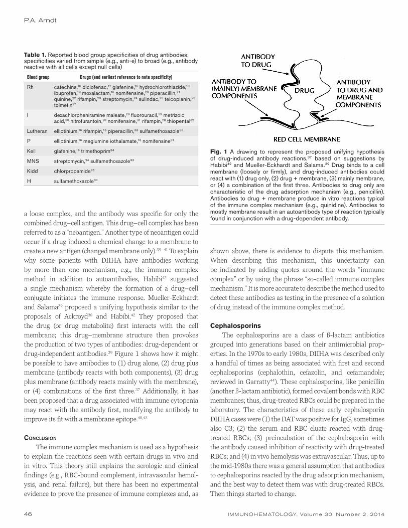
a loose complex, and the antibody was specific for only the combined drug–cell antigen. This drug–cell complex has been referred to as a “neoantigen.” Another type of neoantigen could occur if a drug induced a chemical change to a membrane to create a new antigen (changed membrane only).39–41 To explain why some patients with DIIHA have antibodies working by more than one mechanism, e.g., the immune complex method in addition to autoantibodies, Habibi42 suggested a single mechanism whereby the formation of a drug–cell conjugate initiates the immune response. Mueller-Eckhardt and Salama39 proposed a unifying hypothesis similar to the proposals of Ackroyd38 and Habibi.42 They proposed that the drug (or drug metabolite) first interacts with the cell membrane; this drug–membrane structure then provokes the production of two types of antibodies: drug-dependent or drug-independent antibodies.39 Figure 1 shows how it might be possible to have antibodies to (1) drug alone, (2) drug plus membrane (antibody reacts with both components), (3) drug plus membrane (antibody reacts mainly with the membrane), or (4) combinations of the first three.37 Additionally, it has been proposed that a drug associated with immune cytopenia may react with the antibody first, modifying the antibody to improve its fit with a membrane epitope.40,43
ConClusion
The immune complex mechanism is used as a hypothesis to explain the reactions seen with certain drugs in vivo and in vitro. This theory still explains the serologic and clinical findings (e.g., RBC-bound complement, intravascular hemol-ysis, and renal failure), but there has been no experimental evidence to prove the presence of immune complexes and, as
shown above, there is evidence to dispute this mechanism. When describing this mechanism, this uncertainty can be indicated by adding quotes around the words “immune complex” or by using the phrase “so-called immune complex mechanism.” It is more accurate to describe the method used to detect these antibodies as testing in the presence of a solution of drug instead of the immune complex method.
CephalosporinsThe cephalosporins are a class of ß-lactam antibiotics
grouped into generations based on their antimicrobial prop-erties. In the 1970s to early 1980s, DIIHA was described only a handful of times as being associated with first and second cephalosporins (cephalothin, cefazolin, and cefamandole; reviewed in Garratty44). These cephalosporins, like penicillin (another ß-lactam antibiotic), formed covalent bonds with RBC membranes; thus, drug-treated RBCs could be prepared in the laboratory. The characteristics of these early cephalosporin DIIHA cases were (1) the DAT was positive for IgG, sometimes also C3; (2) the serum and RBC eluate reacted with drug-treated RBCs; (3) preincubation of the cephalosporin with the antibody caused inhibition of reactivity with drug-treated RBCs; and (4) in vivo hemolysis was extravascular. Thus, up to the mid-1980s there was a general assumption that antibodies to cephalosporins reacted by the drug adsorption mechanism, and the best way to detect them was with drug-treated RBCs. Then things started to change.
Table 1. Reported blood group specificities of drug antibodies; specificities varied from simple (e.g., anti-e) to broad (e.g., antibody reactive with all cells except null cells)
Blood group Drugs (and earliest reference to note specificity)
Rh catechins,16 diclofenac,17 glafenine,16 hydrochlorothiazide,18 ibuprofen,19 moxalactam,16 nomifensine,20 piperacillin,21 quinine,22 rifampin,23 streptomycin,24 sulindac,25 teicoplanin,26 tolmetin27
I dexachlorpheniramine maleate,28 fluorouracil,29 metrizoic acid,30 nitrofurantoin,28 nomifensine,31 rifampin,28 thiopental32
Lutheran elliptinium,16 rifampin,16 piperacillin,33 sulfamethoxazole33
P elliptinium,16 meglumine iothalamate,16 nomifensine31
Kell glafenine,16 trimethoprim34
MNS streptomycin,24 sulfamethoxazole33
Kidd chlorpropamide35
H sulfamethoxazole34
Fig. 1 A drawing to represent the proposed unifying hypothesis of drug-induced antibody reactions,37 based on suggestions by Habibi42 and Mueller-Eckhardt and Salama.39 Drug binds to a cell membrane (loosely or firmly), and drug-induced antibodies could react with (1) drug only, (2) drug + membrane, (3) mainly membrane, or (4) a combination of the first three. Antibodies to drug only are characteristic of the drug adsorption mechanism (e.g., penicillin). Antibodies to drug + membrane produce in vitro reactions typical of the immune complex mechanism (e.g., quinidine). Antibodies to mostly membrane result in an autoantibody type of reaction typically found in conjunction with a drug-dependent antibody.

IMMUNOHEMATOLOGY, Volume 30, Number 2, 2014 47
2012 Sally Frank Award Lecture: DIIHA
Cefotaxime
In 1987, Salama et al.45 described a patient with immune complex–mediated intravascular hemolysis caused by an antibody to cefotaxime (a third-generation cephalosporin). The DAT was positive for C3 only, the serum was nonreactive with drug-treated RBCs but did react by the immune complex method (serum plus RBCs in the presence of a solution of drug), and preincubation of drug with the antibody did not cause inhibition of its reactivity.
Ceftriaxone
A year later, our laboratory reported, in an abstract at the 1988 AABB meeting, a fatal case of DIIHA attributable to ceftriaxone (another third-generation cephalosporin).46 The clinical and serologic characteristics were those associated with the immune complex mechanism. The DAT was positive for C3 only, and the serum reacted only by the immune complex method (serum plus RBCs in the presence of a solution of drug); drug-treated RBCs were nonreactive.
Cefotetan
In 1989, there was one AABB abstract and, in 1990, there were four AABB abstracts describing DIIHA (one fatality) attributable to a second-generation cephalosporin, cefotetan (reviewed by Garratty44). These antibodies reacted by more than one method: all five reacted with cefotetan-treated RBCs, three of four reacted in the presence of a solution of cefotetan, and two of four reacted with untreated RBCs (no cefotetan added). Table 2 lists reasons for reactivity of serum in tests when the drug was not added in vitro. In cases of DIIHA caused by cefotetan, we believe this reactivity with untreated RBCs is a result of drug-independent autoantibodies that are found in combination with drug-dependent antibodies (possibly represented by the “mainly membrane” epitope in Fig. 1).
Thus, it was clear by the late 1980s that the best way to detect cephalosporin antibodies was not always by testing drug-treated RBCs. Since then, there have been numerous reports of DIIHA caused by different cephalosporins. Table 3 summarizes how these cephalosporin antibodies were detected. Antibodies to first- and second-generation cephalosporins were primarily detected using drug-treated RBCs with the exception of cefotetan-dependent antibodies, which were detected equally by two methods: drug-treated RBCs and in the presence of a solution of drug. Antibodies to third-generation cephalosporins were primarily detected by testing in the presence of a solution of drug (ceftriaxone antibodies are only detected by this method). Reactivity in
the absence of the in vitro drug was primarily detected in patients with DIIHA caused by cefotetan but was found with some other cephalosporins as well. Our current approach for detection of drug-dependent antibodies of any kind is as follows: (1) if the drug antibody has been well studied, use the previously described method(s) for detection, or (2) if the drug has not been previously described or well studied, test by both methods: (a) serum and RBC eluate with drug-treated RBCs (and untreated RBCs) and (b) serum with untreated or enzyme-treated RBCs in the presence of soluble drug (or saline as a dilution control).47
Table 2. Reasons for serum to be reactive in drug-dependent antibody tests when drug has not been added in vitro (e.g., controls)
Alloantibody
Autoantibody, two types
1. Autoantibody alone
a. Idiopathic
b. Drug-independent antibody (e.g., methyldopa or fludarabine)
2. Autoantibody found in the presence of drug-dependent antibody
Circulating drug or drug plus antibody immune complexes present in the blood sample
Presence of drug in reagent (e.g., low-ionic-strength saline [LISS])
Table 3. Drug-induced immune hemolytic anemia caused by cephalosporins: summary of methods used and results (number positive/number tested) in references in which data on methods were given
Cephalosporin ReferenceDrug-treated
RBCsSerum + RBCs
+ drugNo in vitro
drug
First-generation
Cephalothin 5 5/5 1/1 0/5
Cefazolin 2 2/2 0/1 0/2
Cephalexin 1 1/1 0/0 ?
Second-generation
Cefoxitin 4 4/4 1/2 0/3
Cefamandole 1 1/1 0/0 0/1
Cefotetan 31 31/31 20/22 12/25
Cefuroxime 2 2/2 0/2 0/2
Cefotaxime 4 3/4 3/4 1/4
Third-generation
Ceftazidime 4 2/4 3/4 0/4
Ceftriaxone 28 0/12 28/28 5/19
Ceftizoxime 4 2/4 3/4 2/4
Cefixime 1 1/1 1/1 0/1
RBCs = red blood cells.

48 IMMUNOHEMATOLOGY, Volume 30, Number 2, 2014
Case studies
In the 1970s, the most commonly seen drug-induced antibodies in Dr. Garratty’s laboratory were caused by methyldopa and penicillin (Table 4). This scenario changed in the 1980s, and for the last several decades, cephalosporins, particularly cefotetan and ceftriaxone, have been the most common causes of DIIHA.48 Two cases illustrate common features of DIIHA caused by ceftriaxone or cefotetan.
Patient 1 was a 10-year-old boy with juvenile idiopathic arthritis who had previously received ceftriaxone. He was admitted to the emergency room with fever and vomiting. At 1409 hours, his hemoglobin (Hb) was 10.9 g/dL. Ceftriaxone (tradename Rocephin) was started at 1542 hours. At 1615 hours, his blood pressure dropped and a change in consciousness was noted; therefore, the ceftriaxone was stopped, but it was then restarted at 1745 hours. At 1825 hours, his Hb had plummeted to 0.5 g/dL; transfusions were unsuccessfully attempted, and he was pronounced dead at 1834 hours. Post-mortem samples were sent to our laboratory to determine whether the patient had fatal DIIHA caused by ceftriaxone. RBC-bound IgM and C3 were detected on the patient’s RBCs (no IgG or IgA). Ceftriaxone-treated RBCs cannot be prepared in vitro (we and others have tried several methods without success), so the only way to test for ceftriaxone antibodies is in the presence of a solution of ceftriaxone. The patient’s serum plus ceftriaxone caused direct agglutination (2+) of untreated RBCs; the phosphate-buffered saline (PBS) dilution control was nonreactive. The patient’s serum plus ceftriaxone caused strong agglutination (4+) of ficin-treated RBCs; when fresh normal serum as a source of complement was added, there was also hemolysis and the antiglobulin test was positive (3+), presumably because of the anti-C3 component of the polyspecific antiglobulin sera. The patient’s serum plus PBS also
caused agglutination (2+) of ficin-treated RBCs. This reactivity without adding the drug in vitro was presumably because of the presence of ceftriaxone in the patient’s circulation at the time that the blood sample was drawn but could also have been the result of autoantibody. Two ways to distinguish an autoantibody (which should be persistent) from circulating drug (which would be transient; the time it takes for a drug to clear the circulation depends on the half-life of the drug and the patient’s renal status) are to (1) obtain a new blood sample from the patient after the drug has cleared (e.g., a few days after the drug is stopped); an autoantibody would be expected to still react but a drug antibody should no longer react; and (2) dialyze the sample to remove the drug.47,49
Thus, Patient 1 had fatal DIIHA caused by ceftriaxone. Although DIIHA caused by ceftriaxone occurs in adults, it is usually more acute and severe in children. Often in children, the time from receipt of drug to reaction is 1 hour or less and the nadir Hb is 5 g/dL or less. Twelve of 18 reported ceftriaxone-induced fatalities were in children. Typically, patients with DIIHA caused by ceftriaxone have a history of previous ceftriaxone therapy and signs of intravascular hemolysis (hemoglobinemia, hemoglobinuria, and renal failure). One study performed to determine the prevalence of ceftriaxone antibodies in 64 pediatric patients with HIV or sickle cell disease detected antibodies in 8 patients (12.5%); 1 patient had fatal DIIHA minutes after receiving ceftriaxone.50
Serologic results in DIIHA caused by ceftriaxone include the following: (1) positive DAT (C3, sometimes C3 plus IgG) and (2) drug-dependent antibody (primarily IgM) that can only be detected in the presence of ceftriaxone (reactions include agglutination, hemolysis, and positive antiglobulin test). About two-thirds of the patients’ samples we have tested reacted without the addition of in vitro ceftriaxone; this was most likely because of the presence of circulating drug in the blood samples.51 There have been three reports of DIIHA caused by ceftriaxone in which antibodies were only detected by a gel method using ex vivo samples (e.g., urine from people receiving ceftriaxone).52–54
Patient 2 had cefotetan-induced IHA; details of this case were previously reported in Immunohematology.55 In brief, a woman received two doses of cefotetan (trade name Cefotan) at the time of a cesarean section. Five days later, she had signs of IHA (Hb = 8.7 g/dL, reticulocytes = 10.5%, lactate dehydrogenase [LDH] = 503 U/L, haptoglobin <6 mg/dL) and a positive DAT (IgG and C3). Her serum contained a high-titer drug-dependent antibody that reacted with cefotetan-treated RBCs (hemolysin titer = 128, agglutination titer = 1024, and IAT titer = 128,000) and a weak autoantibody
P.A. Arndt
Table 4. Causes of drug-induced immune hemolytic anemia seen in Dr. Garratty’s laboratories during two different 10-year periods
Drug
Number (percentage) of cases
1969–78 2003–12
Methyldopa 29 (67%)
Penicillin 10 (23%)
Ceftriaxone 17 (19%)
Cefotetan 24 (28%)
Piperacillin 32 (37%)
Platinum-based chemotherapies 6 (7%)
Others 4 (9%) 8 (9%)
Total 43 87

IMMUNOHEMATOLOGY, Volume 30, Number 2, 2014 49
that reacted with untreated RBCs (IAT titer = 1). An eluate prepared from her RBCs reacted with cefotetan-treated RBCs (3+ IAT); the last wash control was weakly reactive (1+ IAT), which is not an unusual finding in cefotetan-induced IHA.56 The serum antibody also reacted when tests were performed in the presence of a solution of cefotetan (1 mg/mL) but to a much lower titer (IAT titer vs. untreated RBCs = 512) than with cefotetan-treated RBCs (prepared using cefotetan at a concentration of 40 mg/mL). It is possible that cefotetan antibodies are reacting with weakly cefotetan-coated RBCs and not by the immune complex mechanism when tested in the presence of soluble cefotetan.56 This woman had received cefotetan 3 years earlier at the time of another cesarean section and at that time had signs of IHA after 13 days (Hb = 5 g/dL, reticulocytes = 15%, haptoglobin <6 mg/dL). Serum drawn just before the current surgery demonstrated preexisting anti-cefotetan (IAT titer vs. cefotetan-treated RBCs = 512). Thus, it was likely that she had two episodes of cefotetan-induced IHA: a primary response 3 years earlier (which was not recognized at the time) and then a later secondary response.
Cefotetan is commonly used prophylactically with surgery, e.g., cesarean sections. The hemolytic anemia becomes apparent 1–2 weeks after receipt of the drug, and the patients return to the emergency room where the differential diagnosis includes sepsis and they are sometimes given more cefotetan with disastrous results.57,58 Only one dose of cefotetan can result in dramatic hemolysis; this could be explained by prior environmental exposure (e.g., in food or water), which primes the immune system.59 The hemolytic anemia sometimes lasts longer than expected; this could be because cefotetan has been shown to remain RBC-bound for a median of 67.5 days.60 In cefotetan-induced IHA, the Hb drops to a mean of about 5 g/dL, signs of intravascular hemolysis are often present (hemoglobinemia, hemoglobinuria, and renal failure), and fatalities occur.61 Patients with anti-cefotetan should be warned to never receive cefotetan again. Can these patients receive other cephalosporins? Serologic studies of anti-cefotetan with other cephalosporins showed little in vitro cross-reactivity, but there are no data to determine whether these in vitro data relate to in vivo reactivity.62
Serologic results in cefotetan-induced IHA include the following: (1) the DAT is positive (usually both IgG and C3 are present); (2) serum contains a drug-dependent antibody that may react to very high titers with, and completely hemolyze, cefotetan-treated RBCs; (3) the serum drug-dependent antibody also reacts with untreated RBCs in the presence of a solution of cefotetan; (4) serum may also contain a low-titer drug-independent autoantibody (enhanced by sensitive
methods, e.g., ficin, polyethylene glycol [PEG]); and (5) an eluate reacts strongly with cefotetan-treated RBCs; the last wash control is often reactive as well.63 Some other issues that arise when testing cefotetan-treated RBCs are that (1) cefotetan causes NIPA, which can lead to positive IATs when testing undiluted serum, and (2) about 80 percent of normal sera contain low-titer IgM anti-cefotetan agglutinins. Testing patients’ and control sera at a 1/100 dilution should overcome both of these problems when testing cefotetan-treated RBCs47; patients with DIIHA caused by cefotetan have very high titer antibodies (median IAT titer = 16,00056) that would not be missed by testing serum at a 1/100 dilution.
ConClusion
In the early 1980s, it was generally well accepted that cephalosporin antibodies, like penicillin antibodies, would be expected to cause extravascular hemolysis in vivo and that cephalosporin antibodies would be detected using drug-treated RBCs. Today we know that cephalosporin antibodies can be associated with quite severe (e.g., fatal) intravascular hemolysis in vivo and that these antibodies may react with drug-treated RBCs, or in the presence of a solution of drug, or by both methods.
Nonimmunologic Protein AdsorptionIn the 1960s to 1980s, NIPA was described with
cephalosporins, diglycoaldehyde, suramin, and cisplatin. There were no recognized cases of hemolytic anemia; thus, the assumption was that NIPA only caused positive DATs and IATs, not decreased RBC survival. Then things started to change.
ß-laCtamase inhibitors
In 1985, clavulanic acid was reported to cause positive DATs (45% of 23 patients) as a result of NIPA.64 In 1992, a high incidence of positive DATs (39%) was reported in 59 patients taking Unasyn (a combination of ampicillin and sulbactam).65 None of the patients in either of these reports showed signs of hemolytic anemia. Clavulanic acid (clavulanate) and sulbactam are both ß-lactamase inhibitors used in combination with ß-lactam antibiotics (e.g., ampicillin and ticarcillin). In 1998, our laboratory reported that both sulbactam and clavulanate caused NIPA.66 In this same report, we also described 3 patients who had temporal relationships of drug administration (after receipt of either Unasyn, which contains sulbactam, or Timentin, which contains clavulanate) with positive DATs and hemolytic anemia; no drug-dependent antibodies were detected. In 2000, we showed that sulbactam-
2012 Sally Frank Award Lecture: DIIHA

50 IMMUNOHEMATOLOGY, Volume 30, Number 2, 2014
P.A. Arndt
and clavulanate-treated RBCs, after incubation with normal plasma, showed increased reactivity with monocytes in an MMA,67 an in vitro method used to predict decreased RBC survival in vivo.68 A third ß-lactam antibiotic, tazobactam, has also been associated with NIPA,69 positive MMAs,67 and hemolytic anemia, possibly as a result of NIPA in a patient with high plasma IgG level.70
ConClusion
In the early 1980s, NIPA was thought to be a cause of positive DATs and IATs but not hemolytic anemia. Today, there is in vitro and in vivo evidence that NIPA may also be the cause of hemolytic anemia.
PenicillinsNumerous cases of DIIHA as a result of the ß-lactam
antibiotic penicillin have been reported since the 1960s. Characteristics included the following: (1) the DAT was positive for IgG with or without C3, (2) the patient’s serum and an eluate contained an IgG anti-penicillin that reacted with penicillin-treated RBCs (serum titer ≥1000), (3) incubation of the anti-penicillin with soluble penicillin inhibited reactivity with penicillin-coated RBCs, and (4) the hemolysis was extravascular in vivo. These characteristics also applied to other drugs in the penicillin family (e.g., ampicillin, amoxicillin, nafcillin, and ticarcillin). Thus, it was assumed that the best way to detect antibodies to penicillins was to test drug-treated RBCs. Because some people without hemolytic anemia had low-titer IgM penicillin antibodies in their serum, it was important when diagnosing DIIHA caused by penicillin to demonstrate high-titer IgG anti-penicillin in the patient’s serum or anti-penicillin in an eluate from the patient’s RBCs. But then in the 1990s things started to change.
PiPeraCillin
In 1994, Johnson et al.21 reported the first serologically well-documented case of DIIHA caused by piperacillin, a semisynthetic penicillin. The DAT was positive (IgG and C3), the serum reacted with both piperacillin-treated RBCs and with untreated RBCs in the presence of soluble piperacillin, and an RBC eluate was nonreactive by both methods. In addition, the piperacillin antibody had relative anti-e specificity. In 2002, our laboratory reported two patients with DIIHA caused by piperacillin; one of the patients’ samples (serum and eluate) reacted only in the presence of soluble piperacillin (piperacillin-treated RBCs were nonreactive).71
In 2008, our laboratory reported that the best way to detect piperacillin antibodies was by the immune complex
method; this differed from the best way to detect antibodies to other penicillins, which was by testing drug-treated RBCs.72 The immune complex method was recommended because the sera of 91 percent of 100 normal donors and 49 percent of 35 random patients directly agglutinated piperacillin-treated RBCs; two donors’ plasma reacted weakly by antiglobulin test only. Samples from donors were nonreactive when testing ficin-treated RBCs in the presence of piperacillin (the immune complex method). Antibodies to piperacillin found in donors were determined to be IgM, and reactivity with piperacillin-treated RBCs could be inhibited by preincubation with piperacillin or penicillin. In contrast, four patients with DIIHA caused by piperacillin had piperacillin antibodies reactive by the immune complex method that were not inhibited by preincubation with solutions of piperacillin.72
In addition to piperacillin-treated RBCs, we have found sera from donors and random patients to directly agglutinate some other drug-treated RBCs; Table 5 shows data from our laboratory. It is interesting to ponder why this occurs. We know that in the United States, our food sources are exposed to drugs (e.g., antibiotics added to cattle feed). Small amounts of drugs can also be detected in tap water because drugs may be discarded down the drain or sewer or excreted in patients’ urine and unfortunately water treatment does not remove drug residue. There is also an increasing environmental presence of some drugs (e.g., platinum) in the air, soil, and water as a byproduct of automobile catalytic converters.73 There may be other environmental sources. Thus, we may all be exposed to drugs for decades without knowing it. The presence of drug antibodies in normal donors and random patients is one reason why it is important to test normal sera as a control when testing drug-treated RBCs.72,73
Table 5. Percentage of agglutinins reacting with drug-treated red blood cells found in our laboratory when screening blood donors’ and random patients’ sera*
Drug Blood donors Random patients
Penicillin (unpublished results) 5% 6%
Cephalothin59 39% NT
Ticarcillin66 33% NT
Cefotetan59 (and unpublished results) 80% 78%
Piperacillin72 91% 49%
Oxaliplatin73 16% 4%
Cisplatin73 7% NT
Meropenem74 93% 60%
* It is interesting to note that, for some drugs, fewer patients than donors have antibodies.
NT = not tested.

IMMUNOHEMATOLOGY, Volume 30, Number 2, 2014 51
2012 Sally Frank Award Lecture: DIIHA
Thus, because many normal donors and random patients may react with piperacillin-treated RBCs, which can confuse the interpretation of a DIIHA workup, testing with piperacillin-treated RBCs is not the best way to detect piperacillin antibodies. Luckily, piperacillin antibodies found in patients with DIIHA also react by the immune complex method (serum plus RBCs in the presence of a solution of drug), making this the preferred method of testing. Table 6 lists the different penicillins associated with DIIHA and how the antibodies were detected. Piperacillin was the only one with reactivity by all three methods; the reactivity seen when no in vitro drug was added was attributable to the presence of the circulating drug at the time the patient’s sample was collected.
Case study
In the 1990s, cefotetan was the single most common drug seen by our laboratory to cause DIIHA.48 In the last decade, this has changed so that now piperacillin is the single most common drug to cause DIIHA seen in our laboratory (Table 4). The following case illustrates some features of DIIHA caused by piperacillin.
Patient 3 had cystic fibrosis and had undergone bilateral lung transplantations. Laboratory data indicated that he was exhibiting hemolysis—the Hb dropped from 11.9 to 6.4 g/dL, the LDH was 1014 U/L, and the total bilirubin was elevated. The patient’s DAT was positive (3+ IgG only); his serum was reactive with all cells tested by saline, low-ionic-strength saline (LISS), PEG, and gel IAT methods (1–3+); and an eluate from his RBCs reacted 2–3+ by PEG-IAT with all RBCs, including the patient’s ethylenediaminetetraacetic acid (EDTA) glycine acid–treated RBCs. Adsorptions removed the serum antibody, and no underlying alloantibodies were detected. Thus, serologically it appeared that the patient had a warm autoantibody but the patient’s physician did not think that he had warm AIHA (the hemolytic anemia was too
severe). The patient had been started on Zosyn (a combination of piperacillin and tazobactam) 2 days before the start of hemolysis and the positive serology. Thus, the Zosyn was stopped, the patient was transfused with 4 units of RBCs, and samples were sent to our laboratory for a DIIHA workup.
The two components of Zosyn are piperacillin, a ß-lactam antibiotic, and tazobactam, a ß-lactamase inhibitor. We believe that when a drug consists of multiple components, each component should be tested separately if possible to avoid misinterpretations.47 Piperacillin and tazobactam can both be obtained commercially (Sigma Chemical Co., St. Louis, MO). Numerous cases of DIIHA caused by piperacillin antibodies have been reported, but there have been no reports of antibodies to tazobactam. Tazobactam does cause NIPA,69,70 but there are no commonly available serologic tests to prove NIPA as a cause of DIIHA. Thus, in the laboratory, we usually only test for antibodies to piperacillin. Piperacillin-treated RBCs can be prepared using a high-pH buffer,72 but we do not use them when testing patients’ sera for the presence of piperacillin antibodies because (1) plasma from donors and random patients without IHA agglutinate piperacillin-treated RBCs,72 (2) piperacillin antibodies in patients with DIIHA do not have high titers so testing a dilution (as is done in cefotetan antibody workups) will not help (unpublished results), and (3) sera from some patients with DIIHA caused by piperacillin are nonreactive with piperacillin-treated RBCs.71 Thus, our preferred method for testing sera for piperacillin antibodies in patients with IHA is to test RBCs in the presence of a solution of piperacillin. Patient 3’s serum reacted weakly (1+ IAT) with untreated RBCs in the presence of piperacillin; the PBS control was negative. The patient’s serum caused strong agglutination (3+) with ficin-treated RBCs in the presence of piperacillin; the IAT was also positive (3+). The control of the patient’s serum (obtained 1 day after the Zosyn was stopped) plus PBS also reacted weakly (1+ IAT) with ficin-treated RBCs, presumably because of the presence of the circulating drug. No in vitro hemolysis was observed, even in tests with fresh normal serum added.
Thus, Patient 3, with cystic fibrosis, had DIIHA caused by piperacillin. There are several other reports of piperacillin-induced IHA in patients with cystic fibrosis who receive antibiotics for lung infections.75 This patient’s initial serology was consistent with warm AIHA, but the clinical picture did not fit (the hemolysis was too severe). Some patients with DIIHA caused by piperacillin have been noted to have intravascular hemolysis, and some fatalities have been reported. Most patients with DIIHA caused by piperacillin have received the combination of piperacillin plus tazobactam (tradename
Table 6. Drug-induced immune hemolytic anemia caused by penicillins: summary of methods used and results (number positive/number tested) in reviewed references in which data on methods were given
Drug ReferenceDrug-treated
RBCsSerum + RBCs
+ drugNo in vitro
drug
Penicillin 15 15/15 1/1 0/10
Ampicillin 4 4/4 0/1 0/4
Nafcillin 3 3/3 0/1 0/3
Ticarcillin 2 2/2 0/0 1/2
Amoxicillin 2 2/2 0/0 0/2
Piperacillin 18 4/5 17/17 11/15
RBCs = red blood cells.

52 IMMUNOHEMATOLOGY, Volume 30, Number 2, 2014
P.A. Arndt
Zosyn in the United States). Typical expected serologic results in DIIHA are a positive DAT with a nonreactive eluate, but this patient had a strongly reactive eluate. This has been observed in some other cases of DIIHA caused by piperacillin and with other drugs (e.g., cefotetan),56 usually when the drug is present in the patient’s circulation. Several days after the drug is stopped, the apparent autoantibody in the serum and in an RBC eluate should no longer be detectable.
Serologic results in DIIHA caused by piperacillin include (1) a positive DAT, usually with both anti-IgG and anti-C3, and (2) reactivity of serum plus RBCs in the presence of soluble piperacillin (agglutination and positive IAT). Seventy percent of the patients we studied with DIIHA caused by piperacillin presented with apparent warm autoantibodies (some with autoanti-e specificity)76; these were most likely piperacillin antibodies reacting with the circulating drug present in the patients’ sera. This result has been reported by others.77
ConClusion
Up through the 1980s, it was assumed that antibodies to the penicillins could only be detected by testing drug-treated RBCs and that these patients had primarily extravascular hemolysis. Penicillin antibodies in sera had high titers (≥1000), and RBC eluates reacted with penicillin-treated RBCs. Now we know that antibodies to at least one semisynthetic penicillin, piperacillin, react with both drug-treated RBCs and in the presence of soluble drug. In patients we studied with DIIHA caused by piperacillin, serum antibodies had low to moderate titers (50% of titers were less than 20; unpublished results), and eluates were often nonreactive when tested with piperacillin-treated RBCs.76 Anti-piperacillin in sera from patients we studied with DIIHA caused by piperacillin also had low titers when tested in the presence of a solution of piperacillin (median titers were 4 with untreated RBCs and 32 with ficin-treated RBCs; unpublished results); 50 percent of eluates reacted in the presence of piperacillin.76
Closing Remarks
In closing, it is important to remember that DIIHA is rare. Millions of doses of these drugs are given to patients without IHA occurring. Drug-dependent antibodies, when they are present, may react by multiple methods: (1) drug-treated RBCs, (2) untreated or enzyme-treated RBCs in the presence of a solution of drug, or (3) untreated RBCs (no drug added in vitro). NIPA may be a cause of DIIHA; the only proof is temporal (e.g., if a patient redevelops hemolytic anemia after rechallenge with the drug). Finally, DIIHA may be confused
with AIHA49 (e.g., see Patient 3) or hemolytic transfusion reactions (e.g., if a patient receives drug and transfusion around the same time) and thus we are most likely missing some cases (e.g., see Patient 2).
Acknowledgments
I would like to acknowledge and thank the blood bankers who encouraged or inspired me to learn more about blood banking and attend an SBB program: LaVonna Hasz, Ron Simpson, Ira Shulman, and Claire McGrath. I would also like to acknowledge and thank my coworkers in the Research Laboratory: Nina Postoway, Sandy Nance, Gina Leger, and of course, my boss and mentor for over 30 years, George Garratty.
References
1. Garratty G. Drug-induced immune hemolytic anemia and/or positive direct antiglobulin tests. Immunohematology 1985; 2:1–8.
2. Harris JW. Studies on the mechanism of a drug-induced hemolytic anemia. J Lab Clin Med 1956;47:760–75.
3. Carstairs KC, Breckenridge A, Dollery CT, Worlledge SM. Incidence of a positive direct Coombs test in patients on α-methyldopa. Lancet 1966;2:133–5.
4. Worlledge SM, Carstairs KC, Dacie JV. Autoimmune haemolytic anaemia associated with alpha-methyldopa therapy. Lancet 1966;2:135–9.
5. Gralnick HR, Wright LD Jr, McGinniss MH. Coombs’ positive reactions associated with sodium cephalothin therapy. JAMA 1967;199:135–6.
6. Molthan L, Reidenberg MM, Eichman MF. Positive direct Coombs tests due to cephalothin. N Eng J Med 1967;277: 123–5.
7. Spath P, Garratty G, Petz L. Studies on the immune response to penicillin and cephalothin in humans. II. Immunohematologic reactions to cephalothin administration. J Immunol 1971;107: 860–9.
8. Petz LD, Garratty G. Acquired immune hemolytic anemias. 1st ed. New York: Churchill Livingstone, 1980.
9. Kirtland HH, Mohler DN, Horwitz DA. Methyldopa inhibition of suppressor-lymphocyte function: a proposed cause of autoimmune hemolytic anemia. N Engl J Med 1980;302: 825–32.
10. Miescher P, Cooper N. The fixation of soluble antigen-antibody-complexes upon thrombocytes. Vox Sang 1960;5:138–42.
11. Shulman NR. A mechanism of cell destruction in individuals sensitized to foreign antigens and its implications in auto-immunity. Ann Intern Med 1964;60:506–21.
12. Christie DJ, Mullen PC, Aster RH. Fab-mediated binding of drug-dependent antibodies to platelets in quinidine- and quinine-induced thrombocytopenia. J Clin Invest 1985;75: 310–14.
13. Smith ME, Reid DM, Jones CE, Jordan JV, Kautz CA, Shulman NR. Binding of quinine- and quinidine-dependent drug antibodies to platelets is mediated by the Fab domain of
IMMUNOHEMATOLOGY, Volume 30, Number 2, 2014 53
2012 Sally Frank Award Lecture: DIIHA
the immunoglobulin G and is not Fc dependent. J Clin Invest 1987;79:912–17.
14. Jordan JV, Smith ME, Reid DM, Jones CE, Kautz CA, Shulman NR. A tolmetin-dependent antibody causing severe intravascular hemolysis binds to erythrocyte band 3 and requires only the F(ab′)2 domain to react (abstract). Blood 1985;66(Suppl):104a.
15. Aster RH, Curtis BR, McFarland JG, Bougie DW. Drug-induced thrombocytopenia: pathogenesis, diagnosis, and management. J Thromb Haemost 2009;7:911–18.
16. Habibi B, Bretagne Y. Blood group antigens may be the receptors for specific drug-antibody complexes reacting with red blood cells (in French). C R Seances Acad Sci III 1983;296:693–6.
17. Johnson ST, Weitekamp LA, Wilkins JL, Heim ED, MacDonald JD, Barylak EJ. Diclofenac-dependent antibody with relative e specificity causing hemolytic anemia and emphasizing the need for metabolite testing (abstract). Transfusion 1993;33(Suppl):63S.
18. Shirey RS, Bartholomew J, Bell W, Pollack B, Kickler TS, Ness PM. Characterization of antibody and selection of alternative drug therapy in hydrochlorothiazide-induced immune hemol-ytic anemia. Transfusion 1988;28:70–2.
19. Johnson ST, Fueger JT, Gottschall JL, Aster RH. Ibuprofen-dependent antibody with Rh specificity reacting with untreated red cells in the presence of drug (abstract). Transfusion 1996; 36(Suppl):54S.
20. Salama A, Mueller-Eckhardt C. Rh blood group-specific antibodies in immune hemolytic anemia induced by nomifensine. Blood 1986;68:1285–8.
21. Johnson ST, Weitekamp LA, Sauer DE, Fueger JT, Aster RH. Piperacillin-dependent antibody with relative e specificity reacting with drug treated red cells and untreated red cells in the presence of drug (abstract). Transfusion 1994;34(Suppl):70S.
22. Osbourne SE, Johnson ST, Weitekamp LA, Curtis BR, Aster RH. Enhanced detection of drug-dependent antibodies reacting with untreated red blood cells in the presence of drug (abstract). Transfusion 1994;34(Suppl):20S.
23. Ahrens N, Genth R, Salama A. Belated diagnosis in three patients with rifampicin-induced immune haemolytic anaemia. Br J Haematol 2002;117:441–3.
24. Letona JML, Barbolla L, Frieyro E, Bouza E, Gilsanz F, Fernández MN. Immune haemolytic anaemia and renal failure induced by streptomycin. Br J Haematol 1977;35:561–71.
25. DeCoteau J, Reis MD, Pinkerton PH, Ward J, Coovadia AS. Positive antiglobulin test in association with sulindac: involvement of the Rh factor. Vox Sang 1993;64:179–83.
26. Colluccio E, Villa MA, Villa E, et al. Immune hemolytic anemia associated with teicoplanin. Transfusion 2004;44:73–6.
27. Weitekamp LA, Johnson ST, Fueger JT, Clark JR, Aster RH. A fatal case of tolmetin-induced hemolytic anemia with relative e specificity (abstract). Transfusion 1991;31(Suppl):52S.
28. Duran-Suarez JR, Martin-Vega C, Argelagues E, Massuet L, Ribera A, Triginer J. Red cell I antigen as immune complex receptor in drug-induced hemolytic anemias. Vox Sang 1981;41:313–15.
29. Sandvei P, Nordhagen R, Michaelsen TE, Wolthuis K. Fluorouracil (5-FU) induced acute immune haemolytic anaemia. Br J Haematol 1987;65:357–9.
30. Nordhagen R, Vik H, Wolthuis K, Bøhn HP, Urdahl P, Michaelsen TE. Immune-mediated hemolysis associated
with the administration of a radiographic contrast medium. Transfusion 1991;31:843–6.
31. Salama A, Mueller-Eckhardt C. On the mechanisms of sensitization and attachment of antibodies to RBC in drug-induced immune hemolytic anemia. Blood 1987;69:1006–10.
32. Habibi B, Basty R, Chodez S, Prunat A. Thiopental-related immune hemolytic anemia and renal failure. Specific involvement of red-cell antigen I. N Engl J Med 1985;312: 353–5.
33. Arndt PA, Revilla J, Leger RM, Garratty G. Studies on specificity of selected drug-dependent antibodies (abstract). Transfusion 2013;53(Suppl):17A.
34. Pham B-N, Gien D, Bensaad F, et al. Antibodies to co-trimoxazole (trimethoprim and/or sulfamethoxazole) related to the presence of the drug in a commercial low-ionic-strength solution. Transfusion 2012;52:844–8.
35. Sosler SD, Behzad O, Garratty G, Lee CL, Postoway N, Khomo O. Acute hemolytic anemia associated with a chlorpropamide-induced apparent auto anti-Jka. Transfusion 1984;24:206–9.
36. Petz LD, Garratty G. Immune hemolytic anemias. 2nd ed. Philadelphia, PA: Churchill Livingstone, 2004.
37. Garratty G. Immune hemolytic anemia associated with drug therapy. Blood Rev 2010;24:143–50.
38. Ackroyd JF. Allergic purpura, including purpura due to foods, drugs and infections. Am J Med 1953;14:605–32.
39. Mueller-Eckhardt C, Salama A. Drug-induced immune cytopenias: a unifying pathogenetic concept with special emphasis on the role of drug metabolites. Transfus Med Rev 1990;4:69–77.
40. Shulman NR, Reid DM. Mechanisms of drug-induced immunologically mediated cytopenias. Transfus Med Rev 1993;7:215–29.
41. Aster RH. Drug-induced immune thrombocytopenia: an overview of pathogenesis. Semin Hematol 1999;36(Suppl 1): 2–6.
42. Habibi B. Drug induced red blood cell autoantibodies co-developed with drug specific antibodies causing haemolytic anaemias. Br J Haematol 1985;61:139–43.
43. Bougie DW, Wilker PR, Aster RH. Patients with quinine- induced immune thrombocytopenia have both “drug-dependent” and “drug-specific” antibodies. Blood 2006;108: 922–7.
44. Garratty G. Immune cytopenia associated with antibiotics. Transfus Med Rev 1993;7:255–67.
45. Salama A, Göttsche B, Schleiffer T, Mueller-Eckhardt C. ‘Immune complex’ mediated intravascular hemolysis due to IgM cephalosporin-dependent antibody. Transfusion 1987;27: 460–3.
46. Garratty G, Postoway N, Schwellenbach J, McMahill PC. A fatal case of ceftriaxone (Rocephin)-induced hemolytic anemia associated with intravascular immune hemolysis. Transfusion 1991;31:176–9.
47. Leger RM, Arndt PA, Garratty G. How we investigate drug-induced immune hemolytic anemia. Immunohematology 2014;30:85–94.
48. Arndt PA, Garratty G. The changing spectrum of drug-induced immune hemolytic anemia. Semin Hematol 2005;42:137–44.
49. Johnson ST, Fueger JT, Gottschall JL. One center’s experience: the serology and drugs associated with drug-induced immune

54 IMMUNOHEMATOLOGY, Volume 30, Number 2, 2014
hemolytic anemia—a new paradigm. Transfusion 2007;47: 697–702.
50. Quillen K, Lane C, Hu E, Pelton S, Bateman S. Prevalence of ceftriaxone-induced red blood cell antibodies in pediatric patients with sickle cell disease and human immunodeficiency virus infection. Pediatr Infect Dis J 2008;27:357–8.
51. Arndt PA, Leger RM, Garratty G. Serologic characteristics of ceftriaxone antibodies in 25 patients with drug-induced immune hemolytic anemia. Transfusion 2012;52:602–12.
52. Meyer O, Hackstein H, Hoppe B, Göbel FJ, Bein G, Salama A. Fatal immune haemolysis due to a degradation product of ceftriaxone. Br J Haematol 1999;105:1084–5.
53. Seltsam A, Salama A. Ceftriaxone-induced immune haemol-ysis: two case reports and a concise review of the literature. Intensive Care Med 2000;26:1390–4.
54. Kim S, Song KS, Kim HO, Lee HM. Ceftriaxone induced immune hemolytic anemia: detection of drug-dependent antibody by ex-vivo antigen in urine. Yonsei Med J 2002;43: 391–4.
55. Arndt PA. Practical aspects of investigating drug-induced immune hemolytic anemia due to cefotetan or ceftriaxone—a case study approach. Immunohematology 2002;18:27–32.
56. Arndt PA, Leger RM, Garratty G. Serology of antibodies to second- and third-generation cephalosporins associated with immune hemolytic anemia and/or positive direct antiglobulin tests. Transfusion 1999;39:1239–46.
57. Garratty G, Leger RM, Arndt PA. Severe immune hemolytic anemia associated with prophylactic use of cefotetan in obstetric and gynecologic procedures. Am J Obstet Gynecol 1999;181:103–4.
58. Perkins J. Fatal drug-induced immune hemolytic anemia due to cefotetan: a case study. Asian J Transfus Sci 2008;2:20–3.
59. Arndt P, Garratty G. Is severe immune hemolytic anemia, following a single dose of cefotetan, associated with the presence of “naturally-occurring” anti-cefotetan (abstract)? Transfusion 2001;41(Suppl):24S.
60. Davenport RD, Judd WJ, Dake LR. Persistence of cefotetan on red blood cells. Transfusion 2004;44:849–52.
61. Viraraghavan R, Chakravarty AG, Soreth J. Cefotetan-induced haemolytic anaemia. A review of 85 cases. Adverse Drug React Toxicol Rev 2002;21:101–7.
62. Arndt PA, Garratty G. Cross-reactivity of cefotetan and ceftriaxone antibodies, associated with hemolytic anemia, with other cephalosporins and penicillin. Am J Clin Pathol 2002;118:256–62.
63. Arndt PA, Leger RM, Garratty G. Reactivity of “last wash” elution controls in investigation of cefotetan antibodies (abstract). Transfusion 1999;39(Suppl):47S.
64. Williams ME, Thomas D, Harman CP, Mintz PD, Donowitz GR. Positive direct antiglobulin tests due to clavulanic acid. Antimicrob Agents Chemother 1985;27:125–7.
65. Lutz P, Dzik W. Very high incidence of a positive direct antiglobulin test (+ DAT) in patients receiving Unasyn® (abstract). Transfusion 1992;32(Suppl):23S.
66. Garratty G, Arndt PA. Positive direct antiglobulin tests and haemolytic anaemia following therapy with beta-lactamase inhibitor containing drugs may be associated with nonimmunologic adsorption of protein onto red blood cells. Br J Haematol 1998;100:777–83.
67. Arndt P, Garratty G. Can nonspecific protein coating of RBCs lead to increased RBC destruction? Drug-treated RBCs with positive antiglobulin tests due to nonspecific uptake give positive monocyte monolayer assays (abstract). Transfusion 2000;40(Suppl):29S.
68. Arndt PA, Garratty G. A retrospective analysis of the value of monocyte monolayer assay results for predicting the clinical significance of blood group alloantibodies. Transfusion 2004;44:1273–81.
69. Arndt PA, Leger RM, Garratty G. Positive direct antiglobulin tests and haemolytic anaemia following therapy with the beta-lactamase inhibitor, tazobactam, may also be associated with non-immunologic adsorption of protein onto red blood cells. Vox Sang 2003;85:53.
70. Broadberry RE, Farren TW, Bevin SV, et al. Tazobactam-induced haemolytic anaemia, possibly caused by non-immunological adsorption of IgG onto patient’s red cells. Transfus Med 2004;14:53–7.
71. Arndt PA, Garratty G, Hill J, Kasper M, Chandrasekaran V. Two cases of immune haemolytic anaemia, associated with anti-piperacillin, detected by the ‘immune complex’ method. Vox Sang 2002;83:273–8.
72. Leger RM, Arndt PA, Garratty G. Serological studies of piperacillin antibodies. Transfusion 2008;48:2429–34.
73. Leger RM, Garratty G. Antibodies to oxaliplatin, a chemotherapeutic, are found in plasma of healthy blood donors. Transfusion 2011;51:1740–4.
74. Arndt PA, Leger RM, Garratty G. IgM agglutinins to meropenem, a beta-lactam antibiotic, are found in plasma of healthy blood donors and random patients (abstract). Transfusion 2012;52(Suppl):26A.
75. Mayer B, Yürek S, Salama A. Piperacillin-induced immune hemolysis: new cases and a concise review of the literature. Transfusion 2010;50:1135–8.
76. Arndt PA, Leger RM, Garratty G. Serological characteristics of anti-piperacillin in seventeen patients with drug-induced immune hemolytic anemia (abstract). Transfusion 2010;50(Suppl):37A.
77. Johnson ST. Warm autoantibody or drug dependent antibody? That is the question! Immunohematology 2007;23:161–4.
Patricia A. Arndt, MS, MT(ASCP)SBB, Senior Research Associate, American Red Cross Blood Services, Southern California Region, 100 Red Cross Circle, Pomona, CA 91768.
P.A. Arndt