Properties of a ternary calcium sulfoaluminate–calcium sulfate–fly ash cement
Environmental Health PerspectivesVol. 51, pp. 195-203, 1983
Hemolytic Activity of Five DifferentCalcium Silicatesby Vidar Skaug* and Bjorn Gylseth*
Mineral characteristics and the in vitro hemolytic activity of three synthetic and two natural cal-cium silicates (CaSi) are compared. Hemolysis is higher for the synthetic compounds than for thenatural ones. The difference is accentuated by weak ultrasonication of the minerals. No variationwas observed within the two groups, including both acicular and fibrous forms. Calcium was re-leased from the minerals during storage in Tris-buffered saline. At the same time, hemolysisdecreased, and crystallographic alterations occurred in the leached minerals. Treatment of the CaSiwith calcium chelators (EGTA and EDTA) did not change hemolytic activity. An increa3e was ob-served when 30 mM calcium was added. Hemolysis is related to specific surface areas and thecrystalline structure of the minerals. Calcium may also be a contributing factor.
IntroductionCalcium silicates (CaSi) are used in various indus-
trial products: ceramics, insulation, fillers and as a
substitute for asbestos (1). The natural wollastonite(CaSiO3) is exploited mainly in the U.S., Mexico andFinland. Synthetic varieties are also available com-
mercially. They are produced by hydrothermal reac-
tion between quartz and calcite. Both categories in-clude acicular and fibrous forms. Significant in vitrohemolytic activity has been shown for wollastonite(2-4) and for other CaSi compounds (5). A cytotoxiceffect of one synthetic CaSi has recently been dem-onstrated in a fibroblast cell line (5), and biologicalreactivity in vivo has been shown following intratra-cheal instillation in rats (6).The objective of the present study is to compare
the hemolytic activity of natural and synthetic CaSi,including both acicular and fibrous forms, and to in-vestigate mechanisms of hemolysis related tomineralogical characteristics of the dust samples.
Material and MethodsCalcium Silicate Minerals (CaSi)
Five CaSi minerals were used: CaSi A, fibrousnatural wollastonite (respirable samples of Ameri-can wollastonite prepared and kindly donated bythe MRC Pneumoconiosis Unit, Penarth, UK); CaSiB, natural wollastonite with few fibers (donated by
*Institute of Occupational Health, P.O. Box 8149, Dep. Oslo1, Norway.
Partek OY, Finland); CaSi C, nonfibrous synthetic (3-wollastonite (donated by YTONG AB, Sweden);CaSi D, nonfibrous (platy) synthetic tobermorite (do-nated by YTONG AB, Sweden); CaSi E, fibrous syn-thetic tobermorite (obtained as pipe line insulationmaterial from an oil exploitation installation in theNorth Sea).UICC Chrysotile B (donated by the MRC Pneu-
moconiosis Unit, Penarth, UK) was included in thestudy as a positive control, and anatase (TiO2 sup-plied by Kronos-Titan Ltd. Norway) was used as anegative control. A sample of respirable a-SiO2 wasalso used for comparative studies.
Preparation and Characterization ofRespirable SamplesA dust cloud was created in a closed chamber by
a Timbrell dust feeder for the fibrous samples and aWright dust feeder for the nonfibrous samples. Res-pirable samples were then collected by a Hexhletdust sampler.A scanning electron microscope (SEM) (Jeol JSM-
35) was used for the morphological studies, and aPhilipsX-raydiffractometerequippedwithagraph-ite crystal monochromator was used for the crys-tallographic investigations.The test tubes with mineral dust suspensions
were always vigorously shaken by hand before thered blood cells were added. The effect of ultrasoni-cation of the dust suspensions for 5 and 10 min be-fore addition of red blood cells also was examined.

SKA UG AND GYLSETH
Determination of specific surface area was doneby the gas adsorption technique (7).
Measurement of HemolysisVariation in hemolysis due to the test tube shape
was observed. Therefore, 10-mL plastic test tubesof the same shape were always used.A 5-mL portion of human blood was collected in
1.5 mL Na-citrate glucose solution (sodium citrate2.2%, citric acid 8%, glucose 2.23%) for each experi-ment. After centrifugation, plasma and the buffycoat were removed and red blood cells (RBC)washed three times with Tris-buffered saline (25mM Tris, 312 mM NaCl) pH 7.3-7.4. Blood from thesame donor was used throughout the experiments.
For the investigations, a 1% RBC suspension wasprepared by adding a 3.5 mL dust suspension to 1.5mL of a stock RBC suspension. The mixture wascarefully shaken and incubated in a shaking water-bath at a temperature of 370C and at 60 vibrations/min. After centrifugation the content of hemoglobinin the supernatant was determined by adding 2 mLof DADE HiCN reagent to 1 mL of the supernatant.The cyanmethemoglobin thus formed was deter-mined by a Vitatron DCP spectrophotometer at 540nm.
In all experiments a fragility control was in-cluded, i.e., hemolysis of RBC without dust. The he-molytic activity was determined as the mean of twoor three parallels and compared to 100% hemolysiscaused by adding one drop of Triton X-100 to 5 mLof the 1% RBC suspension. The same values for100% hemolysis could be obtained by adding 3.5 mLof water to 1.5 mL of the stock RBC suspension.During the experimental period of 3 months, therewas a 15-20% loss in the hemolytic activity of thepositive control UICC chrysotile B which initiallywas higher than hemolysis by water or by Triton X-100. Fragility control values were subtracted beforecalculation of the relative hemolysis.
For practical purposes it is recommended thatthe amount of dust used should induce 50% hemo-lysis or less (8, 9). If higher dust concentrations areused, hemoglobin and cell fragments will adsorb tothe dust particles, which prevents a true reading ofthe degree of hemolysis. In our studies three con-centrations of dust were used, and linearity wasmaintained even for the values exceeding 50% he-molysis. In the experiments a concentration (0.5mg/mL) giving hemolysis generally less than 50%,was chosen.The morphology of RBC was examined by collect-
ing a 0.5 mL sample of the exposed cells 5 min and60 min after the start of incubation. The sampleswere fixed in glutaraldehyde, then postfixed in os-
mium tetroxide, followed by dehydration and criti-cal point drying before gold coating and SEM exam-ination.
Leaching ExperimentsOne natural (CaSi B) and one synthetic (CaSi D)
CaSi (0.5 mg/mb) were suspended in buffered salineat 370C for 20, 40, 80, 120 and 240 min, and furtherfor 1, 2, 3, 5, 8, 16 and 32 days. Each sample wascentrifuged at 100Og for 20 min. The amount of cal-cium leached out from the dust particles was deter-mined in the supernatant by atomic absorptionspectrometry while the remaining minerals wereexamined by SEM and X-ray diffractometry.
Effect of EDTA, EGTA and CalciumAt 15 min before the incubation, 10 PM Tris/
EDTA (ethylendiaminetetraacetic acid) buffer at pH6.2 or pH 7.2 was added to the buffered saline con-taining the dust particles. The natural CaSi B andall the synthetic compounds were also treated for24 hr with a Tris/EGTA (ethyleneglycol bis(amino-ethyl ether)-N, N-tetraacetic acid) (2, 4, 8, 16 and 32mM) and the hemolytic activity recorded.
The effect of calcium was also investigated byadding a solution of CaCl2 (0.8 mM and 30 mM) tothe buffered saline before the RBC and the dustsuspensions were mixed.
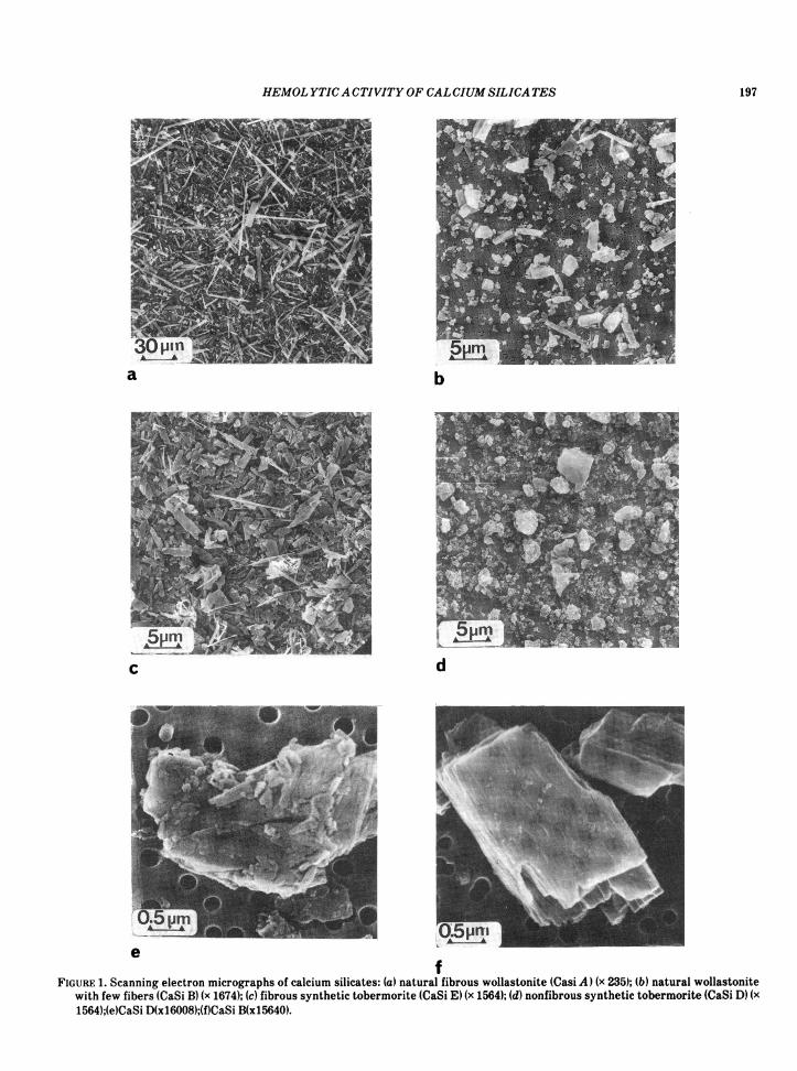
ResultsSEM photographs of the different dusts are
shown in Figure 1. The maximal diameter of theirregular particles of the nonfibrous minerals rarelyexceeded 7 ,m. The length distributions of the fi-brous samples varied, CaSi A presenting the high-est aspect ratio. CaSi B and CaSi E had approxi-mately similar aspect ratios, but in general only halfthe ratio of CaSi A. The detailed data are given inTables 1-3. X-ray diffraction analysis revealed unre-acted quartz and calcite impurities in the syntheticcompounds, the quartz content ranging from 2 to10% (Table 4). The specific surface areas of the dustsamples are shown in Table 5.The hemolytic activity of calcium silicates is
higher for the synthetic compounds than for the na-tural wollastonite. No differences were found withinthe group of synthetic compounds or within thegroup of natural wollastonites (Fig. 2).
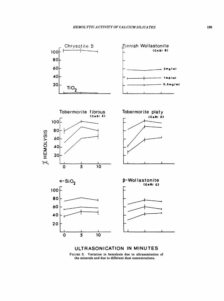
Ultrasonication of the dust samples for 5 or 10min increased the hemolytic activity for all synthet-ic compounds (Fig. 3). A two- to three-fold increasewas observed for CaSi E at a concentration of 0.5mg/mL. Ultrasonication had no effect on the naturalCaSi B or on the positive or negative control dusts.
196
HEMOL YTICA CTIVITYOF CALCIUM SILICA TES
a b
c
e
1. -
IrA-
d
fFIGURE 1. Scanning electron micrographs of calcium silicates: (a) natural fibrous wollastonite (Casi A) (x 235); (b) natural wollastonite
with few fibers (CaSi B) (x 1674); (c) fibrous synthetic tobermorite (CaSi E) (x 1564); (d) nonfibrous synthetic tobermorite (CaSi D) (x1564);(e)CaSi D(xl6008);(f)CaSi B(x15640).
197

SKA UG AND GYLSETH
In all experiments higher dust concentrations in-creased the hemolysis (Fig. 3). Departure from lin-earity was negligible whether the minerals were ul-trasonicated or not. UICC chrysotile- and TiO2-in-duced hemolysis was unaffected by dust concentra-tion.
Table 1. Size distribution of CaSi A.
Distribution of particle diameterLength 0-1 1-2 2-3 3-4range, Am Jm JAm JAm JAm
2-4 16 24-10 26 7 110-30 11 8 8 330-100 3 7 6 2
100- 200 1 1
Table 2. Size distribution of CaSi B.
Length Distribution of particle diameterrange, 0-0.2 0.2-0.4 0.4-0.6 0.6-0.8 0.8-1.0 1.0-1.2 1.2-1.8Pm Pm Jm Pm Pm Pm Pm Pm0-1 11 31-2 20 30 5 12-3 10 18 9 3 13-4 4 9 13 10 5 34-5 3 2 2 5 25-6 2 1 2 1 2 26-7 1 1 2 3 2
'-7 3 1 1 6
Table 3. Size distribution of CaSi E.
Length, Distribution of particle diameterrange, 0-0.2 0.2-0.4 0.4-0.6 0.6-0.8 0.8-1.2pm pm pm pm pm pm0-1 46 71-2 38 38 82-3 9 19 9 33-4 1 7 2 2 14-5 2 15-6 3 1 1
Table 4. X-ray characteristics and main chemical formulasof calcium silicates.
Chemical formula SiO2, FibrousSample of main component % character
CaSi A (fibrous CaSiO3 - + + +natural wollastonite)CaSi B (natural CaSiO3 2 + 1 +wollastonite with fewfibers)CaSi C (nonfibrous CaSiO3 9 ± 1syntheticwollastonite)CaSi D (nonfibrous Ca5iS017 2.5 H20 10 ± 2synthetictobermorite)CaSi E (fibrous Ca3Si40,7 2.5 H20synthetic tobermorite) 2 ± 1 +
Ca.SiS017(OH)2
Reduced hemolytic ability was not found for theCaSi C and CaSi D at pH 7.2 and 6.2 when 10 .MEDTA was added compared to a 30-35% reductionfor UICC chrysotile B. Neither did 4 mM EDTA in-hibit hemolysis of CaSi C at pH 7.4 when the dustwas treated for 1 hr before incubation. EGTA con-centrations up to 32 mM did not inhibit the hemoly-sis of CaSi B, C, D and E.
CaCl2 at a level of 0.8 mM did not affect hemolyticactivity of CaSi C. There was an increase in hemo-lytic activity of CaSi D and E at 30 mM CaCI2,whereas CaSi C-induced hemolysis was not affected(Table 6).
Table 5. Specific surface area of calcium silicates.Specific
surface area,m2/g
CaSi A 5.1 + 0.2CaSi B 5.3 + 0.2CaSi C 18.6 ± 0.5CaSi Da 50.1 ± 0.5CaSi Eb 22.9 ± 0.5
aCrystal water: 4.4°h.bCrystal water: 9.2%.
Table 6. Hemolysis of mineral dusts in the presenceof 30 mM CaCl2.
Hemolysis, %Sample No Ca added 30 mM CaCl2 addedCaSi C 39 ± 2 44 ± 5CaSiD 28 ± 6 61 ± 2CaSiE 56 ± 4 80 ± 6Fragilitycontrol 1 ± 0.1 1 ± 0.1
WU)-I0wIU
CaSi E
FIGURE 2. Hemolysis induced by the different CaSi (mean ±SD).
198
HEMOL YTICA CTIVITY OF CALCIUM SILICA TES
Chrysot iI, E-
Ti_~~~~~~~~~~~~~~~~~~
-TiO2.
Tobermorite f ibrous(CaSi E)
l
0 5 10
Finnish WVollastoniteK (CaSi B)
F _ _ 2mg/mi
L §-I l~~mg/mi__ 0.5mg/ml
I
Tobermorite platy(CaSi D)
I I
p-WoI lastonite(Casi C)
0 5 10
ULTRASONICATION IN MINUTESFIGURE 3. Variation in hemolysis due to ultrasonication of
the minerals and due to different dust concentrations.
100
80
60
40
20
100
80
60
40
20
COC/)-j0
o
a-SiO2
100
80
60
40
20
199
I
I

SKA UG AND GYLSETH
Platy Tobermorite (CaSi D)E
Y1000h.
Hours in medium
FIGURE 4. X-ray intensities of two main peaks of Tris buffer-leached CaSi D related to time in solution.
Finnish wollastonite(CaSi B)
W1_0
hkl202\
_~~~~~~10 100 1lUU
Hours in mediumFIGURE 5. X-ray intensities of two main peaks of Tris buffer-
leached CaSi B related to time in solution.
____ _ ,_.._-_*i_- .....+-~~~~~~~~~~~~~~~~~~...........
101
4
3
2 EEmco
1
1 2 3 8 16Leaching time (Days)
FIGUtRE 6. Amount of calcium released in Tris-buffered saline for ('3 CaSi B and (A)CaSi D and hemolysis induced by leached (X) CaSi B and (A) CaSi D as a function ofleaching time.
Crystallography revealed that the main X-raypeaks from CaSi D almost disappeared after 1-2days in solution, whereas CaSi B was somewhatmore resistant. After 16 days the crystalline struc-ture of CaSi B was completely broken down (Figs. 4and 5).
Figure 6 shows a steady increase in the totalamount of calcium released into the Tris-bufferedsaline from CaSi B and D. The hemolytic activity inleached samples decreased correspondingly. SEMphotographs did not show definite morphologicalchanges of the dust samples in solution. The amountof calcium released was low for all CaSi, CaSi D pre-senting the highest value (3.9 mM after 16 days insolution at 370C).
SEM examination of the RBC showed that thenature of the membrane damage was similar for allCaSi examined. The initial damage was limited to afew RBC with mineral dust adherent on the sur-faces. Some RBC did not show gross abnormalities,although they were covered by CaSi. The main de-formities appeared as invaginations of the mem-
branes (Fig. 7). Major cellular abnormalities were
present after 5 min.However, intact cells were still found after 60
min, also for the most hemolytic samples. Thechrysotile fibers covered more of the total surfaceof the RBC. They were also agglutinated to a largerextent by the chrysotile fibers. Heavy deposits ofTiO2 on the RBC gave only minor change in surfaceshape after 60 min.
5Qr
40
co.F 30
Ea)I 20ax
200
I --- .---.-- .
Y ---.'I
II

HEMOL YTICA CTIVITY OF CALCIUM SILICA TES
a
c
b
d
ef
FIGURE 7. Scanning electron micrographs of RBC and mineral samples after different incubation times: (a) TiO2 (60 min); (b) UICCchrysotile B (5 min); (c) CaSi E (5 min); (d, e) CaSi C (5 min); (fl CaSi C (60 min).
201

202 SKA UG AND GYLSETH
DiscussionHemolytic activity is related to the specific sur-
face area of the CaSi. The specific surface area val-ues are notably high for the synthetic compounds,whereas they are lower for the natural CaSi, ap-proximating those of respirable chrysotile samples(10). However, the value for CaSi D was dispropor-tionately high. For this mineral the surface area de-termination may have been affected by theliberation of crystal water. Particle size (i.e., surfacearea) is thus an important factor for the hemolyticactivity of these dusts.
Ultrasonication increased hemolysis for the syn-thetic CaSi, but not for the natural one. This treat-ment may disperse mineral agglomerates and evendisrupt particles. The surface area available for thereaction with the RBC would thus increase. Basedon this assumption, ultrasonication disintegratessynthetic compounds more easily than the naturalwollastonite. Variation in hemolysis between paral-lels of individual samples was lowest when ultrason-ication was omitted. This may indicate that ultra-sonication causes uneven disintegration of the min-erals in solution.
Mineral morphology does not influence hemolyticactivity, as shown by the comparison of acicular andfibrous minerals within the groups of synthetic ornatural CaSi. Neither do the quartz impurities ofthe minerals increase hemolysis compared to min-erals without quartz within the same group (Table 4and Fig. 2).
In the leaching experiments, the durability of theminerals in solution was examined by the determi-nation of calcium release and by changes in the min-eral structure. Small amounts of calcium are re-leased from the minerals to the saline. Hemolysis in-duced by the leached minerals is less than for theparent samples.A possible role of calcium in the mechanism of
CaSi-induced hemolysis was not confirmed byEDTA or EGTA treatment. EDTA has been shownto inhibit chrysotile hemolysis, probably by com-plexing Mg2+ on the surface layer (11). The chelatortreatment is likely to inactivate calcium releasedfrom the minerals to the solution, but we do notknow whether this treatment inactivates high cal-cium concentrations on the surface of the particlesresponsible for local reactions with the RBC mem-branes. RBC membrane damage and hemolysis maybe related to abnormal influx of calcium into thecells, as shown in the RBC of sickle cell anemia (12).The calcium concentration in RBC cytosol is nor-mally less than 0.1 IAM (13). An abnormal influx ofcalcium may occur secondary to mineral-inducedmechanical damage of the RBC. The steady-state in-
flux of calcium is a function of the extracellular cal-cium concentration (14). These mechanisms may beresponsible for the increased hemolysis of the syn-thetic CaSi D and E when 30 mM CaCl2 is added tothe RBC suspension. Increased intracellular calciummay thus be a contributing mechanism for CaSi-in-duced hemolysis, but the definite role of calcium re-mains to be established.The X-ray diffraction patterns from the two min-
erals indicate that rupture of crystalline structuresis already initiated at the second day in solution, asshown by reduction in the peak intensities followedby the complete disappearance after 3 days for theCaSi D, and after 16 days for the natural CaSi B.The simultaneous loss of hemolytic activity, notablyfrom the third day, indicates that alteration in crys-talline structure may be responsible for decreasedhemolysis.The loss of hemolytic activity of the UICC chryso-
tile B dust samples during the experimental periodmay be due to alteration in mineralogical character-istics by permanent storage at 370C to removehumidity.The SEM microphotographs of the cells and
dusts visualize the adherence of dust particles tothe RBC membranes and agglomeration of cells andcellular debris. This may be an initial event, asthere is no substantial change in the amount of par-ticles adhered to individual cells during the incuba-tion period. Morphological similarities to typicalsickle cells were not seen.
In conclusion, the hemolysis of RBC due to CaSidust exposure depends on chemical and physicalproperties of the dust. The reactions with the RBCmembranes are related to the surface area of theminerals. Crystalline SiO2 content is not likely tocontribute to the hemolytic activity. Calcium mayalso contribute to hemolysis due to high concentra-tions at the interphase between RBC membranesand minerals, and by a postulated increased influxof calcium into the RBC. Another line of investiga-tion is the follow-up of the crystallographic changesduring the leaching experiments, in which the re-leased calcium might be a secondary phenomenon.
The authors want to thank R. Bruun, G. Edholm, A. G. My-rann, A. L. Nordhagen and L. Overaae for excellent technicalassistance.The financial support of the Royal Norwegian Council for
Scientific and Industrial Research is acknowledged.
REFERENCES
1. Elevatorski, E. A. Wollastonite. In: Industrial Mineralsand Rocks (S. J. Lefond, Ed.), American Institute of Min-ing, Metallurgical and Petroleum Engineers, New York,1975, pp. 1227-1233.
HEMOLYTICA CTIVITY OF CALCIUM SILICA TES 203
2. Hefner, R. E., and Gehring, P. J. A comparison of the rel-ative rates of hemolysis induced by various fibrogenic andnon-fibrogenic particles with washed rat erythrocytes invitro. Am. Ind. Hyg. Assoc. J. 35: 734-740 (1975).
3. Potts, W. J., Lederer, T. S., and Gehring, P. J. Hemolysesof washed rat erythrocytes in vitro by dust particles. Am.Ind. Hyg. Assoc. J. 39: 497- 502 (1978).
4. Wright, A., Gormley, I. P., Collings, P. L., and Davis,J. M. G. The cytotoxicities of asbestos and other fibrousdusts. In: The In Vitro Effects of Mineral Dusts, (R. C.Brown, M. Chamberlain, R. Davies, and I. P. Gormley,Eds.), Academic Press, New York, 1980, pp. 25-31.
5. Hunt, J., Pooley, F. D., and Richards, R. Biological reac-tivity of calcium silicate composites-in vitro studies. En-viron. Res. 26: 51-68 (1981).
6. Richards, R., Tetley, T. D., and Hunt, J. The biological re-activity of calcium silicate composites: in vivo studies. En-viron. Res. 26: 243-247 (1981).
7. Braunauer, S., Emmet, H. P., and Teller, N. E. Adsorptionof gases in multimolecular layers. J. Am. Chem. Soc. 60:309-319 (1938).
8. Desai, R., Hext, P., and Richards, R. The prevention of as-bestos-induced hemolysis. Life. Sci. 16: 1931- 1938 (1975).
9. Morgan, A., Holmes, A., and Talbot, R. J. The haemolyticactivity of some fibrous amphiboles and its reaction totheir specific surface areas. Ann. Occup. Hyg. 20: 39-47(1977).
10. Schnitzer, R. J., and Pundsack, F. L. Asbestos hemolysis.Environ. Res. 3: 1-13 (1970).
11. Harington, J. S., Allison, A. C., and Badami, D. V. Mineralfibers: chemical, physicochemical and biological proper-ties. In: Advances in Pharmacology and ChemotherapyVol. 12 (S. Garattini, F. Hawking, A. Goldin and I. J.Kopin, Eds.), Academic Press, New York, 1975, pp.291-391.
12. Burris, S. M., White, J. G., and Eaton, J. W. Calcium-induced erythrocyte shape change: Evidence againstinvolvement of diacylglycerol accumulation. In: Red BloodCell and Lens Metabolism (S. K. Srivastava, Ed.),Elsvier/North Holland, Amsterdam, 1980, pp. 49-60.
13. Downes, P., and Michell, R. H. Human erythrocyte mem-branes exhibit a cooperative calmodulin dependent Ca2+-ATPase of high sensitivity. Nature 290: 270-271 (1981).
14. Borle, A. Control, modulation and regulation of cell cal-cium. Rev. Physiol. Biochem. Pharmacol. 90: 14-118(1981).
Copyright © 2022 FDOKUMEN