Lack of Cyclin-Dependent Kinase 4 Inhibits c-myc Tumorigenic Activities in Epithelial Tissues
Upload
independentCategory
view
2download
0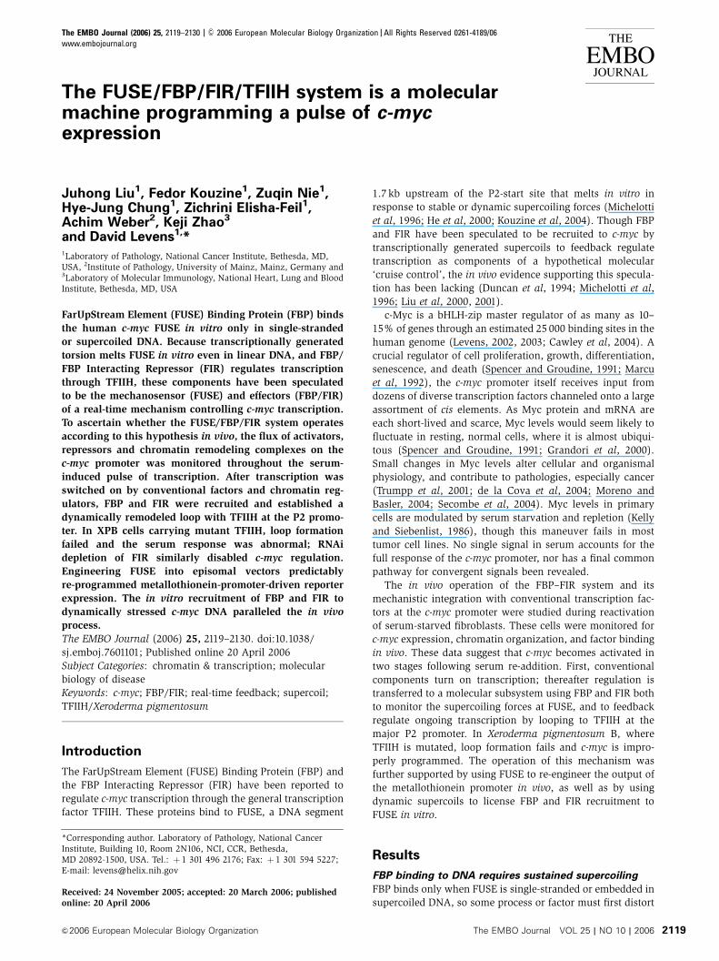
The FUSE/FBP/FIR/TFIIH system is a molecularmachine programming a pulse of c-mycexpression
Juhong Liu1, Fedor Kouzine1, Zuqin Nie1,Hye-Jung Chung1, Zichrini Elisha-Feil1,Achim Weber2, Keji Zhao3
and David Levens1,*1Laboratory of Pathology, National Cancer Institute, Bethesda, MD,USA, 2Institute of Pathology, University of Mainz, Mainz, Germany and3Laboratory of Molecular Immunology, National Heart, Lung and BloodInstitute, Bethesda, MD, USA
FarUpStream Element (FUSE) Binding Protein (FBP) binds
the human c-myc FUSE in vitro only in single-stranded
or supercoiled DNA. Because transcriptionally generated
torsion melts FUSE in vitro even in linear DNA, and FBP/
FBP Interacting Repressor (FIR) regulates transcription
through TFIIH, these components have been speculated
to be the mechanosensor (FUSE) and effectors (FBP/FIR)
of a real-time mechanism controlling c-myc transcription.
To ascertain whether the FUSE/FBP/FIR system operates
according to this hypothesis in vivo, the flux of activators,
repressors and chromatin remodeling complexes on the
c-myc promoter was monitored throughout the serum-
induced pulse of transcription. After transcription was
switched on by conventional factors and chromatin reg-
ulators, FBP and FIR were recruited and established a
dynamically remodeled loop with TFIIH at the P2 promo-
ter. In XPB cells carrying mutant TFIIH, loop formation
failed and the serum response was abnormal; RNAi
depletion of FIR similarly disabled c-myc regulation.
Engineering FUSE into episomal vectors predictably
re-programmed metallothionein-promoter-driven reporter
expression. The in vitro recruitment of FBP and FIR to
dynamically stressed c-myc DNA paralleled the in vivo
process.
The EMBO Journal (2006) 25, 2119–2130. doi:10.1038/
sj.emboj.7601101; Published online 20 April 2006
Subject Categories: chromatin & transcription; molecular
biology of disease
Keywords: c-myc; FBP/FIR; real-time feedback; supercoil;
TFIIH/Xeroderma pigmentosum
Introduction
The FarUpStream Element (FUSE) Binding Protein (FBP) and
the FBP Interacting Repressor (FIR) have been reported to
regulate c-myc transcription through the general transcription
factor TFIIH. These proteins bind to FUSE, a DNA segment
1.7 kb upstream of the P2-start site that melts in vitro in
response to stable or dynamic supercoiling forces (Michelotti
et al, 1996; He et al, 2000; Kouzine et al, 2004). Though FBP
and FIR have been speculated to be recruited to c-myc by
transcriptionally generated supercoils to feedback regulate
transcription as components of a hypothetical molecular
‘cruise control’, the in vivo evidence supporting this specula-
tion has been lacking (Duncan et al, 1994; Michelotti et al,
1996; Liu et al, 2000, 2001).
c-Myc is a bHLH-zip master regulator of as many as 10–
15% of genes through an estimated 25 000 binding sites in the
human genome (Levens, 2002, 2003; Cawley et al, 2004). A
crucial regulator of cell proliferation, growth, differentiation,
senescence, and death (Spencer and Groudine, 1991; Marcu
et al, 1992), the c-myc promoter itself receives input from
dozens of diverse transcription factors channeled onto a large
assortment of cis elements. As Myc protein and mRNA are
each short-lived and scarce, Myc levels would seem likely to
fluctuate in resting, normal cells, where it is almost ubiqui-
tous (Spencer and Groudine, 1991; Grandori et al, 2000).
Small changes in Myc levels alter cellular and organismal
physiology, and contribute to pathologies, especially cancer
(Trumpp et al, 2001; de la Cova et al, 2004; Moreno and
Basler, 2004; Secombe et al, 2004). Myc levels in primary
cells are modulated by serum starvation and repletion (Kelly
and Siebenlist, 1986), though this maneuver fails in most
tumor cell lines. No single signal in serum accounts for the
full response of the c-myc promoter, nor has a final common
pathway for convergent signals been revealed.
The in vivo operation of the FBP–FIR system and its
mechanistic integration with conventional transcription fac-
tors at the c-myc promoter were studied during reactivation
of serum-starved fibroblasts. These cells were monitored for
c-myc expression, chromatin organization, and factor binding
in vivo. These data suggest that c-myc becomes activated in
two stages following serum re-addition. First, conventional
components turn on transcription; thereafter regulation is
transferred to a molecular subsystem using FBP and FIR both
to monitor the supercoiling forces at FUSE, and to feedback
regulate ongoing transcription by looping to TFIIH at the
major P2 promoter. In Xeroderma pigmentosum B, where
TFIIH is mutated, loop formation fails and c-myc is impro-
perly programmed. The operation of this mechanism was
further supported by using FUSE to re-engineer the output of
the metallothionein promoter in vivo, as well as by using
dynamic supercoils to license FBP and FIR recruitment to
FUSE in vitro.
Results
FBP binding to DNA requires sustained supercoiling
FBP binds only when FUSE is single-stranded or embedded in
supercoiled DNA, so some process or factor must first distortReceived: 24 November 2005; accepted: 20 March 2006; publishedonline: 20 April 2006
*Corresponding author. Laboratory of Pathology, National CancerInstitute, Building 10, Room 2N106, NCI, CCR, Bethesda,MD 20892-1500, USA. Tel.: þ 1 301 496 2176; Fax: þ 1 301 594 5227;E-mail: [email protected]
The EMBO Journal (2006) 25, 2119–2130 | & 2006 European Molecular Biology Organization | All Rights Reserved 0261-4189/06
www.embojournal.org
&2006 European Molecular Biology Organization The EMBO Journal VOL 25 | NO 10 | 2006
EMBO
THE
EMBOJOURNAL
THE
EMBOJOURNAL
2119
or destabilize the double helix (Duncan et al, 1994; Bazar
et al, 1995; Michelotti et al, 1996; Kouzine et al, 2004). The
torsional stress generated by the counter-rotation of the
template and RNA polymerase during transcription melts
FUSE even in linear templates (Kouzine et al, 2004). To test
whether these transient dynamic supercoils melt FUSE
long enough to bind FBP and FIR, an experiment was
devised to observe recruitment to and release of these
proteins from binding sites in linear templates upstream of
promoters where the intensity of ongoing transcription was
modulated.
T3 and T7 RNA polymerases were used to transcribe linear
templates in vitro under conditions shown to drive dynamic
supercoiling of upstream DNA (Figure 1A). FUSE was placed
between divergent promoters in order to absorb the mutually
reinforcing, negative supercoils trailing each polymerase
(Kouzine et al, 2004). The rate of transcription, and hence
the amount of torque applied to the template, was tuned with
limiting GTP. Following transcription in the presence of FBP,
formaldehyde was added both to terminate the reactions and
to crosslink DNA-bound protein. The fixed protein–DNA
complexes were harvested with anti-FBP and analyzed by
PCR as for chromatin immunoprecipitation (ChIP). Low-level
RNA synthesis supported little FBP binding (Figure 1B, a-FBP
panel, lanes 4–6); at least 200mM GTP was required for
maximal FBP–template interactions (lanes 7 and 8) corre-
sponding to the threshold of dynamic supercoiling required
to FUSE (Kouzine et al, 2004). Stopping transcription (by
chelating the Mg2þ obligatory for transcription, but not for
FBP–DNA interaction) rapidly evicted most bound FBP (lane
9), as FUSE reverted to B-DNA (Kouzine et al, 2004). These
results were confirmed by real-time PCR (Figure 1B, FBP
IP graph).
To verify that releasing torsional stress discharged DNA-
bound FBP, FBP–FUSE complexes in supercoiled plasmids
were probed with KMnO4 to assess DNA melting with or
without subsequent linearization of the plasmid by rapid
restriction enzyme digestion. Indeed the FUSE melting in-
duced by FBP in supercoiled DNA vanished upon lineariza-
tion (Figure 1C, lanes 2, 6, and 11). Therefore, torsional stress
initiates and sustains FBP binding to FUSE.
Transient torsional stress allows sustained binding
of FIR to FUSE
FIR was originally identified through protein–protein inter-
action with FBP. Owing to its tandem RRMs, a structural
motif that binds single-stranded nucleic acids (usually RNA,
but sometimes DNA), FIR was also tested for binding to
dynamically stressed DNA (DeAngelo et al, 1995; Ding
et al, 1999; Liu et al, 2000; Kielkopf et al, 2004). Although
FIR did not bind to linear DNA, just like FBP, transcription
by T3 and T7 RNA polymerases from divergent promoters
FU
SE
GS
TF
BP
FIR
FB
P+F
IR
GS
TF
BP
FIR
FB
P+F
IR
FIR
FB
PF
BP
+FIR
BS
A
Supercoiled Linearizedbefore binding
Linearizedafter binding
3'-C
CC
TG
GT
TC
CT
AC
TC
TT
CT
TA
-//-
T-/
/-T
TA
TT
GT
GT
TT
TA
TT
TT
TT
AG
GG
CT
CC
CT
TA
TA
TG
TA
AT
AT
AT
AA
TT
-5'
–168
5–1
775
*
**
*
GTP (µM) 400
200
100
500 400+
ED
TA
α-FBP IP
α-FIR IP
Inp
ut
No
DN
A
IgG
T7/FBP
T7/FIR
T3/FBP
T3/FIR
A
IP
PE
B
FUSE
T3
T7
T7
T3FUSE
400+EDTA
400
200
10050
0
Cycle
400+EDTA
400200
10050
0
Cycle
00.10.20.30.40.50.60.70.8
12 15 18 21 24 27 30 33 36
FIR IP
Rel
ativ
e fl
uo
resc
ence
Rel
ativ
e fl
uo
resc
ence
00.10.20.30.40.50.60.7
12 15 18 21 24 27 30 33 36
FBP IP
C
987654321
121110987654321
Figure 1 DNA binding by FBP requires sustained superhelical stress, whereas transient stress is sufficient for FIR. (A) Schematic drawing ofliner in vitro transcription template. Active transcription, even in linear templates, develops dynamic supercoils that convert duplex FUSE (box)into single-stranded DNA (oval) (Kouzine et al, 2004). (B) Negative torsional stress (supercoiling) was dynamically generated by T3 and T7RNA polymerase transcription in vitro of linear templates in the presence of FBP (a-FBP IP panel) or FIR (a-FIR IP panel). Primer extension (PE,lanes 4–8) showed increased transcription with increasing (GTP). EDTA was added to stop transcription prior to fixation (lane 9) to test ifongoing transcription was required for FBP or FIR binding. Immunoprecipitated DNA was amplified (IP, lanes 1–9). Real-time PCR profile of thesame DNA is shown on the right. (C) In vitro KMnO4 footprints of FBP and FIR binding to supercoiled or linear DNA. Hyper-reactive residuesare marked with triangles; hyper-reactive residues resistant to linearization are marked with *. Recombinant GST, FBP, and/or FIR wereincubated with supercoiled (lanes 1–4 and lanes 9–12) or linear (lanes 5–8) DNA-containing FUSE. After binding, all samples received MgSO4
(2 mM final) and XhoI was added in lanes 9–12 for 5 min before KMnO4 treatment.
Mechanical regulation of c-myc transcriptionJ Liu et al
The EMBO Journal VOL 25 | NO 10 | 2006 &2006 European Molecular Biology Organization2120
enabled binding to FUSE when situated between the start
sites. Full FIR binding occurred at 100 mM GTP, somewhat
lower than for FBP (Figure 1B, a-FIR panel, lanes 3–8). The
FIR–FUSE complex was more resistant to transcriptional
arrest by Mg2þchelation than was FBP–FUSE (lane 9, also
real-time PCR graph). To confirm that the supercoiling
authorizing FIR–FUSE complex formation was dispensable
thereafter, FIR was bound to FUSE in supercoiled plasmids
and then KMnO4 treated with or without prior linearization.
FIR elicited prominent KMnO4 reactivity at many bases
throughout the FUSE region only on supercoiled, but not on
linear DNA (Figure 1C, lane 3 versus lane 7). The FIR-induced
hyper-reactivity of FUSE in supercoiled DNA endured (at least
partially) after linearization (Figure 1C, lane 10). Thus, FIR
only binds to relaxed DNA if the reaction pathway first
passages through a supercoiled intermediate. This sort of
reaction scheme typically suggests a kinetic barrier to direct
complex formation; in this case, the melting of a large stretch
of DNA to expose multiple contacts is probably a prerequisite
for FIR binding.
If the binding properties of FBP and FIR to transcriptionally
torqued templates in vitro are paralleled in vivo, then FBP
binding should be strictly dependent on ongoing transcrip-
tion. FBP would be recruited to active promoters, but are
absent at inactive promoters. FIR too would be recruited to
active promoters, but the ability of FIR to remain engaged
after DNA relaxation would prolong its repressive influence
even as torsional stress waned. The dynamics of the FUSE/
FBP/FIR system were interrogated at the human c-myc pro-
moter, mindful of these considerations.
A complex mix of factors precedes FBP and FIR
recruitment to the c-myc promoter
As c-myc is abnormally regulated in most tumors and cell
lines, primary human fibroblasts were studied to relate
changes in FBP and FIR binding and the recruitment of
other c-myc regulators with the dynamics of c-myc expres-
sion. The well-described serum response of c-myc in these
primary cells is not modified by neoplastic antecedents
(Waters et al, 1991). First, the fibroblasts were made quies-
cent by prolonged serum starvation. Then serum was re-
stored, and c-myc mRNA levels, and binding by FBP, FIR or
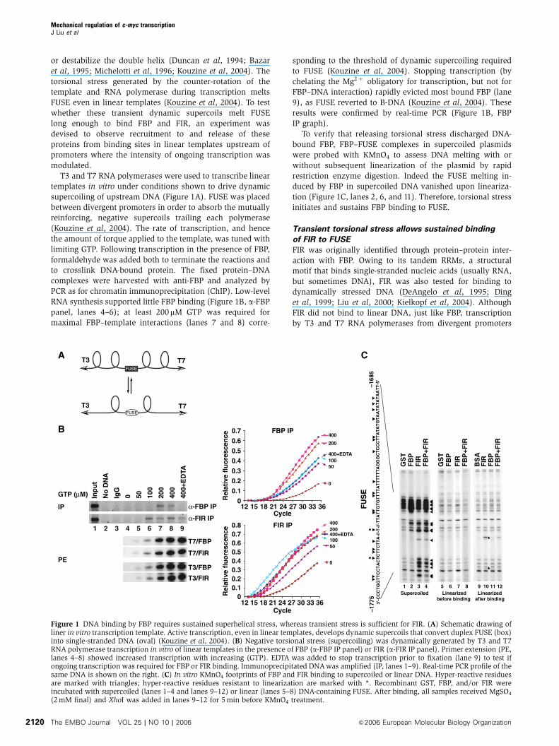
other factors, were monitored at various times. c-myc expres-
sion, virtually absent in serum-starved cells, peaked 2h after
serum re-feeding and then rapidly declined to baseline
(Figure 2A) as reported (Dean et al, 1986; Kelly and
Siebenlist, 1986). To monitor the flux of factors binding to
c-myc regulatory sequences using ChIP, PCR primers were
designed to probe: (1) the major promoter P2, (2) the Far
Upstream Element (FUSE), and (3) the mid-region between
P2 and FUSE (Figure 2E).
Despite the dynamic changes in c-myc mRNA levels, ChIP
revealed that RNA polymerase was loaded at P2 throughout
the experiment (Figure 2B, lane 12), consistent with the
paused RNA polymerase shown to reside there by nuclear
run-on and in vivo footprinting (Bentley and Groudine, 1986;
A B
P2 (+1)
FUSE
–1761/–1671
PCR Frag.:–1951 –1611 –54 +232–783 –500
FUSE Mid P2
TC
F4
TC
F4
TC
F4
E2F
SP
1
YY
1
YY
1
SP
1
E
IgG
Sp
1
E2F
4
Po
l II
No
DN
A
0.5%
Inp
ut
TC
F4
IgG
No
DN
A
0.5%
Inp
ut
P2FUSE
Starved
30 min
2 h
4 h
IP Ab.
IgG
YY
1
E2F
1
No
DN
A
0.5%
Inp
ut
TC
F4
IgG
No
DN
A
0.5%
Inp
ut
Mid
19181716151413121110987654321
Serum 0 30 m
in
1 h
2 h
4 h
8 h
c-myc
GAPDH
1 65432
Starved
30 min
2 h
4 h
IP Ab.
BR
G1
FB
P
FIR
Po
l II
No
DN
A
1% In
pu
t
IgG
IgG
Po
l II
No
DN
A1%
Inp
ut
FUSE P2
C
1.2
1.0
0.8
2 h
Flu
ore
scen
ce (
×10–3
)
Cycles4 h
D Binding at FUSE
1.4
1.6
1.8
2.0
1.0
2.0
3.0
4.0
5.0
6.0IgGBrg1FBPFIRPol II
IgGBrg1FBPFIRPol II
111098765432135312723 35312723
Figure 2 Complex transcription factor choreography precedes FBP and FIR recruitment to the c-myc promoter. Human Hs68 primaryfibroblasts were cultured in DMEM without FBS for 5 days and then stimulated with 10% serum; cells were harvested for RNA or ChIP atthe indicated times after adding serum. (A) RNase protection assay measuring c-myc levels. (B, C) Chromatin from starved and stimulated cellswas immunoprecipitated with indicated antibodies. (D) Real-time PCR amplification of the same ChIP DNA samples shown in (C) (2 and 4 h).(E) Schematic representation of transcription factor binding sites and primer sets for PCR amplification.
Mechanical regulation of c-myc transcriptionJ Liu et al
&2006 European Molecular Biology Organization The EMBO Journal VOL 25 | NO 10 | 2006 2121
Spencer and Groudine, 1991; Marcu et al, 1992; Strobl and
Eick, 1992). The c-myc promoter remained substantially
charged with polymerase even after 7 days of serum starva-
tion (Supplementary Figure S2), so the near absence of c-myc
transcription must reflect, at least in part, inefficient mobili-
zation of the paused polymerase.
Several transcription factors implicated in c-myc regulation
were tested by ChIP to see if any were recruited early enough
to trigger release of the P2-paused RNA polymerase and
whether their binding paralleled the peak of expression.
Among the factors reported to regulate c-myc, E2F1, E2F4,
YY1, Sp1, and TCF4 were tested (Thalmeier et al, 1989;
DesJardins and Hay, 1993; Riggs et al, 1993; He et al,
2000); all were absent from the c-myc promoter in serum-
starved cells (Figure 2B, starved). Upon induction, these
factors displayed various patterns of recruitment and release
from the promoter. Moving in parallel, Sp1 and YY1 bound
quickly and exited early (Figure 2B, lanes 11 and 17). E2F1
and E2F4 were also bound rapidly, but remained throughout
c-myc induction and decline; E2F1 levels were relatively
steady, while E2F4 occupancy gradually increased
(Figure 2B, lanes 10 and 16). As c-myc expression waned,
TCF4 was recruited to its two binding sites, one near FUSE
and the other within the mid-region (Figure 2B, lanes 2 and
6). The differential flux of these factors argues against a rigid
enhanceosome snapped together from synchronously acti-
vated components (Maniatis et al, 1998; Merika and Thanos,
2001). Rather, these results suggest that different stages of
the serum-induced c-myc expression peak coincide with the
binding of different sets of factors.
When c-myc is silent, double-stranded FUSE is fixed upon a
nucleosome (Michelotti et al, 1996), and so FBP cannot bind,
reinforcing the gene’s closed state. The positive action of the
BRG-1 chromatin-remodeling complex has been hypothe-
sized to open FUSE to help re-start c-myc transcription (Chi
et al, 2003). ChIP was performed to monitor the binding of
FBP, FIR, and the BRG-1 near FUSE. Following prolonged
serum starvation (5 days), residual FIR binding remained
at FUSE after BRG-1 and FBP were lost (Figure 2C and
Supplementary Figure S1); after 7 days of serum starvation
FIR too was undetectable (Supplementary Figure S1). At
30 min post-stimulation, BRG-1 was recruited to FUSE when
neither FIR nor FBP was present. At the peak of c-myc
expression 2 h post-stimulation, FBP binding crested, FIR
started to return, and BRG-1 binding waned. Thereafter,
while c-myc expression dropped, FBP lingered, BRG-1 re-
ceded further, and the level of FIR at FUSE rose dramatically
(Figure 2C and D). Once recruited to FUSE, FIR may be
stabilized both through protein–protein interactions
with FBP and by direct DNA binding (see below) within a
ternary complex. Binding of FBP and FIR endured
through later stages of serum stimulation (Supplementary
Figure S2), when cells make the transition to steady-
state growth. Thus, the FUSE region contributes different
factors to c-myc regulation at different phases of the
activation cycle.
As FIR was undetectable at FUSE after 7 days of serum
starvation (Supplementary Figure S1) though c-myc remained
off, some overarching mechanism must convert short-term
repression into an enduring shutoff. This same mechanism
would need to be reversed upon re-activation, and seemed
likely to involve chromatin.
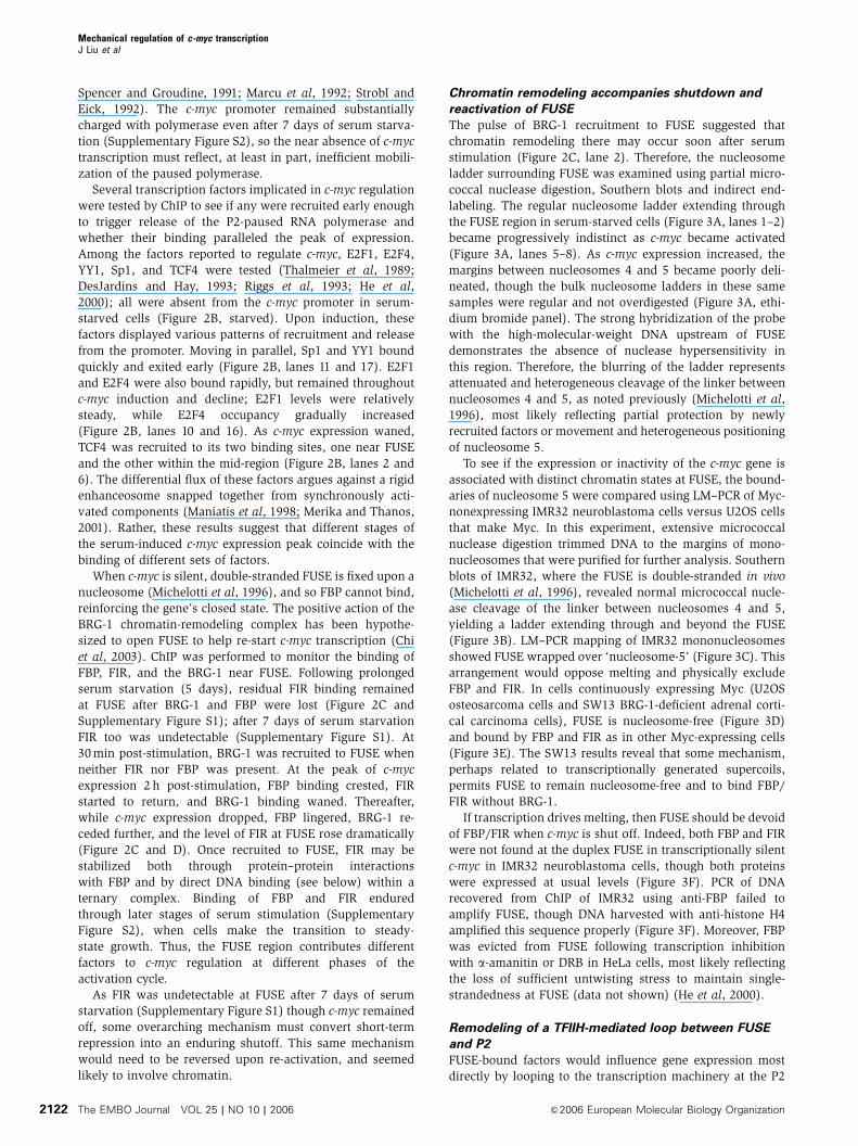
Chromatin remodeling accompanies shutdown and
reactivation of FUSE
The pulse of BRG-1 recruitment to FUSE suggested that
chromatin remodeling there may occur soon after serum
stimulation (Figure 2C, lane 2). Therefore, the nucleosome
ladder surrounding FUSE was examined using partial micro-
coccal nuclease digestion, Southern blots and indirect end-
labeling. The regular nucleosome ladder extending through
the FUSE region in serum-starved cells (Figure 3A, lanes 1–2)
became progressively indistinct as c-myc became activated
(Figure 3A, lanes 5–8). As c-myc expression increased, the
margins between nucleosomes 4 and 5 became poorly deli-
neated, though the bulk nucleosome ladders in these same
samples were regular and not overdigested (Figure 3A, ethi-
dium bromide panel). The strong hybridization of the probe
with the high-molecular-weight DNA upstream of FUSE
demonstrates the absence of nuclease hypersensitivity in
this region. Therefore, the blurring of the ladder represents
attenuated and heterogeneous cleavage of the linker between
nucleosomes 4 and 5, as noted previously (Michelotti et al,
1996), most likely reflecting partial protection by newly
recruited factors or movement and heterogeneous positioning
of nucleosome 5.
To see if the expression or inactivity of the c-myc gene is
associated with distinct chromatin states at FUSE, the bound-
aries of nucleosome 5 were compared using LM–PCR of Myc-
nonexpressing IMR32 neuroblastoma cells versus U2OS cells
that make Myc. In this experiment, extensive micrococcal
nuclease digestion trimmed DNA to the margins of mono-
nucleosomes that were purified for further analysis. Southern
blots of IMR32, where the FUSE is double-stranded in vivo
(Michelotti et al, 1996), revealed normal micrococcal nucle-
ase cleavage of the linker between nucleosomes 4 and 5,
yielding a ladder extending through and beyond the FUSE
(Figure 3B). LM–PCR mapping of IMR32 mononucleosomes
showed FUSE wrapped over ‘nucleosome-5’ (Figure 3C). This
arrangement would oppose melting and physically exclude
FBP and FIR. In cells continuously expressing Myc (U2OS
osteosarcoma cells and SW13 BRG-1-deficient adrenal corti-
cal carcinoma cells), FUSE is nucleosome-free (Figure 3D)
and bound by FBP and FIR as in other Myc-expressing cells
(Figure 3E). The SW13 results reveal that some mechanism,
perhaps related to transcriptionally generated supercoils,
permits FUSE to remain nucleosome-free and to bind FBP/
FIR without BRG-1.
If transcription drives melting, then FUSE should be devoid
of FBP/FIR when c-myc is shut off. Indeed, both FBP and FIR
were not found at the duplex FUSE in transcriptionally silent
c-myc in IMR32 neuroblastoma cells, though both proteins
were expressed at usual levels (Figure 3F). PCR of DNA
recovered from ChIP of IMR32 using anti-FBP failed to
amplify FUSE, though DNA harvested with anti-histone H4
amplified this sequence properly (Figure 3F). Moreover, FBP
was evicted from FUSE following transcription inhibition
with a-amanitin or DRB in HeLa cells, most likely reflecting
the loss of sufficient untwisting stress to maintain single-
strandedness at FUSE (data not shown) (He et al, 2000).
Remodeling of a TFIIH-mediated loop between FUSE
and P2
FUSE-bound factors would influence gene expression most
directly by looping to the transcription machinery at the P2
Mechanical regulation of c-myc transcriptionJ Liu et al
The EMBO Journal VOL 25 | NO 10 | 2006 &2006 European Molecular Biology Organization2122
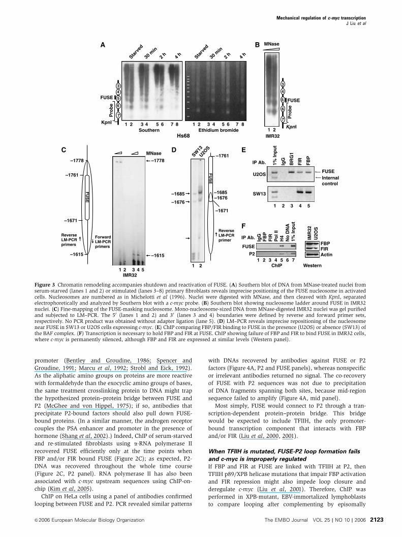
promoter (Bentley and Groudine, 1986; Spencer and
Groudine, 1991; Marcu et al, 1992; Strobl and Eick, 1992).
As the aliphatic amino groups on proteins are more reactive
with formaldehyde than the exocyclic amino groups of bases,
the same treatment crosslinking protein to DNA might trap
the hypothesized protein–protein bridge between FUSE and
P2 (McGhee and von Hippel, 1975); if so, antibodies that
precipitate P2-bound factors should also pull down FUSE-
bound proteins. (In a similar manner, the androgen receptor
couples the PSA enhancer and promoter in the presence of
hormone (Shang et al, 2002).) Indeed, ChIP of serum-starved
and re-stimulated fibroblasts using a-RNA polymerase II
recovered FUSE efficiently only at the time points when
FBP and/or FIR bound FUSE (Figure 2C); as expected, P2-
DNA was recovered throughout the whole time course
(Figure 2C, P2 panel). RNA polymerase II has also been
associated with c-myc upstream sequences using ChIP-on-
chip (Kim et al, 2005).
ChIP on HeLa cells using a panel of antibodies confirmed
looping between FUSE and P2. PCR revealed similar patterns
with DNAs recovered by antibodies against FUSE or P2
factors (Figure 4A, P2 and FUSE panels), whereas nonspecific
or irrelevant antibodies returned no signal. The co-recovery
of FUSE with P2 sequences was not due to precipitation
of DNA fragments spanning both sites, because mid-region
sequence failed to amplify (Figure 4A, mid panel).
Most simply, FUSE would connect to P2 through a tran-
scription-dependent protein–protein bridge. This bridge
would be expected to include TFIIH, the only promoter-
bound transcription component that interacts with FBP
and/or FIR (Liu et al, 2000, 2001).
When TFIIH is mutated, FUSE-P2 loop formation fails
and c-myc is improperly regulated
If FBP and FIR at FUSE are linked with TFIIH at P2, then
TFIIH p89/XPB helicase mutations that impair FBP activation
and FIR repression might also impede loop closure and
deregulate c-myc (Liu et al, 2001). Therefore, ChIP was
performed in XPB-mutant, EBV-immortalized lymphoblasts
to compare looping after complementing by episomally
1
MNase
IMR32
A B
3
4
5
6
7
FUSE
Starv
ed
30 m
in
2 h 4 h
Hs68Southern
Starv
ed
30 m
in
2 h 4 h
Pro
be
KpnI
34
5
6
7
FUSE
Pro
be
KpnI
Ethidium bromide8765432187654321
2
–1778
–1615
MNase
FU
SE
–1761
–1671
IMR32
–1778
–1615
FU
SE
–1761
–1671
C
–1685–1676
–1685
–1676U2O
S
SW13
ReverseLM-PCRprimers
ForwardLM-PCRprimers
ReverseLM-PCRprimer
D
SW13
U2OS
IP Ab. 1% In
pu
t
BR
G1
FB
P
FIR
IgG
FUSEInternalcontrol
E
ChIPF
BP
FIR
Po
l II
No
DN
A1%
Inp
ut
IgG
H4IP Ab.
F
FBPFIRActin
FUSE
P2
IMR
32U
2OS
Western54321
54321
5 6 7432121
Figure 3 Chromatin remodeling accompanies shutdown and reactivation of FUSE. (A) Southern blot of DNA from MNase-treated nuclei fromserum-starved (lanes 1 and 2) or stimulated (lanes 3–8) primary fibroblasts reveals imprecise positioning of the FUSE nucleosome in activatedcells. Nucleosomes are numbered as in Michelotti et al (1996). Nuclei were digested with MNase, and then cleaved with KpnI, separatedelectrophoretically and analyzed by Southern blot with a c-myc probe. (B) Southern blot showing nucleosome ladder around FUSE in IMR32nuclei. (C) Fine-mapping of the FUSE-masking nucleosome. Mono-nucleosome-sized DNA from MNase-digested IMR32 nuclei was gel purifiedand subjected to LM–PCR. The 50 (lanes 1 and 2) and 30 (lanes 3 and 4) boundaries were defined by reverse and forward primer sets,respectively. No PCR product was obtained without adapter ligation (lane 5). (D) LM–PCR reveals imprecise repositioning of the nucleosomenear FUSE in SW13 or U2OS cells expressing c-myc. (E) ChIP comparing FBP/FIR binding to FUSE in the presence (U2OS) or absence (SW13) ofthe BAF complex. (F) Transcription is necessary to hold FBP and FIR at FUSE. ChIP showing failure of FBP and FIR to bind FUSE in IMR32 cells,where c-myc is permanently silenced, although FBP and FIR are expressed at similar levels (Western panel).
Mechanical regulation of c-myc transcriptionJ Liu et al
&2006 European Molecular Biology Organization The EMBO Journal VOL 25 | NO 10 | 2006 2123
encoded, wild-type p89/XPB, or with empty vector. With the
functionally complemented cells (XPB/p89 panels), antibo-
dies to FUSE (FBP and FIR) or P2 factors (RNA polymerase II,
TFIIH, and E2F4) each recovered sequences from both
regions. In contrast, with mutant TFIIH (XPB panels),
anti-FUSE-factors recovered only FUSE and anti-P2-factors
recovered only P2 (Figure 4B). Thus, looping required wild-
type p89/XPB. The recovery of P2 but not FUSE using a-RNA
polymerase II in XPB cells argues that polymerase II does not
bind directly to FUSE. TFIIH was underloaded at P2 in the
XPB-mutant lymphoblasts as noted previously (Weber et al,
2005). Mid-region sequence was not recovered irrespective of
TFIIH status, demonstrating that co-recovery of FUSE with P2
sequences was not due to inadequate DNA fragmentation.
The restoration of the looping in the corrected XPB lympho-
blasts strongly argues that loop failure results directly from
mutation of p89.
To insure that FUSE-P2 loop failure was not peculiar to the
XPB mutation in the EBV-immortalized lymphoblasts, pri-
mary fibroblasts from a kindred with a different XPB muta-
tion were investigated with ChIP. Fibroblasts from a patient
with autosomal recessive XPB disease (GM13025) or from the
patient’s father (GM13028) were interrogated using anti-
FUSE or anti-P2 factors. With paternal fibroblasts, FUSE
and P2 sequences always co-precipitated, just as with normal
fibroblasts, HeLa cells or functionally corrected XPB lympho-
blasts. In contrast, with the patient’s cells just like the mutant
XPB lymphoblasts, anti-FUSE factors pulled down only FUSE,
but not P2, and vice versa (Figure 4C). Moreover, the P2
region in the patient’s cells was deficient in p89/XPB, again
similar to the mutant lymphoblasts. Therefore, p89/XPB
mutation leads to inefficient recruitment and/or retention of
TFIIH at P2, and FBP/FIR-TFIIH loop failure.
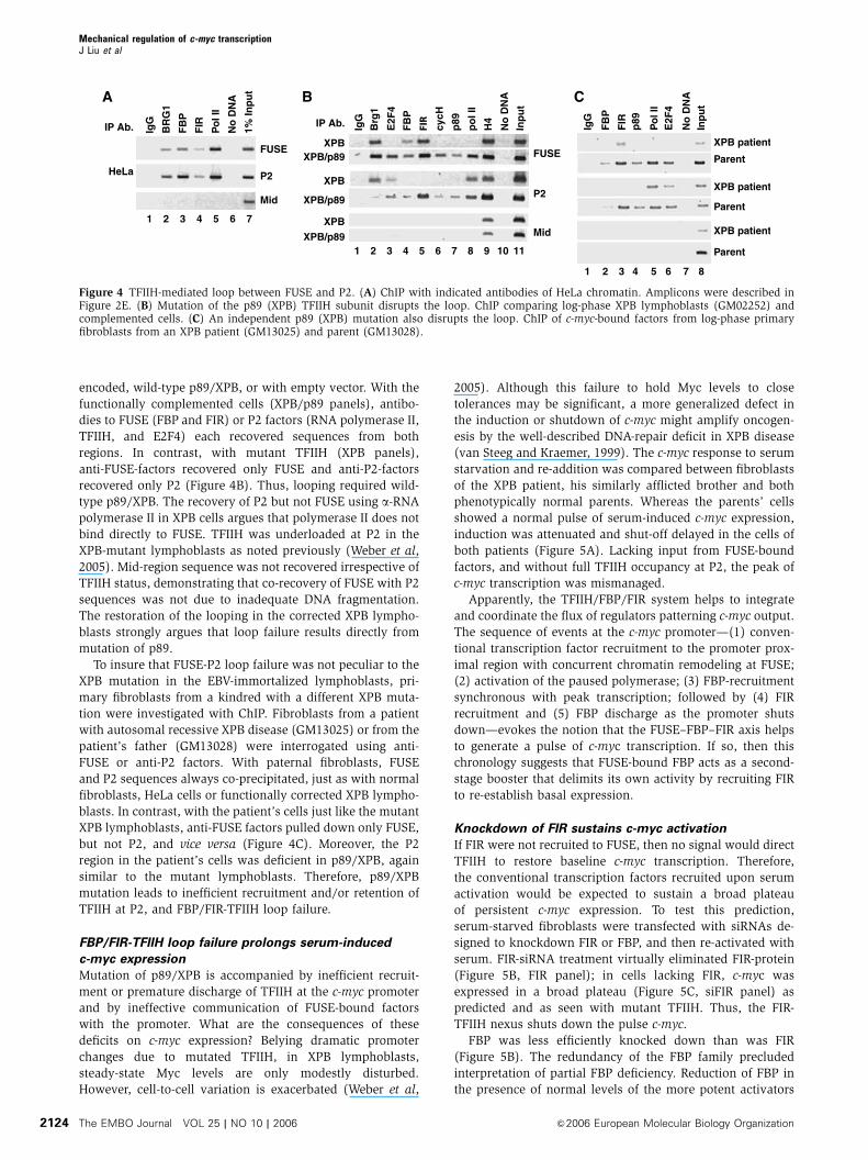
FBP/FIR-TFIIH loop failure prolongs serum-induced
c-myc expression
Mutation of p89/XPB is accompanied by inefficient recruit-
ment or premature discharge of TFIIH at the c-myc promoter
and by ineffective communication of FUSE-bound factors
with the promoter. What are the consequences of these
deficits on c-myc expression? Belying dramatic promoter
changes due to mutated TFIIH, in XPB lymphoblasts,
steady-state Myc levels are only modestly disturbed.
However, cell-to-cell variation is exacerbated (Weber et al,
2005). Although this failure to hold Myc levels to close
tolerances may be significant, a more generalized defect in
the induction or shutdown of c-myc might amplify oncogen-
esis by the well-described DNA-repair deficit in XPB disease
(van Steeg and Kraemer, 1999). The c-myc response to serum
starvation and re-addition was compared between fibroblasts
of the XPB patient, his similarly afflicted brother and both
phenotypically normal parents. Whereas the parents’ cells
showed a normal pulse of serum-induced c-myc expression,
induction was attenuated and shut-off delayed in the cells of
both patients (Figure 5A). Lacking input from FUSE-bound
factors, and without full TFIIH occupancy at P2, the peak of
c-myc transcription was mismanaged.
Apparently, the TFIIH/FBP/FIR system helps to integrate
and coordinate the flux of regulators patterning c-myc output.
The sequence of events at the c-myc promoter—(1) conven-
tional transcription factor recruitment to the promoter prox-
imal region with concurrent chromatin remodeling at FUSE;
(2) activation of the paused polymerase; (3) FBP-recruitment
synchronous with peak transcription; followed by (4) FIR
recruitment and (5) FBP discharge as the promoter shuts
down—evokes the notion that the FUSE–FBP–FIR axis helps
to generate a pulse of c-myc transcription. If so, then this
chronology suggests that FUSE-bound FBP acts as a second-
stage booster that delimits its own activity by recruiting FIR
to re-establish basal expression.
Knockdown of FIR sustains c-myc activation
If FIR were not recruited to FUSE, then no signal would direct
TFIIH to restore baseline c-myc transcription. Therefore,
the conventional transcription factors recruited upon serum
activation would be expected to sustain a broad plateau
of persistent c-myc expression. To test this prediction,
serum-starved fibroblasts were transfected with siRNAs de-
signed to knockdown FIR or FBP, and then re-activated with
serum. FIR-siRNA treatment virtually eliminated FIR-protein
(Figure 5B, FIR panel); in cells lacking FIR, c-myc was
expressed in a broad plateau (Figure 5C, siFIR panel) as
predicted and as seen with mutant TFIIH. Thus, the FIR-
TFIIH nexus shuts down the pulse c-myc.
FBP was less efficiently knocked down than was FIR
(Figure 5B). The redundancy of the FBP family precluded
interpretation of partial FBP deficiency. Reduction of FBP in
the presence of normal levels of the more potent activators
FUSE
P2
IP Ab.
Mid
B
IgG
Brg
1
E2F
4
FB
P
FIR
cycH
p89
po
l II
H4
No
DN
A
Inp
ut
XPB/p89
XPB
XPB/p89
XPBXPB/p89
XPB
1110987654321
XPB patient
Parent
IgG
FB
P
FIR
p89
Po
l II
E2F
4
No
DN
AIn
pu
t
C
XPB patient
Parent
XPB patient
Parent
87654321
A
HeLa
IP Ab. BR
G1
FB
P
FIR
Po
l II
No
DN
A
1% In
pu
t
IgG
FUSE
P2
Mid
7654321
Figure 4 TFIIH-mediated loop between FUSE and P2. (A) ChIP with indicated antibodies of HeLa chromatin. Amplicons were described inFigure 2E. (B) Mutation of the p89 (XPB) TFIIH subunit disrupts the loop. ChIP comparing log-phase XPB lymphoblasts (GM02252) andcomplemented cells. (C) An independent p89 (XPB) mutation also disrupts the loop. ChIP of c-myc-bound factors from log-phase primaryfibroblasts from an XPB patient (GM13025) and parent (GM13028).
Mechanical regulation of c-myc transcriptionJ Liu et al
The EMBO Journal VOL 25 | NO 10 | 2006 &2006 European Molecular Biology Organization2124
FBP2 and FBP3 (Duncan et al, 1996) (H-J Chung, D Levens,
unpublished data) might even be expected to increase c-myc
expression (Figure 5C, siFBP panel). (FIR, in contrast, has no
protein siblings.)
If the FUSE system behaves in vivo as it does in vitro, then
it possesses the essential components of a machine that first
senses the intensity of ongoing transcription through super-
coil-driven, FUSE deformation, and then provides positive or
negative feedback via FBP and FIR to transcription complexes
within c-myc’s expanded proximal promoter.
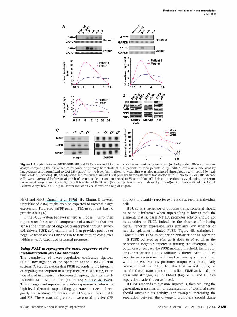
Using FUSE to reprogram the metal response of the
metallothionein (MT) IIA promoter
The complexity of c-myc regulation confounds rigorous
in situ investigation of the operation of the FUSE/FBP/FIR
system. To test the notion that FUSE responds to the intensity
of ongoing transcription in a simplified, in vivo setting, FUSE
was placed in an episome between divergent, identical metal-
inducible MT IIA promoters (Figure 6A; Karin et al, 1984).
This arrangement reprises the in vitro experiments, where the
high-level dynamic supercoiling generated between diver-
gently transcribing promoters melt FUSE, and recruit FBP
and FIR. These matched promoters were used to drive GFP
and RFP to quantify reporter expression in vivo, in individual
cells.
If FUSE is a cis-sensor of ongoing transcription, it should
be without influence when supercoiling to low to melt the
element; that is, basal MT IIA promoter activity should not
be sensitive to FUSE. Indeed, in the absence of inducing
metal, reporter expression was similarly low whether or
not the episomes included FUSE (Figure 6B, uninduced).
Constitutively, FUSE is neither an enhancer nor an operator.
If FUSE behaves in vivo as it does in vitro, when the
reinforcing negative supercoils trailing the diverging RNA
polymerases surpass the FUSE-melting threshold, then repor-
ter expression should be qualitatively altered. Metal-induced
reporter expression was compared between episomes with or
without FUSE. MT IIA promoter output was dramatically
reprogrammed by FUSE. For the first several hours, as
metal-induced transcription intensified, FUSE activated pro-
gressively stronger, up to 10-fold (Figure 6C and D, 3 kb
separation, ratio shown as inset).
If FUSE responds to dynamic supercoils, then reducing the
generation, transmission, or accumulation of torsional stress
should attenuate its activity. For example, increasing the
separation between the divergent promoters should dump
C
0 2 4 6 h
1210
86420N
orm
aliz
ed m
yc le
vel
1.0
6.7
2.1
MocksiFIRsiFBP
0 2 h 4 h 6 h 0 2 h 4 h 6 h
c-myc GAPDH
Mock
siFIR
siFBP
B
Patient 1c-myc
GAPDH
Starv
ed30
min
2 h
4 h
6 h
7 h
Starv
ed30
min
2 h
4 h
6 h
A
c-myc
GAPDH
No
rmal
ized
myc
leve
l
Father
c-myc
GAPDH
c-myc
GAPDH
MotherPatient 2
Patient 2
Mother
1210
8
6
4
20
864
2
00 2 4 6 h
No
rmal
ized
myc
leve
l
FatherPatient 1
4 h
siFB
Psi
FIR
Moc
ksi
FBP
siFI
R
FBP2
FBPActin
FBP3
FIR
Steady-state
Starved
siFB
Psi
FIR
0.50 2 4 6 7 h0.5
1
2
3
4
No
rmal
ized
myc
leve
l FatherPatient 1
0 2 4 8 12 16 20 24 h
Figure 5 Looping between FUSE–FBP–FIR and TFIIH is essential for the normal response of c-myc to serum. (A) Independent RNase protectionassays comparing the c-myc serum response of primary fibroblasts of XPB patients or their parents. c-myc mRNA levels were analyzed byImageQuant and normalized to GAPDH (graph). c-myc level (normalized to g-tubulin) was also monitored throughout a 24-h period by real-time RT–PCR (bottom). (B) Steady-state, serum-starved human Hs68 primary fibroblasts were transfected with siRNA to FIR or FBP. Starvedcells were harvested before or after 4 h of serum repletion and subjected to Western blot. (C) RNase protection assay showing the serumresponse of c-myc in mock, siFBP, or siFIR transfected Hs68 cells (left). c-myc levels were analyzed by ImageQuant and normalized to GAPDH.Relative c-myc levels at 6 h post-serum induction are shown on the plot (right).
Mechanical regulation of c-myc transcriptionJ Liu et al
&2006 European Molecular Biology Organization The EMBO Journal VOL 25 | NO 10 | 2006 2125
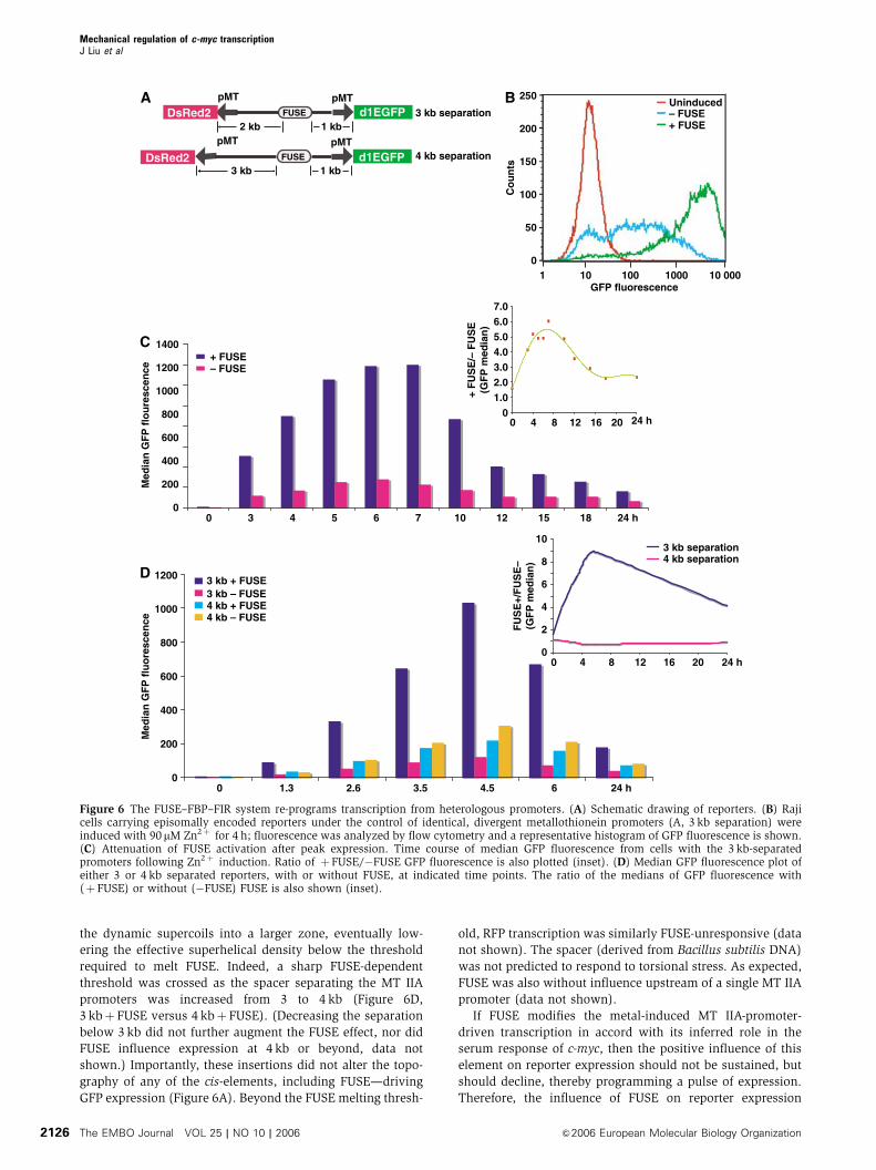
the dynamic supercoils into a larger zone, eventually low-
ering the effective superhelical density below the threshold
required to melt FUSE. Indeed, a sharp FUSE-dependent
threshold was crossed as the spacer separating the MT IIA
promoters was increased from 3 to 4 kb (Figure 6D,
3 kbþ FUSE versus 4 kbþ FUSE). (Decreasing the separation
below 3 kb did not further augment the FUSE effect, nor did
FUSE influence expression at 4 kb or beyond, data not
shown.) Importantly, these insertions did not alter the topo-
graphy of any of the cis-elements, including FUSE—driving
GFP expression (Figure 6A). Beyond the FUSE melting thresh-
old, RFP transcription was similarly FUSE-unresponsive (data
not shown). The spacer (derived from Bacillus subtilis DNA)
was not predicted to respond to torsional stress. As expected,
FUSE was also without influence upstream of a single MT IIA
promoter (data not shown).
If FUSE modifies the metal-induced MT IIA-promoter-
driven transcription in accord with its inferred role in the
serum response of c-myc, then the positive influence of this
element on reporter expression should not be sustained, but
should decline, thereby programming a pulse of expression.
Therefore, the influence of FUSE on reporter expression
B
010 100 1000 10 0001
50
100
150
200
250
Co
un
ts
GFP fluorescence
+ FUSE– FUSEUninduced
DsRed2 d1EGFPFUSE
pMTpMT
1 kb2 kb
A3 kb separation
d1EGFPpMTpMT
1 kb3 kb4 kb separationDsRed2 FUSE
Med
ian
GF
P f
luo
resc
ence
1200
1000
800
600
400
200
0
FU
SE
+/F
US
E–
(GF
P m
edia
n)
10
8
6
4
2
0
3 kb separation4 kb separation
C
D
Med
ian
GF
P f
lou
resc
ence
3 kb + FUSE3 kb – FUSE4 kb + FUSE4 kb – FUSE
1400
1200
1000
800
600
400
200
00
+ FUSE– FUSE
7.06.05.04.03.02.01.0
0
+ F
US
E/–
FU
SE
(GF
P m
edia
n)
0
0 1.3 2.6 3.5 4.5 6 24 h
0 4 8 12 16 20 24 h
4 8 12 16 20 24 h
3 4 5 6 7 10 12 15 18 24 h
Figure 6 The FUSE–FBP–FIR system re-programs transcription from heterologous promoters. (A) Schematic drawing of reporters. (B) Rajicells carrying episomally encoded reporters under the control of identical, divergent metallothionein promoters (A, 3 kb separation) wereinduced with 90 mM Zn2þ for 4 h; fluorescence was analyzed by flow cytometry and a representative histogram of GFP fluorescence is shown.(C) Attenuation of FUSE activation after peak expression. Time course of median GFP fluorescence from cells with the 3 kb-separatedpromoters following Zn2þ induction. Ratio of þFUSE/�FUSE GFP fluorescence is also plotted (inset). (D) Median GFP fluorescence plot ofeither 3 or 4 kb separated reporters, with or without FUSE, at indicated time points. The ratio of the medians of GFP fluorescence with(þ FUSE) or without (�FUSE) FUSE is also shown (inset).
Mechanical regulation of c-myc transcriptionJ Liu et al
The EMBO Journal VOL 25 | NO 10 | 2006 &2006 European Molecular Biology Organization2126
was monitored following peak expression. The reporter ac-
tivity revealed attenuation of FUSE potency following peak
activity, as predicted (Figure 6C and D). (The 2-h half-life of
the GFP reporter precluded a steeper descent from peak
expression.)
Attacking FIR with RNAi augmented and prolonged FUSE-
driven expression when the element was interposed between
the divergent promoters (data not shown), just as it did at the
c-myc locus.
Discussion
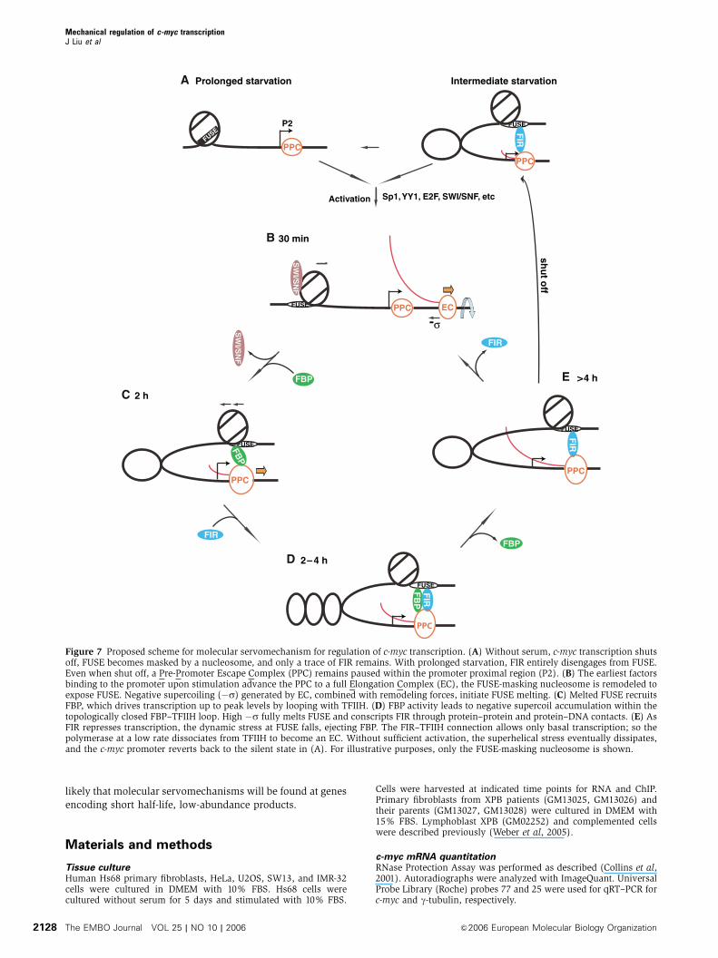
The results above invite consideration of this scheme to
describe transcription at the c-myc promoter:
First, one or more transcription factors responding to an
initiating stimulus (�li) binds the c-myc promoter. Depending
on the nature and strength of the signal, and the cell type,
single factors acting repetitively or multiple factors acting
concertedly advance the P2-paused complex. With serum,
Sp1 is among the first proteins recruited, though multiple
transcription factors are probably activated by this treatment.
The resistance of the c-myc promoter to inactivation by
pathological chromosomal alterations or genetically engi-
neered changes including all manner of mutations, inser-
tions, substitutions, and deletions (Krumm et al, 1995a, b)
may indicate that there is no unique and obligatory pathway
for c-myc induction. Rather, multiple pathways drawn
from a variety of elements and factors may prime c-myc
transcription.
As the density of bound factors increases, the paused
polymerase is ratcheted closer to promoter escape
(Figure 7A). Only after the paused polymerase exits the
promoter can the recruited factors direct re-initiation; the
paused polymerase is a powerful check on overactivation.
Concurrently, the c-myc priming factors attract complexes
that modify and remodel chromatin. For example, Sp1 (or
another BAF-binding factor) recruits chromatin-remodeling
machinery to the c-myc upstream regulatory sequences (Liu
et al, 2002). FUSE is one among the targets for remodeling.
BRG-1 mobilizes a FUSE-masking nucleosome. Remodeling
forces combined with the torsional stress of incipient tran-
scription melts FUSE, allowing FBP to bind (Figure 7B). Via
TFIIH, FBP acts as a second-stage booster to drive transcrip-
tion up to peak levels. As FBP is approximately five times
more abundant than FIR, mass-action-driven kinetics most
likely favor more rapid recruitment of the former to FUSE.
FBP-binding and peak transcription together fully melt FUSE
and conscript FIR through interactions with FBP as well as
with DNA across this broadly melted segment (Figure 7C
and D). As FIR slows transcription, the dynamic stress at
FUSE falls, ejecting FBP, but not FIR (Figure 7E), concordant
with the in vivo and in vitro findings presented here. With this
scheme, the absence of functional TFIIH causes both the FBP
boost and the FIR brake to fail, explaining the broad plateau
of c-myc transcription seen following serum addition in
XPB cells.
In this scheme, the FBP, FIR, and TFIIH constitute a
‘molecular servomechanism’. Servomechanisms sense the
output of a machine (such as an engine) and modify future
output according to its current performance. The minimal
components of a servo are a sensor coupled to positive and
negative actuators. This molecular servo uses FUSE melting
to sense the intensity of ongoing transcription (Kouzine et al,
2004), and FBP and FIR as effectors to provide positive
or negative feedback to TFIIH at the promoter. Note that
this servo cannot initiate transcription. Conventional factors
(such as Sp1) and chromatin-opening complexes must prime
the system. Although it is possible to drive the promoter
without a functional ‘servo’, normal regulation is not main-
tained, Myc levels fluctuate (Weber et al, 2005), and the
temporal profile of the c-myc transcription is improperly
regulated, as shown here.
Using FUSE to reprogram the output of divergent MT IIA
promoters in episomes dramatizes the distinct roles of the
starter- and servomechanisms. FUSE is unable to influence
reporter output until the onset of metal-stimulated transcrip-
tion, and FUSE action is completely dependent on a
permissive topography of promoters and chromatin in
order to respond to the supercoils emanating from nearby
promoters.
Reinforcing supercoils from two nearby promoters were
required for FUSE to work in engineered episomes. Yet, c-myc
is expressed in vivo mainly from the P2 promoter 1.7 kb from
FUSE. Though the architectural elements that license FUSE
action at c-myc in the absence of divergent promoters have
not been fully identified, several possibilities may be ima-
gined. Depending on its detailed architecture, an overarching
topological domain might delay the dissipation of the dy-
namic supercoils first generated upon activation of the
paused transcription complex. Within such an overarching
domain, threading the template through TFIIH as the tran-
scription complex translocates would pump supercoils di-
rectly into the subdomain closed by the FUSE-bound FBP–
TFIIH interaction, obviating further need for the overarching
domain. Looping between one CTCF site at �2 kb and
another immediately downstream of the start site occurs in
repressed cells (A Abdullayev and V Lobanenkov, personal
communication) (Filippova et al, 1996; Gombert et al, 2003)
that transiently trap the dynamic supercoils first generated
upon activation of the paused polymerase. Such a role for
CTCF would be in accord with its well-described insulator
and self-association properties (Burgess-Beusse et al, 2002;
Pant et al, 2004).
If the dynamic supercoils pumped into the FBP–TFIIH
bounded loop were concentrated into the internucleosomal
linker regions (a reasonable expectation, since the fixed DNA
trajectory on the nucleosome should be less accommodating
to additional supercoils), then torsional stress would be
restricted to 500 bp—about 50 helical turns—of linker
between P2 and FUSE. Translocation of the transcription
machinery (including TFIIH) through just 20–40 nucleotides
(2–4 turns) of the extended c-myc proximal promoter would
drive FUSE through its full repertoire of biphasic melting (2–4
turns/50 turns¼s�0.04�0.08) (He et al, 2000).
Pulses of transcription from P0 or P1, each nearer to FUSE
than P2, might help to melt FUSE at lower levels of overall
transcription. According to the local topography of cis-
elements, promoters, and chromatin features, the FBP–FIR
system operating through other FUSE-like elements may
prove a versatile tool to tune transcription. The mechanics
of the servomechanism are governed by the elastic properties
(bending and twisting) of DNA; therefore, modifications that
alter the strength and length of DNA–histone tail interactions
would alter the performance of this molecular device. It is
Mechanical regulation of c-myc transcriptionJ Liu et al
&2006 European Molecular Biology Organization The EMBO Journal VOL 25 | NO 10 | 2006 2127
likely that molecular servomechanisms will be found at genes
encoding short half-life, low-abundance products.
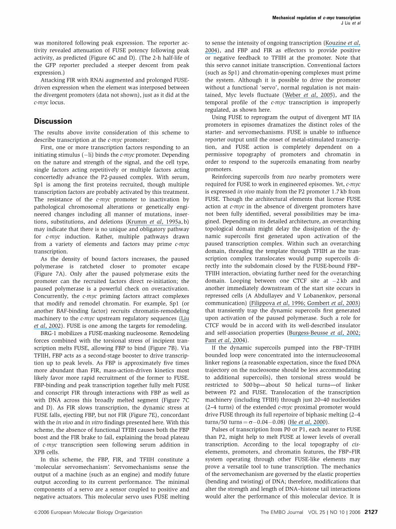
Materials and methods
Tissue cultureHuman Hs68 primary fibroblasts, HeLa, U2OS, SW13, and IMR-32cells were cultured in DMEM with 10% FBS. Hs68 cells werecultured without serum for 5 days and stimulated with 10% FBS.
Cells were harvested at indicated time points for RNA and ChIP.Primary fibroblasts from XPB patients (GM13025, GM13026) andtheir parents (GM13027, GM13028) were cultured in DMEM with15% FBS. Lymphoblast XPB (GM02252) and complemented cellswere described previously (Weber et al, 2005).
c-myc mRNA quantitationRNase Protection Assay was performed as described (Collins et al,2001). Autoradiographs were analyzed with ImageQuant. UniversalProbe Library (Roche) probes 77 and 25 were used for qRT–PCR forc-myc and g-tubulin, respectively.
FUSE
FUSE
PPC
FUSE
FUSE
SW
I/SN
F
SW
I/SN
FFB
P
FB
PF
IR
FUSE
FIR
Sp1, YY1, E2F, SWI/SNF, etc
-σ
FBP
Prolonged starvation
30 min
2 h
2– 4 h
>4 h
PPC
EC
P2
PPC
PPC
PPC
FIR
FIR
FBP
FUSE
FIR
PPC
shu
t off
Activation
Intermediate starvationA
B
C
D
E
Figure 7 Proposed scheme for molecular servomechanism for regulation of c-myc transcription. (A) Without serum, c-myc transcription shutsoff, FUSE becomes masked by a nucleosome, and only a trace of FIR remains. With prolonged starvation, FIR entirely disengages from FUSE.Even when shut off, a Pre-Promoter Escape Complex (PPC) remains paused within the promoter proximal region (P2). (B) The earliest factorsbinding to the promoter upon stimulation advance the PPC to a full Elongation Complex (EC), the FUSE-masking nucleosome is remodeled toexpose FUSE. Negative supercoiling (�s) generated by EC, combined with remodeling forces, initiate FUSE melting. (C) Melted FUSE recruitsFBP, which drives transcription up to peak levels by looping with TFIIH. (D) FBP activity leads to negative supercoil accumulation within thetopologically closed FBP–TFIIH loop. High �s fully melts FUSE and conscripts FIR through protein–protein and protein–DNA contacts. (E) AsFIR represses transcription, the dynamic stress at FUSE falls, ejecting FBP. The FIR–TFIIH connection allows only basal transcription; so thepolymerase at a low rate dissociates from TFIIH to become an EC. Without sufficient activation, the superhelical stress eventually dissipates,and the c-myc promoter reverts back to the silent state in (A). For illustrative purposes, only the FUSE-masking nucleosome is shown.
Mechanical regulation of c-myc transcriptionJ Liu et al
The EMBO Journal VOL 25 | NO 10 | 2006 &2006 European Molecular Biology Organization2128
Chromatin immunoprecipitation and real-time PCRChIP was performed as described (Weinmann and Farnham, 2002).PCR was performed using Platinum PCR Supermix (Invitrogen) for35 cycles with FUSE primers, 30 cycles for P2 and mid-primers.Real-time PCR was performed with Platinum SybrGreen qPCR kit(Invitrogen) with the same primers.
Nucleosome mapping and KMnO4 footprintingLM–PCR nucleosome mapping at FUSE was performed as described(Carey and Smale, 2000), except that mono-nucleosomal DNA wasgel purified prior to ligation. For in vitro KMnO4 footprinting, 10 ngof recombinant GST, FBP, or FIR was incubated with 25 ng of eithersupercoiled or linear DNA in 20 mM HEPES, pH 7.9, 0.1 mg/ml BSA,and 15 mM NaCl for 15 min. MgSO4 was then added to 2 mM in allsamples and XhoI was added as indicated. KMnO4 treatment wasperformed 5 min post-XhoI addition. MgSO4 (2 mM) is sufficient forcomplete digestion of supercoiled DNA.
In vitro transcription and DNA IPAccumulation of negative supercoils in linear DNA by T3 and T7polymerase was documented (Kouzine et al, 2004). Transcriptionrate was limited by GTP. Highly purified recombinant FBP or FIRwas included. After 10 min of transcription, formaldehyde (0.5%final (v/v)) was added to crosslink protein to DNA and stoptranscription. To stop transcription prior to fixation, EDTA wasadded to 20 mM and incubated for 5 min before addition offormaldehyde. The reaction was immunoprecipitated with eithera-FBP or a-FIR. PCR was performed with Platinum PCR Supermixfor 22 cycles. Real-time PCR was performed with PlatinumSybrGreen qPCR kit.
RNA interferenceStealth siRNA to FIR or FBP was synthesized by Invitrogen. Hs68cells were serum starved for 2 days and siRNA (20 nM) wastransfected with Lipofectamine 2000 and cultured in DMEM withoutserum for 2.5 days before serum stimulation. Cells were harvestedfor RNA or protein.
Flow cytometry analysis of zinc-induced GFP expressionFUSE or an irrelevant 91 bp fragment was placed between twodivergently transcribed, identical metallothionein promoters driv-ing either RFP or destabilized GFP. Spacers of different length wereplaced between the promoters and FUSE. Raji cells carrying theepisomes plasmid were selected and cultured in RPMI-1640 with10% FBS and 50mg/ml hygromycin. Transcription was inducedby addition of ZnSO4 to a final of 90mM. GFP fluorescence wasanalyzed by a flow cytometer.
Supplementary dataSupplementary data are available at The EMBO Journal Online.
Acknowledgements
We thank Drs Larry Benjamin, Hui Ge, Lance Liotta, John Lis,Dinah Singer, Susan Mackem, and Carl Wu for comments anddiscussion. This work was supported in part by the IntramuralResearch Program of the NIH, National Cancer Institute, Center forCancer Research. This study was supported by grants from theDeutsche Forschungsgemeinschaft (DFG) to A.W. (AW2397/1-1 andAW2397/2-1).
References
Bazar L, Meighen D, Harris V, Duncan R, Levens D, Avigan M(1995) Targeted melting and binding of a DNA regulatoryelement by a transactivator of c-myc. J Biol Chem 270:8241–8248
Bentley DL, Groudine M (1986) A block to elongation is largelyresponsible for decreased transcription of c-myc in differentiatedHL60 cells. Nature 321: 702–706
Burgess-Beusse B, Farrell C, Gaszner M, Litt M, Mutskov V, Recillas-Targa F, Simpson M, West A, Felsenfeld G (2002) The insulationof genes from external enhancers and silencing chromatin. ProcNatl Acad Sci USA 99 (Suppl 4): 16433–16437
Carey M, Smale ST (2000) Transcriptional Regulation in Eukaryotes:Concepts, Strategies, and Techniques. Cold Spring HarborLaboratory Press: Cold Spring Harbor, NY
Cawley S, Bekiranov S, Ng HH, Kapranov P, Sekinger EA, Kampa D,Piccolboni A, Sementchenko V, Cheng J, Williams AJ, Wheeler R,Wong B, Drenkow J, Yamanaka M, Patel S, Brubaker S, TammanaH, Helt G, Struhl K, Gingeras TR (2004) Unbiased mapping oftranscription factor binding sites along human chromosomes 21and 22 points to widespread regulation of noncoding RNAs. Cell116: 499–509
Chi TH, Wan M, Lee PP, Akashi K, Metzger D, Chambon P, WilsonCB, Crabtree GR (2003) Sequential roles of Brg, the ATPasesubunit of BAF chromatin remodeling complexes, in thymocytedevelopment. Immunity 19: 169–182
Collins I, Weber A, Levens D (2001) Transcriptional consequencesof topoisomerase inhibition. Mol Cell Biol 21: 8437–8451
de la Cova C, Abril M, Bellosta P, Gallant P, Johnston LA (2004)Drosophila myc regulates organ size by inducing cell competition.Cell 117: 107–116
Dean M, Levine RA, Ran W, Kindy MS, Sonenshein GE, Campisi J(1986) Regulation of c-myc transcription and mRNA abundanceby serum growth factors and cell contact. J Biol Chem 261:9161–9166
DeAngelo DJ, DeFalco J, Rybacki L, Childs G (1995) The embryonicenhancer-binding protein SSAP contains a novel DNA-bindingdomain which has homology to several RNA-binding proteins.Mol Cell Biol 15: 1254–1264
DesJardins E, Hay N (1993) Repeated CT elements bound by zincfinger proteins control the absolute and relative activities ofthe two principal human c-myc promoters. Mol Cell Biol 13:5710–5724
Ding J, Hayashi MK, Zhang Y, Manche L, Krainer AR, Xu RM (1999)Crystal structure of the two-RRM domain of hnRNP A1 (UP1)complexed with single-stranded telomeric DNA. Genes Dev 13:1102–1115
Duncan R, Bazar L, Michelotti G, Tomonaga T, Krutzsch H, AviganM, Levens D (1994) A sequence-specific, single-strand bindingprotein activates the far upstream element of c-myc and defines anew DNA-binding motif. Genes Dev 8: 465–480
Duncan R, Collins I, Tomonaga T, Zhang T, Levens D (1996) Aunique transactivation sequence motif is found in the carboxyl-terminal domain of the single-strand-binding protein FBP. MolCell Biol 16: 2274–2282
Filippova GN, Fagerlie S, Klenova EM, Myers C, Dehner Y, GoodwinG, Neiman PE, Collins SJ, Lobanenkov VV (1996) An exception-ally conserved transcriptional repressor, CTCF, employs differentcombinations of zinc fingers to bind diverged promoter se-quences of avian and mammalian c-myc oncogenes. Mol CellBiol 16: 2802–2813
Gombert WM, Farris SD, Rubio ED, Morey-Rosler KM, SchubachWH, Krumm A (2003) The c-myc insulator element and matrixattachment regions define the c-myc chromosomal domain. MolCell Biol 23: 9338–9348
Grandori C, Cowley SM, James LP, Eisenman RN (2000) The Myc/Max/Mad network and the transcriptional control of cell beha-vior. Annu Rev Cell Dev Biol 16: 653–699
He L, Liu J, Collins I, Sanford S, O’Connell B, Benham CJ, Levens D(2000) Loss of FBP function arrests cellular proliferation andextinguishes c-myc expression. EMBO J 19: 1034–1044
Karin M, Haslinger A, Holtgreve H, Cathala G, Slater E, Baxter JD(1984) Activation of a heterologous promoter in response todexamethasone and cadmium by metallothionein gene 50-flank-ing DNA. Cell 36: 371–379
Kelly K, Siebenlist U (1986) The regulation and expression of c-mycin normal and malignant cells. Annu Rev Immunol 4: 317–338
Kielkopf CL, Lucke S, Green MR (2004) U2AF homology motifs:protein recognition in the RRM world. Genes Dev 18: 1513–1526
Kim TH, Barrera LO, Zheng M, Qu C, Singer MA, Richmond TA, WuY, Green RD, Ren B (2005) A high-resolution map of activepromoters in the human genome. Nature 436: 876–880
Kouzine F, Liu J, Sanford S, Chung HJ, Levens D (2004) Thedynamic response of upstream DNA to transcription-generatedtorsional stress. Nat Struct Mol Biol 11: 1092–1100
Mechanical regulation of c-myc transcriptionJ Liu et al
&2006 European Molecular Biology Organization The EMBO Journal VOL 25 | NO 10 | 2006 2129
Krumm A, Hickey LB, Groudine M (1995a) Promoter-proximalpausing of RNA polymerase II defines a general rate-limitingstep after transcription initiation. Genes Dev 9: 559–572
Krumm A, Hickey LB, Groudine M (1995b) Promoter-proximalpausing of RNA polymerase II defines a general rate-limitingstep after transcription initiation. Genes Dev 9: 559–572
Levens D (2002) Disentangling the MYC web. Proc Natl Acad SciUSA 99: 5757–5759
Levens DL (2003) Reconstructing MYC. Genes Dev 17: 1071–1077Liu H, Kang H, Liu R, Chen X, Zhao K (2002) Maximal induction
of a subset of interferon target genes requires the chromatin-remodeling activity of the BAF complex. Mol Cell Biol 22:6471–6479
Liu J, Akoulitchev S, Weber A, Ge H, Chuikov S, Libutti D, WangXW, Conaway JW, Harris CC, Conaway RC, Reinberg D, Levens D(2001) Defective interplay of activators and repressors with TFIHin Xeroderma pigmentosum. Cell 104: 353–363
Liu J, He L, Collins I, Ge H, Libutti D, Li J, Egly JM, Levens D (2000)The FBP interacting repressor targets TFIIH to inhibit activatedtranscription. Mol Cell 5: 331–341
Maniatis T, Falvo JV, Kim TH, Kim TK, Lin CH, Parekh BS, WatheletMG (1998) Structure and function of the interferon-beta enhan-ceosome. Cold Spring Harb Symp Quant Biol 63: 609–620
Marcu KB, Bossone SA, Patel AJ (1992) myc function and regula-tion. Annu Rev Biochem 61: 809–860
McGhee JD, von Hippel PH (1975) Formaldehyde as a probe of DNAstructure. I. Reaction with exocyclic amino groups of DNA bases.Biochemistry 14: 1281–1296
Merika M, Thanos D (2001) Enhanceosomes. Curr Opin Genet Dev11: 205–208
Michelotti GA, Michelotti EF, Pullner A, Duncan RC, Eick D, LevensD (1996) Multiple single-stranded cis elements are associatedwith activated chromatin of the human c-myc gene in vivo. MolCell Biol 16: 2656–2669
Moreno E, Basler K (2004) dMyc transforms cells into super-competitors. Cell 117: 117–129
Pant V, Kurukuti S, Pugacheva E, Shamsuddin S, Mariano P,Renkawitz R, Klenova E, Lobanenkov V, Ohlsson R (2004)Mutation of a single CTCF target site within the H19 imprintingcontrol region leads to loss of Igf2 imprinting and complexpatterns of de novo methylation upon maternal inheritance. MolCell Biol 24: 3497–3504
Riggs KJ, Saleque S, Wong KK, Merrell KT, Lee JS, Shi Y, Calame K(1993) Yin-yang 1 activates the c-myc promoter. Mol Cell Biol 13:7487–7495
Secombe J, Pierce SB, Eisenman RN (2004) Myc: a weapon of massdestruction. Cell 117: 153–156
Shang Y, Myers M, Brown M (2002) Formation of the androgenreceptor transcription complex. Mol Cell 9: 601–610
Spencer CA, Groudine M (1991) Control of c-myc regulation innormal and neoplastic cells. Adv Cancer Res 56: 1–48
Strobl LJ, Eick D (1992) Hold back of RNA polymerase II at thetranscription start site mediates down-regulation of c-myc in vivo.EMBO J 11: 3307–3314
Thalmeier K, Synovzik H, Mertz R, Winnacker EL, Lipp M (1989)Nuclear factor E2F mediates basic transcription and trans-activa-tion by E1a of the human MYC promoter. Genes Dev 3: 527–536
Trumpp A, Refaeli Y, Oskarsson T, Gasser S, Murphy M, Martin GR,Bishop JM (2001) c-Myc regulates mammalian body size bycontrolling cell number but not cell size. Nature 414: 768–773
van Steeg H, Kraemer KH (1999) Xeroderma pigmentosum and therole of UV-induced DNA damage in skin cancer. Mol Med Today 5:86–94
Waters CM, Littlewood TD, Hancock DC, Moore JP, Evan GI (1991)c-myc protein expression in untransformed fibroblasts. Oncogene6: 797–805
Weber A, Liu J, Collins I, Levens D (2005) TFIIH operates throughan expanded proximal promoter to fine-tune c-myc expression.Mol Cell Biol 25: 147–161
Weinmann AS, Farnham PJ (2002) Identification of unknown targetgenes of human transcription factors using chromatin immuno-precipitation. Methods 26: 37–47
Mechanical regulation of c-myc transcriptionJ Liu et al
The EMBO Journal VOL 25 | NO 10 | 2006 &2006 European Molecular Biology Organization2130
Copyright © 2022 FDOKUMEN