The fluid mechanics of scleral buckling surgery for the repair of retinal detachment
16
The Fluid Mechanics of Scleral Buckling Surgery for the Repair of Retinal Detachment William J. Foster 1,3,a , Nadia Dowla 2 , Saurabh Y. Joshi 2 , and Michael Nikolaou 2 1 Department of Physics, The University of Houston, 4800 Calhoun Road Houston, TX 77004. 713-743-8339. 2 Department of Chemical Engineering, The University of Houston, 4800 Calhoun Road Houston, TX 77004. 713-743-8339. 3 Department of Ophthalmology, Weill-Cornell Medical College at The Methodist Hospital, Houston, TX Abstract Background—Scleral buckling is a common surgical technique used to treat retinal detachments that involves suturing a radial or circumferential silicone element on the sclera. Although this procedure has been performed since the 1960’s, and there is a reasonable experimental model of retinal detachment, there is still debate as to how this surgery facilitates the re-attachment of the retina. Methods—Finite element calculations using the COMSOL Multiphysics® system are utilized to explain the influence of the scleral buckle on the flow of sub-retinal fluid in a physical model of retinal detachment. Results—We found that, by coupling fluid mechanics with structural mechanics, laminar fluid flow and the Bernoulli effect are necessary for a physically consistent explanation of retinal reattachment. Improved fluid outflow and retinal reattachment are found with low fluid viscosity and rapid eye movements. A simulation of saccadic eye movements was more effective in removing sub-retinal fluid than slower, reading speed, eye movements in removing subretinal fluid. Conclusions—The results of our simulations allow us to explain the physical principles behind scleral buckling surgery and provide insight that can be utilized clinically. In particular, we find that rapid eye movements facilitate more rapid retinal reattachment. This is contradictory to the conventional wisdom of attempting to minimize eye movements. Keywords Retinal Detachment; Fluid Mechanics; Scleral Buckle; Vitreoretinal Surgery 1 Introduction The retina is an approximately 300 mm thick layer of neural tissue that lines the back chamber (the vitreous cavity) of the eye. It is firmly attached at its most anterior aspect (the ora serrata) and the optic nerve. If a hole develops in the retina, vitreous fluid can enter the hole and cause the retina to separate from the back of the eye, leading to a retinal detachment. This condition requires prompt treatment to avoid irreversible blindness. aTo whom correspondence should be directed, [email protected], 713-743-3550, fax 713-743-3589.. NIH Public Access Author Manuscript Graefes Arch Clin Exp Ophthalmol. Author manuscript; available in PMC 2011 January 1. Published in final edited form as: Graefes Arch Clin Exp Ophthalmol. 2010 January ; 248(1): 31–36. doi:10.1007/s00417-009-1198-z. NIH-PA Author Manuscript NIH-PA Author Manuscript NIH-PA Author Manuscript
Transcript of The fluid mechanics of scleral buckling surgery for the repair of retinal detachment

The Fluid Mechanics of Scleral Buckling Surgery for the Repair ofRetinal Detachment
William J. Foster1,3,a, Nadia Dowla2, Saurabh Y. Joshi2, and Michael Nikolaou21Department of Physics, The University of Houston, 4800 Calhoun Road Houston, TX 77004.713-743-8339.2Department of Chemical Engineering, The University of Houston, 4800 Calhoun Road Houston,TX 77004. 713-743-8339.3Department of Ophthalmology, Weill-Cornell Medical College at The Methodist Hospital, Houston,TX
AbstractBackground—Scleral buckling is a common surgical technique used to treat retinal detachmentsthat involves suturing a radial or circumferential silicone element on the sclera. Although thisprocedure has been performed since the 1960’s, and there is a reasonable experimental model ofretinal detachment, there is still debate as to how this surgery facilitates the re-attachment of theretina.
Methods—Finite element calculations using the COMSOL Multiphysics® system are utilized toexplain the influence of the scleral buckle on the flow of sub-retinal fluid in a physical model ofretinal detachment.
Results—We found that, by coupling fluid mechanics with structural mechanics, laminar fluid flowand the Bernoulli effect are necessary for a physically consistent explanation of retinal reattachment.Improved fluid outflow and retinal reattachment are found with low fluid viscosity and rapid eyemovements. A simulation of saccadic eye movements was more effective in removing sub-retinalfluid than slower, reading speed, eye movements in removing subretinal fluid.
Conclusions—The results of our simulations allow us to explain the physical principles behindscleral buckling surgery and provide insight that can be utilized clinically. In particular, we find thatrapid eye movements facilitate more rapid retinal reattachment. This is contradictory to theconventional wisdom of attempting to minimize eye movements.
KeywordsRetinal Detachment; Fluid Mechanics; Scleral Buckle; Vitreoretinal Surgery
1 IntroductionThe retina is an approximately 300 mm thick layer of neural tissue that lines the back chamber(the vitreous cavity) of the eye. It is firmly attached at its most anterior aspect (the ora serrata)and the optic nerve. If a hole develops in the retina, vitreous fluid can enter the hole and causethe retina to separate from the back of the eye, leading to a retinal detachment. This conditionrequires prompt treatment to avoid irreversible blindness.
aTo whom correspondence should be directed, [email protected], 713-743-3550, fax 713-743-3589..
NIH Public AccessAuthor ManuscriptGraefes Arch Clin Exp Ophthalmol. Author manuscript; available in PMC 2011 January 1.
Published in final edited form as:Graefes Arch Clin Exp Ophthalmol. 2010 January ; 248(1): 31–36. doi:10.1007/s00417-009-1198-z.
NIH
-PA Author Manuscript
NIH
-PA Author Manuscript
NIH
-PA Author Manuscript

Perhaps the oldest and most established method to re-attach the retina during retinal detachmentsurgery is scleral buckling surgery, indenting the wall of the eye by suturing a silicone bandor buckle on the sclera. Frequently, at the end of the surgery, the hole in the retina (theunderlying cause of the retinal detachment) is over the indentation that is caused by the scleralbuckle, but the retina in the area of the hole is not against the back of the eye. Thus, the holeis not closed. While an incision can, at the time of surgery, be made in the wall of the eye andsub-retinal fluid can be externally drained, this extra procedure rarely results in a completeabsence of fluid under the retina. The question that we are asking is: “Why does the retinaattach itself against the back of the eye after the placement of a scleral buckle?”
Previously, Clemens et al1 developed a simplified experimental model [Figure 1] of a retinaldetachment and scleral buckling that reproduced the essential features of this problem. Theyutilized a fish tank with a thin cloth membrane attached at the periphery of the tank. Themembrane contained a small hole and a piece of tubing was placed under the membrane tosimulate indentation from the scleral buckle. The tank was then manually rocked back andforth, in an oscillating manner, and the cloth “retina” was found to reattach itself to the bottomof the tank if the silicone “buckle” were placed under the hole in the “retina”. On the otherhand, if the “buckle” were placed in an inappropriate position, not under the hole, the retinawould remain detached. Our goal is to simulate this model system using finite element analysis,guided by physical reasoning.
There are other mechanisms that can aid in retinal re-attachment, that are not included in thesecalculations:
1. The Retinal Pigment Epithelium (RPE), the tissue directly under the retina, isknown2 to pump subretinal fluid out of the eye. Despite this, the pump cannot keepup with the influx of fluid caused by the hole in the retina. This will be discussed laterin this paper.
2. In addition, by indenting the wall of the eye in the area of the retinal hole, the vitreousgel cannot apply as much traction to the edge of the hole, and the hole may naturallyclose.
3. The osmotic pressure of the existing vitreous might be hypothesized to aid in re-attachment of the retina. This is known to not be the primary mechanism by whichthe retina is re-approximated to the back of the eye, given that the results of sclearalbuckling surgery do not depend upon whether the patient previously underwent avitrectomy3 (i.e., removal of the vitreous gel).
2 Materials and methodsIn this paper, we utilize a simplified model of the eye, previously described experimentally inthe paper by Clemens et al1. The system consists of a solid floor with a membrane suspendedabove it. A single hole is present in the membrane and an elevation, simulating a scleral buckle,can be placed in various positions under the membrane [Figure 3 and Figure 4].
The model was implemented using COMSOL Multiphysics®, version 3.4 with the MEMSmodule to facilitate coupled fluid and structural equations. Unless stated otherwise, theseparation between the retina and the back of the eye was initially 5 mm, and the density anddynamic viscosity of the fluid were taken to be that of water at 37.5 degrees C. The retina,which is composed of neural tissue, is simulated as an isotropic membrane, 300 μm thick, witha Young’s modulus of 1000 Pa. For comparison, simulations were also performed with theretina modeled as a 300 mm thick rigid membrane.
Foster et al. Page 2
Graefes Arch Clin Exp Ophthalmol. Author manuscript; available in PMC 2011 January 1.
NIH
-PA Author Manuscript
NIH
-PA Author Manuscript
NIH
-PA Author Manuscript
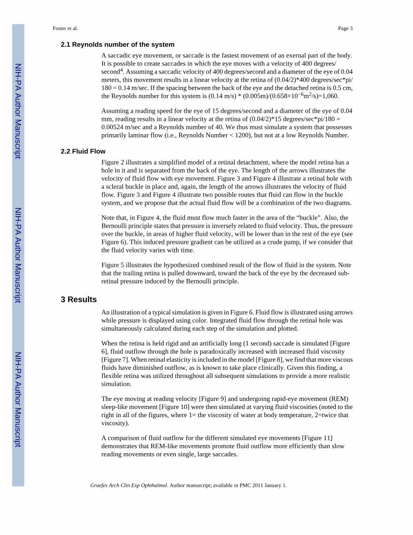
2.1 Reynolds number of the systemA saccadic eye movement, or saccade is the fastest movement of an exernal part of the body.It is possible to create saccades in which the eye moves with a velocity of 400 degrees/second4. Assuming a saccadic velocity of 400 degrees/second and a diameter of the eye of 0.04meters, this movement results in a linear velocity at the retina of (0.04/2)*400 degrees/sec*pi/180 = 0.14 m/sec. If the spacing between the back of the eye and the detached retina is 0.5 cm,the Reynolds number for this system is (0.14 m/s) * (0.005m)/(0.658×10−6m2/s)=1,060.
Assuming a reading speed for the eye of 15 degrees/second and a diameter of the eye of 0.04mm, reading results in a linear velocity at the retina of (0.04/2)*15 degrees/sec*pi/180 =0.00524 m/sec and a Reynolds number of 40. We thus must simulate a system that possessesprimarily laminar flow (i.e., Reynolds Number < 1200), but not at a low Reynolds Number.
2.2 Fluid FlowFigure 2 illustrates a simplified model of a retinal detachment, where the model retina has ahole in it and is separated from the back of the eye. The length of the arrows illustrates thevelocity of fluid flow with eye movement. Figure 3 and Figure 4 illustrate a retinal hole witha scleral buckle in place and, again, the length of the arrows illustrates the velocity of fluidflow. Figure 3 and Figure 4 illustrate two possible routes that fluid can flow in the bucklesystem, and we propose that the actual fluid flow will be a combination of the two diagrams.
Note that, in Figure 4, the fluid must flow much faster in the area of the “buckle”. Also, theBernoulli principle states that pressure is inversely related to fluid velocity. Thus, the pressureover the buckle, in areas of higher fluid velocity, will be lower than in the rest of the eye (seeFigure 6). This induced pressure gradient can be utilized as a crude pump, if we consider thatthe fluid velocity varies with time.
Figure 5 illustrates the hypothesized combined result of the flow of fluid in the system. Notethat the trailing retina is pulled downward, toward the back of the eye by the decreased sub-retinal pressure induced by the Bernoulli principle.
3 ResultsAn illustration of a typical simulation is given in Figure 6. Fluid flow is illustrated using arrowswhile pressure is displayed using color. Integrated fluid flow through the retinal hole wassimultaneously calculated during each step of the simulation and plotted.
When the retina is held rigid and an artificially long (1 second) saccade is simulated [Figure6], fluid outflow through the hole is paradoxically increased with increased fluid viscosity[Figure 7]. When retinal elasticity is included in the model [Figure 8], we find that more viscousfluids have diminished outflow, as is known to take place clinically. Given this finding, aflexible retina was utilized throughout all subsequent simulations to provide a more realisticsimulation.
The eye moving at reading velocity [Figure 9] and undergoing rapid-eye movement (REM)sleep-like movement [Figure 10] were then simulated at varying fluid viscosities (noted to theright in all of the figures, where 1= the viscosity of water at body temperature, 2=twice thatviscosity).
A comparison of fluid outflow for the different simulated eye movements [Figure 11]demonstrates that REM-like movements promote fluid outflow more efficiently than slowreading movements or even single, large saccades.
Foster et al. Page 3
Graefes Arch Clin Exp Ophthalmol. Author manuscript; available in PMC 2011 January 1.
NIH
-PA Author Manuscript
NIH
-PA Author Manuscript
NIH
-PA Author Manuscript
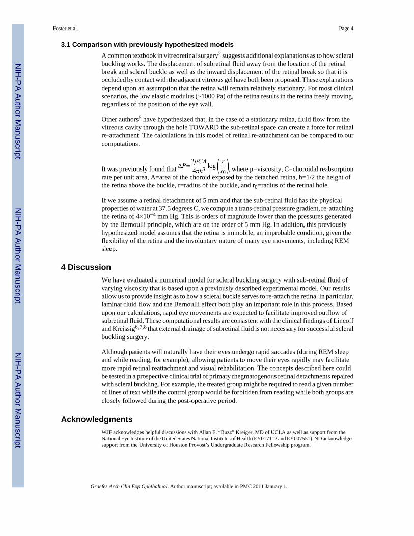
3.1 Comparison with previously hypothesized modelsA common textbook in vitreoretinal surgery2 suggests additional explanations as to how scleralbuckling works. The displacement of subretinal fluid away from the location of the retinalbreak and scleral buckle as well as the inward displacement of the retinal break so that it isoccluded by contact with the adjacent vitreous gel have both been proposed. These explanationsdepend upon an assumption that the retina will remain relatively stationary. For most clinicalscenarios, the low elastic modulus (~1000 Pa) of the retina results in the retina freely moving,regardless of the position of the eye wall.
Other authors5 have hypothesized that, in the case of a stationary retina, fluid flow from thevitreous cavity through the hole TOWARD the sub-retinal space can create a force for retinalre-attachment. The calculations in this model of retinal re-attachment can be compared to ourcomputations.
It was previously found that , where μ=viscosity, C=choroidal reabsorptionrate per unit area, A=area of the choroid exposed by the detached retina, h=1/2 the height ofthe retina above the buckle, r=radius of the buckle, and r0=radius of the retinal hole.
If we assume a retinal detachment of 5 mm and that the sub-retinal fluid has the physicalproperties of water at 37.5 degrees C, we compute a trans-retinal pressure gradient, re-attachingthe retina of 4×10⁻⁴ mm Hg. This is orders of magnitude lower than the pressures generatedby the Bernoulli principle, which are on the order of 5 mm Hg. In addition, this previouslyhypothesized model assumes that the retina is immobile, an improbable condition, given theflexibility of the retina and the involuntary nature of many eye movements, including REMsleep.
4 DiscussionWe have evaluated a numerical model for scleral buckling surgery with sub-retinal fluid ofvarying viscosity that is based upon a previously described experimental model. Our resultsallow us to provide insight as to how a scleral buckle serves to re-attach the retina. In particular,laminar fluid flow and the Bernoulli effect both play an important role in this process. Basedupon our calculations, rapid eye movements are expected to facilitate improved outflow ofsubretinal fluid. These computational results are consistent with the clinical findings of Lincoffand Kreissig6,7,8 that external drainage of subretinal fluid is not necessary for successful scleralbuckling surgery.
Although patients will naturally have their eyes undergo rapid saccades (during REM sleepand while reading, for example), allowing patients to move their eyes rapidly may facilitatemore rapid retinal reattachment and visual rehabilitation. The concepts described here couldbe tested in a prospective clinical trial of primary rhegmatogenous retinal detachments repairedwith scleral buckling. For example, the treated group might be required to read a given numberof lines of text while the control group would be forbidden from reading while both groups areclosely followed during the post-operative period.
AcknowledgmentsWJF acknowledges helpful discussions with Allan E. “Buzz” Kreiger, MD of UCLA as well as support from theNational Eye Institute of the United States National Institutes of Health (EY017112 and EY007551). ND acknowledgessupport from the University of Houston Provost’s Undergraduate Research Fellowship program.
Foster et al. Page 4
Graefes Arch Clin Exp Ophthalmol. Author manuscript; available in PMC 2011 January 1.
NIH
-PA Author Manuscript
NIH
-PA Author Manuscript
NIH
-PA Author Manuscript
WJF received grant support from the National Eye Institute of the National Institutes of Health that provided partialsalary support. ND acknowledges support from the University of Houston Provost’s Undergraduate ResearchFellowship program that provided a stipend.
WJF has full control of all primary data and all authors agree to allow Graefe’s Archive for Clinical and ExperimentalOphthalmology to review the data, if requested.
5 References1. Clemens S, Kroll P, Stein E, Wagner W, Wriggers P. Experimental studies on the disappearance of
subretinal fluid after episcleral buckling procedures without drainage. Graefe’s Arch Exp Ophthalmol1987;225:16–18.
2. Wilkinson, CP.; Rice, TA., editors. Michels Retinal Detachment. Vol. Second Edition. Mosby; NewYork: 1997.
3. Mester U, Anterist N, Kroll P, Brieden-Azvedo S. The Role of the Vitreous in Retinal DetachmentSurgery with External Buckling. Ophthalmologica 2002;216:242–245. [PubMed: 12207125]
4. Stetter M, Sendtner RA, Timberlake GT. A Novel Method for Measuring Saccade Profiles Using theScanning Laser Ophthalmoscope. Vision Research 1996;36:1987–1994. [PubMed: 8759438]
5. Hammer ME. Retinal re-attachment forces created by absorption of subretinal fluid. Doc OphthalmolProc Ser 1981;25:61–75.
6. Kreissig I, Simader E, Fahle M, Lincoff H. Visual acuity after segmental buckling and non-drainage:a 15-year follow-up. Eur J Ophthalmol 1995;5(4):240–6. [PubMed: 8963161]
7. Lincoff H, Ramirez V, Kreissig I, Baronberg N, Kaufman D. Encircling operations without drainageof subretinal fluid. Mod Probl Ophthalmol 1975;15:188–96. [PubMed: 1160899]
8. Lincoff H, Kreissig I. The treatment of retinal detachment without drainage of subretinal fluid.(Modifications of the Custodis procedure. VI). Trans Am Acad Ophthalmol Otolaryngol Sep-Oct;197276(5):1121–33. [PubMed: 4666576]
Foster et al. Page 5
Graefes Arch Clin Exp Ophthalmol. Author manuscript; available in PMC 2011 January 1.
NIH
-PA Author Manuscript
NIH
-PA Author Manuscript
NIH
-PA Author Manuscript

Figure 1.Experimental model of a scleral buckle, as implemented by Clemens et al. Note the piece ofcloth, attached at the lower edge of the tank, with a small central hole in the cloth. A piece oftubing under the cloth serves as the indentation or buckle. Reprinted with permission fromGraefe’s Arch Exp Ophthalmol. 225:16-18 (1987).
Foster et al. Page 6
Graefes Arch Clin Exp Ophthalmol. Author manuscript; available in PMC 2011 January 1.
NIH
-PA Author Manuscript
NIH
-PA Author Manuscript
NIH
-PA Author Manuscript

Figure 2.A simplified model of a retinal detachment.
Foster et al. Page 7
Graefes Arch Clin Exp Ophthalmol. Author manuscript; available in PMC 2011 January 1.
NIH
-PA Author Manuscript
NIH
-PA Author Manuscript
NIH
-PA Author Manuscript

Figure 3.Changes in fluid velocity, with resulting changes in pressure, in the simplified model.
Foster et al. Page 8
Graefes Arch Clin Exp Ophthalmol. Author manuscript; available in PMC 2011 January 1.
NIH
-PA Author Manuscript
NIH
-PA Author Manuscript
NIH
-PA Author Manuscript

Figure 4.Deflection of fluid in the simplified model.
Foster et al. Page 9
Graefes Arch Clin Exp Ophthalmol. Author manuscript; available in PMC 2011 January 1.
NIH
-PA Author Manuscript
NIH
-PA Author Manuscript
NIH
-PA Author Manuscript

Figure 5.Resulting displacement of the retina with increased fluid outflow in the simplified system.
Foster et al. Page 10
Graefes Arch Clin Exp Ophthalmol. Author manuscript; available in PMC 2011 January 1.
NIH
-PA Author Manuscript
NIH
-PA Author Manuscript
NIH
-PA Author Manuscript
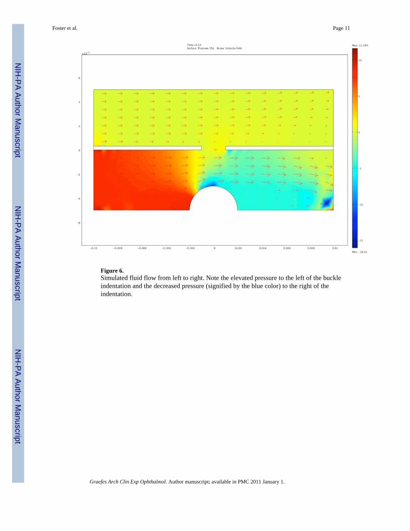
Figure 6.Simulated fluid flow from left to right. Note the elevated pressure to the left of the buckleindentation and the decreased pressure (signified by the blue color) to the right of theindentation.
Foster et al. Page 11
Graefes Arch Clin Exp Ophthalmol. Author manuscript; available in PMC 2011 January 1.
NIH
-PA Author Manuscript
NIH
-PA Author Manuscript
NIH
-PA Author Manuscript
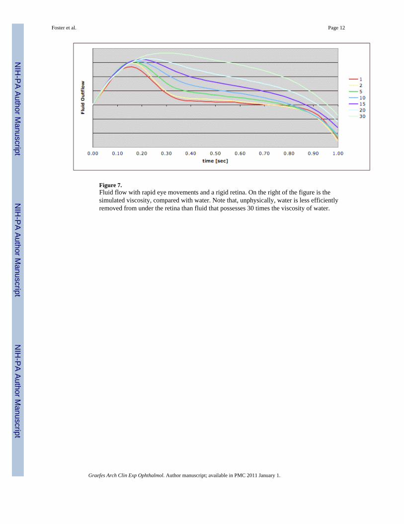
Figure 7.Fluid flow with rapid eye movements and a rigid retina. On the right of the figure is thesimulated viscosity, compared with water. Note that, unphysically, water is less efficientlyremoved from under the retina than fluid that possesses 30 times the viscosity of water.
Foster et al. Page 12
Graefes Arch Clin Exp Ophthalmol. Author manuscript; available in PMC 2011 January 1.
NIH
-PA Author Manuscript
NIH
-PA Author Manuscript
NIH
-PA Author Manuscript

Figure 8.Fluid flow with rapid eye movements and a flexible retina. On the right of the figure is thesimulated viscosity, compared with water. The peaks in fluid outflow correspond to points intime at which the retina was positioned as illustrated in Figure 5. This was a consistent finding,despite the non-periodic nature of the data.
Foster et al. Page 13
Graefes Arch Clin Exp Ophthalmol. Author manuscript; available in PMC 2011 January 1.
NIH
-PA Author Manuscript
NIH
-PA Author Manuscript
NIH
-PA Author Manuscript
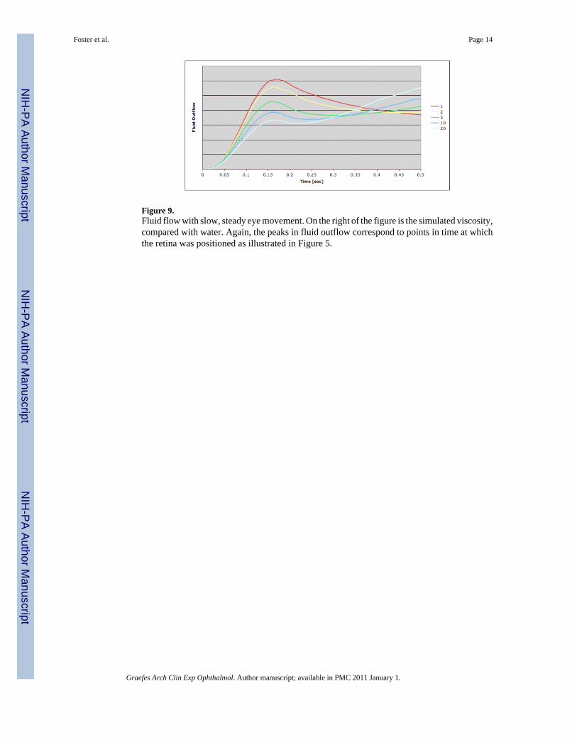
Figure 9.Fluid flow with slow, steady eye movement. On the right of the figure is the simulated viscosity,compared with water. Again, the peaks in fluid outflow correspond to points in time at whichthe retina was positioned as illustrated in Figure 5.
Foster et al. Page 14
Graefes Arch Clin Exp Ophthalmol. Author manuscript; available in PMC 2011 January 1.
NIH
-PA Author Manuscript
NIH
-PA Author Manuscript
NIH
-PA Author Manuscript

Figure 10.Fluid flow in simulated REM sleep (rapid, short saccades). On the right of the figure is thesimulated viscosity, compared with water. The peaks in fluid outflow correspond to points intime at which the retina was positioned as illustrated in Figure 5. This was a consistent finding,despite the non-periodic nature of the data.
Foster et al. Page 15
Graefes Arch Clin Exp Ophthalmol. Author manuscript; available in PMC 2011 January 1.
NIH
-PA Author Manuscript
NIH
-PA Author Manuscript
NIH
-PA Author Manuscript
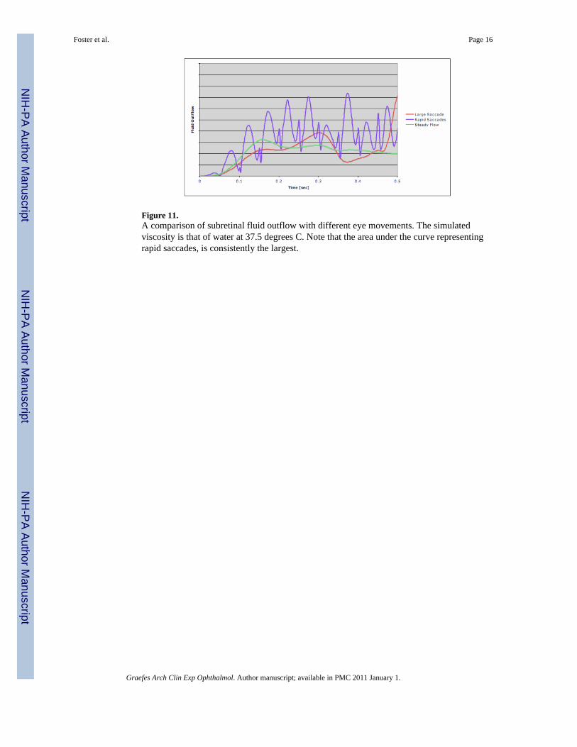
Figure 11.A comparison of subretinal fluid outflow with different eye movements. The simulatedviscosity is that of water at 37.5 degrees C. Note that the area under the curve representingrapid saccades, is consistently the largest.
Foster et al. Page 16
Graefes Arch Clin Exp Ophthalmol. Author manuscript; available in PMC 2011 January 1.
NIH
-PA Author Manuscript
NIH
-PA Author Manuscript
NIH
-PA Author Manuscript