The effects of 4-thujanol on chromosome aberrations, sister chromatid exchanges and micronucleus in...
10
ORIGINAL RESEARCH The effects of 4-thujanol on chromosome aberrations, sister chromatid exchanges and micronucleus in human peripheral blood lymphocytes Ays ¸e Yavuz Kocaman • Eyyu ¨p Rencu ¨zog ˘ulları • Mehmet Topaktas ¸ • Erman Salih I ˙ stifli • Mehmet Bu ¨yu ¨ kleyla Received: 9 May 2011 / Accepted: 20 June 2011 / Published online: 7 July 2011 Ó Springer Science+Business Media B.V. 2011 Abstract 4-Thujanol, a bicyclic monoterpene alco- hol, is present in the essential oils of many medicinal and aromatic plants. It is commonly used as a fragrance and flavouring ingredient in a lot of different products. The potential genotoxic effects of 4-thujanol on human peripheral blood lymphocytes (PBLs) were investigated in vitro by the chromosome aberrations (CAs), sister chromatid exchanges (SCEs), and micronucleus (MN) tests. The cells were treated with 13, 26 and 52 lg/mL 4-thujanol in the presence and absence of a metabolic activator (S9 mix). 4-Thujanol induced CA (P \ 0.001) and MN formation (P \ 0.05) at all concentrations (13, 26 and 52 lg/mL) in the presence and absence of the S9 mix without a concentration-dependent manner. However, the treatment of peripheral lymphocytes with 4-thuj- anol did not produce a statistical difference in the frequency of SCEs when compared with control group. Furthermore, this monoterpene did not signif- icantly decrease the mitotic index (MI), prolifera- tion index (PI), and nuclear division index (NDI). In conclusion, 4-thujanol had a significant clasto- genic effect at the tested concentrations (13, 26 and 52 lg/mL) for human PBLs. In addition, no cytotoxic and/or cytostatic effects were observed regardless of the concentrations used. This work presents the first report on genotoxic properties of 4-thujanol. Keywords 4-Thujanol Á Human peripheral blood lymphocytes Á Genotoxicity Á Chromosome aberrations Á Sister chromatid exchanges Á Micronucleus Abbreviations CA Chromosome aberration SCE Sister chromatid exchange MN Micronucleus PBLs Peripheral blood lymphocytes MNBN Micronucleated binuclear cell MI Mitotic index PI Proliferation index NDI Nuclear division index MMC Mitomycin-C CP Cyclophosphamide Cyt-B Cytochalasin B A. Y. Kocaman (&) Department of Biology, Faculty of Science and Letters, Mustafa Kemal University, 31000 Hatay, Turkey e-mail: [email protected] E. Rencu ¨zog ˘ulları Department of Biology, Faculty of Science and Letters, Adıyaman University, Adıyaman, Turkey M. Topaktas ¸ Department of Biology, Faculty of Science and Letters, C ¸ ukurova University, Adana, Turkey E. S. I ˙ stifli Á M. Bu ¨yu ¨kleyla Department of Biology, Basic and Applied Sciences Institute, C ¸ ukurova University, Adana, Turkey 123 Cytotechnology (2011) 63:493–502 DOI 10.1007/s10616-011-9372-7
-
Upload
independent -
Category
Documents
-
view
5 -
download
0
Transcript of The effects of 4-thujanol on chromosome aberrations, sister chromatid exchanges and micronucleus in...

ORIGINAL RESEARCH
The effects of 4-thujanol on chromosome aberrations, sisterchromatid exchanges and micronucleus in humanperipheral blood lymphocytes
Ayse Yavuz Kocaman • Eyyup Rencuzogulları •
Mehmet Topaktas • Erman Salih Istifli •
Mehmet Buyukleyla
Received: 9 May 2011 / Accepted: 20 June 2011 / Published online: 7 July 2011
� Springer Science+Business Media B.V. 2011
Abstract 4-Thujanol, a bicyclic monoterpene alco-
hol, is present in the essential oils of many medicinal
and aromatic plants. It is commonly used as a
fragrance and flavouring ingredient in a lot of
different products. The potential genotoxic effects
of 4-thujanol on human peripheral blood lymphocytes
(PBLs) were investigated in vitro by the chromosome
aberrations (CAs), sister chromatid exchanges
(SCEs), and micronucleus (MN) tests. The cells were
treated with 13, 26 and 52 lg/mL 4-thujanol in the
presence and absence of a metabolic activator (S9
mix). 4-Thujanol induced CA (P \ 0.001) and MN
formation (P \ 0.05) at all concentrations (13, 26 and
52 lg/mL) in the presence and absence of the S9 mix
without a concentration-dependent manner. However,
the treatment of peripheral lymphocytes with 4-thuj-
anol did not produce a statistical difference in the
frequency of SCEs when compared with control
group. Furthermore, this monoterpene did not signif-
icantly decrease the mitotic index (MI), prolifera-
tion index (PI), and nuclear division index (NDI).
In conclusion, 4-thujanol had a significant clasto-
genic effect at the tested concentrations (13, 26 and
52 lg/mL) for human PBLs. In addition, no cytotoxic
and/or cytostatic effects were observed regardless of
the concentrations used. This work presents the first
report on genotoxic properties of 4-thujanol.
Keywords 4-Thujanol � Human peripheral blood
lymphocytes � Genotoxicity � Chromosome
aberrations � Sister chromatid exchanges �Micronucleus
Abbreviations
CA Chromosome aberration
SCE Sister chromatid exchange
MN Micronucleus
PBLs Peripheral blood lymphocytes
MNBN Micronucleated binuclear cell
MI Mitotic index
PI Proliferation index
NDI Nuclear division index
MMC Mitomycin-C
CP Cyclophosphamide
Cyt-B Cytochalasin B
A. Y. Kocaman (&)
Department of Biology, Faculty of Science and Letters,
Mustafa Kemal University, 31000 Hatay, Turkey
e-mail: [email protected]
E. RencuzogullarıDepartment of Biology, Faculty of Science and Letters,
Adıyaman University, Adıyaman, Turkey
M. Topaktas
Department of Biology, Faculty of Science and Letters,
Cukurova University, Adana, Turkey
E. S. Istifli � M. Buyukleyla
Department of Biology, Basic and Applied Sciences
Institute, Cukurova University, Adana, Turkey
123
Cytotechnology (2011) 63:493–502
DOI 10.1007/s10616-011-9372-7
Introduction
4-Thujanol (sabinene hydrate), a natural bicyclic
monoterpene alcohol, is present in the essential oils
of many medicinal and aromatic plants such as
Laurus nobilis L. (Lauraceae) (Sangun et al. 2007;
Al-Kalaldeh et al. 2010); Sideritis erythrantha Boiss.
and Heldr. (Lamiaceae) (Kose et al. 2010); Achillea
millefolium L. (Asteraceace) (De Sant’anna et al.
2009). 4-Thujanol is a fragrance ingredient found in
many different products including fine fragrances,
shampoos, soaps, household cleaners and detergents
(Bhatia et al. 2008). Due to its woody, minty and
spicy odour (Mosciano 1997), 4-thujanol has also
been commonly used as a flavouring agent in a
variety of food products and beverages (VCF 2010).
Therefore, human exposure to 4-thujanol through the
diet or environment is widespread. Since the expo-
sure to 4-thujanol is very common it is necessary to
identify its possible effects on humans.
Essential oils extracted from plants, consisting of a
variety of monoterpenes, are endowed with many
biological activities, including antioxidant, antimi-
crobial, anti-inflammatory, and antitumor properties
(Pattnaik et al. 1997; Moteki et al. 2002; Candan
et al. 2003; Tepe et al. 2004; Kordalı et al. 2005).
Although monoterpenes are commonly regarded as
safe substances, some extracts from plants containing
monoterpenoid compounds and also some isolated
plant monoterpenes have been found genotoxic,
mutagenic and cytotoxic in various test systems
(Nishida et al. 2005; Azirak and Rencuzogullari
2008; Buyukleyla and Rencuzogullari 2009; De
Sant’anna et al. 2009). Thus, an assessment of the
genotoxic potential of 4-thujanol is necessary to
ensure its safe use as a fragrance and flavouring
ingredient.
However, there is no study available on the
toxicologic effects of 4-thujanol (Bhatia et al.
2008). Furthermore, to the best of our knowledge,
no studies have been carried out that focus on the
genotoxic effects of this substance. Chromosome
aberrations (CAs), sister chromatid exchanges (SCEs)
and micronucleus (MN) formation in human periph-
eral blood lymphocytes (PBLs) are among the most
widely used cytogenetic markers for the detection of
early biological effects induced by DNA damaging
agents (Carrano and Natarajan 1988). CAs are the
result of DNA-level damage (Bonassi et al. 2007).
SCEs involve the exchange of DNA segments
between two sister chromatids in a single chromo-
some during cell proliferation and are regarded as a
manifestation of damage to the genome (Tucker et al.
1993; Helleday 2003).
MN are formed by the effect of compounds that
induce chromosomal breaks or agents that damage
the spindle apparatus. MN can be formed from
acentric chromosomal fragments or whole chromo-
somes fail to be segregated to the daughter nuclei
during mitotic cellular division (Fenech and Bonassi
2011). Thus, these genotoxicity tests are well-estab-
lished markers for the determination of the genotoxic
effects of compounds.
Genotoxic potential of crude plant extracts and of
isolated compounds can be assessed with the CA,
SCE and MN tests that are highly sensitive (Renc-
uzogullari et al. 2006; Ananthi et al. 2010; Di Sotto
et al. 2010; Kayraldiz et al. 2010).
For this reason, the purpose of this study was to
determine the genotoxic effect of 4-thujanol using
CA, SCE and MN tests in cultured human PBLs in
the presence and absence of an exogenous metabolic
activation systems (S9 mix).
Materials and methods
Test samples and chemicals
The study was carried out using human peripheral
blood samples from two healty, non-smoking volun-
teer donors aged 21 years old. Both donors had no
exposure to known genotoxicants.
4-Thujanol (Fluka) was used as the test substance
for the in vitro tests. Test substance was purchased
from Sigma–Aldrich. The chemical structure of
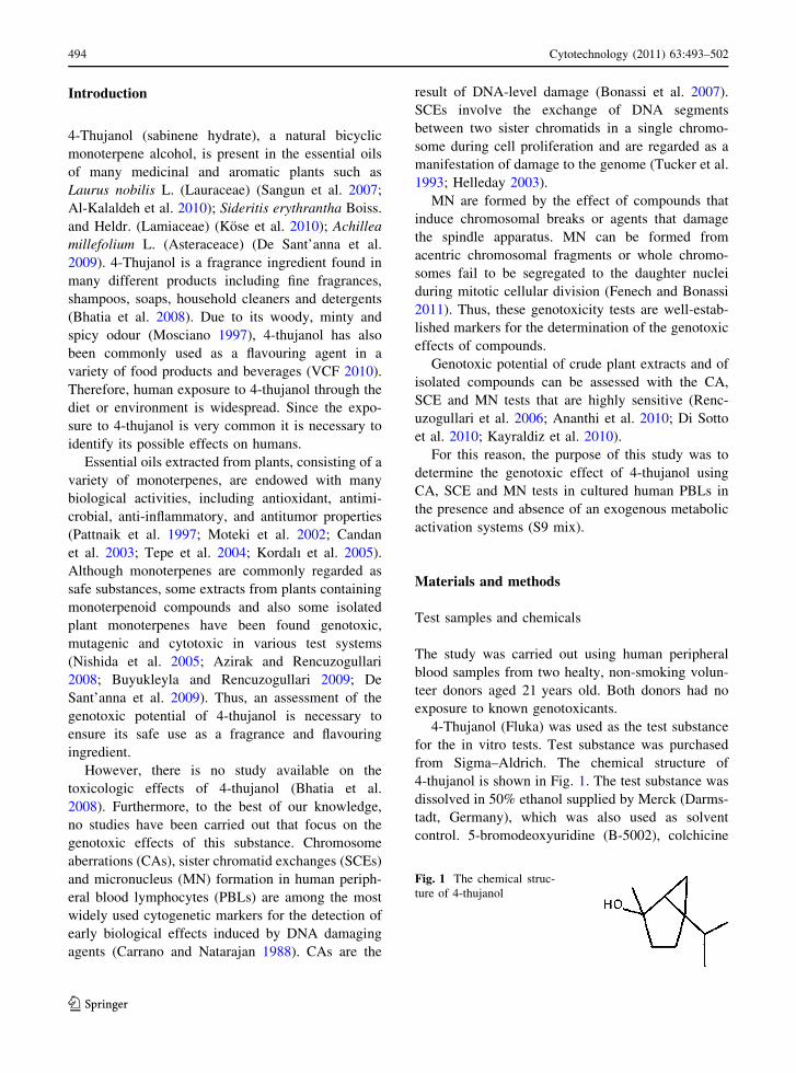
4-thujanol is shown in Fig. 1. The test substance was
dissolved in 50% ethanol supplied by Merck (Darms-
tadt, Germany), which was also used as solvent
control. 5-bromodeoxyuridine (B-5002), colchicine
Fig. 1 The chemical struc-
ture of 4-thujanol
494 Cytotechnology (2011) 63:493–502
123

(C-9754) and cytochalasin B (C-6762) were purchased
from Sigma (St. Louis, MO). In the cultures without
metabolic activation, the positive control was mito-
mycin-C (MMC, Sigma M-05030) at 0.25 lg/mL for
treatments. For cultures with metabolic activation,
cyclophosphamide monohydrate (CP, 28 lg/mL,
Sigma C-0768) was used as a positive control at
28 lg/mL for in vitro tests. Giemsa and all other
chemicals were purchased from Merck (Darmstadt,
Germany). All test solutions were freshly prepared
prior to each experiment.
In vitro assay (without S9 mix)
The CA and SCE tests were performed using the
methods developed by Evans (1984) and Perry and
Thompson (1984), with minor modification. This
study was conducted according to IPCS guidelines
(Albertini et al. 2000). Lymphocyte cultures were set
up by adding 0.2 mL of whole blood from two healty
donors to 2.5 mL of chromosome medium PB Max
(Gibco, cat. no. 12552-013) supplemented with
10 lg/mL of bromodeoxyuridine. Cultures were
incubated at 37 �C for 72 h. The test concentrations
were chosen as 13, 26 and 52 lg/mL based on the top
concentration that resulted in approximately 50%
(LD50) reduction in mitotic index (MI) (52 lg/mL).
The cells were treated with 13, 26 and 52 lg/mL
concentrations of 4-thujanol that dissolved in 50%
ethanol, for 24 h (4-thujanol was added 48 h after
initiating the culture) and 48 h (4-thujanol was added
24 h after initiating the culture). A control (i.e.,
untreated control), a solvent control (50% ethanol,
4 lL/mL) and a positive control (MMC, 0.25 lg/mL)
were also used. To arrest the cells in metaphase, the
cells were exposed to 0.06 ll/mL colchicine 2 h
before harvesting. The cells were treated with a
hypotonic solution (0.4% KCl) for 15 min at 37 �C
and then fixed three times in a cold solution
consisting of methanol:glacial acetic acid (3:1 v/v)
at room temperature (22 �C ± 1 �C). Finally, the
centrifuged cells were dropped onto clean slides. The
staining of the air-dried slides was performed
following the standard methods using 5% Giemsa
stain for CA analysis (i.e., with 5% Giemsa in
Sorensen Buffer, pH 6.8, 15 min), and the modified
Fluorescence Plus Giemsa method (FPG) for SCE
analysis (Speit and Haupter 1985). For FPG staining,
1-day-old slides were covered with Sorensen Buffer
(pH 6.8). Then, slides were irradiated with 30 W,
254 nm UV lamp at 15 cm distance for 30 min. After
irradiation, the slides were incubated in 1 X SSC
(standard saline citrate) at 58–60 �C for 60 min and
then stained with 5% Giemsa prepared with Sorensen
buffer for 20 min.
For the MN test, 0.2 mL of fresh blood was used
to establish the cultures and the cultures were
incubated for 68 h. The cells were treated with 13,
26 and 52 lg/mL concentrations of 4-thujanol for 24
and 48 h (4-thujanol was added 44 and 20 h after
initiating the culture, respectively) treatment periods.
Cytochalasin B (final concentration of 6 lg/mL) was
added after 44 h of incubation in order to block
cytokinesis and obtain binucleated cells. After an
additional 24 h incubation at 37 �C, cells were
harvested by centrifugation and slides were prepared
for MN test (Fenech 2000; Kirsch-Volders et al.
2003).
In vitro assay (with S9 mix)
In general, the same procedure for the CA, SCE and
MN assays described earlier were used to assess the
indirect genotoxic effect of 4-thujanol with minor
modifications depending on the methodology of the
metabolic activation. The lymphocytes were cotreat-
ed with 13, 26 and 52 lg/mL concentrations of
4-thujanol and 0.5 mL S9 mix for 3 h. 4-Thujanol
and S9 mix were added 48 h after initiating the
culture. For every culture of lymphocytes with the
tested compound, a control, a solvent control (50%
ethanol, 4 lL/mL) and a positive control (cyclophos-
phamide monohydrate, 28 lg/mL) were also per-
formed. The test chemical and S9 mix were removed
from the culture by centrifugation 4 min at
2,500 rpm. The pellet of lymphocytes was washed
twice with 2.5 mL RPMI 1,640 medium (Biochrom
AG, F 1215) and resuspended in fresh complete
medium (chromosome medium PB Max). The cul-
tures were incubated for a total of 72 h at 37 �C for
the CA and SCE assays and 68 h at 37 �C for the MN
assay.
Preparation of S9
The albino male rats (Rattus norvegicus var. albinos)
weighing 200 g were pretreated with 80 mg/kg
Cytotechnology (2011) 63:493–502 495
123

concentration of 3-methylcholanthrene (dissolved in
sunflower oil) for 5 days. For the preparation of S9
fraction and the S9 mix, the method described by
Maron and Ames (1983) was employed.
Microscopic evaluation
For each donor, a total of 100 well-spread metaphases
were investigated (a total of 200 metaphase spreads
for two donors) in order to score the CAs at each
concentration and treatment period that showed
structural and/or numerical chromosome aberrations.
However, only the structural CAs were taken into
consideration to determine genotoxicity. The CA was
classified according to ISCN (International System
for Human Cytogenetic Nomenclature) (Paz-y-Mino
et al. 2002) and evaluated as chromatid-type (breaks,
sister unions, and exchanges) and chromosome-type
(breaks, dicentrics, rings, and fragments) aberrations.
Gaps were not considered as CA according to Mace
et al. (1978). Percentage of cells with structural
chromosome aberrations (SCAs) have been calcu-
lated following the scoring of CAs.
The scoring of SCE was performed according to
the IPCS guidelines (Albertini et al. 2000). To score
SCE, 25-well-differentiated second-division meta-
phase cells were analyzed per donor (a total of
50 s-division metaphase for each concentration) and
the frequency of SCE per cell was recorded.
The scoring criteria used for binuclear cell and
MN evaluation were adopted from the Human
MicroNucleus Project (Bonassi et al. 2001). To
determine MN formation, 2,000 binucleated cells
were analyzed for each donor (4,000 binucleated cells
were scored per concentration). We evaluated the
micronucleated binuclear lymphocytes containing
one or more MN per 2,000 binucleated cells.
Mitotic index (MI), proliferation index (PI)
and nuclear division index (NDI)
The mitotic index was calculated from the number of
metaphases in 3,000 cells analyzed per culture for
each donor (6,000 cells per concentration) in the CA
assay.
In the SCE assay, a total of 200 cells (100 cells
from each donor) were scored for the proliferation
index. PI was calculated according to formula as
follows:
PI = [(1 9 M1) ? (2 9 M2) ? (3 9 M3)]/N,
where M1, M2 and M3 represent those metaphases
corresponding to first, second and third divisions, and
N is the total number of metaphases scored (Lamberti
et al. 1983).
Moreover, in the micronucleus assay, cytostaticity
was calculated by using the nuclear division index
(NDI). To this aim, 1,000 lymphocytes per donor
were scored. The number of viable cells with one to
four nuclei was determined in 1,000 cells. NDI was
calculated using the following formula: NDI =
[(1 9 M1) ? (2 9 M2) ? (3 9 M3) ? (4 9 M4)]/N;
where M1 through M4 represent the number of cells
with one to four nuclei and N is the total number of
viable cells scored (Eastmond and Tucker 1989;
Fenech 2000).
Statistical analysis
One-Way ANOVA was used for the statistical
significance of all parameters. The comparisons
between groups were made using a post-hoc analysis,
LSD test. Concentration–response relationships were
determined from the correlation and regression
coefficients for the percentage of cells with CA, the
mean SCE, the percentage of MN and BNMN, as
well as for the MI, PI and NDI.
Results
In these experiments, three tests for evaluation of
genotoxic activity of 4-thujanol were used in the
presence and absence of metabolic activation.
The effects of 4-thujanol on CAs, in the presence
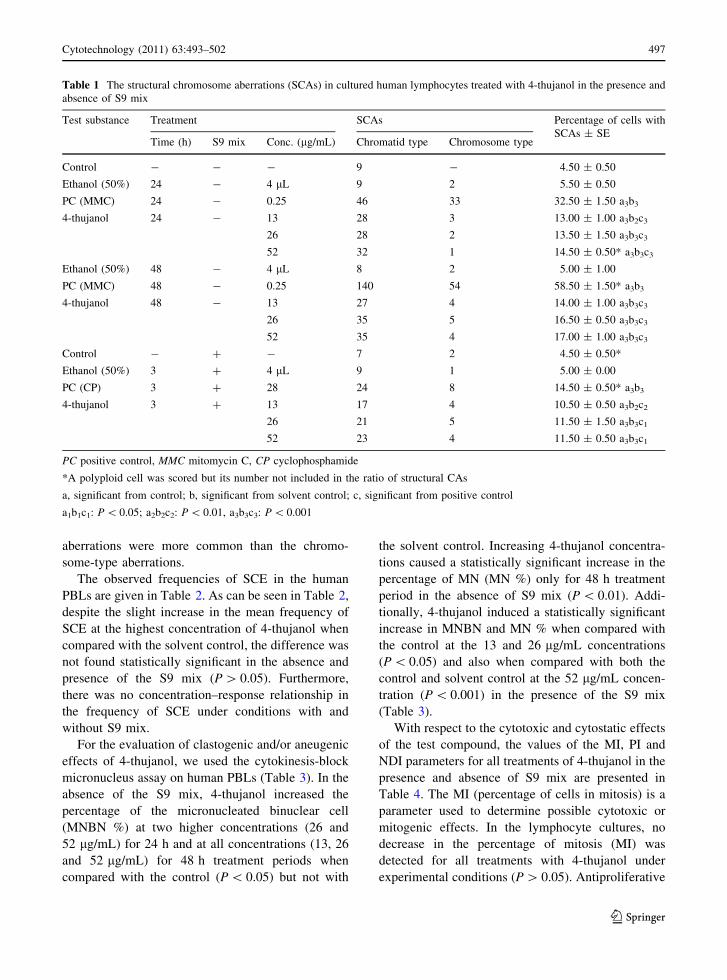
and absence of S9 mix are summarized in Table 1.
4-Thujanol statistically significantly increased
(P \ 0.001) the formation of structural CA when
compared with the control and the solvent control at
all concentrations (13, 26 and 52 lg/mL) in 24 and
48 h treatment periods in the absence of S9 mix;
however, the aberrations were significantly lower in
comparison with the respective positive control. In
parallel, human lymphocyte cultures treated with all
concentrations of 4-thujanol showed a statistically
significant increase (P \ 0.001) in the percentage of
cells with structural CA when compared with both the
control and solvent controls in the presence of S9
mix. As shown in Table 1, the chromatid-type
496 Cytotechnology (2011) 63:493–502
123
aberrations were more common than the chromo-
some-type aberrations.
The observed frequencies of SCE in the human
PBLs are given in Table 2. As can be seen in Table 2,
despite the slight increase in the mean frequency of
SCE at the highest concentration of 4-thujanol when
compared with the solvent control, the difference was
not found statistically significant in the absence and
presence of the S9 mix (P [ 0.05). Furthermore,
there was no concentration–response relationship in
the frequency of SCE under conditions with and
without S9 mix.
For the evaluation of clastogenic and/or aneugenic
effects of 4-thujanol, we used the cytokinesis-block
micronucleus assay on human PBLs (Table 3). In the
absence of the S9 mix, 4-thujanol increased the
percentage of the micronucleated binuclear cell
(MNBN %) at two higher concentrations (26 and
52 lg/mL) for 24 h and at all concentrations (13, 26
and 52 lg/mL) for 48 h treatment periods when
compared with the control (P \ 0.05) but not with
the solvent control. Increasing 4-thujanol concentra-
tions caused a statistically significant increase in the
percentage of MN (MN %) only for 48 h treatment
period in the absence of S9 mix (P \ 0.01). Addi-
tionally, 4-thujanol induced a statistically significant
increase in MNBN and MN % when compared with
the control at the 13 and 26 lg/mL concentrations
(P \ 0.05) and also when compared with both the
control and solvent control at the 52 lg/mL concen-
tration (P \ 0.001) in the presence of the S9 mix
(Table 3).
With respect to the cytotoxic and cytostatic effects
of the test compound, the values of the MI, PI and
NDI parameters for all treatments of 4-thujanol in the
presence and absence of S9 mix are presented in
Table 4. The MI (percentage of cells in mitosis) is a
parameter used to determine possible cytotoxic or
mitogenic effects. In the lymphocyte cultures, no
decrease in the percentage of mitosis (MI) was
detected for all treatments with 4-thujanol under
experimental conditions (P [ 0.05). Antiproliferative
Table 1 The structural chromosome aberrations (SCAs) in cultured human lymphocytes treated with 4-thujanol in the presence and
absence of S9 mix
Test substance Treatment SCAs Percentage of cells with
SCAs ± SETime (h) S9 mix Conc. (lg/mL) Chromatid type Chromosome type
Control - - - 9 - 4.50 ± 0.50
Ethanol (50%) 24 - 4 lL 9 2 5.50 ± 0.50
PC (MMC) 24 - 0.25 46 33 32.50 ± 1.50 a3b3
4-thujanol 24 - 13 28 3 13.00 ± 1.00 a3b2c3
26 28 2 13.50 ± 1.50 a3b3c3
52 32 1 14.50 ± 0.50* a3b3c3
Ethanol (50%) 48 - 4 lL 8 2 5.00 ± 1.00
PC (MMC) 48 - 0.25 140 54 58.50 ± 1.50* a3b3
4-thujanol 48 - 13 27 4 14.00 ± 1.00 a3b3c3
26 35 5 16.50 ± 0.50 a3b3c3
52 35 4 17.00 ± 1.00 a3b3c3
Control - ? - 7 2 4.50 ± 0.50*
Ethanol (50%) 3 ? 4 lL 9 1 5.00 ± 0.00
PC (CP) 3 ? 28 24 8 14.50 ± 0.50* a3b3
4-thujanol 3 ? 13 17 4 10.50 ± 0.50 a3b2c2
26 21 5 11.50 ± 1.50 a3b3c1
52 23 4 11.50 ± 0.50 a3b3c1
PC positive control, MMC mitomycin C, CP cyclophosphamide
*A polyploid cell was scored but its number not included in the ratio of structural CAs
a, significant from control; b, significant from solvent control; c, significant from positive control
a1b1c1: P \ 0.05; a2b2c2: P \ 0.01, a3b3c3: P \ 0.001
Cytotechnology (2011) 63:493–502 497
123

and cytostatic effects of 4-thujanol were measured by
PI and NDI, respectively. In the presence and absence
of the S9 mix, the PI did not significantly decrease in
comparison with the control and solvent control.
Similarly, except for the highest concentration
(52 lg/mL) for 48 h treatment, 4-thujanol did not
cause a significant reduction in the NDI when
compared with the control groups (Table 4).
Discussion
The results of the present study revealed that in
general, 4-thujanol significantly increased the per-
centage of cells with structural CAs and MN forma-
tion at all concentrations (13, 26 and 52 lg/mL) when
compared with the controls in the presence and
absence of the S9 mix. However, the treatment of
peripheral lymphocytes with 4-thujanol in the pres-
ence and absence of the S9 mix did not produce a
statistical difference in the frequency of SCEs when
compared with control group (P [ 0.05). In addition,
with regard to CA, SCE and MN data, there were no
concentration-dependent effects of 4-thujanol on any
of these parameters.
The induction of structural CAs, and micronucle-
ated lymphocytes by all concentrations of 4-thujanol
(13, 26 and 52 lg/mL) suggests the clastogenic
potential of the test compound at these concentra-
tions. It is generally acknowledged that the CA and
MN assays are the mutagenicity tests for the detec-
tion of chromosome mutations, whereas the SCE
assay is an indicator test for genotoxic exposure.
Hence, the results of the CA and MN assays should
be considered of higher significance than the results
of an SCE test (Eke and Celik 2008).
Micronuclei are formed from acentric chromo-
somal fragments which arise as a result of chromo-
some breaks after clastogenic effect or whole
chromosomes that do not migrate during anaphase
as a result of aneugenic affects (Heddle et al. 1991).
The results of the CA test showed that 4-thujanol
Table 2 The mean sister chromatid exchange (SCE) values in cultured human lymphocytes treated with 4-thujanol in the presence
and absence of S9 mix
Test substance Treatment Min-Max SCE SCE/Cell ± SE
Time (h) S9 mix Conc. (lg/mL)
Control - - - 1–14 6.30 ± 0.42
Ethanol (50%) 24 - 4 lL 1–14 6.36 ± 0.36
PC (MMC) 24 - 0.25 10–33 10.20 ± 0.16 a3b3
4-thujanol 24 - 13 2–14 6.84 ± 0.44 c3
26 2–14 6.28 ± 0.28c3
52 3–15 6.66 ± 0.34 c3
Ethanol (50%) 48 - 4 lL 2–12 6.44 ± 0.08
PC (MMC) 48 - 0.25 28–68 46.16 ± 1.96 a3b3
4-thujanol 48 - 13 1–11 6.02 ± 0.66 c3
26 3–12 7.32 ± 0.14 c3
52 2–15 6.30 ± 0.58 c3
Control - ? - 0–13 7.02 ± 0.34
Ethanol (50%) 3 ? 4 lL 2–16 7.16 ± 0.36
PC (CP) 3 ? 28 7–34 18.26 ± 0.94 a3b3
4-thujanol 3 ? 13 2–15 7.00 ± 0.12 c3
26 3–12 6.86 ± 0.22 c3
52 3–14 7.74 ± 0.10 c3
PC Positive control, MMC Mitomycin C, CP Cyclophosphamide
a, significant from control; b, significant from solvent control; c, significant from positive control
a1b1c1: P \ 0.05; a2b2c2: P \ 0.01, a3b3c3: P \ 0.001
498 Cytotechnology (2011) 63:493–502
123

induced more chromatid breaks than other structural
or numerical CAs. Therefore, it can be said that MN
can be formed from acentric chromosomal fragments
in this study.
There are no studies in relation to 4-thujanol
genotoxicity or mutagenicity and toxicity. This study
is the first report on detecting the genotoxic and/or
cytotoxic effects of 4-thujanol using CA, SCE and
MN assay in PBLs in vitro. However, few studies
have been carried out on the potential genotoxic
effects of some monoterpenoid compounds.
Menthol (a monocyclic monoterpene alcohol) and
linalool (an acyclic monoterpene alcohol) did not
increase chromosomal aberrations at concentrations
of up to 200 lg/mL and 250 lg/mL, respectively,
when incubated with Chinese hamster fibroblast cells
(Ishidate et al. 1984). However, in our study, the
lower concentrations of 4-thujanol induced chromo-
somal abnormalities. The lack of agreement of our
results could be due to the differences in chemical
structure of the tested monoterpenes and of different
cell lines used.
Hilliard et al. (1998) found that menthol racemic
induced a weak but statistically significant increase in
chromosome aberrations at concentrations of
1.6–1.9 mM in Chinese hamster ovary cells without
metabolic activation. Additionally, it induced a
significant increase in chromosome aberrations at
1.2 mM in human TK6 cells. Aydın et al. (2005)
reported that concentrations above 0.1 mM thymol
(approximately 15 lg/mL) (a phenolic monoterpene)
and gamma-terpinene (approximately 14 lg/mL) (a
monocyclic monoterpene hydrocarbon) and 0.05 mM
carvacrol (approximately 7.5 lg/mL) (a phenolic
monoterpene) significantly induced DNA damage in
human lymphocytes in the comet assay. Azirak and
Rencuzogullari (2008) found that both carvacrol (10,
30, 50 and 70 mg/kg b.w., intraperitoneally) and
thymol (40, 60, 80 and 100 mg/kg b.w., intraperito-
neally) significantly induced structural CA in rat bone
Table 3 The percentage of micronucleus (MN) and the percentage of micronucleated binuclear cells (MNBN) in cultured human
lymphocytes treated with 4-thujanol in the presence and absence of S9 mix
Test substance Treatment The percentage of
micronucleated binuclear
cells ± SE (%)
The percentage of
micronucleus ± SE (%)Time (h) S9 mix Conc.
(lg/mL)
Control - - - 0.20 ± 0.05 0.25 ± 0.00
Ethanol (50%) 24 - 4 lL 0.37 ± 0.02 0.47 ± 0.07
PC (MMC) 24 - 0.25 0.57 ± 0.02 a3b1 0.95 ± 0.05 a3b3
4-thujanol 24 - 13 0.15 ± 0.05 c3 0.19 ± 0.09 c3
26 0.35 ± 0.05 a1c2 0.35 ± 0.05 c3
52 0.37 ± 0.02 a1c1 0.37 ± 0.02 c3
Ethanol (50%) 48 - 4 lL 0.47 ± 0.02 0.53 ± 0.02
PC (MMC) 48 - 0.25 1.60 ± 0.10 a3b3 2.34 ± 0.04 a3b3
4-thujanol 48 - 13 0.62 ± 0.07 a2c3 0.65 ± 0.05 a2c3
26 0.52 ± 0.07 a1c3 0.60 ± 0.05 a2c3
52 0.52 ± 0.02 a1c3 0.60 ± 0.10 a2c3
Control - ? - 0.12 ± 0.07 0.12 ± 0.07
Ethanol (50%) 3 ? 4 lL 0.28 ± 0.01 0.28 ± 0.01
PC (CP) 3 ? 28 0.46 ± 0.01 a2b1 0.51 ± 0.01 a2b1
4-thujanol 3 ? 13 0.31 ± 0.08 a1 0.32 ± 0.09 a1c1
26 0.40 ± 0.02 a1 0.40 ± 0.02 a2
52 0.67 ± 0.02 a3b3c1 0.68 ± 0.02 a3b3
PC Positive control, MMC Mitomycin C, CP Cyclophosphamide
a, significant from control; b, significant from solvent control; c, significant from positive control
a1b1c1: P \ 0.05; a2b2c2: P \ 0.01, a3b3c3: P \ 0.001
Cytotechnology (2011) 63:493–502 499
123

marrow cells for all treatment periods (6, 12 and
24 h). These results are consistent with the results of
our study, although different cell types and/or test
systems were used.
Finally, similar to the results of our study,
Buyukleyla and Rencuzogullari (2009) reported that
thymol significantly increased the structural CA and
MN formation at 25, 50, 75 and 100 lg/mL concen-
trations for 24 and 48 h treatment period in human
PBLs without S9 mix.
Results show that 4-thujanol most probably, has a
genotoxic risk at the tested concentrations (13, 26 and
52 lg/mL) in human PBLs in the presence and
absence of the S9 mix. In addition, 4-thujanol caused
particularly structural CAs instead of numerical CA,
meaning that 4-thujanol as a clastogen can lead to
formation of CA by breaking the phosphodiester back
bone of DNA. 4-Thujanol also may act on proteins
and DNA with production of reactive oxygen species
(ROS) that may cause DNA strand breaks.
4-Thujanol did not significantly decrease the MI,
PI and NDI in the presence and absence of the S9 mix
(P [ 0.05). Furthermore, there was no concentration–
response relationship for MI, PI and NDI. Therefore,
it could be indicated that 4-thujanol is neither a
cytotoxic nor cytostatic agent in human peripheral
blood lymphocytes. These data also suggest that the
metabolites of 4-thujanol did not have significant
cytotoxic and cytostatic effects on cell proliferation
in vitro. Similar to our results, Horvathova et al.
(2007) found that 1,8-cineole, the monoterpene cyclic
ether known as eucalyptol, did not show cytotoxic
effects on cultured human leukemic K562 cells.
Buyukleyla and Rencuzogullari (2009) reported that
thymol had a cytotoxic effect via decreasing the MI,
RI (replication index) and NDI at the two highest
concentrations (75 and 100 lg/mL) in human lym-
phocytes. In our study, 4-thujanol did not show
cytotoxic/cytostatic effects in the same cell culture
system. But, the highest concentration used in the
present study was 52 lg/mL which is lower than 75
and 100 lg/mL. Based on this aspect, our results are
not contradictory to the results obtained from this
study.
Table 4 Mitotic index (MI), proliferation index (PI) and nuclear division index (NDI) values in cultured human lymphocytes treated
with 4-thujanol in the presence and absence of S9 mix
Test substance Treatment MI ± SE PI ± SE NDI ± SE
Time (h) S9 mix Conc. (lg/mL)
Control - - - 3.63 ± 0.53 2.14 ± 0.01 1.63 ± 0.04
Ethanol (50%) 24 - 4 lL 3.24 ± 0.21 1.87 ± 0.11 1.38 ± 0.05
PC (MMC) 24 - 0.25 2.95 ± 0.42 1.59 ± 0.02 a1 1.36 ± 0.01 a1
4-thujanol 24 - 13 3.86 ± 0.06 2.09 ± 0.06 c1 1.44 ± 0.01
26 3.13 ± 0.56 1.80 ± 0.12 1.47 ± 0.06
52 2.89 ± 0.83 1.78 ± 0.21 1.44 ± 0.24
Ethanol (50%) 48 - 4 lL 4.20 ± 0.40 2.24 ± 0.15 1.60 ± 0.01
PC (MMC) 48 - 0.25 3.15 ± 0.22 1.60 ± 0.01 a2b3 1.39 ± 0.01 a2b2
4-thujanol 48 - 13 4.01 ± 0.18 2.34 ± 0.02 c3 1.54 ± 0.04 c1
26 3.71 ± 0.21 2.35 ± 0.09 c3 1.53 ± 0.03 c1
52 3.53 ± 0.83 2.30 ± 0.08 c3 1.45 ± 0.02 a2c1
Control - ? - 3.92 ± 0.45 2.19 ± 0.11 1.19 ± 0.02
Ethanol (50%) 3 ? 4 lL 3.80 ± 0.47 2.19 ± 0.00 1.18 ± 0.11
PC (CP) 3 ? 28 3.45 ± 0.38 2.21 ± 0.02 1.17 ± 0.02
4-thujanol 3 ? 13 3.68 ± 0.78 2.20 ± 0.02 1.18 ± 0.05
26 3.63 ± 0.63 2.29 ± 0.03 1.23 ± 0.03
52 3.33 ± 0.17 2.09 ± 0.10 1.17 ± 0.03
PC Positive control, MMC Mitomycin C, CP Cyclophosphamide
a, significant from control; b, significant from solvent control; c, significant from positive control
a1b1c1: P \ 0.05; a2b2c2: P \ 0.01, a3b3c3: P \ 0.001
500 Cytotechnology (2011) 63:493–502
123

Horvathova et al. (2009) reported that high
concentrations of borneol (34.28 mg/kg/day) showed
a cytotoxic effect in primary rat hepatocytes due to
the increased levels of DNA damage and that this
effect resulted probably from the induction of apop-
totic and necrotic DNA fragmentation. Furthermore,
Bakkali et al. (2008) reported that the main mecha-
nisms of cytotoxic effects of essential oils in mam-
malian cells were apoptosis and necrosis. Whereas in
our study, all concentrations of 4-thujanol (13, 26 and
52 lg/mL) caused an increase of chromatid breaks,
while at the same concentrations, we did not observe
a significant increase of cytotoxicity in human
lymphocytes. Therefore, it can be thought that the
chromosomal aberrations caused by 4-thujanol were
not potent enough to induce apoptosis.
Taken as a whole, these findings suggest that
4-thujanol had a significantly clastogenic effect at the
tested concentrations (13, 26 and 52 lg/mL) for
human lymphocytes. And, no cytotoxic and/or cyto-
static effects were observed with or without meta-
bolic activation regardless of the concentrations used.
Therefore, we propose that it is necessary to be
careful when using 4-thujanol as a fragrance and
flavouring ingredient. However, the lack of other data
in relation to the genotoxic effect of 4-thujanol, it
should be further examined in different test systems
to better understand its potential genotoxicity.
Acknowledgements This investigation was supported by a
grant from The Scientific and Technical Research Council of
Turkey, Ankara (TUBITAK, Project No. 109T546).
References
Albertini RJ, Anderson D, Douglas GR, Hagmar L, Hemminki
K, Merlo F, Natarajan AT, Norppa H, Shuker DEG, Tice
R, Waters MD, Aitio A (2000) IPCS guidelines for the
monitoring of genotoxic effects of carcinogens in humans.
Mutat Res 463:111–172
Al-Kalaldeh JZ, Abu-Dahab R, Afifi FU (2010) Volatile oil
composition and antiproliferative activity of Laurus no-bilis, Origanum syriacum, Origanum vulgare, and Salviatriloba against human breast adenocarcinoma cells. Nutr
Res 30:271–278
Ananthi R, Chandra N, Santhiya ST, Ramesh A (2010)
Genotoxic and antigenotoxic effects of Hemidesmusindicus R. Br. root extract in cultured lymphocytes.
J Ethnopharmacol 127:558–560
Aydın S, Basaran AA, Basaran N (2005) Modulating effects of
thyme and its major ingredients on oxidative DNA
damage in human lymphocytes. J Agric Food Chem
53:1299–1305
Azirak S, Rencuzogullari E (2008) The in vivo genotoxic
effects of carvacrol and thymol in rat bone marrow cells.
Environ Toxicol 23:728–735
Bakkali F, Averbeck S, Averbeck D, Idaomar M (2008) Bio-
logical effects of essential oils-a review. Food Chem
Toxicol 46:446–475
Bhatia SP, Letizia CS, Api AM (2008) Fragrance material
review on 4-thujanol. Food Chem Toxicol 46:295–296
Bonassi S, Fenech M, Lando C et al (2001) Human micronu-
cleus project: international database comparison for
results with the cytokinesis block micronucleus assay in
human lymphocytes: effect of laboratory protocol, scoring
criteria, and host factors on the frequency of micronuclei.
Environ Mol Mutagen 37:31–45
Bonassi S, Znaor A, Ceppi M, Lando C, Chang WP, Holland
N, Kirsch-Volders M, Zeiger E, Ban S, Barale R, Bigatti
MP, Bolognesi C, Cebulska-Wasilewska A, Fabianova E,
Fucic A, Hagmar L, Joksic G, Martelli A, Migliore L,
Mirkova E, Scarfi MR, Zijno A, Norppa H, Fenech M
(2007) An increased micronucleus frequency in peripheral
blood lymphocytes predicts the risk of cancer in humans.
Carcinogenesis 28:625–631
Buyukleyla M, Rencuzogullari E (2009) The effects of thymol
on sister chromatid exchange, chromosome aberration and
micronucleus in human lymphocytes. Ecotoxicol Environ
Saf 72:943–947
Candan F, Unlu M, Tepe B, Daferera AD, Polissiou M, Sok-
men A, Akpulat HA (2003) Antioxidant and antimicrobial
activity of essential oil and methanol extracts of Achilleamillefolium subsp. Millefolium Afan. (Asteraceae). J Eth-
nopharmacol 87:215–220
Carrano AV, Natarajan AT (1988) Consideration for popula-
tion monitoring using cytogenetic techniques. Mutat Res
204:379–406
De Sant’anna JR, Da Silva Franco CC, Miyamoto CT, Cunico
MM, Miguel OG, Cocco LC, Yamamoto CI, Junior CC,
De Castro-Prado AA (2009) Genotoxicity of Achilleamillefolium essential oil in diploid cells of Aspergillus
nidulans. Phytother Res 23:231–235
Di Sotto A, Mazzanti G, Carbone F, Hrelia P, Maffei F (2010)
Inhibition by b-caryophyllene of ethyl methanesulfonate-
induced clatogenicity in cultured human lymphocytes.
Mutat Res 699:23–28
Eastmond DA, Tucker JD (1989) Identification of aneuploidy-
inducing agents using cytokinesis-blocked human lym-
phocytes and an anti-kinetochore antibody. Environ Mol
Mutagen 13:34–43
Eke D, Celik A (2008) Genotoxicity of thimerosal in cultured
human lymphocytes with and without metabolic activa-
tion sister chromatid exchange analysis proliferation index
and mitotic index. Toxicol In Vitro 22:927–934
Evans HJ (1984) Human peripheral blood lymphocytes for the
analysis of chromosome aberrations in mutagen tests. In:
Kilbey BJ, Legator M, Nichols W, Ramel C (eds) Hand-
book of mutagenicity test procedures, 2nd edn. Elsevier
Science Publishers BV, Amsterdam, pp 405–427
Fenech M (2000) The in vitro micronucleus technique. Mutat
Res 455:81–95
Cytotechnology (2011) 63:493–502 501
123

Fenech M, Bonassi S (2011) The effect of age, gender, diet and
lifestyle on DNA damage measured using micronucleus
frequency in human peripheral blood lymphocytes.
Mutagenesis 26:43–49
Heddle JA, Cimino MC, Hayashi M, Romagna F, Shelby MD,
Tucker JD, Vanparys Ph, Mac Gregor JT (1991) Micro-
nuclei as an index of cytogenetic damage: past, present,
and future. Environ Mol Mutagen 18:277–291
Helleday T (2003) Pathways for mitotic homologous recom-
bination in mammalian cells. Mutat Res 532:103–115
Hilliard CA, Armstrong MJ, Bradt CI, Hill RB, Greenwood
SK, Galloway SM (1998) Chromosome aberrations in
vitro related to cytotoxicity of nonmutagenic chemicals
and metabolic poisons. Environ Mol Mutagen 31:316–326
Horvathova E, Turcaniova V, Slamenova D (2007) Compara-
tive study of DNA-damaging and DNA-protective effects
of selected components of essential plant oils in human
leukemic cells K562. Neoplasma 54:478–483
Horvathova E, Slamenova D, Marsalkova L, Sramkova M,
Wsolova L (2009) Effects of borneol on the level of DNA
damage induced in primary rat hepatocytes and testicular
cells by hydrogen peroxide. Food Chem Toxicol
47:1318–1323
Ishidate Jr, Sofuni T, YoshikawaA K, Hayashi M, Nohmi T,
Sawada M, Matsuoka A (1984) Primary mutagenicity
screening of food additives currently used in Japan. Food
Chem Toxicol 22:623–636
Kayraldiz A, Kocaman AY, Rencuzogulları E, Istifli ES, Ila
HB, Topaktas M, Daglıoglu YK (2010) The genotoxic
and antigenotoxic effects of Aloe vera leaf extract in vivo
and in vitro. Turk J Biol 34:235–246
Kirsch-Volders M, Sofuni T, Aardema M, Albertini S, East-
mond D, Fenech M, Ishidate M Jr, Kirchner S, Lorge E,
Morita T, Norppa H, Surralles J, Vanhauwaert A, Wakata
A (2003) Report from the in vitro micronucleus assay
working group. Mutat Res 540:153–163
Kordalı S, Kotan R, Mavi A, Cakır A, Ala A, Yıldırım A
(2005) Determination of the chemical composition and
antioxidant activity of the essential oil of Artemisia dra-
cunculus and of the antifungal and antibacterial activities
of Turkish Artemisia absinthium, Artemisia dracunculus,
Artemisia santonicum and Artemisia spicigera essential
oils. J Agric Food Chem 53:9452–9458
Kose EO, Deniz IG, Sarıkurkcu C, Aktas O, Yavuz M (2010)
Chemical composition, antimicrobial and antioxidant
activities of the essential oils of Sideritis erythranthaBoiss. and Heldr. (var. erythrantha and var. cedretorumP.H. Davis) endemic in Turkey. Food Chem Toxicol
48:2960–2965
Lamberti L, Ponzetto BP, Ardito G (1983) Cell kinetics and
sister chromatid exchange frequency in human lympho-
cytes. Mutat Res 319:193–199
Mace MLJ, Daskal Y, Wray W (1978) Scanning electron
microscopy of chromosome aberrations. Mutat Res
52:199–206
Maron DM, Ames BN (1983) Revised methods for the Sal-
monella mutagenicity test. Mutat Res 113:173–215
Mosciano G (1997) P&F 22 No. 3, 47
Moteki H, Hibasami H, Yamada Y, Katsuzaki H, Imai K,
Komiya T (2002) Specific induction of apoptosis by 1, 8
cineole in two human leukemia cell lines, but not in
human stomach cancer line. Oncol Rep 9:757–760
Nishida N, Tamotsu S, Nagata M, Saito C, Sakai A (2005)
Allelopathic effects of volatile monoterpenoids produced
by Salvia leucophylla inhibition of cell proliferation and
DNA synthesis in the root apical meristem of Brassicacampestris seedlings. J Chem Ecol 31:187–1203
Pattnaik S, Subramanyam VR, Bapaji M, Kole CR (1997)
Antibacterial and antifungal activity of aromatic constit-
uents of essential oils. Microbios 89:39–46
Paz-y-Mino C, Bustamante G, Sanchez ME, Leone PE (2002)
Cytogenetic monitoring in a population occupationally
exposed to pesticides in Ecuador. Environ Health Perspect
110:1077–1080
Perry PE, Thompson EJ (1984) The methodology of sister
chromatid exchanges. In: Kilbey BJ, Legator M, Nichols
W, Ramel C (eds) Handbook of mutagenicity test proce-
dures, 2nd edn. Elsevier Science Publishers, Amsterdam,
pp 495–529
Rencuzogullari E, Ila HB, Kayraldiz A, Diler SB, Yavuz A,
Arslan M, Kaya FF, Topaktas M (2006) The mutagenic
and antimutagenic effects of Ecballium elaterium fruit
juice in human peripheral lymphocytes. Russ J Genet
42:623–627
Sangun MK, Aydin E, Timur M, Karadeniz H, Caliskan M,
Ozkan A (2007) Comparison of chemical composition of
the essential oil of Laurus nobilis L. leaves and fruits from
different regions of Hatay, Turkey. J Environ Biol
28:731–733
Speit G, Haupter S (1985) On the mechanism of differential
giemsa staining of bromodeoxyuridine-substituted chro-
mosomes. II. differences between the demonstration of
sister chromatid differentiation and replication patterns.
Hum Genet 70:126–129
Tepe B, Donmez E, Unlu M, Candan F, Daferera D, Vardar-
Unlu G, Polissiou M, Sokmen A (2004) Antimicrobial and
antioxidative activities of the essential oils and methanol
extracts of Salvia cryptantha (Montbret et Aucher ex
Benth.) and Salvia multicaulis (Vahl). Food Chem
84:519–525
Tucker JD, Auletta A, Cimino MC, Dearfield KL, Jacobson-
Kram D, Tice RR, Carrano AV (1993) Sister-chromatid
exchange: second report of the gene-tox program. Mutat
Res 297:101–180
VCF (2010) Volatile compounds in food:database/Nijssen, In:
Ingen-Visscher LM, Van CA, Donders, JJH (eds)–Version
12.2—Zeist (The Netherlands):TNO quality of life,
pp 1963–2010
502 Cytotechnology (2011) 63:493–502
123