Xanthium strumarium L. Extracts Produce DNA Damage Mediated by Cytotoxicity in In Vitro Assays but...
9
Research Article Xanthium strumarium L. Extracts Produce DNA Damage Mediated by Cytotoxicity in In Vitro Assays but Does Not Induce Micronucleus in Mice Janet Piloto Ferrer, 1 Renata Cozzi, 2 Tommaso Cornetta, 2 Pasquale Stano, 2 Mario Fiore, 3 Francesca Degrassi, 3 Rosella De Salvia, 3 Antonia Remigio, 1 Marbelis Francisco, 1 Olga Quiñones, 1 Dayana Valdivia, 1 Maria L. González, 1 Carlos Pérez, 4 and Angel Sánchez-Lamar 5 1 Departamento de Genotoxicidad, Centro de Investigaci´ on y Desarrollo de Medicamentos (CIDEM), Avenida 26, No. 1605 e/ Puentes Grandes y Boyeros, La Habana, Cuba 2 Dipartimento di Scienze, Universit` a degli Studi “Roma TRE”, Via G. Marconi 446, 00146 Roma, Italy 3 Istituto di Biologia e Patologia Molecolare, CNR, Via degli Apuli. 4, 00185 Roma, Italy 4 Departamento de Bioqu´ ımica, Instituto de Ciencias B´ asicas y Precl´ ınicas “Victoria de Gir´ on” (ICBP-UCMH), Calle 146, No. 3102 Esq 31, Playa, La Habana, Cuba 5 Departamento de Biolog´ ıa Vegetal, Laboratorio de Toxicolog´ ıa Gen´ etica, Facultad de Biolog´ ıa, Universidad de la Habana, Calle 25, No. 455, e/ I y J, Vedado, C. Habana, Cuba Correspondence should be addressed to Janet Piloto Ferrer; [email protected] Received 7 February 2014; Revised 24 April 2014; Accepted 6 May 2014; Published 15 June 2014 Academic Editor: Manoor Prakash Hande Copyright © 2014 Janet Piloto Ferrer et al. is is an open access article distributed under the Creative Commons Attribution License, which permits unrestricted use, distribution, and reproduction in any medium, provided the original work is properly cited. Xanthium strumarium L. is a member of the Asteraceae commonly used in Cuba, mainly as diuretic. Some toxic properties of this plant have also been reported and, to date, very little is known about its genotoxic properties. e present work aims was to evaluate the potential cytotoxic and genotoxic risk of whole extract from Xanthium strumarium L. whole extract of aerial parts. No positive response was observed in a battery of four Salmonella typhimurium strains, when exposed to concentrations up to 5 mg/plate, with and without mammalian metabolic activation (liver microsomal S9 fraction from Wistar rats). In CHO cells, high concentrations (25–100 g/mL) revealed significant reduction in cell viability. Results from sister chromatid exchanges, chromosome aberrations, and comet assay showed that X. strumarium extract is genotoxic at the highest concentration used, when clear cytotoxic effects were also observed. On the contrary, no increase in micronuclei frequency in bone marrow cells was observed when the extract was orally administered to mice (100, 500, and 2000 mg/Kg doses). e data presented here constitute the most complete study on the genotoxic potential of X. strumarium L. and show that the extract can induce in vitro DNA damage at cytotoxic concentrations. 1. Introduction Xanthium strumarium L. (Asteraceae), a medicinal plant commonly found as a weed, is widely distributed in North America, Brazil, China, Malaysia, and hotter parts of India. e herb is traditionally used mostly in treating several aliments. In Cuba, X. strumarium is commonly called “guisazo de caballo” and it has been used as diuretic [1]. e plant has been employed for a long time in folk therapy. More than 20 properties have been attributed to the decoctions and tinctures from the leaves and roots, for example, antirheumatic, antisyphilitic, appetiser, diaphoretic, diuretic, emollient, laxative, and sedative activities. Other actions have been confirmed by experimental pharmacology: anti-inflammatory, analgesic [2, 3], antibacterial, anticancer [4, 5], antifungal [6], antihypoglycemic [7], antimitotic [8, 9], antitrypanosomal, antimalarial [10], and diuretic activities [11]. Hindawi Publishing Corporation BioMed Research International Volume 2014, Article ID 575197, 8 pages http://dx.doi.org/10.1155/2014/575197
-
Upload
independent -
Category
Documents
-
view
1 -
download
0
Transcript of Xanthium strumarium L. Extracts Produce DNA Damage Mediated by Cytotoxicity in In Vitro Assays but...

Research ArticleXanthium strumarium L. Extracts Produce DNA DamageMediated by Cytotoxicity in In Vitro Assays but Does NotInduce Micronucleus in Mice
Janet Piloto Ferrer,1 Renata Cozzi,2 Tommaso Cornetta,2 Pasquale Stano,2
Mario Fiore,3 Francesca Degrassi,3 Rosella De Salvia,3 Antonia Remigio,1
Marbelis Francisco,1 Olga Quiñones,1 Dayana Valdivia,1 Maria L. González,1
Carlos Pérez,4 and Angel Sánchez-Lamar5
1 Departamento de Genotoxicidad, Centro de Investigacion y Desarrollo de Medicamentos (CIDEM), Avenida 26,No. 1605 e/ Puentes Grandes y Boyeros, La Habana, Cuba
2Dipartimento di Scienze, Universita degli Studi “Roma TRE”, Via G. Marconi 446, 00146 Roma, Italy3 Istituto di Biologia e Patologia Molecolare, CNR, Via degli Apuli. 4, 00185 Roma, Italy4Departamento de Bioquımica, Instituto de Ciencias Basicas y Preclınicas “Victoria de Giron” (ICBP-UCMH), Calle 146,No. 3102 Esq 31, Playa, La Habana, Cuba
5Departamento de Biologıa Vegetal, Laboratorio de Toxicologıa Genetica, Facultad de Biologıa, Universidad de la Habana,Calle 25, No. 455, e/ I y J, Vedado, C. Habana, Cuba
Correspondence should be addressed to Janet Piloto Ferrer; [email protected]
Received 7 February 2014; Revised 24 April 2014; Accepted 6 May 2014; Published 15 June 2014
Academic Editor: Manoor Prakash Hande
Copyright © 2014 Janet Piloto Ferrer et al. This is an open access article distributed under the Creative Commons AttributionLicense, which permits unrestricted use, distribution, and reproduction in any medium, provided the original work is properlycited.
Xanthium strumarium L. is a member of the Asteraceae commonly used in Cuba, mainly as diuretic. Some toxic properties of thisplant have also been reported and, to date, very little is known about its genotoxic properties.The present work aims was to evaluatethe potential cytotoxic and genotoxic risk of whole extract from Xanthium strumarium L. whole extract of aerial parts. No positiveresponse was observed in a battery of four Salmonella typhimurium strains, when exposed to concentrations up to 5mg/plate, withand without mammalian metabolic activation (liver microsomal S9 fraction fromWistar rats). In CHO cells, high concentrations(25–100𝜇g/mL) revealed significant reduction in cell viability. Results from sister chromatid exchanges, chromosome aberrations,and comet assay showed that X. strumarium extract is genotoxic at the highest concentration used, when clear cytotoxic effectswere also observed. On the contrary, no increase in micronuclei frequency in bone marrow cells was observed when the extractwas orally administered to mice (100, 500, and 2000mg/Kg doses). The data presented here constitute the most complete study onthe genotoxic potential of X. strumarium L. and show that the extract can induce in vitroDNA damage at cytotoxic concentrations.
1. Introduction
Xanthium strumarium L. (Asteraceae), a medicinal plantcommonly found as a weed, is widely distributed in NorthAmerica, Brazil, China, Malaysia, and hotter parts of India.The herb is traditionally used mostly in treating severalaliments. In Cuba, X. strumarium is commonly called“guisazo de caballo” and it has been used as diuretic [1].The plant has been employed for a long time in folk
therapy. More than 20 properties have been attributed tothe decoctions and tinctures from the leaves and roots, forexample, antirheumatic, antisyphilitic, appetiser, diaphoretic,diuretic, emollient, laxative, and sedative activities. Otheractions have been confirmed by experimental pharmacology:anti-inflammatory, analgesic [2, 3], antibacterial, anticancer[4, 5], antifungal [6], antihypoglycemic [7], antimitotic [8, 9],antitrypanosomal, antimalarial [10], and diuretic activities[11].
Hindawi Publishing CorporationBioMed Research InternationalVolume 2014, Article ID 575197, 8 pageshttp://dx.doi.org/10.1155/2014/575197
2 BioMed Research International
The toxicity reported for the plant suggests that potentialnegative effects on human health elicited by the consumptionof herbal remedies based on X. strumarium should beconsidered. Genotoxicity is one of the most important toxiceffects and its relevance is greater as we are dealing withevents taking place at sublethal doses leading to long-termeffects such as cancer and reproductive diseases.
The present work aims at assessing the potential cytotoxicand genotoxic risk of such extract. For this purpose, differentgenetic endpoints were assayed, in order to evaluate DNAdamage at different genetic expression levels. We assessed thecell viability of Xanthium strumarium L. extracts on Chinesehamster ovary (CHO) cells using MTT assay. Then we testedthe mutagenicity of the extract in four short-term assays:Salmonella/microsome (the Ames test) for gene mutation,cytogenetic assays on CHO cells for chromosomal damagein vitro, comet assay for primary DNA damage, and themicronucleus test in mouse bone marrow for clastogenic andaneugenic effects in vivo.
2. Materials and Methods
2.1. Plant Material and Extract Preparation. Xanthium stru-marium L. was collected from the Medicinal Plants Experi-mental Station “Dr. Juan Tomas Roig” in Guira de Melena,Artemisa. A voucher specimen with number ROIG 4594 wasdeposited at the herbarium of this institution. A fluid extractwas prepared from the dried material by hydroalcoholicextraction in four rounds of percolation. It was concentredunder reduced pressure to obtain a raw extract (wholeextract), kept in sealed containers at 4∘C. The chemicalcharacterization of the extract was performed by qualitativeanalysis as described by Miranda and Cuellar [12]. Thisanalysis revealed a higher polysaccharides content, phenol,saponin, terpenes, sesquiterpene lactones, steroids, cumarin,and flavonoids.
2.2. Negative Control. In all cases (in vivo and in vitroassays) the negative control was the appropriate solvent usedto dissolve plant extract and all the employed substances.Dimethyl sulfoxide (DMSO) was the solvent in in vitro assaysand ethanol in in vivo test.
2.3. Positive Control. The following positive controls wereused: aminofluorene (2AF), sodium azide, cyclophos-phamide (CP), 9-aminoacridine (9AA), and pichrolonic acid(PA) in Ames test; CP in mouse bone marrow micronucleus;mitomycin C and X-ray in chromosome aberrations assays;hydrogen peroxide (H
2O2) in the alkaline comet assay in
CHO cells.
2.4. In Vitro Assays
2.4.1. Salmonella/Microsome Assay. The plate-incorporationmutagenicity assaywas performed as described byMaron andAmes [13] using Salmonella typhimurium tester strains TA1535, TA 1537, TA98, and TA100. Five concentrations of theextract were tested in triplicate plates in a single experiment,covering a range from 50 to 5000𝜇g of solids/plate. Liver
microsomal S9 fractionwas prepared fromWistar rats treatedwith phenobarbital and 5.6 benzoflavone. S9 mix used forexogenousmetabolic activation contained a 4% S9 fraction ina solution of cofactors (NADP-regenerating system). Rever-tant colonies were counted manually after 72 h of incubationat 37∘C.
2.4.2. Cell Culture and Cytogenetic Assays in Chinese Ham-ster Ovary (CHO) Cells. CHO cells were grown in HamF10 medium supplemented with 10% foetal bovine serum,100 IU/mL penicillin, 100 IU/mL streptomycin, and 2mM L-glutamine in a 5% CO
2humidified atmosphere at 37∘C. All
experiments were performed, seeding 4 × 10⁵ cells per 25 cm2flask. Under these conditions, the cell cycle of this line wasapproximately 12 h.
(1) MTT Cytotoxicity Assay. The MTT [3-(4, 5-dimethylthiazol-2-yl)-2, 5-diphenyltetrazoliumbromide]cytotoxicity assay was carried out in accordance withthe protocol described by Mosmann [14] with somemodifications. Added to each well of a culture microplatewere approximately 1 × 10⁴ CHO cells.The cells were exposedto extract for 24 h. At the end of this period, the cells wereincubated with MTT (5mg/mL) for 4 h.The plates were readin amicroplate spectrophotometer (OMEGA) at 550 nm.TheIC50
were determined from a dose-response curve by using5 different concentrations (6.25, 12.5, 25, 50, and 100 𝜇g/mL).Analyses were done in triplicate for each concentration.
(2) Sister Chromatid Exchanges (SCE) Assay. Cells weretreated for 3 h with a recovery time of 24 h or were contin-uously exposed for 27 h to X. strumarium extracts. Culturesreceived BrdUrd at the final concentration of 1.5 𝜇g/mLduring the last 24 h of incubation. 5 × 10⁻⁷M colchicine wasalways added 2 h before fixation. As previously described,FPG Hoechst- Giemsa technique modified by De Salvia et al.[15] was used for differential staining of sister chromatids.Thescoring of SCEwas according to Sanchez-Lamar et al. [16] and20 metaphases per experimental point were analyzed.
(3) Chromosome Aberration (CA) Assay. The chromosomeaberration test was conducted according to the internationalguidelines for this assay [17]. Exponentially growing CHOcells were treated according to the following experimentalschedule. Two types of treatment were employed: 3 h treat-ment with 15 h recovery time and 18 h continuous treatmentuntil harvesting. 5 × 10⁻⁷M colchicine was added 2 h beforeharvesting. After trypsinisation, cells were treated with ahypotonic solution, fixed with acetic acid andmethanol (1 : 3)solution, and stained as reported by Galloway et al. [18].At least 150 metaphases were scored for each dose of plantextract.
(4) Alkaline Comet Assay. The alkaline comet assay wasperformed as described by Cornetta et al. [19] with minormodifications. Exponentially growingCHOcells were treatedfor 3 h with different concentrations (5, 15, and 45 𝜇g/mL)of the extract. At the end of treatment 40 𝜇L of cellularsuspension was mixed with 5mL trypan blue for 3min. Cellsuspension was pipetted onto a glass microscope slide and
BioMed Research International 3
analyzed under a phase-contrast microscope, for calculatingthe proportion of nonstained (viable) cells. Immediately aftertreatment 20 𝜇L of cells was mixed with 180 𝜇L of 0.7% lowmelting point agarose in PBS (Ca and Mg free) at 37∘C andimmediately pipetted onto a frosted glass microscope slideprecoated with a layer of 1% normal melting point agarose,similarly prepared in PBS. Two slides were prepared for eachexperimental point.The agarosewas allowed to set at +4∘C forthe necessary time and the slides incubated in a lysis solution(2.5M NaCl, CAS number 7647-14-5, 10 mMTris-HCl,CASnumber 77-86-1, 100mM Na
2EDTA, CAS number 6381-92-
6, NaOH, CAS number 1310-73-2, to pH 10, 1% Triton, CASnumber 9002-93-1, and 10% DMSO, CAS number 67-68-5)for 50min. After lysis, slides were placed onto a horizontalelectrophoresis unit containing fresh buffer (1mMNa
2EDTA,
300mMNaOH, pH 13) for 20min to allow for DNA unwind-ing. Electrophoresis was conducted for 15min (25V, 300mA)at 4∘C. Subsequently, slides were gently washed in neutralbuffer solution for 5min (0.4 MTris-HCl, pH 7.5), fixed in100% freshly methanol for 3min, and stained with ethidiumbromide (2 𝜇g/mL, CAS number 001239458). Slides wereanalyzed using a fluorescence microscope (Leica) equippedwith a camera. Fifty comets on each slide, coded and blindlyscored, were acquired using “I.A.S.” software automatic imageanalysis system purchased from Delta System (Rome, Italy).To quantify the induced DNA damage the Tail DNA (TD),which is a measure of the percentage of migrated DNA in thetail, was used [20].
2.5. In Vivo Assays. Swiss mice obtained from the NationalCentre for Production of Laboratory Animals (CENPALAB,Cuba) were bred in our laboratory animal-care facility andemployed in the present work. Adult healthy animals, 8–10weeks old, weighing 25–30 g of either sex were used. Eachexperimental group consisted of five animals. All the animalstudies reported in this work were carried out in accordancewith the Cuban regulations on the protection of animals(Codigo Practico para el uso de Animales de Laboratorio,Centro Nacional para la Produccion de Animales de Lab-oratorio, CENPALAB) and the Declaration of Helsinki. Allexperimental protocols were revised by the Animal Careand Use Committee of the Faculty of Biology, University ofHavana, and conducted humanely.
2.5.1. Mouse Bone Marrow Micronucleus (MN) Assay. Theextract was administered p.o. in a volume of 10mL/kg.Three dose groups (500, 1000, and 2000mg/kg b.w) wereassayed in both sexes. The usual subacute protocol of twoadministrations 24 h apart and a single sacrifice 24 h laterwas employed [21]. Mouse bone marrow was obtained fromfive animals for each dose of extract and controls as reportedby Schmid [22]. Smears were stained with May-Grunwaldand Giemsa (Merck) and analysed for the presence of MN.Genotoxicity index (GI), which is the percentage of polychro-matic erythrocytes (PCE) with micronuclei (PCE-MN), wascalculated from 2000 cells and the cytotoxicity index (CI) wasdetermined as the ratio of PCE to normochromatic erythro-cytes (NCE) (PCE/NCE) calculated from 250 erythrocytesperslide.
DMSO 625 125 25 50 1000
50
100
150
Cel
l via
bilit
y (%
cont
rol)
∗
∗
∗
X. strumarium extract (𝜇g/mL)
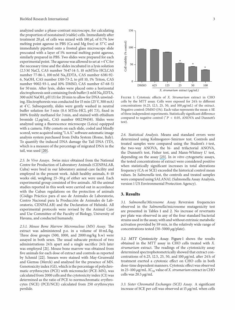
Figure 1: Cytotoxic effects of X. Strumarium extract in CHOcells by the MTT assay. Cells were exposed for 24 h to differentconcentrations (6.25, 12.5, 25, 50, and 100 𝜇g/mL) of the extract.Negative control: DMSO (1%). Each value represents the mean ± SEof three independent experiments. Statistically significant differencecompared to negative control (∗𝑃 < 0.05, ANOVA and Dunnett’stest).
2.6. Statistical Analysis. Means and standard errors weredetermined using Kolmogorov-Smirnov test. Controls andtreated samples were compared using the Student’s t-test,the two-way ANOVA, the bi- and trifactorial ANOVA,the Dunnett’s test, Fisher test, and Mann-Whitney 𝑈 test,depending on the assay [23]. In in vitro cytogenetic assays,the tested concentrations of extract were considered positivewhen statistically significant increases in total alterationsfrequency (CA or SCE) exceeded the historical control meanvalues. In Salmonella test, the controls and treated sampleswere compared using SALANAL (Salmonella Assay Analysis,version 1 US Environmental Protection Agency).
3. Results
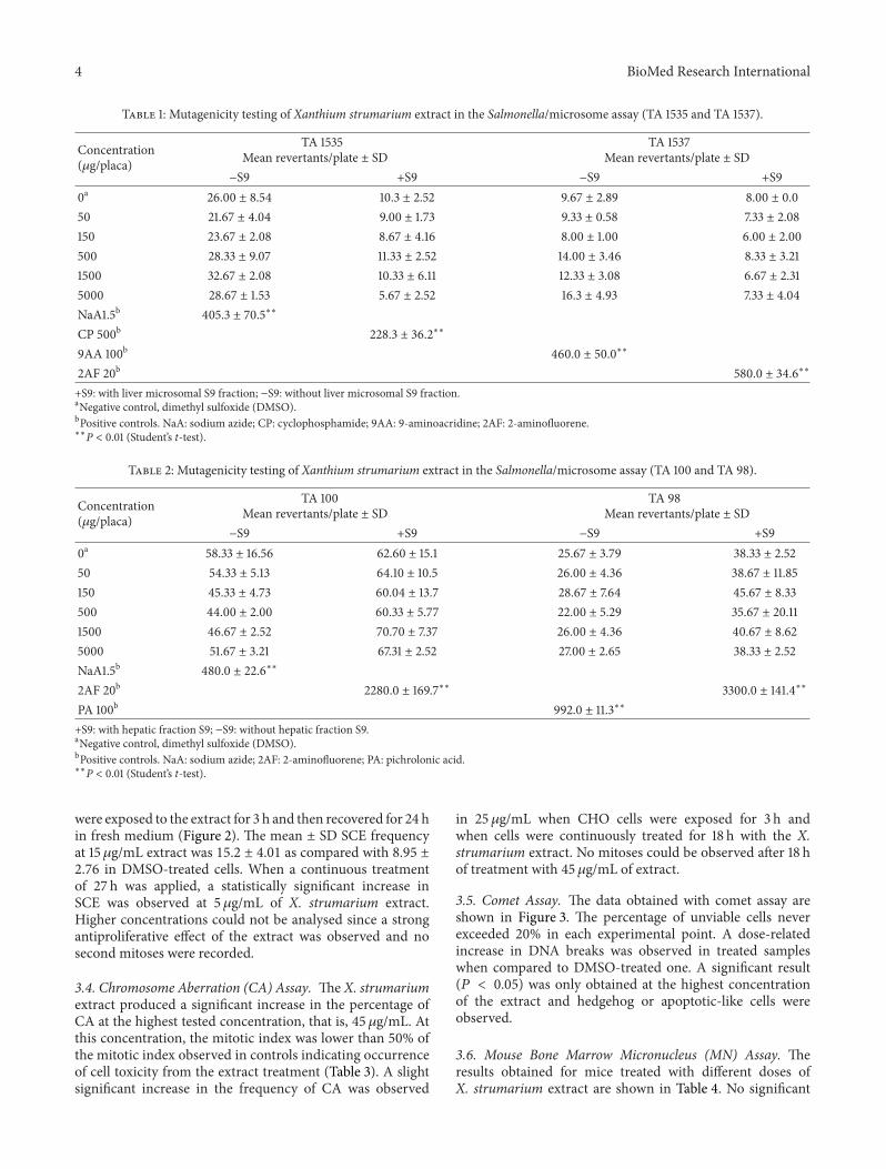
3.1. Salmonella/Microsome Assay. Reversion frequenciesobserved in the Salmonella/microsome mutagenicity testare presented in Tables 1 and 2. No increase of revertantsper plate was observed in any of the four standard bacterialstrains used in the assay, with andwithout extrinsicmetabolicactivation provided by S9 mix, in the relatively wide range ofconcentrations tested (50–5000 𝜇g/plate).
3.2. MTT Cytotoxicity Assay. Figure 1 shows the resultsobtained in the MTT assay in CHO cells treated with X.strumarium extract. The readings of the cytotoxicity assaydetermined spectrophotometrically showed that extract con-centrations of 6.25, 12.5, 25, 50, and 100 𝜇g/mL after 24 h oftreatment exerted a cytotoxic effect on CHO cells in bothdose-time dependentmanners. Cytotoxic effect was observedin 25–100𝜇g/mL. IC
50value ofX. strumarium extract inCHO
cells was 20.5𝜇g/mL
3.3. Sister Chromatid Exchanges (SCE) Assay. A significantincrease of SCE per cell was observed at 15𝜇g/mL when cells
4 BioMed Research International
Table 1: Mutagenicity testing of Xanthium strumarium extract in the Salmonella/microsome assay (TA 1535 and TA 1537).
Concentration(𝜇g/placa)
TA 1535Mean revertants/plate ± SD
TA 1537Mean revertants/plate ± SD
−S9 +S9 −S9 +S90a 26.00 ± 8.54 10.3 ± 2.52 9.67 ± 2.89 8.00 ± 0.050 21.67 ± 4.04 9.00 ± 1.73 9.33 ± 0.58 7.33 ± 2.08150 23.67 ± 2.08 8.67 ± 4.16 8.00 ± 1.00 6.00 ± 2.00500 28.33 ± 9.07 11.33 ± 2.52 14.00 ± 3.46 8.33 ± 3.211500 32.67 ± 2.08 10.33 ± 6.11 12.33 ± 3.08 6.67 ± 2.315000 28.67 ± 1.53 5.67 ± 2.52 16.3 ± 4.93 7.33 ± 4.04NaA1.5b 405.3 ± 70.5∗∗
CP 500b 228.3 ± 36.2∗∗
9AA 100b 460.0 ± 50.0∗∗
2AF 20b 580.0 ± 34.6∗∗
+S9: with liver microsomal S9 fraction; −S9: without liver microsomal S9 fraction.aNegative control, dimethyl sulfoxide (DMSO).bPositive controls. NaA: sodium azide; CP: cyclophosphamide; 9AA: 9-aminoacridine; 2AF: 2-aminofluorene.∗∗P < 0.01 (Student’s t-test).
Table 2: Mutagenicity testing of Xanthium strumarium extract in the Salmonella/microsome assay (TA 100 and TA 98).
Concentration(𝜇g/placa)
TA 100Mean revertants/plate ± SD
TA 98Mean revertants/plate ± SD
−S9 +S9 −S9 +S90a 58.33 ± 16.56 62.60 ± 15.1 25.67 ± 3.79 38.33 ± 2.5250 54.33 ± 5.13 64.10 ± 10.5 26.00 ± 4.36 38.67 ± 11.85150 45.33 ± 4.73 60.04 ± 13.7 28.67 ± 7.64 45.67 ± 8.33500 44.00 ± 2.00 60.33 ± 5.77 22.00 ± 5.29 35.67 ± 20.111500 46.67 ± 2.52 70.70 ± 7.37 26.00 ± 4.36 40.67 ± 8.625000 51.67 ± 3.21 67.31 ± 2.52 27.00 ± 2.65 38.33 ± 2.52NaA1.5b 480.0 ± 22.6∗∗
2AF 20b 2280.0 ± 169.7∗∗ 3300.0 ± 141.4∗∗
PA 100b 992.0 ± 11.3∗∗
+S9: with hepatic fraction S9; −S9: without hepatic fraction S9.aNegative control, dimethyl sulfoxide (DMSO).bPositive controls. NaA: sodium azide; 2AF: 2-aminofluorene; PA: pichrolonic acid.∗∗P < 0.01 (Student’s t-test).
were exposed to the extract for 3 h and then recovered for 24 hin fresh medium (Figure 2). The mean ± SD SCE frequencyat 15𝜇g/mL extract was 15.2 ± 4.01 as compared with 8.95 ±2.76 in DMSO-treated cells. When a continuous treatmentof 27 h was applied, a statistically significant increase inSCE was observed at 5 𝜇g/mL of X. strumarium extract.Higher concentrations could not be analysed since a strongantiproliferative effect of the extract was observed and nosecond mitoses were recorded.
3.4. Chromosome Aberration (CA) Assay. The X. strumariumextract produced a significant increase in the percentage ofCA at the highest tested concentration, that is, 45𝜇g/mL. Atthis concentration, the mitotic index was lower than 50% ofthe mitotic index observed in controls indicating occurrenceof cell toxicity from the extract treatment (Table 3). A slightsignificant increase in the frequency of CA was observed
in 25 𝜇g/mL when CHO cells were exposed for 3 h andwhen cells were continuously treated for 18 h with the X.strumarium extract. No mitoses could be observed after 18 hof treatment with 45𝜇g/mL of extract.
3.5. Comet Assay. The data obtained with comet assay areshown in Figure 3. The percentage of unviable cells neverexceeded 20% in each experimental point. A dose-relatedincrease in DNA breaks was observed in treated sampleswhen compared to DMSO-treated one. A significant result(𝑃 < 0.05) was only obtained at the highest concentrationof the extract and hedgehog or apoptotic-like cells wereobserved.
3.6. Mouse Bone Marrow Micronucleus (MN) Assay. Theresults obtained for mice treated with different doses ofX. strumarium extract are shown in Table 4. No significant

BioMed Research International 5
Table 3: Evaluation of chromosomal aberrations and mitotic index in CHO cells treated with whole extract of Xanthium strumarium L.
Concentration(𝜇g/mL)
Numberof cells Aberrant cellsa (%) Mitotic index (%) Chromatid
breaksChromosome
breaksbExch. Total
Ab.DMSO 1% 150 1.33 74 0.66 0.66 1.33
3 h + 15 h recovery10 150 1.33 77 0.66 0.66 1.3325 150 4.00∗ 62 0.66 3.33 4.0045 150 8.66∗∗ 32 2.00 7.33 9.33
18 h continuous10 150 2.00 44 0.66 1.33 2.0025 150 4.00∗ 53 2.66 2.66 5.3345 150 NM 2Mit Cc 100 23.00∗∗ 21.00 12.00 33.00RXd 100 38.00∗∗ 2.00 48.00 16.00 66.00aExcluding gaps; NM: no mitoses; bchromosome and chromatid exchanges; cmitomycin C positive control (0.05𝜇g/mL); dRX: positive control (2 Gy); ∗P <0.05; ∗∗P < 0.01 versus DMSO sample.
0
5
10
15
20
25
Control DMSO 1%
2 5 15 2 5
SCE/
cell
Concentrations (𝜇g/mL)
Exposed 3h Exposed 27h
∗
∗
Figure 2: SCE assay analysis in CHO cells treated with crude extractof X. strumarium L. (2, 5 and 15𝜇g/mL). Cells were exposed for 3 hor 27 h to X. strumarium extracts. Cultures received BrdUrd at thefinal concentration of 1.5𝜇g/mL during the last 24 h of incubation.Negative control: DMSO (1%). Each value represents the mean ± SDof three independent experiments. Statistically significant differencecompared to negative control (∗𝑃 < 0.05, ANOVA and Dunnett’stest).
difference in the frequency ofMNPCEwas observed betweenmice treated with X. strumarium extract and the negativecontrol for both sexes. A high increase in the frequencyof MNPCE was detected in mice treated with cyclophos-phamide compared to the negative control (𝑃 < 0.01). Nosignificant differences in the PCE/NCE ratio were observedwhen comparing mice treated with X. strumarium extractand the respective negative control. No signs of toxicity werefound in mice at dose levels of 500–2000mg/kg body weight.There was no mortality in any of the groups; the initial andfinal weights of the animals were found to be similar tocontrols.
4. Discussion
Plant remedies are traditionally used in Cuba for the treat-ment of different illness. This knowledge has remarkably
05
1015202530354045
C DMSO 1(%)
5 15 45
Tail
DN
A (%
)
H2O2
Concentrations (𝜇g/mL)
∗ a
H2O2
100 𝜇M
Figure 3: Comet assay analysis in CHO cells treated with fluidextract of Xanthium strumarium L. CHO cells were treated for3 h with different concentrations (5, 15, and 45𝜇g/mL) of theextract. Positive control: hydrogen peroxide (100 𝜇Mfor 20min) andnegative control: DMSO (1%). Each value represents the mean ± SDof three independent experiments. Statistically significant differencecompared to negative control ( ∗𝑃 < 0.05 versus DMSO sample, a𝑃 < 0.05 versus control at Mann-Whitney 𝑈 test).
been improved through the prioritized policy of the CubanMinistry of Public Health of developing pharmaceuticals andsupplements from natural sources as well as a scientificallysustained phytotherapy. To confirm the pharmacologic prop-erties of Xanthium strumarium L., species belonging to theCuban flora are part of that aim.
X. strumarium L is one of the most popular herbalformulas in the world; however, evidence-based informationabout genotoxicity is limited. Many studies have reportedpharmacological efficacies and benefits of X. strumarium [2–11], but there is little information on its risk and safety.
Data presented in this report have involved an extensivegenotoxic assessment of a complexmixture, the whole extractof X. strumarium L. as part of preclinical studies; the use ofin vitro and in vivo assays was decisive in order to obtain apicture of the genotoxic potential of this plant product and to

6 BioMed Research International
Table 4: Micronucleus test results in mice treated with whole extract of Xanthium strumarium L.
Dose (mg/kg) Female MalePCE/NCEa
± SD MNPCE/2000b ± SD PCE/NCEa± SD MNPCE/2000b ± SD
0 0.46 ± 1.32 0.18 ± 0.43 0.35 ± 1.39 0.20 ± 0.24500 0.07 ± 1.57 0.03 ± 0.08 0.10 ± 1.70 0.03 ± 0.111000 0.28 ± 1.58 0.06 ± 0.08 0.16 ± 1.51 0.03 ± 0.072000 0.08 ± 1.46 0.03 ± 0.11 0.07 ± 1.46 0.03 ± 0.11Ethanolc 0.05 ± 1.39 0.01 ± 0.08 0.32 ± 1.45 0.04 ± 0.08CPd 0.06 ± 0.63∗∗ 1.80 ± 7.62∗∗ 0.07 ± 0.53∗∗ 0.75 ± 5.38∗∗aPCE/NCE: polychromatic (PCE) to normochromatic (NCE) erythrocyte ratio; bMNPCE/2000: frequency of micronucleated PCE; cNegative control: ethanol60%; dPositive control: cyclophosphamide 20mg/Kg. ∗∗P < 0.01 (Student’s t-test).
identify potential risk/hazard for human health due to DNAdamage and mutation induction.
The species of the genus Xanthium are an impor-tant source of sesquiterpene lactones which are respon-sible for most of biological activities attributed to thisgenus. Sesquiterpene lactones termed xanthanolides, xan-thatin, 8-epi xanthatin, and 8-epi tomentosin and oth-ers phytocompounds as xanthus-strumarina, hydrochinonas,carboxy-atractyloside, albuminoids and organic acids hasbeen obtained from X. strumarium species, which have beenreported as cytotoxic substance [2, 4, 5]. Moreover, manyreports evidenced that sesquiterpene lactones present inplants have cytotoxic activity [4, 24, 25]. These sesquiterpenelactones trigger mitochondrial membrane transition, loss ofmitochondrial membrane potential, and release of proapop-totic mitochondrial proteins leading to caspase activationand apoptotic cell death [26]. Xanthatin also caused a pro-grammed cell death like in trypanosomes as evidenced by areduction in mitochondrial membrane potential [27].
Our results showed the cytotoxic capacity of X. strumar-ium extract when high concentrations (25–100𝜇g/mL) for24 h were applied to CHO cells in in vitro experimental con-ditions. The decrease in cell viability at high concentrationscan be related to a toxic effect on the metabolism of the cellor to DNA damage. Coincidentally, precedent studies [2, 4,5] conducted in in vitro experimental conditions reportedcytotoxic activity of extracts obtained from this vegetablespecies. However, acute and subacute administration of rootextracts from X. strumarium did not produce any toxicitywhen in vivo assay was assessed [28].
The in vitro assays used in the present study coverdifferent levels of genotoxic damage expression. In theseassays we include toxic concentrations greater than the IC
50
to confirm cytotoxicity-mediated genotoxicity. A first levelwas the induction of point mutations at specific loci inSalmonella typhimurium. The negative outcome of bacterialtesting of extract seems to correspond with the absence ofpotential to induce mutagenic changes in DNA. The secondlevel was the analysis of SCE in mammalian CHO cells. TheX. strumarium extract produced a significant induction ofSCE, which is a sensitive method for the measurement ofDNA damage at primary structure level. The X. strumariumextract also proved to be clastogenic at the cytogenetic levelmeasuringCA induction inCHOcells only at a concentration
that produced severe toxic effects, as assessed by the strongdecrease in the mitotic index. The positive result obtainedwith the highest dose of the extract in the in vitro cometassay where hedgehog or apoptotic-like cells were observedindicated that the test substance induces DNA primarydamage (both single- and double-strand breaks) in culturedmammalian cells.This result is in line with that obtained withthe same concentration in CA analysis.
Maybe the DNA damage can be explained on the basisof a systemic toxic effect. Today, it is known that the doublebonds present in the 𝛼-methylene 𝛾-lactone and cyclopen-tanone moieties confer to sesquiterpene lactones an affinitytowards thiol groups of proteins and glutathione inducingoxidative stress in the cell [29]. There is no evidence thatchromosomal aberrations induced by sesquiterpene lactonesresult from direct interaction with DNA or the chromosomestructure; rather, an indirect mechanism mediated by cyto-toxicity has been suggested [24]. The issue of cell-toxicity-mediated genotoxicity has been intensively addressed andmany related endpoints have been proposed [30]. Amongstthem, inhibition of key enzymes of energetic metabolism(oxidative phosphorylation) and nucleic acid replication(DNA polymerase) and expression of NFkB and caspase 3,which are proapoptotic and induce DNA damage, have beenobserved by sesquiterpene lactones [24, 25, 31, 32].
In addition, the present study reveals that the adminis-tration of 500–2000mg/Kg dose of X. strumarium extractex-hibited no genotoxic activity in our MN in vivo assays.There was no mortality or signs of toxicity in male andfemale animals. The treatment, until sacrifice time, did notproduce any adverse reactions or any significant changes inthe body weights (data not shown) as compared to untreatedcontrols. In a similar work realized by Diaz et al. [11] theauthors did not observe increases in the micronucleatederythrocytes frequency in mouse treated orally with extractsof this plant at doses of 500, 1000, and 2000mg/kg bodyweight. Another study of toxicity performed with chloroformand hexane soluble fractions ofX. strumarium reveals that theadministration of a very high dose (5 g/kg body weight) foracute toxicity determination could not make any abnormalchanges at gross as well as histopathological levels in thetreated animals [28].
In our case, observed genotoxic effects were not con-firmed in vivo with micronucleus test. This experiment
BioMed Research International 7
evidenced our postulation indicating that nontoxic dosesshowed neither DNA damage nor toxic effects. Besides theusual arguments invoked to explain the in vivo negativeperformance of in vitro genotoxins, which deal mainlywith the pharmacokinetic profile of absorption-distribution-metabolism-excretion, onemaywonder if genotoxic responseto X. strumarium extract was related to the reported cyto-toxicity, which could implicate an indirect interference withnuclear functions to arrive to cell death, as it happens formany anticancer agents [24, 25, 31, 32].
We suggest that further examination of the cytotoxicpotential of these compounds should be pursued. Furtherstudies have to be performed using various cancer modelsto elucidate the exact mechanism by which X. strumariumextract exerts its cytotoxic action. In addition, newmolecularstudies are needed in order to elucidate the interaction of thiscompound in cell biology and the consequences for humanhealth.
5. Conclusions
The data presented here constitute the most complete studyon the genotoxic potential the aerial parts of X. strumariumL. and show that the extract can induce in vitroDNA damageat cytotoxic concentrations and, however, does not inducemicronucleus in bone marrow of mice treated orally withextracts of this plant at dose of 500–2000mg/kg body weightin our experimental conditions.
Conflict of Interests
The authors declare that there is no conflict of interestsregarding the publication of this paper.
Acknowledgments
The authors are grateful to Dipartimento di Biologia, Uni-versita degli Studi “Roma TRE”, and Istituto di Biologia ePatologia Molecolare, CNR, for the generous support andgreat encouragement in carrying out the work.
References
[1] R. S. Lizama, M. M. Martınez, and R. E. I. Lantigua, “Plantasmedicinales de uso tradicional en pinar del Rıo. Estudioetnobotanico. I,” Revista Cubana de Farmacia, vol. 32, no. 1, pp.57–62, 1998.
[2] T. Han, H.-L. Li, Q.-Y. Zhang et al., “Bioactivity-guided frac-tionation for anti-inflammatory and analgesic properties andconstituents ofXanthium strumarium L,” Phytomedicine, vol. 14,no. 12, pp. 825–829, 2007.
[3] M. H. Yin, D. G. Kanga, D. H. Cho, T. O. Kwon, and H. S. Lee,“Screening of vaso relaxant activity of some medicinal plantsused in oriental medicines,” Journal of Ethnopharmacology, vol.99, no. 1, pp. 113–117, 2005.
[4] I. Ramırez-Erosa, Y. Huang, R. A. Hickie, R. G. Sutherland, andB. Barl, “Xanthatin and xanthinosin from the burs of Xanthiumstrumarium L. as potential anticancer agents,”Canadian Journal
of Physiology and Pharmacology, vol. 85, no. 11, pp. 1160–1172,2007.
[5] H. S. Kim, T. S. Lee, S. W. Yeo, L. S. Seong, and T. S.Yu, “Isolation and characterization of antitumor agents fromXanthium strumarium L,” Korean Society for Biotechnology andBioengineering Journal, vol. 18, pp. 324–328, 2003.
[6] K. K. Dong, K. S. Chang, W. B. Dong, S. K. Yeon, Y. Min-Suk,and K. K. He, “Identification and biological characteristics ofan antifungal compound extracted from Cocklebur (Xanthiumstrumarium) against Phytophthoradrechsleri,”ThePlant Pathol-ogy Journal, vol. 18, pp. 288–292, 2002.
[7] L. S. Favier, A. O. M. Marıa, G. H. Wendel et al., “Anti-ulcerogenic activity of xanthanolide sesquiterpenes from Xan-thium cavanillesii in rats,” Journal of Ethnopharmacology, vol.100, no. 3, pp. 260–267, 2005.
[8] J. P. Ferrer, T. Stoiber, A. V. Parra et al., “Search of newantimitotics compounds from the Cuban flora,” Boletin Lati-noamericano y del Caribe de Plantas Medicinales y Aromaticas,vol. 10, no. 1, pp. 75–82, 2011.
[9] G. S. Menon, K. Kuchroo, and D. Dasgupta, “Interaction ofmicrotubules with active principles of Xanthium strumarium,”Physiological Chemistry and Physics and Medical NMR, vol. 33,no. 2, pp. 153–162, 2001.
[10] Q. L. Tran, Y. Tezuka, J.-Y. Ueda et al., “In vitro antiplasmodialactivity of antimalarial medicinal plants used in Vietnamesetraditional medicine,” Journal of Ethnopharmacology, vol. 86,no. 2-3, pp. 249–252, 2003.
[11] G. M. Diaz, M. C. Le𝜎n, and E. Iglesias, “Evaluacion del efectogenotoxico del Xhantium strumarium L., (Guisazo de caballo),”Revista Cubana de Plantas Medicinales, vol. 9, pp. 27–32, 2004.
[12] M.Miranda andA. Cuellar,Manual de Practicas de Laboratorio,Farmacognosia y Productos Naturales, Universidad de LaHabana, IFAL, 2000.
[13] D. M. Maron and B. N. Ames, “Revised methods for theSalmonella mutagenicity test,” Mutation Research, vol. 113, no.3-4, pp. 173–215, 1983.
[14] T. Mosmann, “Rapid colorimetric assay for cellular growth andsurvival: application to proliferation and cytotoxicity assays,”Journal of Immunological Methods, vol. 65, no. 1-2, pp. 55–63,1983.
[15] R. De Salvia, M. Fiore, T. Aglitti, F. Festa, R. Ricordy, andR. Cozzi, “Inhibitory action of melatonin on H
2
O2
- andcyclophosphamide-induced DNA damage,” Mutagenesis, vol.14, no. 1, pp. 107–112, 1999.
[16] A. Sanchez-Lamar, G. Fonseca, J. L. Fuentes et al., “Assessmentof the genotoxic risk of Punica granatum L. (Punicaceae) wholefruit extracts,” Journal of Ethnopharmacology, vol. 115, no. 3, pp.416–422, 2007.
[17] ICH, Genotoxicity: Guidance on Specific Aspects of RegulatoryGenotoxicity Test for Pharmaceuticals, 1995.
[18] S. M. Galloway, M. J. Aardema, M. Ishidate Jr. et al., “Reportfrom working group on in vitro tests for chromosomal aber-rations,” Mutation Research: Environmental Mutagenesis andRelated Subjects Including Methodology, vol. 312, no. 3, pp. 241–261, 1994.
[19] T. Cornetta, L. Padua, A. Testa et al., “Molecular biomonitor-ing of a population of nurses handling antineoplastic drugs,”Mutation Research: Fundamental and Molecular Mechanisms ofMutagenesis, vol. 638, no. 1-2, pp. 75–82, 2008.
[20] A. R. Collins, “The comet assay for DNA damage and repair:principles, applications, and limitations,” Applied Biochemistry
8 BioMed Research International
and Biotechnology B: Molecular Biotechnology, vol. 26, no. 3, pp.249–261, 2004.
[21] M. Hayashi, R. R. Tice, J. T. MacGregor et al., “In vivo rodenterythrocyte micronucleus assay,” Mutation Research: Environ-mentalMutagenesis and Related Subjects IncludingMethodology,vol. 312, no. 3, pp. 293–304, 1994.
[22] W. Schmid, “The micronucleus test for cytogenetic analysis,” inChemical Mutagens: Principles and Methods forTheir Detection,A. Hollaender, Ed., vol. 4, pp. 31–53, Plenum Press, New York,NY, USA, 1976.
[23] D. J. Kirkland, Statistical Evaluation of Mutagenicity Test, Cam-bridge University Press, 1989.
[24] A. Ramos, R. Rivero, A. Visozo, J. Piloto, and A. Garcıa,“Parthenin, a sesquiterpene lactone of Parthenium hysteropho-rus L. is a high toxicity clastogen,” Mutation Research: GeneticToxicology and Environmental Mutagenesis, vol. 514, no. 1-2, pp.19–27, 2002.
[25] S. Zhang, C.-N. Ong, and H.-M. Shen, “Critical roles of intra-cellular thiols and calcium in parthenolide-induced apoptosisin human colorectal cancer cells,” Cancer Letters, vol. 208, no. 2,pp. 143–153, 2004.
[26] V. Rosenkranz and M. Wink, “Alkaloids induce programmedcell death in bloodstream forms of trypanosomes (Try-panosoma b. brucei),” Molecules, vol. 13, no. 10, pp. 2462–2473,2008.
[27] J. A. Marco, J. F. Sanz-Cervera, J. Corral, M. Carda, and J.Jakupovic, “Xanthanolides from Xanthium: absolute config-uration of xanthanol, isoxanthanol and their C-4 epimers,”Phytochemistry, vol. 34, no. 6, pp. 1569–1576, 1993.
[28] J. M. Aranjani, C. M. Rao, A. Manuel, J. V. Rao, N. Udupa,and K. Hebbar, “Acute and subacute toxicity of chloroform andhexaneextracts of root of Xanthium strumarium,” ComparativeClinical Pathology, vol. 21, no. 6, pp. 1223–1230, 2012.
[29] A. A. Ahmed, A. A. Mahmoud, and A. A. El-Gamal, “Axanthanolide diol and a dimeric xanthanolide from Xanthiumspecies,” Planta Medica, vol. 65, no. 5, pp. 470–472, 1999.
[30] A. Kovacs, A. Vasas, P. Forgo, B. Rethy, I. Zupko, and J.Hohmann, “Xanthanolides with antitumouractivity from Xan-thium italicum,” Zeitschrift fur Naturforschung C, vol. 64, pp.343–349, 2009.
[31] E. Nibret, M. Youns, R. L. Krauth-Siegel, and M. Wink,“Biological activities of xanthatin from Xanthium strumariumleaves,” Phytotherapy Research, vol. 25, no. 12, pp. 1883–1890,2011.
[32] L. Zhang, J. Ruan, L. Yan et al., “Xanthatin induces cell cyclearrest at G2/M checkpoint and apoptosis via disrupting NF-𝜅Bpathway in A549 non-small-cell lung cancer cells,” Molecules,vol. 17, no. 4, pp. 3736–3750, 2012.

Submit your manuscripts athttp://www.hindawi.com
PainResearch and TreatmentHindawi Publishing Corporationhttp://www.hindawi.com Volume 2014
The Scientific World JournalHindawi Publishing Corporation http://www.hindawi.com Volume 2014
Hindawi Publishing Corporationhttp://www.hindawi.com
Volume 2014
ToxinsJournal of
VaccinesJournal of
Hindawi Publishing Corporation http://www.hindawi.com Volume 2014
Hindawi Publishing Corporationhttp://www.hindawi.com Volume 2014
AntibioticsInternational Journal of
ToxicologyJournal of
Hindawi Publishing Corporationhttp://www.hindawi.com Volume 2014
StrokeResearch and TreatmentHindawi Publishing Corporationhttp://www.hindawi.com Volume 2014
Drug DeliveryJournal of
Hindawi Publishing Corporationhttp://www.hindawi.com Volume 2014
Hindawi Publishing Corporationhttp://www.hindawi.com Volume 2014
Advances in Pharmacological Sciences
Tropical MedicineJournal of
Hindawi Publishing Corporationhttp://www.hindawi.com Volume 2014
Medicinal ChemistryInternational Journal of
Hindawi Publishing Corporationhttp://www.hindawi.com Volume 2014
AddictionJournal of
Hindawi Publishing Corporationhttp://www.hindawi.com Volume 2014
Hindawi Publishing Corporationhttp://www.hindawi.com Volume 2014
BioMed Research International
Emergency Medicine InternationalHindawi Publishing Corporationhttp://www.hindawi.com Volume 2014
Hindawi Publishing Corporationhttp://www.hindawi.com Volume 2014
Autoimmune Diseases
Hindawi Publishing Corporationhttp://www.hindawi.com Volume 2014
Anesthesiology Research and Practice
ScientificaHindawi Publishing Corporationhttp://www.hindawi.com Volume 2014
Journal of
Hindawi Publishing Corporationhttp://www.hindawi.com Volume 2014
Pharmaceutics
Hindawi Publishing Corporationhttp://www.hindawi.com Volume 2014
MEDIATORSINFLAMMATION
of