Multipotent mesenchymal stem cells from adult human eye conjunctiva stromal cells
Upload
independentCategory
view
0download
0
© 2012 Ruiz et al, publisher and licensee Dove Medical Press Ltd. This is an Open Access article which permits unrestricted noncommercial use, provided the original work is properly cited.
Cell Health and Cytoskeleton 2012:4 29–35
Cell Health and Cytoskeleton
The effect of nicotine on the mechanical properties of mesenchymal stem cells
Juan P Ruiz1,2
Daniel Pelaez1,2
Janice Dias1
Noël M Ziebarth1
Herman S Cheung1,2
1Department of Biomedical Engineering, University of Miami College of Engineering, Coral Gables, FL, USA; 2Research Service and Geriatrics Research, Education, and Clinical Center, Veterans Affairs Medical Center, Miami, FL, USA
Correspondence: Herman S Cheung Department of Biomedical Engineering, University of Miami, McArthur Annex Room 214, 1251 Memorial Drive, Coral Gables, FL 33146, USA Tel +1 305 284 4466 Email [email protected]
Purpose: To measure the elasticity of the nucleus and cytoplasm of human mesenchymal stem
cells (MSCs) as well as changes brought about by exposure to nicotine in vitro.
Methods: MSCs were synchronized to the G0 stage of the cell cycle through serum deprivation
techniques. The cells were then treated with medium containing nicotine (0.1 µM, 0.5 µM,
and 1 µM). Atomic force microscopy was then used to measure the Young’s modulus of both
the nucleus and cytoplasm of these cells.
Results: For both unsynchronized and synchronized cells, the nucleus was softer than the
cytoplasm, although this difference was not found to be statistically significant. The nucleus of
cells treated with nicotine was significantly stiffer than the control for all concentrations. The
cytoplasm was significantly stiffer in nicotine-treated cells than in control cells for the 0.5 µM
and 1.0 µM concentrations only.
Conclusions: The results of this study could suggest that nicotine affects the biophysical prop-
erties of human MSCs in a dose-dependent manner, which may render the cells less responsive
to mechanoinduction and other physical stimuli.
Keywords: atomic force microscopy, elasticity, mesenchymal stem cells, nicotine
IntroductionMesenchymal stem cells (MSCs) are fibroblast-like cells, which reside in the bone
marrow, adipose tissue, skin, and periosteum of vertebrate animals, including humans.
MSCs have the capacity to differentiate into cartilage, bone, fat, tendon, and muscle.1
MSCs represent a renewable source of cells that could be used for tissue regenera-
tion or reconstruction of damaged musculoskeletal tissue by cellular transplantation.
However, a person’s habits and lifestyle choices may affect the effectiveness of such
therapies. In fact, many of the patients in need of these cellular therapies, such as those
who smoke, could potentially have niche environments in their body that will render
these stem cell populations ineffective posttransplantation. High levels of cigarette
chemicals in the blood of smokers, for example, could potentially damage the trans-
planted MSCs and render them ineffective. It is therefore important to investigate the
effect of such habits on stem cell populations.
Cigarette smoking is a well-known risk factor for a number of diseases, includ-
ing cardiopulmonary diseases, in the adult population.2 Cigarette smoke consists of
a large number of chemicals, including nicotine, aromatic hydrocarbons, hydrogen
cyanide, and aldehydes, among others.3 Nicotine is the most prevalent chemical in
cigarettes and is absorbed rapidly into the arterial circulation and tissues throughout
the body.4 Nicotine has also been shown to negatively impact human embryonic
Dovepress
submit your manuscript | www.dovepress.com
Dovepress 29
O R i G i N A L R E S E A R C H
open access to scientific and medical research
Open Access Full Text Article
http://dx.doi.org/10.2147/CHC.S24381
Video abstract
Point your SmartPhone at the code above. If you have a QR code reader the video abstract will appear. Or use:
http://bit.ly/zvfH5w

Cell Health and Cytoskeleton 2012:4
stem cells and their development by altering the cell’s
morphology in vitro and downregulating the expression of
certain pluripotency markers, including Oct3/4, Tra-1-81,
and Tra-1-60.5 Nicotine induces angiogenesis via nicotine
acetylcholine receptors;6,7 these receptors are present in both
neurons and other cell types.7 Nicotine has been shown to
delay skeletal healing by as much as 60% following fractures
by influencing gene expression critical to the bone-healing
process.7,8 However, the effects of nicotine on adult MSCs
remain largely unexplored.
Although much attention has been focused on the influ-
ence of bioreactive factors such as cytokines and chemokines
upon MSC differentiation and growth, the effects of mechani-
cal stimuli on these cells, in terms of their differentiation
and phenotypic maintenance capabilities, are still poorly
understood. It has been shown that the biophysical properties
of MSCs determine the commitment of these cells to differ-
ent lineages during differentiation.9 Recently, we reported
that dynamic compressive loading stimulated viability and
chondrogenic differentiation of MSCs in the absence of
exogenous cytokines.10–12 Compressive loading induced
endogenous transforming growth factor (TGF)-β1 as well as
TGF-β receptor gene expression of MSCs, suggesting that
dynamic compression might induce chondrogenesis of these
cells via the TGF-β signal pathway.10 Subsequently, we dem-
onstrated temporal expression patterns of early response and
chondrogenic genes (c-fos, c-jun, TGF-β, TGF-β receptor-1
and -II, Sox9, and type II collagen) and induction of their
corresponding proteins in MSCs stimulated by dynamic
compressive loading.11
During compression-induced chondrogenesis, MSCs
undergo significant deformation, and our preliminary data
indicate that compressive loading exerts its chondrogenic
effect by activating the P42/44 mitogen-activated protein
kinases signal pathways. These cells are therefore able to
take mechanical forces and translate them into chemical
signals that control gene expression, a phenomenon known as
mechanotransduction. It has been reported that the mechani-
cal properties of stem cells play a key role in their potential
to differentiate.13 Stiffer cells are unable to respond to the
external mechanical stimuli necessary to drive differen-
tiation into various cells types.14,15 In fact, the mechanical
properties of stem cells, which dictate their response to
mechanical stimuli, can actually be used as a biomarker to
measure multipotency.13,16,17 In order to assess the mechani-
cal properties of stem cells, previous studies have mea-
sured the Young’s modulus of elasticity using micropipette
aspiration18–20 and atomic force microscopy (AFM).14,15,21–27
Micropipette aspiration measures the elasticity of free float-
ing cells, while AFM measures the elasticity of cells attached
to a substrate, usually tissue culture plastic. Previous AFM
studies on MSCs, however, use a spherical probe that is
5–10 µ in radius, which will measure an average Young’s
modulus of the whole cell. Because the nucleus plays an
important role in a cell’s mechanical properties,28 it is impor-
tant to explore the mechanical properties of both the nuclear
and cytoplasmic regions. Therefore, the first purpose of this
project was to quantify the mechanical properties of the
nucleus and cytoplasm of MSCs independently. The second
objective of this study was to explore the effects of nicotine
on this biophysical property of MSCs.
Materials and methodsCell cultureMesenchymal stem cellsMSCs were commercially obtained from ScienceCell
Research Laboratories (Cat #7500; ScienceCell Research
Laboratories, Carlsbad, CA) as CD73-, CD90-, and CD105-
positive cells capable of adipogenic, chondrogenic, and
osteogenic differentiation. For all experiments, MSCs were
used at either passage 2 or 3.
Plating and synchronizationCells were plated at 10,000 cells/cm2 in high-glucose
Dulbecco’s modified Eagle’s medium (DMEM; Invitrogen,
Carlsbad, CA) supplemented with 10% (v/v) fetal bovine
serum (Invitrogen), 10 µg/mL and 10 µg/mL penicillin/
streptomycin, respectively (Invitrogen), and 0.25 µg/mL
amphotericin B (Thermo Scientific, Waltham, MA) on
35 mm × 10 mm tissue culture dishes (Sarstedt, Newton, NC).
The cells were left in the incubator at 37°C and 5% CO2 for
24 hours to allow for cell attachment. Nonadherent cells were
washed off during the first medium change 24 hours after ini-
tial plating. The desired confluence of 40%–50% was reached
after approximately 48 hours in the incubator. The plates were
then washed with phosphate buffered saline solution twice and
placed in serum-deprived high-glucose DMEM supplemented
with 1% (v/v) penicillin/streptomycin. The cells were placed
in the incubator for an additional 72 hours to allow the cells to
synchronize to the G0 phase of the cell cycle. Unsynchronized
control cells while testing synchronization efficacy were left
in regular culture medium for the 72-hour period.
Nicotine treatmentIn order to test the effects of nicotine on MSCs cultured
in vitro, cells were plated out as described previously, and
submit your manuscript | www.dovepress.com
Dovepress
Dovepress
30
Ruiz et al

Cell Health and Cytoskeleton 2012:4
plates were randomly separated into four treatment groups.
The control group was synchronized as described previously
and, following the 72-hour synchronization period, starvation
medium was removed and replaced with high-glucose
DMEM containing 10% fetal bovine serum, 1% penicillin/
streptomycin, and 0.1% amphoteracin B. The other three
groups were also synchronized, but the starvation medium
was then replaced by regular medium as described previously,
supplemented with different concentrations of nicotine
(nicotine 99% [TLC], liquid, N0267; Sigma-Aldrich, St Louis,
MO): 0.1 µM, 0.5 µM, and 1.0 µM.29–31 The cells were left in
the incubator for 48 hours with the nicotine medium before
being subjected to AFM testing (Figure 1).
Elasticity measurementsA custom-built nanoindenter based on the design of com-
mercial atomic force microscopes has been developed to
measure mechanical properties.32–34 A pyramidal AFM
cantilever tip (0.01 N/m, silicon nitride, 20 nm tip radius,
MLCT series; Bruker AFM Probes, Camarillo, CA) was
used to indent the cells.
After the cells were cultured as described previously, the
Petri dish containing the cells was placed under the holder
containing the AFM cantilever. The location of the cantile-
ver on the sample was observed using the 20× microscope
objective connected to a camera beneath the sample. This
enabled precise positioning of the cantilever above either
the cytoplasmic or the nuclear region of the cell. The tip
was lowered until it was just touching the surface of the cell
(Figure 2). The tip was then lowered using the piezoelectric
control so that it was probing the cell. The measurements
were conducted using a cantilever approach with a retrac-
tion speed of 15 µm/s and a maximal indentation force
of 60 nN. The voltage detected at the photodiode due to
deflection of the cantilever was recorded as a function of
piezoelectric displacement. These recordings were repeated
at least 15 times per cell, and at least ten cells per nicotine
concentration were measured. The experiments were repeated
for both the cytoplasmic and nuclear regions of each cell.
All experiments were performed at room temperature while
the cells remained submerged in DMEM.
Data analysisThe data analysis procedure for the AFM measurements has
been described previously.32 The photodiode voltage versus
piezoelectric displacement recorded during the measure-
ment scans was converted to force versus indentation after
accounting for the cantilever spring constant and its behavior
when probing a rigid substrate. The force versus indentation
relationship was analyzed using the Bilodeau35 model for a
pyramidal indenter:
F
ED=
−( )tanα
ν2 1 2
2 (1)
where F [N] is the measured force, E [N/m2] is Young’s
modulus, ν is Poisson’s ratio (assumed to be 0.5), α is
the pyramidal semiangle (15°), and D is the measured
i ndentation. The determination of Young’s modulus was
carried out using custom-developed Matlab software. Each
curve fit was verified visually. Because at least 15 measure-
ments were performed for each position on the cell, the
average of these values was then used as the modulus for
that position.
All data and graphs represent mean values plus or minus
standard error of the mean. All data sets were tested for
significance using t-tests (one-tailed, two-sample unequal
variance). Studies that tested for significance in differences of
standard deviation were done using an f-test with n-1 degrees
Figure 1 Phase-contrast images of mesenchymal stem cells in culture. images were taken of the control cells as well as of those subjected to the highest concentration of nicotine (1 µM).
Figure 2 image showing view (from bottom) of plate, with atomic force microscopy cantilever placed over the cytoplasm of the mesenchymal stem cell. The image is taken by a camera through a 20× objective positioned underneath the culture dish. The nucleus is signaled by the arrow.
submit your manuscript | www.dovepress.com
Dovepress
Dovepress
31
MSC elasticity
Cell Health and Cytoskeleton 2012:4
of freedom. Statistical significance was determined by
P-values lower than 0.05.
Viability and cytoskeleton stainingIn order to test for cell viability post-treatment, a group of
treatment and control cells were treated with 4 µM calcein
(calcein-AM, C1430; Invitrogen) and 4 µM ethidium
homodimer (ethidium homodimer B, 46043; Sigma-Aldrich),
then imaged using fluorescence microscopy to view live and
dead cells. Similarly, a group of treatment and control cells
were first fixed in 10% formalin solution for 10 minutes,
then rinsed once with phosphate buffered saline, and their
f-actin stained with phalloidin (AlexaFluor 568 phalloidin,
A12380; Invitrogen). The cells were then mounted with
Vectashield (Vectashield mounting medium with DAPI;
Vector Labs, Burlingame, CA) and imaged with fluorescence
microscopy.
ResultsElasticity of nucleus and cytoplasmA total of 10 MSCs cultured as described previously, but
without synchronization to the G0 phase, were measured
using AFM. The elasticity measurements were performed in
the area of the cell cytoplasm and nucleus for all ten cells.
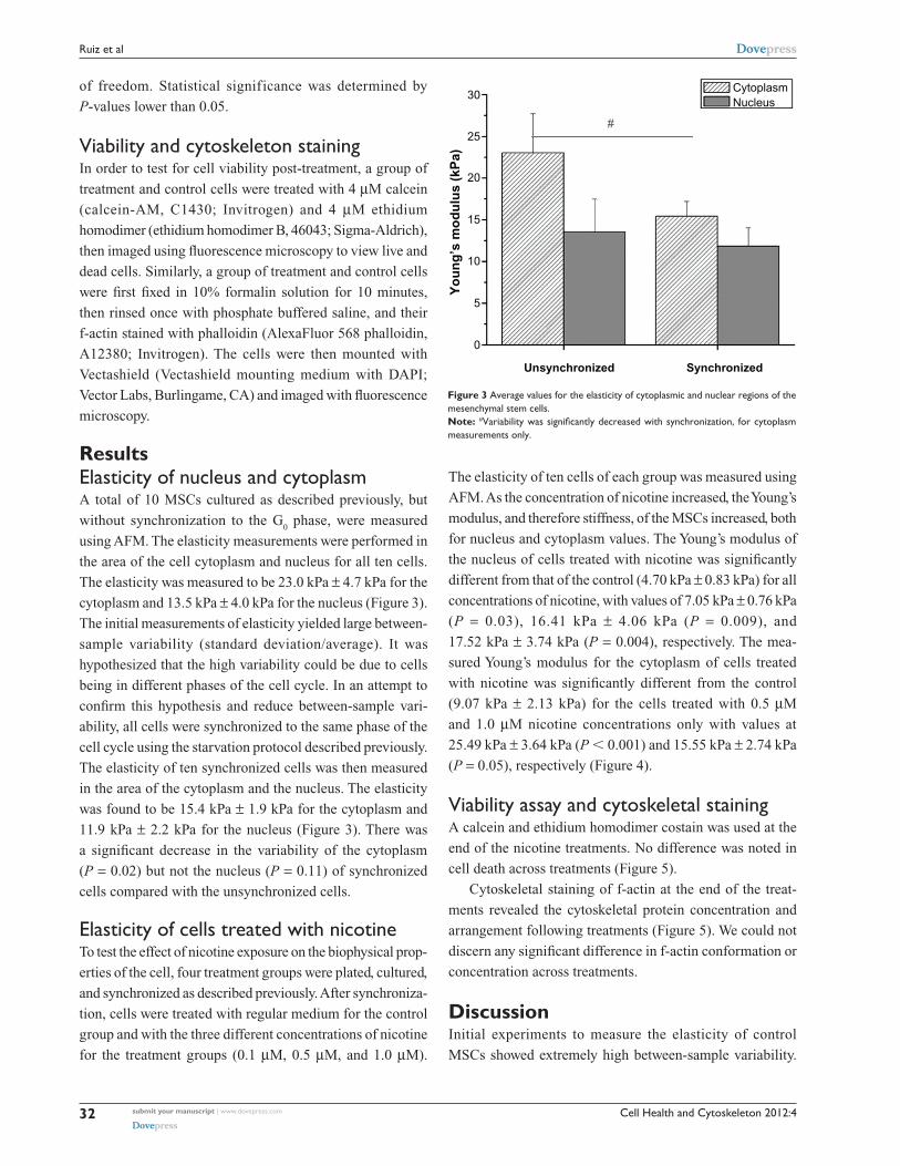
The elasticity was measured to be 23.0 kPa ± 4.7 kPa for the
cytoplasm and 13.5 kPa ± 4.0 kPa for the nucleus (Figure 3).
The initial measurements of elasticity yielded large between-
sample variability (standard deviation/average). It was
hypothesized that the high variability could be due to cells
being in different phases of the cell cycle. In an attempt to
confirm this hypothesis and reduce between-sample vari-
ability, all cells were synchronized to the same phase of the
cell cycle using the starvation protocol described previously.
The elasticity of ten synchronized cells was then measured
in the area of the cytoplasm and the nucleus. The elasticity
was found to be 15.4 kPa ± 1.9 kPa for the cytoplasm and
11.9 kPa ± 2.2 kPa for the nucleus (Figure 3). There was
a significant decrease in the variability of the cytoplasm
(P = 0.02) but not the nucleus (P = 0.11) of synchronized
cells compared with the unsynchronized cells.
Elasticity of cells treated with nicotineTo test the effect of nicotine exposure on the biophysical prop-
erties of the cell, four treatment groups were plated, cultured,
and synchronized as described previously. After synchroniza-
tion, cells were treated with regular medium for the control
group and with the three different concentrations of nicotine
for the treatment groups (0.1 µM, 0.5 µM, and 1.0 µM).
The elasticity of ten cells of each group was measured using
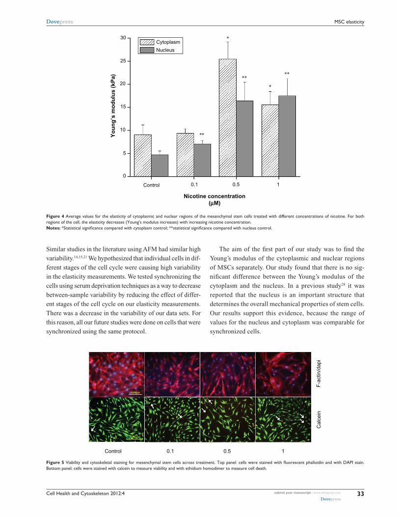
AFM. As the concentration of nicotine increased, the Young’s
modulus, and therefore stiffness, of the MSCs increased, both
for nucleus and cytoplasm values. The Young’s modulus of
the nucleus of cells treated with nicotine was significantly
different from that of the control (4.70 kPa ± 0.83 kPa) for all
concentrations of nicotine, with values of 7.05 kPa ± 0.76 kPa
(P = 0.03), 16.41 kPa ± 4.06 kPa (P = 0.009), and
17.52 kPa ± 3.74 kPa (P = 0.004), respectively. The mea-
sured Young’s modulus for the cytoplasm of cells treated
with nicotine was significantly different from the control
(9.07 kPa ± 2.13 kPa) for the cells treated with 0.5 µM
and 1.0 µM nicotine concentrations only with values at
25.49 kPa ± 3.64 kPa (P , 0.001) and 15.55 kPa ± 2.74 kPa
(P = 0.05), respectively (Figure 4).
Viability assay and cytoskeletal stainingA calcein and ethidium homodimer costain was used at the
end of the nicotine treatments. No difference was noted in
cell death across treatments (Figure 5).
Cytoskeletal staining of f-actin at the end of the treat-
ments revealed the cytoskeletal protein concentration and
arrangement following treatments (Figure 5). We could not
discern any significant difference in f-actin conformation or
concentration across treatments.
DiscussionInitial experiments to measure the elasticity of control
MSCs showed extremely high between-sample variability.
CytoplasmNucleus
30
25
20
15
10
5
0
Unsynchronized Synchronized
Yo
un
g’s
mo
du
lus
(kP
a)
Figure 3 Average values for the elasticity of cytoplasmic and nuclear regions of the mesenchymal stem cells.Note: #Variability was significantly decreased with synchronization, for cytoplasm measurements only.
submit your manuscript | www.dovepress.com
Dovepress
Dovepress
32
Ruiz et al
Cell Health and Cytoskeleton 2012:4
Similar studies in the literature using AFM had similar high
variability.14,15,21 We hypothesized that individual cells in dif-
ferent stages of the cell cycle were causing high variability
in the elasticity measurements. We tested synchronizing the
cells using serum deprivation techniques as a way to decrease
between-sample variability by reducing the effect of differ-
ent stages of the cell cycle on our elasticity measurements.
There was a decrease in the variability of our data sets. For
this reason, all our future studies were done on cells that were
synchronized using the same protocol.
The aim of the first part of our study was to find the
Young’s modulus of the cytoplasmic and nuclear regions
of MSCs separately. Our study found that there is no sig-
nificant difference between the Young’s modulus of the
cytoplasm and the nucleus. In a previous study28 it was
reported that the nucleus is an important structure that
determines the overall mechanical properties of stem cells.
Our results support this evidence, because the range of
values for the nucleus and cytoplasm was comparable for
synchronized cells.
Cytoplasm
Nucleus
30
25
20
15
10
5
0
Nicotine concentration(µM)
Control 0.1 0.5 1
Yo
un
g’s
mo
du
lus
(kP
a) ��
�
��
�
��
Figure 4 Average values for the elasticity of cytoplasmic and nuclear regions of the mesenchymal stem cells treated with different concentrations of nicotine. For both regions of the cell, the elasticity decreases (Young’s modulus increases) with increasing nicotine concentration.Notes: *Statistical significance compared with cytoplasm control; **statistical significance compared with nucleus control.
Control 0.1 0.5
Cal
cein
F-a
ctin
/dap
i
1
100 µM
200 µM
Figure 5 Viability and cytoskeletal staining for mesenchymal stem cells across treatment. Top panel: cells were stained with fluorescent phalloidin and with DAPI stain. Bottom panel: cells were stained with calcein to measure viability and with ethidium homodimer to measure cell death.
submit your manuscript | www.dovepress.com
Dovepress
Dovepress
33
MSC elasticity
Cell Health and Cytoskeleton 2012:4
It is interesting also to note that our values for the Young’s
modulus of MSCs are higher than those of previous studies
that performed similar tests with AFM, which have values
that range from 2.5 kPa to 5.0 kPa1,4,15,21 compared with our
values, which range from 11.9 kPa to 15.4 kPa, depending
on whether cells were measured on the cytoplasm or nucleus.
These differences can most likely be attributed to variations
in methodology. Previous studies have used a spherical tip
5 µ or 10 µ in diameter, thereby limiting the possibility of
accurately determining measurement site within the cell.
We used a more precise pyramidal tip 40 nm in diameter to
enable measurement of the cytoplasm and nucleus separately,
assessed by microscopic observations of tip placement. In
addition, the spring constant of the cantilever used in this
study was significantly lower than that used in previous
studies. A larger spring constant will subsequently result in
a higher degree of cell indentation. Due to these differences
in size and stiffness of the cantilever, our measurements were
most likely more susceptible to local elastic variations in the
cell membrane or cytoskeleton. In addition, previous studies
were performed at a higher speed of indentation. Due to the
intrinsic viscoelastic nature of the MSCs, indentation speed
will affect the final measurements obtained.
Furthermore, all of the studies done on the physical and
mechanical properties of MSCs using AFM yielded higher
Young’s modulus values than studies done using micropi-
pette aspiration, which usually provide values ranging from
0.4 kPa to 0.9 kPa.19,20 Such differences lie in the mechanics
of the test themselves, as AFM studies measure cells attached
to a substrate while micropipette aspiration studies measure
the Young’s modulus of cells that are free floating. Therefore,
micropipette aspiration studies are more representative of
the physical properties of circulating MSCs, while AFM
studies are more representative of the physical properties
of MSCs within their bone marrow niche or after they have
extravasated to sites of injury.
The second goal of these experiments was to quantify
the effect that nicotine, one of the major chemicals found in
cigarette smoke, had on the physical properties of MSCs in
vitro. Our study found that certain levels of nicotine in the
culture medium of MSCs that fall within the range of levels
found in the serum and saliva of smokers29–31 led to MSCs with
higher Young’s moduli, both in the cytoplasmic and nuclear
regions of the cells. Although a nicotine concentration of
0.1 µM did not lead to a significant increase in the Young’s
modulus of these cells, concentrations of 0.5 µM and 1.0 µM
yielded cells with significantly higher Young’s moduli than
the control group, in both cytoplasmic and nuclear regions.
The results of this proof-of-principle study show that
nicotine increases the stiffness of MSCs in vitro in a dose-
dependent manner. The exposure was done in the short time
span of 48 hours. An increase in stiffness due to nicotine
treatment was seen, however. This is relevant for regenerative
medicine applications where MSCs are injected into the blood
or target organs, as studies have shown that MSCs remain in
the body for more than 48 hours after infusion.36 This could
suggest that MSC therapies could be less effective on people
who smoke, as infused MSCs will be exposed to high nico-
tine levels in their blood, limiting their therapeutic potential.
Studies are being performed on longer-term effects of nicotine
exposure, as well as the effect of nicotine in combination with
other smoking derivatives such as benzopyrenes. Further stud-
ies are also needed to discover whether this stiffening effect
by nicotine is a reversible one, as well as measure the Young’s
modulus of different populations of MSCs, including those
circulating in the blood of smokers and nonsmokers.
In summary, we found that there is no significant
d ifference in the stiffness of the nucleus and cytoplasm of
MSCs. However, the data suggested that synchronizing the
cells through serum starvation reduces the between-sample
variability of the data. Moreover, increasing concentration of
nicotine increases the Young’s modulus of both the cytoplasm
and nucleus of MSCs.
DisclosuresThis study was supported in part by a National Institute of
Health Initiative for Maximizing Student Development Grant
(JPR and JD), a VA Merit Review Grant (HSC), and a Senior
VA Research Career Scientist Award (HSC). It was presented
in part at the Biomedical Engineering Society 2010 meeting.
References1. O’Keefe RJ. Summary. Cell therapies for orthopedic applications.
J Musculoskelet Neuronal Interact. 2005;5(4):367–368.2. Erhardt L. Cigarette smoking: an undertreated risk factor for cardiovas-
cular disease. Atherosclerosis. 2009;205(1):23–32.3. Villablanca AC, McDonald JM, Rutledge JC. Clin Chest Med. 2000;
21(1):159–172.4. Benowitz NL. Pharmacology of nicotine: addiction and therapeutics.
Annu Rev Pharmacol Toxicol. 1996;36:597–613.5. Zdravkovic T, Genbacev O, LaRocque N, McMaster M, Fisher S. Human
embryonic stem cells as a model system for studying the effects of smoke exposure on the embryo. Reprod Toxicol. 2008;26:86–93.
6. Wang Y, Pereira EF, Maus AD, et al. Human bronchial epithelial and endothelial cells express alpha7 nicotinic acetylcholine receptors. Mol Pharmacol. 2001;60(6):1201–1209.
7. Ma L, Sham MH, Zheng LW, Cheung LK. Influence of low-dose nicotine on bone healing. J Trauma. 2011;70(6):E117–E121.
8. Zheng LW, Ma L, Cheung LK. Changes in blood perfusion and bone heal-ing induced by nicotine during distraction osteogenesis. Bone. 2008;43: 355–361.
submit your manuscript | www.dovepress.com
Dovepress
Dovepress
34
Ruiz et al

Cell Health and Cytoskeleton
Publish your work in this journal
Submit your manuscript here: http://www.dovepress.com/cell-health-and-cytoskeleton-journal
Cell Health and Cytoskeleton is an international, peer-reviewed open access journal focusing on all aspects of cell structure and function contributing to normal physiology and cell health and exploring the pathogenesis of cell dysfunction leading to adverse conditions and disease in the organism. The journal welcomes papers covering original research,
basic science, reviews and evaluations, guidelines, expert opinion and commentary, case reports and extended reports. The manuscript manage-ment system is completely online and includes a very quick and fair peer-review system, which is all easy to use. Visit http://www.dovepress.com/ testimonials.php to read real quotes from published authors.
Cell Health and Cytoskeleton 2012:4
9. McBeath R, Pirone D, Nelson CM, Bhadriraju K, Chen CS. Cell shape, cytoskeletal tension, and RhoA regulate stem cell lineage commitment. Dev Cell. 2004;6:483–495.
10. Huang CY, Hagar KL, Frost LE, Sun Y, Cheung HS. Effects of cyclic compressive loading on chondrogenesis of rabbit bone-marrow derived mesenchymal stem cells. Stem Cells. 2004;22(3):313–323.
11. Huang CY, Reuben PM, Cheung HS. Temporal expression patterns and corresponding protein inductions of early responsive genes in rabbit bone marrow-derived mesenchymal stem cells under cyclic compressive loading. Stem Cells. 2005;23(8):1113–1121.
12. Pelaez D, Huang CY, Cheung HS. Dynamic compression maintains viability and induces chondrogenesis of human MSC in fibrin gel scaffolds. Stem Cell Dev. 2009;18(1):93–102.
13. Daniels BR, Hale CM, Khatau SB, et al. Differences in the microrhe-ology of human embryonic stem cells and human induced pluripotent stem cells. Biophys J. 2010;99(11):3563–3570.
14. Titushkin I, Cho M. Modulation of cellular mechanics during osteo-genic differentiation of human mesenchymal stem cells. Biophys J. 2007;93(10):3693–3702.
15. Titushkin IA, Cho MR. Controlling cellular biomechanics of human mesenchymal stem cells. Conf Proc IEEE Eng Med Biol Soc. 2009: 2090–2093.
16. Chowdhury F, Na S, Li D, et al. Material properties of the cell dictate stress-induced spreading and differentiation in embryonic stem cells. Nature Mater. 2010;9:82–88.
17. Hammerick KE, Huang Z, Sun N, et al. Elastic properties of induced pluripotent stem cells. Tissue Eng Part A. 2011;17(3–4):495–502.
18. Pan W, Petersen E, Cai N, et al. Viscoelastic properties of human mesenchymal stem cells. Conf Proc IEEE Eng Med Biol Soc. 2005: 4854–4857.
19. Tan SC, Pan WX, Ma G, Cai N, Leong KW, Liao K. Viscoelastic behav-ior of human mesenchymal stem cells. BMC Cell Biol. 2008;9:40.
20. Yu H, Ta CY, Leong WS, Tan SC, Liao K, Tan LP. Mechanical behavior of human mesenchymal stem cells during adipogenic and osteogenic differentiation. Biochem Biophys Res Commun. 2010;393(1):150–155.
21. Darling EM, Zauscher S, Guilak F. Viscoelastic properties of zonal artic-ular chondrocytes measured by atomic force microscopy. Osteoarthritis Cartilage. 2006;14(6):571–579.
22. Chen Q, Xiao P, Chen JN, et al. AFM studies of cellular mechanics during osteogenic differentiation of human amniotic fluid-derived stem cells. Anal Sci. 2010;26(10):1033–1037.
23. Docheva D, Padula D, Popov C, Mutschler W, Clausen-Schaumann H, Schieker M. Researching into the cellular shape, volume and elastic-ity of mesenchymal stem cells, osteoblasts and osteosarcoma cells by atomic force microscopy. J Cell Mol Med. 2008;12(2):537–552.
24. Maloney JM, Nikova D, Lautenschläger F, et al. Mesenchymal stem cell mechanics from the attached to the suspended state. Biophys J. 2010;99(8):2479–2487.
25. Pillarisetti A, Ladjal H, Ferreira A, Keefer C, Desai JP. Mechanical characterization of mouse embryonic stem cells. Conf Proc IEEE Eng Med Biol Soc. 2009:1176–1179.
26. Yim EK, Darling EM, Kulangara K, Guilak F, Leong KW. Nanotopography-induced changes in focal adhesions, cytoskeletal organization, and mechanical properties of human mesenchymal stem cells. Biomaterials. 2010;31(6):1299–1306.
27. Yourek G, Hussain MA, Mao JJ. Cytoskeletal changes of mesenchymal stem cells during differentiation. ASAIO J. 2007;53(2):219–228.
28. Ribeiro AS, Dahl KN. The nucleus as a central structure in defining the mechanical properties of stem cells. Conf Proc IEEE Eng Med Biol Soc. 2010;1:831–834.
29. Giannopoulou C, Geinoz A, Cimasoni G. Effects of nicotine on peri-odontal ligament fibroblasts in vitro. J Clin Periodontal. 1999;26: 49–55.
30. Benowitz NL, Jacob P. Daily intake of nicotine during cigarette smoking. Clin Pharmacol Ther. 1984;35(4):499–504.
31. Brucket E, Jacob N, Lamaire L, Truffert J, Percheron F, Gennes JL. Relationship between smoking status and serum lipids in a hyperlipi-demic populations and analysis of possible confounding factors. Clin Chem. 1992;38(9):1698–1705.
32. Ziebarth NM, Wojcikiewicz EP, Manns F, Moy VT, Parel JM. Atomic force microscopy measurements of lens elasticity in monkey eyes. Mol Vis. 2007;13:504–510.
33. Binnig G, Quate CF, Gerber C. Atomic force microscope. Phys Rev Lett. 1986;56(9):930–933.
34. Tománek D, Overney G, Miyazaki H, Mahanti SD, Güntherodt HJ. Theory for the Atomic Force Microscopy of deformable surfaces. Phys Rev Lett. 1989;63(8):876–879.
35. Bilodeau GG. Regular pyramid punch problem. J Appl Mech. 1992; 59(3):519–524.
36. Gao J, Dennis JE, Muzic RF, Lundberg M, Caplan A. The dynamic in vivo distribution of bone marrow-derived mesenchymal stem cells after infusion. Cells Tissues Organs. 2001;169(1):12–20.
submit your manuscript | www.dovepress.com
Dovepress
Dovepress
Dovepress
35
MSC elasticity
Copyright © 2022 FDOKUMEN