Mesenchymal stem cells derived from vertebrae (vMSCs) show best biological properties
11
HOX and TALE Signatures Specify Human Stromal Stem Cell Populations From Different Sources JACOPO PICCHI, 1 LUISA TROMBI, 2 LAURA SPUGNESI, 1 SERENA BARACHINI, 2 GIORGIA MARONI, 1 GIOVANNI BARBANTI BRODANO, 3 STEFANO BORIANI, 3 MAURO VALTIERI, 4,5 MARIO PETRINI, 2 AND MARIA CRISTINA MAGLI 1 * 1 Institute of Biomedical Technologies, National Research Council (CNR), Pisa, Italy 2 Hematology Division, Department of Oncology, Transplants and New Advances in Medicine, University of Pisa, Pisa, Italy 3 Department of Oncologic and Degenerative Spine Surgery, Rizzoli Institute, Bologna, Italy 4 Department of Hematology, Oncology and Molecular Medicine, Istituto Superiore di Sanita`, Rome, Italy 5 Sbarro Institute for Cancer Research and Molecular Medicine, Center of Biotechnology, College of Science and Technology, Temple University, Philadelphia, Pennsylvania Human stromal stem cell populations reside in different tissues and anatomical sites, however a critical question related to their efficient use in regenerative medicine is whether they exhibit equivalent biological properties. Here, we compared cellular and molecular characteristics of stromal stem cells derived from the bone marrow, at different body sites (iliac crest, sternum, and vertebrae) and other tissues (dental pulp and colon). In particular, we investigated whether homeobox genes of the HOX and TALE subfamilies might provide suitable markers to identify distinct stromal cell populations, as HOX proteins control cell positional identity and, together with their co-factors TALE, are involved in orchestrating differentiation of adult tissues. Our results show that stromal populations from different sources, although immunophenotypically similar, display distinct HOX and TALE signatures, as well as different growth and differentiation abilities. Stromal stem cells from different tissues are characterized by specific HOX profiles, differing in the number and type of active genes, as well as in their level of expression. Conversely, bone marrow-derived cell populations can be essentially distinguished for the expression levels of specific HOX members, strongly suggesting that quantitative differences in HOX activity may be crucial. Taken together, our data indicate that the HOX and TALE profiles provide positional, embryological and hierarchical identity of human stromal stem cells. Furthermore, our data suggest that cell populations derived from different body sites may not represent equivalent cell sources for cell-based therapeutical strategies for regeneration and repair of specific tissues. J. Cell. Physiol. 228: 879–889, 2013. ß 2012 Wiley Periodicals, Inc. Adult tissues harbor stem cells that for their proliferation and differentiation abilities have raised great interest as potential therapeutical tools in regenerative medicine. In recent years, many studies have been focused on mesenchymal stem cells (MSCs), that display biological characteristics particularly suitable for clinical application in a wide spectrum of diseases (Caplan, 2007; Giordano et al., 2007). They can be isolated from different tissues, grown ex vivo, and induced to differentiate in vitro into multiple cell types, including bone, cartilage, fat, and stroma (Prockop, 1997; Pittenger et al., 1999). In addition, MSCs display immunomodulatory properties and secrete bioactive molecules that have both autocrine and paracrine effects (Caplan, 2007; Nauta and Fibbe, 2007; Spaggiari et al., 2008; Zhao et al., 2010). Thus, they can contribute to tissue repair either as multipotent precursors or trophic mediators. MSCs have been originally identified in the bone marrow, however a growing body of evidence indicates that they are present in other tissues or organs, such as teeth, fat, muscle, skin and vessels, and in most fetal and adult tissues (Wagner et al., 2005; da Silva Meirelles et al., 2006; Crisan et al., 2008; Valtieri and Sorrentino, 2008; Huang et al., 2009). Thus, an increasing number of distinct MSC populations has recently been described, some of which have different embryological origin. While MSCs derived from the bone marrow (BM-MSCs) are of mesodermal origin, mesenchymal progenitors identified Abbreviations: BM-MSCs, bone marrow derived mesenchymal stem cells; Ic-MSCs, iliac crest derived mesenchymal stem cells; St-MSCs, sternum derived mesenchymal stem cells; V-MSCs, vertebrae derived mesenchymal stem cells; Co-MSCs, colon derived mesenchymal stem cells; DPSCs, dental pulp stem cells; MPCs, mesodermal progenitor cells. Disclosure statement: No competing financial interests exist.Contract grant sponsor: MIUR-PNR; Contract grant number: RBNEO01R4MJ-002. Contract grant sponsor: CARISBO Fondation; Contract grant number: 2005.9025. Contract grant sponsor: REGIONE EMILIA PRRU 2007-2009; Contract grant number: Area IB ‘‘Regenerative medicine in osteo- articular diseases’’. *Correspondence to: Maria Cristina Magli, Institute of Biomedical Technologies, National Research Council (CNR), Via Moruzzi, 1, 56124 Pisa, Italy. E-mail: [email protected] Manuscript Received: 25 May 2012 Manuscript Accepted: 24 September 2012 Accepted manuscript online in Wiley Online Library (wileyonlinelibrary.com): 27 September 2012. DOI: 10.1002/jcp.24239 ORIGINAL RESEARCH ARTICLE 879 Journal of Journal of Cellular Physiology Cellular Physiology ß 2012 WILEY PERIODICALS, INC.
Transcript of Mesenchymal stem cells derived from vertebrae (vMSCs) show best biological properties

HOX and TALE Signatures SpecifyHuman Stromal Stem CellPopulations From DifferentSourcesJACOPO PICCHI,1 LUISA TROMBI,2 LAURA SPUGNESI,1 SERENA BARACHINI,2
GIORGIA MARONI,1 GIOVANNI BARBANTI BRODANO,3 STEFANO BORIANI,3
MAURO VALTIERI,4,5 MARIO PETRINI,2 AND MARIA CRISTINA MAGLI1*1Institute of Biomedical Technologies, National Research Council (CNR), Pisa, Italy2Hematology Division, Department of Oncology, Transplants and New Advances in Medicine, University of Pisa, Pisa, Italy3Department of Oncologic and Degenerative Spine Surgery, Rizzoli Institute, Bologna, Italy4Department of Hematology, Oncology and Molecular Medicine, Istituto Superiore di Sanita, Rome, Italy5Sbarro Institute for Cancer Research and Molecular Medicine, Center of Biotechnology, College of Science and Technology,
Temple University, Philadelphia, Pennsylvania
Human stromal stem cell populations reside in different tissues and anatomical sites, however a critical question related to their efficientuse in regenerative medicine is whether they exhibit equivalent biological properties. Here, we compared cellular and molecularcharacteristics of stromal stem cells derived from the bonemarrow, at different body sites (iliac crest, sternum, and vertebrae) and othertissues (dental pulp and colon). In particular, we investigated whether homeobox genes of the HOX and TALE subfamilies might providesuitable markers to identify distinct stromal cell populations, as HOX proteins control cell positional identity and, together with theirco-factors TALE, are involved in orchestrating differentiation of adult tissues. Our results show that stromal populations from differentsources, although immunophenotypically similar, display distinctHOX and TALE signatures, as well as different growth and differentiationabilities. Stromal stem cells from different tissues are characterized by specific HOX profiles, differing in the number and type of activegenes, as well as in their level of expression. Conversely, bone marrow-derived cell populations can be essentially distinguished for theexpression levels of specific HOX members, strongly suggesting that quantitative differences in HOX activity may be crucial. Takentogether, our data indicate that the HOX and TALE profiles provide positional, embryological and hierarchical identity of human stromalstem cells. Furthermore, our data suggest that cell populations derived fromdifferent body sitesmay not represent equivalent cell sourcesfor cell-based therapeutical strategies for regeneration and repair of specific tissues.J. Cell. Physiol. 228: 879–889, 2013. � 2012 Wiley Periodicals, Inc.
Adult tissues harbor stem cells that for their proliferation anddifferentiation abilities have raised great interest as potentialtherapeutical tools in regenerative medicine. In recent years,many studies have been focused on mesenchymal stem cells(MSCs), that display biological characteristics particularlysuitable for clinical application in a wide spectrum of diseases(Caplan, 2007;Giordano et al., 2007). They can be isolated fromdifferent tissues, grown ex vivo, and induced to differentiate invitro into multiple cell types, including bone, cartilage, fat, andstroma (Prockop, 1997; Pittenger et al., 1999). In addition,MSCs display immunomodulatory properties and secretebioactive molecules that have both autocrine and paracrineeffects (Caplan, 2007; Nauta and Fibbe, 2007; Spaggiari et al.,2008; Zhao et al., 2010). Thus, they can contribute to tissuerepair either as multipotent precursors or trophic mediators.MSCs have been originally identified in the bone marrow,however a growing body of evidence indicates that they arepresent in other tissues or organs, such as teeth, fat, muscle,skin and vessels, and in most fetal and adult tissues (Wagneret al., 2005; da Silva Meirelles et al., 2006; Crisan et al., 2008;Valtieri and Sorrentino, 2008; Huang et al., 2009). Thus, anincreasing number of distinct MSC populations has recentlybeen described, some of which have different embryologicalorigin.WhileMSCs derived from the bonemarrow (BM-MSCs)are of mesodermal origin, mesenchymal progenitors identified
Abbreviations: BM-MSCs, bone marrow derived mesenchymal stemcells; Ic-MSCs, iliac crest derived mesenchymal stem cells; St-MSCs,sternum derived mesenchymal stem cells; V-MSCs, vertebraederived mesenchymal stem cells; Co-MSCs, colon derivedmesenchymal stem cells; DPSCs, dental pulp stem cells; MPCs,mesodermal progenitor cells.
Disclosure statement: No competing financial interestsexist.Contract grant sponsor: MIUR-PNR;Contract grant number: RBNEO01R4MJ-002.Contract grant sponsor: CARISBO Fondation;Contract grant number: 2005.9025.Contract grant sponsor: REGIONE EMILIA PRRU 2007-2009;Contract grant number: Area IB ‘‘Regenerative medicine in osteo-articular diseases’’.
*Correspondence to: Maria Cristina Magli, Institute of BiomedicalTechnologies, National Research Council (CNR), Via Moruzzi, 1,56124 Pisa, Italy. E-mail: [email protected]
Manuscript Received: 25 May 2012Manuscript Accepted: 24 September 2012
Accepted manuscript online in Wiley Online Library(wileyonlinelibrary.com): 27 September 2012.DOI: 10.1002/jcp.24239
ORIGINAL RESEARCH ARTICLE 879J o u r n a l o fJ o u r n a l o f
CellularPhysiologyCellularPhysiology
� 2 0 1 2 W I L E Y P E R I O D I C A L S , I N C .

in the dental pulp (DPSCs) and the cranio-facial bones developfrom the neural crests (Couly et al., 2002). A relevant questionis whether MSCs of different body sites exhibit equivalentbiological properties and may thus represent equally useful cellsources for regenerative medicine and tissue engineering. It hasbeen reported that MSCs derived from different tissues/organspreferentially differentiate into distinct cell types; for example,while BM-MSCs give rise to progeny of the osteogenic lineage,DPSCs become odontogenic cells (Delorme et al., 2009).However, DPSCs can also contribute to other cell lineagessimilarly to BM-MSCs, although with different potency(Gronthos et al., 2002; Huang et al., 2009).
In addition, other stromal stem cell populations, thatrepresent more primitive cells, have been identified, including amesodermal progenitor cell population (MPC) that can beisolated in the bone marrow and cultured in vitro in thepresence of autologous serum (Petrini et al., 2009). Uponappropriate culture conditions MPCs are able to differentiateinto MSCs and undergo further differentiation into osteoblasts,chondrocytes, or adipocytes; conversely, MSCs cannot beinduced to reverse back to MPCs. Moreover, MPCs display adefined immunophenotype, some embryonic stem cell markersand a differentiation ability not restricted to the solemesenchymal cell lineages (Pacini et al., 2010).
Thus, characterizing stromal stem cell populations derivedfrom different anatomic sites, and understanding the regulatorymechanisms that underlie their properties, is of major clinicalrelevance. Growing evidence indicate that common molecularmechanisms orchestrate embryonic development and adulttissue homeostasis. Interestingly, it has been shown that similarcell types derived from different body sites exhibit differentmolecular signature, particularly with regard to the globalHOXgene family expression profile (HOX code) (Chang et al., 2002).These homeobox-containing genes encode evolutionaryconserved transcription factors that specify cellular positionalidentity during embryonic development (Gehring and Hiromi,1986; Duboule andDolle, 1989; McGinnis and Krumlauf, 1992).The mammalian HOX genes are organized into four clusters(HOX loci) and the chromosomal location of the genes withineach cluster is co-linearwith their patterns of expressionwithinthe body. In addition, the physical order of the genes is reflectedalso in the temporal sequence of their activation (van derHoeven et al., 1996). A number of studies have indicated thatHOX genes are also expressed in adult cells of various tissues,such as blood, muscle, fat, and bone (Magli et al., 1997;Yamamoto et al., 2003; Takahashi et al., 2004; Morgan, 2006;Dutta et al., 2010). Remarkably, HOX proteins in the embryoconfer positional information along the various axes (antero-posterior, proximal-distal, dorso-ventral) and evidencesuggests that they may also play a pivotal role in maintainingpositional identity and in orchestrating adult cell differentiation(Morgan, 2006; Wang et al., 2009). Bone regeneration studiesperformed in murine models have raised the hypothesis thatsuccessful tissue regeneration and repair may depend not onlyon the histologic compatibility between the transplanted stemcells and the injured or damaged tissue, but also on thematchingof their Hox code (Leucht et al., 2008).
In this study we have compared cellular and molecularcharacteristics of human adult MSCs derived from differentbody locations, such as bonemarrow from iliac crest (Ic-MSCs),sternum (St-MSCs) and vertebrae (V-MSCs), as well as colon(Co-MSCs) and dental pulp (DPSCs).
In particular, we have investigated whether HOX genes andtheir TALE (three amino acid loop extension) co-factors mayprovide specific molecular markers for stromal stem cellpopulations derived fromdifferent sources. It is well establishedthat Hox function and DNA-binding specificity is frequentlydependent on interactions with other DNA-binding proteins,which act as co-factors, among which the Pbx and Meis/Prep
classes of homeodomain proteins (TALE) are included (Moensand Selleri, 2006). In addition, to add further complexity, theTALE proteins can act not only as HOX partners, but alsoupstream of HOX genes. TALE genes play multiple key roles inmouse development and ablation of some TALE genes has apleiomorphic, embryonic lethal phenotype, while inactivationof Hox genes results in less severe and more restricted defects(Mallo and Magli, 2006). Furthermore, they can control Hoxexpression both at the transcriptional and translational leveland are involved in normal and leukemic hematopoiesis(Villaescusa et al., 2009; Bach et al., 2010; Shah and Sukumar,2010).
Our results show that cell populations, exhibiting similarimmunophenotypes, display different in vitro growth anddifferentiation properties, and are characterized by distinctHOX codes and TALE expression profiles. Furthermore, ourdata strongly suggest thatmolecular signatures differing only forthe expression levels of specific HOX members may reflectstem cell potency. Taken together, our observations supportthe view that human MSCs derived from different body sitesmay not represent equivalent cell sources to regeneratespecific tissues/organs. These observationsmay have importantclinical implications, suggesting that successful tissueregeneration may depend on the source of MSCs employed forcell therapy or tissue engineering.
Materials and MethodsIsolation and cultures of human stromal cells
Bone marrow samples were either purchased from CambrexPoietics Cell Systems (Gaithersburg, MD) or harvested under localanesthesia from iliac crest, sternum, and vertebrae of differentpatients, undergoing routine orthopedic surgery, followinginformed consent. Primary cultures of bone marrow cells wereestablished as previously described (Petrini et al., 2009), with somemodifications. Briefly, 20ml of bone marrow aspirate werecombined with saline solution (ratio 1:3), gently layered onto adensity gradient (Lymphoprep, Axis-Shields, Oslo, Norway), andcentrifuged at 400g for 25min. The low-density mononuclearfraction was collected, washed twice with saline solution, thenresuspended in standard medium consisting of Dulbecco’smodified Eagle’s medium low-glucose (DMEM, Invitrogen,Manchester, UK), 10% fetal bovine serum (FBS; Invitrogen),100mg/ml gentamicin (Sigma Aldrich, St. Louis, MO), 2mM L-glutamine (Biowhittaker Cambrex, Walkersville, MD), andsubsequently seeded at 2� 105 cells/cm2 in T-75 tissue cultureflask (Corning, Inc., Corning, NY). Cells were grown at 378Cin a humidified atmosphere containing 5% CO2. After 48–72 hnon-adherent cells were discarded and fresh medium was addedthree times a week until 80% confluence was reached.
Alternatively, bone marrow samples were treated for 20min at208C with RosetteSep human MSC enrichment cocktail (StemCellTechnologies, Vancouver, BC, Canada) composed by CD3, CD14,CD19, CD38, CD66b, glycophorin A tetrameric antibody (Ab) toobtain lineage depletion. Cells were then diluted and centrifugedover Ficoll–Hypaque gradient for 25min at 300g and subsequentlywashed, and treated with NH4Cl (StemCell Technologies) for10min on ice to remove residual red blood cells. CD34þ cellswere removed by MACS column (Miltenyi, Bergisch Gladbach,Germany). Cells were then cultured at sub-clonal/low densityconcentration (1–10 cells/cm2) for 3 weeks in a-medium(Invitrogen, Carlsbad, CA), with 20% fetal calf serum (FCS;StemCell Technologies), in T-75 flasks at 378C in 5% CO2
atmosphere. Half medium was replaced twice a week until MSCsreached confluence, defined as passage 0 (P0). At this time theadherent layers were trypsinized and seeded at low density forfurther expansion.
Colon surgical specimens were obtained after informedconsent, thoroughly washed with PBS supplemented with 5�
JOURNAL OF CELLULAR PHYSIOLOGY
880 P I C C H I E T A L .
antibiotic/antimicotic (A/A) solution (Invitrogen), maintained O/Nin PBS 5� A/A at 48C, treated with 30–45ml 1mM EDTA/EGTAPBS for 75min at 208C, vigorously shaken, then processed asdescribed for bone marrow.
Human dental pulp cells were obtained from molars of healthysubjects, after informed consent. Radicular dental pulps wereobtained, using a Gracey curette, from healthy and non-cariousteeth. Pulp tissue explants were placed in tissue flasks containingMinimum Essential Medium alpha modification (a-MEM, SigmaAldrich) supplemented with 20% FBS (Sigma Aldrich), 100 IU/mlpenicillin (Pharmacia & Upjohn SpA, Milano, Italy), and 2mM L-glutamine (Cambrex Bioscience, Inc., Baltimore,MD) in T-25 tissueculture flask (Corning, Inc.) and subsequently cultured as describedfor bone marrow.
For growth curve assays, cumulative population doubling inarbitrary units was calculated according to the following formula:Number of population doublings¼ [log (N/N0)]/log2 where:N¼ number of cells at the end of a period of growth. N0¼ numberof cells plated.
For MPCs cultures, 800,000 cells/cm2 were cultured in DMEMsupplemented with 10% of pooled human AB type serum (PhABS,Lonza,Walkersville,MD) in hydrophobic plastic flasks as previouslyreported (Trombi et al., 2009).
Immunophenotypic analysis
Primary cells were washed with D-PBS supplemented with 0.1%NaN3 (Sigma) and 0.05% bovine serum albumin (BSA; Sigma)and subsequently incubated for 30min at 48C with fluoresceinisothiocyanate (FITC)-conjugated anti-CD105 antibody,phycoerythrin (PE)-conjugated anti-HLA-DR and PE-Cy5-conjugated anti-CD90; all antibodies were purchased from BDBioscience (San Jose, CA). After washing, cells were subjected toflow cytometry analysis using a FACScan flow cytometer (BDBioscience) and CellQuest Analysis Software (BD Bioscience).
MSCs were detached at confluence and characterized asCD105þ CD45� CD34� by triple staining using FITC-conjugatedanti-CD105 antibody, PE-conjugated anti-CD34, and peridininchlorophyll protein-conjugated anti-CD45.
In vitro differentiation assays
MSCs were cultured at passage 2 and grown until 80% confluence.Osteogenic differentiation was induced by culturing 3� 103MSC/cm2 in MSCGMmedium (Cambrex, Poietics Cell Systems) for 24 hat 378C in 5% CO2 atmosphere. MSCGM was replaced withOsteogenesis Induction Medium (Cambrex), and MSCs culturedfor 3 weeks with refeeding every 3–4 days. Von Kossa stainingfor calcium deposition (Sigma) was performed according tomanufacturer’s specifics on differentiated MSCs after 3-weeks ofosteogenic culture (May-Grunwald/Giemsa counterstain).Adipogenic and chondrogenic differentiation were achievedfollowing our previously reported protocols (Sorrentino et al.,2008; Nesti et al., 2011).
Quantitative real-time PCR
Total RNA was isolated from cells with the RNeasy Plus MicroKit (Qiagen, Hilden, Germany). Genomic DNA (gDNA)contamination was excluded by using both a gDNA Eliminatorcolumn and a RNase-free DNase digestion (Qiagen). RNA amountand quality was verified by agarose gel electrophoresis and byreading the OD260/OD280 and OD260/OD230 ratios by using aNanodrop instrument (Thermo Scientific, Wilmington, DE). First-strand cDNA synthesis was performed by using 0.5mg total RNAaccording to the QuantiTect Reverse Transcription Kit (Qiagen)instructions, with oligo-dT and random primers, to allow for highcDNA yield. An identical reaction without the reversetranscriptase was performed, as negative control. PCR assays wereperformed as described by the manufacturer using the QuantiTectSYBR Green PCR Kit (Qiagen) on a Rotor Gene 3000 (Corbett).
The final reaction contained 7.5ml 2� QuantiTect SYBR GreenPCR Master Mix (HotStar Taq DNA Polymerase, QuantiTectSYBR Green PCR Buffer, 5mM MgCl2, dNTP mix, SYBR Green I),0.3mMof each primer and 2ml of cDNA, in a total volume of 15ml.PCR conditions were 958C for 15min, followed by 45 cycles of958C for 15 sec, 50–608C for 30 sec, and 728C for 30 sec.Sequences of the primers used in this study are listed in Table 1.The specificity of each PCR reaction was assessed by performingmelting curve analysis after each reaction, and by running theamplicons on a gel. b-actin and HPRT were used as constitutivegenes to normalize for gene expression levels. All results wererepeated in two independent experiments, and performed induplicate each time. Relative levels of gene expression werecalculated using the efficiency correction method, that calculatesthe relative expression ratio from the real-time PCR efficienciesand the Ct (Pfaffl, 2001).
Cluster analysis
Cluster analysis was performed by the dChip program (Li andWong, 2001). MSCs were hierarchically clustered according to therelative expression ratios of HOX and TALE genes/HPRT gene,following the Pearson correlation test for distance evaluation andcentroid-linkage for dendrogram formation.
Statistical analysis
Results are expressed as mean� SD. The data were analyzed byone-way analysis of variance. Differences between experimentalgroups (MSCs from iliac crest, sternum, vertebrae, colon, anddental pulp) were evaluated by Student’s t-test followed up byBonferroni correction within groups. Statistical analysis wasperformed using Analyse-it software. A value of P< 0.05 wasconsidered statistically significant.
ResultsImmunophenotype and in vitro growth anddifferentiation
MSCs cultures of cells isolated from distinct body districts havebeen FACS-analyzed from passage P0 throughout P12, toensure the maintenance of the initial MSC population. Cellphenotyping included a panel of surface antigens, commonlyused to define mesenchymal stromal cells, and to excludehematopoietic cell markers. Despite their different tissueof origin, all the MSC populations displayed similarimmunophenotypic profiles, being CD45�, CD34�, CD14�
and HLA-DR�, CD90þ, CD105þ, and CD146þ.When we investigated the growth characteristics of MSCs
derived from different body sites, we observed distinct growthkinetics even within bone marrow-derived cell populations.Representative examples are shown in Figure 1. While iliaccrest and sternum-derivedMSCs could bemaintained in culturefor about three months, undergoing a limited expansion,mesenchymal cultures derived from dental pulp and colon grewmuch more rapidly, but could not be maintained longer.Strikingly, MSCs obtained from vertebrae continued topropagate at high rate even after 3.5 months ex vivo.
MSCs from different sources were also induced todifferentiate in vitro into osteogenic, adipogenic, andchondrogenic cells. Results confirmed previous observationsreported by our group, showing that DPSCs and Co-MSCsdisplay a lower ability to generate fat cells (Sorrentino et al.,2008; Nesti et al., 2011; Signore et al., 2012).
Differential expression of HOX and TALE genes inMSCs of different sources
As Hox genes control both cell positional identity anddifferentiation, here we asked whether human MSCs ofdifferent origin exhibit characteristic HOX codes and, using
JOURNAL OF CELLULAR PHYSIOLOGY
H O X / T A L E P R O F I L E S O F H U M A N S T R O M A L S T E M C E L L S 881
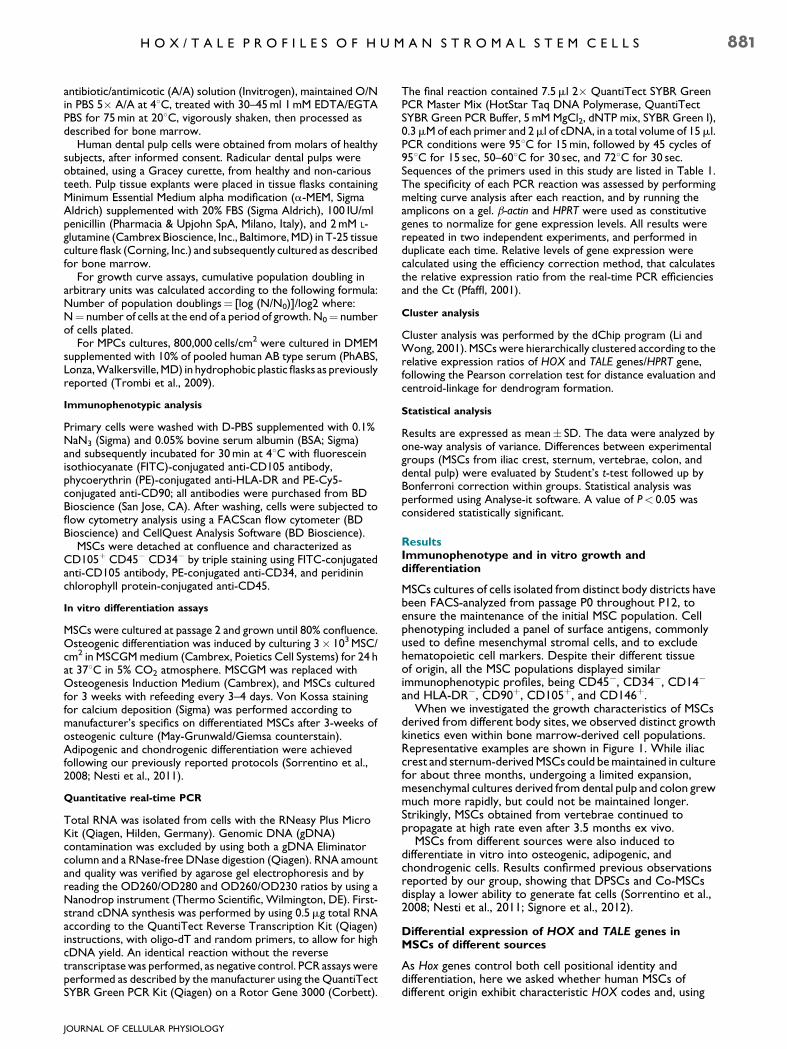
quantitative real-time PCR, we analyzed the expression of the39 HOX genes in our MSC populations at their second passagein culture. In addition, hierarchical cluster analysis was appliedto the average profiling of the MSC populations. Results,illustrated in Figures 2 and 3 highlight several significantdifferences both in the number and type of transcriptionallyactive genes, and in their levels of expression. Figure 2 providesa schematic representation of the HOX status of MSCs fromdifferent sources, shown in detail in Figure 3A. The bonemarrow-derived populations, that is, MSCs grown from iliaccrest, vertebrae, and sternum, exhibit qualitatively identicalprofiles and express the great majority of the HOX network(Fig. 2), although at different levels (Fig. 3A, see below).In contrast, Co-MSCs display a distinct HOX status, mainlycharacterized by the expression of D12, which is instead silentin the otherMSCs (Fig. 2); by the inactivity of virtually thewholeHOXC locus (Figs. 2 and 3A); and by the high expression levels ofseveral HOXA, HOXB, and HOXD cluster genes (Fig. 3A, seebelow). A strikingly different HOX code is found in DPSCs,which have only 5weakly activeHOX genes: A10, B2,C8,C9, andD1 (Figs. 2 and 3A). Therefore, MSCs derived from differentbody sites were highly heterogeneous for the expression levelsof most HOX genes, even within the same tissue, the bonemarrow. In particular, usingHOX gene expression of Ic-MSCs as
internal reference, St-MSCs show higher levels of transcriptionof the 30 genes in the HOXA cluster, significantly A3 (2.37-fold)and A5 (2.14-fold) and some members of the HOXB locus,significantly B6, B7, B8, and B9 (3.24-, 2.72-, 15.45-, and 12-fold,respectively) (Fig. 3A). Interestingly, sternum-derived cellsexpress some genes in the 30 region of theHOXC locus (C4 andC8) at higher levels (4.29- and 2.75-fold, respectively), butthe other half of the cluster at lower levels (mostly C10 andC11, decreased by 9- and 10-fold), apart from C12 and C13,which are virtually inactive (Fig. 3A,C). Moreover, in St-MSCsthe expression of D9 is greatly reduced, as compared to theother cell populations (3-fold decreased), whereasD13 is moreexpressed (2.45-fold). In contrast, an opposite trend wasobserved in V-MSCs which overall exhibited lower levels ofHOX expression, particularly within the HOXA cluster (A1, A6,A7, and A11, decreased by 2- to 6-fold) (Fig. 3A,C). However,mainly B8 and B9 are more transcribed in V-MSCs than in Ic-MSCs (62.7- and 17.2-fold, respectively). In contrast, C10 andC11 aremostly expressed in Ic-MSCs (Fig. 3A,C). Finally, colon-derivedMSCs preferentially express genes located at the 50 endof the HOXA and HOXD loci, including A13 and D13, whoseexpression is 32- and 12-fold higher than in Ic-MSCs, andA11 and D9 (6- and 4.2-fold more transcribed, see Fig. 3B),which are involved in osteogenic differentiation (see below).
TABLE 1. Primer sequences used in quantitative real-time PCR
Forward 50–30 Reverse 50–30
HPRT AGCCAGACTTTGTTGGATTTG TACTAAGCAGATGGCCACAGAb-ACTIN GCACTCTTCCAGCCTTCCTTC GAGCCGCCGATCCACACGRUNX2 CCCAGGCAGTTCCCAAGCATTTC GGTGGCAGGTAGGTGTGGTAGTGALP CAACCCTGGGGAGGAGAC GCATTGGTGTTGTACGTCTTGHOXA1 TCCTGGAATACCCCATACTTAGCA GCCGCCGCAACTGTTGHOXA2 ACAGCGAAGGGAAATGTAAAAGC GGGCCCCAGAGACGCTAAHOXA3 TGCAAAAAGCGACCTACTACGA CGTCGGCGCCCAAAGHOXA4 TCCCCATCTGGACCATAATAGA GCAACCAGCACAGACTCTTAACCHOXA5 TCTCGTTGCCCTAATTCATCTTTT CATTCAGGACAAAGAGATGAACAGAAHOXA6 CCCTCTACCAGGCTGGCTATG CAGGACCGAGTTGGACTGTTGHOXA7 CAAAATGCCGAGCCGACTT TAGCCGGACGCAAAGGGHOXA9 CCGAGAGGCAGGTCAAGATC AAATAAGCCCAAATGGCATCAHOXA10 GAGAGCAGCAAAGCCTCGC CCAGTGTCTGGTGCTTCGTGHOXA11 ACAGGCTTTCGACCAGTTTTTC CCTTCTCGGCGCTCTTGTCHOXA13 AAATGTACTGCCCCAAAGAGCA ATCCGAGGGATGGGAGACCHOXB1 CTCCTCTCCGAGGACAAGGAA CTGTCTTGGGTGGGTTTCTCTTAAHOXB2 TCCTTGGCCGTCTACTGGAA AGTGGATTAAACGCTAATTCAGTAATACCHOXB3 CCTGGCCTGAGAGGTTGCT TCCCGGGCGTGGAATTHOXB4 TTTTCAGCTTTGGCGAAGATG ACCGAGGCCCGTCTTCTCHOXB5 AGCGCCAATTTCACCGAA GGCTGCTTAGCTGGCTTGCHOXB6 AGCAGCCCCCGTTCCA AAAGGAGGAACTGTTGCACGAATHOXB7 TCTTTAATGCTGTCTTTGTGGACTGT GAACACGCGAGTGGTAGGTTTTHOXB8 AACTCACTGTTCTCCAAATACAAAACC GACGGCCCGTGGTAGAACTHOXB9 AGGCCGTGCTGTCTAATCAAA CGAGCGTGCAGCCAGTTHOXB13 CCACTGGCTGCTGGACTGTT TATGACTGGGCCAGGTTCTTTGHOXC4 GGCAGCTACCCCGGGTACT TGTGAGTTATGTTTTATAACCTGGTAATGTCHOXC5 AGGTGCAGGCATCCAGGTACT GGGTTGGCAGCCATGTCTACHOXC6 TCAAACGTGGACCTGAAAGTCA GGGAAAAGGGCCTGTAGACAAHOXC8 CGCACCACGTTCAAGACTTCT TAAGCGAGCACGGGTTCTGHOXC9 GGGCCCATCAGTAACTATTACGTG CGGTGGCCGGAAACCTHOXC10 CCTCGCAATGTAACTCCGAACT ACCCCGCAATTGAAGTCACTHOXC11 GTGAAGGGAAGTGTCTGATGCA AATCCGAGCAGCAAGACATTGHOXC12 TAATCTCCTGAATCCCGGGTTT TGGGTAGGACAGCGAAGGCHOXC13 AAGGTGGTCAGCAAATCGAAAG TGGTACAAAGCGGAGACATAAATAGAHOXD1 CGACCCCCATCCCTATCTAGAC TGGAACTCGGAAGCCAACTAAAHOXD3 CCATAAATCAGCCGCAAGGAT GATGGGTCTCAGACTTACCTTTGGHOXD4 CGGAGGATGAAGTGGAAAAA AGGACGATGACCTGCCTTTAHOXD8 GATAACTTACAGAGACAGCCGATTTTTAC CCAATATTACCACTGGACGATTTACAHOXD9 GGGACGCCTCAAAATGTCTTC GTCGCCCTCATGGCCTATAAHOXD10 ATAAGCGCAACAAACTCATTTCG ATATCCAGGGACGGGAACCTHOXD11 CGGGCTGCGCCTACTATGT AGGACGACGGTTGGGAAAGHOXD12 TGTGTGAGCGCAGTCTCTACAGA CGGCCTCAGGTTGGAGAAGHOXD13 CTGGGCTACGGCTACCACTTC GCGATGACTTGAGCGCATTMEIS1 ATGTGACAATTTCTGCCACCG CCTGAACGAGTAGATGCCGTGMEIS2 GATGAAAGAGACGGCAGCTCC GGGTTGAGGTTGCATCATCGPREP1 AGGGCAAACAGCAGATATTCAGAC ACCAGCCACAAACGCAAACGPBX1 AAGGAGATTGAGCGGATGGTC GGGTAAGGGTTGCTGAGATGGPBX2 CGAACACTCGGACTATCGCAG GCTCCCTCAGCAGGTTCATGPBX3 GAGCTGGCCAAGAAATGCAG GGGCGAATTGGTCTGGTTG
JOURNAL OF CELLULAR PHYSIOLOGY
882 P I C C H I E T A L .

Together with the HOX code, we investigated theexpression patterns of TALE genes in MSCs of various sources.As illustrated in Figure 3B, all TALEmembers are expressed in allthe undifferentiated populations analyzed. Although bonemarrow derived cells (Ic-, St-, and V-MSCs) differentiallyexpress PBX2, PBX3, and MEIS genes, the most dramaticvariations in TALE transcription are found in DPSCs andCo-MSCs. For example,MEISmRNAs are decreased by 10- and50-fold in DPSCs, although in these cells the TALE genes aregenerally rather active (Fig. 3B,D), whereas theHOX network isvirtually silent. In contrast, similarly to what we observed withthe HOX patterns, the strongest TALE signals were detected inCo-MSCs (Fig. 3B,D). Overall, our data show that MSCs ofdifferent sources are characterized by specific HOX and TALEsignatures.
HOX and TALE genes involved in the osteogenicdifferentiation of MSCs from different sources
As a secondary issue, we subsequently addressed the questionof whether MSCs, that exhibit such different HOX codes andTALE profiles, might, following osteogenic induction, modulatethe expression levels of common HOX and/or TALE members.Thus, MSCs derived from different body sites were inducedtowards osteogenic fate and their gene expression andmorphologymonitored at different time points by cytochemicalstaining and quantitative real-time PCR. At 21 days aftertreatment, themajority of the cells were positive for VonKossastaining, as illustrated in Figure 4Aa,b for Ic-MSCs. Molecularanalysis of the osteogenic markers RUNX2 and alkalinephosphatase (ALP) showed an increase in their expression duringthe differentiation process of all theMSCs populations analyzed(Fig. 4Ac,d), with the exception of V-MSCs which alreadyexpress high levels of ALP at the undifferentiated state.
Analysis of HOX profiles showed that during osteogenicdifferentiation of MSCs derived from the bone marrow(Ic-MSCs, St-MSCs, and V-MSCs) the expression of threespecific HOX genes (A10, A11, D9) increases as compared toundifferentiated cells (Fig. 4Ba,b). Furthermore, V-MSCs underthe same conditions up-regulate an additional gene of theHOXD cluster,D10 (Fig. 4Bb). Interestingly,D9 shows themost
significant increase upon osteogenic induction of Co-MSCs atday 7, although they already express relatively high levels ofHOXD genes at an undifferentiated state (Fig. 4Bc). In contrast,DPSCs, although almost HOX negative, when induced towardan osteogenic fate, up-regulate only one HOX member, A10(Fig. 4Bd), barely detectable in the undifferentiated cells. Nochanges were detected in the expression of all the other HOX
Fig. 1. In vitro growth kinetics of MSCs derived from different bodysites. Representative growth curves of MSCs isolated from iliac crestand sternum(Ic/St-MSCs), vertebrae (V-MSCs), dental pulp (DPSCs),and colon (Co-MSCs). For growth curve assay,MSCswere cultured toreach confluence, detached (usually once a week), counted andreplated at the initial plating density. The growth kinetic of a cellpopulation is plotted as the cumulative population doubling (PD)versus days in culture. MSCs derived from different sources exhibitdifferent growth kinetics.
Fig. 2. Schematic representation of specific HOX codes of MSCsderived fromdifferent tissues. Empty boxes represent inactive genes,while active genes are indicated with colored boxes. Iliac crest,sternum, and vertebrae-derived MSCs (BM-MSCs, in yellow), colon-derived MSCs (Co-MSCS, in green), and dental pulp-derived MSCs(DPSCs, in red) are characterized by distinct HOX codes.
JOURNAL OF CELLULAR PHYSIOLOGY
H O X / T A L E P R O F I L E S O F H U M A N S T R O M A L S T E M C E L L S 883

genes between undifferentiated and differentiated cells (datanot shown). Thus, osteogenic differentiation of MSCs fromdifferent body sites is associated with the up-regulation of A10,and, in most cases, of additional members of paralogous groups9–11 in the HOXA and D loci.
A different scenario was observed for TALE genes. Followingosteogenic induction, MSCs from iliac crest and sternum(Fig. 4Ba) showed an increased expression of PBX1 and PBX2. Incontrast, the transcriptional activity of these genes remainedconstant in V-MSCs which, instead, exhibited higher levels ofMEIS1 expression during differentiation (Fig. 4Bb). However, itis in Co-MSCs and DPSCs that commitment toward anosteogenic fate induces the most remarkable changes in TALEexpression. Co-MSCs up-regulate PBX3 at earlier time points,as well as genes of the MEIS/PREP subfamily, such as PREP1(Fig. 4Bc), and DPSCs show a three- to eightfold increasedexpression of PBX1 and MEIS genes (Fig. 4Bd).
These results indicate that osteogenic differentiation ofhuman MSCs from different sources is associated with the up-regulation of commonHOX genes, but different TALEmembers.
HOX and TALE gene expression in mesodermalprogenitor cells (MPCs)
We next compared HOX and TALE signatures in stromal cellpopulations derived from the same anatomic site, but occupyingdifferent positions within stem cells hierarchy. Thus, weanalyzed MPCs and the MSCs they generated, when grownin differentiation conditions (Trombi et al., 2009).Figure 5A shows that MPCs express a significantly lowernumber of HOX genes and, at all times, much lower levelsthan MSCs. All genes of the HOXA cluster, except A13, aretranscribed inMPCs, as well as six out of nineHOXB (B2, B3, B4,B5, B6, and B7) members, although in most cases at barely
Fig. 3. HOX and TALE expression profiles in MSCs derived from different body sites. The relative levels ofHOX (A) and TALE (B) mRNAweredeterminedbyquantitativereal-timePCR,andtheexpressionvalueswerenormalizedusingthehousekeepingHPRTgeneasinternalcontrol.GeneexpressionwasanalyzedinIc-MSCs(nU 6),St-MSCs(nU6),V-MSCs(nU 6),DPSCs(nU 5),andCo-MSCs(nU 4),andresultswererepeatedintwoindependent experiments and performed in duplicate. Results are shown as meanWSD. For statistical analysis, each MSC population wascompared to Ic-MSCs, used as control. MP<0.05; MMP<0.01; MMMP<0.001. Hierarchical clustering of HOX (C) and TALE (D) gene expression inundifferentiatedMSCsofdifferent sources.Theheatmapwasgeneratedbyusing themeanexpression valuesof the real-timePCRdata.Thecolorsaturation indicates the magnitude of the measured expression ratio, being red the highest, and green the lowest. Genes inactive in all the cellpopulations have not been included.
JOURNAL OF CELLULAR PHYSIOLOGY
884 P I C C H I E T A L .

detectable levels. In the HOXC cluster, four genes (C4, C5, C8,and C9) are expressed very weakly in MPCs and the HOXDlocus is completely silent. Therefore, the transition fromMPCsto MSCs is associated with a shift in the HOX code.
As previously observed in MSCs of different sources, alsoMPCs express all TALE members, although PBX1 and both theMEIS genes are very poorly transcribed. In contrast, PBX3 issignificantly more highly expressed in MPCs than in MSCs(Fig. 5B).
Discussion
In this study we investigated specific molecular profiles ofhuman stromal stem cell populations and showed that MSCsderived from different body sites, and displaying similarimmunophenotypes, are characterized by specific HOX codesand TALE signatures. Furthermore, the different levels of HOXexpression detected in stromal cells with different potencystrongly suggest thatHOX genesmay not only reflect positional
Fig. 4. Changes inHOX and TALE gene expression during osteogenic differentiation of distinctMSCs populations. A:Osteogenic differentiationof human MSCs derived from different body sites. Representative Von Kossa staining of (a) untreated Ic-MSCs and (b) differentiated Ic-MSCs.Quantitative real-time PCR of the osteogenic markers RUNX2 (c) and ALP (d). B: MSCs isolated from (a) iliac crest and sternum (Ic/St-MSCs);(b) vertebrae (V-MSCs); (c) colon (Co-MSCs); (d) dental pulp (DPSCs). Gene expression was determined by quantitative real-time PCR, andthe expression values were normalized using the housekeeping HPRT gene as internal control. Cells from each patient were analyzed byquantitative real-time PCR in duplicate in three independent assays. Results are shown as meanWSD. MP<0.05; MMP<0.01; MMMP<0.001.
JOURNAL OF CELLULAR PHYSIOLOGY
H O X / T A L E P R O F I L E S O F H U M A N S T R O M A L S T E M C E L L S 885

and embryological cell identity, but also indicate the cellularposition within the stem cell hierarchy.
A growing body of evidence reports on the proliferative anddifferentiative ability of MSCs derived from different sources,including bone marrow and non-bone marrow tissues. Resultsare often contradictory and the conclusions rathercontroversial, depending on the cell/tissue explantationmethod, the culture conditions, and the assays used in the
different laboratories. Most studies rely on in vitro growth anddifferentiation assays, although one major caveat in theinterpretation of the results is that cultures of MSCs areinherently heterogenous, as they contain multipotent cellscapable of self-renewal, together with committed progenitorsand spontaneously differentiated cells. These cell types mayhave different kinetics, therefore the number of passages inculture reflects the growth of the entire cell population, rather
Fig. 5. Comparison of HOX and TALE gene expression in MPCs and MSCs. HOX (A) and TALE (B) expression levels were determined byquantitative real-time PCR, and the expression values were normalized using the housekeepingHPRT gene as internal control. Gene expressionwas analyzed in MPCs (nU 4) and in the MSCs (nU 4) they generate. Data were obtained in two independent experiments and performed induplicate. Results are shown as meanWSD. MP<0.05; MMP<0.01; MMMP<0.001.
JOURNAL OF CELLULAR PHYSIOLOGY
886 P I C C H I E T A L .
than stem cell proliferation capacity, as extensively discussed byBianco et al. (2008, 2010). Nevertheless, MSCs are commonlyassayed in vitro, on the basis of two main considerations: (i)rigorous, but handy and reproducible in vivo assays are not yetavailable, (ii) MSCs are frequently expanded in vitro prior toclinical use, and therefore characterizing MSCs grown ex vivo isanyway a relevant issue. In this view we have compared the invitro growth and differentiation properties of MSCs fromdifferent anatomical sites. Our results show a heterogeneousbehavior of MSCs even in the context of the same tissue, that is,bone marrow, where Ic-MSCs and St-MSCs display a morelimited growth ability when compared with V-MSCs (Fig. 1).With regard to differentiation ability, recent data from ourgroup confirmed and extended previous studies (Huang et al.,2009), by showing that DPSCs and Co-MSCs, despite a goodproliferation rate, display distinct efficiency in giving rise toadipogenic and chondrogenic cells, although they couldpromptly generate cells of the osteogenic lineage (Sorrentinoet al., 2008; Nesti et al., 2011; Signore et al., 2012). Coherently,stem cells displaying similar immunophenotype, but differentpotency, have been identified also in cord blood (Kogler et al.,2006). Interestingly, a recent report has highlighted that MSCsshow a nonlinear hierarchy of lineage commitment, accountingfor the presence of osteo-adipogenic, osteo-chondrogenic andeven adipo-chondrogenic bipotent progenitors within theMSCs cell heterogeneity (Russell et al., 2010).
HOX and TALE expression signatures in undifferentiatedstromal stem cell populations
In the last decade, several aspects of MSCs have beenextensively investigated and debated to understand whetherMSCs of different sources share identical biological properties.These studies include transcriptional profiles usingdifferentiation, stemness, and signaling pathways (Kultereret al., 2007; Menicanin et al., 2009).We havemainly focused ourattention on the HOX and TALE gene families for severalreasons: (i) they encode transcriptional regulators thatorchestrate embryonic development of multiple tissues andorgans, therefore implying they may be specifying positionalidentities; (ii) they contribute to the control of adult stem andprogenitor cells, such as the hematopoietic ones (Magli et al.,1997; Lappin et al., 2006; Wang et al., 2009; Wheadon et al.,2011) hinting to a potential role in regulating adult MSCs. Ourdata on human adult MSCs support the growing body ofevidence that HOX code provide a ‘‘biological fingerprint’’ todistinguish distinct stem cell populations, as also shown in cordblood and in murine MSCs (Ackema and Charite, 2008; Liedtkeet al., 2010). Here, we specifically found that MSCs fromdifferent tissues, that is, bone marrow, dental pulp, and colonare characterized by specific HOX codes, differing in thenumber and type of active genes, as well as in their level ofexpression. In contrast, MSCs from iliac crest, vertebrae andsternum exhibit qualitatively identical HOX profiles, and theycan be specifically distinguished only for the expression levels ofcertainHOX genes.Despite some intrinsic variability among thesamples explanted from the same anatomical sites,reproducible and highly significant differences were revealed.Overall, all MSCs derived from bone marrow express thehighest number ofHOX genes (35/39), and three of the inactiveHOX members (B1, C12, and D11) are silent also in stem cellsfrom the other sources (Fig. 2). Interestingly, cluster analysishas also highlighted that each bone marrow-derived cellpopulation is characterized by the predominant expression ofspecific sets of genes, frequently adjacent: C10 and C11 in Ic-MSCs, B8 and B9 in V-MSCs, some members of the HOXA (A3and A5),HOXB (B6, B7, and B9) andHOXC (C4 and C8) clustersin St-MSCs (Fig. 3A,C). A different HOX signature identifiesMSCs of colon origin: virtually the entire HOXC cluster is
inactive, as the only two detectable transcripts (C4 and C5) arerather weak. In contrast, the other clusters are expressed athigher levels, as compared to bone-marrow derived MSCs.Remarkably, D12 provides a unique marker for MSCs from thecolon tissue, as it is inactive in the other stromal populationsanalyzed in this study (Fig. 2), as well as in MSCs derived fromadipose tissue or cord blood (Liedtke et al., 2010). Co-MSCspredominantly express genes located at the 50 end of theHOXAand HOXD loci (Fig. 3A), including A13 and D13, barelydetectable in the other cell populations; and A11 and D9, whichare involved in osteogenic differentiation. An opposite trend isobserved in DPSCs, which exhibit extremely low levels ofexpression of a few HOX genes (Fig. 2), confirming previousfindings and in line with the neuroectodermal origin of DPSCs(Couly et al., 2002; D’Anto et al., 2006). It is known that neuralcrests are Hox negative (Couly et al., 2002) and indeed the fewactive HOX genes in DPSCs are expressed at barely detectablelevels (Fig. 3A).
The greatest heterogeneity of HOX profiles among the cellpopulations of different sources was observed in the groupslocated towards the 50 of the clusters. Interestingly, A11,involved in skeletal development (Carapuco et al., 2005; Leuchtet al., 2008), is very weakly expressed in undifferentiated V-MSCs; however it is strongly up-regulated during theirosteogenic differentiation. Intriguingly, in the mouse, Hoxa11,active in skeletal stemcells of the tibia and silent in the same cellsof themandible, seems to be critical for Hoxmatching and boneregeneration (Leucht et al., 2008). V-MSCs express the highestlevels ofHOXB8, which, conversely, is weakly transcribed in theotherMSCpopulations. Interestingly,Hoxb8 is implicated in theexpansion of hematopoietic stem and early progenitor cells(Davidson et al., 2003). Moreover, it is likely involved invertebral development, as its inactivation leads to minorvertebral abnormalities and, conversely, its gain of function canhelp by-pass the block of posterior elongation of axial tissuesobserved in cdxmutant mice (Holstege et al., 2008; Mallo et al.,2010). Thus, it is possible to hypothesize thatHOXB8may play arole also in the regulation of adult V-MSCs, either as amarker ofvertebral identity or as a regulator of MSCs expansion.
A remarkable observation emerging from our data,potentially useful for clinical applications, is that the expressionprofile of a combination of only 6 HOX members (A11, B8, B9,C8,C11, andD12) is diagnostic for cell populations derived fromdifferent sources. In particular, Ic-MSCs are defined by highexpression of C11 and low activity of B8 and B9, whereas St-MSCs are characterized by low expression of C11 and highlevels of B9 and C8; V-MSCs are distinguished for low levels ofA11 and high expression of B8 and B9. Co-MSCs are marked bythe expression of D12 and DPSCs are negative for anycombination of these genes (Fig. 6).
The different HOX signatures exhibited by MSCs derivedfrom bone marrow, dental pulp, and colon are in line with theview that also in adult human cells HOX genes providepositional memory, reflecting their embryonic origin. Actually,Hox expression is conserved along the main body axis, even intissues that have different embryonic origin (Morgan, 2006).Accordingly, Co-MSCs express a large number of HOXmembers, as they derive from a posteriorly located organ. Thedifferent levels of HOX expression detected within bonemarrow-derived populations highlight that quantitativedifferences in HOX activity may be equally important as theirqualitative differences, raising the hypothesis that they mightreflect MSCs potency. Actually, this view might also reconciledistinct HOX functions, such as providing positional,embryological, and hierarchical cell memory. Notably, theimportance of the expression levels has been alreadyemphasized for TALE genes. A significant example is Prep1,which, during mouse embryonic development, is required atdifferent threshold levels for distinct functions (Di Rosa et al.,
JOURNAL OF CELLULAR PHYSIOLOGY
H O X / T A L E P R O F I L E S O F H U M A N S T R O M A L S T E M C E L L S 887

2007). Remarkably, HOX expression profiles of MPCs and theMSCs that they generate, although apparently rather distinct,mainly differ in terms of levels of expression, which aredrastically reduced in MPCs. Transcripts of the HOXA andHOXB clusters, highly represented in MSCs, are still detectablein MPCs, although with very low signals. A similar reduction inthe expression of theHOXC and D clusters leads to a completeblock of the HOX activity. The overall decrease of HOXexpression in MPCs may be accounted for by higher levels ofOCT4 activity. Recent data indicate that this embryonic markermay act as HOX repressors (Schyr et al., 2012) and it is knownthat small variations in OCT4 expression may be sufficientto induce dramatic effects on the gene molecular targets(Lemischka, 2010). This is in line with the very low Hox activityexhibited by ES cells, which, conversely, express high levels ofOct4 (Soshnikova and Duboule, 2008). Alternatively, thesilencing of entire HOX clusters may be accounted for byepigenetic mechanisms or global enhancers, localized outsidethe clusters, which control the expression of severalcontiguous HOX genes (He et al., 2011).
We also comparedMSCsof different sourceswith respect totheir TALE expression profiles. Our data indicate that all TALEgenes, although differentially expressed, are transcriptionallyactive in all the cell populations analyzed, generally at higherlevels than HOX genes. Bone marrow-derived MSCs (Ic-, St-,and V-MSCs) express MEIS1 and MEIS2 at significant higherlevels as compared to the other cell populations, and Co-MSCsexhibit a several fold increased transcription of PBX and MEISgenes. In contrast, MEIS genes are poorly expressed inundifferentiated DPSCs, which, however, display significantlyhigh levels of PBX genes and PREP1, alike bone marrow-derivedcells. Similarly, TALEmRNAs have been detected also in MPCs,which express strikingly high levels of PBX3. As it is known thatPbx1 and Prep1 play important roles in the regulation of HSCs(Di Rosa et al., 2007; Ficara et al., 2008), our results suggest thatTALE genes may also be involved in the control of adult stromalstem cell populations.
Differential expression of HOX and TALE genes duringosteogenic differentiation of MSCs
Although beyond themain question of our study, we addressedwhether MSCs exhibiting differentHOX and TALE signature usea common set of HOX and TALE genes to undertake theosteogenic fate. We observed a significant heterogeneityamong the expression patterns displayed by the MSCs analyzedduring the osteogenetic process (Fig. 4). First, differentiation ofMSCs derived from bone marrow and colon was associatedwith an enhanced expression of at least threeHOX genes (A10,A11, and D9), whereas DPSCs up-regulate only A10. Second,the expression kinetic differs in the various cell populations, asin Ic- and St-MSCs the HOX genes are up-regulated at a ratherlate stage of differentiation (2weeks after osteogenic induction)(Fig. 4Ba), while V-MSCs show significantly enhanced levels ofA11 already at early days of treatment, followed by a furtherincrease of expression at late stages of differentiation (Fig. 4Bb).Moreover, Co-MSCs display an opposite trend showing aninitial peak of expression, followed by a progressive reductionto basal levels (Fig. 4Bc). The different expression kineticsmightreflect the cell heterogeneity of theMSCs cultures derived fromdifferent sources, suggesting, for example, that Co-MSCscontain less primitive cells, consistently with their more limitedpotency (Signore et al., 2012). Third, osteogenic differentiationof MSCs from multiple sources involves the up-regulation of acommon set of HOX genes, but different combinations of TALEmembers: PBX genes for Ic- and St-MSCs, MEIS1 for V-MSCs,PBX3, and MEIS/PREP genes for colon-derived cells. Notably,most TALE genes, particularly PBX1 and MEIS genes aremarkedly up-regulated in DPSCs induced towards anosteogenic fate (Fig. 4Bd), suggesting that threshold levels of atleast some members of the family, such as MEIS1, might berequired for bone cell formation. Future functional studies,using gene silencing or enforced gene expression, will provide ameans to test this hypothesis. Interactions among HOX, PBX,and MEIS homeoproteins have been shown in several systems,including blood, endometrium, and bone (Sarno et al., 2005;Hassan et al., 2007). TheseDNAbinding proteins form dynamicmultimeric complexes that may either activate or represstarget genes, depending on temporal or spatial expression.Further work is needed to gain a better understanding of thedevelopmental processes involved in specification to bone-forming cells.
In conclusion, the distinct HOX and TALE signatures that wedescribed in both undifferentiated and differentiated MSCsfrom various sources indicate that HOX and TALE genes arefinely tuned in adult stem cells and their gene dosage may affecttheir function not only during embryonic development but alsoin adult life.
Clinical implications
Whether MSCs isolated from different tissues share identicalproperties is still a controversial issue. Some reports supportthis view (da Silva Meirelles et al., 2008), whereas other authorsbelieve that bone marrow MSCs present unique properties(Delorme et al., 2009). Our results highlight that MSCs ofdifferent sources display distinct molecular signatures, whichmay be not only biologically, but also clinically relevant. As in themouse system, a mismatch of positional identity can preventgrafted stem cells to participate in regeneration, and, beyondthe HOX code, the pattern of HOX-co-factors is probablyimportant as well, to specify the stromal stem cell populations.Each source of osteogenic human cells has distinct advantages,and bone regeneration at different anatomical sites likelyrequests a specific source. This new perspective may haveimportant implications in spine and dental surgery, for example,where iliac crest cells are currently employed to regenerate
Fig. 6. Minimal HOX signature discriminating MSCs from differentsources. The expression profile of sixHOX genes (A11,B8,B9,C8,C11,D12) is differentially required to identify MSCs from different bodysites. Dental pulp MSCs are negative for all six genes;D12 is a specificmarker for colon derived cells; distinct bonemarrow cell populationsobtained from sternum, vertebrae, and iliac crest are characterizedbyhigh or lowexpression of threeHOXmembers (B9plus two specificgenes).
JOURNAL OF CELLULAR PHYSIOLOGY
888 P I C C H I E T A L .

vertebral or mandibular bone, respectively. Finally, molecularmatching may be a critical prerequisite not only for boneregeneration, but also for exogenous cell therapy of otherdamaged tissues or organs.
Acknowledgments
We would like to thank Dr. E. Levantini and Dr. F. Cremisi fordiscussion and helpful comments on the manuscript. We aregrateful toDr. Paola Italiani andDr. EmiliaMazza for performingCluster Analysis. This study was supported by the MIUR-PNR(to M.C.M.; no. RBNEO01R4MJ-002), CARISBO Fondation(to M.C.M. and G.B.B.; no. 2005.9025), and REGIONE EMILIAPRRU 2007-2009 (to G.B.B.; no. Area IB ‘‘Regenerativemedicine in osteo-articular diseases’’).
Literature Cited
AckemaKB, Charite J. 2008. Mesenchymal stem cells from different organs are characterizedby distinct topographic Hox codes. Stem Cells Dev 17:979–991.
Bach C, Buhl S, Mueller D, Garcia-Cuellar MP, Maethner E, Slany RK. 2010. Leukemogenictransformation by HOXA cluster genes. Blood 115:2910–2918.
Bianco P, Robey PG, Simmons PJ. 2008. Mesenchymal stem cells: Revisiting history, concepts,and assays. Cell Stem Cell 2:313–319.
Bianco P, Robey PG, Saggio I, Riminucci M. 2010. ‘‘Mesenchymal’’ stem cells in human bonemarrow (skeletal stem cells): A critical discussion of their nature, identity, and significancein incurable skeletal disease. Hum Gene Ther 21:1057–1066.
Caplan AI. 2007. Adult mesenchymal stem cells for tissue engineering versus regenerativemedicine. J Cell Physiol 213:341–347.
Carapuco M, Novoa A, Bobola N, Mallo M. 2005. Hox genes specify vertebral types in thepresomitic mesoderm. Genes Dev 19:2116–2121.
ChangHY,Chi JT,Dudoit S, BondreC, van de RijnM, BotsteinD, Brown PO. 2002.Diversity,topographic differentiation, and positional memory in human fibroblasts. Proc Natl AcadSci USA 99:12877–12882.
Couly G, Creuzet S, Bennaceur S, Vincent C, Le Douarin NM. 2002. Interactions betweenHox-negative cephalic neural crest cells and the foregut endoderm in patterning the facialskeleton in the vertebrate head. Development 129:1061–1073.
Crisan M, Yap S, Casteilla L, Chen CW, Corselli M, Park TS, Andriolo G, Sun B, Zheng B,Zhang L, Norotte C, Teng PN, Traas J, Schugar R, Deasy BM, Badylak S, Buhring HJ,Giacobino JP, Lazzari L, Huard J, Peault B. 2008. A perivascular origin for mesenchymalstem cells in multiple human organs. Cell Stem Cell 3:301–313.
da Silva Meirelles L, Chagastelles PC, Nardi NB. 2006. Mesenchymal stem cells reside invirtually all post-natal organs and tissues. J Cell Sci 119:2204–2213.
da Silva Meirelles L, Caplan AI, Nardi NB. 2008. In search of the in vivo identity ofmesenchymal stem cells. Stem Cells 26:2287–2299.
D’Anto V, Cantile M, D’Armiento M, Schiavo G, Spagnuolo G, Terracciano L, Vecchione R,Cillo C. 2006. TheHOXgenes are expressed, in vivo, in human tooth germs: In vitro cAMPexposure of dental pulp cells results in parallel HOX network activation and neuronaldifferentiation. J Cell Biochem 97:836–848.
DavidsonAJ, Ernst P,WangY,DekensMP, Kingsley PD, Palis J, Korsmeyer SJ, DaleyGQ,ZonLI. 2003. cdx4mutants fail to specify blood progenitors and can be rescued bymultiple hoxgenes. Nature 425:300–306.
Delorme B, Ringe J, Pontikoglou C, Gaillard J, Langonne A, Sensebe L, Noel D, Jorgensen C,Haupl T, Charbord P. 2009. Specific lineage-priming of bone marrow mesenchymal stemcells provides the molecular framework for their plasticity. Stem Cells 27:1142–1151.
Di Rosa P, Villaescusa JC, Longobardi E, Iotti G, Ferretti E, Diaz VM,Miccio A, Ferrari G, BlasiF. 2007. The homeodomain transcription factor Prep1 (pKnox1) is required forhematopoietic stem and progenitor cell activity. Dev Biol 311:324–334.
Duboule D, Dolle P. 1989. The structural and functional organization of the murine HOXgene family resembles that of Drosophila homeotic genes. EMBO J 8:1497–1505.
Dutta D, Umashankar M, Lewis EB, Rodrigues V, Vijayraghavan K. 2010. Hox genes regulatemuscle founder cell pattern autonomously and regulate morphogenesis through motorneurons. J Neurogenet 24:95–108.
Ficara F, Murphy MJ, Lin M, Cleary ML. 2008. Pbx1 regulates self-renewal of long-termhematopoietic stem cells by maintaining their quiescence. Cell Stem Cell 2:484–496.
GehringWJ, Hiromi Y. 1986. Homeotic genes and the homeobox. Annu Rev Genet 20:147–173.
Giordano A, Galderisi U, Marino IR. 2007. From the laboratory bench to the patient’sbedside: An update on clinical trials withmesenchymal stem cells. J Cell Physiol 211:27–35.
Gronthos S, Brahim J, LiW, Fisher LW,ChermanN, Boyde A, DenBesten P, Robey PG, Shi S.2002. Stem cell properties of human dental pulp stem cells. J Dent Res 81:531–535.
Hassan MQ, Tare R, Lee SH, Mandeville M,Weiner B, Montecino M, vanWijnen AJ, Stein JL,Stein GS, Lian JB. 2007. HOXA10 controls osteoblastogenesis by directly activating boneregulatory and phenotypic genes. Mol Cell Biol 27:3337–3352.
He H, Hua X, Yan J. 2011. Epigenetic regulations in hematopoietic Hox code. Oncogene30:379–388.
Holstege JC, de Graaff W, Hossaini M, Cano SC, Jaarsma D, van den Akker E, Deschamps J.2008. Loss of Hoxb8 alters spinal dorsal laminae and sensory responses in mice. Proc NatlAcad Sci USA 105:6338–6343.
Huang GT, Gronthos S, Shi S. 2009. Mesenchymal stem cells derived from dental tissues vs.those from other sources: Their biology and role in regenerative medicine. J Dent Res88:792–806.
Kogler G, Sensken S, Wernet P. 2006. Comparative generation and characterization ofpluripotent unrestricted somatic stem cells withmesenchymal stem cells fromhuman cordblood. Exp Hematol 34:1589–1595.
Kulterer B, Friedl G, Jandrositz A, Sanchez-Cabo F, Prokesch A, Paar C, Scheideler M,Windhager R, Preisegger KH, Trajanoski Z. 2007. Gene expression profiling of human
mesenchymal stem cells derived from bone marrow during expansion and osteoblastdifferentiation. BMC Genomics 8:70.
Lappin TR, Grier DG, Thompson A, Halliday HL. 2006. HOX genes: Seductive science,mysterious mechanisms. Ulster Med J 75:23–31.
Lemischka IR. 2010. Hooking up with Oct4. Cell Stem Cell 6:291–292.Leucht P, Kim JB, Amasha R, James AW, Girod S, Helms JA. 2008. Embryonic origin and Hoxstatus determine progenitor cell fate during adult bone regeneration. Development135:2845–2854.
Li C, Wong WH. 2001. Model-based analysis of oligonucleotide arrays: Expression indexcomputation and outlier detection. Proc Natl Acad Sci USA 98:31–36.
Liedtke S, Buchheiser A, Bosch J, Bosse F, Kruse F, Zhao X, Santourlidis S, Kogler G. 2010.The HOX Code as a ‘‘biological fingerprint’’ to distinguish functionally distinct stem cellpopulations derived from cord blood. Stem Cell Res 5:40–50.
Magli MC, Largman C, Lawrence HJ. 1997. Effects of HOX homeobox genes in blood celldifferentiation. J Cell Physiol 173:168–177.
MalloM,Magli MC. 2006. A look at life from the homeodomain.Workshopon homeodomainproteins, hematopoietic development and leukemias. EMBO Rep 7:976–980.
MalloM,WellikDM,Deschamps J. 2010. Hox genes and regional patterning of the vertebratebody plan. Dev Biol 344:7–15.
McGinnis W, Krumlauf R. 1992. Homeobox genes and axial patterning. Cell 68:283–302.Menicanin D, Bartold PM, Zannettino AC, Gronthos S. 2009. Genomic profiling ofmesenchymal stem cells. Stem Cell Rev 5:36–50.
Moens CB, Selleri L. 2006. Hox cofactors in vertebrate development. Dev Biol 291:193–206.Morgan R. 2006. Hox genes: A continuation of embryonic patterning? Trends Genet 22:67–69.
Nauta AJ, Fibbe WE. 2007. Immunomodulatory properties of mesenchymal stromal cells.Blood 110:3499–3506.
NestiC, Pardini C, Barachini S,D’AlessandroD, SicilianoG,Murri L, PetriniM, Vaglini F. 2011.Human dental pulp stem cells protect mouse dopaminergic neurons against MPPþ orrotenone. Brain Res 1367:94–102.
Pacini S, Carnicelli V, Trombi L, Montali M, Fazzi R, Lazzarini E, Giannotti S, Petrini M. 2010.Constitutive expression of pluripotency-associated genes in mesodermal progenitor cells(MPCs). PLoS ONE 5:e9861.
Petrini M, Pacini S, Trombi L, Fazzi R, Montali M, Ikehara S, AbrahamNG. 2009. Identificationand purification of mesodermal progenitor cells from human adult bone marrow. StemCells Dev 18:857–866.
Pfaffl MW. 2001. A new mathematical model for relative quantification in real-time RT-PCR.Nucleic Acids Res 29:e45.
Pittenger MF, Mackay AM, Beck SC, Jaiswal RK, Douglas R, Mosca JD, Moorman MA,Simonetti DW, Craig S, Marshak DR. 1999. Multilineage potential of adult humanmesenchymal stem cells. Science 284:143–147.
Prockop DJ. 1997. Marrow stromal cells as stem cells for nonhematopoietic tissues. Science276:71–74.
Russell KC, Phinney DG, Lacey MR, Barrilleaux BL, Meyertholen KE, O’Connor KC. 2010.In vitro high-capacity assay to quantify the clonal heterogeneity in trilineage potential ofmesenchymal stem cells reveals a complex hierarchy of lineage commitment. Stem Cells28:788–798.
Sarno JL, Kliman HJ, Taylor HS. 2005. HOXA10, Pbx2, and Meis1 protein expression in thehumanendometrium: Formationofmultimeric complexesonHOXA10 target genes. J ClinEndocrinol Metab 90:522–528.
Schyr RB, Shabtai Y, Shashikant CS, Fainsod A. 2012. Cdx1 is essential for the initiation ofHoxC8 expression during early embryogenesis. FASEB J 26:2674–2684.
Shah N, Sukumar S. 2010. The Hox genes and their roles in oncogenesis. Nat Rev Cancer10:361–371.
Signore M, Cerio AM, Boe A, Pagliuca A, Zaottini V, Schiavoni L, Fedele G, Petti S, Navarra S,Ausiello CM, Pelosi E, Fatica A, Sorrentino A, Valtieri M. 2012. Identity and ranking ofcolonic mesenchymal stromal cells. J Cell Physiol 227:3291–3300.
Sorrentino A, Ferracin M, Castelli G, Biffoni M, Tomaselli G, Baiocchi M, Fatica A, Negrini M,Peschle C, Valtieri M. 2008. Isolation and characterization of CD146þ multipotentmesenchymal stromal cells. Exp Hematol 36:1035–1046.
Soshnikova N, Duboule D. 2008. Epigenetic regulation of Hox gene activation: The waltz ofmethyls. BioEssays 30:199–202.
Spaggiari GM, Capobianco A, Abdelrazik H, Becchetti F, Mingari MC, Moretta L. 2008.Mesenchymal stem cells inhibit natural killer-cell proliferation, cytotoxicity, and cytokineproduction: Role of indoleamine 2,3-dioxygenase and prostaglandin E2. Blood 111:1327–1333.
Takahashi Y, Hamada J, Murakawa K, Takada M, Tada M, Nogami I, Hayashi N, Nakamori S,MondenM,MiyamotoM, KatohH,Moriuchi T. 2004. Expression profiles of 39HOXgenesin normal human adult organs and anaplastic thyroid cancer cell lines by quantitative real-time RT-PCR system. Exp Cell Res 293:144–153.
Trombi L, Pacini S, Montali M, Fazzi R, Chiellini F, Ikehara S, Petrini M. 2009. Selective cultureof mesodermal progenitor cells. Stem Cells Dev 18:1227–1234.
Valtieri M, Sorrentino A. 2008. The mesenchymal stromal cell contribution to homeostasis.J Cell Physiol 217:296–300.
van der Hoeven F, Zakany J, Duboule D. 1996. Gene transpositions in the HoxD complexreveal a hierarchy of regulatory controls. Cell 85:1025–1035.
Villaescusa JC, Buratti C, Penkov D, Mathiasen L, Planaguma J, Ferretti E, Blasi F. 2009.Cytoplasmic Prep1 interacts with 4EHP inhibiting Hoxb4 translation. PLoS ONE 4:e5213.
WagnerW,Wein F, Seckinger A, FrankhauserM,Wirkner U, Krause U, Blake J, Schwager C,Eckstein V, AnsorgeW, Ho AD. 2005. Comparative characteristics of mesenchymal stemcells from human bone marrow, adipose tissue, and umbilical cord blood. Exp Hematol33:1402–1416.
Wang KC, Helms JA, Chang HY. 2009. Regeneration, repair and remembering identity: Thethree Rs of Hox gene expression. Trends Cell Biol 19:268–275.
Wheadon H, Ramsey JM, Dobbin E, Dickson GJ, Corrigan PM, Freeburn RW, Thompson A.2011. Differential Hox expression in murine embryonic stem cell models of normal andmalignant hematopoiesis. Stem Cells Dev 20:1465–1476.
Yamamoto M, Takai D, Yamamoto F. 2003. Comprehensive expression profiling of highlyhomologous 39 hox genes in 26 different human adult tissues by the modified systematicmultiplex RT-PCR method reveals tissue-specific expression pattern that suggests animportant role of chromosomal structure in the regulation of hox gene expression in adulttissues. Gene Expr 11:199–210.
Zhao S, Wehner R, Bornhauser M, Wassmuth R, Bachmann M, Schmitz M. 2010.Immunomodulatory properties of mesenchymal stromal cells and their therapeuticconsequences for immune-mediated disorders. Stem Cells Dev 19:607–614.
JOURNAL OF CELLULAR PHYSIOLOGY
H O X / T A L E P R O F I L E S O F H U M A N S T R O M A L S T E M C E L L S 889