The effect of common bacterial growth media on zinc oxide thin films: identification of reaction...
8
The effect of common bacterial growth media on zinc oxide thin films: identification of reaction products and implications for the toxicology of ZnO Sanly Liu,† Elizabeth Killen,† May Lim, * Cindy Gunawan and Rose Amal This study provides a thorough investigation on the effects of the commonly-employed microbial growth medium, namely the peptide-containing luria-bertani broth, tryptic soy broth, the glucose-containing M9 Minimal Salts Media as well as phosphate-buffered saline solution on the dissolution and microstructural transformation of zinc oxide thin film. Morphology and chemical composition of the ZnO film after incubation in the media was thoroughly characterised. In addition, the amount and rate of soluble zinc released by the ZnO thin films was quantified. Exposure of ZnO thin film in the different growth media saw formation of new zinc compounds, resulting from various chemical reactions of zinc with the medium components. Deposition of the new zinc compounds on top of the thin film caused morphological transformation of the film. Zinc leaching was observed in all of the tested media, with significantly higher extent of dissolution observed in peptide-containing organic media, such as luria bertani and tryptic soy broth. Complex organic components, such as amino acids and peptides form complexes with zinc oxide coatings, resulting in complexation-mediated leaching of zinc. Soluble zinc re-precipitates with components in the media, and therefore substantially reduced the amount of dissolved zinc. The results suggest strong influence of solution chemistry on ZnO speciation in a test medium, which have important implications for the mechanistic interpretation of ZnO toxicity. Introduction Metal oxides are currently under investigation as functionalised materials for industrial, medical and chemical applications due to their antibacterial effects. 1,2 Of the metal oxides, zinc oxide has been shown to have an equal or greater antibacterial effect to aluminium, titanium, and tungsten oxides. 3–5 Due to the increased antibacterial effect with decreasing particle size, 6,7 nanoparticulate zinc oxide has attracted attention, demon- strating biocidal effects in nanoparticulate form 8,9 despite having no reported adverse health effects to humans, including no evidence of carcinogenicity, genotoxicity, and reproduction toxicity. 10 Surfaces functionalised with zinc oxide nanoparticles are currently being investigated for their potential to reduce biofouling on metal surfaces, 6 reduce infections due to bacterial contamination of medical implants, 11 and as oxidative photo- catalysts in waste-water treatment. 12 The antibacterial mechanism of zinc oxide in aqueous conditions is debated, with the most widely contested and substantiated theories being cell damage due to the generation of reactive oxygen species through the formation of electron–hole pairs, 13–17 mechanical injury of the target organisms due to nanoparticle internalisation 18 and toxicity due to zinc ions leached from zinc oxide nanoparticles. 19 The lack of consensus on the mechanism of antibacterial action may be due to all three mechanisms contributing to toxicity, with the dominant effect observed being dependent on testing environment. Due to the lack of consensus, tests conducted on zinc oxide materials are hence oen dually aimed at the demonstration of antibacterial qualities and inference of the likely mechanism of toxicity. Tests of zinc oxide's antibacterial efficacy are usually con- ducted on a laboratory scale in a controlled environment using cultures of bacteria, commonly E. coli, 13,20–24 Staphylococcus aureus, 8,25–27 or Pseudomonas aeruginosa 27,28 in growth medium, to allow for results to be obtained quickly and reduce variables. These growth media commonly contain inorganic phosphates, sodium, potassium, and chloride ions, as well as complex organic components including tryptone, yeast extract, soya peptone, and glucose. Zinc oxide has been demonstrated to form precipitates with phosphates, 29,30 complexes with chlo- ride, 31 hydroxide ions, 32 and organic compounds such as citrate 33 and amino acids. 14 Despite the potential for any number of these components to react with the zinc oxide ARC Centre of Excellence for Functional Nanomaterials, School of Chemical Engineering, The University of New South Wales, Sydney, NSW 2052, Australia. E-mail: [email protected] † S. L. and E. K. contributed equally to this work. Cite this: RSC Adv. , 2014, 4, 4363 Received 28th October 2013 Accepted 2nd December 2013 DOI: 10.1039/c3ra46177g www.rsc.org/advances This journal is © The Royal Society of Chemistry 2014 RSC Adv. , 2014, 4, 4363–4370 | 4363 RSC Advances PAPER
Transcript of The effect of common bacterial growth media on zinc oxide thin films: identification of reaction...
RSC Advances
PAPER
ARC Centre of Excellence for Functiona
Engineering, The University of New South
E-mail: [email protected]
† S. L. and E. K. contributed equally to th
Cite this: RSC Adv., 2014, 4, 4363
Received 28th October 2013Accepted 2nd December 2013
DOI: 10.1039/c3ra46177g
www.rsc.org/advances
This journal is © The Royal Society of C
The effect of common bacterial growth media onzinc oxide thin films: identification of reactionproducts and implications for the toxicology ofZnO
Sanly Liu,† Elizabeth Killen,† May Lim,* Cindy Gunawan and Rose Amal
This study provides a thorough investigation on the effects of the commonly-employed microbial growth
medium, namely the peptide-containing luria-bertani broth, tryptic soy broth, the glucose-containing M9
Minimal Salts Media as well as phosphate-buffered saline solution on the dissolution and microstructural
transformation of zinc oxide thin film. Morphology and chemical composition of the ZnO film after
incubation in the media was thoroughly characterised. In addition, the amount and rate of soluble zinc
released by the ZnO thin films was quantified. Exposure of ZnO thin film in the different growth media
saw formation of new zinc compounds, resulting from various chemical reactions of zinc with the
medium components. Deposition of the new zinc compounds on top of the thin film caused
morphological transformation of the film. Zinc leaching was observed in all of the tested media, with
significantly higher extent of dissolution observed in peptide-containing organic media, such as luria
bertani and tryptic soy broth. Complex organic components, such as amino acids and peptides form
complexes with zinc oxide coatings, resulting in complexation-mediated leaching of zinc. Soluble zinc
re-precipitates with components in the media, and therefore substantially reduced the amount of
dissolved zinc. The results suggest strong influence of solution chemistry on ZnO speciation in a test
medium, which have important implications for the mechanistic interpretation of ZnO toxicity.
Introduction
Metal oxides are currently under investigation as functionalisedmaterials for industrial, medical and chemical applications dueto their antibacterial effects.1,2 Of the metal oxides, zinc oxidehas been shown to have an equal or greater antibacterial effectto aluminium, titanium, and tungsten oxides.3–5 Due to theincreased antibacterial effect with decreasing particle size,6,7
nanoparticulate zinc oxide has attracted attention, demon-strating biocidal effects in nanoparticulate form8,9 despitehaving no reported adverse health effects to humans, includingno evidence of carcinogenicity, genotoxicity, and reproductiontoxicity.10 Surfaces functionalised with zinc oxide nanoparticlesare currently being investigated for their potential to reducebiofouling onmetal surfaces,6 reduce infections due to bacterialcontamination of medical implants,11 and as oxidative photo-catalysts in waste-water treatment.12
The antibacterial mechanism of zinc oxide in aqueousconditions is debated, with the most widely contested and
l Nanomaterials, School of Chemical
Wales, Sydney, NSW 2052, Australia.
is work.
hemistry 2014
substantiated theories being cell damage due to the generation ofreactive oxygen species through the formation of electron–holepairs,13–17 mechanical injury of the target organisms due tonanoparticle internalisation18 and toxicity due to zinc ionsleached from zinc oxide nanoparticles.19 The lack of consensuson the mechanism of antibacterial action may be due to all threemechanisms contributing to toxicity, with the dominant effectobserved being dependent on testing environment. Due to thelack of consensus, tests conducted on zinc oxide materials arehence oen dually aimed at the demonstration of antibacterialqualities and inference of the likely mechanism of toxicity.
Tests of zinc oxide's antibacterial efficacy are usually con-ducted on a laboratory scale in a controlled environment usingcultures of bacteria, commonly E. coli,13,20–24 Staphylococcusaureus,8,25–27 or Pseudomonas aeruginosa27,28 in growth medium,to allow for results to be obtained quickly and reduce variables.These growth media commonly contain inorganic phosphates,sodium, potassium, and chloride ions, as well as complexorganic components including tryptone, yeast extract, soyapeptone, and glucose. Zinc oxide has been demonstrated toform precipitates with phosphates,29,30 complexes with chlo-ride,31 hydroxide ions,32 and organic compounds such ascitrate33 and amino acids.14 Despite the potential for anynumber of these components to react with the zinc oxide
RSC Adv., 2014, 4, 4363–4370 | 4363

RSC Advances Paper
nanomaterials, this is rarely accounted for during tests of theantibacterial efficacy of zinc oxide. As the antibacterial mecha-nism of ZnO can be highly dependent on water chemistry andparticle behavior in the media, a thorough characterisation ofthe ZnO nanoparticle and its speciation during antibacterialtesting is essential for proper interpretation of toxicity data. Liet al.34 recently investigated the inuence of medium compo-nents on the toxicity of ZnO nanoparticles to Escherichia coli andfound that generation of precipitate or zinc complexesdramatically decreased the concentration of free Zn2+ ions,resulting in lower toxicity of nanoparticles to the cell.
In this study, we tested the effect of Luria-Bertani (LB) broth,Tryptic Soy Broth (TSB), Phosphate-Buffered Saline (PBS) solu-tion, and M9Minimal Salts media on zinc oxide thin lms, withan aim to understand the reactions, products, and their impli-cations for the testing of antibacterial effects of zinc oxide thinlms. Any change in the morphology and chemical compositionof the ZnO thin lm aer incubation in the media above wasinvestigated. The amount and rate of dissolved zinc released byZnO thin lms in the different media was also quantied.
Experimental proceduresPreparation of zinc oxide thin lms
Round silica slips of 18 mm diameter and 0.16 mm thicknesswere cleaned via immersion in an ultrasonic bath for 10minutes each in dilute nitric acid (HNO3), acetone ((CH3)2CO),and twice in ethanol (CH3CH2OH). The slides were then driedovernight at 110 �C before being used for coating. Zinc acetatedihydrate (Zn(CH3COO)2$2H2O, Analytical, Univar) was addedto ethanol (CH3CH2OH, Absolute, Sigma Aldrich) withmagneticstirring at 500 rpm to make a 1.5 M solution. Monoethanol-amine (MEA) ($99.0%, Sigma Aldrich) was then added drop-wise and the solution was stirred for two hours at 60 �C. Themolar ratio of MEA to zinc acetate was 1 : 1. The resulting solwas ltered through a 0.2 mmmembrane and aged 2 days beforecoating.
Clean glass substrates were attached with double sided tapeto 25 � 25 mm cut glass microscope slides for support. Zinc sol(0.150 mL) was pipetted onto the substrate in the spin coater(Laurel, Model WS-400 BZ-6NPP/Lite). The substrates were spunat 300 rpm for 15 seconds then 5000 rpm for 30 seconds, andplaced on a hotplate to evaporate residual solvent. The coatingprocedure was repeated a total of ve times on each substrate.The spin-coated glass slides were placed in glass Petri dishesand annealed at 550 �C for 3 hours with a heating rate of 7 �Cmin�1. These samples were considered sterile and kept insterile conditions until testing. Quantitative analysis of theamount of zinc oxide coated on each glass substrate was per-formed by digesting the sample in nitric acid, followed by ICP-OES (Perkin Elmer Optima 7300). Approximately 2.5� 0.3 mg ofZnO was coated on each glass substrate.
Characterisation techniques
The morphologies and chemical composition of the thin lmwere characterised using a eld-emission scanning electron
4364 | RSC Adv., 2014, 4, 4363–4370
microscope (FE-SEM) equipped with an energy dispersive X-raydetector. Samples were coated using a Chromium SputterCoater (Emitech K575x) for 30 seconds. The slides were thenimaged using an FEI Nova NanoSEM 230. The spot size wasvaried between 2.0 and 3.0 and the high voltage between 3 and10 kV, dependent on sample charging. Elemental compositionof the samples was analysed using energy-dispersive spectros-copy (EDS) and the data was analysed using Esprit EDS soware.Before EDS analysis, the samples were coated with carbon.
The crystallinity of the samples was characterised with X-raydiffraction. Samples were mounted on a glass slide and imagedwith Philips X'pert Panalytical MRD thin lm system with a1/16� divergence slit and 10 mm mask at 45 kV and 40 mA.Patterns were recorded in the range of 2q ¼ 5� to 75� with a steptime of 50 seconds. Data were analysed with X'pert HighScorePlus soware. The average crystallite size was calculated usingthe Scherrer equation.
Dissolution of zinc oxide coating in different media
Coatings were placed in each well of sterile 12 well plates(Iwaki). Media tested were Tryptic Soy Broth (TSB; Oxoid, 5 g L�1
NaCl, 2.5 g L�1 K2HPO4, 2.5 g L�1 glucose, 3 g L�1 soya peptone,17 g L�1 casein peptone), Phosphate-Buffered Saline solution(PBS; 137 mM NaCl, 10 mM Na2HPO4, 2.0 mM KH2PO4 and2.7 mM KCl, (all reagents Univar, Analytical) (pH 7.2)), Luria-Bertani broth (LB; 10 g L�1 Tryptone (Oxoid), 5 g L�1 Yeastextract (Oxoid), 10 g L�1 NaCl (Univar, Australia)), M9 minimalmedia (M9; 48 mM Na2HPO4, 22 mM KH2PO4, 8.6 mM NaCl,19 mM NH4Cl, 2 mM MgSO4$7H2O, 100 mM CaCl2 and 20 mMglucose (all reagents Univar, Analytical)). A 2 mL volume of themedia being tested was aliquotted into each well, with eachmedia tested in triplicate.
Plates were incubated at 37 �C, shaken at 60 rpm for periodsof 1 day and 1 week. Aer the incubation period, the superna-tant was removed and the amount of soluble zinc ions wasmeasured with ICP-OES (Perkin Elmer OPTIMA 7300). Thebackground level of Zn in each of the media was also measured,and the reported result has been corrected to take this intoaccount. Phosphorus content in the media was also measuredwith ICP-OES (Perkin Elmer OPTIMA 7300). No microbialgrowth was visible over the course of the experiment. Coatingswere washed twice with 1 mL of Milli-Q water, shaken at 60 rpmfor 10 minutes each at 37 �C, and allowed to air dry. Sampleswere then characterised using SEM and XRD.
For the leaching kinetics study, plates with coatings andmedia tested were incubated for periods of 0.5, 1, 1.5, 6 and24 hours, shaken at 60 rpm and 37 �C. Aer incubation, thesupernatant was removed and analysed with ICP-OES analysis.
Results and discussionCharacterisation of as-synthesised zinc oxide thin lms
Sol–gel spin coating synthesis produced consistent lms ofspherical particles and an average particle size of dSEM ¼ 121 �33 nm, as shown below in Fig. 1(a) and (b). The lm was shown
This journal is © The Royal Society of Chemistry 2014

Paper RSC Advances
to be crack free, and composed of a network of dense ZnOaggregates.
The XRD pattern of the as-prepared ZnO thin lm is pre-sented in the topmost spectrum of Fig. 2. All the diffractionpeaks shown were indexed to the hexagonal phase of ZnO(JCPDS 36-1451) with no detection of impurities, which indi-cates that highly crystalline ZnO products were obtained underthe synthesis conditions. XRD analysis shows major peaks at31.8�, 34.4�, and 36.2�, corresponding to the (1 0 0), (0 0 2), and(1 0 1) directions of crystal growth in ZnO. The peaks were ofroughly equivalent height which suggests no preferential crystalgrowth direction in the hexagonal wurtzite structure.
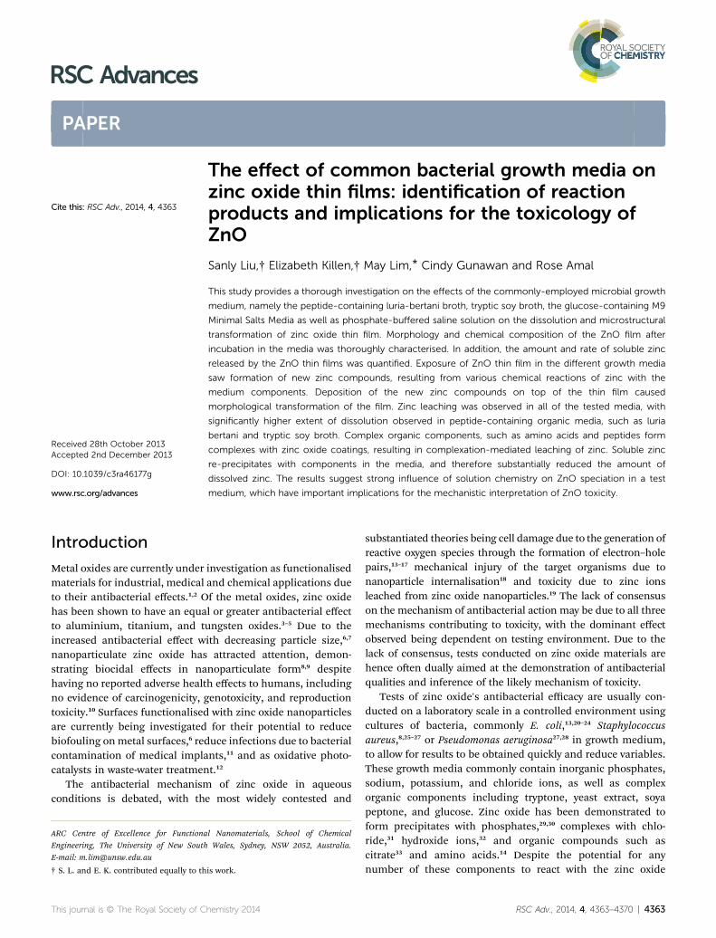
Fig. 1 SEM Images of (a) zinc oxide thin film as synthesised, (b) zincoxide thin film as synthesised with 100k magnification, and thin filmsafter 1 week leaching in different media: (c) Milli-Q water, (d) PBS –phosphate buffered saline, (e) M9 – M9 minimal salts media, (f) TSB –tryptic soy broth, (g) LB – luria bertani broth.
Fig. 2 XRD patterns of zinc oxide thin films (from top to bottom): assynthesised, and after 1 week incubation in Milli-Q water, PBS, M9media, TSB, and LB.
This journal is © The Royal Society of Chemistry 2014
Changes in morphology and chemical composition of ZnOthin lm aer 1 week incubation in the different media
The thin lms were tested for their reactivity in Milli-Q water,Phosphate-Buffered Saline solution (PBS), M9 Minimal SaltsMedia (M9), Tryptic Soy Broth (TSB), and Luria-Bertani broth(LB) for 1 week. Post-exposure crystalline phases were identiedusing XRD, with morphology investigated using SEM andelemental composition analysed using energy-dispersive spec-troscopy (EDS). Fig. 1, 2 and 3 show a collation of resultsobtained from tests of zinc oxide thin lms aer 1 week incu-bation in the different media.
Aer 1 week incubation in Milli-Q water, zinc oxide coatingsdisplayed no-observable structural change (SEM analysis,Fig. 1(c)) with undetected changes in chemical composition(XRD analysis, Fig. 2) compared to that of the as-prepared ZnOcoating. However, according to the literature, the followingreactions most likely occur:35,36
1. Hydrolysis of ZnO in water to form Zn(OH)2 on the surfaceof the coating (eqn (1)). Zn(OH)2 is postulated to have compa-rable physical morphology as that of ZnO, which is notcontradictory to the non-observable structural change of thecoating. The absence of crystalline Zn(OH)2 peaks in the XRD
RSC Adv., 2014, 4, 4363–4370 | 4365

Fig. 3 Typical EDS spectra of zinc oxide thin films after 1 week incu-bation in (from top to bottom): Milli-Q water, PBS, M9 media, TSB, andLB. Carbon signal was from the carbon coating during specimenpreparation.
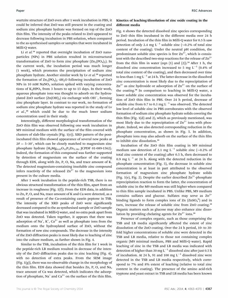
Fig. 4 The concentration of zinc in supernatant over time for mediawith and without complex organic components.
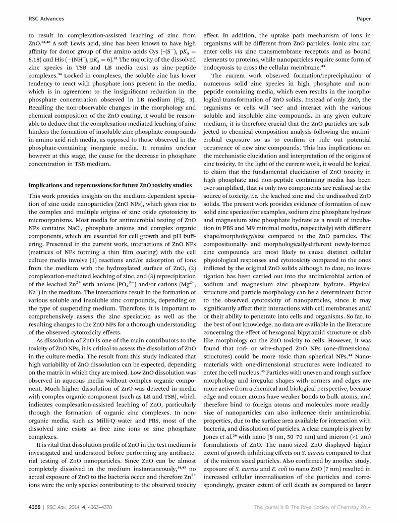
Fig. 5 Concentration of inorganic phosphate over time for thedifferent media.
RSC Advances Paper
spectrum indicates formation of amorphous Zn(OH)2 or alter-natively, the quantity of Zn(OH)2 formed was below the detec-tion limit of the XRD.
ZnO(s) + H2O 4 Zn(OH)2(s) (1)
2. Release of Zn2+ species due to dissociation of Zn(OH)2(eqn (2)), as detected by ICP-OES (Fig. 4).
Zn(OH)2(s) 4 Zn2+(aq) + 2OH�(aq) (2)
Fig. 1(d) shows the SEM image of the ZnO thin lm aer 1week incubation in phosphate buffered saline solution. Thethin lm morphology has been signicantly altered fromuniform spherical structure to hexagonal bipyramids of non-uniform sizes and smooth facets. EDS analysis revealed detec-tion of Zn, P, O, Na, and minor quantity of K on the surface ofthe thin lm. In agreement, the XRD spectrum showed forma-tion of new peaks at 2q values of 9.75, 11.39, 15.29, 16.93, 20.66,22.89, 26.67, 28.61, and 29.20�, which can be matched to the(100), (101), (102), (110), (112), (202), (211), (212), and (114)crystal planes of sodium zinc phosphate hydrate (NaZn-PO4$H2O), respectively (JCPDF 01-089-6669). The results indi-cate that the newly-formed hexagonal bipyramid structure is a
4366 | RSC Adv., 2014, 4, 4363–4370
crystalline sodium zinc phosphate hydrate. Here, the formationof sodium zinc phosphate hydrate is also conrmed by theobserved reduction in aqueous phosphate concentration(Fig. 5). Upon incubation of the ZnO thin lm in PBS solution,aqueous phosphate ions could complex with the released Zn2+
(see eqn (2)); forming zinc–phosphate complexes.37 Reaction ofzinc–phosphate complexes with sodium ions in PBS solutionwas proposed to result in the precipitation of a sodium salt ofthe phosphato–hydroxo–zincate ion, which would reduce theconcentration of dissolved zinc in the media. The diffractionpattern did not contain unidentied peaks, indicating theabsence of unknown phases. In addition, since the XRD spec-trum still displayed the diffraction peaks of the hexagonal
This journal is © The Royal Society of Chemistry 2014
Paper RSC Advances
wurtzite structure of ZnO even aer 1 week incubation in PBS, itcould be inferred that ZnO was still present in the coating andsodium zinc phosphate hydrate was formed on top of the ZnOthin lm. The intensity of the peaks related to ZnO appeared todecrease following incubation in PBS solution, when comparedto the as-synthesised samples or samples that were incubated inMilli-Q water.
Li et al.34 reported that overnight incubation of ZnO nano-particles (NPs) in PBS solution resulted in microstructuraltransformation of ZnO to form zinc phosphate (Zn3(PO4)2). Inthe current work, the incubation period was much longer(1 week), which promotes the precipitation of sodium zincphosphate hydrate. Another similar work by Lv et al.30 reportedthe formation of Zn3(PO4)2$4H2O following incubation of ZnONPs in 10 mM NaNO3 solution spiked with varying concentra-tions of K2HPO4 from 3 hours to up to 15 days. In their work,aqueous phosphate ions was thought to adsorb on the hydrox-ylated ZnO surface (Zn(OH)2) via exchange with OH�, formingzinc phosphate layer. In contrast to our work, no formation ofsodium zinc phosphate hydrate was reported in the study of Lvet al.,30 which could be due to the much lower sodiumconcentration used in their study.
Interestingly, different morphological transformation of theZnO thin lm was observed following one week incubation inM9 minimal medium with the surface of the lm covered withclusters of slab-like crystals (Fig. 1(e)). XRD pattern of the post-incubated thin lm showed appearance of several new peaks at2q ¼ 5–30�, which can be closely matched to magnesium zincphosphate hydrate (H8Mg0.62O12P2Zn2.38, JCPDF 01-089-1562).Indeed, the formation of this compound was further supportedby detection of magnesium on the surface of the coatingthrough EDS, along with Zn, P, O, Na, and trace amount of K.The detected magnesium-containing insoluble zinc compoundinfers reactivity of the released Zn2+ to the magnesium ionspresent in the culture media.
Aer 1 week incubation in the peptide-rich TSB, there is noobvious structural transformation of the thin lm, apart from anincrease in roughness (Fig. 1(f)). From the EDS data, in additionto Zn, P, O, and Na, trace amount of K and Ca were detected, as aresult of presence of the Ca-containing casein peptone in TSB.The intensity of the XRD peaks of ZnO were signicantlydecreased compared to the as-synthesised sample or ZnO samplethat was incubated inMilli-Q water, and no extra peak apart fromZnO was detected. Taken together, it appears that there wasadsorption of Na+, K+, Ca2+ as well as phosphate ions from themedium onto the hydroxylated surface of ZnO, without theformation of new zinc compounds. The decrease in the intensityof the ZnO diffraction peaks is most likely due to leaching of zincinto the culture medium, as further shown in Fig. 4.
Similar to the TSB, incubation of the thin lm for 1 week inthe peptide-rich LB medium resulted in decrease of the inten-sity of the ZnO diffraction peaks due to zinc leaching (Fig. 4),with no detection of extra peaks. From the SEM image(Fig. 1(g)), there was no-observable change in the morphology ofthe thin lm. EDS data showed that besides Zn, P, O, and Na,trace amount of Ca was detected, which indicates the adsorp-tion of phosphate, Na+ and Ca2+ on the surface of the thin lm.
This journal is © The Royal Society of Chemistry 2014
Kinetics of leaching/dissolution of zinc oxide coating in thedifferent media
Fig. 4 shows the detected dissolved zinc species correspondingto ZnO thin lm incubated in the different media over 24 hperiod. Incubation of the thin lm in Milli-Q water for 0.5 h sawdetection of only 2.4 mg L�1 soluble zinc (�0.2% of total zinccontent of the coating). Under the neutral pH condition, thepredominant soluble zinc species is free Zn2+, which is consis-tent with the described two-step reactions for the release of Zn2+
from the thin lm in water (eqn (1) and (2)).34 Aer 1 h, thedissolved zinc concentration increased to 5 mg L�1 (0.4% oftotal zinc content of the coating), and then decreased over timeto less than 1mg L�1 at 24 h. The latter decrease in the dissolvedzinc concentration is most likely due to the reprecipitation ofZn2+ as zinc hydroxide or adsorption of Zn2+ on the surface ofthe coating.38 In comparison to leaching in Milli-Q water, alower soluble zinc concentration was observed for the incuba-tion of ZnO thin lm in PBS. Over 24 h period, decrease ofsoluble zinc from 0.7 to 0.3 mg L�1 was observed. The detectedlow level of soluble zinc in PBS corroborates with the observedformation of sodium zinc phosphate hydrate solids covering thethin lm (Fig. 1(d) and 2), which as previously mentioned, wasmost likely due to the reprecipitation of Zn2+ ions with phos-phate. Indeed, we also detected corresponding reduction in thephosphate concentration, as shown in Fig. 5. In addition,phosphate ions may also adsorb on the surface of the thin lmto inhibit zinc dissolution.34
Incubation of the ZnO thin lm coating in M9 minimalmedium saw detection of 2.1 mg L�1 soluble zinc (�0.2% oftotal zinc content of the coating) aer 0.5 h, then decreased to0.9 mg L�1 at 24 h. Along with the detected reduction in thephosphate concentration (Fig. 5), the decrease in soluble zincconcentration is at least in part attributed to the observedformation of magnesium zinc phosphate hydrate solids(Fig. 1(e), Fig. 2). Despite the earlier described Zn2+-phosphatereprecipitation reaction to form the latter, the concentration ofsoluble zinc in the M9 medium was still higher when comparedto thin lm sample incubated in PBS. Unlike PBS, M9 mediumcontains sulfates and glucose. Sulfates may serve as Zn2+
binding ligands to form complex ions of Zn (ZnSO40) and in
turn, increase the release of soluble zinc from ZnO coating.39
Organic matters such as glucose may also enhance zinc disso-lution by providing chelating agents for Zn2+ ions.39
Presence of complex organic, such as those comprising theTSB and LB media signicantly affected the extent of zincdissolution of the ZnO coating. Over the 24 h period, 10- to 30-fold higher concentrations of soluble zinc were detected in theTSB and LB media, relative to those not containing complexorganic (M9 minimal medium, PBS and Milli-Q water). Rapidleaching of zinc in the TSB and LB media was indicated withdetection of higher than 30mg L�1 dissolved zinc aer just 0.5 hof incubation. At 24 h, 91 and 100 mg L�1 dissolved zinc weredetected in the TSB and LB media respectively, which corre-spond to 7% and 8% extent of leaching (relative to total zinccontent in the coating). The presence of the amino acid-richtryptone and yeast extract in TSB and LBmedia has been known
RSC Adv., 2014, 4, 4363–4370 | 4367
RSC Advances Paper
to result in complexation-assisted leaching of zinc fromZnO.34,40 A so Lewis acid, zinc has been known to have highaffinity for donor group of the amino acids Cys (–(S�), pKa ¼8.18) and His (�(NH+), pKa ¼ 6).41 The majority of the dissolvedzinc species in TSB and LB media exist as zinc–peptidecomplexes.34 Locked in complexes, the soluble zinc has lowertendency to react with phosphate ions present in the media,which is in agreement to the insignicant reduction in thephosphate concentration observed in LB medium (Fig. 5).Recalling the non-observable changes in the morphology andchemical composition of the ZnO coating, it would be reason-able to deduce that the complexation-mediated leaching of zinchinders the formation of insoluble zinc phosphate compoundsin amino acid-rich media, as opposed to those observed in thephosphate-containing inorganic media. It remains unclearhowever at this stage, the cause for the decrease in phosphateconcentration in TSB medium.
Implications and repercussions for future ZnO toxicity studies
This work provides insights on the medium-dependent specia-tion of zinc oxide nanoparticles (ZnO NPs), which gives rise tothe complex and multiple origins of zinc oxide cytotoxicity tomicroorganisms. Most media for antimicrobial testing of ZnONPs contains NaCl, phosphate anions and complex organiccomponents, which are essential for cell growth and pH buff-ering. Presented in the current work, interactions of ZnO NPs(matrices of NPs forming a thin lm coating) with the cellculture media involve (1) reactions and/or adsorption of ionsfrom the medium with the hydroxylated surface of ZnO, (2)complexation-mediated leaching of zinc, and (3) reprecipitationof the leached Zn2+ with anions (PO4
3�) and/or cations (Mg2+,Na+) in the medium. The interactions result in the formation ofvarious soluble and insoluble zinc compounds, depending onthe type of suspending medium. Therefore, it is important tocomprehensively assess the zinc speciation as well as theresulting changes to the ZnO NPs for a thorough understandingof the observed cytotoxicity effects.
As dissolution of ZnO is one of the main contributors to thetoxicity of ZnO NPs, it is critical to assess the dissolution of ZnOin the culture media. The result from this study indicated thathigh variability of ZnO dissolution can be expected, dependingon the matrix in which they are mixed. Low ZnO dissolution wasobserved in aqueous media without complex organic compo-nent. Much higher dissolution of ZnO was detected in mediawith complex organic component (such as LB and TSB), whichindicates complexation-assisted leaching of ZnO, particularlythrough the formation of organic zinc complexes. In non-organic media, such as Milli-Q water and PBS, most of thedissolved zinc exists as free zinc ions or zinc phosphatecomplexes.
It is vital that dissolution prole of ZnO in the test medium isinvestigated and understood before performing any antibacte-rial testing of ZnO nanoparticles. Since ZnO can be almostcompletely dissolved in the medium instantaneously,34,42 noactual exposure of ZnO to the bacteria occur and therefore Zn2+
ions were the only species contributing to the observed toxicity
4368 | RSC Adv., 2014, 4, 4363–4370
effect. In addition, the uptake path mechanism of ions inorganisms will be different from ZnO particles. Ionic zinc canenter cells via zinc transmembrane receptors and as boundelements to proteins, while nanoparticles require some form ofendocytosis to cross the cellular membrane.43
The current work observed formation/reprecipitation ofnumerous solid zinc species in high phosphate and non-peptide containing media, which even results in the morpho-logical transformation of ZnO solids. Instead of only ZnO, theorganisms or cells will ‘see’ and interact with the varioussoluble and insoluble zinc compounds. In any given culturemedium, it is therefore crucial that the ZnO particles are sub-jected to chemical composition analysis following the antimi-crobial exposure so as to conrm or rule out potentialoccurrence of new zinc compounds. This has implications onthe mechanistic elucidation and interpretation of the origins ofzinc toxicity. In the light of the current work, it would be logicalto claim that the fundamental elucidation of ZnO toxicity inhigh phosphate and non-peptide containing media has beenover-simplied, that is only two components are realised as thesource of toxicity, i.e. the leached zinc and the undissolved ZnOsolids. The present work provides evidence of formation of newsolid zinc species (for examples, sodium zinc phosphate hydrateand magnesium zinc phosphate hydrate as a result of incuba-tion in PBS and M9 minimal media, respectively) with differentshape/morphology/size compared to the ZnO particles. Thecompositionally- and morphologically-different newly-formedzinc compounds are most likely to cause distinct cellularphysiological responses and cytotoxicity compared to the onesinicted by the original ZnO solids although to date, no inves-tigation has been carried out into the antimicrobial action ofsodium and magnesium zinc phosphate hydrate. Physicalstructure and particle morphology can be a determinant factorto the observed cytotoxicity of nanoparticles, since it maysignicantly affect their interactions with cell membranes and/or their ability to penetrate into cells and organisms. So far, tothe best of our knowledge, no data are available in the literatureconcerning the effect of hexagonal bipyramid structure or slablike morphology on the ZnO toxicity to cells. However, it wasfound that rod- or wire-shaped ZnO NPs (one-dimensionalstructures) could be more toxic than spherical NPs.44 Nano-materials with one-dimensional structures were indicated toenter the cell nucleus.45 Particles with uneven and rough surfacemorphology and irregular shapes with corners and edges aremore active from a chemical and biological perspective, becauseedge and corner atoms have weaker bonds to bulk atoms, andtherefore bind to foreign atoms and molecules more readily.Size of nanoparticles can also inuence their antimicrobialproperties, due to the surface area available for interaction withbacteria, and dissolution of particles. A clear example is given byJones et al.26 with nano (8 nm, 50–70 nm) and micron (>1 mm)formulations of ZnO. The nano-sized ZnO displayed higherextent of growth inhibiting effects on S. aureus compared to thatof the micron sized particles. Also conrmed by another study,exposure of S. aureus and E. coli to nano ZnO (7 nm) resulted inincreased cellular internalisation of the particles and corre-spondingly, greater extent of cell death as compared to larger
This journal is © The Royal Society of Chemistry 2014
Paper RSC Advances
particles (260 and 800 nm).8 The zinc reprecipitation to formparticles of different shape/morphology/size can complicatethe interpretation of cytotoxicity effect of ZnO towardsmicroorganisms.
Upon exposure to peptide-containing organic media (LBand TSB), the non-observable or minimal changes on themorphology and chemical composition of the zinc solids infersvalid interpretation of the cytotoxicity origins from the leachedsoluble zinc and the undissolved ZnO solids. In those media,the leached zinc exists mostly as zinc–peptide complexes,passivating the tendency to form insoluble zinc solids in thepresence of phosphate.
In many studies, a simple zinc salt system (e.g. ZnCl2, orZnSO4) containing equimolar concentration of zinc has beenemployed to simulate the activity of leached soluble zinc fromZnO NPs. The approach is even used to simulate the activity ofthe overall presence of ZnO NPs, or in other words the leachedzinc and the undissolved ZnO solids altogether. If there werecomparable toxicity effects from the ZnO nanoparticle and thezinc salt at the same Zn2+ concentration as the soluble Zndetected from ZnO dissolution, it could be concluded thatsoluble zinc plays a dominant role in ZnO toxicity. As elucidatedin the current study however, presence of zinc ions in growthmedia without complex organic (peptide), which usuallycontains phosphate anions, may result in the formation ofinsoluble zinc species, such as zinc phosphate. The re-precipi-tation effects are enhanced in culture media without complexorganic (peptide), in which the ZnO NPs-derived soluble zincspecies are not locked in complexes, with signicant fractionexisting as free zinc ions.19,34 Such unintended formation ofinsoluble zinc species will lower not only the NPs-leached Zn2+
concentration, but also the dosed Zn2+ concentration from zincsalt. This will add an additional layer of complexity to theevaluation of the relative toxicity of ZnO nanoparticlescompared to that of the zinc salt control. In such cases, the“soluble zinc” system is in fact containing soluble species withlower Zn2+ concentration than initially dosed and zinc con-taining particulates. Although the initial forms of zinc in theculture medium were well-dened (i.e. Zn2+ and Cl� for ZnCl2,or Zn2+ and SO4
2� for ZnSO4), the chemical transformations ofzinc species must be considered when interpreting bacterialinactivation results.
Conclusions
In conclusion, the work investigates the changes in zinc oxidethin lms aer incubation in commonly used aqueous media,more specically in identifying the formation of newcompounds as a result of reaction with components in themedia and comparing the dissolution of ZnO in the differentmedia. Common bacterial growth media contain manycomponents, which can inuence speciation and dissolution ofZnO, and ultimately, the toxicity of ZnO. The dissolution testresults revealed that the amount of soluble zinc was controlledby water chemistry. Complex organic components in media,such as amino acids and peptides can form complexes with zincoxide coatings, resulting in complexation-mediated leaching of
This journal is © The Royal Society of Chemistry 2014
ZnO. Zinc oxide reacts with components in media withoutcomplex organic, forming precipitates containing phosphates,sodium or magnesium. The formation of precipitates besidesZnO inmedia without complex organic calls for reconsiderationand re-interpretation of past literature on ZnO toxicity usingthis media. Considering the dramatic difference of the specia-tion of ZnO in various aqueous media, future nanotoxicityevaluations should pay more attention on the dynamic trans-formations of ZnO to be able to properly interpret its toxicitymechanisms. Elucidation of origins of toxicity of ZnO will leadto better assessment of its impact to the environment andhuman exposure.
Acknowledgements
This research was supported under Australian Research Coun-cil's Linkage Projects funding scheme (LP110100459). The viewsexpressed herein are those of the authors and are not neces-sarily those of the Australian Research Council. We alsoacknowledge funding from Water Corporation and in-kindcontribution from Australian Water Quality Centre.
Notes and references
1 A. J. Huh and Y. J. Kwon, J. Controlled Release, 2011, 156, 128–145.
2 Q. Li, S. Mahendra, D. Y. Lyon, L. Brunet, M. V. Liga, D. Liand P. J. J. Alvarez, Water Res., 2008, 42, 4591–4602.
3 W. Jiang, H. Mashayekhi and B. Xing, Environ. Pollut., 2009,157, 1619–1625.
4 K. Memarzadeh, M. Vargas, J. Huang, J. Fan and R. Allaker,Key Eng. Mater., 2012, 493, 489–494.
5 A. Kumar, A. K. Pandey, S. S. Singh, R. Shanker andA. Dhawan, Chemosphere, 2011, 83, 1124–1132.
6 J. T. Seil and T. J. Webster, Acta Biomater., 2011, 7, 2579–2584.
7 O. Yamamoto, Int. J. Inorg. Mater., 2001, 3, 643–646.8 G. Applerot, A. Lipovsky, R. Dror, N. Perkas, Y. Nitzan,R. Lubart and A. Gedanken, Adv. Funct. Mater., 2009, 19,842–852.
9 R. Brayner, R. Ferrari-Iliou, N. Brivois, S. Djediat,M. Benedetti and F. Fievet, Nano Lett., 2006, 6, 866–870.
10 A. Moezzi, A. M. McDonagh and M. B. Cortie, Chem. Eng. J.,2012, 185, 1–22.
11 G. Colon, B. C. Ward and T. J. Webster, J. Biomed. Mater. Res.,Part A, 2006, 78, 595–604.
12 S. H. S. Chan, T. Yeong Wu, J. C. Juan and C. Y. Teh, J. Chem.Technol. Biotechnol., 2011, 86, 1130–1158.
13 C. Karunakaran, V. Rajeswari and P. Gomathisankar, J. AlloysCompd., 2010, 508, 587–591.
14 Y. Li, W. Zhang, J. Niu and Y. Chen, ACS Nano, 2012, 6, 5164–5173.
15 R. Gopikrishnan, K. Zhang, P. Ravichandran, S. Biradar,V. Ramesh, V. Goornavar, R. B. Jeffers, A. Pradhan,J. C. Hall and S. Baluchamy, J. Mater. Sci.: Mater. Med.,2011, 22, 2301–2309.
RSC Adv., 2014, 4, 4363–4370 | 4369

RSC Advances Paper
16 Y. Xie, Y. He, P. L. Irwin, T. Jin and X. Shi, Appl. Environ.Microbiol., 2011, 77, 2325–2331.
17 D. Sharma, S. Sharma, B. Kaith, J. Rajput and M. Kaur, Appl.Surf. Sci., 2011, 257, 9661–9672.
18 R. Wahab, A. Mishra, S.-I. Yun, Y.-S. Kim and H.-S. Shin,Appl. Microbiol. Biotechnol., 2010, 87, 1917–1925.
19 N. M. Franklin, N. J. Rogers, S. C. Apte, G. E. Batley,G. E. Gadd and P. S. Casey, Environ. Sci. Technol., 2007, 41,8484–8490.
20 A. Kumar, A. K. Pandey, S. S. Singh, R. Shanker andA. Dhawan, Free Radical Biol. Med., 2011, 51, 1872–1881.
21 R. Dutta, B. P. Nenavathu, M. K. Gangishetty and A. Reddy,Colloids Surf., B, 2012, 94, 143–150.
22 K. Ghule, A. V. Ghule, B. J. Chen and Y. C. Ling, Green Chem.,2006, 8, 1034–1041.
23 S. D. Gittard, J. R. Perfect, N. A. Monteiro-Riviere, W. Wei,C. Jin and R. J. Narayan, Appl. Surf. Sci., 2009, 255, 5806–5811.
24 M. Gondal, M. Dastageer, A. Khalil, K. Hayat and Z. Yamani,J. Nanopart. Res., 2011, 13, 3423–3430.
25 M. A. Ansari, H. M. Khan, A. A. Khan, A. Sultan and A. Azam,World J. Microbiol. Biotechnol., 2012, 1–9.
26 N. Jones, B. Ray, K. T. Ranjit and A. C. Manna, FEMSMicrobiol. Lett., 2008, 279, 71–76.
27 P. Sivakumar, S. Balaji, V. Prabhawathi, R. Neelakandan,P. Manoharan and M. Doble, Carbohydr. Polym., 2010, 79,717–723.
28 C. Jayaseelan, A. A. Rahuman, A. V. Kirthi, S. Marimuthu,T. Santhoshkumar, A. Bagavan, K. Gaurav, L. Karthik andK. Rao, Spectrochim. Acta, Part A, 2012, 90, 78–84.
29 B. Boonchom, R. Baitahe, S. Kongtaweelert andN. Vittayakorn, Ind. Eng. Chem. Res., 2010, 49, 3571–3576.
4370 | RSC Adv., 2014, 4, 4363–4370
30 J. Lv, S. Zhang, L. Luo, W. Han, J. Zhang, K. Yang andP. Christie, Environ. Sci. Technol., 2012, 46, 7215–7221.
31 A. Goux, T. Pauporte, J. Chivot and D. Lincot, Electrochim.Acta, 2005, 50, 2239–2248.
32 A. Zirino and S. Yamamoto, Limnol. Oceanogr., 1972, 661–671.33 I. A. Mudunkotuwa, T. Rupasinghe, C. M. Wu and
V. H. Grassian, Langmuir, 2011, 28, 396–403.34 M. Li, L. Zhu and D. Lin, Environ. Sci. Technol., 2011, 45,
1977–1983.35 J. Han, W. Qiu andW. Gao, J. Hazard. Mater., 2010, 178, 115–
122.36 A. Degen and M. Kosec, J. Eur. Ceram. Soc., 2000, 20, 667–
673.37 S. Ziemniak, M. Jones and K. Combs, J. Solution Chem., 1992,
21, 1153–1176.38 S. Mustafa, P. Shahida, A. Naeem, B. Dilara and N. Rehana,
Langmuir, 2002, 18, 2254–2259.39 H. Ma, P. L. Williams and S. A. Diamond, Environ. Pollut.,
2013, 172, 76–85.40 C. Gunawan, W. Y. Teoh, Ricardo, C. P. Marquis and
R. Amal, Part. Part. Syst. Charact., 2013, 30, 375–380.41 S. J. Lippard and J. M. Berg, Principles of Bioinorganic
Chemistry, University Science Books, Mill Valley, CA, 1994.42 R. B. Reed, D. A. Ladner, C. P. Higgins, P. Westerhoff and
J. F. Ranville, Environ. Toxicol. Chem., 2012, 31, 93–99.43 T. W. Turney, M. B. Duriska, V. N. Jayaratne, A. Elbaz,
S. J. O'Keefe, A. S. Hastings, T. J. Piva, P. Wright andB. N. Feltis, Chem. Res. Toxicol., 2012, 25, 2057–2066.
44 X. Peng, S. Palma, N. S. Fisher and S. S. Wong, Aquat.Toxicol., 2011, 102, 186–196.
45 H. Yang, C. Liu, D. Yang, H. Zhang and Z. Xi, J. Appl. Toxicol.,2008, 29, 69–78.
This journal is © The Royal Society of Chemistry 2014