The early steps of glucose signalling in yeast
32
REVIEW ARTICLE The early steps of glucose signalling in yeast Juana M. Gancedo Department of Metabolism and Cell Signalling, Instituto de Investigaciones Biom ´ edicas Alberto Sols, CSIC-UAM, Madrid, Spain Correspondence: Juana M. Gancedo, Department of Metabolism and Cell Signalling, Instituto de Investigaciones Biom ´ edicas Alberto Sols, CSIC-UAM, Madrid, Spain. Tel: 134 91 585 4433; fax: 134 91 585 4401; e-mail: [email protected] Received 29 October 2007; revised 25 March 2008; accepted 22 April 2008. First published online June 2008. DOI:10.1111/j.1574-6976.2008.00117.x Editor: Martin Kupiec Keywords glucose signalling; sensing elements; yeast. Abstract In the presence of glucose, yeast undergoes an important remodelling of its metabolism. There are changes in the concentration of intracellular metabolites and in the stability of proteins and mRNAs; modifications occur in the activity of enzymes as well as in the rate of transcription of a large number of genes, some of the genes being induced while others are repressed. Diverse combinations of input signals are required for glucose regulation of gene expression and of other cellular processes. This review focuses on the early elements in glucose signalling and discusses their relevance for the regulation of specific processes. Glucose sensing involves the plasma membrane proteins Snf3, Rgt2 and Gpr1 and the glucose- phosphorylating enzyme Hxk2, as well as other regulatory elements whose functions are still incompletely understood. The similarities and differences in the way in which yeasts and mammalian cells respond to glucose are also examined. It is shown that in Saccharomyces cerevisiae, sensing systems for other nutrients share some of the characteristics of the glucose-sensing pathways. Introduction Adaptation to changes in the environment is a key process for the successful survival of organisms. The response of the cells to these changes is mediated by a large variety of signalling pathways that may be sorted into a few basic types, among which two are prominent. In one of them the first element in the pathway is a protein in the plasma membrane, able to bind a nutrient or a hormone and to adopt a new conforma- tion that activates a cascade of reactions, affecting metabolic or regulatory enzymes, transcription factors, etc. (Forsberg & Ljungdahl, 2001a; Kroeze et al ., 2003; Holsbeeks et al., 2004; Mascher et al., 2006; Monge et al ., 2006; Hao et al., 2007). The second type of pathway depends on the uptake of the nutrient and, in most cases, on its metabolism, which causes changes in the concentration of intracellular metabolites with a regulatory function. The metabolites may, in turn, interact with different kinds of proteins and modify their activity, their stability and/or their localization. The regulated proteins can have a variety of functions: they may have enzymatic activity, they may bind other proteins and they are often able to bind specific DNA regions and to modulate the transcription rate of the corresponding genes (Sellick & Reece, 2005). An interesting example of the response of a cell to a change in the environment is the response of Saccharomyces cerevisiae to the presence of glucose. The mechanisms involved are particularly complex, as this sugar is not only a preferential carbon source for the organism but also a molecule that affects yeast physiology at many levels. In the presence of glucose, there are changes in the concentration of intracellular metabolites (Kresnowati et al., 2006), modifications and eventually degradation of some enzymes (Serrano, 1983; Gancedo & Gancedo, 1997; Portela & Moreno, 2006), and alterations in the stability of a number of mRNAs (Mercado et al., 1994; Cereghino & Scheffler, 1996; Scheffler et al., 1998; Yin et al., 2003; Kresnowati et al., 2006). In addition, the transcription of different genes is either induced or repressed (Gancedo, 1998; Johnston, 1999; Wang et al., 2004). This large remodelling of metabolism results in an increase in the rate of growth of the yeast (Johnston et al., 1979). This review focuses on the early elements involved in glucose signalling in yeasts and on their relevance for the regulation of specific processes. There is a special emphasis on the role of the plasma membrane glucose-sensing pro- teins Snf3, Rgt2 and Gpr1, and of Hxk2 in S. cerevisiae, but reference is made to what is known for other species at the end of each section. Signalling involving intracellular metabolites, and other regulatory elements, such as the TOR (target of rapamycin) pathway or phospholipase C, are also discussed. The main targets for glucose signals considered are the genes repressed or induced by glucose, and the proteins activated, inactivated or degraded in the presence FEMS Microbiol Rev 32 (2008) 673–704 c 2008 Federation of European Microbiological Societies Published by Blackwell Publishing Ltd. All rights reserved Downloaded from https://academic.oup.com/femsre/article/32/4/673/1814213 by guest on 03 June 2022
-
Upload
khangminh22 -
Category
Documents
-
view
0 -
download
0
Transcript of The early steps of glucose signalling in yeast

R E V I E W A R T I C L E
The early stepsofglucose signalling inyeastJuana M. Gancedo
Department of Metabolism and Cell Signalling, Instituto de Investigaciones Biomedicas Alberto Sols, CSIC-UAM, Madrid, Spain
Correspondence: Juana M. Gancedo,
Department of Metabolism and Cell
Signalling, Instituto de Investigaciones
Biomedicas Alberto Sols, CSIC-UAM, Madrid,
Spain. Tel: 134 91 585 4433; fax: 134 91
585 4401; e-mail: [email protected]
Received 29 October 2007; revised 25 March
2008; accepted 22 April 2008.
First published online June 2008.
DOI:10.1111/j.1574-6976.2008.00117.x
Editor: Martin Kupiec
Keywords
glucose signalling; sensing elements; yeast.
Abstract
In the presence of glucose, yeast undergoes an important remodelling of its
metabolism. There are changes in the concentration of intracellular metabolites
and in the stability of proteins and mRNAs; modifications occur in the activity of
enzymes as well as in the rate of transcription of a large number of genes, some of
the genes being induced while others are repressed. Diverse combinations of input
signals are required for glucose regulation of gene expression and of other cellular
processes. This review focuses on the early elements in glucose signalling and
discusses their relevance for the regulation of specific processes. Glucose sensing
involves the plasma membrane proteins Snf3, Rgt2 and Gpr1 and the glucose-
phosphorylating enzyme Hxk2, as well as other regulatory elements whose
functions are still incompletely understood. The similarities and differences in the
way in which yeasts and mammalian cells respond to glucose are also examined.
It is shown that in Saccharomyces cerevisiae, sensing systems for other nutrients
share some of the characteristics of the glucose-sensing pathways.
Introduction
Adaptation to changes in the environment is a key process for
the successful survival of organisms. The response of the cells
to these changes is mediated by a large variety of signalling
pathways that may be sorted into a few basic types, among
which two are prominent. In one of them the first element in
the pathway is a protein in the plasma membrane, able to
bind a nutrient or a hormone and to adopt a new conforma-
tion that activates a cascade of reactions, affecting metabolic
or regulatory enzymes, transcription factors, etc. (Forsberg &
Ljungdahl, 2001a; Kroeze et al., 2003; Holsbeeks et al., 2004;
Mascher et al., 2006; Monge et al., 2006; Hao et al., 2007). The
second type of pathway depends on the uptake of the nutrient
and, in most cases, on its metabolism, which causes changes
in the concentration of intracellular metabolites with a
regulatory function. The metabolites may, in turn, interact
with different kinds of proteins and modify their activity, their
stability and/or their localization. The regulated proteins can
have a variety of functions: they may have enzymatic activity,
they may bind other proteins and they are often able to bind
specific DNA regions and to modulate the transcription rate
of the corresponding genes (Sellick & Reece, 2005).
An interesting example of the response of a cell to a change
in the environment is the response of Saccharomyces cerevisiae
to the presence of glucose. The mechanisms involved are
particularly complex, as this sugar is not only a preferential
carbon source for the organism but also a molecule that
affects yeast physiology at many levels. In the presence of
glucose, there are changes in the concentration of intracellular
metabolites (Kresnowati et al., 2006), modifications and
eventually degradation of some enzymes (Serrano, 1983;
Gancedo & Gancedo, 1997; Portela & Moreno, 2006), and
alterations in the stability of a number of mRNAs (Mercado
et al., 1994; Cereghino & Scheffler, 1996; Scheffler et al., 1998;
Yin et al., 2003; Kresnowati et al., 2006). In addition, the
transcription of different genes is either induced or repressed
(Gancedo, 1998; Johnston, 1999; Wang et al., 2004). This
large remodelling of metabolism results in an increase in the
rate of growth of the yeast (Johnston et al., 1979).
This review focuses on the early elements involved in
glucose signalling in yeasts and on their relevance for the
regulation of specific processes. There is a special emphasis
on the role of the plasma membrane glucose-sensing pro-
teins Snf3, Rgt2 and Gpr1, and of Hxk2 in S. cerevisiae,
but reference is made to what is known for other species at
the end of each section. Signalling involving intracellular
metabolites, and other regulatory elements, such as the TOR
(target of rapamycin) pathway or phospholipase C, are also
discussed. The main targets for glucose signals considered
are the genes repressed or induced by glucose, and the
proteins activated, inactivated or degraded in the presence
FEMS Microbiol Rev 32 (2008) 673–704 c� 2008 Federation of European Microbiological SocietiesPublished by Blackwell Publishing Ltd. All rights reserved
Dow
nloaded from https://academ
ic.oup.com/fem
sre/article/32/4/673/1814213 by guest on 03 June 2022

of glucose. Most data correspond to S. cerevisiae but
reference to other yeast species is made, as far as information
is available. The review also addresses briefly the question of
how yeast senses the presence of other nutrients, and
discusses elements shared between the sensing pathways
used by different nutrients. Finally, similarities and differ-
ences between the glucose-sensing systems of yeast and
mammalian cells will be highlighted.
Sensing elements
Plasma membrane glucose-sensing proteins
Any yeast protein able to bind glucose may, in principle, play
a role in a glucose-signalling pathway. In the plasma mem-
brane of S. cerevisiae, there is a large family of at least 20
glucose transporters (Wieczorke et al., 1999), the most
relevant being Hxt1 and Hxt3, with a low affinity for glucose
and high transport capacity, and Hxt2, Hxt4 and Hxt7,
with a high affinity and low transport capacity (Boles &
Hollenberg, 1997). When glucose repression of the genes
MAL2, SUC2 and GAL1 was measured in mutants expressing
different sets of glucose transporters, it was found that the
rate of glucose transport determined the strength of repres-
sion, but that no specific transporter was required for
repression to take place (Reifenberger et al., 1997). It has also
been shown that one of the pathways of glucose signalling,
involving cAMP, can be restored in an hxt1 to hxt6 null
mutant by constitutive expression of GAL2, which encodes a
galactose permease also able to transport glucose (Rolland
et al., 2000). It can be concluded therefore that the glucose
transporters do not play a direct role in glucose signalling.
Snf3 and Rgt2
Snf3 and Rgt2 are plasma membrane proteins, with 12
transmembrane domains, highly similar to the Hxt glucose
transporters (Neigeborn et al., 1986; Ozcan et al., 1996), but
unable to transport glucose themselves (Ozcan et al., 1998).
Because they are required for the induction by glucose of the
HXT genes (Ozcan et al., 1996), Snf3 and Rgt2 act likely as
receptors that sense external glucose, but their capacity to
bind glucose has not been demonstrated directly. Snf3
appears to be a sensor for low levels of glucose, as it is
needed for the induction of HXT genes by low glucose, while
Rgt2 would be a sensor for high levels of glucose, as it is
required for maximal induction of HXT1 by high glucose
(Ozcan et al., 1996). Snf3 and Rgt2, unlike the yeast glucose
transporters, have long C-terminal tails in the cytoplasm,
which play an important role in glucose signalling (Ozcan
et al., 1998; Dlugai et al., 2001), but are not an absolute
requirement, as shown by the fact that an overexpressed,
tail-less Rgt2 can be functional (Moriya & Johnston, 2004).
The replacement by a lysine of an arginine residue in
Snf3 or Rgt2, conserved in all glucose transporters and
situated in a cytoplasmic loop preceding the fifth transmem-
brane domain, allows total induction of HXT2 and partial
induction of HXT1 in the absence of glucose (Ozcan et al.,
1996). This suggests that the mutated receptors adopt a
conformation similar to that of the glucose-bound Snf3 and
Rgt2, independent of the carbon source present in the
medium.
The schematic diagram of the regulation of HXT genes by
Snf3/Rgt2, shown in Fig. 1, is based on the following
observations.
Fig. 1. Snf3/Rgt2-signalling pathway. In the absence of glucose a repressing complex, including Rgt1, Mth1/Std1, Cyc8 and Tup1, binds to the
promoters of the HXT genes, and blocks their transcription. When glucose (Glu) is present, it binds to the Snf3/Rgt2 sensors, thus activating the
membrane bound casein kinase I (Yck1/2). Activated Yck1/2 phosphorylates Mth1/Std1, bound to the C-terminal tails of Snf3 and Rgt2. Phosphorylated
Mth1/Std1 are recognized by the SCFGrr1 complex, which tags them, through ubiquitination, to be degraded by the proteasome. Removal of Mth1/Std1
allows the phosphorylation of Rgt1, which dissociates from the promoter, allowing derepression of the HXT genes.
FEMS Microbiol Rev 32 (2008) 673–704c� 2008 Federation of European Microbiological SocietiesPublished by Blackwell Publishing Ltd. All rights reserved
674 J.M. Gancedo
Dow
nloaded from https://academ
ic.oup.com/fem
sre/article/32/4/673/1814213 by guest on 03 June 2022

There is strong experimental evidence that Rgt2 interacts
with the membrane-bound, type I casein kinases Yck1 and
Yck2 and, upon binding glucose, activates them (Moriya &
Johnston, 2004); it is assumed that Snf3 acts in the same
way. The activated casein kinases phosphorylate the regula-
tory proteins Mth1 and Std1 (Moriya & Johnston, 2004),
and this reaction is facilitated by the recruitment of these
proteins to the C tails of Snf3 and Rgt2 (Schmidt et al., 1999;
Lafuente et al., 2000).
Mth1 and Std1 are paralogous proteins (Hubbard et al.,
1994), which interact with Rgt1 (Tomas-Cobos & Sanz,
2002; Lakshmanan et al., 2003), a transcriptional repressor
of genes induced by glucose (Flick et al., 2003; Kim et al.,
2003; Palomino et al., 2005; Belinchon & Gancedo, 2007a).
This interaction prevents the dissociation from the corre-
sponding promoters of a repressing complex formed by
Rgt1, Mth1/Std1 and the proteins Cyc8 and Tup1 (Polish
et al., 2005). Serine-rich sequences have been identified in
both Mth1 and Std1, which are possible targets for Yck1/
Yck2 (consensus target sequence SXXS). When the serine
residues are replaced by another amino acid, Mth1 and Std1
are converted into constitutive repressors, which are no
longer degraded in the presence of glucose (Moriya &
Johnston, 2004). This indicates that the phosphorylation of
Mth1 and Std1 by Yck1/Yck2 targets them for degradation.
In fact, Grr1, an F-box protein, component of an SCF
ubiquitin ligase complex, and required for the induction
of the HXT genes (Ozcan & Johnston, 1995), binds phos-
phorylated Mth1 or Std1, which are then ubiquitinated by
the SCFGrr1 complex, and degraded via the proteasome
(Spielewoy et al., 2004; Kim et al., 2006). Although both
Mth1 and Std1 are degraded in the presence of high glucose,
cellular levels of Mth1 decrease much more than those of
Std1. This is due to the fact that expression of MTH1 and
STD1 is regulated in a different way: while under these
conditions MTH1 is repressed, the repression of STD1 by
Rgt1 is relieved (Kim et al., 2006).
The DNA-binding capacity of Rgt1 is lost when Rgt1 is
hyperphosphorylated in the presence of glucose (Kim et al.,
2003). This phosphorylation allows an intramolecular inter-
action between a central region of Rgt1 and its DNA-
binding domain, thus preventing the binding of Rgt1 to
DNA (Polish et al., 2005). Because, in the absence of Mth1
and Std1, Rgt1 no longer acts as a repressor (Flick et al.,
2003; Lakshmanan et al., 2003), it has been proposed
that binding of Mth1 to Rgt1 prevents its phosphorylation
(Polish et al., 2005). Although Std1 is, to some degree,
functionally redundant with Mth1, it appears to play a
specific role that has not yet been clarified (Polish
et al., 2005).
It should be stressed that HXT1 expression at high glucose
concentrations is strongly reduced in an rgt1 mutant, and
therefore Rgt1 acts formally as a transcriptional activator
(Ozcan & Johnston, 1995). Moreover, a fusion protein lexA-
Rgt1 acts as an activator of the transcription of a lexO-lacZ
reporter gene in a glucose-grown yeast (Kim et al., 2006).
This contrasts with the observation that the binding of Rgt1
to the HXT1 promoter takes place only in the absence of
glucose (Mosley et al., 2003). A possible interpretation for
these, apparently contradictory, findings is that in yeast
grown in a high-glucose medium, phosphorylated Rgt1
activates the transcription of a putative gene encoding a
transcriptional activator of HXT1 or, less likely, inhibits the
expression of a gene encoding an HXT1 repressor.
Glucose sensing through receptors similar to Snf3/Rgt2 is
not restricted to Saccharomyces. In Kluyveromyces lactis, a
single membrane protein, Rag4, appears to play the same
role as Snf3/Rgt2 (Betina et al., 2001), and there is an Rgt1
orthologue, KlRgt1, that represses RAG1, a gene encoding a
high-capacity, low-affinity glucose transporter (Rolland
et al., 2006). A casein kinase 1, Rag8, has been found to bind
Rag4, and both Rag4 and Rag8 are required to relieve
repression by Rgt1, in the presence of high glucose,
although, in contrast with the situation in S. cerevisiae,
Rgt1 remains bound to the RAG1 promoter under derepres-
sing conditions (Rolland et al., 2006). Another difference
between S. cerevisiae and K. lactis is that KlRgt1 is not
required for maximal expression of RAG1 (Rolland et al.,
2006). The gene KLLA0F15928g encodes a protein similar to
Mth1/Std1 [Genolevures (Sherman et al., 2006)], but its role
in regulating RAG1 has not yet been ascertained. In Pichia
angusta (Hansenula polymorpha), a hexose transport homo-
logue, Gcr1, has been identified as being involved in glucose
repression (Stasyk et al., 2004); there is no information,
however, on other elements of the putative glucose-signal-
ling pathway. In Candida albicans, Hgt4 is the orthologue of
Snf3/Rgt2 and there are also proteins similar to Std1
(orf19.6173) and to Rgt1, which act like the corresponding
proteins of S. cerevisiae (Brown et al., 2006). On the other
hand, no obvious orthologues of the different regulatory
proteins appear to be present in Schizosaccharomyces pombe,
while in Yarrowia lipolytica, although gene YALI0B04796g
encodes a protein with some similarity to Mth1/Std1
[Genolevures (Sherman et al., 2006)], there are no clear
relatives of Snf3/Rgt2 or Rgt1.
Gpr1
Gpr1 is a plasma membrane protein, with seven transmem-
brane domains, coupled to the G protein Gpa2 (Yun et al.,
1997; Xue et al., 1998) and involved in the increase in cAMP
levels triggered by glucose (Yun et al., 1998; Kraakman et al.,
1999a; Rolland et al., 2002). The Gpr1-Gpa2 couple is
responsive to glucose and to sucrose but not to other sugars
such as fructose, 2-deoxyglucose or xylose; mannose acts
as an antagonist (Rolland et al., 2000). Unexpectedly, the
FEMS Microbiol Rev 32 (2008) 673–704 c� 2008 Federation of European Microbiological SocietiesPublished by Blackwell Publishing Ltd. All rights reserved
675The early steps of glucose signalling in yeast
Dow
nloaded from https://academ
ic.oup.com/fem
sre/article/32/4/673/1814213 by guest on 03 June 2022

system has a better affinity for sucrose than for glucose, as
the effector concentration for half-maximum response
(EC50) is about 0.5 mM for sucrose and 20 mM for glucose
(Lemaire et al., 2004). There is no direct evidence for the
binding of glucose to Gpr1. However, the fact that mutants
of Gpr1 have been obtained, deficient in glucose-induced,
but not in sucrose-induced, cAMP increase, strongly sug-
gests that there is a binding site for the sugars in Gpr1 and
that some mutations in this site, such as A640C, Q644C
or E648C, affect specifically the interaction with glucose
(Lemaire et al., 2004). G proteins are usually heterotrimeric
proteins consisting of a, b and g subunits, and ligand
binding to the G protein-coupled receptor stimulates a
GDP to GTP exchange in the Ga subunit and the release of
the GbGg dimer. Gpa2 behaves as a Ga subunit but no
canonical Gb and Gg subunits associate with it; instead
Gpa2 forms a complex with proteins with a different
structure, the kelch-repeat proteins Krh1/Gpb2 and Krh2/
Gpb1 (Harashima & Heitman, 2002; Batlle et al., 2003).
Gpa2 also interacts with the protein Rgs2 that stimulates the
intrinsic GTPase activity of Gpa2, facilitating the formation
of the inactive GDP-bound Gpa2 (Versele et al., 1999), but
there is no information on a possible regulation by glucose
of Rgs2 activity. It is thought that the binding of glucose to
Gpr1 directs the formation of the GTP-bound, active form
of Gpa2 (Fig. 2), and it has been shown that adenylate
cyclase, Cyr1, binds to GTP-Gpa2 but not to GDP-Gpa2
(Peeters et al., 2006). This mechanism explains the rapid
increase in cAMP levels observed when glucose is added
to glucose-deprived cells. Increased levels of cAMP cause
the activation of protein kinase A (PKA), as cAMP binds to
the regulatory subunit Bcy1 and dissociates it from the
alternative catalytic subunits Tpk1, Tpk2 or Tpk3, which
then become active (Toda et al., 1987a, b). Because PKA
phosphorylates a large number of proteins, it can mediate
many of the effects produced by glucose, as it will be
discussed below.
The regulatory role of the Krh1/Krh2 proteins is still
subject to debate. They act as negative regulators of PKA (Lu
& Hirsch, 2005) but two different mechanisms have been
proposed to explain their effect. In the absence of Krh1/2 the
interaction between Tpk1 and Bcy1 is reduced, as shown in a
two-hybrid assay (Peeters et al., 2006). Therefore, Krh1/2
could facilitate the association between the regulatory and
catalytic subunits of PKA. On the other hand, it has been
found that Krh1/2 bind to a conserved C-terminal domain
of Ira1 and Ira2, which function as GTPase-activating
proteins on GTP-bound Ras (Tanaka et al., 1990), and
stabilize them (Harashima et al., 2006). In the absence of
Krh1/2, Ira1 and Ira2 are more readily degraded and
elevated levels of Ras2-GTP lead to increased cAMP produc-
tion. Both mechanisms are not mutually exclusive and it
should be noted that the Gpr1-Gpa2 system is able to
regulate the activity of PKA in an adenylate cyclase mutant
where the system cannot control cAMP levels (Peeters et al.,
2006). In this mutant, both a constitutively active GPA2
allele and a deletion of KRH1/2 lower the concentration of
external cAMP required to allow yeast growth.
A particularly relevant substrate of PKA is the transcrip-
tional repressor Rgt1, as the hyperphosphorylation of Rgt1
triggered by glucose integrates glucose signals relayed
through Snf3/Rgt2 and Gpr1. It has been shown that after
Rgt1 is phosphorylated by the Tpk3 isoenzyme (or by a
Tpk3-dependent protein kinase), it dissociates from the
HXK2 promoter, thereby relieving repression of the gene
(Palomino et al., 2006). There is also direct evidence that the
serine residues from Rgt1 located in consensus sequences for
phosphorylation by PKA are required for the intramolecular
Fig. 2. Gpr1-signalling pathway. In the absence of glucose, Gpa2 and the Ras proteins are mainly in their GDP-bound, inactive form, their GTPase
activity being stimulated by Rgs2 and Ira1/2, respectively. Protein kinase A (Tpk) has a low activity, as Krh1/2 favors its binding to the regulatory subunit
Bcy1, and cAMP levels are low due to a reduced activity of adenylate cyclase (Cyr1). When glucose is present, it binds Gpr1, which then interacts with
Gpa2, facilitating the formation of the GTP-bound, active form of the protein. Glucose metabolism also causes an activation of the Ras proteins, possibly
due to inhibition of Ira1/2, and the activated Ras and Gpa2 stimulate together Cyr1 activity. In the presence of glucose, there is also a decrease in the
activity of Krh1/2, which destabilizes Ira1/2 and facilitates the dissociation of the Bcy1-Tpk complex.
FEMS Microbiol Rev 32 (2008) 673–704c� 2008 Federation of European Microbiological SocietiesPublished by Blackwell Publishing Ltd. All rights reserved
676 J.M. Gancedo
Dow
nloaded from https://academ
ic.oup.com/fem
sre/article/32/4/673/1814213 by guest on 03 June 2022

interaction that regulates the capacity of Rgt1 to bind DNA
(Kim & Johnston, 2006).
In S. pombe, the proteins git3 and Spgpa2 have been
identified as being involved in the activation of adenylate
cyclase triggered by glucose, thus being functionally similar
to Gpr1 and Gpa2 (Welton & Hoffman, 2000). However, the
putative glucose-binding protein git3 has only a partial
sequence homology with Gpr1 (Welton & Hoffman, 2000),
and Spgpa2, able to bind to an N-terminal domain of
adenylate cyclase (Ivey & Hoffman, 2005), is associated with
canonic Gb and Gg subunits (git5 and git11), which are
required for a normal response to glucose in S. pombe
(Landry & Hoffman, 2001).
In C. albicans, there are conflicting reports on the role of
the homologues of Gpr1 and Gpa2: in one case, they were
found to be required for a glucose-dependent increase in
cellular cAMP (Miwa et al., 2004), while in another it was
concluded that Gpr1 and Gpa2 did not mediate glucose-
induced cAMP signalling (Maidan et al., 2005). While there
is a functional Gpa2 protein in K. lactis, involved in cAMP
signalling (Savinon-Tejeda et al., 1996), the role of a protein
with homology to Gpr1, encoded by ORF KLLA0F24750g
[Genolevures (Sherman et al., 2006)], has not been in-
vestigated. No information on a cAMP-signalling pathway
in Y. lipolytica is available, although a gene search shows the
existence of ORF YALI0A09592g encoding a protein similar
to Gpa2 and of ORFs YALI0D13552g and YALI0F19426g
encoding proteins with some similarities to Gpr1 [Genolevures
(Sherman et al., 2006)].
Hxk2
Relevant intracellular proteins from S. cerevisiae able to bind
glucose are the hexose kinases Hxk1, Hxk2 and Glk1 (Lobo
& Maitra, 1977). It has long been known that in hxk2
mutant strains, glucose repression of some genes such as
SUC2 or GAL1 is very much reduced (Zimmermann &
Scheel, 1977; Entian, 1980). While it was initially proposed
that Hxk2 has a specific regulatory domain required for
glucose repression (Entian & Frohlich, 1984), later experi-
ments with strains expressing mutated forms of Hxk2
showed a parallelism between glucose repression and the
capacity of the strains to phosphorylate glucose, and sug-
gested that Hxk2 plays only a metabolic role (Ma et al.,
1989b; Rose et al., 1991). Although there have been more
recent reports on mutant forms of Hxk2 with reduced
catalytic activity but still functional in glucose signalling
(Kraakman et al., 1999b; Mayordomo & Sanz, 2001), there is
not yet evidence for a mutant form of Hxk2 devoid of
catalytic activity but maintaining its regulatory capacity.
New perspectives were provided when the intracellular
location of Hxk2 was investigated, using isolated nuclei and
specific antibodies or expressing an Hxk2-green fluorescent
protein (GFP) fusion. It was found that a small proportion
of Hxk2 is located within the nucleus (Herrero et al., 1998;
Randez-Gil et al., 1998a) and that under conditions where
Hxk2 does not enter the nucleus glucose repression
of the genes SUC2, HXK1 or GLK1 does not take place
(Herrero et al., 1998; Rodrıguez et al., 2001). These results
indicate a nonmetabolic role for Hxk2 that requires a
nuclear localization.
Another question to be solved is the identification of the
molecular target(s) for Hxk2. A first candidate was the
protein kinase Snf1, required for the expression of glucose-
repressed genes. When a high concentration of glucose is
available to yeast cells, Snf1 is inactivated (Carlson, 1999)
and it was assumed that this inactivation does not take place
in the absence of Hxk2 (Treitel et al., 1998), an assumption
consistent with the following observations. In an hxk2
mutant, glucose repression of some genes that require Snf1
to be transcribed is weak (Zimmermann & Scheel, 1977;
Entian & Zimmermann, 1980), and Snf1 phosphorylates the
repressor Mig1, even in the presence of high glucose (Treitel
et al., 1998; Ahuatzi et al., 2007). The interaction between
Snf1 and the regulatory protein Snf4, low at high glucose
and required to lift Snf1 autoinhibition, is considerably
increased in the absence of Hxk2 (Jiang & Carlson, 1996;
Sanz et al., 2000). REG1 overexpression partially suppresses
the effect of an hxk2 mutation on glucose repression (Sanz
et al., 2000), an observation suggesting that the presence of
Hxk2 facilitates the dephosphorylation, and subsequent
inactivation, of Snf1 by the protein phosphatase complex
Glc7-Reg1. However, while an interaction of Snf1 with Hxk2
was observed in extracts from cells grown in either high or
low glucose (Ahuatzi et al., 2007), no interaction was
observed, in a two-hybrid assay, between Hxk2 and Reg1 or
Glc7 (Sanz et al., 2000). Moreover, actual measurements of
the capacity of Snf1 to phosphorylate a peptide substrate in
vitro did not show an increased activity of Snf1 in extracts
from an hxk2 mutant grown on glucose (K. Hedbacker &
M. Carlson, pers. commun.). Therefore, it remains unclear
whether Hxk2 controls the intrinsic activity of Snf1, or only
its capacity to phosphorylate Mig1.
Other possible targets for Hxk2 have been uncovered
during the last years, suggesting a model for the Hxk2 role in
the control of transcription of some glucose-repressed genes
(Fig. 3). An interaction of Hxk2 with the protein Med8, a
subunit of the Srb/mediator complex (Myers et al., 1998;
Myers & Kornberg, 2000), has been observed, using two-
hybrid assays, coprecipitation experiments and gel mobility
analysis with purified proteins (de la Cera et al., 2002). This
interaction may be physiologically relevant, as Med8 binds
to regulatory elements in genes controlled by glucose, such
as those found in the promoter of the SUC2 gene and also in
the coding region of the HXK2 gene (Chaves et al., 1999;
Moreno-Herrero et al., 1999; Palomino et al., 2005). In the
FEMS Microbiol Rev 32 (2008) 673–704 c� 2008 Federation of European Microbiological SocietiesPublished by Blackwell Publishing Ltd. All rights reserved
677The early steps of glucose signalling in yeast
Dow
nloaded from https://academ
ic.oup.com/fem
sre/article/32/4/673/1814213 by guest on 03 June 2022
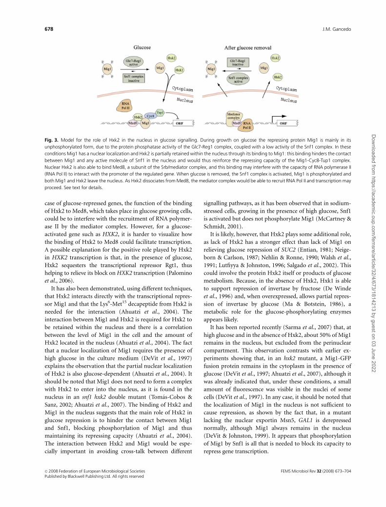
case of glucose-repressed genes, the function of the binding
of Hxk2 to Med8, which takes place in glucose growing cells,
could be to interfere with the recruitment of RNA polymer-
ase II by the mediator complex. However, for a glucose-
activated gene such as HXK2, it is harder to visualize how
the binding of Hxk2 to Med8 could facilitate transcription.
A possible explanation for the positive role played by Hxk2
in HXK2 transcription is that, in the presence of glucose,
Hxk2 sequesters the transcriptional repressor Rgt1, thus
helping to relieve its block on HXK2 transcription (Palomino
et al., 2006).
It has also been demonstrated, using different techniques,
that Hxk2 interacts directly with the transcriptional repres-
sor Mig1 and that the Lys6-Met15 decapeptide from Hxk2 is
needed for the interaction (Ahuatzi et al., 2004). The
interaction between Mig1 and Hxk2 is required for Hxk2 to
be retained within the nucleus and there is a correlation
between the level of Mig1 in the cell and the amount of
Hxk2 located in the nucleus (Ahuatzi et al., 2004). The fact
that a nuclear localization of Mig1 requires the presence of
high glucose in the culture medium (DeVit et al., 1997)
explains the observation that the partial nuclear localization
of Hxk2 is also glucose-dependent (Ahuatzi et al., 2004). It
should be noted that Mig1 does not need to form a complex
with Hxk2 to enter into the nucleus, as it is found in the
nucleus in an snf1 hxk2 double mutant (Tomas-Cobos &
Sanz, 2002; Ahuatzi et al., 2007). The binding of Hxk2 and
Mig1 in the nucleus suggests that the main role of Hxk2 in
glucose repression is to hinder the contact between Mig1
and Snf1, blocking phosphorylation of Mig1 and thus
maintaining its repressing capacity (Ahuatzi et al., 2004).
The interaction between Hxk2 and Mig1 would be espe-
cially important in avoiding cross-talk between different
signalling pathways, as it has been observed that in sodium-
stressed cells, growing in the presence of high glucose, Snf1
is activated but does not phosphorylate Mig1 (McCartney &
Schmidt, 2001).
It is likely, however, that Hxk2 plays some additional role,
as lack of Hxk2 has a stronger effect than lack of Mig1 on
relieving glucose repression of SUC2 (Entian, 1981; Neige-
born & Carlson, 1987; Nehlin & Ronne, 1990; Walsh et al.,
1991; Lutfiyya & Johnston, 1996; Salgado et al., 2002). This
could involve the protein Hxk2 itself or products of glucose
metabolism. Because, in the absence of Hxk2, Hxk1 is able
to support repression of invertase by fructose (De Winde
et al., 1996) and, when overexpressed, allows partial repres-
sion of invertase by glucose (Ma & Botstein, 1986), a
metabolic role for the glucose-phosphorylating enzymes
appears likely.
It has been reported recently (Sarma et al., 2007) that, at
high glucose and in the absence of Hxk2, about 50% of Mig1
remains in the nucleus, but excluded from the perinuclear
compartment. This observation contrasts with earlier ex-
periments showing that, in an hxk2 mutant, a Mig1-GFP
fusion protein remains in the cytoplasm in the presence of
glucose (DeVit et al., 1997; Ahuatzi et al., 2007), although it
was already indicated that, under these conditions, a small
amount of fluorescence was visible in the nuclei of some
cells (DeVit et al., 1997). In any case, it should be noted that
the localization of Mig1 in the nucleus is not sufficient to
cause repression, as shown by the fact that, in a mutant
lacking the nuclear exportin Msn5, GAL1 is derepressed
normally, although Mig1 always remains in the nucleus
(DeVit & Johnston, 1999). It appears that phosphorylation
of Mig1 by Snf1 is all that is needed to block its capacity to
repress gene transcription.
Fig. 3. Model for the role of Hxk2 in the nucleus in glucose signalling. During growth on glucose the repressing protein Mig1 is mainly in its
unphosphorylated form, due to the protein phosphatase activity of the Glc7-Reg1 complex, coupled with a low activity of the Snf1 complex. In these
conditions Mig1 has a nuclear localization and Hxk2 is partially retained within the nucleus through its binding to Mig1: this binding hinders the contact
between Mig1 and any active molecule of Snf1 in the nucleus and would thus reinforce the repressing capacity of the Mig1-Cyc8-Tup1 complex.
Nuclear Hxk2 is also able to bind Med8, a subunit of the Srb/mediator complex, and this binding may interfere with the capacity of RNA polymerase II
(RNA Pol II) to interact with the promoter of the regulated gene. When glucose is removed, the Snf1 complex is activated, Mig1 is phosphorylated and
both Mig1 and Hxk2 leave the nucleus. As Hxk2 dissociates from Med8, the mediator complex would be able to recruit RNA Pol II and transcription may
proceed. See text for details.
FEMS Microbiol Rev 32 (2008) 673–704c� 2008 Federation of European Microbiological SocietiesPublished by Blackwell Publishing Ltd. All rights reserved
678 J.M. Gancedo
Dow
nloaded from https://academ
ic.oup.com/fem
sre/article/32/4/673/1814213 by guest on 03 June 2022

A disputed point has been the role of the serine 14 of
Hxk2 in the capacity of the enzyme to mediate catabolite
repression of genes such as SUC2. This serine was called
Ser15 (corresponding to codon 15) until Hxk2 was se-
quenced and it was found that the N-terminal amino acid
is valine (corresponding to codon 2) and not methionine
(Behlke et al., 1998). Although serine 14 can be phosphory-
lated by PKA in vitro (Kriegel et al., 1994), PKA is not likely
to catalyze the reaction in vivo, because phosphorylation of
serine 14 is reduced under conditions where the cAMP-
dependent protein kinases are activated (Vojtek & Fraenkel,
1990). Different groups have looked at the consequences of
replacing serine 14 by an alanine residue and have reported
contradictory results. While some laboratories found that
the modified enzyme was still functional in glucose signal-
ling (Herrero et al., 1998; Mayordomo & Sanz, 2001),
another group reported that the mutation impaired the
functionality of Hxk2 and concluded that it is the phos-
phorylated Hxk2 that transmits the glucose signal (Randez-
Gil et al., 1998b). This, however, does not appear to agree
with the observation that the proportion of phosphorylated
Hxk2 is higher under derepressing than under repressing
conditions (Vojtek & Fraenkel, 1990; Randez-Gil et al.,
1998b). It has been suggested that the discrepancy between
the results of different groups investigating the effects of
Hxk2S14A could be due to variations in the amount of
mutated hexokinase synthesized in the diverse strains used
(Moreno & Herrero, 2002) and, in fact, the number of
copies of the mutated HXK2 gene was not the same in the
different experiments, as the gene was carried either in an
integrative plasmid (Randez-Gil et al., 1998b) or in centro-
meric (Mayordomo & Sanz, 2001) or multicopy (Herrero
et al., 1998) episomal plasmids. Taking into account that a
modified Hxk2 lacking the 14 N-terminal amino acids can
still mediate repression of SUC2 or induction of HXT1 by
glucose (Ma et al., 1989a; Mayordomo & Sanz, 2001), it can
be concluded that serine 14 is dispensable for glucose
signalling, and that a more relevant feature is the hexokinase
activity in the cell. The functionality of an Hxk2 lacking its
14 N-terminal amino acids would also indicate that, at least
under some circumstances, full glucose repression may take
place in a strain with an Hxk2 lacking the Lys6–Met15
decapeptide, although this decapeptide is required for
interaction with Mig1 (Ahuatzi et al., 2004).
For most other yeasts, there is little evidence for hexoki-
nases playing a nonmetabolic role in glucose repression. In
K. lactis there is a single hexokinase, Rag5, required for
glucose repression (Bar et al., 2003) and a glucokinase that
allows growth on glucose at a very slow rate (35 h doubling
time) in the absence of Rag5 (Kettner et al., 2007). Although
this makes it hard to distinguish between a metabolic and a
regulatory role for Rag5, it has been found that Rag5 is not
efficient for restoring glucose repression of invertase in hxk2
S. cerevisiae mutants (Prior et al., 1993; Petit & Gancedo,
1999). In P. angusta (H. polymorpha), a hexokinase,
HpHxk1, and a glucokinase, HpGlk1, have been identified
and any of them supports repression by glucose of maltase,
alcohol oxidase or catalase (Kramarenko et al., 2000).
However, although HpGlk1 allows growth on glucose of an
S. cerevisiae triple kinase mutant, in the S. cerevisiae
transformants synthesis of invertase and maltase remains
insensitive to glucose repression (Laht et al., 2002). Whereas
HpHxk1 has been cloned (Karp et al., 2003), its effect in
S. cerevisiae has not been reported. In S. pombe, two
hexokinases have been identified: SpHxk1 with low affinity
for glucose and much higher activity with fructose than with
glucose as a substrate, and SpHxk2, a kinetically conven-
tional hexokinase (Petit et al., 1996). As occurred with the
kinases from K. lactis and P. angusta, SpHxk1 could not
restore glucose repression of invertase in an S. cerevisiae
triple mutant (Petit et al., 1998) and repression in the
presence of SpHxk2 was only partial (Petit & Gancedo,
1999). In contrast, in a similar experiment, Hxk1 from
Y. lipolytica was fully effective (Petit & Gancedo, 1999) and
there is preliminary evidence that YlHxk1 is involved in
glucose repression of the LIP2 gene encoding an extracellu-
lar lipase in Y. lipolytica (Fickers et al., 2005). It has also been
reported that in mutants of Schwanniomyces occidentalis or
Pachysolen tannophilus, which lack a specific isoenzyme of
hexokinase, glucose repression is defective (McCann et al.,
1987; Wedlock & Thornton, 1989).
Signalling by intracellular metabolites
Because most glucose effects require glucose metabolism
(Belinchon & Gancedo, 2007a, b), it may be concluded that
binding of glucose to a receptor is usually not sufficient to
signal the presence of glucose in yeast. Although there have
been some reports on glucose signalling, independent of
glucose uptake (Liang & Gaber, 1996; Ozcan, 2002), they
should be considered with caution, because the hxt strain
used showed a residual growth on glucose, indicating that
glucose could still be transported and metabolized. While
growth could be completely blocked by adding antimycin to
the medium, the experiments on the signalling capacity of
glucose were performed in the absence of antimycin.
An intracellular metabolite that plays an important role
in glucose signalling is cAMP, as shown by the fact that many
of the transcriptional changes elicited by glucose can be
reproduced by activation of the GTP-binding proteins Ras2
or Gpa2. This can be done, in the absence of glucose, using
genes encoding constitutively active forms of Ras2 or Gpa2,
placed under the control of a conditional promoter (Wang
et al., 2004). Activation of either Ras2 or Gpa2 causes an
increase in cAMP levels (Toda et al., 1985; Nakafuku et al.,
1988), which stimulates the cAMP-dependent protein
FEMS Microbiol Rev 32 (2008) 673–704 c� 2008 Federation of European Microbiological SocietiesPublished by Blackwell Publishing Ltd. All rights reserved
679The early steps of glucose signalling in yeast
Dow
nloaded from https://academ
ic.oup.com/fem
sre/article/32/4/673/1814213 by guest on 03 June 2022

kinases Tpk1, Tpk2 and Tpk3 (Toda et al., 1987b). Surpris-
ingly, from the many potential substrates already identified
for the three isoenzymes Tpk1, Tpk2 and Tpk3 (thereafter
PKA) present in S. cerevisiae (Budovskaya et al., 2005;
Ptacek et al., 2005), there is none clearly related to the
regulation of important elements mediating glucose control,
such as the Snf1 kinase and the Glc7 phosphatase complexes
(Santangelo, 2006).
Glucose signalling through the Ras-cAMP system requires
the phosphorylation of the sugar (Rolland et al., 2000,
2001). It has been suggested that the metabolism of glucose
causes an inhibition of Ira1/Ira2, stimulators of the GTPase
activity of the Ras proteins, and therefore, increases the
GTP-loading of Ras1 and Ras2 (Colombo et al., 2004). GTP-
loaded Ras proteins in turn activate adenylate cyclase,
increasing the intracellular concentration of cAMP [see a
recent review (Santangelo, 2006) for details].
As indicated above, it is also possible to mimic the glucose
response, in the absence of the sugar. Although the changes
in transcript levels resulting from Ras2 or Gpa2 activation
are dependent on PKA, it has also been shown that most
glucose-responsive genes can also be regulated through a
PKA-independent pathway (Wang et al., 2004). Information
is not yet available on the elements of this regulatory
pathway, but it may start with changes in the concentra-
tion of some intracellular metabolite(s) other than cAMP
(Boles & Zimmermann, 1993; Goncalves et al., 1997).
Glucose-6-phosphate and fructose-1,6-bisphosphate may
appear as good candidates, as their concentrations are
strongly responsive to the presence of glucose (Banuelos
et al., 1977); however, there is no clear correlation between
the intracellular concentration of these hexose phosphates
and the degree of repression by different carbon sources
of the gluconeogenic enzyme fructose-1,6-bisphosphatase
(FbPase) (Rodrıguez & Gancedo, 1999; Belinchon & Gance-
do, 2003). Although studies on the induction of glycolytic
enzymes in a variety of mutants suggest that different
metabolites control the expression of different genes (Boles
& Zimmermann, 1993; Muller et al., 1995; Boles et al.,
1996), the identity of the regulatory metabolites has not
been established and their mechanism of action is not
known.
In mammalian cells, the AMP-dependent protein kinase,
a homologue of the protein kinase Snf1 from yeast, is
activated under stress conditions due to an increase in the
concentration of intracellular AMP (Hardie, 1999). In S.
cerevisiae, however, there is no evidence that AMP controls
Snf1, or even that the AMP concentration decreases in the
presence of glucose (Kresnowati et al., 2006). It has been
proposed that yeast could, in some unknown way, sense
the ‘glucose flux’ or the rate of glucose consumption and
adjust in this way the degree of repression of genes involved
in the utilization of alternative carbon sources (Bisson &
Kunathigan, 2003). However, there is evidence that glucose
repression of a gene such as SUC2 is not directly related to
the glucose flux (Meijer et al., 1998). The identification of
regulatory elements in pathways alternative to that con-
trolled by PKA therefore remains an important point that
requires further investigation.
An interesting regulatory metabolite that accumulates in
yeast cells metabolizing glucose is fructose-2,6-bisphosphate
(Lederer et al., 1981). It is worth noting that this compound
has been found to activate the phosphorylation of FbPase by
PKA in vitro (Gancedo et al., 1983), and is therefore likely
to control the phosphorylation and partial inactivation
of FbPase, which takes place on addition of glucose to
derepressed yeast cells (Mazon et al., 1982).
Other early elements in glucose signallingin S. cerevisiae
The TOR pathway
Although the TOR-signalling pathway has been investigated
mainly in relation to the nitrogen source in the medium
(Cooper, 2002), it may also mediate some of the yeast
responses to glucose (Hardwick et al., 1999). In the presence
of rapamycin, an inhibitor of Tor1 and Tor2, the transcript
levels of several genes induced by glucose decrease, while a
number of glucose-repressed genes are expressed at higher
levels (Hardwick et al., 1999). In the case of the low-affinity
glucose transporter Hxt1, rapamycin not only decreases the
levels of HXT1 mRNA (Hardwick et al., 1999; Tomas-Cobos
et al., 2005) but also causes instability and mislocalization of
Hxt1 (Schmelzle et al., 2004). It therefore appears that the
TOR pathway acts both on transcription and on posttran-
scriptional processes. It has been proposed that glucose
signalling through TOR takes place independently of the
TOR effectors Tap2 and Sit4, and is mediated by the Ras/
cAMP pathway (Schmelzle et al., 2004). It is not yet known,
however, whether the Tor proteins sense intracellular glu-
cose or some other indicator of carbon source availability.
The lack of Tor1 also impairs the downregulation by
glucose of different amino acid permeases (Peter et al.,
2006). In addition, in the absence of Tor1, mitochondrial
respiration is increased during growth on glucose. This
increase, which is not observed in glycerol-growing cells, is
due to an enhanced translation of mRNAs corresponding to
subunits of the oxidative phosphorylation complex encoded
by mit-DNA (Bonawitz et al., 2007).
14-3-3 proteins
The 14-3-3 proteins Bmh1/Bmh2, positive regulators of the
TOR kinase pathway (Bertram et al., 1998), affect some
processes regulated by glucose. In a double mutant
bmh1bmh2 catabolite inactivation of maltose permease is
FEMS Microbiol Rev 32 (2008) 673–704c� 2008 Federation of European Microbiological SocietiesPublished by Blackwell Publishing Ltd. All rights reserved
680 J.M. Gancedo
Dow
nloaded from https://academ
ic.oup.com/fem
sre/article/32/4/673/1814213 by guest on 03 June 2022

impaired (Mayordomo et al., 2003), glucose repression of
ADH2 is partially relieved (Dombek et al., 2004) and
induction of HXT1 by high glucose is prevented (Tomas-
Cobos et al., 2005). It has been shown that Bmh1 and Bmh2
interact with Reg1, a regulatory subunit of the type 1 protein
phosphatase complex (Dombek et al., 2004) and that the
complex Bmh1/2-Reg1 interacts with Grr1, a component of
the SCFGrr1 ubiquitination complex (Tomas-Cobos et al.,
2005). This would suggest that the Bmh proteins mediate
HXT1 induction by facilitating, in a Reg1-dependent pro-
cess, the interaction of Grr1 with Std1. The Bmh proteins
may also play a Reg1-independent role, possibly related to
the activation of the TOR pathway, as the bmh1bmh2 and
reg1 mutations release synergistically glucose repression of
ADH2 (Dombek et al., 2004). It has also been reported that,
in the presence of glucose, Bmh1/2 interacts with the protein
kinase Yak1 (Moriya et al., 2001); under these conditions,
Yak1 is phosphorylated by PKA (Zappacosta et al., 2002)
and is exported from the nucleus in a Bmh-dependent
process (Moriya et al., 2001). Upon glucose depletion Yak1
is activated, enters the nucleus and phosphorylates Pop2, a
subunit of the Ccr4-Not complex that mediates mRNA
deadenylation, causing cell-cycle arrest (Moriya et al.,
2001). Activated Yak1 also phosphorylates the transcription
inhibitor Crf1, causing a decrease in the transcription of the
ribosomal genes (Martin et al., 2004).
Phospholipase C
Another regulatory pathway controlled by glucose involves
phospholipase C (Plc1). Addition of glucose to starved yeast
cells activates Plc1 and thereby increases phosphatidylinosi-
tol turnover and the intracellular level of different species of
diacylglycerols (Coccetti et al., 1998). The fact that Plc1
interacts with the glucose sensor Gpr1 and its coupled G
protein Gpa2 (Ansari et al., 1999) suggests that activation of
Plc1 is mediated by glucose binding to Gpr1. This is further
supported by the observation that the Plc1-dependent,
glucose-triggered increase in free intracellular Ca21 requires
Gpr1 and Gpa2 (Tisi et al., 2002). It has also been observed
that the transient elevation of cytosolic calcium caused by
glucose requires the phosphorylation of the sugar (Tokes-
Fuzesi et al., 2002) and it has been suggested that the
intracellular calcium concentration is related to the ratio
glucose-1-P/glucose-6-P (Aiello et al., 2002). There is also
preliminary evidence for inositol triphosphate acting as a
mediator of glucose-induced calcium signalling (Tisi et al.,
2004). The primary target of the diacylglycerols formed
upon activation of Plc1 is the protein kinase C (PKC), and
PKC may be involved in the control of carbon metabolism.
This control, however, is complex because pkc1 mutants
show defects both in the induction of HXT genes by glucose
and in the derepression of SUC2, alcohol dehydrogenase
or glycerol kinase that takes place on glucose depletion
(Brandao et al., 2002; Salgado et al., 2002; Gomes et al.,
2005). For this function, PKC does not require the MAP kinase
cascade formed by the kinases Bck1, Mkk1/2 and Mpk1, but
may act by regulating the intracellular localization of DNA-
binding proteins such as the repressor Mig1 or the transcrip-
tion factor Adr1 (Salgado et al., 2002; Gomes et al., 2005).
Others
A different way for glucose to act, when it reaches the
100 mM range, is as a weak osmolyte, able to activate the
Sln1-dependent branch of the HOG pathway (Tomas-Cobos
et al., 2004). This activation has been shown to be absolutely
required for glucose to induce transcription of the HXT1
gene (Tomas-Cobos et al., 2004). Still other elements have
been shown to affect processes controlled by glucose, but
there have only been suggestions on their mode of action,
rather than clear evidence. The increased induction of
HXT1, which takes place in the absence of Cdc55, has been
attributed to interference of Cdc55 with the activity of the
protein kinases Yck1/2 that phosphorylate Std1 and Mth1,
negative regulators of HXT1 transcription (Tomas-Cobos
et al., 2005). The protein kinase Pho85 is required to control
the important remodelling of gene expression that takes
place when the glucose of the medium is depleted; in its
absence downregulation of glycolytic genes is defective,
induction of genes involved in gluconeogenesis and in the
glyoxylate cycle occurs before the glucose of the medium is
completely exhausted, and the expression of genes required
for proper mitochondrial function is impaired (Nishizawa
et al., 2004). The molecular mechanisms underlying these
effects are not known, but it is likely that the Pho85-
associated cyclins Pcl6 and Pcl7 are involved in the forma-
tion of functional mitochondria, as mutants lacking Pho85,
Pcl6 or Pcl7 are unable to grow on nonfermentable carbon
sources (Gilliquet & Berben, 1993; Lee et al., 2000). The
observation that in a mutant defective in Tps1, a subunit of
the trehalose synthase complex, glucose-induced effects are
impaired led Thevelein & Hohmann (1995) to conclude that
Tps1 is involved in the transduction of a glucose signal.
Although the inability of tps1 mutants to grow on glucose
complicated the interpretation of the earlier observations,
later work showed that Tps1 is dispensable for glucose
signalling (Rodrıguez & Gancedo, 1999).
Targets for glucose signals
In S. cerevisiae
Genes repressed by glucose
An important signal for glucose repression is an increase in
intracellular cAMP, which allows the activation of PKA. This
FEMS Microbiol Rev 32 (2008) 673–704 c� 2008 Federation of European Microbiological SocietiesPublished by Blackwell Publishing Ltd. All rights reserved
681The early steps of glucose signalling in yeast
Dow
nloaded from https://academ
ic.oup.com/fem
sre/article/32/4/673/1814213 by guest on 03 June 2022
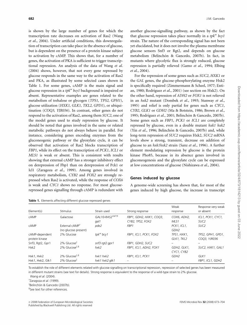
is shown by the large number of genes for which the
transcription rate decreases on activation of Ras2 (Wang
et al., 2004). Under artificial conditions, this downregula-
tion of transcription can take place in the absence of glucose,
but is dependent on the presence of a protein kinase subject
to activation by cAMP. This shows that, for a number of
genes, the activation of PKA is sufficient to trigger transcrip-
tional repression. An analysis of the data of Wang et al.
(2004) shows, however, that not every gene repressed by
glucose responds in the same way to the activation of Ras2
and PKA, as illustrated by some selected cases shown in
Table 1. For some genes, cAMP is the main signal and
glucose repression in a tpkw bcy1 background is impaired or
absent. Representative examples are genes related to the
metabolism of trehalose or glycogen (TPS1, TPS2, GPH1),
glucose utilization (HXK1, GLK1, TKL2, GPD1), or ubiqui-
tination (COQ3, YJR036). In contrast, other genes do not
respond to the activation of Ras2, among them SUC2, one of
the model genes used to study repression by glucose. It
should be noted that genes involved in the same or related
metabolic pathways do not always behave in parallel. For
instance, considering genes encoding enzymes from the
gluconeogenic pathway or the glyoxylate cycle, it can be
observed that activation of Ras2 blocks transcription of
FBP1, while its effect on the transcription of PCK1, ICL1 or
MLS1 is weak or absent. This is consistent with results
showing that external cAMP has a stronger inhibitory effect
on derepression of Fbp1 than on derepression of Pck1 or
Icl1 (Zaragoza et al., 1999). Among genes involved in
respiratory metabolism, CYB2 and FOX2 are strongly re-
pressed when Ras2 is activated, while the response of COX6
is weak and CYC1 shows no response. For most glucose-
repressed genes signalling through cAMP is redundant with
another glucose-signalling pathway, as shown by the fact
that glucose repression takes place normally in a tpkw bcy1
strain. The nature of the corresponding signal has not been
yet elucidated, but it does not involve the plasma membrane
glucose sensors Snf3 or Rgt2, and depends on glucose
metabolism (Belinchon & Gancedo, 2007b). In fact, in
mutants where glycolytic flux is strongly reduced, glucose
repression is partially relieved (Gamo et al., 1994; Elbing
et al., 2004).
For the repression of some genes such as SUC2, HXK1 or
the GAL genes, the glucose-phosphorylating enzyme Hxk2
is specifically required (Zimmermann & Scheel, 1977; Enti-
an, 1980; Rodrıguez et al., 2001) (see section on Hxk2). On
the other hand, repression of ADH2 or FOX1 is not relieved
in an hxk2 mutant (Dombek et al., 1993; Stanway et al.,
1995) and relief is only partial for genes such as CYC1,
CYB2, GLK1 or GDH2 (Ma & Botstein, 1986; Brown et al.,
1995; Rodrıguez et al., 2001; Belinchon & Gancedo, 2007b).
Some genes such as FBP1, PCK1 or ICL1 are completely
repressed by glucose, even in a double mutant hxk1 hxk2
(Yin et al., 1996; Belinchon & Gancedo, 2007b) and, while
long-term repression of SUC2 requires Hxk2, SUC2 mRNA
levels show a strong, transient, decrease on addition of
glucose to an hxk1hxk2 strain (Sanz et al., 1996). A further
element modulating repression by glucose is the protein
kinase Pho85, because in its absence genes involved in
gluconeogenesis and the glyoxylate cycle can be expressed
at low concentrations of glucose (Nishizawa et al., 2004).
Genes induced by glucose
A genome-wide screening has shown that, for most of the
genes induced by high glucose, the increase in transcript
Table 1. Elements affecting different glucose repressed genes
Element(s) Stimulus Strain used Strong response
Weak
response
Response very weak
or absent
cAMP Galactose� GAL10-RAS2Val19
gal1
FBP1, GDH2, HXK1, COQ3,
CYB2, TPS2, FOX2
COX6, ADH2,
MLS1
ICL1, PCK1, CYC1,
SUC2
cAMP External cAMPw
(no glucose present)
pde2 FBP1 PCK1, ICL1,
GDH2
SUC2
cAMP-dependent
protein kinase
2% Glucose� tpkw bcy1 FBP1, ICL1, PCK1, FOX2 TPS1, HXK1,
GLK1, TKL2
TPS2, GPH1, GPD1,
COQ3, YJR036
Snf3, Rgt2, Gpr1 2% Glucosez snf3 rgt2 gpr1 FBP1, GDH2, SUC2 – –
Hxk2 2% Glucosez,‰ hxk2 FBP1, ICL1, ADH2, FOX1 GDH2, GLK1,
CYC1, CYB2
SUC2, HXK1, GAL1
Hxk1, Hxk2 2% Glucosez,‰ hxk1 hxk2 FBP1, ICL1, PCK1 GDH2 GLK1
Hxk1, Hxk2, Glk1 2% Glucosez hxk1 hxk2 glk1 – – FBP1, ICL1, GDH2
To establish the role of different elements related with glucose signalling on transcriptional repression, repression of selected genes has been measured
in different mutant strains (see text for details). Strong response is equivalent to the response of a wild-type strain to 2% glucose.�Wang et al. (2004).wZaragoza et al. (1999).zBelinchon & Gancedo (2007b).‰See text for other references.
FEMS Microbiol Rev 32 (2008) 673–704c� 2008 Federation of European Microbiological SocietiesPublished by Blackwell Publishing Ltd. All rights reserved
682 J.M. Gancedo
Dow
nloaded from https://academ
ic.oup.com/fem
sre/article/32/4/673/1814213 by guest on 03 June 2022

levels triggered by glucose is modest, between 1.5- and
4-fold. This increase is usually rapid, o 20 min, but it is
sometimes transient, mRNA levels decreasing markedly at
60 min. There are exceptions, as for some genes the degree of
induction is higher, about fivefold for RPA12 or RPC53, 20-
fold for HXT3, and even 100-fold for HXT1, while for genes
such as TRP3, ARG3 or MET14, the kinetics of induction are
slower (Wang et al., 2004). The role that different sensing
elements play in the induction of different genes is examined
in the next paragraphs.
Binding of glucose to the plasma membrane sensors Snf3/
Rgt2 triggers the induction of a limited number of genes
through a cascade of reactions discussed previously. Most of
these genes encode glucose transporters (Ozcan & Jonhston,
1999), but a few others have been identified (Kaniak et al.,
2004), and among them MIG2 is of special interest, as
increased levels of the DNA-binding protein Mig2 contri-
bute to glucose repression (Lutfiyya et al., 1998). The other
plasma membrane sensor, Gpr1 (see the corresponding
section), is often required for a maximal response to glucose;
in its absence, the degree of induction decreases by a factor
of two in many cases (Wang et al., 2004). Gpr1 is not
needed, however, for the complete induction of HXT1 or
pyruvate decarboxylase (Belinchon & Gancedo, 2007a)
(Table 2).
Artificial activation of Ras2, in the absence of glucose (see
section on ‘Signalling by intracellular metabolites’), can
cause an increase in mRNA levels for many genes; for some
genes the extent of induction is similar to that triggered by
glucose, while for others induction is low, or even absent for
genes such as HXK2 or ENO2 (Table 2). Ras2 activation
experiments performed in a strain with a weak PKA
insensitive to cAMP (tpkw bcy1 background) have led to the
conclusion that Ras signalling takes place exclusively
through a cAMP-regulated PKA (Wang et al., 2004). It
should be noted, however, that for HXT1 and HXT3 there
was at least a 10-fold induction by activated Ras2 in this
background (supplementary information in Wang et al.,
2004). Although these results have not yet been confirmed
by northern analysis or quantitative reverse transcriptase
(RT)-PCR, it would be interesting to follow this lead and
investigate a potential alternative pathway for Ras signalling.
To investigate whether a cAMP-regulated PKA is not only
sufficient, but also necessary, for glucose induction of
transcription, glucose was added to a tpkw bcy1 mutant
strain growing on glycerol (Wang et al., 2004), because in
such a strain PKA is no longer sensitive to the changes in
cAMP intracellular concentration triggered by glucose. In a
large number of cases there was a strong induction of
transcription by glucose (at least 10-fold), thus showing that
metabolism of glucose can provide for most genes an
induction mechanism independent of an increase in PKA
activity. It may be observed that for many genes, mRNA
levels were much lower (5–20-fold) in the mutant strain
than in the wild type, during growth in glycerol. This
suggests that in a wild-type yeast grown in glycerol, the
PKA activity is greater than in the mutant and sufficient to
cause a partial induction of the corresponding genes.
Surprisingly, while galactose does not act as an inducer in
the wild-type strain, for some genes it can cause large
increases in mRNA levels in the tpkw bcy1 mutant, although
the strain lacks Gal1 and is therefore unable to metabolize
galactose (Wang et al., 2004). This is a result that remains
difficult to interpret.
The role of the glucose phosphorylating enzymes in the
induction of transcription by high glucose is not the same
for different genes (Table 2). In mutant strains lacking Hxk2,
there is a strong decrease in the induction of HXT1 and
HXK2, but HXT3 and pyruvate decarboxylase are fully
induced (Ozcan & Johnston, 1995; Rodrıguez et al., 2001;
Table 2. Elements affecting different genes induced by glucose
Element(s) Stimulus Strain used Strong response Weak response
Response very weak
or absent
cAMP Galactose GAL10-RAS2Val19 gal1 THR4, RPC53, RPO31 ARG3, MET14, HXT3 HXT1, HXK2, ENO2
cAMP-dependent
protein kinase
Glucose� tpkw bcy1 ENO2, ARG3, THR4,
GCN3, RPA43, RPC53
HXT1, HXT3, RPC82 MET14, RPO31
Snf3, Rgt2 Glucose� snf3 rgt2 PDC1 SUC2w HXT1, HXT3
Gpr1 Glucose� gpr1 PDC1, HXT1 SUC2w
Hxk2 Glucose� hxk2 PDC1, HXT3 HXT1, HXT2w, HXT3w –
Hxk1, Hxk2 Glucose� hxk1 hxk2 SUC2w, PDC1w PDC1 –
Hxk1, Hxk2, Glk1 Glucose� hxk1 hxk2 glk1 SUC2 HXT1 PDC1
Hxk1, Hxk2, Glk1, Gpr1 Glucose� hxk1 hxk2 glk1 gpr1 – HXT1 SUC2
To establish the role of different elements related with glucose signalling on transcriptional induction, induction of selected genes has been measured in
different mutant strains (see text for details). Strong response is equivalent to the response of a wild-type strain to the same concentration of glucose.�Induction by 2–4% glucose, unless otherwise indicated.wInduction by 0.05% glucose.
See text for references.
FEMS Microbiol Rev 32 (2008) 673–704 c� 2008 Federation of European Microbiological SocietiesPublished by Blackwell Publishing Ltd. All rights reserved
683The early steps of glucose signalling in yeast
Dow
nloaded from https://academ
ic.oup.com/fem
sre/article/32/4/673/1814213 by guest on 03 June 2022

Belinchon & Gancedo, 2007a). While induction of pyruvate
decarboxylase is decreased by 50% in a double mutant hxk1
hxk2 and blocked in the triple hxk1 hxk2 glk1 mutant, in this
last mutant there is a partial induction of HXT1 and SUC2 is
highly induced. The need for glucose phosphorylation to
induce pyruvate decarboxylase is in agreement with the
observation that this induction is dependent on products
of glucose metabolism such as glucose-6P and three-carbon
metabolites (Boles & Zimmermann, 1993). It should be
noted that, in the absence of glucose phosphorylation, Gpr1
is absolutely required for the induction of SUC2 but is
dispensable for that of HXT1 (Belinchon & Gancedo,
2007a).
A low concentration of glucose, o 0.1%, is able to induce
a number of genes, and in this case also, there are differ-
ent signalling pathways controlling the induction process
(Ozcan & Johnston, 1995; Belinchon & Gancedo, 2007a).
The induction of HXT2, HXT3 and HXT4, encoding glucose
transporters, requires the glucose sensor Snf3 and the
expression of these genes decreases two- to fivefold in the
absence of Hxk2 (Ozcan & Johnston, 1995). The induction
of SUC2 has only a partial requirement for Snf3/Rgt2 and
does not depend on Hxk2, or Hxk1, but it is slightly
impaired in the absence of Gpr1 (Belinchon & Gancedo,
2007a) and considerably decreased in mutants lacking PKA
(J.M. Gancedo, unpublished results). Induction of pyruvate
decarboxylase by low glucose is little affected by the lack of
both Hxk1 and Hxk2 (Belinchon & Gancedo, 2007a).
Proteins activated by glucose
When glucose becomes available to glucose-starved yeast,
the activity of a number of enzymes increases. Prominent
among them, because it initiates an important signalling
pathway, is adenylate cyclase. Activation of adenylate cyclase
requires the Ras1/Ras2 proteins, located in the plasma
membrane and able to interact directly with the adenylate
cyclase complex (Shima et al., 2000). The exact mechanism
for activation of the Ras proteins is not known, but it has
been established that glucose causes a modest increase in
Ras1/2 GTP loading and that this increase requires glucose
phosphorylation; the increase is impaired in a strain lacking
Cdc25, the GTP/GDP exchange factor for the Ras proteins.
Inhibition of the Ira1/Ira2 proteins, which increase the
GTPase activity of Ras1/2, contributes to the activation of
the Ras proteins triggered by glucose (Colombo et al., 2004).
The increased GTP loading of the Ras proteins does not
depend on the glucose receptor Gpr1 or on its G-binding
protein Gpa2 (Colombo et al., 2004); however, the Gpr1/
Gpa2 system is required for full activation of adenylate
cyclase (Rolland et al., 2000). Only the GTP-bound, active
form of Gpa2 is able to bind adenylate cyclase (Peeters et al.,
2006), but this binding is not enough to trigger a full
response, because in strains unable to phosphorylate glucose
the activation of adenylate cyclase observed, which is
dependent on Gpr1 and on a high concentration of glucose,
is very weak (Kraakman et al., 1999a). The activation of
adenylate cyclase causes an increase in the intracellular
concentration of cAMP, which in turn activates PKA.
Activated PKA phosphorylates different enzymes such as
the neutral trehalase Nth1 (Ortiz et al., 1983; Uno et al.,
1983; Francois & Parrou, 2001) or the 6-phosphofructo-2-
kinase Pfk26 (Francois et al., 1984; Dihazi et al., 2003) and
thereby increases their activity.
The activation by glucose of the plasma membrane
H1-ATPase is also related to phosphorylation of the protein
at different positions, but PKA is not involved in the process,
at least directly (Portillo et al., 1991). While phosphoryla-
tion of Thr912 from the H1-ATPase appears to mediate an
increase in the Vmax of the enzyme (Portillo et al., 1991),
phosphorylation of Ser899 would cause a decrease in the Km
for ATP (Eraso & Portillo, 1994). Phosphorylation of Ser899
is performed by the protein kinase Ptk2 (Eraso et al., 2006),
but the protein kinase responsible for Thr912 phosphoryla-
tion has not been identified, in spite of a systematic search
using protein kinase mutants (Goossens et al., 2000). The
activity of Ptk2 is the same in membranes isolated from cells
incubated in the presence or in the absence of glucose and is
not affected by glucose itself (Eraso et al., 2006), but it could
be modulated by some glucose-derived metabolite as in-
dicated by the fact that activation of the H1-ATPase is
strongly reduced in an hxk1 hxk2 mutant and extremely low
in a mutant unable to phosphorylate glucose (Belinchon &
Gancedo, 2007b). On the other hand, the activation of the
H1-ATPase is independent of the glucose sensors Snf3/Rgt2
and Gpr1 (Belinchon & Gancedo, 2007b).
Proteins inactivated by glucose
Glucose also triggers the inactivation of different proteins, a
phenomenon called catabolite inactivation (Holzer, 1976).
Phosphorylation of FbPase by PKA causes a partial inactiva-
tion of the enzyme (Gancedo et al., 1983; Rittenhouse et al.,
1987); this process is impaired in a mutant unable to
phosphorylate glucose and the impairment is stronger when
both the glucose phosphorylating enzymes and the glucose
sensor Gpr1 are absent (Belinchon & Gancedo, 2007b). This
is consistent with the observation that phosphorylation of
FbPase by PKA is strongly activated by fructose-2,6-bispho-
sphate, a regulatory metabolite that is formed when glucose
is metabolized (Gancedo et al., 1983). While partial inacti-
vation of isocitrate lyase by glucose is also dependent on
phosphorylation by PKA (Ordiz et al., 1996), there is no
information on the protein kinase(s) performing the phos-
phorylation of the malate dehydrogenase isoenzyme Mdh2
FEMS Microbiol Rev 32 (2008) 673–704c� 2008 Federation of European Microbiological SocietiesPublished by Blackwell Publishing Ltd. All rights reserved
684 J.M. Gancedo
Dow
nloaded from https://academ
ic.oup.com/fem
sre/article/32/4/673/1814213 by guest on 03 June 2022

that takes place on glucose addition and causes inactivation
of the enzyme (Minard & McAlister-Henn, 1994).
Glucose not only causes the inactivation of metabolic
enzymes, but also that of the protein kinase Snf1, which
plays a major role in allowing the transcription of glucose-
repressed genes. In this case inactivation is due to depho-
sphorylation of Snf1 by the Glc7-Reg1 complex (Sanz et al.,
2000). Glucose may shift the equilibrium between the
phosphorylated and nonphosphorylated forms of Snf1,
by activating Glc7-Reg1 and/or by inhibiting the protein
kinases Sak1, Tos3 and Elm1 that phosphorylate Snf1 (Hong
et al., 2003; Nath et al., 2003; Sutherland et al., 2003).
However, the mechanism by which glucose modifies Snf1
activity has not yet been unravelled (Hedbacker & Carlson,
2008; Rubenstein et al., 2008).
Proteins degraded by glucose
Glucose also triggers the degradation of a number of
enzymes, a process that has been most thoroughly studied
for FbPase and Mdh2. Two conflicting views on the mechan-
ism of FbPase degradation have been offered. One of them
contemplates a regulated transfer of FbPase to the vacuole and
its degradation by vacuolar proteases (Chiang & Schekman,
1991; Shieh et al., 2001); the other one proposes that FbPase
is first subject to ubiquitination (Schork et al., 1995) and then
degraded by the proteasome (Schork et al., 1994). These views
may be reconciled by the observation that, depending on the
physiological conditions of the yeast cells, degradation of
FbPase, and also of Mdh2, can take place in the vacuole or in
the proteasome (Hung et al., 2004). Degradation in the
vacuole requires the phosphorylation of glucose but may
proceed in the absence of Hxk1 and Hxk2 (Hung et al., 2004;
Belinchon & Gancedo, 2007b), while degradation by the
proteasome is strictly dependent on Hxk2 (Horak et al.,
2002). Reg1 and Grr1 play a role in the degradation of FbPase
in the proteasome (Horak et al., 2002), perhaps related to their
possible involvement in the ubiquitination process. Reg1 is
also required for the transport of FbPase from intermediate
vesicles to the vacuoles (Cui et al., 2004). On the other hand,
the cAMP-signalling pathway is required for the vacuolar-
dependent degradation of FbPase (Hung et al., 2004;
Belinchon & Gancedo, 2007b) but not for degradation by the
proteasome (Horak et al., 2002; Hung et al., 2004). The
transcriptional repressor Rgt1 is required for FbPase degrada-
tion in some conditions (Belinchon & Gancedo, 2007b) but
not in others (Horak et al., 2002); its specific role, however,
has not been elucidated. Degradation of phosphoenolpyruvate
carboxykinase appears to proceed by a mechanism similar to
that described for FbPase and Mdh2 (Muller et al., 1981;
Hammerle et al., 1998).
Mth1, a regulatory protein involved in the transcription
control of the HXT genes, is also degraded on glucose
addition as described in the section on Snf3 and Rgt2. This
degradation is considerably reduced in a reg1 mutant, and
this seems to be related to the fact that the Glc7-Reg1
complex is required for glucose activation of the kinases
Yck1/2 (Gadura et al., 2006), as activation of Yck1/2 is an
early step in the pathway leading to Mth1 ubiquitination
and degradation (Moriya & Johnston, 2004). This role of
Reg1 could explain the observation that HXT1 induction is
strongly impaired in a reg1 mutant but appears in contra-
diction with the fact that the HXT2, HXT3 or HXT4 genes
are normally induced in this mutant (Ozcan & Johnston,
1995).
On glucose addition different kinds of transporters are
internalized and degraded, among them the maltose/H1
symporter Mal61 (Riballo et al., 1995), the galactose per-
mease Gal2 (Horak & Wolf, 1997), the monocarboxylate/H1
symporter Jen1 (Paiva et al., 2002) and the glycerol/H1
symporter Stl1 (Ferreira et al., 2005). Because the first step
in the internalization of the transporters is their ubiquitina-
tion (Horak & Wolf, 1997; Lucero & Lagunas, 1997; Paiva
et al., 2002), the corresponding signal is likely to be the
binding of glucose to Snf3/Rgt2 and the subsequent activa-
tion of Yck1/2 as occurs for Mth1 (Gadura et al., 2006). The
fact that Gal2 inactivation is not prevented in an snf3 or in
an rgt2 mutant (Horak et al., 2002) may be due to the
partial functional redundancy of Snf3 and Rgt2. Degrada-
tion of the ubiquitinated transporters is independent of the
proteasome and takes place within the vacuole. Although
Grr1, the F-box protein of one of the SCF ubiquitin ligase
complexes, is required for the degradation of the Gal2
transporter, it does not seem to be required for the ubiqui-
tination of Gal2 itself (Horak & Wolf, 2005). Degradation
of the maltose permease Mal61 by glucose requires Rgt2
and Grr1 but it is not impaired in an rgt1 mutant (Jiang
et al., 1997).
Other processes controlled by glucose
In addition to the processes already considered, the presence
of glucose affects yeast physiology in many other ways.
Glucose increases the turnover of a number of mRNA
species (Lombardo et al., 1992; Mercado et al., 1994), while
it causes a transient stabilization of ribosomal protein
mRNAs (Yin et al., 2003). The increased turnover of SDH2
mRNA, encoding the iron–sulfur protein subunit of succi-
nate dehydrogenase, takes place only at high levels of
glucose; in contrast, the degradation rates of the FBP1
and PCK1 mRNAs, encoding gluconeogenic enzymes, are
already increased by the addition of 0.02% glucose (Yin
et al., 2000). Glucose phosphorylation is required to en-
hance the turnover of SDH2, FBP1, PCK1 or SUC2 mRNAs,
but Hxk2 is not specifically needed, except for SDH2
(Cereghino & Scheffler, 1996; Yin et al., 2000). In all cases
FEMS Microbiol Rev 32 (2008) 673–704 c� 2008 Federation of European Microbiological SocietiesPublished by Blackwell Publishing Ltd. All rights reserved
685The early steps of glucose signalling in yeast
Dow
nloaded from https://academ
ic.oup.com/fem
sre/article/32/4/673/1814213 by guest on 03 June 2022

the effect of glucose is blocked in an reg1 mutant, where the
activity of the protein kinase Snf1 is less sensitive to glucose
(Sanz, 2007). Although external cAMP (in a pde2 back-
ground) can trigger degradation of PCK1 and SDH2
mRNAs, the increased degradation of PCK1 mRNA ob-
served in the presence of 0.02% glucose also occurs in the
absence of a Ras-cAMP pathway (Yin et al., 2000). It appears
therefore that glucose controls mRNA turnover through
alternative, partially redundant signalling pathways. The
same applies to the stabilization by glucose of different
ribosomal protein encoding mRNAs (Yin et al., 2003).
Specifically, the effect of glucose on RPL3 mRNA stability,
but not on that of RPS6 mRNA, is retained in the absence of
glucose phosphorylation, while the Ras-cAMP pathway is
required for stabilization of RPL3 and RPL24 mRNAs but
not for that of RPS6 mRNA.
Glucose also affects the translation rate of mRNA. On
transfer of glucose-growing yeast to a medium lacking
glucose, there is a very rapid (1–2 min) inhibition of
translation (Ashe et al., 2000). This effect of carbon source
removal is specific for glucose (or fructose), as it does not
occur in yeast grown in sucrose, maltose or galactose.
Inhibition of translation upon glucose withdrawal does not
involve the pathways implicated in the inhibition of transla-
tion caused by amino acid starvation (Ashe et al., 2000).
Translation is restored rapidly (5 min) by addition of
glucose (Ashe et al., 2000) and slowly (hours) by incubation
of the yeast cells under aerobic conditions in the absence of a
carbon source (Uesono et al., 2004). Although several
proteins such as Pat1, Ddh1 or Sbp1 have been identified as
destabilizing translation initiation complexes upon glucose
removal (Coller & Parker, 2005; Segal et al., 2006), there is
no information on the mechanism(s) through which glu-
cose may control their activity. Inhibition of translation after
glucose removal is prevented if yeast cells are in a metabolic
condition that mimicks to some extent the absence of
glucose. This occurs in mutants with a very low PKA activity
or in reg1 or hxk2 mutants, provided Snf1 is present (Ashe
et al., 2000). Translation is also strongly inhibited when yeast
cells are transferred from a medium with glucose to a
medium with ethanol, and this inhibition requires the Gat1
protein, involved in signalling the quality of the carbon
source through the Tor pathway (Kuruvilla et al., 2001).
The yeast vacuolar H1ATPase (V-ATPase) is a multi-
subunit complex responsible for the acidification of the
organelle. The assembly of this V-ATPase is regulated by
glucose, 0.1% glucose increasing the level of assembly from
10 to 25%, while in the presence of 2% glucose, 60% of the
subunits are assembled in complexes (Parra & Kane, 1998).
Glucose metabolism beyond glucose-6-phosphate is re-
quired for triggering V-ATPase assembly but a cAMP signal
is neither sufficient nor necessary for the process to take
place (Parra & Kane, 1998). Because aldolase is specifically
needed for assembly, it has been suggested that aldolase may
act in this process as a sensor for the presence of glucose (Lu
et al., 2004). V-ATPase assembly is also dependent on the
RAVE complex, formed by the Rav1, Rav2 and Skp1 proteins
(Seol et al., 2001), but it is not known whether glucose
directly controls RAVE.
The growth rate of S. cerevisiae and its cell size depend on
the carbon source in the medium and both are highest in
glucose-grown cells (Johnston et al., 1979). The effect of
glucose on cell size is mediated by the Gpr1-Gpa2 system
(Tamaki et al., 2005; Vanoni et al., 2005) and requires
Cdc25, the Ras GTP–GDP exchange protein (Belotti et al.,
2006). Glucose increases the growth rate by inducing a
diverse set of genes: glycolytic and ribosomal protein genes,
as well as genes related to the cell division cycle. An
important signal for this process is the activation of PKA
by cAMP, which increases the transcription rate of Rap1
target genes (Klein & Struhl, 1994) and causes an increase
in the level of the cyclin Cln3 (Hall et al., 1998). The increase
in Cln3 concentration triggered by glucose is due both
to a PKA-dependent stimulation of its synthesis (Hall
et al., 1998) and to a PKA-independent activation of
CLN3 transcription, involving the transcription factor Azf1
(Newcomb et al., 2002). It has been shown that the induc-
tion by glucose of genes such as CLN3, BCK2 and CDC28,
which promote progress through the start phase of the cell
cycle, does not require signalling through Snf3/Rgt2 or
Hxk2 (Newcomb et al., 2003). On the other hand, it does
not occur in pfk1 or pfk2 mutants or in the presence of
iodoacetate, an inhibitor of glyceraldehyde-3-phosphate
dehydrogenase, thus suggesting that some product of glu-
cose metabolism plays a role in the control of growth rate
(Newcomb et al., 2003). The transcriptional activator Gcr1
also affects the response to glucose, as it is required to
stimulate cellular metabolism and transcription of CLN
genes (Willis et al., 2003; Barbara et al., 2007). Although
the effects of Gcr1 may be indirect, it appears more likely
that Gcr1 acts in concert with the transcription factor Rap1
(Santangelo, 2006).
Glucose blocks the sporulation developmental pathway,
initiated in diploid cells by the phosphorylation of the
transcriptional activator Ime1. Glucose regulates the process
by inhibiting Rim11, a homologue of glycogen synthase
kinase 3-b, able to phosphorylate a tyrosine residue, and
possibly a serine residue, in Ime1 (Rubin-Bejerano et al.,
2004). The glucose signal is transmitted through the cAMP/
PKA pathway that mediates, directly or indirectly, the
phosphorylation of Ser5, Ser8 and/or Ser12 in Rim11, and
the inhibition of its kinase activity (Rubin-Bejerano et al.,
2004). An additional element involved in sporulation is the
protein kinase Ime2, able to phosphorylate Sic1, an inhibi-
tor of meiotic DNA replication, thus allowing its degrada-
tion. Ime2 also stimulates a transcription factor, Ndt80,
FEMS Microbiol Rev 32 (2008) 673–704c� 2008 Federation of European Microbiological SocietiesPublished by Blackwell Publishing Ltd. All rights reserved
686 J.M. Gancedo
Dow
nloaded from https://academ
ic.oup.com/fem
sre/article/32/4/673/1814213 by guest on 03 June 2022

involved in the G2-M transition (Benjamin et al., 2003). In
the presence of glucose Ime2 is destabilized, a process
dependent on ubiquitination by the SCFGrr1 ubiquitin ligase
(Purnapatre et al., 2005). The mode of action of glucose has
not yet been established, but by analogy with the mechanism
of regulation of Mth1 (Moriya & Johnston, 2004) it could
involve the binding of glucose to the Snf3/Rgt2 receptors.
In some strains of S. cerevisiae, a further developmental
pathway may take place, pseudohyphal growth caused by
nitrogen starvation (Gimeno et al., 1992; Gancedo, 2001).
Under such conditions, glucose (or sucrose) and the Gpr1-
Gpa2 system are required to trigger the formation of
pseudohyphae (Lorenz et al., 2000). It has been reported
recently that pseudohyphal growth can be triggered by
maltose or maltotriose through a signalling pathway inde-
pendent of Gpr1 (Van de Velde & Thevelein, 2008).
In other yeasts
In other yeasts, there is only scattered information on the
elements controlling repression by glucose. In S. pombe,
repression of fbp1, encoding FbPase, is dependent on the
phosphorylation of the transcription factor Rst2 by a cAMP-
dependent protein kinase (Higuchi et al., 2002). In mutants
lacking this protein kinase, or unable to activate it, fbp1 is no
longer sensitive to repression by glucose (Byrne & Hoffman,
1993; Hoffman, 2005b). In contrast, glucose repression of
inv1, encoding invertase, is maintained in the absence of the
cAMP-signalling pathway (Tanaka et al., 1998). While the
lack of Scr1, a repressor protein orthologous of Mig1,
relieves repression of inv1, it has no effect on the repression
of fbp1 (Tanaka et al., 1998). Nevertheless, in the presence of
glucose Scr1 binds to the UAS2 site of the fbp1 promoter,
and when glucose is removed Scr1 is exported to the
cytoplasm, in a process not regulated by the cAMP pathway,
and replaced by the activator Rst2 (Hirota et al., 2006). In
K. lactis, glucose repression of different genes is impaired in
strains with a decreased rate of glucose transport (Weirich
et al., 1997; Milkowski et al., 2001). The fact that, in K. lactis,
the strong repression of invertase by glucose is independent
of Mig1 (Georis et al., 1999) may be related to the observa-
tion that expression of invertase in this yeast does not
require Fog2, the orthologue of Snf1 (Goffrini et al., 1996).
In P. angusta, glucose fails to repress peroxisomal enzymes in
a mutant lacking the orthologue of Snf3, Gcr1 (Stasyk et al.,
2004), while repression of alcohol oxidase is only partially
relieved in a mig1 mig2 double mutant (Stasyk et al., 2007).
Regarding the signals controlling induction of transcrip-
tion by glucose in other yeasts, there are few data available.
In K. lactis, induction of pyruvate decarboxylase by glu-
cose is abolished in a mutant where the genes KHT1 and
KHT2, encoding for glucose transporters, have been de-
leted (Milkowski et al., 2001). Induction of RAG1, a gene
encoding a glucose transporter, requires the RAG5 gene,
encoding hexokinase; although the mode of action of Rag5
is not known, it should be noted that induction of RAG1 by
galactose is also dependent on Rag5, suggesting that this
protein plays a regulatory role independent of its metabolic
function (Prior et al., 1993). Induction by glucose of
RAG1 and of several glycolytic genes is only partial in the
absence of the transcriptional activator Sck1 (Lemaire et al.,
2002). Because expression of both RAG1 and SCK1 is
blocked by the repression factor Rgt1 (Rolland et al., 2006;
Neil et al., 2007), the Rag4/Rag8 pathway that inactivates
Rgt1 (Rolland et al., 2006)[cf. section above on Snf3/Rgt2] is
essential for the induction by glucose of RAG1 and of
other glycolytic genes. In C. albicans, induction by glucose
of genes encoding hexose transporters requires Hgt4, an
orthologue of the Snf3 glucose sensor (Brown et al., 2006).
In yeast genera different from Saccharomyces, there is little
evidence for activation or inactivation of proteins by glu-
cose. An exception is the activation of adenylate cyclase by
glucose in S. pombe (Hoffman, 2005a, b). As discussed in the
section on Gpr1, it depends on the binding of glucose to
Git3, a plasma membrane protein that plays the same role as
ScGpr1, and on the trimeric G-protein formed by the Gpa2,
Git5 and Git11 subunits. In contrast with the situation in S.
cerevisiae, the SpRas1 protein from S. pombe plays no role in
glucose/cAMP signalling (Fukui et al., 1986) but the direct
activation of adenylate cyclase by Gpa2 requires the Git1
protein (Kao et al., 2006). Git1 lacks sequence homologues
in other fungi and its mechanism of action remains to be
determined. Regarding catabolite inactivation, only a few
systems have been studied and it was found that it does not
affect FbPase in S. pombe (Vassarotti et al., 1982) or
isocitrate lyase in K. lactis (Lopez et al., 2004). In methylo-
trophic yeasts, peroxisomal enzymes can be degraded after
peroxisomes have been engulfed within the vacuole (Dunn
et al., 2005), but this process of pexophagy is induced both
by ethanol and by glucose and is therefore not equivalent to
the classical catabolite inactivation. There is, nevertheless,
a case where degradation is specifically triggered by glu-
cose; this is the glucose-induced microautophagy in Pichia
pastoris, which requires the a-subunit of phosphofructoki-
nase (Yuan et al., 1997). The role of phosphofructokinase
has not yet been established, but a catalytically inactive
phosphofructokinase is still functional for this process.
Interestingly, the glucose-induced degradation of peroxiso-
mal enzymes in P. pastoris does not require the Gpr1/Gpa2
proteins, while in S. cerevisiae lack of Gpr1 or Gpa2
suppresses the degradation of the peroxisomal thiolase
induced by glucose (Nazarko et al., 2007, 2008).
As observed in S. cerevisiae for pseudohyphal growth,
hyphae formation in C. albicans, in a glucose-containing
medium, is also dependent on both Gpr1 and Gpa2 (Miwa
et al., 2004).
FEMS Microbiol Rev 32 (2008) 673–704 c� 2008 Federation of European Microbiological SocietiesPublished by Blackwell Publishing Ltd. All rights reserved
687The early steps of glucose signalling in yeast
Dow
nloaded from https://academ
ic.oup.com/fem
sre/article/32/4/673/1814213 by guest on 03 June 2022

Signalling pathways for other nutrients
Amino acids
Saccharomyces cerevisiae may use a large number of com-
pounds as sources of nitrogen, but it does not grow at the
same rate on the different nitrogen sources. A regulatory
mechanism, called nitrogen catabolite repression, allows the
yeast to use preferentially the nitrogen sources that support
the faster growth (Cooper, 2002). Superimposed to this
control system, other regulatory pathways respond to the
presence of amino acids in the growth medium.
An important sensor of external amino acids is the
plasma membrane protein Ssy1, with strong structural
homology with amino acid permeases (Didion et al., 1998;
Poulsen et al., 2005). The targets of this signalling pathway
are the transcription factors Stp1 and Stp2, able to activate
the expression of a number of genes encoding amino acid
permeases (Andreasson & Ljungdahl, 2002). As shown in
Fig. 4, Ssy1 forms a complex with the Prt3 protein and the
endoprotease Ssy5 (Forsberg & Ljungdahl, 2001b) and,
upon binding an amino acid, activates Ssy5 (Abdel-Sater
et al., 2004; Andreasson et al., 2006). Branched amino acids,
in particular leucine, are most effective, while charged
amino acids have a smaller effect and proline is ineffective
(Iraqui et al., 1999; Gaber et al., 2003). Activated Ssy5, in
turn, causes the endoproteolytic processing of the transcrip-
tion factors Stp1 and Stp2 (Andreasson & Ljungdahl, 2002).
In the absence of amino acids, Stp1 and Stp2, that are not
integral membrane proteins, are associated with the plasma
membrane through their N-terminal domains. After the
limited endoproteolysis brought about by Ssy5, the released
C-terminal domains from Stp1/Stp2 enter the nucleus and
activate the expression of different genes encoding amino
acid permeases. Although the unprocessed forms of Stp1/
Stp2 are not totally excluded from the nucleus, they are
prevented to act as inducers of transcription by three inner
nuclear membrane proteins, Asi1, Asi2 and Asi3, acting in
concert (Zargari et al., 2007).
The processing of Stp1/Stp2 requires the SCFGrr1 ubiqui-
tin–ligase complex (Bernard & Andre, 2001) and the casein
kinases Yck1/2 (Abdel-Sater et al., 2004). Yck1/2 phosphor-
ylate Stp1, and possibly Stp2, but this phosphorylation is
not dependent on the presence of external amino acids
(Abdel-Sater et al., 2004). The substrate of the ubiquitin–
ligase complex has not yet been identified, but it has been
established that Grr1 acts upstream of the protease Ssy5
(Abdel-Sater et al., 2004), that the protein Ptr3 is modified
in the presence of amino acids (Forsberg & Ljungdahl,
2001b) and that the modification of the Ptr3 migration
profile is Grr1-dependent (Abdel-Sater et al., 2004). It is
therefore tempting to propose that Ptr3 is ubiquitinated
by SCFGrr1, and that this modification is required for Ssy5
activation (Fig. 4). It is interesting to note that, while the
SCFGrr1 complex is required for amino acid signalling,
the activity of the proteasome is not necessary (Andreasson
& Ljungdahl, 2002; Abdel-Sater et al., 2004), thus suggesting
that in this case ubiquitination does not tag a protein
for degradation but modifies its capacity to interact with
other proteins, a function for ubiquitination that has been
discussed previously (Schnell & Hicke, 2003).
An authenticated amino acid permease, Gap1, also plays a
role as nutrient sensor, because its interaction with amino
acids results in activation of PKA. This activation, however,
does not seem to involve an increase in the intracellular
Fig. 4. Amino acid-signalling pathway. In the absence of amino acids the transcription factors Stp1/2 are phosphorylated by the casein kinases Yck1/2
but are not further processed. Although these forms of Stp1/2 are not fully excluded from the nucleus, they are prevented from activating gene
transcription by the inner nuclear membrane proteins Asi1, Asi2, Asi3, acting together. When amino acids are present, they bind the transmembrane
protein Ssy1 and activate the protease Ssy5, in a process that requires the protein Ptr3 and the SCFGrr1 complex, that may play a role in ubiquitinating
Ptr3. Ssy5 performs a limited endoproteolysis of phosphorylated Stp1/2 and the processed Stp1/2 enter the nucleus and activate transcription. See text
for details.
FEMS Microbiol Rev 32 (2008) 673–704c� 2008 Federation of European Microbiological SocietiesPublished by Blackwell Publishing Ltd. All rights reserved
688 J.M. Gancedo
Dow
nloaded from https://academ
ic.oup.com/fem
sre/article/32/4/673/1814213 by guest on 03 June 2022

concentration of cAMP (Donaton et al., 2003), but depends
on Sch9, a protein kinase paralogous to mammalian protein
kinase B (Crauwels et al., 1997). It is interesting to note that
signals given by different nutrients are integrated to mod-
ulate the activity of PKA and control the growth rate of the
yeast cells, as well as other processes. For instance, in
glucose-grown, nitrogen-starved yeast, the addition of a
nitrogen source triggers different effects, such as the activa-
tion of trehalase or changes in the rate of transcription of a
variety of genes (Pernambuco et al., 1996). Some of these
effects depend on the presence of glucose (or fructose) but
do not require the phosphorylation of the sugar. Other
effects, such as the induction of genes encoding ribosomal
proteins, are stronger in the presence of glucose, but only
when the sugar can be phosphorylated (Pernambuco et al.,
1996).
Intracellular sensors for amino acids have also been
described. When the intracellular concentration of amino
acids declines, the corresponding tRNAs remain partially
uncharged and activate the protein kinase Gcn2 that phos-
phorylates the translation initiation factor eIF2; phosphory-
lated eIF2, in turn, stimulates the translation of the GCN4
gene (Hinnebusch, 1997). The subsequent increase in the
intracellular concentration of the transcription factor Gcn4
causes large modifications in the pattern of transcription of
genes involved in amino acid metabolism (Natarajan et al.,
2001). Besides Gcn4, there are other transcription factors
that respond to the presence or absence of specific amino
acids. Put3, for instance, binds proline and can then activate
the transcription of the genes PUT1 and PUT2, which
encode proteins involved in proline catabolism (Sellick &
Reece, 2005), while the activity of the transcription factor
Arg81 depends on its binding to arginine (Amar et al.,
2000). Lys14, which activates the transcription of genes
involved in lysine biosynthesis, is only operative when
bound to a-aminoadipate semialdehyde, an intermediate of
the biosynthetic pathway that accumulates in the absence of
lysine (Feller et al., 1994). The transcription factor Leu3
responds indirectly to leucine, as it is able to bind isopro-
pylmalate, a product of leucine catabolism. In the absence of
isopropylmalate Leu3 represses the transcription of a num-
ber of genes involved in amino acid metabolism, while Leu3
bound to isopropylmalate can act as a transcriptional
activator (Sze et al., 1992; Kohlhaw, 2003).
Sulfur-containing compounds
The transcription of genes encoding enzymes involved in the
assimilation of sulfur-containing compounds and in the
biosynthesis of methionine is the subject of a particularly
complex regulatory network. This may be explained by the
fact that S-adenosylmethionine (SAM) serves as a methyl
donor for a very large number of transmethylation reactions
involving not only proteins but also nucleic acids or lipids
(Menant et al., 2006a). Transcription of many genes is
repressed in the presence of methionine or SAM in the
medium (Cherest et al., 1969) but also of cysteine or
glutathione (Ono et al., 1996). It has been established that
under these conditions the expression of most MET genes is
turned off, due to degradation or inactivation of the
transcription factor Met4, mediated by the ubiquitin ligase
SCFMet30 (Kaiser et al., 2000; Rouillon et al., 2000).
Although it had been proposed that the signal for repression
of the MET genes is an increase in the intracellular concen-
tration of SAM (Thomas & Surdin-Kerjan, 1997), there is
now convincing evidence that it is the intracellular concen-
tration of cysteine that regulates the rate of transcription of
these genes (Ono et al., 1996; Hansen & Johannesen, 2000;
Srikanth et al., 2005; Menant et al., 2006b). It is not yet
known, however, whether cysteine controls directly the
activity of SCFMet30. Although sulfur amino acid metabo-
lism in S. pombe presents some particularities with respect to
S. cerevisiae, it also appears to be regulated by the intracel-
lular concentration of cysteine (Brzywczy et al., 2002).
Ammonium
In S. cerevisiae, there are three ammonium transporters,
Mep1, Mep2 and Mep3, with similar sequences (Marini
et al., 1997). It has been proposed that these proteins include
11 transmembrane domains, an extracytosolic N-terminus
and an intracellular C-terminal tail (Marini & Andre, 2000).
Mep2 is specifically required for pseudohyphal differentia-
tion in response to nitrogen starvation, and it has therefore
been suggested that it acts as an ammonium sensor (Lorenz
& Heitman, 1998). Although Mep2 is N-glycosylated in an
asparagine residue at its N-terminus, while Mep1 and Mep3
are not, the glycosylation is not necessary for pseudohyphal
growth (Marini & Andre, 2000). Mep2, and to a lesser extent
Mep1, mediate the activation of PKA triggered by ammo-
nium (Van Nuland et al., 2006). As also reported for the
activation triggered by amino acids, this process is not
associated with an increase in cAMP levels (Thevelein et al.,
2005). Because specific mutations in Mep2 did not affect
activation of the PKA pathway and pseudohyphal growth in
the same way, it has been suggested that the two processes
are not regulated through the same mechanism (Van Nu-
land et al., 2006). This contrasts with the observation that
the requirement for Mep2 to trigger pseudohyphal growth
when ammonium is limiting is relieved when the PKA
pathway is activated by dominant-active forms of Gpa2 or
Ras2 or by addition of exogenous cAMP (Lorenz & Heit-
man, 1998). In C. albicans, when ammonium is absent or
present at low concentrations, CaMep2 activates, via its
C-terminal cytoplasmic tail, both a cAMP-dependent path-
way and a MAP kinase cascade (Biswas & Morschhauser,
FEMS Microbiol Rev 32 (2008) 673–704 c� 2008 Federation of European Microbiological SocietiesPublished by Blackwell Publishing Ltd. All rights reserved
689The early steps of glucose signalling in yeast
Dow
nloaded from https://academ
ic.oup.com/fem
sre/article/32/4/673/1814213 by guest on 03 June 2022

2005). It would be interesting to examine whether a MAP
kinase is also a target for ScMep2.
Phosphate
Saccharomyces cerevisiae responds to phosphate starvation
by increasing the transcription rate of genes encoding
phosphate transporters and acid and alkaline phosphatases.
These genes are activated by the transcription factor Pho4,
which is regulated by the proteins Pho85, Pho80 and Pho81.
When there is enough phosphate available, the complex
Pho80-Pho85, formed by a cyclin and a cyclin-dependent
kinase, phosphorylates Pho4, which is then excluded from
the nucleus; when phosphate is limiting Pho81, the inhibitor
of the cyclin-dependent kinase, inhibits Pho85 activity and
Pho4 remains in the nucleus (reviewed in Lenburg &
O’Shea, 1996; Oshima, 1997; Persson et al., 2003). It is not
yet clear, however, how phosphate availability is sensed and
how the corresponding signal is transmitted to Pho81
(Mouillon & Persson, 2006).
As Pho84, a high-affinity phosphate transporter, has
some similarity to the glucose sensors Snf3 and Rgt2, it
could be a phosphate sensor; however, it is not essential for
sensing the external phosphate concentration (Wykoff &
O’Shea, 2001). It has been proposed that the low-affinity
transporters Pho87, Pho90 and Pho91 are the sensors that
monitor external phosphate (Pinson et al., 2004). More
recent evidence indicates that, whereas Pho87 and Pho90
are localized in the plasma membrane, Pho91 is found at the
vacuolar periphery and is likely to export phosphate from
the vacuolar lumen to the cytosol (Hurlimann et al., 2007).
While it has been shown that the concentration of intracel-
lular phosphate serves as a signal for the control of the
genes regulated by phosphate, the identity of the intracel-
lular phosphate sensor is not known, and a potential
candidate, Pho81, is not regarded as likely (Auesukaree
et al., 2004).
Addition of phosphate to phosphate-starved yeast cells
activates the protein kinase pathway, although it does not
trigger a cAMP signal. In this case the phosphate transpor-
ters Pho84 and Pho87 may act as phosphate sensors (Giots
et al., 2003). The stimulation of processes activated by PKA,
triggered by phosphate, requires the presence of glucose in
the medium, but may proceed in the absence of glucose
phosphorylation, as long as the glucose-signalling pathway
depending on Gpr1 is operative (Giots et al., 2003). While
the activation of PKA is required for downregulation of
Pho84 and its translocation to the vacuole where it is
degraded, inhibition of PKA does not affect the down-
regulation of acid phosphatase in response to high phos-
phate (Mouillon & Persson, 2005). This supports the
suggestion that the response to phosphate involves two
different pathways: there would be a rapid response to
external phosphate levels, dependent on Pho84/Pho87,
PKA and an abundant carbon source, and a long-term
response dependent on the internal concentration of phos-
phate and a phosphate sensor that remains to be identified
(Thomas & O’Shea, 2005; Mouillon & Persson, 2006). Other
proteins involved in nucleotide metabolism (adenylate ki-
nase, Adk1, and adenosine kinase, Ado1) or the synthesis of
inositol polyphosphate (phosphoinositide-specific phos-
pholipase, Plc1, inositol polyphosphate kinase, Arg82, and
inositol hexaphosphate kinase, Kcs1) function upstream of
Pho81, but it is not clear whether they play some role in
transmitting a phosphate signal (El Alami et al., 2003;
Auesukaree et al., 2005; Huang & O’Shea, 2005).
Final remarks
Gene regulation via reverse recruitment
The effects of glucose on the rate of transcription of different
yeast genes have been explained as a consequence of the
modification of transcriptional activators and repressors
that takes place in the presence of glucose. In the classical
recruitment paradigm these modifications affect the capa-
city of these factors, and also of diverse elements of the
polymerase II complex, to assemble on each gene. Recently,
it has been proposed that induction of a gene occurs when
this gene associates with a preassembled transcription com-
plex situated at the internal nuclear periphery and con-
nected with a nuclear pore; this process has been called gene
regulation via reverse recruitment (Santangelo, 2006; Sarma
et al., 2007). The mechanism of reverse recruitment is an
intriguing possibility, but it is not clear how glucose would
act to modify the capacity of the genes to associate with the
preformed transcription complex. If the association depends
on the presence or on the absence of activators or repressors
bound to the gene, the models of direct and reverse
recruitment would be equivalent with respect to the way in
which early signalling elements control transcription in
response to glucose. The difference between the models,
however, remains important to establish, from a mechanistic
point of view, how the transcription process is regulated.
Glucose signalling in mammalian systems
A comparison between glucose signalling in yeast and in
mammalian systems shows a number of common themes,
but also important variations. In yeast, glucose acts by
causing changes in the conformation of the proteins Snf3/
Rgt2 and Gpr1 in the plasma membrane, by interacting
with the intracellular protein Hxk2 or by modifying the
pattern of intracellular metabolites, a process that requires
metabolism, except for a small increase in the cAMP
concentration, which can be observed in the absence of
glucose phosphorylation.
FEMS Microbiol Rev 32 (2008) 673–704c� 2008 Federation of European Microbiological SocietiesPublished by Blackwell Publishing Ltd. All rights reserved
690 J.M. Gancedo
Dow
nloaded from https://academ
ic.oup.com/fem
sre/article/32/4/673/1814213 by guest on 03 June 2022

In mammalian cells, glucose operates through two kind of
signals, either by causing a depolarization of the plasma
membrane that affects the secretion of the hormones insulin
and glucagon, or by modifying the levels of intermediary
metabolites, which in turn control the activity of different
regulatory proteins (Towle, 2005).
Although two proteins in the plasma membrane of
mammalian cells, GLUT2 and SGLT3, act as glucose sensors,
their mode of action is unrelated to those of Snf3/Rgt2 or
Gpr1. GLUT2, a low-affinity glucose transporter, appears to
work mainly through its control of the glycolytic flux in liver
cells (Antoine et al., 1997), but not in pancreatic b cells
(Efrat et al., 1994). In hepatic cells, GLUT2 also plays a more
direct role in the regulation of transcription, through
specific binding to karyopherin a2, a protein required for
induction of glucose-responsive genes such as L-pyruvate
kinase (Guillemain et al., 2002). Glucose induces karyopher-
in a2 nuclear efflux, but the mechanism(s) regulating this
efflux and the precise role of karyopherin in the control of
transcription have not yet been elucidated (Cassany et al.,
2004). SGLT3, a protein belonging to the sodium/glucose
cotransporter family, causes in the presence of glucose a
depolarization of the plasma membrane as a result either of
Na1/glucose cotransport or of the activation of an ion
channel sensitive to glucose (Dıez-Sampedro et al., 2003).
This depolarization triggers an increase in intracellular
Ca11 that, in the case of pancreatic b cells, stimulates
insulin secretion (Tarasov et al., 2004) and, in the case of
glucose-sensing neurons, inhibits glucagon secretion from
pancreatic a cells, mediated by the autonomous nervous
system (Gromada et al., 2007).
Mammalian glucokinase works as an intracellular glucose
sensor and could be seen as the counterpart of yeast
hexokinase. However, its mode of action is again different.
The low affinity of mammalian glucokinase for glucose
makes it able to couple the rate of glucose metabolism to
the concentration of glucose available to the cells. Although
glucokinase, like yeast hexokinase, is able to shuttle between
the nucleus and the cytoplasm, at low concentrations of
glucose glucokinase binds the nuclear glucokinase regula-
tory protein GKRP, and has a nuclear localization, while at a
high glucose concentration the proteins dissociate and
glucokinase is found mostly in the cytoplasm (de la Iglesia
et al., 1999). Binding of glucokinase to 6-phosphofructo-2-
kinase/fructose-2,6-bisphosphatase facilitates the retention
of glucokinase in the cytoplasm (Payne et al., 2005). Binding
is enhanced in the presence of glucose (Baltrusch et al.,
2006) and increases 6-phosphofructo-2-kinase activity
(Smith et al., 2007), which yields fructose-2,6-bisphosphate,
an activator of phosphofructokinase (Hers, 1984). This
allows the coordination of two key steps of glucose metabo-
lism; glucose phosphorylation and phosphorylation of
fructose-6P.
A high rate of glucose metabolism increases the ATP/ADP
ratio and, in mammalian cells, the elevated ATP concentra-
tion causes the closure of an ATP-sensitive K1 channel and
membrane depolarization (Cook & Hales, 1984), with
consequences similar to those described above for glucose
binding to SGLT3. Other intracellular metabolites, whose
concentration changes in relation with the rate of glucose
metabolism and that have been shown to act as glucose
signals in mammalian cells, are cAMP, AMP, glucose-6P and
xylulose-5P, an intermediary in the pentose cycle pathway. It
is interesting to note that the relationship between cAMP
concentration and nutritional status varies depending of the
organism (Gancedo et al., 1985). While the intracellular
concentration of cAMP increases in yeast in the presence of
glucose (Eraso & Gancedo, 1984), it decreases in mamma-
lian cells due to a lowered glucagon secretion. The decrease
in the concentrations of cAMP and AMP diminishes the
activity of the cAMP-dependent protein kinase (PKA) and
of AMPK, a protein kinase activated by AMP, homologous
to the protein kinase Snf1 from yeast (Hardie et al., 1998),
while the increase in xylulose-5P activates the protein
phosphatase PP2A (Nishimura & Uyeda, 1995). This is
important for the activation of the glucose-responding
protein ChREBP that, together with Mlx, activates the
transcription of different genes, such as those encoding
acetylCoA carboxylase, fatty acid synthase or L-pyruvate
kinase (Ma et al., 2006). Because ChREBP is phosphorylated
by both PKA and AMPK (Kawaguchi et al., 2001, 2002), and
dephosphorylated by PP2A (Kabashima et al., 2003), glu-
cose facilitates the formation of the dephosphorylated form
of ChREBP, which is the active form able to accumulate in
the nucleus and to bind DNA. Finally, glucose-6P and
glucose itself are able to bind LXR, a transcription factor
found in liver cells that targets genes involved in cholesterol,
fatty acid and carbohydrate metabolism (Mitro et al., 2007).
While in yeast Snf1 is required for the induction of
gluconeogenic genes (Scholer & Schuller, 1994), in liver
active AMPK decreases the expression of genes encoding
gluconeogenic enzymes such as phosphoenolpyruvate car-
boxykinase or glucose-6-phosphatase (Lochhead et al., 2000;
Inoue & Yamauchi, 2006).
Glucose signalling vs. signalling for othernutrients
The yeast systems sensing the presence of different nutrients
share a number of features, regarding both their structure
and their regulatory elements. For instance, the first element
in a signalling pathway is often a membrane protein, acting
as a sensor for external nutrients. This protein can be a
transporter, such as Gap1 for amino acids, Mep2 for
ammonium, and Pho84, Pho87, Pho90 and Pho91 for
phosphate, or a transporter homologue, such as Snf3 and
FEMS Microbiol Rev 32 (2008) 673–704 c� 2008 Federation of European Microbiological SocietiesPublished by Blackwell Publishing Ltd. All rights reserved
691The early steps of glucose signalling in yeast
Dow
nloaded from https://academ
ic.oup.com/fem
sre/article/32/4/673/1814213 by guest on 03 June 2022

Rgt2 for glucose or Ssy1 for amino acids. However, only in
the case of glucose (and sucrose) has a G-protein-coupled
nutrient receptor, Gpr1, been identified. Once the cell has
taken up the nutrient, intracellular sensors may interact
directly with it, or with some metabolite whose concentra-
tion changes in the presence of the nutrient. Well-character-
ized examples of such sensors are transcription factors such
as Put3, Arg81, Lys14 or Leu3, which bind either an amino
acid (proline or arginine) or an intermediary metabolite
in a biosynthetic (lysine) or a catabolic (leucine) pathway.
A case presenting singular characteristics is the sensing of
biotin, as it is not mediated by the biotin transporter or by
free intracellular biotin (Pirner & Stolz, 2006). Sensing
requires biotin-protein ligase and it has been proposed that
biotinyl-AMP formed as intermediary in the reaction cata-
lyzed by biotin-protein ligase acts as a signal for the presence
of biotin; however, the downstream molecular target of
biotinyl-AMP remains unknown (Pirner & Stolz, 2006).
An important common target for many nutrients is PKA,
activated by fermentable carbon sources, amino acids,
ammonium or phosphate (Thevelein et al., 2005). In the
case of fermentable carbon sources such as glucose, fructose
or galactose, activation requires metabolism of the sugar,
proceeds through Ras1/Ras2 and involves an increase in
intracellular cAMP. Glucose and sucrose produce an addi-
tional activation mediated by Gpr1 and its G-coupled
protein Gpa2. In contrast, activation of PKA by other
nutrients is not accompanied by changes in intracellular
cAMP and the activation mechanism is not known (Thevelein
et al., 2005).
Other components shared by different nutrient-signalling
pathways are the casein kinases Yck1/2 and SCF ubiquitin
ligase complexes (Spielewoy et al., 2004). However, while the
casein kinases phosphorylate the transcription factor Stp1,
independently of the presence of amino acids (Abdel-Sater
et al., 2004), their phosphorylation of the regulatory pro-
teins Mth1 and Std1 is dependent on the binding of glucose
to Rgt2, or possibly to Snf3 (Moriya & Johnston, 2004). The
complex SCFGrr1 is required for both glucose and amino
acid signalling through the transporter homologues Snf3/
Rgt2 and Ssy1, respectively, but here again the precise
regulatory mechanisms differ. In the case of glucose, the
ubiquitination of Mth1 and Std1 tags these proteins for
degradation in the proteasome (Moriya & Johnston, 2004),
while the transmission of the amino acids signal, presum-
ably mediated by ubiquitination of Ptr3 (Abdel-Sater et al.,
2004), does not require the activity of the proteasome
(Andreasson & Ljungdahl, 2002; Abdel-Sater et al., 2004).
SCFGrr1 is also involved in the ubiquitination of the protein
kinase Ime2, required for sporulation, and there is evidence
that ubiquitination destabilizes Ime2, but it is not known
whether degradation takes place within the proteasome
(Purnapatre et al., 2005). A different SCF complex,
SCFMet30, plays a specific role in the control of methionine
metabolism. In the presence of high levels of methionine,
SCFMet30 ubiquitinates the transcriptional activator Met4
and reduces its activity. Although inactivation of Met4 is not
necessarily linked to subsequent proteolysis, release of poly-
ubiquitinated Met4 from SCFGrr1 results in proteasome-
dependent degradation (Chandrasekaran et al., 2006). A
number of connections between the changes induced in yeast
by different nutrients have become apparent. However, our
knowledge is still patchy and we lack a real understanding
of how the multiple signalling pathways are integrated to
produce an appropriate response (Schneper et al., 2004).
Conclusions
Some aspects of glucose signalling in yeast are now well
established. The cascade of reactions triggered by the bind-
ing of glucose to the sensors Snf3/Rgt2 in the plasma
membrane has been delineated in detail, showing the role
of the proteins Yck1/2, Mth1/Std1, Grr1 in the relief of the
inhibition by Rgt1 of the transcription of a number of genes.
While Hxk2 is required for glucose to affect the transcrip-
tion of some genes, it is dispensable for the effects of glucose
on many other genes. An important consequence of the
availability of glucose is an increase in the activity of PKA,
mediated both by Gpr1 and by a signalling pathway depen-
dent on metabolism. It has been shown that activation of
PKA is sufficient to modify the pattern of gene transcription,
with some genes being activated and others repressed. There
is also clear evidence that there are no single systems able to
cause either repression or induction of transcription by
glucose, but that the rate of transcription of different genes
responds to different combinations of signals. The effects of
glucose are not limited to changes in transcription rates but
include modifications in the stability of mRNAs and pro-
teins and in the activity of enzymes, as well as in the
concentration of intracellular metabolites.
Transporters have been recruited to act as sensors for
many nutrients and in some cases (Snf3/Rgt2, Ssy1) the
sensors have lost the capacity to take up the corresponding
ligand. Among the intermediary elements in the corre-
sponding signalling pathways, some are shared by different
nutrients (Yck1/2, Grr1, PKA), but the role they play may
not be the same in all cases.
Although glucose-signalling pathways in yeasts and in
mammalian cells share a number of similar elements (Hxk2/
Glucokinase, Snf1/AMPK), these elements are regulated
differently and have different functions in the two kinds of
cell.
An important aspect of yeast glucose signalling remains
to be elucidated: the identification of intracellular metabo-
lites able to play a regulatory role and the determination of
their targets.
FEMS Microbiol Rev 32 (2008) 673–704c� 2008 Federation of European Microbiological SocietiesPublished by Blackwell Publishing Ltd. All rights reserved
692 J.M. Gancedo
Dow
nloaded from https://academ
ic.oup.com/fem
sre/article/32/4/673/1814213 by guest on 03 June 2022

Noteadded in proof
It has recently been reported that it is hyperphosphorylation
of Ptr3 that mediates activation of SPS signalling, and it has
been suggested that Grrl is required to allow the phosphor-
ylation of Ptr3 by the kinases Yck1/2 (Liu et al., 2008).
Acknowledgements
I acknowledge the work performed by my students in the
field of glucose signalling, with special thanks to M.M.
Belinchon. I am grateful to C. Gancedo for useful insights
during the writing of this review and for carefully reading
the manuscript. This work has been supported by grant
BFU2004-02855-C02-01 from the Direccion General de
Investigacion Cientıfica y Tecnica (DGICYT).
References
Abdel-Sater F, El Bakkoury M, Urrestarazu A, Vissers S & Andre B
(2004) Amino acid signaling in yeast: casein kinase I and the
Ssy5 endoprotease are key determinants of endoproteolytic
activation of the membrane-bound Stp1 transcription factor.
Mol Cell Biol 24: 9771–9785.
Ahuatzi D, Herrero P, de la Cera T & Moreno F (2004) The
glucose-regulated nuclear localization of hexokinase 2 in
Saccharomyces cerevisiae is Mig1-dependent. J Biol Chem 279:
14440–14446.
Ahuatzi D, Riera A, Pelaez R, Herrero P & Moreno F (2007) Hxk2
regulates the phosphorylation state of Mig1 and therefore its
nucleocytoplasmic distribution. J Biol Chem 282: 4485–4493.
Aiello DP, Fu L, Miseta A & Bedwell DM (2002) Intracellular
glucose 1-phosphate and glucose 6-phosphate levels modulate
Ca21 homeostasis in Saccharomyces cerevisiae. J Biol Chem
277: 45751–45758.
Amar N, Messenguy F, El Bakkoury M & Dubois E (2000) ArgRII,
a component of the ArgR-Mcm1 complex involved in the
control of arginine metabolism in Saccharomyces cerevisiae, is
the sensor of arginine. Mol Cell Biol 20: 2087–2097.
Andreasson C & Ljungdahl PO (2002) Receptor-mediated
endoproteolytic activation of two transcription factors in
yeast. Genes Dev 16: 3158–3172.
Andreasson C, Heessen S & Ljungdahl PO (2006) Regulation of
transcription factor latency by receptor-activated proteolysis.
Genes Dev 20: 1563–1568.
Ansari K, Martin S, Farkasovsky M, Ehbrecht I-M & Kuntzel H
(1999) Phospholipase C binds to the receptor-like GPR1
protein and controls pseudohyphal differentiation in
Saccharomyces cerevisiae. J Biol Chem 274: 30052–30058.
Antoine B, Lefrancois-Martinez AM, Le Guillou G, Leturque A,
Vandewalle A & Kahn A (1997) Role of the GLUT 2 glucose
transporter in the response of the L-type pyruvate kinase gene
to glucose in liver-derived cells. J Biol Chem 272: 17937–17943.
Ashe MP, De Long SK & Sachs AB (2000) Glucose depletion
rapidly inhibits translation initiation in yeast. Mol Biol Cell 11:
833–848.
Auesukaree C, Homma T, Tochio H, Shirakawa M, Kaneko Y &
Harashima S (2004) Intracellular phosphate serves as a signal
for the regulation of the PHO pathway in Saccharomyces
cerevisiae. J Biol Chem 279: 17289–17294.
Auesukaree C, Tochio H, Shirakawa M, Kaneko Y & Harashima S
(2005) Plc1p, Arg82p, and Kcs1p, enzymes involved in inositol
pyrophosphate synthesis, are essential for phosphate
regulation and polyphosphate accumulation in Saccharomyces
cerevisiae. J Biol Chem 280: 25127–25133.
Baltrusch S, Langer S, Massa L, Tiedge M & Lenzen S (2006)
Improved metabolic stimulus for glucose-induced insulin
secretion through GK and PFK-2/FBPase-2 coexpression in
insulin-producing RINm5F cells. Endocrinology 147:
5768–5776.
Banuelos M, Gancedo C & Gancedo JM (1977) Activation by
phosphate of yeast phosphofructokinase. J Biol Chem 252:
6394–6398.
Bar D, Golbik R, Hubner G et al. (2003) The unique hexokinase
of Kluyveromyces lactis. Molecular and functional
characterization and evaluation of a role in glucose signaling.
J Biol Chem 278: 39280–39286.
Barbara KE, Haley TM, Willis KA & Santangelo GM (2007) The
transcription factor Gcr1 stimulates cell growth by
participating in nutrient-responsive gene expression on a
global level. Mol Genet Genomics 277: 171–188.
Batlle M, Lu A, Green DA, Xue Y & Hirsch JP (2003) Krh1p and
Krh2p act downstream of the Gpa2p Ga subunit to negatively
regulate haploid invasive growth. J Cell Sci 116: 701–710.
Behlke J, Heidrich K, Naumann M, Muller EC, Otto A, Reuter R
& Kriegel T (1998) Hexokinase 2 from Saccharomyces
cerevisiae: regulation of oligomeric structure by in vivo
phosphorylation at serine-14. Biochemistry 37: 11989–11995.
Belinchon MM & Gancedo JM (2003) Xylose and some non-
sugar carbon sources cause catabolite repression in
Saccharomyces cerevisiae. Arch Microbiol 180: 293–297.
Belinchon MM & Gancedo JM (2007a) Different signalling
pathways mediate glucose induction of SUC2, HXT1 and
pyruvate decarboxylase in yeast. FEMS Yeast Res 7: 40–47.
Belinchon MM & Gancedo JM (2007b) Glucose controls multiple
processes in Saccharomyces cerevisiae through diverse
combinations of signaling pathways. FEMS Yeast Res 7:
808–818.
Belotti F, Tisi R & Martegani E (2006) The N-terminal region of
the Saccharomyces cerevisiae RasGEF Cdc25 is required for
nutrient-dependent cell-size regulation. Microbiology 152:
1231–1242.
Benjamin KR, Zhang C, Shokat KM & Herskowitz I (2003)
Control of landmark events in meiosis by the CDK Cdc28 and
the meiosis-specific kinase Ime2. Genes Dev 17: 1524–1539.
Bernard F & Andre B (2001) Ubiquitin and the SCF(Grr1)
ubiquitin ligase complex are involved in the signalling pathway
FEMS Microbiol Rev 32 (2008) 673–704 c� 2008 Federation of European Microbiological SocietiesPublished by Blackwell Publishing Ltd. All rights reserved
693The early steps of glucose signalling in yeast
Dow
nloaded from https://academ
ic.oup.com/fem
sre/article/32/4/673/1814213 by guest on 03 June 2022

activated by external amino acids in Saccharomyces cerevisiae.
FEBS Lett 496: 81–85.
Bertram PG, Zeng C, Thorson J, Shaw AS & Zheng XF (1998) The
14-3-3 proteins positively regulate rapamycin-sensitive
signaling. Curr Biol 8: 1259–1267.
Betina S, Goffrini P, Ferrero I & Wesolowski-Louvel M (2001)
RAG4 gene encodes a glucose sensor in Kluyveromyces lactis.
Genetics 158: 541–548.
Bisson LF & Kunathigan V (2003) On the trail of an elusive flux
sensor. Res Microbiol 154: 603–610.
Biswas K & Morschhauser J (2005) The Mep2p ammonium
permease controls nitrogen starvation-induced filamentous
growth in Candida albicans. Mol Microbiol 56: 649–669.
Boles E & Hollenberg CP (1997) The molecular genetics of hexose
transport in yeasts. FEMS Microbiol Rev 21: 85–111.
Boles E & Zimmermann FK (1993) Induction of pyruvate
decarboxylase in glycolysis mutants of Saccharomyces cerevisiae
correlates with the concentrations of three-carbon glycolytic
metabolites. Arch Microbiol 160: 324–328.
Boles E, Muller S & Zimmermann FK (1996) A multi-layered
sensory system controls yeast glycolytic gene expression. Mol
Microbiol 19: 641–642.
Bonawitz ND, Chatenay-Lapointe M, Pan Y & Shadel GS (2007)
Reduced TOR signaling extends chronological life span via
increased respiration and upregulation of mitochondrial gene
expression. Cell Metab 5: 265–277.
Brandao RL, Etchebehere L, Queiroz CC et al. (2002) Evidence
for involvement of Saccharomyces cerevisiae protein kinase C
in glucose induction of HXT genes and derepression of SUC2.
FEMS Yeast Res 2: 93–102.
Brown TA, Evangelista C & Trumpower BL (1995) Regulation of
nuclear genes encoding mitochondrial proteins in
Saccharomyces cerevisiae. J Bacteriol 177: 6836–6843.
Brown V, Sexton J & Johnston M (2006) A glucose sensor in
Candida albicans. Eukaryot Cell 5: 1726–1737.
Brzywczy J, Sienko M, Kucharska A & Paszewski A (2002)
Sulphur amino acid synthesis in Schizosaccharomyces pombe
represents a specific variant of sulphur metabolism in fungi.
Yeast 19: 29–35.
Budovskaya YV, Stephan JS, Deminoff SJ & Herman PK (2005)
An evolutionary proteomics approach identifies substrates of
the cAMP-dependent protein kinase. Proc Natl Acad Sci USA
102: 13933–13938.
Byrne SM & Hoffman CS (1993) Six git genes encode a glucose-
induced adenylate cyclase activation pathway in the fission
yeast Schizosaccharomyces pombe. J Cell Sci 105: 1095–1100.
Carlson M (1999) Glucose repression in yeast. Curr Opin
Microbiol 2: 202–207.
Cassany A, Guillemain G, Klein C, Dalet V, Brot-Laroche E &
Leturque A (2004) A karyopherin a2 nuclear transport
pathway is regulated by glucose in hepatic and pancreatic cells.
Traffic 5: 10–19.
Cereghino GP & Scheffler IE (1996) Genetic analysis of glucose
regulation in Saccharomyces cerevisiae: control of transcription
versus mRNA turnover. EMBO J 15: 363–374.
Chandrasekaran S, Deffenbaugh AE, Ford DA, Bailly E, Mathias
N & Skowyra D (2006) Destabilization of binding to cofactors
and SCFMet30 is the rate-limiting regulatory step in
degradation of polyubiquitinated Met4. Mol Cell 24: 689–699.
Chaves RS, Herrero P & Moreno F (1999) Med8, a subunit of the
mediator CTD complex of RNA polymerase II, directly binds
to regulatory elements of SUC2 and HXK2 genes. Biochem
Biophys Res Commun 254: 345–350.
Cherest H, Eichler F & Robichon-Szulmajster H (1969) Genetic
and regulatory aspects of methionine biosynthesis in
Saccharomyces cerevisiae. J Bacteriol 97: 328–336.
Chiang HL & Schekman R (1991) Regulated import and
degradation of a cytosolic protein in the yeast vacuole. Nature
350: 313–318.
Coccetti P, Tisi R, Martegani E, Souza Teixeira L, Lopes Brandao
R, de Miranda Castro I & Thevelein JM (1998) The PLC1
encoded phospholipase C in the yeast Saccharomyces cerevisiae
is essential for glucose-induced phosphatidylinositol turnover
and activation of plasma membrane H1-ATPase. Biochim
Biophys Acta 1405: 147–154.
Coller J & Parker R (2005) General translational repression by
activators of mRNA decapping. Cell 122: 875–886.
Colombo S, Ronchetti D, Thevelein JM, Winderickx J &
Martegani E (2004) Activation state of the Ras2 protein and
glucose-induced signaling in Saccharomyces cerevisiae. J Biol
Chem 279: 46715–46722.
Cook DL & Hales CN (1984) Intracellular ATP directly blocks K1
channels in pancreatic b-cells. Nature 311: 271–273.
Cooper TG (2002) Transmitting the signal of excess nitrogen in
Saccharomyces cerevisiae from the Tor proteins to the GATA
factors: connecting the dots. FEMS Microbiol Rev 26: 223–238.
Crauwels M, Donaton MC, Pernambuco MB, Winderickx J, de
Winde JH & Thevelein JM (1997) The Sch9 protein kinase in
the yeast Saccharomyces cerevisiae controls cAPK activity and is
required for nitrogen activation of the fermentable-growth-
medium-induced (FGM) pathway. Microbiology 143:
2627–2637.
Cui DY, Brown CR & Chiang HL (2004) The type 1 phosphatase
Reg1p-Glc7p is required for the glucose-induced degradation
of fructose-1,6-bisphosphatase in the vacuole. J Biol Chem 279:
9713–9724.
de la Cera T, Herrero P, Moreno-Herrero F, Chaves RS & Moreno
F (2002) Mediator factor Med8p interacts with the hexokinase
2: implication in the glucose signalling pathway of
Saccharomyces cerevisiae. J Mol Biol 319: 703–714.
de la Iglesia N, Veiga-da-Cunha M, Van Schaftingen E, Guinovart
JJ & Ferrer JC (1999) Glucokinase regulatory protein is
essential for the proper subcellular localisation of liver
glucokinase. FEBS Lett 456: 332–338.
DeVit MJ & Johnston M (1999) The nuclear exportin Msn5 is
required for nuclear export of the Mig1 glucose repressor of
Saccharomyces cerevisiae. Curr Biol 9: 1231–1241.
DeVit MJ, Waddle JA & Johnston M (1997) Regulated nuclear
translocation of the Mig1 glucose repressor. Mol Biol Cell 8:
1603–1618.
FEMS Microbiol Rev 32 (2008) 673–704c� 2008 Federation of European Microbiological SocietiesPublished by Blackwell Publishing Ltd. All rights reserved
694 J.M. Gancedo
Dow
nloaded from https://academ
ic.oup.com/fem
sre/article/32/4/673/1814213 by guest on 03 June 2022

De Winde JH, Crauwels M, Hohmann S, Thevelein JM &
Winderickx J (1996) Differential requirement of the yeast
sugar kinases for sugar sensing in establishing the catabolite-
repressed state. Eur J Biochem 241: 633–643.
Didion T, Regenberg B, Jorgensen MU, Kielland-Brandt MC &
Andersen HA (1998) The permease homologue Ssy1p controls
the expression of amino acid and peptide transporter genes in
Saccharomyces cerevisiae. Mol Microbiol 27: 643–650.
Dıez-Sampedro A, Hirayama BA, Osswald C et al. (2003) A
glucose sensor hiding in a family of transporters. Proc Natl
Acad Sci USA 100: 11753–11758.
Dihazi H, Kessler R & Eschrich K (2003) Glucose-induced
stimulation of the Ras-cAMP pathway in yeast leads to
multiple phosphorylations and activation of 6-
phosphofructo-2-kinase. Biochemistry 42: 6275–6282.
Dlugai S, Hippler S, Wieczorke R & Boles E (2001) Glucose-
dependent and -independent signalling functions of the yeast
glucose sensor Snf3. FEBS Lett 505: 389–392.
Dombek KM, Camier S & Young ET (1993) ADH2 expression is
repressed by REG1 independently of mutations that alter the
phosphorylation of the yeast transcription factor ADH1. Mol
Cell Biol 13: 4391–4399.
Dombek KM, Kacherovsky N & Young ET (2004) The Reg1-
interacting proteins, Bmh1, Bmh2, Ssb1, and Ssb2, have roles
in maintaining glucose repression in Saccharomyces cerevisiae.
J Biol Chem 279: 39165–39174.
Donaton MC, Holsbeeks I, Lagatie O, Van Zeebroeck G, Crauwels
M, Winderickx J & Thevelein JM (2003) The Gap1 general
amino acid permease acts as an amino acid sensor for
activation of protein kinase A targets in the yeast
Saccharomyces cerevisiae. Mol Microbiol 50: 911–929.
Dunn WA Jr, Cregg JM, Kiel JA et al. (2005) Pexophagy: the
selective autophagy of peroxisomes. Autophagy 1: 75–83.
Efrat S, Tal M & Lodish HF (1994) The pancreatic b-cell glucose
sensor. Trends Biochem Sci 19: 535–538.
El Alami M, Messenguy F, Scherens B & Dubois E (2003) Arg82p
is a bifunctional protein whose inositol polyphosphate kinase
activity is essential for nitrogen and PHO gene expression but
not for Mcm1p chaperoning in yeast. Mol Microbiol 49:
457–468.
Elbing K, Stahlberg A, Hohmann S & Gustafsson L (2004)
Transcriptional responses to glucose at different glycolytic
rates in Saccharomyces cerevisiae. Eur J Biochem 271:
4855–4864.
Entian KD (1980) Genetic and biochemical evidence for
hexokinase PII as a key enzyme involved in carbon catabolite
repression in yeast. Mol Gen Genet 178: 633–637.
Entian KD (1981) A carbon catabolite repression mutant of
Saccharomyces cerevisiae with elevated hexokinase activity:
evidence for regulatory control of hexokinase PII synthesis.
Mol Gen Genet 184: 278–282.
Entian KD & Frohlich KU (1984) Saccharomyces cerevisiae
mutants provide evidence of hexokinase PII as a bifunctional
enzyme with catalytic and regulatory domains for triggering
carbon catabolite repression. J Bacteriol 158: 29–35.
Entian KD & Zimmermann FK (1980) Glycolytic enzymes and
intermediates in carbon catabolite repression mutants of
Saccharomyces cerevisiae. Mol Gen Genet 177: 345–350.
Eraso P & Gancedo JM (1984) Catabolite repression in yeasts is
not associated with low levels of cAMP. Eur J Biochem 141:
195–198.
Eraso P & Portillo F (1994) Molecular mechanism of regulation
of yeast plasma membrane H1-ATPase by glucose. Interaction
between domains and identification of new regulatory sites. J
Biol Chem 269: 10393–10399.
Eraso P, Mazon MJ & Portillo F (2006) Yeast protein kinase Ptk2
localizes at the plasma membrane and phosphorylates in vitro
the C-terminal peptide of the H1-ATPase. Biochim Biophys
Acta 1758: 164–170.
Feller A, Dubois E, Ramos F & Pierard A (1994) Repression of the
genes for lysine biosynthesis in Saccharomyces cerevisiae is
caused by limitation of Lys14-dependent transcriptional
activation. Mol Cell Biol 14: 6411–6418.
Ferreira C, van Voorst F, Martins A et al. (2005) A member of the
sugar transporter family, Stl1p is the glycerol/H1 symporter in
Saccharomyces cerevisiae. Mol Biol Cell 16: 2068–2076.
Fickers P, Nicaud JM, Destain J & Thonart P (2005) Involvement
of hexokinase Hxk1 in glucose catabolite repression of LIP2
encoding extracellular lipase in the yeast Yarrowia lipolytica.
Curr Microbiol 50: 13313–13317.
Flick KM, Spielewoy N, Kalashnikova TI, Guaderrama M, Zhu Q,
Chang HC & Wittenberg C (2003) Grr1-dependent
inactivation of Mth1 mediates glucose-induced dissociation of
Rgt1 from HXT promoters. Mol Biol Cell 14: 3230–3241.
Forsberg H & Ljungdahl PO (2001a) Sensors of extracellular
nutrients in Saccharomyces cerevisiae. Curr Genet 40: 91–109.
Forsberg H & Ljungdahl PO (2001b) Genetic and biochemical
analysis of the yeast plasma membrane Ssy1p-Ptr3p-Ssy5p
sensor of extracellular amino acids. Mol Cell Biol 21: 814–826.
Francois J & Parrou JL (2001) Reserve carbohydrates metabolism
in the yeast Saccharomyces cerevisiae. FEMS Microbiol Rev 25:
125–145.
Francois J, Van Schaftingen E & Hers HG (1984) The mechanism
by which glucose increases fructose 2,6-bisphosphate
concentration in Saccharomyces cerevisiae. A cyclic-AMP-
dependent activation of phosphofructokinase 2. Eur J Biochem
145: 187–193.
Fukui Y, Kozasa T, Kaziro Y, Takeda T & Yamamoto M (1986)
Role of a ras homolog in the life cycle of Schizosaccharomyces
pombe. Cell 44: 329–336.
Gaber RF, Ottow K, Andersen HA & Kielland-Brandt MC (2003)
Constitutive and hyperresponsive signaling by mutant forms
of Saccharomyces cerevisiae amino acid sensor Ssy1. Eukaryot
Cell 2: 922–929.
Gadura N, Robinson LC & Michels CA (2006) Glc7-Reg1
phosphatase signals to Yck1,2 casein kinase 1 to regulate
transport activity and glucose-induced inactivation of
Saccharomyces maltose permease. Genetics 172: 1427–1439.
Gamo FJ, Lafuente MJ & Gancedo C (1994) The mutation DGT1-
1 decreases glucose transport and alleviates carbon catabolite
FEMS Microbiol Rev 32 (2008) 673–704 c� 2008 Federation of European Microbiological SocietiesPublished by Blackwell Publishing Ltd. All rights reserved
695The early steps of glucose signalling in yeast
Dow
nloaded from https://academ
ic.oup.com/fem
sre/article/32/4/673/1814213 by guest on 03 June 2022

repression in Saccharomyces cerevisiae. J Bacteriol 176:
7423–7429.
Gancedo JM (1998) Yeast carbon catabolite repression. Microbiol
Mol Biol Rev 62: 334–361.
Gancedo JM (2001) Control of pseudohyphae formation in
Saccharomyces cerevisiae. FEMS Microbiol Rev 25: 107–123.
Gancedo JM & Gancedo C (1997) Gluconeogenesis and
catabolite inactivation. Yeast Sugar Metabolism (Zimmermann
FK & Entian K-D, eds), pp. 359–377. Technomic Publishing
Co, Lancaster-Basel.
Gancedo JM, Mazon MJ & Gancedo C (1983) Fructose 2,6-
bisphosphate activates the cAMP-dependent phosphorylation
of yeast fructose-1,6-bisphosphatase in vitro. J Biol Chem 258:
5998–5999.
Gancedo J, Mazon M & Eraso P (1985) Biological roles of cAMP:
similarities and differences between organisms. Trends
Biochem Sci 10: 210–212.
Georis I, Cassart JP, Breuning KD & Vandenhaute J (1999)
Glucose repression of the Kluyveromyces lactis invertase gene
KlINV1 does not require Mig1p. Mol Gen Genet 261: 862–870.
Gilliquet V & Berben G (1993) Positive and negative regulators of
the Saccharomyces cerevisiae ‘PHO system’ participate in
several cell functions. FEMS Microbiol Lett 108: 333–339.
Gimeno CJ, Ljungdahl PO, Styles CA & Fink GR (1992) Unipolar
cell divisions in the yeast Saccharomyces cerevisiae lead to
filamentous growth: regulation by starvation and RAS. Cell 68:
1077–1090.
Giots F, Donaton MC & Thevelein JM (2003) Inorganic
phosphate is sensed by specific phosphate carriers and acts in
concert with glucose as a nutrient signal for activation of the
protein kinase A pathway in the yeast Saccharomyces cerevisiae.
Mol Microbiol 47: 1163–1181.
Goffrini P, Ficarelli A, Donnini C, Lodi T, Puglisi PP & Ferrero I
(1996) FOG1 and FOG2 genes, required for the transcriptional
activation of glucose-repressible genes of Kluyveromyces lactis,
are homologous to GAL83 and SNF1 of Saccharomyces
cerevisiae. Curr Genet 29: 316–326.
Gomes KN, Freitas SM, Pais TM et al. (2005) Deficiency of Pkc1
activity affects glycerol metabolism in Saccharomyces
cerevisiae. FEMS Yeast Res 5: 767–776.
Goncalves P, Griffioen G, Bebelman J & Planta R (1997)
Signalling pathways leading to transcriptional regulation of
genes involved in the activation of glycolysis in yeast. Mol
Microbiol 25: 483–493.
Goossens A, de La Fuente N, Forment J, Serrano R & Portillo F
(2000) Regulation of yeast H1-ATPase by protein kinases
belonging to a family dedicated to activation of plasma
membrane transporters. Mol Cell Biol 20: 7654–7661.
Gromada J, Franklin I & Wollheim CB (2007) a-Cells of the
endocrine pancreas: 35 years of research but the enigma
remains. Endocr Rev 28: 84–116.
Guillemain G, Munoz-Alonso MJ, Cassany A, Loizeau M, Faussat
AM, Burnol AF & Leturque A (2002) Karyopherin a2: a
control step of glucose-sensitive gene expression in hepatic
cells. Biochem J 364: 201–209.
Hall DD, Markwardt DD, Parviz F & Heideman W (1998)
Regulation of the Cln3-Cdc28 kinase by cAMP in
Saccharomyces cerevisiae. EMBO J 17: 4370–4378.
Hammerle M, Bauer J, Rose M et al. (1998) Proteins of newly
isolated mutants and the amino-terminal proline are essential
for ubiquitin-proteasome-catalyzed catabolite degradation of
fructose-1,6-bisphosphatase of Saccharomyces cerevisiae. J Biol
Chem 273: 25000–25005.
Hansen J & Johannesen PF (2000) Cysteine is essential for
transcriptional regulation of the sulfur assimilation genes in
Saccharomyces cerevisiae. Mol Gen Genet 263: 535–542.
Hao N, Behar M, Elston TC & Dohlman HG (2007) Systems
biology analysis of G protein and MAP kinase signaling in
yeast. Oncogene 26: 3254–3266.
Harashima T, Anderson S, Yates JR III & Heitman J (2006) The
kelch proteins Gpb1 and Gpb2 inhibit Ras activity via
association with the yeast RasGAP neurofibromin homologs
Ira1 and Ira2. Mol Cell 22: 819–830.
Harashima T & Heitman J (2002) The Ga protein Gpa2 controls
yeast differentiation by interacting with kelch repeat proteins
that mimic Gb subunits. Mol Cell 10: 163–173.
Hardie DG (1999) Roles of the AMP-activated/SNF1 protein
kinase family in the response to cellular stress. Biochem Soc
Symp 64: 13–27.
Hardie DG, Carling D & Carlson M (1998) The AMP-activated/
SNF1 protein kinase subfamily: metabolic sensors of the
eukaryotic cell? Annu Rev Biochem 67: 821–855.
Hardwick JS, Kuruvilla FG, Tong JK, Shamji AF & Schreiber SL
(1999) Rapamycin-modulated transcription defines the subset
of nutrient-sensitive signaling pathways directly controlled by
the Tor proteins. Proc Natl Acad Sci USA 96: 14866–14870.
Hedbacker K & Carlson M (2008) SNF1/AMPK pathways in
yeast. Front Biosci 13: 2408–2420.
Herrero P, Martınez-Campa C & Moreno F (1998) The
hexokinase 2 protein participates in regulatory DNA-protein
complexes necessary for glucose repression of the SUC2 gene
in Saccharomyces cerevisiae. FEBS Letters 434: 71–76.
Hers HG (1984) The discovery and the biological role of fructose
2,6-bisphosphate. Biochem Soc Trans 12: 729–735.
Higuchi T, Watanabe Y & Yamamoto M (2002) Protein kinase A
regulates sexual development and gluconeogenesis through
phosphorylation of the Zn finger transcriptional activator
Rst2p in fission yeast. Mol Cell Biol 22: 1–11.
Hinnebusch AG (1997) Translational regulation of yeast GCN4. A
window on factors that control initiator-tRNA binding to the
ribosome. J Biol Chem 272: 21661–21664.
Hirota K, Hoffman CS & Ohta K (2006) Reciprocal nuclear
shuttling of two antagonizing Zn finger proteins modulates
Tup family corepressor function to repress chromatin
remodeling. Eukaryot Cell 5: 1980–1989.
Hoffman CS (2005a) Except in every detail: comparing and
contrasting G-protein signaling in Saccharomyces cerevisiae
and Schizosaccharomyces pombe. Eukaryot Cell 4: 495–503.
FEMS Microbiol Rev 32 (2008) 673–704c� 2008 Federation of European Microbiological SocietiesPublished by Blackwell Publishing Ltd. All rights reserved
696 J.M. Gancedo
Dow
nloaded from https://academ
ic.oup.com/fem
sre/article/32/4/673/1814213 by guest on 03 June 2022

Hoffman CS (2005b) Glucose sensing via the protein kinase A
pathway in Schizosaccharomyces pombe. Biochem Soc Trans 33:
257–260.
Holsbeeks I, Lagatie O, Van Nuland A, Van de Velde S &
Thevelein JM (2004) The eukaryotic plasma membrane as a
nutrient-sensing device. Trends Biochem Sci 29: 556–564.
Holzer H (1976) Catabolite inactivation. Trends Biochem Sci 1:
178–181.
Hong SP, Leiper FC, Woods A, Carling D & Carlson M (2003)
Activation of yeast Snf1 and mammalian AMP-activated
protein kinase by upstream kinases. Proc Natl Acad Sci USA
100: 8839–8843.
Horak J & Wolf DH (1997) Catabolite inactivation of the
galactose transporter in the yeast Saccharomyces cerevisiae:
ubiquitination, endocytosis, and degradation in the vacuole.
J Bacteriol 179: 1541–1549.
Horak J & Wolf DH (2005) The ubiquitin ligase SCF(Grr1) is
required for Gal2p degradation in the yeast Saccharomyces
cerevisiae. Biochem Biophys Res Commun 335: 1185–1190.
Horak J, Regelmann J & Wolf DH (2002) Two distinct proteolytic
systems responsible for glucose-induced degradation of
fructose-1.6-bisphosphatase and the gal2p transporter in the
yeast Saccharomyces cerevisiae share the same protein
components of the glucose signaling pathway. J Biol Chem 277:
8248–8254.
Huang S & O’Shea EK (2005) A systematic high-throughput
screen of a yeast deletion collection for mutants defective in
PHO5 regulation. Genetics 169: 1859–1871.
Hubbard EJ, Jiang R & Carlson M (1994) Dosage-dependent
modulation of glucose repression by MSN3 (STD1) in
Saccharomyces cerevisiae. Mol Cell Biol 14: 1972–1978.
Hung GC, Brown CR, Wolfe AB, Liu J & Chiang HL (2004)
Degradation of the gluconeogenic enzymes fructose-1,
6-bisphosphatase and malate dehydrogenase is mediated by
distinct proteolytic pathways and signaling events. J Biol Chem
279: 49138–49150.
Hurlimann HC, Stadler-Waibel M, Werner TP & Freimoser FM
(2007) Pho91 is a vacuolar phosphate transporter that
regulates phosphate and polyphosphate metabolism in
Saccharomyces cerevisiae. Mol Biol Cell 18: 4438–4445.
Inoue E & Yamauchi J (2006) AMP-activated protein kinase
regulates PEPCK gene expression by direct phosphorylation of
a novel zinc finger transcription factor. Biochem Biophys Res
Commun 351: 793–739.
Iraqui I, Vissers S, Bernard F, de Craene JO, Boles E, Urrestarazu
A & Andre B (1999) Amino acid signaling in Saccharomyces
cerevisiae: a permease-like sensor of external amino acids and
F-Box protein Grr1p are required for transcriptional induction
of the AGP1 gene, which encodes a broad-specificity amino
acid permease. Mol Cell Biol 19: 989–1001.
Ivey FD & Hoffman CS (2005) Direct activation of fission yeast
adenylate cyclase by the Gpa2 Ga of the glucose signaling
pathway. Proc Natl Acad Sci USA 102: 6108–6113.
Jiang H, Medintz I & Michels CA (1997) Two glucose sensing/
signaling pathways stimulate glucose-induced inactivation of
maltose permease in Saccharomyces. Mol Biol Cell 7:
1293–1304.
Jiang R & Carlson M (1996) Glucose regulates protein
interactions within the yeast SNF1 protein kinase complex.
Genes Dev 10: 3105–3115.
Johnston GC, Ehrhardt CW, Lorincz A & Carter BL (1979)
Regulation of cell size in the yeast Saccharomyces cerevisiae.
J Bacteriol 137: 1–5.
Johnston M (1999) Feasting, fasting and fermenting. Glucose
sensing in yeast and other cells. Trends Genet 15: 29–33.
Kabashima T, Kawaguchi T, Wadzinski BE & Uyeda K (2003)
Xylulose 5-phosphate mediates glucose-induced lipogenesis by
xylulose 5-phosphate-activated protein phosphatase in rat
liver. Proc Natl Acad Sci USA 100: 5107–5112.
Kaiser P, Flick K, Wittenberg C & Reed SI (2000) Regulation of
transcription by ubiquitination without proteolysis: Cdc34/
SCF(Met30)-mediated inactivation of the transcription factor
Met4. Cell 102: 303–314.
Kaniak A, Xue Z, Macool D, Kim JH & Johnston M (2004)
Regulatory network connecting two glucose signal
transduction pathways in Saccharomyces cerevisiae. Eukaryot
Cell 3: 221–231.
Kao RS, Morreale E, Wang L, Ivey FD & Hoffman CS (2006)
Schizosaccharomyces pombe Git1 is a C2-domain protein
required for glucose activation of adenylate cyclase. Genetics
173: 49–61.
Karp H, Jarviste A, Kriegel TM & Alamae T (2003) Cloning and
biochemical characterization of hexokinase from the
methylotrophic yeast Hansenula polymorpha. Curr Genet 44:
268–276.
Kawaguchi T, Takenoshita M, Kabashima T & Uyeda K (2001)
Glucose and cAMP regulate the L-type pyruvate kinase gene by
phosphorylation/dephosphorylation of the carbohydrate
response element binding protein. Proc Natl Acad Sci USA 98:
13710–13715.
Kawaguchi T, Osatomi K, Yamashita H, Kabashima T & Uyeda K
(2002) Mechanism for fatty acid ‘‘sparing’’ effect on glucose-
induced transcription: regulation of carbohydrate-responsive
element-binding protein by AMP-activated protein kinase.
J Biol Chem 277: 3829–3835.
Kettner K, Muller EC, Otto A, Rodel G, Breunig KD & Kriegel TM
(2007) Identification and characterization of a novel glucose-
phosphorylating enzyme in Kluyveromyces lactis. FEMS Yeast
Res 7: 683–692.
Kim JH & Johnston M (2006) Two glucose-sensing pathways
converge on Rgt1 to regulate expression of glucose transporter
genes in Saccharomyces cerevisiae. J Biol Chem 281:
26144–26149.
Kim JH, Polish JA & Johnston M (2003) Specificity and
regulation of DNA binding by the yeast glucose transporter
gene repressor Rgt1. Mol Cell Biol 23: 5208–5216.
Kim JH, Brachet V, Moriya H & Johnston M (2006) Integration of
transcriptional and posttranslational regulation in a glucose
signal transduction pathway in Saccharomyces cerevisiae.
Eukaryot Cell 5: 167–173.
FEMS Microbiol Rev 32 (2008) 673–704 c� 2008 Federation of European Microbiological SocietiesPublished by Blackwell Publishing Ltd. All rights reserved
697The early steps of glucose signalling in yeast
Dow
nloaded from https://academ
ic.oup.com/fem
sre/article/32/4/673/1814213 by guest on 03 June 2022

Klein C & Struhl K (1994) Protein kinase A mediates growth-
regulated expression of yeast ribosomal protein genes by
modulating RAP1 transcriptional activity. Mol Cell Biol 14:
1920–1928.
Kohlhaw GB (2003) Leucine biosynthesis in fungi: entering
metabolism through the back door. Microbiol Mol Biol Rev 67:
1–15.
Kraakman L, Lemaire K, Ma P et al. (1999a) A Saccharomyces
cerevisiae G-protein coupled receptor, Gpr1, is specifically
required for glucose activation of the cAMP pathway during
the transition to growth on glucose. Mol Microbiol 32:
1002–1012.
Kraakman LS, Winderickx J, Thevelein JM & De Winde JH
(1999b) Structure-function analysis of yeast hexokinase:
structural requirements for triggering cAMP signalling and
catabolite repression. Biochem J 343: 159–168.
Kramarenko T, Karp H, Jarviste A & Alamae T (2000) Sugar
repression in the methylotrophic yeast Hansenula polymorpha
studied by using hexokinase-negative, glucokinase-negative
and double kinase-negative mutants. Folia Microbiol (Praha)
45: 521–529.
Kresnowati MT, van Winden WA, Almering MJ et al. (2006)
When transcriptome meets metabolome: fast cellular
responses of yeast to sudden relief of glucose limitation. Mol
Syst Biol 2: 49.
Kriegel TM, Rush J, Vojtek AB, Clifton D & Fraenkel DG (1994)
In vivo phosphorylation site of hexokinase 2 in Saccharomyces
cerevisiae. Biochemistry 33: 148–152.
Kroeze WK, Sheffler DJ & Roth BL (2003) G-protein-coupled
receptors at a glance. J Cell Sci 116: 4867–4869.
Kuruvilla FG, Shamji AF & Schreiber SL (2001) Carbon- and
nitrogen-quality signaling to translation are mediated by
distinct GATA-type transcription factors. Proc Natl Acad Sci
USA 98: 7283–7288.
Lafuente MJ, Gancedo C, Jauniaux JC & Gancedo JM (2000)
Mth1 receives the signal given by the glucose sensors Snf3 and
Rgt2 in Saccharomyces cerevisiae. Mol Microbiol 35: 161–172.
Laht S, Karp H, Kotka P, Jarviste A & Alamae T (2002) Cloning
and characterization of glucokinase from a methylotrophic
yeast Hansenula polymorpha: different effects on glucose
repression in H. polymorpha and Saccharomyces cerevisiae.
Gene 296: 195–203.
Lakshmanan J, Mosley AL & Ozcan S (2003) Repression of
transcription by Rgt1 in the absence of glucose requires Std1
and Mth1. Curr Genet 44: 19–25.
Landry S & Hoffman CS (2001) The git5 Gb and git11 Gg form an
atypical Gbg dimer acting in the fission yeast glucose/cAMP
pathway. Genetics 157: 1159–1168.
Lederer B, Vissers S, Van Schaftingen E & Hers HG (1981)
Fructose 2,6-bisphosphate in yeast. Biochem Biophys Res
Commun 103: 1281–1287.
Lee M, O’Regan S, Moreau JL, Johnson AL, Johnston LH &
Goding CR (2000) Regulation of the Pcl7-Pho85 cyclin-cdk
complex by Pho81. Mol Microbiol 38: 411–422.
Lemaire K, Van de Velde S, Van Dijck P & Thevelein J (2004)
Glucose and sucrose act as agonist and mannose as antagonist
ligands of the G protein-coupled receptor Gpr1 in the yeast
Saccharomyces cerevisiae. Mol Cell 16: 293–299.
Lemaire M, Guyon A, Betina S & Wesolowski-Louvel M (2002)
Regulation of glycolysis by casein kinase I (Rag8p) in
Kluyveromyces lactis involves a DNA-binding protein, Sck1p, a
homologue of Sgc1p of Saccharomyces cerevisiae. Curr Genet
40: 355–364.
Lenburg ME & O’Shea EK (1996) Signaling phosphate starvation.
Trends Biochem Sci 21: 383–387.
Liang H & Gaber RF (1996) A novel signal transduction pathway
in Saccharomyces cerevisiae defined by Snf3-regulated
expression of HXT6. Mol Biol Cell 7: 1953–1966.
Liu Z, Thornton J, Spirek M & Butow RA (2008) Activation of the
SPS amino acid-sensing pathway in Saccharomyces cerevisiae
correlates with the phosphorylation state of a sensor
component, Ptr3. Mol Cell Biol 28: 551–563.
Lobo Z & Maitra PK (1977) Physiological role of glucose-
phosphorylating enzymes in Saccharomyces cerevisiae. Arch
Biochem Biophys 182: 639–645.
Lochhead PA, Salt IP, Walker KS, Hardie DG & Sutherland C
(2000) 5-Aminoimidazole-4-carboxamide riboside mimics the
effects of insulin on the expression of the 2 key gluconeogenic
genes PEPCK and glucose-6-phosphatase. Diabetes 49:
896–903.
Lombardo A, Cereghino GP & Scheffler IE (1992) Control of
mRNA turnover as a mechanism of glucose repression in
Saccharomyces cerevisiae. Mol Cell Biol 12: 2941–2948.
Lopez ML, Redruello B, Valdes E, Moreno F, Heinisch JJ &
Rodicio R (2004) Isocitrate lyase of the yeast Kluyveromyces
lactis is subject to glucose repression but not to catabolite
inactivation. Curr Genet 44: 305–316.
Lorenz MC & Heitman J (1998) The MEP2 ammonium permease
regulates pseudohyphal differentiation in Saccharomyces
cerevisiae. EMBO J 17: 1236–1247.
Lorenz MC, Pan X, Harashima T, Cardenas ME, Xue Y, Hirsch JP
& Heitman J (2000) The G protein-coupled receptor Gpr1 is a
nutrient sensor that regulates pseudohyphal differentiation in
Saccharomyces cerevisiae. Genetics 154: 609–622.
Lu A & Hirsch JP (2005) Cyclic AMP-independent regulation of
protein kinase A substrate phosphorylation by kelch repeat
proteins. Eukaryot Cell 4: 1794–1800.
Lu M, Sautin YY, Holliday LS & Gluck SL (2004) The glycolytic
enzyme aldolase mediates assembly, expression, and activity of
vacuolar H1-ATPase. J Biol Chem 279: 8732–8739.
Lucero P & Lagunas R (1997) Catabolite inactivation of the yeast
maltose transporter requires ubiquitin-ligase npi1/rsp5 and
ubiquitin-hydrolase npi2/doa4. FEMS Microbiol Lett 147:
273–277.
Lutfiyya LL & Johnston M (1996) Two zinc-finger-containing
repressors are responsible for glucose repression of SUC2
expression. Mol Cell Biol 16: 4790–4797.
Lutfiyya LL, Iyer VR, DeRisi J, DeVit MJ, Brown PO & Johnston
M (1998) Characterization of three related glucose repressors
FEMS Microbiol Rev 32 (2008) 673–704c� 2008 Federation of European Microbiological SocietiesPublished by Blackwell Publishing Ltd. All rights reserved
698 J.M. Gancedo
Dow
nloaded from https://academ
ic.oup.com/fem
sre/article/32/4/673/1814213 by guest on 03 June 2022

and genes they regulate in Saccharomyces cerevisiae. Genetics
150: 1377–1391.
Ma H & Botstein D (1986) Effects of null mutations in the
hexokinase genes of Saccharomyces cerevisiae on catabolite
repression. Mol Cell Biol 6: 4046–4052.
Ma H, Bloom LM, Dakin SE, Walsh CT & Botstein D (1989a) The
15 N-terminal amino acids of hexokinase II are not required
for in vivo function: analysis of a truncated form of hexokinase
II in Saccharomyces cerevisiae. Proteins 5: 218–223.
Ma H, Bloom LM, Walsh CT & Botstein D (1989b) The residual
enzymatic phosphorylation activity of hexokinase II mutants
is correlated with glucose repression in Saccharomyces
cerevisiae. Mol Cell Biol 9: 5643–5649.
Ma L, Robinson LN & Towle HC (2006) ChREBP�Mlx is the
principal mediator of glucose-induced gene expression in the
liver. J Biol Chem 281: 28721–28730.
Maidan MM, De Rop L, Serneels J et al. (2005) The G protein-
coupled receptor Gpr1 and the Ga protein Gpa2 act through
the cAMP-protein kinase A pathway to induce morphogenesis
in Candida albicans. Mol Biol Cell 16: 1971–1986.
Marini AM & Andre B (2000) In vivo N-glycosylation of the
Mep2 high-affinity ammonium transporter of Saccharomyces
cerevisiae reveals an extracytosolic N-terminus. Mol Microbiol
38: 552–564.
Marini AM, Soussi-Boudekou SSV & Andre B (1997) A family of
ammonium transporters in Saccharomyces cerevisiae. Mol Cell
Biol 17: 4282–4293.
Martin DE, Soulard A & Hall MN (2004) TOR regulates
ribosomal protein gene expression via PKA and the Forkhead
transcription factor FHL1. Cell 119: 969–979.
Mascher T, Helmann JD & Unden G (2006) Stimulus perception
in bacterial signal-transducing histidine kinases. Microbiol Mol
Biol Rev 70: 910–938.
Mayordomo I & Sanz P (2001) Hexokinase PII: structural
analysis and glucose signalling in the yeast Saccharomyces
cerevisiae. Yeast 18: 923–930.
Mayordomo I, Regelmann J, Horak J & Sanz P (2003)
Saccharomyces cerevisiae 14-3-3 proteins Bmh1 and Bmh2
participate in the process of catabolite inactivation of maltose
permease. FEBS Lett 544: 160–164.
Mazon MJ, Gancedo JM & Gancedo C (1982) Phosphorylation
and inactivation of yeast fructose-bisphosphatase in vivo by
glucose and by proton ionophores. A possible role for cAMP.
Eur J Biochem 127: 605–608.
McCann A, Hilberg F, Kenworthy P & Barnett J (1987) An
unusual hexose-ATP-kinase with two catabolite sites and a role
in carbon catabolite repression in the yeast Schwanniomyces
occidentalis. J Gen Microbiol 133: 381–389.
McCartney RR & Schmidt MC (2001) Regulation of Snf1 kinase.
Activation requires phosphorylation of threonine 210 by an
upstream kinase as well as a distinct step mediated by the Snf4
subunit. J Biol Chem 276: 36460–36466.
Meijer MM, Boonstra J, Verkleij AJ & Verrips CT (1998) Glucose
repression in Saccharomyces cerevisiae is related to the glucose
concentration rather than the glucose flux. J Biol Chem 273:
24102–24107.
Menant A, Barbey R & Thomas D (2006a) Substrate-mediated
remodeling of methionine transport by multiple ubiquitin-
dependent mechanisms in yeast cells. EMBO J 25: 4436–4447.
Menant A, Baudouin-Cornu P, Peyraud C, Tyers M & Thomas D
(2006b) Determinants of the ubiquitin-mediated degradation
of the Met4 transcription factor. J Biol Chem 281:
11744–11754.
Mercado JJ, Smith R, Sagliocco FA, Brown AJP & Gancedo JM
(1994) The levels of yeast gluconeogenic mRNAs respond to
environmental factors. Eur J Biochem 224: 473–481.
Milkowski C, Krampe S, Weirich J, Hasse V, Boles E & Breunig
KD (2001) Feedback regulation of glucose transporter gene
transcription in Kluyveromyces lactis by glucose uptake.
J Bacteriol 183: 5223–5229.
Minard KI & McAlister-Henn L (1994) Glucose-induced
phosphorylation of the MDH2 isozyme of malate
dehydrogenase in Saccharomyces cerevisiae. Arch Biochem
Biophys 315: 302–309.
Mitro N, Mak PA, Vargas L et al. (2007) The nuclear receptor LXR
is a glucose sensor. Nature 445: 219–223.
Miwa T, Takagi Y, Shinozaki M et al. (2004) Gpr1, a putative
G-protein-coupled receptor, regulates morphogenesis and
hypha formation in the pathogenic fungus Candida albicans.
Eukaryot Cell 3: 919–931.
Monge RA, Roman E, Nombela C & Pla J (2006) The MAP kinase
signal transduction network in Candida albicans. Microbiology
152: 905–912.
Moreno F & Herrero P (2002) The hexokinase 2-dependent
glucose signal transduction pathway of Saccharomyces
cerevisiae. FEMS Microbiol Rev 26: 83–90.
Moreno-Herrero F, Herrero P, Colchero J, Baro AM & Moreno F
(1999) Analysis by atomic force microscopy of Med8 binding
to cis-acting regulatory elements of the SUC2 and HXK2 genes
of Saccharomyces cerevisiae. FEBS Lett 459: 427–432.
Moriya H & Johnston M (2004) Glucose sensing and signaling in
Saccharomyces cerevisiae through the Rgt2 glucose sensor and
casein kinase I. Proc Natl Acad Sci USA 101: 1572–1577.
Moriya H, Shimizu-Yoshida Y, Omori A, Iwashita S, Katoh M &
Sakai A (2001) Yak1p, a DYRK family kinase, translocates to
the nucleus and phosphorylates yeast Pop2p in response to a
glucose signal. Genes Dev 15: 1217–1228.
Mosley AL, Lakshmanan J, Aryal BK & Ozcan S (2003) Glucose-
mediated phosphorylation converts the transcription factor
Rgt1 from a repressor to an activator. J Biol Chem 278:
10322–10327.
Mouillon JM & Persson BL (2005) Inhibition of the protein
kinase A alters the degradation of the high-affinity phosphate
transporter Pho84 in Saccharomyces cerevisiae. Curr Genet 48:
226–234.
Mouillon JM & Persson BL (2006) New aspects on phosphate
sensing and signalling in Saccharomyces cerevisiae. FEMS Yeast
Res 6: 171–176.
FEMS Microbiol Rev 32 (2008) 673–704 c� 2008 Federation of European Microbiological SocietiesPublished by Blackwell Publishing Ltd. All rights reserved
699The early steps of glucose signalling in yeast
Dow
nloaded from https://academ
ic.oup.com/fem
sre/article/32/4/673/1814213 by guest on 03 June 2022

Muller M, Muller H & Holzer H (1981) Immunochemical studies
on catabolite inactivation of phosphoenolpyruvate
carboxykinase in Saccharomyces cerevisiae. J Biol Chem 256:
723–727.
Muller S, Boles E, May M & Zimmermann FK (1995) Different
internal metabolites trigger the induction of glycolytic gene
expression in Saccharomyces cerevisiae. J Bacteriol 177:
4517–4519.
Myers LC & Kornberg RD (2000) Mediator of transcriptional
regulation. Annu Rev Biochem 69: 729–749.
Myers LC, Gustafsson CM, Bushnell DA, Lui M, Erdjument-
Bromage H, Tempst P & Kornberg RD (1998) The Med
proteins of yeast and their function through the RNA
polymerase II carboxy-terminal domain. Genes Dev 12: 45–54.
Nakafuku M, Obara T, Kaibuchi K et al. (1988) Isolation of a
second yeast Saccharomyces cerevisiae gene (GPA2) coding for
guanine nucleotide-binding regulatory protein: studies on its
structure and possible functions. Proc Natl Acad Sci USA 85:
1374–1378.
Natarajan K, Meyer MR, Jackson BM, Slade D, Roberts C,
Hinnebusch AG & Marton MJ (2001) Transcriptional profiling
shows that Gcn4p is a master regulator of gene expression
during amino acid starvation in yeast. Mol Cell Biol 21:
4347–4368.
Nath N, McCartney RR & Schmidt MC (2003) Yeast Pak1 kinase
associates with and activates Snf1. Mol Cell Biol 23: 3909–3917.
Nazarko VY, Thevelein JM & Sibirny AA (2007) G-protein-
coupled receptor Gpr1 and G-protein Gpa2 of cAMP-
dependent signaling pathway are involved in glucose-induced
pexophagy in the yeast Saccharomyces cerevisiae. Cell Biol Int
doi: 10.1016/j.cellbi.2007.11.001.
Nazarko VY, Futej KO, Thevelein JM & Sibirny AA (2008)
Differences in glucose sensing and signaling for pexophagy
between the baker’s yeast Saccharomyces cerevisiae and the
methylotrophic yeast Pichia pastoris. Autophagy 4: 381–384.
Nehlin JO & Ronne H (1990) Yeast MIG1 repressor is related to
the mammalian early growth response and Wilms tumour
finger proteins. EMBO J 9: 2891–2898.
Neigeborn L & Carlson M (1987) Mutations causing constitutive
invertase synthesis in yeast: genetic interactions with snf
mutations. Genetics 115: 247–253.
Neigeborn L, Schwartzberg P, Reid R & Carlson M (1986) Null
mutations in the SNF3 gene of Saccharomyces cerevisiae cause a
different phenotype than do previously isolated missense
mutations. Mol Cell Biol 6: 3569–3574.
Neil H, Hnatova M, Wesolowski-Louvel M, Rycovska A &
Lemaire M (2007) Sck1 activator coordinates glucose
transport and glycolysis and is controlled by Rag8 casein
kinase I in Kluyveromyces lactis. Mol Microbiol 63: 1537–1548.
Newcomb LL, Hall DD & Heideman W (2002) AZF1 is a glucose-
dependent positive regulator of CLN3 transcription in
Saccharomyces cerevisiae. Mol Cell Biol 22: 1607–1614.
Newcomb LL, Diderich JA, Slattery MG & Heideman W (2003)
Glucose regulation of Saccharomyces cerevisiae cell cycle genes.
Eukaryot Cell 2: 143–149.
Nishimura M & Uyeda K (1995) Purification and
characterization of a novel xylulose 5-phosphate-activated
protein phosphatase catalyzing dephosphorylation of
fructose-6-phosphate,2-kinase: fructose-2,6-bisphosphatase.
J Biol Chem 270: 26341–26346.
Nishizawa M, Katou Y, Shirahige K & Toh-e A (2004) Yeast Pho85
kinase is required for proper gene expression during the
diauxic shift. Yeast 21: 903–918.
Ono B, Kijima K, Ishii N, Kawato T, Matsuda A, Paszewski A &
Shinoda S (1996) Regulation of sulphate assimilation in
Saccharomyces cerevisiae. Yeast 12: 1153–1162.
Ordiz I, Herrero P, Rodicio R & Moreno F (1996) Glucose-
induced inactivation of isocitrate lyase in Saccharomyces
cerevisiae is mediated by the cAMP-dependent protein kinase
catalytic subunits Tpk1 and Tpk2. FEBS Lett 385: 43–46.
Ortiz CH, Maia JC, Tenan MN, Braz-Padrao GR, Mattoon JR &
Panek AD (1983) Regulation of yeast trehalase by a
monocyclic, cyclic AMP-dependent
phosphorylation–dephosphorylation cascade system.
J Bacteriol 153: 644–651.
Oshima Y (1997) The phosphatase system in Saccharomyces
cerevisiae. Genes Genet Syst 72: 323–334.
Ozcan S (2002) Two different signals regulate repression and
induction of gene expression by glucose. J Biol Chem 277:
46993–46997.
Ozcan S & Johnston M (1995) Three different regulatory
mechanisms enable yeast hexose transporter (HXT) genes to
be induced by different levels of glucose. Mol Cell Biol 15:
1564–1572.
Ozcan S & Jonhston M (1999) Function and regulation of yeast
hexose transporters. Microbiol Mol Biol Rev 63: 554–569.
Ozcan S, Dover J, Rosenwald AG, Wolf S & Johnston M (1996)
Two glucose transporters in Saccharomyces cerevisiae are
glucose sensors that generate a signal for induction of gene
expression. Proc Natl Acad Sci USA 93: 12428–12432.
Ozcan S, Dover J & Johnston M (1998) Glucose sensing and
signaling by two glucose receptors in the yeast Saccharomyces
cerevisiae. EMBO J 17: 2566–2573.
Paiva S, Kruckeberg AL & Casal M (2002) Utilization of green
fluorescent protein as a marker for studying the expression
and turnover of the monocarboxylate permease Jen1p of
Saccharomyces cerevisiae. Biochem J 363: 737–744.
Palomino A, Herrero P & Moreno F (2005) Rgt1, a glucose
sensing transcription factor, is required for transcriptional
repression of the HXK2 gene in Saccharomyces cerevisiae.
Biochem J 388: 697–703.
Palomino A, Herrero P & Moreno F (2006) Tpk3 and Snf1
protein kinase regulate Rgt1 association with Saccharomyces
cerevisiae HXK2 promoter. Nucleic Acids Res 34: 1427–1438.
Parra KJ & Kane PM (1998) Reversible association between the
V1 and V0 domains of yeast vacuolar H1-ATPase is an
unconventional glucose-induced effect. Mol Cell Biol 18:
7064–7074.
Payne VA, Arden C, Wu C, Lange AJ & Agius L (2005) Dual role
of phosphofructokinase-2/fructose bisphosphatase-2 in
FEMS Microbiol Rev 32 (2008) 673–704c� 2008 Federation of European Microbiological SocietiesPublished by Blackwell Publishing Ltd. All rights reserved
700 J.M. Gancedo
Dow
nloaded from https://academ
ic.oup.com/fem
sre/article/32/4/673/1814213 by guest on 03 June 2022

regulating the compartmentation and expression of
glucokinase in hepatocytes. Diabetes 54: 1949–1957.
Peeters T, Louwet W, Gelade R, Nauwelaers D, Thevelein JM &
Versele M (2006) Kelch-repeat proteins interacting with the
Ga protein Gpa2 bypass adenylate cyclase for direct regulation
of protein kinase A in yeast. Proc Natl Acad Sci USA 103:
13034–13039.
Pernambuco MB, Winderickx J, Crauwels M, Griffioen G, Mager
WH & Thevelein JM (1996) Glucose-triggered signalling in
Saccharomyces cerevisiae: different requirements for sugar
phosphorylation between cells grown on glucose and those
grown on non-fermentable carbon sources. Microbiology 142:
1775–1782.
Persson BL, Lagerstedt JO, Pratt JR, Pattison-Granberg J, Lundh
K, Shokrollahzadeh S & Lundh F (2003) Regulation of
phosphate acquisition in Saccharomyces cerevisiae. Curr Genet
43: 225–244.
Peter GJ, During L & Ahmed A (2006) Carbon catabolite
repression regulates amino acid permeases in Saccharomyces
cerevisiae via the TOR signaling pathway. J Biol Chem 281:
5546–5552.
Petit T & Gancedo C (1999) Molecular cloning and
characterization of the gene HXK1 encoding the hexokinase
from Yarrowia lipolytica. Yeast 15: 1573–1584.
Petit T, Blazquez MA & Gancedo C (1996) Schizosaccharomyces
pombe possesses an unusual and a conventional hexokinase:
biochemical and molecular characterization of both
hexokinases. FEBS Lett 378: 185–189.
Petit T, Herrero P & Gancedo C (1998) A mutation Ser213/Asn in
the hexokinase 1 from Schizosaccharomyces pombe increases its
affinity for glucose. Biochem Biophys Res Commun 251:
714–719.
Pinson B, Merle M, Franconi JM & Daignan-Fornier B (2004)
Low affinity orthophosphate carriers regulate PHO gene
expression independently of internal orthophosphate
concentration in Saccharomyces cerevisiae. J Biol Chem 279:
35273–35280.
Pirner HM & Stolz J (2006) Biotin sensing in Saccharomyces
cerevisiae is mediated by a conserved DNA element and
requires the activity of biotin-protein ligase. J Biol Chem 281:
12381–12389.
Polish JA, Kim JH & Johnston M (2005) How the Rgt1
transcription factor of Saccharomyces cerevisiae is regulated by
glucose. Genetics 169: 583–594.
Portela P & Moreno S (2006) Glucose-dependent activation of
protein kinase A activity in Saccharomyces cerevisiae and
phosphorylation of its TPK1 catalytic subunit. Cell Signal 18:
1072–1086.
Portillo F, Eraso P & Serrano R (1991) Analysis of the regulatory
domain of yeast plasma membrane H1-ATPase by directed
mutagenesis and intragenic suppression. FEBS Lett 287: 71–74.
Poulsen P, Wu B, Gaber RF, Ottow K, Andersen HA & Kielland-
Brandt MC (2005) Amino acid sensing by Ssy1. Biochem Soc
Trans 33: 261–264.
Prior C, Mamessier P, Fukuhara H, Chen XJ & Wesolowski-
Louvel M (1993) The hexokinase gene is required for
transcriptional regulation of the glucose transporter gene
RAG1 in Kluyveromyces lactis. Mol Cell Biol 13: 3882–3889.
Ptacek J, Devgan G, Michaud G et al. (2005) Global analysis of
protein phosphorylation in yeast. Nature 438: 679–684.
Purnapatre K, Gray M, Piccirillo S & Honigberg SM (2005)
Glucose inhibits meiotic DNA replication through SCFGrr1p-
dependent destruction of Ime2p kinase. Mol Cell Biol 25:
440–450.
Randez-Gil F, Herrero P, Sanz P, Prieto JA & Moreno F (1998a)
Hexokinase PII has a double cytosolic-nuclear localisation in
Saccharomyces cerevisiae. FEBS Lett 425: 475–478.
Randez-Gil F, Sanz P, Entian KD & Prieto JA (1998b) Carbon
source-dependent phosphorylation of hexokinase PII and its
role in the glucose-signaling response in yeast. Mol Cell Biol 18:
2940–2948.
Reifenberger E, Boles E & Ciriacy M (1997) Kinetic
characterization of individual hexose transporters of
Saccharomyces cerevisiae and their relation to the triggering
mechanism of glucose repression. Eur J Biochem 245: 324–333.
Riballo E, Herweijer M, Wolf DH & Lagunas R (1995) Catabolite
inactivation of the yeast maltose transporter occurs in the
vacuole after internalization by endocytosis. J Bacteriol 177:
5622–5627.
Rittenhouse J, Moberly L & Marcus F (1987) Phosphorylation in
vivo of yeast (Saccharomyces cerevisiae) fructose-1,6-
bisphosphatase at the cyclic AMP dependent site. J Biol Chem
262: 10114–10119.
Rodrıguez A, de la Cera T, Herrero P & Moreno F (2001) The
hexokinase 2 protein regulates the expression of GLK1, HXK1
and HXK2 genes of Saccharomyces cerevisiae. Biochem J 355:
625–631.
Rodrıguez C & Gancedo JM (1999) Glucose signaling in yeast is
partially mimicked by galactose and does not require the Tps1
protein. Mol Cell Biol Res Commun 1: 52–58.
Rolland F, De Winde J, Lemaire K, Boles E, Thevelein J &
Winderickx J (2000) Glucose-induced cAMP signalling in
yeast requires both a G-protein coupled receptor system for
extracellular glucose detection and a separable hexose kinase-
dependent sensing process. Mol Microbiol 38: 348–358.
Rolland F, Winderickx J & Thevelein JM (2001) Glucose-sensing
mechanisms in eukaryotic cells. Trends Biochem Sci 26:
310–317.
Rolland F, Winderickx J & Thevelein JM (2002) Glucose-sensing
and -signalling mechanisms in yeast. FEMS Yeast Res 2:
183–201.
Rolland S, Hnatova M, Lemaire M, Leal-Sanchez J & Wesolowski-
Louvel M (2006) Connection between the Rag4 glucose sensor
and the KlRgt1 repressor in Kluyveromyces lactis. Genetics 174:
617–626.
Rose M, Albig W & Entian KD (1991) Glucose repression in
Saccharomyces cerevisiae is directly associated with hexose
phosphorylation by hexokinases PI and PII. Eur J Biochem 199:
511–518.
FEMS Microbiol Rev 32 (2008) 673–704 c� 2008 Federation of European Microbiological SocietiesPublished by Blackwell Publishing Ltd. All rights reserved
701The early steps of glucose signalling in yeast
Dow
nloaded from https://academ
ic.oup.com/fem
sre/article/32/4/673/1814213 by guest on 03 June 2022

Rouillon A, Barbey R, Patton EE, Tyers M & Thomas D (2000)
Feedback-regulated degradation of the transcriptional
activator Met4 is triggered by the SCF(Met30) complex.
EMBO J 19: 282–294.
Rubenstein EM, McCartney RR, Zhang C, Shokat KM, Shirra
MK, Arndt KM & Schmidt MC (2008) Access denied: Snf1
activation loop phosphorylation is controlled by availability
of the phosphorylated threonine 210 to the PP1 phosphatase.
J Biol Chem 283: 222–230.
Rubin-Bejerano I, Sagee S, Friedman O, Pnueli L & Kassir Y
(2004) The in vivo activity of Ime1, the key transcriptional
activator of meiosis-specific genes in Saccharomyces cerevisiae,
is inhibited by the cyclic AMP/protein kinase A signal pathway
through the glycogen synthase kinase 3-b homolog Rim11.
Mol Cell Biol 24: 6967–6979.
Salgado AP, Schuller D, Casal M et al. (2002) Relationship
between protein kinase C and derepression of different
enzymes. FEBS Lett 532: 324–332.
Santangelo G (2006) Glucose signaling in Saccharomyces
cerevisiae. Microbiol Mol Biol Rev 70: 253–282.
Sanz P (2007) Yeast as a model system to study glucose-mediated
signalling and response. Front Biosci 12: 2358–2371.
Sanz P, Nieto A & Prieto JA (1996) Glucose repression may
involve processes with different sugar kinase requirements.
J Bacteriol 178: 4721–4723.
Sanz P, Alms GR, Haystead TA & Carlson M (2000) Regulatory
interactions between the Reg1-Glc7 protein phosphatase and
the Snf1 protein kinase. Mol Cell Biol 20: 1321–1328.
Sarma NJ, Haley TM, Barbara KE, Buford TD, Willis KA &
Santangelo GM (2007) Glucose-responsive regulators of gene
expression in Saccharomyces cerevisiae function at the nuclear
periphery via a reverse recruitment mechanism. Genetics 175:
1127–1135.
Savinon-Tejeda AL, Ongay-Larios L, Ramırez J & Coria R (1996)
Isolation of a gene encoding a G protein a subunit involved in
the regulation of cAMP levels in the yeast Kluyveromyces lactis.
Yeast 12: 1125–1133.
Scheffler IE, de la Cruz BJ & Prieto S (1998) Control of mRNA
turnover as a mechanism of glucose repression in
Saccharomyces cerevisiae. Int J Biochem Cell Biol 30:
1175–1193.
Schmelzle T, Beck T, Martin DE & Hall MN (2004) Activation of
the RAS/cyclic AMP pathway suppresses a TOR deficiency in
yeast. Mol Cell Biol 24: 338–351.
Schmidt MC, McCartney RR, Zhang X et al. (1999) Std1 and
Mth1 proteins interact with the glucose sensors to control
glucose-regulated gene expression in Saccharomyces cerevisiae.
Mol Cell Biol 19: 4561–4571.
Schnell JD & Hicke L (2003) Non-traditional functions of
ubiquitin and ubiquitin-binding proteins. J Biol Chem 278:
35857–35860.
Schneper L, Duvel K & Broach JR (2004) Sense and sensibility:
nutritional response and signal integration in yeast. Curr Opin
Microbiol 7: 624–630.
Scholer A & Schuller H-J (1994) A carbon source-responsive
promoter element necessary for activation of the isocitrate
lyase gene ICL1 is common to genes of the gluconeogenic
pathway in the yeast Saccharomyces cerevisiae. Mol Cell Biol 14:
3613–3622.
Schork SM, Bee G, Thumm M & Wolf DH (1994) Catabolite
inactivation of fructose-1,6-bisphosphatase in yeast is
mediated by the proteasome. FEBS Lett 349: 270–274.
Schork SM, Thumm M & Wolf DH (1995) Catabolite
inactivation of fructose-1,6-bisphosphatase of Saccharomyces
cerevisiae. Degradation occurs via the ubiquitin pathway. J Biol
Chem 270: 26446–26450.
Segal SP, Dunckley T & Parker R (2006) Sbp1p affects
translational repression and decapping in Saccharomyces
cerevisiae. Mol Cell Biol 26: 5120–5130.
Sellick CA & Reece RJ (2005) Eukaryotic transcription factors as
direct nutrient sensors. Trends Biochem Sci 30: 405–412.
Seol JH, Shevchenko A & Deshaies RJ (2001) Skp1 forms multiple
protein complexes, including RAVE, a regulator of V-ATPase
assembly. Nat Cell Biol 3: 384–391.
Serrano R (1983) In vivo glucose activation of the yeast plasma
membrane ATPase. FEBS Lett 156: 11–14.
Sherman D, Durrens P, Iragne F, Beyne E, Nikolski M & Souciet
JL (2006) Genolevures complete genomes provide data and
tools for comparative genomics of hemiascomycetous yeasts.
Nucleic Acids Res 34: D432–D435.
Shieh HL, Chen Y, Brown CR & Chiang HL (2001) Biochemical
analysis of fructose-1,6-bisphosphatase import into vacuole
import and degradation vesicles reveals a role for UBC1 in
vesicle biogenesis. J Biol Chem 276: 10398–10406.
Shima F, Okada T, Kido M et al. (2000) Association of yeast
adenylyl cyclase with cyclase-associated protein CAP forms a
second Ras-binding site which mediates its Ras-dependent
activation. Mol Cell Biol 20: 26–33.
Smith WE, Langer S, Wu C, Baltrusch S & Okar DA (2007)
Molecular coordination of hepatic glucose metabolism by the
6-phosphofructo-2-kinase/fructose-2,6- bisphosphatase:
glucokinase complex. Mol Endocrinol 21: 1478–1487.
Spielewoy N, Flick KM, Kalashnikova TI, Walker JR & Wittenberg
C (2004) Regulation and recognition of SCFGrr1 targets in the
glucose and amino acid signaling pathways. Mol Cell Biol 24:
8994–9005.
Srikanth CV, Vats P, Bourbouloux A, Delrot S & Bachhawat AK
(2005) Multiple cis-regulatory elements and the yeast sulphur
regulatory network are required for the regulation of the yeast
glutathione transporter, Hgt1p. Curr Genet 47: 345–358.
Stanway CA, Gibbs JM & Berardi E (1995) Expression of the
FOX1 gene of Saccharomyces cerevisiae is regulated by carbon
source, but not by the known glucose repression genes. Curr
Genet 27: 404–408.
Stasyk OG, van Zutphen T, Ah Kang H, Stasyk OV, Veenhuis M &
Sibirny AA (2007) The role of Hansenula polymorpha MIG1
homologues in catabolite repression and pexophagy. FEMS
Yeast Res 7: 1103–1113.
FEMS Microbiol Rev 32 (2008) 673–704c� 2008 Federation of European Microbiological SocietiesPublished by Blackwell Publishing Ltd. All rights reserved
702 J.M. Gancedo
Dow
nloaded from https://academ
ic.oup.com/fem
sre/article/32/4/673/1814213 by guest on 03 June 2022

Stasyk OV, Stasyk OG, Komduur J, Veenhuis M, Cregg JM &
Sibirny AA (2004) A hexose transporter homologue controls
glucose repression in the methylotrophic yeast Hansenula
polymorpha. J Biol Chem 279: 8116–8125.
Sutherland CM, Hawley SA, McCartney RR, Leech A, Stark MJ,
Schmidt MC & Hardie DG (2003) Elm1p is one of three
upstream kinases for the Saccharomyces cerevisiae SNF1
complex. Curr Biol 13: 1299–1305.
Sze JY, Woontner M, Jaehning JA & Kohlhaw GB (1992) In vitro
transcriptional activation by a metabolic intermediate:
activation by Leu3 depends on a-isopropylmalate. Science 258:
1143–1145.
Tamaki H, Yun CW, Mizutani T et al. (2005) Glucose-dependent
cell size is regulated by a G protein-coupled receptor system in
yeast Saccharomyces cerevisiae. Genes Cells 10: 193–206.
Tanaka K, Nakafuku M, Satoh T et al. (1990) S. cerevisiae genes
IRA1 and IRA2 encode proteins that may be functionally
equivalent to mammalian ras GTPase activating protein. Cell
60: 803–807.
Tanaka N, Ohuchi N, Mukai Y et al. (1998) Isolation and
characterization of an invertase and its repressor genes from
Schizosaccharomyces pombe. Biochem Biophys Res Commun
245: 246–253.
Tarasov A, Dusonchet J & Ashcroft F (2004) Metabolic regulation
of the pancreatic b-cell ATP-sensitive K1 channel: a pas de
deux. Diabetes 53(suppl 3): S113–S122.
Thevelein JM & Hohmann S (1995) Trehalose synthase: guard to
the gate of glycolysis in yeast? Trends Biochem Sci 20: 3–10.
Thevelein JM, Gelade R, Holsbeeks I et al. (2005) Nutrient
sensing systems for rapid activation of the protein kinase A
pathway in yeast. Biochem Soc Trans 33: 253–256.
Thomas D & Surdin-Kerjan Y (1997) Metabolism of sulfur amino
acids in Saccharomyces cerevisiae. Microbiol Mol Biol Rev 61:
503–532.
Thomas MR & O’Shea EK (2005) An intracellular phosphate
buffer filters transient fluctuations in extracellular phosphate
levels. Proc Natl Acad Sci USA 102: 9565–9570.
Tisi R, Baldassa S, Belotti F & Martegani E (2002) Phospholipase
C is required for glucose-induced calcium influx in budding
yeast. FEBS Lett 520: 133–138.
Tisi R, Belotti F, Wera S, Winderickx J, Thevelein JM & Martegani
E (2004) Evidence for inositol triphosphate as a second
messenger for glucose-induced calcium signalling in budding
yeast. Curr Genet 45: 83–89.
Toda T, Uno I, Ishikawa T et al. (1985) In yeast, RAS proteins are
controlling elements of adenylate cyclase. Cell 40: 27–36.
Toda T, Cameron S, Sass P et al. (1987a) Cloning and
characterization of BCY1, a locus encoding a regulatory
subunit of the cyclic AMP-dependent protein kinase in
Saccharomyces cerevisiae. Mol Cell Biol 7: 1371–1377.
Toda T, Cameron S, Sass P, Zoller M & Wigler M (1987b) Three
different genes in S. cerevisiae encode the catalytic subunits of
the cAMP-dependent protein kinase. Cell 50: 277–287.
Tokes-Fuzesi M, Bedwell DM, Repa I, Sipos K, Sumegi B, Rab A
& Miseta A (2002) Hexose phosphorylation and the putative
calcium channel component Mid1p are required for the
hexose-induced transient elevation of cytosolic calcium
response in Saccharomyces cerevisiae. Mol Microbiol 44:
1299–1308.
Tomas-Cobos L & Sanz P (2002) Active Snf1 protein kinase
inhibits expression of the Saccharomyces cerevisiae HXT1
glucose transporter gene. Biochem J 368: 657–663.
Tomas-Cobos L, Casadome L, Mas G, Sanz P & Posas F (2004)
Expression of the HXT1 low affinity glucose transporter
requires the coordinated activities of the HOG and glucose
signalling pathways. J Biol Chem 279: 22010–22019.
Tomas-Cobos L, Viana R & Sanz P (2005) TOR kinase pathway
and 14-3-3 proteins regulate glucose-induced expression of
HXT1, a yeast low-affinity glucose transporter. Yeast 22:
471–479.
Towle HC (2005) Glucose as a regulator of eukaryotic gene
transcription. Trends Endocrinol Metab 16: 489–494.
Treitel MA, Kuchin S & Carlson M (1998) Snf1 protein kinase
regulates phosphorylation of the Mig1 repressor in
Saccharomyces cerevisiae. Mol Cell Biol 18: 6273–6280.
Uesono Y, Ashe MP & Toh EA (2004) Simultaneous yet
independent regulation of actin cytoskeletal organization and
translation initiation by glucose in Saccharomyces cerevisiae.
Mol Biol Cell 15: 1544–1556.
Uno I, Matsumoto K, Adachi K & Ishikawa T (1983) Genetic and
biochemical evidence that trehalase is a substrate of cAMP-
dependent protein kinase in yeast. J Biol Chem 258:
10867–10872.
Van de Velde S & Thevelein JM (2008) cAMP-PKA and Snf1
signaling mechanisms underlie the superior potency of sucrose
for induction of filamentation in yeast. Eukaryot Cell 7:
286–293.
Van Nuland A, Vandormael P, Donaton M et al. (2006)
Ammonium permease-based sensing mechanism for rapid
ammonium activation of the protein kinase A pathway in
yeast. Mol Microbiol 59: 1485–1505.
Vanoni M, Rossi RL, Querin L, Zinzalla V & Alberghina L (2005)
Glucose modulation of cell size in yeast. Biochem Soc Trans 33:
294–296.
Vassarotti A, Boutry M & Colson MA (1982) Fructose
bisphosphatase deficient mutants of the yeast
Schizosaccharomyces pombe. Arch Microbiol 133: 131–136.
Versele M, de Winde JH & Thevelein JM (1999) A novel regulator
of G protein signalling in yeast, Rgs2, downregulates glucose-
activation of the cAMP pathway through direct inhibition of
Gpa2. EMBO J 18: 5577–5591.
Vojtek AB & Fraenkel DG (1990) Phosphorylation of yeast
hexokinases. Eur J Biochem 190: 371–375.
Walsh RB, Clifton D, Horak J & Fraenkel DG (1991)
Saccharomyces cerevisiae null mutants in glucose
phosphorylation: metabolism and invertase expression.
Genetics 128: 521–527.
Wang Y, Pierce M, Schneper L, Guldal CG, Zhang X, Tavazoie S &
Broach JR (2004) Ras and Gpa2 mediate one branch of a
FEMS Microbiol Rev 32 (2008) 673–704 c� 2008 Federation of European Microbiological SocietiesPublished by Blackwell Publishing Ltd. All rights reserved
703The early steps of glucose signalling in yeast
Dow
nloaded from https://academ
ic.oup.com/fem
sre/article/32/4/673/1814213 by guest on 03 June 2022

redundant glucose signaling pathway in yeast. PLoS Biol 2:
610–622.
Wedlock D & Thornton R (1989) A hexokinase associated with
catabolite repression in Pachysolen tannophilus. J Gen Microbiol
135: 2013–2018.
Weirich J, Goffrini P, Kuger P, Ferrero I & Breunig KD (1997)
Influence of mutations in hexose-transporter genes on glucose
repression in Kluyveromyces lactis. Eur J Biochem 249: 248–257.
Welton RM & Hoffman CS (2000) Glucose monitoring in fission
yeast via the Gpa2/Ga, the git5 Gb and the git3 putative glucose
receptor. Genetics 156: 513–521.
Wieczorke R, Krampe S, Weierstall T, Freidel K, Hollenberg CP &
Boles E (1999) Concurrent knock-out of at least 20 transporter
genes is required to block uptake of hexoses in Saccharomyces
cerevisiae. FEBS Lett 464: 123–128.
Willis KA, Barbara KE, Menon BB, Moffat J, Andrews B &
Santangelo GM (2003) The global transcriptional activator of
Saccharomyces cerevisiae, Gcr1p, mediates the response to
glucose by stimulating protein synthesis and CLN-dependent
cell cycle progression. Genetics 165: 1017–1029.
Wykoff DD & O’Shea EK (2001) Phosphate transport and sensing
in Saccharomyces cerevisiae. Genetics 159: 1491–1499.
Xue Y, Battle M & Hirsch JP (1998) GPR1 encodes a putative G
protein-coupled receptor that associates with the Gpa2p Ga
subunit and functions in a Ras-independent pathway. EMBO J
17: 1996–2007.
Yin Z, Smith RJ & Brown AJ (1996) Multiple signalling pathways
trigger the exquisite sensitivity of yeast gluconeogenic mRNAs
to glucose. Mol Microbiol 20: 751–764.
Yin Z, Hatton L & Brown AJ (2000) Differential post-
transcriptional regulation of yeast mRNAs in response to high
and low glucose concentrations. Mol Microbiol 35: 553–565.
Yin Z, Wilson S, Hauser NC, Tournu H, Hoheisel JD & Brown AJ
(2003) Glucose triggers different global responses in yeast,
depending on the strength of the signal, and transiently
stabilizes ribosomal protein mRNAs. Mol Microbiol 48:
713–724.
Yuan W, Tuttle DL, Shi YJ, Ralph GS & Dunn WA Jr (1997)
Glucose-induced microautophagy in Pichia pastoris requires
the a-subunit of phosphofructokinase. J Cell Sci 110:
1935–1945.
Yun CW, Tamaki H, Nakayama R, Yamamoto K & Kumagai H
(1997) G-protein coupled receptor from yeast Saccharomyces
cerevisiae. Biochem Biophys Res Commun 240: 287–292.
Yun C-W, Tamaki H, Nakayama R, Yamamoto K & Kumagai H
(1998) Gpr1p, a putative G-protein coupled receptor regulates
glucose-dependent cellular cAMP level in yeast Saccharomyces
cerevisiae. Biochem Biophys Res Commun 252: 29–33.
Zappacosta F, Huddleston MJ, Karcher RL, Gelfand VI, Carr SA &
Annan RS (2002) Improved sensitivity for phosphopeptide
mapping using capillary column HPLC and microionspray
mass spectrometry: comparative phosphorylation site
mapping from gel-derived proteins. Anal Chem 74:
3221–3231.
Zaragoza O, Lindley C & Gancedo JM (1999) Cyclic AMP can
decrease expression of genes subject to catabolite repression in
Saccharomyces cerevisiae. J Bacteriol 181: 2640–2642.
Zargari A, Boban M, Heessen S, Andreasson C, Thyberg J &
Ljungdahl PO (2007) Inner nuclear membrane proteins Asi1,
Asi2, and Asi3 function in concert to maintain the latent
properties of transcription factors Stp1 and Stp2. J Biol Chem
282: 594–605.
Zimmermann FK & Scheel I (1977) Mutants of Saccharomyces
cerevisiae resistant to carbon catabolite repression. Mol Gen
Genet 154: 75–82.
FEMS Microbiol Rev 32 (2008) 673–704c� 2008 Federation of European Microbiological SocietiesPublished by Blackwell Publishing Ltd. All rights reserved
704 J.M. Gancedo
Dow
nloaded from https://academ
ic.oup.com/fem
sre/article/32/4/673/1814213 by guest on 03 June 2022