Automated analysis of neuronal morphology, synapse number and synaptic recruitment
Molecular and Cellular Neuroscience 18, 149–167 (2001)
doi:10.1006/mcne.2001.1012, available online at http://www.idealibrary.com on MCN
A
The Dynamics of SAP90/PSD-95 Recruitmentto New Synaptic Junctions
Tal Bresler, Yaron Ramati, Pedro L. Zamorano,* Rong Zhai,*Craig C. Garner,* and Noam E. ZivRappaport Institute and the Department of Anatomy and Cell Biology, Bruce RappaportFaculty of Medicine, Technion, Haifa, Israel; and *Department of Neurobiology,The University of Alabama at Birmingham, Birmingham, Alabama
TimdhpntK
aPa2tapcf2
SAP90/PSD-95 is thought to be a central organizer of theglutamatergic synapse postsynaptic reception apparatus.To assess its potential role during glutamatergic synapseformation, we used GFP-tagged SAP90/PSD-95, timelapse confocal microscopy, and cultured hippocampalneurons to determine its dynamic recruitment into newsynaptic junctions. We report that new SAP90/PSD-95clusters first appeared at new axodendritic contact siteswithin 20–60 min of contact establishment. SAP90/PSD-95 clustering was rapid, with kinetics that fit a singleexponential with a mean time constant of ;23 min. Mostnew SAP90/PSD-95 clusters were found juxtaposed tofunctional presynaptic boutons as determined by labelingwith FM 4–64. No evidence was found for the existence ofdiscrete transport particles similar to those previouslyreported to mediate presynaptic active zone cytoskeletonassembly. Instead, we found that SAP90/PSD-95 is re-cruited to nascent synapses from a diffuse dendritic cy-toplasmic pool. Our findings show that SAP90/PSD-95 isrecruited to nascent synaptic junctions early during theassembly process and indicate that its assimilation isfundamentally different from that of presynaptic activezone components.
INTRODUCTION
The genesis of an individual axodendritic synapticconnection is a concerted process that involves struc-tural and functional rearrangements on both sides ofthe nascent synaptic junction. The process begins withthe formation of physical contact between potentialsynaptic partners. Such contacts may be initiated byaxonal growth cones or by dendritic growth cones and
filopodia. The axonal and dendritic compartments inthe vicinity of such contact sites thereafter differentiatecd
1044-7431/01 $35.00Copyright © 2001 by Academic Press
ll rights of reproduction in any form reserved.
into presynaptic boutons and postsynaptic receptionapparatuses, respectively.
The differentiation of the presynaptic compartmentat the new contact site includes the clustering of syn-aptic vesicles (SVs) and the formation of active zones.The latter are specialized regions of the presynapticplasma membrane, where SVs dock, fuse, and recycle.The active zone is characterized ultrastructurally as anelectron dense meshwork of cytoskeletal filaments (re-ferred to as the CAZ), which is in intimate associationwith the plasma membrane and into which clusters ofSVs are embedded (Burns and Augustine, 1995; Garneret al., 2000a; Dresbach et al., 2001; Harlow et al., 2001).
he differentiation of the postsynaptic compartmentnto a functional reception apparatus involves the for-
ation of the post synaptic density (PSD). This electronense structure is characterized by the presence of aighly specialized cytoskeletal matrix juxtaposed to theresynaptic CAZ that serves to cluster and localizeeurotransmitter receptors as well as other molecules to
he postsynaptic membrane (Garner et al., 2000b;ennedy, 2000).Over the last several years, much has been learned
bout the molecular composition of the CAZ and theSD associated with glutamatergic synapses (Shengnd Pak, 1999; Kim and Huganir, 1999; Garner et al.,000a,b; Kennedy, 2000). Furthermore, studies utilizinghe yeast two hybrid system and mass spectroscopicnalysis (Walikonis et al., 2000; Husi et al., 2000) haverovided important information on how higher orderomplexes within the CAZ and PSD can be formedrom individual synaptic molecules (Sheng and Lee,000). Nonetheless, little is known concerning the pro-
esses that underlay the assembly of synaptic junctionsuring development. For example, does assembly in-149

150 Bresler et al.
volve sequential, in situ recruitment of individual com-ponents or is it realized by the insertion of preformedmultimolecular complexes into pre/postsynaptic mem-branes? While initial studies indicate that preformedvesicular complexes play a pivotal role in the rapidformation of presynaptic neurotransmitter release sites(Ahmari et al., 2000; Zhai et al., 2001), it is unknown ifthe postsynaptic reception apparatus is assembled in asimilar fashion.
One important component of the PSD is the postsyn-aptic scaffold molecule SAP90/PSD-95 (Cho et al., 1992;Kistner et al., 1993, reviewed in Garner et al., 2000b;Kennedy, 2000). This molecule is part of a multi-molec-ular complex containing more that 77 different proteinswhich include the N-methyl-d-aspartate (NMDA) typeglutamate receptor, as well as scaffolding/adaptor pro-teins, kinases, phosphatases, signaling molecules andcell adhesion molecules (see Garner et al., 2000b; Shengand Lee, 2000, and references therein). Given its mul-tidomain structure [three PDZ domains, a src homology3 (SH3) domain and a guanylate kinase like domain], ithas been suggested that SAP90/PSD-95 is itself a scaf-fold protein that serves to cluster postsynaptic NMDAreceptors and promote the assembly of macro-molecu-lar signaling complexes. Whether SAP90/PSD-95 per-forms these functions is currently unclear given thatNMDA receptors are still clustered and synaptic mor-phology is unaltered in SAP90/PSD-95 2/2 knockoutmice (Migaud et al., 1998; see also Passafaro et al., 1999),although it should be noted that the functional proper-ties of glutamatergic synapses in these knockout miceare significantly altered.
Several findings suggest that SAP90/PSD-95 maynonetheless play important roles in synapse formation.SAP90/PSD-95 assumes a synaptic localization earlyduring network formation (Rao et al., 1998). Discs large,the Drosophila homologue of this molecule, was shownto have an essential role during the development of thelarval neuromuscular junction (Lahey et al., 1994).SAP90/PSD-95 has been found to bind to the carboxy-terminus of Neuroligin (Irie et al., 1997), a moleculerecently shown to display a capacity for triggering theformation of functional presynaptic specializations invitro when expressed in surrogate postsynaptic targets(Scheiffele et al., 2000; see also Cantallops et al., 2000).This would place SAP90/PSD-95 at a potentially stra-tegic location in a molecular cascade proposed to beinitiated by the binding of Neuroligins to their presyn-aptic counterparts, b Neurexins (Irie et al., 1997; Misslerand Sudhof, 1998). A recent report (El-Husseini et al.,
2000b) revealed that the over expression of SAP90/PSD-95 in cultured hippocampal neurons enhances thematuration of glutamatergic synapses, increases thenumber of synapses formed on neurons over express-ing SAP90/PSD-95, elevates the number of a-amino-3-hydroxy-5-methyl-4-isoxazole proprionate (AMPA)-type glutamate receptors in such synapses andincreases the number of SVs per synapse. Finally, it hasbeen recently suggested that the targeting of AMPAtype glutamate receptors to synaptic sites is mediatedby Stargazin via its associations with both SAP90/PSD-95 and AMPA receptors (Chen et al., 2000). It thusseems that SAP90/PSD-95 could play important (al-though not necessarily exclusive) roles in orchestratingthe assembly of glutamatergic synapses.
We have previously performed a retrospective im-munohistochemical analysis of new synaptic junctionsbased on recurrent labeling with the fluorescent endo-cytotic marker FM 4–64 (Vardinon-Friedman et al.,2000). This analysis revealed that by the time new pre-synaptic boutons acquire a capacity for evoked endo-cytosis and exocytosis, about half of them are associatedwith clusters of SAP90/PSD-95, suggesting thatSAP90/PSD-95 is recruited to nascent synaptic junc-tions early during their assembly process. However, asthis study was based on a retrospective immunohisto-chemistry analysis of fixed tissue, it did not address keyquestions related to the dynamics and mode of recruit-ment of SAP90/PSD-95 to new synaptic junctions:When is SAP90/PSD-95 recruited to new axodendriticcontact sites? What are the kinetics of SAP90/PSD-95accumulation at such sites? Is SAP90/PSD-95 recruitedfrom cytosolic pools or from discrete transport vesiclesas suggested for other components of the pre (Ahmari etal., 2000; Zhai et al., 2000) and post (Setou et al., 2000)synapse? Clearly, an understanding of the dynamics ofSAP90/PSD-95 recruitment into newly forming syn-apses is likely to provide fundamental clues to its role inthe assembly of PSDs.
Here we describe experiments designed to addressthese questions. We report that new SAP90/PSD-95clusters appear at new axodendritic contact sites within20–60 min of contact formation. This recruitment pro-cess is rapid and seems to be completed within onehour on average. FM 4–64 labeling confirms that mostnew SAP90/PSD-95 clusters are juxtaposed to func-tional presynaptic boutons, supporting our previousfindings that synaptic assembly may occur over a timescale of 1–2 h. No evidence was found for the existenceof small discrete SAP90/PSD-95 transport particles. In-stead, we found evidence that SAP90/PSD-95 is re-cruited to nascent synaptic junctions from a diffuse
dendritic cytoplasmic pool of SAP90/PSD-95 that ispartly cytosolic and partly associated with a light mem-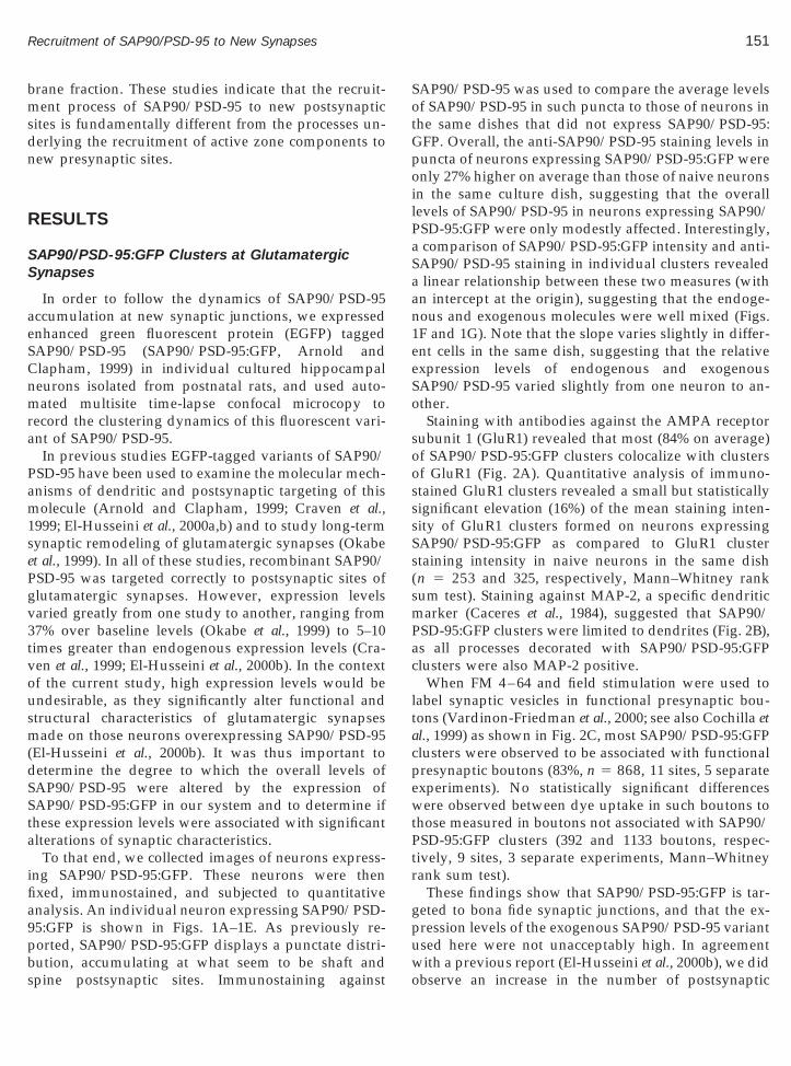
151Recruitment of SAP90/PSD-95 to New Synapses
brane fraction. These studies indicate that the recruit-ment process of SAP90/PSD-95 to new postsynapticsites is fundamentally different from the processes un-derlying the recruitment of active zone components tonew presynaptic sites.
RESULTS
SAP90/PSD-95:GFP Clusters at GlutamatergicSynapses
In order to follow the dynamics of SAP90/PSD-95accumulation at new synaptic junctions, we expressedenhanced green fluorescent protein (EGFP) taggedSAP90/PSD-95 (SAP90/PSD-95:GFP, Arnold andClapham, 1999) in individual cultured hippocampalneurons isolated from postnatal rats, and used auto-mated multisite time-lapse confocal microcopy torecord the clustering dynamics of this fluorescent vari-ant of SAP90/PSD-95.
In previous studies EGFP-tagged variants of SAP90/PSD-95 have been used to examine the molecular mech-anisms of dendritic and postsynaptic targeting of thismolecule (Arnold and Clapham, 1999; Craven et al.,1999; El-Husseini et al., 2000a,b) and to study long-termsynaptic remodeling of glutamatergic synapses (Okabeet al., 1999). In all of these studies, recombinant SAP90/PSD-95 was targeted correctly to postsynaptic sites ofglutamatergic synapses. However, expression levelsvaried greatly from one study to another, ranging from37% over baseline levels (Okabe et al., 1999) to 5–10times greater than endogenous expression levels (Cra-ven et al., 1999; El-Husseini et al., 2000b). In the contextof the current study, high expression levels would beundesirable, as they significantly alter functional andstructural characteristics of glutamatergic synapsesmade on those neurons overexpressing SAP90/PSD-95(El-Husseini et al., 2000b). It was thus important todetermine the degree to which the overall levels ofSAP90/PSD-95 were altered by the expression ofSAP90/PSD-95:GFP in our system and to determine ifthese expression levels were associated with significantalterations of synaptic characteristics.
To that end, we collected images of neurons express-ing SAP90/PSD-95:GFP. These neurons were thenfixed, immunostained, and subjected to quantitativeanalysis. An individual neuron expressing SAP90/PSD-95:GFP is shown in Figs. 1A–1E. As previously re-ported, SAP90/PSD-95:GFP displays a punctate distri-
bution, accumulating at what seem to be shaft andspine postsynaptic sites. Immunostaining againstSAP90/PSD-95 was used to compare the average levelsof SAP90/PSD-95 in such puncta to those of neurons inthe same dishes that did not express SAP90/PSD-95:GFP. Overall, the anti-SAP90/PSD-95 staining levels inpuncta of neurons expressing SAP90/PSD-95:GFP wereonly 27% higher on average than those of naive neuronsin the same culture dish, suggesting that the overalllevels of SAP90/PSD-95 in neurons expressing SAP90/PSD-95:GFP were only modestly affected. Interestingly,a comparison of SAP90/PSD-95:GFP intensity and anti-SAP90/PSD-95 staining in individual clusters revealeda linear relationship between these two measures (withan intercept at the origin), suggesting that the endoge-nous and exogenous molecules were well mixed (Figs.1F and 1G). Note that the slope varies slightly in differ-ent cells in the same dish, suggesting that the relativeexpression levels of endogenous and exogenousSAP90/PSD-95 varied slightly from one neuron to an-other.
Staining with antibodies against the AMPA receptorsubunit 1 (GluR1) revealed that most (84% on average)of SAP90/PSD-95:GFP clusters colocalize with clustersof GluR1 (Fig. 2A). Quantitative analysis of immuno-stained GluR1 clusters revealed a small but statisticallysignificant elevation (16%) of the mean staining inten-sity of GluR1 clusters formed on neurons expressingSAP90/PSD-95:GFP as compared to GluR1 clusterstaining intensity in naive neurons in the same dish(n 5 253 and 325, respectively, Mann–Whitney ranksum test). Staining against MAP-2, a specific dendriticmarker (Caceres et al., 1984), suggested that SAP90/PSD-95:GFP clusters were limited to dendrites (Fig. 2B),as all processes decorated with SAP90/PSD-95:GFPclusters were also MAP-2 positive.
When FM 4–64 and field stimulation were used tolabel synaptic vesicles in functional presynaptic bou-tons (Vardinon-Friedman et al., 2000; see also Cochilla etal., 1999) as shown in Fig. 2C, most SAP90/PSD-95:GFPclusters were observed to be associated with functionalpresynaptic boutons (83%, n 5 868, 11 sites, 5 separateexperiments). No statistically significant differenceswere observed between dye uptake in such boutons tothose measured in boutons not associated with SAP90/PSD-95:GFP clusters (392 and 1133 boutons, respec-tively, 9 sites, 3 separate experiments, Mann–Whitneyrank sum test).
These findings show that SAP90/PSD-95:GFP is tar-geted to bona fide synaptic junctions, and that the ex-pression levels of the exogenous SAP90/PSD-95 variantused here were not unacceptably high. In agreement
with a previous report (El-Husseini et al., 2000b), we didobserve an increase in the number of postsynaptic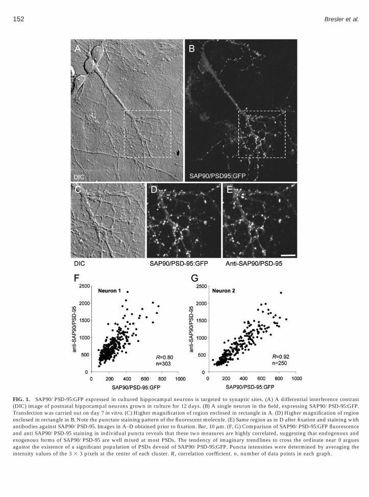
152 Bresler et al.
FIG. 1. SAP90/PSD-95:GFP expressed in cultured hippocampal neurons is targeted to synaptic sites. (A) A differential interference contrast(DIC) image of postnatal hippocampal neurons grown in culture for 12 days. (B) A single neuron in the field, expressing SAP90/PSD-95:GFP.Transfection was carried out on day 7 in vitro. (C) Higher magnification of region enclosed in rectangle in A. (D) Higher magnification of regionenclosed in rectangle in B. Note the punctate staining pattern of the fluorescent molecule. (E) Same region as in D after fixation and staining withantibodies against SAP90/PSD-95. Images in A–D obtained prior to fixation. Bar, 10 mm. (F, G) Comparison of SAP90/PSD-95:GFP fluorescenceand anti SAP90/PSD-95 staining in individual puncta reveals that these two measures are highly correlated, suggesting that endogenous andexogenous forms of SAP90/PSD-95 are well mixed at most PSDs. The tendency of imaginary trendlines to cross the ordinate near 0 argues
against the existence of a significant population of PSDs devoid of SAP90/PSD-95:GFP. Puncta intensities were determined by averaging theintensity values of the 3 3 3 pixels at the center of each cluster. R, correlation coefficient. n, number of data points in each graph.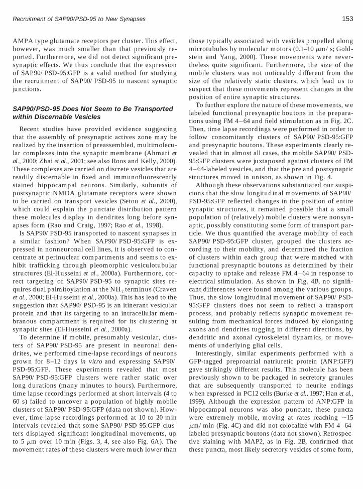
aTrspt
spbs
tdgPSlt6ceittm
1hw
153Recruitment of SAP90/PSD-95 to New Synapses
AMPA type glutamate receptors per cluster. This effect,however, was much smaller than that previously re-ported. Furthermore, we did not detect significant pre-synaptic effects. We thus conclude that the expressionof SAP90/PSD-95:GFP is a valid method for studyingthe recruitment of SAP90/PSD-95 to nascent synapticjunctions.
SAP90/PSD-95 Does Not Seem to Be Transportedwithin Discernable Vesicles
Recent studies have provided evidence suggestingthat the assembly of presynaptic actives zone may berealized by the insertion of preassembled, multimolecu-lar complexes into the synaptic membrane (Ahmari etl., 2000; Zhai et al., 2001; see also Roos and Kelly, 2000).hese complexes are carried on discrete vesicles that areeadily discernable in fixed and immunofluorescentlytained hippocampal neurons. Similarly, subunits ofostsynaptic NMDA glutamate receptors were shown
o be carried on transport vesicles (Setou et al., 2000),which could explain the punctate distribution patternthese molecules display in dendrites long before syn-apses form (Rao and Craig, 1997; Rao et al., 1998).
Is SAP90/PSD-95 transported to nascent synapses ina similar fashion? When SAP90/PSD-95:GFP is ex-pressed in nonneuronal cell lines, it is observed to con-centrate at perinuclear compartments and seems to ex-hibit trafficking through pleomorphic vesiculotubularstructures (El-Husseini et al., 2000a). Furthermore, cor-rect targeting of SAP90/PSD-95 to synaptic sites re-quires dual palmitoylation at the NH2 terminus (Cravenet al., 2000; El-Husseini et al., 2000a). This has lead to theuggestion that SAP90/PSD-95 is an itinerant vesicularrotein and that its targeting to an intracellular mem-ranous compartment is required for its clustering atynaptic sites (El-Husseini et al., 2000a).
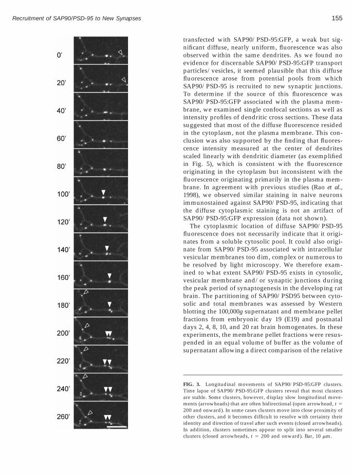
To determine if mobile, presumably vesicular, clus-ers of SAP90/PSD-95 are present in neuronal den-rites, we performed time-lapse recordings of neuronsrown for 8–12 days in vitro and expressing SAP90/SD-95:GFP. These experiments revealed that mostAP90/PSD-95:GFP clusters were rather static overong durations (many minutes to hours). Furthermore,ime lapse recordings performed at short intervals (4 to0 s) failed to uncover a population of highly mobilelusters of SAP90/PSD-95:GFP (data not shown). How-ver, time-lapse recordings performed at 10 to 20 minntervals revealed that some SAP90/PSD-95:GFP clus-ers displayed significant longitudinal movements, up
o 5 mm over 10 min (Figs. 3, 4, see also Fig. 6A). Theovement rates of these clusters were much lower than
those typically associated with vesicles propelled alongmicrotubules by molecular motors (0.1–10 mm/s; Gold-stein and Yang, 2000). These movements were never-theless quite significant. Furthermore, the size of themobile clusters was not noticeably different from thesize of the relatively static clusters, which lead us tosuspect that these movements represent changes in theposition of entire synaptic structures.
To further explore the nature of these movements, welabeled functional presynaptic boutons in the prepara-tions using FM 4–64 and field stimulation as in Fig. 2C.Then, time lapse recordings were performed in order tofollow concomitantly clusters of SAP90/PSD-95:GFPand presynaptic boutons. These experiments clearly re-vealed that in almost all cases, the mobile SAP90/PSD-95:GFP clusters were juxtaposed against clusters of FM4–64-labeled vesicles, and that the pre and postsynapticstructures moved in unison, as shown in Fig. 4.
Although these observations substantiated our suspi-cions that the slow longitudinal movements of SAP90/PSD-95:GFP reflected changes in the position of entiresynaptic structures, it remained possible that a smallpopulation of (relatively) mobile clusters were nonsyn-aptic, possibly constituting some form of transport par-ticle. We thus quantified the average mobility of eachSAP90/PSD-95:GFP cluster, grouped the clusters ac-cording to their mobility, and determined the fractionof clusters within each group that were matched withfunctional presynaptic boutons as determined by theircapacity to uptake and release FM 4–64 in response toelectrical stimulation. As shown in Fig. 4B, no signifi-cant differences were found among the various groups.Thus, the slow longitudinal movement of SAP90/PSD-95:GFP clusters does not seem to reflect a transportprocess, and probably reflects synaptic movement re-sulting from mechanical forces induced by elongatingaxons and dendrites tugging in different directions, bydendritic and axonal cytoskeletal dynamics, or move-ments of underlying glial cells.
Interestingly, similar experiments performed with aGFP-tagged preproatrial natriuretic protein (ANP:GFP)gave strikingly different results. This molecule has beenpreviously shown to be packaged in secretory granulesthat are subsequently transported to neurite endingswhen expressed in PC12 cells (Burke et al., 1997; Han et al.,999). Although the expression pattern of ANP:GFP inippocampal neurons was also punctate, these punctaere extremely mobile, moving at rates reaching ;15
mm/min (Fig. 4C) and did not colocalize with FM 4–64-labeled presynaptic boutons (data not shown). Retrospec-
tive staining with MAP2, as in Fig. 2B, confirmed thatthese puncta, most likely secretory vesicles of some form,
een)
154 Bresler et al.
were being transported along dendritic processes (datanot shown). Although ANP is an exogenous molecule, itsuse in these experiments was instrumental for providingsome idea as to the characteristics of fast dendritic trans-port processes in cultured hippocampal neurons, andserved to demonstrate that if SAP90/PSD-95:GFP hadexhibited similar characteristics, this would have beendetected in our system.
To summarize, we found no evidence in favor of the
FIG. 2. SAP90/PSD-95:GFP clusters are postsynaptic and are assocneurons expressing SAP90/PSD-95:GFP with antibodies against tcorrespondence between clusters of SAP90/PSD-95:GFP and GluRglutamatergic synapses. (B) Retrospective staining of neurons exprdendritic market MAP2 (blue) confirms the dendritic localization of Swith FM 4–64 (red) reveals that most SAP90/PSD-95:GFP clusters (gr5 mm (B, C).
possibility that SAP90/PSD-95 is transported alongdendrites within readily discernable transport vesicles
as shown for other PSD components such as NMDAreceptor subunit 2 (Setou et al., 2000) or presynapticCAZ components such as Bassoon and Piccolo (Zhai etal., 2001), suggesting that SAP90/PSD-95 is recruited tonascent synaptic junctions in an alternative fashion.
Dendritic SAP90/PSD-95 Is Found in Both LightMembranal and Cytosolic Fractions
with functional presynaptic boutons. (A) Retrospective staining ofMPA-type glutamate receptor subunit GluR1. Note the excellentuggesting that most SAP90/PSD-95:GFP clusters are localized tog SAP90/PSD-95:GFP (green) with antibodies against the specific/PSD-95:GFP puncta. (C) Labeling of functional presynaptic boutonsare associated with functional presynaptic boutons. Bars, 10 mm (A),
iatedhe A
1, sessin
AP90
In addition to the punctate synaptic distribution ob-served on somata and dendritic profiles of neurons
155Recruitment of SAP90/PSD-95 to New Synapses
transfected with SAP90/PSD-95:GFP, a weak but sig-nificant diffuse, nearly uniform, fluorescence was alsoobserved within the same dendrites. As we found noevidence for discernable SAP90/PSD-95:GFP transportparticles/vesicles, it seemed plausible that this diffusefluorescence arose from potential pools from whichSAP90/PSD-95 is recruited to new synaptic junctions.To determine if the source of this fluorescence wasSAP90/PSD-95:GFP associated with the plasma mem-brane, we examined single confocal sections as well asintensity profiles of dendritic cross sections. These datasuggested that most of the diffuse fluorescence residedin the cytoplasm, not the plasma membrane. This con-clusion was also supported by the finding that fluores-cence intensity measured at the center of dendritesscaled linearly with dendritic diameter (as exemplifiedin Fig. 5), which is consistent with the fluorescenceoriginating in the cytoplasm but inconsistent with thefluorescence originating primarily in the plasma mem-brane. In agreement with previous studies (Rao et al.,1998), we observed similar staining in naive neuronsimmunostained against SAP90/PSD-95, indicating thatthe diffuse cytoplasmic staining is not an artifact ofSAP90/PSD-95:GFP expression (data not shown).
The cytoplasmic location of diffuse SAP90/PSD-95fluorescence does not necessarily indicate that it origi-nates from a soluble cytosolic pool. It could also origi-nate from SAP90/PSD-95 associated with intracellularvesicular membranes too dim, complex or numerous tobe resolved by light microscopy. We therefore exam-ined to what extent SAP90/PSD-95 exists in cytosolic,vesicular membrane and/or synaptic junctions duringthe peak period of synaptogenesis in the developing ratbrain. The partitioning of SAP90/PSD95 between cyto-solic and total membranes was assessed by Westernblotting the 100,000g supernatant and membrane pelletfractions from embryonic day 19 (E19) and postnataldays 2, 4, 8, 10, and 20 rat brain homogenates. In theseexperiments, the membrane pellet fractions were resus-pended in an equal volume of buffer as the volume ofsupernatant allowing a direct comparison of the relative
FIG. 3. Longitudinal movements of SAP90/PSD-95:GFP clusters.Time lapse of SAP90/PSD-95:GFP clusters reveal that most clustersare stable. Some clusters, however, display slow longitudinal move-ments (arrowheads) that are often bidirectional (open arrowhead, t 5200 and onward). In some cases clusters move into close proximity ofother clusters, and it becomes difficult to resolve with certainty theiridentity and direction of travel after such events (closed arrowheads).
In addition, clusters sometimes appear to split into several smallerclusters (closed arrowheads, t 5 200 and onward). Bar, 10 mm.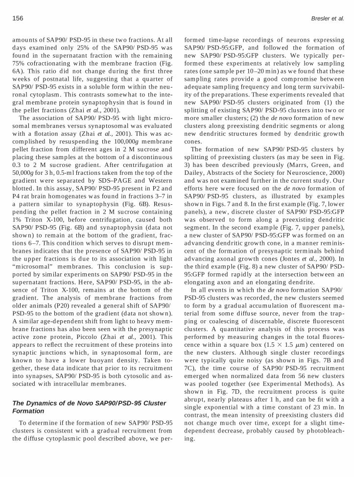
pp05gbPap1Sstbt“pssgoPAbaaskgis
TF
ct
fSnfrsainsmcnc
s3DaeSspwsaacat9e
Pttpcpc
156 Bresler et al.
amounts of SAP90/PSD-95 in these two fractions. At alldays examined only 25% of the SAP90/PSD-95 wasfound in the supernatant fraction with the remaining75% cofractionating with the membrane fraction (Fig.6A). This ratio did not change during the first threeweeks of postnatal life, suggesting that a quarter ofSAP90/PSD-95 exists in a soluble form within the neu-ronal cytoplasm. This contrasts somewhat to the inte-gral membrane protein synaptophysin that is found inthe pellet fractions (Zhai et al., 2001).
The association of SAP90/PSD-95 with light micro-somal membranes versus synaptosomal was evaluatedwith a flotation assay (Zhai et al., 2001). This was ac-complished by resuspending the 100,000g membrane
ellet fraction from different ages in 2 M sucrose andlacing these samples at the bottom of a discontinuous.3 to 2 M sucrose gradient. After centrifugation at0,000g for 3 h, 0.5-ml fractions taken from the top of theradient were separated by SDS–PAGE and Westernlotted. In this assay, SAP90/PSD-95 present in P2 and4 rat brain homogenates was found in fractions 3–7 inpattern similar to synaptophysin (Fig. 6B). Resus-
ending the pellet fraction in 2 M sucrose containing% Triton X-100, before centrifugation, caused bothAP90/PSD-95 (Fig. 6B) and synaptophysin (data nothown) to remain at the bottom of the gradient, frac-ions 6–7. This condition which serves to disrupt mem-ranes indicates that the presence of SAP90/PSD-95 inhe upper fractions is due to its association with lightmicrosomal” membranes. This conclusion is sup-orted by similar experiments on SAP90/PSD-95 in theupernatant fractions. Here, SAP90/PSD-95, in the ab-ence of Triton X-100, remains at the bottom of theradient. The analysis of membrane fractions fromlder animals (P20) revealed a general shift of SAP90/SD-95 to the bottom of the gradient (data not shown).similar age-dependent shift from light to heavy mem-
rane fractions has also been seen with the presynapticctive zone protein, Piccolo (Zhai et al., 2001). Thisppears to reflect the recruitment of these proteins intoynaptic junctions which, in synaptosomal form, arenown to have a lower buoyant density. Taken to-ether, these data indicate that prior to its recruitment
nto synapses, SAP90/PSD-95 is both cytosolic and as-ociated with intracellular membranes.
he Dynamics of de Novo SAP90/PSD-95 Clusterormation
To determine if the formation of new SAP90/PSD-95
lusters is consistent with a gradual recruitment fromhe diffuse cytoplasmic pool described above, we per-ormed time-lapse recordings of neurons expressingAP90/PSD-95:GFP, and followed the formation ofew SAP90/PSD-95:GFP clusters. We typically per-ormed these experiments at relatively low samplingates (one sample per 10–20 min) as we found that theseampling rates provide a good compromise betweendequate sampling frequency and long term survivabil-ty of the preparations. These experiments revealed thatew SAP90/PSD-95 clusters originated from (1) theplitting of existing SAP90/PSD-95 clusters into two orore smaller clusters; (2) the de novo formation of new
lusters along preexisting dendritic segments or alongew dendritic structures formed by dendritic growthones.
The formation of new SAP90/PSD-95 clusters byplitting of preexisting clusters (as may be seen in Fig.) has been described previously (Marrs, Green, andailey, Abstracts of the Society for Neuroscience, 2000)
nd was not examined further in the current study. Ourfforts here were focused on the de novo formation ofAP90/PSD-95 clusters, as illustrated by exampleshown in Figs. 7 and 8. In the first example (Fig. 7, loweranels), a new, discrete cluster of SAP90/PSD-95:GFPas observed to form along a preexisting dendritic
egment. In the second example (Fig. 7, upper panels),new cluster of SAP90/PSD-95:GFP was formed on an
dvancing dendritic growth cone, in a manner reminis-ent of the formation of presynaptic terminals behinddvancing axonal growth cones (Jontes et al., 2000). Inhe third example (Fig. 8) a new cluster of SAP90/PSD-5:GFP formed rapidly at the intersection between anlongating axon and an elongating dendrite.
In all events in which the de novo formation SAP90/SD-95 clusters was recorded, the new clusters seemed
o form by a gradual accumulation of fluorescent ma-erial from some diffuse source, never from the trap-ing or coalescing of discernable, discrete fluorescentlusters. A quantitative analysis of this process waserformed by measuring changes in the total fluores-ence within a square box (1.5 3 1.5 mm) centered on
the new clusters. Although single cluster recordingswere typically quite noisy (as shown in Figs. 7B and7C), the time course of SAP90/PSD-95 recruitmentemerged when normalized data from 56 new clusterswas pooled together (see Experimental Methods). Asshown in Fig. 7D, the recruitment process is quiteabrupt, nearly plateaus after 1 h, and can be fit with asingle exponential with a time constant of 23 min. Incontrast, the mean intensity of preexisting clusters didnot change much over time, except for a slight time-
dependent decrease, probably caused by photobleach-ing.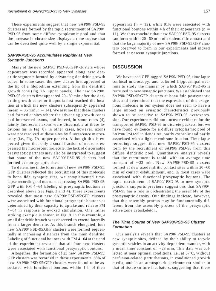
157Recruitment of SAP90/PSD-95 to New Synapses
These experiments suggest that new SAP90/PSD-95clusters are formed by the rapid recruitment of SAP90/PSD-95 from some diffuse cytoplasmic pool and thatthe increase in cluster size displays a time course thatcan be described quite well by a single exponential.
SAP90/PSD-95 Accumulates Rapidly at NewSynaptic Junctions
Many of the new SAP90/PSD-95:GFP clusters whoseappearance was recorded appeared along new den-dritic segments formed by advancing dendritic growthcones. In some cases, the new cluster first appeared atthe tip of a filopodium extending from the dendriticgrowth cone (Fig. 7A, upper panels). The new SAP90/PSD-95 clusters first appeared 20–60 min after the den-dritic growth cones or filopodia first reached the loca-tion at which the new clusters subsequently appeared(mean 5 36 min, n 5 14). We assume that these clustershad formed at sites where the advancing growth coneshad intersected axons, and indeed, in some cases (4),faintly fluorescent axons were discernable at these lo-cations (as in Fig. 8). In other cases, however, axonswere not resolved at these sites by fluorescence micros-copy (i.e., Fig. 7A, upper panels). Although this is ex-pected given that only a small fraction of neurons ex-pressed the fluorescent molecule, the lack of discernablepresynaptic partners may also be interpreted to suggestthat some of the new SAP90/PSD-95 clusters hadformed at non-synaptic sites.
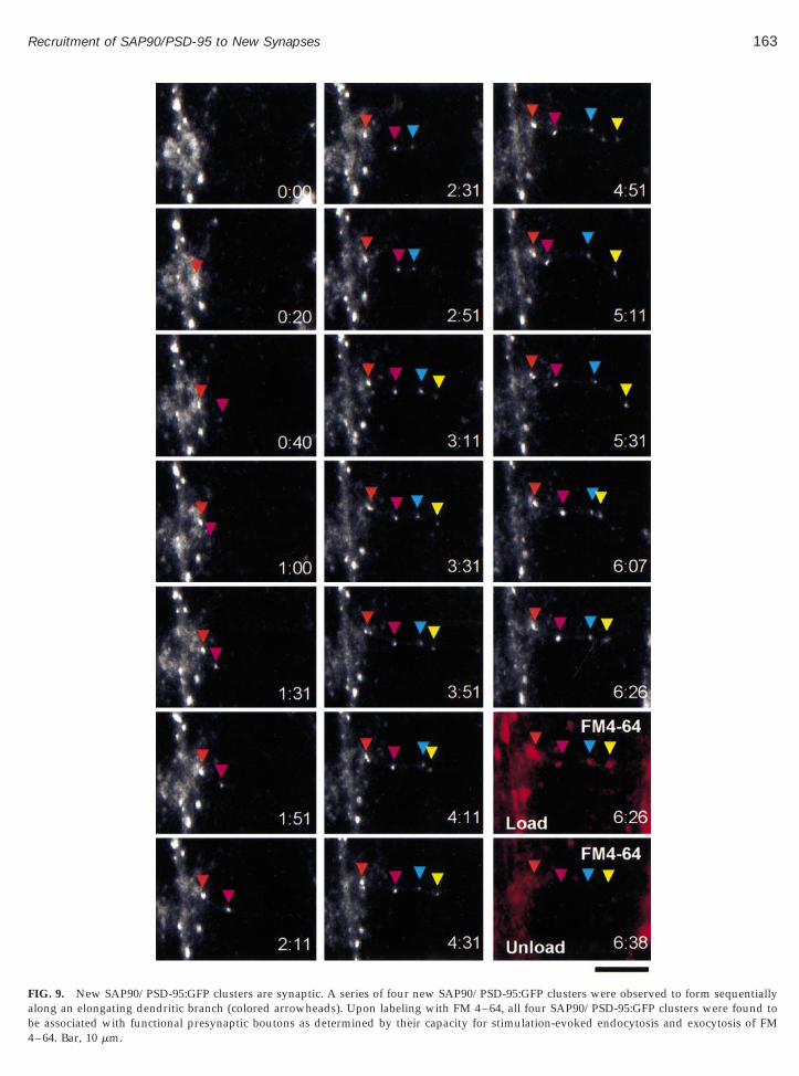
To determine if the formation of new SAP90/PSD-95:GFP clusters reflected the recruitment of this moleculeto bona fide synaptic sites, we complemented time-lapse recordings of neurons expressing SAP90/PSD-95:GFP with FM 4–64 labeling of presynaptic boutons asdescribed above (see Figs. 2 and 4). These experimentsrevealed that most new SAP90/PSD-95:GFP clusterswere associated with functional presynaptic boutons asdetermined by their capacity to uptake and release FM4–64 in response to evoked stimulation. One ratherstriking example is shown in Fig. 9. In this example, asmall dendritic branch was observed to extend laterallyfrom a larger dendrite. As this branch elongated, fournew SAP90/PSD-95:GFP clusters were formed sequen-tially at increasing distances from the main dendrite.Labeling of functional boutons with FM 4–64 at the endof the experiment revealed that all four new clusterswere associated with functional presynaptic boutons.
Altogether, the formation of 23 new SAP90/PSD-95:GFP clusters was recorded in these experiments. 58% of
new SAP90/PSD-95:GFP clusters were found to be as-sociated with functional boutons within 1 h of theirappearance (n 5 12), while 91% were associated withfunctional boutons within 4 h of their appearance (n 511). We thus conclude that new SAP90/PSD-95 clusterscan form within 20–60 min of axodendritic contact andthat the large majority of new SAP90/PSD-95:GFP clus-ters observed to form in our experiments had indeedformed at nascent synaptic junctions.
DISCUSSION
We have used GFP-tagged SAP90/PSD-95, time lapseconfocal microscopy, and cultured hippocampal neu-rons to study the manner by which SAP90/PSD-95 isrecruited to new synaptic junctions. We established thatSAP90/PSD-95:GFP clusters at bona fide postsynapticsites and determined that the expression of this exoge-nous molecule in our system does not seem to have alarge impact on synaptic characteristics previouslyshown to be sensitive to SAP90/PSD-95 overexpres-sion. Our experiments did not uncover evidence for thetransport of SAP90/PSD-95 in discrete packets, but wehave found evidence for a diffuse cytoplasmic pool ofSAP90/PSD-95 in dendrites, partly cytosolic and partlyassociated with a light membrane fraction. Time lapserecordings suggest that new SAP90/PSD-95 clustersform by the recruitment of SAP90/PSD-95 from thisdiffuse dendritic pool. Quantitative analysis revealsthat the recruitment is rapid, with an average timeconstant of ;23 min. New SAP90/PSD-95 clustersformed at new axodendritic contact sites within 20–60min of contact establishment, and in most cases wereassociated with functional presynaptic boutons. Therapid recruitment of SAP90/PSD-95 to new synapticjunctions supports previous suggestions that SAP90/PSD-95 has a role in orchestrating the assembly of thepostsynaptic density. Our findings indicate, however,that this assembly process may be fundamentally dif-ferent from the assembly process of the presynapticactive zone cytoskeleton.
The Time Course of New SAP90/PSD-95 ClusterFormation
Our analysis reveals that SAP90/PSD-95 clusters atnew synaptic sites, defined by their ability to recyclesynaptic vesicles in an activity-dependent manner, witha mean time constant of ;23 min. This data was col-lected at near optimal conditions, i.e., at 37°C, withoutperfusion-related perturbations, in conditioned growth
media, and in an atmospheric environment similar tothat of tissue culture incubators, suggesting that these
rvalnsityplaye
158 Bresler et al.
experiments provide a reasonable approximation of
FIG. 4. Most slow longitudinal movements of SAP90/PSD-95:GFP cluSAP90/PSD-95:GFP clusters (green) and functional presynaptic boutons95:GFP clusters usually occurred in unison with the migration of the pseparate synapses). The functional status of these presynaptic boutons wtrain of action potentials (Unload I, 120 s at 10 Hz), reloading them withclarity the FM 4–64 images shown here were offset by 5 pixels to the left(B) Quantitative analysis revealed no significant correlation between thethe probability that the cluster is associated with a functional presynaptinonsynaptic, SAP90/PSD-95:GFP clusters. All data was obtained fromhippocampal neurons. Left panel, low magnification image of a cultureof the fluorescent neuropeptide along the dendritic segments shown hestaining with MAP2, not shown here). Time lapse sequence of the regionof mobility displayed by ANP:GFP clusters (the inter-frame time inte(presumably vesicles of some sort) precluded the display of maximal intetime required to collect an image stack, thus only single sections are dis
physiological rates of SAP90/PSD-95 clustering at newsynaptic sites. Yet we cannot completely rule out the
possibility that the rapid accumulation rates are some-
represent the migration of entire synapses. (A) Concomitant imaging ofled with FM 4–64 (red) reveals that the slow migration of SAP90/PSD-
aptic bouton associated with this cluster (pink and cyan arrows, twonfirmed by releasing the dye trapped within presynaptic vesicles by a
4–64 (Load II), and unloading them again (Unload II). For purposes of5 pixels to the top relative to the SAP90/PSD-95:GFP images. Bar, 5 mm.ge longitudinal mobility displayed by a SAP90/PSD-95:GFP cluster andton, arguing against the existence of a significant population of mobile,rons grown in culture for 8–12 days. (C) Expression of ANP:GFP inpocampal neuron expressing ANP:GFP. Note the punctate distributione dendritic identity of these segments was confirmed by retrospectivesed in rectangle is shown in the right-hand panels. Note the high degreehere is only 2 min). In fact, the mobility displayed by these punctaprojections of image stacks, as puncta changed their position during thed here. Bar, 5 mm.
sterslaberesynas coFM
andaverac bou
neud hipre (thenclo
how related to the use of SAP90/PSD-95:GFP or cellcultures. Interestingly, the formation of new SAP90/
ns
159Recruitment of SAP90/PSD-95 to New Synapses
PSD-95 clusters in older and more stable hippocampalcultures seems to occur much more slowly, on a timescale of hours (Okabe et al., 1999). We suspect that the
FIG. 5. Diffuse dendritic fluorescence of SAP90/PSD-95:GFP origi-ates in cytoplasm. (A) A summed intensity projection of 4 sectionspaced 1.2 mm apart of a hippocampal neuron expressing SAP90/PSD-
95:GFP. Note that the diffuse fluorescence is not brighter along thedendrite’s sides. (B) The intensity of the diffuse dendritic fluorescencewas measured at 13 points along the dendrite indicated by arrow pairsin A. In all cases the intensity was measured from a narrow region at thecenter of the dendrite (between the arrow pairs). Background fluores-cence values were then measured outside the dendrite for each regionseparately, and subtracted from the measurements made for these re-gions. The corrected data was then compared with the diameter of thedendrite at the point of measurement. This comparison revealed a linearrelationship between dendrite diameter and diffuse fluorescence inten-sity suggesting that this diffuse fluorescence originates in the cytoplasm,not the dendritic plasma membrane. Bar, 10 mm.
different kinetics may reflect a difference in the abun-dance of SAP90/PSD-95 free to accumulate at new sites.
The relatively rapid recruitment of SAP90/PSD-95 tonew synaptic sites is in agreement with a prior studywe performed in which FM 4–64 labeling was used todetect new functional presynaptic boutons and retro-spective immunohistochemistry was used to analyzethe composition of these presumably new synapticjunctions (Vardinon-Friedman et al., 2000). This analysisrevealed that by the time such new presynaptic boutonsacquire a capacity for evoked endocytosis and exocyto-sis, about half of them are associated with clusters ofSAP90/PSD-95. We proposed that the recruitment ofSAP90/PSD-95 to nascent synapses may be delayed inrespect to the formation of the presynaptic bouton, butwe also pointed out that these findings are consistentwith the possibility that presynaptic bouton formationand SAP90/PSD-95 recruitment occur in parallel. Ourcurrent finding that about 60% of new SAP90/PSD-95clusters are associated with functional presynaptic bou-tons within one hour tends to support the latter possi-bility. In any case, the current study (and in particularobservations such as those in Fig. 8) strongly supportour previous conclusions that new glutamatergic syn-apses may form rapidly, within 1–2 h from contactestablishment (Vardinon-Friedman et al., 2000; see alsoAhmari et al., 2000).
Source of SAP90/PSD-95 Recruited to NewSynaptic Sites
We have not found any evidence for the existence ofa population of discrete transport vesicles containingSAP90/PSD-95 or for the assembly of new SAP90/PSD-95:GFP puncta by the coalescence of small discrete clus-ters. It could be argued that such events could havebeen missed by the relatively slow sampling rate wetypically used (one sample per 10–20 min). As notedabove, however, time-lapse recordings performed athigher sampling rates (up to one sample per 4 s) failedto reveal the existence of a dynamic pool of transportparticles even remotely similar to those observed inneurons expressing ANP:GFP.
We assume that the diffuse cytoplasmic SAP90/PSD-95 observed in dendrites is composed of cytosolicSAP90/PSD-95, but we cannot rule out that this diffusedistribution also reflects SAP90/PSD-95 associated withmembranal organelles too dim, too numerous or tooconvoluted to be resolved at the light microscopy level.Indeed, our biochemical analysis points to the existenceof two pools of SAP90/PSD-95, one cytosolic and asecond associated with light membranes (Fig. 6). It is
worth noting that AMPA-type glutamate receptors, thatare integral membranal proteins, also display a diffuse,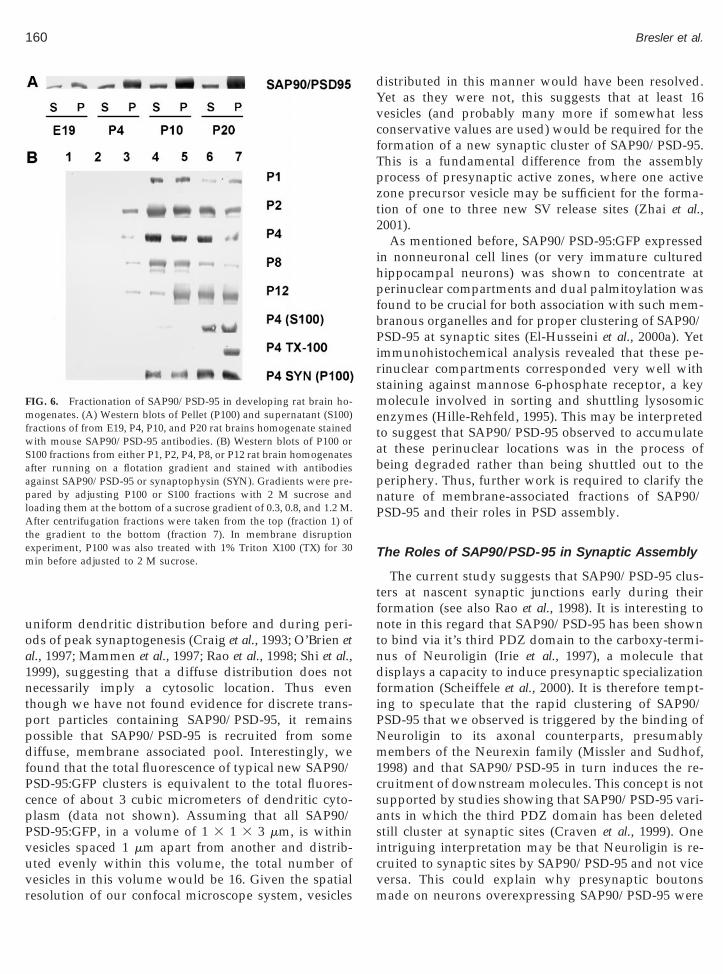
mfwSaap
160 Bresler et al.
uniform dendritic distribution before and during peri-ods of peak synaptogenesis (Craig et al., 1993; O’Brien etal., 1997; Mammen et al., 1997; Rao et al., 1998; Shi et al.,1999), suggesting that a diffuse distribution does notnecessarily imply a cytosolic location. Thus eventhough we have not found evidence for discrete trans-port particles containing SAP90/PSD-95, it remainspossible that SAP90/PSD-95 is recruited from somediffuse, membrane associated pool. Interestingly, wefound that the total fluorescence of typical new SAP90/PSD-95:GFP clusters is equivalent to the total fluores-cence of about 3 cubic micrometers of dendritic cyto-plasm (data not shown). Assuming that all SAP90/PSD-95:GFP, in a volume of 1 3 1 3 3 mm, is withinvesicles spaced 1 mm apart from another and distrib-uted evenly within this volume, the total number of
FIG. 6. Fractionation of SAP90/PSD-95 in developing rat brain ho-ogenates. (A) Western blots of Pellet (P100) and supernatant (S100)
ractions of from E19, P4, P10, and P20 rat brains homogenate stainedith mouse SAP90/PSD-95 antibodies. (B) Western blots of P100 or
100 fractions from either P1, P2, P4, P8, or P12 rat brain homogenatesfter running on a flotation gradient and stained with antibodiesgainst SAP90/PSD-95 or synaptophysin (SYN). Gradients were pre-ared by adjusting P100 or S100 fractions with 2 M sucrose and
loading them at the bottom of a sucrose gradient of 0.3, 0.8, and 1.2 M.After centrifugation fractions were taken from the top (fraction 1) ofthe gradient to the bottom (fraction 7). In membrane disruptionexperiment, P100 was also treated with 1% Triton X100 (TX) for 30min before adjusted to 2 M sucrose.
vesicles in this volume would be 16. Given the spatialresolution of our confocal microscope system, vesicles
distributed in this manner would have been resolved.Yet as they were not, this suggests that at least 16vesicles (and probably many more if somewhat lessconservative values are used) would be required for theformation of a new synaptic cluster of SAP90/PSD-95.This is a fundamental difference from the assemblyprocess of presynaptic active zones, where one activezone precursor vesicle may be sufficient for the forma-tion of one to three new SV release sites (Zhai et al.,2001).
As mentioned before, SAP90/PSD-95:GFP expressedin nonneuronal cell lines (or very immature culturedhippocampal neurons) was shown to concentrate atperinuclear compartments and dual palmitoylation wasfound to be crucial for both association with such mem-branous organelles and for proper clustering of SAP90/PSD-95 at synaptic sites (El-Husseini et al., 2000a). Yetimmunohistochemical analysis revealed that these pe-rinuclear compartments corresponded very well withstaining against mannose 6-phosphate receptor, a keymolecule involved in sorting and shuttling lysosomicenzymes (Hille-Rehfeld, 1995). This may be interpretedto suggest that SAP90/PSD-95 observed to accumulateat these perinuclear locations was in the process ofbeing degraded rather than being shuttled out to theperiphery. Thus, further work is required to clarify thenature of membrane-associated fractions of SAP90/PSD-95 and their roles in PSD assembly.
The Roles of SAP90/PSD-95 in Synaptic Assembly
The current study suggests that SAP90/PSD-95 clus-ters at nascent synaptic junctions early during theirformation (see also Rao et al., 1998). It is interesting tonote in this regard that SAP90/PSD-95 has been shownto bind via it’s third PDZ domain to the carboxy-termi-nus of Neuroligin (Irie et al., 1997), a molecule thatdisplays a capacity to induce presynaptic specializationformation (Scheiffele et al., 2000). It is therefore tempt-ing to speculate that the rapid clustering of SAP90/PSD-95 that we observed is triggered by the binding ofNeuroligin to its axonal counterparts, presumablymembers of the Neurexin family (Missler and Sudhof,1998) and that SAP90/PSD-95 in turn induces the re-cruitment of downstream molecules. This concept is notsupported by studies showing that SAP90/PSD-95 vari-ants in which the third PDZ domain has been deletedstill cluster at synaptic sites (Craven et al., 1999). Oneintriguing interpretation may be that Neuroligin is re-cruited to synaptic sites by SAP90/PSD-95 and not vice
versa. This could explain why presynaptic boutonsmade on neurons overexpressing SAP90/PSD-95 were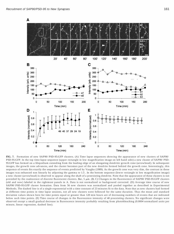
161Recruitment of SAP90/PSD-95 to New Synapses
FIG. 7. Formation of new SAP90/PSD-95:GFP clusters. (A) Time lapse sequences showing the appearance of new clusters of SAP90/PSD-95:GFP. In the top time-lapse sequence (upper rectangle in low magnification image on left hand side) a new cluster of SAP90/PSD-95:GFP has formed on a filopodium extending from the leading edge of an elongating dendritic growth cone (arrowhead). In subsequentimages, the growth cone advances, and the cluster becomes part of the new dendrite formed behind the growth cone. Interestingly, thissequence of events fits exactly the sequence of events predicted by Vaughn (1989). As the growth cone was very dim, the contrast in theseimages was enhanced non linearly by adjusting the gamma to 1.5 . In the bottom sequence (lower rectangle in low magnification image)a new cluster (arrowhead) is observed to appear along the shaft of a preexisting dendrite. Note that the appearance of these clusters is notpreceded by the coalescence of discrete fluorescent clusters. Bar, 5 mm. (B, C) Changes in the fluorescence of SAP90/PSD-95:GFP clusters(old and new) labeled in the rightmost panels in A. Data is not normalized or background corrected. (D) Average time course of newSAP90/PSD-95:GFP cluster formation. Data from 56 new clusters was normalized and pooled together as described in ExperimentalMethods. The dashed line is of a single exponential with a time constant of 23 minutes fit to the data. Note that as new clusters had formedat different time points in time lapse sessions, not all new clusters were followed for the same duration. Thus the mean and standarddeviation values shown here for time points equal or greater than 120 min hours are of decreasing numbers of events that are indicatedbelow such time points. (E) Time course of changes in the fluorescence intensity of 40 preexisting clusters. No significant changes were
observed except a small gradual decrease in fluorescence intensity probably resulting from photobleaching (0.0004 normalized units perminute, linear regression, dashed line).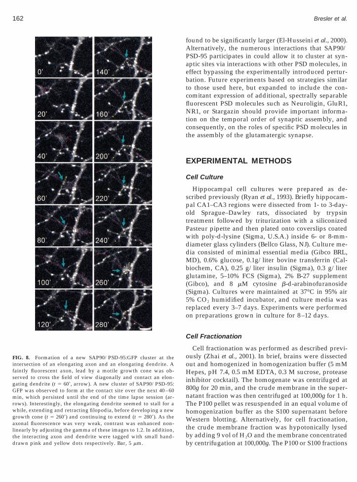
oHi8nThWtb
Gmrwg
the interacting axon and dendrite were tagged with small hand-drawn pink and yellow dots respectively. Bar, 5 mm.
162 Bresler et al.
found to be significantly larger (El-Husseini et al., 2000).Alternatively, the numerous interactions that SAP90/PSD-95 participates in could allow it to cluster at syn-aptic sites via interactions with other PSD molecules, ineffect bypassing the experimentally introduced pertur-bation. Future experiments based on strategies similarto those used here, but expanded to include the con-comitant expression of additional, spectrally separablefluorescent PSD molecules such as Neuroligin, GluR1,NR1, or Stargazin should provide important informa-tion on the temporal order of synaptic assembly, andconsequently, on the roles of specific PSD molecules inthe assembly of the glutamatergic synapse.
EXPERIMENTAL METHODS
Cell Culture
Hippocampal cell cultures were prepared as de-scribed previously (Ryan et al., 1993). Briefly hippocam-pal CA1–CA3 regions were dissected from 1- to 3-day-old Sprague–Dawley rats, dissociated by trypsintreatment followed by triturization with a siliconizedPasteur pipette and then plated onto coverslips coatedwith poly-d-lysine (Sigma, U.S.A.) inside 6- or 8-mm-diameter glass cylinders (Bellco Glass, NJ). Culture me-dia consisted of minimal essential media (Gibco BRL,MD), 0.6% glucose, 0.1g/liter bovine transferrin (Cal-biochem, CA), 0.25 g/liter insulin (Sigma), 0.3 g/literglutamine, 5–10% FCS (Sigma), 2% B-27 supplement(Gibco), and 8 mM cytosine b-d-arabinofuranoside(Sigma). Cultures were maintained at 37°C in 95% air5% CO2 humidified incubator, and culture media wasreplaced every 3–7 days. Experiments were performedon preparations grown in culture for 8–12 days.
Cell Fractionation
Cell fractionation was performed as described previ-ously (Zhai et al., 2001). In brief, brains were dissected
ut and homogenized in homogenization buffer (5 mMepes, pH 7.4, 0.5 mM EDTA, 0.3 M sucrose, protease
nhibitor cocktail). The homogenate was centrifuged at00g for 20 min, and the crude membrane in the super-atant fraction was then centrifuged at 100,000g for 1 h.he P100 pellet was resuspended in an equal volume ofomogenization buffer as the S100 supernatant beforeestern blotting. Alternatively, for cell fractionation,
he crude membrane fraction was hypotonically lysed
FIG. 8. Formation of a new SAP90/PSD-95:GFP cluster at theintersection of an elongating axon and an elongating dendrite. Afaintly fluorescent axon, lead by a motile growth cone was ob-served to cross the field of view diagonally and contact an elon-gating dendrite (t 5 609, arrow). A new cluster of SAP90/PSD-95:
FP was observed to form at the contact site over the next 40 – 60in, which persisted until the end of the time lapse session (ar-
ows). Interestingly, the elongating dendrite seemed to stall for ahile, extending and retracting filopodia, before developing a new
rowth cone (t 5 2609) and continuing to extend (t 5 2809). As theaxonal fluorescence was very weak, contrast was enhanced non-linearly by adjusting the gamma of these images to 1.2. In addition,
y adding 9 vol of H2O and the membrane concentratedby centrifugation at 100,000g. The P100 or S100 fractions
ab4
163Recruitment of SAP90/PSD-95 to New Synapses
FIG. 9. New SAP90/PSD-95:GFP clusters are synaptic. A series of four new SAP90/PSD-95:GFP clusters were observed to form sequentiallylong an elongating dendritic branch (colored arrowheads). Upon labeling with FM 4–64, all four SAP90/PSD-95:GFP clusters were found to
e associated with functional presynaptic boutons as determined by their capacity for stimulation-evoked endocytosis and exocytosis of FM–64. Bar, 10 mm.
FgtiLG
EH
woHgowpcd(a
164 Bresler et al.
were then adjusted to 2 M sucrose and loaded as a layerat the bottom of a discontinuous sucrose gradient un-derneath layers of 1.2, 0.8, and 0.3 M sucrose. Thesucrose gradient was centrifuged at 268,000g for 3 h.
ractions (1.5 ml) were then taken from the top of theradient to the bottom, separated on an 8% SDS–PAGE,ransferred to Nitrocellulose and probed with antibod-es against SAP90/PSD-95 (Upstate Biotechnology;ake Placid, NY) or synaptophysin (Roche DiagnosticsmbH, Mannheim, Germany).
xpression of EGFP-Tagged Proteins inippocampal Neurons
SAP90/PSD-95:GFP (Arnold and Clapham, 1999)as provided as a generous gift by Dr. David Clapham
f Howard Hughes Medical Institute and Children’sospital (Boston, MA). ANP:GFP was provided as a
enerous gift by Dr. Edwin S. Levitan of the Universityf Pittsburgh. Transfection of hippocampal neuronsith these plasmids was based on the calcium phos-hate transfection method described by Kohrmann andoworkers (1999). Briefly, cells raised in culture for 5–7ays were washed with fresh, serum free medium
MEM 1 5 g/liter glucose), after saving original media,nd left in incubator for 45 min. Then 6 ml of a calcium-
phosphate precipitate-DNA mixture (see below) wasadded to each glass cylinder, and cells were returned toan incubator with an atmospheric environment of 2.5%CO2, 97.5% air, for 90–105 min. Cells were then washedtwice with Hepes-buffered media (HBS), twice withserum-free medium, after which the original growthmedium was returned to the glass cylinders. Precipitatewas prepared by adding 5 mg DNA to 60 ml of 2 MCaCl2, and then adding 60 ml 23 BES solution at pH6.95, while vigorously mixing the test tube (see Kohr-mann et al., 1999 for HBS and BES solution formula-tions). Precipitate mixture was adding immediately tocloning cylinders. Transfection was evaluated after 24 hby fluorescence microscopy. Expression of exogenousDNA was typically detected in 2–10 neurons per clon-ing cylinder.
Microscopy
Scanning fluorescence and DIC images were acquiredusing a confocal laser scanning microscope designed byDrs. S. J. Smith (Stanford University School of Medi-cine) and T. A. Ryan (Weill Medical College of CornellUniversity), using a Zeiss 403 1.3 N.A. Fluar objective.
The system is controlled by software written by one ofus (N.E.Z.) and includes provisions for automated, mul-tisite time lapse microscopy (Vardinon-Friedman et al.,2000; Zhai et al., 2001). EGFP and FM 4–64 were excitedusing the 488 nm line of an argon laser. Fluorescenceemissions were read using 500–545 nm band-pass and.630 nm long-pass filters, respectively (Chroma, VT).DIC images were acquired by collecting the transmittedlaser light with a photomultiplier placed at the end ofthe Zeiss microscope DIC optical train.
Time-lapse recordings were carried out by averagingtwo frames collected at each of 5–7 focal planes spaced0.8 mm apart. All data was collected at 640 3 480resolution, at 12 bits/pixel, with the confocal aperturepartially open. To increase experimental throughput,data was collected sequentially from up to 12 pre-defined sites, using the CLSM robotic XYZ stage tocycle automatically through these sites at predeter-mined time intervals. Focal drift during the experimentwas corrected automatically using the CLSM “autofo-cus” feature developed by Dr. S. J. Smith: Each time theXYZ stage reached a predefined site, the focal plane ofthe medium/coverslip interface was determined auto-matically and image stacks were collected at predefinedoffsets above this plane.
In most experiments, preparations were imagedwithin their cloning cylinders in the presence of theirgrowth media (no perfusion). The coverslips weremounted in a modified heated chamber (Warner Instru-ment Corp., CT), placed in a custom designed enclosureflooded with a sterile mixture of 5% CO2 and 95% air.The chamber and objective were heated to 37–38°Cusing resistors and thermal foil and were controlledseparately. This setup resulted in stable intrachambertemperatures of 37°C.
Functional Labeling of Presynaptic Boutons withFM 4–64
Functional presynaptic boutons were visualized byloading them with FM 4–64 (N-(3-triethylammonium-propyl)-4-(p-dibutylaminostyryl)pyridinium, dibro-mide, Molecular Probes, OR). For these experiments thecloning cylinder was removed and cells were perfusedwith Tyrodes saline solution (119 mM NaCl, 2.5 mMKCl, 2 mM CaCl2, 2 mM MgCl2, 25 mM Hepes, 30 mMglucose, buffered to pH 7.4), to which the ionotropicglutamate receptor antagonist 6,7-dinitroquinoxaline-2,3-dione (DNQX, Research Biochemicals International,MA) was added from 20003 stocks in dimethyl sulfox-ide (DMSO) to a final concentration of 10 mM. Cellswere exposed to FM 4–64 by flooding the perfusion
chamber with Tyrodes containing 15 mM FM 4–64 and10 mM DNQX. The neurons were then stimulated to fire
fll
165Recruitment of SAP90/PSD-95 to New Synapses
action potentials by passing 1 ms current pulsesthrough platinum electrodes placed on both sides of thechamber. The cells were stimulated for 30 s at 10 Hz, leftin the dye for an additional 30 s, washed for 1 min with1mM ADVASEP 7 (b-Cyclodextrin Sulfbutyl Ether, Cy-dex, KS) (Kay et al., 1999) in Tyrodes solution contain-ing 10 mM DNQX and rinsed with Tyrodes for 2–3 min.Dye unloading was performed by stimulating the neu-rons for 120 s at 10 Hz.
Retrospective Immunohistochemistry
Neurons were fixed by flooding the perfusion cham-ber with a fixative solution consisting of 4% formalde-hyde and 120 mM sucrose in phosphate buffered saline(PBS) for 20 min. The cells were permeabilized for 10min in fixative solution to which 0.25% Triton X-100(Sigma) was added. The cells were washed three timesin PBS, incubated in 10% bovine serum albumin (BSA)for 1 h at 37°C, and incubated overnight at 4°C or roomtemperature with primary antibodies in PBS and 1%BSA. The cells were then rinsed three times for 10 minwith PBS and incubated for 1 h at room temperaturewith secondary antibodies in PBS and 1% BSA. The cellswere rinsed again with PBS, mounted, and imagedimmediately. In some experiments designed to examinethe cytoplasmic distribution of SAP90/PSD-95, Saponin(0.025%, Sigma) was used instead of Triton X-100 forplasma membrane permeabilization.
Primary antibodies used in this study includedmonoclonal mouse anti-SAP90/PSD-95 (clone 7E3-1B8;Affinity Bioreagents Inc. CO, and clone K28/43; Up-state Biotechnology NY), rabbit anti-GluR1 (ChemiconInternational Inc., CA) and monoclonal anti MAP2(Sigma, clone HM-2). Secondary antibodies used wereCy3 anti-rabbit (Jackson ImmunoResearch Laborato-ries, U.S.A.) or tetramethylrhodamine goat anti-rabbit(Molecular probes) and Cy-5 Donkey anti-mouse(Chemicon).
To facilitate the location of the same sites for whichdata was collected during experiments, a small crosswas etched on a cell-free region of each coverslip, andcoordinates of all sites were stored as offsets from thisfiduciary mark to computer files. These coordinate setswere then used to locate the same sites after remountingthe fixed and processed specimens on the microscope.Specimen position was then corrected manually bycomparing DIC images obtained during the experimentwith those of the fixed tissue, and images of immuno-labeled cells were collected by averaging 4 frames at 6
sections spaced 0.5 mm apart with the confocal aperturenearly fully closed. Cy3/Tetramethylrhodamine fluo-rescence was recorded at 532 nm excitation (frequencydoubled NdYag laser line)/.565 nm emission and Cy-5
uorescence was recorded at 633 nm excitation (He-ium-Neon 633 line)/.650 emission.
Image Analysis
All data analysis was performed using software(“OpenView”) written for this purpose by one of us(N.E.Z.). Except where stated otherwise, analysis wasperformed on maximal intensity projections of Z sec-tion stacks. Digital movies of time lapse sequences wereprepared for each site and thereafter used for evaluat-ing the dynamics of fluorescent puncta, and for detect-ing new SAP90/PSD-95:GFP clusters.
Quantitative analysis of SAP90/PSD-95:GFP expres-sion levels and there effects on GluR1 expression wereperformed as follows: analysis boxes were centeredover fluorescent puncta in maximal intensity projectionimages of neurons expressing SAP90/PSD-95:GFP andof images obtained at the same regions after fixationand immunolabeling against SAP90/PSD-95 or GluR1.The average intensity of the central 3 3 3 pixels in eachbox was then determined. In each dish, immunolabeledpuncta were divided into two groups according to thepresence or absence of SAP90/PSD-95:GFP clusters atthe same locations (presumably reflecting puncta be-longing to neurons expressing SAP90/PSD-95:GFP andto naive neurons respectively), and the average fluores-cence intensities of immunolabeled puncta in eachgroup were calculated and compared. In some experi-ments relationships between GFP and antibody fluores-cence were determined for individual, SAP90/PSD-95:GFP cluster-matched puncta. The linear relationshipsbetween the two measures revealed in Figs. 1F and 1Gindicate that both measures scale linearly with thequantity of GFP-tagged and total SAP90/PSD-95, re-spectively, although it remains possible, if unlikely, thatboth measures deviate from linearity in an identicalfashion.
Changes in the fluorescence intensity of new SAP90/PSD-95:GFP clusters were measured in original imagesets by centering an 11 3 11 pixels box (about 1.5 3 1.5mm) on the new cluster and measuring the averageintensity in this box. These measurements were thenperformed in all consecutive frames. The backgroundfluorescence was measured separately for each newcluster, by measuring the average fluorescence in theimage collected just prior to the first appearance of thenew cluster at the location where it appeared in the
subsequent frame. All intensity measurements of allnew clusters were normalized by subtracting the back-
A
B
B
C
C
C
C
C
C
C
D
E
E
G
G
G
H
H
H
H
I
166 Bresler et al.
ground intensity from all subsequent measurements,and dividing all resulting values by the (background-subtracted) peak intensity value collected for that clus-ter during the time lapse session. The normalized val-ues for all new clusters were then averaged for eachtime point, where time 0 was considered to be the timeof the frame collected just prior to the first appearanceof the new cluster. Only clusters followed for at least100 min were included in this analysis.
Fluorescence intensity measurements of preexistingclusters and normalization of this data was performedin a similar manner except that in this case, no back-ground subtraction was performed.
Due to the tendency of many SAP90/PSD-95:PSDclusters to change their position slowly over time (as inFig. 3), we did not include in our analysis new clustersthat had formed or moved too close (,3 mm) to otherSAP90/PSD-95:GFP clusters.
Quantitative assessment of association of SAP90/PSD-95:PSD clusters with FM 4–64 labeled puncta wasperformed as described previously (Vardinon-Fried-man et al., 2000), except that in this case, as fluorescenceimages of SAP90/PSD-95:GFP and FM 4–64 were col-lected concomitantly in live neurons, the criteria weremore severe, and only SAP90/PSD-95:PSD clusters indirect contact with a FM 4–64 punctum were scored asclusters associated with a functional presynaptic bou-ton.
Figures were prepared using commercial software(Adobe Photoshop, Microsoft Excel and MicrosoftPowerPoint).
ACKNOWLEDGMENTS
We are grateful to Larisa Goldfeld and Vladimir Lyakhov for theirinvaluable technical assistance, to Dr. David Clapham for the provi-sion of the SAP90/PSD-95:GFP construct, to Edwin Levitan for pro-vision of the ANP:GFP construct, and to Drs. Tim Ryan, EckartGundelfinger, and Thomas Dresbach for their generous assistance.This work was supported by grants from the Israel Science Founda-tion (139/98) to N.E.Z. and the NIH (RO1 NS39471, PO1 AG06569) toC.C.G. N.E.Z. is a member of the Bernard Katz Minerva Center forCell Biophysics.
REFERENCES
Ahmari, S. E., Buchanan, J., and Smith, S. J. (2000). Assembly ofpresynaptic active zones from cytoplasmic transport packets. Nat.Neurosci. 3: 445–451.rnold, D. B., and Clapham, D. E. (1999). Molecular determinants for
subcellular localization of PSD-95 with an interacting K1 channel.Neuron 23: 149–157. Jurke, N. V., Han, W., Li, D., Takimoto, K., Watkins, S. C., andLevitan, E. S. (1997). Neuronal peptide release is limited by secre-tory granule mobility. Neuron 19: 1095–1102.
urns, M. E., and Augustine, G. J. (1995). Synaptic structure andfunction: Dynamic organization yields architectural precision. Cell83: 187–194.
aceres, A., Banker, G., Steward, O., Binder, L., and Payne, M. (1984).MAP2 is localized to the dendrites of hippocampal neurons whichdevelop in culture. Brain Res. 315: 314–318.
antallops, I., and Cline, H. T. (2000). Synapse formation: If it lookslike a duck and quacks like a duck. . . . Curr. Biol. 10: R620–R623.
hen, L., Chetkovich, D. M., Petralia, R. S., Sweeney, N. T., Kawasaki,Y., Wenthold, R. J., Bredt, D. S., and Nicoll, R. A. (2000). Stargazinregulates synaptic targeting of AMPA receptors by two distinctmechanisms. Nature 408: 936–943.
ho, K. O., Hunt, C. A., and Kennedy, M. B. (1992). The rat brainpostsynaptic density fraction contains a homolog of the Drosophiladiscs-large tumor suppressor protein. Neuron 9: 929–942.
ochilla, A. J., Angleson, J. K., and Betz, W. J. (1999). Monitoringsecretory membrane with FM1–43 fluorescence. Annu. Rev. Neuro-sci. 22: 1–10.
raig, A. M., Blackstone, C. D., Huganir, R. L., and Banker, G. (1993).The distribution of glutamate receptors in cultured rat hippocam-pal neurons: Postsynaptic clustering of AMPA-selective subunits.Neuron 10: 1055–1068.
raven, S. E., El-Husseini, A. E., and Bredt, D. S. (1999). Synaptictargeting of the postsynaptic density protein PSD-95 mediated bylipid and protein motifs. Neuron 22: 497–509.resbach, T., Qualmann, B., Kessels, M., Garner, C. C., and Gun-delfinger, E. D. (2001). The presynaptic cytomatrix of brain syn-apses. Cell. Mol. Life. Sci. 58: 94–116.
l-Husseini, A. E., Craven, S. E., Chetkovich, D. M., Firestein, B. L.,Schnell, E., Aoki, C., and Bredt, D. S. (2000a). Dual palmitoylationof PSD-95 mediates its vesiculotubular sorting, postsynaptic target-ing, and ion channel clustering. J. Cell Biol. 148: 159–172.
l-Husseini, A. E., Schnell, E., Chetkovich, D. M., Nicoll, R. A., andBredt, D. S. (2000b). PSD-95 involvement in maturation of excita-tory synapses. Science 290: 1364–1368.arner, C. C., Kindler, S., and Gundelfinger, E. M. (2000a). Moleculardeterminants of presynaptic active zones. Curr. Opin. Neurobiol. 10:321–327.arner, C. C., Nash, J., and Huganir, R. L. (2000b). PDZ proteins insynapse assembly and signaling. Trends Cell Biol. 10: 274–280.oldstein, L. S., and Yang, Z. (2000). Microtubule-based transportsystems in neurons: The roles of kinesins and dyneins. Annu. Rev.Neurosci. 23: 39–71.an, W., Li, D., Stout, A. K., Takimoto, K., and Levitan, E. S. (1999).Ca21-induced deprotonation of peptide hormones inside secretoryvesicles in preparation for release. J. Neurosci. 19: 900–905.arlow, M. L, Ress, D., Stoschek, A., Marshall, R. M., and Mcmahan,U. J. (2001). The architecture of active zone material at the frog’sneuromuscular junction. Nature 409: 479–484.ille-Rehfeld, A. (1995). Mannose 6-phosphate receptors in sortingand transport of lysosomal enzymes. Biochim. Biophys. Acta 1241:177–194.usi, H., Ward, M. A., Choudhary, J. S., Blackstock, W. P., and Grant,S. G. (2000). Proteomic analysis of NMDA receptor-adhesion pro-tein signaling complexes. Nat. Neurosci. 3: 661–669.
rie, M., Hata, Y., Takeuchi, M., Ichtchenko, K., Toyoda, A., Hirao, K.,Takai, Y., Rosahl, T. W., and Sudhof, T. C. (1997). Binding of
neuroligins to PSD-95. Science 277: 1511–1515.ontes, J. D., Buchanan, J., and Smith, S. J. (2000). Growth cone and
L
R
S
S
S
S
S
V
V
W
Z
167Recruitment of SAP90/PSD-95 to New Synapses
dendrite dynamics in zebrafish embryos: Early events in synapto-genesis imaged in vivo. Nat. Neurosci. 3: 231–237.
Kay, A. R., Alfonso, A., Alford, S., Cline, H. T., Holgado, A. M.,Sakmann, B., Snitsarev, V. A., Stricker, T. P., Takahashi, M., andWu, L. G. (1999). Imaging synaptic activity in intact brain and sliceswith FM1–43 in C. elegans, lamprey, and rat. Neuron 24: 809–817.
Kennedy, M. B. (2000). Signal-processing machines at the postsynap-tic density. Science 290: 750–754.
Kim, J. H., and Huganir, R. L. (1999). Organization and regulation ofproteins at synapses. Curr. Opin. Cell Biol. 11: 248–254.
Kistner, U., Wenzel, B. M., Veh, R. W., Cases-Langhoff, C., Garner,A. M., Appeltauer, U., Voss, B., Gundelfinger, E. D., and Garner,C. C. (1993). SAP90, a rat presynaptic protein related to the productof the Drosophila tumor suppressor gene dlg-A. J. Biol. Chem. 268:4580–4583.
Kohrmann, M., Haubensak, W., Hemraj, I., Kaether, C., Lessmann,V. J., and Kiebler, M. A. (1999). Fast, convenient, and effectivemethod to transiently transfect primary hippocampal neurons.J. Neurosci. Res. 58: 831–835.
ahey, T., Gorczyca, M., Jia, X. X., and Budnik, V. (1994). The Dro-sophila tumor suppressor gene dlg is required for normal synapticbouton structure. Neuron 13: 823–835.
Mammen, A. L., Huganir, R. L., and O’Brien, R. J. (1997). Redistribu-tion and stabilization of cell surface glutamate receptors duringsynapse formation. J. Neurosci. 17: 7351–7358.
Migaud, M., Charlesworth, P., Dempster, M., Webster, L. C., Watabe,A. M., Makhinson, M., He, Y., Ramsay, M. F., Morris, R. G., Mor-rison, J. H., O’Dell, T. J., and Grant, S. G. (1998). Enhanced long-term potentiation and impaired learning in mice with mutantpostsynaptic density-95 protein. Nature 396: 433–439.
Missler, M., and Sudhof, T. H. (1998). Neurexins: Three genes and1001 products. Trends Genet. 14: 20–26.
O’Brien, R. J., Mammen, A. L., Blackshaw, S., Ehlers, M. D., Rothstein,J. D., and Huganir, R. L. (1997). The development of excitatorysynapses in cultured spinal neurons. J. Neurosci. 17: 7339–7350.
Okabe, S., Kim, H. D., Miwa, A., Kuriu, T., and Okado, H. (1999).Continual remodeling of postsynaptic density and its regulation bysynaptic activity. Nat. Neurosci. 2: 804–811.
Passafaro, M., Sala, C., Niethammer, M., and Sheng, M. (1999). Mi-crotubule binding by CRIPT and its potential role in the synaptic
clustering of PSD-95. Nat. Neurosci. 2: 1063–1069.localization of the NMDA receptor in hippocampal neurons. Neu-ron 19: 801–812.
Rao, A., Kim, E., Sheng, M., and Craig, A. M. (1998). Heterogeneity inthe molecular composition of excitatory postsynaptic sites duringdevelopment of hippocampal neurons in culture. J. Neurosci. 18:1217–1229.
Roos, J., and Kelly, R. B. (2000). Preassembly and transport of nerveterminals: A new concept of axonal transport. Nat. Neurosci. 3:415–417.
yan, T. A., Reuter, H., Wendland, B., Schweizer, F. E., Tsien, R. W.,and Smith, S. J. (1993). The kinetics of synaptic vesicle recyclingmeasured at single presynaptic boutons. Neuron 11: 713–724.
cheiffele, P., Fan, J., Choih, J., Fetter, R., and Serafini, T. (2000).Neuroligin expressed in nonneuronal cells triggers presynapticdevelopment in contacting axons. Cell 101: 657–669.
etou, M., Nakagawa, T., Seog, D. H., and Hirokawa, N. (2000).Kinesin superfamily motor protein KIF17 and mLin-10 in NMDAreceptor-containing vesicle transport. Science 288: 1796–1802.
heng, M., and Lee, S. H. (2000). Growth of the NMDA receptorindustrial complex. Nat. Neurosci. 3: 633–635.
heng, M., and Pak, D. T. (1999). Glutamate receptor anchoring pro-teins and the molecular organization of excitatory synapses. Ann.N.Y. Acad. Sci. 868: 483–493.
hi, S. H., Hayashi, Y., Petralia, R. S., Zaman, S. H., Wenthold, R. J.,Svoboda, K., and Malinow, R. (1999). Rapid spine delivery andredistribution of AMPA receptors after synaptic NMDA receptoractivation. Science 284: 1811–1816.
aughn, J. E. (1989). Fine structure of synaptogenesis in the vertebratecentral nervous system. Synapse 3: 255–285.
ardinon-Friedman, H., Bresler, T., Garner, C. C., and Ziv, N. E.(2000). Assembly of new individual excitatory synapses—Timecourse and temporal order of synaptic molecule recruitment. Neu-ron 27: 57–79.alikonis, R. S., Jensen, O. N., Mann, M., Provance, D. W., Jr., Mercer,J. A., and Kennedy, M. B. (2000). Identification of proteins in thepostsynaptic density fraction by mass spectrometry. J. Neurosci. 20:4069–4080.
hai, R., Vardinon Friedman, H., Cases-Langhoff, C., Becker, B.,Eckart, D., Gundelfinger, E. D., Ziv, N. E., and Garner, C. C. (2001).
Assembling the presynaptic active zone: Characterization of anRao, A., and Craig, A. M. (1997). Activity regulates the synaptic active zone precursor vesicle. Neuron 29: 131–143.
Received April 10, 2001Revised May 29, 2001
Accepted June 6, 2001
Copyright © 2022 FDOKUMEN